Microsphere size, precipitation kinetics and drug distribution control drug release from...
13
Microsphere size, precipitation kinetics and drug distribution control drug release from biodegradable polyanhydride microspheres Cory Berkland a , Matt J. Kipper b , Balaji Narasimhan b , Kyekyoon (Kevin) Kim c , Daniel W. Pack a, * a Department of Chemical and Biomolecular Engineering, University of Illinois, Box C-3, 600 S. Matthews Ave., Urbana, IL 61801, USA b Department of Chemical Engineering, Iowa State University, Ames, IA 50011, USA c Department of Electrical and Computer Engineering, University of Illinois, Urbana, IL 61801, USA Received 24 September 2003; accepted 27 September 2003 Abstract A thorough understanding of the factors affecting drug release mechanisms from surface-erodible polymer devices is critical to the design of optimal delivery systems. Poly(sebacic anhydride) (PSA) microspheres were loaded with three model drug compounds (rhodamine B, p-nitroaniline and piroxicam) with a range of polarities (water solubilities). The drug release profiles from monodisperse particles of three different sizes were compared to release from polydisperse microspheres. Each of the model drugs exhibited different release mechanisms. Drug distribution within the polymer was investigated by laser scanning confocal microscopy and scanning electron microscopy. Rhodamine, the most hydrophilic compound investigated, was localized strongly toward the microsphere surface, while the much more hydrophobic compound, piroxicam, distributed more evenly. Furthermore, all three compounds were most uniformly distributed in the smallest microspheres, most likely due to the competing effects of drug diffusion out of the nascent polymer droplets and the precipitation of polymer upon solvent extraction, which effectively ‘‘traps’’ the drug in the polymer matrix. The differing drug distributions were manifested in the drug release profiles. Rhodamine was released very quickly independent of microsphere size. Thus, extended release profiles may not be obtainable if the drug strongly redistributes in the microspheres. The release of p-nitroaniline was more prolonged, but still showed little dependence on microsphere size. Hence, when water-soluble drugs are encapsulated with hydrophobic polymers, it may be difficult to tailor release profiles by controlling microsphere size. The piroxicam-loaded microspheres exhibit the most interesting release profiles, showing that release duration can be increased by decreasing microsphere size, resulting in a more uniform drug distribution. D 2003 Elsevier B.V. All rights reserved. Keywords: Controlled release; Uniform microspheres; Polyanhydrides 1. Introduction Biodegradable, microsphere-based controlled re- lease systems are widely studied drug delivery devi- ces. By delivering therapeutic at a controlled rate 0168-3659/$ - see front matter D 2003 Elsevier B.V. All rights reserved. doi:10.1016/j.jconrel.2003.09.011 * Corresponding author. Tel.: +1-217-244-2816; fax: +1-217- 333-5052. E-mail address: [email protected] (D.W. Pack). www.elsevier.com/locate/jconrel Journal of Controlled Release 94 (2004) 129 – 141
Transcript of Microsphere size, precipitation kinetics and drug distribution control drug release from...

www.elsevier.com/locate/jconrel
Journal of Controlled Release 94 (2004) 129–141
Microsphere size, precipitation kinetics and drug distribution
control drug release from biodegradable
polyanhydride microspheres
Cory Berklanda, Matt J. Kipperb, Balaji Narasimhanb,Kyekyoon (Kevin) Kimc, Daniel W. Packa,*
aDepartment of Chemical and Biomolecular Engineering, University of Illinois, Box C-3, 600 S. Matthews Ave., Urbana, IL 61801, USAbDepartment of Chemical Engineering, Iowa State University, Ames, IA 50011, USA
cDepartment of Electrical and Computer Engineering, University of Illinois, Urbana, IL 61801, USA
Received 24 September 2003; accepted 27 September 2003
Abstract
A thorough understanding of the factors affecting drug release mechanisms from surface-erodible polymer devices is critical
to the design of optimal delivery systems. Poly(sebacic anhydride) (PSA) microspheres were loaded with three model drug
compounds (rhodamine B, p-nitroaniline and piroxicam) with a range of polarities (water solubilities). The drug release profiles
from monodisperse particles of three different sizes were compared to release from polydisperse microspheres. Each of the
model drugs exhibited different release mechanisms. Drug distribution within the polymer was investigated by laser scanning
confocal microscopy and scanning electron microscopy. Rhodamine, the most hydrophilic compound investigated, was
localized strongly toward the microsphere surface, while the much more hydrophobic compound, piroxicam, distributed more
evenly. Furthermore, all three compounds were most uniformly distributed in the smallest microspheres, most likely due to the
competing effects of drug diffusion out of the nascent polymer droplets and the precipitation of polymer upon solvent
extraction, which effectively ‘‘traps’’ the drug in the polymer matrix. The differing drug distributions were manifested in the
drug release profiles. Rhodamine was released very quickly independent of microsphere size. Thus, extended release profiles
may not be obtainable if the drug strongly redistributes in the microspheres. The release of p-nitroaniline was more prolonged,
but still showed little dependence on microsphere size. Hence, when water-soluble drugs are encapsulated with hydrophobic
polymers, it may be difficult to tailor release profiles by controlling microsphere size. The piroxicam-loaded microspheres
exhibit the most interesting release profiles, showing that release duration can be increased by decreasing microsphere size,
resulting in a more uniform drug distribution.
D 2003 Elsevier B.V. All rights reserved.
Keywords: Controlled release; Uniform microspheres; Polyanhydrides
0168-3659/$ - see front matter D 2003 Elsevier B.V. All rights reserved.
doi:10.1016/j.jconrel.2003.09.011
* Corresponding author. Tel.: +1-217-244-2816; fax: +1-217-
333-5052.
E-mail address: [email protected] (D.W. Pack).
1. Introduction
Biodegradable, microsphere-based controlled re-
lease systems are widely studied drug delivery devi-
ces. By delivering therapeutic at a controlled rate

C. Berkland et al. / Journal of Controlled Release 94 (2004) 129–141130
over a prolonged time, such devices can maintain
optimal drug concentrations [1–4], protect and sta-
bilize the drug [5] and aid patient compliance by
reducing the frequency of administration. In addition,
microspheres are easily administered by injection,
and they do not require surgical removal. Drug
release rates can be controlled by manipulation of
the particle size [6–9], the polymer degradation and/
or erosion rates [9,10], and polymer erosion mecha-
nism (bulk vs. surface erosion), among other factors.
Surface eroding polymers, such as polyanhydrides,
may simplify the drug release kinetics because water
penetration into the microsphere interior is mini-
mized, and the drug release rate becomes dependent
predominantly on the polymer erosion rate [11].
However, in practice the situation is often not so
simple (see Ref. [12] for examples). Efficient, repro-
ducible design of controlled release delivery systems
will require increased understanding of the release
mechanisms.
Polyanhydrides are a promising class of materials
for controlled release devices [1–4,13–17]. These
polymers are relatively hydrophobic, yet hydrolysis
of the anhydride bond is typically fast, resulting in
predominantly surface-eroding devices [4,11]. Varia-
tion of monomer chemistries and co-monomer ratios
provides erosion durations ranging from weeks to
months [4]. In addition, they are relatively non-toxic
and biocompatible [16,17]. Polyanhydride-based wa-
fers, employing poly(1,3-bis-p-carboxyphenoxy pro-
pane-co-sebacic acid) (20:80 CPP:SA), that deliver
carmustine for treatment of brain cancer have been
approved by the FDA.
A variety of factors affect drug release rates from
polyanhydride microspheres. Because the polymer is
surface eroding, the polymer degradation/erosion
often dominates release. However, drug release and
polymer erosion rates are sometimes not correlated,
depending on the copolymer composition, the rela-
tive hydrophobicity of the co-monomers and the
miscibility of the drug with the polymer [18,19].
Previous studies have shown that copolymers rich in
one component may microphase separate forming
two amorphous phases with different compositions
[12,20]. Thus, encapsulated compounds may parti-
tion within the microsphere to the more thermody-
namically favorable phase, resulting in drug release
profiles controlled by the more complex erosion
kinetics of the microphase separated device. Clearly,
the interactions between the polymer and drug
should be considered in understanding and manipu-
lating the mechanisms of release from polyanhydride
microspheres.
Microsphere size is another critical factor deter-
mining drug release rates. In many cases, smaller
microspheres release faster due to increased surface
area/volume ratio. However, the effects of micro-
sphere size can be more complex. For example,
particle size can affect the relative importance of
polymer degradation in controlling release rates.
Small (< 20-Am diameter), uniform poly(lactide-co-
glycolide) (PLG) microspheres exhibit diffusion-con-
trolled release of small-molecule compounds such as
rhodamine and piroxicam, whereas for uniform par-
ticles larger than 50 Am, polymer degradation plays
an increasingly important role [21]. In addition, we
have recently found that microsphere size can strong-
ly affect the spatial distribution of encapsulated
compounds within PLG particles [22]. Highly wa-
ter-soluble compounds tended to migrate toward the
microsphere surface during the production process.
Yet, we observed that small microspheres, which
harden quickly, appear to kinetically trap the drug
in the microsphere interior, resulting in a relatively
uniform drug distribution in the particle. Thus, the
relative hydrophilicity/hydrophobicity of the drug
and polymer, the microsphere size and the precipi-
tation kinetics all may play key roles in determining
drug distribution within and drug release mecha-
nism(s) from surface erodible polyanhydrides.
This work examines the role of all three of these
factors on the mechanisms of drug release from
poly(sebacic anhydride) (PSA) microspheres. Our
ability to fabricate monodisperse particles [23] allows
us to investigate these effects without interference
from the broad size distributions typical in previous
studies. We chose three model compounds, rhodamine,
p-nitroaniline and piroxicam, with varying water sol-
ubilities (Table 1) to investigate the role of different
‘‘compatibility’’ with the relatively non-polar PSA on
the drug release mechanism. However, the water-
solubilities of the drugs were not sufficient to interpret
the release profiles. Hence, we have examined the
effect of particle size on drug distribution within and
release from uniform and non-uniform microspheres.
These studies provide new insights into the factors

Table 1
Microsphere size, theoretical drug loading and encapsulation efficiency of various microsphere samples
Drug Water
solubility
(mg/ml)
Mean diameter
F S.D. (Am)
Drug
released
(Ag/mg)
Theoretical
loading
(Ag/mg)
Encapsulation
efficiency
Rhodamine B 8 18F 7 6.2 30 21%
52F 8 6.6 30 18%
f 88F 37 (non-uniform) 9.4 30 31%
p-Nitroaniline 1 18F 7 14 100 14%
52F 7 16 100 16%
f 120F n/a 21 100 21%
f 124F 27 (non-uniform) 34 100 34%
Piroxicam 0.1 20F 6 34 100 34%
54F 6 13 100 13%
f 112F n/a 15 100 15%
f 111F 37 (non-uniform) 50 100 50%
C. Berkland et al. / Journal of Controlled Release 94 (2004) 129–141 131
affecting the drug release kinetics from surface eroding
polymer microspheres.
2. Materials and methods
2.1. Materials
PSA was synthesized by melt polycondensation of
acetylated prepolymer under vacuum as previously
described [20]. The polymer was characterized by
GPC (Mw = 17000, PDI = 3.7), DSC (Tm = 81 jC,X = 61%; DSC7, Perkin Elmer, Shelton CT) and 1H
NMR (in deuterated chloroform; VXR 300, Varian
Palo Alto, CA). GPC was conducted in chloroform
with PL Gel columns (Polymer Laboratories, Amherst,
MA) and calibrated to poly(methyl methacrylate)
standards (Fluka, Milwaukee, WI). Poly(vinyl alcohol)
(PVA; 88% hydrolyzed) was supplied by Polysciences
(Warrington, PA). Rhodamine B chloride and p-nitro-
aniline were obtained from Sigma. Piroxicamwas a gift
fromDongwha Pharmaceuticals (Seoul, Korea). HPLC
grade dichloromethane (DCM) was purchased from
Fisher Scientific.
2.2. Preparation of microspheres
Precision particle fabrication technology, originally
developed by K.K. [24–29] and previously reported
[23,30], was used to fabricate uniform PSA micro-
spheres. Briefly, solutions of 20% w/v PSA in DCM
containing 3% w/w rhodamine B, 10% w/w p-nitro-
aniline or 10% w/w piroxicam were pumped through
a small-gauge hypodermic needle at various flow
rates. An ultrasonic transducer (Branson Ultrasonics)
controlled by a frequency generator (Hewlett Packard
model 3325A) acoustically excited the emerging
polymer jet, disrupting the stream into a train of
uniform droplets. A carrier stream (1% w/v PVA in
deionized water) flowed around the DCM/PSA/drug
stream to thin the jet to the desired diameter. The
emerging droplets were collected inf 1 l of 1% PVA.
Microspheres were hardened with stirring for 3 h, then
filtered and rinsed with deionized water. Microspheres
were lyophilized for a minimum of 48 h and stored at
� 20 jC under desiccant.
Non-uniform PSA microspheres were fabricated
using a traditional stirred emulsion method wherein
2 ml of polymer solution containing the same polymer
and drug concentrations to those used for uniform
microsphere production were added to 50 ml of 1%
PVA and stirred vigorously for 30 s. To facilitate
solvent extraction, an additional 50 ml of PVA solu-
tion were then added. Non-uniform PSA microspheres
were hardened, filtered and rinsed analogously to
uniform microsphere preparations.
2.3. In vitro drug release
To determine drug release, f 5 mg of each
microsphere formulation were suspended in 1.3 ml
of phosphate buffered saline (PBS, pH 7.4) contain-
ing 0.5% Tween 20. Microsphere suspensions were
continuously agitated by inversion (at f 10 rpm) in a

C. Berkland et al. / Journal of Controlled Release 94 (2004) 129–141132
37 jC incubator. At certain time points, the samples
were centrifuged, 1 ml of supernatant was removed
and the medium was replaced with fresh PBS. Tubes
were briefly vortexed to resuspend the microspheres.
Concentration of rhodamine B in the supernatant was
determined by measuring the absorbance at 550 nm
in a multi-well plate spectrophotometer (Molecular
Devices Spectra Max 340PC). p-Nitroaniline and
piroxicam release were subjected to the same release
Fig. 1. Scanning electron micrographs depicting the uniformity of unlo
morphology. Scale bar = 10 Am.
conditions. However, the concentrations of p-nitro-
aniline and piroxicam in the supernatant were
obtained by measuring the absorbance at 387 and
276 nm, respectively. The microsphere loading is
reported as the total amount of drug obtained at the
conclusion of the release experiments, and these
values are within 5–10% of loading measured by
assaying for concentration of drug recovered from
fresh microspheres (by accelerated degradation of
aded 20-Am PSA microspheres often exhibiting a rough surface

C. Berkland et al. / Journal of Controlled Release 94 (2004) 129–141 133
microspheres in 0.2 N NaOH at 37 jC for 48 h).
After the release experiment, no residual, unreleased
drug was detectable by fluorescence microscopy or
spectrophotometric analysis of the residual samples
dissolved/degraded in 0.2 N NaOH. Absorbance
spectra of drug, obtained from fresh solutions and
supernatant sampled at various times during the
release experiment, were identical in shape indicating
that there was no degradation of drug during the
release period.
2.4. Scanning electron microscopy
Microsphere surface and interior morphology was
observed by scanning electron microscopy (Hitachi S-
4700). Several droplets containing different formula-
tions of microspheres suspended in water were placed
onto a SEM sample stage. The samples were lyophi-
Fig. 2. Size distributions of p-nitroaniline- (A and C) an
lized overnight. Cross-sections were obtained by freez-
ing lyophilized microspheres on the SEM stage with
liquid nitrogen and chopping with a razor blade.
Sectioned and whole microsphere samples were sputter
coated with gold prior to imaging.
2.5. Confocal fluorescence microscopy
Drug distribution within PSA microspheres was
determined by confocal fluorescence microscopy prior
to in vitro release. A small amount (f 1 mg) of each
sample was suspended in distilled water, placed on
Petri dishes and dried overnight. Microspheres were
then imaged using an Olympus Fluoview FV300 Laser
Scanning Biological Microscope. Rhodamine was
excited with a krypton laser, while p-nitroaniline and
piroxicam were excited with a helium/neon laser.
Optical cross-sections were taken at various depths
d piroxicam-loaded (B and D) PSA microspheres.
C. Berkland et al. / Journal of Controlled Release 94 (2004) 129–141134
for each microsphere size and drug type to determine
drug distribution at the microsphere midline.
2.6. Particle size distribution
A Coulter Multisizer 3 (Beckman Coulter) deter-
mined the size distribution of the various sphere
preparations. The lyophilized particles were resus-
pended in Isoton electrolyte with a type II-A disper-
sant to prevent microsphere aggregation. A minimum
of 5000 microspheres was analyzed for each sample.
3. Results
3.1. Production of uniform poly(sebacic anhydride)
microspheres
Monodisperse PSA microspheres were produced
using precision particle fabrication technology as
reported previously for PLG microspheres [23,30].
PSA microspheres exhibited a non-spherical surface
morphology (Fig. 1) that has been observed previ-
ously [31]. ‘‘Folds’’ in the surface often penetrate to
near the microsphere center. Typically, over 90% of
microspheres are within 1.5 Am of the average
diameter. To our knowledge, this is the first study
reporting the production of uniform polyanhydride
microspheres. Size distributions for p-nitroaniline-
loaded and piroxicam-loaded microspheres show
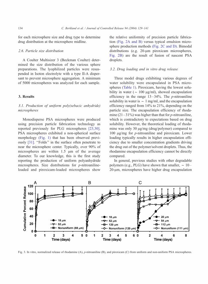
Fig. 3. In vitro, normalized release of rhodamine (A), p-nitroaniline (B), an
the relative uniformity of precision particle fabrica-
tion (Fig. 2A and B) versus typical emulsion micro-
sphere production methods (Fig. 2C and D). Bimodal
distributions (e.g. 20-Am piroxicam microspheres,
Fig. 2B) are the result of fusion of nascent PSA
droplets.
3.2. Drug loading and in vitro drug release
Three model drugs exhibiting various degrees of
water solubility were encapsulated in PSA micro-
spheres (Table 1). Piroxicam, having the lowest solu-
bility in water (f 100 Ag/ml), showed encapsulation
efficiency in the range 13–34%. The p-nitroaniline
solubility in water isf 1 mg/ml, and the encapsulation
efficiency ranged from 14% to 21%, depending on the
particle size. The encapsulation efficiency of rhoda-
mine (21–31%) was higher than that for p-nitroaniline,
which is contradictory to expectations based on drug
solubility. However, the theoretical loading of rhoda-
mine was only 30 Ag/mg (drug/polymer) compared to
100 Ag/mg for p-nitroaniline and piroxicam. Lower
loading typically results in higher encapsulation effi-
ciency due to smaller concentration gradients driving
the drug out of the polymer/solvent droplets. Thus, the
rhodamine encapsulation efficiency cannot be directly
compared.
In general, previous studies with other degradable
polymers (e.g., PLG) have shown that smaller,f 10–
20-Am, microspheres have higher drug encapsulation
d piroxicam (C) from uniform and non-uniform PSA microspheres.

C. Berkland et al. / Journal of Controlled Release 94 (2004) 129–141 135
efficiency than larger microspheres, probably due to
rapid precipitation of these small microspheres [22].
Also, larger f 80- to 120-Am microspheres tend to
Fig. 4. Scanning laser confocal fluorescence micrographs revealing the
microspheres (A and B), 18-, 52- and 120-Am p-nitroaniline-loaded m
microspheres (F–H). Scale bar = 50 Am (same magnification in all image
encapsulate drugmore efficiently than ‘‘medium’’-size,
f 50-Am microspheres. The dependence of encapsu-
lation efficiency on particle size is most likely the result
distribution of drug in uniform 18- and 52-Am rhodamine-loaded
icrospheres (C–E), and 20-, 54- and 112-Am piroxicam-loaded
s).

Fig. 5. Scanning electron micrographs of uniform 18- and 52-Am rhodamine-loaded microspheres (A and B, respectively), 18- and 52-Am p-
nitroaniline-loaded microspheres (C and D, respectively), and 20- and 54-Am piroxicam-loaded microspheres (E and F, respectively). Scale
bar = 10 Am (scale bar in E applies to A and C; scale bar in F applies to B and D).
C. Berkland et al. / Journal of Controlled Release 94 (2004) 129–141136

C. Berkland et al. / Journal of Controlled Release 94 (2004) 129–141 137
of competition between (1) decreased diffusion of drug
out of the particle as size increases, which tends to
increase encapsulation efficiency, and (2) decreased
rate of entrapment of drug due to slower ‘‘hardening’’
of the particles as size increases, which tends to
decrease encapsulation efficiency. Thus, there is a
minimum in encapsulation efficiency for particles near
50-Am in diameter where the rate of hardening is
significantly slower than in 10-Am particles, allowing
time for drug diffusion out of the particles, and the rate
of diffusional loss is still faster than in the larger
particles. The results reported here, as well as previous
Fig. 6. Scanning electron micrographs of the surface structure of unif
respectively), 18- and 52-Am p-nitroaniline-loaded microspheres (C and D
(E and F, respectively). Arrows indicate drug crystals. Scale bar = 3 Am
studies [30], are consistent with these hypotheses. Non-
uniform microspheres exhibited the highest drug en-
capsulation efficiency, possibly due to the presence of
many small, < 10-Am diameter, microspheres and
faster polymer precipitation due to rapid stirring rates
in the non-solvent hardening bath.
The effect of microsphere size on drug release was
very different for the three compounds (Fig. 3).
Rhodamine release was rapid and complete within
3 days. The profiles exhibited a smooth concave
downward shape that was independent of micro-
sphere size. p-Nitroaniline release profiles were sim-
orm 18- and 52-Am rhodamine-loaded microspheres (A and B,
, respectively), and 20- and 54-Am piroxicam-loaded microspheres
(same magnification in all panels).

C. Berkland et al. / Journal of Contro138
ilar to rhodamine with a longer duration of release.
Again, no distinction could be made between micro-
spheres of different diameter. However, p-nitroaniline
release appeared to be biphasic, exhibiting a drug
burst followed by a period of slower release. Pirox-
icam release, in contrast, varied significantly with
microsphere size. Large, uniform (f 112 Am) and
non-uniform (average = 111 Am) microspheres re-
leased drug rapidly, similar to drug-release profiles
described for rhodamine and p-nitroaniline. Interme-
diate, 54-Am microspheres showed a burst of pirox-
icam followed by a 5-day ‘‘lag’’ during which little
drug was released and a more rapid release phase
between days 5 and 8. Finally, the 20-Am micro-
spheres exhibited a much smaller burst followed by a
shorter lag and a 5- to 6-day period of near zero-order
release. It is clear from these data that microsphere
size alone does not determine the drug release profile.
Fig. 7. Scanning electron micrographs of the interior structure of unifo
piroxicam-loaded microspheres (C and D). Arrows indicate drug crystals. S
applies to D).
3.3. Distribution of drugs within the polymer matrix
The distribution of drugs within the microspheres
was examined via laser scanning confocal microsco-
py (Fig. 4). Rhodamine appears to be distributed
preferentially to the surface of the 18- and 52-Ammicrospheres. p-Nitroaniline distributed more evenly
throughout the smallest microspheres, but was local-
ized increasingly toward the microsphere surface as
the size increased. Similarly, piroxicam distributed to
the microsphere surface in large (f 112-Am) micro-
spheres and was rather evenly distributed in the 20-Ammicrospheres. The grainy appearance of the fluores-
cence may be indicative of the larger drug crystals
present in p-nitroaniline and piroxicam-loaded micro-
spheres (vide infra). The ‘‘lines’’ of drug apparently
running through the center of 52-Am p-nitroaniline-
loaded and 54-Am piroxicam-loaded microspheres are
lled Release 94 (2004) 129–141
rm 18-Am rhodamine-loaded microspheres (A and B) and 20-Amcale bar = 3 Am (scale bar in A also applies to C; scale bar in B also

C. Berkland et al. / Journal of Controlled Release 94 (2004) 129–141 139
the result of folds in the microsphere surface (see Fig.
1) and actually represent surface localized drug.
Scanning electron micrographs confirm the distri-
bution of drug in microspheres and provide a compar-
ison of microsphere morphology amongst the different
drug formulations. Rhodamine-loaded microspheres
exhibit a folded and somewhat porous microsphere
surface for both 18-Am (Fig. 5A) and 52-Am micro-
spheres (Fig. 5B) as well as a large amount of surface
localized drug crystals. The size and density of rhoda-
mine crystals on the surface appear to be similar for
both microsphere sizes (Fig. 6A and B). Folds are less
apparent in 18-Am (Fig. 5C) and 52-Am (Fig. 5D) p-
nitroaniline-loaded microspheres. However, larger
drug crystals are evident, especially in 52-Am micro-
spheres (compare Fig. 6C and D). Piroxicam-loaded
microspheres also exhibit larger crystals and a crack-
like pore structure for 20-Am (Fig. 5E) and 54-Am (Fig.
5F) microspheres. The density of piroxicam crystals on
the microsphere surface is lower compared to the p-
nitroaniline or rhodamine crystals (compare Fig. 6E
and F to A, B, C and D).
Scanning electron micrographs of fractured micro-
spheres reveal dense interior structures, and the
presence or absence of drug crystals can be visual-
ized. A cross-section of an 18-Am rhodamine-loaded
microsphere shows no evidence of rhodamine inside
the microsphere (Fig. 7A and B). However, a cross-
section of a 20-Am piroxicam-loaded microsphere
reveals large, f 1- to 3-Am drug crystals in the
microsphere interior confirming the presence of drug
throughout the microsphere matrix (Fig. 7C and D).
4. Discussion
Drug delivery applications utilizing biodegradable
polymeric microspheres are becoming an important
means of delivering therapeutic agents since micro-
spheres offer facile, noninvasive administration via
injection, protection of encapsulated drug and sus-
tained drug release over periods from hours to months
or even years [32,33]. In the past, many factors have
been adjusted to produce a desired drug release profile
including microsphere fabrication conditions, which
usually affect the resulting microsphere size distribu-
tion [6,34], and polymer chemistry (e.g., polymer
composition, co-monomer ratios, molecular weight,
etc.) [9,35]. Defining a set of these formulation param-
eters offers a degree of reproducibility, but the precise
mechanisms affecting drug release kinetics can be
difficult to determine. Here, we have utilized a tech-
nology for precision particle fabrication to more accu-
rately investigate factors influencing drug release. By
examining uniform microspheres over a size range
suitable for subcutaneous or intramuscular drug depots
(f 10–120 Am), we are able to see differences in
encapsulation efficiency, drug distribution within the
microsphere matrix and, most importantly, drug release
kinetics, which would be difficult to ascertain from a
non-uniform microsphere population.
Several different drug types exemplifying various
degrees of solubility in water were encapsulated in
uniform and non-uniform PSA microspheres. The
encapsulation efficiency of these drugs appeared to
increase slightly as drug solubility in water de-
creased. Polyanhydrides are relatively hydrophobic
polymers [4,11]. Therefore, improved incorporation
of drugs having low water solubility may be ex-
pected. Additionally, the relative hydrophobicity of
the drug determined the distribution of drug within
the microspheres. Water-soluble rhodamine (f 8 mg/
ml) exhibited a strong preference toward the micro-
sphere surface while the relatively water insoluble
piroxicam (f 100 Ag/ml) remained more evenly
distributed within the hydrophobic microsphere ma-
trix, especially for small (20-Am), rapidly precipitat-
ing microspheres (Fig. 4).
The effect of drug distribution, as controlled by
microsphere size, was manifested in the drug re-
lease profiles. Surface-localized crystals, evident in
scanning electron micrographs (Fig. 5), may be
responsible for the rapid initial release of rhoda-
mine and p-nitroaniline. The surface localization of
rhodamine and p-nitroaniline for all microsphere
sizes, and for piroxicam in large (>50-Am) micro-
spheres, further explains the rapid initial drug
release and short duration of release for these
formulations. Hydrophilic drugs, especially organic
bases, are also known to increase the rate of
polymer degradation [36], which may further ex-
plain the rapid release of rhodamine. In addition,
polymer-soluble drugs like p-nitroaniline can de-
crease the crystallinity of PSA, increasing the
polymer erosion rate [20]. This effect may also
contribute to the rapid release of p-nitroaniline.

C. Berkland et al. / Journal of Controlled Release 94 (2004) 129–141140
Piroxicam release appears to be controlled primarily
by the distribution of drug within the polymer matrix
(Fig. 4). Surface-localized piroxicam was not as prev-
alent as rhodamine or p-nitroaniline (Fig. 6), and
piroxicam was present throughout the polymer matrix
(Fig. 7C and D). Laser scanning confocal microscopy
confirmed an increasingly uniform drug distribution as
microsphere size decreased, probably due to faster
precipitation of the smaller nascent PSA drops. Uni-
form drug distribution throughout the 20-Am micro-
sphere matrix resulted in nearly zero-order piroxicam
release controlled by polymer degradation. Further, the
20-Am piroxicam microsphere formulation was the
only one that sustained drug release during the life of
the microspheres [37], which supports the supposition
that drug is distributed throughout the microsphere
matrix and not just on the surface. The complex
piroxicam release profile generated by 54-Am micro-
spheres may best be explained as a combination of
release of surface localized drug, as in the f 112-Ampiroxicam-loaded microspheres, plus controlled re-
lease of drug distributed to the microsphere interior,
as in the 20-Am microspheres.
Several factors are desirable to create an improved
PSA microsphere system capable of zero-order re-
lease. Encapsulating drug as a suspension of solids
or increasing polymer solution viscosity by increas-
ing the initial polymer concentration or molecular
weight would decrease drug mobility improving
encapsulation efficiency and reducing surface local-
ization of drug. More rapid phase inversion of
nascent polymer droplets by rapid stirring or in-
creased volume of non-solvent would also minimize
redistribution of encapsulated drug. Each of these
strategies may be most efficiently investigated by
producing uniform microspheres to avoid the con-
founding effects of broad size distributions.
5. Conclusions
Precision particle fabrication technology can be
used to create monodisperse PSA microspheres of a
desired size suitable for subcutaneous or intramuscular
injection. As a direct result, different microsphere sizes
were found to exhibit various degrees of surface
localization of drug according to the relative solubility
of the drug in water and the precipitation rate of nascent
polymer droplets. Water-soluble compounds, rhoda-
mine and p-nitroaniline, localized preferentially to the
microsphere surface, resulting in rapid drug release for
only a few days. For piroxicam-loaded microspheres,
the particle size dictated the release profile, albeit in a
surprising manner. If the drug is evenly distributed in
the microspheres, the release duration should increase
as the particle size increases. However, we showed here
that the opposite trend is obtained when microsphere
precipitation kinetics play an important role in deter-
mining drug distribution. In this study, the smallest,
most rapidly precipitating microspheres (20-Am) had
the most homogeneous drug distribution and thus the
most extended release profile, while the largest par-
ticles released the fastest. This study emphasizes the
importance of the interplay between the drug water
solubility (suggestive of ‘‘compatibility’’ with the
highly non-polar polymer matrix), microsphere size
and polymer precipitation kinetics on the drug distri-
bution within and release from surface-eroding poly-
mer microspheres. A more thorough understanding of
all of the factors governing drug release kinetics is
essential for precise design of controlled-release devi-
ces based on this and similar polymer systems.
Acknowledgements
We thank Roshelle Silverman for assistance with
drug release studies. This work was supported by
grants from the Whitaker Foundation, Roy S. Carver
Trust and Iowa State University Office of Biotech-
nology to BN.
References
[1] M. Chiba, J. Hanes, R. Langer, Controlled protein delivery
from biodegradable tyrosine-containing poly(anhydride-co-
imide) microspheres, Biomaterials 18 (1997) 893–901.
[2] E. Mathiowitz, D. Kline, R. Langer, Morphology of polyan-
hydride microsphere delivery systems, Scanning Microsc. 4
(1990) 329–340.
[3] E. Mathiowitz, W.M. Saltzman, A. Domb, R. Langer, Polyan-
hydride microspheres as drug carriers: II. Microencapsulation
by solvent removal, J. Appl. Polym. Sci. 35 (1988) 755–774.
[4] J. Tamada, R. Langer, The development of polyanhydrides for
drug delivery applications, J. Biomater. Sci., Polym. Ed. 3
(1992) 315–353.
[5] I.J. Castellanos, W.O. Cuadrado, K. Griebenow, Prevention of

C. Berkland et al. / Journal of Controlled Release 94 (2004) 129–141 141
structural perturbations and aggregation upon encapsulation of
bovine serum albumin into poly(lactide-co-glycolide) micro-
pheres using the solid-in-oil-in water technique, J. Pharm.
Pharmacol. 53 (2001) 1099–1107.
[6] S. Cohen, T. Yoshika, M. Lucarelli, L.H. Hwang, R. Langer,
Controlled delivery systems for proteins based on poly(lactic/
glycolic acid) microspheres, Pharm. Res. 8 (1991) 713–720.
[7] R. Narayani, K. Panduranga Rao, Gelatin microsphere cock-
tails of different sizes for the controlled release of anticancer
drugs, Int. J. Pharm. 143 (1996) 255–258.
[8] P. Sansdrap, A.J. Moes, In vitro evaluation of the hydrolytic
degradation of dispersed and aggregated poly(DL-lactide-co-
glycolide) microspheres, J. Control. Release 43 (1997) 47–58.
[9] J.M. Bezemer, R. Radersma, D.W. Grijpma, P.J. Dijkstra, C.A.
van Blitterswijk, J. Feijen, Microspheres for protein delivery
prepared from amphiphilic multiblock copolymers: 2. Modu-
lation of release rate, J. Control. Release 67 (2000) 249–260.
[10] M.A. Tracy, K.L. Ward, L. Firouzabadian, Y. Wang, N. Dong,
R. Qian, Y. Zhang, Factors affecting the degradation rate of
poly(lactide-co-glycolide) microspheres in vivo and in vitro,
Biomaterials 20 (1999) 1057–1062.
[11] A. Gopferich, Mechanisms of polymer degradation and ero-
sion, Biomaterials 17 (1996) 103–114.
[12] E. Shen, M.J. Kipper, B. Dziadul, M.K. Lim, B. Narasim-
han, Mechanistic relationships between polymer microstruc-
ture and drug release kinetics in bioerodible polyanhydrides,
J. Control. Release 82 (2002) 115–125.
[13] E. Mathiowitz, J.S. Jacob, Y.S. Jong, G.P. Carino, D.E. Chick-
ering, P. Chaturvedi, C.A. Santos, K. Vijayaraghavan, S.
Montgomery, M. Bassett, C. Morrell, Biologically erodable
microspheres as potential oral drug delivery systems, Nature
386 (1997) 410–414.
[14] N. Peppas, R. Langer, New challenges in biomaterials, Science
263 (1994) 1715–1720.
[15] R. Langer, New methods of drug delivery, Science 249 (1990)
1527–1533.
[16] K.W. Leong, B.C. Brott, R. Langer, Bioerodible polyanhy-
drides as drug-carrier matrices: I. Characterization, degrada-
tion, and release characteristics, J. Biomed. Mater. Res. 19
(1985) 941–955.
[17] K.W. Leong, P. D’Amore, M. Marletta, R. Langer, Bioerodi-
ble polyanhydrides as drug-carrier matrices: II. Biocompati-
bility and chemical reactivity, J. Biomed. Mater. Res. 20
(1986) 51–64.
[18] E. Mathiowitz, R. Langer, Polyanhydride microspheres as
drug carriers: I. Hot melt microencapsulation, J. Control. Re-
lease 5 (1987) 13–22.
[19] M. Chasin, D.D. Lewis, R. Langer, Polyanhydrides for con-
trolled drug delivery, Biopharm Manuf., (1988) 33–39.
[20] E. Shen, R. Pizsczek, B. Dziadul, B. Narasimhan, Microphase
separation in bioerodible copolymers for drug delivery, Bio-
materials 22 (2001) 201–210.
[21] C. Berkland, K. Kim, D.W. Pack, Precision polymer micro-
particles for controlled release drug delivery, ACS Sympo-
sium Series (2003) (in press).
[22] C. Berkland, K. Kim, D.W. Pack, PLG microsphere size con-
trols drug release rate through several competing factors,
Pharm. Res. 20 (2003) 1055–1062.
[23] C. Berkland, K. Kim, D.W. Pack, Fabrication of PLG micro-
spheres with precisely controlled and monodisperse size dis-
tributions, J. Control. Release 73 (2001) 59–74.
[24] C.A. Foster, K. Kim, R.J. Turnbull, C.D. Hendricks, Appara-
tus for producing uniform solid spheres of hydrogen, Rev. Sci.
Instrum. 48 (1977) 625–631.
[25] R.P. Gilliard, K. Kim, R.J. RTurnball, Spherical hydrogen
pellet generator for magnetic confinement fusion research,
Rev. Sci. Instrum. 52 (1981) 183–190.
[26] C.D. Hendricks, K. Kim, Interaction of a stream of dielectric
spheres in an electric field in a high vacuum, IEEE Trans. Ind.
Appl. IA-21 (1985) 705–708.
[27] J.E. Kirwan, T.A. Lee, G.N. Schroering, H. Krier, J.E. Peters,
J.P. Renie, K. Kim, An experimental and theoretical study of
a monodisperse spray, AIAA J. Propuls. Power 4 (1988)
299–307.
[28] N.K. Kim, K. Kim, D.A. Payne, R.S. Upadhye, Fabrication of
hollow silica aerogel spheres by a droplet generation method
and sol –gel processing, J. Vac. Sci. Technol., A 7 (1989)
1181–1184.
[29] K. Kim, Fabrication of glass micro- and nanospheres from
liquid precursors using droplet generation and sol–gel pro-
cessing, Mater. Res. Soc. Symp. Proc. 372 (1995) 25–32.
[30] C. Berkland, M. King, A. Cox, K. Kim, D.W. Pack, Precise
control of PLG microsphere size provides enhanced control of
drug release rate, J. Control. Release 82 (2002) 137–147.
[31] M.J. Kipper, E. Shen, A. Determan, B. Narasimhan, Design of
an injectable system based on bioerodible polyanhydride mic-
rospheres for sustained drug delivery, Biomaterials 23 (2002)
4405–4412.
[32] D.L. Wise, D.J. Trantolo, R.T. Marino, J.P. Kitchell, Oppor-
tunities and challenges in the design of implantable biodegra-
dable polymeric systems for the delivery of anti-microbial
agents and vaccines, Adv. Drug Deliv. Rev. 1 (1987) 19–39.
[33] B.H. Woo, J.W. Kostanski, S. Gebrekidan, B.A. Dani, B.C.
Thanoo, P.P. DeLuca, Preparation, characterization and in vivo
evaluation of 120-day poly(DL-lactide) leuprolide micro-
spheres, J. Control. Release 75 (2001) 307–315.
[34] J. Akbuga, Effect of microsphere size and formulation factors
on drug release from controlled-release furosemide micro-
spheres, Drug Devel. Ind. Pharm. 17 (1991) 593–607.
[35] Y. Men, C. Thomasin, H.P. Merkle, B. Gander, G. Corradin, A
single administration of tetanus toxoid in biodegradable mic-
rospheres elicits T cell and antibody responses similar or supe-
rior to those obtained with aluminum hydroxide, Vaccine 13
(1995) 683–689.
[36] J. Wang, B.M. Wang, S.P. Schwendeman, Characterization of
the initial burst release of a model peptide from poly(DL-lac-
tide-co-glycolide) microspheres, J. Control. Release 82 (2002)
289–307.
[37] D.L. McCann, F. Heatley, A. D’Emanuele, Characterization of
chemical structure and morphology of eroding polyanhydride
copolymer by liquid-state and solid-state 1H NMR, Polymer
40 (1999) 2151–2232.