Ultra-high ordered, centimeter scale preparation of microsphere Langmuir films
7
Ultra-high ordered, centimeter scale preparation of microsphere Langmuir films L.N. Deepak Kallepalli, C. Constantinescu ⇑ , P. Delaporte, O. Utéza, D. Grojo Aix Marseille Université/CNRS, Laboratoire LP3 (UMR CNRS 7341), F-13288 Marseille, France graphical abstract article info Article history: Received 17 October 2014 Accepted 16 January 2015 Available online 2 February 2015 Keywords: Langmuir–Blodgett Monolayer Microspheres Silica Polystyrene Photonics abstract Controlling the preparation of nano/microsphere monolayers on large areas remains a difficult task but is crucial for several fabrication methods of highly-ordered periodic nanostructures. We demonstrate the preparation of ordered monolayers of few square centimeters with an extremely high coverage ratio (>98%) by implementing a modified protocol (MP) Langmuir Blodgett (LB) technique. We use octadecyl type hydrocarbon (C18) functionalized spherical particles (polystyrene and silica) with diameters in the range 1–5 lm, and a selected mixture of solvents for accurate control of the surface tension and par- ticles’ mobility at the water surface. This leads to a delicate growth of crystal-like monolayers which are subsequently transferred to glass or silicon substrates. While operating the Langmuir–Blodgett trough, a key enabling the quality enhancement resides not only on surface tension measurements but also on sim- ple visual inspections of the water surface supporting the monolayer. The protocol yields a strong reduc- tion of sensitivity to thermodynamical and mechanical disturbances leading to a robust method that could be automated by adding a feedback on the operated system based real-time image processing. A simple analytical approach is used to explain why this MP-LB technique is more appropriate in growing micrometric-sized objects in comparison to standard protocols optimized for the preparation of molecu- lar films. Ó 2015 Elsevier Inc. All rights reserved. 1. Introduction Langmuir–Blodgett (LB) films are a set of monolayers, or layers made of various compounds/materials that are one molecule/par- ticle thick, grown on a solid substrate [1,2]. By transferring mono- layers of various compounds/materials from a liquid to a solid substrate, the structure of the film can be controlled at the mole- cules/particles level [3–5]. Methods to generate and/or deposit self-assembled monolayers of hexagonally close-packed (HCP) of micro- and nanospheres include spin coating, dip coating, drop casting, thermoelectrically cooled angle coating, electrophoretic deposition, assembly at air/liquid and liquid/liquid interface, http://dx.doi.org/10.1016/j.jcis.2015.01.047 0021-9797/Ó 2015 Elsevier Inc. All rights reserved. ⇑ Corresponding author. E-mail addresses: [email protected] (L.N.D. Kallepalli), constantinescu@ lp3.univ-mrs.fr (C. Constantinescu), [email protected] (D. Grojo). Journal of Colloid and Interface Science 446 (2015) 237–243 Contents lists available at ScienceDirect Journal of Colloid and Interface Science www.elsevier.com/locate/jcis
Transcript of Ultra-high ordered, centimeter scale preparation of microsphere Langmuir films

Journal of Colloid and Interface Science 446 (2015) 237–243
Contents lists available at ScienceDirect
Journal of Colloid and Interface Science
www.elsevier .com/locate / jc is
Ultra-high ordered, centimeter scale preparation of microsphereLangmuir films
http://dx.doi.org/10.1016/j.jcis.2015.01.0470021-9797/� 2015 Elsevier Inc. All rights reserved.
⇑ Corresponding author.E-mail addresses: [email protected] (L.N.D. Kallepalli), constantinescu@
lp3.univ-mrs.fr (C. Constantinescu), [email protected] (D. Grojo).
L.N. Deepak Kallepalli, C. Constantinescu ⇑, P. Delaporte, O. Utéza, D. GrojoAix Marseille Université/CNRS, Laboratoire LP3 (UMR CNRS 7341), F-13288 Marseille, France
g r a p h i c a l a b s t r a c t
a r t i c l e i n f o
Article history:Received 17 October 2014Accepted 16 January 2015Available online 2 February 2015
Keywords:Langmuir–BlodgettMonolayerMicrospheresSilicaPolystyrenePhotonics
a b s t r a c t
Controlling the preparation of nano/microsphere monolayers on large areas remains a difficult task but iscrucial for several fabrication methods of highly-ordered periodic nanostructures. We demonstrate thepreparation of ordered monolayers of few square centimeters with an extremely high coverage ratio(>98%) by implementing a modified protocol (MP) Langmuir Blodgett (LB) technique. We use octadecyltype hydrocarbon (C18) functionalized spherical particles (polystyrene and silica) with diameters inthe range 1–5 lm, and a selected mixture of solvents for accurate control of the surface tension and par-ticles’ mobility at the water surface. This leads to a delicate growth of crystal-like monolayers which aresubsequently transferred to glass or silicon substrates. While operating the Langmuir–Blodgett trough, akey enabling the quality enhancement resides not only on surface tension measurements but also on sim-ple visual inspections of the water surface supporting the monolayer. The protocol yields a strong reduc-tion of sensitivity to thermodynamical and mechanical disturbances leading to a robust method thatcould be automated by adding a feedback on the operated system based real-time image processing. Asimple analytical approach is used to explain why this MP-LB technique is more appropriate in growingmicrometric-sized objects in comparison to standard protocols optimized for the preparation of molecu-lar films.
� 2015 Elsevier Inc. All rights reserved.
1. Introduction
Langmuir–Blodgett (LB) films are a set of monolayers, or layersmade of various compounds/materials that are one molecule/par-
ticle thick, grown on a solid substrate [1,2]. By transferring mono-layers of various compounds/materials from a liquid to a solidsubstrate, the structure of the film can be controlled at the mole-cules/particles level [3–5]. Methods to generate and/or depositself-assembled monolayers of hexagonally close-packed (HCP) ofmicro- and nanospheres include spin coating, dip coating, dropcasting, thermoelectrically cooled angle coating, electrophoreticdeposition, assembly at air/liquid and liquid/liquid interface,

238 L.N.D. Kallepalli et al. / Journal of Colloid and Interface Science 446 (2015) 237–243
convective assembly, and even laser-induced forward transfer(LIFT) [6–23]. Such deposition methods require that spheres candiffuse freely across the substrates, seeking their lowest energyconfiguration. This is often achieved by chemically modifying thesurface of spheres with a negatively charged functional group, i.e.carboxylate (R-COO�) or sulfate (SO4
2�), thus making them electro-statically repelled by the negatively charged surface of a substratesuch as quartz, glass or silicon. Capillary forces draw the spherestogether, packing them in a tight hexagonally pattern on the sub-strates, as the solvent (water or alcohol) evaporates.
Today, the trend in nanoscience and nanotechnology is to real-ize structures that are smaller than the wavelength of light. How-ever, fabrication of such structures by using conventional opticallithography is limited by the diffraction limit and the wavelengthof the light used [24,25]. Photonic nanojet lithography is one solu-tion to fabricate structures below the wavelength of the light used.This technology relies on the principle of optical near-fieldenhancement underneath a monolayer of nano- or micro-spheresthat can lead to laser spots with size well below the wavelength,while the structures thereby obtained have features at the nano-meter scale. Thus, it is possible to micro- and/or nanostructurethe substrates of choice using a sphere-assisted photonic nanojetlithography technique [26–32]. These sub-diffraction lithographytechniques find applications in areas related to optical devices,spectroscopy, optical microscopy, data storage, and imaging[27–29,33]. Another alternative using microspheres is the so called‘‘colloidal lithography’’ technique, where sphere monolayers areused as shadow masks in standard physical depositions. Afterremoving the colloidal mask, ordered quasi-triangular metalprisms remain on the substrate, which can be further processed(hollowed or dewetted into dots/spheres) by adequate thermal orlaser annealing [6,7].
Here, we report on a modified protocol for the Langmuir–Blodg-ett (MP-LB) technique, developed for the preparation of monolay-ers of particles. In recent work, we have shown that suchmonolayers are compatible with photonic applications [34,35],and in this paper we present results and discuss on monolayerquality improvement. An important point in this paper is to high-light the utility of such monolayers with ease preparation and highcoverage ratio, over large area and with high stability, reliable forthe final laser engineered photonic structures and throughout theoverall fabrication process.
2. Langmuir–Blodgett films
Although the LB deposition process was originally developed formolecules [1–5], it can also be extended to nanoparticles andmicroparticles. The LB technique provides one of the few methodsof preparing organized assemblies of monolayers, which are pre-requisite in various applications where high-accuracy spatiallyresolved methods are needed [33]. Such monolayers made withtransparent microspheres find applications in the structuration ofbulk substrates, resulting in either nanohole or nanobump arrays[34–39], but have also been explored in the literature in the con-text of electronics, optics, microlithography, chemical sensors,and biosensors [32–41]. It has been demonstrated that properlycoated nanoparticles can be organized at the air–water interfaceby using the same procedure. The corresponding films possessstriking similarities to those observed for classical molecular Lang-muir monolayers. However, one must be aware that the coating/functionalization of the nanoparticles is an important factor inthe ability of such particles to form high quality monolayers. Iftheir hydrophobicity is too low, the particles float randomly atthe surface of water or sink (if density is higher than that of water),whereas the particles tend to stick to unwanted aggregates if the
coating is too hydrophobic [34,36]. Solid to super-liquid phasetransitions in Langmuir monolayers are related to the long-rangerepulsive interactions in the case of molecules, by stabilizing theundulating phases at thermodynamic equilibrium. Several theoret-ical and experimental approaches discuss this aspect, in two limits:close to the liquid–gas critical point via a Ginzburg–Landau expan-sion of the free energy (and within a mean-field approximation),and at low temperatures by free energy minimization [42–47].However, when dealing with microscale objects, i.e. spheres rang-ing between several hundred of nm and up to tens of lm, the sur-face tension at the water–air interface is of extreme importance inreaching free energy minimization of the particles and monolayer,as it will be further shown.
3. The deposition process: standard vs. modified protocol
LB films are a good example of what it is called ‘‘supramolecularassembly’’. In principle, the LB deposition method simply consistsof dipping and pulling a pair of solid substrates (e.g. quartz or boro-silicate Schott (BK7) glass slides, silicon wafers, etc) through thecoating monolayer, while controlling certain parameters, and thusproviding accurate control over the monolayer structure. The LBtechnique requires water insoluble substances or particles, but sol-uble in volatile solvents (i.e. ethanol, chloroform, etc). Commonly,LB compatible materials consist of two fundamental parts: thehead which is hydrophilic and the tail which is hydrophobic – typ-ically a long aliphatic chain (i.e. C18). These structures are referredto as amphiphiles [48]. When designing such level units at themicroscale (e.g. microspheres), their density becomes importantin LB experiments. As we will see later on, C18 functionalizedmicrospheres can then be used to prepare monolayers with theLB method. As presented in Fig. 1, the LB technique consists of atrough, Wilhelmy plate that records surface pressure [4,5], andtwo barriers that control the density (coverage ratio) of the Lang-muir film. The conventional way to prepare LB films is to dis-solve/suspend the molecules/particles of interest in their suitablesolvents, then spread them in a Langmuir film on water subphasein the LB tank, with the help of a syringe. The solvents evaporateand/or mix quickly while the surfactant molecules spread overthe subphase surface, as shown in Fig. 1. First, the LB tank is filledwith water till the meniscus is observed at the barriers. The nextstep is to record surface pressure, i.e. area per molecule isothermcurve, when compressing the barriers [2,47]. The molecules/parti-cles are required to be compressed in order to obtain a high densityLangmuir film. The amount of energy per unit area required isreferred to as surface tension; the process also depends on aspectssuch as: nature of liquid, surrounding medium, and temperature[49]. To prevent the collapse of molecules/particles with respectto each other, the reduction of surface tensions must be measuredthoroughly and throughout the process; this is also known as thesurface pressure [50–52]. The surface pressure is measured byimmersing a plate (Wilhelmy plate) across the air/water interfaceand measuring the downward force (mN/m) experienced by thisplate [53,54]. The continuing closure of the barriers increases thesurface pressure causing an additional ordering of the film, themonolayer behaving as a quasi-solid. This pseudo solid state ischaracterized by an isotherm, usually with a linear relationshipbetween surface pressure and molecular area. By compressingthe barriers the monolayer undergoes through several phases. Asthe monolayer is compressed, the pressure rises and phase changesoccur; this is referred to as two-dimensional liquid expanded (LE)state. Upon further compression of barriers, the pressure repeat-edly rises and eventually collapses, represented by sudden dipsin the curve. Hence, to get a monolayer of good quality, the surfacepressure should be selected near the steepness of the curve. In

Fig. 1. Schematic of typical Langmuir–Blodgett setup (top-right corner), and actual setup showing tank, barriers, and the substrates (middle-right). A floating monolayer of5 lm PS spheres at the air–water interface and the substrate holder with partially dipped samples, between the barriers of the LB tank: different colors appear revealingmonolayer quality (bottom-right corner). Left side caption: microscope image of grown monolayer.
L.N.D. Kallepalli et al. / Journal of Colloid and Interface Science 446 (2015) 237–243 239
addition to surface pressure, the role of compression rate is alsoworth to mention. Surfactants also play an important role, as theyallow the spreading of the deposited molecules by decreasing thesurface tension between two liquids or between a liquid and asolid [49]. As a result of this orientation at the air/water interface,surfactant molecules form a ‘‘bridge’’ between the polar and non-polar phases, thus making the transition between the two phasesless abrupt.
The LB technique directly depends on how good the monolayerwas initially spread in the water sub-phase near the substrates[55]. In our earlier works [34,35], we adopted the standard proto-col of LB film preparation and the obtained monolayers exhibitnearly 60% coverage ratio, at a surface pressure of 15 mN/m and10 mm/min compression rate as the substrates were evidentlynot completely void free. The best results obtained by the standardprotocol reveal the limit on what can be achieved for the prepara-tion of microsphere monolayers when the operation a LB systemrelies only on surface tension measurements.
In this article, we modify the protocol and add a simple criterionduring the operation. We monitor the surface quality of the mono-layer throughout the procedure, by observing visually changes atthe surface while operating the LB trough. ln fact, visible light isdiffracted from a perfectly ordered 2D arrangement of micro-spheres in a similar manner to the diffraction of X-rays from ionicor molecular crystals. Accordingly, different colours arise underwhite light illumination. Then, this effect and its uniformity atthe interface can be used to evaluate the degree of order in thesphere assembly with a macro camera or by naked eye observa-tions. These observations are used to decide when to transfer themonolayer but also if and when rearrangement of the Langmuirfilm is needed. The latter is achieved by adding drops of ethanolin the Langmuir tank to control the surface tension and thusachieve higher density packing of the spheres.
In step 1, after the spheres are dispersed on the water in the LBtank, the barriers are compressed by fixing a surface pressure value
at some high value (e.g. 60–100 mN/m), so that surface pressureread by the balance cannot actually reach it easily. When the qual-ity of the Langmuir film at the air–water interface is imaged to behomogenous/void-free, we immediately reduce the surface pres-sure value to the near immediate read of surface pressure, so thatthe barriers stop further compression of the monolayer. At thismoment, we proceed with step 2: the substrates can be pulled inorder to grow the monolayers. If any voids are monitored duringthe procedure – either at the air–water interface, in the proximityor on the substrates, then the pulling is stopped and we repeatsteps 1 and 2. Thus, by repeating these steps as needed in thecourse of one experiment, the quality of such monolayers mayreach up to 99% coverage ratio in one single pull (as demonstratedhereafter). We tested 2 � 2 cm2 and 4 � 2 cm2 sized substrates, asthe LB trough cannot hold larger substrates. It is worth mentioningthat a few groups that have succeeded in preparing PS monolayeron large surface area by using surfactants like TX 100, withoutusing LB technique [56]. Vertical surface method [57], and verticalspreading [58] methods also have been reported without using LBtechnique, but their usage is limited to large size particles (hun-dreds of lm).
4. Experimental section
We used three different types of substrates, i.e. silicon, quartzand optical quality borosilicate glass (BK7), and C18-functional-ized microbeads made of silica with diameters of 1 lm and poly-styrene (PS), with diameters of 1 lm, 2 lm, and 5 lm. Siliconsubstrates (siltronix, France), slides made in quartz (1 mm thick,Heraeus SUPRASIL 3) and in BK7 glass (1 mm thick, ThermoScientific/Gerhard Menzel GmbH) were cut into pieces(2 � 4 cm2) to fit exactly into the LB tank. The substrates werecleaned in ultrasound bath for at least 20 min, using acetoneand then isopropanol. Finally, the samples were rinsed with
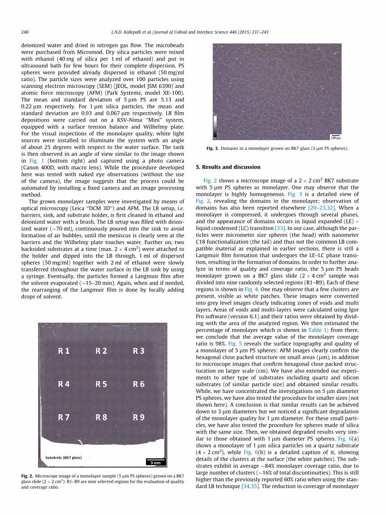
Fig. 3. Domains in a monolayer grown on BK7 glass (5 lm PS spheres).
240 L.N.D. Kallepalli et al. / Journal of Colloid and Interface Science 446 (2015) 237–243
deionized water and dried in nitrogen gas flow. The microbeadswere purchased from Micromod. Dry silica particles were mixedwith ethanol (40 mg of silica per 1 ml of ethanol) and put inultrasound bath for few hours for their complete dispersion. PSspheres were provided already dispersed in ethanol (50 mg/mlratio). The particle sizes were analyzed over 100 particles usingscanning electron microscopy (SEM) (JEOL, model JSM 6390) andatomic force microscopy (AFM) (Park Systems, model XE-100).The mean and standard deviation of 5 lm PS are 5.13 and0.22 lm respectively. For 1 lm silica particles, the mean andstandard deviation are 0.93 and 0.067 lm respectively. LB filmdepositions were carried out on a KSV-Nima ‘‘Mini’’ system,equipped with a surface tension balance and Wilhelmy plate.For the visual inspections of the monolayer quality, white lightsources were installed to illuminate the system with an angleof about 25 degrees with respect to the water surface. The tankis then observed in an angle of view similar to the image shownin Fig. 1 (bottom right) and captured using a photo camera(Canon 400D, with macro lens). While the procedure developedhere was tested with naked eye observations (without the useof the camera), the image suggests that the process could beautomated by installing a fixed camera and an image processingmethod.
The grown monolayer samples were investigated by means ofoptical microscopy (Leica ‘‘DCM 3D’’) and AFM. The LB setup, i.e.barriers, sink, and substrate holder, is first cleaned in ethanol anddeionized water with a brush. The LB setup was filled with deion-ized water (�70 ml), continuously poured into the sink to avoidformation of air bubbles, until the meniscus is clearly seen at thebarriers and the Wilhelmy plate touches water. Further on, twobacksided substrates at a time (max. 2 � 4 cm2) were attached tothe holder and dipped into the LB through. 1 ml of dispersedspheres (50 mg/ml) together with 2 ml of ethanol were slowlytransferred throughout the water surface in the LB sink by usinga syringe. Eventually, the particles formed a Langmuir film afterthe solvent evaporated (�15–20 min). Again, when and if needed,the rearranging of the Langmuir film is done by locally addingdrops of solvent.
Fig. 2. Microscope image of a monolayer sample (5 lm PS spheres) grown on a BK7glass slide (2 � 2 cm2): R1–R9 are nine selected regions for the evaluation of qualityand coverage ratio.
5. Results and discussion
Fig. 2 shows a microscope image of a 2 � 2 cm2 BK7 substratewith 5 lm PS spheres as monolayer. One may observe that themonolayer is highly homogeneous. Fig. 3 is a detailed view ofFig. 2, revealing the domains in the monolayer; observation ofdomains has also been reported elsewhere [20–23,32]. When amonolayer is compressed, it undergoes through several phases,and the appearance of domains occurs in liquid expanded (LE) –liquid condensed (LC) transition [33]. In our case, although the par-ticles were micrometer size spheres (the head) with nanometerC18 functionalization (the tail) and thus not the common LB com-patible material as explained in earlier sections, there is still aLangmuir film formation that undergoes the LE–LC phase transi-tion, resulting in the formation of domains. In order to further ana-lyze in terms of quality and coverage ratio, the 5 lm PS beadsmonolayer grown on a BK7 glass slide (2 � 4 cm2 sample wasdivided into nine randomly selected regions (R1–R9). Each of theseregions is shown in Fig. 4. One may observe that a few clusters arepresent, visible as white patches. These images were convertedinto grey level images clearly indicating zones of voids and multilayers. Areas of voids and multi-layers were calculated using IgorPro software (version 6.1) and their ratios were obtained by divid-ing with the area of the analyzed region. We then estimated thepercentage of monolayer which is shown in Table 1; from there,we conclude that the average value of the monolayer coverageratio is 98%. Fig. 5 reveals the surface topography and quality ofa monolayer of 5 lm PS spheres: AFM images clearly confirm thehexagonal close packed structure on small areas (lm), in additionto microscope images that confirm hexagonal close packed struc-turation on larger scale (cm). We have also extended our experi-ments to other type of substrates including quartz and siliconsubstrates (of similar particle size) and obtained similar results.While, we have concentrated the investigations on 5 lm diameterPS spheres, we have also tested the procedure for smaller sizes (notshown here). A conclusion is that similar results can be achieveddown to 3 lm diameters but we noticed a significant degradationof the monolayer quality for 1 lm diameter. For these small parti-cles, we have also tested the procedure for spheres made of silicawith the same size. Then, we obtained degraded results very sim-ilar to those obtained with 1 lm diameter PS spheres. Fig. 6(a)shows a monolayer of 1 lm silica particles on a quartz substrate(4 � 2 cm2), while Fig. 6(b) is a detailed caption of it, showingdetails of the clusters at the surface (the white patches). The sub-strates exhibit in average �84% monolayer coverage ratio, due tolarge number of clusters (�16% of total discontinuities). This is stillhigher than the previously reported 60% ratio when using the stan-dard LB technique [34,35]. The reduction in coverage of monolayer
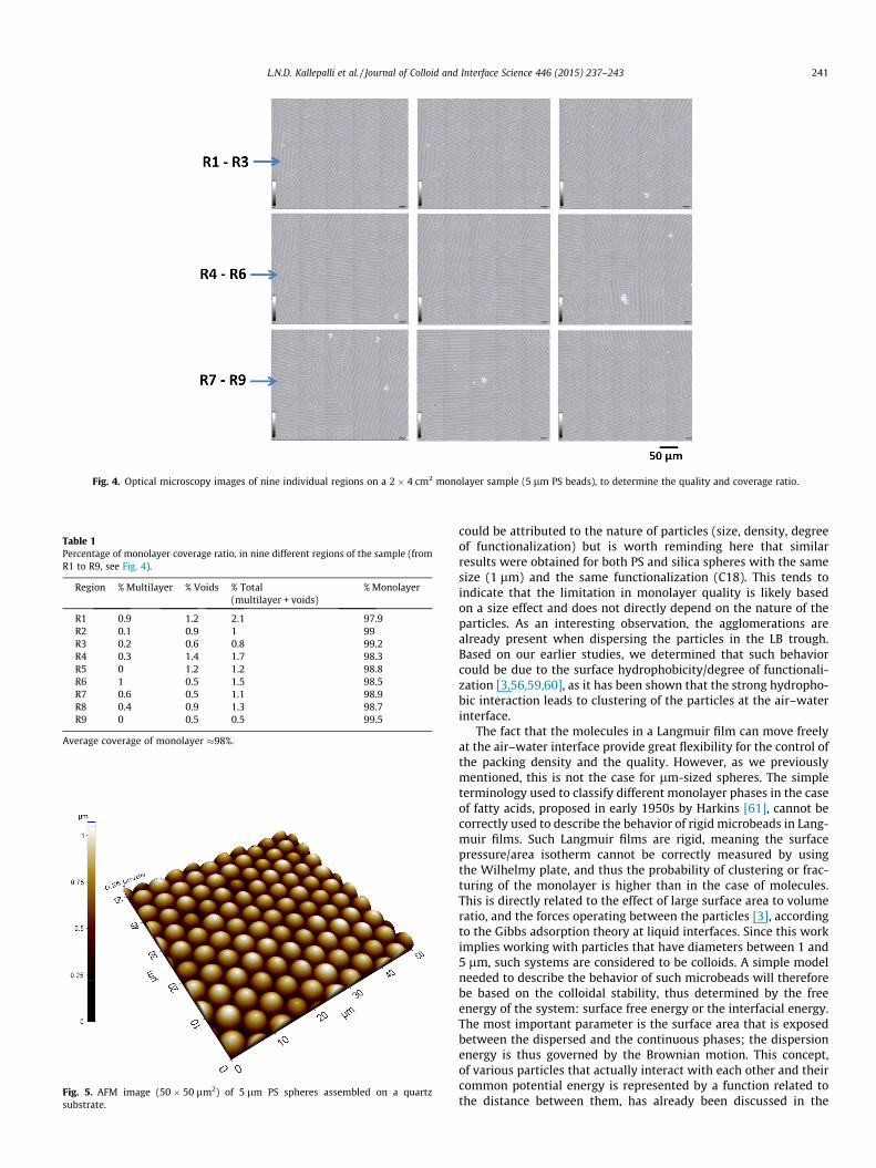
Fig. 4. Optical microscopy images of nine individual regions on a 2 � 4 cm2 monolayer sample (5 lm PS beads), to determine the quality and coverage ratio.
Table 1Percentage of monolayer coverage ratio, in nine different regions of the sample (fromR1 to R9, see Fig. 4).
Region % Multilayer % Voids % Total(multilayer + voids)
% Monolayer
R1 0.9 1.2 2.1 97.9R2 0.1 0.9 1 99R3 0.2 0.6 0.8 99.2R4 0.3 1.4 1.7 98.3R5 0 1.2 1.2 98.8R6 1 0.5 1.5 98.5R7 0.6 0.5 1.1 98.9R8 0.4 0.9 1.3 98.7R9 0 0.5 0.5 99.5
Average coverage of monolayer �98%.
Fig. 5. AFM image (50 � 50 lm2) of 5 lm PS spheres assembled on a quartzsubstrate.
L.N.D. Kallepalli et al. / Journal of Colloid and Interface Science 446 (2015) 237–243 241
could be attributed to the nature of particles (size, density, degreeof functionalization) but is worth reminding here that similarresults were obtained for both PS and silica spheres with the samesize (1 lm) and the same functionalization (C18). This tends toindicate that the limitation in monolayer quality is likely basedon a size effect and does not directly depend on the nature of theparticles. As an interesting observation, the agglomerations arealready present when dispersing the particles in the LB trough.Based on our earlier studies, we determined that such behaviorcould be due to the surface hydrophobicity/degree of functionali-zation [3,56,59,60], as it has been shown that the strong hydropho-bic interaction leads to clustering of the particles at the air–waterinterface.
The fact that the molecules in a Langmuir film can move freelyat the air–water interface provide great flexibility for the control ofthe packing density and the quality. However, as we previouslymentioned, this is not the case for lm-sized spheres. The simpleterminology used to classify different monolayer phases in the caseof fatty acids, proposed in early 1950s by Harkins [61], cannot becorrectly used to describe the behavior of rigid microbeads in Lang-muir films. Such Langmuir films are rigid, meaning the surfacepressure/area isotherm cannot be correctly measured by usingthe Wilhelmy plate, and thus the probability of clustering or frac-turing of the monolayer is higher than in the case of molecules.This is directly related to the effect of large surface area to volumeratio, and the forces operating between the particles [3], accordingto the Gibbs adsorption theory at liquid interfaces. Since this workimplies working with particles that have diameters between 1 and5 lm, such systems are considered to be colloids. A simple modelneeded to describe the behavior of such microbeads will thereforebe based on the colloidal stability, thus determined by the freeenergy of the system: surface free energy or the interfacial energy.The most important parameter is the surface area that is exposedbetween the dispersed and the continuous phases; the dispersionenergy is thus governed by the Brownian motion. This concept,of various particles that actually interact with each other and theircommon potential energy is represented by a function related tothe distance between them, has already been discussed in the

Fig. 6. (a) Optical microscopy images of 1 lm silica particles monolayers(2 � 4 cm2) on quartz in, and (b) random highlighted zone, showingagglomerations.
242 L.N.D. Kallepalli et al. / Journal of Colloid and Interface Science 446 (2015) 237–243
literature [3,4]. The surface tension (c) is defined as the free energyexcess per unit area, or the surface energy minus the surfaceentropy. The attraction between two portions of a fluid decreaseswith distance, and may be considered as equal to zero at a thresh-old distance, i.e. the range of molecular action. The surface tension(c) acts tangentially to the interfacial area, and is equal to:
c ¼Zðpex � ptÞdz
where pex is the external pressure, pt is the tangential pressure, andz is the normal to the air–water interface plane. In our case, theinterface is made of a Langmuir film of C18 functionalized micro-spheres, that are several orders of magnitude larger than the watermolecules and typical fatty acids. Since the silica or PS microsphereswe use are intrinsically rigid, and the self-assembled Langmuir filmof HCP of microspheres is also very stable, any gap (missing sphere)or discontinuity (due to size dispersion of the spheres) in the
structure of the film triggers the formation of domains in order toachieve minimum energy, as a (meta)stable phase. This can be fur-ther corrected by adding drops of ethanol in the Langmuir tank, inorder to control the surface tension and thus the mobility of themicrospheres, achieving higher density packing of the spheres.When the film is then transferred to a substrate, during the LBdeposition procedure, any domain frontier among the microbeadsmay favor the appearing of a tear due to localized variations inthe surface pressure, as the Langmuir film behaves as a quasi-solidsheet. This behavior is similar to cleavage in crystalline materials, asthe grown film actually splits along a definite pseudo-crystallo-graphic structural plane. However, these fractures can simply becorrected by further compressing the barriers of the setup immedi-ately after they are observed, by using the modified protocoldescribed in this paper.
6. Conclusion
Results on large scale (cm2) preparation of monolayers by mod-ified Langmuir–Blodgett technique, i.e. repetitive steps of compres-sion and pulling the substrate, are presented. We discussed on thequality of the monolayer coverage, i.e. more than 98% ratio on2 � 4 cm2 substrates, when using this technique for 5 lm polysty-rene spheres. We presented the qualitative and quantitative meth-ods that were adopted to characterize the homogeneity andcoverage ratio of the monolayers, and the advantages of the mod-ified protocol over usual standard protocol. Finally, we underlinethat growing self-assembled films of high homogeneity over largearea, as it was demonstrated here, is a gateway towards successfulstructuring of materials using microsphere-assisted methods.
Acknowledgments
The authors thank to the French ANR agency for financial sup-port through the FELINS-ANR-10-BLAN-946. Part of this researchhas also received funding from the French PACA (Provence-Alpes-Cote d’Azur) Regional Council (Grant APEX2012 NANO-LIFT).
References
[1] M.C. Petty, Langmuir–Blodgett Films. An Introduction, Cambridge UniversityPress, UK, 1996.
[2] G.G. Roberts, Langmuir–Blodgett Films, Plenum Press, New York, USA, 1990.[3] K.S. Birdi, Handbook of Surface and Colloid Chemistry, third ed., CRC Press,
Taylor & Francis Group, Boca Raton, USA, 2009, ISBN 978-0-8493-7327-5.[4] K. Holmberg, Handbook of Applied Surface and Colloid Chemistry, Wiley and
Sons, New York, USA, 2001, ISBN 978-0-471-49083-8.[5] H.J. Butt, G. Karlheinz, M. Kappl, Physics and Chemistry of Interfaces, Wiley-
VCH-Verlag, Weinheim, 2006, ISBN 9783527406296.[6] C.L. Haynes, R.P. Van Duyne, J. Phys. Chem. B 105 (2001) 5599–5611.[7] K. Kempa, B. Kimball, J. Rybczynski, Z.P. Huang, P.F. Wu, D. Steeves, M. Sennett,
M. Giersig, D.V.G.L.N. Rao, D.L. Carnahan, D.Z. Wang, J.Y. Lao, W.Z. Li, Z.F. Ren,Nano Lett. 3 (2003) 13–18.
[8] N.D. Denkov, D. Velev, P.A. Kralchevsky, I.B. Ivanov, H. Yoshimura, K.Nagayama, Langmuir 8 (1992) 3183–3190.
[9] M. Trau, S. Sankaran, D.A. Saville, I.A. Aksay, Nature 374 (1995) 437–439.[10] A.E. Larsen, D.G. Grier, Phys. Rev. Lett. 76 (1996) 3862–3865.[11] R. Micheletto, H. Fukuda, M. Ohtsu, Langmuir 11 (1995) 3333–3336.[12] Y. Lin, H. Skaff, T. Emrick, A.D. Dinsmore, T.P. Russell, Science 299 (2003) 226–
229.[13] B.G. Prevo, O.D. Velev, Langmuir 20 (2004) 2099–2107.[14] D. Wang, H. Möhwald, Adv. Mater. 16 (2004) 244–247.[15] A. Kosiorek, W. Kandulski, P. Chudzinski, K. Kempa, M. Giersig, Nano Lett. 4
(2004) 1359–1363.[16] L. Malaquin, T. Kraus, H. Schmid, E. Delamarche, H. Wolf, Langmuir 23 (2007)
11513–11521.[17] B.G. Jung, S.H. Min, C.W. Kwon, S.H. Park, K.B. Kim, T.S. Yoon, J. Electrochem.
Soc. 156 (2009) K86–K90.[18] A. Palla-Papavlu, V. Dinca, I. Paraico, A. Moldovan, J. Shaw-Stewart, C.W.
Schneider, E. Kovacs, T. Lippert, M. Dinescu, J. Appl. Phys. 108 (2010) 033111.[19] A. Palla-Papavlu, V. Dinca, C. Luculescu, J. Shaw-Stewart, M. Nagel, T. Lippert,
M. Dinescu, J. Opt. 12 (2010) 124014.
L.N.D. Kallepalli et al. / Journal of Colloid and Interface Science 446 (2015) 237–243 243
[20] N. Vogel, S. Goerres, K. Landfester, C.K. Weiss, Macromol. Chem. Phys. 212(2011) 1719–1734.
[21] P. Born, S. Blum, A. Munoz, T. Kraus, Langmuir 27 (2011) 8621–8633.[22] J. Perlich, M. Schwartzkopf, V. Körstgens, D. Erb, J.F.H. Risch, P.M. Buschbaum,
R. Röhlsberger, S.V. Roth, R. Gehrke, Phys. Status Solidi 6 (2012) 253–255.[23] T. Kraus, D. Brodoceanu, N. Pazos-Perez, A. Fery, Adv. Funct. Mater. 23 (2013)
4529–4541.[24] A.V. Itagi, W.A. Challener, J. Opt. Soc. Am. A 22 (2005) 2847–2858.[25] P.N. Prasad, Nanophotonics, Wiley-IEEE, 2004. ISBN: 0-471-64988-O.[26] P. Jiang, T. Prasad, M.J. McFarland, V.L. Colvin, Appl. Phys. Lett. 89 (2006)
011908.[27] J. Wenger, D. Gerard, H. Aouani, H. Rigneault, Anal. Chem. 80 (2008) 6800–
6804.[28] S.H. Sun, C.B. Murray, Dieter Weller, Lies Folks, Andreas Moser, Science 287
(2000) 1989–1992.[29] J.J. Schwartz, S. Stavrakis, S.R. Quake, Nat. Nanotechnol. 5 (2010) 127–132.[30] A. Pereira, D. Grojo, M. Chaker, P. Delaporte, D. Guay, M. Sentis, Small 4 (2008)
572–576.[31] Y. Zhou, M.H. Hong, F. Fuh, L. Lu, B.S. Luk’yanchuck, Z.B. Wang, L.P. Shi, T.C.
Chong, Appl. Phys. Lett. 88 (2006) 023110.[32] Y. Shen, L.V. Wang, J.T. Shen, Opt. Lett. 39 (2014) 4120–4123.[33] D. Grojo, N. Sandeau, L. Boarino, C. Constantinescu, N. De Leo, M. Laus, K.
Sparnacci, Opt. Lett. 39 (2014) 3989–3992.[34] L.N. Kallepalli, D. Grojo, L. Charmasson, P. Delaporte, O. Uteza, A. Merlen, A.
Sangar, P. Torchio, J. Phys. D: Appl. Phys. 46 (2013) 145102.[35] A. Merlen, A. Sangar, P. Torchio, L.N.D. Kallepalli, D. Grojo, O. Utéza, P.
Delaporte, Appl. Surf. Sci. 284 (2013) 545–548.[36] S.M. Huang, Z. Sun, B.S. Luk’yanchuk, M.H. Hong, L.P. Shi, Appl. Phys. Lett. 86
(2005) 161911 (3 pp).[37] W. Cai, R. Piestum, Appl. Phys. Lett. 88 (2006) 111112.[38] D. Grojo, A. Cros, P. Delaporte, M. Sentis, Appl. Surf. Sci. 253 (2007) 8309–8315.[39] D. Grojo, L. Charmasson, A. Pereira, M. Sentis, P. Delaporte, J. Nanosci.
Nanotechnol. 11 (2011) 9129–9135.
All in-text references underlined in blue are linked to publications on Rese
[40] A.V. Kabashin, P. Delaporte, A. Pereira, D. Grojo, R. Torres, M. Sentis, NanoscaleRes. Lett. 5 (2010) 454–463.
[41] D. Grojo, A. Cros, P. Delaporte, M. Sentis, Appl. Phys. B 84 (2006) 517–521.[42] D. Andelman, F. Broçhard, J.F. Joanny, J. Chem. Phys. 86 (1987) 3673–3681.[43] S. Karaborni, S. Toxvaerd, J. Chem. Phys. 96 (1992) 5505–5515.[44] D.R. Swanson, R.J. Hardy, C.J. Eckhardt, J. Chem. Phys. 99 (1993) 8194–8199.[45] V.M. Kaganer, E.B. Loginov, Phys. Rev. E 51 (1995) 2237–2249.[46] M.D. Gibson, D.R. Swanson, C.J. Eckhardt, X.C. Zeng, J. Chem. Phys. 106 (1997)
1961–1966.[47] V.M. Kaganer, H. Mohwald, P. Dutta, Rev. Modern Phys. 71 (1999) 779–819.[48] T. Richardsson, M.C. Petty, M.R. Bryce, D. Bloor, Introduction to Molecular
Electronics, Oxford University Press, New York, USA, 1995 (Chapter 10, pp.221–242).
[49] Edgar J. Cabrera, Sabine Portal, Esther Pascual, Enric Bertran, in: InternationalConference on Manipulation, Manufacturing and Measurement on theNanoscale (3M-NANO), 2012, pp. 28–33.
[50] P. Dynarowicz, Adv. Colloid Interface Sci. (45) (1993) 215–241.[51] J. Mingins, N.F. Owens, Thin Solid Films 152 (1987) 9–28.[52] M. Vandevyver, A. Barraud, J. Mol. Electron. 4 (1998) 207–214.[53] M. Máte, J.H. Fendler, J.R. Ramsden, J. Szalma, Z. Horvolgyi, Langmuir 14 (1998)
6501–6504.[54] P.B. Welzel, I. Weis, G. Schwarz, Colloids Surf. A 144 (1998) 229–234.[55] Kim Su II., F. Pradal, H. Song, S. Kim, J. Vac. Sci. Technol. B 29 (2011) 021804.[56] Z. Lu, M. Zhou, J. Colloid Interface Sci. 361 (2011) 429–435.[57] F. Pan, J. Zhang, C. Cai, T. Wang, Langmuir 22 (2006) 7101–7104.[58] J.T. Zhang, L. Wang, X. Chao, S.S. Velankar, S.A. Asher, J. Mater. Chem. C 1
(2013) 6099–6102.[59] M. Szekeres, O. Kamalin, R.A. Schoonheydt, K. Wostyn, K. Clays, A. Persoons, I.
Dekany, J. Mater. Chem. 12 (2002) 3268–3274.[60] M. Shishido, D. Kitagawa, Colloids Surf. A: Physicochem. Eng. Aspects 311
(2007) 32–41.[61] W.D. Harkins, The Physical Chemistry of Surface Films, Reinhold, New York,
USA, 1952.
archGate, letting you access and read them immediately.