TO TREATMENT - Review of Ophthalmology
68
reviewofophthalmology.com August 2016 reviewofophthalmology.com August 2016 ANNUAL RETINA ISSUE SURGICAL VIDEOGRAPHY ON THE CHEAP P. 20 • INSIDIOUS CAUSES OF DME P. 45 QUANTIFYING EXTERNAL DISEASE SIGNS P. 48 • GLAUCOMA: REFINING TRABECULAR BYPASS P. 52 WILLS EYE RESIDENT CASE SERIES P. 63 • TACKLING PRE-LASIK DRY EYE P. 56 CUTTING-EDGE APPROACHES TO TREATMENT • First-line Therapy for Wet AMD P. 24 • The Best Treatment Protocols for DME P. 32 • The Evolution of Retinal Vein Occlusion Therapy P. 38 • Reusing Anti-VEGF Drug Vials P. 42
-
Upload
khangminh22 -
Category
Documents
-
view
0 -
download
0
Transcript of TO TREATMENT - Review of Ophthalmology

reviewofophthalmology.com
August 2016
reviewofophthalmology.com
August 2016
ANNUAL RETINA ISSUE
Review
of Oph
thalm
ology Vol. X
XIII, N
o. 8 • Au
gust 2016 • F
irst-line W
et AM
D Treatm
ent • D
iabetic M
acular E
dema P
rotocols • BR
VO
/CR
VO
Th
erapies • Su
rgical Videograph
y
SURGICAL VIDEOGRAPHY ON THE CHEAP P. 20 • INSIDIOUS CAUSES OF DME P. 45
QUANTIFYING EXTERNAL DISEASE SIGNS P. 48 • GLAUCOMA: REFINING TRABECULAR BYPASS P. 52
WILLS EYE RESIDENT CASE SERIES P. 63 • TACKLING PRE-LASIK DRY EYE P. 56
CUTTING-EDGE APPROACHES
TO TREATMENT • First-line Therapy for
Wet AMD P. 24
• The Best TreatmentProtocols for DME P. 32
• The Evolution of Retinal Vein Occlusion Therapy P. 38
• Reusing Anti-VEGFDrug Vials P. 42
001_rp0716_fc.indd 1001_rp0716_fc.indd 1 7/22/16 1:32 PM7/22/16 1:32 PM

Visit www.HumiraPro.com to learn more about our education programs for NI uveitis.*
*Intermediate, posterior, and panuveitis.
SERIOUS INFECTIONSPatients treated with HUMIRA are at increased risk for developing serious infections that may lead to hospitalization or death. Most patients who developed these infections were taking concomitant immunosuppressants such as methotrexate or corticosteroids.
Discontinue HUMIRA if a patient develops a serious infection or sepsis.
Reported infections include:
Active tuberculosis (TB), including reactivation of latent TB. Patients with TB have frequently presented with disseminated or extrapulmonary disease. Test patients for latent TB before HUMIRA use and during therapy. Initiate treatment for latent TB prior to HUMIRA use.
Invasive fungal infections, including histoplasmosis, coccidioidomycosis, candidiasis, aspergillosis, blastomycosis, and pneumocystosis. Patients with histoplasmosis or other invasive fungal infections may present with disseminated, rather than localized, disease. Antigen and antibody testing for histoplasmosis may be negative in some patients with active infection. Consider empiric anti-fungal therapy in patients at risk for invasive fungal infections who develop severe systemic illness.
Bacterial, viral, and other infections due to opportunistic pathogens, including Legionella and Listeria.
Carefully consider the risks and benefits of treatment with HUMIRA prior to initiating therapy in patients: 1. with chronic or recurrent infection, 2. who have been exposed to TB, 3. with a history of opportunistic infection, 4. who resided in or traveled in regions where mycoses are endemic, 5. with underlying conditions that may predispose them to infection. Monitor patients closely for the development of signs and symptoms of infection during and after treatment with HUMIRA, including the possible development of TB in patients who tested negative for latent TB infection prior to initiating therapy.
Do not start HUMIRA during an active infection, including localized infections.
Patients older than 65 years, patients with co-morbid conditions, and/or patients taking concomitant immunosuppressants may be at greater risk of infection.
If an infection develops, monitor carefully and initiate appropriate therapy.
Drug interactions with biologic products: A higher rate of serious infections has been observed in rheumatoid arthritis patients treated with rituximab who received subsequent treatment with a TNF blocker. Concurrent use of HUMIRA with biologic DMARDs (e.g., anakinra or abatacept) or other TNF blockers is not recommended based on the possible increased risk for infections and other potential pharmacological interactions.
MALIGNANCYLymphoma and other malignancies, some fatal, have been reported in children and adolescent patients treated with TNF blockers, including HUMIRA. Postmarketing cases of hepatosplenic T-cell lymphoma (HSTCL), a rare type of T-cell lymphoma, have been reported in patients treated with TNF blockers, including HUMIRA. These cases have had a very aggressive disease course and have been fatal. The majority of reported TNF blocker cases have occurred in patients with Crohn’s disease or ulcerative colitis and the majority were in adolescent and young adult males. Almost all of these patients had received treatment with azathioprine or 6-mercaptopurine concomitantly with a TNF blocker at or prior to diagnosis. It is uncertain whether the occurrence of HSTCL is related to use of a TNF blocker or a TNF blocker in combination with these other immunosuppressants.
Consider the risks and benefits of HUMIRA treatment prior to initiating or continuing therapy in a patient with known malignancy.
In clinical trials, more cases of malignancies were observed among HUMIRA-treated patients compared to control patients.
Non-melanoma skin cancer (NMSC) was reported during clinical trials for HUMIRA-treated patients. Examine all patients, particularly those with a history of prolonged immunosuppressant or PUVA therapy, for the presence of NMSC prior to and during treatment with HUMIRA.
In HUMIRA clinical trials, there was an approximate 3-fold higher rate of lymphoma than expected in the general U.S. population. Patients with chronic inflammatory diseases, particularly those with highly active disease and/or chronic exposure to immunosuppressant therapies, may be at higher risk of lymphoma than the general population, even in the absence of TNF blockers.
IMPORTANT SAFETY INFORMATION FOR HUMIRA® (adalimumab)1
HUMIRA is administered by subcutaneous injection
New Indication. New Dosing Regimen.
INITIAL DOSE
40 mgFOLLOWED BY
80 mg given every other week starting 1 week after the initial dose
The fi rst injection should be given under the supervision of a healthcare professional. A patient may self-inject HUMIRA after appropriate training and monitoring by a healthcare professional.
Indication¹Uveitis: HUMIRA is indicated for the treatment of non-infectious intermediate, posterior, and panuveitis in adult patients.
RP0816_Abbvie.indd 2RP0816_Abbvie.indd 2 7/21/16 3:47 PM7/21/16 3:47 PM

NOW APPROVED
HUMIRA for NI intermediate, posterior, and panuveitis* A steroid-sparing option proven to prolong time to a combination of disease fl are† and decrease of visual acuity.1
Postmarketing cases of acute and chronic leukemia were reported with TNF blocker use. Approximately half of the postmarketing cases of malignancies in children, adolescents, and young adults receiving TNF blockers were lymphomas; other cases included rare malignancies associated with immunosuppression and malignancies not usually observed in children and adolescents.
HYPERSENSITIVITYAnaphylaxis and angioneurotic edema have been reported following HUMIRA administration. If a serious allergic reaction occurs, stop HUMIRA and institute appropriate therapy.
HEPATITIS B VIRUS REACTIVATIONUse of TNF blockers, including HUMIRA, may increase the risk of reactivation of hepatitis B virus (HBV) in patients who are chronic carriers. Some cases have been fatal.
Evaluate patients at risk for HBV infection for prior evidence of HBV infection before initiating TNF blocker therapy.
Exercise caution in patients who are carriers of HBV and monitor them during and after HUMIRA treatment.
Discontinue HUMIRA and begin antiviral therapy in patients who develop HBV reactivation. Exercise caution when resuming HUMIRA after HBV treatment.
NEUROLOGIC REACTIONSTNF blockers, including HUMIRA, have been associated with rare cases of new onset or exacerbation of central nervous system and peripheral demyelinating diseases, including multiple sclerosis, optic neuritis, and Guillain-Barré syndrome.
Exercise caution when considering HUMIRA for patients with these disorders; discontinuation of HUMIRA should be considered if any of these disorders develop.
There is a known association between intermediate uveitis and central demyelinating disorders.
HEMATOLOGIC REACTIONSRare reports of pancytopenia, including aplastic anemia, have been reported with TNF blockers. Medically significant cytopenia has been infrequently reported with HUMIRA.
Consider stopping HUMIRA if significant hematologic abnormalities occur.
CONGESTIVE HEART FAILUREWorsening or new onset congestive heart failure (CHF) may occur; exercise caution and monitor carefully.
AUTOIMMUNITYTreatment with HUMIRA may result in the formation of autoantibodies and, rarely, in development of a lupus-like syndrome. Discontinue treatment if symptoms of a lupus-like syndrome develop.
IMMUNIZATIONSPatients on HUMIRA should not receive live vaccines.
Pediatric patients, if possible, should be brought up to date with all immunizations before initiating HUMIRA therapy.
The safety of administering live or live-attenuated vaccines in infants exposed to HUMIRA in utero is unknown. Risks and benefits should be considered prior to vaccinating (live or live-attenuated) exposed infants.
ADVERSE REACTIONSThe most common adverse reactions in HUMIRA clinical trials (>10%) were: infections (e.g., upper respiratory, sinusitis), injection site reactions, headache, and rash.
Please see Brief Summary of full Prescribing Information on the following page.
Reference: 1. HUMIRA Injection [package insert]. North Chicago, IL: AbbVie Inc.
† Disease flare is defined by an increase in 1 or more inflammatory markers: AC cells, vitreous haze, and/or development of
new chorioretinal, and/or retinal vascular lesions.
©2016 AbbVie Inc. North Chicago, IL 60064 64C-1859319 June 2016 Printed in U.S.A.
RP0816_Abbvie.indd 3RP0816_Abbvie.indd 3 7/21/16 3:47 PM7/21/16 3:47 PM

HUMIRA® (adalimumab) PROFESSIONAL BRIEF SUMMARY
CONSULT PACKAGE INSERT FOR FULL PRESCRIBING INFORMATION
WARNING: SERIOUS INFECTIONS AND MALIGNANCY
SERIOUS INFECTIONS
Patients treated with HUMIRA are at increased risk for developing serious infections that may lead to hospitalization or death [see Warnings and Precautions]. Most patients who developed these infections were taking concomitant immunosuppressants such as methotrexate or corticosteroids.
Discontinue HUMIRA if a patient develops a serious infection or sepsis.
Reported infections include:
• Active tuberculosis (TB), including reactivation of latent TB. Patients with TB have frequently presented with disseminated or extrapulmonary disease. Test patients for latent TB before HUMIRA use and during therapy. Initiate treatment for latent TB prior to HUMIRA use.
• Invasive fungal infections, including histoplasmosis, coccidioidomycosis, candidiasis, aspergillosis, blastomycosis, and pneumocystosis. Patients with histoplasmosis or other invasive fungal infections may present with disseminated, rather than localized, disease. Antigen and antibody testing for histoplasmosis may be negative in some patients with active infection. Consider empiric anti-fungal therapy in patients at risk for invasive fungal infections who develop severe systemic illness.
• Bacterial, viral and other infections due to opportunistic pathogens, including Legionella and Listeria.
Carefully consider the risks and benefits of treatment with HUMIRA prior to initiating therapy in patients with chronic or recurrent infection.
Monitor patients closely for the development of signs and symptoms of infection during and after treatment with HUMIRA, including the possible development of TB in patients who tested negative for latent TB infection prior to initiating therapy [see Warnings and Precautions and Adverse Reactions].MALIGNANCY
Lymphoma and other malignancies, some fatal, have been reported in children and adolescent patients treated with TNF blockers including HUMIRA [see Warnings and Precautions]. Post-marketing cases of hepatosplenic T-cell lymphoma (HSTCL), a rare type of T-cell lymphoma, have been reported in patients treated with TNF blockers including HUMIRA. These cases have had a very aggressive disease course and have been fatal. The majority of reported TNF blocker cases have occurred in patients with Crohn’s disease or ulcerative colitis and the majority were in adolescent and young adult males. Almost all these patients had received treatment with azathioprine or 6-mercaptopurine (6–MP) concomitantly with a TNF blocker at or prior to diagnosis. It is uncertain whether the occurrence of HSTCL is related to use of a TNF blocker or a TNF blocker in combination with these other immunosuppressants [see Warnings and Precautions].
INDICATIONS AND USAGE
Rheumatoid Arthritis
HUMIRA is indicated for reducing signs and symptoms, inducing major clinical response, inhibiting the progression of structural damage, and improving physical function in adult patients with moderately to severely active rheumatoid arthritis. HUMIRA can be used alone or in combination with methotrexate or other non-biologic disease-modifying anti-rheumatic drugs (DMARDs). Juvenile Idiopathic Arthritis
HUMIRA is indicated for reducing signs and symptoms of moderately to severely active polyarticular juvenile idiopathic arthritis in patients 2 years of age and older. HUMIRA can be used alone or in combination with methotrexate. Psoriatic Arthritis
HUMIRA is indicated for reducing signs and symptoms, inhibiting the progression of structural damage, and improving physical function in adult patients with active psoriatic arthritis. HUMIRA can be used alone or in combination with non-biologic DMARDs. Ankylosing Spondylitis
HUMIRA is indicated for reducing signs and symptoms in adult patients with active ankylosing spondylitis. Adult Crohn’s Disease
HUMIRA is indicated for reducing signs and symptoms and inducing and maintaining clinical remission in adult patients with moderately to severely active Crohn’s disease who have had an inadequate response to conventional therapy. HUMIRA is indicated for reducing signs and symptoms and inducing clinical remission in these patients if they have also lost response to or are intolerant to infliximab. Pediatric Crohn’s Disease
HUMIRA is indicated for reducing signs and symptoms and inducing and maintaining clinical remission in pediatric patients 6 years of age and older with moderately to severely active Crohn’s disease who have had an inadequate response to corticosteroids or immunomodulators such as azathioprine, 6-mercaptopurine, or methotrexate. Ulcerative Colitis
HUMIRA is indicated for inducing and sustaining clinical remission in adult patients with moderately to severely active ulcerative colitis who have had an inadequate response to immunosuppressants such as corticosteroids, azathioprine or 6-mercaptopurine (6-MP). The effectiveness of HUMIRA has not been established in patients who have lost response to or were intolerant to TNF blockers. Plaque Psoriasis
HUMIRA is indicated for the treatment of adult patients with moderate to severe chronic plaque psoriasis who are candidates for systemic therapy or phototherapy, and when other systemic therapies are medically less appropriate. HUMIRA should only be administered to patients who will be closely monitored and have regular follow-up visits with a physician [see Boxed Warning and Warnings and Precautions]. Hidradenitis Suppurativa
HUMIRA is indicated for the treatment of moderate to severe hidradenitis suppurativa.
Uveitis
HUMIRA is indicated for the treatment of non-infectious intermediate, posterior and panuveitis in adult patients. CONTRAINDICATIONS
None. WARNINGS AND PRECAUTIONS
Serious Infections
Patients treated with HUMIRA are at increased risk for developing serious infections involving various organ systems and sites that may lead to hospitalization or death [see Boxed Warning]. Opportunistic infections due to bacterial, mycobacterial, invasive fungal, viral, parasitic, or other opportunistic pathogens including aspergillosis, blastomycosis, candidiasis, coccidioidomycosis, histoplasmosis, legionellosis, listeriosis, pneumocystosis and tuberculosis have been reported with TNF blockers. Patients have frequently presented with disseminated rather than localized disease. The concomitant use of a TNF blocker and abatacept or anakinra was associated with a higher risk of serious infections in patients with rheumatoid arthritis (RA); therefore, the concomitant use of HUMIRA and these biologic products is not recommended in the treatment of patients with RA [see Warnings and Precautions and Drug Interactions]. Treatment with HUMIRA should not be initiated in patients with an active infection, including localized infections. Patients greater than 65 years of age, patients with co-morbid conditions and/or patients taking concomitant immunosuppressants (such as corticosteroids or methotrexate), may be at greater risk of infection. Consider the risks and benefits of treatment prior to initiating therapy in patients: • with chronic or recurrent infection;• who have been exposed to tuberculosis;• with a history of an opportunistic infection;• who have resided or traveled in areas of endemic tuberculosis or
endemic mycoses, such as histoplasmosis, coccidioidomycosis, or blastomycosis; or
• with underlying conditions that may predispose them to infection.TuberculosisCases of reactivation of tuberculosis and new onset tuberculosis infections have been reported in patients receiving HUMIRA, including patients who have previously received treatment for latent or active tuberculosis. Reports included cases of pulmonary and extrapulmonary (i.e., disseminated) tuberculosis. Evaluate patients for tuberculosis risk factors and test for latent infection prior to initiating HUMIRA and periodically during therapy. Treatment of latent tuberculosis infection prior to therapy with TNF blocking agents has been shown to reduce the risk of tuberculosis reactivation during therapy. Consider anti-tuberculosis therapy prior to initiation of HUMIRA in patients with a past history of latent or active tuberculosis in whom an adequate course of treatment cannot be confirmed, and for patients with a negative test for latent tuberculosis but having risk factors for tuberculosis infection. Despite prophylactic treatment for tuberculosis, cases of reactivated tuberculosis have occurred in patients treated with HUMIRA. Consultation with a physician with expertise in the treatment of tuberculosis is recommended to aid in the decision whether initiating anti-tuberculosis therapy is appropriate for an individual patient. Strongly consider tuberculosis in the differential diagnosis in patients who develop a new infection during HUMIRA treatment, especially in patients who have previously or recently traveled to countries with a high prevalence of tuberculosis, or who have had close contact with a person with active tuberculosis. MonitoringClosely monitor patients for the development of signs and symptoms of infection during and after treatment with HUMIRA, including the development of tuberculosis in patients who tested negative for latent tuberculosis infection prior to initiating therapy. Tests for latent tuberculosis infection may also be falsely negative while on therapy with HUMIRA. Discontinue HUMIRA if a patient develops a serious infection or sepsis. For a patient who develops a new infection during treatment with HUMIRA, closely monitor them, perform a prompt and complete diagnostic workup appropriate for an immunocompromised patient, and initiate appropriate antimicrobial therapy. Invasive Fungal InfectionsIf patients develop a serious systemic illness and they reside or travel in regions where mycoses are endemic, consider invasive fungal infection in the differential diagnosis. Antigen and antibody testing for histoplasmosis may be negative in some patients with active infection. Consider appropriate empiric antifungal therapy, taking into account both the risk for severe fungal infection and the risks of antifungal therapy, while a diagnostic workup is being performed. To aid in the management of such patients, consider consultation with a physician with expertise in the diagnosis and treatment of invasive fungal infections. Malignancies
Consider the risks and benefits of TNF-blocker treatment including HUMIRA prior to initiating therapy in patients with a known malignancy other than a successfully treated non-melanoma skin cancer (NMSC) or when considering continuing a TNF blocker in patients who develop a malignancy. Malignancies in AdultsIn the controlled portions of clinical trials of some TNF-blockers, including HUMIRA, more cases of malignancies have been observed among TNF-blocker-treated adult patients compared to control-treated adult patients. During the controlled portions of 39 global HUMIRA clinical trials in adult patients with rheumatoid arthritis (RA), psoriatic arthritis (PsA), ankylosing spondylitis (AS), Crohn’s disease (CD), ulcerative colitis (UC) plaque psoriasis (Ps), hidradenitis suppurativa (HS), and uveitis (UV) malignancies, other than non-melanoma (basal cell and squamous cell) skin cancer, were observed at a rate (95% confidence interval) of 0.7 (0.48, 1.03) per 100 patient-years among 7973 HUMIRA-treated patients versus a rate of 0.7 (0.41, 1.17) per 100 patient-years among 4848 control-treated patients (median duration of treatment of 4 months for HUMIRA-treated patients and 4 months for control-treated patients). In 52 global controlled and uncontrolled clinical trials of HUMIRA in adult patients with RA, PsA, AS, CD, UC, Ps, HS and UV, the most frequently observed malignancies, other than lymphoma and NMSC, were breast, colon, prostate, lung, and melanoma. The malignancies in HUMIRA-treated patients in the controlled and uncontrolled portions of the studies were similar in type and number to what would be expected in the general U.S. population according to the SEER database (adjusted for age, gender, and race).
In controlled trials of other TNF blockers in adult patients at higher risk for malignancies (i.e., patients with COPD with a significant smoking history and cyclophosphamide-treated patients with Wegener’s granulomatosis), a greater portion of malignancies occurred in the TNF blocker group compared to the control group. Non-Melanoma Skin CancerDuring the controlled portions of 39 global HUMIRA clinical trials in adult patients with RA, PsA, AS, CD, UC, Ps, HS and UV, the rate (95% confidence interval) of NMSC was 0.8 (0.52, 1.09) per 100 patient-years among HUMIRA-treated patients and 0.2 (0.10, 0.59) per 100 patient-years among control-treated patients. Examine all patients, and in particular patients with a medical history of prior prolonged immunosuppressant therapy or psoriasis patients with a history of PUVA treatment for the presence of NMSC prior to and during treatment with HUMIRA. Lymphoma and LeukemiaIn the controlled portions of clinical trials of all the TNF-blockers in adults, more cases of lymphoma have been observed among TNF-blocker-treated patients compared to control-treated patients. In the controlled portions of 39 global HUMIRA clinical trials in adult patients with RA, PsA, AS, CD, UC Ps, HS and UV, 2 lymphomas occurred among 7973 HUMIRA-treated patients versus 1 among 4848 control-treated patients. In 52 global controlled and uncontrolled clinical trials of HUMIRA in adult patients with RA, PsA, AS, CD, UC, Ps, HS and UV with a median duration of approximately 0.7 years, including 24,605 patients and over 40,215 patient-years of HUMIRA, the observed rate of lymphomas was approximately 0.11 per 100 patient-years. This is approximately 3-fold higher than expected in the general U.S. population according to the SEER database (adjusted for age, gender, and race). Rates of lymphoma in clinical trials of HUMIRA cannot be compared to rates of lymphoma in clinical trials of other TNF blockers and may not predict the rates observed in a broader patient population. Patients with RA and other chronic inflammatory diseases, particularly those with highly active disease and/or chronic exposure to immunosuppressant therapies, may be at a higher risk (up to several fold) than the general population for the development of lymphoma, even in the absence of TNF blockers. Post-marketing cases of acute and chronic leukemia have been reported in association with TNF-blocker use in RA and other indications. Even in the absence of TNF-blocker therapy, patients with RA may be at a higher risk (approximately 2-fold) than the general population for the development of leukemia. Malignancies in Pediatric Patients and Young AdultsMalignancies, some fatal, have been reported among children, adolescents, and young adults who received treatment with TNF-blockers (initiation of therapy ≤ 18 years of age), of which HUMIRA is a member [see Boxed Warning]. Approximately half the cases were lymphomas, including Hodgkin’s and non-Hodgkin’s lymphoma. The other cases represented a variety of different malignancies and included rare malignancies usually associated with immunosuppression and malignancies that are not usually observed in children and adolescents. The malignancies occurred after a median of 30 months of therapy (range 1 to 84 months). Most of the patients were receiving concomitant immunosuppressants. These cases were reported post-marketing and are derived from a variety of sources including registries and spontaneous postmarketing reports. Postmarketing cases of hepatosplenic T-cell lymphoma (HSTCL), a rare type of T-cell lymphoma, have been reported in patients treated with TNF blockers including HUMIRA [see Boxed Warning]. These cases have had a very aggressive disease course and have been fatal. The majority of reported TNF blocker cases have occurred in patients with Crohn’s disease or ulcerative colitis and the majority were in adolescent and young adult males. Almost all of these patients had received treatment with the immunosuppressants azathioprine or 6-mercaptopurine (6–MP) concomitantly with a TNF blocker at or prior to diagnosis. It is uncertain whether the occurrence of HSTCL is related to use of a TNF blocker or a TNF blocker in combination with these other immunosuppressants. The potential risk with the combination of azathioprine or 6-mercaptopurine and HUMIRA should be carefully considered. Hypersensitivity Reactions
Anaphylaxis and angioneurotic edema have been reported following HUMIRA administration. If an anaphylactic or other serious allergic reaction occurs, immediately discontinue administration of HUMIRA and institute appropriate therapy. In clinical trials of HUMIRA in adults, allergic reactions (e.g., allergic rash, anaphylactoid reaction, fixed drug reaction, non-specified drug reaction, urticaria) have been observed. Hepatitis B Virus Reactivation
Use of TNF blockers, including HUMIRA, may increase the risk of reactivation of hepatitis B virus (HBV) in patients who are chronic carriers of this virus. In some instances, HBV reactivation occurring in conjunction with TNF blocker therapy has been fatal. The majority of these reports have occurred in patients concomitantly receiving other medications that suppress the immune system, which may also contribute to HBV reactivation. Evaluate patients at risk for HBV infection for prior evidence of HBV infection before initiating TNF blocker therapy. Exercise caution in prescribing TNF blockers for patients identified as carriers of HBV. Adequate data are not available on the safety or efficacy of treating patients who are carriers of HBV with anti-viral therapy in conjunction with TNF blocker therapy to prevent HBV reactivation. In patients who develop HBV reactivation, stop HUMIRA and initiate effective anti-viral therapy with appropriate supportive treatment. The safety of resuming TNF blocker therapy after HBV reactivation is controlled is not known. Neurologic Reactions
Use of TNF blocking agents, including HUMIRA, has been associated with rare cases of new onset or exacerbation of clinical symptoms and/or radiographic evidence of central nervous system demyelinating disease, including multiple sclerosis (MS) and optic neuritis, and peripheral demyelinating disease, including Guillain-Barré syndrome. Exercise caution in considering the use of HUMIRA in patients with preexisting or recent-onset central or peripheral nervous system demyelinating disorders; discontinuation of HUMIRA should be considered if any of these disorders develop. There is a known association between intermediate uveitis and central demyelinating disorders. Hematological Reactions
Rare reports of pancytopenia including aplastic anemia have been reported with TNF blocking agents. Adverse reactions of the hematologic system, including medically significant cytopenia (e.g., thrombocytopenia, leukopenia) have been infrequently reported with HUMIRA. The causal relationship of these reports to HUMIRA remains unclear. Advise all patients to seek immediate medical attention if they develop signs and symptoms suggestive of blood dyscrasias or infection (e.g., persistent fever, bruising, bleeding, pallor) while on HUMIRA. Consider discontinuation of HUMIRA therapy in patients with confirmed significant hematologic abnormalities.
RP0816_Abbvie PI 1.indd 1RP0816_Abbvie PI 1.indd 1 7/21/16 3:55 PM7/21/16 3:55 PM

Use with Anakinra
Concurrent use of anakinra (an interleukin-1 antagonist) and another TNF-blocker, was associated with a greater proportion of serious infections and neutropenia and no added benefit compared with the TNF-blocker alone in patients with RA. Therefore, the combination of HUMIRA and anakinra is not recommended [see Drug Interactions].Heart Failure
Cases of worsening congestive heart failure (CHF) and new onset CHF have been reported with TNF blockers. Cases of worsening CHF have also been observed with HUMIRA. Exercise caution when using HUMIRA in patients who have heart failure and monitor them carefully. Autoimmunity
Treatment with HUMIRA may result in the formation of autoantibodies and, rarely, in the development of a lupus-like syndrome. If a patient develops symptoms suggestive of a lupus-like syndrome following treatment with HUMIRA, discontinue treatment [see Adverse Reactions].Immunizations
In a placebo-controlled clinical trial of patients with RA, no difference was detected in anti-pneumococcal antibody response between HUMIRA and placebo treatment groups when the pneumococcal polysaccharide vaccine and influenza vaccine were administered concurrently with HUMIRA. Patients on HUMIRA may receive concurrent vaccinations, except for live vaccines. No data are available on the secondary transmission of infection by live vaccines in patients receiving HUMIRA. It is recommended that pediatric patients, if possible, be brought up to date with all immunizations in agreement with current immunization guidelines prior to initiating HUMIRA therapy. Patients on HUMIRA may receive concurrent vaccinations, except for live vaccines. The safety of administering live or live-attenuated vaccines in infants exposed to HUMIRA in utero is unknown. Risks and benefits should be considered prior to vaccinating (live or live-attenuated) exposed infants [see Use in Specific Populations]. Use with Abatacept
In controlled trials, the concurrent administration of TNF-blockers and abatacept was associated with a greater proportion of serious infections than the use of a TNF-blocker alone; the combination therapy, compared to the use of a TNF-blocker alone, has not demonstrated improved clinical benefit in the treatment of RA. Therefore, the combination of abatacept with TNF-blockers including HUMIRA is not recommended [see Drug Interactions]. ADVERSE REACTIONS
The most serious adverse reactions described elsewhere in the labeling include the following: • Serious Infections [see Warnings and Precautions]• Malignancies [see Warnings and Precautions]Clinical Trials Experience
The most common adverse reaction with HUMIRA was injection site reactions. In placebo-controlled trials, 20% of patients treated with HUMIRA developed injection site reactions (erythema and/or itching, hemorrhage, pain or swelling), compared to 14% of patients receiving placebo. Most injection site reactions were described as mild and generally did not necessitate drug discontinuation. The proportion of patients who discontinued treatment due to adverse reactions during the double-blind, placebo-controlled portion of studies in patients with RA (i.e., Studies RA-I, RA-II, RA-III and RA-IV) was 7% for patients taking HUMIRA and 4% for placebo-treated patients. The most common adverse reactions leading to discontinuation of HUMIRA in these RA studies were clinical flare reaction (0.7%), rash (0.3%) and pneumonia (0.3%). InfectionsIn the controlled portions of the 39 global HUMIRA clinical trials in adult patients with RA, PsA, AS, CD, UC, HS and UV, the rate of serious infections was 4.3 per 100 patient-years in 7973 HUMIRA-treated patients versus a rate of 2.9 per 100 patient-years in 4848 control-treated patients. Serious infections observed included pneumonia, septic arthritis, prosthetic and post-surgical infections, erysipelas, cellulitis, diverticulitis, and pyelonephritis [see Warnings and Precautions].Tuberculosis and Opportunistic InfectionsIn 52 global controlled and uncontrolled clinical trials in RA, PsA, AS, CD, UC, Ps, HS and UV that included 24,605 HUMIRA-treated patients, the rate of reported active tuberculosis was 0.20 per 100 patient-years and the rate of positive PPD conversion was 0.09 per 100 patient-years. In a subgroup of 10,113 U.S. and Canadian HUMIRA-treated patients, the rate of reported active TB was 0.05 per 100 patient-years and the rate of positive PPD conversion was 0.07 per 100 patient-years. These trials included reports of miliary, lymphatic, peritoneal, and pulmonary TB. Most of the TB cases occurred within the first eight months after initiation of therapy and may reflect recrudescence of latent disease. In these global clinical trials, cases of serious opportunistic infections have been reported at an overall rate of 0.05 per 100 patient-years. Some cases of serious opportunistic infections and TB have been fatal [see Warnings and Precautions]. AutoantibodiesIn the rheumatoid arthritis controlled trials, 12% of patients treated with HUMIRA and 7% of placebo-treated patients that had negative baseline ANA titers developed positive titers at week 24. Two patients out of 3046 treated with HUMIRA developed clinical signs suggestive of new-onset lupus-like syndrome. The patients improved following discontinuation of therapy. No patients developed lupus nephritis or central nervous system symptoms. The impact of long-term treatment with HUMIRA on the development of autoimmune diseases is unknown. Liver Enzyme Elevations There have been reports of severe hepatic reactions including acute liver failure in patients receiving TNF-blockers. In controlled Phase 3 trials of HUMIRA (40 mg SC every other week) in patients with RA, PsA, and AS with control period duration ranging from 4 to 104 weeks, ALT elevations ≥ 3 x ULN occurred in 3.5% of HUMIRA-treated patients and 1.5% of control-treated patients. Since many of these patients in these trials were also taking medications that cause liver enzyme elevations (e.g., NSAIDS, MTX), the relationship between HUMIRA and the liver enzyme elevations is not clear. In a controlled Phase 3 trial of HUMIRA in patients with polyarticular JIA who were 4 to 17 years, ALT elevations ≥ 3 x ULN occurred in 4.4% of HUMIRA-treated patients and 1.5% of control-treated patients (ALT more common than AST); liver enzyme test elevations were more frequent among those treated with the combination of HUMIRA and MTX than those treated with HUMIRA alone. In general, these elevations did not lead to discontinuation of HUMIRA treatment. No ALT elevations ≥ 3 x ULN occurred in the open-label study of HUMIRA in patients with polyarticular JIA who were 2 to <4 years.
In controlled Phase 3 trials of HUMIRA (initial doses of 160 mg and 80 mg, or 80 mg and 40 mg on Days 1 and 15, respectively, followed by 40 mg every other week) in adult patients with CD with a control period duration ranging from 4 to 52 weeks, ALT elevations ≥ 3 x ULN occurred in 0.9% of HUMIRA-treated patients and 0.9% of control-treated patients. In the Phase 3 trial of HUMIRA in pediatric patients with Crohn’s disease which evaluated efficacy and safety of two body weight based maintenance dose regimens following body weight based induction therapy up to 52 weeks of treatment, ALT elevations ≥ 3 x ULN occurred in 2.6% (5/192) of patients, of whom 4 were receiving concomitant immunosuppressants at baseline; none of these patients discontinued due to abnormalities in ALT tests. In controlled Phase 3 trials of HUMIRA (initial doses of 160 mg and 80 mg on Days 1 and 15 respectively, followed by 40 mg every other week) in patients with UC with control period duration ranging from 1 to 52 weeks, ALT elevations ≥3 x ULN occurred in 1.5% of HUMIRA-treated patients and 1.0% of control-treated patients. In controlled Phase 3 trials of HUMIRA (initial dose of 80 mg then 40 mg every other week) in patients with Ps with control period duration ranging from 12 to 24 weeks, ALT elevations ≥ 3 x ULN occurred in 1.8% of HUMIRA-treated patients and 1.8% of control-treated patients. In controlled trials of HUMIRA (initial doses of 160 mg at Week 0 and 80 mg at Week 2, followed by 40 mg every week starting at Week 4), in subjects with HS with a control period duration ranging from 12 to 16 weeks, ALT elevations ≥ 3 x ULN occurred in 0.3% of HUMIRA-treated subjects and 0.6% of control-treated subjects. In controlled trials of HUMIRA (initial doses of 80 mg at Week 0 followed by 40 mg every other week starting at Week 1) in patients with uveitis with an exposure of 165.4 PYs and 119.8 PYs in HUMIRA-treated and control-treated patients, respectively, ALT elevations ≥ 3 x ULN occurred in 2.4% of HUMIRA-treated patients and 2.4% of control-treated patients. ImmunogenicityPatients in Studies RA-I, RA-II, and RA-III were tested at multiple time points for antibodies to adalimumab during the 6- to 12-month period. Approximately 5% (58 of 1062) of adult RA patients receiving HUMIRA developed low-titer antibodies to adalimumab at least once during treatment, which were neutralizing in vitro. Patients treated with concomitant methotrexate (MTX) had a lower rate of antibody development than patients on HUMIRA monotherapy (1% versus 12%). No apparent correlation of antibody development to adverse reactions was observed. With monotherapy, patients receiving every other week dosing may develop antibodies more frequently than those receiving weekly dosing. In patients receiving the recommended dosage of 40 mg every other week as monotherapy, the ACR 20 response was lower among antibody-positive patients than among antibody-negative patients. The long-term immunogenicity of HUMIRA is unknown. In patients with polyarticular JIA who were 4 to 17 years of age, adalimumab antibodies were identified in 16% of HUMIRA-treated patients. In patients receiving concomitant MTX, the incidence was 6% compared to 26% with HUMIRA monotherapy. In patients with polyarticular JIA who were 2 to <4 years of age or 4 years of age and older weighing <15 kg, adalimumab antibodies were identified in 7% (1 of 15) of HUMIRA-treated patients, and the one patient was receiving concomitant MTX. In patients with AS, the rate of development of antibodies to adalimumab in HUMIRA-treated patients was comparable to patients with RA. In patients with PsA, the rate of antibody development in patients receiving HUMIRA monotherapy was comparable to patients with RA; however, in patients receiving concomitant MTX the rate was 7% compared to 1% in RA. In adult patients with CD, the rate of antibody development was 3%. In pediatric patients with Crohn’s disease, the rate of antibody development in patients receiving HUMIRA was 3%. However, due to the limitation of the assay conditions, antibodies to adalimumab could be detected only when serum adalimumab levels were < 2 mcg/mL. Among the patients whose serum adalimumab levels were < 2 mcg/mL (approximately 32% of total patients studied), the immunogenicity rate was 10%. In patients with moderately to severely active UC, the rate of antibody development in patients receiving HUMIRA was 5%. However, due to the limitation of the assay conditions, antibodies to adalimumab could be detected only when serum adalimumab levels were < 2 mcg/mL. Among the patients whose serum adalimumab levels were < 2 mcg/mL (approximately 25% of total patients studied), the immunogenicity rate was 20.7%. In patients with Ps, the rate of antibody development with HUMIRA monotherapy was 8%. However, due to the limitation of the assay conditions, antibodies to adalimumab could be detected only when serum adalimumab levels were < 2 mcg/mL. Among the patients whose serum adalimumab levels were < 2 mcg/mL (approximately 40% of total patients studied), the immunogenicity rate was 20.7%. In Ps patients who were on HUMIRA monotherapy and subsequently withdrawn from the treatment, the rate of antibodies to adalimumab after retreatment was similar to the rate observed prior to withdrawal. In subjects with moderate to severe HS, the rate of anti-adalimumab antibody development in subjects treated with HUMIRA was 6.5%. However, because of the limitation of the assay conditions, antibodies to adalimumab could be detected only when serum adalimumab levels were < 2 mcg/mL. Among subjects who stopped HUMIRA treatment for up to 24 weeks and in whom adalimumab serum levels subsequently declined to < 2 mcg/mL (approximately 22% of total subjects studied), the immunogenicity rate was 28%. In patients with non-infectious uveitis, anti-adalimumab antibodies were identified in 4.8% (12/249) of patients treated with adalimumab. However, due to the limitation of the assay conditions, antibodies to adalimumab could be detected only when serum adalimumab levels were < 2 mcg/mL. Among the patients whose serum adalimumab levels were < 2 mcg/mL (approximately 23% of total patients studied), the immunogenicity rate was 21.1%. Using an assay which could measure an anti-adalimumab antibody titer in all patients, titers were measured in 39.8% (99/249) of non-infectious uveitis patients treated with adalimumab. No correlation of antibody development to safety or efficacy outcomes was observed. The data reflect the percentage of patients whose test results were considered positive for antibodies to adalimumab or titers, and are highly dependent on the assay. The observed incidence of antibody (including neutralizing antibody) positivity in an assay is highly dependent on several factors including assay sensitivity and specificity, assay methodology, sample handling, timing of sample collection, concomitant medications, and underlying disease. For these reasons, comparison of the incidence of antibodies to adalimumab with the incidence of antibodies to other products may be misleading. Other Adverse ReactionsRheumatoid Arthritis Clinical StudiesThe data described below reflect exposure to HUMIRA in 2468 patients, including 2073 exposed for 6 months, 1497 exposed for greater than one year and 1380 in adequate and well-controlled studies (Studies RA-I, RA-II,
RA-III, and RA-IV). HUMIRA was studied primarily in placebo-controlled trials and in long-term follow up studies for up to 36 months duration. The population had a mean age of 54 years, 77% were female, 91% were Caucasian and had moderately to severely active rheumatoid arthritis. Most patients received 40 mg HUMIRA every other week. Table 1 summarizes reactions reported at a rate of at least 5% in patients treated with HUMIRA 40 mg every other week compared to placebo and with an incidence higher than placebo. In Study RA-III, the types and frequencies of adverse reactions in the second year open-label extension were similar to those observed in the one-year double-blind portion. Table 1. Adverse Reactions Reported by ≥5% of Patients Treated
with HUMIRA During Placebo-Controlled Period of Pooled RA Studies (Studies RA-I, RA-II, RA-III, and RA-IV)
HUMIRA 40 mg subcutaneous
Every Other Week
Placebo
Adverse Reaction (Preferred Term) (N=705) (N=690)
Respiratory
Upper respiratory infection 17% 13%
Sinusitis 11% 9%
Flu syndrome 7% 6%
Gastrointestinal
Nausea 9% 8%
Abdominal pain 7% 4%
Laboratory Tests*
Laboratory test abnormal 8% 7%
Hypercholesterolemia 6% 4%
Hyperlipidemia 7% 5%
Hematuria 5% 4%
Alkaline phosphatase increased 5% 3%
Other
Headache 12% 8%
Rash 12% 6%
Accidental injury 10% 8%
Injection site reaction ** 8% 1%
Back pain 6% 4%
Urinary tract infection 8% 5%
Hypertension 5% 3%
* Laboratory test abnormalities were reported as adverse reactions in European trials
** Does not include injection site erythema, itching, hemorrhage, pain or swelling
Juvenile Idiopathic Arthritis Clinical StudiesIn general, the adverse reactions in the HUMIRA-treated patients in the polyarticular juvenile idiopathic arthritis (JIA) trials (Studies JIA-I and JIA-II) were similar in frequency and type to those seen in adult patients [see Warnings and Precautions and Adverse Reactions]. Important findings and differences from adults are discussed in the following paragraphs. In Study JIA-I, HUMIRA was studied in 171 patients who were 4 to 17 years of age, with polyarticular JIA. Severe adverse reactions reported in the study included neutropenia, streptococcal pharyngitis, increased aminotransferases, herpes zoster, myositis, metrorrhagia, and appendicitis. Serious infections were observed in 4% of patients within approximately 2 years of initiation of treatment with HUMIRA and included cases of herpes simplex, pneumonia, urinary tract infection, pharyngitis, and herpes zoster. In Study JIA-I, 45% of patients experienced an infection while receiving HUMIRA with or without concomitant MTX in the first 16 weeks of treatment. The types of infections reported in HUMIRA-treated patients were generally similar to those commonly seen in polyarticular JIA patients who are not treated with TNF blockers. Upon initiation of treatment, the most common adverse reactions occurring in this patient population treated with HUMIRA were injection site pain and injection site reaction (19% and 16%, respectively). A less commonly reported adverse event in patients receiving HUMIRA was granuloma annulare which did not lead to discontinuation of HUMIRA treatment. In the first 48 weeks of treatment in Study JIA-I, non-serious hypersensitivity reactions were seen in approximately 6% of patients and included primarily localized allergic hypersensitivity reactions and allergic rash. In Study JIA-I, 10% of patients treated with HUMIRA who had negative baseline anti-dsDNA antibodies developed positive titers after 48 weeks of treatment. No patient developed clinical signs of autoimmunity during the clinical trial. Approximately 15% of patients treated with HUMIRA developed mild-to-moderate elevations of creatine phosphokinase (CPK) in Study JIA-I. Elevations exceeding 5 times the upper limit of normal were observed in several patients. CPK levels decreased or returned to normal in all patients. Most patients were able to continue HUMIRA without interruption. In Study JIA-II, HUMIRA was studied in 32 patients who were 2 to <4 years of age or 4 years of age and older weighing <15 kg with polyarticular JIA. The safety profile for this patient population was similar to the safety profile seen in patients 4 to 17 years of age with polyarticular JIA. In Study JIA-II, 78% of patients experienced an infection while receiving HUMIRA. These included nasopharyngitis, bronchitis, upper respiratory tract infection, otitis media, and were mostly mild to moderate in severity. Serious infections were observed in 9% of patients receiving HUMIRA in the study and included dental caries, rotavirus gastroenteritis, and varicella. In Study JIA-II, non-serious allergic reactions were observed in 6% of patients and included intermittent urticaria and rash, which were all mild in severity. Psoriatic Arthritis and Ankylosing Spondylitis Clinical StudiesHUMIRA has been studied in 395 patients with psoriatic arthritis (PsA) in two placebo-controlled trials and in an open label study and in 393 patients with ankylosing spondylitis (AS) in two placebo-controlled studies. The safety profile for patients with PsA and AS treated with HUMIRA 40 mg every other
RP0816_Abbvie PI2.indd 1RP0816_Abbvie PI2.indd 1 7/21/16 4:02 PM7/21/16 4:02 PM

week was similar to the safety profile seen in patients with RA, HUMIRA Studies RA-I through IV. Adult Crohn’s Disease Clinical StudiesHUMIRA has been studied in 1478 adult patients with Crohn’s disease (CD) in four placebo-controlled and two open-label extension studies. The safety profile for adult patients with CD treated with HUMIRA was similar to the safety profile seen in patients with RA. Pediatric Crohn’s Disease Clinical Studies HUMIRA has been studied in 192 pediatric patients with Crohn’s disease in one double-blind study (Study PCD-I) and one open-label extension study. The safety profile for pediatric patients with Crohn’s disease treated with HUMIRA was similar to the safety profile seen in adult patients with Crohn’s disease. During the 4 week open label induction phase of Study PCD-I, the most common adverse reactions occurring in the pediatric population treated with HUMIRA were injection site pain and injection site reaction (6% and 5%, respectively). A total of 67% of children experienced an infection while receiving HUMIRA in Study PCD-I. These included upper respiratory tract infection and nasopharyngitis. A total of 5% of children experienced a serious infection while receiving HUMIRA in Study PCD-I. These included viral infection, device related sepsis (catheter), gastroenteritis, H1N1 influenza, and disseminated histoplasmosis. In Study PCD-I, allergic reactions were observed in 5% of children which were all non-serious and were primarily localized reactions. Ulcerative Colitis Clinical StudiesHUMIRA has been studied in 1010 patients with ulcerative colitis (UC) in two placebo-controlled studies and one open-label extension study. The safety profile for patients with UC treated with HUMIRA was similar to the safety profile seen in patients with RA. Plaque Psoriasis Clinical StudiesHUMIRA has been studied in 1696 subjects with plaque psoriasis (Ps) in placebo-controlled and open-label extension studies. The safety profile for subjects with Ps treated with HUMIRA was similar to the safety profile seen in subjects with RA with the following exceptions. In the placebo-controlled portions of the clinical trials in Ps subjects, HUMIRA-treated subjects had a higher incidence of arthralgia when compared to controls (3% vs. 1%). Hidradenitis Suppurativa Clinical StudiesHUMIRA has been studied in 727 subjects with hidradenitis suppurativa (HS) in three placebo-controlled studies and one open-label extension study. The safety profile for subjects with HS treated with HUMIRA weekly was consistent with the known safety profile of HUMIRA. Flare of HS, defined as ≥25% increase from baseline in abscesses and inflammatory nodule counts and with a minimum of 2 additional lesions, was documented in 22 (22%) of the 100 subjects who were withdrawn from HUMIRA treatment following the primary efficacy timepoint in two studies. Uveitis Clinical StudiesHUMIRA has been studied in 464 patients with uveitis (UV) in placebo-controlled and open-label extension studies. The safety profile for patients with UV treated with HUMIRA was similar to the safety profile seen in patients with RA. Postmarketing Experience
The following adverse reactions have been identified during post-approval use of HUMIRA. Because these reactions are reported voluntarily from a population of uncertain size, it is not always possible to reliably estimate their frequency or establish a causal relationship to HUMIRA exposure. Gastrointestinal disorders: Diverticulitis, large bowel perforations including perforations associated with diverticulitis and appendiceal perforations associated with appendicitis, pancreatitis General disorders and administration site conditions: Pyrexia Hepato-biliary disorders: Liver failure, hepatitis Immune system disorders: Sarcoidosis Neoplasms benign, malignant and unspecified (including cysts and polyps): Merkel Cell Carcinoma (neuroendocrine carcinoma of the skin) Nervous system disorders: Demyelinating disorders (e.g., optic neuritis, Guillain-Barré syndrome), cerebrovascular accident Respiratory disorders: Interstitial lung disease, including pulmonary fibrosis, pulmonary embolism Skin reactions: Stevens Johnson Syndrome, cutaneous vasculitis, erythema multiforme, new or worsening psoriasis (all sub-types including pustular and palmoplantar), alopecia Vascular disorders: Systemic vasculitis, deep vein thrombosis DRUG INTERACTIONS
Methotrexate
HUMIRA has been studied in rheumatoid arthritis (RA) patients taking concomitant methotrexate (MTX). Although MTX reduced the apparent adalimumab clearance, the data do not suggest the need for dose adjustment of either HUMIRA or MTX. Biological Products
In clinical studies in patients with RA, an increased risk of serious infections has been seen with the combination of TNF blockers with anakinra or abatacept, with no added benefit; therefore, use of HUMIRA with abatacept or anakinra is not recommended in patients with RA [see Warnings and Precautions]. A higher rate of serious infections has also been observed in patients with RA treated with rituximab who received subsequent treatment with a TNF blocker. There is insufficient information regarding the concomitant use of HUMIRA and other biologic products for the treatment of RA, PsA, AS, CD, UC, Ps, HS and UV. Concomitant administration of HUMIRA
with other biologic DMARDS (e.g., anakinra and abatacept) or other TNF blockers is not recommended based upon the possible increased risk for infections and other potential pharmacological interactions. Live Vaccines
Avoid the use of live vaccines with HUMIRA [see Warnings and Precautions].Cytochrome P450 Substrates
The formation of CYP450 enzymes may be suppressed by increased levels of cytokines (e.g., TNFα, IL-6) during chronic inflammation. It is possible for a molecule that antagonizes cytokine activity, such as adalimumab, to influence the formation of CYP450 enzymes. Upon initiation or discontinuation of HUMIRA in patients being treated with CYP450 substrates with a narrow therapeutic index, monitoring of the effect (e.g., warfarin) or drug concentration (e.g., cyclosporine or theophylline) is recommended and the individual dose of the drug product may be adjusted as needed. USE IN SPECIFIC POPULATIONS
Pregnancy
Limited clinical data are available from the Humira Pregnancy Registry. Excluding lost-to-follow-up, data from the registry reports a rate of 5.6% for major birth defects with first trimester use of adalimumab in pregnant women with rheumatoid arthritis (RA), and a rate of 7.8% and 5.5% for major birth defects in the disease-matched and non-diseased comparison groups [see Data]. Adalimumab is actively transferred across the placenta during the third trimester of pregnancy and may affect immune response in the in-utero exposed infant [see Clinical Considerations]. In an embryo-fetal perinatal development study conducted in cynomolgus monkeys, no fetal harm or malformations were observed with intravenous administration of adalimumab during organogenesis and later in gestation, at doses that produced exposures up to approximately 373 times the maximum recommended human dose (MRHD) of 40 mg subcutaneous without methotrexate [see Data].The estimated background risk of major birth defects and miscarriage for the indicated populations is unknown. In the U.S. general population, the estimated background risk of major birth defects and miscarriage in clinically recognized pregnancies is 2-4% and miscarriage is 15-20%, respectively. Clinical Considerations Fetal/Neonatal adverse reactionsMonoclonal antibodies are increasingly transported across the placenta as pregnancy progresses, with the largest amount transferred during the third trimester [see Data]. Risks and benefits should be considered prior to administering live or live-attenuated vaccines to infants exposed to HUMIRA in utero [see Use in Specific Populations]. Data Human DataIn a prospective cohort pregnancy exposure registry conducted in the U.S. and Canada between 2004 and 2013, 74 women with RA treated with adalimumab at least during the first trimester, 80 women with RA not treated with adalimumab and 218 women without RA (non-diseased) were enrolled. Excluding lost-to-follow-up, the rate of major defects in the adalimumab-exposed pregnancies (N=72), disease-matched (N=77), and non-diseased comparison groups (N=201) was 5.6%, 7.8% and 5.5%, respectively. However, this study cannot definitely establish the absence of any risk because of methodological limitations, including small sample size and non-randomized study design. Data from the Crohn’s disease portion of the study is in the follow-up phase and the analysis is ongoing. In an independent clinical study conducted in ten pregnant women with inflammatory bowel disease treated with HUMIRA, adalimumab concentrations were measured in maternal serum as well as in cord blood (n=10) and infant serum (n=8) on the day of birth. The last dose of HUMIRA was given between 1 and 56 days prior to delivery. Adalimumab concentrations were 0.16-19.7 μg/mL in cord blood, 4.28-17.7 μg/mL in infant serum, and 0-16.1 μg/mL in maternal serum. In all but one case, the cord blood level of adalimumab was higher than the maternal serum level, suggesting adalimumab actively crosses the placenta. In addition, one infant had serum levels at each of the following: 6 weeks (1.94 μg/mL), 7 weeks (1.31 μg/mL), 8 weeks (0.93 μg/mL), and 11 weeks (0.53 μg/mL), suggesting adalimumab can be detected in the serum of infants exposed in utero for at least 3 months from birth. Lactation
Risk SummaryLimited data from case reports in the published literature describe the presence of adalimumab in human milk at infant doses of 0.1% to 1% of the maternal serum level. There are no reports of adverse effects of adalimumab on the breastfed infant and no effects on milk production. The developmental and health benefits of breastfeeding should be considered along with the mother’s clinical need for HUMIRA and any potential adverse effects on the breastfed child from HUMIRA or from the underlying maternal condition. Pediatric Use
Safety and efficacy of HUMIRA in pediatric patients for uses other than polyarticular juvenile idiopathic arthritis (JIA) and pediatric Crohn’s disease have not been established. Due to its inhibition of TNFα, HUMIRA administered during pregnancy could affect immune response in the in utero-exposed newborn and infant. Data from eight infants exposed to HUMIRA in utero suggest adalimumab crosses the placenta [see Use in Specific Populations]. The clinical significance of elevated adalimumab levels in infants is unknown. The safety of administering live or live-attenuated vaccines in exposed infants is unknown. Risks and benefits should be considered prior to vaccinating (live or live-attenuated) exposed infants. Post-marketing cases of lymphoma, including hepatosplenic T-cell lymphoma and other malignancies, some fatal, have been reported among children, adolescents, and young adults who received treatment with
TNF-blockers including HUMIRA [see Boxed Warning and Warnings and Precautions]. Juvenile Idiopathic ArthritisIn Study JIA-I, HUMIRA was shown to reduce signs and symptoms of active polyarticular JIA in patients 4 to 17 years of age [see Clinical Studies]. In Study JIA-II, the safety profile for patients 2 to <4 years of age was similar to the safety profile for patients 4 to 17 years of age with polyarticular JIA [see Adverse Reactions]. HUMIRA has not been studied in patients with polyarticular JIA less than 2 years of age or in patients with a weight below 10 kg. The safety of HUMIRA in patients in the polyarticular JIA trials was generally similar to that observed in adults with certain exceptions [see Adverse Reactions]. Pediatric Crohn’s DiseaseThe safety and effectiveness of HUMIRA for reducing signs and symptoms and inducing and maintaining clinical remission have been established in pediatric patients 6 years of age and older with moderately to severely active Crohn’s disease who have had an inadequate response to corticosteroids or immunomodulators such as azathioprine, 6-mercaptopurine, or methotrexate. Use of HUMIRA in this age group is supported by evidence from adequate and well-controlled studies of HUMIRA in adults with additional data from a randomized, double-blind, 52-week clinical study of two dose levels of HUMIRA in 192 pediatric patients (6 to 17 years of age) with moderately to severely active Crohn’s disease [see Clinical Studies]. The safety and effectiveness of HUMIRA has not been established in pediatric patients with Crohn’s disease less than 6 years of age. Geriatric Use
A total of 519 RA patients 65 years of age and older, including 107 patients 75 years of age and older, received HUMIRA in clinical studies RA-I through IV. No overall difference in effectiveness was observed between these patients and younger patients. The frequency of serious infection and malignancy among HUMIRA treated patients over 65 years of age was higher than for those under 65 years of age. Because there is a higher incidence of infections and malignancies in the elderly population, use caution when treating the elderly. OVERDOSAGE
Doses up to 10 mg/kg have been administered to patients in clinical trials without evidence of dose-limiting toxicities. In case of overdosage, it is recommended that the patient be monitored for any signs or symptoms of adverse reactions or effects and appropriate symptomatic treatment instituted immediately. NONCLINICAL TOXICOLOGY
Carcinogenesis, Mutagenesis, Impairment of Fertility
Long-term animal studies of HUMIRA have not been conducted to evaluate the carcinogenic potential or its effect on fertility. PATIENT COUNSELING INFORMATION
Patient CounselingProvide the HUMIRA “Medication Guide” to patients or their caregivers, and provide them an opportunity to read it and ask questions prior to initiation of therapy and prior to each time the prescription is renewed. If patients develop signs and symptoms of infection, instruct them to seek medical evaluation immediately. Advise patients of the potential benefits and risks of HUMIRA. • Infections
Inform patients that HUMIRA may lower the ability of their immune system to fight infections. Instruct patients of the importance of contacting their doctor if they develop any symptoms of infection, including tuberculosis, invasive fungal infections, and reactivation of hepatitis B virus infections.
• Malignancies
Counsel patients about the risk of malignancies while receiving HUMIRA. • Allergic Reactions
Advise patients to seek immediate medical attention if they experience any symptoms of severe allergic reactions. Advise latex-sensitive patients that the needle cap of the prefilled syringe contains latex.
• Other Medical Conditions
Advise patients to report any signs of new or worsening medical conditions such as congestive heart failure, neurological disease, autoimmune disorders, or cytopenias. Advise patients to report any symptoms suggestive of a cytopenia such as bruising, bleeding, or persistent fever.
AbbVie Inc. North Chicago, IL 60064, U.S.A. US License Number 1889
Ref: 03-B374 Revised July 2016
64C-1865519 MASTER
64C-1859319
RO0816_Abbvie PI3.indd 1RO0816_Abbvie PI3.indd 1 7/21/16 4:06 PM7/21/16 4:06 PM
NEWSRE
VIE
W
August 2016 | reviewofophthalmology.com | 7
Volume XXIII • No. 8 • August 2016
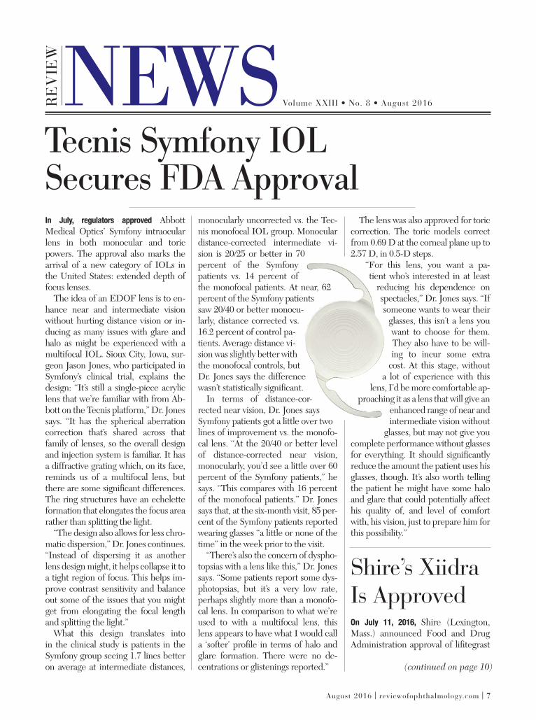
In July, regulators approved Abbott Medical Optics’ Symfony intraocular lens in both monocular and toric powers. The approval also marks the arrival of a new category of IOLs in the United States: extended depth of focus lenses.
The idea of an EDOF lens is to en-hance near and intermediate vision without hurting distance vision or in-ducing as many issues with glare and halo as might be experienced with a multifocal IOL. Sioux City, Iowa, sur-geon Jason Jones, who participated in Symfony’s clinical trial, explains the design: “It’s still a single-piece acrylic lens that we’re familiar with from Ab-bott on the Tecnis platform,” Dr. Jones says. “It has the spherical aberration correction that’s shared across that family of lenses, so the overall design and injection system is familiar. It has a diffractive grating which, on its face, reminds us of a multifocal lens, but there are some signifi cant differences. The ring structures have an echelette formation that elongates the focus area rather than splitting the light.
“The design also allows for less chro-matic dispersion,” Dr. Jones continues. “Instead of dispersing it as another lens design might, it helps collapse it to a tight region of focus. This helps im-prove contrast sensitivity and balance out some of the issues that you might get from elongating the focal length and splitting the light.”
What this design translates into in the clinical study is patients in the Symfony group seeing 1.7 lines better on average at intermediate distances,
monocularly uncorrected vs. the Tec-nis monofocal IOL group. Monocular distance-corrected intermediate vi-sion is 20/25 or better in 70 percent of the Symfony patients vs. 14 percent of the monofocal patients. At near, 62 percent of the Symfony patients saw 20/40 or better monocu-larly, distance corrected vs. 16.2 percent of control pa-tients. Average distance vi-sion was slightly better with the monofocal controls, but Dr. Jones says the difference wasn’t statistically signifi cant.
In terms of distance-cor-rected near vision, Dr. Jones says Symfony patients got a little over two lines of improvement vs. the monofo-cal lens. “At the 20/40 or better level of distance-corrected near vision, monocularly, you’d see a little over 60 percent of the Symfony patients,” he says. “This compares with 16 percent of the monofocal patients.” Dr. Jones says that, at the six-month visit, 85 per-cent of the Symfony patients reported wearing glasses “a little or none of the time” in the week prior to the visit.
“There’s also the concern of dyspho-topsias with a lens like this,” Dr. Jones says. “Some patients report some dys-photopsias, but it’s a very low rate, perhaps slightly more than a monofo-cal lens. In comparison to what we’re used to with a multifocal lens, this lens appears to have what I would call a ‘softer’ profi le in terms of halo and glare formation. There were no de-centrations or glistenings reported.”
The lens was also approved for toric correction. The toric models correct from 0.69 D at the corneal plane up to 2.57 D, in 0.5-D steps.
“For this lens, you want a pa-tient who’s interested in at least
reducing his dependence on spectacles,” Dr. Jones says. “If someone wants to wear their
glasses, this isn’t a lens you want to choose for them. They also have to be will-ing to incur some extra
cost. At this stage, without a lot of experience with this
lens, I’d be more comfortable ap-proaching it as a lens that will give an
enhanced range of near and intermediate vision without
glasses, but may not give you complete performance without glasses for everything. It should signifi cantly reduce the amount the patient uses his glasses, though. It’s also worth telling the patient he might have some halo and glare that could potentially affect his quality of, and level of comfort with, his vision, just to prepare him for this possibility.”
Shire’s XiidraIs ApprovedOn July 11, 2016, Shire (Lexington, Mass.) announced Food and Drug Administration approval of lifi tegrast
Tecnis Symfony IOLSecures FDA Approval
(continued on page 10)
007_rp0816_news.indd 7007_rp0816_news.indd 7 7/22/16 3:51 PM7/22/16 3:51 PM

8 | Review of Ophthalmology | August 2016
RE
VIE
W
Since this column often explores topics related to funding, development strategy, and the identifi cation and fi lling of
unmet needs in the marketplace, this month’s installment explores the case of the dry-eye device-maker Oculeve, which sits at the inter-section of all these elements. Oculeve started with only one mission—to recognize an unmet need through a blank-slate observation of clinical practice—and leveraged an inter-disciplinary approach to think out-of-the-box for addressing it. If you’re working on a new device or therapy and looking to gain traction with it, the Oculeve story is a great example of managing the early stage funding process, moving a project through the development cycle and driving the program to a point where it registers with large pharma.
Oculeve was born out of the Stanford Uni-versity Biodesign Fellowship program, a men-tored program in which physicians and scien-tists are put into small teams and assigned an area in which to innovate. The focus of this particular team, including scientist/engineer Michael Ackermann, PhD, was ophthalmology. The team, consisting of a biomaterials scientist/engineer (Garrett Smith), a board-certifi ed surgeon (Victor McCray, MD) and a mechanical engineer (Brandon Felkins), along with Dr. Ackermann, were new to ophthalmology, and set out to embed themselves in a busy clinical ophthalmology practice. They observed the physicians and patients in the offi ce and operating room daily and tracked such things as the fl ow of patients and reasons for their offi ce visits. After about a month, they had ac-cumulated a list of approximately 350 unmet needs. Recognizing that almost one in every three patients who were seen had dry eye, that’s what they focused on.
To determine how they were going to ap-proach dry-eye therapy, the team broke down the disease into two basic components: de-creased production of the critical components of the tear fi lm, and infl ammation, the latter leading to further breakdown of the body’s ability to create tears from the lacrimal glands. They identifi ed the unmet clinical need—and gap in therapy—for an immediate restoration of the tear fi lm. Leveraging their background in neuroengineering, and after evaluating many different approaches, the team focused on neurostimulation for creating tears.
The initial focus for stimulating tear produc-tion was with an implant to directly stimulate the lacrimal gland, which they tested in more than 40 subjects. To carry out this testing,
however, the new business needed funding.In order to fund the early stages of proof-of-
concept testing, a start-up company should be open to seeking funding from alterna-tive sources, which may be grants, friends and family, seed investors, service-in-kind investments from development partners or even prize money from winning business-plan competitions. Early data generation can then drive subsequent larger rounds from the traditional VCs or pharma partners. This is an important point for the new entrepreneur to recognize. Many times we see early-stage start-ups that attempt to pursue large rounds of fi nancing from the start but don’t gain trac-tion. However, when possible and appropriate given the nature of the project, leveraging off-the-shelf devices, or already approved and marketed drugs of a similar pharmacologic
class with a repurposing approach, can facili-tate small, pilot clinical testing to generate the required proof-of-principle, and a reason for others to believe in it.
In Oculeve’s case, its early development was funded through winning business plan competitions, which brought in up to $50,000. After that, Oculeve was formed, and at-tracted seed capital of $100,000 from Kleiner, Perkins, Caufi eld and Byers, and $100,000 from “sophisticated angel investors.” This supported the development of a prototype off-the-shelf stimulator that they used to directly stimulate the lacrimal gland via a needle electrode through the eyelid. The data supported raising $500,000 additional capital through debt fi nancing, followed by closing a Series A sale of preferred stock in the com-pany of $7.59 million in August 2012, from Kleiner, NEA and Versant. This allowed them to spin-out the technology from the university and with the capital, run additional studies outside of the United States, including studies in Mexico, New Zealand and Australia.
Early data from the orbital implant were promising in some patients, but inconsis-
tent in others. During the clinical trials of the implant, Oculeve identifi ed an interesting fi nding that patients with the best response had tearing in both eyes despite a unilateral implant. This led to the realization that tear-ing could be more powerfully stimulated by activating a refl ex. Oculeve changed its tack and developed a nasal stimulation device that activates the nasolacrimal refl ex. The device is handheld, non-invasive and doesn’t involve an implant or needle. Of course, that realization and evolution of the product delivery transformed the nature of the device into a product that was acceptable to an even greater number of patients.
In refl ecting on the impetus of going outside the United States with the implant, Dr. Acker-mann indicated that the main driver behind going to Mexico and the Philippines was speed, given that the timeline for opening an investigational device exemption in the United States was prohibitive for them. However,
after the evolution to the non-invasive nasal stimulator, which is considered a non-signifi cant risk device and therefore didn’t need an IDE in the United States, he chose to keep running the studies in
Mexico, despite a more straightforward institutional review board approval that
would have been needed in the United States. This was done mainly to stay under the radar until the patent portfolio was locked down and the scientifi c foundation of the treatment was better understood. In Oculeve’s case, the technology is disruptive in the way in which it is delivered, and protection of intellectual property was a main driver of the decision.
In prior columns, we’ve discussed situations where going outside the country for studies is warranted and have explored other case studies. In the case of Oculeve, partnering with an investigator with whom there is a relationship and trust, and setting up the study so the sponsor could be on-site in a collaborative fashion, helped ensure success and quality data.
In March 2014, Oculeve completed a larger round of $16 million in fi nancing. This infusion funded a large, well-controlled Phase II dry-eye study with a focus on precise endpoints designed to support the regulatory process and build value for partner discussions. As a result, Oculeve was acquired by Allergan in August 2015. Allergan’s CEO Brent Saunders has espoused the open-science model of research and development, in which innova-tion is sought from both internal laboratories and external start-ups and university settings
Ophthalmic Product Development InsightsOphthalmic Product Development InsightsMatthew Chapin and David A. Hollander, MD, MBA • Ora Inc., Andover, Mass.
An Interdisciplinary Approach to Addressing an Unmet Need
007_rp0816_news.indd 8007_rp0816_news.indd 8 7/22/16 4:26 PM7/22/16 4:26 PM

3360 Scherer Drive, Suite B, St. Petersburg, FL 33716
AGBC1371 Rev.A
)
1)k
)
#8
1
in order to increase R&D effi ciency. The company then follows through with the clinical development and regulatory process in order to bring these innovative products to patients. To that end, Allergan has 70 programs in mid- to late-stage development. Oculeve was a great strategic fi t for Allergan, which markets Restasis and Refresh tears and has multiple clinical development programs in dry eye. “At Allergan we use our open-science model to harness the power of the new innovation eco-system,” Mr. Saunders states. “This approach allows us to develop de-risked, late-stage assets and cutting-edge early-stage programs within our targeted therapeutic areas, such as eye care. This provides signifi cant rewards to the entrepreneurs and higher return on our R&D investments and is a true win-win for the innovation ecosystem.”
The Oculeve story shows the value of fi rst seeking an unmet need as opposed to a prod-uct, building a company around a structured process of mentorship, and throwing in a little serendipity, as well. Leveraging expertise from other areas of development, Oculeve’s researchers and owners drove effi cient pilot testing with off-the-shelf products, raised seed funds to support prototyping and then, when things didn’t go as planned, they learned from their observations and adapted effectively, creating a superior, less-invasive approach to delivering the neurostimulation.
An important take-home point from Oculeve’s story is the value of early proof of concept, built around effi ciently executing small, high-quality studies that both prove the concept and demonstrate the basic science—which was needed, given the novelty of the approach. The testing was designed to ensure that their data would inform decision-making, shifting and revision of plans, and the ability to secure additional capital to run the defi nitive Phase II clinical trial that supported industry partnering.
Mr. Chapin is senior vice president of the Corporate Development Group and Dr. Hol-lander is chief medical offi cer at Ora Inc. and assistant clinical professor of ophthalmology at the Jules Stein Eye Institute, UCLA. Ora pro-vides a comprehensive range of development, clinical-regulatory and product consulting for developers, investors and buyers; preclinical and turnkey clinical trial services and regula-tory submissions; and integration with asset, business partnering and fi nancing support in ophthalmology. They welcome your product development comments or questions. Please send correspondence to [email protected] or visit www.oraclinical.com.
007_rp0816_news.indd 9007_rp0816_news.indd 9 7/22/16 4:26 PM7/22/16 4:26 PM

News RE
VIE
W
10 | Review of Ophthalmology | August 2016
ophthalmic solution 5% (Xiidra), a twice-daily eye drop indicated for the treatment of the signs and symptoms of dry-eye disease in adult patients. Shire notes that Xiidra is the only prescription eye drop indicated for the treatment of both signs and symptoms of dry eye.
According to the company, the in-fl ammation associated with dry eye is thought to be primarily mediated by T-cells and associated cytokines. One effect of this process may be over-expression of intracellular adhesion molecule 1 in corneal and conjunctival tissues, which may contribute to T-cell activation and migration to target tis-sues. Although the exact mechanism of action of lifi tegrast is not known, in vitro studies demonstrated that it may inhibit T-cell adhesion to ICAM-1 in a human T-cell line and the secretion of cytokines in human peripheral blood mononuclear cells.
The safety and effi cacy of Xiidra were studied in 1,181 patients in four placebo-controlled 12-week trials; 1,067 patients received lifi tegrast 5%. Each of the four studies assessed the effect of Xiidra on both the signs and symptoms of dry eye at baseline and weeks two, six and 12. In all four stud-ies, a larger reduction in eye dryness score was observed with Xiidra at six and 12 weeks; in two of the four stud-ies, an improvement in EDS was also seen with Xiidra at two weeks. At week 12, a larger reduction in inferior cor-neal staining score, favoring Xiidra, was observed in three of the four studies. The most common adverse reactions reported in 5 to 25 percent of patients were instillation site irritation, altered taste sensation (dysgeusia) and re-duced visual acuity.
Edward Holland, MD, professor of clinical ophthalmology at the Univer-sity of Cincinnati and a clinical trial in-vestigator for Xiidra, says the approval is a boon to clinicians. “There have been more than 20 dry-eye trials that
failed to reach signifi cance in both signs and symptoms,” he says. “Credit goes to the Shire folks for designing a trial that got across the fi nish line with both signs and symptoms of dry eye.”
Dr. Holland explains that the reason Shire succeeded where others failed was their trial design. “Everyone else tried to achieve signifi cance in both signs and symptoms in the same pa-tient population,” he says. “Shire real-ized that wasn’t going to work. So the Shire scientists analyzed the data and saw that one group of patients did very well clearing the clinical sign of corneal staining, while another group had symptoms signifi cantly reduced. So they did two separate trials and then repeated both of them. This was a totally new strategy. It made the time-table much longer and the number of patients much greater, but it certainly worked for them, and it might work for other formulations as well.”
Asked how he expects Xiidra to fi t into his armamentarium, Dr. Holland says he expects it to be fi rst-line. “Ar-tifi cial tears are palliative,” he notes. “This is a very effective anti-infl amma-tory that gets at the pathogenesis of dry eye. For me it will be primary therapy.”
Shire expects to launch Xiidra in the United States in the third quarter of 2016.
Humira Cleared for UveitisIn mid-July, the FDA approved AbbVie’s non-steroidal immunomodulatory agent Humira (adalimumab) for use in non-infectious intermediate uveitis, posterior uveitis and panuveitis. This approval fi nally gives ophthalmologists an approved immunomodulatory op-tion, obviating the headaches involved with using such agents off-label.
The approval was based on the results of two large-scale, Phase III studies, VISUAL-1 and -2. “VISU-AL-1 was designed to test Humira in patients with active uveitis, with the goal of seeing if patients with ac-tive infl ammation—despite being on steroids—could be tapered off their steroids while Humira helped fi ght the infl ammation,” explains Glenn Jaffe, MD, chief of the retina divi-sion at Duke University, and Robert Machemer, MD, professor of oph-thalmology at Duke and consultant to AbbVie. “The other study, dubbed VISUAL-2, recruited uveitis patients whose eyes were quiet because they were currently on steroids, with the goal being to get them off the ste-roids and to substitute the Humira.” The main endpoint in VISUAL-1 was the time until treatment failure. InVISUAL-2, the researchers wanted to see how likely a fl are-up would be as patients were tapered off the steroids and kept on Humira.
“The way I think of the results of the VISUAL-1 study is the phrase, ‘twice as long or half as likely,’ ” says Dr. Jaffe, “meaning that the time to treatment failure was about twice as long—24 weeks for the treatment group vs. 13 for the placebo—and the likelihood of getting failure was approximately half for the Humira group. In many cases, the Humira group didn’t fail. So, not only did it take longer, in general, for them to get a fl are-up, but they were half as likely to get one, as well.
“Similarly, in VISUAL-2, the study in which the patients’ infl ammation was quiet with systemic steroids to be-gin with, the idea was to see if patients were less likely to get a fl are-up if they were treated with Humira than if they weren’t,” Dr. Jaffe continues. “Both studies mandated a steroid taper over 15 weeks. In VISUAL-2, the patients who were switching over to Humira were signifi cantly less likely to have treatment failure when the steroids were tapered, and they had less of a
(continued from page 7)
007_rp0816_news.indd 10007_rp0816_news.indd 10 7/22/16 4:27 PM7/22/16 4:27 PM

August 2016 | reviewofophthalmology.com | 11
chance of visual acuity going down.”In terms of the best patients for the
use of Humira, Dr. Jaffe notes that a risk of infection always lurks whenever an immunomodulatory agent is used. “Certainly, if someone has a history of tuberculosis, especially if it’s not treat-ed, you don’t want to put him on one of these agents,” he avers. “However, you can cautiously treat someone with TB as long as it’s been completely treated, but that’s something you have to con-fi rm before starting these agents. Also, in general, I’d say patients with an un-derlying infectious cause of the uveitis wouldn’t be good candidates.
“The tumor necrosis factor inhibi-tors like Remicade and Humira can potentially cause a fl are-up of multiple sclerosis or other demyelinating dis-eases,” adds Dr. Jaffe. “Also, individu-als who have intermediate uveitis are at an increased risk of having MS as an association with the infl ammation. In intermediate uveitis patients, I’d recommend a brain MRI to ensure there’s no evidence of MS.”
FDA Approves The RaindropSurgeons now have a new option for their presbyopic patients, thanks to the Food and Drug Administration approval of ReVision Optics’ Rain-drop Near Vision Inlay in July. The inlay is designed to give patients improved near and intermediate visual performance, and was approved based on the results of a one-year prospective, nonrandomized, multi-center clinical trial.
Dallas surgeon and Raindrop inves-tigator Jeffrey Whitman, MD, says the inlay did give presbyopes a wider focal range. “The question is, does it work for reading?” he says. “Yes. It does well for intermediate, as well. In this day and age people aren’t really looking for just
distance and close up. They’re looking at their phones and computers, and the inlay does well at that distance.”
The inlay uses corneal shape change—not refractive power—to get its effect. “It’s a 2-mm-diameter, meniscus-shaped inlay with an index of refraction that’s equal to the cor-nea’s,” explains Dr. Whitman. “It’s 30 µm thick in the center. The Raindrop works by steepening the center of the cornea, which gives you a greater depth of focus.” It’s approved for use in the non-dominant eye of patients with a manifest refraction spherical equiva-lent of -0.5 to +1 D, with 0.75 D or less of cylinder. The inlay is approved for implantation beneath a LASIK fl ap at one-third corneal depth, and a corneal pocket study is imminent. Though it’s not approved for it, surgeons may opt to perform LASIK beforehand to get patients in the emmetropic range and then implant the inlay.
The study looked at 373 non-dom-inant eyes of emmetropic, presbyopic subjects that received the Raindrop at 11 sites. During the one-year follow-up visit, researchers studied 340 of the nondominant eyes.
“By one year 93 percent of subjects were 20/25 or better at near. They gained an average of 5.1 lines of read-ing vision,” Dr. Whitman says. “So, it was pretty dramatic; 97 percent of subjects saw 20/30 at intermediate
distance postop. The inlay is clear, so there’s very little loss of contrast sensi-tivity and no loss of binocular vision.”
At one year in the treated eye, un-corrected intermediate visual acuity improved by 2.5 lines while best-cor-rected distance visual acuity decreased by 1.2 lines. From three months through one year, 93 percent of sub-jects achieved uncorrected near visual acuity of 20/25 or better, 97 percent achieved UIVA of 20/32 or better and 95 percent achieved UDVA of 20/40 or better. Binocularly, the mean UDVA exceeded 20/20 from three months through one year.
Dr. Whitman says the procedure is straightforward. “You lift the fl ap up and place the device over the light-constricted pupil. Wait about a minute for it to dry in place before putting the fl ap down. Then let it dry as you would a normal fl ap. Then, there’s the recom-mended drop regimen afterward that’s similar to LASIK’s.”
In terms of candidates for the inlay, Dr. Whitman states, “If they’re not healthy enough to be a LASIK candi-date, they aren’t a good candidate for this. Because, basically, the issues re-late to fl ap technology.”
Dr. Whitman says adverse events were low in the study. “Visual symp-toms such as glare and halo had less than a 4-percent incidence,” he says. “Eighteen inlays were replaced, most of those for decentration. After re-placement, they had the same un-corrected near vision as the rest of the study population. Eleven cases required explantation, for reasons such as decentration, dissatisfaction with vision and foreign-body reac-tion. Only one eye experienced the foreign-body reaction. The foreign-body reaction can usually be treated with steroids, but if it persists, in the study we recommended removal of the inlay. You can remove it and patients get back to within one line, on average, of their best-corrected vision within three months.”
The Raindrop inlay is implanted beneath a
LASIK fl ap, and its presence steepens the
cornea, yielding greater depth of focus.
Jeff
rey
Wh
itm
an
, MD
007_rp0816_news.indd 11007_rp0816_news.indd 11 7/22/16 4:27 PM7/22/16 4:27 PM

12 | Review of Ophthalmology | August 2016
EditorialBoardR
EV
IEW
CONTRIBUTORS
ADVISORY BOARD
REVIEW OF OPHTHALMOLOGY (ISSN 1081-0226; USPS No. 0012-345) is published monthly, 12 times per year by Jobson Medical Informa-tion. 100 Avenue of the Americas, New York, NY 10013-1678. Periodicals postage paid at New York, NY and additional mailing offi ces. Postmaster: Send address changes to Review of Ophthalmology, PO Box 71, Congers, NY 10929-0071. Subscription Prices: US One Year $63.00, US Two Year $112.00, Canada One Year $99.00, Canada Two Year $181.00, Int’l One Year $158.00, Int’l Two Year $274.00. For subscription information call (877) 529-1746 (USA only); outside USA, call (845-267-3065. Or email us at [email protected]. Canada Post: Publications Mail Agreement #40612608. Canada Returns to be sent to Bleuchip International, P.O. Box 25542, London, ON N6C 6B2.
BUSINESS OFFICES
11 CAMPUS BOULEVARD, SUITE 100
NEWTOWN SQUARE, PA 19073
SUBSCRIPTION INQUIRIES (877) 529-1746
(USA ONLY); OUTSIDE USA, CALL (847) 763-9630
BUSINESS STAFF
PUBLISHER
JAMES HENNE
(610) 492-1017 [email protected]
REGIONAL SALES MANAGER
MICHELE BARRETT
(610) 492-1014 [email protected]
REGIONAL SALES MANAGER
MICHAEL HOSTER
(610) 492-1028 [email protected]
CLASSIFIED ADVERTISING
(888)-498-1460
VICE PRESIDENT OF OPERATIONS
CASEY FOSTER
(610) 492-1007 [email protected]
PRODUCTION MANAGER
SCOTT TOBIN
(610) 492-1011 [email protected]
SUBSCRIPTIONS
$63 A YEAR, $99 (U.S.) IN CANADA,
$158 (U.S.) IN ALL OTHER COUNTRIES.
SUBSCRIPTIONS E-MAIL:
CIRCULATION
PO BOX 71, CONGERS, NY 10920-0071
(877) 529-1746
OUTSIDE USA: (845) 267-3065
SENIOR CIRCULATION MANAGER
HAMILTON MAHER
(212) 219-7870 [email protected]
CEO, INFORMATION GROUP SERVICES
MARC FERRARA
SENIOR VICE PRESIDENT, OPERATIONS
JEFF LEVITZ
VICE PRESIDENT, HUMAN RESOURCES
TAMMY GARCIA
VICE PRESIDENT, CREATIVE SERVICES & PRODUCTION
MONICA TETTAMANZI
CORPORATE PRODUCTION DIRECTOR
JOHN ANTHONY CAGGIANO
VICE PRESIDENT, CIRCULATION
EMELDA BAREA
100 Avenue of the Americas
New York, NY 10013
PENNY A. ASBELL, MD, NEW YORK CITY
WILLIAM I. BOND, MD, PEKIN, ILL.
ALAN N. CARLSON, MD, DURHAM, N.C.
Y. RALPH CHU, MD, EDINA, MINN.
ADAM J. COHEN, MD, DOWNERS GROVE, ILL.
UDAY DEVGAN, MD, FACS, LOS ANGELES
ERIC DONNENFELD, MD, ROCKVILLE CENTRE, N.Y.
DANIEL S. DURRIE, MD, KANSAS CITY, MO.
ROBERT EPSTEIN, MD, MCHENRY, ILL.
ROBERT D. FECHTNER, MD, NEWARK, N.J.
WILLIAM J. FISHKIND, MD, TUCSON, ARIZ.
JAMES P. GILLS, MD, TARPON SPRINGS, FLA.
HARRY GRABOW, MD, SARASOTA, FLA.
DOUGLAS K. GRAYSON, MD, NEW YORK CITY
R. BRUCE GRENE, MD, WICHITA, KAN.
THOMAS S. HARBIN, MD, MBA, ATLANTA
DAVID R. HARDTEN, MD, MINNEAPOLIS
KENNETH J. HOFFER, MD, SANTA MONICA, CALIF.
JACK T. HOLLADAY, MD, MSEE, HOUSTON
JOHN D. HUNKELER, MD, KANSAS CITY, MO.
THOMAS JOHN, MD, TINLEY PARK, ILL.
ROBERT M. KERSHNER, MD, MS, FACS, BOSTON
GUY M. KEZIRIAN, MD, PARADISE VALLEY, ARIZ.
TERRY KIM, MD, DURHAM, N.C.
TOMMY KORN, MD, SAN DIEGO
DAVID A. LEE, MD, HOUSTON
FRANCIS S. MAH, MD, SAN DIEGO
NICK MAMALIS, MD, SALT LAKE CITY
WILLIAM G. MARTIN, MD, OREGON, OHIO
MIKE S. MCFARLAND, MD, PINE BLUFF, ARK.
JEFFREY B. MORRIS, MD, MPH, ENCINITAS, CALIF.
MARLENE R. MOSTER, MD, PHILADELPHIA
ROBERT J. NOECKER, MD, FAIRFIELD, CONN.
ROBERT OSHER, MD, CINCINNATI
MARK PACKER, MD, BOULDER, COLO.
STEPHEN PASCUCCI, MD, BONITA SPRINGS, FLA.
PAUL PENDER, MD, BEDFORD, N.H.
CHRISTOPHER J. RAPUANO, MD, PHILADELPHIA
AUGUST READER III, MD, SAN FRANCISCO
TONY REALINI, MD, MORGANTOWN, W.V.
KENNETH J. ROSENTHAL, MD, GREAT NECK, N.Y.
ERIC ROTHCHILD, MD, DELRAY BEACH, FLA.
SHERI ROWEN, MD, BALTIMORE
JAMES J. SALZ, MD, LOS ANGELES
INGRID U. SCOTT, MD, MPH, HERSHEY, PA.
JOEL SCHUMAN, MD, PITTSBURGH
GAURAV SHAH, MD, ST. LOUIS
DAVID R. STAGER JR., MD, DALLAS
KARL STONECIPHER, MD, GREENSBORO, N.C.
JAMES C. TSAI, MD, NEW YORK CITY
VANCE THOMPSON, MD, SIOUX FALLS, S.D.
FARRELL C. TYSON, MD, CAPE CORAL, FLA.
R. BRUCE WALLACE III, MD, ALEXANDRIA, LA.
ROBERT G. WILEY, MD, CLEVELAND
FRANK WEINSTOCK, MD, CANTON, OHIO
JACQUELINE M.S. WINTERKORN, MD, PHD, NEW YORK CITY
CHIEF MEDICAL EDITOR
Mark H. Blecher, MD
BOTTOM LINE
Dennis D. Sheppard, MD
CONTACT LENSES
Penny Asbell, MD
CORNEA / ANTERIOR SEGMENT
Thomas John, MD
GLAUCOMA MANAGEMENT
Peter Netland, MD, PHDKuldev Singh, MD
MASTERS OF SURGERY
Taliva D. Martin, MDSara J. Haug, MD, PhD
PEDIATRIC PATIENT
Wendy Huang, MD
PLASTIC POINTERS
Ann P. Murchison, MD, MPH
REFRACTIVE SURGERY
Arturo S. Chayet, MD
RETINAL INSIDER
Carl Regillo, MD, FACSEmmett T. Cunningham Jr., MD, PHD, MPH
TECHNOLOGY UPDATE
Steven T. Charles, MDMichael Colvard, MD
THERAPEUTIC TOPICS
Mark Abelson, MD
WILLS RESIDENT CASE SERIES
Alison Huggins, MD
007_rp0816_news.indd 12007_rp0816_news.indd 12 7/22/16 3:52 PM7/22/16 3:52 PM

www.compulinkadvantage.com/onetab 800.456.4522
Dr. Rick Anderson, ICON Eyecare Founder & CEO
F A S T E A S Y E F F I C I E N T E N T E R P R I S E P R O V E N
All-in-one database solution: EHR, PM, E-Prescribing, ASC, Optical, Patient Portal, Patient Engagement, Marketing, RCM
Unique OneTab™ EHR technology: Document entire exam from a single screen
MACRA-ready guarantee
1-hour support response guarantee
Ophthalmology’s business partner for 31 years
The Partner of Choice for ICON EyecareSM Clinics and Surgery Centers
Used by more ophthalmologists in more locations nationwide
“We chose Compulink
because of the completeness
of their solution, commitment
to customer service, and the
ability of Compulink’s system
to meet the needs of our
growing business.”
Request a Demo
Save up to $100,000 on your migration to CompulinkReceive 100% trade-in credit for your current EHR/PM software!
RP0816_Compulink.indd 1 7/13/16 2:57 PM

14 | Review of Ophthalmology | August 2016
August 2016 • Volume XXIII No. 8 | reviewofophthalmology.com
Cover FocusFirst-line Therapy for Wet AMD
By Christopher Kent, Senior EditorThe treat-and-extend protocol works well, and may be needed indefinitely.
Treatment That Fits DME Patients to a ‘T’
By Walter Bethke, Editor in ChiefPhysicians use the results of Protocol T to inform their owntreatment protocols.
The Evolution of Vein Occlusion Therapy
By Michelle Stephenson, Contributing EditorInvestigators are experimenting with combination therapies that may enhance outcomes.
Anti-VEGF Everywhere, But Not a Drop to Use
By Richard T. Atallah, Manuela Von Sneidern,Alan T. Sheyman, MD, and Ben Z. Cohen, MDReusing Lucentis and Eylea vials may be feasible, the authors say.
7 |
20 |
22 |
45 |
48 |
52 |
56 |
58 |
60 |
61 |
63 |
Review News
Technology UpdateOR Videography on the Cheap
Medicare Q&AUnderstanding HEDIS
Retinal InsiderTargeting VEGF’s Silent Partners in DME
Therapeutic TopicsQuantifying the Qualitative
Glaucoma ManagementMIGS and Meds: Refining Trabecular Bypass
Refractive SurgeryTips for Pre-LASIK Resurfacing
Research ReviewResults of the LUNAR Study
Advertising Index
Classified Ads
Wills Eye Resident Case Series
Departments24 |
32 |
38 |
42 |
32
An
dre
w A
nto
szyk, M
D
014_rp0816_toc.indd 14014_rp0816_toc.indd 14 7/22/16 2:46 PM7/22/16 2:46 PM

beee
llliieevvinn
gggg..
Create a Doctor-Patient Dialogue with iNview.Trusted Volk Optics. iPhone Convenience.
iPhone fundus camera
Limited time offers. Online customers only.
10% OFF Use Code: AUG16BUY TODAY
volk.com/inview
RP0816_Volk.indd 1 7/12/16 10:50 AM

16 | Review of Ophthalmology | August 2016
Editor’s Page
RE
VIE
W
Here’s toRepurposing
Walter C. Bethke, Editor in Chief
Reading through the retina specialists’ comments in this month’s articles on treatments for diseases like wet AMD and DME, you get a sense of how completely anti-VEGF drugs have changed ophthalmology.
For years, laser therapy was the best treatment for wet age-related macular degeneration, even though it some-times left patients with blind spots in their visual fi elds. Genentech’s clini-cal trial of Lucentis changed all that. Building upon the success clinicians were having with Macugen, Lucen-tis—and, later, Eylea—showed that anti-VEGF drugs could actually allow patients to recover vision, an effect un-heard of with laser treatments. It was a drug innovation success story.
However, at the same time, the dawn of anti-VEGF in ophthalmol-ogy was also a story about innovative repurposing.
As the Lucentis trial was compiling its impressive data, groups of intrepid physicians decided to take Genen-tech’s cancer drug Avastin and repur-pose it for use in the eye, with simi-larly impressive results. Though this sparked controversy, this use of com-pounded Avastin gave ophthalmology at-large its fi rst taste of the ability of these drugs to change people’s lives.
Lucentis and Eylea were eventually approved, and have been embraced by retina specialists, but Avastin remains an option for patients with insurance issues for whom the companies’ co-pay assistance doesn’t help for one reason or another. The Avastin option wouldn’t be there for them, though, if certain creative ophthalmologists
didn’t step back, ask “What if ..?” and repurpose it to fi ght blindness.
Drugs aren’t the only thing that can be repurposed: people can be, too. In 1994, I started on Review as a produc-tion editor for the magazine’s second issue, laying out other editors’ articles and making sure everything looked pretty—or at least not ugly—on the page. In his wisdom, the founding editor of Review, Stan Herrin, knew I could do more, and I was repurposed into a writer and associate editor. Since then, the repurposing continued, and I entered various new roles on the mag-azine, ensuring that I was always re-searching and writing more about this fascinating specialty, that I was always learning. Stan’s successor, Chris Glenn came on in 1999, and followed suit.
In June, Chris retired after 17 years at Review, and he’s now in the enviable position of being able to repurpose himself as often as he wants, to follow his bliss. I want to wish him well, and thank him for sharing his knowledge, experience and, most of all, friendship.
I’m proud to say this cycle of re-purposing has culminated with me being named Review’s new editor in chief. I’d like to take this opportunity to make a pledge that the one purpose that will never change is the one at the heart of Review: To provide readers with the timely, practical information they’ve come to expect, with an abid-ing respect for editorial integrity in-stilled in me by the editors that have gone before.
—Walt Bethke, Editor in Chief
ED I T O R I A L STA F F
Editor in Chief
Walter C. Bethke(610) 492-1024
Senior Editor
Christopher Kent(814) 861-5559
Associate Editor
Liam Jordan(610) 492-1025
Chief Medical Editor
Mark H. Blecher, MD
Art Director
Jared Araujo(610) 492-1032
Senior Graphic Designer
Matt Egger(610) 492-1029
International coordinator, Japan
Mitz [email protected]
Business Offi ces
11 Campus Boulevard, Suite 100Newtown Square, PA 19073
(610) 492-1000 Fax: (610) 492-1039
Subscription inquiries:
United States — (877) 529-1746Outside U.S. — (845) 267-3065
E-mail:
[email protected]: www.reviewofophthalmology.com
®
016_rp0816_edit.indd 16016_rp0816_edit.indd 16 7/22/16 1:31 PM7/22/16 1:31 PM

RP0815_Lombart.indd 1RP0815_Lombart.indd 1 7/21/15 10:07 AM7/21/15 10:07 AM

Retina World Congress (RWC) – An international professional congress that will
support global scientific and clinical exchanges on advances in retina health.
R E G I S T R AT I O N I S N O W O P E N
w w w. r e t i n a w o r l d c o n g r e s s . o r g
I N A U G U R A LR E T I N A W O R L D C O N G R E S S
G E T R E A D Y T O U N I T E T H E W O R L D O F R E T I N A
F E B R U A R Y 2 3 - 2 6 , 2 0 1 7F O R T L A U D E R D A L E M A R R I O T T
H A R B O R B E A C H R E S O R T & S P AF O R T L A U D E R D A L E , F L O R I D A , U S A
U N I T E . E X C H A N G E . A D V A N C E .
This activity has been planned and implemented in accordance with the Essential Areas and Policies of the Accreditation Council for Continuing Medical Education (ACCME) through the joint providership of Global Education Group (Global), MCME Global, and Retina World Congress (RWC). Global is accredited by the ACCME to provide continuing medical education for physicians.
RP0516_HouseRWC.indd 1RP0516_HouseRWC.indd 1 4/18/16 1:37 PM4/18/16 1:37 PM

NOVEMBER 11-12, 2016
CSE GCSE GLAUCOMALAUCOMA F FELLOWSELLOWS
Course Director:
Kuldev Singh, MDStanford, CA
Wet Lab Director:
Douglas J. Rhee, MDCleveland, OH
SAVE THE DATE
Fort Worth, Texas
GLAUCOMA FELLOWSCONTINUING SPECIALIZED EDUCATION
Participants in this course must be enrolled in a glaucoma fellowship program at the time of the course.There is no registration fee for these activities. Air, ground transportation in Fort Worth, hotel accommodations and
modest meals will be provided through an educational scholarship for qualifi ed participants.
Accreditation StatementThis activity has been planned and implemented in accordance with the accreditation requirements and policies of the Accreditation Council for
Continuing Medical Education (ACCME) through the joint providership of Amedco and Postgraduate Healthcare Education. Amedco is accredited by the
ACCME to provide continuing medical education for physicians.
Credit Designation StatementAmedco designates this live activity for a maximum of 13.50 AMA PRA Category 1 CreditsTM. Physicians should claim only the credit commensurate with
the extent of their participation in the activity.
For More Information & to Register:
www.revophth.com/2016cseglaucomaEmail: [email protected] or call Denette Holmes 866-627-0714
Faculty:
Dear Fellowship Program Director and Coordinator,
We would like to invite you to review the upcoming 2016 Glaucoma Fellowship Program in Fort Worth at the Renaissance Worthington hotel.
The program offers a unique educational opportunity for fellows by providing the chance to meet and exchange ideas with some of the most
respected thought leaders in glaucoma. The Glaucoma Fellows Program is designed to provide your fellows with a state-of-the-art didactic
and wet lab experience. The program also serves as an opportunity for your fellows to network with fellows from other programs.
After reviewing the material, it is our hope that you will select and encourage your fellows to attend this educational activity which is CME
accredited to ensure fair balance.
Sincerely,
Postgraduate Healthcare Education
Endorsed by
Review of Ophthalmology®
Supported by an unrestricted independent medical educational grant from
AlconJointly provided by
Lee Alward, MDDonald Budenz, MDRonald Fellman, MDBrian Flowers, MD
Steven Gedde, MDDale Heuer, MD
Shan Lin, MDEydie Miller-Ellis, MDLouis Pasquale, MD
Harry Quigley, MDArthur Sit, MD
George Spaeth, MDJaney Wiggs, MD, PhD
2016_CSE_GlaucFellow-HouseAd.indd 862016_CSE_GlaucFellow-HouseAd.indd 86 7/15/16 2:58 PM7/15/16 2:58 PM

Technology Update Edited by Michael Colvard, MD, and Steven Charles, MDR
EV
IEW
20 | Review of Ophthalmology | August 2016 This article has no commercial sponsorship.
Today, it’s become increasingly important to be able to share what
you see through the oculars during surgery. Digital technology has made that easier than ever, but technology can be costly. Here, Samaresh Sriv-astava, DNB, of the Raghudeep Eye Hospital in Ahmedabad, India, shares some secrets for capturing, displaying and recording high-quality video—in 2-D or 3-D—without breaking the bank.
Recording with an SLR Camera
Dr. Srivastava notes that taking high-resolution video using a digital single-lens refl ex camera connected to your surgical microscope can be far less expensive than using a standard video camera. “Most of the new digi-tal SLRs have HD digital recording capability,” he says. “Some, like Sony’s a7R II, have 4K recording ability as well. A full-frame DLSR camera costs about $2,000; a ballpark cost for the video mount to connect it to your mi-croscope is $1,500 to $2,000 if you buy it directly from the microscope manufacturer, and less-expensive off-brand mounts are available as well.
That means you’re investing about $4,000 for the whole setup, which is a third of what you might pay for a full-HD video camera with a C-mount [the connector that joins the beam-splitter of the operating microscope to the camera].
“Once the SLR is plugged in, you can record directly onto the SLR camera, or you can use the camera’s live video-out port to export the video onto another device such as a com-puter with a video capture card or a digital recorder,” he says. “File size is limited on a camera, but a separate recording device should allow you to record streaming video for as long as you want—maybe a two- or three-hour surgery, for example.
“If you’re using a computer, you’ll need a good video-capture card, such as the one made by Blackmagic De-sign,” he adds. “The card goes into a PCI slot on the motherboard; because it’s directly connected to the mother-board it doesn’t lose any frames and produces a high-quality video. You also can choose the compression, fi le size and format you want, such AVI or PAL. Most of these capture cards can take video input from any camera.”
Using Your Smartphone
Another option is recording a sur-gical video using your smartphone. “There are many free apps available through the Android store and Apple App store that will allow you to do the recording,” he notes. “The special fea-tures of these newer phones can also be used: We’ve used an iPhone’s slow-motion mode to capture the stages of phaco and phacodonesis.
“Unlike an SLR or other camera, you can’t mount a smartphone on your microscope’s beam splitter,” he continues. “To do that, the micro-scope would need a video adapter tube. However, a smartphone can be mounted on an assistant’s viewing scope, which most microscopes have. To do this, you can use any adapter for a smartphone that’s available off the rack, such as one designed for slit lamp attachment; some ophthalmic companies also make them, and one person in India designed a mount you can make at home for $10 to $15.” [Instructions for making one are avail-able on the website of the Journal of Mobile Technology in Medicine at http://www.journalmtm.com/2014/
Christopher Kent, Senior Editor, and Walter Bethke, Editor in Chief
Capturing live surgery for simultaneous viewing—or later viewing—can be done in 2-D or 3-D without breaking the bank.
Creating Great Surgical Videos (for less money)
020_rp0816_tech update.indd 20020_rp0816_tech update.indd 20 7/21/16 11:38 AM7/21/16 11:38 AM

August 2016 | reviewofophthalmology.com | 21
diy-smartphone-slit-lamp-adaptor/)Dr. Srivastava notes that it can
be tricky to export live video from a smartphone. “Most of these phones have an attachment that allows you to duplicate the phone’s screen on a smart TV or computer screen,” he says. “This is called screen mirror-ing. If you don’t have a computer available, you can insert an Android Dongle into your TV’s HDMI port to allow it to mirror the smartphone screen. With an iPhone, you can use Apple TV.”
Working Live in 3-D
Dr. Srivastava says it’s possible to do both 3-D recording and live 3-D video displays without investing in expensive equipment. “All 3-D vid-eos work the same way,” he explains. “Your left eye sees one image while your right eye sees a similar but slight-ly different image; your mind fuses them to create a perception of depth. When you mount one camera on the left side of a beam splitter and an-other camera on the right side, you’re capturing slightly dissimilar images, like those your eyes perceive. These two images can then be sent to two different projectors. By polarizing the light emitted by each projector, you can throw two different images with different polarizations onto a silver polarized screen. Then a viewer can use polarized glasses to see very good 3-D images—better quality than what you’d see at the movies.
“To do this, you need two identical cameras with identical video adapter tubes so the optics are the same,” he continues. “To project the resulting images, you need two projectors, re-ferred to as stackable projectors. Most multimedia companies use stackable projectors to increase the brightness of their presentations. In this case, you run the video from the left side of the beam splitter through one projec-tor and the video from the right side
through the other projector.”Dr. Srivastava explains how polar-
ization makes 3-D viewing possible. “When you watch 3-D movies, each lens in the glasses you wear has a dif-ferent polarization,” he says. “If the left eye has a vertical polarization, the right eye will have a horizontal polarization. As a result, your left eye will only see things that are vertically polarized, and your right eye will only see things that are horizontally polar-ized. Now, if you polarize the light coming from each projector differ-ently, the left eye will see only one of the two images and the right eye will see only the other.
“Accomplishing this is easy; just or-der some 3-D glasses,” he continues. “Break one pair in half and place the left lens in front of the fi rst projector and the right lens in front of the sec-ond projector. That will polarize the light from each projector in a differ-ent direction. Then, when you wear that same type of 3-D glasses and watch the video, you’ll see the video in 3-D.”
Dr. Srivastava adds that this will only work if you project the two im-ages onto a silver screen. “A silver screen will reflect the polarization back; a white screen will just absorb the polarization, causing you to see both images in both eyes,” he says. “Silver screens are readily available;
mine was shipped from China, from aliexpress.com, a big, online shopping portal. It cost less than $800 dollars. I also ordered my 3-D glasses from that site.”
Recording in 3-D
“Recording your surgery in 3-D is a little different, but it’s not complicat-ed,” says Dr. Srivastava. “Your record-ing device needs to have a 3-D cap-ture card. Instead of projecting the video, you send the two video feeds to the video capture card; it simultane-ously records both the left and right views of the surgery. Once completed, you’ll have a large digital fi le; the soft-ware will then let you send the left im-age to one projector and the right im-age to the other. A 3-D capture card costs about $1,500, but other than the computer itself—which needs to have a good processor that can buffer a large video fi le—that’s the only added expense. In my experience, as with a standard video card, the best 3-D video capture card is the one available from Blackmagic Design.
“To project the surgery recording, you just need to connect your com-puter to the two projectors, as de-scribed above,” he concludes. “If you want to view the video in 3-D on your computer screen, you’ll need a special 3-D monitor.”
Above, left: Two similar cameras mounted on either side of a microscope’s beam splitter
using video adapter tubes of the same focal length. Above, right: Two stackable projectors
with their projected images aligned. A pair of polarized eyeglasses has been broken in half,
with one lens placed in front of each projector’s lens to polarize the projected images.
Sam
are
sh S
rivasta
va, D
NB
020_rp0816_tech update.indd 21020_rp0816_tech update.indd 21 7/21/16 11:38 AM7/21/16 11:38 AM

RE
VIE
W Medicare Q&A Donna McCune, CCS-P, COE, CPMA
This article has no commercial sponsorship.22 | Review of Ophthalmology | August 2016
Q What does HEDIS stand for?
A HEDIS is an acronym for Healthcare Effectiveness Data
and Information Set; a set of health-care performance measures. The National Committee for Quality As-surance oversees the development, implementation and all facets of the HEDIS tool. HEDIS consists of 81 measures across fi ve domains of care. The measures compare how well a health plan performs in areas of qual-ity of care, access to care and member satisfaction with the plan itself.
Q Who utilizes the HEDIS tool?
A Health plans primarily use HEDIS, collecting and report-
ing data on the various HEDIS mea-sures. With a defined set of mea-sures, HEDIS permits a comparison of health-plan performance that’s con-sistent from plan to plan.
Health plans combine their HEDIS scores with Consumer Assessment of Healthcare Providers and Systems scores and NCQA Accreditation standards scores to receive a Health Insurance Plan Rating from the NCQA. The NCQA ratings list in-
cludes commercial payers, Medicare and Medicaid plans.
Q What type of rating do insurance plans receive?
AHealth Insurance Plan Rating is a score from one to fi ve in 0.5 in-
crements, with fi ve being the highest possible rating. This system is similar to CMS’ Five-Star Quality Rating Sys-tem, although the CMS system only rates nursing homes.
Compared with other plans in their category, those that get a fi ve are in the top 10 percent, those that get a four are above average, those that get a three are average, those that get a two are below average and those that get a one are in the bottom 10 percent.
Q What is the signifi cance of the rating to a health plan?
A Consumers analyze the ratings of different health plans when ei-
ther selecting a plan for the fi rst time or considering switching plans. The ratings provide consumers with a clear picture of how the various plans perform in key quality areas.
Individual health plans can use their star ratings as marketing tools.
For example,“XYZ Medicare Preferred HMO
plans and the XYZ Health Plan Senior Care Options Plan earned a 5 out of 5 Star Rating from Medicare for 2016. This is Medicare’s highest rating and makes these plans among the best in the country! In fact, out of nearly 400 plans rated for 2016, only 12 received a 5-star rating!”
Q How does the Centers for Medicare & Medicaid
Services use HEDIS scores?
A CMS can impose financial and contractual penalties on health
plans that score poorly on HEDIS measures and Star Ratings, while health plans with high ratings benefi t with potential fi nancial bonuses from the federal government. Plans with a score of four or higher are eligible for federal bonuses that run from mil-lions to billions. A recent article from Kaiser Health News, also published in USA Today, describes some of the ways health plans improved their rat-ings and the return on investment for having done so.
Good or bad results provide a health plan with the opportunity to analyze its results and make improve-ments in the quality of care and ser-
An overview of HEDIS, covering insurance plans and the role of ophthalmologists and optometrists.
Understanding HEDIS For Ophthalmology
022_rp0816_MQA.indd 22022_rp0816_MQA.indd 22 7/21/16 11:40 AM7/21/16 11:40 AM

vices it provides to its members.
Q Are third party-payers required to submit HEDIS
data to the NCQA?
A Yes. Private insurance plans with Medicare and Medicaid con-
tracts are required to collect and sub-mit HEDIS data as a component of their contracts. Without the data, a payer will not receive a rating, which is critical to its ability to sell its insur-ance plans.
Q How do ophthalmology and optometry play a role
in HEDIS scores and Health Insurance Plan Ratings?
A One of the HEDIS measures is “Comprehensive Diabe-
tes Care.” This measure requires health plans to gather data show-ing that their subscribers (patients) with diabetes get recommended screenings and exams. The 2016 measure includes:
• Hemoglobin A1c testing (HbA1c poor control >9, HbA1c control <8);
• Blood pressure contro l (<140/90 mmHg);
• Eye exam (retinal) performed; and
• Medical attention for nephrop-athy.
Because a retinal exam is required to meet this quality measure, oph-thalmologists and optometrists are an integral part of achieving the quality measure.
Q How does the health plan know that the patient
received these services?
A Claims submission data pro-vides the necessary information
to the payer to validate meeting the
HEDIS measure. For example, a claim for a lab test with CPT 83036 or 83037 confi rms that the patient’s A1c was tested. There are several other ways that the health plan con-fi rms performance of an eye exam. Per HEDIS, any provider may sub-mit codes to substantiate the retinal exam. Eye exam codes (920xx) or evaluation and management codes (992xx), paired with a diabetes ICD-10 code as well as the CPT Category II codes used in PQRS reporting of this measure all suffi ce to support that this component of the measure was satisfi ed. The Category II codes are:
• CPT II 2022F: Dilated retinal eye exam with interpretation by an ophthalmologist or optometrist doc-umented and reviewed;
• CPT II 2024F: Seven standard field stereoscopic photos with in-terpretation by an ophthalmologist or optometrist documented and re-viewed;
• CPT II 2026F: Eye imaging validated to match diagnosis from seven standard field stereoscopic photos results documented and re-viewed; and
• CPT II 3072F: Low risk for ret-inopathy (no evidence of retinopa-thy in the prior year).
In addition, payers request pro-vider medical records and conduct chart reviews to gather necessary data to report to the NCQA.
Q How do primary-care providers fi t into this
matrix if the primary objective is to have the plan receive a high rating?
A A provider can be removed from the payer’s provider panel for
not participating in HEDIS data col-lection. Most, if not all, participation contracts require providers to make
available the medical record informa-tion necessary for the payer to meet regulatory and accreditation require-ments.
Q If ophthalmologists and optometrists can report
on the eye exam component of the HEDIS measure, why are primary-care providers concerned about insuffi cient data being reported to the health plan for this measure?
A According to the Centers for Disease Control and Preven-
tion, “The age-adjusted percentage of adults aged 18 years or older with diagnosed diabetes receiving a di-lated eye exam in the last year was 57 percent in 1994 and 62.8 percent in 2010.” Other organizations report less than 50 percent of diabetics re-ceiving an eye exam.
If 85 percent of diabetic patients see a physician regarding their dia-betes, and, according to the CDC, only 63 percent of diabetics receive an eye exam, there’s a large gap in reporting to satisfy the HEDIS mea-sure.
Q How are primary-care providers working to
achieve compliant results to report?
A Some primary-care providers are incorporating technology in their
offi ces that takes a retinal photograph. An ophthalmologist or an optometrist interprets the photograph remotely. This approach assures the primary-care provider that his diabetic patients are receiving the required component of the HEDIS measure.
Ms. McCune is vice pres ident of the Cor coran Con sult ing Group. Con tact her at [email protected].
August 2016 | reviewofophthalmology.com | 23
022_rp0816_MQA.indd 23022_rp0816_MQA.indd 23 7/21/16 11:40 AM7/21/16 11:40 AM

This article has no commercial sponsorship.
RE
VIE
W
24 | Review of Ophthalmology | August 2016
Retina
Anti-VEGF agents are proving to be useful for addressing a number of conditions, includ-
ing diabetic macular edema and pro-liferative diabetic retinopathy, but to-day they are most often used to treat wet age-related macular degenera-tion. As experience with bevacizumab (Avastin), ranibizumab (Lucentis) and aflibercept (Eylea) grows, our un-derstanding of how best to use these agents to treat wet AMD in the short and long run keeps expanding.
Here, three retina specialists with extensive experience using these agents share the latest thinking about the pros and cons of using anti-VEGF drugs; the different treatment proto-cols being used; differences between the drugs; what to do when they don’t work; and what the future may hold.
Using Treat-and-Extend
“Today, the standard of care for a patient who has been identified as having wet macular degeneration is to treat with a potent anti-VEGF agent monthly until the patient is dry on OCT,” notes David M. Brown, MD, FACS, who practices at Retina Con-sultants of Houston, is clinical pro-fessor of ophthalmology at Baylor College of Medicine and runs the Greater Houston Retina Research
Center. “If you treat with any of these agents, even with afl ibercept—which the studies suggest is probably the best drying agent—about 20 percent of patients still have fl uid after three monthly shots. In that situation you just keep treating. If it takes 10 or 20 shots to dry them out, you keep going until they’re dry.”
“Once the eye is dry, you have a decision to make with the patient,” he continues. “You can try treating PRN, or you can begin a treat-and-extend protocol. Some patients—maybe 10 or 20 percent—dry out after several injections and don’t need any more injections for a while. If you’re willing, once the patient is totally dried out, you can choose to just watch the pa-tient closely—see the patient at four weeks and six weeks and eight weeks and look for new leakage. Once you find leakage, though, you know the patient still needs treatment. At that point we typically begin treating the patient at an interval a little short-er than the amount of time that has passed since the last injection.”
Dr. Brown says that not all patients want to take this wait-and-see ap-proach. “Most patients would rather start the treat-and-extend protocol at the beginning of their dryness,” he says. “If I’ve dried them out and the shot was four weeks ago, I’d say, ‘Let’s
It’s becoming clear
that the treat-and-
extend protocol
works well—and
may be needed
indefi nitely.
Christopher Kent, Senior Editor
Treating Wet AMD With Anti-VEGF Drugs
Cover Focus
024_rp0816_F1.indd 24024_rp0816_F1.indd 24 7/21/16 12:37 PM7/21/16 12:37 PM

August 2016 | reviewofophthalmology.com | 25
give you another shot and I’ll see you in six weeks.’ Then, when I see them six weeks later, I give them an-other shot and try ex-tending the interval to seven weeks, eight weeks, etc., until they re-leak. If the pa-tient re-leaks at nine or 10 weeks, I tell him that I’ll be treat-ing him every eight weeks for the rest of his life. The goal in all of these treat-ment paradigms is to treat patients with the smallest num-ber of injections that gives them the least amount of recurrent disease, leakage or hemorrhage. It’s unusual for a patient to dry out and stay dry over time.
“Most of the time we do a fl uores-cein angiogram at the beginning, but sometimes we don’t,” he adds. “If it walks and talks and quacks like a duck, it’s probably a duck. On the other hand, if we treat with an anti-VEGF agent and it’s not getting better, we look harder. At that point, if we haven’t already done a fl uorescein angiogram, we certainly do one then.”
According to Dr. Brown, a recent subanalysis of the VIEW studies by Glenn Jaffe, MD, and colleagues, [in press] found that only 20 percent of the patients still had fl uid after three shots of Eylea, meaning they would probably need monthly therapy. “Of the patients in the Q8 arm of VIEW, 55 percent were dry at each visit, so another 35 percent of these patients probably needed injections more fre-quently than every eight weeks, but maybe not as frequently as monthly,” he says. “That gives you some sense of how many patients need this frequen-cy of injection—at least with Eylea.”
Charles Wykoff, MD, PhD, a part-ner at Retina Consultants of Houston, co-director of the Greater Houston Retina Research Foundation and deputy chair of ophthalmology at the Blanton Eye Institute, Houston Meth-odist Hospital, also favors treat-and-extend. “My goal is to get the patient out to about 12 weeks,” he says. “Occa-sionally there are lesions that are well outside of the fovea on the periphery of the macula that I might choose to observe or laser, but in all other cases I start with monthly dosing and con-tinue that until the macula is com-pletely dry. The number of months that takes is variable; generally, larger lesions with more fl uid at the outset take longer to dry out, although that isn’t always the case. Once the macula is dry, I typically pursue a treat-and-extend protocol.
Dr. Wykoff notes that fluid is not the only consideration. “At baseline I typically perform a fl uorescein angio-gram in the majority of my wet AMD patients,” he says. “If I have any sus-picion of an alternate diagnosis, I also obtain an indocyanine green angio-
gram. If I get them out to a 12 to 16-week interval with treat-and-extend dosing and there is no evidence of recurrent exudative disease activity, I’ll repeat the fl uorescein angiogram and evaluate the choroidal neovascu-lar lesion compared to what it was at the beginning. If a lesion is present on angiography, typically I continue dos-ing the patient. If the lesion appears larger on the angiogram, then I have a discussion with the patient; I may decrease the interval to eight weeks to try to prevent further growth of the lesion, even if the OCT shows no fl uid. Sometimes I don’t see much evidence of a lesion; maybe just some staining. In those patients I’ll have a discus-sion with the patient and sometimes try withholding injections and chang-ing over to a PRN follow-up pattern, where I don’t necessarily inject the patient every time I see him.”
As far as clinical validation of the treat-and-extend protocol, Dr. Wykoff notes that evidence is limited. “We do have a few trials,” he says. “The largest treat-and-extend trial was the Lucentis compared to Avastin study,
Patients in the TREX-AMD trial were randomized to receive intravitreal 0.5-mg ranibizumab either monthly or
according to a treat-and-extend protocol. Mean BCVA gains were similar at 12 months (p=0.60).5
Comparing Monthly Treatment of AMD to Treat-and-Extend
Chan
ge in
BCV
A (E
TDRS
lette
rs)
0 2 4 6 8 1 0
80
75
70
65
60
55
50
Months 1 2
MonthlyTREX
024_rp0816_F1.indd 25024_rp0816_F1.indd 25 7/21/16 12:37 PM7/21/16 12:37 PM

CoverFocus R
EV
IEW
26 | Review of Ophthalmology | August 2016
Retina
or LUCAS, a Norwegian-based study in which both arms were treat-and-extend; there was no monthly comparison arm. The only treat-and-extend trial that directly compared treat-and-extend to monthly dosing was TREX-AMD. This was a small trial—only 60 patients—but it appeared to show comparable outcomes between monthly and treat-and-extend dosing of ranibizumab. [See chart, p.25]Hopefully, in the future there will be a larger treat-and-extend trial that directly compares treat-and-extend to monthly dosing.
“One challenge is that we don’t have data on what to do with patients who are at a 12- to 16-week interval,” he notes. “Do they need to continue to receiv-ing dosing indefi nitely? Some of those patients stop getting injec-tions and remain dry, but others will have a recurrence. Fortunately, some prospective trials are currently looking at this question.”
W. Lloyd Clark, MD, who practices at the Palmetto Retina Center in West Columbia, S.C., and is assistant clini-cal professor of ophthalmology at the University of South Carolina School of Medicine, believes most patients will probably need to stay on a treat-and-extend protocol long-term. “Given how diffi cult severe recurrences are to manage,” he says, “I think we’re look-ing at a need for chronic therapy in a large percentage of patients.”
The Issues Surrounding PRN
Dr. Wykoff notes that there are ex-ceptions to his use of treat-and-ex-tend. “If the eye dries out very quickly after one or two shots, and it was a small lesion to begin with, sometimes I will use PRN dosing,” he says. “In that situation I’ll observe the patient, usually at monthly intervals for many months, to see if exudative disease
activity will recur. A small percentage of patients don’t need long-term anti-VEGF injections. We’ve learned this from all of the PRN trials—originally from PrONTO, but also from CATT and HARBOR. In my hands, fewer than 10 percent of patients require no additional injections once the fl uid has dried out the fi rst time.”
“The PRN approach is, I think, ac-ceptable if used one time,” says Dr. Brown. “In other words, if you think you may have dried the patient out, and everyone thinks that this is go-ing to be the rare home run, letting the patient go without an injection is reasonable—once. But to require disease activity at every visit, to make an eye demonstrate fluid or hemor-rhage each time before you treat, gives PRN a different meaning: ‘Progressive Retinal Neglect.’ When using PRN treatment, by defi nition, the patient has to get recurrent edema before you retreat. Using PRN repeatedly is very likely to lead to decreased visual acuity over time, and it gives you the highest risk of losing the visual gains the pa-
tient achieved with the initial loading doses. The other problem with PRN treatment is that it makes no sense unless your patient lives or works in your building, because you’re mak-ing him come in all the time looking for recurrences. If you stretch those observational visits out and you miss early recurrences, you have an even greater risk of trouble.”
Studies have found that after the initial monthly dosing, many doctors fall back upon PRN treatment and fewer injections. Dr. Brown says he understands why doctors would tend to shift in this direction. “I think both the doctor and the patient get fatigued by the protocol,” he says. “Doctors are certainly aware that patients would prefer to come in less often. On the other hand, most patients don’t want to go blind. So I don’t think you can make a good argument for any para-digm that leads to undertreatment.
“As with anything in medicine, you can look at it in terms of the risks and benefi ts of the therapy,” he adds. “The risk associated with an extra injection
Between weeks 52 and 96 in the two VIEW trials that compared intravitreal ranizumab to
intravitreal afl ibercept, among the eyes that had the greatest need for frequent injections (quartile
4), eyes receiving afl ibercept required fewer injections than eyes receiving ranibizumab.6
Number of Injections: Afl ibercept vs. Ranibizumab
9
7
5
3
Num
ber o
f Inj
ectio
ns
Quartile 1 Quartile 2 Quartile 3 Quartile 4
Increased dosing required
0.5-mg ranibizumab every 4 weeks2-mg afl ibercept every 4 weeks0.5-mg afl ibercept every 4 weeks2-mg afl ibercept every 8 weeks
*95% CI (0.9693,1.8361)
~1.4*
024_rp0816_F1.indd 26024_rp0816_F1.indd 26 7/21/16 12:37 PM7/21/16 12:37 PM

is the risk of endophthalmitis, which is about 1 in every 3,000 injections. The risk of a recurrence of the disease is 80 or 90 percent. So it seems to me that the pendulum should always swing in favor of a fi xed dosing schedule deter-mined by treat-and-extend.”
Which Drug to Use?
“All three anti-VEGF drugs can work very well,” notes Dr. Wykoff. “We have very good comparative da-ta from CATT and IVAN and other trials comparing Avastin and Lucentis, showing similar outcomes, although there are some signals from those tri-als that Lucentis may be a better dry-ing agent than Avastin and may lead to a lower treatment burden over time. The only major head-to-head com-parison data that we have for Lucentis and Eylea comes from the VIEW1 and VIEW2 trials, which were the reg-istration trials that led to FDA approv-al of Eylea for wet AMD in the United States. Those trials compared monthly Lucentis and monthly Eylea, but there was also an arm that received Eylea every other month after three monthly doses. All the protocols turned out to have very similar effi cacy in terms of visual acuity at the end of one and two years.” Dr. Wykoff adds that his pref-erence is to start treatment with an on-label medication—i.e., Lucentis or Eylea. “However, in many cases we’re encouraged or mandated by insurance providers to start with the least-expen-sive option, which is Avastin,” he says.
“The general consensus is that most patients with new-onset macular de-generation will respond pretty well to all three agents,” says Dr. Clark. “Furthermore, it’s diffi cult to predict which patients might have a better response to any one of them. With that being said, my view is that there are patients who respond better to Lucen-tis and Eylea than to Avastin. Those are the eyes that have more aggressive disease, recalcitrant eyes that require
more therapy. There is some anecdot-al evidence about patients that have been on chronic therapy with Avastin or Lucentis switching to Eylea and improving, and there is some evidence from clinical trials that patients with the most severe, treatment-resistant wet AMD may respond best to Eylea. However, those recalcitrant, chronic cases probably only represent about 5 percent of eyes.
“Probably the best clinical data re-lating to treatment-resistant eyes is from the VIEW1 and VIEW2 trials,” he notes. “If you look at year two in the Eylea registration trials, a higher per-centage of eyes in the Lucentis arm required numerous PRN injections compared to the Eylea arm. So eyes that needed a lot of treatment in year two were more likely to have been treated with Lucentis than Eylea. [See chart, facing page]. Of course, this was a small sample; there weren’t a lot of patients that needed a lot of treat-ment in year two. But if you add that to the anecdotal experience that many doctors have had, suggesting that the worst eyes tend to respond best to Eylea, I think it starts to tell a story for this small subset of patients.”
Dr. Wykoff believes the anti-VEGF drug a patient is treated with it less im-portant than the treatment regimen. “No matter which drug is being used, the goal should be to achieve and maintain a dry retina,” he says. “Pa-tients need to be treated on an individ-ual basis. If they have fl uid, they need to be treated aggressively. A study by Glenn Jaffe, MD, et al, that has been accepted by the journal Ophthalmol-ogy, looks at visual acuity outcomes in VIEW1 and VIEW2 with a focus on those patients who were consistently wet at the fi rst four visits.1 Those pa-tients gained signifi cantly more visual acuity over the remainder of the trial if they continued with monthly Eylea instead of switching to every-other-month Eylea dosing. In fact, the FDA label for Eylea was recently clarifi ed to
SEE MORE WITHExceptional Optics
S4OPTIK’s converging binoculars
of fusion.
Europeancraftsmanshipand engineering experience
Vertical and compact
H-Series
Z-Series
www.s4optik.com | 888-224-6012
LED Illumination
IK’sging binoculars
n.
eanmanshipgineering
ence
umination
SLITLAMPS
024_rp0816_F1.indd 27024_rp0816_F1.indd 27 7/21/16 12:37 PM7/21/16 12:37 PM

28 | Review of Ophthalmology | August 2016 This article has no commercial sponsorship.
specifi cally include that some patients do benefit from continued monthly dosing after the first three monthly loading doses of Eylea.”
Nevertheless, Dr. Wykoff says he isn’t convinced that one drug should be used exclusively in all cases. “In my clinic, regardless of which drug I’m using, I have a low threshold for switching drugs if I don’t feel that I’m getting a maximum response.”
When Anti-VEGF Doesn’t Work
Dr. Wykoff notes that it’s rare to fi nd an individual who has no response to anti-VEGF therapy. “A signifi cant number of wet AMD patients are re-calcitrant,” he says. “We inject them repeatedly, but they continue to show fl uid. However, that’s not the same as being a ‘nonresponder.’
“If the patient is a true nonre-sponder, then I have a low threshold for switching to a different anti-VEGF agent early on and/or looking for an alternative diagnosis,” he says. “Other processes can cause fl uid in the mac-ula, and it’s critical to rule those out in a nonresponder; I want to make sure I’m not looking at central serous retinopathy or IPCV. If the problem truly is wet AMD, I think almost all eyes will respond at least partially to anti-VEGF therapy. If the patient is responding partially, consider switch-ing the patient to an alternate anti-VEGF agent and see if you obtain a better effect.”
Dr. Clark agrees that the fi rst thing to do in this situation is exhaust all of the available approved anti-VEGF agents. “Whatever your initial drug of choice is, if you have a sub-maxi-mal response after several months of monthly injections, move on to the next agent,” he says. “If that agent isn’t suffi cient, try the third agent. You may have preconceived notions about which agent works better, but some-times you’ll be surprised, so keep an open mind. Ultimately, you’ll fi nd an
anti-VEGF agent that’s effective with monthly therapy for more than 90 percent of patients.”
Dr. Wykoff notes another alter-native: treating this type of patient more frequently. “In most cases I feel confident treating with anti-VEGF monthly, even in the presence of some persistent fl uid,” he says. “The ones that I do treat more frequently are those that develop, for example, new intraretinal or subretinal hemorrhages
while on monthly dosing, or those that show progressive macular edema de-spite monthly dosing—usually after having tried all three agents. As a re-sult, I have a small number of patients who receive injections more often than monthly, a clinical approach that has been reported previously.2 Less than 1 percent of my wet AMD eyes need more than monthly dosing. Of course, care must be taken from an insurance-coverage perspective if us-
One of the concerns some surgeons have about the long-term use of anti-VEGF agents is the possibility that their use contributes to the development of geographic atrophy. “Whether anti-VEGF drugs infl uence the development or progression of macular atrophy is still unclear,” says Charles Wykoff, MD, PhD, co-director of the Greater Houston Retina Research Foundation. “Data from the CATT and HARBOR trials suggests that monthly dosing seems to increase the rate of growth of macular atrophy, with no consistent difference between the anti-VEGF drugs. However, it’s important to remember that undertreatment of active wet AMD leads to sub-optimal visual outcomes, as we fi rst learned through the PIER trial, where patients were transitioned to quarterly dosing after three monthly doses. On average, patients lost all of their visual gains. A sub-analysis revealed that patients who maintained a dry retina during quarterly dosing also main-tained their visual gains; those who had a recurrence of exudative disease on OCT lost their visual gains. So we have very good prospective data showing that the presence of fl uid on OCT should be taken seriously and treated aggressively with ongoing anti-VEGF injections, even if we ultimately fi nd out that the anti-VEGFs do play a role in accelerating macular atrophy.”
David M. Brown, MD, FACS, who is clinical professor of ophthalmology at Baylor Col-lege of Medicine and runs the Greater Houston Retina Research Center, says he doesn’t fi nd the evidence of a connection between the use of anti-VEGF and geographic atrophy convincing. “Geographic atrophy happens in a lot of these patients with or without treat-ment, and it happens in fellow eyes,” he points out.
W. Lloyd Clark, MD, who practices at the Palmetto Retina Center in West Columbia, S.C., believes the action of anti-VEGF drugs and the processes leading to geographic atrophy are independent. “I see geographic atrophy as part of the spectrum of dry macular degeneration disease,” he says. “Wet macular degeneration is a complication of the underlying dry macular degeneration, refl ecting damage to the Bruch’s membrane/choriocapillaris complex, leading to neovascularization underneath the retina. I suspect these patients would be getting geographic atrophy whether or not they developed wet macular degeneration.
“I can’t defend the idea of limiting anti-VEGF therapy in a patient with active wet macular degeneration because of concerns about geographic atrophy,” he continues. “If the patient has progression of the wet macular degeneration, or a subretinal hemorrhage or a retinal pigment epithelial tear, that’s clearly a worse clinical outcome than the slow progression of geographic atrophy. I don’t believe they’re related, and I don’t see the benefi ts of maximizing anti-VEGF therapy being outweighed by the potential progression of geographic atrophy.”
—CK
Anti-VEGF and Geographic Atrophy
CoverFocus R
EV
IEW
Retina
024_rp0816_F1.indd 28024_rp0816_F1.indd 28 7/21/16 12:38 PM7/21/16 12:38 PM

ing anti-VEGF injections more fre-quently than monthly.”
Dr. Clark notes that for the small percentage of patients that have an unacceptable amount of disease ac-tivity even with monthly therapy, treatment options are very limited. “Currently, we have no approved ad-junctive agent to anti-VEGF,” he says. “There are a few new agents under in-vestigation targeting other pathogenic proteins, most notably the anti-plate-let derived growth factor, or PDGF agents, as well as some co-formulated investigational compounds.”
Dr. Clark says he believes there’s still a role for photodynamic therapy in a small number of patients who don’t respond to anti-VEGF drugs. “PDT is a good option for a patient who’s already had significant vision loss either due to chronic disease or other comorbid conditions, especial-ly if treatment burden has become overwhelming,” he says. “PDT can inactivate the lesion, decreasing the treatment burden and maintaining the patient at a level of moderate vi-sion loss, maybe between 20/80 and 20/200. On the other hand, if you’re managing a patient with very good vision who has persistent disease ac-tivity, it’s not clear to me that there’s a better choice today than continued anti-VEGF therapy.”
Dr. Brown believes that when a pa-tient fails to dry completely with any of the available agents, the best option is to keep treating. “You really don’t have any other option, except ones that might interfere with macular vi-sual acuity,” he says.
Issues Surrounding Avastin
As the least expensive anti-VEGF treatment option, many doctors start with Avastin (and insurers often insist on it). However, a couple of issues have arisen concerning the use of this drug in the clinic.
“One risk with Avastin is the need
to use compounding pharmacies, which may introduce a small risk of endophthalmitis,” says Dr. Brown. “The bigger risk is variability in the availability of the drug itself. Anti-VEGF proteins in the drug aggregate to the walls of plastic syringes, and the proteins also denature with heat. In many cases, we don’t know how long the drug has been in the plastic vial.”
“Unfortunately, the Avastin that was used in CATT, as well as in the DRCR.Net Protocol T trial that compared Avastin to the other anti-VEGFs, is not the same Avas-tin that retina specialists have access to in community practice,” notes Dr. Wykoff. “The Avastin used in the trials was delivered in individual glass vi-als, whereas the Avastin used by most retina specialists is provided in plastic syringes. The process of compound-ing and storing bevacizumab in plastic syringes can result in high variability in its integrity and potency.3 That very likely has a signifi cant impact on the biological activity of the drug inside of the eye. In fact, we don’t have any clinical data confi rming that the Avas-tin that retina specialists use in plastic syringes has the same effi cacy as the Avastin used in the head-to-head trials.
“Because of that,” he says, “if I don’t see a robust response anatomically when treating with Avastin, I have a low threshold to ask patients’ insur-ance carriers if I can switch to a more expensive on-label medication.”
Dr. Brown adds that a fair number of patients don’t dry out with Avastin. “In the CATT study, after very care-fully searching for evidence of fl uid, Avastin dried out 30 or 40 percent of the patients,” he says. “That means about 60 percent of patients would be switched to a branded drug, if it was possible for them.”
Managing Patients Long-term
“Long-term follow-up data from the CATT and MARINA/ANCHOR
Powerful, precise, automated.
EXAMINATIONCHAIR
KleenTech Lift System®
Weight bearing armrests
Robust footrest
Precision engineered
2500-CH
FULL POWERED
250 Cooper Ave., Suite 100 Tonawanda NY 14150
Sensible equipment. Well made, well priced.
024_rp0816_F1.indd 29024_rp0816_F1.indd 29 7/21/16 12:38 PM7/21/16 12:38 PM

30 | Review of Ophthalmology | August 2016
trials, at five and seven years after initiation of anti-VEGF therapy, re-spectively, are fairly sobering,” says Dr. Wykoff. “The visual gains achieved at the end of one or two years of inten-sive therapy were not maintained over three to fi ve additional years of follow-up.” He notes that the reasons for this are not clear. “Were the eyes in both of these follow-up studies undertreated, meaning that wet AMD was the main cause of vision loss,” he wonders, “or was it that these eyes were developing concurrent macular atrophy? The inci-dence of that in the SEVENUP cohort was very high; 98 percent of patients had macular atrophy. In the CATT fi ve-year follow-up analysis, the per-centage was lower, 41.4 percent, but a substantial portion of the patients developed macular atrophy and that probably contributed to some of those visual declines.”
“At least two clinical trials have demonstrated that once we get away from protocol-based, criteria-driven treatment, trouble ensues,” agrees Dr. Clark. “In the CATT trial, we treated people on a criteria-driven, protocol-based schedule for two years. The pa-tients did pretty well with all three agents, but after the two-year period they were left to the treatment of their own physicians. These patients were recalled at year five, at which point we obtained EDTRS visual acuity and collected OCT data. It turned out that all of the visual gains seen in year two were gone, and 80 percent of the OCTs showed fl uid. In fact, the cohort lost, on average, eight letters by year fi ve. Investigators were asked to count the number of injections they’d given from years three through five. They averaged fi ve injections per year. So when you have signifi cantly reduced treatments—in the neighborhood of one treatment every 10 weeks or so—all the visual gains are lost.”
Dr. Clark notes that the same thing happened in the Lucentis clinical tri-als. “The patients were initially en-
rolled into a variety of monthly treat-ment protocols,” he explains. “Then they were followed in a long-term study called HORIZON. In HORI-ZON, they were treated according to investigator discretion. Again, at the end of several years, all the visual gains were lost.”
Dr. Clark says that there is some good news, however. “Right now, I’m the principal investigator for a multi-center long-range follow-up to VIEW1 called the RANGE study,” he says. “We’re looking at protocol-based treat-ment of the patients who were initially in VIEW1 who went on to be part of the VIEW extension trial; they’re now in RANGE. We have five-year data based on criteria-driven therapy, meaning that we saw these patients frequently and treated them for any disease activity, rather than allowing treatment to become less-frequent. Our data shows that this approach has maintained the patients’ visual gains. It’s a very small cohort—only 35 pa-tients—but it’s the fi rst long-term pro-tocol with anti-VEGF therapy to show maintenance of the gains.
“The take-home message is that if you maintain aggressive, long-term, disease-activity-based therapy, we be-lieve you can maintain visual gains,” he concludes. “It’s clear that consistent dosing, even chronically, appears to be important.”
Strategies for Success
To help ensure the best results with the fewest diffi culties, surgeons sug-gest following this advice:
• Make sure to get patients on-board at the outset. “The initial dis-cussion with patients is critical, be-cause with current technology you’re looking at a long-term treatment for most patients,” says Dr. Wykoff. “I of-ten make the analogy to treating blood pressure. I point out that blood pres-sure medications work very well, but they’re not a cure for high blood pres-
sure; if you stop taking them, the high blood pressure will likely return again. Similarly, we have really good treat-ments for wet macular degeneration today, but one of their shortcomings is that they don’t cure the disease. This helps my patients understand the need for ongoing injections.”
• Remember that treat-and-ex-tend helps with patient through-put. “Aside from the fact that the evi-dence doesn’t support the use of PRN therapy, adopting a treat-and-extend approach adds effi ciency to the pro-cess of managing these patients,” says Dr. Clark. “I’ve found that if you treat these patients PRN, they’re confused a lot of the time; they come in not knowing what’s going to happen. As a result, you often spend more time talking and relitigating the decision. If you use a fairly strict treat-and-extend approach with patients, they get into a rhythm and they understand what’s happening. When your patients come in, you know they’re getting an injec-tion and they know they’re getting an injection. As a result, the workfl ow is relatively consistent, and the patients’ and doctor’s expectations visit-to-visit are consistent. Following this protocol also means most of these patients will come in less frequently than once a month, making it easier to manage more patients.”
• Don’t do a dilated exam every time an injection patient comes in. “If a patient needs more than six injec-tions a year, I don’t think you necessar-ily have to examine the patient every time you see him,” says Dr. Brown. “I look at the OCT of the eye—and the fellow eye—because I want to catch fl uid if it’s there. But I don’t perform a clinical exam (or bill for one) in some of the visits when patients are com-ing in routinely. I think it’s reason-able to examine this type of patient at least every two to three months, or if they have any new symptoms such as flashes, floaters, decreased vision or blurred vision, because sometimes
CoverFocus R
EV
IEW
Retina
024_rp0816_F1.indd 30024_rp0816_F1.indd 30 7/21/16 12:38 PM7/21/16 12:38 PM

you’ll fi nd things you wouldn’t detect on an OCT. But from a patient-fl ow perspective and a patient convenience standpoint, it makes sense to skip the dilated exam at some visits.
“In our clinic we have what’s we call a ‘fast track’ or ‘über track’ for visits where I’ve examined a patient the time before and given her an in-jection,” he continues. “After an un-dilated OCT, we check the pressure and vision; then we prep the eye and do the injection and the patient is out the door. That allows me to get three or four quick patients an hour added to my regular schedule, and that really helps with the volume.”
Dr. Brown adds that these patients get express access to the OCT ma-chine. “They basically go straight from the waiting room to the OCT, and then to a dedicated injection room. Many of these patients are in and out of the office in 30 or 40 minutes in one-third to two-thirds of their visits.”
• If you think a patient is not re-sponding, check the patient before a month has elapsed. “The patient may have responded at one week after the injection, but the response wore off substantially by the time you saw her again,” notes Dr. Wykoff. “So if you think you have a nonresponder, it’s important to see the patient at a short interval after an injection.”
• Make sure your staff is well-versed in managing a large num-ber of injection patients. “A well-trained, experienced staff can help to extend your efforts, in terms of educa-tion and patient management,” says Dr. Clark.
Relief May Be In Sight
With the baby boomers aging, the potential explosion in the number of patients with wet AMD has many sur-geons concerned about the impact on their practices. Dr. Wykoff, however, believes this may be less of an issue than some fear, thanks to new technol-
ogy still in the pipeline. “There are a lot of very exciting new therapeutic approaches currently in Phase II and Phase III trials,” he points out. “I be-lieve these are going to signifi cantly alter our treatment landscape and ap-proach to treating these diseases.
“I see four major avenues under in-vestigation,” he continues. “The fi rst is novel anti-VEGF agents. Abicipar (Al-lergan) and RTH258 (Alcon) are both currently in Phase III trials ; the hope is that these new agents will prove more potent and/or have longer du-rability than the current anti-VEGF agents.
“The second avenue is agents that target complementary signaling path-ways,” he says. “Blocking VEGF only impacts one growth factor. Other promising drugs under investigation block platelet-derived growth factor or angiopoietin-2, both being tested in combination with anti-VEGF drugs in human clinical trials being conducted by Genentech, Ophthotech and Re-generon. We’ve already seen positive clinical trial data from testing a simul-taneous PDGF and VEGF blockade.4 Other medications under investigation as standalone approaches compared to VEGF blockade include ICON-1 (Iconic Therapeutics), an inhibitor of tissue factor. I suspect that some of these will be clinically available within the next two to fi ve years.”
Dr. Wykoff says the third avenue under investigation is reservoir de-vices. “For example, Genentech’s on-going Phase II LADDER study uses a reservoir placed through the sclera that slowly releases ranibizumab over time,” he says. “It might be possible to refi ll that reservoir a couple of times a year in the clinic.
“The fourth avenue is gene therapy and stem cells, and all of the asso-ciated derivatives,” he adds. “These are fascinating approaches with great promise, with limited data to date, that continue to be pursued.”
Dr. Wykoff also points out another
development that could alleviate some of the in-offi ce patient crunch: home monitoring. “Improving our home-monitoring systems so we can get pho-tographs and OCT imaging of patients at home, to see if and when a patient converts to wet AMD or needs a treat-ment, could allow us to decrease clinic volumes across the country,” he notes.
Dr. Brown points out that we’ve come a long way when it comes to treating macular degeneration. “In 2004, if you weren’t in a clinical trial, you went blind,” he says. “Today, there are a lot of ongoing pharmaceutical trials and a lot of research and devel-opment taking place. Hopefully, we can continue the momentum we now have in fi ghting blindness.”
Dr. Brown is a consultant for Re-generon/Bayer, Genentech/Roche, Al-lergan, Alimera, Alcon, Novartis and Thrombogenics. He has contracted research with Regeneron, Genentech, Allergan, Alimera, Novartis, GSK and Thrombogenics. Dr. Wykoff con-sults for and receives research sup-port from Genentech and Regeneron, as well as numerous companies with related products still under develop-ment. Dr. Clark is a consultant for Genentech/Roche, Regeneron, Bayer and Santen and has received grant support from Allergan.
1. Jaffe GJ, Kaiser PK, Thompson D, et al. Differential Response to Anti-VEGF Regimens in Age-Related Macular Degeneration Patients with Early Persistent Retinal Fluid. Ophthalmology 2016; June 28. pii: S0161-6420(16)30320-7. [Epub ahead of print].2. Stewart MW, Rosenfeld PJ, Penha FM, Wang F, Yehoshua Z, Bueno-Lopez E, Lopez PF. Pharmacokinetic rationale for dosing every 2 weeks versus 4 weeks with intravitreal ranibizumab, bevacizumab, and afl ibercept (vascular endothelial growth factor Trap-eye). Retina 2012;32:3:434-57.3. Yannuzzi NA, Klufas MA, Quach L, et al. Evaluation of compounded bevacizumab prepared for intravitreal injection. JAMA Ophthalmology 2015;133:1:32-39.4. Jaffe GJ, Eliott D, Wells JA, Prenner JL, Papp A, Patel S. A Phase 1 Study of Intravitreous E10030 in Combination with Ranibizumab in Neovascular Age-Related Macular Degeneration. Ophthalmology 2016;123:1:78-85.5. Wykoff C, Croft D, Brown D, et al. Prospective Trial of Treat-and-Extend versus Monthly Dosing for Neovascular Age-Related Macular Degeneration. (Article in press).6. Schmidt-Erfurth U, Kaiser PK, et al. Intravitreal afl ibercept injection for neovascular age-related macular degeneration: ninety-six-week results of the VIEW studies. Ophthalmology 2014;121:1:193-201.
August 2016 | reviewofophthalmology.com | 31
024_rp0816_F1.indd 31024_rp0816_F1.indd 31 7/21/16 12:38 PM7/21/16 12:38 PM

32 | Review of Ophthalmology | August 2016
Cover Focus RE
VIE
W
This article has no commercial sponsorship.
Retina
Walter Bethke, Editor in Chief
Occasionally, a clinical trial will come along that’s so well-de-signed and comprehensive,
physicians feel compelled to work its findings into their daily practice, in whole or in part. One such trial was the DRCR.net Protocol T, which looked at the effi cacy of Eylea (afl ibercept), Lucentis (ranibizumab) and Avastin (bevacizumab) in the treatment of diabetic macular edema.1 Though the Protocol T fi ndings were solid, they still leave room for ophthalmologists to interpret them for their particu-lar patients. Here, retinal specialists share their treatment protocols for DME, and discuss how clinical trial data infl uences them.
Protocol T’s Algorithm
Though physicians will develop their own algorithm for treating DME, the process used in Protocol T can serve as a well-studied starting point.
Jack Wells, MD, a retinal specialist in Columbia, S.C., and lead author on the Protocol T study, says the study’s algorithm is sometimes unfortunately viewed as overly complicated. “Many physicians—with some justifi cation—claim it’s complicated,” he says. “It’s really not that complicated. Basically, you start injecting with intravitreal
anti-VEGF and keep injecting for six months until you get resolution. Af-ter six months, if there’s persistent edema, you perform laser, but you don’t keep giving injections after six months if it’s not improving. If, how-ever, there’s been gradual improve-ment at six months, you don’t do the laser but instead keep giving the injec-tions. I think the algorithm is hard for people in clinical practice to follow because they can get impatient. After two, three or four injections, if the pa-tient doesn’t show dramatic improve-ment, some might say the treatment isn’t working and switch to another anti-VEGF drug or to steroids. But this algorithm demands patience, that you keep plugging away until, ultimately, you get to a good place. It’s a gradual, continuous improvement over a span of months. It does require a lot of treatment.”
The First Line
Physicians reference the results of Protocol T when describing their fi rst-line options, since the drugs seemed to perform differently for different patient presentations.
Dr. Wells says the Protocol T inves-tigators prespecifi ed an analysis based on baseline vision, hypothesizing that eyes with worse vision might have
Physicians use the
results of Protocol
T to inform their
own treatment
protocols.
Treatment That Fits DME Patients to a ‘T’
032_rp0816_f2.indd 32032_rp0816_f2.indd 32 7/21/16 3:03 PM7/21/16 3:03 PM

August 2016 | reviewofophthalmology.com | 33
thicker maculas because they had higher vascular endothelial growth factor levels. “A drug that has a higher VEGF binding capacity might give a better outcome,” explains Dr. Wells. “So, we drew the acuity line at 20/50, because that was the median visual acuity in all the previous DRCR stud-ies of DME. In the fi rst year, we found that in eyes with better than 20/50 vision at baseline there was no differ-ence in visual improvement between the three drugs; all three gained a mean of about eight letters. However, in eyes with vision worse than 20/50, afl ibercept was better than the other two at one year: Eylea eyes gained 19 letters; Lucentis eyes gained 14; and Avastin subjects gained 12, differences that were highly statistically signifi-cant. The other thing we found at the fi rst year follow-up was that eyes treat-ed with Avastin didn’t show as much of a reduction in edema as eyes treated with the other two drugs, regardless of baseline vision. So, Avastin didn’t give the same drying effect. This didn’t seem to matter in terms of vision in the better baseline vision group, but it did seem to matter in the group with worse baseline vision.
In the second year, we found that the vision gains and OCT improve-ment that were seen at one year were sustained,” Dr. Wells adds. “So, you still had a very good improvement in vision and a reduction in DME, and that was maintained with a little more than half as many injections and with much less laser than was given in the first year: In the first year, eyes got nine or 10 injections, and in the sec-ond year they got fi ve or six. Statisti-cally, the difference between Eylea and Lucentis that was seen in year one diminished. By year two, the Eylea group had gained a mean of 19 let-ters, and Lucentis had gained a mean of 16, a difference that was no longer statistically signifi cant. The difference between Eylea and Avastin was still statistically signifi cant, however. A lot
of people look at these results, then, and say, ‘It doesn’t matter which drug you start with.’ There’s some truth to that, because eventually you’re going to get to the same place, but in that fi rst year Eylea is so much better that I wouldn’t want to deny patients that rapid improvement over that fi rst year. Also, there were half as many laser treatments administered in the second year as in the fi rst.”
Following the results of the study, Charlotte, N.C., retinal specialist An-drew Antoszyk alters his approach based on the patient’s baseline vision.
“Based on the results of Protocol T, if the patient has good visual acuity (20/32 to 20/40), I’ll begin treatment using Lucentis because of its better drying effects than Avastin and the availability of pharmaceutical-funded co-pay assistance,” he says. “If they have worse VA (20/50 or less), then I’ll start with Eylea because of the better visual acuity results in the first year of therapy. In situations where there are insurance issues for coverage of the injections, I’d use Avastin. The availability of drug makers’ assistance programs makes the patient’s out-of-
Figure 1. A Successful Course of Ranibizumab for DME
Pre-Treatment Baseline: Ranibizumab Injection # 1
Month 1:Ranibizumab Injection # 2
Month 3Ranibizumab Injection # 4
Month 5:Ranibizumab Injection # 6
Month 7
20/100
20/80
20/70
20/60
20/30
This 58-year-old diabetic macular edema patient was treated with monthly injections of
ranibizumab (Lucentis) for seven months (not all follow-up visits are shown). The patient
shows improvement each month, and his vision improved from 20/100 to 20/30.
Case courtesy of Carl Regillo, MD
032_rp0816_f2.indd 33032_rp0816_f2.indd 33 7/21/16 3:03 PM7/21/16 3:03 PM

Endorsed by: Review of Ophthalmology®
Video and Web Production by:Captured Light Studio, Inc.
Supported by an unrestricted independent medical educational grant from:
Alcon
Commercially supported by:
GlaukosCrestpoint Management
MST
Jointly Provided by:
&IAHB
Advancement of Human Behaviornstitute for theI
Accreditation Statement
This activity has been planned and implemented in accordance with the accreditation requirements and policies of the Accreditation Council for Continuing Medical Education (ACCME) through the joint providership of the Institute for the Advancement of Human Behavior (IAHB) and Postgraduate Healthcare Education, LLC (PHE). IAHB is accredited by the ACCME to provide continuing medical education for physicians.
Credit Designation Statement
IAHB designates this live activity for a maximum of .25 AMA PRA Category 1 CreditsTM. Physicians should claim only the credit commensurate with the extent of their participation in the activity.
Richard Mackool, MD, a world renowned anterior segment ophthalmic microsurgeon, has assembled a web-based video collection of surgicalcases that encompass both routine and challenging cases, demonstrating both familiar and potentially unfamiliar surgical techniques using a varietyof instrumentation and settings.
This educational activity aims to present a series of Dr. Mackool’s surgical videos, carefully selected to address the specifi c learning objectives of this activity, with the goal of making surgical training available as needed online for surgeons motivated to improve or expand their surgical repertoire.
Learning Objectives:
After viewing the video, participants should be able to: 1. To demonstrate a nucleus disassembly technique that minimizes stress to the lens zonule, and is of particular value in eyes with pseudoexfoliation. 2. To demonstrate methods to protect the posterior capsule from accidental aspiration during removal of both the nucleus and lens cortex.
CME Accredited Surgical Training Videos Now Available Online: www.MackoolOnlineCME.com
I would like to welcome you to a new concept in surgeon education, Mackool Online CME.
Demonstrating ophthalmic surgical techniques has long been part of my everyday practice. Now, thanks to educational grants from several ophthalmic companies, you are able to virtually sit at the microscope with me and see the techniques and instrumentation I use with my own patients. The only editing is to show a diff erent camera view or to remove down time – every step of every
procedure will be shown just as if you are with me in the OR. We will release one new surgical video every month, allowing you to earn CME credits or simply watch the video.
Richard J. Mackool, MD
MackoolOnlineCME.com MONTHLY Video Series
Video Overview:
This female patient presents with very obvious
pseudoexfoliation and cataract. While paying
attention to her suboptimal zonular status, using my
chop technique, I remove her cataract, demonstrate
the insertion of 2 stents to treat her Secondary
Pseudoexfoliative Glaucoma and complete the case with
IOL implantation.
Episode 8:
“Cataract Removal
with Insertion of Two
Stents for Treatment
of Secondary
Pseudoexfoliative
Glaucoma”
Surgical Video by:Richard J. Mackool, MD
To view CME videogo to:
www.MackoolOnlineCME.com
IntroducingIntroducing
MACKOOL ONLINE CMECCCCCMMMMMEEEEE SSSSSEEEEERRRRRIIIIIEEEEESSSSS | SURGICAL VIDEOS
Mackool_OnlineSeries-Episode8.indd 1Mackool_OnlineSeries-Episode8.indd 1 7/19/16 12:10 PM7/19/16 12:10 PM

August 2016 | reviewofophthalmology.com | 35
pocket cost for these injections less.”Some clinicians wonder if the pres-
ence of macular non-perfusion will alter therapy. “If the macula is still swollen, I’ll still try to treat it,” says Carl Regillo, MD, director of the reti-na service at Wills Eye Hospital. “I’ve had scenarios where there’s obvious non-perfusion and you wouldn’t think there’d be a visual acuity improve-ment—but there still can be if there’s also edema. There are also scenarios where non-perfusion can improve on anti-VEGF therapy. So, if there’s ede-ma, perfused or even non-perfused, I’ll start treatment and try to make the edema, and the vision, as good as possible. The presence of macu-lar non-perfusion doesn’t influence things at first—it explains things. It could infl uence whether I stop treat-ment earlier, and the intensity of the treatment may vary in the patient with non-perfusion.”
Gauging Improvement
Just as they use Protocol T as a jumping-off point for choosing their initial therapy, physicians also use its protocol as a guide for determining the need for re-injection.
Dr. Wells says the study used as pa-rameters a five-letter change in vi-sion, better or worse; and a 10-percent change on OCT, for better or worse. “Basically, after you started treatment, if the patient came back for his next visit and the vision was fi ve letters bet-ter or worse and/or the OCT was 10 percent better or worse, you retreat-ed,” he explains. “It was only if there was no change for two consecutive visits per these defi nitions that treat-ment was withheld. Injections then only resumed if the patient got worse. Then, at six months, if there was im-provement compared to the previous visits, you didn’t do laser, but instead kept injecting. So, after six months, you would only add laser if there was no improvement or the patient was
getting worse.” Dr. Antoszyk modifi es Protocol T’s
algorithm slightly. “If the patient has had a fi ve-letter decline from his last visit or an increase in the central sub-fi eld thickness of 10 percent on OCT, then I’ll retreat,” he says. “Part of this decision is determined by symptoms. If the patient isn’t bothered by this 10-percent change on OCT or the change in vision, I’ll discuss the pos-sibility of waiting a month to see what the disease does at that point. During the discussion, I’ll show him the im-ages and discuss the potential result of having edema over an extended period of time that can lead to moder-ate vision loss.
“If, after the treatments, the indi-vidual returns with excellent vision—which I’d define as 20/20, and the OCT is in the normal range—some-where less than 300 µm—then I’d hold treatment and have him come back in a month,” Dr. Antoszyk adds. “If the condition is the same then, I’d hold and have the patient come back in two months. If, after two months, he is still stable, in other words there’s no change in visual acuity or recur-rent fluid formation, then I’d go to four months. The visits would then be every four months for the fi rst year. If there’s no recurrence of macular ede-ma, then follow-up would be dictated by the severity of the retinopathy. In other words, if it’s mild, then follow-up could be yearly. If it’s moderate, the follow-up could be every six months, and if it’s severe, every four months.”
Dr. Regillo says he tends to take more of a traditional PRN approach, which he describes as a little bit of a modified treat-and-extend. “Be-cause many patients with DME can come off treatment at some point in their course—maybe six to 12 months into it—and because most need frequent, regular injections to get to their best point, which could be six to 12 months of therapy, for me treatment is essentially monthly
until the macula is dry for all signifi -cant, center-involving DME, regard-less of the drug used,” he explains. “And the term ‘signifi cant’ means, for a given patient, enough edema to start to cause some decreased acuity. Obvi-ously, this doesn’t mean treating ev-ery little bit of edema. Some patients will do well on their own with just small amounts of edema, even center-involving edema, and have good vi-sion. I’ve always favored the on-label drugs, and the more edema there is, the more likely I am to use Eylea. Nothing’s hard and fast though—we have to take all the information and put it together for any one patient.
“I’ll treat until the macula gets to be as good as I think it’s going to get,” Dr. Regillo continues. “That may mean it’s not completely dry, but I’ll keep pushing until the macula looks like it’s not getting any better. If it’s dry, then great; that might be 60 or 70 percent of patients. And if it’s not quite dry but there’s good vision, I might be happy with that.”
When to Switch
Unfortunately, not every patient will respond to the initial anti-VEGF therapy, and surgeons say there are no good, well-controlled studies look-ing at the various effects of switching to an alternative anti-VEGF drug or to a steroid—physicians just have to rely on their clinical experience and what they know about different drugs’ mechanisms.
Dr. Antoszyk likes to give the an-ti-VEGF therapy some time before he’ll contemplate a switch. “Usually, I’ll treat a patient with an anti-VEGF drug for six months before I consider switching to an alternate therapy,” he says. “So, if I start with Lucentis or Avastin and, after six months, the patient has come to a standstill in response, I’d consider switching to Eylea to see if there might be an addi-tional benefi t. This is a good way to see
RE
VIE
W CoverFocus Retina
032_rp0816_f2.indd 35032_rp0816_f2.indd 35 7/21/16 3:04 PM7/21/16 3:04 PM

36 | Review of Ophthalmology | August 2016
RE
VIE
W CoverFocus Retina
if you’ll get additional benefi t without the side-effects of intraocular steroids, which include ocular hypertension and cataract formation. However, if the patient doesn’t get a response after six Eylea injections, I’ll switch to an Ozurdex implant. If they get a good response from Ozurdex, but require it on a repeated basis, they would be a good candidate for an Iluvien im-plant.”
Dr. Wells, however, thinks some pa-tience is in order when it comes to treating DME, and that it can take some time for the drugs to get the eye to an optimal condition. “Since I was in the study, I understand that it requires a lot of treatment to get to a good place, so I keep plugging away,” he says. “We at the DRCR.net have even polled physicians about how many anti-VEGF injections they’ll give before switching to a steroid, and they usually reply that it’s between three and six. However, if you look at the study, nine to 10 injections were given in the fi rst year, so most got an injection almost every time they came in. If you keep plugging away with the DRCR algorithm, you eventually get there. I’ll start with Lucentis in eyes with good vision because I like to see the edema go away, and I don’t think I’m going to see that as much with Avastin. However, if there’s an insurance coverage issue for the pa-tient, I’ll start with Avastin. If I don’t get the response I want from Lucen-tis treatment, I’ll consider switching the patient to Eylea, which dries the edema a little bit better, especially in the worse-vision eyes.
“I stick to anti-VEGF therapy for a year or even more as long as there’s im-provement. It’s really only after a year that I’ll think about doing something else,” Dr. Wells continues. “In my clin-ic, I add laser treatment if they’re not getting better. In some cases, though, you perform an angiogram and ob-serve this massive cystoid edema. I view that more like an infl ammatory
situation, and will therefore think of using a steroid at an earlier stage, such as six months, if they haven’t gotten better.”
As Dr. Wells alluded to, physicians still use laser in select cases as an ad-junctive therapy. “It’s still an effective technique,” Dr. Regillo avers. “How-ever, I treat with laser much less of-ten now, and do so less aggressively. I’ll use laser for non-center involving edema, especially with angiographi-cally identifi able leaking microaneu-rysms that I can target with focal laser. I’ll occasionally introduce it later in the course of anti-VEGF therapy if I’ve been able to dry things up but I get some recurrences of the edema, especially if it’s non-center involving. When administering the treatment, I don’t tend to push it quite as close to the center of the macula as we would have done in the old days when la-ser was all we had. Now it’s careful and cautious use of laser, introduced for very specifi c, non-center-involving scenarios.”
Safety Signals
Over the years, some physicians have wondered about the systemic impact of frequent injections of an anti-VEGF drug, especially in patients who already have health problems, as diabetic patients often do. Though ret-inal specialists say there’s been noth-ing overtly negative reported on the topic of safety of anti-VEGF drugs, you can adjust your approach in cer-tain patients.
Dr. Antoszyk says the timing of a patient’s health events might change therapy. “None of the currently pub-lished studies have been powered adequately enough to do an overall clinical safety assessment,” he says. “However, with the anti-VEGF drugs, there have been signals that there may be an increased rate of [Anti-platelet Trialists’ Collaboration] events. In my practice, if a patient has had a stroke
or heart attack within the past month, I might defer treatment—especially in DME—and may consider switch-ing to a steroid within the fi rst three months of an event. One of the is-sues in these patients is, once they’ve had an APTC event, they’re at an increased risk for another, so is it the anti-VEGF that’s causing the next event or their natural history due to multiple risk factors? It is possible that future meta-analyses may pro-vide the answer to this question.”
Dr. Regillo will also keep systemic risks in the back of his mind when initi-ating treatment. “Diabetics in general, at a given age, tend to be sicker from a cardiovascular standpoint, and prob-ably are at an increased risk across the board for cardiovascular or cerebro-vascular events,” he says. “So, theo-retically, these drugs might increase the risk, though there’s no proof to this day that they do. It doesn’t alter what I do. I might speak to someone with a history of stroke about this theoretical risk, but it’s unlikely to alter what I do.
“If someone had a recent stroke and has a lot of other medical issues, I might offer a steroid,” Dr. Regillo continues. “Theoretically, we might think of one anti-VEGF agent as being safer than another—the pharmacoki-netics seem to suggest that Lucentis would be the safest from a systemic standpoint, or have the least expo-sure to risk. But studies really haven’t borne out any differences between the drugs. If anything, they’ve been confl icting. It’s hard to say if there re-ally are any safety differences at this time.”
Dr. Regillo has consulted for Genen-tech and Regeneron. Dr. Wells has re-ceived grant support from Genentech, Regeneron and Allergan. Dr. Antoszyk has no fi nancial interest in the prod-ucts discussed.
1. Wells J, Glassman A, Ayala A, The Diabetic Retinopathy Clinical Research Network. Afl ibercept, bevacizumab or ranibizumab for diabetic macular edema. N Engl J Med 2015;26;372:13:1193.
032_rp0816_f2.indd 36032_rp0816_f2.indd 36 7/21/16 3:04 PM7/21/16 3:04 PM

optomap fa optomap icg
optomap plus optomap af
optos.comBuilding The Retina Company
Just two additional patients/day, four days/week at $150 per patient encounter increases
practice billing almost $5,000/month! Optos has the only imaging technology that can capture
ultra-widefield images of 200° or 82% of the retina, in color, autofluorescence, fluorescein
angiography, and ICG angiography.
What is your time Worth?
Test drive optomap at one of these upcoming shows:ASRS - booth # 411 ▪ Retina Society - booth # 3 ▪ AAO2016 - booth # 3355
©2016 Optos. All rights reserved. Optos, optos and optomap are registered trademarks of Optos plc. Registered in Scotland Number: SC139953 Registered Office: Queensferry House, Carnegie Campus, Dunfermline, Fife KY11 8GR
Images courtesy of SriniVas Sadda, MD - Doheny Eye Institute
optomap® guided ophthalmoscopy is revolutionizing clinic efficiency.
RP0816_Optos.indd 1 7/13/16 12:04 PM

38 | Review of Ophthalmology | August 2016
Cover Focus RE
VIE
W
This article has no commercial sponsorship.
Retina
Michelle Stephenson, Contributing Editor
As the old saying goes, when you’re vexed by a problem, sometimes two heads are bet-
ter than one. However, in the case of vein occlusion treatment, it might be two drugs, and researchers are look-ing into the possible enhanced effi-cacy that may result from employing combination therapy for branch and central retinal vein occlusions. Here’s a look at the current therapies, and what lies on the horizon.
Anti-VEGF Therapy
When treating branch or central retinal vein occlusion, anti-VEGF medications remain the gold stan-dard. “The fi rst line of treatment is still anti-VEGF medications. Avastin (bevacizumab), Lucentis (ranibizum-ab) and Eylea (afl ibercept intravitreal injection) have all been used to suc-cessfully treat retinal vein occlusions. Corticosteroids are also effective. We have used triamcinolone and, more recently and more effectively, Ozur-dex (dexamethasone intravitreal im-plant),” says Michael Singer, MD, who is in practice in San Antonio.
To determine the effi cacy of anti-VEGF injection, a recent retrospec-tive study included 81 patients with retinal vein occlusion and macular edema who were naïve to anti-VEGF
therapy.1 The researchers treated 26 eyes with ranibizumab and 33 eyes with bevacizumab. They also treated 22 eyes with bevacizumab and then switched them to ranibizumab. The main outcome measure was the change in acuity at three months, six months and at the fi nal visit.
In the ranibizumab and bevaci-zumab groups, the mean visual acuity improved from 20/80 to 20/40 and from 20/125 to 20/60, respectively. The mean change in central subfi eld thickness was -186 µm for ranibi-zumab and -212 µm for bevacizumab. The mean time between injections was 94 ±21.1 days in the ranibizumab group and 103.8 ±10.5 days in the bevacizumab group. In the group that switched from bevacizumab to ranibi-zumab, mean initial visual acuity was 20/125. Visual acuity reached 20/60 at crossover and remained at 20/60 through the remainder of the study.
“However, the longer we have been treating retinal vein occlusion patients with anti-VEGF, the more we have realized that many of these patients can’t get off treatment and require in-jections anywhere from every four to six weeks to every three months,” says David Boyer, MD, who is in practice in Los Angeles. It appears that a true unmet need exists with anti-VEGF monotherapy.
Investigators are
experimenting
with combination
therapies that
may enhance
outcomes.
The Evolution of Vein Occlusion Therapy
038_rp0816_f3.indd 38038_rp0816_f3.indd 38 7/22/16 1:34 PM7/22/16 1:34 PM
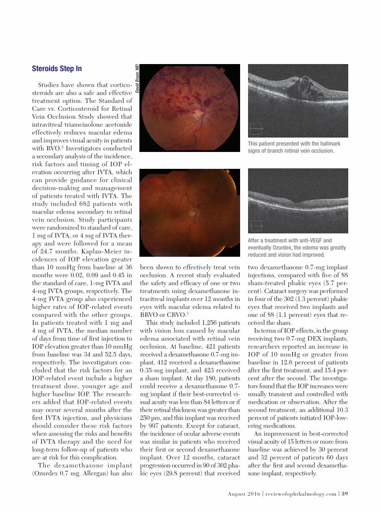
August 2016 | reviewofophthalmology.com | 39
Steroids Step In
Studies have shown that cortico-steroids are also a safe and effective treatment option. The Standard of Care vs. Corticosteroid for Retinal Vein Occlusion Study showed that intravitreal triamcinolone acetonide effectively reduces macular edema and improves visual acuity in patients with RVO.2 Investigators conducted a secondary analysis of the incidence, risk factors and timing of IOP el-evation occurring after IVTA, which can provide guidance for clinical decision-making and management of patients treated with IVTA. The study included 682 patients with macular edema secondary to retinal vein occlusion. Study participants were randomized to standard of care,1 mg of IVTA, or 4 mg of IVTA ther-apy and were followed for a mean of 24.7 months. Kaplan-Meier in-cidences of IOP elevation greater than 10 mmHg from baseline at 36 months were 0.02, 0.09 and 0.45 in the standard of care, 1-mg IVTA and 4-mg IVTA groups, respectively. The 4-mg IVTA group also experienced higher rates of IOP-related events compared with the other groups. In patients treated with 1 mg and4 mg of IVTA, the median number of days from time of fi rst injection to IOP elevation greater than 10 mmHg from baseline was 34 and 52.5 days, respectively. The investigators con-cluded that the risk factors for an IOP-related event include a higher treatment dose, younger age and higher baseline IOP. The research-ers added that IOP-related events may occur several months after the fi rst IVTA injection, and physicians should consider these risk factors when assessing the risks and benefi ts of IVTA therapy and the need for long-term follow-up of patients who are at risk for this complication.
The dexamethasone implant (Ozurdex 0.7 mg, Allergan) has also
been shown to effectively treat vein occlusion. A recent study evaluated the safety and effi cacy of one or two treatments using dexamethasone in-travitreal implants over 12 months in eyes with macular edema related to BRVO or CRVO.3
This study included 1,256 patients with vision loss caused by macular edema associated with retinal vein occlusion. At baseline, 421 patients received a dexamethasone 0.7-mg im-plant, 412 received a dexamethasone 0.35-mg implant, and 423 received a sham implant. At day 180, patients could receive a dexamethasone 0.7-mg implant if their best-corrected vi-sual acuity was less than 84 letters or if their retinal thickness was greater than 250 µm, and this implant was received by 997 patients. Except for cataract, the incidence of ocular adverse events was similar in patients who received their first or second dexamethasone implant. Over 12 months, cataract progression occurred in 90 of 302 pha-kic eyes (29.8 percent) that received
two dexamethasone 0.7-mg implant injections, compared with fi ve of 88 sham-treated phakic eyes (5.7 per-cent). Cataract surgery was performed in four of the 302 (1.3 percent) phakic eyes that received two implants and one of 88 (1.1 percent) eyes that re-ceived the sham.
In terms of IOP effects, in the group receiving two 0.7-mg DEX implants, researchers reported an increase in IOP of 10 mmHg or greater from baseline in 12.6 percent of patients after the fi rst treatment, and 15.4 per-cent after the second. The investiga-tors found that the IOP increases were usually transient and controlled with medication or observation. After the second treatment, an additional 10.3 percent of patients initiated IOP-low-ering medications.
An improvement in best-corrected visual acuity of 15 letters or more from baseline was achieved by 30 percent and 32 percent of patients 60 days after the fi rst and second dexametha-sone implant, respectively.
This patient presented with the hallmark
signs of branch retinal vein occlusion.
After a treatment with anti-VEGF and
eventually Ozurdex, the edema was greatly
reduced and vision had improved.
Davi
d B
oye
r, M
D
038_rp0816_f3.indd 39038_rp0816_f3.indd 39 7/22/16 1:35 PM7/22/16 1:35 PM

RE
VIE
W
40 | Review of Ophthalmology | August 2016
RetinaCoverFocus
“Ozurdex works remarkably well in some patients. It is able to stabilize vi-sion for longer periods of time, and in some cases, it can reduce the number of treatments that are necessary,” Dr. Boyer adds.
A Combination Approach
While anti-VEGF and cortico-steroid treatments work well alone, there’s evidence that they work even better in combination, and accord-ing to Chicago-based vitreoretinal surgeon Seenu Hariprasad, MD, the treatment paradigm is headed toward combination therapy. “There is re-ally good evidence in the literature that vein occlusion is a multifactorial disease. Of all the retinal diseases that we treat, branch retinal vein occlu-sion and central retinal vein occlu-sion cause the highest level of VEGF in the eye. There is also very good evidence that there is an infl amma-tory component that anti-VEGF monotherapy does not address. This may contribute to the high treatment burden with anti-VEGF therapy, so I am a big believer in incorporating other modalities of treatment, such as Ozurdex. I think a combination of an anti-VEGF and Ozurdex can be very powerful,” adds Dr. Hariprasad.
A 2014 study found that bevaci-zumab combined with dexametha-sone implants produced greater improvements in macular thickness than bevacizumab therapy alone and required fewer bevacizumab injec-tions in both BRVO and CRVO.4
The study included 30 eyes that were randomly assigned to receive either combination therapy or mono-therapy with bevacizumab. All pa-tients received intravitreal bevaci-zumab at baseline, followed a week later by dexamethasone implants or sham injections. Monthly bevacizum-ab injections were given if the cen-tral subfi eld thickness was less than 250 µm, and the combined group re-
ceived a second implant after four or fi ve months if the central subfi eld thickness was less than 250 µm.
At six months, patients receiving combined therapy required fewer bevacizumab re-injections compared to those receiving monotherapy (two compared to three). They also experienced greater mean reduc-tions in central subfield thickness (-56 µm compared to +45 µm) and were more likely to have resolved all edema, which was considered to be a central subfi eld thickness less than250 µm (seven of 11 eyes compared to two of 14 eyes). Mean visual acuity changes from baseline were similar between groups.
Dr. Hariprasad notes that imaging is critical in this disease. “Using OCT is one of the best ways to track treat-ment response,” he says. “We get a baseline OCT, and then we can de-termine whether the pharmacologic agent is working or not. So, it is a great way to track and quantify treatment. We also use fl uorescein angiography to determine the amount of macular and peripheral ischemia and address both accordingly. I would recommend ultra-wide-fi eld fl uorescein angiogra-phy and OCT testing.”
According to Dr. Hariprasad, a potentially important advance in the treatment of retinal vein occlusion is CLS-1003 (Clearside Biomedical), which consists of a microneedle injec-tion of the company’s Zuprata (triam-cinolone acetonide) into the supra-choroidal space in combination with an intravitreal injection of Eylea. The therapy was developed for the treat-ment of macular edema associated with retinal vein occlusion.
In April, Clearside completed a small Phase II trial of 46 patients.5 In this study, patients who received con-comitant administration of Zuprata and Eylea qualifi ed for approximately 60 percent fewer intravitreal Eylea treatments than those patients who received Eylea alone. This is the fi rst
controlled, masked, randomized trial conducted in patients with retinal vein occlusion where the drug was admin-istered through the suprachoroidal space. No serious adverse events were reported during the trial, and treat-ment was generally well-tolerated. “In the past, we just used an anti-VEGF,” Dr. Hariprasad says, commenting on the results. “Then, the corticosteroids came along and enhanced the way we treat the disease.”
According to Daniel H. White, CEO and president of Clearside, “Based on these results, Clearside in-tends to follow a 505(b)(2) NDA regu-latory approval pathway and expedite the preparations for a Zuprata Phase III registration program for the treat-ment of macular edema associated with RVO.”
Dr. Singer is a consultant for Al-lergan, Genentech and Regeneron. Dr. Boyer is a consultant for Allergan, Ge-nentech, Roche, Bayer, Novartis and Regeneron. Dr. Hariprasad is a con-sultant for Alcon, Allergan, Bayer, Al-imera Sciences, Clearside Biomedical, Janssen, Ocular Therapeutix, OD-OS, Optos, Regeneron and Spark.
1. Yuan A, Ahmad BU, Xu D, Singh RP, Kaiser PK, Martin DF, Sears JE, Schachat AP, Ehlers JP. Comparison of intravitreal ranibizumab and bevacizumab for the treatment of macular edema secondary to retinal vein occlusion. Int J Ophthalmol 2014;7:1:86-91.2. Aref AA, Scott IU, Oden NL, et al; SCORE Study Investigator Group. Incidence, risk factors, and timing of elevated intraocular pressure after intravitreal triamcinolone acetonide injection for macular edema secondary to retinal vein occlusion: SCORE study report 15. JAMA Ophthalmol 2015;133:9:1022-1029.3. Haller JA, Bandello F, Belfort R Jr, et al. Dexamethasone intravitreal implant in patients with macular edema related to branch or central retinal vein occlusion: Twelve-month study results. Ophthalmology 2011;118:12:2453-2460.4. Maturi RK, Chen V, Raghinaru D, Bleau L, Stewart MW. A 6-month, subject-masked, randomized controlled study to assess effi cacy of dexamethasone as an adjunct to bevacizumab compared with bevacizumab alone in the treatment of patients with macular edema due to central or branch retinal vein occlusion. Clin Ophthalmol 2014;8:1057-1064.5. Data on fi le, Clearside Biomedical. http://ir.clearsidebio.com/phoenix.zhtml?c=253669&p=irol-newsArticle&ID=2162115.
Suggested Reading:1. Hariprasad S. Management of Retinal Vein Occlusion: Current Concepts. 2014. Slack Incorporated. https://www.amazon.com/Management-Retinal-Vein-Occlusion-Concepts/dp/1617116165/ref=sr_1_1?ie=UTF8&qid=1465932449&sr=8-1&keywords=hariprasad+retinal
038_rp0816_f3.indd 40038_rp0816_f3.indd 40 7/22/16 1:35 PM7/22/16 1:35 PM

Dear Resident Program Director and Coordinator,
We would like to invite you to review the upcoming 3rd-Year Resident Programs for 2016 in Fort Worth. The programs
offer a unique educational opportunity for third-year residents by providing the chance to meet and exchange ideas with
some of the most respected thought leaders in ophthalmology. The programs are designed to provide your residents
with a state-of-the-art didactic and wet lab experience. The programs also serve as an opportunity for your residents to
network with residents from other programs.
After reviewing the material, it is our hope that you will select and encourage your residents to attend one of these
educational activities, which are CME accredited to ensure fair balance.
Sincerely,
Postgraduate Healthcare Education
Third-Year Resident Programs 2016:
August 19-20
Fort Worth, TX
Course Director
Mitchell Weikert, MD
September 9-10
Fort Worth, TX
Course Director
Anthony Arnold, MD
September 16-17Fort Worth, TX
Course Director
Tommy Korn, MD
For more information: Visit the registration site above orEmail: [email protected]
Call: Denette Holmes 866-627-0714
Courses are restricted to 3rd-year residents enrolled in an ophthalmology resident program and within their third year at the time of the course.
There is no registration fee for these activities. Air, ground transportation in Forth Worth, hotel accommodations and
modest meals will be provided through an educational scholarship for qualifi ed participants.
Accreditation StatementThis activity has been planned and implemented in accordance with the accreditation requirements and policies of the Accreditation Council for Continuing Medical
Education (ACCME) through the joint providership of Amedco and Postgraduate Healthcare Education. Amedco is accredited by the ACCME to provide continuing
medical education for physicians.
Credit Designation StatementThis activity has been approved for AMA PRA Category 1 CreditTM.
www.revophth.com/ResEdu2016
Jointly provided byEndorsed by
Review of Ophthalmology®
Supported by an unrestricted independent medical educational grant from
Alcon
2016
*Subject to change
2016_CSE_HouseAd.indd 862016_CSE_HouseAd.indd 86 7/12/16 10:29 AM7/12/16 10:29 AM

42 | Review of Ophthalmology | August 2016
Cover Focus RE
VIE
W
This article has no commercial sponsorship.
Retina
Richard T. Atallah, Manuela Von Sneidern, Alan T. Sheyman, MD, Ben Z. Cohen, MD
New York City
Intravitreal anti-vascular endothe-lial growth factor therapies have become an important treatment
for patients with neovascular age re-lated macular degeneration, diabetic macular edema, proliferative diabetic retinopathy and many other retinal conditions. When you draw a dose of an anti-VEGF drug, however, current regulations prohibit you from using any more from that vial, even though there may be several doses left.1 Judg-ing by the cost of injecting certain drugs such as Lucentis (ranibizum-ab) and Eylea (aflibercept), leaving these vials unused leads to hundreds of thousands of dollars in wasted drug each year. In this article, we take a look at the pros and cons of a compounded drug such as Avastin (bevacizumab), and the feasibility of reusing the vials of ostensibly safer, non-compounded drugs such as Lucentis for more than just one injection.
The Cost of Compounding
Though ophthalmologists want to do what’s best for their patients, as in most areas of life, cost is a major factor; and there is a signifi cant cost difference between the anti-VEGF medications.
In 2015, the American Society of Retina Specialists asked its members,
“If Avastin, Lucentis and Eylea were the same cost for each patient, which drug would you use primarily for the treatment of new-onset exudative AMD?”2 Less than 10 percent indi-cated that they would use Avastin fi rst, even though, in the real world, it’s actually the preferred fi rst-line drug for 64 percent of ophthalmologists. Findings such as this show just how important cost is when choosing anti-VEGF medications.3
However, even though Avastin is cost-effective, it has a potential draw-back, too: It needs to be compounded in order for it to be usable in the eye. This drawback looms large due to an outbreak of fungal meningitis from three lots of preservative-free meth-ylprednisolone acetate compounded at the New England Compounding Center in September 2012. Further, in an article in JAMA Ophthalmology in 2013 by one of this article’s co-au-thors, Alan Sheyman, MD, he and his colleagues described a series of eight patients who received an intravitreal injection of compounded combined bevacizumab-triamcinolone from the same lot—who were later diagnosed with fungal endophthalmitis.4 This outbreak occurred despite the current practice of compounding pharmacies and outsourcing facilities performing microbiological testing on their com-
Reusing Lucentis
and Eylea vials
might be feasible
the authors say.
Anti-VEGF Everywhere, But Not a Drop to Use
042_rp0816_f4.indd 42042_rp0816_f4.indd 42 7/22/16 1:37 PM7/22/16 1:37 PM
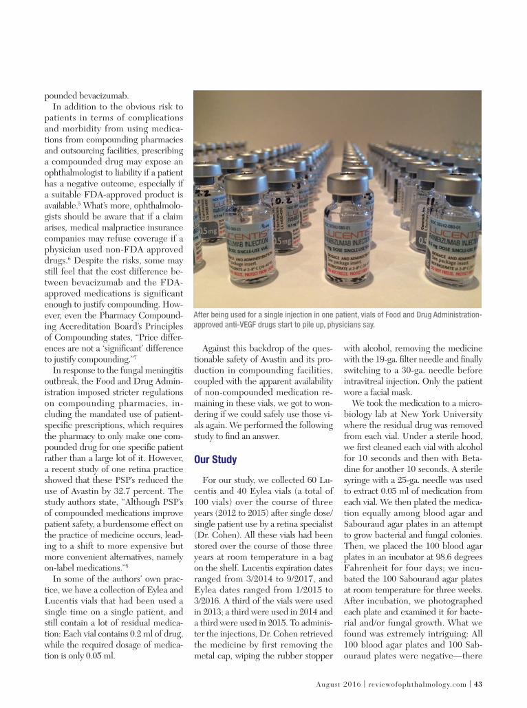
August 2016 | reviewofophthalmology.com | 43
pounded bevacizumab.In addition to the obvious risk to
patients in terms of complications and morbidity from using medica-tions from compounding pharmacies and outsourcing facilities, prescribing a compounded drug may expose an ophthalmologist to liability if a patient has a negative outcome, especially if a suitable FDA-approved product is available.5 What’s more, ophthalmolo-gists should be aware that if a claim arises, medical malpractice insurance companies may refuse coverage if a physician used non-FDA approved drugs.6 Despite the risks, some may still feel that the cost difference be-tween bevacizumab and the FDA-approved medications is significant enough to justify compounding. How-ever, even the Pharmacy Compound-ing Accreditation Board’s Principles of Compounding states, “Price differ-ences are not a ‘signifi cant’ difference to justify compounding.”7
In response to the fungal meningitis outbreak, the Food and Drug Admin-istration imposed stricter regulations on compounding pharmacies, in-cluding the mandated use of patient-specifi c prescriptions, which requires the pharmacy to only make one com-pounded drug for one specifi c patient rather than a large lot of it. However, a recent study of one retina practice showed that these PSP’s reduced the use of Avastin by 32.7 percent. The study authors state, “Although PSP’s of compounded medications improve patient safety, a burdensome effect on the practice of medicine occurs, lead-ing to a shift to more expensive but more convenient alternatives, namely on-label medications.”8
In some of the authors’ own prac-tice, we have a collection of Eylea and Lucentis vials that had been used a single time on a single patient, and still contain a lot of residual medica-tion: Each vial contains 0.2 ml of drug, while the required dosage of medica-tion is only 0.05 ml.
Against this backdrop of the ques-tionable safety of Avastin and its pro-duction in compounding facilities, coupled with the apparent availability of non-compounded medication re-maining in these vials, we got to won-dering if we could safely use those vi-als again. We performed the following study to fi nd an answer.
Our Study
For our study, we collected 60 Lu-centis and 40 Eylea vials (a total of 100 vials) over the course of three years (2012 to 2015) after single dose/single patient use by a retina specialist (Dr. Cohen). All these vials had been stored over the course of those three years at room temperature in a bag on the shelf. Lucentis expiration dates ranged from 3/2014 to 9/2017, and Eylea dates ranged from 1/2015 to 3/2016. A third of the vials were used in 2013; a third were used in 2014 and a third were used in 2015. To adminis-ter the injections, Dr. Cohen retrieved the medicine by first removing the metal cap, wiping the rubber stopper
with alcohol, removing the medicine with the 19-ga. fi lter needle and fi nally switching to a 30-ga. needle before intravitreal injection. Only the patient wore a facial mask.
We took the medication to a micro-biology lab at New York University where the residual drug was removed from each vial. Under a sterile hood, we fi rst cleaned each vial with alcohol for 10 seconds and then with Beta-dine for another 10 seconds. A sterile syringe with a 25-ga. needle was used to extract 0.05 ml of medication from each vial. We then plated the medica-tion equally among blood agar and Sabouraud agar plates in an attempt to grow bacterial and fungal colonies. Then, we placed the 100 blood agar plates in an incubator at 98.6 degrees Fahrenheit for four days; we incu-bated the 100 Sabouraud agar plates at room temperature for three weeks. After incubation, we photographed each plate and examined it for bacte-rial and/or fungal growth. What we found was extremely intriguing: All 100 blood agar plates and 100 Sab-ouraud plates were negative—there
After being used for a single injection in one patient, vials of Food and Drug Administration-
approved anti-VEGF drugs start to pile up, physicians say.
042_rp0816_f4.indd 43042_rp0816_f4.indd 43 7/22/16 1:36 PM7/22/16 1:36 PM

RE
VIE
WRetina
CoverFocus
was no bacterial or fungal growth.In our view, even though this was
a small-scale study, the lack of con-tamination of 100 vials of Lucentis and Eylea calls into question the FDA’s and Medicare’s policies on the use of residual medication in such vials. Drawing more medication from a sin-gle vial isn’t unprecedented: It may be surprising to know that in a survey by the Joint Commission, 6 percent of health-care practitioners admitted to sometimes or always using single dose vials for multiple patients. Also, according to a JAMA study in 2010, 28 percent of all ambulatory surgery cen-ters admitted to using single-dose vials for multiple patients.9 Nonetheless, governmental guidelines are intended to protect patients from infections where vials become contaminated from unsafe injection practices, and we completely support the spirit of the law. The fact remains, however,
that our study showed no bacterial or fungal growth in the residual medica-tion in these 100 vials over the course of three years of storage, a strong testament to the fact that these vials can most likely be reused safely. In the coming years, we hope to see a larger-scale evaluation of the stability of these drugs over time to reinforce our fi ndings and possibly convince the powers-that-be to eliminate risk—and waste—from the system.
Mr. Atallah and Ms. Von Sneidern are in the pre-med program at New York University. Dr. Sheyman is a retinal specialist at Northwestern University in Chicago. Dr. Cohen is a retinal specialist at Mt. Sinai Med-ical Center in New York City. The authors would like to acknowledge Joel S. Mindel, MD, PhD, director of neuro-ophthalmology at Mt. Sinai School of Medicine in New York City,
for his editorial review. None of the authors have a fi nancial interest in the products discussed.
1. Bagian JP, Federico F, Barnsteiner JH, Battles JB, Beeson WH, Benjamin, Swenson D. Preventing infection from the misuse of vials. Sentinel Event Alert 2014;52:1-6.2. Stone TW, ed. American Society of Retina Specialists 2015 Preferences and Trends Membership Survey. Chicago: American Society of Retina Specialists.3. Thompson JT. Patient and physician convenience and the choice of intravitreal anti-vascular endothelial growth factor medications. JAMA Ophthalmol 2016;134:1:49. 4. Sheyman AT, Cohen BZ, Friedman AH, Ackert JM. An outbreak of fungal endophthalmitis after intravitreal injection of compounded combined bevacizumab and triamcinolone. JAMA Ophthalmology 2013;131:7:864. 5. Seller S, Utian WH. Pharmacy compounding primer for physicians: Prescriber beware. Drugs 2012;72:16:2043-2050.6. Kabnick LS. Compounded sclerosants and foam: What should you know about this controversial area? Legal guidelines for use of polidocanol and sodium tetradecyl sulfate for sclerotherapy. Veith Symposium;19-23 Nov 2008;New York.7. Pharmacy Compounding Accreditation Board: PCAB Principles of Compounding, 2012. https://secure.pcab.info/about/downloads/principles-of-compounding.pdf. Accessed February 2016.8. Holfi nger S, Miller AG, Rao LJ, Rowland DY, Hornik JH, Miller DG. Effect of regulatory requirement for patient-specifi c prescriptions for off-label medications on the use of intravitreal bevacizumab. JAMA Ophthalmol 2016;134:1:45-8.9. Schaefer MK, Jhung M, Dahl M, Schillie S, Simpson C, Llata E, Perz JF. Infection control assessment of ambulatory surgical centers. JAMA 2010;303:22;2273-2279.
Local Eye Site
AssessmentHiring? Use an assessment tool that is scientifically proven
to predict performance in eye care jobs.
localeyesite.com/assessment
042_rp0816_f4.indd 44042_rp0816_f4.indd 44 7/22/16 1:36 PM7/22/16 1:36 PM

Retinal Insider R
EV
IEW
August 2016 | reviewofophthalmology.com | 45This article has no commercial sponsorship.
Edited by Carl Regillo, MD, and Emmett T. Cunningham, Jr., MD, PhD, MPH
Vascular endothelial growth fac-tor is the primary treatment tar-
get in many cases of diabetic macular edema, and the disease process often responds to intravitreal anti-VEGF injections. But what happens if you lock down VEGF with your injec-tions but the patient doesn’t respond and continues to lose vision? It turns out there are other mediators lurk-ing behind the scenes, influencing the pathophysiology of DME. Oph-thalmic research is dragging more of these mediators into the light every day, however. This review will cover the various contributors to vision loss in DME and how emerging drugs are targeting them. (It should be noted that many of these drugs affect sev-eral of these pathways; consequently, listing a particular drug under one pathway represents a simplifi cation to enhance readability.)
Permeability
Vascular permeability, leading to macular edema, causes vision loss through mechanical distortion of the orderly retinal architecture as well as the secondary signal interference. In
diabetes, chronic hyperglycemia con-tributes to alterations in the structural and cellular components of retinal microvasculature. Early on, there is damage to the pericytes responsible for regulating capillary perfusion, which results in microaneurysm for-mation and impaired regulation of retinal blood fl ow.1 Damage to endo-thelial cells responsible for maintain-ing the blood-retinal barrier allows ac-cumulation of extracellular fl uid in the macula. There’s also thickening of the capillary basement membrane and increased deposition of extracellu-lar matrix components.1,2,3 Over time, continued retinal microvasculature damage causes capillary nonperfusion and retinal ischemia, resulting in ac-tivation of hypoxia-inducible factor-1 alpha (HIF-1a) via signaling through phosphoinositide 3-kinase and its downstream target, mammalian tar-get of rapamycin (mTOR).4 HIF-1a causes upregulation of VEGF, which mediates vascular permeability.5
Though there are several different VEGF isoforms in the body due to alternative splicing, VEGF-A and pla-cental growth factor mediate vascular permeability and pathologic angio-
genesis in the eye. Current agents, ranibizumab (Lucentis, Genentech/Roche), afl ibercept (Eylea, Regener-on) and off-label bevacizumab (Avas-tin, Genentech/Roche), bind VEGF to prevent activation of VEGF recep-tors, thereby decreasing angiogenesis and vascular permeability, causing regression of diabetic neovasculariza-tion and reduction in DME, respec-tively.2,6
Another target of interest is the Tie-2 receptor tyrosine kinase, which is expressed by endothelial cells. When stimulated, it produces reinforcement of junctional proteins, more inter-action with surrounding cells and matrix, and stabilization of the vas-culature. Tie-2 is activated by Angio-poietin-1, which results in reduced permeability, whereas Angiopoietin-2 serves as a competitive antagonist that diminishes Tie-2 activity. Isch-emic retina results in elevated Ang-2, which is also elevated in patients with DME.7 Following are two investiga-tional agents that target permeability.
• Nesvacumab. Ang-2 antibody Nesvacumab (Regeneron) binds Ang-2, allowing more Ang-1 to activate Tie-2, so that permeability can be di-
Rehan M. Hussain, MD, Thomas A. Ciulla, MD, MBA, Indianapolis
A look at what’s going on behind the scenes in patients for whom anti-VEGF therapy has little or no effect.
Targeting VEGF’s Silent Partners in DME
045_rp0816_rtinsider.indd 45045_rp0816_rtinsider.indd 45 7/21/16 3:12 PM7/21/16 3:12 PM

RetinalInsider R
EV
IEW
46 | Review of Ophthalmology | August 2016
minished. Trials comparing afliber-cept alone to afl ibercept with Ang-2 antibodies in DME patients are on the horizon.
• AKB-9778. Also affecting the same pathway, AKB-9778 inactivates vascular endothelial-protein tyrosine phosphatase. VE-PTP dephosphory-lates Tie-2, causing it to shift to an inactive state. Therefore, AKB-9778 helps keep Tie-2 active by inactivat-ing VE-PTP. This drug is adminis-tered by subcutaneous injection, and may serve as an alternative option for patients averse to intravitreal injec-tions. A Phase Ib trial showed that AKB-9778 was well-tolerated, caused signifi cant reductions in retinal thick-ness and signifi cantly improved visual acuity.8
The Vitreoretinal Interface
For refractory cases of DME, it’s important to assess the vitreoretinal interface carefully, as vitreomacular traction and epiretinal membrane can contribute to macular edema and as-sociated visual loss via tractional forc-es that exacerbate hyperpermeability. Here’s a look at two approaches ad-dressing interface issues.
• Jetrea (ocriplasmin). The vit-reoretinal interface is anchored by extracellular matrix proteins such as laminin, integrins, fibronectin and collagen types VI, VII and XVIII.9 Ocriplasmin, a key ingredient in ThromboGenics’ drug Jetrea, is a re-combinant truncated form of human plasmin that has proteolytic activity against these proteins. However, it’s not active against type IV collagen, a component of the internal limiting membrane, thus limiting its toxicity to the retina. When injected into the eye, ocriplasmin can cause pharmaco-logic vitreolysis and posterior vitreous detachment, offering an alternative to surgery. In one study, ocriplasmin caused resolution of VMT in 26.5 per-cent of eyes compared to 10.1 percent
of placebo eyes within 28 days.10 Ocri-plasmin could be used in cases involv-ing VMT with narrow vitreomacular adhesions.
Surgical intervention may be re-quired in cases not amenable to ocri-plasmin injection, however, such as those with broad vitreomacular ad-hesions or epiretinal membrane. In such cases, pars plana vitrectomy with or without membrane peel can serve to release the anteroposterior and tangential tractional forces of the vitreous on the ILM. Gradual res-toration of the retinal architecture may result in reduction of edema and improvement in visual acuity. Dia-betic eyes may have diffuse residual vitreous cortex after surgical creation of a PVD, which can lead to con-tinued traction and development of ERM.11 Surgical removal of the ILM eliminates the vitreous cortex and any potential proliferative scaffold that could contribute to ERM formation. In eyes with diffuse DME, the ILM becomes thickened and a variety of cells, including myofi broblasts and in-fl ammatory cells, adhere to the inner surface of the ILM, with the potential to further exacerbate edema.11
• Luminate. Another potential treatment targets integrins, which are cell adhesion and cell signaling recep-tors. The integrin peptide antagonist Luminate (Allegro Ophthalmics) tar-gets receptors involved in both angio-
genesis and vitreolysis.Integrins mediate cell functions,
interactions among cells and the ex-tracellular matrix, and activate intra-cellular pathways that promote angio-genesis. In the body, there are many different types of integrin receptors, but the αvβ3, α5β1, and αvβ5 subtypes are expressed more in diabetic reti-nopathy.12 Inhibiting α5β1 has been shown to inhibit endothelial cell pro-liferation and produce regression of choroidal neovascular membranes.13,14 Integrin α3β1 mediates attachment of the vitreous to the retinal surface. In-hibiting α3β1 results in vitreolysis and PVD, which can help patients with VMT, and may serve as an alternative treatment to ocriplasmin. Luminate is currently in Phase II trials for treat-ment of DME, VMT and AMD.
Infl ammation
Inflammation plays an impor-tant role in diabetic retinopathy and DME, as leukostasis, prostaglandin upregulation and retinal accumula-tion of macrophages all occur in dia-betes. Diabetics may have abnormally large and rigid leukocytes that adhere to the vascular endothelium, which can contribute to vascular occlusion and ischemia; these leukocytes gener-ate toxic superoxide radicals and pro-teolytic enzymes.15 The end result is endothelial dysfunction, vascular per-
Early (left) and late phase (right) fl uorescein angiograms show the typical appearance of
an eye with diabetic macular edema.
045_rp0816_rtinsider.indd 46045_rp0816_rtinsider.indd 46 7/21/16 3:12 PM7/21/16 3:12 PM

August 2016 | reviewofophthalmology.com | 47
meability, retinal nonperfusion and, potentially, angiogenesis, all of which lead to vision loss.
VEGF possesses inflammatory properties through its capacity to mediate microvascular permeabil-ity and increase adhesion of leuko-cytes. VEGF was found to stimulate expression of intracellular adhesion molecule-1 and vascular cell adhe-sion molecule-1,6 therefore involving the infl ammatory cascade, initiating early diabetic retinal leukocyte adhe-sion and contributing to the develop-ment of diabetic vasculopathy. Thus, anti-VEGF agents may also have a therapeutic mechanism that involves limitation of infl ammation. Other in-fl ammatory mediators such as tumor necrosis factor-alpha, interleukin-6, interleukin-8, cyclo-oxygenase-2 and monocyte chemotactic protein-1 may also be upregulated in DME.17
• Ozurdex and Iluvien. Recently, there’s been interest in sustained-release corticosteroids, such as the dexamethasone intravitreal implant (Ozurdex, Allergan)18 and the fluo-cinolone acetonide implant (Iluvien, Alimera),19,20 both of which were ap-proved for the treatment of DME in 2014. Research has suggested that inflammatory cytokines play a larg-er role in chronic DME than non-chronic DME.18 For this reason, anti-VEGF therapy may not be effective in all patients, because targeting VEGF does not suppress all the infl amma-tory cytokines involved in chronic DME. Corticosteroids can address this aspect of the pathophysiology by inhibiting the arachidonic acid path-way, resulting in downregulation of prostaglandins, prostacyclin, throm-boxanes and leukotrienes. In addition to this anti-infl ammatory mechanism, corticosteroids can alter the compo-sition of the endothelial basement membrane by changing the local ratio of two laminin isoforms, suppress-ing basement membrane dissolution and strengthening tight junctions to
limit leakage that can cause macular edema.22,23 In the future, the com-bination of anti-VEGF therapy and sustained-release corticosteroids is an area of research that may hold prom-ise for treating refractory DME and decreasing the treatment burden of repeated injections.
There are also a couple of new therapies being explored to target the inflammatory component of DME that might serve as alternatives to cor-ticosteroids:
• Infl iximab. The drug infl iximab inhibits the pro-inflammatory cyto-kine TNF-alpha, which has been im-plicated in breakdown of the blood-retinal barrier in diabetic animal models.24 Small studies have yielded inconsistent results in regards to its effi cacy in treating refractory DME, however. 25,26 IL-6 is another pro-in-fl ammatory cytokine that may induce expression of VEGF or may directly increase vascular permeability.27,28 IL-6 levels were found to be elevated in patients with DME without PVD, and correlated signifi cantly with se-verity of disease.29 An IL-6 antibody is currently in development for trials.
• KKS inhibitors. The kallikrein-kinin system is activated in response to vascular injury and leads to a local increase in bradykinin, which further results in pro-inflammatory effects such as vascular permeability, vaso-dilation and immune cell activation.30 Rodent model studies have shown that diabetes and hypertension in-crease KKS components in the retina, and inhibition of plasma kallikrein was found to reduce vascular perme-ability.31 VEGF also leads to increased kallikrein levels in the retina. A KKS inhibitor is being explored for thera-peutic effi cacy in Phase II trials.
Fibrosis
Retinal fibrosis, which severely distorts and destroys retinal tissue, can result from chronic DME, espe-
cially when hard exudates occupy the fovea. Consequently, limiting DME via any of the pathways noted above can minimize fi brosis. As previously mentioned, hypoxia-inducible factor-1a accumulates under hypoxic con-ditions and serves as a stimulator of VEGF. Hence, there is benefi t to in-hibiting this pathway that is regulated by the mTOR kinase.4
• Sirolimus. Also known as rapamy-cin, Pfizer’s Sirolimus is an mTOR inhibitor with the ability to inhibit angiogenesis, permeability, infl amma-tion and fi brosis, making it potentially useful for variety of retinal vascular diseases. It also inhibits protein kinase C and pro-inflammatory mediators such IL-8, endothelial-monocyte ac-tivating peptide-II, COX-1 and COX-2.32,33 Rapamycin’s ability to inhibit fi brosis,34 an important contributor to irreversible photoreceptor death and severe visual loss, may fi ll a void in the currently available DME treatments.
Ischemia
In diabetic retinopathy, enlarge-ment of the foveal avascular zone is detected by fl uorescein angiography; it is considered an indication of isch-emia and may contribute to macular edema.35 Foveal ischemia in DME causes photoreceptor outer segment shortening and inner segment-outer segment disruption, resulting in outer retinal atrophic changes that correlate with visual loss.36 Unfortunately, there are no treatments available to reverse the atrophic photoreceptor changes associated with macular ischemia.
The pathophysiology of DME is complex and allows for many poten-tial therapeutic targets. While cur-rently laser photocoagulation, anti-VEGF injections and corticosteroids are the most commonly used treat-ments, there are many intriguing op-tions being explored to address other
(continued on page 60)
045_rp0816_rtinsider.indd 47045_rp0816_rtinsider.indd 47 7/21/16 3:12 PM7/21/16 3:12 PM

Therapeutic Topics R
EV
IEW
This article has no commercial sponsorship.
Playwright and cr i t ic George Bernard Shaw once wrote, “The
only man I know who behaves sensi-bly is my tailor; he takes my measure-ments anew each time he sees me. The rest go on with their old mea-surements and expect me to fi t them.” Measurement scales are so much a part of our daily routines we almost never give them a second thought. Yet, just as the tailor knows to ad-just and re-measure every time, as ophthalmologists we should, from time to time, mentally recalibrate the scales we use to assess signs and symptoms, subject as they are to drift and bias. This is particularly true in the case of the subjective scales we use to measure qualitative data: our use of these metrics refl ects our in-herent knowledge and clinical expe-rience, and their application should bear the scrutiny of comparisons with the broader scientifi c commu-nity. When it comes to scales, we should be asking the question: are we all on the same page? Is your 2+ redness the same as mine?
This month we’ll consider the elements of these metrics and dis-cuss the scales we use for grading the signs of ocular disease. In a fu-ture column, we’ll explore symptom
scales, questionnaires and patient-reported outcomes, all necessary tools of the trade that also need oc-casional sharpening. Understanding the statistical parameters and tests commonly used for assessing grading systems is an essential fi rst step to-wards critically thinking about these grading scales and the responses and symptoms they measure.
The Measure of A Measure
The purpose of any scale is to pro-vide a means to measure a trait, a physical change or any parameter not readily amenable to direct quantita-tion. Ultimately, we use the scores generated for comparison, as a func-tion of age, health status or treat-ment regimen. All scales are not the same however, and when developing a new scale it’s necessary to test for precision, for repeatability (see the glossary on p. 51) and for agreement between graders, as well as to test for agreement with existing disease metrics.
There are a number of statistical tests that measure agreement, such as the intra-class correlation coef-ficient. The ICC is a ratio derived from the analysis of measurement
variance. The closer the ratio is to 1.0, the greater the agreement be-tween tests.2 One statistics text sug-gests that an ICC of 0.4 indicates poor agreement, 0.4 to 0.75 as fair to good agreement, and greater than 0.75 as excellent agreement.3
Agreement is also presented by Bland Altman plots,4 which elegantly illustrate the difference in paired ob-servations on the y-axis versus the mean of the observations on the x-axis, including horizontal lines for the mean difference ± two standard deviations. Bland Altman plots are a very useful snapshot of agreement between two devices, systems or graders. They originated the concept of limits of agreement between two methods. A correlation coeffi cient (r) is commonly and incorrectly used to measure agreement, when r actually measures the strength of relation be-tween two variables, not the agree-ment between them.
There can be high concordance in using a scale because the catego-ries are simply too broad. We could all throw a tennis ball through the width of a barn door, but what about through a window? In the case of wide confi dence limits, the grading system won’t be sensitive enough to
Mark B. Abelson, MD, CM, FRCSC, FARVO, Lisa M Smith, David A. Hollander, MD, MBA, and Dale Usner, PhD
Andover, Mass.
How Do You Quantify The Qualitative?
48 | Review of Ophthalmology | August 2016
048_rp0816_ttops.indd 48048_rp0816_ttops.indd 48 7/21/16 11:42 AM7/21/16 11:42 AM

measure change, regardless of the fact that we all agreed as to each step in the scale. Thus, we want a system that is not so coarse that sensitivity is lost, and not so fi ne that there is no agree-ment among users and, consequently, no ability to have comparable data.
A fi ne example of this was laid out in a fascinating 1991 paper on the use of scales.5 Using the example of a lens opacity grading system, the authors had two graders score 87 lenses. In-stead of using the 0 to 4 scale, they broke down the scale into fi ner granu-larity, with each integer divided into nine decimals, such as 2.1, 2.2,…, 2.9, etc. With the integer scale, the con-cordance was 74 percent, with vari-ance equal to ±1 grading units. To fall out of the confi dence limits, there had to be a difference of two grading units between the paired observa-tions. When the decimalized scale was used, the concordance was only 9 percent between graders, but the confi dence limits were at 0.8 grading units. Thus, the decimalized scale gave confi dence limits 2.5 times fi ner than those given by the integer scale. While practicality drives us to contin-ue to use integer scales, incorporat-ing 0.5 increments increases granu-larity and improves the sensitivity of our grading.
Ideally, we want our measurement scales to be able to identify clinical change in our patients. In practice, we need to know if a keratitis or fl are reaction is worse this week than last, and we use scales to measure the progression of disease.
Change can also be associated with treatment, either in practice or in research. In a clinical trial, treatment can be either positive (comparator) or negative (vehicle, placebo). It’s particularly critical to have a sen-sitive measurement when dealing with a bioequivalence clinical trial of one active to a test compound. When both agents being studied in the trial have the same therapeutic effect—
we must be sure differences in effect are measured sensitively and accu-rately to establish bioequivalence.6
The opposite can also be true, however. For the drug approval pro-cess using the conjunctival challenge model of ocular allergy, the data are highly reproducible, with tight stan-dard deviations. This provides a level of sensitivity that is well above what occurs in seasonal allergic conjuncti-vitis studies. Because of this sensitiv-ity level, the U.S. Food and Drug Ad-ministration mandates that not only statistical signifi cance be provided by the dataset, but also that a one-unit change in integer increments dem-onstrates clinical signifi cance, or an effect that would be perceptible to both the patient and the physician. In the case of dry-eye disease, one symptom and one sign are required to demonstrate effi cacy by showing statistical significance in drug ap-proval studies.7
Staining Scales
Some of scales the more common-ly used to assess staining are the Bay-lor scale,8 the National Eye Institute grading system,9 the Oxford scale10 and the van Bijsterfeld scale.11
The NEI scale for grading of fl u-orescein and lissamine staining is probably the most widely used by clinicians (See Figure 1).9 While this 0 to 3 grading system is fairly simple and easy to use, it divides the cornea into regions that, with the exception of the center, do not readily refl ect physiologically-relevant anatomy. The NEI scale does not consider the importance of the limbus, which contains Langerhans cells essential for active participation in a corneal infl ammatory response, as well as a depository of stem cells to enrich the cornea when new cells are needed. Also, the NEI scale rates all regions of the cornea as equivalent, while
August 2016 | reviewofophthalmology.com | 49
Figure 1. Fluorescein Staining Scale
1 12
23 4
5
2
34
5
2
3 3
4 4
5
OD OS
Grade 0
Grade 1
Grade 2
Grade 3
NEI Fluorescein Staining Scale
56 6
Figure 1. The NEI scale for grading fl uorescein staining divides the corneal and
conjunctival surfaces to help measure fl uorescein uptake.9 A standardized grading
system of 0 to 3 is used for each of the fi ve areas on each cornea. Grade 0 is specifi ed
when no staining is present, and the maximum score is 15.
048_rp0816_ttops.indd 49048_rp0816_ttops.indd 49 7/21/16 11:43 AM7/21/16 11:43 AM

TherapeuticTopics R
EV
IEW
50 | Review of Ophthalmology | August 2016
our experience tells us that some ar-eas convey more information about the underlying disease activity than others. In fact, the scales we use for grading fluorescein and lissamine green staining in clinical trials12-14
home in on these specifi c regions and provide a more reproducible measure of the corneal responses that appear to be the most modifi able and respon-sive to treatments and exacerbation with exposure.15
While we all agree that counting keratitic dots would be an ineffi cient way to spend our day in the clinic, we’ve developed an automated meth-od of counting the dots in slit lamp photos using computer software, and thus have an improved, continuous, numerical scale for grading fluore-scein staining. While presently this
software is used only in research, it’s not unthinkable that one day a slit lamp photo will be scanned into the computer to yield an exact count while the patient waits.16
Rating Redness
Redness scales abound in the oph-thalmic literature. These ideally are specific to disease, as we all know that many diseases respond with a characteristic hyperemia (a topic we have explored at length in past col-umns). The Ephron illustrated scales are often used, although these were developed for the grading of redness associated with contact lens wear. In-terpretation is provided for the Efron scales as clinical action not required (0, normal), clinical action rarely required (1, trace), clinical action may be re-quired (2, mild), clinical action usually required (3, moderate) and clinical ac-tion certainly required (4, severe).17, 18
We have developed redness scales specifi c to allergy, dry eye (See Figure 2, left),19 meibomian gland dysfunc-tion and blepharitis (See the April and May 2016 Therapeutic Topics col-umns). These have been calibrated with thousands of patients in clinical trials among investigators all over the United States. Again, allowing for half increments improves the granularity and sensitivity of the scale. Image anal-ysis techniques for redness assessment have been the focus of research by us and others,19,20 and have been found to be six to 35 times more repeatable than subjective grading.21 We’ve also devel-oped automated software designed specifi cally to grade dry-eye redness, which has a unique horizontal com-ponent and which is best predicted by the combined characteristics of hue intensity and vessel geometry.19 Our work confi rms an earlier demonstra-tion that edge detection and vessel area are as important as color intensity, using comparisons with the Annuziato, Efron, Brian Holden Vision Institute
(formerly CCLRU) and Vistakon red-ness scales, all of which are based on photographic or illustrated reference images for each increment.21 Both the Brian Holden scales, published in 1993, and the Efron scales, first published in 1997,17,18 were developed with contact lens wear complications in mind. Johnson & Johnson (The Vi-sion Care Institute) has also published photographic and pictorial scales of redness, staining and MGD, as well as pinguecula, pterygium and cortical cataract severity to aid in the manage-ment of chronic UV exposure.22 Of course, redness scales are also useful in diagnosis of bacterial conjunctivitis and glaucoma. There are even apps for iPhones that give the clinician a slid-ing photographic scale with 0.1 incre-ments to use during patient visits.
Scleritis and Uveitis
Other complex clinical presentations such as scleritis also need to be man-aged over time. Uveitis specialists have developed standardized photographs to consistently grade the severity of scleral inflammation.23 Automating these types of scales through imaging is becoming more common, as OCT-derived measurements have been shown to have good agreement with clinical vitreous haze scores, and may be incorporated into the monitoring and retreatment regimens of patients with uveitis.24
It’s inevitable that, in the not-too-distant future, imaging techniques and new technologies will ultimate-ly replace the qualitative grading of signs that we currently use. However, the clinician will always be needed to assess and corroborate the results of these new approaches. Until then, we must bridge these techniques with accurate and comparable grading systems that function over time and over different graders, using statistical tools to assess how well the new or old method measures up to the gold stan-
Figure 2. This automated dry-eye
redness software system measures hue
and horizontality.
048_rp0816_ttops.indd 50048_rp0816_ttops.indd 50 7/21/16 11:43 AM7/21/16 11:43 AM

August 2016 | reviewofophthalmology.com | 51
dards. Recalibrating your thinking on a periodic basis about the scales you use in ophthalmic practice will im-prove the quality of your observations in the clinic and help you tailor the long-term care you provide to each of your patients.
Dr. Abelson is a clinical professor of ophthalmology at Harvard Medi-cal School. Ms. Smith is Manager of Medical Writing at Ora. Dr. Hollander is chief medical offi cer at Ora, and as-sistant clinical professor of ophthal-mology at the Jules Stein Eye Institute
at the University of California, Los An-geles. Dr. Usner is president of Statis-tics and Data Corporation of Tempe, Ariz. Dr. Abelson may be reached at [email protected].
1. McAlinden C, Khada J, Pesudovs K. Statistical methods for conducting agreement (comparison of clinical tests) and precision (repeatability and reproducibility) studies in optometry and ophthalmology. Ophth Physiol Optic 2011;31:330-338.2. Zaki R, Bulgiba A, Nordin N, Ismail NA. A systematic review of statistical methods used to test for reliability of medical instruments measuring continuous variables. Iran J Basic Med Sci 2013;16:803-807. 3. Rosner B. Fundamentals of Biostatistics. 6th Ed. Duxbury, Mass.: Thomson Brooks/Cole, Cengage Learning 2006. 4. Bland JM, Altman DG. Statistical methods for assessing agreement between two methods of clinical measurement. Lancet 1986;i:307-310.5. Bailey IL, Bullimore MA, Raasch TW, Taylor HR. Clinical grading and the effects of scaling. Inv Ophthalmol Vis Sci 1991;32:2:422-432.6. Temple R, Ellenberg SS. Placebo-controlled trials and active-control trials in the evaluation of new treatments. Part 1: Ethical and Scientifi c Issues. Ann Int Med 2000;133:6:455-463.7. Ousler GW, Gomes PJ, Welch D, Abelson MB. Methodologies for the study of ocular surface disease. Ocul Surf 2005;3:3:143-154.8. DePaiva CS, Pfl ugfelder SC. Corneal epitheliopathy of dry eye hyperesthesia to mechanical air jet stimulation. Am J Ophthalmol 2004;137:109-115. 9. Lemp MP. Report of the National Eye Institute: Industry workshop on clinical trials. CLAO J 1995;21:221-232.10. Bron AJ, Evans VE, Smith JA. Grading of corneal and conjunctival staining in the context of other dry eye tests. Cornea 2003;22:7:640-650. 11. van Bijsterveld OP. Diagnostic tests in the Sicca Syndrome. Arch Ophthalmol 1969;82:10-14. 12. Meerovitch K, Torkildsen G, Lonsdale J et al. Safety and effi cacy of MIM-D3 ophthalmic solutions in a randomized, placebo-controlled Phase II clinical trial in patients with dry eye. Clin Ophthalmol 2013;7:1275-1285.13. Semba CP, Torkildsen GL, Lonsdale JD, et al. A Phase II randomized, double-masked, placebo-controlled study of a novel integrin antagonist (SAR 1118) for the treatment of dry eye. Am J Ophthalmol 2012;153:6:1050-1060.14. Shappard JD, Torkildsen GL, Lonsdale JD, et al. Lifi tegrast ophthalmic solution 5.0% for treatment of dry eye disease. Results of the OPUS-1 Phase III study. Ophthalmology 2014;121:475-483. 15. Fenner BJ, Tong L. Corneal staining characteristics in limited zones compared with whole cornea documentation for the detection of dry eye subtypes. Invest Ophthalmol Vis Sci 2013;54:8013-8019. 16. Rodriguez JD, Lane KJ, Ousler GW, Angjeli E, Smith LM, Abelson MB. Automated grading system for evaluation of superfi cial punctate keratitis associated with dry eye. Invest Ophthalmol Vis Sci 2015;56:4:2340-7.17. Efron N, Morgan PB, Katsara SS. Validation of grading scales for contact lens complications. Ophthalmic Physiol Opt 2001;21:17-29.18. Efron N. Grading scales for contact lens complications. Ophthalmic Physiol Opt 1998;18:182-186.19. Rodriguez JD, Johnston PR, Ousler GW, Smith LM, Abelson MB. Automated grading system for evaluation of ocular redness associated with dry eye. Clin Ophthalmol 2013;7:1197-1204.20. Amparo F, Wang H, Emami-Naeini P, Karimian P, Dana R. The Ocular Redness Index: A novel automated method for measuring ocular injection. Invest Ophthalmol Vis Sci 2013;54:7:4821-6. 21. Wolffsohn JS. Incremental nature of anterior eye grading scales determined by objective image analysis. Br J Ophthalmol 2004;l88:1434-1438.22. The Vision Care Institute. http://www.thevisioncareinstitute.ae/sites/default/f i les/content/ae/pdf/3424%20UV%20Grading%20Scale%20A4_v6.pdf accessed 22 June 2016.23. Nida Sen H, Sangave AA, Goldstein DA, et al. A standardized grading system for scleritis. Ophthalmology 2011;118:4:768. 24. Keane PA, Karampelas M, Sim DA, et al. Objective measurement of vitreous infl ammation using optical coherence tomography. Ophthalmology 2014;121:9:1706-1714.
We hear these words bandied about all the time: precision; accuracy; reliability; agreement; concordance; reproducibility; correlation. Understanding their contextual meaning is key to understanding the value of data derived from them, so a brief terminology primer puts us all on the same page.1,2
Precision of a test measures its reproducibility; the degree to which repetitive measure-ments are given the same grade. A perfectly precise scale will always rate a 2+ red eye a 2+ red eye, just as a precise balance measures a standard weight the same every time.
Repeatability refers to the ability of an operator to consistently repeat the same measure-ment of the same part, using the same gauge, under the same conditions. Repeatability is, then, the variation between measurements that occurs when one person measures the same item several times, using the same measuring equipment. Repeatability of a system is used in the market approval process for all the medical devices we use on a daily basis. This repeatability is synonymous with concordance, or the frequency of perfect agree-ment over time and repeated measurements, or among graders scoring the same outcome.
Repeatability is different from precision, or reproducibility, which is the ability of a gauge, used by multiple operators, to consistently reproduce the same measurement of the same part, under the same conditions. Reproducibility is, then, the variation in average measure-ments of different appraisers who measure the same items using the same measuring equipment. It may also be used to compare different measuring devices at different loca-tions.
Accuracy is bit more abstract, defi ned by the degree of closeness of measurements to the true value, or reference value. A grading system can of course be accurate but not precise, and vice versa. For example, a balance that measures a 1-gram weight standard at 2 grams every time is precise, but not accurate. If that same balance always measures that same standard somewhere between 0.8 and 1.2 grams, no matter who does the measuring, it would be considered accurate but not precise.
The example above also allows us to distinguish between bias and error. An example of a non-random bias could be the readings of two tonometers. If one was found to regularly add 2 mmHg to intraocular pressure, this bias can be accounted for in the outcome by recalibra-tion of the instrument. However, you only know which tonometer is off by having a known standard. Without this, identifying which machine is off is impossible. Error, conversely, is random variability not accountable by any identifi ed bias. In practice, identifying a true reference value is not a simple task when assessing qualitative data such as that used in non-continuous grading systems.
Confidence interval is a range that defi nes whether a measure is different from the expected result. For confi dence intervals to be valuable there must be reproducibility in assigning grades to clinical signs. Grading systems that provide narrow confi dence limits provide measures sensitive enough to detect clinical change. Confi dence limits are often placed at 95 percent, which is to say that observations occurring outside of the confi dence limits would occur under the reference value with only a 5 percent, or 1 in 20, chance.
A term that is used more of late is granularity, a measure of how sensitive a scale is to change. The degree or need for granularity is dependent upon the metric in question: Vitality works with a low granularity scale (dead or alive), while a complex metric such as pain often employs scales of 10 points or more.
A Grading System Lexicon
048_rp0816_ttops.indd 51048_rp0816_ttops.indd 51 7/21/16 11:43 AM7/21/16 11:43 AM

52 | Review of Ophthalmology | August 2016 This article has no commercial sponsorship.
Glaucoma Management Edited by Kuldev Singh, MD, MPH, and Peter A. Netland, MD, PhDR
EV
IEW
Given that most of the aqueous leaves the eye by passing through
the trabecular meshwork and the canal of Schlemm into collector channels—at least in a healthy eye—treating reduced outflow by finding ways to bypass the trabecular meshwork is a familiar idea. However, until recently this has been easier said than done.
Now, thanks to advances in tech-nology such as dyes and imaging, that include optical coherence to-mography, we’re beginning to sort out how best to make this approach work. Hopefully, we’ll soon be able to determine which eyes are good or poor candidates for trabecular bypass. As we learn more about how the outflow system works and how to repair it when it malfunctions, we’re beginning to get a sense of the place trabecular bypass should hold in glaucoma treatment, and how we can best use it to care for our patients.
Here, I’d like to talk about some of what we’ve learned in this area and where it might lead us. In particular, I’d like to challenge some key popular ideas about what constitutes “successfully” treating glaucoma—ideas that may be preventing many
of us from offering patients effective treatment options that are far more patient-friendly than a trabeculectomy or tube shunt.
Placing Stents Effectively
Many of the so-called micro-in-vasive glaucoma surgeries, or MIGS, offer minimally invasive ways to bypass the trabecular meshwork. I was involved in the fi rst study of the iStent (Glaukos), which was also the first study ever to test so small a device. We wanted to find out how much pressure reduction we could achieve by using a device to get into the canal of Schlemm and bypass the trabecular meshwork. Unfortunately, there was a lot we didn’t know about the device and how to use it most effectively. Also, we were focused on meeting the requirements and expectations of the U.S. Food and Drug Administration, and we were restricted to implanting the device during cataract surgery. When designing the trial we had not refi ned some important things, such as washout periods, control groups, adding medications during the trial and other factors.
The result was that the data from the iStent trial was kind of messy. Overall, the data seemed to indicate that there wasn’t a big difference between phaco alone and the iStent combined with phaco. At the time, I told the company that those results didn’t make sense to me because I had some patients who got terrific results—as much as a 35-percent reduction in IOP. Of course, these were the early days; there was no agreement about technique, or where to put the stent, or how to determine if it was even in the canal.
I was pretty obsessive about making sure the stent was actually in the canal. I would look for aqueous veins, and we knew from histopathological studies that there is a larger abundance of collector channels inferonasally, so I tried to target the inferonasal area. After I inserted the stent I would look at it under high magnification and check for blood reflux. Perhaps not surprisingly, a subset of patients in the trial looked really good. During the trial I did get to observe a few other surgeons who were participating, and I saw one surgeon who failed to get the stent into the canal at all. That
E. Randy Craven, MD, Baltimore
Implanting a MIGS device and adding one drop may be a much better option for many glaucoma patients than a trab or tube.
MIGS & Meds: Refi ning Trabecular Bypass
052_rp0816_gm.indd 52052_rp0816_gm.indd 52 7/21/16 11:51 AM7/21/16 11:51 AM

August 2016 | reviewofophthalmology.com | 53
data is part of the study as well.
Today, we’ve learned a lot more about the trabecular meshwork, the canal and the collector channels, and the mixed results from that trial are beginning to make sense. For one thing, because outflow is segmental, we now know that it’s beneficial to implant the device close to a patent collector channel. Recently, other studies have confirmed this. Reay Brown, MD, and his colleagues published a paper last year involving 47 eyes, in which they validated that the stents were placed effectively;1 they reported a mean IOP reduction of 3.2 mmHg, which was statistically significant and much better pressure control than we achieved in the first iStent study. So it’s clear that targeted place-ment makes a big difference in the outcome.
Their study’s fi ndings highlight the importance of correctly identifying the canal (to ensure that the device is actually in the canal) and then either being able to identify the collector channels and their condition, so the correctly implanted device has a chance to work, or using multiple stents and hoping that one (or both) are close to a functioning collector channel. Technology like OCT is beginning to make targeted placement possible, thanks to im-proved visualization of these struc-tures.
Because placing the stent close to a functioning collector channel can be challenging, another way to increase the odds of effective
pressure lowering is to implant more than one stent. That’s the idea behind the iStent Inject Trabecular Micro-Bypass Stent, which comes preloaded with two stents. Results from a clinical trial of that device are indeed showing better pressure reduction than is seen in patients who receive a single iStent. A 17.5-percent between-group treatment difference in favor of the iStent Inject group was statistically significant (p=0.02) at the >50 per-cent level of IOP reduction.2 It seems that implanting more stents does a better job of tapping into Schlemm’s canal and gets us closer to the areas where outfl ow is functioning. In my experience, using two iStents—or a single well-placed iStent—gives us pressures somewhere in the mid to upper teens.
The importance of ac-cess to patent collector channels is also supported by the work being done with the Hydrus Micro-stent (Ivantis), which di-lates and scaffolds a sec-tion of the canal. Trials are showing pretty good pressure reduction with the Hydrus, similar to what we’re seeing with two iStents.3 (See chart, left.)
“MIGS and Meds”
That brings us to the issue of what constitutes a “successful” glaucoma treatment. Many ophthal-mologists believe that ideally we should get our patients completely off of medications. Given that the target pressure for many patients with significant glaucoma is 10 to 14 mmHg, the tra-becular bypass approach is seen as a failure because it can’t get most patients to
that level by itself.I believe that thinking needs to
change, because the alternatives we have to offer these patients are problematic. The issues accompany-ing trabeculectomy and tube shunts are well-known. At the same time, implanting one or two well-placed iStents or a Hydrus, and adding a single medication, can easily get most patients into the 10- to 14-mmHg range. The end result is that you’ve done a minimally invasive surgery and the patient has to use one drop a day, and you’ve achieved a result equal to what you would have achieved with a trabeculectomy.
Make no mistake: Patients are happier with an iStent than they are after a trabeculectomy or tube shunt. I often have newer patients
Washed-out Mean Diurnal IOP Post-cataract
29
27
25
23
21
19
17
15
26.3 26.6
16.6
mm
Hg
17.4
19.2
24 Monthsp=0.0093
Baseline 12 Months 24 Months
Hydrus+CS CS
16.9
♦
♦ ♦
♦
In this 2015 study, 100 eyes with open-angle glaucoma and cataract
and a washed-out diurnal intraocular pressure between 21 and 36
mmHg underwent cataract surgery, with or without implanting a Hydrus
Microstent. At 24 months, washed-out mean diurnal IOP was
signifi cantly lower in the group that received the Hydrus implant. There
were no differences in follow-up visual acuity between the groups.3
052_rp0816_gm.indd 53052_rp0816_gm.indd 53 7/21/16 11:51 AM7/21/16 11:51 AM

GlaucomaManagement R
EV
IEW
54 | Review of Ophthalmology | August 2016
ask for the iStent because they’ve spoken to others who have had a trabeculectomy or tube shunt and they didn’t like what they heard. In addition to being an associate professor at Johns Hopkins University in Baltimore, I’m chief of glaucoma at King Khaled Eye Specialist Hospital in Riyadh, Saudi Arabia, and I’ve found that globally, the acceptance of the iStent is much higher than trabeculectomy. Combining a MIGS surgery like the iStent or Hydrus with a single medication may achieve the same level of pressure reduction with far less irritation for the patient.
This is why I came up with the phrase “MIGS and meds.” It refl ects the idea that this combination has some appeal. In the past, MIGS has been unfairly cast as an inferior option for patients, simply because they have to continue using one drop a day. (In reality, quite a few patients still end up using a drop every day after a trabeculectomy or tube shunt, too.) Reaching the target pressure with a minimally invasive surgery and one drop a day is a better success, in my opinion, than a trabeculectomy. At the least, it’s reasonable to start with this and move to the more aggressive surgeries if this method fails. Patients are much more comfortable, and complications are much less likely.
Seeing the Unseen
Of course, there are patients for whom trabecular bypass isn’t a good option. If the patient has raised episcleral venous pressure or atrophied collector systems, then repairing or improving the trabecular bypass system probably won’t lower intraocular pressure much, if at all (and of course, certain types of glaucoma, such as neovascular glaucoma, wouldn’t be helped by this approach.) The problem is, we don’t have any tools right now that can do a good job of measuring
either one of those things. So for now when a patient needs surgery I fi rst opt to try implanting an iStent—or two of them—and see if it works. If it doesn’t, then I move on to other options, because the failure suggests that the episcleral venous pressure is high and/or the collector system is not functioning.
Meanwhile, with the steady im-provement in technologies like OCT and dyes, we may soon be able to look at the canal and identify which patients will be good or poor candidates for trabecular bypass. At this point, the latest OCT technology can reveal which areas of the canal are more dilated and more collapsed, and we can sometimes see collector channels. What this means is still unclear; however, thanks to the work of Murray Johnstone, MD, we know that the canal of Schlemm is a dynamic entity that functions as a biomechanical pump. In a healthy eye, blinking or moving the eye causes the trabecular meshwork to move and push aqueous out. Since the current OCT images we have are snapshots, it’s possible that the collapsed areas of the canal are actually the sections that are functioning; the image may have caught the canal as it has pushed some aqueous into the collector channels. As time goes on, we’ll fi nd out. (Note: The OCT we’ve been using for this purpose is not yet FDA-approved.)
We’re in the infancy of being able to do this, but it seems clear that as our ability to see these details and our understanding of what they are telling us improves, we’re going to start getting answers about who would be a good trabecular bypass candidate.
The Cataract Factor
Another factor that has impacted how surgeons see MIGS options like the iStent is another legacy of the original trial of the iStent: the idea that this surgery makes the most sense
when it’s combined with cataract surgery. (The FDA approval for that circumstance alone, of course, has limited reimbursement as well.)
I’ve been doing a lot of Hydrus and iStent implantations in the Middle East as standalone procedures—no cataract surgery involved—and I can testify that it works in either pseudophakic or phakic eyes. Unfortunately, we have limited clinical data on the iStent as a standalone procedure outside of cataract surgery, although some studies do support this premise. There is currently a clinical trial evaluating the effectiveness of the Hydrus without cataract surgery. Also, a study conducted in Armenia, where patients received two iStents and a single medication, showed that all 39 patients achieved an IOP reduction of at least 20 percent and had an IOP of 14 mmHg or less at 18 months.4 So, I believe cataract surgery is a secondary issue. If our perception of MIGS as not being “successful” for many patients were to shift, this might be more obvious.
Finding the Best Option
I think the biggest hurdle with the idea of trabecular bypass and devices like the iStent is that so many surgeons accept the idea that it doesn’t work. They may look at the fi rst paper and take that data as evidence that this conclusion is justifi ed. But in reality, trabecular bypass with the iStent works very well in many patients. If some of those patients need to use one drop a day to reach our target, that’s a pretty small burden to bear compared to the side effects of a trabeculectomy or tube shunt. My patients who have had an implant are much happier than my patients who have had trabeculectomies and tubes.
As our ability to judge which patients are most likely to benefi t from trabecular bypass improves, success rates should increase. Nevertheless,
052_rp0816_gm.indd 54052_rp0816_gm.indd 54 7/21/16 11:51 AM7/21/16 11:51 AM

the ultimate popularity of this approach may depend on another factor: other MIGS devices in the pipeline. Some of these are aiming to lower pressure by siphoning aqueous into the suprachoroidal space. Early data does indicate that this approach can work well; data from the trial of the CyPass device (Alcon) is signifi -cantly better than the data from the fi rst iStent trial (article in press). The safety data has also been very strong for the suprachoroidal devices that are in the pipeline; they seem to be well-tolerated without any signifi cant issues beyond a little bit of bleeding, which is usually transient. There’s been almost no hypotony. So these will probably be good options as well.
Which approach ends up being more popular may depend on timing; if it’s still unclear which patients are good candidates for trabecular bypass when a suprachoroidal device is
approved, surgeons may be inclined to move in that direction. On the other hand, my personal belief is that taking advantage of the physiological outflow pathway through the tra-becular meshwork makes the most sense. With the possible addition of a single medication, I think it’s a great fi rst-line surgical procedure, at least in patients for whom I think it will work.
In order for “MIGS and meds” to work in the long run, we will defi nitely need to become better at visualizing the canal and collector channels so we can readily identify patients who are the best candidates. In the meantime, using more than one stent, or a new device like the Hydrus, will help surgeons ensure that bypassing the trabecular meshwork provides as much pressure relief as possible. With the addition of a single medication, even challenging patients should be able to reach our target pressures.
In my experience, patients who have gone down this path are far happier than those with trabeculectomies or tube shunts—and that’s not a bad defi nition of success.
Dr. Craven is an associate professor at Johns Hopkins University and chief of glaucoma at King Khaled Eye Specialist Hospital in Riyadh, Saudi Arabia. He is a consultant to Allergan, Alcon, Aerie, Transcend and Ivantis.
1. Brown RH, Gibson Z, Zhong L, Lynch MG. Intraocular pressure reduction after cataract surgery with implantation of a trabecular microbypass device. J Cataract Refract Surg 2015;41:6:1318-9.2. Fea AM, Belda JI, Rekas M, Junemann A, Chang L, Pablo L, et al. Prospective unmasked randomized evaluation of the iStent inject versus two ocular hypotensive agents in patients with primary open-angle glaucoma. Clin Ophthalmol 2014;8:875-82.3. Pfeiffer N, Garcia-Feijoo J, Martinez-de-la-Casa JM, Larrosa JM, Fea A, Lemij H, et al. A randomized trial of a Schlemm’s canal microstent with phacoemulsifi cation for reducing intraocular pressure in open-angle glaucoma. Ophthalmology 2015;122:7:1283-93.4. Ahmed, II, Katz LJ, Chang DF, Donnenfeld ED, Solomon KD, Voskanyan L, et al. Prospective evaluation of microinvasive glaucoma surgery with trabecular microbypass stents and prostaglandin in open-angle glaucoma. J Cataract Refract Surg 2014;40:8:1295-300.
052_rp0816_gm.indd 55052_rp0816_gm.indd 55 7/21/16 11:51 AM7/21/16 11:51 AM

Refractive Surgery Edited by Arturo Chayet, MDR
EV
IEW
This article has no commercial sponsorship.56 | Review of Ophthalmology | August 2016
As any contractor or weekend do-it-yourselfer will tell you,
prepping a surface before you start working on it is half the battle. The same goes for pre-LASIK prepara-tion of the ocular surface. Surgeons say that dry eye due to causes such as aqueous defi ciency, meibomian gland dysfunction or other disease entities can turn an otherwise successful case into a hard slog for patients and their surgeons as they deal with healing is-sues and exacerbated signs and symp-toms postop. To avoid postop ocular surface issues, follow these refractive surgeons’ tips.
Diagnostic Steps
By asking the right questions, sur-geons say you can catch a lot of poten-tial problems preop during your exam:
• History. Refractive surgeons say listening to patients’ experiences with contact lenses can give a lot of clues. “We’ll ask patients if they can wear contact lenses comfortably or if they have a history of wearing them but then having to stop because of irri-tation,” says Edward Manche, MD, director of cornea and refractive sur-
gery at Stanford University’s Eye La-ser Center. “We also get a handle on symptoms they’re having that might be related to dryness.”
Julie Schallhorn, MD, assistant professor of clinical ophthalmology at USC’s Keck School of Medicine, says you can drill down deeper into the con-tact lens complaints. “A lot of contact lens wearers have irritation after wear-ing their lenses for a while,” she says. “However, some may say that when they wake up in the morning their eyes are really irritated; that clues me in to the possibility that they may have a nocturnal exposure problem. A lot of young people, in particular, have some
nocturnal exposure in which they open their lids a little at night. If you ask them about it, they’ll say their eyeballs feel as if they’re ‘Velcroed’ to their lids in the morning. Others may say that their eyes feel terrible when they’re not wearing contact lenses, but as soon as they put in their lenses they feel great. This is a sign that there may be another ocular surface issue going on that the contact lens is masking, such as really bad meibomian gland dys-function with a rapid tear-fi lm breakup time. For them, the lens acts like a bandage and keeps tears on the cornea. But, when they remove it, they have a rapid TFBUT and sense the dryness.” Dr. Schallhorn also says to be sure to check about systemic diseases like sec-ondary Sjögren’s, rheumatoid arthritis and scleroderma, all of which can be related to dry eye.
• Exam. Dr. Manche says a thor-ough anterior segment exam should turn up any issues. “We do vital dye staining with lissamine green and fl uo-rescein,” he says. “We’ll do Schirmer’s tests to make sure we don’t have some-one with Sjögren’s. We then do a good external ocular exam looking for mei-bomian gland dysfunction.” Surgeons
Walter Bethke, Editor in Chief
Doing your prep work ahead of time will make things smoother in the long run, surgeons say.
Tips for Pre-LASIK Resurfacing
Patients with meibomian gland issues and
rosacea will need special attention preop.
Bru
ce J
ack
son
, MD
056_rp0816_rs.indd 56056_rp0816_rs.indd 56 7/22/16 1:41 PM7/22/16 1:41 PM

August 2016 | reviewofophthalmology.com | 57
will press a little on the lid margin to try to express some meibum. “See if it runs clear or if she has meibomian glands that are backed-up and not working,” says Dr. Schallhorn. “I’m also looking at the conjunctiva. Is there chalasis? If there is, it can be a sign of chronic dry-eye irritation and a cause of that irritation. I also evert all the lids of contact lens wearers to look for giant papillary conjunctivitis. On the cornea, note if they have any punctate epithe-liopathy, and the exposure pattern. Is the pattern inferior or is it diffuse, as in toxic keratopathy?”
Bakersfi eld, Calif., surgeon Daniel Chang, MD, also says your corneal topographer can clue you in to surface issues. “Corneal topographers that do not employ a smoothing algorithm, such as the Zeiss Humphrey Atlas 900 that I use, can give a sense of the regu-larity of the ocular surface,” he says. “If the surface is irregular, the topography will be bumpy and granular.
“After the initial consultation, about a week or so before surgery, my pa-tients come back for preoperative measurements,” Dr. Chang continues. “At that visit, we will have stressed the ocular surface—performed refrac-tions, multiple diagnostic tests and di-lation—and if their ocular surface can hold up to that, they’ll probably do OK with LASIK. If it doesn’t hold up, that’s a red fl ag.”
Treatment
Though ocular surface disease is of-ten a mix of MGD and aqueous defi -ciency, physicians note that different treatments are more effective on one form or the other.
• MGD. “For meibomian gland disease, I’ll recommend lid hygiene and lid washes,” Dr. Schallhorn says. “There are many products out there that you can use. I also tend to supple-ment these patients with omega-3 fatty acids and fl axseed oil, at least leading up to the refractive surgery. And, de-
pending on how bad it is, I at least get them to do some warm compresses. Then, depending on the severity of the disease, you can do oral tetracy-clines. I like doxycycline or azithromy-cin, or a course of a steroid/antibiotic combination, depending on how bad the disease is. For most patients with MGD, however, lid hygiene, warm compresses and dietary supplementa-tion will resolve the problem.”
• Aqueous deficiency. Dr.Manche discusses the possibility of Restasis with these patients. “A lot like to start on Restasis initially,” he says. “I give that a six- to eight-week trial and see how they respond. If they respond well and the surface looks good, then I’d consider performing refractive sur-gery. If they have a poor or half-way response, then I’d consider moving to punctal occlusion. If they do well with that approach, with clearing of any signs or symptoms, then they’re reasonable candidates for surgery.”
Dr. Schallhorn takes more time with aqueous-defi ciency patients. “If some-one is totally aqueous-defi cient or has tear production problems prior to sur-gery, I’d take more time to observe them to see if they actually do or not, because that’s something that will get worse after surgery,” she says. “And, it’s more concerning than a tear-fi lm insufficiency/MGD-type picture. I’d consider putting them on topical cy-closporin and monitoring them to see if they start making more tears or if on the fi rst day they saw me they were aberrantly low and they’re actually fi ne after that. I’d want to see them again to look at their tear fi lm and supply over a few visits before deciding to do refrac-tive surgery.”
• The red flags. Despite all this attention and therapy, there will be patients who simply aren’t candidates for refractive surgery. “If they’re truly aqueous-defi cient—they’re not mak-ing tears at baseline and they have the classic epitheliopathy and exposure pattern—I don’t do refractive surgery
on them,” Dr. Schallhorn says. “If they have severe, unremitting MGD, with an incredibly unstable tear film that doesn’t get better with treatment, I’d also consider not operating on them.”
• Postop therapy. Though preop screening and treatment will help most patients, some will need ocular sur-face assistance for a short while post-op. “For transient dry eyes following LASIK, my approach is primarily tear supplementation,” says Dr. Chang. “If symptoms are still signifi cant, I’ll start Restasis, reminding them that it does take some time to have an effect. In some cases, I’ll offer punctal plugs for a more immediate improvement. Pa-tients often improve in a few weeks. With good preoperative screening, postoperative ocular surface issues are rarely severe.” Dr. Manche says if the cornea becomes inflamed, he can extend the patient’s topical ste-roids for several weeks to help quiet it. “Generally, though, if the eyes are well-controlled before surgery, they tend to recover quickly,” he says.
Dr. Chang has received consult-ing fees from Allergan and Zeiss. Drs. Manche and Schallhorn have no inter-est in the products discussed.
For surgeons hoping to get more func-tionality from Avedro’s suite of cross-linking products, their hopes were answered in late July when the U.S. Food and Drug Admin-istration approved the company’s Photrexa, Photrexa Viscous and KXL system for use on patients with post-refractive surgery corneal ectasia. This approval comes hot on the heels of the system’s initial approval for use in keratoconus. Avedro notes that ectasia, which can occur within a week of a corneal refractive procedure and can decrease best-corrected and uncorrected vision and induce visual aberrations, affects about 160,000 indi-viduals in the United States. The Photrexa photoenhancers and the KXL system will be available by the end of the year, Avedro says.
Refractive News
056_rp0816_rs.indd 57056_rp0816_rs.indd 57 7/22/16 1:41 PM7/22/16 1:41 PM

Research Review R
EV
IEW
58 | Review of Ophthalmology | August 2016 This article has no commercial sponsorship.
LUNAR GlaucomaStudy Results Researchers at Bausch + Lomb
recently conducted a prospec-tive, randomized, double-masked, parallel-group, noninferiority clini-cal trial to compare the intraoc-ular pressure-lowering effect of latanoprostene bunod 0.024% with timolol maleate 0.5% in subjects with open-angle glaucoma or ocu-lar hypertension. The study’s fi nd-ings demonstrate that LBN 0.024% once daily in the evening is nonin-ferior to timolol 0.5% administered twice daily and results in signifi-cantly greater IOP lowering over three months of treatment in sub-jects with OAG or OHT.
The researchers looked at adults with OAG or OHT from 46 clinical sites and randomized them 2:1 to LBN instilled once daily in the eve-ning or timolol instilled twice a day for three months. The researchers then recorded IOP at weeks two and six, and at month three. A to-tal of 387 subjects completed the study.
The analysis of covariance showed that average IOP reduction with LBN was not only noninferior to timolol but also signifi cantly great-er than timolol at all but the fi rst time point in this study (p≤0.025). Regarding LBN- and timolol-treat-ed subjects, respectively, 31 per-cent and 18.5 percent (p=0.007) had their IOP reduced ≥25 per-
cent from baseline, and 17.7 per-cent and 11.1 percent, respectively, (p=0.084) had their IOP reduced to ≤18 mm Hg over all time points/visits in this study. Ocular emergent adverse events, while uncommon, appeared more frequently in the LBN group, all of which were mild to moderate except one case of se-vere hyperemia.
Am J Ophthalmol 2016;168:250-259.Medeiros F, Martin K, Peace J, Sforzolini BS, Vittitow JL, Weinreb RN.
Diagnostic Accuracy forIdentifying Glaucoma in Myopes
Ruling out glaucoma in myopic eyes often poses a diagnostic
challenge because of atypical optic disc morphology and visual field defects that can mimic glaucoma. This case-control, cross-section-al study determined that Bruch’s membrane opening minimum rim width is more sensitive than disc margin rim area and similar to reti-nal nerve fi ber layer thickness for the identification of glaucoma in myopic eyes, and offers a valuable tool for diagnosing glaucoma pa-tients with myopic optic discs.
The study looked at 56 myopic patients with glaucoma, with 74 non-glaucoma patients with myo-pia used as controls. Researchers
recruited myopic subjects with re-fractive errors greater than -2 D and typical myopic optic disc mor-phology, with and without glauco-ma, from a glaucoma clinic and a local optometry practice. The fi nal classification of myopic glaucoma or myopic control was based on a consensus assessment by three cli-nicians using visual fi elds and optic disc photographs. The participants underwent imaging with confocal scanning laser tomography for mea-surement of DM rim area and with spectral domain optical coherence tomography for quantifi cation of a BMO-based neuroretinal rim pa-rameter, minimum rim width and RNFL thickness.
The researchers measured the sensitivity of DM-RA, BMO-MRW and RNFL thickness at a fixed specifi city of 90 percent and partial area under the curves for global and sectoral parameters for speci-fi cities ≥90 percent.
The sensitivities at 90-percent specifi city were 30 percent for DM-RA and 71 percent for both BMO-MRW and RNFL thickness. The pAUC was higher for the BMO-MRW compared with DM-RA (p<0.001), but similar to RNFL thickness (p>0.5). The sectoral val-ues of BMO-MRW tended to have a higher, but not signifi cantly higher, pAUC across all sectors compared
058_rp0816_rr.indd 58058_rp0816_rr.indd 58 7/21/16 12:32 PM7/21/16 12:32 PM

August 2016 | reviewofophthalmology.com | 59
with RNFL thickness. The researchers claim that fur-
ther studies are necessary to better evaluate the utility of sectoral anal-ysis in myopic eyes, but that these results suggest that it might provide useful diagnostic information.
Ophthalmology 2016;123:1181-1189.Malik R, Belliveau AC, Sharpe GP, Shuba LM.
The Degradation of Mitomycin-C Under Various Storage Methods
Because pharmacies store MMC under various conditions before
ophthalmic surgical use, research-ers decided to study the storage conditions’ effects on the drug. This study found similar high stabili-ties (96 to 98 percent) for all stor-age methods when MMC solutions were analyzed immediately after warming to room temperature. For MMC that had been refrigerated for two weeks or shipped on ice, sitting at room temperature for 24 hours resulted in negative slopes suggesting degradation; howev-er, all MMC samples maintained greater than 90-percent retention.
The researchers used c18 re-versed-phase high-performance liquid chromatography to deter-mine the stability of 0.4 mg/mL MMC solutions, and liquid chro-matography-electrospray ioniza-tion-mass spectrometry to identify degradation products. The condi-tions that this study compared were as follows: compounding and stor-age by refrigeration (one and two weeks); freezing (23 days); ship-ment on ice (one month frozen fol-lowed by one-week refrigeration); and immediately compounding dry powder. The researchers evalu-ated three samples for each stor-age method when samples reached room temperature, as well as one, four and 24 hours later, using MMC peak area as a percentage of
total area detected with high-per-formance liquid chromatography as a measure of stability.
At time 0, there were similar sta-bilities for MMC (p=0.599) among all storage methods: one-week re-frigerated (97.9 ±0.2 percent); dry powder (97.5 ±0.3 percent); two-week refrigerated (96.9 ±0.2 per-cent); 23-day frozen (96.7 ±3.1 per-cent); and shipment on ice (96 ±1.2 percent). However, MMC demon-strated signifi cant degradation over a 24-hour period with two-week refrigeration (95.7 ±0.3 percent, B=-0.1 percent/h, p<0.001) and shipment on ice (93.1 ±1.8 percent, B=-0.1 percent/h, p=0.013). There were small amounts (<3.2 percent) of two degradants, cis-hydroxymi-tosene and trans-hydroxymitosene, across all samples.
The clinical signifi cance of small amounts of MMC degradants is un-clear.
J Glaucoma 2016;25:477-481.Kinast RM, Akula KK, DeBarber AE, Barker GT.
Combined Trabectome and PPV for Treatment of Glaucoma and Vitreoretinal Pathology
Researchers at the Cincinnati Eye Institute studied patients who
underwent combined Trabectome and pars plana vitrectomy for un-controlled glaucoma and visually
significant retinal pathology. The postoperative study looked at four patients during a one-year follow-up period. Optical coherence to-mography outcomes at one year showed improvement in macular anatomy and foveal contour with-out cystoid macular edema.
Although the visual results and intraocular pressure reduction after the procedures didn’t reach statisti-cal signifi cance, the researchers say there was a trend toward improve-ment in both macular anatomy and glaucoma control, suggesting that further study of combined vitrec-tomy and Trabectome surgery may be warranted. Another possible ad-vantage of the procedure is that a combined approach that simulta-neously addresses both conditions would be advantageous to patients in terms of reduced costs and po-tentially faster visual recovery.
Surgeons performed the com-bined Trabectome MIGS proce-dure and 25-gauge PPV on three eyes of four patients for primary open-angle glaucoma. There were two male and two female patients with their ages ranging from 63 to 80 years.
The mean preoperative LogMAR visual acuity was 0.39 (20/49) and 12-month LogMAR visual acuity was 0.21 (20/32) (p=0.06). The average maximum preoperative IOP was 27 mmHg, and the mean preoperative IOP was 17 mmHg, measured over a period of six to 12 months before the combined sur-gery. The 12-month average IOP was 12.8 mmHg (p=0.07). One po-stop patient developed a hyphema requiring anterior chamber wash-out at one week. No other compli-cations occurred. Researchers note that the limitations of this study include its small sample size and case selection bias.
Retina 2016;36:1076-1080.Toussaint B, Petersen MR, Sisk RA, Riemann CD.
This study found similar high stabilities for all storage methods when MMC solutions were analyzed after warming to room
temperature.
058_rp0816_rr.indd 59058_rp0816_rr.indd 59 7/21/16 12:33 PM7/21/16 12:33 PM

60 | Review of Ophthalmology | August 2016
RetinalInsider R
EV
IEW
aspects of DME pathophysiology, in-cluding relief of vitreous traction, in-fl ammation and fi brosis. Unfortunate-ly, treatment strategies specifically targeting macular ischemia are lack-ing, and chronic macular edema can severely limit functional outcomes even if we’re able to reduce macular thickness.37
Dr. Hussain is an ophthalmology resident at the Indiana University School of Medicine. Dr. Ciulla is a vol-unteer Clinical Professor of Ophthal-mology at Indiana University School of Medicine and has an employment relationship with Ophthotech Corpo-ration. He also has been an investiga-tor for Genentech/Roche, Regeneron, Thrombogenics, Allegro, Allergan,Alimera and Pfi zer.
1. Ciulla TA, Harris A, Latkany P, et al. Ocular perfusion abnormalities in diabetes. Acta Ophthalmol Scand 2002;80:468.2. Ciulla TA, Amador AG, Zinman B. Diabetic retinopathy and diabetic macular edema: Pathophysiology, screening, and novel therapies. Diabetes care 2003;26:9:2653-64. 3. Ehrlich R, Harris A, Ciulla TA, Kheradiya N, Winston DM, Wirostko B. Diabetic macular oedema: Physical, physiological and molecular factors contribute to this pathological process. Acta ophthalmol 2010;88:3:279-91. 4. Hudson CC, Liu M, Chiang GG, et al. Regulation of hypoxia-inducible factor 1alpha expression and function by the mammalian target of rapamycin. Mol Cell Biol 2002;22:7004.5. Miller JW, Le Couter J, Strauss EC, Ferrara N. Vascular endothelial growth factor in intraocular vascular disease. Ophthalmology 2013;120:1:106-14.6. Morello CM. Etiology and natural history of diabetic retinopathy: An overview. Am J Health Syst Pharm 2007;64:S3-7.7. Hackett SF, Ozaki H, Strauss RW, et al. Angiopoietin 2 expression in the retina: upregulation during physiologic and pathologic neovascularization. J Cell Physiol 2000;184:275-284.8. Campochiaro PA, Sophie R, Tolentino M, et al. Treatment of diabetic macular edema with an inhibitor of vascular endothelial-protein tyrosine phosphatase that activates Tie2. Ophthalmology 2015;122:3:545-54. 9. Kohno T, Sorgente N, Ishibashi T, Goodnight R, Ryan SJ. Immunofl uorescent studies of fi bronectin and laminin in the human eye. Invest Ophthalmol Vis Sci 1987;28:506-514.10. Stalmans P, Benz MS, Gandorfer A, Kampik A, Girach A, Pakola S, Haller JA, Group M-TS. Enzymatic vitreolysis with ocriplasmin for vitreomacular traction and macular holes. N Engl J Med 2012;367:606-615.11. Kumagai K, Hangai M, Ogino N, Larson E. Effect of Internal Limiting Membrane Peeling on Long-Term Visual Outcomes for Diabetic Macular Edema. Retina 2015;35:7:1422-8. 12. Friedlander M, Theesfeld CL, Sugita M, et al. Involvement of integrins alpha v beta 3 and alpha v beta 5 in ocular neovascular diseases. Proc Natl Acad Sci U S A 1996;93:9764-9769.13. Ramakrishnan V, Bhaskar V, Law DA, et al. Preclinical evaluation of an anti-alpha5beta1 integrin antibody as a novel anti-angiogenic agent. J Exp Ther Oncol 2006;5:273-286.14. Umeda N, Kachi S, Akiyama H, et al. Suppression and regression of choroidal neovascularization by systemic administration of an alpha5beta1 integrin antagonist. Mol Pharmacol 2006;69:1820-1828.15. Miyamoto K, Ogura Y. Pathogenetic potential of leukocytes in
diabetic retinopathy. Semin Ophthalmol 1999;14:233-239.16. Kim I, Moon SO, Kim SH, Kim HJ, Koh YS, Koh GY. Vascular endothelial growth factor expression of intercellular adhesion molecule 1 (ICAM-1), vascular cell adhesion molecule 1 (VCAM-1), and E-selectin through nuclear factor-kappa B activation in endothelial cells. J Biol Chem 2001;276:7614-7620.17. Hussain RM, Ciulla TA. Treatment strategies for refractory diabetic macular edema: Switching anti-VEGF treatments, adopting corticosteroid-based treatments, and combination therapy. Expert Opin Biol Ther 2016;16:365-374.18. Boyer DS, Yoon YH, Belfort R, Jr., et al. Three-year, randomized, sham-controlled trial of dexamethasone intravitreal implant in patients with diabetic macular edema. Ophthalmology 2014;121:10:1904-14.19. Campochiaro PA, Brown DM, Pearson A, et al. Sustained delivery fl uocinolone acetonide vitreous inserts provide benefi t for at least 3 years in patients with diabetic macular edema. Ophthalmology 2012;119:10:2125-32. 20. Campochiaro PA, Brown DM, Pearson A, et al. Long-term benefi t of sustained-delivery fl uocinolone acetonide vitreous inserts for diabetic macular edema. Ophthalmology 2011;118:4:626-35 e2.21. Cunha-Vaz J, Ashton P, Iezzi R, et al. Sustained delivery fl uocinolone acetonide vitreous implants: Long-term benefi t in patients with chronic diabetic macular edema. Ophthalmology 2014;121:10:1892-903.22. Tokida Y, Aratani Y, Morita A, Kitagawa Y. Production of two variant laminin forms by endothelial cells and shift of their relative levels by angiostatic steroids. J Biol Chem 1990;265:18123-18129.23. Stokes CL, Weisz PB, Williams SK, Lauffenburger DA. Inhibition of microvascular endothelial cell migration by beta-cyclodextrin tetradecasulfate and hydrocortisone. Microvasc Res 1990;40:279-284. 24. Joussen AM, Doehmen S, Le ML, et al. TNF-alpha mediated apoptosis plays an important role in the development of early diabetic retinopathy and long-term histopathological alterations. Mol Vis 2009;15:1418-1428.25. Wu L, Hernandez-Bogantes E, Roca JA, Arevalo JF, Barraza K, Lasave AF. Intravitreal tumor necrosis factor inhibitors in the treatment of refractory diabetic macular edema: a pilot study from the Pan-American Collaborative Retina Study Group. Retina 2011;31:298-303.26. Sfi kakis PP, Grigoropoulos V, Emfi etzoglou I, et al. Infl iximab for diabetic macular edema refractory to laser photocoagulation: A randomized, double-blind, placebo-controlled, crossover, 32-week study. Diabetes Care 2010;33:1523-1528.27. Cohen T, Nahari D, Cerem LW, Neufeld G, Levi BZ. Interleukin 6 induces the expression of vascular endothelial growth factor. J Biol Chem 1996;271:736-741.28. Maruo N, Morita I, Shirao M, Murota S. IL-6 increases endothelial permeability in vitro. Endocrinology 1992;131:710.29. Funatsu H, Yamashita H, Ikeda T, Mimura T, Eguchi S, Hori S. Vitreous levels of interleukin-6 and vascular endothelial growth factor are related to diabetic macular edema. Ophthalmology 2003;110:9:1690-6.30. Couture R, Blaes N, Girolami JP. Kinin receptors in vascular biology and pathology. Curr Vasc Pharmacol 2014;12:223-248.31. Clermont A, Chilcote TJ, Kita T, et al. Plasma kallikrein mediates retinal vascular dysfunction and induces retinal thickening in diabetic rats. Diabetes 2011;60:1590-1598.32. Rokaw MD, West M, Johnson JP. Rapamycin inhibits protein kinase C activity and stimulates Na+ transport in A6 cells. J Biol Chem 1996;271:32468-32473.33. Attur MG, Patel R, Thakker G, et al. Differential anti-infl ammatory effects of immunosuppressive drugs: Cyclosporin, rapamycin and FK-506 on inducible nitric oxide synthase, nitric oxide, cyclooxygenase-2 and PGE2 production. Infl am Res 2000;49:1:20-6.34. Salas-Prato M, Assalian A, Mehdi AZ, Duperre J, Thompson P, Brazeau P. Inhibition by rapamycin of PDGF- and bFGF-induced human tenon fi broblast proliferation in vitro. J Glaucoma 1996;5:54-59.35. Sakata K, Funatsu H, Harino S, Noma H, Hori S. Relationship between macular microcirculation and progression of diabetic macular edema. Ophthalmology 2006;113:8:1385-91.36. Lee DH, Kim JT, Jung DW, Joe SG, Yoon YH. The relationship between foveal ischemia and spectral-domain optical coherence tomography fi ndings in ischemic diabetic macular edema. Invest Ophthalmol Vis Sci 2013;54:1080-1085.37. Douvali M, Chatziralli IP, Theodossiadis PG, Chatzistefanou KI, Giannakaki E, Rouvas AA. Effect of macular ischemia on intravitreal ranibizumab treatment for diabetic macular edema. Ophthalmologica 2014;232:136-143.
(continued from page 47)
This advertiser index is published as a convenience and not as part of the advertising contract. Every care will be taken to index correctly. No allowance will be made for errors due to spelling, incorrect page number, or failure to insert.
AbbVie, Inc. 2-3, 4, 5, 6
www.humirapro.com
Alimera Sciences, Inc. 18 A-D
Phone (844) 445-8843
www.alimerasciences.com
Compulink 13
Phone (800) 456-4522
www.compulinkadvantage.com
Lombart Instruments 17
Phone (800) 446-8092
Fax (757) 855-1232
Optos North America 37
Phone (877) 455-8855 x 100
Fax (508) 486-9310
Rhein Medical 9
Phone (800) 637-4346
Fax (727) 341-8123
S4OPTIK 27, 29
Phone (888) 224-6012
Shire Ophthalmics 66, 68
www.shire.com
Sun Ophthalmics 67
SunIsOnTheRise.com
Volk Optical, Inc. 15
Phone (800) 345-8655
Fax (440) 942-2257
AdvertisingIndexR
EV
IEW
For advertising opportunities contact:
Michelle Barrett (610) 492-1014 or [email protected]
James Henne (610) 492-1017or [email protected]
Michael Hoster (610) 492-1028 or [email protected]
045_rp0816_rtinsider.indd 60045_rp0816_rtinsider.indd 60 7/21/16 3:12 PM7/21/16 3:12 PM

August 2016 | Revophth.com | 61
REVIEW Classifi eds
Practice For Sale
Equipment and Supplies
CONTACT US TODAY
classifiedsoptiFIND MORE PRACTICES FOR SALE ON
Career Opportunities
OPHTHALMOLOGY
General Ophthalmologist
EEO Employer
ROPH0816.indd 61 7/15/16 8:43 AM

62 | Review of Ophthalmology | August 2016
REVIEW Classifi eds
Merchandise Offered
WWW.EYEDESIGNS.COM 800.346.8890
CUSTOM INTERIORS + FURNITURE
CREATE BUILD
INSPIRE
VEW BOOTH 16076
CONTACT US TODAY888-498-1460
classifiedsopti
FIND MORE PRACTICES FOR SALE ON
Do you haveProducts and
Services for sale?CLASSIFIED
ADVERTISING WORKS
• JOB OPENINGS • CME PROGRAMS
• PRODUCTS • AND MORE. . .
Contact us today for classified advertising:
Toll free: 888-498-1460E-mail: [email protected]
ROPH0816.indd 62 7/15/16 8:43 AM

RE
VIE
WResident Case SeriesWills EyeWills EyeWills EyeWills Eye
August 2016 | reviewofophthalmology.com | 63
Edited by Alison Huggins, MD
What is your differential diagnosis? What further workup would you pursue? Please turn to p. 64.
Presentation
A 59-year-old male was referred to Wills Eye Hospital for evaluation of three months of bilateral blurred vision. Recent medical history revealed fatigue and weight loss necessitating inpatient admission, with an evaluation revealing a diagnosis of acute myeloblastic leukemia. At that time, his white blood cell count was 46,000 cells/mcL (normal: 4,500 to 10,000 cells/mcL); it peaked at 92,000 cells/mcL during his hospitalization. His course was complicated by admission to the intensive care unit for fi ndings of leukostasis, acute respiratory distress syndrome and distributive shock that resolved with emergent leukophoresis. He was managed with induction chemotherapy using cytarabine and daunorubicin. He did not receive ra-diotherapy or surgery.
Medical History
Past medical history was signifi cant for a 10-year history of type II diabetes mellitus, hypertension and hyperlipidemia. Prior to hospitalization, a local optometrist had diagnosed the patient with non-proliferative diabetic retinopathy.
Social history was unremarkable, but family history was signifi cant for diabetes mellitus, renal cell carcinoma (sister) and colon carcinoma (brother). The patient’s medications included metoprolol, metformin and insulin. He had no known drug allergies.
Examination
On examination, uncorrected visual acuity was 20/150 with pinhole im-provement to 20/50 in the right eye and 20/70 in the left eye. External examination was unrevealing. Intra-ocular pressure was 11 mmHg OD and 13 mmHg OS. The anterior seg-ment was normal in each eye except for mild nuclear sclerotic lens opacity in both eyes.
Funduscopically, both eyes showed similar features of scattered dot and blot intraretinal hemorrhages involv-ing the entire fundus (See Figures 1A, 1B). There was no evidence of leuke-mic infi ltration in the retina, choroid, vitreous or optic disc. Occasional white-centered hemorrhages were noted in the macular region. In the circumpapillary region, there were several nerve fi ber layer infarctions. A coincidental fl at choroi-dal nevus was noted along the inferotemporal vascular arcade, measuring only 0.5 mm in diameter.
Thomas L. Jenkins, MD, and Carol L. Shields, MD
A patient with cancer and various sequelae also seeks treatment for three months of blurred vision.
Figure 1. Montage fundus photography of the right (A) and left (B) eyes demonstrating
scattered dot and blot retinal hemorrhages in both eyes and circumpapillary and macular
nerve fi ber layer infarctions, predominantly in the right eye.
A B
063_rp0816_wills.indd 63063_rp0816_wills.indd 63 7/21/16 11:46 AM7/21/16 11:46 AM

RE
VIE
W
64 | Review of Ophthalmology | August 2016
Resident Case Series
Diagnosis, Workup and Treatment
Discussion
Leukemia is a malignancy charac-terized by a proliferation of hemato-poietic stem cells in the bone marrow. This malignancy is classifi ed as either acute or chronic based on whether the tumor cells are predominantly
immature blast cells or well-differen-tiated mature leukocytes. Leukemia can affect any structure in the eye through primary infi ltration or, more commonly, through secondary chang-es. The retina is the most commonly
involved tissue of the eye in patients with leukemia.1
Secondary retinal vascular changes can result from systemic abnormali-ties in the setting of leukemia, includ-ing anemia, thrombocytopenia, hy-
Given this patient’s clinical history and examination, leukemic retinopathy was the leading diagnosis. Leuke-mic retinopathy can manifest with a spectrum of features ranging from multifocal retinal hemorrhages secondary to underlying blood dycrasias to frank leukemic infi ltration of the ocular tissues. Other diagnostic considerations includ-ed predominantly retinovascular disorders such as diabetic retinopathy, hypertensive retinopathy, ocular ischemic syndrome and retinal venous occlusive disease.
Given the funduscopic fi ndings, a detailed history was completed revealing fevers, night sweats, weight loss and shortness of breath. Vital signs were obtained and found to be within normal limits: blood pressure of 110/70 mmHg; a heart rate of 73 beats per minute and a respiratory rate of 16 breaths per minute.
Laboratory evaluation included a recent complete blood count with differential indicating the leukemic profile, normal blood glucose and elevated hemoglobin A1C of 7.4 percent (normal: 4 to 5.6 percent).
Further ophthalmic evaluation of our patient included spectral-domain optical coherence tomography to inves-tigate the macular anatomy. In both eyes, OCT revealed intraretinal optically dense thickening in the nerve fi ber layer, representing nerve fi ber layer infarction and swell-ing. Additionally, there were multilayer focal densities consistent with intraretinal hemorrhage. The fovea OD revealed slight opacification, edema and minor cystoid changes, whereas the fovea OS showed blunted contour and mild edema from deep retinal density suggestive of blood (See Figure 2). There was no subretinal fl uid. Fluo-rescein angiography demonstrated intact vascular fi lling with numerous areas of hypofl uorescence corresponding to hemorrhage and nerve fi ber layer infarction. There were extensive pinpoint areas of hyperfl uorescence in all quad-rants of both eyes. Mild retinal vascular staining was noted in the late frames (See Figure 3).
These fi ndings were suggestive of leukemic retinopathy. The patient was followed while on chemotherapy to docu-ment the resolution of fundus features.
Figure 2. Optical coherence tomography of the right (A) and
left (B) eyes revealing focal retinal opacifi cation and trace
edema in the right eye, and retinal distortion with foveal
blunting from deep retinal hemorrhage in the left eye. There
was no evidence of choroidal infi ltration at this time.
Figure 3.
Fluorescein
angiography of
the right (top) and
left (bottom) eyes
showing
scattered
hypofl uorescence
from retinal
hemorrhages and
nerve fi ber layer
infarctions,
hyperfl uorescence
from pinpoint
leaks in the retina
and mild vascular
staining.
063_rp0816_wills.indd 64063_rp0816_wills.indd 64 7/21/16 11:46 AM7/21/16 11:46 AM

August 2016 | reviewofophthalmology.com | 65
perviscocity and immunosuppression. In these cases, there can be evidence of vascular dilation and tortuosity, hemorrhage, ischemia or a combi-nation of these features.2 Common clinically recognizable retinal signs of leukemia include:
• retinal or vitreous hemorrhage;• microaneurysms;• nerve fi ber layer infarctions;• peripheral neovascularization;• vascular occlusion;• venous tortuosity;• perivascular infi ltration;• macular edema;• retinal detachment; and• retinitis secondary to infection.2,3 As these fi ndings are largely non-
specific, in the absence of a docu-mented clinical history of malignancy, it’s possible to mistake ophthalmic signs of leukemia for other common disease entities, especially in patients with diabetes mellitus or other vas-cular diseases.4,5 This was the case for our patient, whose funduscopic picture was initially mischaracterized as diabetic retinopathy by the primary caregiver before the retinal fi ndings progressed in severity and before leu-kemia was documented.
While improvements in chemo-therapy and radiotherapy treatment techniques for leukemia have made primary retina and choroid involve-ment quite rare, patients with acute leukemia may still manifest infi ltra-tion through the retinal arteries and veins, as well as in the choroidal vas-culature. In fact, in autopsy speci-mens, the choroid is the most com-mon ocular structure infi ltrated with leukemic cells.6
A number of studies have evalu-ated the prevalence of ophthalmic signs in patients with leukemia at the time of diagnosis. In a study by Prof. Nicholas Jackson and colleagues, 63 newly diagnosed patients with acute leukemia were examined funduscopi-cally and 33 (52 percent) were found to have at least one of the following
findings: intra-retinal hemorrhage; white-centered hemorrhage; nerve fi ber layer infarction; or macular hem-orrhage. There was no association of the retinal fi ndings with the severity of leukemic diagnosis, although there was an association with a higher white blood cell count.7 In a prospective evaluation of 120 patients with newly diagnosed adult and pediatric leuke-mia, Andrew P. Schachat, MD, and colleagues found similar secondary findings of leukemia in 47 patients (39 percent) and primary leukemic infi ltrates in four patients (3 percent).1 James Karesh, MD, and colleagues conducted a two-year prospective study of 53 patients with acute my-eloid leukemia wherein 34 patients (64 percent) were found to have reti-nal hemorrhages or nerve fi ber layer infarctions. In their study, there was no association with age, sex or severity of disease, although there was a cor-relation with thrombocytopenia. Dur-ing the follow-up period, no associa-tion was found between fundoscopic changes and treatment response. In-terestingly, all patients who survived the induction phase of chemotherapy experienced complete resolution of all ocular fi ndings.8
In our patient, the development of white-centered hemorrhages, frequently termed “Roth spots,” is strongly suggestive of a systemic dis-order. Once thought to be hemor-rhages associated with septic emboli, WCH are now thought to be non-spe-cifi c intraretinal hemorrhages with a white center of various compositions, most frequently fi brin deposition at a site of acute retinal capillary rupture.3 They can be seen in numerous other conditions including connective tissue disorders, vasculitis, hypertension, anemia, trauma, disseminated fungal or bacterial infection, environmental toxicity and leukemia.9 WCH can be rarely found with diabetic mellitus, but this would be unusual and might be expected only with a dramatic
increase in glucose rather than the typical indolent progressive process of diabetic vascular change.10
In conclusion, leukemia can affect any segment of the eye either through primary or secondary involvement. Leukemic retinopathy is the most common ophthalmic manifestation of the disease and is frequently seen in patients with active leukemia. In numerous publications, retinal hemorrhage from thrombocytope-nia and nerve fiber layer infarction from anemia and leukostasis can be identifi ed, although the presence of retinal changes does not appear to correlate with prognosis or disease severity. Due to the non-specifi c reti-novascular manifestations in this dis-order, in the absence of an established diagnosis of leukemia, the findings can be mistaken for signs of more common processes like hypertensive or diabetic retinopathy. If you notice a sudden change in the severity of funduscopic appearance, or fi ndings out of proportion to the severity of the patient’s other systemic processes, this should prompt a detailed review of systems and appropriate laboratory evaluation.
1. Schachat AP, Markowitz JA, Guyer DR, Burke PJ, Karp JE, Graham ML. Ophthalmic manifestations of leukemia. Arch Ophthalmol 1989;107:5:697-700.2. Sharma T, Grewal J, Gupta S, Murray PI. Ophthalmic mani-festations of acute leukaemias: The ophthalmologist’s role. Eye (Lond) 2004;18:7:663-672. doi: 10.1038/sj.eye.6701308 [doi].3. Kincaid MC, Green WR. Ocular and orbital involvement in leukemia. Surv Ophthalmol. 1983;27:4:211-232. doi: 0039-6257(83)90123-6 [pii].4. Raynor MK, Clover A, Luff AJ. Leukaemia manifesting as uncontrollable proliferative retinopathy in a diabetic. Eye (Lond). 2000;14:Pt 3A:400-401. doi: 10.1038/eye.2000.102 [doi].5. Tong P, Ozaki R. Roth spots in diabetes mellitus. Lancet 2003;361:9358:689. doi: S0140-6736(03)12568-8 [pii].6. Brown GC, Shields JA, Augsburger JJ, Serota FT, Koch P. Leukemic optic neuropathy. Int Ophthalmol 1981;3:2:111-116.7. Jackson N, Reddy SC, Hishamuddin M, Low HC. Retinal fi nd-ings in adult leukaemia: Correlation with leukocytosis. Clin Lab Haematol 1996;18:2:105-109.8. Karesh JW, Goldman EJ, Reck K, Kelman SE, Lee EJ, Schiffer CA. A prospective ophthalmic evaluation of patients with acute myeloid leukemia: Correlation of ocular and hematologic fi nd-ings. J Clin Oncol 1989;7:10:1528-1532.9. Lepore FE. Roth’s spots in leukemic retinopathy. N Engl J Med 1995;332:5:335. doi: 10.1056/NEJM199502023320516 [doi].10. Zaidi FH. Roth spots obscure the picture. Lancet. 2003;361:9374::2086; author reply 2086. doi: S0140-6736:03:13664-1[pii].
063_rp0816_wills.indd 65063_rp0816_wills.indd 65 7/21/16 11:46 AM7/21/16 11:46 AM

BRIEF SUMMARY:Consult the Full Prescribing Information for complete product information.
INDICATIONS AND USAGE
for the treatment of the signs and symptoms of dry eye
DOSAGE AND ADMINISTRATIONInstill one drop of Xiidra twice daily (approximately 12
Discard the single use container immediately after using in each eye. Contact lenses should be removed prior to
minutes following administration.
ADVERSE REACTIONSClinical Trials ExperienceBecause clinical studies are conducted under widely varying conditions, adverse reaction rates observed in clinical studies of a drug cannot be directly compared to rates in the clinical trials of another drug and may
ophthalmic solution, 1401 patients received at least
were instillation site irritation, dysgeusia and reduced
hyperemia, eye irritation, headache, increased lacrimation, eye discharge, eye discomfort, eye pruritus and sinusitis.
USE IN SPECIFIC POPULATIONSPregnancy
women to inform any drug associated risks. Intravenous
teratogenicity at clinically relevant systemic exposures.
rabbits during organogenesis produced an increased incidence of omphalocele at the lowest dose tested,
the recommended human ophthalmic dose [RHOD],
ocular administration of Xiidra at the RHOD is low, the
humans during pregnancy is unclear.
Marks designated ®
pending patent applications.
Rx Only
Animal Data
and an increased incidence of several minor skeletal
the human plasma exposure at the RHOD of Xiidra, based on AUC. No teratogenicity was observed in the rat at
incidence of omphalocele was observed at the lowest
Lactation
milk, the effects on the breastfed infant, or the effects on
along with the mother’s clinical need for Xiidra and any potential adverse effects on the breastfed child from Xiidra.
Pediatric Use
Geriatric Use No overall differences in safety or effectiveness have been observed between elderly and younger adult patients.
NONCLINICAL TOXICOLOGYCarcinogenesis, Mutagenesis, Impairment of Fertility Carcinogenesis: Animal studies have not been conducted
Mutagenesis: in vitro
in vivo mouse micronucleus assay. In an in vitro chromosomal aberration assay using mammalian cells (Chinese
concentration tested, without metabolic activation. Impairment of fertility:
fertility and reproductive performance in male and female treated rats.
RP0816_Shire PI.indd 1RP0816_Shire PI.indd 1 7/22/16 9:17 AM7/22/16 9:17 AM

FROM SUN OPHTHALMICS
Sun Ophthalmics is a division of Sun Pharmaceutical Industries, Inc. © 2016 Sun Pharmaceutical Industries, Inc. All rights reserved. BromSite is a trademark of Sun Pharma Global FZE. SUN-OPH-BRO-001 06/2016
Visit bromsite.com to find out more.
Announcing BromSite, the first product from Sun Ophthalmics.
RP0816_Sun.indd 1RP0816_Sun.indd 1 7/15/16 10:45 AM7/15/16 10:45 AM

Marks designated ® and ™
S12515 07/16
For additional safety information, see accompanying Brief Summary of Safety Information on the following page and Full Prescribing Information on Xiidra-ECP.com.
IndicationXiidra™
Important Safety Information
JUST IINXiidra is the only prescription eye drop FDA-approved to treat both the signs and symptoms of Dry Eye Disease
Xiidra improved patient-reported symptoms of eye dryness and improved signs of inferiorcorneal staining.
Check it out at Xiidra-ECP.com
RP0816_Shire.indd 1 7/22/16 9:14 AM