The Ophthalmology Examinations Review, 2e - FMCLEYE
524
-
Upload
khangminh22 -
Category
Documents
-
view
3 -
download
0
Transcript of The Ophthalmology Examinations Review, 2e - FMCLEYE


Second Edition
The
Ophthalm logy
Second Edition
Examinations Review
7719tp.indd 1 1/4/11 8:13 PM

FA B1037 The Ophthalmology Examinations Review
B1037_FM.indd viB1037_FM.indd vi 12/24/2010 2:31:16 PM12/24/2010 2:31:16 PM
This page intentionally left blankThis page intentionally left blank

The
Examinations ReviewSecond Edition
N E W J E R S E Y • L O N D O N • S I N G A P O R E • B E I J I N G • S H A N G H A I • H O N G K O N G • TA I P E I • C H E N N A I
World Scientific
Tien Yin WONGNational University of Singapore, Singapore& University of Melbourne, Australia
With Contributions From
Chelvin SNGNational University Health System, Singapore
Laurence LIMSingapore National Eye Centre, Singapore
Ophthalm logy
7719tp.indd 2 1/4/11 8:13 PM

Library of Congress Cataloging-in-Publication DataWong, Tien Yin.
The ophthalmology examinations review / Tien Yin Wong ; with contributions from Chelvin Sng,Laurence Lim. -- 2nd ed.
p. ; cm.Includes index.ISBN-13: 978-981-4304-40-5 (hardcover : alk. paper)ISBN-10: 981-4304-40-9 (hardcover : alk. paper)ISBN-13: 978-981-4304-41-2 (pbk. : alk. paper)ISBN-10: 981-4304-41-7 (pbk. : alk. paper)
1. Ophthalmology--Outlines, syllabi, etc. 2. Ophthalmology--Examinations, questions, etc.I. Sng, Chelvin. II. Lim, Laurence. III. Title. [DNLM: 1. Eye Diseases--Examination Questions. 2. Ophthalmologic Surgical Procedures--ExaminationQuestions. WW 18.2]
RE50.W66 2011617.7--dc22
2010054298
British Library Cataloguing-in-Publication DataA catalogue record for this book is available from the British Library.
For photocopying of material in this volume, please pay a copying fee through the Copyright Clearance Center,Inc., 222 Rosewood Drive, Danvers, MA 01923, USA. In this case permission to photocopy is not requiredfrom the publisher.
Typeset by Stallion PressEmail: [email protected]
All rights reserved. This book, or parts thereof, may not be reproduced in any form or by any means, electronicor mechanical, including photocopying, recording or any information storage and retrieval system now knownor to be invented, without written permission from the Publisher.
Copyright © 2011 by World Scientific Publishing Co. Pte. Ltd.
Published by
World Scientific Publishing Co. Pte. Ltd.
5 Toh Tuck Link, Singapore 596224
USA office: 27 Warren Street, Suite 401-402, Hackensack, NJ 07601
UK office: 57 Shelton Street, Covent Garden, London WC2H 9HE
Printed in Singapore.
SC - The Ophthalmology Exams (2nd Ed).pmd 1/24/2011, 10:33 AM1

B1037 The Ophthalmology Examinations Review FA
Introduction to Second Edition v
INTRODUCTION TO SECOND EDITION
In the past decade, there have been many discoveries made in understanding the pathogenesis of, and risk factors for, eye diseases. There have been developments in new diagnostic procedures including advances in ocular imaging, and in new treatments, particularly of retinal diseases such as age-related macular degeneration and diabetic retinopathy.
We therefore felt a revised edition of this book refl ecting updated knowledge is warranted. In this new edition, we have specifi cally included topics covering new investigations (e.g. OCT), procedures and treatments (e.g. use of anti-vascular endothelial growth factors and new refractive surgical techniques).
The scope and aim of the book remains consistent with the fi rst edition in that it will provide a broad review for the fi nal year ophthalmology residents and trainees taking the specialist ophthalmology ex-aminations. We have made sure the book deals primarily with key facts and topics that are important from an examination perspective. We have tried to ensure that only information that is considered rel-evant to the examinations is covered, with topics that may be of broader scientifi c interest, but which are not commonly assessed in the examinations, specifi cally left out.
We have received extremely good feedback from the fi rst edition and feel this book will still fi ll a gap in helping the residents organize and synthesize knowledge acquired from various other sources or textbooks.
We emphasize once again it is not meant to replace standard textbooks, although we know now that many residents/trainees believe there is suffi cient information contained in our book to serve as the main revision text nearer the exams. “Caveat emptor”!
Professor Tien Y WongMBBS, MMED (Ophth), MPH, PhD (Johns Hopkins), FRCS (Edin), FRANZCO, FAMSDirector, Singapore Eye Research InstituteProfessor, Department of Ophthalmology, National University of SingaporeSenior Consultant Ophthalmologist, Singapore National Eye Centre and National University HospitalProfessor, Centre for Eye Research Australia, University of Melbourne, Australia
Dr Chelvin SngBA, MB BChir, MA (Cambridge), MMED (Ophth), MRCS (Edin)Registrar, National University Health System, Singapore
Dr Laurence LimMBBS, MMED, MRCSRegistrar, Singapore National Eye Centre
B1037_FM.indd vB1037_FM.indd v 12/24/2010 2:31:16 PM12/24/2010 2:31:16 PM

FA B1037 The Ophthalmology Examinations Review
B1037_FM.indd viB1037_FM.indd vi 12/24/2010 2:31:16 PM12/24/2010 2:31:16 PM
This page intentionally left blankThis page intentionally left blank

B1037 The Ophthalmology Examinations Review FA
About the Authors vii
ABOUT THE AUTHORS
Tien Y Wong is currently Professor and Director of the Singapore Eye Research Institute and Senior Con-sultant Ophthalmologist at the Singapore National Eye Centre and National University Health System. Professor Wong balances clinical practice as a retinal specialist with a broad-based research program focused on diabetic retinopathy, age-related macular degeneration and the use of retinal imaging to predict cardiovascular risk. He has published more than 500 papers, including papers in the New England Journal of Medicine and The Lancet. Prof Wong was previously Chairman of the Department of Ophthalmology at the University of Melbourne.
For his clinical service, academic leadership and research, Professor Wong has been recognized in-ternationally with numerous awards, including the Ten Outstanding Young Persons of the World for “academic leadership in people younger than 40 years of age” (1999), the Sandra Doherty Award from the American Heart Association (2004), the Woodward Medal from the University of Melbourne (2005), the Alcon Research Institute Award (2006), the Novartis Prize in Diabetes (2006), the Australian Com-monwealth Health Minister’s Award for Excellence in Health and Medical Research (2006), and the Australian Society of Medical Research Medical Research of the Year Award (2006). In 2008, Prof Wong was the recipient of the inaugural Singapore Translational Researcher Award (STaR), in 2009, the Out-standing Researcher Award from National University of Singapore, and in 2010, the National Clinician Scientist Award. He is the recipient of the 2010 President’s Science Award in Singapore.
He is married to a very supportive family physician wife and have two boys, aged 9 and 13, who do not see a lot of him (although he is trying to change this). He enjoys movies, reading, jogging and squash.
Chelvin Sng is a typical struggling Ophthalmology resident with “too much to do” and “too little time.” To compound matters, there is “too much to learn” and “too little cerebral capacity.” Hence, she is forever indebted to the fi rst edition of “Ophthalmology Examinations Review,” without which she would not have passed her postgraduate assessments. Dr Sng is currently a registrar at the National University Health System in Singapore, where she has developed a healthy interest in glaucoma, myopia and epi-demiology. She has a few publications under her belt, but certainly hopes to acquire many more. She has ambitions of traveling the world and owning a dog, but is soon coming to the realization that both may not be compatible with the lifestyle of an Ophthalmology resident.
Dr Sng graduated from Gonville and Caius College in Cambridge University with First Class Honours, and distinctions in Medicine, Pathology and Obstetrics and Gynecology. For her academic achievements, she was the elected senior scholar at Gonville and Caius College (2001), and also received the Max A. Barrett Prize (2002), William Harvey Studentship (2002–2004) and the Charles and Iris Brook Prize (2004). After graduation, she was awarded the Singhealth House Offi cer Award (2006), the Interna-tional Travel Grant from the Association for Research in Vision and Ophthalmology (2008), and the Australian and New Zealand Glaucoma Interest Group Scholarship (2010).
B1037_FM.indd viiB1037_FM.indd vii 12/24/2010 2:31:17 PM12/24/2010 2:31:17 PM

viii The Ophthalmology Examinations Review
FA B1037 The Ophthalmology Examinations Review
Laurence S Lim is currently a registrar at the Singapore National Eye Centre. He hopes to complete his Advanced Specialty Training in Ophthalmology soon and thereafter pursue sub-specialty training in vitreo-retinal diseases. Dr Lim is privileged to have worked under the mentorship of Prof Wong, and the opportunity of participating in this latest edition of the Ophthalmology Examinations Review.
Dr Lim pursued medical studies at the National University of Singapore, in the course of which he earned several medals and prizes for best performance in various subjects. He was recognized on the Dean’s List every year and was also a University Scholar. Graduating in 2002 as the best student through-out the entire course of study, he was awarded the Lee Kuan Yew Gold Medal. He has a strong interest in research and published his fi rst paper as an undergraduate. He has continued to do research and, to date, has published 25 papers in international journals including the Lancet, Ophthalmology, Investiga-tive Ophthalmology and Visual Sciences, and the Archives of Ophthalmology. He also serves frequently as a peer reviewer for several ophthalmic publications. He has attended many international, regional and local scientifi c meetings at which he has participated as an invited speaker, a course instructor, ses-sion moderator, judge and presenter.
He is married to a fellow ophthalmologist in training, and their preferred distractions include running, painting, and spending time in the kitchen.
B1037_FM.indd viiiB1037_FM.indd viii 12/24/2010 2:31:17 PM12/24/2010 2:31:17 PM
Contents ix
B1037 The Ophthalmology Examinations Review FA
CONTENTS
Introduction to Second Edition v
About the Authors vii
Section 1: Cataract and Cataract Surgery 1
Topic 1: The Lens 3Topic 2: Cataracts 7Topic 3: Congenital Cataracts 11Topic 4: Cataract Surgery 15Topic 5: Anesthesia and Viscoelastics 19Topic 6: Intraocular Lens 23Topic 7: Cataract Surgery in Special Situations 29Topic 8: Cataract Surgery Complications 35Topic 9: Subluxed Lens and Marfan’s Syndrome 41
Section 2: Glaucoma and Glaucoma Surgery 45
Topic 1: Limbus, Ciliary Body and Trabecular Meshwork 47Topic 2: Aqueous Humor and Intraocular Pressure 51Topic 3: Optic Disc Changes in Glaucoma 55Topic 4: The Visual Fields 57Topic 5: Gonioscopy 61Topic 6: Congenital Glaucomas 65Topic 7: Open Angle Glaucomas 71Topic 8: Angle Closure Glaucoma 77Topic 9: Secondary Glaucomas 81Topic 10: Medical Treatment of Glaucoma 89Topic 11: Laser Therapy for Glaucoma 95Topic 12: Surgical Treatment for Glaucoma 99
B1037_FM.indd ixB1037_FM.indd ix 12/24/2010 2:31:17 PM12/24/2010 2:31:17 PM

x The Ophthalmology Examinations Review
FA B1037 The Ophthalmology Examinations Review
Section 3: Corneal and External Eye Diseases 107
Topic 1: The Cornea 109Topic 2: Congenital Corneal Abnormalities 111Topic 3: Chemical Injury 113Topic 4: Corneal Opacity, Scarring and Edema 117Topic 5: Corneal Ulcers 121Topic 6: Herpetic Eye Diseases 125Topic 7: Peripheral Ulcerative Keratitis 129Topic 8: Interstitial Keratitis 133Topic 9: Corneal Dystrophy 135Topic 10: Keratoconus 139Topic 11: Crystalline Keratopathy and Miscellaneous Keratopathies 143Topic 12: Scleritis 147Topic 13: Corneal Grafts 151Topic 14: Basics in Contact Lens 159Topic 15: Refractive Surgery 163Topic 16: Miscellaneous Corneal Procedures 169
Section 4: Surgical Retina 171
Topic 1: The Retina 173Topic 2: The Vitreous 177Topic 3: Retinal Breaks and Degenerations 181Topic 4: Retinal Detachment Surgery 185Topic 5: Vitrectomy and Vitreous Replacement 191
Section 5: Medical Retina 195
Topic 1: The Macula 197Topic 2: Fundal Fluorescein Angiography 199Topic 3: Optical Coherence Tomography 203Topic 4: Electrophysiology 205Topic 5: Age-related Macular Degeneration 209Topic 6: Other Macular Diseases 217Topic 7: Diabetic Retinopathy 223Topic 8: Management of Diabetic Retinopathy 227Topic 9: Retinal Artery Occlusion 233Topic 10: Retinal Vein Occlusion 235Topic 11: Cardiovascular Disease 241Topic 12: Retinopathy of Prematurity 245Topic 13: Other Retinal Vascular Disorders 249Topic 14: Retinitis Pigmentosa 257Topic 15: Fleck Retina Syndromes and Related Dystrophies 261
B1037_FM.indd xB1037_FM.indd x 12/24/2010 2:31:17 PM12/24/2010 2:31:17 PM

Contents xi
B1037 The Ophthalmology Examinations Review FA
Section 6: Neuroophthalmology 267
Topic 1: Ocular Motility and Multiple Cranial Nerve Palsies 269Topic 2: Third Cranial Nerve Palsy 273Topic 3: Sixth Cranial Nerve Palsy 277Topic 4: Neurological Approach to Ptosis 281Topic 5: Myasthenia Gravis 283Topic 6: Nystagmus 287Topic 7: Pupils 291Topic 8: Optic Neuropathies 297Topic 9: Optic Neuritis and Multiple Sclerosis 303Topic 10: Visual Field Defects in Neuroophthalmology 309Topic 11: Pituitary and Chiasmal Disorders 313Topic 12: Papilledema and Intracranial Tumors 317Topic 13: Strokes, Migraines & Other Vascular Disorders 321Topic 14: Neuroophthalmic Manifestations of Cerebral Aneurysms 327Topic 15: Neurocutaneous Syndromes 329Topic 16: Head Injury 335Topic 17: Coma, Disorders of Higher Functions & Psychiatric Diseases 337Topic 18: Other Neuroophthalmic Problems 341
Section 7: Oculoplastic and Orbital Diseases 345
Topic 1: The Eyelids and Orbit 347Topic 2: Ptosis 349Topic 3: Entropian and Ectropian 353Topic 4: Lid Tumors 357Topic 5: Facial Nerve Palsy 363Topic 6: Thyroid Eye Disease 367Topic 7: Proptosis & Orbital Tumors 375Topic 8: Epiphora 381Topic 9: Enucleation, Evisceration & Other Orbital Surgeries 385
Section 8: Uveitis, Systemic Diseases and Tumors 389
Topic 1: Introduction to Uveitis 391Topic 2: Systemic Infectious Diseases and the Eye I 395Topic 3: Systemic Infectious Diseases and the Eye II 401Topic 4: Toxoplasmosis and the Eye 403Topic 5: Arthritis and the Eye 407Topic 6: Connective Tissue Diseases and the Eye 413Topic 7: Specifi c Uveitis Syndromes I 417Topic 8: Specifi c Uveitis Syndromes II 421Topic 9: Anterior Segment Tumors 427
B1037_FM.indd xiB1037_FM.indd xi 12/24/2010 2:31:17 PM12/24/2010 2:31:17 PM

xii The Ophthalmology Examinations Review
FA B1037 The Ophthalmology Examinations Review
Topic 10: Posterior Segment Tumors 431Topic 11: Immunosuppressive Therapy, Steroids and Atropine 437
Section 9: Squints and Pediatric Eye Diseases 443
Topic 1: Assessment of Strabismus 445Topic 2: Binocular Single Vision 449Topic 3: Amblyopia 455Topic 4: Esotropia 459Topic 5: Exotropia 463Topic 6: Vertical Squints and Other Motility Syndromes 465Topic 7: Strabismus Surgery 469Topic 8: Retinoblastoma 473
Section 10: Miscellaneous Examination Problems 481
Topic 1: Ocular Trauma 483Topic 2: Color Vision 487Topic 3: Lasers in Ophthalmology 491Topic 4: Vitamins, Alcohol, Drugs and Skin 493Topic 5: Epidemiology, Public Health and Research Methods 497Topic 6: Last Minute Physiology 501
Explanation of Terms 503
Common Abbreviations 505
Index 509
B1037_FM.indd xiiB1037_FM.indd xii 12/24/2010 2:31:17 PM12/24/2010 2:31:17 PM

Section 1
Cataract and
Cataract Surgery
B1037 The Ophthalmology Examinations Review FA
B1037_Ch-01.indd 1B1037_Ch-01.indd 1 12/24/2010 2:34:07 PM12/24/2010 2:34:07 PM

FA B1037 The Ophthalmology Examinations Review
B1037_Ch-01.indd 2B1037_Ch-01.indd 2 12/24/2010 2:34:08 PM12/24/2010 2:34:08 PM
This page intentionally left blankThis page intentionally left blank

Section 1: Cataract and Cataract Surgery 3
B1037 The Ophthalmology Examinations Review FA
“The lens is located between the anterior and posterior segments of the eye.”
TOPIC 1
THE LENSOverall yield: ⍟ ⍟ Clinical exam: Viva: ⍟ ⍟ Essay: ⍟ MCQ: ⍟ ⍟ ⍟
Q Opening Question: What is the Anatomy of the Lens?
Anatomy of the Lens
1. Gross anatomy• Biconvex, transparent structure that divides eye
into anterior and posterior segment• General dimensions: 10 mm diameter, 4 mm
thickness, 10 mm anterior surface radius, 6 mmposterior surface radius
2. Microscopic anatomy• Capsule
• Acellular elastic structure• Similar to basement membrane (type 4
collagen)• Zonules run from ciliary processes and fuse
onto outer layer of capsule• Main function is to mould the shape of lens
in response to tension on zonules• Important barrier to forward migration of the
vitreous during/after lens extraction• Anterior epithelium
• Functionally divided into 2 zones:• Equatorial zone
• Actively dividing and differentiating intolens cell fi bers
• Non-equatorial zone• Transports solutes between lens and
aqueous humor• Secretes capsular material
• All epithelial cells are nucleated• Cytoplasm contains organelles (ribosomes,
sER, rER, GA, mitochondria)• Lens fi bers
• Divided into cortex and nucleus• Cortex
• Suture lines (anterior Y shape, posteriorinverted Y)
• Only the young lens fi bers have normalcellular organelles which subsequentlydisintegrates with aging
• Newly formed cortical fi bers elongate,with one end of the cell moving anteriorlyand the other end posteriorly
• Nucleus• Consists of cells that have been retained
throughout life• Metabolism of cells in the nuclear region
is minimal
Exam tips:• Not a very common question, but considered “basic” anatomical knowledge.• In general, anatomy questions like “What is the anatomy of….” can be
answered by fi rst dividing the structure into gross and microscopic anatomy.
Q What is the Function of the Lens? Why is it Transparent?
“The main functions of the lens are…”
B1037_Ch-01.indd 3B1037_Ch-01.indd 3 12/24/2010 2:34:08 PM12/24/2010 2:34:08 PM

4 The Ophthalmology Examinations Review
FA B1037 The Ophthalmology Examinations Review
• Small differences in refractive index between components
• Little cellular organelles• Little extracellular space (tight ‘ball and socket’
junctions)
Functions of Lens
1. Function of lens• Refraction
• Accounts for 35% of total refractive power of eye (15D out of total of 58D)
• Light transmission
2. Maintenance of transparency• Regular arrangement of lens fi bers
Q What is the Embryology of the Lens?
Embryology
1. Formation of lens vesicle• 4 mm stage (4 weeks)• Optic vesicle induces lens placode from
ectoderm• Lens placode invaginates and becomes lens pit• Optic vesicle also invaginates and becomes the
optic cup• Lens pit separates from ectoderm to become
lens vesicle
2. Formation of lens fi bers and zonules• Primary lens fi bers
• Cells in posterior portion of lens vesicle elongate to fi ll vesicle
• Secondary lens fi bers• Cells in anterior portion of vesicle divide
actively and elongate• Tertiary lens fi bers
• Cells in equatorial zone of lens epithelium divide and differentiate into long lens fi bers
• Lens zonules• Develop from neuroepithelium running from
inner surface of ciliary body to fuse with lens capsule
Q How is Glucose Metabolized in the Lens?
Carbohydrate and Energy Metabolism1. Energy production entirely dependent on glucose
metabolism• Glucose enters lens by simple diffusion and
facilitated diffusion• Glucose is rapidly metabolized via glycolysis so
that level of free glucose in lens < 1/10 glucose in aqueous
2. 4 pathways• Anaerobic glycolysis
• Accounts for 85% of glucose metabolism by lens
• Provides > 70% of energy for lens• 1 mole of glucose gives 2 moles of ATP• Lactate generated undergoes 2 pathways of
metabolism:• Further metabolism via Kreb’s cycle• Diffusion from lens into aqueous
• Aerobic metabolism (Kreb’s cycle)• Limited to epithelium
• 1 mole of glucose gives 38 moles of ATP• Only 3% of lens glucose metabolized by this
pathway• But generates up to 20% of total ATP needs
of lens• Hexosemonophosphate shunt
• Accounts for 5% of glucose metabolism by lens
• Important source of NADPH required for other metabolic pathways e.g. sorbitol pathway and glutathione reductase
• Sorbitol pathway• Glucose → sorbitol via aldose reductase →
fructose via polyol dehydrogenase• Accounts for 5% of glucose metabolism by
lens• When sorbitol accumulates within cells of
lens, it sets up an osmotic gradient that induces infl ux of water and lens swelling and ultimately loss of lens transparency
Q What is the Biochemical Structure of Lens Proteins?
“There are 2 types of lens proteins….”
B1037_Ch-01.indd 4B1037_Ch-01.indd 4 12/24/2010 2:34:08 PM12/24/2010 2:34:08 PM

Section 1: Cataract and Cataract Surgery 5
B1037 The Ophthalmology Examinations Review FA
• Gamma crystallin• Smallest crystallin• Least abundant
2. Water insoluble proteins – includes: • Membrane proteins – urea insoluble• Cytoskeletal proteins and crystallin
aggregates – urea soluble
Biochemical Structure of Lens Proteins
1. Water soluble lens crystallins • 90% of total lens protein• Alpha crystallin
• Largest crystallin• Accounts for 35% of total lens protein
• Beta crystallin• Most abundant crystalline; accounts for 55%
of total lens protein• Most heterogenous group; 4 distinct
subgroups
B1037_Ch-01.indd 5B1037_Ch-01.indd 5 12/24/2010 2:34:09 PM12/24/2010 2:34:09 PM

FA B1037 The Ophthalmology Examinations Review
B1037_Ch-01.indd 6B1037_Ch-01.indd 6 12/24/2010 2:34:09 PM12/24/2010 2:34:09 PM
This page intentionally left blankThis page intentionally left blank

B1037 The Ophthalmology Examinations Review FA
Section 1: Cataract and Cataract Surgery 7
“By far the most common cause of cataract is aging.”“Other causes can be classifi ed into 2 types: congenital and acquired….”
Q Opening Question: What are the Causes of Cataracts?
Overall yield: ⍟ ⍟ ⍟ Clinical exam: ⍟ ⍟ Viva: ⍟ ⍟ ⍟ Essay: ⍟ ⍟ MCQ: ⍟ ⍟ ⍟
TOPIC 2
CATARACTS
2. Acquired• Age-related cataract• Traumatic cataract• Metabolic diseases
• DM• Toxic
• Steroid use, chlorpromazine• Secondary to ocular disease
• Uveitis• Angle closure glaucoma (glaukomfl ecken)
Etiology of Cataracts
1. Congenital • Genetic and metabolic diseases
• Down’s syndrome, galactosemia, Lowe’s syndrome
• Intrauterine infection• Rubella
• Ocular anomalies• Aniridia
• Hereditary cataract
Exam tips:• In general, etiology questions like “What are the causes of…?” can be
answered in a common opening statement: “The causes can be classifi ed into congenital or acquired. Congenital causes include….”
• In other situations, it may be best to answer directly the most common cause fi rst (which gives the impression that you’re not memorizing from the book!).
• Do not list the rare conditions. For example, under metabolic diseases, say “diabetes,” and avoid “hyperparathyroidism.”
Q What is the Pathophysiology of Age-related Cataracts?
Pathophysiology of Age-related Cataracts
1. General risk factors• Age• Smoking• Ultraviolet light exposure• Medications and other environmental exposures
(controversial)
2. Cortical cataract• Usually results from derangement of electrolyte
and water balance• Increased levels of sodium, chloride and
calcium• Decreased levels of potassium
B1037_Ch-01.indd 7B1037_Ch-01.indd 7 12/24/2010 2:34:09 PM12/24/2010 2:34:09 PM

8 The Ophthalmology Examinations Review
FA B1037 The Ophthalmology Examinations Review
• Other lens metabolism changes• Increase in proteolysis• Decrease in ATP production• Decrease in glutathione levels
• Inability to withstand oxidative stress
• Associated with marked increase in lens membrane permeability
• Associated with liquefaction of lens material and osmotic phenomena (lens intumescence)
3. Nuclear cataract• Associated with protein modifi cation and
increased coloration (urochrome pigment)
Q What are the Ocular Signs in Down’s Syndrome?
“The ocular features of Down’s syndrome can be divided into anterior segment and posterior segment signs.”
Q What is the Pathophysiology of Diabetic Cataracts?
Pathophysiology of Diabetic Cataracts
1. Osmotic effect• Glucose → sorbitol via aldose reductase (rapid)
→ fructose via polyol dehydrogenase (slow)• Sorbitol cannot diffuse out of intracellular
compartment → accumulates in lens → creates an osmotic gradient and movement of water
into cells → swelling and rupture of cells → results in opacifi cation and cataract formation
2. Direct damage• Glucose may directly interact with lens proteins
via glycosylation, leading to protein aggregation and cataract formation
“There are two pathogenic mechanisms in diabetic cataracts….”
Q Tell me about Galactosemia.
“Galactosemia is an inborn error in metabolism.” “The inheritance is AR and there are 2 types.”
Galactosemia
1. Galactosemia (type II or classic galactosemia)• Pathophysiology
• Defi ciency of galactose-1-phosphate uridyl transferase (GPUT)
• Galactose → dulcitol/galactitol via aldose reductase (no further metabolism)
• Accumulation of dulcitol results in osmotic disturbance in lens, leading to cataract formation
• Clinical features• Central oil droplet cataract• Nonglucose reducing substance present in
urine• Generally sick (failure to thrive,
hepatosplenomegaly, CNS disease, renal disease)
2. Galactokinase defi ciency (type I)• Pathophysiology
• Defi ciency of galactokinase • Galactose → dulcitol/galactitol via aldose
reductase (no further metabolism)• Accumulation of dulcitol results in similar
pathway as in Galactosemia type II• Clinical features
• Lamellar cataract (early cataract formation that sinks into lens substance with subsequent growth following institution of dietary control)
• Generally healthy
Exam tips:• Note that aldose reductase is important in the pathogenesis of both diabetic
and galactosemic cataracts.
B1037_Ch-01.indd 8B1037_Ch-01.indd 8 12/24/2010 2:34:09 PM12/24/2010 2:34:09 PM

Section 1: Cataract and Cataract Surgery 9
B1037 The Ophthalmology Examinations Review FA
• Mongoloid facies• Congenital heart defects
3. Ocular features• Anterior segment
• Lid (blepharitis, epicanthal fold, mongoloid slant)
• Nasolacrimal duct obstruction• Cornea (keratoconus)• Iris (brushfi eld spots, iris atrophy)• Cataract
• Posterior segment• Increased retinal vessels across optic disc
• Others• High myopia• Strabismus, nystagmus and amblyopia• Macular hypoplasia
Down’s Syndrome
1. Inheritance• Nondisjunction (95%)
• 47 chromosomes (3 chromosome 21)• Nonhereditary• Risk to siblings 1%
• Translocation (4%)• 46 chromosomes (segment of chromosome
14 translocates to chromosome 21)• Hereditary• Risk to siblings 10% (with high rates of
spontaneous abortion)• Mosaic (1%)
• 47 chromosomes in some cells, 46 in others• Nonhereditary
2. Systemic features• Mental retardation• Stunted growth
Exam tips:• Questions like “What are the ocular signs of…?” can be answered with a
common opening statement, “The ocular signs can be divided into anterior segment or posterior segment signs. Anterior segment signs include….”
• You may consider either answering directly the commonest eye sign fi rst, “The commonest ocular feature is….”
• Or answering the most important eye sign fi rst: ”The most important eye sign is….”
B1037_Ch-01.indd 9B1037_Ch-01.indd 9 12/24/2010 2:34:09 PM12/24/2010 2:34:09 PM

FA B1037 The Ophthalmology Examinations Review
This page intentionally left blankThis page intentionally left blank

B1037 The Ophthalmology Examinations Review FA
Section 1: Cataract and Cataract Surgery 11
Overall yield: ⍟ ⍟ ⍟ Clinical exam: Viva: ⍟ ⍟ ⍟ Essay: ⍟ ⍟ ⍟ ⍟ MCQ: ⍟ ⍟ ⍟
TOPIC 3
CONGENITAL CATARACTS
“Congenital cataracts can be classifi ed into 2 types: primary and secondary.”“Secondary causes include….”
Q Opening Question No. 1: What are the Causes of Congenital Cataracts?
• Maternal infections (toxoplasmosis)• Ocular developmental disorders
• Persistent hyperplastic primary vitreous• Anterior segment dysgenesis, aniridia• Congenital ectropian uvea, nanophthalmos
• Ocular diseases• Trauma, uveitis, retinoblastoma
Classifi cation of Congenital Cataract
1. Primary• Idiopathic (50% of all congenital cataracts)• Hereditary (30%, usually AD)
2. Secondary• Systemic disorders
• Chromosomal disorders (Down’s syndrome)• Metabolic disorders (galactosemia, Lowe’s
syndrome)
Exam tips:• Do not list the rare causes of congenital cataract. For example, remember
“galactosemia” but avoid “Alport’s syndrome” (unless you know it well!)• The classifi cation is identical to that of congenital glaucoma (see page 57)
and subluxed lens (see page 35).
“The management of congenital cataract is diffi cult.”“And involves a multidisciplinary team approach.”“The important issues are….”“And factors that will infl uence the decisions include….”“The management consists of a thorough history….”
Q Opening Question No. 2: How do You Manage Congenital Cataracts?
Management of Congenital Cataracts1. Important issues (see below for detailed
discussion)• Indications for cataract extraction• Timing of surgery• Type of surgery• Aphakic correction
2. Factors that infl uence the decisions• Cataract factors (type of cataract, location of
cataract, severity of cataract, unilateral or bilateral cataract)
• Child factors (age of onset, associated systemic diseases)
B1037_Ch-01.indd 11B1037_Ch-01.indd 11 12/24/2010 2:34:09 PM12/24/2010 2:34:09 PM

12 The Ophthalmology Examinations Review
FA B1037 The Ophthalmology Examinations Review
• Posterior segment• Vitreous strands (persistent fetal
vasculature, Stickler’s syndrome)• Retinal abnormalities
• ROP, retinoblastoma, • Pigmentation (rubella, Bardet-Biedl
syndrome, Refsum’s disease)• Atrophy (Cockayne’s syndrome)• White fl ecks (Alport’s syndrome)
• Optic nerve abnormalities• Associated systemic anomalies
• Chromosomal (Down’s syndrome and others)
• Skin rash (atopic dermatitis)• Deafness (Alport’s syndrome, Rubella,
Refsum’s disease)• Hepatic dysfunction (Wilson’s disease,
Zellweger’s syndrome)• Renal disease (Lowe, Alport’s, Zellweger’s
syndrome)• CNS disease (Zellweger’s syndrome, Lowe)
5. Investigations• Serum
• Complete blood count• Renal function tests, serum calcium
(hypoparathyroidism)• Serology for virus (toxoplasmosis)• GPUT and galactokinase activity in red blood
cells (galactosemia)• Arterial blood gas (Lowe’s syndrome)
• Urine• Reducing substance (galactosemia)• Amino acid (Lowe’s syndrome)• Sediments (Fabry’s disease)• Copper (Wilson’s disease)• Blood (Alport’s syndrome)
• SXR for calcifi cations (toxoplasomosis, hypoparathyroidism)
• Others• Karyotyping (chromosomal disorders)• Cultured fi broblasts with low mannosidase
level (mannosidosis)• Conjunctival biopsy with birefringent cell
inclusions (Fabry’s disease)• Audiological evaluation
• Parent factors (motivation of amblyopia correction)
3. History• Age of presentation• Unilateral/bilateral• Family history (AD, AR, etc), consanguinity• Birth history – Low birth weight (ROP), trauma
at birth (cataract)?• Maternal infection?• Drug exposure?
• Naphthalene, phenothiazines, steroids, sulphonamides
• Radiation exposure?
4. Clinical examination• Visual acuity
• Forced preferential looking charts (hundreds and thousands), Catford drum, optokinetic drum
• Kay picture chart, Sheridan Gardiner, Illiterate “E”
• Lens opacity• Location
• Polar, subcortical, cortical, lamellar, total• Type
• Spoke-like (Fabry’s disease, mannosidosis)• Vacuoles (DM, hyperalimentation, ROP)• Multi-color fl ecks (hypoparathyroidism,
myotonic dystrophy)• Oil droplet (galactosemia, Alport’s
syndrome)• Thin, wafer-shaped (Lowe’s syndrome)• Green sunfl ower (Wilson’s disease)
• Associated ocular anomalies• Anterior segment
• Microophthalmos (rubella)• Megalocornea, sclerocornea, keratoconus• Cloudy cornea (Peter’s plus syndrome,
Lowe’s syndrome, Fabry’s disease, glaucoma)
• Uveitis (juvenile rheumatoid arthritis)• Aniridia, mesenchymal dysgenesis,
coloboma• Glaucoma (aniridia, Peter’s plus syndrome,
Lowe’s syndrome, rubella, trisomy 18)
Exam tips:• This is a diffi cult question to answer. Provide a precise opening statement to
capture the gist of problem.• The issues are important and must be addressed.• The factors that help in addressing the issues are derived from the history and
examination.
B1037_Ch-01.indd 12B1037_Ch-01.indd 12 12/24/2010 2:34:10 PM12/24/2010 2:34:10 PM

Section 1: Cataract and Cataract Surgery 13
B1037 The Ophthalmology Examinations Review FA
• Primary posterior capsulotomy• Anterior vitrectomy: At least 1/3 of anterior
vitreous must be removed• May be either corneal/transpupillary or
pars plana approach• IOL material: Single piece PMMA lens has
longest safety record, acrylic foldable lenses increasingly utilized
• IOL frequently captured in posterior CCC to improve centration and block vitreous migration
• Additional considerations• < 18 months (corneal 2-stab incision
lensectomy, 1 for AC maintainer, 1 for ocutome/vitrector)
• 18–24 months (scleral tunnel lensectomy, with phacotome)
4. Aphakic correction• Depends on laterality and age• IOL implantation
• Indication: unilateral aphakia in children 6 months to 1 year or older (not indicated for children < 6 months)
• Biometry done under GA prior to operation• “How do you choose the IOL power?”
• Diffi cult to estimate because of the progressive myopic shift with age (axial length of 16.8 mm at birth, this space becomes 20 mm by 1 year)
• 5 different approaches• Preferred approach: Undercorrecting eye
by 1–4D from IOL power that is calculated for emmetropia based on age of child (aim for initial hypermetropia)
• IOL based on emmetropia• IOL that matches refractive error of
fellow eye• IOL based on axial length alone• IOL of 21–22D in all normal sized eyes
in children more than 12 months old• Aphakic glasses
• Indication: Bilateral aphakia in older child• Contact lens
• Indication: • bilateral or unilateral aphakia in infants • frequently needed for correction of
anisometropia after IOL implantation in very young children/infants
• Family support/compliance essential• Extended wear soft lens
• Keratometry under GA • Lens diameter of 13.5• Overcorrect by 2.5–3D (near vision more
important to prevent amblyopia)• Infantile Aphakia Treatment Study (in progress)
Important Issues in Cataract Management
1. Indications for surgery• “Severe” cataract
• Frequent assessment of visual function needed
• In general, operate when cataract is severe enough to affect visual function and development of the eye
• Earlier surgery indicated when cataract is unilateral (more amblyogenic)
• Common indications — cataract is associated with:• Compromised fi xation (infants)• Snellen VA or equivalent VA < 6/24 (older
baby)• Strabismus• Poor visualization of fundus• Opacity occupying central 3 mm
2. Timing of surgery• Depends on laterality and severity• Bilateral severe
• < 2–3 months• Operate fellow eye within 1 week of fi rst
operation• Unilateral severe
• < 4 months (best outcomes when performed < 16 weeks)
• If persistent hyperplastic primary vitreous present, consider operating earlier
• After 9 years of age, operate for cosmetic results only
• Bilateral or unilateral mild • May consider waiting until child is older
3. Type of surgery• “What are the problems associated with
congenital cataract surgery?”• Intraoperative problems
• Risk of GA (prematurity, systemic diseases)• Small eye• Hard to dilate pupil• Low scleral rigidity• Solid vitreous• Elastic anterior capsule
• Postoperative problems• Higher incidence of posterior capsule
opacifi cation• Increased infl ammation• IOL decentration• Diffi culty in refraction• Refractive correction and amblyopia
treatment• Standard technique is usually combination
of:• Lens aspiration/lensectomy
B1037_Ch-01.indd 13B1037_Ch-01.indd 13 12/24/2010 2:34:10 PM12/24/2010 2:34:10 PM

14 The Ophthalmology Examinations Review
FA B1037 The Ophthalmology Examinations Review
• Aim: To compare the use of contact lenses and IOL implantation in the correction of unilateral aphakia
• Study groups: Infants up to 7 months of age with unilateral cataracts randomized to cataract surgery with IOL vs contact lens correction
• Background: • IOL use for correction of infantile apahakia
still controversial due to problems with IOL power selection and safety concerns
• Two-thirds of aphakic infants treated with contact lenses remain legally blind in the aphakic eye
B1037_Ch-01.indd 14B1037_Ch-01.indd 14 12/24/2010 2:34:10 PM12/24/2010 2:34:10 PM

B1037 The Ophthalmology Examinations Review FA
Section 1: Cataract and Cataract Surgery 15
Overall yield: ⍟ ⍟ ⍟ Clinical exam: Viva: ⍟ ⍟ ⍟ ⍟ Essay: ⍟ MCQ: ⍟ ⍟
TOPIC 4
CATARACT SURGERY
“There are 3 basic types of cataract surgery.”
Q Opening Question: What are the Types of Cataract Surgery?
Cataract Surgery
1. Intracapsular cataract extraction (ICCE)
2. Extracapsular cataract extraction (ECCE)
3. Phacoemulsifi cation
Exam tips:• In general, give short simple answers to straightforward questions.
Advantages of ECCE over ICCE
1. Smaller wound size• Faster healing time• Fewer wound problems (wound leak and iris
prolapse)• Less astigmatism• Less risk of iris incarceration
2. No vitreous in AC• Lower risk of bullous keratopathy
Q What are the Advantages of ECCE over ICCE?
• Lower risk of cystoid macular edema• Lower risk of retinal detachment• Lower risk of glaucoma
3. Intact posterior capsule• PC IOL implantation possible• Eliminate complications associated with
AC IOL
Q What are the Advantages (and Disadvantages) of Phacoemulsifi cation over ECCE?
Phacoemulsifi cation vs ECCE
1. Advantages• Smaller wound size
• Faster healing time• Fewer wound problems (wound leak and iris
prolapse)• Less astigmatism
• Less risk of expulsive hemorrhage• Operation can be performed under topical
anesthesia• Conjunctival sparing (important in patients
with glaucoma)
B1037_Ch-01.indd 15B1037_Ch-01.indd 15 12/24/2010 2:34:10 PM12/24/2010 2:34:10 PM

16 The Ophthalmology Examinations Review
FA B1037 The Ophthalmology Examinations Review
• Higher complication rate during learning curve
2. Disadvantages• Machine dependent• Longer learning curve
Q How do you Perform an Extracapsular Cataract Extraction?
“I would perform an extracapsular cataract extraction with IOL implantation as follows....”
ECCE with IOL
1. Preparation • Retrobulbar or peribulbar anesthesia• Superior rectus suture with 4/0 silk
2. Conjunctival peritomy
3. Partial thickness limbal incision• 2-plane technique (vertical and horizontal)• Vertical component made with Beaver blade to
2/3 of scleral thickness, from 10–2 o’clock
4. Anterior capsulotomy • Enter AC with 27 G needle• Fill AC with viscoelastic • Perform anterior capsutomy using “tin can”
technique
5. Nucleus expression• Complete horizontal component of the limbal
incision with scissors
• Express nucleus with alternating superior and inferior pressure
6. Soft lens aspiration • Temporarily close the wound and reform the AC
with adequate 10/0 nylon sutures • Aspirate soft lens with infusion-aspiration
cannula
7. IOL implantation• Reform AC with viscoelastics• Insert IOL into PC capsular bag
8. Wound closure• 10/0 nylon sutures• Subconjunctival steroid/antibiotic injection
Exam tips:• When asked about a certain surgical technique, describe what you are
familiar with and make your own notes. • Be prepared to answer further questions related to the procedure you choose. • Be concise but accurate with the steps, as if you had done the procedure a
hundred times. Say, “I will make a 2-plane limbal incision from 10 to 2 o’clock with a beaver blade” rather than “I will make an incision at the limbus.”
• Avoid abbreviations. Say “extracapsular cataract extraction” instead of “ECCE.”
Q What are Some Potential Problems with Anterior Capsulotomy and Nucleus Expression During ECCE?
Anterior Capsulotomy and Nucleus Expression
1. Problems with anterior capsulotomy• Zonulolysis • Endothelium damage• Miosis• Loss of aqueous (AC shallowing)
2. Problems with nucleus expression• Wound too small (PCR)• Incomplete capsulotomy (PCR)• Diffi cult to express nucleus in soft eye• Sphincter rupture with small pupil• Tumbling of nucleus (endothelium damage)
Q How do you Perform an Intracapsular Cataract Extraction?
“Currently, the only common indication for planned ICCE is a subluxed lens.”“I would perform an intracapsular cataract extraction with IOL implantation as follows…”
B1037_Ch-01.indd 16B1037_Ch-01.indd 16 12/24/2010 2:34:10 PM12/24/2010 2:34:10 PM

Section 1: Cataract and Cataract Surgery 17
B1037 The Ophthalmology Examinations Review FA
ICCE with AC IOL
1. Preparation • Retrobulbar or peribulbar anesthesia• Superior rectus suture with 4/0 silk
2. Conjunctival peritomy
3. Full thickness limbal incision with Beaver blade
and complete incision with scissors, from 9–3
o’clock (larger than ECCE)
4. Peripheral iridectomy with Vanna scissors
5. Lens removal• Dry lens with Weck sponge
• Apply cryoprobe between iris and cornea onto lens
• Alternatively, use vectis and forceps to remove lens
6. AC IOL implantation• Constrict pupil • Reform AC with viscoelastics • Insert AC IOL with help of lens glide
7. Wound closure• 10/0 nylon sutures• Subconjunctival steroid/antibiotic injection
“I would perform phacoemulsifi cation as follows….”
Q How do You Perform Phacoemulsifi cation?
Phacoemulsifi cation
1. Preparation • Retrobulbar, peribulbar or topical anesthesia
2. Clear corneal tunnel• Stab incision with 2.5 mm keratome for main
wound at 12 o’clock• Stab incision with Beaver blade for side port at
3 or 9 o’clock
3. Capsulorrhexis• Fill AC with viscoelastic• Perform continuous circular capsulorrhexis
with 27 G bent needle• Perform hydrodissection (cleavage between
cortex and capsule) and hydrodelineation (cleavage between nucleus and epinucleus)
4. Phacoemulsifi cation of nucleus• The aim of phacodynamic control is to remove
lens material in a stable chamber, thereby minimizing the risk of complications related to chamber instability
• Main parameters:• Flow/aspiration • Vacuum• Power• Bottle height
• Peristaltic systems: Flow and vacuum dissociated and can be controlled individually
• Venturi systems: Only vacuum can be controlled, and serves to determine fl ow
• ‘Common’ settings:• Sculpting:
• Continuous power, 30%
• Low vacuum: 30 mm Hg• Low fl ow: 20–30 cc/min
• Nucleus disassembly:• Burst/pulse/hyperpulse power: 25–30%• High vacuum: 250–300 mm Hg• High fl ow: 30–40 cc/min
• Dealing with chamber instability:• Proper wound construction to prevent leaks• Lower vacuum and fl ow/aspiration• Raise bottle height• Check infl ow for kinks/interruptions in tubing
or connections• Viscoelastic tamponade
• Start with central sculpting• Remove rest of nucleus with various techniques
• Divide and conquer• Chop techniques
5. Soft lens aspiration• Aspirate soft lens with automated infusion-
aspiration cannula
6. IOL implantation• Reform AC with viscoelastics • Enlarge main wound to 3 mm (depending on
lens insertion technique)• Insert foldable IOL into capsular bag
7. Wound closure• One 10/0 nylon suture if necessary• Subconjunctival steroid/antibiotic injection
B1037_Ch-01.indd 17B1037_Ch-01.indd 17 12/24/2010 2:34:10 PM12/24/2010 2:34:10 PM

FA B1037 The Ophthalmology Examinations Review
B1037_Ch-01.indd 18B1037_Ch-01.indd 18 12/24/2010 2:34:11 PM12/24/2010 2:34:11 PM
This page intentionally left blankThis page intentionally left blank

B1037 The Ophthalmology Examinations Review FA
Section 1: Cataract and Cataract Surgery 19
Overall yield: ⍟ ⍟ ⍟ Clinical exam: Viva: ⍟ ⍟ ⍟ Essay: ⍟ ⍟ ⍟ MCQ: ⍟ ⍟ ⍟
TOPIC 5
ANESTHESIA AND VISCOELASTICS
Q Opening Question: How do You Administer Regional Anesthesia…? What are the Possible Complications?
Retrobulbar and Peribulbar Anesthesia
1. Amount• 5 ml of lignocaine 1% +• Wydase (1 ml diluted into 20 ml of
lignocaine) +• Adrenaline (2/3 drops of 1:1000)
2. Advantages of peribulbar over retrobulbar
anesthesia• Lower risk of optic nerve damage• Lower risk of systemic neurological effects of
anesthesia• Lower risk of globe perforation (controversial)• No need for facial block
3. Disadvantages of peribulbar over retrobulbar
anesthesia• Need more anesthetic• Less akinesia• Longer onset (30 min to attain maximum
effect)• Higher intraorbital pressure • Greater degree of chemosis• May need additional superior oblique block
(therefore ending up with 2 injections around the globe)
4. Complications and management • Retrobulbar hemorrhage
• Most common complication• Proceed with surgery if hemorrhage is
small• Abort surgery if hemorrhage is large
• Apply intermittent pressure to compress eye• Lateral canthotomy if pressure is too high
• Globe penetration• Abort surgery• Fundal examination • Usually no need to explore scleral wound
(self-sealing)• B-scan if vitreous hemorrhage obscures
view • Refer for retinal consult
• Cryotherapy or laser photocoagulation for the retinal break
• Optic nerve damage• Direct damage or ischemia
• Extraocular muscle damage (post-operative motility problems)
• Neurological effects of anesthetic agents• Ptosis (from compression/ocular massage)
5. Alternatives to regional blocks• Topical anesthesia
• Advantages:• No complications of injection• No increase in orbital volume• Avoids periorbital bruising
• Disadvantages:• No akinesia• Less anesthetic effect (intraocular
structures till sensitive; e.g. iris)• Sub-Tenon’s injection• Intracameral anesthesia
• 1% preservative-free lignocaine
“I prefer to use the peribulbar anesthesia technique….”
B1037_Ch-01.indd 19B1037_Ch-01.indd 19 12/24/2010 2:34:11 PM12/24/2010 2:34:11 PM

20 The Ophthalmology Examinations Review
FA B1037 The Ophthalmology Examinations Review
Exam tips:• Be precise about the concentration of drugs and amount you give. Say “In my
practice, I’ll use 5 ml of 1% lignocaine….” rather than “I’ll use ligno-caine….”
Exam tips:• Remember the properties of ideal viscoelastic and compare the advantages
and disadvantages of cohesive vs dispersive viscoelastics in terms of these factors.
• Epinephrine/antibiotics can be added as well
2. BSS – Plus• Enriched with bicarbonate, dextrose,
glutathione• Less endothelial damage and better lens
nutrition (not proven)
Irrigating Solutions
1. Balance salt solution (BSS)• Physiological balanced salt solution• Sterile• Isotonic• Preservative-free• Includes: Sodium chloride, potassium chloride,
calcium chloride, magnesium chloride, acetate, citrate
Q How are the Irrigating Solutions Used During Cataract Surgery?
Q Tell me about Viscoelastics.
“Viscoelastics are substances with dual properties of a viscous liquid and elastic solid.”“They are used extensively in intraocular surgeries.”“They display various physical properties….”“The ideal viscoelastic material has the following characteristics….”
Viscoelastics
1. Physical properties• Related to chain length and molecular
interaction of substances• 4 characteristics:
• Viscoelasticity• Refers to tendency to retain original shape
and size → shock absorption and endothelial protection
• Viscosity
• Refers to resistance to fl ow → maintain anterior chamber and intraocular volume
• Pseudoplasticity• Refers to ability to transform under
pressure from gel to liquid → ease of insertion with increase in pressure
• Surface tension• Refers to coating ability → endothelial
protection and surface coating
2. Ideal viscoelastic• Optically clear, nontoxic, noninfl ammatory• Chamber maintenance• Shock absorption• Endothelial protection
• Surface coating• Ease of insertion• Ease of removal• No IOP rise
B1037_Ch-01.indd 20B1037_Ch-01.indd 20 12/24/2010 2:34:11 PM12/24/2010 2:34:11 PM

Section 1: Cataract and Cataract Surgery 21
B1037 The Ophthalmology Examinations Review FA
3. Example
Cohesive Dispersive
Examples Healon Viscoat• Sodium hyaluronate 1% • Hyaluronate 3% + Chondroitin sulfate 4%• Derived from rooster combs • Derived from shark cartilage• Generally better shock
absorption, easier to insertand remove, better view
• Generally better coating and endothelial protection, but poorer shock absorption, poorer view, diffi cult to insert and remove
Properties
1. Optically clear +++ +
2. Chamber maintenance +++ +++
3. Shock absorption +++ +
4. Endothelial protection + +++
5. Surface coating + +++
6. Ease of insertion +++ +
7. Ease of removal +++ +
8. IOP rise ++ ++
4. Indications• Cataract surgery — commonly used during:
• Anterior capsulotomy• Prior to nuclear expression (for ECCE)• IOL insertion• Other scenarios in which it is used – pupil
dilation, free soft lens matter during
aspiration of soft lens, tamponade vitreous after PCR
• Penetrating keratoplasty• Glaucoma surgery• Corneal laceration• Retinal detachment surgery (retinal
incarceration during SRF drainage)
B1037_Ch-01.indd 21B1037_Ch-01.indd 21 12/24/2010 2:34:11 PM12/24/2010 2:34:11 PM

FA B1037 The Ophthalmology Examinations Review
B1037_Ch-01.indd 22B1037_Ch-01.indd 22 12/24/2010 2:34:11 PM12/24/2010 2:34:11 PM
This page intentionally left blankThis page intentionally left blank

B1037 The Ophthalmology Examinations Review FA
Section 1: Cataract and Cataract Surgery 23
1. Rigid posterior chamber intraocular lens (PC IOL)• Divided into: Optic and haptic components• Either one or three pieces• Overall length (12–14 mm)• Optic
• Material (PMMA — Polymethylmethacrylate)• Diameter (4.5–7 mm)• Design (piano-convex, biconvex, meniscus)
• Haptic• Material (PMMA, prolene — easily deformed,
nylon — gradually hydrolyzed)• Confi guration (closed loop, J or C loop)• Angulation of haptic to optic (fl at, 10°
posterior bowing)• Additional features
• Positioning hole• Problem of postoperative diplopia
• Laser ridges• Prevent PCO and decrease damage with
Nd:YAG laser capsulotomy• Problem of postoperative diplopia
• Multifocal• Central portion for near vision• Mechanisms – 3 types
• Refractive, diffractive and aspherical• Problems of postoperative diplopia,
haloes, glare and loss of VA• Heparin-coated
• Surface more hydrophilic• Decrease infl ammation, pigment
dispersion and synechiae formation (not proven)
2. Anterior chamber intraocular lens (AC IOL)
(see below)
3. Foldable PC IOL• For small incision cataract surgery• Materials:
• Silicone• Acylic
• Hydrophobic• Hydrophilic
• Optic color/tints:• Clear• Yellow (blue-blocker)• Violet
• Optic design• Convexity (Cornea has positive spherical
aberration (SA), normal lens has negative SA)• Biconvex std IOL – Positive (SA)• Aspheric:• Tecnis: Prolate anterior surface =
negative SA• Acrysof IQ: Negative SA on posterior
surface• Sorport AO: Pos aspheric — SA free (even
power from centre to periphery — not affected by centration/tilt)
• Edge design:• Square• Round• OptiEdge (Tecnis)
• Toricity• Multifocality
• Refractive• Diffractive
• Haptic design• Material:
• PMMA (dyed)• Prolene – MA60
“An intraocular lens (IOL) is a clear optical device.”“Implanted into the eye to replace the crystalline lens.”“Intraocular lens can be classifi ed as follows….”
Intraocular Lens
Q Opening Question: What are the Types of Intraocular Lens?
Overall yield: ⍟ ⍟ ⍟ Clinical exam: Viva: ⍟ ⍟ ⍟ Essay: ⍟ MCQ: ⍟ ⍟
TOPIC 6
INTRAOCULAR LENS
B1037_Ch-01.indd 23B1037_Ch-01.indd 23 12/24/2010 2:34:11 PM12/24/2010 2:34:11 PM

24 The Ophthalmology Examinations Review
FA B1037 The Ophthalmology Examinations Review
4. Injectable IOL
5. Scleral-fi xated IOL
6. Phakic IOL
• Same as optic• PVDF (Tecnis)
• Shape:• Plate• Loop• Four-point
Exam tips:• Remember that you are implanting this foreign object into a patient’s eye.
You are therefore expected to know quite a bit about it!
Q Tell me about AC IOL.
“AC IOL is divided into 3 different types.“It is usually indicated for….”
• During planned ICCE
3. Complications with AC IOL• Endothelial fallout (bullous keratopathy)• CME (most common cause of poor VA after AC
IOL implant)• Chronic pain and ache (older AC IOL with rigid
haptics)• Glaucoma (uveitis-glaucoma-hyphema (UGH)
syndrome)
4. Calculations of AC IOL power (see below)
AC IOL
1. Types• Pupil plane – suture or clip• Iris supported – suture or clip• Angle supported – divided into:
• Rigid angle supported• Flexible angle supported – further divided
into:• Closed loop• Open loop “S/Z” shaped or 2/3/4 legs
2. Indications (3 scenarios)• Secondary IOL implant• During ECCE/phaco after PC rupture/zonulolysis
Clinical approach to anterior chamber IOL
“This patient is pseudophakic with an AC IOL.”“There is a peripheral surgical iridectomy seen at 2 o’clock.”
Look for• Bullous keratopathy• AC activity (uveitis)• Vitreous in AC/vitreoendothelial touch• Outline of PCR/shape of pupil (round/peaked)
I’ll like to• Check IOP• Check fundus (CME, RD)
B1037_Ch-01.indd 24B1037_Ch-01.indd 24 12/24/2010 2:34:11 PM12/24/2010 2:34:11 PM

Section 1: Cataract and Cataract Surgery 25
B1037 The Ophthalmology Examinations Review FA
• Persistent iritis (IOL instability)• CME• Pupil distortion• Hyphema• Endophthalmitis• Suture erosion over time
1. Indications• Similar to AC IOL indications plus• Relative contraindication to AC IOL implant
• Younger patient• Glaucoma• Peripheral anterior synechiae• Corneal/endothelial problems
2. Complications of PC scleral-fi xated IOL• IOL tilt/dislocation
Q What are the Indications and Complications with PC Scleral-Fixated IOL?
Q Under What Situation During Cataract Surgery would You Consider NOT Implanting an IOL?
“In certain scenarios, IOL is not routinely used….”
Patients With• Congenital cataract (most common
contraindication)• Aphakia in fellow eye• Recurrent uveitis• Posterior segment problems (proliferative DR,
RD) and IOL will interfere with treatment of such problems
• PCR with signifi cant vitreous loss• Severe glaucoma• Corneal endothelial dystrophy
Q What are the Different IOL Materials?
“What are the different IOL materials?”“There are many different types of IOL materials….”“These include….”
IOL Materials
1. Ideal material• High optical quality• High refractive index (RI)• Lightweight• Durable• Nontoxic/inert (no infl ammation, antigenicity,
carcinogenicity)• Ease of manufacture and sterilization
2. PMMA• All the above properties except ease of
sterilization (altered by heat, steam, gamma radiation)
3. Glass• Potential advantages
• Good optical quality• Autoclavable
• But• Heavy• Crack after Nd:YAG capsulotomy
4. Silicone• Potential advantages
• Foldable, inserted into small wound
• Similar optical quality as PMMA• Cast/injection molded (no polishing required)• Autoclavable• Minimal trauma to tissues
• But (compared to PMMA)• Low RI (thicker)• Low tensile strength (tears easily)• Slippery (needs dry instruments)• Elastic (needs controlled release in anterior
chamber)• Capsular phimosis• Discoloration• Contraindicated in patients who need silicon
oil later (e.g. DM)
5. Acrylic• Potential advantages
• More control over folding and release of IOL• Sticky (less PCO, less capsular phimosis)• Less infl ammation• More resistant to Nd:YAG capsulotomy
• But (compared to silicone)• Less compliant (longer time to compress and
results in larger wound)• Low tensile strength (tears easily)• Higher cost
B1037_Ch-01.indd 25B1037_Ch-01.indd 25 12/24/2010 2:34:12 PM12/24/2010 2:34:12 PM

26 The Ophthalmology Examinations Review
FA B1037 The Ophthalmology Examinations Review
4. Choice of formula• Main factor is axial length of eye• Royal College of Ophthalmologists’
recommendations:• AL < 22.0 mm – Hoffer/SRK-T• AL 22.0–24.5 mm – SRK-T/Holladay, Haigis• AL > 24.6mm: SRK-T
IOL Power Calculation
1. Theoretical formulas• Based on optics/vergence equations• E.g. Holliday, Binkhorst
2. Empirical formulas• Based on regression analysis on refraction results
from patients with cataract surgery and IOL• E.g. SRK (Sander-Retzlaif-Kraff)
3. Combination formulas• Theoretical formula with regression analysis
added to optimize the equations• E.g. SRK-T
Q How do You Calculate IOL Power?
“There are 3 types of formulas available….”
Q Tell me about the SRK Formula.
“The SRK is an IOL power calculation formula and stands for….”
SRK Formula
1. Power = (A constant) – 2.5 (axial length) – 0.9
(keratometry)
2. Factors which affect the A constant• Position of IOL in eye (closer the IOL to retina,
higher the A constant, therefore AC IOL has lower A constant!)
• Shape of IOL (convex, biconvex etc)• Haptic angulation
3. Choosing power of AC IOL when PCR occurs• Suppose a 20D PC IOL was chosen with an A
constant of 118• The AC IOL has A constant of 114• Then the desired power of AC IOL is
20D – (118 – 114) = 16D
Q How do You Choose the Final IOL Power?
“Selection is based on the patient’s refractive status in the eye due for cataract surgery, the patient’s visual requirements and the state of the fellow eye.”
IOL Power Selection
1. Emmetropic eye (−0.5 to +0.5D)• Active patient → aim for emmetropia• Sedentary, elderly → aim for slight myopia
2. Slight hyperopia (+0.5 to 3.0D)• Aim for emmetropia
3. High hyperopia (> +3.0D)• Fellow eye needs cataract operation → aim for
emmetropia• Fellow eye does not need operation → aim for
slight hyperopia
4. Slight myopia (−1.0 to 3.0D)• Active patient → aim for emmetropia• Sedentary, elderly → aim for slight myopia of
–2.0 to 2.5D
5. High myopes• Many surgical issues involved must be
explained (see page 27)• Fellow eye needs cataract operation → aim for
slight myopia (then aim for emmetropia in fellow eye)
• Fellow eye is as myopic but does not need cataract operation → aim for myopia with 2–3D difference compared with fellow eye (or aim for emmetropia and use contact lens for fellow eye)
• Fellow eye is emmetropic → consider the possibility that operated eye may have amblyopia!
B1037_Ch-01.indd 26B1037_Ch-01.indd 26 12/24/2010 2:34:12 PM12/24/2010 2:34:12 PM

Section 1: Cataract and Cataract Surgery 27
B1037 The Ophthalmology Examinations Review FA
• Accuracy of axial length• 0.1 mm = error of 0.25D in an emmetropic
eye, more in a short eye and less in a longer eye (see SRK formula)
• Standard dimensions• Multiple measurements between two eyes
should be within 0.2 mm and difference in length between two eyes should be within 0.3 mm
• Mean values• Axial length = 23.5 mm• Corneal thickness = 0.55 mm• AC depth = 3.24 mm• Lens thickness = 4.63 mm
3. Measurement errors and other issues• Artifi cially too short
• Corneal compression• Sound velocity too slow, improper gate
settings or gain too high• Misalignment of sound beam
• Artifi cially too long• Fluid between cornea and probe• Sound velocity too fast, improper gate
settings or gain too low• Misalignment of beam• Staphyloma• Silicone oil
• Pseudophakia• PMMA IOL – eye measure shorter• Silicone IOL – eye measure longer• Conversion factors (measure using aphakic
sound velocity and add the above factors)• PMMA (+0.4)• Silicone (−0.8)• Acrylic (+0.2)
Ultrasound Biometry
1. Principles• Ultrasound = acoustic (sound) waves at
frequency > 20 kHz (20,000 cycles/sec)• Produced from an electric pulse in piezoelectric
crystal (keyword)• Echoes
• A-scan (time–amplitude)• B-scan (brightness modulated)
• Frequency• Increase in frequency is associated with a
decrease in penetration, but an increase resolution
• Ophthalmic use (8–12 MHz) vs obstetric use (1 MHz)
• Sound velocity• Faster through denser medium• Velocities
• Cornea/lens (1,641 m/sec)• Aqueous/vitreous (1,532 m/sec)• PMMA (2,718 m/sec)• Silicone (980 m/sec)
• Acoustic impedance• Impedance = density × sound velocity
• Acoustic interface• Formed when sound travels between media
of differing acoustic impedances
2. Measurements• A-scan ultrasound determines time required for
sound to travel from cornea to retina and then back to probe• (Distance = velocity × time/2)
• Gain• Increase in gain is associated with an
increase in tissue penetration and sensitivity but decrease in resolution
Q What are the Principles of Ultrasound Biometry?
Q What is Partial Coherence Biometry?
“Partial coherence biometry (e.g. IOL Master) is a new biometric tool that utilizes infrared light (wavelength 780 nm) instead of sound waves for biometric measurements.”
Advantages Over Ultrasound Biometry:• Non-contact• Faster (< 1 sec)• Easier to perform
• Measures along fi xation beam: More accurate for high myopia (almost mandatory in presence of staphyloma!)
• No correction needed for silicone oil fi lled eyes
B1037_Ch-01.indd 27B1037_Ch-01.indd 27 12/24/2010 2:34:12 PM12/24/2010 2:34:12 PM

28 The Ophthalmology Examinations Review
FA B1037 The Ophthalmology Examinations Review
Diagnostic
1. Anterior segment• Pachymetry• Biometry• Biomicroscopy (AC angles)• Lens thickness
2. Post segment• Vitreous opacity (vitreous hemorrhage, posterior
vitreous detachment)• Retina (RD, tumors)• Choroid (choroidal detachment, tumors)• IOFB
3. Orbit• Tumor, cyst, mucocoele, FB (superceded by CT
scan)
4. Doppler• Carotid duplex• Blood fl ow to optic nerve head• Orbital color doppler imaging• Ophthalmic artery duplex
Therapeutic
1. Phacoemulsifi cation
2. Ciliary body destruction for end stage glaucoma
Q What is the Role of Ultrasound in Ophthalmology?
“Ultrasound is used for diagnosis and treatment.”
Post-Keratorefractive Surgery Biometry
1. Main challenges in post-refractive surgery biometry• Keratometers only measure K in central zone
which may not encompass entire treatment/ablation zone
• Standard Keratometry index (1.3375) assumes constant relationship between anterior and posterior corneal surfaces – not true after corneal ablation
2. Approaches to biometry:• Methods which do not rely on current data
(historical data methods)• Based on records of pre-refractive surgery
K values, with adjustment based on amount of refractive correction
• Various formulae• Methods based on current data
• Contact lens overrefraction: Effective K = contact lens base curve – difference between refraction with and without contact lens
Pentacam based formulae (direct measurement of posterior corneal curvature)
B1037_Ch-01.indd 28B1037_Ch-01.indd 28 12/24/2010 2:34:12 PM12/24/2010 2:34:12 PM

B1037 The Ophthalmology Examinations Review FA
Section 1: Cataract and Cataract Surgery 29
“In this patient, there are 2 clinical problems that have to be managed simultaneously.”“This would depend on the severity of each condition….”“Factors to consider would include….”
Q Opening Question: How do You Manage this Patient with Glaucoma and Cataract?
Overall yield: ⍟ ⍟ ⍟ ⍟ ⍟ Clinical exam: ⍟ ⍟ Viva: ⍟ ⍟ ⍟ ⍟ ⍟ Essay: ⍟ ⍟ ⍟ ⍟ ⍟ MCQ: ⍟ ⍟
TOPIC 7
CATARACT SURGERYIN SPECIAL SITUATIONS
3. Patient factors• Age• Race (blacks higher rate of glaucoma
progression)• Family history of blindness from glaucoma• Fellow eye blinded from glaucoma• Concomitant risk factors for glaucoma
(DM, HPT, myopia, other vascular diseases)• Compliance to follow-up and medication use
Factors that Determine the Management of Glaucoma and Cataract
1. Severity and progression of glaucoma• IOP level (most important factor)• Optic nerve head changes• Visual fi eld changes• Ocular risk factors (CRVO, Fuch’s endothelial
dystrophy, retinitis pigmentosa)
2. Severity and progression of cataract• VA and visual requirements
Management of Glaucoma and Cataract
Severity of glaucoma Severity of cataract Possible options
+++ + • Trabeculectomy fi rst, cataract operation later • Alternatively, discuss with patient about advantages of combined cataract operation and trabeculectomy (triple procedure) (see below)
+ +++ • Cataract operation fi rst, manage glaucoma conservatively • Alternatively, discuss with patient about advantages of triple procedure (see below)
+++ +++ • Consider triple procedure
B1037_Ch-01.indd 29B1037_Ch-01.indd 29 12/24/2010 2:34:12 PM12/24/2010 2:34:12 PM

30 The Ophthalmology Examinations Review
FA B1037 The Ophthalmology Examinations Review
4. Alternative ways to perform the combined
operation• Corneal section ECCE plus trabeculectomy
• Advantages• More control• Less conjunctival manipulation• Smaller wound (lower risk of leakage and
shallow AC)• Disadvantages
• Longer• Higher corneal astigmatism
• Limbal section ECCE plus trabeculectomy• Advantages
• Faster• Less astigmatism
• Disadvantages• Larger wound• More conjunctival manipulation• Increased risk of fl at AC
• Phacoemulsifi cation plus trabeculectomy• Advantages
• More control of AC• Less conjunctival manipulation (main
reason)• Smallest wound of the 3 techniques• Less astigmatism• Faster
• Disadvantage:• More diffi cult operation for the
inexperienced surgeon
Combined Cataract Extraction andTrabeculectomy
1. Indications• General principle: Indications for
trabeculectomy (when IOP is raised to a level that there is evidence of progressive VF or ON changes despite maximal medical treatment) plus indication for cataract surgery (visual impairment)
2. Advantages• One operation• Faster visual rehabilitation• Patient may be able to be taken off all glaucoma
medications• Prevents post-op IOP spikes• HVF monitoring easier with clear media• No subsequent cataract operation needed
(lower risk of bleb failure)
3. Disadvantages• Strong evidence that IOP control with
trabeculectomy alone is better than combined surgery
• More manipulation during the combined operation (higher risk of bleb failure)
• Vitreous loss during cataract surgery (higher risk of bleb failure)
• Larger wounds created (higher risk of wound leakage and shallow AC)
Exam tips:• Remember there are no RIGHT or WRONG answers. You must be able to
come up with a position and defend it.• Be as conservative as possible. Therefore give extremes of each scenario fi rst
(least controversial), then go on to the more diffi cult and controversial areas.• Opening statement is similar in all situations in which there are 2 problems,
“There are 2 clinical problems that must be managed simultaneously. Factors to consider in these patients include….”
• See factors that determine glaucoma management (page 63).
“In general, this procedure is indicated when there is SIMULTANEOUS need for trabeculectomy and cataract operation.”
Q What are the Indications for a Combined Cataract Extraction and Trabeculectomy?
Exam tips:
• Essentially identical to the indications for trabeculectomy (page 80).
B1037_Ch-01.indd 30B1037_Ch-01.indd 30 12/24/2010 2:34:12 PM12/24/2010 2:34:12 PM

Section 1: Cataract and Cataract Surgery 31
B1037 The Ophthalmology Examinations Review FA
2. Intraoperative stage• Risk of perforation with retrobulbar anesthesia
(consider topical anesthesia or GA)• Lower IOP (harder to express nucleus during
ECCE)• Deeper AC (harder to aspirate soft lens material)• Increased risk of PCR (weak zonules)
3. Postoperative stage• Risk of RD
High Myopia and Cataract Surgery
1. Preoperative stage• Need to assess visual potential (amblyopia,
myopic macular degeneration)• Choose IOL power carefully (counseling for
anisometropia)• Harder to do biometry (need special formulas to
adjust for longer axial lengths)• IOL Master biometry in view of high prevalence
of staphylomata
“There are several potential problems, which can be divided into….”
Q What are the Potential Problems in Removing a Cataract in a Patient with High Myopia?
Notes• “What are the common scenarios for trabeculectomy?”
• Uncontrolled POAG with maximal medical treatment • Failure of medical treatment (IOP not controlled with progressive VF
or ON damage)• Side effects of medical treatment• Noncompliance with medical treatment• Additional considerations
• Young patient with good quality of vision• One-eyed patient (other eye blind from glaucoma)• Family history of blindness from glaucoma• Glaucoma risk factors (HPT, DM)
• Uncontrolled PACG after laser PI and medical treatment• Secondary OAG or ACG
Q What are the Potential Problems in Removing a Cataract in a Patient with Uveitis?
Uveitis and Cataract Surgery
1. Preoperative stage• Need to control infl ammation
• Consider waiting 2 to 3 months until infl ammation settles after an acute episode
• Consider course of preoperative steroids• Assess visual potential (CME, optic disc edema)• Dilate pupil in advance (atropine,
subconjunctival mydriacaine)• Perform gonioscopy (if synechiae is severe
superiorly, consider corneal section)
2. Intraoperative stage• Problem of small pupil (see below)• Increased risk of PCR (weak zonules)
• Increased infl ammation (consider heparin coated IOL or leave aphakic)
• Increased risk of bleeding
3. Postoperative stage• Higher risk of complications
• Corneal edema• Flare up of infl ammation• Glaucoma or hypotony• Choroidal effusion• CME
• Consider prophylaxis for infectious etiologies (e.g. herpetic lesions)
B1037_Ch-01.indd 31B1037_Ch-01.indd 31 12/24/2010 2:34:13 PM12/24/2010 2:34:13 PM

32 The Ophthalmology Examinations Review
FA B1037 The Ophthalmology Examinations Review
• Use viscoelastics to dilate pupil• Remove pupillary membrane (previous
infl ammation)• Stretch pupil gently (with Kuglen hooks)• Perform sphincterotomy at 6-, 3-, 9- and 12
o’clock positions• Perform broad iridectomy at 12 o’clock position• Perform basal iridectomy and mid-peripheral
iridotomy (better apposition than broad iridectomy)
• Iris hooks• Pupil expansion devices (e.g. Morcher pupil
expansion ring)
Small Pupil During Cataract Surgery
1. Preoperative stage• High risk patients (uveitis, DM,
pseudoexfoliation syndrome, Marfan’s syndrome, glaucoma on pilocarpine treatment)
• Prior to operation, prescribe mydriatics (3 days of homatropine 2% three times a day or atropine)
• 2 hours before operation, intensive dilation with• Tropicamide 1%• Ocufen 0.03%• Phenylephrine 10%
2. Intraoperative stage• Infuse AC with balanced salt solution mixed
with a few drops of 1:1000 Adrenaline
Q How do You Manage a Small Pupil During Cataract Surgery?
Exam tips:• Common follow-up question of pseudoexfoliation (page 18) and uveitis (see
above).• Give practical answers. Do not say “iris hooks” fi rst or you will be asked in
detail how to do it!
Q What are the Problems Operating on a Mature Cataract?
Mature Cataract
1. Need to assess visual potential• Pupils (optic nerve function)• Light projection (gross retinal integrity), color
perception• Potential acuity meter (macular function)• B-scan ultrasound (gross retinal anatomy)
2. Poor view of capsulotomy/capsulorrhexis edge• Consider endocapsular technique
• Consider using air instead of viscoelastics• Use of capsular stains (vision blue/trypan blue):
possibly toxic to endothelium, capsular fragility, teratogenic
3. High intra-capsular pressure• CCC runs out/splits easily
4. Floppy capsule due to chronic bulky lens• Viscoelastic tamponade
Q What are the Issues in Cataract Extraction for Diabetic Patients?
Diabetes and Cataract
1. Issues• Diffi cult cataract surgery• Progression of diabetic retinopathy after operation
2. Preoperative stage• Assess visual potential
• Consider FFA
“The 2 main issues are….”
• Laser PRP if necessary prior to the surgery• Medical consult• List for fi rst case in morning
3. Intraoperative stage• Protect corneal epithelium (risk of abrasion and
poor healing)
B1037_Ch-01.indd 32B1037_Ch-01.indd 32 12/24/2010 2:34:13 PM12/24/2010 2:34:13 PM

Section 1: Cataract and Cataract Surgery 33
B1037 The Ophthalmology Examinations Review FA
• Avoid AC IOL• Consider heparin-coated IOL
4. Postoperative stage• Control infl ammation (especially in eyes with PDR)• Risk of PDR/CSME• Risk of glaucoma• Risk of PCO
• Problems with small pupil (see above)• Consider stitching wound• Selection of lOL
• Large optics (7 mm)• Use acrylic IOL (avoid silicone lOL)• Avoid IOL if PDR (risk of neovascular
glaucoma)
Notes• “Why does diabetic retinopathy progress?”
• Removal of anti-angiogenic factor in lens• Secretion of angiogenic factors from iris• Increased intraocular infl ammation• Decreased anti-angiogenic factor from RPE• Migration of angiogenic factors into AC
B1037_Ch-01.indd 33B1037_Ch-01.indd 33 12/24/2010 2:34:13 PM12/24/2010 2:34:13 PM

FA B1037 The Ophthalmology Examinations Review
B1037_Ch-01.indd 34B1037_Ch-01.indd 34 12/24/2010 2:34:13 PM12/24/2010 2:34:13 PM
This page intentionally left blankThis page intentionally left blank

B1037 The Ophthalmology Examinations Review FA
Section 1: Cataract and Cataract Surgery 35
Overall yield: ⍟ ⍟ ⍟ ⍟ ⍟ Clinical exam: ⍟ Viva: ⍟ ⍟ ⍟ ⍟ ⍟ Essay: ⍟ ⍟ ⍟ MCQ: ⍟ ⍟ ⍟ ⍟
Q Opening Question: What are the Issues in Cataract Extraction for Diabetic Patients?
“The complications can be classifi ed into 3 categories: Preoperative, intraoperative and postoperative complications….”
TOPIC 8
CATARACT SURGERY COMPLICATIONS
• Undetected intraoperative PCR with vitreous in AC
• Cystoid macular edema (CME)
3. Late postoperative• Late endophthalmitis• Wound astigmatism• Glaucoma• Bullous keratopathy• Posterior capsule opacifi cation• Retinal detachment
Complications of Cataract Surgery
1. Intraoperative• Posterior capsule rupture (PCR) and vitreous loss• Suprachoroidal hemorrhage• Dropped nucleus
2. Early postoperative• Endophthalmitis• Wound leak• IOP related problems (raised IOP, low IOP and
shallow AC)• Corneal edema (striate keratopathy)
Exam tips:• Complications of all eye operations are extremely important, because you are
expected to manage them.• There are a few ways to answer these questions, choose one and be comfort-
able with it.• The most common complication answer, “The commonest ocular complica-
tion is….”• The most important complication answer, ”The most important complication
is endophthalmitis….”• The clinical classifi cation answer, “The complications can be classifi ed into
preoperative, intraoperative and postoperative complications….”• The anatomical classifi cation answer, “The complications can be divided into
anterior or posterior segment….”
Exam tips:• Notice an intentional grouping of early postoperative and late postoperative
complications into similar groups (i.e. endophthalmitis, would problems, IOP problems, corneal problems, PC problems and retinal problems)!
B1037_Ch-01.indd 35B1037_Ch-01.indd 35 12/24/2010 2:34:13 PM12/24/2010 2:34:13 PM

36 The Ophthalmology Examinations Review
FA B1037 The Ophthalmology Examinations Review
• Phaco over Sheets glide (reduce parameters)
• Large fragment – convert to ECCE/posterior assisted techniques
• Remove remaining soft lens matter with gentle and “dry” aspiration
• Vitreous loss• Anterior vitrectomy (sponge vitrectomy or
automated vitrectomy) settings• Consider IOL implantation
• PC IOL (small PCR) – convert rupture to round posterior CCC
• Sulcus IOL (moderate to large PCR with adequate PC support)• Avoid one-piece lenses: Chafi ng and
instability• IOL power has to be adjusted (reduced)
to account for more anterior position• AC IOL (large PCR with inadequate
posterior capsule support)• Leave aphakic (large PCR with inadequate
posterior capsule support)• Avoid silicone lenses in PCR – higher rate
of endophthalmitis• Consider IOL capture in CCC for stability
• Checklist at the end of operation• Obvious vitreous at pupil borders?• Inject miotic agent → round pupil
observed?• Traction at wound edge with weck
sponge → peaking of pupil?(Marionette sign)
• Inject air bubble → regular round bubble observed?
• Sweep iris → movement in AC• Postoperative – risk of
• Endophthalmitis• Glaucoma• Infl ammation• Bullous keratopathy• Suprachoroidal hemorrhage• CME• RD
Management of PCR
1. Management depends on• Stage of operation at which PCR occurs,
commonly during:• Nucleus expression• Aspiration of soft lens• IOL insertion
• Size and extent of PCR• Presence or absence of vitreous loss
2. Risk factors• Ocular factors
• Diffi cult cataracts (brunescent, morgagnian, pseudoexfoliation, posterior polar cataracts)
• Glaucoma• High myopia• Increase in vitreous pressure observed after
retrobulbar and peribulbar anesthesia• Small pupils• Small CCC
• Patient factors• HPT• Chronic lung disease• Obese patient with short thick neck
3. Clinical signs of PCR• Presence of ring refl ex in the posterior capsule• Inability to aspirate soft lens matter (vitreous
stuck to port)• Outline of PCR seen• Peaked pupil• Vitreous seen in AC• Sudden deepening of AC• Fragments disappear form view• Pupil-snap sign: PC rupture during
hydrodissection
4. General principles of management• Intraoperative stage
• Stop surgery immediately and assess situation
• Limit size of PCR (inject viscoelastic into AC)• Dealing with remaining lens matter
• Small fragment• Enlarge wound and express with Vectis
Q How do You Manage a Posterior Capsule Rupture (PCR) During Cataract Surgery?
“The management depends on the stage of the operation, the size and extent of PCR and whether vitreous loss has occurred.”“The risk factors include….”
Q How do You Manage a Suprachoroidal Hemorrhage?
“Suprachoroidal hemorrhage is a rare but blinding complication of cataract extraction.”
B1037_Ch-01.indd 36B1037_Ch-01.indd 36 12/24/2010 2:34:13 PM12/24/2010 2:34:13 PM

Section 1: Cataract and Cataract Surgery 37
B1037 The Ophthalmology Examinations Review FA
• Dark mass behind pupil seen• Extrusion of all intraocular contents
3. General principles of management• Intraoperative
• Stop surgery• Immediate closure with 4/0 silk suture (use
superior rectus stitch)• IV mannitol• Posterior sclerostomy
• Controversial and may exacerbate bleeding• Postoperative
• Risk of glaucoma (need timolol) and infl ammation (need predforte)
• May need to drain blood later on (vitrectomy)
Suprachoroidal Hemorrhage
1. Risk factors• Ocular factors
• Glaucoma• Severe myopia• PCR during surgery
• Patient factors• HPT• Chronic lung disease• Obese patient with short thick neck
2. Clinical signs• Progressive shallowing of AC• Increased IOP• Prolapse of iris• Vitreous extrusion• Loss of red refl ex
Exam tips:• The risk factors are nearly identical to that for PCR!
Q How do You Manage a Dropped Nucleus During Phacoemulsifi cation?
“The management of a dropped nucleus depends on the stage of the operation, the amount of the lens fragment dropping into the vitreous and whether vitreoretinal surgical help is available.”
Dropped Nucleus
1. Why in phacoemulsifi cation, but not in ECCE?• PCR more diffi cult to see in phacoemulsifi cation• High pressure AC system (infusion solutions)
2. Types of dropped nucleus• Prior to nucleus removal
• Whole nucleus drop• Runaway capsulorrhexis or during
hydrodissection• During nucleus removal
• Nuclear fragment drop• Phacoemulsifi cation of posterior capsule,
puncture or aspirate capsule• After nucleus removal
• PCR is associated with vitreous loss but no nuclear drop
• Management similar to PCR in ECCE
3. General principle of prevention• Good sized and shaped capsulorrhexis• Careful hydrodissection• Clear endpoints in nuclear management• Recognition of occult PCR
4. Management• Stabilize AC with viscoelastics and remove
phacoprobe. Enlarge wound• Inject viscoelastics under nucleus if possible• Retrieve fragments with vectis/forceps/posterior
assisted techniques• Controversial: May increase vitreous traction
and risk of retinal breaks• Either close wound and remove fragments at
later date, or proceed with immediate vitrectomy and nucleus removal
Notes• “What are the signs of an impending nuclear drop?”
• Runaway capsulorrhexis• “Pupil snap” sign (pupil suddenly constricts)• Diffi culty in rotation of nucleus• Nuclear tilt• Receding nucleus
B1037_Ch-01.indd 37B1037_Ch-01.indd 37 12/24/2010 2:34:14 PM12/24/2010 2:34:14 PM

38 The Ophthalmology Examinations Review
FA B1037 The Ophthalmology Examinations Review
• Vitreous tap to isolate organism (see below)• Medical treatment
• Intravitreal antibiotics• Intensive fortifi ed topical antibiotics• Systemic antibiotics (controversial)• Steroids (controversial – usually better given
systemically)• Surgical treatment
• Vitrectomy• Endophthalmitis vitrectomy study
(Arch Ophthalmol 1995; 113:1479)• 420 patients with post cataract surgery
endophthalmitis• Randomly assigned to either early
vitrectomy vs vitreous tap and IV antibiotics vs topical and intravitreal antibiotics.
• Results: Immediate vitrectomy only benefi cial in patients with perception of light vision or worse. No benefi t of IV antibiotics
Classifi cation and Microbial Spectrum
Q Tell me about Postoperative Endophthalmitis.
“Postoperative endophthalmitis is a rare but blinding complication after cataract surgery.”“The management depends on isolation of organism, intensive medical treatment and surgical intervention if necessary.”
Postoperative Endophthalmitis
1. Clinical features• Pain• Decreased VA• Lid edema and chemosis• Corneal haze• AC activity, hypopyon, fi brin• Absent red refl ex• Vitritis
2. General principles of management• Prevention
• Preoperatively: Only irrigation of conjunctival sac with 5% povidone iodine has been shown to reduce endophthalmitis risk• Other measures like antibiotics not proven
but commonly employed• Intraoperatively:
• ESCRS multi-center study on intra-cameral antibiotic prophylaxis showed that use of intra-cameral cefuroxime (1 mg/0.1 ml) was associated with 5x reduced risk of endophthalmitis
Classifi cation Types Incidence Microbial spectrum Onset
Endogenous • Generalized septicemia • Klebsiella and gram negatives • Localized infections • Depending on source (endocarditis, pyelonephritis, osteomyelitis)
Exogenous • Postoperative (cataract) • 0.1% • S. epidermidis (70%) • 1–14 days • S. aureus, Streptococcus • Gram negatives • Propionibacterium species (chronic) • Postoperative • 1% • Streptococcus • Early to late (glaucoma) • Hemophilus infl uenze • Post traumatic • 5–10% • S. epidermidis • 1–5 days • S. aureus • Bacillus • Gram negatives
B1037_Ch-01.indd 38B1037_Ch-01.indd 38 12/24/2010 2:34:14 PM12/24/2010 2:34:14 PM

Section 1: Cataract and Cataract Surgery 39
B1037 The Ophthalmology Examinations Review FA
• Enter pars plana from temporal side of the globe, 4 mm behind limbus, directed towards center of vitreous• If no fl uid aspirated, reposition or consider
use of hand-held automated vitrector• Withdraw 0.2 ml of vitreous, remove syringe
and inject pus/contents onto culture media• Inject 0.2 ml of antibiotics
Vitreous Tap
1. Perform in OR under sterile conditions
2. Prepare antibiotics and culture media before
procedure• 0.2 ml of antibiotic
• Cephazolin 2.5 mg in 0.1 ml• Vancomycin 1 mg in 0.1 ml• (Alternatives: Amikacin 0.4 mg in 0.1 ml,
ceftazidime 2.25 mg in 0.1 ml)• Topical LA, clean eye with iodine
3. Procedure• Use 23G needle mounted on Mantoux syringe
with artery forceps clamped 10 mm from tip of needle
Q How do You Perform a Vitreous Tap?
“I would perform a vitreous tap in the operating room under sterile conditions.”“First I would prepare the antibiotics and culture….”
Exam tips:• Be careful, “postoperative endophthalmitis” is not the same as
“endophthalmitis” (the latter includes endogenous and post-traumatic endophthalmitis).
• The incidence after cataract surgery is 1 in 1,000 (0.1%) but is 10 times higher in glaucoma surgery (1%) and 100 times higher after trauma (5–10%).
Q Tell me about Posterior Capsule Opacifi cation (PCO) after Cataract Surgery.
“Posterior capsule opacifi cation is a common complication after cataract surgery.”“There are 3 types of PCO….”
Management of PCO
1. Types of PCO• Proliferation of epithelium (Elschnig’s pearls and
Soemmering’s ring)• Primary opacifi cation of capsule• Primary fi brosis of capsule
2. Problems with PCO• Visual dysfunction (VA, contrast, color)• Decrease view of fundus – management of
• Diabetic retinopathy• RD
• IOL decentration with capsular phimosis
3. Risk factors for PCO• Young patient• DM, uveitis• Retained lens matter during surgery• IOL design (controversial) – square edged
designs reduce PCO risk
4. General principles of management• Intraoperative stage – prevention of PCO
• Surgical factors• Complete removal of soft lens matter• Polish posterior capsule• Consider primary posterior capsulotomy
(pediatric cataract)• IOL design factors
• Acrylic IOL (lower risk because more IOL/posterior capsule apposition)
• Posterior bowing of optic (more IOL/posterior capsule apposition)
• Laser barrier ridges (prevent epithelium from migrating behind IOL)
• Heparin-coated IOL (not proven)
5. Postoperative treatment• Nd:YAG capsulotomy [timing]
B1037_Ch-01.indd 39B1037_Ch-01.indd 39 12/24/2010 2:34:14 PM12/24/2010 2:34:14 PM

40 The Ophthalmology Examinations Review
FA B1037 The Ophthalmology Examinations Review
Q What are the Causes of Raised IOP/Low IOP/Shallow AC after Cataract Surgery?
“Management depends on the severity and cause of the shallow AC….”“The severity is graded as follows (see page 82).”“The possible causes of shallow anterior chamber are….”
• Avoid wound burns• Limbal relaxing incisions• Corneal relaxing incisions (less predictable)
• Newer technique: Femtosecond assisted keratotomy for greater predictability
• Toric IOLs: Currently correct up to 3D astigmatism• Prerequisites:
• Astigmatically neutral/predictable incision
• Stable in the bag IOL fi xation
3. Postoperative management• Manipulate frequency of steroid drops
• With-the-rule astigmatism → more steroids (delay healing, wound will slide)
• Against-the-rule → less steroids (increase healing and fi brosis)
• Selective suture removal according to astigmatism
• Toric contact lens• Photorefractive keratectomy/excimer laser
procedures• Accurate keratotomy
Corneal Astigmatism after Cataract Surgery
1. Preoperative stage• Assess amount of astigmatism
• Use keratometry readings (not manifest refraction because astigmatism may be due to lenticular astigmatism)
• Consider astigmatism of the other eye (with- or against-the-rule astigmatism)
• Plan surgery (ECCE vs phacoemulsifi cation)
2. Intraoperative — prevention• ECCE
• Decrease size of incision• Less diathermy• Place IOL centrally• Wound closure/suture techniques
• Regularly placed sutures, short, deep bits• If there is overlapping of wound edges,
sutures are too tight (with-the-rule astigmatism)
• Phacoemulsifi cation• Site of incision
• Temporary or superior incision (based on preoperative astigmatism)
• Cornea, limbal or scleral tunnel (less astigmatism with scleral tunnel)
Q How do You Control Postoperative Corneal Astigmatism?
Exam tips:• Very similar causes to shallow AC after trabeculectomy (see page 82).
IOP Shallow AC Deep AC
High • Malignant glaucoma • Retained viscoelastics • Suprachoroidal hemorrhage • Retained soft lens matter • Pupil block glaucoma • Inflammation, hyphema
Low • Wound leak • Ciliary body shutdown • Choroidal effusion • Retinal detachment
B1037_Ch-01.indd 40B1037_Ch-01.indd 40 12/24/2010 2:34:14 PM12/24/2010 2:34:14 PM

B1037 The Ophthalmology Examinations Review FA
Section 1: Cataract and Cataract Surgery 41
“Subluxed lens can be classifi ed into 2 groups: Primary or secondary.”
Q Opening Question: What are the Causes of Subluxed or Dislocated Lens?
Overall yield: ⍟ ⍟ ⍟ Clinical exam: ⍟ ⍟ ⍟ ⍟ ⍟ Viva: ⍟ ⍟ Essay: ⍟ ⍟ MCQ: ⍟ ⍟ ⍟
TOPIC 9
SUBLUXED LENS AND MARFAN’S SYNDROME
• Ocular developmental disorders• Big eyes and cornea (megalocornea, high
myopia, bulphthalmos)• Iris anomalies (aniridia, uveal coloboma,
corectopia)• Ocular diseases/acquired
• Trauma• Uveitis• Hypermature cataracts, pseudoexfoliation
syndrome• Anterior uveal tumors (ciliary body
melanoma)
Classifi cation of Subluxed Lens
1. Primary• Idiopathic• Familial ectopic lentis (usually AD)
2. Secondary• Systemic disorders
• Marfan’s syndrome• Other connective tissue disorders (Weil
Marchesani, Stickler’s, Ehler Danlo’s syndromes)
• Metabolic disorders (homocystinuria, hyperlysinema)
Exam tips:• The classifi cation is identical for congenital glaucoma (page 57) and congential
cataract (page 9)!
Q What are the Symptoms and Signs of Subluxed or Dislocated Lens?
Clinical Features
1. Symptoms• Fluctuating vision• Diffi culty in accommodation• Monocular diplopia• High monocular astigmatism
2. Signs• Phacodonesis
• Iridodonesis (better seen with undilated pupil)• Deep or uneven AC• Uneven shadowing of iris on lens• Superior or inferior border of lens and zonules
seen• Acute ACG
B1037_Ch-01.indd 41B1037_Ch-01.indd 41 12/24/2010 2:34:14 PM12/24/2010 2:34:14 PM

42 The Ophthalmology Examinations Review
FA B1037 The Ophthalmology Examinations Review
Management of Subluxed Lens
1. Dislocation• Into AC
• Ocular emergency, immediate surgical removal
• Into vitreous• Lens capsule intact and no infl ammation,
consider leaving it alone• Lens capsule ruptured with infl ammation,
surgical removal indicated (pars plana lensectomy)
2. Subluxed lens• If asymptomatic, conservative treatment
(spectacles or contact lens)• Surgical removal indicated if there is
• Lens-induced glaucoma• Persistent uveitis• Corneal decompensation• Cataract• Severe optical distortion (despite conservative
treatment)• Surgical techniques
• Standard ECCE/phaco (minimal subluxation, intact zonules)
• ICCE (moderate subluxation, weaken zonules)
• ICCE with anterior vitrectomy (associated with vitreous loss)
Q How would You Manage a Patient with Subluxed Lens?
“I would need to assess the cause of the subluxation and manage both the ocular and systemic problems.”“If the lens is dislocated into the AC….”
• Phaco with devices for capsular stabilization• Surgical pearls:
• Intact CCC is critical to successful implantation of capsular stabilization devices
• CCC initiation may be diffi cult — use sharp needle
• Start CCC small, use shear method to control size
• Aim is to implant CTR as late as possible during surgery, ideally after nucleus removed (CTR is bulky and diffi cult to implant smoothly when lens still present)• Temporary capsular stabilization
during surgery can be achieved with capsular tension segments and iris/capsular hooks
• Lower phaco parameters• Use chop techniques to minimize
zonular stress• < 1 quadrant zonulysis: Capsular tension
ring (CTR)• 1–2 quadrants zonulysis: Cionni 1L
modifi ed CTR• > 2 quadrants zonulysis: Cionno 2L
modifi ed CTR
Q What are the Clinical Features of Marfan’s Syndrome?
“Marfan’s syndrome is a systemic connective tissue disorder.”“There are characteristic systemic and ocular features.”
Marfan’s Syndrome
1. Systemic features• AD inheritance• Skeletal
• Tall and long arms (inappropriately long armspan to height)
• Fingers (arachnodactyly, joint laxity)• High arched palate• Scoliosis and pectus abnormalities• Hernias
• Cardiac• Mitral valve prolapse• Aortic aneurysm, aortic incompetence and
aortic dissection
2. Ocular features• Anterior segment
• Subluxed lens (bilateral, upward, symmetrical)
• Glaucoma (angle anomaly)• Keratoconus• Hypoplasia of dilator pupillae (diffi cult to
dilate pupils)• Posterior segment
• Axial myopia• RD
B1037_Ch-01.indd 42B1037_Ch-01.indd 42 12/24/2010 2:34:15 PM12/24/2010 2:34:15 PM

Section 1: Cataract and Cataract Surgery 43
B1037 The Ophthalmology Examinations Review FA
Exam tips:• Listen to the question, “What are the CLINICAL FEATURES?” which is
different from “What are the OCULAR features?”
Clinical approach to Marfan’s syndrome
“On SLE, there is bilateral upward dislocation of lens.”“However, the lens is not cataractous and the zonules can be seen inferiorly.”
Look for• Corneal evidence of keratoconus• Dilated pupil• Systemic features
• High arched palates• Arachnodactyl, joint fl exibility• Tall, wide armspan, scoliosis, chest deformity
I’d like to• Check the IOP• Perform a gonioscopy• Refract the patient (high myopia)• Examine the fundus (myopic changes and RD)• Examine the cardiovascular system (aortic incompetence, mitral valve
prolapse)• Evaluate family members (for Marfan’s syndrome)
Q What are the Differences between Marfan’s Syndrome, Homocystinuria and Weil Marchesani Syndrome?
Marfan’s Homocystinuria Weil Marchesani
Inheritance AD AR ADIntellect Normal Mental retardation Mental retardationFingers Arachnodactyly — Short stubby fingersOsteoporosis — Severe —Vascular complications — Severe —Cardiac Severe — —Lens subluxation Upwards Downwards Downwards Zonules present Zonules absent MicrospherophakiaAccommodation Intact Lost —
B1037_Ch-01.indd 43B1037_Ch-01.indd 43 12/24/2010 2:34:15 PM12/24/2010 2:34:15 PM

B1037 The Ophthalmology Examinations Review FA
Section 2
Glaucoma AND Glaucoma Surgery
B1037_Ch-02.indd 45B1037_Ch-02.indd 45 12/22/2010 4:07:33 PM12/22/2010 4:07:33 PM

FA B1037 The Ophthalmology Examinations Review
B1037_Ch-02.indd 46B1037_Ch-02.indd 46 12/22/2010 4:07:34 PM12/22/2010 4:07:34 PM
This page intentionally left blankThis page intentionally left blank

B1037 The Ophthalmology Examinations Review FA
Section 2: Glaucoma and Glaucoma Surgery 47
Exam tips:• Some of the most commonly asked anatomy or physiology questions in the
examinations.
Q What is the Anatomy of the Ciliary Body?
“The ciliary body is a triangular structure located at the junction between the anterior and posterior segment.”“Anatomically it is part of the uveal tract.”
TOPIC 1
LIMBUS, CILIARY BODY AND TRABECULAR
MESHWORKOverall yield: ⍟ ⍟ ⍟ ⍟ Clinical exam: Viva: ⍟ ⍟ ⍟ ⍟ Essay: ⍟ ⍟ ⍟ MCQ: ⍟ ⍟ ⍟ ⍟
“The limbus is the structure between the cornea and the sclera.”“It can be defi ned in 3 ways….”
Q Opening Question: Where is the Limbus?
Limbus
1. Anatomical limbus:• Anterior limit of limbus formed by a line joining
end of Bowman’s and end of Descemet’s (Schwalbe’s line)
• Posterior limit is a curved line marking transition between regularly arranged corneal collagen fi bres to haphazardly arranged scleral collagen fi bres
2. Pathological limbus:• Anterior limit same as in one
• Posterior limit formed by line perpendicular to surface of conjunctival epithelium about 1.5 mm behind end of Bowman’s membrane
3. Surgical limbus:• Annular band 2 mm wide with posterior limit
overlying scleral spur• Divided into:
• Anterior blue zone (between Bowman’s and Schwalbe’s line)
• Posterior white zone (between Schwalbe’s line and scleral spur)
B1037_Ch-02.indd 47B1037_Ch-02.indd 47 12/22/2010 4:07:34 PM12/22/2010 4:07:34 PM

FA B1037 The Ophthalmology Examinations Review
48 The Ophthalmology Examinations Review
Ciliary Body
1. Function of the ciliary epithelium• Secretion of aqueous humor by ciliary
nonpigmented epithelium (NPE)• Accommodation• Control of aqueous outfl ow• Part of BLOOD-AQUEOUS Barrier:
• Formed by tight junctions between NPE (as well as nonfenestrated iris capillaries)
• Maintain clarity of aqueous humor required for optical function
• Secretion of hyaluronic acid into vitreous
2. Gross anatomy• Ciliary body, iris and choroid comprise vascular
uveal coat• Ciliary body:
• 6 mm wide ring in inner lining of globe• Extending from ora serrata posteriorly to
scleral spur anteriorly• Triangular in cross section:
• Anterior surface (uveal portion of trabecular meshwork)
• Outer surface (next to sclera, potential suprachoroidal space between ciliary body and sclera)
• Inner surface (next to vitreous cavity)• Smooth pars plana (posterior 2/3)• Ridged pars plicata (anterior 1/3)
• Pars plicata 70 ciliary processes
3. Blood supply• Arterial supply:
• Seven anterior ciliary arteries and two long posterior ciliary arteries
• Anastomosis of the two forms the major arterial circle of iris• Located at base of the iris within ciliary
process stroma• Venous drainage
• Ciliary processes venules drain into pars plana veins, which drain into vortex system
4. Nerve supply• Main innervation from branches of long
posterior ciliary and short ciliary nerves • Parasympathetic fi bers from Edinger-Westphal
nucleus to sphincter pupillae as follows:• Edinger-Westphal nucleus
• III CN• Branch to IO muscle• Ciliary ganglion• Short ciliary nerves• Sphincter pupillae
• Sympathetic fi bers from superior cervical ganglion to ciliary body as follows:• Superior cervical ganglion• Ciliary ganglion• Short ciliary nerves• Muscle and blood vessels of ciliary body
• Sensory fi bers from ciliary body to CNS as follows:• Ciliary body • Long posterior ciliary nerves• Nasociliary nerve• Ophthalmic division of V CN • Brainstem
5. Microscopic anatomy• Histologically divided into three parts:
• Ciliary epithelium (double layer)• Ciliary stroma• Ciliary muscle
• Longitudinal, radial and circumferential• Inner nonpigmented epithelium (NPE)
• Direct contact with aqueous humor • Columnar cells with numerous organelles• Extension of sensory retina with basal
membrane, an extension of inner limiting membrane
• Outer pigmented epithelium (PE)• Between NPE and stroma• Cuboidal cells, with numerous melanosomes,
fewer organelles compared to NPE• Extension of RPE with basal membrane, an
extension of Bruch’s membrane• NPE and PE lie apex to apex• Different types of intercellular junction join NPE
and PE:• Tight junctions between NPE (with
nonfenestrated iris vessels) form BLOOD-AQUEOUS Barrier
• Desmosomes found between internal surfaces of NPE cells
• Gap junctions found between NPE and PE
B1037_Ch-02.indd 48B1037_Ch-02.indd 48 12/22/2010 4:07:35 PM12/22/2010 4:07:35 PM

B1037 The Ophthalmology Examinations Review FA
Section 2: Glaucoma and Glaucoma Surgery 49
Trabecular Meshwork
1. Gross anatomy• Triangular in shape:
• Base located at scleral spur• Anterior tip located at Schwalbe’s line
(= termination of Descemet’s)
2. Microscopic anatomy• Three zones (from innermost to outermost):
• Uveal meshwork:
Exam tips:• Compare and contrast the two epithelial layers (nonpigmented vs pigmented
epithelium). Note that while the pigmented epithelium is an extension of the RPE (as expected), it is NOT part of the BLOOD-AQUEOUS Barrier (unexpected, as the RPE forms the blood retinal barrier).
Q What is the Anatomy of the Trabecular Meshwork?
“The trabecular meshwork is located at the angle of the anterior chamber, beneath the limbus.”“Its main function is drainage of aqueous.”
Exam tips:• The pore diameter in the juxtacanalicular meshwork is 10 times smaller than
the uveal meshwork, while the corneoscleral meshwork is 2 times smaller.
• From root of iris to Schwalbe’s line• 70 μm in diameter (least resistance to fl ow)
• Corneoscleral meshwork:• From scleral spur to Schwalbe’s line• 35 μm in diameter (moderate resistance)
• Juxtacanalicular:• Lines the endothelium of Schlemm’s canal• 7 μm in diameter (highest resistance to
fl ow)
Q What are the Blood Ocular Barriers? When are They Breached?
“There are two blood ocular barriers….” “They are breached in certain circumstances….”
Blood Ocular Barriers
1. Classifi cation• Blood-aqueous barrier (BAB):
• Nonfenestrated iris capillaries• Tight junctions between ciliary
nonpigmented epithelium (NPE)• Blood retinal barrier (BRB):
• Nonfenestrated retinal capillaries• Tight junctions between RPE
• Ciliary processes and choroidal capillaries are fenestrated and do not contribute to barrier
2. Breach of the barriers• Physiological:
• Defect in BRB exists at level of optic disc:• Water-soluble substances may enter ON
head by diffusion from extravascular space in choroid
• Endocrine modifi cations:• Rapid, reversible increments in
permeability via secretion of hormones (histamine, serotonin, bradykinin, etc.)
• Pathological:• Defect in BRB in vascular diseases:
• Diabetic retinopathy and hypertensive retinopathy
• BRVO, CRVO• Defect in BAB and BRB after cataract or other
intraocular surgery• Defect in BAB and BRB in ocular tumors• Defect in BAB and BRB in ocular infl ammatory
or infectious diseases
B1037_Ch-02.indd 49B1037_Ch-02.indd 49 12/22/2010 4:07:35 PM12/22/2010 4:07:35 PM

FA B1037 The Ophthalmology Examinations Review
B1037_Ch-02.indd 50B1037_Ch-02.indd 50 12/22/2010 4:07:35 PM12/22/2010 4:07:35 PM
This page intentionally left blankThis page intentionally left blank

B1037 The Ophthalmology Examinations Review FA
Section 2: Glaucoma and Glaucoma Surgery 51
Overall yield: ⍟ ⍟ ⍟ ⍟ ⍟ Clinical exam: Viva: ⍟ ⍟ ⍟ ⍟ ⍟ Essay: ⍟ ⍟ ⍟ MCQ: ⍟ ⍟ ⍟ ⍟
Q Opening Question: What is the Aqueous Humor?
TOPIC 2
AQUEOUS HUMOR AND INTRAOCULAR PRESSURE
“The aqueous humor is the fl uid in the anterior (AC) and posterior chamber (PC).”“It has the following properties….”“And its functions include, fi rst, the maintenance of….”“The aqueous humor is formed in the PC by….”
• Optical role
3. Formation and outfl ow• Three formation mechanisms from ciliary body
process (nonpigmented epithelium):• Active transport (most important)• Ultrafi ltration• Diffusion
• Three outfl ow mechanisms:• Trabecular meshwork/pressure dependent
fl ow• 90% of outfl ow• Related to IOP via Goldman equation (see
below) • Uveoscleral/pressure independent fl ow:
• 10% of fl ow• Aqueous enters ciliary body into the
suprachoroidal space and vortex veins• Rate of aqueous fl ow quite constant and
independent of IOP• Other routes
• Iris veins• Retinal veins• Via the cornea
Aqueous Humor
1. Properties• Clear fl uid • Composition:
• No cells and less than 1% of proteins compared to plasma
• Same sodium and chloride, slightly lower potassium and 30% lower bicarbonate than plasma
• Thirty times higher ascorbate than plasma• Refractive index (RI) = 1.33
• Therefore, diverges light (!) because RI of cornea: 1.37
• Volume in AC and PC = 0.30 ml• 0.25 ml in AC • 0.05 ml in PC
• Rate of secretion = 3 ul/min (therefore takes 100 min to completely reform AC and PC!)
2. Three functions• Maintains volume and IOP• Nutrition for avascular ocular tissue:
• Posterior cornea, trabecular meshwork, lens and anterior vitreous
Exam tips:• Notice the importance of number “3” in aqueous humor physiology!• Another possible question is, “What are the differences between aqueous and
plasma?”
B1037_Ch-02.indd 51B1037_Ch-02.indd 51 12/22/2010 4:07:35 PM12/22/2010 4:07:35 PM

FA B1037 The Ophthalmology Examinations Review
52 The Ophthalmology Examinations Review
Q What is the Goldman Equation?
“The Goldman equation states that the IOP is determined by three interrelated factors.”
The Goldman Equation:
1. Goldman equation states that the following
factors determine IOP• IOP = F/C + Pr• Rate of aqueous secretion (F, in μl/in)• Level of episcleral venous pressure (Pr, in
mmHg)
• Resistance encountered in outfl ow channel (R in ul/min/mmHg):• Resistance to fl ow related to facility of
outfl ow • R = 1/C, where R is resistance and C is facility
of outfl ow
Q How does IOP Vary?
“The IOP can have long term variation and short term fl uctuations, and are affected by drugs.”
IOP Variation
1. Long term variations• Age:
• Increase with age• Blood pressure:
• Increase with BP but not linearly• Body weight:
• Increase with increase in body mass • Climate:
• Increase in winter
2. Short term fl uctuations• CVP changes
• Change in body position and valsalva maneuver
• Diurnal variation:• Increase in morning• Normal variation = 4 mmHg (> 10 mmHg in
glaucoma)• Correlates with increase in endogenous
cortisol and catecholamines• Accentuated in POAG
• Eye movement• Clinically important in restrictive
ophthalmopathy (e.g. thyroid eye disease, pseudotumor)
• Exercise:• Decrease in IOP • Correlated to metabolic acidosis and changes
in extracellular fl uid volume and osmolality
3. Pharmacological effects• Miotics
• Generally decreases IOP• Effects:
• Contraction of iris sphincter• Contraction of ciliary muscle pulls scleral
spur, leading to change in trabecular meshwork and increase in outfl ow
• Direct parasympathomimetics (pilocarpine, carbachol) and indirect (phospholine iodide)
• Mydriatics:• Generally increases IOP• Effects
• Allows peripheral portion of anterior iris stroma to move forward towards inner aspect of uveoscleral meshwork, leading to decrease in trabecular outfl ow facility
• Others:• Beta adrenergic blockers (e.g. timolol)
reduces production of aqueous and IOP• Carbonic anhydrase inhibitors (e.g. diamox)
affects membrane transport of bicarbonate and water across ciliary epithelium, thereby reducing production of aqueous and IOP
• Hyperosmotic agents (e.g. mannitol, glycerol) elevates blood osmolality with resulting fl uid shift out of the vitreous
• Steroids (page 36)
Exam tips:• The variations can be easily remembered as A, B, C, D, E and F (pharmaco-
logical)!
B1037_Ch-02.indd 52B1037_Ch-02.indd 52 12/22/2010 4:07:35 PM12/22/2010 4:07:35 PM

B1037 The Ophthalmology Examinations Review FA
Section 2: Glaucoma and Glaucoma Surgery 53
Q What is Tonometry?
“Tonometry is the measurement of the IOP.”“There are three types of tonometry….”
Tonometry
1. Applanation tonometry• Determines force necessary to fl atten a fi xed
area of cornea • Based on the Imbert-Fick principle:
• Force required to fl atten an area of a perfect sphere is proportional to pressure inside sphere
• Goldman tonometer:• Double prism in tonometer tip to facilitate
visualization• Tonometer tip has diameter of 3.06 mm
• Surface tension of tear meniscus = corneoscleral rigidity (cancels each other)
• End point occurs when inner border of two semicircular halves of tear meniscus touch one another
• IOP measured in mmHg• Air puff tonometer:
• Non-contact• Pressurized air current directed against a
fi xed area of cornea• Not reliable in extremes
2. Indentation tonometry • Determines extent of indentation of cornea by
fi xed weight • Schiotz tonometer
• Plunger with known weight indents cornea• Weights of 5.5, 7.5 or 10 g
• Method:• Patient in supine position• Topical anesthetic• Tip of plunger allowed to rest on surface of
eye forcing an indentation • Depth of indentation registered on scale in
mm• IOP in mmHg read off from conversion
chart
3. Combined applanation–indentation tonometry• Mackay-Marg tonometer:
• Contains a spring-mounted plunger and surface footplate• Plunger has 1.5 mm area that protrudes
10 µm through center of footplate• Initially plunger indents cornea (indentation)• End point occurs when plunger indents
enough to be fl ushed with footplate (applanation)
• Other examples (e.g. tonopen)
4. Tonometry that reduces the infl uence of the
corneal biomechanical properties • Dynamic control tonometry (DCT)• Ocular response analyzer tonometry (ORA)
B1037_Ch-02.indd 53B1037_Ch-02.indd 53 12/22/2010 4:07:35 PM12/22/2010 4:07:35 PM

FA B1037 The Ophthalmology Examinations Review
B1037_Ch-02.indd 54B1037_Ch-02.indd 54 12/22/2010 4:07:36 PM12/22/2010 4:07:36 PM
This page intentionally left blankThis page intentionally left blank

B1037 The Ophthalmology Examinations Review FA
Section 2: Glaucoma and Glaucoma Surgery 55
• Splinter (Drance) hemorrhage• Nerve fi ber layer thinning
2. Less specifi c signs:• “Lamellar dot” sign• Nasalization of vessels• Peripapillary crescent (Beta zone)• Barring of circumlinear vessels
Optic Disc Changes In Glaucoma
1. Specifi c signs• Optic disc cupping:
• Large optic cup (vertical CDR 0.7 or more)• Asymmetry of optic cup (difference of CDR
0.2 or more)• Progressive enlargement of optic cup• Does not obey the “ISNT” rule
• Focal signs:• Notching of rim• Regional pallor
Overall yield: ⍟ ⍟ Clinical exam: ⍟ ⍟ ⍟ Viva: ⍟ ⍟ Essay: ⍟ MCQ: ⍟
Q Opening Question: What are the Optic Disc Changes in Glaucoma?
“Optic disc changes in glaucoma can be divided into specifi c and less specifi c signs.”“Specifi c signs include an increase in cup disc ratio (CDR)….”
TOPIC 3
OPTIC DISC CHANGESIN GLAUCOMA
Q What are the Clues that a Large Optic Cup is Physiological?
Exam tips:• There are four cup signs, four focal signs and four less specifi c signs.
Physiological Cupping
1. Optic disc:• No progression in cupping• Symmetrical cupping• Follows “ISNT” rule
• Optic disc may be large• No focal changes or vessel abnormalities
2. Associated with consistently normal IOP and VF
B1037_Ch-02.indd 55B1037_Ch-02.indd 55 12/22/2010 4:07:36 PM12/22/2010 4:07:36 PM

FA B1037 The Ophthalmology Examinations Review
56 The Ophthalmology Examinations Review
• HRT software automatically defi nes a reference plane:• Cup: Structures below reference plane• Rim: Structures above reference plane
• Optic nerve head analysis (HRT-III)• Stereometric parameters:
• Linear cup/disc ratio• Cup shape measure• Rim area• Rim volume• Height variation contour• Mean RNFL thickness • Disc size
• Moorfi elds Regression Analysis (MRA)• Glaucoma Probability Score (GPS)• Progression analysis
• Advantages:• Higher resolution• Miotic pupils and media clarity not
important• Automated defi nition of disc margin
(HRT-III)• Superior to OCT in detecting progression
• Disadvantages:• Physiologic variability of optic nerve head
confi guration high• Poor accuracy in:
• Tilted discs• Very large or small discs• Peripapillary atrophy
• Optical coherence tomography:• Image formation based on optical
backscatter, similar to “ultrasound B scan of optic disc”
• Advantages:• Highest resolution• Noncontact, noninvasive• Miotic pupils and media clarity not
important• Disadvantages:
• Time domain OCT inferior to HRT in detecting glaucoma progression
• Glaucoma software for spectral domain OCT still in initial phases and expensive
• RNFL reduced in other conditions e.g. myopia
Imaging Techniques in Glaucoma
1. Anterior segment• Ultrasound biomicroscopy:
• Evaluate angle of AC and ciliary body• Requires contact of ultrasound probe with
ocular surface• Indications:
• Angle closure glaucoma• Malignant glaucoma• Plateau iris syndrome
• Anterior segment optical coherence tomography (AS-OCT):• Evaluates angle of AC, but penetration
insuffi cient for imaging the ciliary body• Advantages:
• Non-contact modality – no distortion of AC angle
• More objective than gonioscopy• Faster and easier to obtain images
compared to ultrasound biomicroscopy• Disadvantages:
• Scleral spur not easily identifi ed in at least 20% of AS-OCT images
• Unable to image ciliary body• Quality of vertical images inferior to
horizontal images
2. Posterior segment• Stereoscopic optic disc photography (stereodisc
photography):• Document optic disc changes• Advantages:
• Cheap and simple• More objective than clinical evaluation
• Glaucomascope:• Computer raster stereography where a series
of equidistant parallel lines are projected onto optic disc at an oblique angle. Defl ection of lines gives an indication of depth of optic cup
• Advantage:• More quantitative than stereodisc photos
• Needs minimal pupil size of 4 mm and clear media
• Confocal scanning laser ophthalmoscopy: Heidelberg Retinal Tomograph (HRT):• Sequential images of coronal sections of
optic disc are obtained via laser
Q What are the New Imaging Techniques Available for Glaucoma Evaluation?
“The imaging techniques can be classifi ed into anterior segment and posterior segment techniques….”
B1037_Ch-02.indd 56B1037_Ch-02.indd 56 12/22/2010 4:07:36 PM12/22/2010 4:07:36 PM

B1037 The Ophthalmology Examinations Review FA
Section 2: Glaucoma and Glaucoma Surgery 57
Overall yield: ⍟ ⍟ ⍟ ⍟ Clinical exam: Viva: ⍟ ⍟ ⍟ ⍟ Essay: ⍟ ⍟ ⍟ MCQ: ⍟ ⍟ ⍟ ⍟
“The visual fi eld (VF) is one of the functional components of vision.”“It is defi ned as the area that is perceived simultaneously by a fi xating eye.”
Q Opening Question: What is the Visual Field? What is an Isopter? And What is a Scotoma?
Visual Field Basics
1. Defi nition:• Area that is perceived simultaneously by a
fi xating eye • Not 2- but 3-dimensional• “Island of vision in a sea of darkness” (Traquair’s
defi nition)
2. Limits:• 60° nasally, 50° superiorly, 90–110° temporally,
70° inferiorly• Blind spot 15° temporal to fi xation
3. Isopter• Line in VF connecting points with same visual
threshold• Encloses an area within which a target of a
given size and intensity is visible
4. Scotoma and VF defect• Scotoma
• Absolute or relative decrease in retinal sensitivity within VF, bounded by areas of normal retinal sensitivity
TOPIC 4
THE VISUAL FIELDS
Q What is Perimetry? What are the Types and Advantages of Each?
“Perimetry is the quantifi cation of the VF.”“It can be divided into….”
• VF defect• Absolute or relative decrease in retinal
sensitivity extending from edge of VF
5. Luminance and visual threshold• Luminance:
• Intensity of light• Apostilb (asb) is an absolute unit of
luminance• Normal human range: 2 to 9,000 asb• Humphrey VF can measure from 0.08 to
10,000 asb• Decibel (dB) is a relative unit of luminance
• Inverse log scale• 10,000 asb = 0 dB; 1 asb = 40 dB
• Visual threshold:• Luminance of stimulus which is perceived
50% of time• The brighter the stimulus needed to be
perceived, the lower the visual threshold • Therefore, bright stimulus = high asb =
low dB = low visual threshold
Exam tips:• See also the visual fi eld examination in neuroophthalmology (page 255).
B1037_Ch-02.indd 57B1037_Ch-02.indd 57 12/22/2010 4:07:36 PM12/22/2010 4:07:36 PM

FA B1037 The Ophthalmology Examinations Review
58 The Ophthalmology Examinations Review
• Extend beyond 30° (peripheral fi elds)• Humphrey visual fi eld analyzer (HVF):
• Automated and static• Test central 30°
2. Advantages of automated (over manual):• More quantitative• No examiner bias• Constant monitoring of fi xation• Automated re-testing of abnormal points• Computer software for analysis
3. Advantages of static (over kinetic):• More objective and quantitative• More sensitive to shallow scotomas• Random presentation of stimuli (less
anticipation of subject)• Faster
Perimetry Basics
1. Classifi cation• Campimetry (fl at surface):
• Tangent screen:• Manual and kinetic • Test central 30°• Subject seated 1 or 2 meters from black
screen• Target is presented by examiner
• Perimetry (curved surface):• Lister:
• Manual and kinetic• Extend beyond 30° (peripheral fi elds)
• Goldman bowl perimeter:• Manual and kinetic or static• Hemispherical bowl with radius of 33 cm
(subject at 33 cm) • Stimuli has different intensities (1–4) and
sizes (I–V)
Exam tips:• Comparison between Goldman and HVF is a common question.
Uses of Visual Field
1. Diagnosis of • Glaucoma • Optic nerve diseases (optic neuritis, anterior
ischemic optic neuropathy, toxic neuropathy)
• Unexplained visual loss• Malingering patients
2. Follow-up of• Glaucoma • Tumors (pituitary adenoma)
Q What are the Uses of Visual Field in Ophthalmology?
Q Tell me about the Humphrey Visual Field Analyzer.
“VF is used for diagnosis and follow-up of ophthalmic conditions.”
“Humphrey visual fi eld analyzer is an automated static perimetry.”“Maps the VF by quantifying the visual threshold of the retinal at predetermined locations.”
Humphrey Visual Field Analyzer
1. Basic • Automated static perimetry• Stimuli (size = Goldman size III with duration of
0.2 s)• Background illumination = 31.5 asb
2. Test strategies• Full threshold strategy:
• Uses the “4–2 bracketing” algorithm at each retinal point
• Stimuli intensity increases in 4 dB steps until threshold is crossed (patients see stimuli)
• Threshold is recrossed with stimuli intensity decreasing in 2 dB steps
• Test pattern:• 24–2 test pattern:
• Test central 24° of fi xation and on either side of meridian (24–2) as opposed to tests on meridians as well (24–1)
• 30–1 or 30–2 (Test central 30° of fi xation)
B1037_Ch-02.indd 58B1037_Ch-02.indd 58 12/22/2010 4:07:36 PM12/22/2010 4:07:36 PM

B1037 The Ophthalmology Examinations Review FA
Section 2: Glaucoma and Glaucoma Surgery 59
Q How do You Read the Humphrey Visual Field?
“This is a HVF for the left and right eyes respectively.”“Done on January 2nd, 1999, using the 24–2 threshold test pattern….”“First, the reliability indices are….”
• Related threshold strategies:• Full threshold with prior data:
• Faster, uses prior VF data, presents each point at 2 dB higher than patient’s previous threshold values and tests each point in 2 dB decrement
• Fast threshold:• Even faster, presents entire fi eld at 2 dB
higher than patient’s previous threshold
values and then tests only abnormal points
• Suprathreshold test strategy:• Fast screening test• Presents stimuli at 6 dB higher than expected
threshold• Each point is recorded as normal vs
abnormal
Evaluating the HVF
1. Reliability indices • Fixation loss:
• Positive response to blind spot stimulation• “Moving eyes around”• Normal: Less than 20%
• False positive:• Positive response but no stimuli• “Happy clicker”• Normal: Less than 33%
• False negative:• Negative response with brighter than
threshold stimuli• “Falling asleep”• Normal: Less than 33%
• Other clues:• Short-term fl uctuation signifi cantly raised• “Clover leaf pattern” on grayscale
(inattentiveness with time)• Increased eye (upstroke) or lid (downstroke)
movement on eye tracker line
2. Global indices• Mean deviation (MD):
• Average deviation of each point from age-corrected normal (e.g. –5 dB MD means that on average, each point has a 5 dB lower threshold than normal)
• Minus is bad• Pattern standard deviation (PSD):
• Standard deviation of each point from age-corrected normal
• Measures the variability/irregularity of fi eld (e.g. a high PSD indicates irregular fi eld)
• Plus is bad
• Short-term fl uctuation (SF):• Retests 10 pre-selected points and calculates
differences between original test and retest thresholds
• Measures consistency • One of earliest signs of glaucoma (patient
sometimes sees it and sometimes does not)• May also be indicator of reliability
• Corrected pattern standard deviation (CPSD):• PSD corrected for SF• Measures localized variability/irregularity (a
high CPSD indicates localized irregularity)• Glaucoma hemifi eld test (GHT):
• 5 clusters of points in upper and lower half of VF are compared
• Early indicator of glaucoma
3. Figures • Raw numeric values and grayscale:
• Threshold at each point (e.g. 25 dB at that point means visual threshold is 25 dB)
• Total deviation:• Age-corrected normal minus raw threshold
value (e.g. −2 dB at that point means visual threshold is 2 dB less than what is normal at that point)
• Similar to MD • Pattern deviation:
• Total deviation adjusted upwards or downwards to pick up localized defects
• Measures localized defects• Similar to CSPD
B1037_Ch-02.indd 59B1037_Ch-02.indd 59 12/22/2010 4:07:36 PM12/22/2010 4:07:36 PM

FA B1037 The Ophthalmology Examinations Review
60 The Ophthalmology Examinations Review
MD CSPDTotal deviationplot
Pattern deviationplot Diagnosis
Normal Normal Clean Clean Normal
Abnormal Normal Many abnormalpoints
Clean Generalized loss of sensitivity (e.g. cataract)
Normal Abnormal Many abnormalpoints
Similar abnormalpoints
Localized defect(e.g. glaucoma)
Abnormal Abnormal Many abnormalpoints
Less abnormal pointsbut not clean
Both generalized and localized defects
Interpretation
Glaucoma Visual Field Defects
1. Early:• Paracentral nasal defect • Nasal step/temporal wedge• Scotoma in Bjerrum’s area (10–20° of fi xation)• Siedal’s scotoma (defect extending from blind spot)
2. Late:• Arcuate defect/double arcuate• Temporal island with central vision• Central vision only (tunnel vision)
Q What are the Visual Field Defects in Glaucoma?
Q What are the Newer VF Techniques?
“The VF defects can be divided into early and late.”
Newer Perimetry Techniques
1. SITA (Swedish Interactive Thresholding Algorithm)• Aims to increase speed without losing accuracy• SITA Standard:
• Full version comparable to standard threshold algorithm in sensitivity and accuracy twice but as fast
• SITA Fast:• Similar to suprathreshold algorithm in
sensitivity and accuracy but twice as fast• How SITA works:
• Smart questions and smart pacing• All factors considered as test occurs,
producing estimate of threshold at each point
• Uses normal age-corrected threshold values as starting point
• Real time calculation and re-calculation of threshold values as test proceeds
• Knows when to quit when standardized amount of information is obtained
• Uses all information from every point
2. SWAP (Short Wavelength Automated Perimetry)• Blue on yellow perimetry• Blue-yellow ganglion cells fi rst lost in
glaucoma• SWAP detects abnormal VF 2–5 years before
white on white VF abnormal• The presence of cataracts may affect the
accuracy of SWAP
3. Frequency doubling perimetry• Low spatial frequency sinusoidal grating
undergoing high temporal frequency fl icker• Tests magnocellular pathway, which appears to
be fi rst lost in glaucoma• Possible screening tool for future
Exam tips:• You may be given a HVF printout to read. You need to be systematic and not
jump at the obvious VF defect seen.• Remember Mean deviation = Minus is bad. Pattern standard deviation = Plus
is bad.
B1037_Ch-02.indd 60B1037_Ch-02.indd 60 12/22/2010 4:07:36 PM12/22/2010 4:07:36 PM

B1037 The Ophthalmology Examinations Review FA
Section 2: Glaucoma and Glaucoma Surgery 61
Overall yield: ⍟ ⍟ ⍟ Clinical exam: Viva: ⍟ ⍟ ⍟ Essay: ⍟ MCQ: ⍟ ⍟ ⍟
“Gonioscopy is an evaluation of the AC angle.”“It overcomes total internal refl ection….”“There are two types of lens used to evaluate the angle.”
Q Opening Question: What is Gonioscopy?
Gonioscopy
1. Principle• Light from AC angle exceeds critical angle at
cornea-air interface, undergoes total internal refl ection and cannot be seen
• Goniolens has similar refractive index as cornea and alters cornea-air interface to allow light to pass from AC through cornea into lens
• Critical angle of new interface between lens and air is not exceeded and therefore images from angle can be visualized
• Indirect goniolens provide mirror image of angle, while direct lens provide actual view of angle
2. Indirect goniolens• Goldman goniolens:
• Diameter of 12 mm• Stabilizes globe and therefore good for argon
laser trabeculoplasty• Needs coupling fl uid• 2-mirror lens angled at 62°• 3-mirror lens:
• Largest mirror at 73° (visualize posterior pole to equator)
TOPIC 5
GONIOSCOPY
• Second largest at 67° (visualize equator to retinal periphery)
• Smallest (semicircular) at 59° (visualize angles)
• Zeiss goniolens:• Diameter of 9 mm and fl atter than cornea
• Can be used for indentational gonioscopy• Lack stability and therefore not good for
argon laser trabeculoplasty• 4-mirror angled at 64°
• Can see entire extent of angle• No coupling fl uid needed
• Better visualization• Posner 4-mirror and Sussman 4-mirror lens
(modifi ed Zeiss with handle)
3. Direct goniolens• Diagnostic:
• Koeppe (prototype diagnostic goniolens):• Stable • Entire extent of angle can be viewed
• Surgical:• Barkan (prototype surgical goniolens),
Medical Workshop, Thorpe and others
Exam tips:• Candidates seldom answer the principle of gonioscopy well.• The comparison between Goldman and Zeiss is another favorite question.
B1037_Ch-02.indd 61B1037_Ch-02.indd 61 12/22/2010 4:07:37 PM12/22/2010 4:07:37 PM

FA B1037 The Ophthalmology Examinations Review
62 The Ophthalmology Examinations Review
Gonioscopic Examination
1. Grade the angle (see below)• Standard gonioscopy• Indentational gonioscopy
• Differentiate appositional closure from synechial closure
2. Assess the structures• Anterior displacement of Schwalbe’s line
• Posterior embryotoxon• Trabeculum pigmentation:
• P seudoexfoliation and pigment dispersion syndrome
• Ι ritis• G laucoma (post angle closure glaucoma)
• M elanomosis of angles (oculodermal melanosis)
• E ndocrine (diabetes and Addison’s syndrome)
• N evus (Cogan Reese syndrome)• T rauma
• Peripheral anterior synechiae• Blood in Sclemms’ canal (raised episcleral
venous pressure):• Carotid cavernous fi stula• Sturge-Weber syndrome• Superior vena cava obstruction• Ocular hypotony• Post gonioscopy
Exam tips:• The differential diagnoses for trabecular pigmentation can be remembered by
the mnemonic “PIGMENT”!
Notes• Compared to iris processes, peripheral anterior synechiae are denser, more
irregular and extend beyond scleral spur
Q How is the Angle Clinically Graded?
“The angle is graded according to classifi cation systems such as….”
Grading of Angle
Shaffer system (1– V. 4) Scheie’s system (I–IV)
Grade 4 (40°)Ciliary body seen Grade I
Grade 3 (30°)Scleral spur seen Grade II
Grade 2 (20°)Trabeculum seen Grade III
Grade 1 (10°)Schwalbe’s line seen Grade IV
Grade 0 (closed angle)Iridocorneal contact
Q How do You Perform a Gonioscopic Examination?
“Gonioscopy is an evaluation of the AC angle.”“A systematic evaluation of the structures of the angle is done as follows….”
B1037_Ch-02.indd 62B1037_Ch-02.indd 62 12/22/2010 4:07:37 PM12/22/2010 4:07:37 PM

B1037 The Ophthalmology Examinations Review FA
Section 2: Glaucoma and Glaucoma Surgery 63
Clinical AC Depth Assessment
1. Peripheral (Van Herick’s method):• Narrow slit beam from 60°• Beam aligned vertically and directed just inside
limbus• Distance between corneal endothelium and iris
compared with corneal thickness• AC considered shallow when this distance is
< 1/4 corneal thickness
Q How do You Clinically Assess the AC Depth?
“We can clinically assess separately the central or peripheral AC at the slit lamp.”
Spaeth system
Angular approach (degrees)
Curvature of peripheral iris
Regular (normal)
Steep (risk of closure)
Queer (aphakia, pigmentary glaucoma, subluxed lens)
Iris insertion
A = Above Schwalbe’s line
B = Below Schwalbe’s line
C = At scleral spur
D = Anterior part of ciliary body
E = Ciliary body
2. Central (Redman-Smith method):• Narrow slit beam from 60°• Beam is aligned horizontally and focused
between the corneal endothelium and lens• Adjust width of slit:
• Align the nasal end of “corneal slit” with temporal end of “lens slit”
• This length is measured in mm• AC depth = length of slit (mm) + 1.1 + 0.5
B1037_Ch-02.indd 63B1037_Ch-02.indd 63 12/22/2010 4:07:37 PM12/22/2010 4:07:37 PM

FA B1037 The Ophthalmology Examinations Review
B1037_Ch-02.indd 64B1037_Ch-02.indd 64 12/22/2010 4:07:37 PM12/22/2010 4:07:37 PM
This page intentionally left blankThis page intentionally left blank

B1037 The Ophthalmology Examinations Review FA
Section 2: Glaucoma and Glaucoma Surgery 65
Q Opening Question: What are the Causes of Congenital Glaucomas?
Overall yield: ⍟ ⍟ ⍟ Clinical exam: Viva: ⍟ ⍟ ⍟ Essay: ⍟ ⍟ MCQ: ⍟ ⍟ ⍟
Classifi cation of Congenital Glaucoma
1. Primary: • Congenital (birth), infantile (1–2), juvenile
(2–16)
2. Secondary:• Systemic disorders:
• Chromosomal disorders• Metabolic disorders (Lowe’s syndrome,
Zellweger’s syndrome)
TOPIC 6
CONGENITAL GLAUCOMAS
“Congenital glaucoma can be classifi ed into two groups: Primary or secondary.”“Secondary causes include….”
• Phakomatoses (Sturge-Weber syndrome)• Ocular developmental disorders:
• Anterior segment dysgenesis, aniridia• Congenital ectropian uvea, nanophthalmos
• Ocular diseases:• Retinoblastoma, ROP, persistent hyperplastic
primary vitreous, trauma, uveitis
Exam tips:• The classifi cation is exactly the same as for congenital cataracts (page 9)!
Cloudy Cornea at Birth
1. Large eye:• Congenital glaucoma• Mesenchymal dysgenesis
2. Small eye:• Microphthalmos• Severe prenatal infection• Mesenchymal dysgenesis
3. Normal size eye:• Diffuse opacity:
• Congenital hereditary endothelial dystrophy• Congenital hereditary stromal dystrophy• Sclerocornea• Mucopolysaccharidosis• Mucolipidosis• Interstitial keratitis• Congenital glaucoma
Q What are the Causes of Cloudy Cornea at Birth?
“Cloudy corneas can be caused by many different disorders.”“They can be classifi ed based on the size of the eye.”
B1037_Ch-02.indd 65B1037_Ch-02.indd 65 12/22/2010 4:07:37 PM12/22/2010 4:07:37 PM

FA B1037 The Ophthalmology Examinations Review
66 The Ophthalmology Examinations Review
• Regional opacity• Linear:
• Forceps injury• Congenital glaucoma (Haab’s striae)
• Round:• Infective keratitis• Peter’s anomaly• Localized mesenchymal dysgenesis
Exam tips:• Differential diagnosis of cloudy cornea at birth:
S – SclerocorneaT – Trauma U – UlcerM – Metabolic diseaseP – Peter’s anomalyE – Endothelial dystrophyD – Descemet’s membrane breaks
Management of Congenital Glaucoma
1. Issues in management• Assessing etiology and inheritance of congenital
glaucoma• Managing systemic problems of secondary
congenital glaucoma• Deciding on type of surgery (corneal diameter
as a guide):• < 13 mm: Goniotomy/trabeculotomy• > 14 mm: Trabeculotomy/trabeculectomy/
valve implant• > 16 mm: Cyclodestructive procedures
(usually very poor prognosis)• Managing associated ocular problems and
amblyopia:• Refractive errors• Corneal opacity• Cataract• Squint
2. Physical examination• Symptoms:
• Tearing
• Photophobia• Blepharospasm• Red eye• Poor vision
• Signs:• A xial myopia• B uphthalmos• C loudy cornea• D escemet’s breaks (Haab’s striae)• D iameter of cornea enlarged• D isc cupping• E xamination under anesthesia
• Examination under anesthesia:• Ketamine anesthetic (other agents like
isofl urane, halothane give falsely low IOP)• IOP (tonopen or Perkins)• Opthalmoscopy (disc)• Gonioscopy (Koeppe)• Refraction (retinoscopy)• Corneal diameter (horizontal and vertical)
Q How do You Manage Congenital Glaucomas?
“The management of congenital glaucoma is diffi cult.”“And involves a multidisciplinary team approach.”“The important issues include….”“A complete history and physical examination, usually under anesthesia, is needed.”
Exam tips:• Like management of congenital cataracts (page 9), this is a fairly diffi cult
question to handle.• Provide precise opening statements to capture spectrum of related problems.
B1037_Ch-02.indd 66B1037_Ch-02.indd 66 12/22/2010 4:07:37 PM12/22/2010 4:07:37 PM

B1037 The Ophthalmology Examinations Review FA
Section 2: Glaucoma and Glaucoma Surgery 67
Q What is Goniotomy and Trabeculotomy?
“Goniotomy and trabeculotomy are surgical operations for congenital glaucoma.”
Treatment Options for Congenital Glaucoma
1. Goniotomy• Establish communication between AC and
Schlemm’s canal• Indications:
• Usually in children < 3 years• Common conditions: Primary congenital
glaucoma, Sturge-Weber’s syndrome, Lowe’s syndrome
• Requires clear cornea• Procedure:
• Incision made at superfi cial layer of meshwork, midpoint of trabecular band (midpoint of Schwalbe’s line and scleral spur)
• Each sweep for 120° • Iris should drop posteriorly• Repeat from opposite side
• Results:• Good initial results (85% success) • However, 40% need re-operation• Repeated up to three times
2. Trabeculotomy• Establish communication between AC and
Schlemm’s canal by removal of a portion of trabecular meshwork (goniotomy ab externo)
• Indications:• Usually in children > 3 years• Common conditions: Juvenile glaucoma,
Axenfeld’s anomaly, Peter’s anomaly• Poor corneal visibility
• Procedure:• Scleral fl ap fashioned, usually inferotemporal
region (preserve superotemporal conjunctiva for trabeculectomy later)
• Radial incision made over Schlemm’s canal until it is inserted
• Check location of Schlemm’s canal by threading 5/0 nylon into canal
• Trabeculotome inserted into canal and rotated into AC, tearing meshwork
• Withdraw trabeculotome and introduce it in the opposite direction
• Result:• Similar results as goniotomy but conjunctiva
violated
3. Trabeculodialysis• Similar to goniotomy• Usually for children with secondary glaucoma
from infl ammation ( Juvenile chronic arthritis)• Differs from goniotomy in that knife cuts are
made at Schwalbe’s line• Meshwork is pushed inferiorly using fl at side of
blade and is disinserted from scleral spur
4. Trabeculectomy• Needs mitomycin C/5 fl uorouracil application• Problems with trabeculectomy in children
• Thick Tenon’s• Thin sclera• Diffi culty in identifying limbus • Higher rates of scarring and trabeculectomy
failure• Risk of endophthalmitis
• Chronic post-op hypotony frequent
5. Glaucoma drainage implants• Mitomycin C or 5-fl uorouracil application
usually indicated in congenital glaucoma• Some advocate the use of glaucoma drainage
implants as a primary surgical intervention in congenital glaucoma, due to the problems with trabeculectomy in children
• Often more than one implant needed• Indication: Refractory primary congenital
glaucoma• Complications (similar to those in adults):
• Hypotony• Tube exposure• Endophthalmitis• Corneal decompensation• Persistent Iritis
Notes• The clinical signs can be remembered as “ABCDE”
B1037_Ch-02.indd 67B1037_Ch-02.indd 67 12/22/2010 4:07:38 PM12/22/2010 4:07:38 PM

FA B1037 The Ophthalmology Examinations Review
68 The Ophthalmology Examinations Review
6. Cyclodestruction• Indications:
• Painful blind eye• Glaucoma refractory to other forms of
treatment• Patient not fi t for surgery
• Complications:• Chronic hypotony/phthisis• Vision loss• Cataracts
7. Medical therapy• Problems
• Not very effective
• Compliance:• Toxicity, especially systemic toxicity • Alpha2 agonists: Bradycardia, hypotension,
apnea (cross blood-brain barrier in children)
• Prostaglandin analogues and miotics: Often ineffective in congenital glaucoma
• Beta blockers: Asthma, bradycardia• Side effects are usually very different from
those of adults (e.g. failure to thrive, bed wetting, abnormal school behavior)
Q What are Mesodermal Dysgeneses?
“Mesodermal dysgeneses are a group of congenital disorders.”“Which involves the cornea, iris and AC angle.”“And frequently associated with congenital glaucoma.”
Mesodermal Dysgeneses and Aniridia
Axenfeld’s anomalyRieger’s anomalyand syndrome Peter’s anomaly Aniridia
Inheritance • AD • AD • AD • AD• AR (Mental
retardation)• Sporadic (Wilm’s
tumor)
Iris • Posterior embryo-toxon
• Iris strands
• Posterior embryo-toxon
• Iris hypoplasia, corectopia, polycoria, ectropion uvea
• Posterior embryo-toxon
• Iris hypoplasia, corectopia, polycoria, ectropion uvea
• Aniridia
Cornea • Corneal opacity• Keratolenticular
adhesions• Corneal plana,
sclerocornea
• Corneal opacity• Keratolenticular
adhesions• Corneal plana,
sclerocornea
Others • Cataract • Cataract• Foveal hypoplasia• Nystagmus• Choroidal
coloboma
Glaucoma • Glaucoma rare • Glaucoma in 50% • Glaucoma in 50% • Glaucoma in 50%
Systemic • None • Dental and facial malformations in Reiger’s syndrome
• None • Wilm’s tumor in AR trait
B1037_Ch-02.indd 68B1037_Ch-02.indd 68 12/22/2010 4:07:38 PM12/22/2010 4:07:38 PM

B1037 The Ophthalmology Examinations Review FA
Section 2: Glaucoma and Glaucoma Surgery 69
Exam tips:• Another name is iridocorneal dysgenesis. Do not confuse this with the
iridocorneal endothelial syndromes (ICE).• Aniridia is NOT part of the spectrum, but included in the table for comparison.• Remember that Wilm’s tumor is associated with AR type of aniridia.
Clinical approach to mesodermal dysgenesis
“The most obvious abnormality is the presence of posterior embryotoxon.”“There are also diffuse areas of iris atrophy, corectopia, ectropian uvea.”
Look for• Corneal opacity (Peters plus syndrome)• Lenticular opacities – anterior polar cataracts (Peters syndrome)• Keratolenticular adhesions (Peters plus syndrome)• Abnormality in fellow eye (bilateral condition)• Maxillary hypoplasia, teeth (hypodontia, microdontia)
“This young patient has mesodermal dysgenesis.”
I’ll like to• Check IOP• Perform gonioscopy• Assess optic disc• Look at visual fi elds• Assess family members for similar condition
Clinical approach to aniridia
“The most obvious abnormality is the absence of iris….”
Look for• Corneal opacity, microcornea, sclerocornea • Limbal dermoid• Lenticular opacities• Keratolenticular adhesions• Check fellow eye (bilateral condition)• Nystagmus
“This young patient has aniridia.”
I’ll like to• Check IOP• Perform gonioscopy• Check fundus (foveal and disc hypoplasia, choroidal coloboma)• Assess the family members for similar condition• If sporadic, refer to renal physician to exclude Wilm’s tumor
B1037_Ch-02.indd 69B1037_Ch-02.indd 69 12/22/2010 4:07:38 PM12/22/2010 4:07:38 PM

FA B1037 The Ophthalmology Examinations Review
B1037_Ch-02.indd 70B1037_Ch-02.indd 70 12/22/2010 4:07:38 PM
This page intentionally left blankThis page intentionally left blank

B1037 The Ophthalmology Examinations Review FA
Section 2: Glaucoma and Glaucoma Surgery 71
Overall yield: ⍟ ⍟ ⍟ ⍟ Clinical exam: ⍟ Viva: ⍟ ⍟ ⍟ Essay: ⍟ ⍟ ⍟ MCQ: ⍟ ⍟ ⍟ ⍟
Q Opening Question: What is Glaucoma?
TOPIC 7
OPEN ANGLE GLAUCOMAS
“Glaucoma is a specifi c type of optic neuropathy.”“With characteristic optic disc changes and VF abnormalities.”“The major risk factor is an increase in IOP.”“Glaucoma can be classifi ed into open and closed angle and also as primary and secondary….”
Glaucoma
1. Defi nition• Optic neuropathy with characteristic optic disc
changes and VF abnormalities• IOP is one of the risk factors
2. Classifi cation• Open angle glaucoma (OAG):
• Primary (POAG)• Secondary:
• Paratrabecular (membrane)
• Trabecular (pigment dispersion, pseudoexfoliation syndrome, neovascular glaucoma)
• Post-trabecular (raised episcleral venous pressure)
• Angle closure glaucoma (ACG):• Primary (PACG)• Secondary:
• Posterior pushing forces (posterior synechiae, phacomorphic glaucoma)
• Anterior pulling forces (peripheral anterior synechiae, neovascular glaucoma)
Q What is the Pathogenesis of Primary Open Angle Glaucoma?
1. Genetics• MYOC: Myocilin (GLC1A), associated with
juvenile open angle glaucoma and ~4% of adults with POAG
• OPTN: Optineurin (GLC1E)• Other loci: GLC1B, GLC1C etc.
2. Pressure-dependent (mechanical) factors• Increased IOP → compression and backward
bowing of lamina cribrosa → obstruction of axoplasmic transport → ganglion cell death
3. Ischemic factors (especially signifi cant for NTG)• Vascular perfusion compromise (DM, HPT,
migraine, Raynaud’s phenomenon)
• Abnormal blood coagulability• Nocturnal hypotension, signifi cant blood loss
4. Neurodegenerative factors• Primary ON damage leads to release of
glutamate, which interacts with cell receptors that leads to an increase in intracellular calcium levels
• This triggers cell death via apoptosis and leads to further release of glutamate and a vicious cycle occurs
B1037_Ch-02.indd 71B1037_Ch-02.indd 71 12/22/2010 4:07:38 PM12/22/2010 4:07:38 PM

FA B1037 The Ophthalmology Examinations Review
72 The Ophthalmology Examinations Review
Steroid Response
1. Defi nition• Based on 6-week course of topical
betamethasone, there are three groups of persons:• High responders (> 30 mmHg):
• 5% of population• 90% of POAG• 25% of POAG relatives
• Moderate responder (22–30 mmHg):• 35% of population
• Low responder (21 mmHg or less):• 60% of population
Q What are the Factors Which Infl uence the Management of Open Angle Glaucoma?
Factors that Determine the Managementof Open Angle Glaucoma
1. Severity and progression of disease:• IOP level (most important factor)• Optic nerve head changes • Visual fi eld changes• Ocular risk factors (CRVO, Fuch’s endothelial
dystrophy, retinitis pigmentosa)
2. Patient factors:• Age• Race (blacks higher rate of progression)
• Family history of blindness from glaucoma• Only eye or fellow eye blind from glaucoma• Concomitant risk factors (DM, HPT, myopia,
other vascular diseases)• Compliance to follow-up and medication use• Socioeconomic status (costs of drugs vs
surgery)Resources Available to the Patient
• Surgery for POAG in places which has no resources for long term follow-up
“The factors which will infl uence the management of a patient with open angle glaucoma include….”
Exam tips:• Similar to factors affecting management of cataract and glaucoma (page 25).• A very useful approach to many different glaucoma questions. For example,
the examiner may ask, ”How do you manage a 70-year-old man with uncontrolled POAG in one eye and is blind in the other eye from advanced POAG?”
2. Risk in IOP dependent on:• Strength of steroids
• Strong steroids (dexamethasone, betamethasone and prednisolone, etc.) more likely to produce IOP rise than weak steroids (fl uorometholone etc.)
• Route of administration:• Systemic steroids less likely to produce IOP rise
• Duration, frequency and dose
3. Mechanism• Decrease phagocytosis• Interfere with transport in trabeculum• Decrease in prostaglandin activity
Q What is Steroid Response?
“Steroid response is the change in IOP with steroid administration.”
B1037_Ch-02.indd 72B1037_Ch-02.indd 72 12/22/2010 4:07:38 PM12/22/2010 4:07:38 PM

B1037 The Ophthalmology Examinations Review FA
Section 2: Glaucoma and Glaucoma Surgery 73
Q What is Ocular Hypertension (OHT)? How do You Manage OHT?
“Ocular hypertension is defi ned as an IOP > 95th percentile of the normal distribution in that population.”“The ON and VF are normal.”“But the IOP is consistently > 21 mmHg.”“The management has to be individualized.”“I would discuss the management options with the patient: He can either be observed or glaucoma treatment can be commenced.” “I would be more inclined to start treatment if these risk factors for developing POAG are present: ….”
Spectrum of POAG, Ocular Hypertensionand NTG
Q What is the Relationship between IOP and Glaucoma? What are Ocular Hypertension and Normal Tension Glaucoma (NTG)?
“Ocular hypertension is defi ned as IOP > 95th percentile of the normal distribution in that population(see below).” “NTG is defi ned as…. (see below).”
Ocular Hypertension
1. Natural history• VF loss about 2% per year• Treatment decreases VF loss to 1% per year• However, mean years from initial VF loss to
death (12 years in whites, 16 years in blacks)• Therefore elderly patient with OHT rarely
becomes blind even without treatment!
2. Risk factors for developing POAG (Ocular
Hypertension Treatment Study)• Age of patient (older)• Larger vertical CDR• Higher IOP• Thinner CCT (< 555 µm) greater PSD on HVF
IOP Optic disc VF Diagnosis Clinical approach
Increased Abnormal Abnormal Glaucoma (POAG)
Normal Abnormal Abnormal NTGExclude POAG with diurnal IOP variationExclude optic neuropathyDecide on treatment approach
Increased Normal Normal Ocular hypertensionDetermine risk of POAGDecide on treatment approach
Normal Normal Abnormal Glaucoma suspect Exclude either POAG or NTG
Normal Abnormal Normal Glaucoma suspect Exclude either POAG or NTG
Normal Normal Normal Normal
• Family history of glaucoma (though not signifi cant in OHTS)
• Myopia, migraine, hypertension were not found to be risk factors
• DM was protective against development of POAG
3. Management• Establish baseline and follow-up optic disc
appearance (stereodisc photos, HRT)• Establish baseline and follow-up VF (to detect
progression and to improve patient reliability)• Determine central corneal thickness (CCT)• Patient’s preference is an important factor in the
management
Notes• Refer to the Collaborative Normal Tension Glaucoma Study (Am J
Ophthalmol 1998;126:487– 497)• Multi-center randomized controlled trial to determine whether a substantial
drop in IOP would halt/slow the progression of NTG
B1037_Ch-02.indd 73B1037_Ch-02.indd 73 12/22/2010 4:07:39 PM12/22/2010 4:07:39 PM

FA B1037 The Ophthalmology Examinations Review
74 The Ophthalmology Examinations Review
Normal Tension Glaucoma
1. Clinical examination and diagnostic approach• Aim
• Exclude other types of glaucoma:• POAG with diurnal variation? (daily changes)
Intermittent ACG?• Old secondary glaucoma?
• Exclude optic neuropathy:• Compressive optic neuropathy (consider
neurological consultation and CT scan)• Congenital disc anomalies• Anterior ischemic optic neuropathy• Radiation and toxic optic neuropathy
• History• Risk factors for NTG:
• Severe vascular compromise (shock, major accidents, major surgery)
• Vascular diseases (DM, HPT, migraine, smoking, Raynaud’s disease)
• Family history of POAG• Intermittent ocular pain (intermittent ACG)• Radiotherapy, TB treatment (optic neuropathy)
• Establish baseline and follow-up optic disc appearance (stereodisc photos, HRT)
• Establish baseline and follow-up VF (to detect progression and to improve patient reliability)
• Determine CCT to exclude artifi cially low IOP readings
• If there are reliable and progressive optic disc and VF changes, consider phasing:• If raised IOP found, treat as POAG• If normal IOP with wide diurnal fl uctuation
found, treat as for NTG• If normal IOP with no fl uctuation, exclude
optic nerve disease, and consider neurological consultation and CT scan
• If neurological consultation and CT scan are normal, treat as for NTG
2. Treatment• No proven treatment• Maintain IOP as low as possible (latanoprost
and other new drugs):• CNTGS found that decreasing IOP by 30%
reduced rate of VF progression from 35% to 12%
• Treat associated vascular risk factors (DM, HPT)• Systemic vasodilator and Calcium channel
blockers (nifedipine, namodipine, lisinopril)• Neuroprotective agents (betoptic, brominidine,
akatinol/memantine)• Trabeculectomy
• Has been shown in some studies to preserve VF in NTG
Q What is Normal Tension Glaucoma (NTG)? How do You Manage NTG?
“Normal tension glaucoma is a common form of POAG.”“In which the ON and VF changes are characteristic of POAG.”“But the IOP is consistently < 21 mmHg.” “The management of NTG is diffi cult and controversial.”“It includes establishing the diagnosis and follow-up for progression of disease.”“The fi ndings of the Collaborative Normal Tension Glaucoma Study (CNTGS) suggest that NTG is often non-progressive. When VFD or optic nerve damage is progressive, medical and surgical treatment is indicated.”“Some patients progress despite treatment, suggesting that factors other than IOP may play a more important role in optic nerve damage.”
Notes• “What optic discs changes are more common in NTG compared with POAG?”• Greater rim thinning• Peripapillary crescent more common• Splinter hemorrhage more common• Optic disc pallor more than cupping• Optic disc pits more common
Notes• “What VF changes are more common in NTG compared to POAG?”• VF loss closer to fi xation• Steeper slopes
B1037_Ch-02.indd 74B1037_Ch-02.indd 74 12/22/2010 4:07:39 PM12/22/2010 4:07:39 PM

B1037 The Ophthalmology Examinations Review FA
Section 2: Glaucoma and Glaucoma Surgery 75
1. Importance of CCT:• Thicker corneas cause falsely higher IOP
readings, and thinner corneas cause falsely lower IOP readings
• Thin CCT may be an independent risk factor for the development of glaucoma (evidence lacking)
2. Measurement of CCT:• Ultrasound pachymetry: Contact required,
accuracy dependent on technician’s expertise
Q What are the Other Important Glaucoma Studies?
Early ManifestGlaucoma Trial
Advanced Glaucoma Intervention Study
Collaborative Initial Glaucoma Treatment Study
Aim To compare the effects of immediate treatment (Betaxolol and ALT) vsno/late treatment on the progression of newly detected POAG
To determine the extent of IOP reduction attained by treatment and explore factors that can infl uence glaucoma progression
To assess the long-term outcome of intervention sequences (TAT vs ATT) in eyes that have failed initial medical treatment for glaucoma
To compare long-term effect of treating newly diagnosed POAG with standard medical treatment (meds → ALT → trab) vs fi ltration surgery (trab → ALT → meds)
Design Multicenter, randomized controlled trial
Multicenter, randomized controlled trial
Multicenter, randomized controlled trial
Inclusion Newly diagnosed and untreated POAG
POAG uncontrolled by medication
Newly diagnosed and untreated POAG
Endpoint Glaucoma progression (visual fi eld and optic disc)
Visual acuity, visual fi eld Visual acuity, visual fi eld, IOP, quality of life
Results 25% reduction in IOP reduced progression from 49% to 30% at 4 years
Black patients tended to pre-serve vision with ATT while whites did better with TAT
Eyes with IOP < 18 mmHg did not progress on the visual fi elds over 6 years
Both groups had substantial and sustained reduction in IOP (surgical group had IOP about 2–3 mmHg lower)
No substantial difference in VF or quality of life between the 2 groups
Q What is the Relevance of Central Corneal Thickness to Glaucoma?
“The Ocular Hypertension Treatment Study (OHTS) showed CCT to be a powerful predictor of the development of glaucoma.”“Eyes with CCT < 555 µm had a threefold greater risk of developing glaucoma than those who hadCCT > 588 µm.”
• Scanning slit topography (Orbscan): Non-contact, higher readings compared to pachymetry
• Specular microscopy: Provides pachymetric measurements and specular microscopy simultaneously
3. Tonometry that reduces the infl uence of the
corneal biomechanical properties:• Dynamic control tonometry (DCT)• Ocular response analyzer tonometry (ORA)
B1037_Ch-02.indd 75B1037_Ch-02.indd 75 12/22/2010 4:07:39 PM12/22/2010 4:07:39 PM

FA B1037 The Ophthalmology Examinations Review
B1037_Ch-02.indd 76B1037_Ch-02.indd 76 12/22/2010 4:07:39 PM
This page intentionally left blankThis page intentionally left blank

B1037 The Ophthalmology Examinations Review FA
Section 2: Glaucoma and Glaucoma Surgery 77
Q Opening Question: What is Primary Angle Closure Glaucoma? How do You Get Angle Closure?
Overall yield: ⍟ ⍟ ⍟ ⍟ Clinical exam: ⍟ Viva: ⍟ ⍟ ⍟ Essay: ⍟ ⍟ ⍟ MCQ: ⍟ ⍟ ⍟ ⍟
Primary Angle Closure
1. Pathogenesis risk factors• Patient factors:
• Age (increases with age)• Sex (females)• Race (more common in orientals, Eskimos)
• Ocular factors:• Anatomical:
• Shallow AC• Narrow angle
TOPIC 8
ANGLE CLOSURE GLAUCOMA
“Primary Angle Closure Glaucoma (PACG) is a specifi c type of glaucoma.”“Aqueous outfl ow is blocked as a result of closure of the angles.”“The risk factors can be divided into patient and ocular factors.”
Stages Clinical presentation Treatment options
Primary Angle ClosureSuspect
• Iridotrabecular contact of 180 degrees or moreNormal IOP, no peripheral anterior synechiae (PAS)
• Consider no treatment vs benefi t of Laser PI
Primary Angle Closure • Iridotrabecular contact of 180 degrees or more
• Raised IOP or PAS present
• Laser PI for both eyes• Medical treatment or surgery if
IOP still raised
Primary Angle ClosureGlaucoma
• Iridotrabecular contact of 180 degrees or more
• Glaucomatous optic neuropathy and visual fi eld loss
• Laser PI for both eyes• Medical treatment or surgery if
IOP still raised
Acute PAC • Acute presentation • Acute management• Laser PI• Surgery might be required
• Relative anterior location of iris-lens diaphragm
• Risks increases with increasing lens thickness, small corneal diameters and short axial lengths (hypermetropia)
• Physiological• Relative pupil block:
• Mid-dilated pupil (semi-dark lighting)• Autonomic neuropathy (loss of pupil
hippus)
2. Stages
B1037_Ch-02.indd 77B1037_Ch-02.indd 77 12/22/2010 4:07:39 PM12/22/2010 4:07:39 PM

FA B1037 The Ophthalmology Examinations Review
78 The Ophthalmology Examinations Review
Exam tips:• The pathogenesis of PACG is usually not well answered. There must be a clear
and systematic plan.
Mechanisms of Angle Closure
1. Pupil block (most common)
2. Abnormalities anterior to iris• PAS• ICE syndrome: Abnormal endothelial cells• Rubeotic glaucoma: Neovascular membranes
3. Abnormalities of iris and ciliary body• Plateau iris
Q What are the Mechanisms of Angle Closure?
“Pupil block is the most common mechanism of angle closure.”“However, mechanisms other than pupil block are now increasingly recognized….”
• Iris and ciliary body cysts• Thick peripheral iris, peripheral iris roll
4. Abnormalities of lens • Thick intumescent lens: Phacomorphic
glaucoma• Subluxed lens
5. Abnormalities posterior to lens• Malignant glaucoma
Notes• Causes of raised IOP post PI for angle closure:• Plateau iris• PAS • Steroid response• Non-patent PI • Malignant glaucoma• Subluxed lens
Q What is the Plateau Iris Syndrome?
“Plateau Iris syndrome is a form of angle closure glaucoma.”“There is no pupil block but the ciliary body is anteriorly positioned and the iris is inserted anteriorly.”“With characteristic clinical features.”
Plateau Iris Syndrome
1. Clinical features:• Younger patient• Less hypermetropic (may be myopic)• AC normal depth, fl at iris plane• Gonioscopy
• Iris inserted anteriorly (A or B under Spaeth classifi cation, page 56)
• Angle crowding (keyword)• Indentation gonioscopy does not open angles
(“double hump sign”)
2. Treatment:• Laser PI should still be performed (plateau iris
syndrome is diagnosed only after PI)• Laser iridoplasty
B1037_Ch-02.indd 78B1037_Ch-02.indd 78 12/22/2010 4:07:39 PM12/22/2010 4:07:39 PM

B1037 The Ophthalmology Examinations Review FA
Section 2: Glaucoma and Glaucoma Surgery 79
Clinical approach to angle closure glaucoma
“On examination, the AC is shallow….”
Look for• Pigmented deposits on the endothelium of cornea, which is otherwise clear• Shallow AC, especially peripheral AC• Iris:
• Widespread iris atrophy (spiral atrophy)• Patent laser PI at superonasal quadrant• Old laser iridoplasty scars• Posterior synechiae on pupil margin• Pupil may be dilated and fi xed (sphincter ischemia)
• Glaukomfl ecken (grayish opacities)• Trabeculectomy blebs
“This patient has a previous attack of angle closure glaucoma.”
I’ll like to• Check IOP • Perform a gonioscopy• Assess optic disc• Look at VF
B1037_Ch-02.indd 79B1037_Ch-02.indd 79 12/22/2010 4:07:39 PM12/22/2010 4:07:39 PM

FA B1037 The Ophthalmology Examinations Review
B1037_Ch-02.indd 80B1037_Ch-02.indd 80 12/22/2010 4:07:40 PM12/22/2010 4:07:40 PM
This page intentionally left blankThis page intentionally left blank

B1037 The Ophthalmology Examinations Review FA
Section 2: Glaucoma and Glaucoma Surgery 81
Overall yield: ⍟ ⍟ ⍟ ⍟ ⍟ Clinical exam: ⍟ ⍟ ⍟ ⍟ Viva: ⍟ ⍟ ⍟ Essay: ⍟ ⍟ ⍟ MCQ: ⍟ ⍟ ⍟ ⍟
“Neovascular glaucoma is a secondary glaucoma.”“Can be either open angle or closed angle.”“The most common etiologies are diabetic retinopathy, CRVO and ocular ischemia.”“Management is extremely diffi cult and prognosis is usually very poor.”“Treat underlying condition and the glaucoma.”
Q Opening Question: What are the Causes of Neovascular Glaucoma?
Etiology of Neovascular Glaucoma:• R etinopathy and Retinal vein occlusion
(proliferative diabetic retinopathy, CRVO)• R etinal detachment • U veitis• B RVO
TOPIC 9
SECONDARY GLAUCOMAS
• E ales disease• O cular ischemic syndrome• T rauma • I ntraocular tumors (choroidal melanoma)• C arotid cavernous fi stula
Clinical approach to neovascular glaucoma
“On examination of the anterior segment, the most obvious abnormality is at the iris.”“New vessels are seen at the pupil border at 3 o’clock….”
Look for• Ciliary injection• Cornea clarity• AC activity (microscopic hyphema) and AC depth• Trabeculectomy/fi ltering shunt • Iris:
• Peripheral anterior synechiae at limbus• Posterior synechiae on pupil margin• Pupillary distortion and ectropion uveae• Pupil may be fi xed and dilated
• Lens clarity (cataract)
“This patient has Rubeosis iridis.”
I’ll like to• Check IOP • Perform gonioscopy (new vessels and peripheral anterior synechiae at the
angle) (Continued )
B1037_Ch-02.indd 81B1037_Ch-02.indd 81 12/22/2010 4:07:40 PM12/22/2010 4:07:40 PM

FA B1037 The Ophthalmology Examinations Review
82 The Ophthalmology Examinations Review
(Continued )
• Do a fundus examination to look for proliferative DM retinopathy, CRVO, RD, retinal tumors
• Assess optic disc• Examine other eye• Ask for VA and whether patient has any pain (management purpose)
Management of Neovascular Glaucoma
1. Scenario 1: Good visual potential• Treat underlying condition (PRP for DM
retinopathy and CRVO)• Control IOP with medications• Consider early surgical fi ltering operation if IOP
not controlled• Shunts• Modifi ed trabeculectomy with MMC
• Cyclodestructive procedure as a last resort
2. Scenario 2: Poor visual potential• Not in pain
• Treat underlying condition• Symptomatic relief (steroids, timolol,
atropine)• In pain with high IOP
• Control IOP with medication• Cyclodestructive procedure early
Q How would You Manage this Patient?
“Management of this condition is diffi cult and prognosis is usually very poor.”“I’ll need to manage both the underlying disease and the neovascular glaucoma in both eyes.”“This will depend on the patient’s visual potential and whether there is signifi cant pain….”“In the case of good visual potential, I’ll….”
Q Tell me about Pigment Dispersion Syndrome and Pseudoexfoliation.
“Pigment dispersion syndrome is a type of secondary open angle glaucoma.”“Pseudoexfoliation syndrome is a type of secondary open angle glaucoma.”
Pigment Dispersion Syndrome and Pseudoexfoliation Syndrome
Pigment dispersion syndrome Pseudoexfoliation syndrome
Demographics • 30–50 years (a decade younger)• Men• Related to myopia• White race
• 60 years• Men and women• Related to aortic aneurysms
(abnormal basement membrane)• Scandinavian countries
Pathogenicmechanisms
• Posterior bowing of iris • Constant rubbing of posterior
pigment iris and zonules • Release of pigments• Trabecular block
• Systemic disease of abnormal basement membrane (skin, viscera, eyes)
• Secretion of amyloid-like material (oxytalon) in AC
• Deposit in zonules and trabeculum• Trabecular block
Exam tips:• A very good anterior segment examination case.• Remember the etiology by the mnemonic, "RUBEOTIC."
(Continued)
B1037_Ch-02.indd 82B1037_Ch-02.indd 82 12/22/2010 4:07:40 PM12/22/2010 4:07:40 PM

B1037 The Ophthalmology Examinations Review FA
Section 2: Glaucoma and Glaucoma Surgery 83
Q Tell me about Uveitic Glaucoma.
“Uveitic glaucoma is a type of secondary glaucoma.”“Management can be diffi cult and involves controlling both the infl ammation and the raised IOP.”
Uveitic Glaucoma
1. Mechanisms of raised IOP• Angle closure with pupil block (seclusion
pupillae)• Angle closure without pupil block (PAS)• Open angle glaucoma• Steroid responders• Specifi c hypertensive uveitis syndromes:
• Fuch’s heterochromic uveitis• Posner-Schlossman syndrome
2. Management• Controlling infl ammation:
• Steroids: Topical, periocular, intravitreal or systemic
• NSAIDS: Topical, systemic• Immunomodulators
• Lowering IOP• Medication:
• β-blockers, α2 agonists and carbonic anhydrase inhibitors are fi rst-line agents
• Relatively contraindicated: Miotics, prostaglandin analogues (though can be used cautiously in stable, well-controlled uveitis)
• Hyperosmotic agents (e.g. mannitol): Rapid reduction of IOP in acute settings
• Surgery:• When IOP is not well-controlled with
medication
Pigment dispersion syndrome Pseudoexfoliation syndrome
Clinical features • Krukenberg’s spindle• Deep AC, with iris bowing
posteriorly (reverse pupil block)• Iris atrophy in periphery of iris• Pigment deposit on lens
(Zentmayer’s line)
• Pseudoexfoliative material, dandruff-like appearance throughout AC
• Pupil diffi cult to dilate• Iris atrophy at edge of pupil margin• Deposit on lens is characteristic
(target-like appearance, called hoarfrost ring)
• Lens subluxation (weak zonules)
Gonioscopy • Heavily pigmented over entire angle
• Queer iris confi guration
• Sampaolesi’s line (pigmented line anterior to Schwalbe’s line)
• Pseudoexfoliative material
Treatment • Glaucoma risk: 10% at 5 years, 15% at 15 years
• Bilateral disease: 90%• Good prognosis• Medical treatment same as POAG• Argon laser trabeculoplasty more
effective • Pilocarpine and laser PI may work
sometimes (reverse pupil block)• Trabeculectomy same as POAG
• Glaucoma risk: 1% per year (5% in 5 years, 15% in 15 years)
• Bilateral disease: 30%• Fair prognosis• Medical treatment not very
effective• Argon laser trabeculoplasty more
effective in the short term• Trabeculectomy same as POAG
• Cataract surgery is particularly diffi cult:• Weakened zonules• Small pupil• Raised IOP (risk of
suprachoroidal hemorrhage)
(Continued)
B1037_Ch-02.indd 83B1037_Ch-02.indd 83 12/22/2010 4:07:40 PM12/22/2010 4:07:40 PM

FA B1037 The Ophthalmology Examinations Review
84 The Ophthalmology Examinations Review
• Ensure infl ammation is well-controlled before surgery: Consider preoperative steroids
• Higher risk of complications: Hypotony, infl ammation, malignant glaucoma, choroidal effusion
• Trabeculectomy: With antimetabolites (5FU, MMC)
• Glaucoma drainage devices: Increasingly used for uveitic glaucoma
• Laser:• Longer duration of steroids post-laser• PI: When pupil block present• TCP: Use with caution because high rates
of hypotony post-TCP• No role for ALT
Q Tell me about Lens-Induced Glaucomas.
“Lens-induced glaucomas are a group of common secondary glaucomas.”“They are classifi ed into….”
Lens-Induced Glaucomas
1. Classifi cation• Phacomorphic:
• Secondary ACG• Intumescent lens causing pupil block
• Phacolytic:• Secondary OAG• Hypermature cataracts, leakage of lens
proteins through an intact capsule • Phaco-antigenic:
• Secondary OAG• Autoimmune granulomatous reaction to
exposed lens proteins from a ruptured capsule
• Lens particle glaucoma:• Secondary OAG• Following cataract extraction, capsulotomy or
trauma. Lens particles obstruct trabecular meshwork
• Lens subluxation and dislocation
2. Management• Phacomorphic:
• Manage acute attack like for acute PACG• Avoid miotics• If IOP is not controlled, carry out urgent
cataract surgery
Clinical approach to trabeculectomy patients
“This patient has had a trabeculectomy operation.”“The bleb is at 10 o’clock and is avascular, not infl amed….”
Look for signs of• Post acute angle closure glaucoma:
• Pigments on endothelium• Shallow AC• Spiral iris atrophy• Glauckomfl ecken• Laser PI
• Pigment dispersion syndrome:• Krukenberg’s spindle• Deep AC
NotesPossible questions:• “How would you manage this patient if he had a symptomatic cataract?”• “How would you remove this patient’s cataract?”
B1037_Ch-02.indd 84B1037_Ch-02.indd 84 12/22/2010 4:07:40 PM12/22/2010 4:07:40 PM

B1037 The Ophthalmology Examinations Review FA
Section 2: Glaucoma and Glaucoma Surgery 85
3. Ghost cell glaucoma
4. Vitreous hemorrhage• Synchysis scintillans• Tractional retinal detachment
5. Expulsive suprachoroidal hemorrhage
Intraocular Hemorrhage
1. Hyphema• Acute glaucoma (trabecular blockage)• Chronic glaucoma (trabecular damage)• Corneal blood staining (hemosiderin)
2. Hemosiderosis bulbi
• Peripheral iris atrophy• Posterior bowing of the iris• Pigmentary deposits on lens
• Pseudoexfoliation syndrome:• Pigments on endothelium• Deep AC• Gray white dandruff-like deposits at the pupil border• Iris atrophy at pupil border• Hoarfrost ring • Subluxed lens
I’ll like to• Check IOP• Perform gonioscopy (pigmentation, Sampaolesi’s line and pseudoexfoliation
material)• Assess optic disc• Perform VF• Phacolytic and phaco-antigenic glaucoma
• Medical control of IOP• Semi-elective cataract surgery
Q What are the Effects of Intraocular Hemorrhage?
Exam tips:• Remember that size affects rebleeding rate, both of which affect IOP levels,
and all three increase risk of corneal blood staining!• Therefore, surgical intervention is targeted specially at these complications.
Q How do You Manage a Patient with Hyphema?
“Hyphema is commonly caused by blunt ocular injury, but may also occur under other circumstances.”“The main management issues are….”
Hyphema
1. Etiology• Trauma (blunt, penetrating)• Spontaneous:
• Vascular abnormalities (rubeosis and its causes, page 68)
• Tumors • Clotting disorders (sickle cell, anticoagulant
treatment, blood dyscrasias)
2. Clinical classifi cation• Microscopic• Grade I (< 1/3 AC volume)
B1037_Ch-02.indd 85B1037_Ch-02.indd 85 12/22/2010 4:07:41 PM12/22/2010 4:07:41 PM

FA B1037 The Ophthalmology Examinations Review
86 The Ophthalmology Examinations Review
• Hyphemas that remain unresolved for 9 days (to prevent PAS)
• Patient factors:• Risk of glaucoma damage (elderly, glaucoma
patient, vascular diseases)• Sickle cell anemia• Risk of corneal blood staining and amblyopia
(children)
5. Management• Conservative:
• Bed rest, head elevation• Topical steroids and cycloplegic agents
• Others:• Topical glaucoma medication• Aminocaproic acid• Tranexamic acid• Avoid aspirin
• Types of surgical treatment:• AC paracentesis and washout• Clot expression and limbal delivery• Automated hyphectomy
• Grade II (1/3–4/5 AC volume)• Grade III ( > 1/2 AC volume)• Grade IV (total)
3. Problems and complications• Rebleeding:
• Dependent on size of hyphema• Grade I hyphema (25% will rebleed)• Grade III hyphema (75% will rebleed)
• Increased IOP• Dependent on size and rebleeding
• Corneal blood staining• Dependent on size, IOP and rebleeding
4. Indications for surgical treatment• Ocular factors (Ann Ophthalmol 1975;7:
659–662):• Corneal blood staining• Total hyphemas with IOP > 50 mmHg for
5 days (to prevent optic nerve damage)• Hyphemas that are initially total and do not
resolve below 50% at 6 days with IOP ≥ 25 mmHg (to prevent corneal blood staining)
Angle Recession Glaucoma
1. Defi nition:• Angle recession: Rupture of anterior surface of
ciliary body, extending between longitudinal and oblique/circular fi bers
• Cyclodialysis: Disinsertion of longitudinal fi bers of ciliary body from scleral spur
• Iridodialysis: Rupture of iris diaphragm at iris base from ciliary body
2. Risk of glaucoma:• More than 50% of patients with gross traumatic
hyphema have angle recession but only 10% develop glaucoma
• Risk of glaucoma depends on extent of recession (risk signifi cant if > 180°)
• Mechanism: Trabecular damage (not the recession itself)
Q Tell me about Angle Recession Glaucoma.
“Angle recession glaucoma is a complication of blunt ocular trauma.”“The main management issues are….”
Q Tell me about Aphakic Glaucoma.
“Aphakic glaucoma is a diffi cult glaucoma to manage.”
Aphakic Glaucoma
1. Mechanisms• Irido-vitreal/pupillo-vitreal block (secondary
ACG)
• Vitreal-trabeculectomy contact (secondary OCG)
• Vitreal-peripheral iridectomy block
Exam tips:• Know the difference between angle recession, iridodialysis and cyclodialysis.
B1037_Ch-02.indd 86B1037_Ch-02.indd 86 12/22/2010 4:07:41 PM12/22/2010 4:07:41 PM

B1037 The Ophthalmology Examinations Review FA
Section 2: Glaucoma and Glaucoma Surgery 87
3. Treatment• Miose pupils with pilocarpine• Decrease production (diamox, mannitol)• High risk trabeculectomy
2. Prevention• Two or more peripheral iridectomies during
surgery• Extensive anterior vitrectomy during surgery
Clinical approach to iridocorneal endothelial (ICE) syndromes
“On examination of the anterior segment of this middle aged lady.”“There are diffuse areas of iris atrophy seen.”
Look for• Corectopia, ectropion uvea, peripheral anterior synechiae• Iris nevus (Cogan-Reese’s syndrome)• Corneal edema (Chandler’s syndrome)• Lenticular opacities• Trabeculectomy blebs • Symptoms in fellow eye (should be normal)
“This patient has iridocorneal endothelial syndrome.”
I’ll like to• Check IOP• Perform gonioscopy• Assess optic disc• Look at VF
Exam tips:• Remember ICE as consisting of Iris nevus, Chandler’s syndrome (with Corneal
involvement) and Essential iris atrophy!
B1037_Ch-02.indd 87B1037_Ch-02.indd 87 12/22/2010 4:07:41 PM12/22/2010 4:07:41 PM

FA B1037 The Ophthalmology Examinations Review
B1037_Ch-02.indd 88B1037_Ch-02.indd 88 12/22/2010 4:07:41 PM12/22/2010 4:07:41 PM
This page intentionally left blankThis page intentionally left blank

B1037 The Ophthalmology Examinations Review FA
Section 2: Glaucoma and Glaucoma Surgery 89
Ideal Drug
1. Effective (in lowering IOP)
2. Active on multiple fronts (decrease production,
increase outfl ow, neuroprotective)
3. Minimal side effects
4. Convenient dosage regiments
5. Relatively inexpensive
Q Opening Question: What is the Ideal Drug for Glaucoma?
“The ideal drug carries certain characteristics….”
Overall yield: ⍟ ⍟ ⍟ Clinical exam: Viva: ⍟ ⍟ ⍟ Essay: ⍟ ⍟ MCQ: ⍟ ⍟ ⍟ ⍟ ⍟
TOPIC 10
MEDICAL TREATMENT OF GLAUCOMA
Exam tips:• This is a good approach to most “What is the ideal steroid for uveitis?” or
“What is the ideal antibiotic for endophthalmitis?”
Q What are the Current Drugs Available for Treatment of Glaucoma?
“Current drugs available can be divided into their effectiveness in lowering IOP….”
Effectiveness in lowering IOP Examples
Class I (30% reduction in IOP) Beta blockers LatanoprostAlpha 2 agonist (brimonidine)Unoprostone
Class II (20%) Pilocarpine DorzolamideAlpha agonist (apraclonidine)Beta 1 blockers (betoptic)
Class III (10%) PropineOther older alpha agonists
B1037_Ch-02.indd 89B1037_Ch-02.indd 89 12/22/2010 4:07:41 PM12/22/2010 4:07:41 PM

FA B1037 The Ophthalmology Examinations Review
90 The Ophthalmology Examinations Review
Exam tips:• One of the most important pharmacological questions in the examinations.• Classify according to IOP effect, mode of action (diffi cult) or traditional
vs new drugs.
Q What are the Traditional Drugs for Treatment of Glaucoma?
Traditional Drugs
Drug Pharmacodynamics Effectiveness/Advantages Side effects
Beta blockers(timolol, betaxolol, carteolol, meti-pranolol)
• Decrease aqueous production
• Twice daily dosage(T1/2 = 12 hours
• Concentration: 0.25
and/or to 0.5%)
• Class I prototype• 30% drop in IOP in
80–90% of patients (e.g. 24 to 16 mmHg)
• Good compliance• Additive effects with
pilocarpine but not with sympathetic agents
• Cheap
• Mild local side effects (decrease corneal sensation, allergic reaction, cicatricial conjunctivitis)
• Severe systemic side effects (pulmonary bronchospasm, brady-cardia, hypoglycemia)
• Common systemic side effects (lethargy, decreased libido, depression)
Miotics (pilocarpine)
• Increase aqueous drainage (miosis with opening of angle and contraction of longitudi-nal fi bers of ciliary body)
• Works only if sphincter pupillae is not ischemic
• Four times daily dosage• Concentration: 1–16%
• Class II prototype• 20% drop in IOP • Additive effects with
beta blockers and sympathetic agents
• Cheap
• Miosis (impairment of night vision)
• Myopia and headache (spasm of accommoda-tion from circular muscle contraction)
• Retinal detachment (longitudinal muscle contraction)
• Uveitis (increased permeability for blood-aqueous barrier)
• Angle closure glaucoma
Sympathetic agents (adrenaline and propine)
• Decrease aqueous production (alpha 2 effect)
• Increase aqueous drainage (beta 2 effect)
• Twice daily dosage• Concentration: 0.5%,
1%, 2% (adrenaline)• Concentration: 0.1%
(propine)
• Class III prototype• 10% drop in IOP• Additive with pilo-
carpine but not with beta blockers
• Cheap
• Allergic conjunctivitis (20% in one year, 50% in 5 years)
• Angle closure glaucoma• Adrenachrome deposi-
tion• Aphakic cystoid macular
edema• Risk factor for
trabeculectomy failure
(Continued)
B1037_Ch-02.indd 90B1037_Ch-02.indd 90 12/22/2010 4:07:42 PM12/22/2010 4:07:42 PM
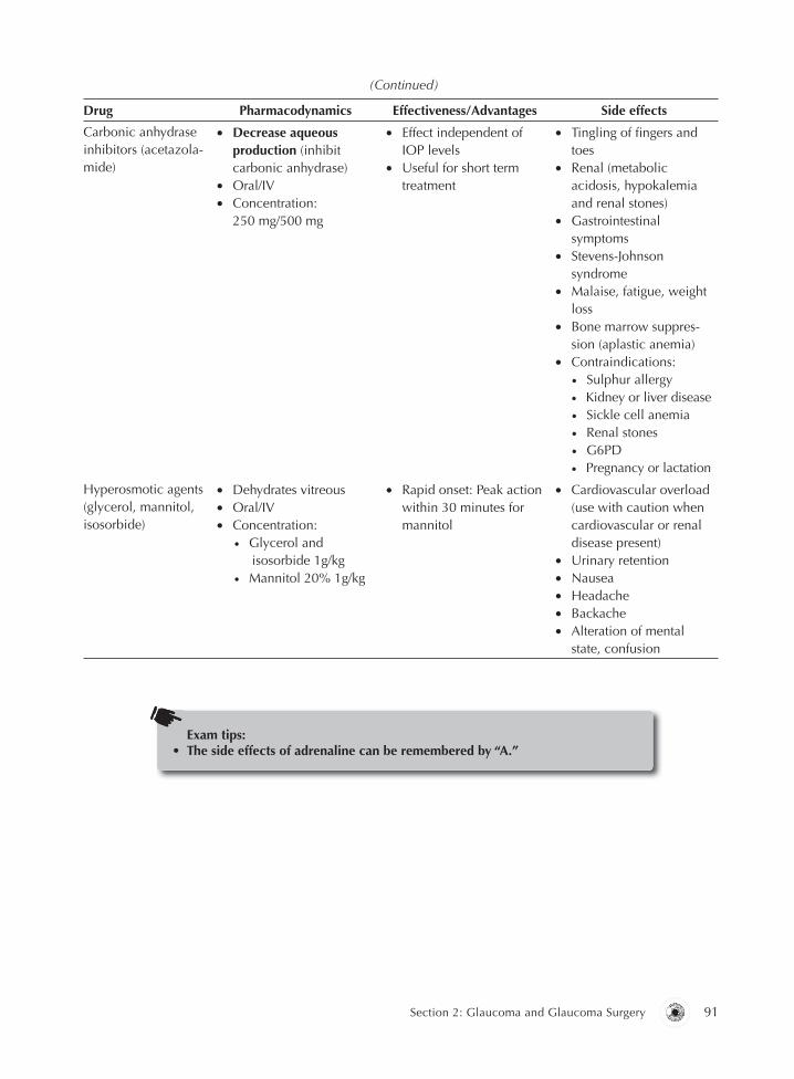
B1037 The Ophthalmology Examinations Review FA
Section 2: Glaucoma and Glaucoma Surgery 91
Drug Pharmacodynamics Effectiveness/Advantages Side effects
Carbonic anhydrase inhibitors (acetazola-mide)
• Decrease aqueous production (inhibit carbonic anhydrase)
• Oral/IV• Concentration:
250 mg/500 mg
• Effect independent of IOP levels
• Useful for short term treatment
• Tingling of fi ngers and toes
• Renal (metabolic acidosis, hypokalemia and renal stones)
• Gastrointestinal symptoms
• Stevens-Johnson syndrome
• Malaise, fatigue, weight loss
• Bone marrow suppres-sion (aplastic anemia)
• Contraindications: • Sulphur allergy • Kidney or liver disease • Sickle cell anemia • Renal stones • G6PD • Pregnancy or lactation
Hyperosmotic agents (glycerol, mannitol, isosorbide)
• Dehydrates vitreous• Oral/IV• Concentration:
• Glycerol and isosorbide 1g/kg
• Mannitol 20% 1g/kg
• Rapid onset: Peak action within 30 minutes for mannitol
• Cardiovascular overload (use with caution when cardiovascular or renal disease present)
• Urinary retention • Nausea• Headache• Backache• Alteration of mental
state, confusion
Exam tips:• The side effects of adrenaline can be remembered by “A.”
(Continued)
B1037_Ch-02.indd 91B1037_Ch-02.indd 91 12/22/2010 4:07:42 PM12/22/2010 4:07:42 PM

FA
B1037
The Ophthalm
ology Examinations R
eview
92
The Ophthalm
ology Examinations R
eview
New Drugs
Q What are the New Drugs for Treatment of Glaucoma?
Drug Pharmacodynamics Effectiveness/Advantages Side effects
Latanoprost(Xalatan)Travoprost(Travatan)Bimatoprost(Lumigan)
• PGF2 alpha agonist• Increase uveoscleral
outfl ow• Once nightly dosage
(T1/2 = 12 hours)• Concentration: 0.005%
• Better or as effective astimolol (depending on which study)
• Class I drug. 30% drop in IOP in 80–90% of patients (e.g. 24 to 16 mmHg)
• IOP effect at night• Good compliance• Additive effects with other
medications• Effective for 2 years with no drift
• Little systemic SE (T1/2 in plasma = 7 s): occasional headache/URTI symptoms
• Conjunctival injection (10% will complain of redness, 30% objective injection)
• Infl ammation (contraindicated in uveitis)• Hypertrichosis (increase in length, number and
thickness)• Iris pigmentation (melanin deposition, no melano-
cytic hyperplasia, therefore no risk of melanoma)• Cystoid macular edema (pseudophakics/
aphakics)• Expensive
Brimonidine(Alphagan)
Apraclonidine(Iopidine)
• Alpha 2 agonist – three effects:1. Decreases aqueous
production 2. Increase uveoscleral
outfl ow 3. Neuroprotective• Twice daily dosage• Concentration: 0.2%• Rapid onset (30 min)
• Class I drug• Alpha 2 selectivity – aqueous production
suppression (without vasoactivity effects of alpha 1)
• Less side effects compared with older non-specifi c alpha agonists (apraclonidine)
1. Tachyphylaxis (30%) 2. Chemosis and stinging (30%)• Additive effects with other medications
• Allergic blepharoconjunctivitis (10%)• Corneal irritation (10%)• Dry mouth (10%)
(Continued)
B1037_C
h-02.indd 92B
1037_Ch-02.indd 92
12/22/2010 4:07:43 PM
12/22/2010 4:07:43 PM

B1037
The Ophthalm
ology Examinations R
eview FA
Section 2: Glaucom
a and Glaucom
a Surgery
93
(Continued)
Drug Pharmacodynamics Effectiveness/Advantages Side effects
Dorzolamide(Trusopt)Brinzolamide(Azopt)
• Topical carbonic anhydrase inhibitor
• Only 1/3 as effective as oral • Three times daily dosage• Concentration: 0.2%
• Class II drug• Less side effects compared with oral
• Injection and stinging (30%)• Less effective than timolol• Corneal opacifi cation in compromised corneas
(inhibits endothelial pump function)• Less systemic SE compared to
acetazolamide
Unoprostone(Rescula)
• PGF2 alpha metabolite agonist: 1. Increase conventional
outfl ow 2. Increase uveoscleral outfl ow• Twice daily dosage • Concentration: 0.12%
• Class I drug• As effective as timolol • May also increase optic nerve head
perfusion
• Similar to latanoprost
B1037_C
h-02.indd 93B
1037_Ch-02.indd 93
12/22/2010 4:07:43 PM
12/22/2010 4:07:43 PM

FA B1037 The Ophthalmology Examinations Review
94 The Ophthalmology Examinations Review
Combination Eyedrops:1. Cosopt: Timolol + dorzolamide2. Xalacom: Timolol + latanoprost3. TimPilo: Timolol + pilocarpine
4. Combigan: Timolol + alphagan5. Duotrav: Timolol + travoprost
B1037_Ch-02.indd 94B1037_Ch-02.indd 94 12/22/2010 4:07:43 PM12/22/2010 4:07:43 PM

B1037 The Ophthalmology Examinations Review FA
Section 2: Glaucoma and Glaucoma Surgery 95
2. Laser retinal doppler fl owmetry (optic nerve head
perfusion)
Diagnostic
1. Confocal scanning laser ophthalmoscope (optic
nerve head evaluation)
Q Opening Question: What are the Uses of Lasers in Glaucoma?
“Lasers can be used for diagnostic and therapeutic purposes.”“Therapeutic use can be divided anatomically into….”
Overall yield: ⍟ ⍟ ⍟ ⍟ Clinical exam: Viva: ⍟ ⍟ ⍟ Essay: ⍟ MCQ: ⍟ ⍟ ⍟ ⍟
TOPIC 11
LASER THERAPYFOR GLAUCOMA
Anatomical site
Procedurename
Type oflaser Indications Notes
Iris Peripheraliridotomy (PI)
Nd:YAG or sequential Argon-YAG
• PACG• Narrow, occludable
angles• Secondary ACG
(phacomorphic, uveitic)
Settings:Argon (1.1 W, 0.05 s, 50 μm)followed by Nd:YAG(2–3 mL)Lens:Abraham’s syndrome orWise’s syndrome
Laser iridoplasty Argon • Medicallyunresponsive PACG
• Angle crowding• Plateau iris• Laser PI block• Prior to ALT in POAG
with narrow angles
Laser 1 ring around iris (stretches angles anddilates pupil to relieve pupil block)
Laser pupilloplasty Argon As in laser iridoplasty Laser 3 rings aroundpupils (dilates pupil to relieve pupil block)
Therapeutic
(Continued)
B1037_Ch-02.indd 95B1037_Ch-02.indd 95 12/22/2010 4:07:43 PM12/22/2010 4:07:43 PM

FA B1037 The Ophthalmology Examinations Review
96 The Ophthalmology Examinations Review
Anatomical site
Procedurename
Type oflaser Indications Notes
Angles Lasertrabeculoplasty(ALT)
Argon Temporizing procedure that tends to fail in the long term. Less effective than medica-tions and surgery in VF preservation• Medically unresponsive
POAG• Pigment dispersion and
pseudoexfoliation• Elderly patient not fi t for
surgeryDoes not cause structural/coagulative damage to TMunlike ALT
Settings:Argon (0.2 W, 0.1 s, 50 μm)Extent: 180 or 360o
Number of shots:40
1/3 of patients do not respond at allAverage decrease inIOP ~ 30% (replacesone eyedrop)
Selective laser trabeculoplasty (SLT)
Settings:400 μm, 0.6–1.2 mJ, 3 ns Extent and number of shots: 45–55 spots over 180o
Anatomical site
Procedurename
Type oflaser Indications Notes
Ciliarybody
Laser trabeculo-coagulation
Ciliary body ablation1. Transscleral
cyclophotocoa-gulation (TCP)
2. Transpupillary cyclophotocoa-gulation
3. Endoscopic cyclophotocoa-gulation
Argon
Diode(1.8–2 W)Continuous wave YAG(8–9 W)
Neovascular glaucoma
Refractory glaucomas• Neovascular• Uveitic• Traumatic• Failed trabeculec-
tomy• Congenital
Laser new vessels at iris
Why not cryotherapy?Advantages of laser, lower risk of:• Phthisis bulbi• Sympathetic
ophthalmia• Chemosis and pain
Sclera Laser sclerostomy Holium YAG POAG Makes 300 μm hole in scleraLittle collateral damage because using picosec-onds pulsesHigh incidence of failure
Laser suture lysis Argon Post trabeculectomy(Useful 1–3 weeks after trabeculectomy to improve fi ltration)
Settings:Argon (0.2 W, 0.1 s, 50 μm)Lens: Hoskins
Vitreous YAG capsulotomy for malignant glaucoma
YAG Malignant glaucoma Settings:YAG (2–2.5 mJ, 1 pulse per burst)Lens:Capsulotomy lens
(Continued)
B1037_Ch-02.indd 96B1037_Ch-02.indd 96 12/22/2010 4:07:43 PM12/22/2010 4:07:43 PM

B1037 The Ophthalmology Examinations Review FA
Section 2: Glaucoma and Glaucoma Surgery 97
Exam tips:• One of the more commonly asked lasers and procedures questions in
examinations. Develop notes based on your own technique.
Q How do You Perform Cyclodestruction Using Laser?
“I would use a diode laser to perform a transscleral cyclophotocoagulation (TCP).”
Diode TCP
1. Procedure:• Retrobulbar anesthesia• Contact fi ber-optic probe• Settings:
• 1.8 to 2 W• 1–2 s• 30–40 shots
• Extent: 360° 1–3 mm from limbus, sparing 3 and 9 o’clock areas
• Hear “pop” sound (microablation of ciliary body epithelium)
2. Post-procedure:• Analgesics• Steroids• Check IOP 3 weeks later
3. Complications:• Pain• Hypotony, phthisis bulbi• Infl ammation, iritis• Scleral thinning• Decrease in VA• Hyphema• Malignant glaucoma
Q When do You Perform a Laser Peripheral Iridotomy (PI)?
“The laser peripheral iridotomy is indicated for therapeutic and prophylactic purposes.”
Indications for a Laser Peripheral Iridotomy
1. Therapeutic:• PACG (acute ACG, intermittent ACG, chronic
ACG)• POAG with narrow angles
• Secondary ACG (irido-IOL block, irido vitreal block, subluxed lens with pupil block)
2. Prophylactic:• Fellow eye of patient with PACG• Narrow occludable angles
Q How do You Perform a Laser PI?
“I would perform a Nd:YAG laser PI as follows….”or “I would perform a sequential Argon YAG laser PI as follows….”
Procedure for Laser Peripheral Iridotomy
1. Prepare the patient:• Miosed pupil with 2% pilocarpine• Instil 1% apraclonidine 1 hour before procedure• Topical anesthetic and position patient at laser
machine
2. Argon blue green laser settings: 1.1 W, 0.05 s,
50 μm
3. Abraham’s iridotomy lens
4. Location of PI:• Upper nasal iris (to avoid diplopia and macular
burn)• 1/3 distance from limbus to pupil• Iris crypt if possible• Apply 20–30 burns until iris is penetrated
5. Signs of penetration:• Plomb of iris pigments• Deepening of AC
B1037_Ch-02.indd 97B1037_Ch-02.indd 97 12/22/2010 4:07:43 PM12/22/2010 4:07:43 PM

FA B1037 The Ophthalmology Examinations Review
98 The Ophthalmology Examinations Review
Exam tips:• Remember “MIC.”
Q What are the Complications of a Laser PI?
Complications
1. Contiguous damage:• Cornea• Cataract
2. Iris:• Iris bleeding • Iritis• Increased IOP
3. Malignant glaucoma
4. Monocular diplopia
Notes• “What are the unique features of Abraham’s iridotomy lens?”• Contact lens with +66D lenticule• Stabilizes globe during procedure• High magnifi cation• Increases cone angle and energy at site by 4x:
• Therefore, the spot area is effectively reduced 4× and radius reduced 2× (square root of 4) (i.e. 50 μm spot size is reduced to 25 μm)
• In addition, the energy around the cornea and iris reduced by 4×
• Retroilluminate to see patent PI• Gonioscopy to see opened angles
6. Nd:YAG laser setting: 2.5 mJ, 3–5 shots:• PI size should ideally be 300–500 μm
7. Post-procedure:• Instil 1% apraclonidine• Check IOP 1 hour later• Topical steroids for 1 day
B1037_Ch-02.indd 98B1037_Ch-02.indd 98 12/22/2010 4:07:43 PM12/22/2010 4:07:43 PM

B1037 The Ophthalmology Examinations Review FA
Section 2: Glaucoma and Glaucoma Surgery 99
• Noncompliance with medical treatment• Additional considerations:
• Young patient with good quality of vision
• One-eyed patient (other eye blind from glaucoma)
• Family history of blindness from glaucoma
• Glaucoma risk factors (HPT, DM)• Uncontrolled PACG after laser PI and
medical treatment• Secondary OAG or ACG
Indications
1. Treatment should be individualized with no fi xed
rule
2. General principle: When IOP is raised to a level
where there is evidence of progressive VF or ON
changes which will threaten quality of visual
function, despite adequate medical treatment
3. Common scenarios include:
• Uncontrolled POAG with maximal medical treatment:• Failure of medical treatment (IOP not
controlled with progressive VF or ON damage)
• Side effects of medical treatment
Overall yield: ⍟ ⍟ ⍟ ⍟ ⍟ Clinical exam: Viva: ⍟ ⍟ ⍟ ⍟ Essay: ⍟ ⍟ MCQ: ⍟ ⍟ ⍟ ⍟
Q Opening Question: What are the Indications for Trabeculectomy in Glaucoma?
“There are no absolute indications for trabeculectomy….” “In general….”“Common scenarios include….”
TOPIC 12
SURGICAL TREATMENT FOR GLAUCOMA
Q How does Medical Therapy Compare with Surgical Therapy in Glaucoma?
“It is diffi cult to compare medical with surgical treatment, with new research showing both having advantages and disadvantages. We can compare the two in four major areas….”
B1037_Ch-02.indd 99B1037_Ch-02.indd 99 12/22/2010 4:07:43 PM12/22/2010 4:07:43 PM

FA B1037 The Ophthalmology Examinations Review
100 The Ophthalmology Examinations Review
Medical treatment Surgical treatment
Effectiveness • 40% respond readily andconsistently to low dose medicine
• 50% eventually requirecomplex medical regimen,adjuvant ALT andfi ltration surgery
• 5–10% poor response to medical treatmentin fi rst instance andrequire surgery
• Improved surgicaltechnique has led to 80–90% success rates
• Better control of IOP (delay VF/ON progression)
• Increase morbidity associated with delaying surgery until evidence of VF/ON damage
Cost • Cheaper initially, but accumulates over years• In the United States, cost of
bilateral surgery = cost of 8 yearsof topical medication
• Actual cost may be lessin the long term
Safety/Problems • Poor compliance with multiplemedications
• Less control of IOP withcontinuing ON damage
• Minor side effects is troublesome• Major side effects can occur:
• Aplastic anemia (with diamox)• Respiratory and cardiac side
effects (beta blockers)• Increase risk of bleb failure (with
chronic topical eyedrop use)
• Even after surgery, may require adjuvant medical treatment
• No long term proof that good IOP control alonewill stop ON damage(IOP only one risk factor)
• Usually no minor side effects
• Major side effects (com-mon):• Anesthetic and surgical
morbidity • Risk of endophthalmitis
and malignant glaucoma • Shallow AC, hypotony,
progression of cataracts
Quality of life • Poorer quality of life (with use ofmultiple eyedrops)
• Better quality of life
Exam tips:• As expected, this topic will be a constant debate with new research fi ndings
every month. Stick to a conservative approach but keep an open mind about new ideas.
B1037_Ch-02.indd 100B1037_Ch-02.indd 100 12/22/2010 4:07:44 PM12/22/2010 4:07:44 PM

B1037 The Ophthalmology Examinations Review FA
Section 2: Glaucoma and Glaucoma Surgery 101
Q How do You Perform a Trabeculectomy?
“I would perform a trabeculectomy as follows.”
Trabeculectomy
1. Preparation:• Retrobulbar anesthesia• Inferior corneal traction suture with 7/0 silk
2. Conjunctival fl ap:• Superonasally or superotemporally• Fornix or limbal based fl ap (stick to one
approach, see below)• Dissect Tenons with Wescott scissors• Remove all episcleral tissue
3. Scleral fl ap:• Outline fl ap with diathermy• Size 4 x 3 mm• Cut with beaver blade 1/2 to 2/3 scleral
thickness
• Dissection with crescent blade until surgical limbus is seen (page 41)• “Where is the surgical limbus?”
4. Paracentesis performed at distant location
5. Sclerostomy:• Enter AC through scleral fl ap with a beaver blade• Excise 2 × 1 mm block of sclera with Kelly’s
punch or Vanna scissor
6. Peripheral iridectomy:• Prevent blockage of sclerectomy site by iris
7. Closure:• Scleral fl ap sutured with 8/0 vicryl or 10/0 nylon• Reform AC and check aqueous egress• Conjunctiva sutured with 8/0 vicryl
Notes• Why perform a paracentesis?
• Decompress AC prior to sclerectomy• Reform AC later• Check aqueous egress later
Q What are the Advantages and Disadvantages of Fornix-Based vs Limbal-Based Flaps?
Fornix-based vs Limbal-based Conjunctival Flap
Fornix-based Limbal-based
Advantages • Faster to create and close• Good exposure• Easier to identify limbal landmarks• Less dissection (less bleeding
and risk of button hole)• Avoids posterior conjunctival scarring
(limits posterior fi ltration of aqueous)
• Easier to excise Tenon’s• Less risk of wound leak and fl at AC• No limbal irregularity (dellen)• Allows adjunctive use of anti-
metabolites with less corneal toxicity
Disadvantages • Increase risk of fl at AC• Harder to excise Tenon’s• IOP control not as good as with
limbal-based fl ap
• Slower and more surgical experience needed
• Poorer exposure• Risk of button hole higher
Exam tips:• As in cataract surgery, be concise but accurate with the steps, as if you had
done the procedure a hundred times. Say, “I will perform a superotemporal limbal-based conjunctival fl ap” instead of “conjunctival fl ap.”
B1037_Ch-02.indd 101B1037_Ch-02.indd 101 12/22/2010 4:07:44 PM12/22/2010 4:07:44 PM

FA B1037 The Ophthalmology Examinations Review
102 The Ophthalmology Examinations Review
Complications
1. Intraoperative (not common, usually due to poor
surgical techniques):• Suprachoroidal hemorrhage (most important
complication, as in cataract surgery)• Button hole in conjunctival fl ap• Subconjunctival hemorrhage from bridle suture• Hyphema
2. Early postoperative:• Flat AC and malignant glaucoma (see below)• Endophthalmitis
• Hyphema • Suprachoroidal hemorrhage • “Wipe-out” syndrome• Cystoid macular edema
3. Late postoperative:• Filtration failure (see below)• Endophthalmitis, blebitis• Cataract progression• VF loss progression• Refractive errors• Hypotony
Q How do You Manage a Shallow AC after a Trabeculectomy?
“Management involves an assessment of the severity of shallowing and the etiology.”“This depends on the IOP and presence/absence of a bleb.”
Shallow AC
1. Grades of shallow AC:• Grade I: Irido-corneal touch (can afford to be
conservative)
• Grade II: Pupillo-corneal touch• Grade III: Lenticulo-corneal touch (need to
intervene surgically)
2. Etiology
IOP Bleb Differential diagnoses Management
High • No bleb• Siedal’s sign +ve
• Malignant glaucoma• Suprachoroidal Hb• Pupil block glaucoma
• See below• Fundus examination (dark brown mass)• Dilate pupil (AC may deepen)• Enlarge surgical PI with laser
Low • No bleb +ve• Siedal’s sign +ve
• Wound leak Conservative:• Usually will resolve within 24 hours• Decrease steroids and increase antibiotics
(gentamicin) to induce scarring• Dilate pupil with mydriatic (atropine)• Decrease aqueous production (timolol and
diamox)• Pressure pad/bolster• Simmon’s shell/oversized (fl ex) contact lens
Surgical:• Resuture
• Good bleb• Siedal’s sign −ve
• Excessive fi ltration Conservative:• Decrease steroids, increase antibiotics,
dilate pupil and decrease aqueous produc-tion
• Pressure pad/bolster• Inject gas (air or SF6) or viscoelastic into AC• Injection of autologous blood into bleb• Compression mattress suture
Surgical:• Resuture
Q What are the Complications of a Trabeculectomy?
“The complications can be divided into intraoperative, early postoperative and late postoperative.”
B1037_Ch-02.indd 102B1037_Ch-02.indd 102 12/22/2010 4:07:44 PM12/22/2010 4:07:44 PM

B1037 The Ophthalmology Examinations Review FA
Section 2: Glaucoma and Glaucoma Surgery 103
Malignant Glaucoma
1. Conservative:• Topical mydriatics (atropine)• Lower IOP (diamox and osmotic agents)• Enlarge PI• Nd:YAG laser to disrupt anterior vitreous face
(see laser therapy, page 78)
2. Surgical:• Chandler’s procedure (see vitreous tap, page 33):
• 19G needle inserted into vitreal cavity, about 12 mm from tip to drain 1–1.5 ml of aqueous and separate solid vitreous from trapped aqueous
• Vitrectomy
Q How do You Manage a Malignant Glaucoma?
“Malignant glaucoma is a serious complication of glaucoma surgery.”“Management involves an assessment of the severity (grades of AC shallowing).”“And can be conservative or surgical.”
Q How do You Manage a Filtration Failure?
“Management involves an evaluation of the causes of failure.”“And can be conservative or surgical.”
Filtration Failure
1. Etiology• Early:
• Blockage by ocular components (lens, iris, Descemet’s membrane, vitreous, scleral remnants)
• Blockage by surgical intervention (blood, viscoelastic)
• Late:• Subconjunctival fi brosis
2. Conservative• Increase topical steroids• Medical control of IOP• Scleral depression at posterior lip of scleral fl ap
• Digital massage• Laser suture lysis:
• Done usually at 1–3 weeks after surgery (see laser therapy, page 78)
• Needling of bleb:• Done usually at 6 weeks after surgery• Topical anesthetics under sterile conditions• Approach from unoperated conjunctiva with
27G needle• May be combined with 5 FU injection
3. Surgical• Revision of trabeculectomy/new trabeculectomy
with antimetabolites
Notes• Risk factors for subconjunctival fi brosis = indications for antimetabolite
used
Q What are the Indications for Using Antimetabolites in Trabeculectomy?
“Antimetabolites are used in trabeculectomy when a high risk of failure with the conventional operation is anticipated.”“This is related to either patient or ocular factors.”
Indications for Antimetabolites
1. Patient factors:• Young (< 40 years)• Black race• Previous chronic medical therapy (especially
with Adrenaline)
• Previous failed trabeculectomy• Previous conjunctival surgery (e.g. pterygium
surgery)
2. Ocular factors:• Secondary glaucomas (neovascular and uveitic
glaucoma)
B1037_Ch-02.indd 103B1037_Ch-02.indd 103 12/22/2010 4:07:44 PM12/22/2010 4:07:44 PM

FA B1037 The Ophthalmology Examinations Review
104 The Ophthalmology Examinations Review
• Traumatic glaucomas• Aphakic/pseudophakic glaucomas• Iridocorneal endothelial syndromes (ICE)
• Congenital/pediatric glaucomas• Conjunctival scarring
Q Tell me about Antimetabolites Used in Glaucoma Surgery.
Notes
• What additional measures must be taken in trabeculectomies withantimetabolites?
• Prevent antimetabolites from entering eye:• Limbal-based fl aps• Watertight wound (interrupted non-absorbable conjunctival sutures)• Careful dissection to prevent button hole formation
1. Inclusion criteria• 213 patients considered to be at a higher risk of
surgical failure on the basis of having previous
5 Fluorouracil (5 FU) Mitomycin C (MMC)
Pharmacology • Fluorinated pyrimidine analogue• Binds intracellular thymidylate
synthetase (inhibits thymidine and DNA synthesis)
• Affects only cells in mitotic phase ofcell cycle
• In the eye, inhibits fi broblast proliferation and delays fi brosis
• Natural antibiotic compound/alkylating agent
• Cross-links with DNA strands by formation of covalent bonds
• Affects cells in all phases• Kills fi broblast permanently and stop
fi brosis
Dosage • Intraoperative dose: 25–50 mg/ml• Postoperative drops: 5 mg/ml for 1 week
• Intraoperative dose: 0.2–0.4 mg/ml
Results • Improves success rate of fi ltration operation
• No randomized trial results• Better than 5 FU?
Complications • Corneal epithelial toxicity• Hyphema• Wound leak• Infection
• Prolonged hypotony • Avascular bleb• Endothelial, ciliary body and retinal damage• Hyphema• Wound leak• Infection
Q What is the Fluorouracil Filtering Surgery Study?
“The Fluorouracil Filtering Surgery Study is a multicenter prospective randomized clinical trial which evaluates the effectiveness of adjuvant subconjunctival 5 FU following trabeculectomy.”
cataract extraction or previous failed fi ltering surgery in a phakic eye
Exam tips:• Refer to three-year follow-up of the Fluorouracil Filtering Surgery Study (Am
J Ophthalmol 1993;115:82–92).
B1037_Ch-02.indd 104B1037_Ch-02.indd 104 12/22/2010 4:07:44 PM12/22/2010 4:07:44 PM

B1037 The Ophthalmology Examinations Review FA
Section 2: Glaucoma and Glaucoma Surgery 105
2. Treatment• Trabeculectomy with adjuvant postop
subconjunctival 5 FU injections vs trabeculectomy alone
3. Outcome • Treatment failure defi ned as:
• Reoperation to decrease IOP, or • An IOP > 21 mmHg with or without adjuvant
IOP-lowering medications
4. Results• Treatment failure at 3 years: 51% with 5 FU
vs 74% without 5 FU• Complications associated with 5 FU:
• Corneal epithelial erosions• Early wound leaks
5. Conclusion• 5 FU indicated after trabeculectomy only in
patients at high risk of failure
Q Tell me about Filtering Shunts.
“Filtering shunts are communications between AC and sub Tenon’s space.”“They are indicated when a high risk of failure with the conventional operation is anticipated.”“This is related to either patient or ocular factors.”“The shunts can be divided into….”“The complications include….”
Filtering Shunts
1. Indications• Patient factors:
• Previous failed trabeculectomy• Previous major anterior segment surgery
• Ocular factors:• Secondary glaucomas (neovascular and
uveitic glaucoma)• Traumatic glaucomas• Aphakic/pseudophakic glaucomas• Iridocorneal endothelial syndromes (ICE)• Pediatric glaucomas
2. Type of shunts• Vary with shape and size• Valves (Krupin-Denver, Ahmed) vs no valves
(Molteno, Baerveldt)• Material: PMMA, silicon, polypropylene
3. Complications• Intraoperative:
• Hyphema• Lens damage and cataract• Globe perforation• Muscle disinsertion and laceration
• Postoperative:• Functional (excessive drainage, blockage of
tube by blood and uveal tissue)• Mechanical (corneal endothelial
decompensation, cataract)• Diplopia due to extraocular muscle limitation• Bleb encapsulation over footplate → poor
drainage• Late endophthalmitis
4. Effi cacy compared to trabeculectomy • Tube vs Trabeculectomy Study (Am J
Ophthalmol 2009;148:670–684)• Tube shunt surgery had a higher success rate
compared to trabeculectomy with MMC during fi rst 3 years of follow-up
• Both procedures associated with similar IOP reduction and use of supplemental medical therapy at 3 years
• Postoperative complications higher following trabeculectomy with MMC relative to tube shunt surgery, but most complications were transient and self-limited
Exam tips:• The indications are nearly IDENTICAL to that of antimetabolite use.
B1037_Ch-02.indd 105B1037_Ch-02.indd 105 12/22/2010 4:07:44 PM12/22/2010 4:07:44 PM

B1037 The Ophthalmology Examinations Review FA
Section 3
Corneal and External EyeDiseases
B1037_Ch-03.indd 107B1037_Ch-03.indd 107 12/22/2010 5:44:40 PM12/22/2010 5:44:40 PM

FA B1037 The Ophthalmology Examinations Review
B1037_Ch-03.indd 108B1037_Ch-03.indd 108 12/22/2010 5:44:42 PM12/22/2010 5:44:42 PM
This page intentionally left blankThis page intentionally left blank

B1037 The Ophthalmology Examinations Review FA
Section 3: Corneal and External Eye Diseases 109
TOPIC 1
THE CORNEA
“The cornea is a transparent structure in the anterior segment of eye… .”
Overall yield: ⍟ ⍟ ⍟ ⍟ Clinical exam: Viva: ⍟ ⍟ ⍟ ⍟ Essay: ⍟ ⍟ MCQ: ⍟ ⍟ ⍟ ⍟
Q Opening Question: What is the Anatomy of the Cornea?
Anatomy of the Cornea
1. Gross anatomy• General dimensions:
• 11.5 mm horizontal diameter• 10.5 mm vertical diameter• 1 mm thick periphery• 0.5 mm thick centrally• Anterior surface radius 7.7 mm• Posterior surface radius 6.8 mm
2. Microscopic anatomy• Five layers:
• Epithelium:• Stratifi ed squamous, nonkeratinized,
nonsecretory epithelium (keyword)• 5–6 layers deep• Superfi cial cells have microvilli (needs
tears to keep cornea smooth)• Basement membrane — strongly attached
to Bowman’s layer• Bowman’s layer:
• 8-12 µm• Acellular• Consists of interwoven type IV collagen fi brils
which are anterior condensation of substantia propia
• Incapable of regeneration, replaced by fi brous tissue if damaged (i.e. scars)
• Ends abruptly at limbus• Deep layers appear to merge into stroma
• Stroma (substantial propia):• 90% of cornea thickness, 400 µm centrally• 80% water• Glycosaminoglycans in extracellular matrix
• Three major fractions:• Keratan sulphate (50%)• Chondroitin phosphate (25%)• Chondroitin sulphate (25%)
• Descemet’s membrane:• Basement membrane of the endothelium
(keyword)• 10 µm thick• Secreted and regenerated by endothelial cells • Type IV collagen fi brils• Hassall-Henle bodies • Terminates abruptly at limbus (Schwalbes
line) • Endothelium:
• Single layer, polygonal, cuboidal cells • Tight junctions (control of corneal hydration)• Microvilli • Incapable of regeneration: Repair occurs by
cellular hypertrophy and sliding• Line passages of trabecular meshwork
Exam tips:• One of the most common basic science questions in life viva and MCQ
examinations.
B1037_Ch-03.indd 109B1037_Ch-03.indd 109 12/22/2010 5:44:42 PM12/22/2010 5:44:42 PM
FA B1037 The Ophthalmology Examinations Review
110 The Ophthalmology Examinations Review
• Glucose:• Cornea obtains glucose mainly from aqueous • Tears and limbal capillaries appear to provide
minimal contribution • Glucose can be stored in epithelium as
glycogen • ATP obtained through glycolysis and Kreb's
cycle • Oxygen:
• Endothelium acquires oxygen from aqueous • Epithelium acquires oxygen from either
capillaries at the limbus or precorneal fi lm
Q What is the Function of the Cornea?
Physiology of Cornea
1. Three main functions:• Light transmission (400–700 nm light)• Light refraction:
• Total refractive power of cornea 43 D (70% of eye’s refractive power)
• Refractive index of cornea 1.376• Protection
2. Corneal metabolism:• Energy needed for maintenance of transparency
and dehydration
“Corneal transparency is due to a combination of factors including… .”
Q Why is the Cornea Transparent?
Cornea Transparency• Relative dehydration of cornea due to:
• Anatomic integrity of endothelium and epithelium
• Endothelial pump removes fl uids from stroma• Evaporation of water from tear increases
osmolarity of tear, which draws water from cornea
• Normal intraocular pressure (if too high, relative hydration occurs)
• Relative acellularity, lack of blood vessels and pigments
• Regular matrix structure of corneal fi brils:• Destructive interference of light occurs
• Consistent refractive index of all layers
Exam tips:• Variations to questions include “What are the factors which keep the cornea
dehydrated?”
Q What is the Nerve Supply of the Cornea?
“The cornea is innervated by the V CN.”
Nerve Supply• V CN• Ophthalmic division• Long posterior ciliary nerves gives off:
• Annular plexus at limbus
• Subepithelial plexus just below Bowman’s membrane
• Intraepithelial plexus
B1037_Ch-03.indd 110B1037_Ch-03.indd 110 12/22/2010 5:44:42 PM12/22/2010 5:44:42 PM

B1037 The Ophthalmology Examinations Review FA
Section 3: Corneal and External Eye Diseases 111
• Facial hemiatrophy• Dwarfi sm
Microcornea
1. Corneal diameter < 10 mm
2. May occur as:• Isolated cornea abnormality• Nanophthalmos (small but normal eye)• Microphthalmos (small and abnormal eye)
3. Inheritance: AD, AR, sporadic
4. Ocular associations:• Shallow AC• Glaucoma• Hyperopia• Persistent hyperplastic primary vitreous• Congenital cataract• Anterior segment dysgenesis• Optic nerve hypoplasia
5. Systemic associations:• Dwarfi sm• Achondroplasia• Myotonic dystrophy• Fetal alcohol syndrome
Cornea Plana
1. Flat cornea:• Radius of curvature < 43D (may be 20–30D)• Pathognomonic when corneal curvature is the
same as adjacent sclera!
2. Inheritance: AD, AR, sporadic
Overall yield: ⍟ Clinical exam: Viva: Essay: ⍟ MCQ: ⍟
TOPIC 2
CONGENITAL CORNEAL ABNORMALITIES
Megalocornea
1. Corneal diameter > 13 mm (or 12 mm at birth):• Buphthalmos must fi rst be excluded (no axial
myopia, no cornea opacity, normal IOP)• Distinction between simple megalocornea
(large cornea without structural abnormalities and associated ocular malformations) and anterior megalophthalmos (large cornea with structural abnormalities/ocular malformations)
2. Inheritance: AD (simple megalocornea) or SLR
(anterior megalophthalmos)
3. Clinical features (anterior megalophthalmos):• Congenital • Males (90%) • Bilateral (80%), symmetrical, nonprogressive• Normal cornea• Normal thickness and endothelial cell density • No Descemet’s rupture (i.e. no Haab’s straie)• Normal posterior segment• Normal visual development
4. Ocular associations (anterior megalophthalmos):• Astigmatism• Atrophy of iris stroma• Ectopic lentis and cataract• Glaucoma (but not congenital glaucoma!)
5. Systemic associations (anterior megalophthalmos):• Down’s• Connective tissue diseases (Marfan’s syndrome,
Ehlers-Danlos syndrome)• Craniosynostosis (Apert’s syndrome)• Alport’s syndrome
Q Opening Question: What are the Congenital Abnormalities of the Cornea?
B1037_Ch-03.indd 111B1037_Ch-03.indd 111 12/22/2010 5:44:42 PM12/22/2010 5:44:42 PM

FA B1037 The Ophthalmology Examinations Review
112 The Ophthalmology Examinations Review
3. Bilateral, peripheral opacifi cation of cornea
4. Ocular associations:• Sclerocornea• Microcornea• Congenital cataract• Glaucoma
Sclerocornea
1. Diffuse scarring and vascularization of cornea
2. Epithelium thickened, Bowman’s membrane
absent
3. May be AD/sporadic
4. Classifi cation:• Isolated sclerocornea: No other abnormalities• Sclerocornea plana: With fl at cornea
(mean K < 38D)• Peripheral sclerocornea with anterior chamber
cleavage abnormalities: Peter’s, Rieger’s anomalies
• Total sclerocornea
Clinical Features
1. Ocular features:• Megalocornea• Coloboma of iris and lids• Squint, Duane’s syndrome• Fundus — optic nerve hypoplasia, coloboma• Refractive errors
Q Tell me about Goldenhar’s Syndrome.
2. Systemic features:• Wide mouth• Maxillary and mandibular hypoplasia• Preauricular tags and hearing loss• Vertebral defects
B1037_Ch-03.indd 112B1037_Ch-03.indd 112 12/22/2010 5:44:43 PM12/22/2010 5:44:43 PM

B1037 The Ophthalmology Examinations Review FA
Section 3: Corneal and External Eye Diseases 113
Complications of Chemical Injury
1. Acute problems:• Corneal abrasion and perforation• Infection• Glaucoma
2. Long-term problems:• Ocular surface:
• Trichiasis, distichiasis, entropion• Cicatricial conjunctivitis, dry eyes,
symblepharon, ankyloblepharon
• Cornea:• Persistent epithelial defect• Limbal stem cell failure and persistent ocular
surface disease• Stromal scar
• Intraocular complications:• Glaucoma • Diffuse trabecular damage iritis• Cataract
“Chemical injuries are ocular emergency.”“They can be mild or potentially blinding.”
Overall yield: ⍟ ⍟ ⍟ Clinical exam: Viva: ⍟ ⍟ ⍟ ⍟ Essay: ⍟ ⍟ MCQ: ⍟ ⍟
TOPIC 3
CHEMICAL INJURY
Q Opening Question: What are the Complications of Chemical Injuries?
Grade Signs Prognosis
1 Corneal epithelial damage No limbal ischemia
Excellent
2 Corneal hazy but iris details seen Ischemia < 1/3 of limbus
Good
3 Corneal hazy but iris details hazy Ischemia < 1/2 of limbus
Fair
4 Opaque cornea Ischemia > 1/2 of limbus
Poor
Classifi cation of Chemical Injury (Hugh’s Classifi cation)
Updated classifi cation (ref Dua H et al, Br J Ophthal-mol 85(11):1379
• Updated grading scheme with prognostic value• 6 grades• Main criteria for classifi cation:
• Extent of limbal staining (vs ischemia): Staining of greater value in predicting epithelial recovery
• Area of bulbar and forniceal conjunctival staining: Conjunctiva important in ocular surface stabilization when limbus severely compromised
B1037_Ch-03.indd 113B1037_Ch-03.indd 113 12/22/2010 5:44:43 PM12/22/2010 5:44:43 PM

FA B1037 The Ophthalmology Examinations Review
114 The Ophthalmology Examinations Review
Exam tips:• One of the few true ocular emergencies.• Need to know difference between “acidic” and “alkaline” injuries.
Notes:• “What are the possible mechanisms of glaucoma?”
• Acute shrinkage of collagen• Uveitis, trabeculitis• Lens-induced infl ammation• Peripheral anterior synechiae• Steroid response
Q How do You Manage a Patient with Severe Chemical Injury?
“Chemical injury is an ocular emergency… .”
• Lid closure (taping, pressure pad, tarsorraphy)• Tissue glue• Conjunctival fl ap• Lysis of conjunctival adhesions (glass rods)
3. Long-term management• Ocular surface reconstruction:
• Lid surgery (cicatricial entropion, trichiasis and distichiasis)
• Conjunctival replacement (AMT or cultivated conjunctiva)
• Ocular surface surgery • Principles:
• Removal of dysplastic epithelium• Wide excision of fi broblastic
undergrowth• Judicious diathermy• Epithelial replacement (autografts/
allografts/cultivated transplants)• Cornea:
• Keratoplasty (limbal, lamellar, penetrating)• Intraocular:
• Antiglaucoma treatment• Cataract surgery
• Controversial:• Retinoic acid (promote surface keratinization)• Fibronectin (growth factor)• Epidemal growth factor• Subconjuctival heparin (to facilitate limbal
reperfusion)
Management of Chemical Injury
1. Acute management• Irrigate eyes immediately copious amounts of
sterile fl uid (normal saline preferred):• Remove particulate matter• Debride devitalize tissues
• Start antimicrobial treatment• Start steroids immediately (to decrease
mediators of infl ammation and stabilize lysosome in white blood cells)• Minimize steroids after 10 days (because
steroids decrease fi broblast and collagen synthesis)
• Alternatively, medroxyprogesterone can be used (no anti-anabolic effects)
2. Manage epithelial defect• Conservative:
• Tear substitutes and lubricants• Vitamin C (antioxidant, cofactor in collagen
synthesis)• Ascorbate or citrate (antioxidant, cofactor in
collagen synthesis)• N acetylcysteine (collagenase inhibitor,
contributes to cross-linkages and maturation of collagen)
• Sodium EDTA (collagenase inhibitor — calcium chelator, calcium required for collagenase activity)
• Bandage contact lens• Surgical:
• Punctal occlusion in severe dry eyes
B1037_Ch-03.indd 114B1037_Ch-03.indd 114 12/22/2010 5:44:43 PM12/22/2010 5:44:43 PM

B1037 The Ophthalmology Examinations Review FA
Section 3: Corneal and External Eye Diseases 115
“Cicatricial conjunctivitis can be divided into….”
• Vernal/atopic keratoconjunctivitis• Dermatological:
• Ocular rosacea• Scleroderma
• Neoplasia:• Squamous cell carcinoma, Bowen’s disease
• Trauma:• Mechanical, chemical injury
• Others:• Long-term timolol use
Cicatricial Conjunctivitis
1. Infectious:• Adenovirus• Herpes simplex • Trachoma• Corynebacterium diphtheriae• Beta hemolytic streptococcus
2. Noninfectious:• Autoimmune:
• Ocular cicatricial pemphigoid• Steven-Johnson syndrome
Q What are the Other Causes of Cicatricial Conjunctivitis?
B1037_Ch-03.indd 115B1037_Ch-03.indd 115 12/22/2010 5:44:43 PM12/22/2010 5:44:43 PM

FA B1037 The Ophthalmology Examinations Review
B1037_Ch-03.indd 116B1037_Ch-03.indd 116 12/22/2010 5:44:43 PM12/22/2010 5:44:43 PM
This page intentionally left blankThis page intentionally left blank
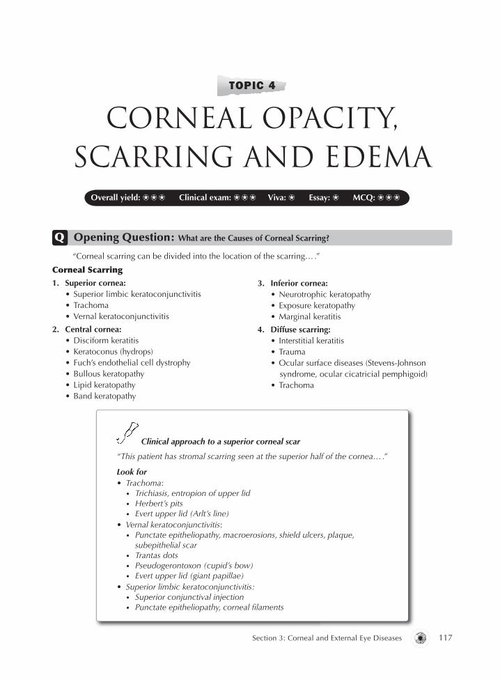
B1037 The Ophthalmology Examinations Review FA
Section 3: Corneal and External Eye Diseases 117
3. Inferior cornea:• Neurotrophic keratopathy • Exposure keratopathy • Marginal keratitis
4. Diffuse scarring:• Interstitial keratitis• Trauma• Ocular surface diseases (Stevens-Johnson
syndrome, ocular cicatricial pemphigoid)• Trachoma
“Corneal scarring can be divided into the location of the scarring… .”
Overall yield: ⍟ ⍟ ⍟ Clinical exam: ⍟ ⍟ ⍟ Viva: ⍟ Essay: ⍟ MCQ: ⍟ ⍟ ⍟
TOPIC 4
CORNEAL OPACITY, SCARRING AND EDEMA
Q Opening Question: What are the Causes of Corneal Scarring?
Corneal Scarring
1. Superior cornea:• Superior limbic keratoconjunctivitis• Trachoma• Vernal keratoconjunctivitis
2. Central cornea:• Disciform keratitis• Keratoconus (hydrops)• Fuch’s endothelial cell dystrophy• Bullous keratopathy• Lipid keratopathy• Band keratopathy
Clinical approach to a superior corneal scar
“This patient has stromal scarring seen at the superior half of the cornea… .”
Look for• Trachoma:
• Trichiasis, entropion of upper lid• Herbert’s pits• Evert upper lid (Arlt’s line)
• Vernal keratoconjunctivitis:• Punctate epitheliopathy, macroerosions, shield ulcers, plaque,
subepithelial scar• Trantas dots• Pseudogerontoxon (cupid’s bow)• Evert upper lid (giant papillae)
• Superior limbic keratoconjunctivitis:• Superior conjunctival injection• Punctate epitheliopathy, corneal fi laments
B1037_Ch-03.indd 117B1037_Ch-03.indd 117 12/22/2010 5:44:43 PM12/22/2010 5:44:43 PM

FA B1037 The Ophthalmology Examinations Review
118 The Ophthalmology Examinations Review
Clinical approach to central corneal scar or edema
“This patient has a central corneal stromal scar/edema.”“The visual axis is involved.”
Look for• Disciform keratitis:
• Lid scarring (usually very subtle)• Epithelial edema• Descemet’s folds• Wessley ring• Keratic precipitates• AC activity
• Keratoconus:• Parastromal thinning• Vogt’s straie• Fleischer’s ring• Prominent corneal nerves
• Fuch’s endothelial cell dystrophy:• Epithelial edema • Subepithelial scarring• Stromal thickening• Corneal guttata
• Pseudophakic bullous keratopathy:• Epithelial bullae• IOL
I’ll like to• Check corneal sensation (disciform keratitis)• Check IOP (Fuch’s endothelial dystrophy, disciform keratitis)
Q How do You Manage a Patient with Bullous Keratopathy?
“Management of bullous keratopathy depends on the etiology, severity and visual potential and whether patient has symptoms of pain.”“In mild cases, conservative treatment is usually adequate… .” “In severe cases, if the visual potential is good… .”“On the other hand, if the visual potential is poor and the eye is painful… .”
Bullous Keratopathy
1. Etiology:• Pseudophakic bullous keratopathy• Fuch’s endothelial cell dystrophy• End-stage glaucoma• Long-standing infl ammation• Chemical burns
2. Conservative treatment:• Lubricants• Hypertonic saline• Lower intraocular pressure• Avoid steroids
• Therapeutic contact lens• Hair-dryer
3. Surgical treatment:• If good visual potential, consider optical
keratoplasty• If poor visual potential and eye is painful,
consider palliative procedures for pain relief:• Tarsorrhaphy, botox to lids• Conjunctival fl ap (see page 139)• Retrobulbar alcohol• Enucleation (very last resort)
B1037_Ch-03.indd 118B1037_Ch-03.indd 118 12/22/2010 5:44:44 PM12/22/2010 5:44:44 PM

B1037 The Ophthalmology Examinations Review FA
Section 3: Corneal and External Eye Diseases 119
Exam tips:• Very similar approach to management of neovascular glaucoma! (see page
69).• See also management of Fuch’s endothelial dystrophy (page 113) and
glaucoma and cataract (page 25).
B1037_Ch-03.indd 119B1037_Ch-03.indd 119 12/22/2010 5:44:44 PM12/22/2010 5:44:44 PM

FA B1037 The Ophthalmology Examinations Review
B1037_Ch-03.indd 120B1037_Ch-03.indd 120 12/22/2010 5:44:44 PM12/22/2010 5:44:44 PM
This page intentionally left blankThis page intentionally left blank

B1037 The Ophthalmology Examinations Review FA
Section 3: Corneal and External Eye Diseases 121
Overall yield: ⍟ ⍟ ⍟ Clinical exam: Viva: ⍟ ⍟ ⍟ ⍟ Essay: ⍟ ⍟ MCQ: ⍟ ⍟ ⍟ ⍟
TOPIC 5
CORNEAL ULCERS
Q Opening Question No. 1: How do You Manage a Patient with a Corneal Ulcer?
“Corneal ulcer is a potentially blinding condition which needs immediate ophthalmic management.”
Management of Corneal Ulcer
1. Admit patient if necessary
2. Identify predisposing factors:• Contact lens wear• Ocular trauma• Ocular surface disease• Systemic immunosuppression
3. Perform a corneal scrape for microbiological
analyses
4. Intensive topical antibiotic treatment:• Gutt. gentamicin 15 mg/ml hourly (or gutt
tobramycin)• Gutt. cephazolin 50 mg/ml hourly (or gutt
cefuroxime)
5. Systemic antibiotic treatment if:• Ulcer near limbus (scleral extension)• Perforated ulcer (endophthalmitis)
Exam tips:• “Prepare specifi c antibiotic regimes with exact dosage and frequency of
treatment” gentamicin frequently does not sound as impressive as “I would prescribe topical gentamicin 15 mg/ml hourly for the next 24 hours.”
Q When will You Consider Using Monotherapy with Antibiotics?
• Cautious in using monotherapy• Broad spectrum antibiotic (e.g. gutt.
ciprofl oxacin)• Indications:
• Small, peripheral ulcer
• Culture positive• Organism is NOT Pseudomonas or
Streptococcus• Organism sensitive to antibiotics• Patient follow-up and compliance good
Q When will You Consider Using Steroids?
• Use of steroids is controversial, extreme caution needed
• Use only after adequate antimicrobial treatment• Indications:
• Culture positive
• Sensitive to antibiotics• Responding clinically• Ulcer has been sterilized• Patient follow-up and compliance good
B1037_Ch-03.indd 121B1037_Ch-03.indd 121 12/22/2010 5:44:45 PM12/22/2010 5:44:45 PM

FA B1037 The Ophthalmology Examinations Review
122 The Ophthalmology Examinations Review
• Lagophthalmos • Proptosis
5. Nutritional keratitis (Vitamin A defi ciency)
6. Neoplasia (acute leukemia)
7. Immune-mediated• Connective tissue diseases:
• Rheumatoid arthritis• Wegener’s granulomatosis• Systemic lupus erythematosis• Polyarteritis nodosa
• Mooren’s, Terrien’s• Marginal keratitis• Allergic conjunctivitis
8. Iatrogenic/trauma • Postsurgical, topical eyedrops• Chemical, thermal, radiation injury
Q What do You do When the Ulcer is not Responding to Treatment?
• Stop antibiotics for 24 hours• Rescrape and/or corneal biopsy
• Steps in corneal biopsy:• LA or GA• Corneal tissue on standby (for tectonic
replacement)• Dermatological trephine with biopsy
area encompassing base and active edge of ulcer
• Specimen divided and sent for microbio-logical analyses and histological staining
• Re-start intensive antibiotics• Consider other diagnosis (e.g. sterile ulcers?)• Consider penetrating keratoplasty
Q What are the Causes of Sterile Ulcers?
Sterile Ulcers
1. Post infection (treated, resolved)• Herpes (metaherpetic ulcer)• Bacterial• Fungal
2. Nearby (contiguous) ocular surface infl ammation• Lids and lashes (entropian, ectropian, trichiasis,
lid defects)• Skin (Stevens-Johnson syndrome, ocular
pemphigoid, ocular rosacea)• Lacrimal gland (keratoconjunctivitis sicca)
3. Neurotrophic keratitis• DM • V CN palsy • Herpes zoster
4. Exposure keratitis• VII CN palsy
Exam tips:• Remember “N”on “I”nfected ulcers.
Q Opening Question No. 2: Tell me about Fungal Keratitis
“Fungal keratitis is a potentially blinding condition which needs immediate ophthalmic management.”
Fungal Keratitis
1. Types of fungi:• Filamentous fungi (multicellular, hyphae
present):• Septate (most common cause of fungal
keratitis)• Monilial (Fusarium, Aspergillus,
Penicillium)• Dermatiaceous (Curvularia)
• Nonseptate (cause orbital infections)• Mucor, Rhizopus
• Yeasts (unicellular, no hyphae) • Candida, Cryptococcus
• Dimorphic (fi lamentous at 25° and yeasts at 37ο):• Blastomyces, Coccidioides (orbital infections,
rarely affect cornea)
2. Predisposing factors:• Ocular trauma (fi lamentous), particularly with
organic matter (e.g. vegetation, soil)• Ocular surface disease and systemic
immunosuppression (yeasts)
B1037_Ch-03.indd 122B1037_Ch-03.indd 122 12/22/2010 5:44:45 PM12/22/2010 5:44:45 PM

B1037 The Ophthalmology Examinations Review FA
Section 3: Corneal and External Eye Diseases 123
3. Clinical features:• Grayish white ulcer• Elevated • Indistinct borders, feathery edges• Satellite lesions• Ring infi ltrate• Endothelial plaque (may be pigmented)
4. Stains:• Indian ink• Gram stain• Giemsa• Periodic acid shift• Methanamine silver
Exam tips:• Another common variation is, “Tell me about fungal keratitis.”
Q How do You Treat Fungal Keratitis?
“Fungal keratitis is a potentially blinding condition which needs immediate ophthalmic treatment.”“It is diffi cult to treat, requires multiple drugs, long duration of treatment and may involve surgery.”
Treatment
1. All medication work by interfering with
ergosterol metabolism (keyword)
2. Polyenes• Amphotericin B:
• Forms complex with ergosterol that destabilizes the fungal wall
• Good for yeasts• Epithelial debridement may improve
penetration (highly lipophilic)• Unstable, rapid degradation to light• Systemic toxicity: Renal, anemia, fever
• Natamycin:• Mechanism of action similar to
amphotericin B• Good for fi lamentous fungi
3. Imidazoles• Act by interfering with CYP450 mediated
pathways in ergosterol synthesis• Miconazole, fl uconazole, ketoconazole• Voriconazole:
• Relatively new agent with good oral bioavailability (96%) and ocular penetration
• Effective in cases that have failed therapy with other agents
• Toxicity: Transient visual disturbances, liver, renal
4. Flucytosine• Converted to 5 Fluorouracil• Adjunct treatment
Q Opening Question No. 3: What are the Characteristics of Acanthamoeba Keratitis?
“Acanthamoeba keratitis is a potentially blinding condition which needs immediate treatment.”
Acanthamoeba Keratitis
1. Microbiology• Protozoan:
• Active trophozoite form• Dormant cystic form
• Highly resistant to hostile environment (e.g. chlorinated water)
2. Predisposing factors:• Contact lens wear• Ocular trauma
3. Clinical features:• In early cases, mimics herpetic epithelial
keratitis
• Pain (severe and disproportionate to lesion) (keyword)
• Multifocal infi ltrates and microabscess• Ring infi ltrate (keyword)• Keratoneuritis (keyword)• Complication: Scleritis, secondary bacterial
keratitis
4. Stains:• Giemsa• Calcofl uor white• Acridine orange
5. Culture (nonnutrient agar with E. coli lawns)
B1037_Ch-03.indd 123B1037_Ch-03.indd 123 12/22/2010 5:44:45 PM12/22/2010 5:44:45 PM

FA B1037 The Ophthalmology Examinations Review
124 The Ophthalmology Examinations Review
Q How do You Treat Acanthamoeba Keratitis?
“Acanthamoeba keratitis is a potentially blinding condition which needs immediate treatment.”“It is diffi cult to treat, needs multiple drugs, long duration of treatment and may involve surgery.”
Treatment:
1. Aminoglycosides:• Neomycin (but not gentamicin)
2. Biguanides (disrupts DNA):• Polyhexamethylene biguanide (PHMB)• Chlorhexidine
3. Diamidines (disrupts cell membrane):• Propamidine isethionate (brolene)• Hexamidine
4. Imidazoles:• Econazole
Exam tips:• Notice the answer to this question is identical to the answer to the question
on fungal keratitis treatment.• The drugs are diffi cult to remember. A simple mnemonic is “ABCDE.”
B1037_Ch-03.indd 124B1037_Ch-03.indd 124 12/22/2010 5:44:45 PM12/22/2010 5:44:45 PM

B1037 The Ophthalmology Examinations Review FA
Section 3: Corneal and External Eye Diseases 125
Overall yield: ⍟ ⍟ ⍟ Clinical exam: ⍟ ⍟ ⍟ ⍟ Viva: ⍟ ⍟ Essay: ⍟ ⍟ MCQ: ⍟ ⍟ ⍟
TOPIC 6
HERPETIC EYE DISEASES
Q Opening Question: What is the Difference between the Ocular Manifestations of Herpes Simplex vs Herpes Zoster?
“Herpes simplex is caused by the virus herpes simplex virus type 1.”“Herpes zoster is caused by the virus zoster varicella virus.”“The different manifestations can be divided into… .”
Herpes simplex Herpes zoster
Age pattern
Skin manifestations:1. Dermatome2. Bilaterality3. Pain4. Postherpetic neuralgia5. Skin scarring
• Primary — < 5 years• Recurrent — Middle ages
• Incomplete• Rarely• Less• Rare• Rare
• Elderly• Immunosuppressed
• Complete• Never• More• Common• Common
Ocular manifestations
1. Dendritic keratitis • Central• Large• Well-defi ned dendrite• Central ulceration (stains with
Fluorescein)• Terminal bulbs
• Peripheral• Small• Broad, stellate-shaped• Raised, plaque-like (stains with
Rose Bengal)• No terminal bulbs
2. Spectrum 1. Blepharoconjunctivitis: • Follicular • Cicatricial2. Epithelial disease: • Dendritic ulcer • Geographic ulcer • Marginal ulcer • Neurotrophic/metaherpetic
ulcer
Each stage has skin, ocular and neural complicationsA) Acute herpes zoster 1. Episcleritis/scleritis 2. Conjunctivitis 3. Keratitis: • Punctate epithelial keratitis • Microdendrite • Nummular keratitis
(Continued)
B1037_Ch-03.indd 125B1037_Ch-03.indd 125 12/22/2010 5:44:46 PM12/22/2010 5:44:46 PM

FA B1037 The Ophthalmology Examinations Review
126 The Ophthalmology Examinations Review
antivirals — effective in uveitis (Ophthalmol 1996; 114: 1065)
2. Additional trial:• Oral Acyclovir (400 mg bd for 1 year after
resolution of ocular HSV) in preventing recurrence of HSV — decreased rate of recurrence of ocular HSV (all disease types), especially important after resolution of stromal keratitis (New Engl J Med 1998; 339: 306)
Q What are the Results of the Herpetic Eye Disease Study?
1. Three components: To assess effectiveness of:• Topical steroids in stromal keratitis in patients
on topical antivirals — safe and effective in stromal keratitis (Ophthalmol 1994; 101: 1871)
• Oral Acyclovir (400 mg 5x/day) in stromal keratitis in patients on topical steroids and topical antivirals — no benefi t in stromal keratitis (Ophthalmol 1994; 101; 1871)
• Oral Acyclovir (400 mg 5x/day) in uveitis in patients on topical steroids and topical
Q What are the Causes of Iris Atrophy?
“The causes of iris atrophy include… .”
Causes of Iris Atrophy
1. Iatrogenic (postoperative)
2. Injury to iris
3. Infl ammation:• Herpes simplex (sectoral atrophy), herpes zoster• Fuch’s uveitis, Posner Schlosmann syndrome
4. Increased IOP (glaucoma):• Post angle closure glaucoma (spiral atrophy)
• Iridocorneal endothelial syndromes (scattered atrophy with corectopia, pseudopolycoria)
• Pigment disperson syndrome (atrophy at periphery of iris)
• Pseudoexfoliation syndrome (atrophy at pupil border)
5. Ischemia:• Anterior segment ischemia
Herpes simplex Herpes zoster
3. Stromal keratitis: • Disciform keratitis
• Necrotizing keratitis 4. Anterior uveitis
• Interstitial (immune) keratitis 5. Acute retinal necrosis
4. Endothelitis: B) Chronic herpes zoster
• Disciform 1. Mucous secreting conjunctivits
• Diffuse 2. Keratitis:
• Linear • Nummular keratitis
5. Corneal complications: • Disciform neurotrophic
• Pannus, stromal vascularization and scarring
• Neutrophic and exposure keratitis
• Trophic keratitis • Mucous plaque
• Lipid keratopathy
6. Acute uveitis
7. Episcleritis / Scleritis
8. Acute retinal necrosis
(Continued)
B1037_Ch-03.indd 126B1037_Ch-03.indd 126 12/22/2010 5:44:46 PM12/22/2010 5:44:46 PM

B1037 The Ophthalmology Examinations Review FA
Section 3: Corneal and External Eye Diseases 127
Exam tips:• Be careful, this clinical sign can be easily missed.• The causes can be remembered by “I”ris atrophy.
Q What are the Causes of Corneal Hypoesthesia?
“Corneal hypoesthesia can be physiological or pathological.”
Corneal Hypoesthesia
1. Physiological:• Increasing age• Peripheral cornea• In the morning• Brown eyes
2. Pathological• Congenital:
• Riley Day syndrome• Congenital corneal hypoesthesia• Corneal dystrophies (Reis Buckler dystrophy,
lattice dystrophy)
• Acquired:• Diabetes mellitus• Leprosy• Herpes simplex
• Iatrogenic:• Topical eyedrops (timolol, atropine, sulphur
drugs)• Surgery (limbal section ECCE, penetrating
keratoplasty, epikeratophakia)• Contact lens wear
B1037_Ch-03.indd 127B1037_Ch-03.indd 127 12/22/2010 5:44:46 PM12/22/2010 5:44:46 PM

FA B1037 The Ophthalmology Examinations Review
B1037_Ch-03.indd 128B1037_Ch-03.indd 128 12/22/2010 5:44:46 PM12/22/2010 5:44:46 PM
This page intentionally left blankThis page intentionally left blank

B1037 The Ophthalmology Examinations Review FA
Section 3: Corneal and External Eye Diseases 129
Overall yield: ⍟ ⍟ ⍟ Clinical exam: ⍟ ⍟ ⍟ Viva: ⍟ ⍟ Essay: ⍟ MCQ: ⍟ ⍟ ⍟
TOPIC 7
PERIPHERAL ULCERATIVE KERATITIS
Causes of Peripheral Ulcerative Keratitis
Q Opening Question: What are the Causes of Peripheral Ulcerative Keratitis?
1. Systemic• Connective tissue diseases:
• Rheumatoid arthritis (RA)• Systemic lupus erythematosus• Wegener’s granulomatosis • Polyarteritis nodosa (PAN)• Relapsing polychondritis
• Sarcoidosis• Leukemia
2. Ocular• Infective:
• Bacterial, viral, acanthamoeba, fungi• Noninfective:
• Mooren’s ulcer• Terrien’s marginal degeneration• Marginal keratitis• Pellucid marginal degeneration• Acne rosacea• Exposure keratopathy• Neurotrophic keratopathy• Trauma
Exam tips:• Peripheral ulcerative keratitis (or PUK) differential diagnoses can be classifi ed
either as systemic and ocular or infective and noninfective.• PUK is a limbal-based disease with infl ammatory changes in the limbus;
therefore, it is more “immune”-related than “infective.”• See also “Connective tissue diseases and the eye” (page 34).
Clinical approach to peripheral ulcerative keratitis
“The most obvious lesion in this patient is peripheral corneal thinning seen at the interpalpebral region.”
Look for• Mooren’s ulcer:
• Overhanging central edge of ulcer• Stromal white infi ltrate central edge of ulcer• Epithelial defect• Cataract
B1037_Ch-03.indd 129B1037_Ch-03.indd 129 12/22/2010 5:44:46 PM12/22/2010 5:44:46 PM

FA B1037 The Ophthalmology Examinations Review
130 The Ophthalmology Examinations Review
• Terrien’s marginal degeneration:• Gradual outer slope and central steep slope (but not overhanging)• Intact epithelium• Gray white demarcation line central edge of thinning
• Other eye:• Unilateral (Mooren’s ulcer)• Bilateral (Terrien’s, connective tissue diseases)
• Scleral involvement (important sign):• Scleritis (connective tissue diseases)• No scleral involvement (Mooren’s ulcer, Terrien’s marginal degeneration)
• Exclude:• Blepharitis (marginal keratitis)• Skin hyperemia, telangiectasia, papule, nodules, rhinophyma (rosacea)• Systemic features:
• Hands (RA)• Malar rash (systemic lupus erythematosus)
I’ll like to• Examine fundus for evidence of vasculitis, optic neuropathy (connective
tissue diseases)• Examine patient for systemic signs (connective tissue diseases)
Q How would You Manage a Patient with PUK?
“I would like to investigate the specifi c etiology of the PUK and manage accordingly.”
Management of PUK
1. Investigation• Ocular:
• Scrapings for culture and sensitivity• Systemic:
• CBC, ESR• VDRL, FTA• ANA, dsDNA
• C-ANCA• RF• CXR• Mantoux test
2. Treatment• Systemic steroids• Immunosuppressives
Q How do you Differentiate Terrien’s Marginal Degeneration from Mooren’s Ulcer?
Terrien’s Marginal Degeneration vs Mooren’s Ulcer
Terrien’s marginal degeneration Mooren’s ulcer
1. Early onset: 1. Two forms:
• Males (75%) • Early onset: Progressive, bilateral • Bilateral • Later onset: Limited, unilateral2. Symptoms: 2. Symptoms: • Little pain and redness • Severe pain and redness3. Clinical features: 3. Clinical features: • Starts at superior and
inferior quadrant • Starts at interpalpebral
cornea • Epithelium intact • Epithelial defect
(Continued)
B1037_Ch-03.indd 130B1037_Ch-03.indd 130 12/22/2010 5:44:47 PM12/22/2010 5:44:47 PM

B1037 The Ophthalmology Examinations Review FA
Section 3: Corneal and External Eye Diseases 131
Stepwise Treatment Approach forMooren’s Ulcer
• Step 1: Topical steroids• Step 2: Oral steroids and immunosuppressives
Terrien’s marginal degeneration Mooren’s ulcer
• Sloping inner edge of “ulcer” with lipid line and bridging vessels
• Overhanging inner edge of ulcer
• No clear interval • Clear interval between lumbus and ulcer
• Low risk of perforation • Risk of perforation • Eye not infl amed • Eye is infl amed • Otherwise eye is normal • Cataract, glaucoma may be
present
Q How do You Manage a Patient with Mooren’s Ulcer?
“The management of Mooren’s ulcer depends on the severity of disease.”“And involves both medical and surgical treatment.”
• Step 3: Conjunctival excision/recession• Step 4: Tectonic Lamellar keratoplasty/
penetrating keratoplasty
Q Tell me about Acne Rosacea.
“Acne rosacea is a skin disease of idiopathic origin.”“It commonly occurs in middle-age women.”“It has both skin and ocular manifestations.”
Acne Rosacea
1. Skin involvement:• Persistent erythema• Telangiectasia• Papules, pustules• Hypertrophy of sebeceous glands• Rhinophyma
2. Ocular involvement:• Blepharitis almost always develops at some time • Severe lesions occur in region of 3%
• 20% eyes involved fi rst• 50% skin involved fi rst • 25% simultaneous skin and eye involvement
• Eyelids:• Recurrent blepharitis• Meibomitis• Styes, Chalazoins
• Conjunctiva:• Papillary conjunctivitis
• Cornea:• Punctate epithelial keratitis• Stromal keratitis, peripheral thinning,
vascularization• Subepithelial opacifi cation• Ulceration, scarring and melting
Treatment• Oral tetracycline:
• Effective for both skin and ocular lesions• Basis of therapeutic response unknown, not
related to antibacterial effect on Staph aurevs• Ampicillin and Erythromycin also found to be
effective• Possible to taper and stop therapy but
recurrence is high (50%)• Also given topically
(Continued)
B1037_Ch-03.indd 131B1037_Ch-03.indd 131 12/22/2010 5:44:47 PM12/22/2010 5:44:47 PM

FA B1037 The Ophthalmology Examinations Review
B1037_Ch-03.indd 132B1037_Ch-03.indd 132 12/22/2010 5:44:47 PM12/22/2010 5:44:47 PM
This page intentionally left blankThis page intentionally left blank

B1037 The Ophthalmology Examinations Review FA
Section 3: Corneal and External Eye Diseases 133
“Interstitial keratitis is a nonsuppurative, chronic infl ammation of the stroma.”“Without primary involvement of the epithelium or endothelium.”“The common causes include… .”
Overall yield: ⍟ ⍟ Clinical exam: ⍟ ⍟ ⍟ Viva: ⍟ Essay: MCQ: ⍟
TOPIC 8
INTERSTITIAL KERATITIS
Causes of Interstitial Keratitis
1. Infective • Congenital (or acquired) syphilis• TB• Leprosy• Herpes
• Onchocerciasis• Lyme disease
2. Noninfective• Cogan’s disease (associated with polyarteritis
nodosa)• Sarcoidosis
Clinical approach to interstitial keratitis
“On examination of the anterior segment… .”“There is midstromal corneal opacity.”“Involving the visual axis.”“There are ghost vessels seen within the lesion.”
Look for• Mutton fat keratic precipitates (TB, syphilis, leprosy, sarcoid)• AC activity• Lens opacity• Fellow eye — bilateral (congenital syphilis)
I’ll like to• Check corneal sensation (herpes, leprosy)• Check fundus for: Optic atrophy, salt and pepper retinopathy (syphilis)• Ask for history of deafness, tinnitis, vertigo (Cogan’s dystrophy)• Systemic examination for rheumatic conditions• Investigate for cause:
• CBC, ESR• CXR• Mantoux test• VDRL, FTA• Connective tissue screen (polyarteritis nodosa)
Q Opening Question: What is Interstitial Keratitis?
B1037_Ch-03.indd 133B1037_Ch-03.indd 133 12/22/2010 5:44:47 PM12/22/2010 5:44:47 PM

FA B1037 The Ophthalmology Examinations Review
B1037_Ch-03.indd 134B1037_Ch-03.indd 134 12/22/2010 5:44:47 PM12/22/2010 5:44:47 PM
This page intentionally left blankThis page intentionally left blank

B1037 The Ophthalmology Examinations Review FA
Section 3: Corneal and External Eye Diseases 135
Overall yield: ⍟ ⍟ ⍟ ⍟ ⍟ Clinical exam: ⍟ ⍟ ⍟ ⍟ ⍟ Viva: ⍟ ⍟ ⍟ Essay: ⍟ ⍟ MCQ: ⍟ ⍟ ⍟ ⍟
TOPIC 9
CORNEAL DYSTROPHY
“Corneal dystrophies are a group of inherited, noninfl ammatory corneal conditions characterized by… .”“Corneal degenerations are a group of sporadic, age-related corneal conditions characterized by… .”
Q Opening Question: What are Corneal Dystrophies? How are They Different from Corneal Degenerations
Dystrophy Degeneration
Inherited, noninfl ammatory condition of cornea Sporadic, age-related condition of cornea
1. Inherited, AD (1) 1. Sporadic2. Early onset 2. Late onset3. Nonprogressive/slowly progressive 3. Progressive4. Clinical features: 4. Clinical features:
• Bilateral • Unilateral or bilateral
• Symmetrical • Asymmetrical if bilateral
• Axial, do not extend to periphery (2) • Peripheral
• One layer of cornea • Different corneal layers
• Otherwise eye is normal • Other age-related changes present
Q What are the Pathological Features of Epithelial Corneal Dystrophies?
“Epithelial dystrophies affect the epithelium, basement membrane (BM) and Bowman’s membrane of the cornea.”
Microcystic (map-dot-fi ngerprint) Reis-Buckler Meesmann’s
Inheritance • AD (Incomplete penetrance) • AD • AD
Pathology • Abnormal epithelial cells with microcysts
• Thickened BM• Duplication of BM• Fibrillar material deposited between
BM and Bowman’s membrane
• Granular deposis in BM that stain with Masson’s trichrome
• Periodic acid shift positive substance deposited in BM
(Continued)
• Except for Macular (AR)• Except for Macular and Meesmann’s Dystrophy
(Extends to periphery)
B1037_Ch-03.indd 135B1037_Ch-03.indd 135 12/22/2010 5:44:47 PM12/22/2010 5:44:47 PM

FA B1037 The Ophthalmology Examinations Review
136 The Ophthalmology Examinations Review
Microcystic (map-dot-fi ngerprint) Reis-Buckler Meesmann’sClinical features • Recurrent corneal erosion
(RCE) in 10%; most asymptomatic
• Lesions look like dots, cysts, lines, fi ngerprint, maps
• RCE• Honeycomb appearance• Corneal hypoesthesia
• Photophobia• Tiny epithelial cysts
extend to periphery
Treatment • Conservative• Treat RCE
• One of earliest to require PKP
• Conservative
• Highest risk of recurrence after PKP
Q What are the Pathological Features of Stromal Corneal Dystrophies?
“Stromal corneal dystrophies affect the stroma of the cornea.”“There are three classical types… .”
Lattice Granular Macular
Inheritance • AD • AD • ARPathology • Amyloid material
• Stains: • Congo red • PAS positive • Birefringent • Dichroism • Crystal violet
metachromasia
• Hyaline material• Stains Masson trichrome
• Mucopolysaccharides• Stains Alcian blue
Clinical features
• RCE• Linear, branch-like
pattern• Intervening stroma clear• Peripheral stroma clear
• RCE• Bread-like crumbs• Intervening stroma clear• Peripheral stroma clear
• Decreased VA• Gray opaque spots• Stroma diffusely cloudy• Peripheral stroma involved
Notes • Type 2: • Patients older • VA better • Systemic amyloidosis
associated • Less numerous lines • Lines more peripheral • PKP rarely needed
• Type 2 • Patients older • VA better • Larger ring-shaped
lesions
• Occurs in much younger patients compared with the other two stromal dystrophies
Treatment • Treat RCE • PKP needed early • PKP needed very early• PKP by 40 years • Highest recurrence rate
of stromal dystrophies
(Continued )
Exam tips:• Common clinical and viva examination• Remember the mneumonic, “Marilyn Monroe Always Gets Her Man in L A
City” = “Macular Mucopolysaccharides Alcian Granular Hyaline Masson Lattice Amyloid Congo”
B1037_Ch-03.indd 136B1037_Ch-03.indd 136 12/22/2010 5:44:48 PM12/22/2010 5:44:48 PM

B1037 The Ophthalmology Examinations Review FA
Section 3: Corneal and External Eye Diseases 137
Q What are the Features of Amyloidosis?
“Amyloid is an eosinophilic hyaline substance with some characteristic staining characteristics.”“The manifestations can be classifi ed as… .”
Amyloidosis
1. Staining characteristics• Congo red positive• Birefringent and dichroism• Crystal violet metachromasia• Fluorescence in ultraviolet light with thiofl avin
T stain• Typical fi lamentous structure on electron
microscopy
2. Classifi cation• Primary localized amyloidosis:
• Most common form of ocular amyloidosis• Conjunctival involvement• Lattice dystrophy
• Primary systemic amyloidosis • Secondary localized amyloidosis:
• Long standing ocular infl ammation e.g. trachoma, interstitial keratitis
• Secondary systemic amyloidosis:• Long standing chronic systemic diseases
e.g. RA, leprosy
• Stromal haze• Associated with corneal arcus and Vogt’s limbal
girdle• Associated with hypercholesterolemia in 50%• Stain: Oil Red O
Q What is Crystalline Dystrophy of Schnyder?
“Crystalline dystrophy of Schnyder is a stromal dystrophy associated with abnormal cholesterol metabolism.”“The clinical features include… .”
Crystalline Dystrophy of Schnyder• AD• Localized abnormality in cholesterol
metabolism• Minute crystals in stroma
Q What are the Pathological Features of Endothelial Dystrophies?
“Endothelial dystrophies affect the Descemet’s membrane and endothelium of the cornea.”“There are three classical types… .”
Fuch’s endothelial dystrophy
Posterior Polymorphous dystrophy (PPMD)
Congenital Hereditary Endothelial dystrophy (CHED)
Inheritance • AD • AD or AR • AR
Pathology • Abnormal deposition of collagen material in Descemet’s membrane
• Focal thickening of Descemet’s membrane
• Multilayering of endothelium (pseudoepithelium)
Clinical features • Middle aged women• 4 signs: • Corneal guttata • Stromal edema • Bowman’s membrane
scarring • Epithelial
edema/bullous keratopathy
• At birth or young• “Polymorphous” picture• Vesicles, geographical or
band-like opacities on Descemet’s membrane(“tram-tracks”)
• May be associated with glaucoma (progressive synechial angle closure) and Alport’s syndrome
• Endothelium not visible• Stroma diffusely thickened
and opacifi ed
Treatment • See below • PKP in about 10% • PKP needed very early
B1037_Ch-03.indd 137B1037_Ch-03.indd 137 12/22/2010 5:44:48 PM12/22/2010 5:44:48 PM

FA B1037 The Ophthalmology Examinations Review
138 The Ophthalmology Examinations Review
Q How do You Manage a Patient with Fuch’s Endothelial Dystrophy and Cataract?
“The management of Fuch’s endothelial dystrophy with cataract can be a diffi cult decision.”“There are two clinical problems which must be managed simultaneously, depending on the severity of each condition.”“Factors to consider include patient and ocular factors….”
Management of Fuch’s Endothelial Dystrophy
1. Patient factors — consider surgery early if • Young age• High visual requirements• Poor vision in fellow eye
2. Ocular factors • Severity of cataract
• Severity of cornea decompensation:• History of blurring of vision in morning• Greater than 10% difference in corneal
thickness between early morning and measurements later in the day
• Severity of edema on clinical examination• Pachymetry > 650 µm corneal thickness• Endothelial cell count < 800 cells/mm2
Exam tips:• Remember there are no RIGHT or WRONG answers.• First, be as conservative as possible. Give extremes of each scenario, then go
to the more controversial middle ground.• Opening statement is similar in all situations: “There are two clinical prob-
lems which must be managed simultaneously. Factors to consider include… .” Then give your own scenario.
• See also management of neovascular glaucoma (page 69), bullous keratopa-thy (page 97) and glaucoma and cataract (page 25).
Severity of corneal decompensation Severity of cataract Possible options
+ 0 • Conservative treatment (lubricants, hypertonic saline, lower IOP, soft bandage contact lens)
+++ +++ • Combined cataract extraction and PKP (triple procedure)
+++ 0 • PKP fi rst • Cataract extraction later, after development of cataract
+++ + • Triple procedure indicated• Alternatively PKP fi rst, cataract extraction later but discuss with
patient about advantages of triple procedure*
+ +++ • Cataract extraction fi rst, PKP later• Alternatively, discuss with patient about advantages of triple
procedure
0 +++ • Cataract extraction fi rst• Corneal decompensation likely to develop, PKP later
*Disadvantages of individual procedures (PKP and cataract extraction in separate sittings): • Two operations, increased cost and increased rehabilitation time • Corneal graft more likely to fail • Visibility poor during the second procedure • IOL power diffi cult to calculate*Fuch’s endothelial dystrophy is one of the main indications for endothelial keratoplasty
B1037_Ch-03.indd 138B1037_Ch-03.indd 138 12/22/2010 5:44:48 PM12/22/2010 5:44:48 PM

B1037 The Ophthalmology Examinations Review FA
Section 3: Corneal and External Eye Diseases 139
Overall yield: ⍟ ⍟ ⍟ ⍟ Clinical exam: ⍟ ⍟ ⍟ ⍟ ⍟ Viva: ⍟ ⍟ Essay: ⍟ ⍟ MCQ: ⍟ ⍟ ⍟
Clinical Features of Keratoconus
1. Early signs:• Keratoscopy/Placido’s disc (irregular rings)• Retinoscopy (scissoring refl ex)• Direct fundoscopy (oil drop sign)• Vogt’s striae• Prominent corneal nerves
“Keratoconus is a noninfl ammatory ecstatic corneal condition.”“Characterized by central or paracentral stromal thinning, apical protrusion and irregular astigmatism.” (classical triad)“The clinical features can be early and subtle or late and gross.”
Q Opening Question: What are the Clinical Features of Keratoconus?
TOPIC 10
KERATOCONUS
2. Late signs:• Paracentral stromal thinning• Fleischer’s ring• Corneal scarring• Munson’s sign (bulging of lower lids when
patient looks down)• Rizutti’s sign (conical refl ection off nasal cornea
with slit lamp light from temporal side)
Q What are the Causes of Keratoconus?
Causes of Keratoconus
1. Primary• Idiopathic (prevalence: 400/100,000)• AD in 10%
2. Secondary• Systemic:
• Chromosomal disorders (e.g. Down’s syndrome)
• Connective tissue disorders (e.g. Marfan’s syndrome, osteogenesis imperfecta)
• Cutaneous disorders (e.g. atopic dermatitis)• Ocular:
• Congenital ocular anomalies (e.g. aniridia, Leber’s congenital amaurosis, retinitis pigmentosa)
• Contact lens wear
Exam tips:• Remember the causes of CONES are the 5 “C”s!
Q What are the Histological Characteristics of Keratoconus?
Triad of• Thinned stroma• Epithelial iron deposit
• Breaks in Bowman’s membrane layer(Descemet’s membrane and endothelium are normal unless hydrops has developed)
B1037_Ch-03.indd 139B1037_Ch-03.indd 139 12/22/2010 5:44:48 PM12/22/2010 5:44:48 PM

FA B1037 The Ophthalmology Examinations Review
140 The Ophthalmology Examinations Review
Clinical approach to keratoconus
“On examination of this patient’s anterior segment, there is evidence of keratoconus.”
Look for• Classical signs:
• Paracentral stromal thinning• Vogt’s striae• Prominent corneal nerves• Fleischer’s ring• Corneal scarring
• Secondary causes:• Down’s syndrome, Turner’s syndrome• Marfan’s syndrome• Aniridia, ectopic lentis
I’ll like to• Evert lids to look for features of vernal keratoconjunctivitis• Examine fundus to exclude RP
Exam tips:• Be very careful when you are asked to examine a young patient with an
otherwise NORMAL SLIT LAMP EXAM (page 121) because the ocular fi ndings of keratoconus can be subtle.
• Clue: A Placido’s disc may be conveniently located next to the patient!
Q When would You Consider Corneal Grafting for Keratoconus?
1. Conservative treatment fi rst (usually good enough
in 90% of patients):• Spectacles• Special contact lens• Collagen cross-linking:
• New technique in which photo-sensitizing agent (ribofl avin) is applied to cornea and collagen cross-links are induced with UV-irradiation
• Shown to retard keratoconus progression
2. Indications for corneal grafting:• Unable to achieve good vision with contact lens• Intolerant to contact lens wear• Scarring after acute hydrops
3. Special preoperative and intraoperative factors to
consider:• Most cases are treated with lamellar
keratoplasty these days• Lower risk of endothelial rejection • Young patients
• Treat vernal keratoconjunctivitis aggressively• Large graft (but reduce graft over-sizing to
reduce myopia)• Eccentric graft• Trephination:
• Hard to fi t trephine (may need hot probe to fl atten cornea)
• Shallow trephine (0.3 mm)
B1037_Ch-03.indd 140B1037_Ch-03.indd 140 12/22/2010 5:44:49 PM12/22/2010 5:44:49 PM

B1037 The Ophthalmology Examinations Review FA
Section 3: Corneal and External Eye Diseases 141
Q What are the Other Causes of Prominent Corneal Nerves?
Causes of Prominent Corneal Nerves
1. Ocular diseases:• Keratoconus• Keratoconjunctivitis sicca• Fuch’s endothelial dystrophy• Trauma• Congenital glaucoma
2. Systemic diseases:• Leprosy
• Neurofi bromatosis• Multiple endocrine neoplasia type IIb
(medullary CA thyroid, parathyroid CA, pheochromocytoma)
• Refsum’s disease• Ichthyosis• Normal variant with increasing age
B1037_Ch-03.indd 141B1037_Ch-03.indd 141 12/22/2010 5:44:50 PM12/22/2010 5:44:50 PM

FA B1037 The Ophthalmology Examinations Review
B1037_Ch-03.indd 142B1037_Ch-03.indd 142 12/22/2010 5:44:50 PM12/22/2010 5:44:50 PM
This page intentionally left blankThis page intentionally left blank

B1037 The Ophthalmology Examinations Review FA
Section 3: Corneal and External Eye Diseases 143
Crystalline Keratopathy
1. Infectious diseases• Infectious crystalline keratopathy:
• Occurs when there is suboptimal infl ammatory response to organisms (e.g. post PKP)
• Common organisms: Streptococcus viridans, Staphylococcus epidermidis)
2. Noninfectious diseases• Lipid deposit
• Crystalline dystrophy of Schnyders • Mineral deposit:
• Argyrosis (silver)• Band keratopathy (calcium)
• Chrysiasis (gold)• Protein deposit:
• Cystinosis• Dysproteinemia (multiple myeloma)
• Medication deposit:• Topical ciprofl oxacin• Amiodarone • Tamoxifen • Phenothiazines • Indomethacin• Chloroquine
• Idiopathic:• Crystalline dystrophy of Bietti
Overall yield: ⍟ ⍟ Clinical exam: ⍟ ⍟ Viva: ⍟ Essay: ⍟ MCQ: ⍟ ⍟ ⍟
TOPIC 11
CRYSTALLINE KERATOPATHY
AND MISCELLANEOUS KERATOPATHIES
Q Opening Question: What are the Causes of Crystalline Keratopathy?
Clinical approach to vortex keratopathy
“On slit lamp examination, there are… .”“Grayish/brownish corneal epithelial deposits.”“Radiating from a point below the pupillary axis.”“The lesions are seen in both eyes.”“And are consistent with a diagnosis of vortex keratopathy.”
Look for• Lens opacity (amiodarone, Fabry’s disease)• Bull’s eye maculopathy (chloroquine), crystalline retinopathy (tamoxifen)• Optic disc (tamoxifen)
B1037_Ch-03.indd 143B1037_Ch-03.indd 143 12/22/2010 5:44:50 PM12/22/2010 5:44:50 PM

FA B1037 The Ophthalmology Examinations Review
144 The Ophthalmology Examinations Review
I’ll like to • Ask patient for a history of:
• Arthritis (indomethacin)• Breast CA (tamoxifen)• Cardiac diseases (amiodarone)• Connective tissue diseases (chloroquine)• Dementia, psychiatric diseases (chlorpromazine)
Exam tips:• One of few differential diagnoses for NORMAL SLIT LAMP EXAM (page 121).• The causes can be remembered as “ABCD.”
Q Tell me about the Mucopolysaccharidosis.
“Mucopolysaccharidoses are a group of systemic storage diseases due to defi ciency of lysosomal enzymes.”“There are numerous specifi c types, each with its own systemic and ocular features.”“The systemic features include mental retardation, coarse facies, skeletal abnormalities and cardiac diseases.”“In general, the ocular features include corneal deposits, retinal degeneration and optic atrophy.”
Type NameCornea
depositionRetinal
degeneration Optic atrophy Notes
1 H Hurler +++ + + All are AR except Hunter’s (SLR)
1 S Scheie +++ + + Hurler and Scheie have the most severe corneal lesions
2 Hunter − ++ +++ “Hunter” are males and have clear corneas
3 Sanfi lippo − +++ +
4 Morqio + − + 4 and 6 no retinal degeneration
5 None 5 became “S”cheie
6 Maroteaux — Lamy
+ − +
Q Tell me about Wilson’s Disease.
“Wilson’s disease is a metabolic systemic disease.”“Characterized by defi ciency in alpha 2 globulin (ceruloplasmin).”“Resulting in deposition of copper throughout the body.”
Wilson’s Disease
1. Systemic features• Liver (40%)
• CNS (40%)• No mental retardation• Basal ganglia (fl apping tremors)
B1037_Ch-03.indd 144B1037_Ch-03.indd 144 12/22/2010 5:44:50 PM12/22/2010 5:44:50 PM

B1037 The Ophthalmology Examinations Review FA
Section 3: Corneal and External Eye Diseases 145
• Spasticity, dysarthria, dysphagia• Psychiatric problems
• Laboratory results:• Normal total serum copper• Low serum ceruloplasmin• High urine copper
2. Ocular features• Kayser Fleisher ring (KF ring):
• 90% of all patients: Almost 100% if CNS involved
• Deposition in Descemet’s membrane• Green “sunfl ower” cataract• Accommodation diffi culty (deposition in ciliary
muscles)
3. Treatment• Decrease copper intake• Penicillamine (KF ring will resolve with
treatment)
Exam tips:• KF rings in one of few differential diagnoses for NORMAL SLIT LAMP EXAM
(page 121).
B1037_Ch-03.indd 145B1037_Ch-03.indd 145 12/22/2010 5:44:51 PM12/22/2010 5:44:51 PM

FA B1037 The Ophthalmology Examinations Review
B1037_Ch-03.indd 146B1037_Ch-03.indd 146 12/22/2010 5:44:51 PM12/22/2010 5:44:51 PM
This page intentionally left blankThis page intentionally left blank

B1037 The Ophthalmology Examinations Review FA
Section 3: Corneal and External Eye Diseases 147
Overall yield: ⍟ ⍟ Clinical exam: ⍟ Viva: ⍟ ⍟ ⍟ Essay: ⍟ MCQ: ⍟ ⍟ ⍟
Scleritis
1. Classifi cation• Anterior scleritis (Watson and Hayreh)
• Non-necrotizing (40%):• Diffuse (benign disease)• Nodular (visual loss in 25%)
• Necrotizing (40%):• With infl ammation (visual loss in 75%:
Mortality in 25%):• Veno-occlusive• Granulomatous• SINS
• Without infl ammation (scleromalacia perforans (benign))
• Posterior scleritis (20%)
2. Systemic associations (50%)• Noninfective:
• Rheumatoid arthritis (RA) (40%)• Systemic lupus erythematous, Wegener’s
granulomatous, polyarteritis nodosa, relapsing polychondritis
• Surgically induced necrotizing scleritis (SINS)• Infective:
• Herpes zoster• TB, syphilis
3. Investigations• CBC, ESR• VDRL, FTA• Collagen disease markers• CXR
4. Treatment• Treat associated systemic diseases• Treat associated ocular complications
(glaucoma, cataract)• Treatment of scleritis depends on type and severity:
• Anterior scleritis, non-necrotizing (NSAIDs) • Posterior scleritis (oral systemic steroids)• Anterior scleritis, necrotizing with
infl ammation (IV steroids and immunosuppressive agents)
“Scleritis is an infl ammatory disease of the sclera.”“It can be classifi ed into… .”
TOPIC 12
SCLERITIS
Q What are the Clinical Features of Posterior Scleritis?
“Posterior scleritis is an infl ammatory disease of the sclera posterior to the equator.”“It represents about 20% of all scleritis… .”
Posterior Scleritis
1. 20% of all scleritis
2. 20% associated with systemic diseases
3. 80% associated with concomitant anterior
scleritis
4. Visual prognosis is poor (80% develop visual loss)
5. Clinical presentation vary
• 80% present as either disc swelling or exudative RD (important differential diagnosis of VKH)
• Other presentations:• Subretinal mass (more common in females)• Ring choroidal detachment (more common in
males)• Vitritis• Macular edema, subretinal exudation,
choroidal folds
Q Opening Question: What is Scleritis?
B1037_Ch-03.indd 147B1037_Ch-03.indd 147 12/22/2010 5:44:51 PM12/22/2010 5:44:51 PM

FA B1037 The Ophthalmology Examinations Review
148 The Ophthalmology Examinations Review
Exam tips:• 20:80 rule.
Clinical approach to scleritis
“There is a yellowish necrotic nodule seen in the superior sclera.““There is associated infl ammation of the surrounding sclera and injection of scleral vessels.”
Or
“There is marked thinning of the superior sclera with little infl ammation seen.”
Look for• Corneal peripheral thinning (important sign for RA, systemic lupus,
Wegener’s granulomatosis, polyarteritis nodosa)• AC activity and keratitic precipitates• Previous cataract or pterygium surgery (SINS) • Bilateral disease (RA, systemic lupus, Wegener’s granulomatosis,
polyarteritis nodosa) • Lid scarring (herpes zoster)• Systemic features (RA, systemic lupus, Wegener’s
granulomatosis, polyarteritis nodosa)
I’ll like to• Check IOP (glaucoma in scleritis)• Check fundus for optic disc swelling, choroidal folds, RD• Examine patient systemically (RA, systemic lupus, Wegener’s
granulomatosis, polyarteritis nodosa)
Exam tips:• Be very careful when you see NORMAL SLIT LAMP EXAM (see below). Look
at the sclera!• Clue: Patients may have systemic features such as rheumatoid arthritis (RA)
or appear Cushingoid from prolonged steroid therapy!
B1037_Ch-03.indd 148B1037_Ch-03.indd 148 12/22/2010 5:44:51 PM12/22/2010 5:44:51 PM

B1037 The Ophthalmology Examinations Review FA
Section 3: Corneal and External Eye Diseases 149
NotesDifferential diagnoses for a NORMAL SLIT LAMP EXAM
1. Cornea: • Keratoconus • Vortex keratopathy • Microcystic epithelial corneal dystrophy • Kayser–Fleischer ring • Fuch’s endothelial dystrophy
2. Iris: • Rubeosis • Atrophy • Peripheral anterior synechiae
3. Lens: • Phacodonesis • Glankomfl ecken
4. Sclera: • Scleritis
B1037_Ch-03.indd 149B1037_Ch-03.indd 149 12/22/2010 5:44:52 PM12/22/2010 5:44:52 PM

FA B1037 The Ophthalmology Examinations Review
B1037_Ch-03.indd 150B1037_Ch-03.indd 150 12/22/2010 5:44:52 PM12/22/2010 5:44:52 PM
This page intentionally left blankThis page intentionally left blank

B1037 The Ophthalmology Examinations Review FA
Section 3: Corneal and External Eye Diseases 151
Overall yield: ⍟ ⍟ ⍟ Clinical exam: ⍟ ⍟ ⍟ ⍟ ⍟ Viva: ⍟ ⍟ Essay: ⍟ ⍟ MCQ: ⍟ ⍟ ⍟
“Corneal graft is a surgical procedure in which diseased host cornea is replaced by healthy donor cornea.”“Broadly, corneal grafts can be either partial thickness/lamellar or full thickness/penetrating.”“The indications for full thickness corneal graft are… .”“Prior to the operation, the patient must be evaluated for… .”
Q Opening Question No. 1: Tell me about Corneal Grafts.
TOPIC 13
CORNEAL GRAFTS
Exam tips:• This is a gift question! You should be able to talk for at least a few minutes
without any interruption.
Q Opening Question No. 2: What are the Indications for Penetrating Keratoplasty (PKP)?
“The indications for corneal grafts can be… .”
Indications for PKP
1. Optical • Bullous keratopathy (pseudophakic and
aphakic)• Keratoconus • Corneal dystrophy• Corneal infl ammatory diseases — interstitial
keratitis, HSV
• Corneal traumatic scars• Failed grafts
2. Tectonic• Corneal perforation• Peripheral corneal thinning
3. Therapeutic• Infective keratitis
Q What are the Preoperative Factors and Manage Poor Prognostic Factors Prior to PKP?
1. Evaluate patient’s ocular condition and manage
poor prognostic factors prior to PKP
• Factors (Big 4 poor prognostic factors)• Ocular infl ammation• Glaucoma• Corneal vascularization • Ocular surface abnormalities:
• Associated lid abnormality (entropion, ectropion)
• Tear fi lm dysfunction and dry eyes• Other factors to consider:
• Corneal hypoesthesia
• Cornea irregularity• Pre-existing cataract (consider triple procedures)• Structural changes of AC (peripheral anterior
synechiae, rubeosis)
2. Assess visual potential• Retinal and macular conditions (e.g. cystoid
macular edema)• Amblyopia• Optic atrophy
Topical antibiotics / steroids / cyclosporin A if necessary
B1037_Ch-03.indd 151B1037_Ch-03.indd 151 12/22/2010 5:44:52 PM12/22/2010 5:44:52 PM

FA B1037 The Ophthalmology Examinations Review
152 The Ophthalmology Examinations Review
Exam tips:• Remember the BIG 4 poor prognostic factors well.
Q Opening Question No. 3: How do You Perform a PKP?
Steps in PKP
1. Preoperative preparation• GA• Maumenee speculum • Superior and inferior rectus bridle suture with
4/O silk:• Flieringa ring if necessary (indications: Post
vitrectomy, aphakia, trauma, children)• Overlay suture if necessary (7/0 silk at limbus)
• Check recipient bed size with Weck trephine (usually 7.5 mm)
2. Donor button• Check corneoscleral disc • Harvest donor cornea button with Weck
trephine on Troutman punch:• Approach from posterior endothelial side• Use trephine size 0.25–0.5 mm larger than
recipient bed • Keep button moist with viscoelastic
3. Recipient bed• 3-point fi xation (two from bridle suture, one
with forceps)• Weck trephine imprint to check size and
centration• Other types of trephine:
• Baron Hessberg trephine and Hannah trephine (suction mechanism)
• Set trephine to 0.4 mm depth • Enter into AC with blade• Complete incision with corneal scissors
4. Fixation of graft• Fill AC with viscoelastic • Place donor button on recipient bed• Four cardinal sutures with 10/0 nylon (at
12 o’clock fi rst, followed by 6, 3 and then 9)• 16 interrupted sutures
• Advantages of interrupted sutures:• Easier for beginners• Better for infl amed eyes and eyes with
vascularization• Other suture techniques
• Continuous suture:• Faster• Better astigmatism control • Not for cases where selective suture
removal may be needed (e.g. infections)• Combined continuous and interrupted
sutures
5. End of operation• Check water tightness and astigmatism with
keratometer• Subconjunctival steroids/antibiotics
Notes• “How do you check the corneoscleral disc?”
• Container (name, date of harvest, etc.)• Media (clarity and color)• Corneal button (clarity, thickness, irregularity, surface damage)
Notes• “Why is the donor button made larger than the recipient bed?”
• Because donor button is punched from posterior endothelial surface• Tighter wound seal for graft• Increases convexity of button (less peripheral anterior synechiae postop)• More endothelial cells with larger button
B1037_Ch-03.indd 152B1037_Ch-03.indd 152 12/22/2010 5:44:52 PM12/22/2010 5:44:52 PM

B1037 The Ophthalmology Examinations Review FA
Section 3: Corneal and External Eye Diseases 153
Q How is the Donor Corneal Button Stored?
“Storage media can be divided into… .”
Storage Media
1. Short term (days)• Moist chamber:
• Humidity 100%• Temp 4°C• Storage duration: 48 hours
• McCarey-Kaufman medium:• Standard tissue culture medium (TC199,
5% dextran, antibiotics)• Temp 4°C• Storage duration: 2–4 days
2. Intermediate term (weeks)• Dexsol/Optisol/Ksol/Procell:
• Standard tissue culture medium (TC 199) plus chondroitin sulphate, HCO3 buffer, amino acid, gentamicin
• Temp 4°C• Storage duration: 1–2 weeks
• Organ culture:• Advantages: Decreased rejection rate?
(Culture kills antigen-presenting cells) • Disadvantages: Increased infection rate?• Temp 37°C• Storage duration: 4 weeks
3. Long term (months)• Cryopreservation:
• Liquid nitrogen • Temp — 196°C• Storage duration: 1 year• Disadvantages: Expensive and unpredictable
results; usually not suitable for optical grafts
Q What are the Contraindications for Donation of Corneas?
“The contraindications included patients with… .”
Contraindications for Cornea Donation
1. Systemic diseases• Death from unknown cause• CNS diseases of unknown cause
• Creutzfeld-Jakob disease, CMV encephalitis, slow virus diseases
• Infections:• Congenital rubella, rabies, hepatitis, AIDS,
Syphilis• Septicemia
• Malignancies• Leukemias, lymphomas, disseminated cancer
2. Ocular diseases• Intraocular surgery
• History of glaucoma and iritis• Intraocular tumors
3. Age• < 1 year old
• Corneas are diffi cult to handle• Small diameter; friable• Very steep cornea (average K = 50D)
• > 65 years• Low endothelial cell count
4. Duration of death > 6 hours
5. Severe hemodilution: Affects accuracy of
serological testing
Q What are the Complications of Corneal Grafts?
“The complications can be divided into complications specifi c to corneal grafts and general complications of intraocular surgery.”“They can occur in the early or late postoperative period… .”
Complication of Corneal Grafts
1. Early postoperative:• Glaucoma or hypotony• Persistent epithelial defect• Endophthalmitis• Wound leak • Recurrence of primary disease
2. Late postoperative:• Rejection • Infective keratitis• Recurrence of disease• Astigmatism• Persistent iritis• Late endothelial failure
B1037_Ch-03.indd 153B1037_Ch-03.indd 153 12/22/2010 5:44:52 PM12/22/2010 5:44:52 PM

FA B1037 The Ophthalmology Examinations Review
154 The Ophthalmology Examinations Review
3. Other complications of intraocular surgery:• Cataract• RD
• Expulsive hemorrhage• Retrocorneal membrane• CME
Q What are the Causes of Graft Failure?
“Graft failure can be divided into early failure and late failure.”
Graft Failure
1. Early failure (< 72 hours):• Primary donor cornea failure
• Unrecognized ocular disease• Low endothelial cell count • Storage problems
• Surgical and postoperative trauma:• Handing• Trephination • Intraoperative damage
• Recurrence of disease process (e.g. infective keratitis)
• Others:• Glaucoma• Infective keratitis
2. Late failure (> 72 hours):• Rejection (30% of late graft failures)• Glaucoma• Persistent epithelial defect• Infective keratitis• Recurrence of disease process• Late endothelial failure
Exam tips:• Do not confuse graft failure with graft rejection (which is one of the causes of
graft failure and may or may not lead to failure).
Q What are the Factors Which Affect Graft Survival?
“The factors which affect graft survival can be divided into… .”Graft Survival
1. Factors associated with higher risk of graft
rejection:• Young age• Blood group incompatibility (Collaborative
Corneal Transplant Study)• Repeat grafts• Size of graft (large graft)
• Position of graft (eccentric graft)• Presence of peripheral anterior synechiae• Exposed sutures• Deep stromal vascularization
2. Other factors associated with graft failure:• Preexisting glaucoma and high IOP• Ocular surface (lids, tears)• Intraocular infl ammation (iritis)
Exam tips:• Remember the BIG 4 poor prognostic factors!
B1037_Ch-03.indd 154B1037_Ch-03.indd 154 12/22/2010 5:44:53 PM12/22/2010 5:44:53 PM

B1037 The Ophthalmology Examinations Review FA
Section 3: Corneal and External Eye Diseases 155
Q How do You Grade Corneal Graft Prognosis According to Disease Categories?
Brightbill’s Classifi cationGRADE I (Excellent)
• Keratoconus• Lattice and granular dystrophy• Traumatic leukoma• Superfi cial stromal scars
GRADE II (Good)• Bullous keratopathy• Fuch’s dystrophy• Macular dystrophy• Small vascularized scars• Interstitial keratitis• Failed Grade I PKP• Combined PKP and cataract op
GRADE III (Fair)• Active bacterial keratitis• Vascularized cornea
• Active HSV keratitis• Congenital hereditary endothelial dystrophy• Failed Grade II PKP
GRADE IV (Guarded)• Active fungal keratitis• Congenital glaucoma• Pediatric grafts• Mild keratoconjunctivitis sicca• Mild chemical burns• Corneal blood staining• Corneal staphylomas• Failed Grade III PKP
GRADE V (Poor)• Severe keratoconjunctivitis sicca (Stevens
Johnson ocular cicatrical pemphigoid, chemical and thermal burns)
Exam tips:• Just remember the ones in BOLD!
Q Tell me about Graft Rejection.
“Graft rejection is a type 4 immune reaction.”“It can be divided into epithelial, subepithelial, stromal and endothelial rejection.”
Graft Rejection
1. Pathophysiological basis of rejection:• Type 4 immunological reaction• Divided into: Epithelial, subepithelial, stromal
and endothelial rejection• Immunological phenomenon
2. Risk factors:• Age (young age)• Repeat grafts• Size of graft (large grafts) • Position of graft (eccentric graft)• Peripheral anterior synechiae• Exposed sutures• Deep stromal vascularization
3. Clinical features:• Two weeks onwards (if less than two weeks,
consider other diagnosis)• Epithelial rejection:
• Epithelial rejection line (advancing lymphocytes, replaced by epithelial cells from recipient)
• Usually low grade, asymptomatic, eye is quiet
• Subepithelial rejection:• Nummular white infi ltrates (Krachmer’s spots)• Mild AC activity
• Stromal rejection:• Most important of the 4 types• Symptoms:
• Decreased VA• Redness• Pain
• Signs:• Limbal injection• AC activity• Keratic precipitates• Endothelial rejection line (Khodadoust
line)• Stromal edema
• Endothelial rejection:• Combination of stromal and endothelial
rejection
B1037_Ch-03.indd 155B1037_Ch-03.indd 155 12/22/2010 5:44:53 PM12/22/2010 5:44:53 PM

FA B1037 The Ophthalmology Examinations Review
156 The Ophthalmology Examinations Review
Clinical approach to corneal grafts
“This patient has a corneal graft… .”“The graft has interrupted sutures… .”
Look for• Type of graft (important in modern context)
• Anterior lamellar graft• Interface usually visible (may have fi ne debris)
• Endothelial graft: • Posterior lamella (look for the edge)• Venting incisions (aid egress of fl uid from graft-host interface)• Temporal scleral tunnel incision• PIs (prevent pupil block during air tamponade)
• Age of graft: Interface scarring, sutures• Pseudophakic/aphakic (pseudophakic or aphakic bullous keratopathy)• Rejection:
• Hazy graft/local edema• Keratic precipitates, AC cells, Khodadoust line• Peripheral anterior synechiae • Stromal vascularization
• Other eye for corneal dystrophies, Keratoconus, (underlying disease)• Peripheral anterior synechiae
I’ll like to • Check IOP • Assess with Placido disc
Notes• “What is the evidence that rejection is an immune phenomenon?”
• Rejection of 2nd graft from same donor begins after shorter interval and progresses more rapidly
• Brief period of latency (2 weeks) before rejection• Rejection correlates with amount of antigen introduced in graft• Neonatally thymectomized animals reject grafts with diffi culty
Notes• “What are the problems of large grafts?”
• Increased risk of rejection (nearer vessels)• Increase IOP (more peripheral anterior synechiae)• Large epithelial defect (limbal stem cell failure)
B1037_Ch-03.indd 156B1037_Ch-03.indd 156 12/22/2010 5:44:53 PM12/22/2010 5:44:53 PM

B1037 The Ophthalmology Examinations Review FA
Section 3: Corneal and External Eye Diseases 157
1. Indications (high risk of graft rejection):• Young patient• Repeat grafts• Large grafts/sclerokeratoplasty• Deep stromal vascularization• Limbal allografts (chemical injury, SJS)• Post-graft rejection
2. Investigations prior to treatment• Blood tests:
• CBC• Renal function tests and uric acid levels• Fasting blood glucose and HB A1C• Liver function tests• Hepatitis B screen and serology for
hepatitis C, herpes zoster, CMV and HIV
Q What is the Role of Cyclosporin A in Corneal Grafts?
• Urine tests• CXR• ECG
3. Treatment regime:• Cyclosporin A (neoral) 5 mg/kg/day in two
divided doses• Treatment continued for at least one year• Dosage gradually tapered after three months
4. Monitoring during treatment:• BP, height and weight• CBC, renal function, liver function• CXR, ECG• Serum cyclosporin level• Co-management with renal transplant physician
Q Tell me about Lamellar Keratoplasty.
“Lamellar keratoplasty is a partial thickness corneal graft.”
Lamellar Keratoplasty
1. Indications• Partial thickness corneal diseases:
• Superfi cial corneal dystrophies (Reis-Buckler)• Superfi cial corneal scars• Recurrent pterygium• Corneal thinning (Terrien’s marginal
degeneration)• Corneal perforation• Congenital lesions (limbal dermoid)• Superfi cial tumors• Endothelial dystrophies/endothelial failure
2. Advantages• Anterior lamellar keratoplasty:
• Minimal donor tissue requirements• No intraocular entry• Faster wound healing and rehabilitation• Lower risk of rejection and less use of topical
steroids• Endothelial keratoplasty (DSAEK — Descemet’s
Stripping Automated Endothelial Keratoplasty):• Reduced astigmatism
• Smaller wound — lower risk of wound rupture
• Replaceable and repeatable• Reduced risk of rejection (controversial)
3. Disadvantages• Interface scarring• Technically more diffi cult• Not suitable when residual lamella (Descemet’s/
endothelium in anterior lamellar keratoplasty, anterior stroma in endothelial keratoplasty) is not clear/intact
4. Complications• Anterior lamellar keratoplasty:
• Intraoperative perforation• Astigmatism• Double anterior chamber• Interface haze/scarring
• Endothelial keratoplasty:• Donor dislocation• Primary endothelial failure
B1037_Ch-03.indd 157B1037_Ch-03.indd 157 12/22/2010 5:44:54 PM12/22/2010 5:44:54 PM

FA B1037 The Ophthalmology Examinations Review
B1037_Ch-03.indd 158B1037_Ch-03.indd 158 12/22/2010 5:44:54 PM12/22/2010 5:44:54 PM
This page intentionally left blankThis page intentionally left blank

B1037 The Ophthalmology Examinations Review FA
Section 3: Corneal and External Eye Diseases 159
Overall yield: ⍟ ⍟ ⍟ Clinical exam: Viva: ⍟ ⍟ ⍟ Essay: ⍟ ⍟ MCQ: ⍟ ⍟
• Leukocoria• Phthsis bulbi
4. Diagnostic and surgical (goniolens, fundus
contact lens)
TOPIC 14
BASICS IN CONTACT LENS
“The indications can be divided into… .”Indications for Contact Lens
1. Refractive (most common)
2. Therapeutic (see below)
3. Cosmetic:• Corneal scar
Q What are the Therapeutic Indications for Contact Lens?
Therapeutic Indications for Contact Lens
1. Optical• Uniocular aphakia• Irregular astigmatism — keratoconus
2. Pain relief• Bullous keratopathy• Corneal abrasions• Post-photorefractive keratectomy
3. Promote corneal healing• Recurrent corneal erosion• Persistent epithelial defect
• Thygeson’s keratitis• Superior limbic keratoconjunctivitis• Filamentary keratitis
4. Protect cornea• Exposure keratopathy• Entropion, trichiasis• Post-ptosis operation or “Post-op ptosis”
5. Perforated corneas• Descemetocele
6. Pharmaceutical delivery device
Exam tips:• One “O” and fi ve “P”s!
Q What are the Materials Used for Contact Lens?
“The ideal material for contact lens should be… .””Currently, (the) materials include… .”
Ideal Material
1. Optically clear
2. High oxygen transmission • Water soluble
• Thin• Related to Dk/L, where Dk = Permeability,
L = thickness
Q Opening Question: What are the Indications for Contact Lens in Ophthalmology?
B1037_Ch-03.indd 159B1037_Ch-03.indd 159 12/22/2010 5:44:54 PM12/22/2010 5:44:54 PM

FA B1037 The Ophthalmology Examinations Review
160 The Ophthalmology Examinations Review
3. Comfortable• Soft• Surface wettability
4. Low complication rate
5. Durable• High tensile strength• Resistant to deformation, tear
6. Ease of sterilization
Current Contact Lens Material
1. Hard — PMMA (polymethylmethacrylate)
2. Soft — hydrogel (HEMA)• High water content — extended wear soft
contact lens (EWSCL)• Low water content — daily wear soft contact
lens (DWSCL)• Silicone hydrogels — high gas permeability, for
extended wear
3. Semi-fl exible/rigid gas permeable (RGP)• CAB (cellulose acetate butyrate)• Silicone• Polycon (90% PMMA and 10% silicone)
Q Tell me about Contact Lens. What are the Advantages and Disadvantages?
“Soft contact lens can be broadly divided into extended wear (EWSCL) or daily wear (DWSCL).”“They are made of hydrogel, with varying water contents… .”
Soft Contact Lens
1. Advantages of soft CL:• Comfortable• Greater stability• Ease of fi tting• Ease of adaptation• Rarely get overwear syndrome• Lack of spectacle blur
2. Disadvantages:• Poorer VA in eyes with astigmatism
• Higher risk of complications• Durability low
3. Indications for DWSCL:• First time wearer• Part time wearer• Failed extended wear
4. Indications for EWSCL:• Infants, children and elderly• Lack of manual dexterity• Therapeutic indications
Q What are the Pathophysiological Changes to the Eye with Contact Lens Wear?
“The pathophysiological changes included… .”
Pathophysiological Changes to the Eye
1. Desiccation
2. Microtrauma
3. Hypoxia
4. Hypersensitivity/toxicity
5. Endothelial blebbing (transient)
Q What are the Complications of Contact Lens?
“Contact lens complication can be divided into blinding and nonblinding complication.”
Complications of Contact Lens Wear
1. Blinding• Infective keratitis• Corneal scarring• Corneal warping (rarely)
2. Nonblinding (Note: Related to the four
pathophysiological changes!)• Related to desiccation
• Dry eye syndrome
• Related to microtrauma:• Punctate epithelial erosions• Corneal abrasion• Superior limbic keratoconjunctivitis
• Related to hypoxia:• Corneal edema• Epithelial microcysts, acute overwear
syndrome (rupture of cysts)• Corneal vascularization
B1037_Ch-03.indd 160B1037_Ch-03.indd 160 12/22/2010 5:44:54 PM12/22/2010 5:44:54 PM

B1037 The Ophthalmology Examinations Review FA
Section 3: Corneal and External Eye Diseases 161
• Related to hypersensitivity/toxicity:• Giant papillary conjunctivitis• Allergic conjunctivitis (disinfectant,
preservative — thiomersal)• Sterile infi ltrates
3. Contact lens changes• Distortion, breakage• Deposits:
• Minerals — iron, calcium• Organic — mucin, lipid, protein• Microorganisms — bacteria, fungi
Q Tell me about Giant Papillary Conjunctivitis.
“GPC is one of the common contact lens complication… .”“Secondary to hypersensitivity.”“GPC presents in different stages… .”
GPC
1. Stages• Stage 1: Preclinical GPC (symptoms only)• Stage 2: Macropapillae (0.3 mm–1 mm)• Stage 3: Giant papillae (> 1 mm)• Stage 4: Subconjunctival scarring
2. Zones• Zone I: Forniceal• Zone II: Tarsal• Zone II: To lid margin
3. Etiology• Contact lens wear:
• 30% of patients with EWSCL• 15% of patients with DWSCL• 1–5% of patients with RGP
• Hypersensitivity (asthma, hay fever)• Trauma (foreign body and prosthesis)
4. Management• Stage 1 and 2:
• Lens hygiene• Decrease wearing time• Reevaluate fi t and material/change to RGP if
needed• Topical antihistamines and mast cell
stabilizers• Topic steroids if necessary
• Stage 3 and 4:• Consider discontinuation of contact lens
wear
Q How do You Fit Contact Lens?
Contact Lens Fitting
1. History• Visual requirements, ocular diseases
2. Fitting procedure for soft contact lens• Base curve — inversely proportional to the
keratometry (K) reading:• Take mean K + 1 (aim for fl atter contact lens)• Choose from three standard curves available
(8.1, 8.4, 8.7 mm)• Refraction• Corneal diameter (13, 13.5, 14 mm)• Ocular examination:
• Palpebral aperture and tightness• SLE• Fundus exam
• Select trial lens (base curve/refraction/corneal diameter e.g. 8.4/–4.0D/13.5)
• Assess fi t (with fl uorescein staining):• Tightness (too fl at or too steep)• Centering• Mobility • Aim for three-point touch
• Over-refract with contact lens on (e.g. If –1.5D gives VA of 20/20)
• Prescribe fi nal fi t (e.g. 8.4/–5.5D/13.5)
3. Fitting procedure for hard contact lens• Base curve:
• Take mean K (do not need to add 1)• Choose from different individual curves
(7.2 to 8.5)• Refraction (choose from different powers
for each base curve)• Corneal diameter (8.8, 9.2, 9.6 mm)
B1037_Ch-03.indd 161B1037_Ch-03.indd 161 12/22/2010 5:44:54 PM12/22/2010 5:44:54 PM

FA B1037 The Ophthalmology Examinations Review
B1037_Ch-03.indd 162B1037_Ch-03.indd 162 12/22/2010 5:44:54 PM12/22/2010 5:44:54 PM
This page intentionally left blankThis page intentionally left blank

B1037 The Ophthalmology Examinations Review FA
Section 3: Corneal and External Eye Diseases 163
Overall yield: ⍟ ⍟ ⍟ ⍟ ⍟ Clinical exam: Viva: ⍟ ⍟ ⍟ ⍟ Essay: ⍟ ⍟ ⍟ ⍟ MCQ: ⍟ ⍟ ⍟ ⍟
TOPIC 15
REFRACTIVE SURGERY
“Refractive surgery is a procedure to alter the refractive status of the eye.”“This usually involves procedure on the cornea or the lens.”“They can be broadly divided into incisional procedures, laser procedures or intraocular surgical procedures.”
Correction of Myopia
1. Incisional procedures• RK (radial keratotomy)
• Up to –5D• PERK (prospective evaluation of RK) study
showed that 40% had hyperopic shift of 1D or more after 10 years
• Obsolete procedure• Disadvantages of RK:
• Weakened cornea• Diurnal variation in refraction• Hyperopic shift
• Epikeratoplasty• Remove corneal epithelium and create
peripheral annular keratotomy incision• Frozen donor corneal lenticule fi xed to
recipient cornea• Current indications (not many left with
advances in PRK and LASIK):• Childhood aphakia• Keratoconus• Extremely high myopia
• Keratomileusis• Cornea sliced off with microkeratome • Cornea cap then frozen, shaped and
reapplied to corneal bed• ALK (automated lamellar keratoplasty)
• Cornea cap sliced off with automated microkeratome
• Second pass of microkeratome to cut a corneal disc from stromal bed
• Cornea cap is then reapplied to cornea bed
2. Laser procedures• PRK (photorefractive keratectomy)
• Up to –6D
• LASIK (laser in-situ keratomileusis)• Modifi cation of ALK, using laser for the
second pass• Up to –15D• Variants:
• Epi-LASIK: “Bladeless LASIK”: Epithelial sheet cleaved off surface (no fl ap)
• LASEK: Surface epithelial sheet loosened with alcohol
• Wavefront optimized ablations: Reduce spherical aberration — reduce night vision problems, glare and halos
• Wavefront guided ablations: Customized ablation guided by individual wavefront map
3. Intraocular surgery• ICSR (intracorneal stromal ring)
• PMMA half rings are threaded into peripheral mid stroma to effect a fl attening of the cornea
• Up to –6D• High myopia procedures (> −12D)
• Clear lens extraction (with IOL)• AC phakic IOL implantation
• Conventional four-point fi xated AC IOL• Iris-fi xated phakic IOL• Suffers from complications of ACIOLs:
• Glaucoma• Iris chafi ng/uveitis• Hyphema• Corneal endothelial damage
• PC phakic IOL implantation• Sulcus fi xated phakic IOL• Silicon injectable IOL (implantable collamer
lens)
Q Opening Question: What are the Different Types of Refractive Surgery Available?
B1037_Ch-03.indd 163B1037_Ch-03.indd 163 12/22/2010 5:44:54 PM12/22/2010 5:44:54 PM

FA B1037 The Ophthalmology Examinations Review
164 The Ophthalmology Examinations Review
• Complications:• Glaucoma (angle closure, hence need for PI)• Iris chafi ng/uveitis• Hyphema• Cataract
• Scleral sling:• Up to 18–22D• Use donor sclera/synthetic materials to sling
around globe
Exam tips:• Extremely common and important essay or viva topic. Keep up with the latest
refractive surgery trends.
Q What are the Options in the Correction of Hyperopia?
Hyperopia
1. Hexagonal keratotomy
2. Epikeratoplasty
3. ALK
4. PRK and LASIK
5. Radial intrastromal thermokeratoplasty• Small coagulation burns applied to cornea
stroma with retractable cautery probe
6. Laser thermokeratoplasty• Small coagulation burns applied to cornea
stroma with holmium laser
7. AC phakic IOL
Q What are the Options in the Correction of Astigmatism?
Astigmatism
1. AK (Astigmatic keratotomy)• Preoperatively need to have keratometer
readings, corneal topography and pachymetry• Procedure:
• Guarded diamond knife• 95% corneal depth cut• 45 degrees at the steep axis
• 6–8 mm optical zone• Each cut corrects 1 to 1.5D of astigmatism
2. PARK (Photoastigmatic refractive keratectomy)
and LASIK
3. Toric IOL• Plate haptic silicon design IOL after lens removal• Need precise axis orientation
Q Tell me about PRK.
“PRK is photorefractive keratectomy and is a form of refractive surgery.”
PRK
1. Procedure• 193 nm argon fl uoride excimer laser used to
ablate cornea• Every 10 micron = −1D of myopia• Three types of ablation:
• Wide area ablation• Scanning slit• “Flying spot”
2. Indications and limitations• PRK works well for low and moderate myopia
and astigmatism
• For myopia < –6D• 80–90% see 20/40 or better• 70–80% predictability• 1% signifi cant corneal haze• 1–5% loss of BCVA
• High myopia > –6D • 50–75% see 20/40 or better• 30–70% predictability• 5–15% corneal haze• Up to 20% loss of BCVA• More regression• Higher retreatment rate
3. Advantages and disadvantages of PRK (see below)
B1037_Ch-03.indd 164B1037_Ch-03.indd 164 12/22/2010 5:44:55 PM12/22/2010 5:44:55 PM

B1037 The Ophthalmology Examinations Review FA
Section 3: Corneal and External Eye Diseases 165
Q What is LASIK?
“LASIK stands for Laser In-situ Keratomileusis and is a form of refractive surgery.”
LASIK
1. Procedure:• Microkeratome creates corneal fl ap that is
hinged, either nasally or superiorly• Flap is refl ected• Excimer laser ablates stroma of cornea for
refractive correction• Flap is replaced without sutures• Femtosecond laser
• Newer technique for creating fl aps using multiple extremely short (femtosecond) pulses of IR light
• Pattern of pulses programmable• Theoretical advantages:
• Lower risk of fl ap complications• Fewer higher order aberrations induced
• Possible disadvantages• Irregularity/haze at interface• Cost• Longer operating time
2. Indications and limitations:• Maximum refractive errors that can be treated
are dependent on central corneal thickness• Contraindications:
• Thin corneas and ectatic disorders (keratoconus) — absolute contraindication
• Wound healing problems (e.g. connective tissue disease, diabetes)
• Corneal infections (HSV)• Pregnancy (unstable refraction)• Glaucoma (relative contraindication — high
pressure applied during procedure)• Dry eye and ocular surface problems
• Current limits:• Myopia up to –5D• Hyperopia up to –5D• Astigmatism up to 4D
• Compound myopic and hyperopic astigmatism
3. Advantages of LASIK (fi ve distinct advantages):• Better predictability• More stability• Minimal pain• Rapid visual rehabilitation (< 24 hrs)• Low risk of corneal haze/scarring and therefore,
less steroids needed
4. Disadvantages:• Expensive and complex microkeratome
required, in addition to an excimer laser• More technical and surgical expertise required
with steep learning curve• Risk of visually threatening complications• Risk of fl ap complications (see below) and
reduced residual stromal thickness because of need for tissue for fl ap
5. Complications:• Flap complications:
• Free fl aps/incomplete fl aps/button hole fl aps• Flap striae/dislodged fl aps• Flap melts• Flap striae
• Interface complications:• Epithelial ingrowth• Interface debris• Interface haze• Diffuse lamellar keratitis (“Sands of
Sahara”)• Induced irregular astigmatism• Decentration of ablation zone• Night vision problems (hence aspheric
corrections)• Bacterial keratitis• Progressive ectasia of cornea
PRK LASIK
Predictability/Accuracy • Up to –6D • Up to –15D
Stability • Up to –6D • Up to –15D
Pain and rehabilitation • Pain from epithelial defect (1–2 days)• Prolonged visual rehabilitation
(up to 1 week)
• Minimal pain• Rapid visual rehabilitation
(< 24 hrs)
(Continued)
B1037_Ch-03.indd 165B1037_Ch-03.indd 165 12/22/2010 5:44:55 PM12/22/2010 5:44:55 PM

FA B1037 The Ophthalmology Examinations Review
166 The Ophthalmology Examinations Review
PRK LASIK
Corneal haze • Up to 10% (destruction of Bowman’s layer)
• Poor contrast sensitivity • Halos • Glare
• Minimal haze
Complications • Rare • Uncommon
Irregular astigmatism • 1% • 3–10%
Training and equipment • Short training period • Steep learning curve
• Less expensive equipment • More expensive equipment
Retreatment • Easier • More diffi cult
Q What is Corneal Astigmatism?
“An optical aberration resulting from variation in the refractive power of the cornea due to an asymmetry in its curvature.”
Classifi cation
1. Regular• Steepest and fl attest meridian are 90° from each
other• Subdivided into “with the rule” and “against the
rule”• Blurred retinal images can be improved with an
appropriate cylindrical correction
2. Oblique• Steepest and fl attest meridians are not at 90°
from each other
3. Irregular• Amount of astigmatism changes along a given
meridian and varies from meridian to meridian• Secondary to irregular corneal surface
Further Classifi cation
4. Simple myopic astigmatism• Emmetropic in one meridian and myopic in
other
5. Compound myopic astigmatism• Both steepest and fl attest meridians focused in
front of retina
6. Simple hyperopic astigmatism
7. Compound hyperopic astigmatism
8. Mixed astigmatism• One meridian focused in front of retina, one
behind
Causes
1. Idiopathic
2. Secondary to ocular diseases• Developmental — keratoconus• Degeneration — pellucid marginal
degeneration, Terrien’s degeneration• Infection — scar formation• Infl ammation — peripheral ulcerative keratitis
(RA, Mooren’s ulcer)• Traumatic — scar formation
3. Iatrogenic• Large incision cataract surgery• Penetrating keratoplasty
Assessment of Astigmatism
1. Refraction
2. Keratometry
3. Corneal topography• Placido disc• Computerized videokeratometry• Elevation based systems• Orbscan:
• One of the commonest systems used• Combination of Placido and elevation based
systems• Generates four maps:
• Anterior fl oat (anterior elevation)• Posterior fl oat (posterior elevation)• Keratometry• Pachymetry
(Continued )
B1037_Ch-03.indd 166B1037_Ch-03.indd 166 12/22/2010 5:44:55 PM12/22/2010 5:44:55 PM

B1037 The Ophthalmology Examinations Review FA
Section 3: Corneal and External Eye Diseases 167
• Indications:• Assessment of regular and irregular
astigmatism• Diagnosis of “forme fruste” keratoconus
(important in pre-keratorefractive surgery assessment)
• Signs:• Inferior-superior asymmetry• Inferior steepening• Elevated anterior and posterior fl oat• Inferior thinning• Inter-eye asymmetry
Q What are the Options in the Management of Corneal Astigmatism?
1. Glasses
2. Contact lens
3. Photorefractive keratectomy
4. Surgery — cuts in steep incision• Transverse and arcuate keratotomy
• Semiradial incision• Trapezoidal keratotomy
B1037_Ch-03.indd 167B1037_Ch-03.indd 167 12/22/2010 5:44:55 PM12/22/2010 5:44:55 PM

FA B1037 The Ophthalmology Examinations Review
B1037_Ch-03.indd 168B1037_Ch-03.indd 168 12/22/2010 5:44:55 PM12/22/2010 5:44:55 PM
This page intentionally left blankThis page intentionally left blank

B1037 The Ophthalmology Examinations Review FA
Section 3: Corneal and External Eye Diseases 169
Overall yield: ⍟ Clinical exam: Viva: ⍟ Essay: ⍟ MCQ:
TOPIC 16
MISCELLANEOUS CORNEAL PROCEDURES
Q Opening Question: When and How do You Perform a Corneal Biopsy?
1. Indications:• Infective keratitis (culture negative, not
responding to treatment)• Amoebic keratitis• Carcinoma intraepithelial neoplasia
2. Procedure:• Stop antibiotic for 24– 48 hours• Topical anesthesia
• Debride slough• Avoid visual axis• Choose between lesion and good cornea• Use a dermatological trephine with 2, 3
or 4 mm diameter to mark tissue• Lamellar dissection of tissue with blade • Divide tissue for histology and culture
Q When and How do You Perform Corneal Gluing?
1. Composition:• Corneal glue made of isobutyl cyanoacrylate
(histoacryl)
2. Indications:• Small perforation < 1 mm in size
3. Procedure:• Topical anesthesia
• Debride slough and necrotic tissue• Dry cornea • Apply glue onto cellophane plastic disc• Apply glue and cellophane disc on
perforation — leave to dry• Apply bandage contact lens
Q When do You Perform a Conjunctival Flap?
1. Indications:• POOR visual potential• Chronic epithelial/stromal ulcer after resolution
of active infective disease• Neurotrophic ulcer• Chemical injury• Bullous keratopathy• Descematocoele
2. Problems with conjunctival fl ap:• Temporary treatment• No view of cornea• Low drug penetration• Postoperative complication (button hole,
epithelial cyst, retraction of fl ap, bleeding, ptosis)
B1037_Ch-03.indd 169B1037_Ch-03.indd 169 12/22/2010 5:44:55 PM12/22/2010 5:44:55 PM

B1037 The Ophthalmology Examinations Review FA
Section 4
Surgical Retina
B1037_Ch-04.indd 171B1037_Ch-04.indd 171 12/22/2010 5:48:08 PM12/22/2010 5:48:08 PM

FA B1037 The Ophthalmology Examinations Review
B1037_Ch-04.indd 172B1037_Ch-04.indd 172 12/22/2010 5:48:10 PM12/22/2010 5:48:10 PM
This page intentionally left blankThis page intentionally left blank

B1037 The Ophthalmology Examinations Review FA
Section 4: Surgical Retina 173
TOPIC 1
THE RETINAOverall yield: ⍟ ⍟ ⍟ ⍟ ⍟ Clinical exam: Viva: ⍟ ⍟ ⍟ ⍟ Essay: ⍟ ⍟ MCQ: ⍟ ⍟ ⍟ ⍟
Q Opening Question No. 1: What is the Anatomy of the Retina?
Exam tips:• Remember there are 10 layers in the retina, 5 in Bruch’s membrane and 3 in
the choroid.
Q Opening Question No. 2: Tell me about the Rods and Cones.
Gross Anatomy of the Retina
1. Neurosensory retina and RPE• Classic 10 layers:
• RPE• Outer segment of photoreceptor• Outer limiting membrane• Outer nuclear layer (nuclei of photoreceptor)• Outer plexiform layer (Henle’s layer, synapse
between photoreceptor and bipolar cells)• Inner nuclear layer (nuclei of bipolar cells,
plus nuclei of horizontal, amacrine, Muller’s cells)
• Inner plexiform layer (synapse between bipolar cells and ganglion cells)
• Ganglion cell layer• Nerve fi ber layer• Inner limiting membrane
“The retina is the innermost layer of the globe… .”“It is divided into the inner neurosensory layer and the outer retinal pigment epithelium.”
• Ora serrata:• Anterior limit of retina (8 mm from nasal
limbus, but 8.5 mm from temporal limbus)• Vitreous base fi rmly adherent to ora serrata
2. Bruch’s membrane• Separates RPE from choriocapillaris• Classic fi ve layers:
• Basement membrane of RPE• Inner collagen layer• Middle elastic layer• Outer collagen layer • Basement membrane of choriocapillaris
3. Choroid• Separates retina from sclera• Classic three layers:
• Choriocapillaris• Middle vascular layer• Outer vascular layer
Rods and Cones
1. Rods• 120 million; 50 μm long • Nucleus• Inner segment:
• Inner (myoid) contains Golgi apparatus
• Outer (ellipsoid) contains mitochondria• Outer segment:
• Composed of 1,000 stacked discs• Discs separate from cell membrane• Discs have visual pigments• Renewal of outer segment
B1037_Ch-04.indd 173B1037_Ch-04.indd 173 12/22/2010 5:48:10 PM12/22/2010 5:48:10 PM

FA B1037 The Ophthalmology Examinations Review
174 The Ophthalmology Examinations Review
• Discs formed at proximal end (i.e. near inner segment) and shed at distal end (next to RPE)
• Old discs phagocytosed by RPE cells• Rate of shedding: 1–5 per hour• Regeneration over 14 days• Shedding maximal in early light cycle, when
functionally less active
2. Cones• 6 million (i.e. only 5% of rods) and 25 μm long
(i.e. half as long as rods)
• Nucleus• Inner segment• Outer segment:
• Stacked disc connected to cell membrane• Renewal of outer segment:
• Diffuse renewal (no proximal to distal direction)
• Regeneration over 9 months• Shedding maximal in early dark cycle,
when functionally less active
Q What are the Visual Pigments?
Visual Pigments
1. Outer segment discs made up of lipid bilayer membrane
2. Visual pigments contained in lipid bilayer membrane
3. Visual pigments made up of chromophore plus protein (opsin)
4. Chromophore:• Linked to opsin via Schiff base reaction• 11-cis retinal is chromophore in all four types of
visual pigments• Chromophore aligned parallel to plane of lipid
bilayer (to increase light capture)5. Four types of visual pigments (based on different
absorption characteristics):• Rods — contains rhodopsin (max absorption:
500 nm)
• Blue cone — contains short wavelength sensitive/blue sensitive iodopsin (max: 440 nm)
• Green cone — contains medium wavelength sensitive/green sensitive iodopsin (max: 535 nm)
• Red cone — contains long wavelength sensitive/red sensitive iodopsin (max: 570 nm)
6. Opsin in rods called rhodopsin:• Transmembranous protein• N terminus exposed to intradisc space• C terminus exposed to interdisc (cytoplasmic)
space
Q What is the Visual Cycle?
Visual Cycle
1. In the dark:• Outer segment cell membranes allow entry of
sodium ions• Inner segment actively secretes sodium out via
sodium potassium ATPase pump → dark current (electric current fl ows from inner to outer segment)
2. In the light (bleaching):• Light causes change in visual pigments • 11-cis retinal converted to all-trans-retinal
• All-trans-retinal converted to all-trans-retinol • All-trans-retinal transported out of
photoreceptor into RPE cells• Intermediate retinal (metarhodopsin II) causes a
series of reactions which blocks sodium channels in outer segment → decreased intracellar sodium → graded hyperpolarization (From − 40 mV to − 70 mV) → reduced neurotransmitter release
Vitamin A Cycle
1. Vitamin A occur in four forms:• Acid (retinoic acid)
• Aldehyde (retinal)• Alcohol (retinol)• Ester (retinyl ester)
Q What is the Vitamin A Cycle?
B1037_Ch-04.indd 174B1037_Ch-04.indd 174 12/22/2010 5:48:10 PM12/22/2010 5:48:10 PM

B1037 The Ophthalmology Examinations Review FA
Section 4: Surgical Retina 175
2. Three sources of vitamin A• From diet and liver:
• Vitamin A stored in liver as retinyl ester• Hydrolyzed to retinol → combined with
serum retinol binding protein → delivered to RPE →
• Stored as retinyl ester• From fragments of rod outer segments during
shedding and phagocytosis• From rod outer segments during bleaching
• During bleaching → all-trans-retinal released from opsin → converted to all-trans-retinol in outer segment
• All-trans-retinol transported to RPE → converted back to 11-cis retinol by isomerase
• 11-cis retinol transported back to outer segment → converted to 11-cis retinol by reductase → combined with opsin to form rhodopsin again
Q Tell me about the Bipolar Cells.
Bipolar Cells
1. Anatomy:• 30 million • Located in inner nuclear layer• First-order neurons • Account for “b” wave of ERG• Synapses:
• Single or multiple dendrites synapse with cones and rods (and other cells)
• Single axon synapse with ganglion cell (2nd order)
• In fovea (single cone synapse with single bipolar cell and then with single ganglion cell)
• In periphery (100 rods synapse with single bipolar cell)
2. Five types• Rod bipolar cells• Invaginating:
• Midget • Diffuse
• Flat:• Midget • Diffuse
“Bipolar cells are fi rst-order neurons of the visual pathway.”“They are located in… .”
Ganglion Cells
1. Anatomy• 1 million (ratio of rods: cones: ganglion cells =
120:6:1)• Located in ganglion cell layer:
• Macula (more than one layer of ganglion cells)
• Fovea (piled eight layers high)• Foveola (absent)
• Second-order neurons • Synapse (connect bipolar cells to lateral
geniculate body)
2. Functions• At fovea:
• Cone: ganglion cell ratio is 1:1
Q Tell me about the Ganglion Cells in the Retina.
“Ganglion cells are second-order neurons along the visual pathway.”“They are located in….”
• PARVO cellular pathway to lamella 1–4 in lateral geniculate body
• Responsible for visual sensation of “What do I see?”
• At periphery:• Rod: ganglion cell ratio may be up to
10,000:1 • MAGNO cellular pathway to lamella 5–6 in
lateral geniculate body • Responsible for visual sensation of “Where
do I see it?”
B1037_Ch-04.indd 175B1037_Ch-04.indd 175 12/22/2010 5:48:10 PM12/22/2010 5:48:10 PM

FA B1037 The Ophthalmology Examinations Review
176 The Ophthalmology Examinations Review
RPE
1. Anatomy• 6 million (like cones!)• Single layer of cuboidal epithelium• Base (in contact with Bruch’s membrane,
extensive basal infoldings) • Apex (in contact with neurosensory layer,
extensive apical microvilli)• Side (zona occludens for blood retinal barrier)• Melanin granules (absorb light)
2. Functions• Physical:
• Outer blood retinal barrier• Adhesion to neurosensory retina:
• Secretion of mucopolysaccharides
Q What are the Functions of the Retinal Pigment Epithelium?
“RPE is a single layer of cells interposed between Bruch’s membrane/choroid and the neurosensory layer.”
• Active transport of water from subretinal space via ocular dipole
• Embryogenesis (development of photoreceptors)
• Optical:• Absorption of stray light
• Metabolic:• Vitamin A cycle (uptake, transport, storage,
metabolism, re-isomerization of vitamin A)• Transportation of materials to and from retina• Phagocytosis (recognition, ingestion and
phagocytosis of shed outer segment)• Detoxifi cation
B1037_Ch-04.indd 176B1037_Ch-04.indd 176 12/22/2010 5:48:10 PM12/22/2010 5:48:10 PM

B1037 The Ophthalmology Examinations Review FA
Section 4: Surgical Retina 177
Overall yield: ⍟ ⍟ Clinical exam: Viva: ⍟ ⍟ Essay: ⍟ MCQ: ⍟ ⍟
Q Opening Question: Tell me about the Anatomy of the Vitreous.
• Area of Martegiani:• Posterior funnel shaped region of Cloquet’s
canal; clinically seen as Weis ring with posterior vitreous detachment occurs
2. Microscopic anatomy• Water content: 98–99.7%• PH: 7.5• Refractive index: 1.33 (less than aqueous)• > 90% of visible light transmitted through vitreous• Acellular, normal vitreous cells restricted to
cortical layers• Main constituent:
• Type II collagen fi bers, which are entrapped in hyaluronic acid (HA) molecules
• Collagen provides solid structure to vitreous, which is “infl ated” and “stabilized” by HA:• If collagen is removed → vitreous becomes
viscous solution• If HA is removed → gel shrinks
• Large domains of HA spread apart collagen fi bers to minimize light scattering
Vitreous
1. Gross anatomy• Transparent viscoelastic gel• Located behind lens and in front of retina• 4 ml (80% of volume of globe) and 4 g• Shape is a sphere with anterior depression
(hyaloid fossa):• Central Cloquet’s canal • Intermediate zone• Vitreous cortex
• Various named regions:• Hyaloideocapsular ligament (of Weiger):
• Annular region 2 mm wide and 8 mm in diameter where vitreous is attached to posterior lens capsule
• Berger’s space:• Center of hyaloideocapsular ligament;
potential space behind posterior capsule• Cloquet’s canal:
• Arises from Berger’s space and courses posteriorly through central vitreous
TOPIC 2
THE VITREOUS
Exam tips:• The three most important sections here are the functions, attachments and
embryology. You may want to “skip” the rest!
Functions of the Vitreous
1. Mechanical function• Prevents globe from collapsing• Viscoelastic property of HA-collagen interaction
Q What are the Functions of the Vitreous?
“The vitreous has several functions… .”
• Shock absorbing function for lens and retina during eye movement and physical activity
• Blunt trauma: Direct force is dissipated within vitreous
• Prevents retinal detachment
B1037_Ch-04.indd 177B1037_Ch-04.indd 177 12/22/2010 5:48:10 PM12/22/2010 5:48:10 PM

FA B1037 The Ophthalmology Examinations Review
178 The Ophthalmology Examinations Review
• Majority of retinal breaks not associated with RD: Sealed by post-vitreous cortex
• “Simple “ RD: Intact, albeit detached post-vitreous cortex → requires only SB or internal tamponade operations (e.g. pneumatic retinopexy), retinal breaks will be sealed by intact vitreous cortex
• “Complex” RD: Derangement of post-vitreous cortex → more diffi cult to repair, increased risk of proliferative vitreoretinopathy
2. Metabolic functions• Lens clarity and retinal function dependent on
presence of normal vitreous• Oxygenation of interocular tissues• Metabolic repository:
• Presence of glucose, galactose, mannose, fructose and amino acid
• Provides nutrients to retina in emergency situations (e.g. ischemia)
• Waste depository:• Physical depository for lactic acid, which is
toxic to retina
• Vitreous ascorbic acid scavenger for free radicals from lens and retina metabolism
• HA acts as an “anionic shield” against potentially destructive electrons from ionizing radiation
• Movement of water and solutes within eye• Transvitreous diffusion and bulk fl ow across
retina involved in maintaining retinal attachment
3. Optical functions• Refractive index: 1.33 (same as aqueous)• Transmits 90% of light between 300–1,400 nm • Optical transparency achieved by:
• HA-collagen interaction: Large HA molecules separating collagen fi bers
• Lack of macromolecular solutes: Molecular sieve as a barrier to infl ux
4. Role in accommodation• Supporting role to ciliary body: Vitreous may
push lens forward during accommodation (however, vitrectomized eyes can still accommodate)
Q What are the Attachments of the Vitreous?
“The attachments can be physiological or pathological.”
Attachment of Vitreous
1. Physiological attachments• The vitreous is adjacent to retina posteriorly and
behind ciliary body and lens anteriorly• Areas of attachment:
• Ora serrata (via the vitreous base at pars plana)
• Post lens capsule (via the hyaloideocapsular ligament of Weiger)
• Optic disc margins (via Cloquet’s canal)• Retinal vessels (via vitreoretinovascular bands)• Macula (via “attachment plaques”)
• At all sites, interface with adjacent tissues consists of a complex formed by vitreous cortex and basal membrane (BM) of adjacent cells
• Only region not adjacent to basal laminae is peripheral annulus of anterior vitreous cortex • Directly exposed to zonules and aqueous
humor of posterior chamber (important in malignant glaucoma pathogenesis where
aqueous actually accumulates in this space!)
• Anterior to ora serrata, cortex attaches to BM of ciliary body
• Posterior to ora serrata, cortex attaches to BM of Muller cells of the retina (i.e. internal limiting membrane or ILM)• Complex of post-vitreous cortex and ILM acts
as a “molecular sieve,” preventing cell infi ltration (when this is breached, formation of epiretinal membrane)
• ILM is thin over macula but vitreous attachment is strong (pathogenesis of macular hole)
• ILM is absent over optic disc (increased frequency of neovascularization at optic disc)
2. Pathological attachments:• New vessels• Lattice and other retinal degenerations
Exam tips:• This is a rather complex but fairly important topic for the pathogenesis of
most disorders.
B1037_Ch-04.indd 178B1037_Ch-04.indd 178 12/22/2010 5:48:11 PM12/22/2010 5:48:11 PM

B1037 The Ophthalmology Examinations Review FA
Section 4: Surgical Retina 179
Zonules
1. Referred to as “tertiary vitreous”
2. Resemble collagen fi brils in terms of diameter:• More tightly packed• Resist collagenase digestion• Solubilized by alpha-chymotrypsin (basis of its
use in ICCE)• Have amino acid composition that resembles
elastin
3. Arise from ciliary processes to insert onto lens
capsule via two bundles• Orbiculo-anterocapsular bundle• Orbiculo-posterocapsular bundle
• Between the two the canal of Hannover• Between orbiculo-posterocapsular bundle
and anterior vitreous cortex is canal of Petit
Q What is the Anatomy of the Zonules?
Q What is the Embryology of the Vitreous?
“There are classically three overlapping phases of vitreous development.”
Embryology of Vitreous
1. Primary vitreous (vascular vitreous) • 4th week of gestation: Early vitreous forms in
space between lens plate and optic vesicle• 5th week: Optic fi ssure fuses, vitreous becomes
closed compartment• 6th week: Hyaloid artery develops and reaches
post pole of lens vesicle• Primary vitreous atrophies during development of
secondary vitreous, by 7th month, hyaloid artery no longer carries blood and is resorbed at birth
2. Secondary vitreous (avascular vitreous)• 6th week: Secondary vitreous formation begins
between the retina and posterior branches of hyaloid vessels
• Essentially acellular, this consists of extracellular matrix of type II collagen, with little HA at this stage
• Demarcation line between primary and secondary vitreous becomes walls of Cloquet’s canal
3. Tertiary vitreous (zonules)• Begins at 6th month, product of ciliary
epithelium
4. Anomalies of hyaloid vessel regression • Mittendorf’s dot:
• Post lens surface• Site of anastomosis between hyaloid artery
and tunica vasculosa lentis
• Bergmeister’s papillae:• Posterior portion of hyaloid artery with
associated glial tissues• Vitreous cysts:
• Benign lesions with abnormal regression of either anterior or post hyaloid vascular system
• Persistent fetal vasculature (formerly known as Persistent hyperplastic primary vitreous (PHPV)):• Anterior or posterior manifestations• Abnormal regression and hyperplasia of
primary vitreous• Adherent to post lens capsule and extends
laterally to ciliary processes• 90% unilateral (although fellow eyes may
have Mittendorf’s dot)• Usually retina not involved (posterior PFV is
less common)• Important differential diagnosis of leukocoria• Signs:
• Persistent pupillary membrane• Iridohyaloid vessels: Curve around pupillary
margin causing atypical “colobomas’ ‘brittle star’” posterior capsular opacity
• Mittendorf’s dot• Persistant hyaloid vessel• Muscae volitantes• Bergmeister’s papilla• Congenital non-adherence of the retina/
congential retinal detachment (rare and severe form)
• Microphthalmia
B1037_Ch-04.indd 179B1037_Ch-04.indd 179 12/22/2010 5:48:11 PM12/22/2010 5:48:11 PM

FA B1037 The Ophthalmology Examinations Review
180 The Ophthalmology Examinations Review
Vitreous Changes with Age
1. Development of vitreous from childhood to adult• Development not complete until eye reaches
adult size• Human embryonal vitreous is dense and scatters
light (collagen and HA concentration low at birth)
• With age, increasing concentration of collagen and HA:• HA separates collagen fi brils → vitreous
becomes less dense and optically more transparent
• HA concentration reaches adult levels by 12 years of age
• Total collagen content also does not change after 20–30 years of age
2. Posterior vitreous detachment• During childhood, vitreous is homogeneous and
collagen fi brils not seen
Asteroid hyalosis Synchisis scintillans
Etiology • Vitreous fi bril degeneration of unknowncause
• Prevalence: 0.5% of population
• Chronic vitreous hemorrhage
Biochemicalstructure
• Calcium soaps • Cholesterol crystals
Clinical features • Yellow-white spherical opacities• Absolute acoustic shadowing on B-scan
• Flat, refractile bodies, golden-brown
Associations • Intimately associated with vitreous gel,move with vitreous displacement
• Freely mobile, associated with liquid vitreous, settles in dependent portionof eye
Comparison Between Asteroid Hyalosis and Synchisis Scintillans
Q What are the Changes in the Vitreous with Age?
“There are two distinct phases in the development of the vitreous… .”
• In adult, alteration of HA-collagen interaction causes dissociation of the two components:• Collagen fi brils aggregate with each other →
macroscopic fi bers seen clinically in adult vitreous coursing antero posteriorly
• Pooling and redistribution of HA in areas adjacent to collagen → “liquid” vitreous in between fi bers
• Central vitreous fi rst to liquefy• Reduction in size of vitreous → posterior
vitreous cortex detaches • Escape of liquid vitreous into subhyaloid
space leads to posterior vitreous detachment (PVD)
• Abnormalities in retinal adhesions lead to retinal traction (pathogenesis of macular pucker/ holes)
Exam tips:• Alternate question is “What is the pathogenesis of posterior vitreous
detachment?”
Q What are the Differences between Asteroid Hyalosis and Synchisis Scintillans?
B1037_Ch-04.indd 180B1037_Ch-04.indd 180 12/22/2010 5:48:11 PM12/22/2010 5:48:11 PM

B1037 The Ophthalmology Examinations Review FA
Section 4: Surgical Retina 181
2. Tear
Retinal Breaks Classifi cation:
1. Hole:• Atrophic• Operculated
Overall yield: ⍟ ⍟ ⍟ Clinical exam: ⍟ ⍟ Viva: ⍟ ⍟ ⍟ ⍟ Essay: ⍟ ⍟ MCQ: ⍟ ⍟ ⍟ ⍟
Q Opening Question: Tell me about Retinal Breaks.
TOPIC 3
RETINAL BREAKS AND DEGENERATIONS
“A retinal break is a full-thickness defect in the neurosensory retina.”
Q When do You Need to Treat Retinal Breaks?
“Not all retinal breaks need to be treated.”“Treatment depends on the characteristics of the break, the type of eye and the type of patient.”
Indications for Treatment of Retinal Break
1. Type of break:• Acute, symptomatic (absolute indication)• Associated with subclinical RD• U-shaped tear (absolute indication)• Large (relative indication)• Superotemporal or posterior location (relative
indication)• Absent pigments (relative indication)
2. Type of eye:• Only eye• Fellow eye has history of RD
• Aphakic or pseudophakic eye• High myopia• Vitreoretinopathy (e.g. Wagner’s syndrome)
3. Type of patient:• Family history of RD• Systemic conditions (Marfan’s, Stickler’s,
Ehlers-Danlos syndrome)• No access to health care• Not compliant to follow-up • High risk occupation
Q Tell me about Retinal Degenerations.
“Retinal degenerations can be broadly divided into benign degenerations and those associated with higher risks of RD.”
Retinal Degenerations
1. Benign degenerations:• Retinal hyperplasia/hypertrophy• Microcystoid changes
• Snowfl akes• Pavingstone degenerations• Peripheral drusens
B1037_Ch-04.indd 181B1037_Ch-04.indd 181 12/22/2010 5:48:11 PM12/22/2010 5:48:11 PM

FA B1037 The Ophthalmology Examinations Review
182 The Ophthalmology Examinations Review
2. Degenerations with increased risks of RD:• Lattice degenerations (see below)• Acquired retinoschisis (see below)
• White with pressure and white without pressure (associated with giant retinal tear)
• Cystic retinal tufts
Q When do You Need to Treat Retinal Degenerations?
Indications for Treatment of RetinalDegenerations
1. White without pressure — fellow eye with giant
retinal tear
2. Retinoschisis — 2-layer retinal break plus risk
factors
3. Lattice degeneration:• Associated with retinal breaks• Associated with other risk factors (type of eye,
type of patient)
Lattice Degeneration
1. Epidemiology• 8–10% of general population (but 20–40% of
RD)• Higher frequency in myopia, Marfan’s
syndrome, Ehlers-Danlos syndrome
2. Pathology• Discontinuity of internal limiting membrane• Atrophy of inner layers of retina• Overlying pocket of liquefi ed vitreous
Q Tell me about Lattice Degeneration.
“Lattice degeneration is a common retinal degeneration.”
• Adherence of vitreous to edge of lattice (posterior edge)
• Sclerosis of retinal vessels
3. Clinical features• Well defi ned areas of retinal thinning• Circumferentially orientated• Location:
• Between equator and ora serrata• Temporal• Superior
Q How do You Distinguish Acquired Retinoschisis from Retinal Detachment?
“Acquired retinoschisis is a retinal degeneration in which… .”“The retina is split into two layers, an outer choroidal layer and an inner vitreous layer… .”“The typical type involves a split in the plexiform layer, while the reticular type involves a splitin the nerve fi ber layer.”
Acquired Retinoschisis
Retinal detachment Acquired retinoschisis
High risk group • Myopia • Hypermetropia
Location • Superior temporal • Inferior temporal
Scotoma • Relative • Absolute
Pigments • Present • Absent
Surface • Corrugated • Smooth
Shifting fl uid • May be present • Absent
Reaction to photocoagulation • No reaction • Present
B1037_Ch-04.indd 182B1037_Ch-04.indd 182 12/22/2010 5:48:11 PM12/22/2010 5:48:11 PM

B1037 The Ophthalmology Examinations Review FA
Section 4: Surgical Retina 183
Proliferative Vitreoretinopathy
1. Pathology • Retinal tear or detachment causes break in
inner limiting membrane and blood retinal barrier
• RPE cells migrate into vitreous• RPE cells proliferate and transform into
myofi broblasts• Further stimulus for migration and proliferation
from blood derived products• RPE cells release transforming growth factors
(TGF) which stimulates fi brosis and collagen production
• Membranes contract (in anteroposterior and tangential directions) and this leads to tractional RD
2. Risk factors• RD factors:
• Retinal break (large and multiple)• Associated vitreous hemorrhage and
infl ammation• Re-detachment
• Iatrogenic factors:• Excessive cryotherapy or laser
photocoagulation• Use of viscoelastic, gas or silicone oil• Iris trauma
3. Classifi cation (know the differences between the
Retina Society and Silicone Study grading
schemes)• Grade A
• Vitreous haze or pigment clumps
• Grade B• Wrinkling of inner retina• Retinal stiffness• Vessel tortuosity• Rolled edge of retinal break• Decreased vitreous mobility
• Grade C • Full thickness retinal folds:
• Focal• Diffuse• Circumferential• Subretinal• Anterior
• Defi ned as either anterior or posterior and by number of clock hours
4. Treatment• Surgery usually required:
• Pneumoretinopexy/scleral buckling (grade B and below)
• Vitrectomy usually required• Pharmacological adjuncts: 5FU, low molecular
weight heparin (not shown to be effective)• Timing of surgery:
• Balance between threat to macula (if still attached) and easier and more complete removal of mature membranes
• In general, if macula attached and chance for preservation of useful vision → early surgery
• If macular chronically detached, better to await membrane maturation (4–8 weeks) before surgery
Exam tips:• The typical type is split in the plexiform layer while the reticular type is split
in the nerve fi ber layer.
Q What is Proliferative Vitreoretinopathy?
“Proliferative vitreoretinopathy (PVR) is the commonest cause of late failure after RD operation.”
Q What is the Silicone Study?
“The Silicone study was randomized trial to compare the use of silicone oil (SO) vs gas in the treatment of PVR.”
B1037_Ch-04.indd 183B1037_Ch-04.indd 183 12/22/2010 5:48:11 PM12/22/2010 5:48:11 PM

FA B1037 The Ophthalmology Examinations Review
184 The Ophthalmology Examinations Review
Silicone Study (Arch Ophthalmol, 1992;110:770 and 780)
1. Aim:
• Study effect of SO vs gas in treatment of PVR
• Cases had rhegmatogenous RD and silicone
study PVR grade C-3 and above
• Report 1: SO vs SF6
• Report 2: SO vs C3F8
2. Conclusions:• SO more effective than SF6
• SO and C3F8 equally effective in maintaining retinal attachment
• Hypotony higher in C3F8
• Keratopathy same between SO and C3F8
• Re-detachment occurs in 20% of eyes after SO removal
B1037_Ch-04.indd 184B1037_Ch-04.indd 184 12/22/2010 5:48:11 PM12/22/2010 5:48:11 PM

B1037 The Ophthalmology Examinations Review FA
Section 4: Surgical Retina 185
Overall yield: ⍟ ⍟ ⍟ ⍟ Clinical exam: Viva: ⍟ ⍟ ⍟ ⍟ Essay: ⍟ ⍟ ⍟ MCQ: ⍟ ⍟ ⍟ ⍟
Principles of RD Surgery
1. Find all retinal breaks• Indirect ophthalmoscopy with scleral
indentation• Based on Lincoff’s rule (see below)
2. Seal all retinal breaks• Cryopexy OR
• Laser photocoagulation
3. Drain subretinal fl uid (SRF) if necessary
4. Relieve vitreoretinal traction• Scleral buckle OR• Vitrectomy OR• Pneumatic retinopexy
“The principles of RD surgery are… .”
Q Opening Question: What are the Principles of Retinal Detachment Surgery?
TOPIC 4
RETINAL DETACHMENT SURGERY
Exam tips:• These principles are shown step-by-step in the scleral buckles section
(page 158)
Lincoff’s Rules (Arch Ophthalmol 1971;85:565–69)
1. “Lateral” RD (inferior RD with SRF higher on one
side of the disc) (98%)• Break is at one and a half clock hours from
higher side of RD
2. “Superior” RD (SRF crosses the vertical midline
above disc) (93%)• Break is superior, within a triangle with its apex
at 12 o’clock at ora serrata and base at 11 and 1 o’clock at equator
3. “Inferior” RD (inferior RD with equal SRF on
both sides of disc) (95%)• Break is inferior, at 6 o’clock position
4. “Inferior bullous” RD • Break is on higher side of RD, above horizontal
meridian, with SRF tracking inferiorly
Q What are Lincoff’s Rules?
“Lincoff’s rules are a set of guidelines on fi nding the retinal break based on the confi guration of the RD.”• Based on retrospective analysis of 1,000 cases of RD.• Only applicable to primary Rhegmatogenous RD (96% of cases conformed to these rules).
B1037_Ch-04.indd 185B1037_Ch-04.indd 185 12/22/2010 5:48:12 PM12/22/2010 5:48:12 PM

FA B1037 The Ophthalmology Examinations Review
186 The Ophthalmology Examinations Review
Cryotherapy
1. Mechanism of action• Freezing temperature causes conversion of
liquids to solids, and intracellular and extracellular water to ice
• This leads to tissue death and a sterile infl ammatory reaction
2. Cryoprobe• Probe temperature is between –40oC (carbon
dioxide) or –70oC (liquid nitrogen), temperature at tissue around –10°C
• Thermal energy is absorbed by rapid expansion of carbon dioxide and liquid nitrogen into gaseous state (Jules-Thomson effect)
• Expansion occurs at gas tip with an exhaust system drawing away gas
• An insulation compartment limits freezing to tip of probe
• Heater wire in probe defrosts tip after each freeze
• Retinal adhesion takes about two weeks to gain strength (slower than laser retinopexy)
3. Indications in ophthalmology:• Adhesion (retinal breaks)
• “When would you prefer to use cryopexy instead of laser photocoagulation?”• Small pupil• Peripheral retina which cannot be treated
adequately with lasers• Opaque media
• Blood vessels (PDR new vessels, telangiectasia of Coat’s disease)• “When would you prefer to use cryopexy for
PDR?” (same as for retinal breaks)
Q What is Cryotherapy?
“Cryotherapy is the treatment technique involving the use of cold temperature.”
• Small pupil• Peripheral retina which cannot be treated
adequately with lasers• Opaque media
• Ciliary body (cyclodestructive procedures for glaucoma)
• Cataract extraction (ICCE)• Destruction (lid and intraocular tumors,
trichiasis)
4. Procedure:• Check cryoprobe by freezing and unfreezing a
few times• Place probe over scleral area with indirect
ophthalmoscopic view of retinal break• Initiate freezing • Observe for whitening of retina• Spray water on cryoprobe and wait for complete
melting of ice-ball before removing from sclera• Thaw and repeat again
5. Complications:• Early:
• Pain and chemosis• Conjunctival fi brosis (increase risk of
trabeculectomy failure)• Vitritis• CME
• Late:• PVR • ERM • Diplopia from muscle injury• Scleral necrosis• Depigmentation (cutaneous application)
Exam tips:• The indications for cryotherapy are “ABCD.”
Q When and How do You Perform Subretinal Fluid (SRF) Drainage?
“SRF drainage is not an essential part of RD surgery.”“This is because in most cases, SRF will usually be absorbed spontaneously with adequate support and sealing of the retinal breaks.”“SRF drainage can be divided into internal or external drainage.”
B1037_Ch-04.indd 186B1037_Ch-04.indd 186 12/22/2010 5:48:12 PM12/22/2010 5:48:12 PM

B1037 The Ophthalmology Examinations Review FA
Section 4: Surgical Retina 187
SRF Drainage
1. Indications• RD is:
• Bullous (unable to appose retina for adequate retinopexy)
• Long-standing (viscid SRF)• Immobile because of PVR• Inferior (usually bullous, long-standing and
breaks cannot be localized)• Break cannot be localized or sealed• Patient factors:
• Elderly (less effi ciency of RPE pump)• Preexisting glaucoma• Undergone recent cataract surgery• Thin sclera
2. Procedure• Choose site of drainage• Cauterize site of drainage• Incise sclera radially until choroid can be seen• Cauterize lips of incision, pre-place 5/0 vicryl
sutures to lips
• Use either 27G needle or endolaser to puncture choroid at an oblique angle:• Controlled SRF drainage with cotton tips and
fi nger• “Milk out” viscid SRF if necessary
• Check for fl attening of retina:• Check for hypotony (have syringe of air or
saline on standby)• Suture lips with 5/0 vicryl
3. Complications• Hypotony (commonest complication):
• Choroidal folds• Macular and disc edema• Corneal edema
• Suprachoroidal hemorrhage (most dangerous complication)
• Iatrogenic break formation• Retinal prolapse and incarceration• Vitreous prolapse• Postoperative endophthalmus
Notes• “Where would you choose your site of drainage?”
• Above or below horizontal recti (avoid vortex veins near the vertical recti)
• Between ora and equator• Temporal retina (usually more accessible)• Away from break (less risk of vitreous incarceration)• At the point where RD is most bullous• In the bed of the scleral buckle
Q Tell me about Scleral Buckles.
“Scleral buckles (SB) are devices to relieve vitreoretinal traction in RD surgery.”“They can be divided into… .”
Scleral Buckles
1. Classifi cation:• Radial• Segmental circumferential• Encirclage circumferential
2. Materials• Silicon:
• Tires• Provide even indentation, low risk of
infection, extrusion or migration• Sponges
• Imbibe fl uid postoperatively to increase tension, but more complications
• Band• Usually for encirclage
• Hydrogels:• Soft and elastic, nontoxic and nonpyogenic,
imbibe fl uid postoperatively to increase tension but more expensive than silicon
• No longer in use due to high risk of long-term complications — continued swelling leads to mass effect, diplopia, infections
• Gelatin • Temporary (lasts 3 to 6 months)
B1037_Ch-04.indd 187B1037_Ch-04.indd 187 12/22/2010 5:48:12 PM12/22/2010 5:48:12 PM

FA B1037 The Ophthalmology Examinations Review
188 The Ophthalmology Examinations Review
3. Factors affecting choice of SB:• Retinal break (size, number and location)• Distribution of SRF• Amount of vitreous traction and PVR• Phakic status• Available eye volume• State of sclera
4. Indications for radial SB• Usually in two situations:
• Large U-shaped tears with “fi shmouthing”• Posterior breaks
5. Indications for segmental SB• “Standard” buckles for most RD:
• Small — medium size breaks in single location
• Multiple small — medium size breaks in one or two quadrants
6. Indications for encirclage SB• Used for more “complicated RD” (although
some surgeons use this routinely):• Large breaks and multiple breaks in three or
more quadrants• Extensive RD without detectable breaks• Mild PVR• Aphakic RD• Lattice degeneration in three or more quadrants• Excessive drainage of SRF• Failed segmental buckle without apparent
reason
7. Procedure:• GA or LA• Conjunctival peritomy — 360o or limited peritomy
• Dissect tenons and isolate recti with squint hook
• Sling recti with 5/0 silk suture• Position buckle beneath recti• Localized all breaks with indirect
ophthalmoscopy and scleral indentation (Principle No. 1)
• Seal all breaks with cryopexy or indirect laser (Principle No. 2)
• Decide whether to perform SRF drainage (Principle No. 3)
• Relieve vitreoretinal traction by suturing SB with 8/0 nylon (Principle No. 4)
• Check for position of buckle and check pulsation of central retinal artery (to exclude CRAO)
• Close conjunctiva with 8/0 vicryl
8. Complications• Intraoperative:
• Scleral perforation• Rectus muscle trauma• Complications of SRF drainage• Conjunctival buttonholing, tears• Complications of gas/SO tamponade
• Postoperative:• Fish-mouthing (large breaks, high buckles)
and posterior slippage (GRT)• Extrusion, exposure• Refractive changes: myopia, astigmatism• Anterior segment ischemia• Diplopia/motility problems• Glaucoma (vortex vein compression)
Exam tips:• The principles of RD surgery (page 155) are shown in the procedures section.
Indications for Vitrectomy in RD
1. Rhegmatogenous RD• Uncomplicated:
• Posterior breaks and macular holes• Multiple breaks in different meridians• Associated vitreous hemorrhage• Controversial (high myopes, pseudophakics,
bullous superior RD with no breaks seen)
Q What are the Indications for Vitrectomy in Retinal Detachment?
“The indication for vitrectomy can be divided into uncomplicated RD and complicated RD.”
• Complicated RD:• Severe proliferative vitreoretinopathy grade
C or more• Giant retinal tear
2. Tractional RD threatening fovea
B1037_Ch-04.indd 188B1037_Ch-04.indd 188 12/22/2010 5:48:12 PM12/22/2010 5:48:12 PM

B1037 The Ophthalmology Examinations Review FA
Section 4: Surgical Retina 189
Pneumoretinopexy
1. Principles:• Works by intravitreal injection of an expansile
volume of gas (100% concentration)• 0.6 ml of 100% SF6 will give 1.2 ml after full
expansion• 0.3 ml of 100% C3F8 will give 1.2 ml after full
expansion• The retinal break is sealed with tamponade from
buoyancy and surface tension of gas (see intraocular gas, page 161)
2. Indications:• Retinal breaks in superior 8 o’clock hours• Not indicated when there is:
• Preexisting glaucoma• Grade C PVR• Hazy media• Unable to move or travel by air
3. Procedure:• Injection site: Pars plana• Aim to inject gas into cortical vitreous (prevent
sub-hyaloid gas), forming single large bubble (avoid fi sh-eggs)
Q What is Pneumoretinopexy?
“Pneumoretinopexy is a form of RD surgery.”“It is indicated for… .”
• If macula threatened: Steamroller technique (position patient such that gas bubble tamponades macula fi rst, then slowly reposition bubble over break)
4. Advantages:• No hospitalization• No complications of SB• Minimal tissue trauma• Shown to be as effective as SB (~80%
reattachment rate with single procedure in both)
5. Disadvantages and complications:• Failure of retinal attachment• New retinal tears/breaks (up to 20%)• Risk of glaucoma and CRAO• Vitreous hemorrhage and vitreous
incarceration• Gas can migrate into subretinal space with
extension of RD
Q What are the Complications of RD Surgery?
Complications of RD Surgery
1. Early:• Missed breaks and re-detachment (commonest
cause of EARLY failure)• Acute ACG (forward displacement and
congestion of ciliary body)• Anterior segment ischemia • Vitritis (usually from cryopexy)• Choroidal detachment (hypotony usually from
SRF drainage)• Endophthalmitis (SRF drain)
2. Late:• PVR (commonest cause of LATE failure)• Induced refractive error• Diplopia• Scleral buckle problems (infection, dislocation
and extrusion)• CME• ERM
B1037_Ch-04.indd 189B1037_Ch-04.indd 189 12/22/2010 5:48:12 PM12/22/2010 5:48:12 PM

FA B1037 The Ophthalmology Examinations Review
B1037_Ch-04.indd 190B1037_Ch-04.indd 190 12/22/2010 5:48:12 PM12/22/2010 5:48:12 PM
This page intentionally left blankThis page intentionally left blank
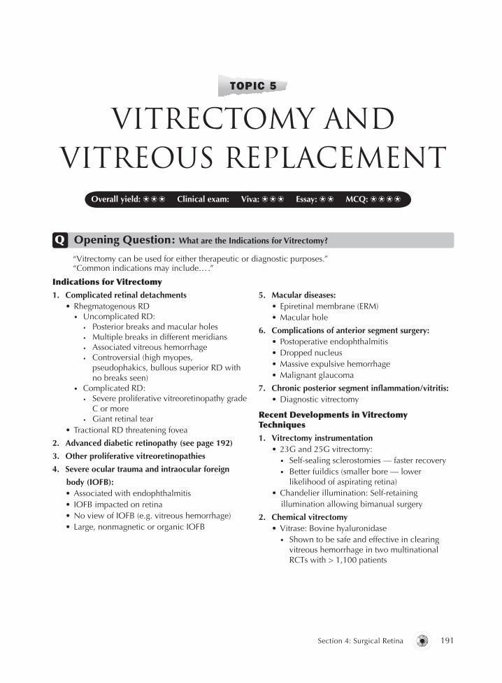
B1037 The Ophthalmology Examinations Review FA
Section 4: Surgical Retina 191
Overall yield: ⍟ ⍟ ⍟ Clinical exam: Viva: ⍟ ⍟ ⍟ Essay: ⍟ ⍟ MCQ: ⍟ ⍟ ⍟ ⍟
Q Opening Question: What are the Indications for Vitrectomy?
“Vitrectomy can be used for either therapeutic or diagnostic purposes.”“Common indications may include… .”
TOPIC 5
VITRECTOMY AND VITREOUS REPLACEMENT
Indications for Vitrectomy
1. Complicated retinal detachments• Rhegmatogenous RD
• Uncomplicated RD:• Posterior breaks and macular holes• Multiple breaks in different meridians• Associated vitreous hemorrhage• Controversial (high myopes,
pseudophakics, bullous superior RD with no breaks seen)
• Complicated RD:• Severe proliferative vitreoretinopathy grade
C or more• Giant retinal tear
• Tractional RD threatening fovea
2. Advanced diabetic retinopathy (see page 192)
3. Other proliferative vitreoretinopathies
4. Severe ocular trauma and intraocular foreign
body (IOFB):• Associated with endophthalmitis• IOFB impacted on retina• No view of IOFB (e.g. vitreous hemorrhage)• Large, nonmagnetic or organic IOFB
5. Macular diseases:• Epiretinal membrane (ERM)• Macular hole
6. Complications of anterior segment surgery:• Postoperative endophthalmitis• Dropped nucleus• Massive expulsive hemorrhage• Malignant glaucoma
7. Chronic posterior segment infl ammation/vitritis:• Diagnostic vitrectomy
Recent Developments in Vitrectomy Techniques
1. Vitrectomy instrumentation• 23G and 25G vitrectomy:
• Self-sealing sclerostomies — faster recovery• Better fuildics (smaller bore — lower
likelihood of aspirating retina)• Chandelier illumination: Self-retaining
illumination allowing bimanual surgery
2. Chemical vitrectomy• Vitrase: Bovine hyaluronidase
• Shown to be safe and effective in clearing vitreous hemorrhage in two multinational RCTs with > 1,100 patients
B1037_Ch-04.indd 191B1037_Ch-04.indd 191 12/22/2010 5:48:12 PM12/22/2010 5:48:12 PM

FA B1037 The Ophthalmology Examinations Review
192 The Ophthalmology Examinations Review
Complications of Vitrectomy
Intraoperative Postoperative
• Retinal break • Suction near mobile retina• Intraocular hemorrhage • Suprachoroidal hemorrhage • Vitreous hemorrhage• Cataract • Lens trauma with instruments• Raised IOP • Infusion bottle too high• Decreased IOP • Infusion bottle too low• Miosis of pupils• Subretinal infusion
• Retinal break • Intraocular hemorrhage • Choroidal hemorrhage • Vitreous hemorrhage• Cataract• Glaucoma• Cornea • Recurrent corneal erosion • Filamentary keratitis • Bullous keratopathy• Phthsis bulbi• Endophthalmitis • Failure of surgery
Vitreous Substitutes
1. Ideal substitute (important):• Clear/transparent• Inert/nontoxic• Low viscosity• Immiscible with H2O• Durable/slowly absorbed
Q What are Vitreous Substitutes?
“They are substances injected into the vitreous cavity during a vitrectomy.”“The main purpose is either for volume replacement or as a tamponade.”“The common vitreous substitutes include… .”
2. Classifi cation:• Intraocular gas• Saline • Silicone oil• Heavy liquids
Exam tips:• The ideal substitute has a LIST of properties that can be used for ALL the
individual substitutes and therefore is well worth to be remembered.
Intraocular Gas
1. Classifi cation:• Nonexpansile
• Air, helium, argon, nitrogen, xenon • Expansile
• Sulphur hexafl oride (SF6), perfl uoreopropane (C3F8), C2F6, C4F10
Q Tell me about Intraocular Gases.
“Intraocular gases are common vitreous substitutes… .”“They can be divided into… .”“The common indications are for either volume replacement (nonexpansile volume) or as a tamponade (expansile volume)… .”
2. Biomechanical properties• Physical properties:
• Properties of ideal substitute (page 161)• Clear/transparent• Inert/nontoxic• Low viscosity
Q What are the Complications of Vitrectomy?
B1037_Ch-04.indd 192B1037_Ch-04.indd 192 12/22/2010 5:48:13 PM12/22/2010 5:48:13 PM

B1037 The Ophthalmology Examinations Review FA
Section 4: Surgical Retina 193
• Immiscible with water• Durable/slowly absorbed PLUS
• High surface tension• Bubble of gas does not enter retinal break
and prevents fl uid from entering break• High buoyancy
• 10 times greater force than SO• Force maximal at apex of angle
• Dynamic properties• Three phases:
• Expansion • Oxygen, carbon dioxide diffuses in
• Maximum at 6–8 hours (therefore be careful of CRAO during this time)
• Equilibrium• Nitrogen last to diffuse• Maximal expansion when gas diffusion
in = diffusion out• Dissolution
• Exponential decline • Depends on size and water solubility• First order kinetics — bubble has useful
ARC of contact for approximately 25% of total duration of gas in eye
3. Comparison of air, SF6, C3F8
Gas DurationTime of maximal expansion rate
Time to maximal expansion
Expansionvolume
Nonexpansile concentration
Air 5 days NA NA ×1 NA
SF6
C2F6
2 weeks 3 weeks
6−8 hours6−8 hours
24−48 hours48−72 hours
×2×3
20%18%
C3F8 2 months 6−8 hours (same asSF6)
72−96 hours ×4 15%
4. Indications for SF6 or C3F8
• Volume replacement after vitrectomy (nonexpansile volume)• Indications for vitrectomy (see page 160)
• Tamponade retina (expansile volume):• Adjunct to RD surgery (posterior breaks or
macular holes)• Adjunct to DRF drainage• Selected giant retinal tear• Flatten radial folds on a high buckle • Pneumoretinopexy (use 100% gas without
vitrectomy; see page 159)
5. Complications:• Glaucoma and CRAO
• Especially during maximum rate of expansion (6–8 hours after operation)
• Cataract (posterior subcapsular “feathery” cataract)
• Bullous keratopathy• New/enlarged breaks• Subretinal seepage• Dislocation of IOLs• PVR
Q Tell me about Silicone Oil.
“Silicone oil is a vitreous substitute.”“It has the following properties… .”
Silicone Oil (SO)
1. Properties:• Properties of ideal substitute (page 161)
• Clear/transparent• Inert/nontoxic• Low viscosity• Immiscible with water
• Surface tension of SO however lower than gases — tendency to emulsify
• Durable/not absorbed PLUS
• Refractive index close to vitreous:• Acts as plus lens in aphakic eyes• Acts as minus lens in phakic eyes
• High viscosity• 1,000–30,000 centistokes (water = 1
centistoke)• Lighter than water (0.93G)
• Heavy silicone oil (denser than water) occasionally used for inferior detachments
B1037_Ch-04.indd 193B1037_Ch-04.indd 193 12/22/2010 5:48:13 PM12/22/2010 5:48:13 PM

FA B1037 The Ophthalmology Examinations Review
194 The Ophthalmology Examinations Review
2. Indications:• Long-lasting volume replacement following
vitrectomy• Common indications:
• PVR• Giant retinal tear• Intraoperative control of vitreous
hemorrhage• Elderly patient who cannot posture• One-eyed patient who needs immediate good
vision postoperation• Patient who needs to travel by air
3. Advantages • “What are the advantages of SO over intraocular
gases?”• Intraoperative advantages:
• Better intraoperative visualization• Easier retinopexy• Control of hemorrhage and effusion
• Postoperative advantages:• Longer-lasting tamponade • Posturing less critical• Better immediate VA
• Air travel not contraindicated• Lower risk of hypotony
• Control over timing of repeat surgery
4. Complications• Glaucoma:
• ACG (from pupil block, therefore inferior iridectomy (Ando) is usually needed)
• Delayed OAG (from emulsifi cation)• Hypotony (not common)
• Cataract • Filamentary keratitis, band keratopathy• Emulsifi cation of SO:
• Uveitis• Subretinal seepage• Subconjunctival cyst formation• Retinal toxicity? (ERG and EOG detected
abnormalities)• ERM/retro-silicone proliferation:
• Accumulation of growth factors, infl ammatory debris at lower meniscus of bubble
• Cause of recurrent epiretinal proliferation• Recurrent RD (25–40%)
Q What are Heavy Liquids?
“Heavy liquids are vitreous substitutes used as intraoperative tools.”“They are essentially extension of perfl uorocarbon gases with seven or more carbon atoms (and therefore liquid at room and body temperature).”
Heavy Liquids
1. Properties• Properties of the ideal substitute (page 161):
• Clear/transparent• Inert/nontoxic (with short-term use)• Low viscosity (0.8–8 centistokes) • Immiscible with water• Durable/slowly absorbed PLUS
• High specifi c gravity (2G, two times more than water or SO)
• High tamponade force (4G, like gas and 10 times more than SO)
2. Examples• Perfl uorodecalin (C10F18)• Perfl uoro-N-octane (C8F18)
3. Indications• Intraoperative tool for complicated VR surgery
(not for prolonged tamponade):• PVR• Giant retinal tear• Subluxed/dislocated lens• IOFB• Subretinal macular hem• Traumatic RD
B1037_Ch-04.indd 194B1037_Ch-04.indd 194 12/22/2010 5:48:13 PM12/22/2010 5:48:13 PM

B1037 The Ophthalmology Examinations Review FA
Section 5
M Retina
B1037_Ch-05.indd 195B1037_Ch-05.indd 195 12/28/2010 9:35:18 AM12/28/2010 9:35:18 AM

FA B1037 The Ophthalmology Examinations Review
B1037_Ch-05.indd 196B1037_Ch-05.indd 196 12/28/2010 9:35:21 AM12/28/2010 9:35:21 AM
This page intentionally left blankThis page intentionally left blank

B1037 The Ophthalmology Examinations Review FA
Section 5: Medical Retina 197
“The macula is an area in the posterior pole of the fundus….”
TOPIC 1
THE MACULAOverall yield: ⍟ ⍟ ⍟ Clinical exam: Viva: ⍟ ⍟ ⍟ Essay: MCQ: ⍟ ⍟ ⍟
Q Opening Question: What is the Anatomy of the Macula?
• Thick internal limiting membrane
3. Foveola• Location: Central fl oor of fovea• Size: 0.35 mm in diameter/0.2 disc
diameter/0.54 minutes of visual angle• Histologically,
• Thinnest part of retina• No ganglion cells or rods• Only cones: 150,000/mm2
4. Foveal avascular zone (FAZ)• Location: Area bounded by fovea and foveola • Histologically (accounts for “darkness” in FFA),
• Avascular• Tall RPE cells with increased melanin• Increased xanthophyll pigments
Q How do You Evaluate Macular Function?
“The macula can be assessed clinically and with appropriate tests….”
Anatomy of the Macula
1. Macula• Location: Area bounded by temporal arcades,
4 mm temporal, 0.8 mm inferior to optic disc• Size: 5 mm in diameter/3.5 disc diameter/18o of
visual angle• Histologically,
• > 1 layer of ganglion cells• Xanthophyll pigments
2. Fovea• Location: Depression inside macula• Size: 1.5 mm in diameter/1 disc diameter/5o of
visual angle• Histologically,
• 6–8 layers of ganglion cells• Tall RPE cells
Macular Function
1. Clinical • VA• Color vision and pupil (should be normal unless
the macular is extensively damaged)• Binocular ophthalmoscopy and slit lamp
biomicroscopy (with 78D or 90D lens)• Confrontational VF and light projection
2. Ancillary tests• Amsler grid:
• Screening test for macular function• Evaluates 20° of visual angle (macular
subtends only 18°)
• Standard chart with 10 cm large square and 5 mm small square
• Chart should be read at 1/3 of meter (small square subtends 1°)
• Seven charts in total:• Chart 1: Standard chart• Chart 2: Diagonal lines to help central
fi xation, when central scotoma is present
• Chart 3: Standard chart, but red lines on black background, for color scotoma
• Chart 4: No lines, only dots, reveals only scotoma
B1037_Ch-05.indd 197B1037_Ch-05.indd 197 12/28/2010 9:35:21 AM12/28/2010 9:35:21 AM

FA B1037 The Ophthalmology Examinations Review
198 The Ophthalmology Examinations Review
• Chart 5: Parallel horizontal lines, to show metamorphopsia
• Chart 6: Similar to Chart 5, but with fi ner horizontal lines in the central area
• Chart 7: Similar to Chart 1, but with fi ner lines in central area
• Photostress test:• Principle of the dark adaptation test (evaluate
recovery time of photoreceptors to re-synthesize visual pigments)
• Procedure:• Snellen VA assessed• Patient fi xates on torch light for 10 seconds• Photostress recovery time = time taken to
read Snellen letters one line above the pre-test level (normal: 30 seconds)
• Compare with other eye• Flying corpuscle test:
• Principle of the entoptic phenomenon (subjective perception of white blood cells moving in perifoveal capillaries)
• Procedure:• Patient asked to look into blue light of
entoptoscope
• Patient should see 15 or more white blood cells in entire area
• Abnormal macular function:• No corpuscles/decreased number of
corpuscles• Slow speed of corpuscles• No corpuscles in a specifi c area
• Laser interferometer:• Principle of interference • Two coherent light beams creates fringe
pattern (black and bright bands) by process of interference
• Fringe pattern in different orientations and progressively fi ner gratings are used to estimate VA and macular function
• Potential acuity meter:• Projection of a miniature Snellen acuity chart
into retina, through a clear area of cataract or other media opacities
• Usually best for VA < 20/200
Exam tips:• An alternate question may be, “How do you assess the macular function in a
patient with a dense cataract?”• Remember to talk about the clinical examination fi rst (what you would
normally do in your daily practice), before going on to the more esoteric tests.
B1037_Ch-05.indd 198B1037_Ch-05.indd 198 12/28/2010 9:35:22 AM12/28/2010 9:35:22 AM

B1037 The Ophthalmology Examinations Review FA
Section 5: Medical Retina 199
• Siedal’s test (wound leak)• Contact lens fi tting (assess contact lens fi t)
3. Anterior chamber• Detect iris neovascularization• Applanation tonometry
4. Retina• FFA
Clinical Uses of Fluorescein
1. Lacrimal system• Tear break-up time (dry eyes)• Jones test: Dye disappearance test (blockage of
lacrimal system)
2. Cornea• Detect epithelial defect (corneal abrasion,
superfi cial punctate keratopathy)
Q Opening Question: What are the Clinical Uses of Fluorescein?
TOPIC 2
FUNDAL FLUORESCEIN ANGIOGRAPHY
Q What are the Principles of Fundal Fluorescein Angiography (FFA)?
“Fundal fl uorescein angiography is based on two principles….”“The principle of fl uorescence, which is the ability of….”“And the principle of the blood-retinal barrier, which consists of….”
Principles of FFA
1. Fluorescence• Fluorescence: Ability of a substance to emit
light energy of a longer wavelength (emission wavelength) when stimulated by light of a shorter wavelength (excitation wavelength)
• Fluorescein:• Excitation wavelength peak: 490 nm (blue)• Emission wavelength peak: 530 nm (green)
• Basic FFA:• White light from retinal camera passes
through a blue excitation fi lter, which allows only blue light to enter eye
• Interaction of blue light with fl uorescein molecules in blood vessels, with emission of yellow-green light (fl uorescence)
• Yellow-green and refl ected blue light travel out of retina to camera, passes through blue interference fi lter, allowing only yellow-green light to be imaged onto fi lm
Exam tips:• Fundal fl uorescein angiography (FFA) is NOT the only use of fl uorescein!
Overall yield: ⍟ ⍟ ⍟ Clinical exam: ⍟ Viva: ⍟ ⍟ ⍟ Essay: MCQ: ⍟ ⍟
B1037_Ch-05.indd 199B1037_Ch-05.indd 199 12/28/2010 9:35:22 AM12/28/2010 9:35:22 AM

FA B1037 The Ophthalmology Examinations Review
200 The Ophthalmology Examinations Review
2. Blood-retinal barrier (BRB) • Inner blood-retinal barrier: Tight junctions of
retinal capillary endothelial cells• Outer blood-retinal barrier: Tight junction of
RPE• When fl uorescein is injected into blood stream,
inner BRB prevents leakage of fl uorescein and allows retinal vessels to be seen
• On the other hand, there is leakage of free fl uorescein from choroidal vessels, but outer BRB prevents free fl uorescein from traveling across RPE into sensory retina
• Therefore, leakage of fl uorescein from either retinal vessel or RPE is abnormal
Notes• “What is autofl uorescence?”
• Ability of a substance to emit yellow-green light when stimulated by blue light in the absence of fl uorescein
• Classic example: Drusens
Notes• “What is pseudofl uorescence?”
• Ability of substance to emit yellow-green light when stimulated by blue light in the presence of mismatched fi lters
Q What are the Indications for FFA?
“FFA is useful as an aid in the diagnosis and management of various posterior segment diseases.”
Indications for FFA
1. Aid in diagnosis of:• Macular diseases:
• AMD, central serous retinopathy, CME• Retinal vascular diseases:
• Neovascularization in DR, CRVO, BRVO, retinal telangiectasia
• Infl ammatory retinal/choroidal diseases:• Posterior uveitis, CMV retinitis
• Optic nerve disorders:• Disc drusens, papilledema, optic neuritis
• Tumors:• Choroidal hemangiomas
2. Aid in laser treatment of:• CNV, central serous retinopathy, DM
maculopathy, CRVO and BRVO
Q What are the Side Effects of FFA?
“FFA is usually a safe procedure with few side effects.”
1. Mild and more common side effects:• Nausea and vomiting• Yellow discoloration of skin, urine and tears• Pain at injection site (especially if there is
extravasation)• Rashes
2. Serious but rare side effects:• Anaphylactic shock • Airway spasm• Collapse and death
B1037_Ch-05.indd 200B1037_Ch-05.indd 200 12/28/2010 9:35:22 AM12/28/2010 9:35:22 AM

B1037 The Ophthalmology Examinations Review FA
Section 5: Medical Retina 201
Results of FFA
1. Normal FFA• Phase 1: Pre-arterial phase/choroidal fi lling
• Arm-retina time: 9–12 seconds• Phase 2: Arterial phase
• One second after Phase 1• Phase 3: Arteriovenous phase/capillary phase
• Complete fi lling of arterioles and capillaries• Early venous fi lling (lamellar fl ow)
• Phase 4: Venous phase• Complete venous fi lling• Recirculation of dye• Intensity of fl uorescence decreases
2. Cause of hyperfl uorescence• Window defect (transmission of dye from
choroid):• Atrophy or destruction of RPE cells (e.g.
AMD), RPE tear• Occurs early in FFA
• Pooling (increase in intensity of hyperfl uorescence but not in size):• Dye in sub-RPE space (e.g. pigment epithelial
detachment)• Dye in sub-retinal space (e.g. exudative RD)
NotesResuscitation for anaphylactic shock
• Secure the airway, give 100% oxygen (intubate if respiratory obstruction is imminent)
• IM adrenaline 0.5 mg (repeat every 5 min if needed)• Secure IV access• IV chlorpheniramine 10 mg and hydrocortisone 200 mg• IV normal saline: Titrate against blood pressure• May require ventilator support
Q Tell me about the Normal FFA. What Abnormalities can FFA Detect?
“The abnormalities are either related to hyperfl uorescence or hypofl uorescence….”
• Occurs early in FFA• Leakage (increase in size and intensity of
hyperfl uorescence) from:• Choroidal vessels occur early (e.g. choroidal
neovascularization)• Retinal vessels occur late (e.g. NVD in DR)• Optic nerve head occurs late (e.g.
papilledema)• Staining (dye in tissue):
• Retinal scars, sclera, abnormal vessels• Occurs late
3. Cause of hypofl uorescence• Masking (blockage of dye transmission from
choroid):• Retinal hemorrhages• Edema and hard exudates• Pigments (melanin and xanthophyll)• Lipofuscin (e.g. Best disease)
• Filling defect (delay of fi lling or occlusion of vessels):• Retinal ischemia (e.g. CRVO, DR)• Choroidal ischemia (e.g. HPT retinopathy)• Retinal atrophy (e.g. myopia)
Notes• “Why is the fovea dark?”
• Blockage of choroidal fl uorescein by RPE• Increases: Melanin pigments in RPE• Increases: Height of RPE cells
• Increased xanthophyll pigments• Avascular
B1037_Ch-05.indd 201B1037_Ch-05.indd 201 12/28/2010 9:35:22 AM12/28/2010 9:35:22 AM

FA B1037 The Ophthalmology Examinations Review
202 The Ophthalmology Examinations Review
Indocyanine Green Angiography
1. Advantages over FFA• ICG is highly bound to plasma proteins (98%)
as compared with FFA (80%)• Choroidal circulation is more easily seen
• ICG has maximum absorption at 805 nm and fl uorescence at 835 nm• Wavelength of ICG can penetrate RPE and
macular xanthophyll better
Q Tell me about Indocyanine Green (ICG) Angiography.
“ICG is a complementary test to the FFA in the diagnosis and management of various posterior segment diseases.”
2. Indications• AMD:
• Occult and recurrent CNV• PED• Submacular hemorrhage
• Infl ammatory choroidal diseases:• White dot syndromes • Vogt-Koyanagi-Harada syndrome
• Tumors:• Choroidal melanoma
B1037_Ch-05.indd 202B1037_Ch-05.indd 202 12/28/2010 9:35:23 AM12/28/2010 9:35:23 AM

B1037 The Ophthalmology Examinations Review FA
Section 5: Medical Retina 203
Q Opening Question: What is Optical Coherence Tomography?
“OCT is a low coherence interferometry which allows non-contact in vivo imaging of ocular tissues at a high resolution.”
TOPIC 3
OPTICAL COHERENCE TOMOGRAPHY
OCT
1. Principle• Works as an “optical ultrasound,” by using light
refl ections to provide cross-sectional images
2. Types• Time domain OCT:
• Mirrors used to scan tissue and provide cross-sectional image of optic nerve
• Acquires images at 400 axial scans/second• Artifacts from eye movement
• Axial resolution of 10 µm• More widely used
• Frequency domain OCT:• Spectral/Fourier domain OCT • Mirrors not used, all wavelengths of returning
light analyzed simultaneously• Acquires images at 20,000 axial scans/
second• Less artifacts and improved quality of
images• Axial resolution 5 µm• More expensive
Q What are the Applications of Optical Coherence Tomography in Ophthalmology?
“OCT is widely used in ophthalmology….”“It is used to image the macula both the anterior and posterior segment.”
Applications of OCT
1. Posterior segment• Macula:
• CSME• AMD and PCV: Macular edema, PED• CSR• CME• ERM and VMT• Macular hole• RPE atrophy• Maculoschisis
• RNFL:• Reduced in:
• Glaucoma• High myopia
• Increased in:• Swollen disc
2. Anterior segment• AC angles: Open or closed• Mechanisms of angle closure: Pupil block, angle
crowding, bulky lens
Overall yield: ⍟ ⍟ Clinical exam: ⍟ Viva: ⍟ ⍟ Essay: MCQ: ⍟ ⍟
B1037_Ch-05.indd 203B1037_Ch-05.indd 203 12/28/2010 9:35:23 AM12/28/2010 9:35:23 AM

FA B1037 The Ophthalmology Examinations Review
B1037_Ch-05.indd 204B1037_Ch-05.indd 204 12/28/2010 9:35:23 AM12/28/2010 9:35:23 AM
This page intentionally left blankThis page intentionally left blank

B1037 The Ophthalmology Examinations Review FA
Section 5: Medical Retina 205
Q Opening Question: What is the Electroretinogram, Electrooculogram and Visual Evoked Potential?
• ERG = Measure of electrical mass response of the retina, refl ecting electrical activity of photoreceptor and bipolar cells
• EOG = Measure of electrical mass response of the eye, refl ecting the metabolic activity of the RPE
• VEP = Measure of electrical response of the occipital visual cortex to stimulation of the retina with either light or pattern stimulus, refl ecting activity of the entire visual system (from retina, especially macula to cortex)
TOPIC 4
ELECTROPHYSIOLOGY
Q Tell me about the Principles of Electroretinography.
Electroretinography (ERG)
1. Anatomical basis• Rods
• Distribution:• 120 million• Absent in foveola, increase to peak density
15ο from foveola center, then decrease slightly towards periphery
• Sensitivity:• Maximal in scotopic conditions• Maximal to blue green light• Unable to follow fl icker greater than 8 to
10 cycles/second (longer refractory period)• Cones
• Distribution:• 6 million• Peak density in foveola, decrease in
density 15o from foveola center, lowest density in peripheral retina
• However, majority of cones lie outside fovea (therefore localized disease of the fovea will still result in a normal cone ERG
and abnormal cone ERG implies widespread retinal disorder)
• Sensitivity:• Maximal in photopic conditions• Maximal to green-yellow light• Able to follow fl icker greater than 8 to 10
cycles/second
2. Normal ERG waveform• A-wave:
• Negative waveform• Photoreceptor hyper-polarization when
exposed to light stimulus • Refl ects photoreceptor function
• B-wave:• Positive waveform• Midretinal cells, initiated by bipolar cells,
magnifi ed by Müller cells• Refl ects Müller cells and bipolar cells
• C-wave:• Occasionally present• Refl ects RPE cells
Exam tips:• A clear defi nition of the three main types of electrophysiological tests is
important.
Overall yield: ⍟ ⍟ ⍟ Clinical exam: Viva: ⍟ ⍟ ⍟ Essay: MCQ: ⍟ ⍟ ⍟
B1037_Ch-05.indd 205B1037_Ch-05.indd 205 12/28/2010 9:35:23 AM12/28/2010 9:35:23 AM

FA B1037 The Ophthalmology Examinations Review
206 The Ophthalmology Examinations Review
• Oscillatory potential:• Wavelets on rising B-wave
• Interaction between Amacrine and Interplexiform cells
• Refl ects primarily cone function
Q What are the Types of ERG and How do You Use/Apply Each of Them?
Types of ERG
1. Equipment• Stimulator:
• Ganzfeld stimulator, diffuse illumination of entire retina
• Electrodes:• Active electrode (contact lens electrode, gold
foil lid electrode)• Reference electrode (forehead)• Ground electrode (earlobe)
• Amplifi cation and display system
2. ERG waveform measurement• Amplitude:
• Trough of “A-wave” to the peak of “B-wave” (microvolts)
• Refl ects effi ciency of the retina in producing an electrical signal, dependent on pupil size, refractive error, fundal pigmentation and age
• Implicit latency:• Time of stimulus to peak of B-wave
(milliseconds)
3. Types of ERG• Maximal response ERG:
• Refl ects a combination of cone and rod functions
• Dark adaptation• Bright fl ash stimulus
• Dark adapted ERG:• Refl ects rod function• Dark adaptation (for 20 to 30 minutes)• Low intensity blue fl ash or low intensity
white fl ash stimulus• Absent/minimal A-wave
• Light adapted ERG:• Refl ects cone function• Light adaptation (for 10 minutes)• Bright fl ash stimulus• Waveform amplitude is about 30% smaller
• Flicker ERG:• Refl ects cone function• Light adaptation• Flicker 30 Hz stimulus
• Focal ERG (FERG):• ERG evoked by a small focal stimulus • Retina is bleached by background light• Focal stimulus applied on to retina• Usually only cone function at macula can be
assessed easily• Pattern ERG (PERG):
• Refl ects ganglion cell layer function• Stimulus is a pattern reversal checkerboard• May have applications in glaucoma and optic
nerve disease
Q Tell me about the Electrooculogram.
Electrooculogram (EOG)
1. Principle• Measures standing potential between electrically
positive cornea and negative retina/RPE • Exposure to light causes a rise in standing
potential (apical portion of RPE cells depolarize, giving rise to a positive wave seen on the EOG)
2. Procedure• Electrodes are placed on medial and lateral
canthal area on either side of eye • The eye is made to perform saccades between
two points about 30o apart• The electrodes pick up voltage differences
between front and back of the eye as it rotates back and forth
• Amplitude of voltage is recorded
3. Interpretation• Amplitude swings increase with light exposure
and decrease in darkness:• The swings are expressed as light peak to
dark trough ratio (Arden ratio)• Normal ratio = 1.65
• Abnormal ratio refl ects widespread RPE abnormality:• EOG generally parallels ERG readings in
assessing rod function. However, EOG cannot assess cone function well
• Most useful in Best’s disease (EOG light rise is absent but ERG is normal)
B1037_Ch-05.indd 206B1037_Ch-05.indd 206 12/28/2010 9:35:24 AM12/28/2010 9:35:24 AM

B1037 The Ophthalmology Examinations Review FA
Section 5: Medical Retina 207
Q Tell me about the Visual Evoked Potential.
Visual Evoked Potential (VEP)
1. Principle• Measures the potentials generated at occipital
lobe by visual stimuli• Primarily the foveal areas are represented at
superfi cial part of occipital lobe where the potential is measured
• Abnormalities of the VEP are caused by lesions anywhere between photoreceptor and occipital lobe
2. Procedure• Stimulus:
• Flash (variable response, useful in opaque media)
• Pattern reversal (reversing checks or stripes, generates maximal cortical activity)
• Amplitude of voltage is recorded
3. Interpretation • Waveform:
• Extremely variable in size and shape. Amplitude may vary
• Relatively constant positive waveform occurs at 100 millisecond (P100 wave)
• The P100 latency is therefore the most useful clinical indicator
4. Clinical indications • VEP acuity (decrease checkerboard size until
VEP approaches zero)• Optic nerve disease (increased latency/
decreased amplitude to fl ash or pattern VEP)• Fovea or macular disease (increased latency/
decreased amplitude to pattern VEP)• Amblyopia (increased latency/decreased
amplitude to pattern VEP)• Malingering (should be normal)
B1037_Ch-05.indd 207B1037_Ch-05.indd 207 12/28/2010 9:35:24 AM12/28/2010 9:35:24 AM

FA B1037 The Ophthalmology Examinations Review
B1037_Ch-05.indd 208B1037_Ch-05.indd 208 12/28/2010 9:35:24 AM12/28/2010 9:35:24 AM
This page intentionally left blankThis page intentionally left blank

B1037 The Ophthalmology Examinations Review FA
Section 5: Medical Retina 209
Q Opening Question: What is Age-Related Macular Degeneration (AMD)?
“Age-related macular degeneration refers to a spectrum of disease associated with….”“The pathogenesis is poorly understood, but the risk factors include….”“There are two classic forms of AMD….”
TOPIC 5
AGE-RELATED MACULAR DEGENERATION
Age-Related Macular Degeneration
1. Defi nition• Spectrum of disease• Visual loss, RPE changes, drusens, geographical
atrophy of retina, choroidal neovascularization (CNV)
• Usually in persons aged 50 years or above• Drusens alone without visual loss is not
considered AMD
2. Pathogenesis• Multifactorial etiology:
• Genetic predisposition, racial patterns• Environmental risk factors:
• Smoking• HPT, cardiovascular disease• UV exposure
• Initial changes include RPE dysfunction, followed by deposition of drusens, thickeningof Bruch’s membrane and RPE atrophy
• Later, PED occurs and CNV follows (sequence here is unclear)
3. Classifi cation• Early AMD:
• Drusens• +/− RPE changes
• Late AMD:• Geographic atrophy (dry AMD):
• Extensive RPE atrophy• Exudative AMD (wet AMD):
• Classic/occult CNV• Pigment epithelial detachment• Subretinal hemorrhage• Vitreous hemorrhage• Disciform scar• Massive exudation (intra/subretinal)
Exam tips:• The defi nition of AMD is important but usually poorly answered.
Overall yield: ⍟ ⍟ ⍟ ⍟ ⍟ Clinical exam: ⍟ ⍟ ⍟ ⍟ Viva: ⍟ ⍟ ⍟ Essay: ⍟ ⍟ ⍟ MCQ: ⍟ ⍟ ⍟ ⍟
B1037_Ch-05.indd 209B1037_Ch-05.indd 209 12/28/2010 9:35:24 AM12/28/2010 9:35:24 AM

FA B1037 The Ophthalmology Examinations Review
210 The Ophthalmology Examinations Review
Drusens
1. Defi nition and pathology• Lipid-like/lipofuscin material deposited in
Bruch’s membrane (between basement membrane of RPE and inner collagen layer)
2. Types• Hard drusens• Soft drusens• Confl uent drusens• Calcifi ed drusens
• Nodular drusens (younger onset, may have family history)
3. Risk of AMD• Size of drusens• Type of drusens:
• Hard drusens → nonexudative AMD• Soft/confl uent drusens → exudative AMD
• Family history of AMD• Fellow eye has AMD
Q What are the Causes of CNV?
“The commonest cause of CNV is AMD, but other causes include….”
CNV
1. Defi nition • Proliferation of fi brovascular lesions from
choriocapillaris through defects in Bruch’s membrane into subretinal space
2. Causes • Degenerative:
• AMD• Pathological myopia
• Others (optic disc drusen, angioid streaks)• Infl ammatory disease:
• POHS (presumed ocular histoplasmosis syndrome)
• Posterior uveitis (toxoplasmosis, Vogt-Koyanagi-Harada syndrome)
• Traumatic (choroidal rupture, laser photocoagulation)
• Tumor (choroidal nevus, choroidal hemangioma)
Exam tips:• There are a number of possible scenarios. If drusens are seen, it is important
to exclude CNV. If a disciform scar is seen, it is important to not only consider AMD, but myopic degeneration and trauma as well.
Q What are Drusens?
“Drusens are lipid-like materials deposited in….”
Clinical approach to AMD/subretinal hemorrhage
“On examination of this patient’s fundus….”“The most obvious lesion is at the macula where a large subretinal hemor-rhage/disciform scar about 2 disc diameter in size is seen.”
Look for• Drusens (AMD)• Tessellated fundus, peripapillary atrophy (myopic degeneration)• Rare causes of CNV:
• Optic disc drusen, angioid streaks, choroidal nevus• Iatrogenic (excessive laser photocoagulation)
• Other eye:• Bilateral drusens, disciform scar (Likely CNV)• Normal fellow eye (likely PCV)
(Continued)
B1037_Ch-05.indd 210B1037_Ch-05.indd 210 12/28/2010 9:35:25 AM12/28/2010 9:35:25 AM

B1037 The Ophthalmology Examinations Review FA
Section 5: Medical Retina 211
FFA Changes in AMD
1. Early AMD• Drusens:
• Autofl uorescence on red-free photograph• Staining• Can be hypo- or hyperfl uorescent
• RPE atrophy:• Window defects
2. Late AMD• Geographic atrophy:
• Window defects• Exudative AMD:
• Pigment epithelial detachment (PED):• Pooling• RPE tear: Window defects,
hypofl uorescence (masking)• CNV:
• Leaking• Hemorrhage: Blocked fl uorescence
Q What are the FFA Changes in AMD?
• Scars: Staining
3. MPS defi nition (“How is classic and occult CNV
defi ned in MPS?”)• “Classic” CNV — well defi ned CNV occurring
early in the course of FFA:• Extrafoveal (> 200 microns from foveola/FAZ)• Juxtafoveal (< 200 microns from foveola/FAZ
but not involving center itself)• Subfoveal (encroaching into center of
foveola/FAZ)• “Occult” CNV — 2 basic patterns:
• Fibrovascular PED (irregular elevation of RPE fi lls more slowly than classic CNV)
• Late leakage of undetermined source (region of hyperfl uorescence at the level of RPE, best appreciated in late phases, without the features of classic CNV in the early phases)
Notes
Classifi cation of occult CNV by ICG (Ophthalmology 1996;103:2054–2060)• Hot spots (< 1 DD):
• Second most common• Can be treated by laser
• Plaques (> 1 DD):• Most common• Poor natural history
• Combination of hot spots and plaques:• Rare
Q What are the Results of the Macular Photocoagulation Study (MPS)?
“The MPS is a multicenter randomized clinical trial to evaluate the effectiveness of….”
MPS
1. Hypothesis: “Is argon laser photocoagulation
effective in preventing severe visual loss in eye
with….”• Extrafoveal CNV (Arch Ophthalmol
1982;100:192–198)
• Juxtafoveal CNV (Arch Ophthalmol 1990;108:825–831)
• Subfoveal CNV (Arch Ophthalmol 1991;109:1220–1231)
2. Treatment• Argon laser photocoagulation vs no treatment
I’ll like to• Check VA• Perform a FFA to delineate site of leakage from CNV, and ICG to determine
presence of polyps
(Continued)
B1037_Ch-05.indd 211B1037_Ch-05.indd 211 12/28/2010 9:35:25 AM12/28/2010 9:35:25 AM

FA B1037 The Ophthalmology Examinations Review
212 The Ophthalmology Examinations Review
3. Outcome• SVL (severe visual loss) defi ned as loss of six
lines or more of VA
4. Results• Extrafoveal: 45% SVL (laser) vs 64% SVL (no
laser) at 5 years• Juxtafoveal: 47% SVL (laser) vs 58% SVL (no
laser) at 3 years
• Subfoveal: • No prior laser: 20% SVL (laser) vs 37% SVL
(no laser) at 2 years• Recurrent CNV: 9% SVL (laser) vs 37% SVL
(no laser) at 2 years
5. Conclusion• Laser benefi cial in all types of classic CNV• SVL occurs in both treated and untreated cases• Risk of immediate decrease in VA following
treatment
Q What is the Role of PDT in Exudative AMD?
“Verteporfi n is an intravenous photosensitizer which is activated by a diode laser source….”“With the introduction of anti-VEGF, the use of PDT is less common….”
1. Mechanism of action• Selectively damages CNV by:
• Preferential localization to CNV (complexes with LDL and CNV has increased LDL receptors)
• Irradiation confi ned to CNV• Light activation causes free radical release,
endothelial cell damage and thrombus formation
2. Clinical trials• Treatment of AMD with photodynamic therapy
(TAP) study• Inclusion criteria:
• Subfoveal, classic/predominantly classic CNV, ≤ 5400 µm
• VA between 6/12 and 6/60• Treatment groups: PDT vs placebo• Main outcome measures: Stable (< 15 logMAR
letters lost) or improved vision• Results:
• Stable/improved vision at 12 months: 61% treated, 46% placebo (Arch Ophthalmol 1999;117:1161–1173)
• Stable/improved vision at 24 months: 53% treated, 38% placebo (Arch Ophthalmol 2001;119:198–207)
• Results better for pure classic CNV compared to predominantly classic CNV
• Verteporfi n also shown to be effective in:• Pure occult CNV: Verteporfi n in
Photodynamic Therapy (VIP) study• Minimally classic CNV: Verteporfi n in
Minimally Classic CNV (VIM) study• CNV associated with pathological myopia:
VIP study
3. Treatment regime• Intravenous verteporfi n (6 mg/kg body weight)
infused over 10 minutes• After fi ve minutes, lesion treated with diode
laser for 83 seconds• Patient reviewed every three months, treatment
repeated if there is persistent or new leakage
4. Complications• Ocular:
• Hemorrhage from CNV• RPE rip
• Systemic:• Pain at injection site• Photosensitivity• Low back pain
Notes
Relative indications for PDT• PCV• Myopic CNV• Small classic CNV• CNV associated with POHS• Chronic CSR• Hemangiomas• When anti-VEGF contraindicated
B1037_Ch-05.indd 212B1037_Ch-05.indd 212 12/28/2010 9:35:25 AM12/28/2010 9:35:25 AM

B1037 The Ophthalmology Examinations Review FA
Section 5: Medical Retina 213
Q What is the Role of Anti-Vascular Endothelial Growth Factor (VEGF) Antibodies in Exudative AMD?
“Anti-VEGF is the established treatment for exudative AMD….”“The optimal treatment regime is still being evaluated….”
1. Mechanism of VEGF• VEGF is an important mediator in the
pathogenesis of exudative AMD• It stimulates angiogenesis and the development
of CNV, and also promotes vascular leakage
2. Anti-VEGF antibodies used in the treatment of
exudative AMD• Ranibizumab (Lucentis): Recombinant,
humanized murine antigen-binding fragment. Approved by the FDA for the treatment of exudative AMD
• Bevacizumab (Avastin): Humanized, full-length monoclonal antibody. Off-label use in the treatment of exudative AMD
• Pegaptanib (Macugen): Aptamer that binds and inhibits only the extracellular isoforms of VEGF
3. Which lesions should be treated?• All major CNV subtypes (predominantly classic,
minimally classic, occult) in subfoveal and juxtafoveal location
• “Active” disease:• Retinal thickening• Intraretinal/subretinal hemorrhage• New/persistent leakage on FFA• Enlargement of CNV size on FFA• Worsening VA
• Any baseline VA
• Serous PED, retinal angiomatous proliferation (RAP), PCV: Can be considered for treatment but detailed evidence not available
4. Treatment regime• Loading dose of monthly injections for 3
months (ANCHOR and MARINA trials)• Maintenance dose (controversial):
• Monthly: MARINA and ANCHOR trials (evidence for best VA outcomes)
• 3-Monthly: Initial VA improvement not maintained
• Individualized• Can be combined with PDT or IVTA
(combination therapy)
5. Outcomes (ANCHOR and MARINA)• Gain of 15+ ETDRS letters: 33–40% 0.5 mg
Lucentis vs 4–9% sham• Loss of < 15 ETDRS letters: ~ 95% 0.5 mg
Lucentis vs 62–64% sham
6. Complications• Ocular:
• Retinal detachment• Vitreous hemorrhage• Uveitis• Endophthalmitis • RPE rip
• Systemic (uncommon):• Stroke, myocardial infarction
Notes• “What is the natural history of exudative AMD if left untreated?”
• Four lines lost over 24 months• Three lines lost over 12 months• Two lines lost over 6 months• One line lost over 3 months
Q How do You Manage a 70-Year-Old Patient with AMD?
“Management of AMD must be individualized to the patient and depends on the type and severity of AMD and the amount of visual disability and visual requirements of the patient.”“If the patient has the nonexudative type of AMD, there is no….” “On the other hand, if the patient has exudative type of AMD, I will consider…..”
B1037_Ch-05.indd 213B1037_Ch-05.indd 213 12/28/2010 9:35:26 AM12/28/2010 9:35:26 AM

FA B1037 The Ophthalmology Examinations Review
214 The Ophthalmology Examinations Review
2. Exudative AMD• FFA to detect and localize CNV • Treatment would depend on:
• Location of CNV• Duration of symptoms• Presence of “active” disease• Presence of scarring
• Extrafoveal CNV: Direct laser• Juxtafoveal and subfoveal CNV:
• Offer monthly injections of intravitreal anti-VEGF for at least three months
• Offer combination therapy with PDT if PCV is present
• No proven benefi t in treating disciform scar. Long-term follow-up required
• 20% risk to fellow eye in three years
Management of AMD
1. Early AMD and geographic atrophy• No effective curative treatment• Stop smoking• High levels of antioxidants (Vit C, Vit E, Vit A),
zinc and copper reduce progression to advanced AMD and vision loss: Age-Related Eye Disease Study (AREDS)• High levels of beta-carotene increases risk of
lung cancer in smokers• Reassure patient that total blindness rarely
occurs• Follow-up patient with Amsler grid monitoring• Monitor fellow eye• Low vision aid if necessary
Notes• “How do you monitor a patient with exudative AMD?”
• Visual symptoms• Visual acuity• New hemorrhages/exudation• OCT• Repeat FFA/ICG when new signs/symptoms occur
Q How do you Administer an Intravitreal Injection?
“I would administer intravitreal injections under aseptic conditions….”
1. Topical anesthesia
2. Clean and drape patient
3. Lid speculum
4. ½ strength iodine to sterilize eye
5. (Optional) Subconjunctival lignocaine
6. Measure and mark injection site with calipers:• Pseudophakic: 3 mm behind limbus• Phakic: 4 mm behind limbus
7. Administer intravitreal injection at marked
injection site with 27G–30G needle
8. Withdraw needle and apply pressure at injection
site with sterile cotton bud
9. Check that patient has at least “Counting Fingers”
vision post-injection
10. (Optional) Check IOP
11. Prescribe topical antibiotics for a week after the
injection
12. Warn the patient of risk of:• Endophthalmitis: Red eyes, pain, blurring of
vision• Retinal detachment: Floaters, fl ashes, visual
fi eld defects
Exam tips:• Give a short concise answer, be as conservative as possible, and lead the
examiner to ask you about a topic you know well.
B1037_Ch-05.indd 214B1037_Ch-05.indd 214 12/28/2010 9:35:26 AM12/28/2010 9:35:26 AM

B1037 The Ophthalmology Examinations Review FA
Section 5: Medical Retina 215
1. Pathogenesis• Etiology is idiopathic, but similar genetic
mutations are associated with PCV and AMD• Dilated network consisting of multiple terminal
aneurysmal protuberances in a polypoidal confi guration
• Predilection for macular and peripapillary areas
2. Signs• Orange nodules may be visible• Serosanguinous maculopathy• Vitreous hemorrhage and massive subretinal/
sub-RPE bleeds more common than CNV associated with AMD
Q What is Polypoidal Choroidal Vasculopathy (PCV)?
“PCV is an abnormality of the inner choroidal vessels….”“It typically affects darker-skinned races such as Asians and African Americans….”
3. ICG fi ndings• Large choroidal vascular network associated
with localized terminal polyp-like bulbs• “Bunch of grapes”
4. Treatment• Extrafoveal: Direct laser• Juxtafoveal and subfoveal:
• PDT• Anti-VEGF• Combination therapy
Q What is Retinal Angiomatous Proliferation (RAP)?
“RAP is an uncommon cause of exudative AMD, in which the abnormal vessels arise from the retinal vasculature rather than the choriocapillaris.”
1. Pathogenesis• Etiology is idiopathic but thought to be age-
related• Angiomatous proliferation originates from the
retina and extends posteriorly into the subretinal space, eventually intersecting, in some cases, with choroidal new vessels
2. Signs• Similar to AMD• Patients tend to be older than patients with CNV
3. Stages• Stage I: Intraretinal neovascularization• Stage II: Subretinal neovascularization• Stage III: CNV
4. Angiography• FFA: Indistinct leaking simulating occult CNV• ICG: Hot spot in mid or late frames
• CSLO-ICGA: Hot spot appears after, instead of before, retinal artery fi lling
5. Treatment• Anti-VEGF• PDT with adjunctive triamcinolone/anecortave
may be successful• Conventional laser photocoagulation often
disappointing• Experimental: Surgical excision of feeder artery
and draining vein
6. Prognosis• Aggressive course and poorer prognosis
B1037_Ch-05.indd 215B1037_Ch-05.indd 215 12/28/2010 9:35:26 AM12/28/2010 9:35:26 AM

FA B1037 The Ophthalmology Examinations Review
B1037_Ch-05.indd 216B1037_Ch-05.indd 216 12/28/2010 9:35:26 AM12/28/2010 9:35:26 AM
This page intentionally left blankThis page intentionally left blank

B1037 The Ophthalmology Examinations Review FA
Section 5: Medical Retina 217
Q Opening Question:Tell me about Pathological Myopia.
“Degenerative or pathological myopia is defi ned as a progressive form of severe myopia.”“Usually in patients with axial length of > 26 mm.”“It may be an isolated anomaly or associated with….”
TOPIC 6
OTHER MACULAR DISEASES
Pathological Myopia
1. Associations• Ocular:
• ROP• Congenital glaucoma• Albinism, congenital stationary night
blindness• Ectopic lentis• RP• Wagner’s syndrome
• Systemic association:• Marfan’s, Stickler’s, Ehlers-Danlos syndrome• Down’s syndrome• Alport’s syndrome
2. Problems• Higher risk of nuclear sclerosis cataract and
POAG• Myopic macular degeneration, CNV, macular hole• Retinal breaks and RD
Clinical approach to myopic fundus
“On examination of this patient’s fundus….”
Describe• Tessellated fundus, chorioretinal atrophy• Tilted disc, peripapillary atrophy• Posterior staphyloma• Lacquer cracks• CNV • Foster Fuch’s spots (old subretinal hemorrhage and pigmentation)• Related complications
• Macular changes• Macular hole• Lattice degeneration, retinal breaks, RD
I’ll like to• Examine the anterior segment for cataract• Check the IOP for POAG
Overall yield: ⍟ ⍟ ⍟ ⍟ ⍟ Clinical exam: ⍟ ⍟ ⍟ ⍟ Viva: ⍟ ⍟ ⍟ Essay: ⍟ ⍟ ⍟ MCQ: ⍟ ⍟ ⍟ ⍟
B1037_Ch-05.indd 217B1037_Ch-05.indd 217 12/28/2010 9:35:26 AM12/28/2010 9:35:26 AM

FA B1037 The Ophthalmology Examinations Review
218 The Ophthalmology Examinations Review
Atropine and Myopia Progression• Atropine reduces childhood myopia progression
and axial elongation• Atropine in the Treatment of Myopia
(ATOM) study (Ophthalmology 2006;113:2285–2291)
• After cessation of treatment, eyes treated with atropine demonstrated higher rates of myopia progression compared with eyes treated with placebo • However, the absolute myopia progression
after three years was signifi cantly lower in the atropine group compared with placebo
• Side effects of atropine:• Ocular:
• Poor near vision• Photophobia• Conjunctival irritation• Long-term side effects of pupillary
dilatation (e.g. retinal phototoxicity, early cataract formation) unknown
• Systemic (rare):• Hypotension, arrhythmias• Dry mouth, abdominal distension• Respiratory depression• Confusion, hallucinations, drowsiness
Exam tips:• Also refer to “What are the potential problems with high myopia?” (page 27).
Q How does Atropine Affect Myopia Progression?
“Atropine has been shown to be effective in reducing myopia progression.”“It is generally well-tolerated, but the ocular and systemic side effectsmay be bothersome in some children.”
Q What is an Epiretinal Membrane?
“Epiretinal membrane (ERM) is a common acquired maculopathy….”“It may be idiopathic or associated with….”
Epiretinal Membrane
1. Pathogenesis• Histology: Fibro-cellular layer with varying
degrees of cellularity (retinal glial cells, astrocytes, hyalocytes, fi brocytes, myofi brocytes)
• Pathogenesis: PVD → vitreomacular traction → dehiscence of internal limiting membrane at the macular → migration and proliferation of cells → epiretinal membrane → contraction of membrane → macular pucker
2. Classifi cation• Idiopathic:
• Age-related• Up to 20% bilateral
• Secondary associations:• Vascular disease (diabetic retinopathy, CRVO)• Infl ammatory disease (posterior uveitis)• Trauma• Retinal surgery (RD surgery, laser
photocoagulation, cryotherapy)
B1037_Ch-05.indd 218B1037_Ch-05.indd 218 12/28/2010 9:35:27 AM12/28/2010 9:35:27 AM

B1037 The Ophthalmology Examinations Review FA
Section 5: Medical Retina 219
Clinical approach to ERM
“On examination of this patient’s fundus, the most obvious lesion is at the macula....”“There is a translucent membrane seen, associated with tortuosity of sur-rounding retinal vessels.”“This patient has an epiretinal membrane….” “On examination of the rest of the retina, there is evidence/no evidence of:”
Look for• Diabetic retinopathy• Retinal vein occlusion• Retinal detachment• Photocoagulation or cryotherapy scars • Retinitis pigmentosa• Choroiditis (POHS, white dot syndromes)
I’ll like to• Examine fellow eye — bilateral in 20%
I’ll like to ask for this patient’s• VA• Duration of poor vision• Visual requirements
Management of ERM
1. Factors affecting management:• Patient factors:
• Age of patient• Duration of visual loss• Visual requirements
• Ocular factors:• VA• Well-defi ned ERM edge• Associated CME
2. Indications for surgery (vitrectomy and
membrane peeling) include:• High visual requirements (occupation, young
age)
Q How Would You Manage a Patient with ERM?
“The management must be individualized….”“Factors to consider are….”
• Duration of visual loss < 6 months• VA < 20/60• Well-defi ned ERM edge• No associated CME
3. Complications of surgery:• Recurrence of ERM• Progression of cataract• Iatrogenic breaks/RD• VH• Endophthalmitis
B1037_Ch-05.indd 219B1037_Ch-05.indd 219 12/28/2010 9:35:27 AM12/28/2010 9:35:27 AM

FA B1037 The Ophthalmology Examinations Review
220 The Ophthalmology Examinations Review
Macular Hole
1. Pathogenesis:• Pathogenesis: Cellular infi ltration of internal
limiting membrane/posterior hyaloid face of vitreous → tangential vitreomacular traction → occult macular hole → secondary contraction → fully developed macular hole → PVD
2. Classifi cation:• Idiopathic:
• Age-related• Post-menopausal women• Up to 20% bilateral
• Secondary associations:• High myopia• Trauma• Solar retinopathy
3. Stages (Gass’s macular hole classifi cation):• Stage 1a (impending macular hole):
• Absent foveal refl ex
• Yellow spot seen at foveola (xanthophyll), intrafoveal cyst
• Stage 1b (occult macular hole):• Centrifugal displacement of foveolar retina
and xanthophylls• Characterized by yellow ring• Decrease in visual acuity and metamorphopsia• ~ 50% of Stage 1 holes resolve spontaneously
• Stage 2 (early macular hole)• Enlargement of yellow ring• < 400 micron in size
• Stage 3 (fully developed macular hole):• Punched out area surrounded by rim of
subretinal fl uid• Yellow deposits within hole• > 400 micron in size
• Stage 4 (macular hole associated with PVD):• > 400 micron in size • Associated with PVD
Clinical approach to macular hole
“There is a full thickness round punched out defect sees at the fovea.”“With a rim of subretinal fl uid surrounding the lesion.”
Look for• ERM• Myopic fundus• Retinal detachment• Weiss ring (PVD)
I’ll like to• Examine the fellow eye — bilateral in 20%
I’ll like to ask patient for• A history of trauma or solar exposure• VA (If < 20/200 → usually means Stage 3 or 4 hole)• Duration of decreased VA
Exam tips:• The pathogenesis and management of the macular hole and epiretinal
membrane are almost identical but the pathogenesis sequence is different. PVD occurs early in the course of epiretinal membrane, but is a late event in the macular hole.
Q Tell me about Macular Holes.
“Macular holes are common acquired maculopathies….”“They can be idiopathic or associated with….”
B1037_Ch-05.indd 220B1037_Ch-05.indd 220 12/28/2010 9:35:27 AM12/28/2010 9:35:27 AM

B1037 The Ophthalmology Examinations Review FA
Section 5: Medical Retina 221
Notes• Why macular?
• Macula is thin• Macula is avascular• Increased vitreoretinal traction at this location
NotesDiagnostic tests for macular hole:• Watzke-Allen test• Laser beam aiming test• OCT• FFA
Q How Would You Manage this Patient with a Macular Hole?
“The management must be individualized….”“Factors to consider are….”
Management of Macular Hole
1. Factors affecting management:• Patient factors:
• Age of patient• Duration of visual loss• Visual requirements
• Ocular factors:• VA• Etiology of macular hole• Stage of macular hole• Associated RD
2. Macular hole not associated with RD:• Full thickness macular hole:
• Conservative treatment if:• Elderly• > 1 year duration of visual loss• Low visual need
• Good VA in fellow eye• Surgical treatment if:
• Young• Recent onset of visual loss• High visual needs• Poor VA in fellow eye
• Principles of surgery: Vitrectomy/gas exchange/laser/posture
• Partial thickness macular hole:• Conservative treatment will usually suffi ce
• Follow up patient with Amsler grid monitoring
3. Macular hole associated with RD:• Need to look for peripheral retinal breaks• Not common in idiopathic type of macular hole
(usually traumatic and myopic types)
Q What is Central Serous Retinopathy (CSR)?
“Central serous retinopathy is a common acquired macular disorder….”“High risk groups include….”
Central Serous Retinopathy (CSR)
1. Pathogenesis:• Choroidal vascular abnormalities (including
increased permeability of choriocapillaris) and RPE pump dysfunction/reversal of RPE pump → breakdown of outer blood retinal barrier → accumulation of subretinal fl uid → CSR
2. Classifi cation:• Idiopathic:
• Young • Males • Type-A personality, psychiatric disorders• High serum epinephrine level• Associated with steroid consumption
B1037_Ch-05.indd 221B1037_Ch-05.indd 221 12/28/2010 9:35:28 AM12/28/2010 9:35:28 AM

FA B1037 The Ophthalmology Examinations Review
222 The Ophthalmology Examinations Review
3. Secondary associations:• Optic disc pit• Optic disc coloboma
4. Choroidal tumor clinical presentation:• Patient presents with blurred vision, relative
scotoma, micropsia and metamorphopsia• VA moderately reduced (20/30 to 20/40),
correctable with weak plus lens• Serous RD
• “Blister”-like localized detachment• May be associated with:
• PED• Subretinal precipitates • RPE atrophic changes• “Pseudo” RP changes• CNV
5. FFA:• Classic “smoke stack” pattern (10–20% of cases)• “Ink blot” pattern (80–90% of cases)• Others:
• PED• RPE window defect• Extramacular RPE atrophic tract
• RPE window defect in fellow eye (indicates previous subclinical CSR in that eye)
6. Management:• Prognosis:
• 60% spontaneous resolution in three months• 80% spontaneous resolution in six months• Near 100% within one year • Minority will have chronic course with
decreased VA• Recurrence:
• 40% of all cases• “What are indications for laser
photocoagulation?”• High visual requirements • Persistent leakage beyond six months• Recurrent CSR with decreased VA after each
attack• Fellow eye with CSR associated with
decreased VA• “Does laser work?”
• Laser speeds up resolution, but does not alter:• Final VA• Recurrence rate• Risk of chronicity
Q Tell me about Cystoid Macular Edema.
“Cystoid macular edema is a condition in which fl uid accumulates within the retina (outer plexiformand inner nuclear layers) in the macula region.”
Cystoid Macular Edema
1. Pathogenesis:• Breakdown of the blood-retinal barrier →
leakage of fl uid → formation of cystoid spaces• Infl ammatory mediators and mechanical factors
may play a role
2. Investigations:• OCT: Intraretinal thickening with cystoid spaces• FFA: “Petaloid” pattern of leakage in macula
3. Causes and treatment:• Retinal vascular disease (DR, CRVO etc)
• Treatment: Laser photocoagulation (not for CRVO), intravitreal anti-VEGF and steroids
• Intraocular infl ammation• Treatment: Immunosuppression,
acetazolamide• Post-surgery (Irvine-Gass)
• Treatment: Acetazolamide, steroids (topical), NSAIDS (oral and topical), vitrectomy
• Drug-induced (adrenaline, latanoprost)• Treatment: Cessation of medication
• Vitreomacular traction and ERM• Treatment: Vitrectomy and relief of traction
• Others: Retinal dystrophies, tumors
B1037_Ch-05.indd 222B1037_Ch-05.indd 222 12/28/2010 9:35:28 AM12/28/2010 9:35:28 AM

B1037 The Ophthalmology Examinations Review FA
Section 5: Medical Retina 223
Q Opening Question No. 1: What are the Ocular Manifestations of DM?
“Diabetic retinopathy (DR) is the most important and common ocular complication.”“Other ocular manifestations can be classifi ed into….”
TOPIC 7
DIABETIC RETINOPATHY
Ocular Manifestations of DM
1. Anterior segment:• Cornea:
• Corneal hypoesthesia (risk of neurotrophic keratitis)
• Decrease corneal healing (risk of recurrent corneal erosion)
• Iris and pupils:• Ectropion uvea• Increase pigment at angles• Diffi culty in dilating pupils• Argyll Robertson pupils
• Glaucoma:• POAG and neovascular glaucoma
• Lens:• Cataract
2. Posterior segment:• DR• Retinal vascular occlusions• Asteroid hyalosis• Lipemia retinalis
3. Neurological manifestations:• CN palsies (classically pupil sparing III CN
palsy)• Anterior ischemic optic neuropathy
4. Others:• Xanthelasma• Orbital mucormycosis
Exam tips:• Listen carefully to the question; this question is related to “ocular manifesta-
tions of DM,” not “ocular features of DR” (see next question).
Q Opening Question No. 2: What are the Ocular Features of DR?
DETAILED ANSWER
Diabetic Retinopathy
1. Nonproliferative retinopathy (NPDR)• Mild NPDR
• Microaneurysm (one or more)• Moderate NPDR
• Microaneurysm• Retinal hemorrhages (dot and blot)• Hard exudates• Cotton wool spots (CWS)
• Venous beading• Arteriolar narrowing • Intra-retinal microvascular abnormalities
(IRMA)• Severe NPDR (= preproliferative DR):
• All of above plus any one of the following three (the famous 4:2:1 rule in ETDRS):• Blot hemorrhages in four quadrants
Overall yield: ⍟ ⍟ ⍟ Clinical exam: ⍟ ⍟ ⍟ ⍟ ⍟ Viva: ⍟ ⍟ ⍟ Essay: ⍟ ⍟ MCQ: ⍟ ⍟ ⍟
B1037_Ch-05.indd 223B1037_Ch-05.indd 223 12/28/2010 9:35:28 AM12/28/2010 9:35:28 AM

FA B1037 The Ophthalmology Examinations Review
224 The Ophthalmology Examinations Review
• Venous beading in two quadrants• IRMA in one quadrant
2. Proliferative retinopathy (PDR)• Early PDR:
• New vessels at disc or within 1 DD of disc (NVD) or elsewhere (NVE)
• High risk PDR:• NVD greater than 1/4 disc diameter• NVD less than 1/4 disc diameter with
vitreous hemorrhage• NVE greater than 1/2 disc diameter with
vitreous hemorrhage
3. Macular edema• Early macular edema:
• Retinal thickening or hard exudates within 1 disc diameter from fovea
• Clinically signifi cant macular edema (CSME):• Retinal thickening or edema less than 500
micron from fovea• Hard exudates less than 500 micron from
fovea associated with retinal thickening• Retinal thickening greater than 1,500 micron
in size, any part of which lies within 1,500 micron from fovea
VIVA ANSWER“Diabetic retinopathy (DR) can be divided into two stages.”“And can occur with or without macular edema.”
Diabetic Retinopathy
1. Nonproliferative retinopathy (NPDR)• Microaneurysms, dot and blot hemorrhages,
hard exudates• Pre-proliferative stage (CWS, venous beading,
arteriolar narrowing and IRMA)
2. Proliferative retinopathy (PDR)• New vessels at the disc (NVD) or elsewhere
(NVE)• Vitreous hemorrhage, tractional RD and
neovascular glaucoma
3. Maculopathy• Exudative maculopathy• Edematous maculopathy• Ischemic maculopathy
Exam tips:• The latest convention uses “nonproliferative vs proliferative” and NOT
“background vs proliferative.”• The DETAILED answer is based on defi nitions used by DRS and ETDRS
(page 189).• The VIVA answer ignores these research defi nitions and is more useful from a
clinical perspective.
Clinical approach to diabetic retinopathy
“On examination of this patient’s fundus, there are…““Diffuse dot and blot hemorrhages seen in four quadrants.” “Associated with scattered hard exudates, cotton wool spots and venous tortuosity.”
Look for• Disc new vessels (NVD)• New vessels at arcades (NVE)• Macular edema and thickening
(I’ll like to confi rm the macular edema by further examination using a 78D lens at the slit lamp)
“This patient has: Mild NPDR, severe NPDR or proliferative DR and/or CSME.”
(Continued)
B1037_Ch-05.indd 224B1037_Ch-05.indd 224 12/28/2010 9:35:29 AM12/28/2010 9:35:29 AM

B1037 The Ophthalmology Examinations Review FA
Section 5: Medical Retina 225
Q When is FFA Useful in the Diagnosis of DR?
“FFA is not routinely indicated in the diagnosis of DR and macular edema.”
I’ll like to• Check fellow eye • Examine anterior segment for cataract and rubeosis iridis• Check IOP and perform gonioscopy (rubeosis at angles)• Ask for associated risk factors for progression (DM control, DM
complications like nephropathy and neuropathy and HPT)
Follow-up question: “How would you manage this patient?” (page 188)
Indications for FFA
1. Diagnosis:• Ischemic maculopathy• Areas of capillary nonperfusion• Differentiate new vessels from IRMA
2. Aid in treatment:• Delineate fovea and fovea avascular zone• Delineate area of leakage
Q What are the Risk Factors for Developing Retinopathy in a Patient with DM?
Risk Factors for Developing DR
1. Increased duration of DM
2. Poor metabolic control
3. Hypertension
4. Nephropathy
5. Pregnancy
6. Others: Smoking, obesity, hyperlipidemia, anemia
(Continued)
Notes• “What are the causes of visual loss from diabetic retinopathy?”
• Diabetic macular edema• Ischemic maculopathy• Consequences of neovascularization (VH, TRD)
B1037_Ch-05.indd 225B1037_Ch-05.indd 225 12/28/2010 9:35:29 AM12/28/2010 9:35:29 AM

FA B1037 The Ophthalmology Examinations Review
B1037_Ch-05.indd 226B1037_Ch-05.indd 226 12/28/2010 9:35:29 AM12/28/2010 9:35:29 AM
This page intentionally left blankThis page intentionally left blank

B1037 The Ophthalmology Examinations Review FA
Section 5: Medical Retina 227
DETAILED ANSWER
Summary of DRS and ETDRS Defi nitions and Treatment
Q Opening Question No. 1: How do You Manage a Patient with DR?
TOPIC 8
MANAGEMENT OF DIABETIC RETINOPATHY
Defi nition Criteria Treatment
Mild NPDR • 1 microaneurysm • Observe (ETDRS)
Moderate NPDR • Microaneurysm, hard exudates, hemorrhages,cotton wool spots, etc (not meeting criteria below)
• Observe (ETDRS)
Severe NPDR (Preproliferative)
• Blot hemorrhages in four quadrants• Venous bead in two quadrants• IRMA in one quadrant
• PRP (ETDRS)
Early PDR • NVD or NVE (not fulfi lling criteria below) • PRP (ETDRS)
High risk PDR • NVD > ¼ disc diameter• NVD < ¼ disc diameter with VH• NVE > ½ disc diameter with VH
• PRP (DRS)
Advanced PDR and VH
• High risk PDR with tractional RD involvingmacular or with VH
• Early vitrectomy forType I DM (DRVS)
Macular edema • Retinal thickening or hard exudates within 1disc diameter from fovea
• Observed at 6-monthly intervals (ETDRS)
CSME • Retinal edema < 500 micron from fovea• Hard exudates < 500 micron from fovea with
adjacent retinal thickening• Retinal edema > 1,500 micron, any part of which
is within 1,500 micron from fovea
• Focal/grid laser(ETDRS)
Exam tips:• The DETAILED answer is for your information, based on treatment guidelines
used by DRS and ETDRS. It is not wise to start talking about the multicenter studies at this stage.
• The VIVA answer ignores these research defi nitions, summarizes the main problems and gives the examiner the impression that you know the issues, have thought through the results of the trials, and are now applying it for patients in your clinical practice!
Overall yield: ⍟ ⍟ ⍟ ⍟ ⍟ Clinical exam: ⍟ ⍟ ⍟ ⍟ ⍟ Viva: ⍟ ⍟ ⍟ ⍟ ⍟ Essay: ⍟ ⍟ ⍟ MCQ: ⍟ ⍟ ⍟ ⍟ ⍟
B1037_Ch-05.indd 227B1037_Ch-05.indd 227 12/28/2010 9:35:29 AM12/28/2010 9:35:29 AM

FA B1037 The Ophthalmology Examinations Review
228 The Ophthalmology Examinations Review
VIVA ANSWER
“The management of DR involves an assessment of the risk of progression….”“Management depends on the stage of DR and the presence or absence of macular edema.”
• Prior to cataract operation or pregnancy4. Proliferative DR
• I would consider this an ocular emergency• I would perform full PRP immediately with
2,000–3,000 laser shots over 2–3 sittings• Watch patient very closely
5. Macular edema• I would perform macular laser (focal / grid) if
there is clinically signifi cant macular edema• I would follow-up the patient at 4-month
intervals, and would repeat the macular laser if the edema is not resolved
• In cases of persistent macular edema despite multiple laser attempts, I would consider administering intravitreal anti-VEGF or corticosteroids
• If the macular edema is secondary to vitreomacular traction, I would offer the patient vitrectomy to relieve the traction
Q What are the Major Clinical Trials in the Management of Diabetic Retinopathy?
DRS (Diabetic Retinopathy Study) (Ophthalmology 1981;88:583–600 Ophthalmology 1987;94:739–760)
1. Aim: Assess effect of PRP on PDR. Determine if PRP reduces the incidence of severe visual loss (VA < 5/200)
2. Inclusion criteria: PDR in both eyes (1,758 patients)3. Treatment: PRP in one eye vs no treatment in
other eye4. Outcome: SVL (severe visual loss) defi ned as VA <
5/200 on two follow-up visits5. Results:
• 50% decrease in rates of SVL in treated eyes as compared with controls at fi ve years
• Eyes that benefi ted most from PRP were high-risk PDR
• Complication of Argon laser (10% decrease in VA by one or more lines)
6. Conclusion: Early PRP recommended for high risk PDR
ETDRS (Early Treatment Diabetic Retinopathy Study) (Arch Ophthalmol 1985;88:583–600 Ophthalmol-ogy 1991;98:757; Ophthalmology 1991;98:766–785)
1. Aims:• Assess effect of PRP on DR and less than
high-risk PDR• Assess effect of aspirin on preventing the
progression of DR• Assess effect of macular laser on diabetic
macular edema
2. Inclusion criteria: Mild DR to PDR (not meeting
criteria for high-risk PDR) with or without
macular edema in both eyes (3,711 patients).
VA ≥ 20/200
3. Treatment:• PRP in one eye vs no treatment in other eye
until high-risk PDR developed • Grid laser vs no treatment for macular
edema• Aspirin vs placebo
4. Outcome: MVL (moderate visual loss) defi ned as
doubling of visual angle, drop of 15 or more
letters or three or more Snellen acuity lines
Management of DR
1. Assess risk of progression of disease and control
high risk factors• Joint management with family physician or
endocrinologists• Ensure good DM control• Treat associated systemic disease (e.g. HPT, DM,
hyperlipidemia)
2. Mild NPDR• Follow-up patient and watch for progression
and macular edema3. Severe NPDR
• Follow up patient very closely• In my practice, I would consider scatter PRP if:
• Patient is a young insulin dependent diabetic (IDDM)
• Patient has poor DM control with associated DM complications (nephropathy)
• Fellow eye is blind from DR• Family history of blindness from DR• Poor patient compliance to follow-up
B1037_Ch-05.indd 228B1037_Ch-05.indd 228 12/28/2010 9:35:30 AM12/28/2010 9:35:30 AM

B1037 The Ophthalmology Examinations Review FA
Section 5: Medical Retina 229
5. Results:• Effi cacy of laser treatment on CSME — 50%
decrease in rates of MVL in treated eyes• Optimal timing of PRP:
• PRP recommended for severe NPDR and early PDR (decreases incidence of developing high risk PDR)
• Follow-up for mild or moderate NPDR• Aspirin treatment:
• No effect on rates of progression• No effect on VA• No increased risk of VH• Not contraindicated for use in cardiovascular
or medical conditions
DRVS (Diabetic Retinopathy Vitrectomy Study)(Arch Ophthalmol 1985;103:1644–1652; Ophthalmol-ogy 1988;95:1307–1320)
1. Aim: Assess effect of early vitrectomy on advanced
PDR and vitreous hemorrhage (VH)
2. Treatment: Early vitrectomy vs late vitrectomy
(1 year)
3. Inclusion criteria: VH or advanced PDR with
useful vision (VA < 5/200)
4. Outcome: Percentages of eyes with 20/40 VA at 2
and 4 years
5. Results:• VH (in Type I DM, 36% recovered to 20/40 for
early vitrectomy vs only 12% for late vitrectomy)
• Advanced PDR with useful vision (44% recovered to 20/40 for early vitrectomy vs 28% for late vitrectomy)
6. Conclusion: Early vitrectomy recommended for
vitreous hemorrhage and advanced PDR in
Type I DM
DCCT (Diabetic Control and Complications Trial) (New Engl J Med 1993;329:977–986; New Engl J Med 2000;342:381–389)
1. Aim: • Primary prevention: Assess effect of tight
glycemic control on DM complications (nephropathy, neuropathy and DR)
• Secondary prevention: Assess effect of tight glycemic control on rate of progression of DM complications (nephropathy, neuropathy and DR)
2. Treatment: Tight glycemic control vs normal
control
3. Inclusion criteria: Type I DM
4. Outcome:• Rates of onset or progressive DR from
baseline• Rates of progression to high risk PDR• Rates of laser treatment
5. Results:• Tight control delays onset and progression of
DR, neuropathy and nephropathy• Tight control lowers the onset of DR in
the primary prevention group by 50% at fi ve years
• Tight control decreases the rate of progression of DR in the secondary prevention group by 50%
• ∼50% decrease in the development of severe NPDR, PDR and the need for PRP
• Early worsening of DR in the fi rst year of tight glycemic control. At least three years needed to demonstrate the benefi cial effect of tight control
• However, tight control increases severe hypoglycemia events by 2–3 times, and there is a 33% increased risk of becoming overweight
UKPDS (United Kingdom Prospective Diabetes Study)
1. Aim:• Assess effect of intensive glycemic control on
the risk of microvascular complications of DM (including DR)
• Assess effect of BP control in hypertensive patients on the risk of microvascular complications of DM (including DR)
2. Treatment: • Diet control vs intensive treatment with
hypoglycemic agents• Tight vs less tight BP control
3. Inclusion criteria: Type 2 DM, with and without
hypertension
4. Outcome: Progression of DR and other
microvascular and macrovascular complications
of DM
5. Results:• Intensive glycemic control slows the
progression of DR and reduces the risk of other microvascular complications of DM
• Tight BP control slows progression of DR and reduces the risk of other microvascular and macrovascular complications of DM
B1037_Ch-05.indd 229B1037_Ch-05.indd 229 12/28/2010 9:35:30 AM12/28/2010 9:35:30 AM

FA B1037 The Ophthalmology Examinations Review
230 The Ophthalmology Examinations Review
Exam tips:• You must be fairly comfortable with the fi ve major studies in DR over the
past three decades.
Indications for PRP
1. PRP for high-risk PDR
2. Consider PRP in cases of less than high-risk PDR:• Early PDR (any NVD or NVE)• Severe NPDR• Ischemic NPDR (FFA indicates ischemia)• In my practice, I would consider scatter PRP for
these cases, especially if:• Patient is a young insulin dependent diabetic
(IDDM)
Q What are the Indications for Laser PRP in Your Practice?
“While the DRS and ETDRS defi ned the ideal indications for PRP….” “In my practice, I would consider PRP in the following patients if….”
• Patient has poor DM control with associated DM complications (nephropathy)
• Fellow eye is blind from DR• Family history of blindness from DR• Poor patient compliance to follow-up• Prior to cataract operation or pregnancy
Exam tips:• This is an opportunity to show that you have developed your own approach
based on practice guidelines.
Q How do You Perform PRP?
“I would elect to perform fractionated PRP with an aim of between 2,000–3,000 laser shots in patients with PDR, divided over 2 to 3 laser sessions.”
Procedure for PRP
1. I would use the following:• Contact lens: Mainster wide fi eld• Laser type: Argon blue-green laser• Laser settings: 200 μm size, 0.18 s, 0.18 W
2. I would fi rst instill topical LA, position patient,
fi xation target
3. The laser is then performed:• Mark vascular arcades with two rows of laser• Start on inferior fundus• Avoid disc, macular, vessels, fi brovascular
membranes and areas with vitreoretinal traction • Target: Gray-white burns
4. Follow-up patient within next week for top-up
PRP
Q What are the Signs that DR is Less Active Post-PRP?
Post-PRP Changes
1. Regression of new vessels and increased fi brosis
2. Vessel attenuation
3. Resolution of retinal hemorrhages
4. Disc pallor
B1037_Ch-05.indd 230B1037_Ch-05.indd 230 12/28/2010 9:35:30 AM12/28/2010 9:35:30 AM

B1037 The Ophthalmology Examinations Review FA
Section 5: Medical Retina 231
Q What are the Complications of PRP?
Complications
1. Early:• Iris burns• Macular burns• Retinal tears• VH• CME• Choroidal detachment• Malignant glaucoma
2. Late:• Loss of VA of one line (11%) and two lines (3%)• VF defects• Deterioration of night vision and color vision• Tractional RD• ERM• CNV
Q How does PRP Work?
Mechanisms of PRP
1. Decrease retinal demand for oxygen • PRP destroys healthy peripheral retina, allowing
diseased retinal vessels to deliver limited oxygen to remaining central retina
2. Decrease release of angiogenic factors • PRP decreases amount of hypoxic retina and
therefore less angiogenic factors are released
3. Mechanical inhibition of NV formation• Scars contain new vessel growth
Q What are the Indications for Vitrectomy in DR?
“The common indications for vitrectomy include….”
Indications for Vitrectomy
1. Common:• Tractional RD involving macula• Combined tractional and rhegmatogenous RD• Persistent VH (more than 6 months for NIDDM,
3 months for IDDM)
2. Less common:• Progressive fi brovascular proliferation
(especially anterior hyaloid fi brovascular proliferation)
• Rubeosis with VH (preventing adequate PRP)• Dense premacular VH• Ghost cell glaucoma• Macular edema with macular traction• Signifi cant recurrent VH despite maximal PRP
B1037_Ch-05.indd 231B1037_Ch-05.indd 231 12/28/2010 9:35:30 AM12/28/2010 9:35:30 AM

FA B1037 The Ophthalmology Examinations Review
B1037_Ch-05.indd 232B1037_Ch-05.indd 232 12/28/2010 9:35:31 AM12/28/2010 9:35:31 AM
This page intentionally left blankThis page intentionally left blank

B1037 The Ophthalmology Examinations Review FA
Section 5: Medical Retina 233
Q Opening Question No. 1: What are the Causes of Retinal Artery Occlusion?
“Retinal artery occlusion can be caused by systemic or ocular conditions….”
TOPIC 9
RETINAL ARTERY OCCLUSION
Retinal Artery Occlusion
1. Systemic• Carotid emboli (most common cause of BRAO):
• Cholesterol emboli (small size, causes Hollenhorst plaques in retinal arterioles)
• Fibrinoplatelet emboli (medium size, causes transient ischemic attacks/amaurosis fugax)
• Calcifi c emboli (large, causes CRAO or BRAO)
• Cardiac emboli (most common cause in young patient with either BRAO or CRAO):• Thrombus (from myocardial infarct or mitral
valve stenosis)• Calcifi c (from aortic valve)• Bacteria (from endocarditis)• Myxomatous (from atrial myxoma) – very rare
• Vasculitis:• Giant cell arteritis• Systemic lupus, polyarteritis nodosa and
others• Coagulation disorders: Protein C/S defi ciencies,
antithrombin-III defi ciency, anti-phospholipid syndrome
2. Ocular• Raised IOP:
• Retrobulbar hemorrhage during retrobulbar anesthesia
• Orbital tumor, orbital infl ammatory disease• RD surgery• Neurosurgery
Q How do You Manage a Patient with CRAO?
“CRAO is an ocular emergency….”“The acute treatment is aimed at restoring normal circulation as far as possible….”
Exam tips:• As a general rule, CRAO is caused by atherosclerosis of the carotid and
retinal arteries, while BRAO is caused by an emboli.
Management of CRAO
1. Clinical features:• Acute decrease in VA (10% bilateral)• RAPD
• “Cherry red spot” in white retina• Macular sparing due to macular perfusion
from cilioretinal artery (20% of population)
Overall yield: ⍟ ⍟ ⍟ Clinical exam: Viva: ⍟ ⍟ ⍟ Essay: ⍟ ⍟ MCQ: ⍟ ⍟
B1037_Ch-05.indd 233B1037_Ch-05.indd 233 12/28/2010 9:35:31 AM12/28/2010 9:35:31 AM

FA B1037 The Ophthalmology Examinations Review
234 The Ophthalmology Examinations Review
• Isolated cilioretinal artery occlusion leads to macular infarct (rare)
• Attenuated retinal arterioles• Optic disc pallor
2. Acute management:• Patient should lie fl at• Patient given carbogen (mixture of 5% carbon
dioxide and 95% oxygen)• Ocular massage for 15 minutes• Intravenous Diamox• Anterior chamber paracentesis• Other treatment options:
• Intra-arterial fi brinolysis
• EAGLE study (Graefe’s Archive of Clinical and Experimental Ophthalmology 2006; 244L:950–956): terminated due to adverse events)
• Retrobulbar tolazoline (to decrease retrobulbar resistance to fl ow)
3. Manage systemic disease:• Mortality in 20% over 5 years
• Investigate and treat systemic disease• Iris neovascularization in 20% within 3 months
(lower risk than CRVO)• But neovascular glaucoma < 5%
Notes• “What are the causes of “cherry red spot” in the macula?”
• Acquired• CRAO• Macular hole• Commotio retinae• Drug (quinine toxicity)
• Congenital• Niemann-Pick disease• Tay Sach’s disease• Gangliosidoses
B1037_Ch-05.indd 234B1037_Ch-05.indd 234 12/28/2010 9:35:31 AM12/28/2010 9:35:31 AM
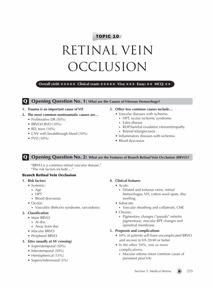
B1037 The Ophthalmology Examinations Review FA
Section 5: Medical Retina 235
Q Opening Question No. 1: What are the Causes of Vitreous Hemorrhage?
TOPIC 10
RETINAL VEIN OCCLUSION
1. Trauma is an important cause of VH
2. The most common nontraumatic causes are…• Proliferative DR (50%)• BRVO/CRVO (10%)• RD, tears (10%)• CNV with breakthrough bleed (10%)• PVD (10%)
3. Other less common causes include…• Vascular diseases with ischemia
• HPT, ocular ischemic syndrome• Eales disease• ROP/familial exudative vitreoretinopathy• Retinal telangiectasia
• Infl ammatory diseases with ischemia• Blood dyscrasias
Q Opening Question No. 2: What are the Features of Branch Retinal Vein Occlusion (BRVO)?
“BRVO is a common retinal vascular disease.”“The risk factors include….”
Branch Retinal Vein Occlusion
1. Risk factors• Systemic:
• Age• HPT• Blood dyscrasias
• Ocular:• Vasculitis (Behcet’s syndrome, sarcoidosis)
2. Classifi cation• Main BRVO:
• At disc• Away from disc
• Macular BRVO• Peripheral BRVO
3. Sites (usually at AV crossing) • Superotemporal (50%)• Inferotemporal (30%)• Hemispherical (15%)• Supero/inferonasal (5%)
4. Clinical features• Acute:
• Dilated and tortuous veins, retinal hemorrhages, VH, cotton wool spots, disc swelling
• Subacute:• Vascular sheathing and collaterals, CME
• Chronic:• Pigmentary changes (“pseudo” retinitis
pigmentosa), macular RPE changes and epiretinal membrane
5. Prognosis and complications• 50% of patients will have uncomplicated BRVO
and recover to VA 20/40 or better• In the other 50%, one or more
complications:• Macular edema (most common cause of
persistent poor VA)
Overall yield: ⍟ ⍟ ⍟ ⍟ ⍟ Clinical exam: ⍟ ⍟ ⍟ ⍟ ⍟ Viva: ⍟ ⍟ ⍟ Essay: ⍟ ⍟ MCQ: ⍟ ⍟
B1037_Ch-05.indd 235B1037_Ch-05.indd 235 12/28/2010 9:35:31 AM12/28/2010 9:35:31 AM

FA B1037 The Ophthalmology Examinations Review
236 The Ophthalmology Examinations Review
• Macular ischemia• Combined macular edema and macular
ischemia
• Neovascularization (40% of ischemic BRVO): 10% NVD; 20–30% NVE
Q Opening Question No. 3: What are the Features of Central Retinal Vein Occlusion (CRVO)?
Central Retinal Vein Occlusion
1. Risk factors• Systemic:
• Age• HPT• Blood dyscrasias
“CRVO is a common retinal vascular disease.”“The risk factors include….”
• Ocular:• Raised IOP• Hypermetropia• Congenital anomaly of central retinal vein• Vasculitis (Behcet’s syndrome, sarcoidosis,
AIDS, systemic lupus)
2. Classifi cation and clinical features
Ischemic Nonischemic
Frequency 75% 25%
VA 20/400 or worse (90%) Better than 20/400 (90%)
RAPD Marked Slight
VF defect Common Rare
Fundus fi ndings Extensive hemorrhages and cottonwool spots
Less extensive hemorrhages, fewcotton wool spots
FFA Widespread capillary non-perfusion Good perfusion
ERG Reduced “b”-wave amplitudeReduced “b:a” wave ratio
Normal
Prognosis 50% develop rubeosis and neovascular glaucoma in 3 months (100 dayglaucoma)VA improves in 1/3, stable in 1/3, and deteriorates in 1/3
3% develop rubeosis and neovascular glaucoma while 50% return to VA of 20/40 or betterConversion to ischemic CRVO: 34%in 3 years
Clinical approach to BRVO/CRVO
“On examination of this patient’s fundus, there are….”“Flame-shaped hemorrhages seen along the superotemporal vascular arcade.”“Associated with scattered hard exudates, cotton wool spots, vessel tortuosity and dilatation.”
Look for• Disc swelling• Cup disc ratio (glaucoma can lead to CRVO and vice versa)
Exam tips:• The differentiation between ischemic and nonischemic CRVO is important.
B1037_Ch-05.indd 236B1037_Ch-05.indd 236 12/28/2010 9:35:31 AM12/28/2010 9:35:31 AM

B1037 The Ophthalmology Examinations Review FA
Section 5: Medical Retina 237
Management of BRVO
1. Investigate and treat associated systemic disease
(e.g. HPT, DM, hyperlipidemia)
2. Ocular management• Two main complications are: Macular edema
and neovascularization at disc• Wait for hemorrhage to clear (3 months)• Perform OCT and FFA at 3 months• If macular edema is present and VA < 20/40:
• Grid laser photocoagulation• Intravitreal anti-VEGF if grid laser treatment
failure or in eyes with extensive macular hemorrhage (BRAVO Study: Ophthalmology 2010; Epub)
• Consider intravitreal anti-VEGF as primary treatment if VA > 20/40
• If 5 disc diameter of nonperfusion is seen → close follow-up to look for neovascularization or sector PRP
• New vessels (NVD, NVE)• Macular edema
(I’d like to confi rm the edema using a 78D lens at the slit lamp)• Treatment (PRP scars and ERM)
I’ll like to• Check fellow eye (10% bilateral)• Check IOP and perform gonioscopy (new vessels at the angle)• For CRVO:
• Undilated SLE for new vessels on the iris• Check RAPD
• Ask for VA• Ask whether patient has a history of DM, HPT, hyperlipidemia • Check BP• Conduct the following investigations:
• CBC (polycythemia and hyperviscosity), blood sugar levels, lipids• FFA (after 3 months)
Exam tips:• Be careful here, “old” BRVO or CRVO can have features similar to RP
(“pseudo” RP).
• Once new vessels are seen → sector PRP is indicated
• Other treatment modalities:• IVTA: Does not improve VA in macular
edema from BRVO and there are signifi cant side effects of raised IOP and cataract (SCORE Study: Arch Ophthalmol 2009:127:1115–1128)
• Dexamethasone implant: Improves VA in macular edema from BRVO, but side effects of raised IOP and cataract TPPV with AV sheathotomy
Management of CRVO
1. Investigate and treat associated systemic disease
(e.g. HPT, DM, hyperlipidemia)
Q How do You Manage BRVO/CRVO?
“The management of BRVO/CRVO must be individualized….”“The factors to consider are the patient’s VA and whether there are associated complications like macular edema or neovascularization….”“The prognosis for CRVO is poor….”
B1037_Ch-05.indd 237B1037_Ch-05.indd 237 12/28/2010 9:35:35 AM12/28/2010 9:35:35 AM

FA B1037 The Ophthalmology Examinations Review
238 The Ophthalmology Examinations Review
2. Ocular management• Two main complications are: Macular edema
and neovascular glaucoma• Need to differentiate between ischemic/
non-ischemic CRVO • Wait for hemorrhage to clear (3 months)• Perform OCT and FFA at 3 months:
• If nonischemic → no treatment is needed, prognosis is good (50% 20/40 of better VA)
• If ischemic → prognosis is poor (50% neovascular glaucoma in 3 months), close follow-up is needed to look for new vessels at iris; or full PRP
• Once new vessels at iris are seen → full PRP is indicated
• If macular edema is present and VA < 20/40:• Intravitreal anti-VEGF (CRUISE Study:
Ophthalmology 2010; Epub)
• IVTA: Improves VA in macular edema secondary to non-ischemic CRVO, but signifi cant side effects are raised IOP and cataracts (SCORE study)
• No benefi t in treating macular edema in CRVO with grid laser
• Other treatment modalities:• Dexamethasone implant: Improves VA but
signifi cant side effects are raised IOP and cataracts
• Laser-induced chorioretinal venular anastomosis (Central Retinal Vein Bypass Study: Ophthalmology 2010; in press):
• May improve VA in non-ischemic CRVO• Complicated by CNV and vitreous hemorrhage • Radial optic neurotomy and retinal vein
cannulation with tPA infusion
BVOS CVOS
Macular edema Is argon grid laser useful in improvingVA in eyes with BRVO and macular edema with VA ≤ 20/40?
Conclusion: YesGain of at least two lines of VA from baselineWhen should laser be performed? Not answered in the study
Is argon grid laser useful in preservingor improving VA in eyes with CRVO and macular edema with VA ≤ 20/50?
Conclusion: NoTreatment clearly reduced FFA evidence of macular edema but no difference in fi nal VA
Neovascularization Can peripheral argon sector PRPprevent1. Neovascularization?2. VH?
Conclusion: Yes PRP prevents neovascularization andVH
When should laser be performed?Should be started after developmentof neovascularization
Can prophylactic argon PRP in ischemic CRVO prevent two clock hours of iris or angle neovascularization? Or is it more appropriate to start PRP only when new vessels are seen?
Conclusion: 1. No, PRP does not prevent iris or
angle neovascularization2. Regression is faster in untreated eyes 3. Therefore, PRP should be started
after development of iris or angle neovascularization
Recommendations 1. FFA when hemorrhage clears (3 months)
2. If macular edema and VA < 20/40seen → grid laser
3. If 5 disc diameter of nonperfusion, follow-up closely for new vessels
4. Once new vessels are seen →sector PRP is indicated
1. If 10 disc diameter of nonperfusion, careful observation with undilated SLE and gonioscopy
2. PRP indicated only after neovascu-larization develops
3. No benefi t for treatment of macular edema
Q What are the Main Findings of the Branch Vein Occlusion Study (BVOS)? The Central Vein Occlusion Study (CVOS)?
B1037_Ch-05.indd 238B1037_Ch-05.indd 238 12/28/2010 9:35:35 AM12/28/2010 9:35:35 AM

B1037 The Ophthalmology Examinations Review FA
Section 5: Medical Retina 239
Exam tips:• One of the more common clinical trials asked in exams. Refer to BVOS (Am J
Ophthalmol 1984;98:271–282; Arch Ophthalmol 1986;194:34–41) and CVOS (Ophthalmol 1995;102:1425–1433).
Exam tips:• Refer to BRAVO and CRUISE (Ophthalmology 2010; Epub).
Q What are the Main Findings of the BRAVO and CRUISE Studies?
BRAVO CRUISE
Aim To assess effi cacy and safety of 0.3 mg or 0.5mg ranibizumab in patients with macular edema following BRVO
To assess effi cacy and safety of 0.3 mg or 0.5mg ranibizumab in patients with macular edema following CRVO
Design Prospective randomized multicenter studies
Inclusion criteria 1. Macular edema involving foveal center secondary to BRVO/CRVO2. Central foveal thickness ≥ 250µm3. BCVA < 20/40
Treatment groups Monthly intraocular injections of 0.3mg or 0.5mg ranibizumab, or sham injections
Outcome measures 1. Mean change from baseline BCVA at month 62. Gain of 3 lines (15 letters) of BCVA at month 6 Central foveal thickness at month 6
Results Mean change in baseline BCVA at month 6:• 0.3mg ranibizumab: 16.6 letters• 0.5mg ranibizumab: 18.3 letters• Sham: 7.3 letters
Mean change in baseline BCVA at month 6:• 0.3mg ranibizumab: 12.7 letters• 0.5mg ranibizumab: 14.9 letters• Sham: 0.8 letters
Gain in 3 lines (15 letters) at month 6:• 0.3mg ranibizumab: 55.2%• 0.5mg ranibizumab: 61.1%• Sham: 28.8%
Gain in 3 lines (15 letters) at month 6:• 0.3mg ranibizumab: 46.2%• 0.5mg ranibizumab: 47.7%• Sham: 16.9%
Mean reduction in foveal thickness atmonth 6:• 0.3mg ranibizumab: 97.0%• 0.5mg ranibizumab: 97.6%• Sham: 27.9%
Safety profi le consistent with previous ranibizumab trials
Mean reduction in foveal thickness at month 6:• 0.3mg ranibizumab: 94.0%• 0.5mg ranibizumab: 97.3%• Sham: 23.9%
Safety profi le consistent with previous ranibizumab trials
Conclusions Intraocular injections of 0.3mg or 0.5mg ranibizumab provided rapid, effective and safe treatment for macular edema following BRVO
Intraocular injections of 0.3mg or 0.5mg ranibizumab provided rapid, effective and safe treatment for macular edema following CRVO
B1037_Ch-05.indd 239B1037_Ch-05.indd 239 12/28/2010 9:35:35 AM12/28/2010 9:35:35 AM

FA B1037 The Ophthalmology Examinations Review
240 The Ophthalmology Examinations Review
CRVO in Young Patient
1. Basic evaluation• Medical conditions:
• HPT, DM, hyperlipidemia, coagulopathy• Cardiac disease (mitral valve prolapse),
autoimmune disease, AIDS• Ocular conditions:
• Glaucoma• Medication (oral contraceptive pills, hormone
therapy)
2. Physical exam• Carotid bruit
Q How do You Investigate a 20-Year-Old Female with CRVO?
“CRVO in a young patient is not common….”“I would need to evaluate the patient carefully through a detailed history, physical examination and appropriate investigations.”
• Heart murmur• BP
3. Laboratory investigation• CBC, ESR• Blood sugar levels, lipid levels• VDRL, FTA• HIV• CXR• Coagulation (PTT, APTT), homocysteine,
antiphospholipid antibody• Autoimmune markers
Q What is the Antiphospholipid Syndrome?
“The antiphospholipid syndrome refers to a coagulation disorder.”“Characterized by circulating antiphospholipid antibodies….”
Antiphospholipid Syndrome
1. Classifi cation• Primary:
• No secondary disease• Circulating antiphospholipid antibodies
(includes lupus anticoagulant, anticardiolipin antibodies)
• Affi nity for phospholipids (important in conversion of prothrombin to thrombin)
• Secondary:• Systemic lupus, other auto-immune diseases• AIDS• Phenothiazine and procainamide use
2. Clinical features• In vitro, antiphospholipid antibodies cause
anticoagulation (i.e. bleeding)• In vivo, they are associated with paradoxically
with coagulation (i.e. thrombosis):• CRVO and BRVO, CRAO and BRAO• Retinal vasculitis• Choroidal infarction• Arteritic anterior ischemic optic neuropathy
Exam tips:• Think of SECONDARY causes (page 196).
B1037_Ch-05.indd 240B1037_Ch-05.indd 240 12/28/2010 9:35:35 AM12/28/2010 9:35:35 AM

B1037 The Ophthalmology Examinations Review FA
Section 5: Medical Retina 241
Q Opening Question: What are Ocular Associations of Cardiac Disease?
“The ocular effects are usually secondary to an embolic phenomenon from cardiac disease.”
TOPIC 11
CARDIOVASCULAR DISEASE
Cardiac Disease
1. Embolic (thrombotic, calcifi c, bacterial material)• Ophthalmic artery:
• Ophthalmic artery occlusion• Retinal artery:
• Amaurosis fugax• CRAO
• Retinal arterioles:• BRAO (cardiac emboli is most common
cause of BRAO in younger persons)• Precapillary arterioles:
• Cotton wool spots, Roth’s spot
2. Generalized decreased perfusion state from heart
failure• Fainting spells• Ocular ischemic syndrome:
• “What are the features of ocular ischemic syndrome?” (see page 202)• Anterior segment ischemia• Hypoperfusion retinopathy
3. Right heart failure• Superior vena cavae congestion
• Increase in episcleral venous pressure → glaucoma
Notes• “What are the features of ophthalmic artery occlusion?”
• Anterior segment ischemia (anterior ciliary artery)• Posterior segment ischemia (posterior ciliary artery)• Ophthalmoplegia (extraocular muscle involvement)• ERG shows decreased “a” and “b” wave amplitude affected)
Q What are the Ocular Associations of Carotid Artery Disease?
“The ocular effects are usually secondary to either a thrombotic or embolic phenomenon.”
Carotid Artery Disease
1. Embolic (cholesterol, fi brinoplatelet, calcifi c)• Similar spectrum to above (carotid emboli is
most common cause of BRAO in older persons)
2. Thrombotic• Carotid artery
• Ocular ischemic syndrome (most common cause of ocular ischemic syndrome)
Overall yield: ⍟ ⍟ ⍟ Clinical exam: Viva: ⍟ ⍟ ⍟ Essay: ⍟ ⍟ MCQ: ⍟ ⍟
B1037_Ch-05.indd 241B1037_Ch-05.indd 241 12/28/2010 9:35:35 AM12/28/2010 9:35:35 AM

FA B1037 The Ophthalmology Examinations Review
242 The Ophthalmology Examinations Review
• Ophthalmic artery:• Ophthalmic artery occlusion• Anterior ischemic optic neuropathy
• Retinal artery:• Amaurosis fugax (most common cause of
amaurosis fugax)• CRAO (most common cause of CRAO)
3. Others• Horner’s syndrome• Stroke (branches of carotid arteries):
• Anterior choroidal artery (homonymous hemianopia)
• Anterior cerebral artery (hemialexia)• Middle cerebral artery (homonymous
hemianopia, Gaze palsies)• Aneurysm (branches of carotid arteries):
• Subarachnoid hemorrhage from ruptured aneurysm
• Compressive III CN palsy
Q What are the Principles of Management of a Patient with Carotid Artery Disease?
Management of Carotid Artery Disease
1. Modify risk factors• HPT, DM, hyperlipidemia, smoking
2. Antiplatelet therapy• Aspirin• Dipyridamole• Clopidogrel
3. Anticoagulation therapy• Indications:
• If aspirin fails • Recurrent cardiac source of emboli (atrial
fi brillation, mitral valve stenosis, etc.)
4. Carotid endarterectomy• North American Symptomatic Carotid
Endarterectomy Trial (New Engl J Med 1998;339:1415–1425)• Indications for carotid endarterectomy:
• Symptomatic patients with amaurosis, hemispheric TIA, nondisabling strokes
• Plus 70–99% carotid artery stenosis• Prognosis
• 2-year stroke rate is 9% (endarterectomy) vs 26% (no surgery)
• European Carotid Endarterectomy Trial(Lancet 1998;351:1379–1387)• No benefi t with carotid endarterectomy
Q What is Amaurosis Fugax?
“Amaurosis fugax is a transient monocular blindness less than 24 hours by defi nition.”“The causes can be either systemic or ocular in nature….”
Amaurosis Fugax
1. Systemic• Carotid artery atherosclerosis (most common
cause)• Carotid emboli• Cardiac emboli• Vasculitis:
• Giant cell arteritis• Migraine
• Systemic lupus, polyarteritis nodosa and others
• Coagulation disorders
2. Ocular• Raised IOP• Drusens• Papilledema• Anterior ischemic optic neuropathy
Exam tips:• Causes almost identical to those for retinal artery occlusion (page 233).
B1037_Ch-05.indd 242B1037_Ch-05.indd 242 12/28/2010 9:35:36 AM12/28/2010 9:35:36 AM

B1037 The Ophthalmology Examinations Review FA
Section 5: Medical Retina 243
Q What is the Ocular Ischemic Syndrome?
“The ocular ischemic syndrome is a disorder secondary to hypoperfusion of the globe due to either chronic carotid artery obstruction or ophthalmic artery obstruction.”
Ocular Ischemic Syndrome
1. Cause• Carotid atherosclerosis (most common cause):
• 90% or more carotid artery obstruction before symptoms
• Bilateral in 20%• Generalized decreased perfusion from cardiac
failure• Others (GCA, arteritis)
2. Symptoms• Decrease VA (for weeks and months)• Aching ocular pain• Light-induced visual loss (with prolonged
recovery from exposure to bright light)
3. Signs• Anterior segment ischemia:
• Injected eye• Corneal edema• Iris neovascularization, iris atrophy,
iridoplegia• AC fl are (more fl are than cells)• Swollen mature cataract• Raised IOP (50%), low IOP (50%)
• Posterior segment (hypoperfusion retinopathy):• Vessel tortuosity, venous dilation• Microaneurysm, retinal hemorrhage, hard
exudate • New vessels, VH• Choroidopathy (Elschnig’s spots, serous RD)• Papilledema, macular star
4. Investigations• FFA:
• Delayed choroidal fi lling time (60%)• Delayed arteriole-to-venule transit time
(95%)• Capillary nonperfusion• Prominent arteriolar wall staining
• ERG:• Decreased amplitude of both “a” and “b”
waves (like ophthalmic artery occlusion, see above)
• Systemic conditions:• ESR, lipids levels
5. Prognosis• Systemic associations:
• 50% have ischemic heart disease• 25% have previous stroke• 20% have severe peripheral vascular
disease • 5 year mortality is 40% (higher than for CRAO;
page 194)
6. Treatment• Ocular:
• Laser PRP for new vessels (regression in small percentage)
• Manage NVG• Manage systemic risk factors• Carotid endarterectomy
B1037_Ch-05.indd 243B1037_Ch-05.indd 243 12/28/2010 9:35:36 AM12/28/2010 9:35:36 AM

FA B1037 The Ophthalmology Examinations Review
B1037_Ch-05.indd 244B1037_Ch-05.indd 244 12/28/2010 9:35:36 AM12/28/2010 9:35:36 AM
This page intentionally left blankThis page intentionally left blank

B1037 The Ophthalmology Examinations Review FA
Section 5: Medical Retina 245
Q Opening Question: What is Retinopathy of Prematurity?
“Retinopathy of prematurity (ROP) is a proliferative vascular retinal condition.”“It commonly occurs in premature and low birth weight babies exposed to high ambient oxygen.”“There are classically two phases, the active ROP phase and cicatricial ROP phase….”
TOPIC 12
RETINOPATHY OF PREMATURITY
Retinopathy of Prematurity
1. Risk factors:• Prematurity < 32 weeks• Low birth weight < 1,500 g• Supplemental oxygen• Others:
• Intraventricular hemorrhage• Necrotizing enterocolitis• Maternal theophylline treatment• Abruptio placentae• Quality of NICU care• Others: Twins, respiratory distress syndrome,
history of blood transfusion
2. Active ROP• Location:
• Zone I: Circle centered on disc with radius twice the distance from disc to fovea
• Zone II: Circle from edge of Zone 1 to a point tangential to nasal ora serrata and around to an area near temporal ora
• Zone III: Remaining crescent from Zone 2 to temporal ora
• Extent:• Number of clock hours
• Stage:• Stage 1: Demarcation line• Stage 2: Ridge• Stage 3: Extraretinal fi brovascular
proliferation• Stage 4: Subtotal RD (4a: Extrafoveal; 4b:
Foveal involvement)• Stage 5: Total RD
• Plus disease:• Dilatation and tortuosity of veins• Vitreous haze• Engorged iris vessels • Poor pupillary reaction
3. Cicatricial ROP:• 20% of active ROP will progress to cicatricial
ROP without treatment• Stages:
• Stage 1: Myopia, pigmentary changes• Stage 2: Temporal vitreoretinal fi brosis,
dragged disc • Stage 3: Peripheral fi brosis and falciform
retinal fold• Stage 4: Partial RD• Stage 5: Total RD, secondary glaucoma
Exam tips:• The defi nition of the various parameters (zones, extent and stage) has to be
committed to memory. Many candidates do not defi ne zones well.
Overall yield: ⍟ ⍟ ⍟ Clinical exam: ⍟ Viva: ⍟ ⍟ ⍟ Essay: ⍟ ⍟ MCQ: ⍟ ⍟
B1037_Ch-05.indd 245B1037_Ch-05.indd 245 12/28/2010 9:35:36 AM12/28/2010 9:35:36 AM

FA B1037 The Ophthalmology Examinations Review
246 The Ophthalmology Examinations Review
Pathogenesis
1. Normal retinal angiogenesis• Starts at 16 weeks• Reaches nasal ora at 36 weeks• Complete vascularization at 40 weeks (up to
one month post-term)
2. Biphasic theory of Aston and Patz• High ambient oxygen → vasoconstriction and
toxic obliteration of retinal capillaries → on return to room air → relative ischemia develops → angiogenic factors secreted → vascular proliferation
Q What is the Pathogenesis of ROP?
“ROP occurs as a result of failure of vascularization of the immature retina.”“There are two theories….”
3. Spindle cell theory of Kretzer and Hittner• Nascent blood vessels form in normally hypoxic
uterine condition by canalization and endothelial cell differentiation behind a migrating sheet of spindle cells
• High ambient oxygen triggers extensive gap junction formation between spindle cells which interferes with migration and canalization of blood vessels
• Spindle cells secrete angiogenic factors → vascular proliferation
Notes• “What is Rush disease?”
• Rapidly progressive Stage 3 ROP in Zone 1 or posterior Zone 2• Poor prognosis
Q How do You Screen a Baby for ROP Born at 32 Weeks of Gestation?
“The examination schedule for ROP in our center is….”“The indications for treatment of ROP are….”
Screening and Management of ROP
1. Who should be screened?• ≤ 1,500g, born ≤ 32 weeks gestation• Selected infants 1,500g–2,000g or born at
gestational age > 32 weeks, with unstable clinical course
2. When to start screening?• Median age of ROP = 37 weeks• 90% of ROP occur between 34 to 42 weeks• Therefore, start screening at 34 weeks
(alternatively, can start screening four weeks postnatally)
3. How often should subsequent screening be? • If fi rst screening examination shows:
• No ROP → repeat exam in four weeks → if no ROP → repeat exam in three months
• ROP in Zone III → repeat in two weeks• Prethreshold ROP (Zone I or II) → repeat in
one week → if threshold ROP → treatment
4. What are the indications for treatment?• Threshold ROP:
• Zone I or II• Five contiguous clock hours or eight
noncontiguous clock hours• Stage 3 • Plus disease
• Threshold ROP is associated with 50% riskof having VA 20/200 or worse without treatment
• More recent evidence support the treatment of prethreshold ROP• Early Treatment for Retinopathy of
Prematurity Study (ETROP) recommends treatment for:• Zone I any stage with plus• Zone I Stage 3, no plus• Zone II Stage 2 or 3 with plus
B1037_Ch-05.indd 246B1037_Ch-05.indd 246 12/28/2010 9:35:36 AM12/28/2010 9:35:36 AM

B1037 The Ophthalmology Examinations Review FA
Section 5: Medical Retina 247
5. What treatments are available?• Prevention: PaO2 < 80 mmHg• Cryotherapy:
• Ablate avascular retina anterior to ridge• Multicenter Cryotherapy for ROP Study
(Arch Ophthalmol 1996;114:417–424):• 50% reduction in poor VA with
cryotherapy• 50% reduction in poor fundal status with
cryotherapy
• Indirect laser photocoagulation• Vitamin E therapy:
• Controversial• Inhibit gap junction formation in spindle cells• Antioxidant• Complications (necrotizing enterocolitis,
vitreous hemorrhage)• Stage 4 ROP might require sclera buckle or
vitrectomy
Clinical approach to dragged disc
“There is dragging of the optic disc by temporal vitreoretinal fi brotic tissues.”“There is also a divergent squint seen.”
I’d like to ask patient for a history• Prematurity• Contact with dogs• Family history of blindness
The possible causes are:
1. Proliferative vitreoretinopathy• ROP • Familial exudative vitreoretinopathy (AD inheritance)• Incontinentia pigmenti (SLD inheritance)
2. Uveitis• Toxocara • Pars planitis
3. Tumor• Combined hamartoma of RPE and retina
4. Trauma
Exam tips:• Refer to ETROP study (Arch of Ophthalmol 2003;121:1684–1696).
Notes• “Why not earlier?”
• Limited value in picking up ROP• Diffi culty in screening (poor pupil dilation, vitreous haze)• Complications of mydriatic eyedrops (cardiac, respiratory effects) and
ocular examination (oculocardiac refl ex, hypotension, apnea)
B1037_Ch-05.indd 247B1037_Ch-05.indd 247 12/28/2010 9:35:37 AM12/28/2010 9:35:37 AM

FA B1037 The Ophthalmology Examinations Review
B1037_Ch-05.indd 248B1037_Ch-05.indd 248 12/28/2010 9:35:37 AM12/28/2010 9:35:37 AM
This page intentionally left blankThis page intentionally left blank

B1037 The Ophthalmology Examinations Review FA
Section 5: Medical Retina 249
Q Opening Question: What are the Ocular Effects of Pregnancy?
TOPIC 13
OTHER RETINAL VASCULAR DISORDERS
Ocular Effects of Pregnancy
1. Physiological• Lid — Telangiectasia• Cornea
• Decreased corneal sensitivity• Corneal edema → increased corneal
thickness → change in refractive error • Increased incidence of Krukenbergs spindle
• IOP — Increased facility of aqueous outfl ow and decreased episcleral venous pressure → lower IOP
• Lens — Transient loss of accommodative ability• Enlarging pituitary gland → various VF changes
towards end of term
2. Pathological (5 “Cs”)• CSR
• Secondary to hormonal and hemodynamic changes → change in permeability of blood-retinal barrier
• Resolve post-partum with residual RPE mottling and pigmentation
• CRVO, CRAO, hypertensive retinopathy• Secondary to hypertension• CRVO may be sign of impending fi t in
pre-eclampsia• Cortical blindness
• Transient phenomenon, secondary to chronic edema of occipital lobe
• Pseudotumor cerebri• Secondary to chronic edema (controversial)
• Coagulation disorders• Disseminated intravascular coagulation • Thrombotic thrombocytopenic purpura
3. Preexisting conditions (endocrine and tumors)• Diabetic retinopathy
• Increased incidence of background DR• Increased incidence of macular edema• Increased progression to PDR• Prophylactic photocoagulation important
• Grave’s disease• Progression during pregnancy
• Pituitary adenoma• Normal pituitary gland enlarges with
increased prolactin secreting cells• May present for the fi rst time during
pregnancy• Uveal melanoma
• Increased size secondary to increased melanin-stimulating hormone secretion
• Meningioma• Secondary to increased estrogen/progesterone • May present for the fi rst time during
pregnancy• Ocular pharmacology
• Avoid FFA → fl uorescein can pass through placenta
• Avoid timolol if breastfeeding• Diamox may be teratogenic• Topical steroids are not contraindicated
Overall yield: ⍟ ⍟ ⍟ Clinical exam: ⍟ Viva: ⍟ ⍟ ⍟ Essay: ⍟ ⍟ MCQ: ⍟ ⍟ ⍟ ⍟
B1037_Ch-05.indd 249B1037_Ch-05.indd 249 12/28/2010 9:35:37 AM12/28/2010 9:35:37 AM

FA B1037 The Ophthalmology Examinations Review
250 The Ophthalmology Examinations Review
Exam tips:• Remember the 3 “Ps” (physiological, pathological, pre-existing) and 5 “Cs”!
Q What are the Ocular Effects of Radiation?
“There are two kinds of radiation: Non-ionizing and Ionizing radiation.”
Ocular Effects of Radiation
1. Non-ionizing radiation• Microwave (> 12,000 nm):
• Cataract• Infrared (12,000–770 nm):
• True exfoliation of lens (glassblower’s cataract)
• Visible light (760–400 nm):• Photic damage:
• Mechanical (e.g. photodisruption)• Thermal (e.g. photocoagulation)• Photobiochemical (e.g. solar retinopathy,
photic retinopathy)• Ultraviolet (180–390 nm):
• Surface epithelial disease and radiation keratitis
2. Ionizing radiation• Damage depends on tissue sensitivity:
• Lens > cornea > retina > optic nerve• Damage can be:
• Direct — on actively reproducing cells• Indirect — on blood vessels
• Anterior segment• Lids and conjunctiva:
• Dermatitis of lids• Damage to eyelashes• Damage to meibomian glands (dry eyes)• Punctal occlusion (wet eyes)• Cicatricial conjunctivitis
• Cornea:• Radiation keratitis• Limbal stem cell failure• Aseptic necrosis and perforation
• Lens:• Cataract• Equatorial cells damaged by radiation
• Posterior segment• Radiation retinopathy (see below)• Optic neuropathy
Q What is Radiation Retinopathy?
Radiation Retinopathy
1. Pathology• Damage to retinal vasculature after exposure to
ionizing radiation• Microangiopathy (like DR)• Dose dependent (high risk if > 7,000 rads)
2. Presentation• Asymptomatic early on• Present with decreased VA four months to three
years after treatment (external beam or local plaque therapy)
• Progressive loss of VA
3. Clinical fi ndings• Retinopathy (hemorrhages, cotton wool spots,
hard exudates, microaneurysms)
• Perivascular sheathing, telangiectasia• Complications
• New vessels → vitreous hemorrhage, tractional RD, neovascular glaucoma
• CRAO and CRVO• Maculopathy (exudative, edematous,
ischemic — like diabetic maculopathy)• Papillopathy
4. Treatment• FFA → look for capillary nonperfusion• Focal laser and PRP• Papillopathy: Systemic steroids
B1037_Ch-05.indd 250B1037_Ch-05.indd 250 12/28/2010 9:35:37 AM12/28/2010 9:35:37 AM

B1037 The Ophthalmology Examinations Review FA
Section 5: Medical Retina 251
Sickle Cell Hemoglobinopathy
1. Pathogenesis• Mutant S or C hemoglobin allele:
• Substituted for normal hemoglobin A allele• Valine substituted for glutamate at beta-6
chain location of hemoglobin• Hypoxia leads to sickling, which causes
obstruction of small blood vessels and tissue ischemia (leading to further sickling)
2. Types (classifi ed on abnormal hemoglobin
combinations)• AS (sickle cell trait):
• 8% of black population• Mild systemic disease
• SS (sickle cell anemia):• 0.4% of black population• Severe systemic disease and anemia• Mild ocular disease
• SC (sickle C disease) and Sthal (sickle cell thalassemia):• Mild systemic disease• Severe ocular disease
3. Proliferative retinopathy• Stage (note: Stages in ROP are shown in
brackets):• Stage 1: Peripheral arteriolar occlusion
(demarcation line)
Q Tell me about Sickle Cell Hemoglobinopathy.
“Sickle cell disease is a red blood cell disorder.”“Characterized by the presence of abnormal hemoglobin and “sickling” of the red blood cells in condi-tions of hypoxia.”“The ocular features can be divided into proliferative retinopathy, nonproliferative retinal disease and anterior segment disease….”
• Stage 2: Peripheral arteriovenous anastomosis (ridge)
• Stage 3: “Sea-fan” neovascularization (extraretinal fi brovascular proliferation)
• Stage 4: Vitreous hemorrhage (subtotal RD)• Stage 5: RD (total RD)
4. Non-proliferative retinal disease• “Salmon patch” (fresh intraretinal hemorrhage)• “Black sunburst” (old subretinal hemorrhage)• “Silver wiring” of peripheral arterioles• Angioid streaks• Retinal breaks and detachment
5. Anterior segment• Tortuous “corkscrew” conjunctival vessels• Ischemic iris atrophy and rubeosis• Hyphema
6. Management• Management of hyphema:
• Higher risk of optic nerve damage with hyphema (compared to “normal” person)
• Indication for surgical intervention: > 24 mmHg > 24 hours
• RD surgery:• Risk of anterior segment ischemia with
scleral buckle
Exam tips:• The stages are VERY SIMILAR to ROP stages (page 245).
Notes• “How do you prevent anterior segment ischemia during RD surgery?”
• Intraoperative oxygen, no epinephrine given• Minimize manipulation of muscles• SRF drainage (lower IOP)• Postoperative oxygen• Consider vitrectomy instead of scleral buckling• Prophylactic laser photocoagulation of all breaks
B1037_Ch-05.indd 251B1037_Ch-05.indd 251 12/28/2010 9:35:38 AM12/28/2010 9:35:38 AM

FA B1037 The Ophthalmology Examinations Review
252 The Ophthalmology Examinations Review
Q What are the Causes of Roth’s Spots?
“Roth’s spots are essentially retinal hemorrhages with a fi brin thrombus occluding the vessel.”
Differential Diagnoses of Roth’s Spots
1. Blood disorders• Anemia• Leukemia• Scurvy
2. Infective• Infective endocarditis
• Sepsis• AIDs retinopathy• Candida retinopathy
3. Vasculitis• DM• Systemic vasculitis (systemic lupus etc.)
Q What are the Ocular Effects of Renal Disease?
Renal Disease
1. Congenital (concurrent ocular involvement):• Lowe’s syndrome
• SLR inheritance• Renal problems (aminoaciduria, metabolic
acidosis, renal rickets)• CNS problems (mental retardation)• Ocular effects:
• Cataract and microphakia • Glaucoma
• Alport’s syndrome• AD inheritance• Renal problems (proteinuria, HPT and renal
failure)• CNS problems (sensorineural deafness)• Ocular effects:
• Anterior polar cataract, anterior lenticonus
• Posterior polymorphous dystrophy• RPE abnormalities (looks like “fundus
albipunctatus”)• Aniridia
• Sporadic form• Renal problems (Wilms’ Tumor)• Ocular effects (page 60)
2. Acquired (ocular involvement occurs LATER)• Secondary effects (more common, especially
after renal transplant)• HPT retinopathy• DM retinopathy• Anemia• Bleeding diathesis• Opportunistic infections (CMV, candida)• Steroid induced glaucoma and cataract
Q What are the Ocular Effects of Leukemia?
“Ocular effects of leukemia are usually only seen in advanced cases of acute or relapsing disease.”“They are related to both direct and indirect effects of leukemia (e.g. anemia, immunosuppression).
Leukemia
1. Direct effects• Anterior segment:
• Subconjunctival hemorrhage• Orbital infi ltration• Iris:
• Diffuse white nodular thickening• Heterochromia• Pseudohypopyon • Spontaneous hyphema
• Secondary glaucoma • Posterior segment:
• “Leopard spot” retina (deposits in choroid)• Flame-shaped hemorrhage, hard exudates,
cotton wool spots, Roth’s spots
• Venous tortuosity and dilatation, CRVO• Neovascularization • Exudative RD• Optic nerve infi ltration
2. Indirect effects• Anemia (fl ame-shaped hemorrhage, cotton wool
spots, Roth’s spots, etc.)• Thrombocytopenia• Hyperviscosity (ischemic optic neuropathy,
proliferative retinopathy)• Immunosuppression (opportunistic infections)
B1037_Ch-05.indd 252B1037_Ch-05.indd 252 12/28/2010 9:35:38 AM12/28/2010 9:35:38 AM
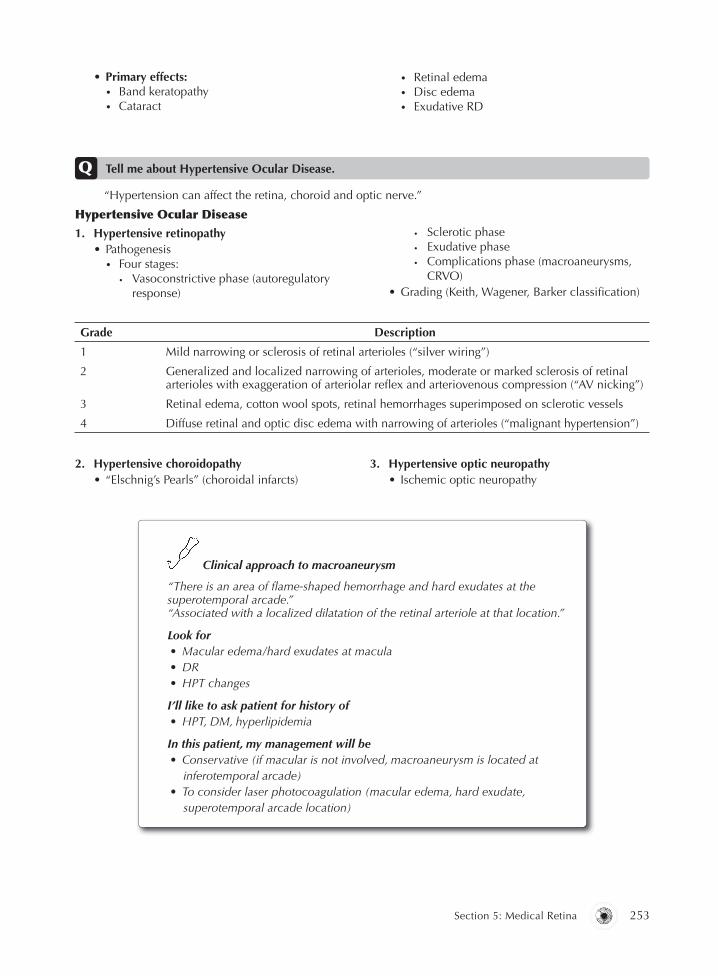
B1037 The Ophthalmology Examinations Review FA
Section 5: Medical Retina 253
• Primary effects:• Band keratopathy• Cataract
• Retinal edema• Disc edema• Exudative RD
Q Tell me about Hypertensive Ocular Disease.
“Hypertension can affect the retina, choroid and optic nerve.”
Hypertensive Ocular Disease
1. Hypertensive retinopathy• Pathogenesis
• Four stages:• Vasoconstrictive phase (autoregulatory
response)
• Sclerotic phase• Exudative phase• Complications phase (macroaneurysms,
CRVO)• Grading (Keith, Wagener, Barker classifi cation)
Grade Description
1 Mild narrowing or sclerosis of retinal arterioles (“silver wiring”)
2 Generalized and localized narrowing of arterioles, moderate or marked sclerosis of retinal arterioles with exaggeration of arteriolar refl ex and arteriovenous compression (“AV nicking”)
3 Retinal edema, cotton wool spots, retinal hemorrhages superimposed on sclerotic vessels
4 Diffuse retinal and optic disc edema with narrowing of arterioles (“malignant hypertension”)
2. Hypertensive choroidopathy• “Elschnig’s Pearls” (choroidal infarcts)
3. Hypertensive optic neuropathy• Ischemic optic neuropathy
Clinical approach to macroaneurysm
“There is an area of fl ame-shaped hemorrhage and hard exudates at the superotemporal arcade.”“Associated with a localized dilatation of the retinal arteriole at that location.”
Look for• Macular edema/hard exudates at macula• DR• HPT changes
I’ll like to ask patient for history of• HPT, DM, hyperlipidemia
In this patient, my management will be• Conservative (if macular is not involved, macroaneurysm is located at
inferotemporal arcade)• To consider laser photocoagulation (macular edema, hard exudate,
superotemporal arcade location)
B1037_Ch-05.indd 253B1037_Ch-05.indd 253 12/28/2010 9:35:38 AM12/28/2010 9:35:38 AM

FA B1037 The Ophthalmology Examinations Review
254 The Ophthalmology Examinations Review
Exam tips:• Hypertensive retinopathy is only ONE of the three manifestations.
NotesOcular conditions associated with hypertension• Retinal vein occlusion• Retinal artery occlusion• Retinal artery macroaneurysm• AION• Ocular motor nerve palsy• Uncontrolled hypertension may adversely affect DR
Q What is Parafoveal Telangiectasia?
“Parafoveal telangiectasia is an uncommon retinal vascular anomaly that primarily involves thejuxtafoveolar capillaries….”“It can be congenital or acquired, primary (idiopathic) or secondary to underlying systemic disease(e.g. diabetes)....”
Idiopathic Parafoveal Telangiectasia• Pathogenesis:
• Poorly understood• Characterized by dilatation and tortuosity of
retinal blood vessels and multiple aneurysms confi ned to the parafoveal region. These leak and result in deposition of hard exudates
• Primarily involves the capillary bed, although arterioles and venules may also be involved
• Gass classifi cation of parafoveal telangiectasia:• Group 1: Unilateral
• Group 1A: Congenital• Group 1B: Idiopathic focal
• Group 2: Bilateral• Group 2A: Idiopathic acquired (most
common)• Group 2B: Juvenile familial occult
• Group 3: Occlusive
• Group 3A: Associated with systemic disease (e.g. polycythemia, multiple myeloma etc)
• Group 3B: Associated with neurological disease
• Clinical presentation:• Patient presents mostly in the 5th to 6th decades
with slowly progressive loss of central vision• FFA:
• Leakage from telangiectactic vessels confi ned to the parafoveal region
• Staining of scars (advanced disease)• Treatment:
• There is no defi nitive treatment, although several modalities have been studied:• Laser photocoagulation• PDT• Intravitreal anti-VEGF • Intravitreal steroids
Q What is Coat’s Disease?
“Coat’s disease is an idiopathic, non-hereditary, mostly unilateral retinal vasculopathy characterized by the presence of retinal telangiectasias leading to intraretinal and subretinal exudation, and frequently, exudative RD.”“Coat’s disease, Leber’s miliary aneurysms and idiopathic parafoveal telangiectasia are thought torepresent a spectrum of the same disease, with Coat’s disease being the most extensive.”“Before reaching the diagnosis of Coat’s disease, it is important to exclude retinoblastoma in a young patient with leukokoria and RD.”
B1037_Ch-05.indd 254B1037_Ch-05.indd 254 12/28/2010 9:35:39 AM12/28/2010 9:35:39 AM

B1037 The Ophthalmology Examinations Review FA
Section 5: Medical Retina 255
Coat’s Disease• Clinical presentation:
• Mostly in the fi rst decade (average fi ve years) with unilateral visual loss, strabismus or leukokoria
• Occasionally may present in later childhood, and rarely in adult life
• Examination:• Retinal telangiectasia• Intraretinal and subretinal yellowish
exudation:• Often affects areas remote from vascular
abnormalities, especially macula• Complications:
• Exudative RD• Rubeosis iridis• Uveitis• Glaucoma• Phthisis bulbi
• Investigations:• OCT• FFA:
• Leakage from telangiectactic vessels• Hypofl uorescence: Blockage from hard
exudates• Ultrasonography ± CT/MRI: Exclude
retinoblastoma• Management:
• Observation:• Mild, non-vision-threatening disease or • Total RD with no hope of restoring vision
• Laser photocoagulation: On telangiectatic vessels
• Cryotherapy: For telangiectatic vessels• Vitreoretinal surgery: Very bullous RD
unsuitable for cryotherapy• Enucleation: Painful eyes with NVG
B1037_Ch-05.indd 255B1037_Ch-05.indd 255 12/28/2010 9:35:39 AM12/28/2010 9:35:39 AM

FA B1037 The Ophthalmology Examinations Review
B1037_Ch-05.indd 256B1037_Ch-05.indd 256 12/28/2010 9:35:39 AM12/28/2010 9:35:39 AM
This page intentionally left blankThis page intentionally left blank

B1037 The Ophthalmology Examinations Review FA
Section 5: Medical Retina 257
Q Opening Question: What is Retinitis Pigmentosa?
“Retinitis pigmentosa refers to a heterogeneous group of photoreceptor dystrophies.”“Characterized by a/the triad of….”
TOPIC 14
RETINITIS PIGMENTOSA
Retinitis Pigmentosa (RP)
1. Defi nition• Heterogeneous group of disorders,
characterized by a TRIAD of:• Night blindness• Progressive visual fi eld loss from
photoreceptor and RPE dysfunction • Abnormal ERG fi ndings
• Prevalence: 1/4,000 to 1/7,000 in population• Abnormality of both rods and cones, but rods
are affected more than cones
2. Clinical features• Symptoms:
• Bilateral eye involvement, but may be asymmetrical
• Loss of peripheral vision fi rst• Night blindness
• Signs:• Classical TRIAD of:
• Bone-spicule pigmentation• Arteriolar attenuation (earliest feature)• Waxy optic disc pallor (least reliable
feature)• Macular involvement, TRIAD of:
• CME
• ERM• Atrophic degeneration
• Other posterior segment signs, TRIAD of:• Optic disc drusen• Myopic degenerative changes• PVD
• Anterior segment signs, TRIAD of:• Keratoconus• Open angle glaucoma• Posterior subcapsular cataract
3. ERG • Amplitude is markedly subnormal:
• Both “a” and “b” waves are affected• Both rod and cone ERG are affected
(although rod abnormality is predominant)• Parallel EOG abnormality
4. “What is atypical RP?” • Variants of RP, characterized by unilateral,
asymmetrical and atypical clinical fi ndings• Four classical atypical RPs:
• Retinitis punctata albescens• Sector RP• Pericentric RP• Exudative RP
Exam tips:• Remember the different “TRIADS”!
Overall yield: ⍟ ⍟ ⍟ ⍟ Clinical exam: ⍟ ⍟ ⍟ ⍟ Viva: ⍟ ⍟ ⍟ Essay: ⍟ ⍟ ⍟ MCQ: ⍟ ⍟ ⍟ ⍟
B1037_Ch-05.indd 257B1037_Ch-05.indd 257 12/28/2010 9:35:39 AM12/28/2010 9:35:39 AM

FA B1037 The Ophthalmology Examinations Review
258 The Ophthalmology Examinations Review
Clinical approach to retinitis pigmentosa
“On examination of this young patient’s fundus….”“The most obvious lesions are bone spicule-like pigmentations….” “Distributed along the vascular arcades in the peripheral retina….”“The retinal vessels are attenuated….”“The optic disc is pale and waxy in appearance (not common, so be careful here)….”
Look for• Macula:
• CME• ERM• Atrophic scar
• Optic disc:• Peripapillary atrophy (myopia)• Optic disc drusen
• Other eye:• Bilateral (if unilateral, think of “pseudo” RP)
I’d like to• Examine anterior segment for evidence of keratoconus, cataract• Check IOP (glaucoma)• Check EOM and presence of ptosis (Kearne-Sayre syndrome)• Examine systemically for diseases associated with RP:
• Hearing (Usher, Refsum’s, Kearne-Sayre syndrome)• Neurologically (Bassen-Kornzweig, Refsum’s, Kearne-Sayre syndrome)• Cardiac (Kearne-Sayre)• Examine family members for RP
Q What are the Differential Diagnoses for RP (Causes of “Pseudo” RP)?
1. Drugs• Quinine / hydroxychloroquine• Phenothiazine
2. Infective• Syphilis• Rubella• Measles
3. Scarring• Chronic CSR
• Laser PRP scars• RD• Trauma• Uveitis (Vogt-Koyanagi-Harada syndrome)
4. Vascular• CRAO• Ophthalmic artery occlusion
5. Cancer-related retinopathy
Exam tips:• “Pseudo” RP is usually unilateral, asymmetrical and atypical. Remember
“DISC.”• Do not confuse “pseudo” RP with atypical RP.
B1037_Ch-05.indd 258B1037_Ch-05.indd 258 12/28/2010 9:35:40 AM12/28/2010 9:35:40 AM

B1037 The Ophthalmology Examinations Review FA
Section 5: Medical Retina 259
Molecular Genetics of RP
1. Genes for normal retinal proteins • Rhodopsin (Chromosome 3)• Red and green pigments (Chromosome X)• Blue pigments (Chromosome 7)
2. Rhodopsin mutations• Rhodopsin, Pro23His mutation:
• Classic molecular genetic defect • Substitution of histidine with proline at 23
amino acid position
• 25–30% of AD type of RP• Rhodopsin, Pro347LeuHis mutation:
• Less common• Poorer visual prognosis than rhodopsin,
Pro23His mutation• RDS (retinal degeneration slow) gene mutation:
• Encodes for peripherin• Nonsense mutation in rhodopsin:
• In AR type of RP
Q Tell me about the Genetics of RP.
“Different mutations have been isolated for RP….”
Q How do You Provide Genetic Counseling Advice to Patients with RP?
Genetic Counseling
1. AD• 20% of all RP• Defi ned as three consecutive generations of
parent-to-child transmission• Best prognosis• Retain VA after 60 years• Affected patient has one in two chances of
passing defect to child
2. AR/isolated RP• Worst prognosis• Legally blind by 30–40 years
3. SLR• Same visual prognosis as AR• If patient is male, all sons will be normal, all
daughters will be carriers
Q What are the Systemic Associations of RP?
“There are numerous systemic diseases associated with RP.”“These disorders have in common several features….”
Systemic Associations
1. Common features• Systemic:
• Inherited as AR condition• Mental handicap• Neurological abnormalities• Metabolic abnormalities• Skeletal abnormalities• Deafness (fairly common)
• Pigmentary retinopathy:• Usually unilateral, asymmetrical and atypical • VA may be normal• ERG may be normal
2. Kearns-Sayre syndrome• Key features, TRIAD of:
• Ptosis• Chronic progressive external
ophthalmoplegia• Heart block
3. Bassen-Kornzweig syndrome• Key features, TRIAD of:
• Ataxia• Acanthocytosis (red blood cell abnormality)• Abetalipoproteinemia (fat malabsorption)
• Treatment:• Vitamin E may be benefi cial (page 413)
4. Refsum’s syndrome• Key features, TRIAD of:
• Phytanic acid metabolic defect • Peripheral neuropathy• Palpitations (cardiac arrhythmia)
5. Usher syndrome• Key features, TRIAD of:
• Deafness• Ataxia (vestibular dysfunction)• Neurological abnormalities
B1037_Ch-05.indd 259B1037_Ch-05.indd 259 12/28/2010 9:35:40 AM12/28/2010 9:35:40 AM

FA B1037 The Ophthalmology Examinations Review
260 The Ophthalmology Examinations Review
6. Bardet-Biedl syndrome• Key features, TRIAD of:
• Obesity • Hypogenitalism• Polydactyly
7. Laurence-Moon syndrome• Key features, TRIAD of:
• Spastic paraplegia • Hypogenitalism• Mental handicap
Q What Ocular Conditions are Associated with Deafness?
Ocular Associations of Deafness
1. Systemic diseases associated with pigmentary
retinopathy• Usher syndrome • Kearns-Sayre syndrome• Refsum’s syndrome• Bardet-Biedl syndrome• Mucopolysaccharidoses (page 118)
2. Retinal dystrophies• Leber’s congenital amaurosis (page 219)• Norrie’s disease • Alport’s syndrome (page 210)
3. Uveitis• Congenital syphilis• Congenital rubella• Vogt-Koyanagi-Harada’s syndrome
4. Interstitial keratitis• Cogan’s interstitial keratitis
5. Metabolic diseases• DIDMOAD (diabetes insipidus, diabetes
mellitus, optic atrophy and deafness)
Exam tips:• This is potentially a diffi cult question. Discuss fi rst only systemic diseases you
are familiar with (e.g. start with Kearns-Sayre syndrome).• The triad of Bassen-Kornzweig can be remembered by “A.”
B1037_Ch-05.indd 260B1037_Ch-05.indd 260 12/28/2010 9:35:40 AM12/28/2010 9:35:40 AM

B1037 The Ophthalmology Examinations Review FA
Section 5: Medical Retina 261
“Group of genetically heterogeneous disorders involving the retina….”“Isolated abnormality in otherwise normal patients or may be associated with systemic abnormalities….”
Q Opening Question: What are Retinal Dystrophies?
TOPIC 15
FLECK RETINA SYNDROMES AND
RELATED DYSTROPHIES
Retinal Dystrophies Classifi cation
1. Stationary (congenital stationary night blindness)
or progressive (most others)
2. Anatomical• Diffuse or isolated (photoreceptor, RPE,
choroidal or vitreous)
• Widespread (entire retina) or focal (macula)
3. Age of onset• Congenital, infantile or childhood
Q What are the Fleck Retina Syndromes?
“The classical causes of Fleck retina syndromes are Stargardt’s disease, fundus fl avimaculatus and familial dominant drusen….”“Other causes include….”
Fleck Retina Syndromes
1. RPE dystrophies (“classic” fl eck retinas)• Stargardt’s disease• Fundus fl avimaculatus• Familial dominant drusen• Others:
• Pattern dystrophy• Fleck retina of Kandori• Alport syndrome
2. Photoreceptor dystrophies• Retinitis punctata albescens
• Atypical RP
• Fundus albipunctatus• Congenital stationary night blindness
syndrome with abnormal fundus (see below)
3. Posterior uveitis• Presumed ocular histoplasmosis syndrome• Birdshot disease
4. Others• Crystalline retinopathy• Peau d’orange (pseudoxanthoma elasticum)
Overall yield: ⍟ ⍟ Clinical exam: ⍟ Viva: ⍟ ⍟ ⍟ Essay: ⍟ ⍟ ⍟ MCQ: ⍟ ⍟ ⍟ ⍟
B1037_Ch-05.indd 261B1037_Ch-05.indd 261 12/28/2010 9:35:40 AM12/28/2010 9:35:40 AM

FA B1037 The Ophthalmology Examinations Review
262 The Ophthalmology Examinations Review
Night Blindness
1. Stationary night blindness• Congenital stationary night blindness (CSNB)
with normal appearing fundus• CSNB with abnormal looking fundus
• Fundus albipunctata (“What is fundus albipunctata?”):• Form of CSNB with abnormality in visual
pigment regeneration• Patients have slow dark adaptation
• Oguchi’s disease (“What is Oguchi’s disease?”):• Form of CSNB with abnormality in retinal
circuitry
• Patients demonstrate Mizuo’s phenomenon (retina exhibits yellow sheen with light exposure, which disappears with dark adaptation)
2. Progressive night blindness• Retinal dystrophies:
• RP and RP variants• Choroidal dystrophies:
• Gyrate atrophy• Choroideremia
• Vitreous dystrophies:• Goldmann-Favre disease
Q What are the Causes of Blindness at Birth?
Q What are the Causes of Night Blindness?
“The different causes can be classifi ed into those with gross abnormalities, those with….”
“Night blindness can be classifi ed as stationary or progressive.”
Differential Diagnoses of Poor Vision at Birth
1. Gross ocular abnormality• Bilateral cataract• Bilateral glaucoma• Bilateral retinoblastoma
2. “Normal looking” eyes with nystagmus • Optic nerve hypoplasia (septooptic dysplasia)• Macula diseases:
• Foveal hypoplasia (idiopathic, congenital albinism, aniridia)
• Juvenile retinoschisis • Infectious disease (toxoplasmosis, CMV retinitis)
• Photoreceptor dystrophies:• Leber’s congenital amaurosis• Achromatopsia (severe photophobia)• CSNB
3. “Normal looking” eyes with no nystagmus• Severe ametropia• Cortical blindness• Ocular motor apraxia• Delayed visual maturation (VA usually normal
by six months)
Notes• “How do you differentiate Leber’s congenital amaurosis, achromatopsia
and CSNB?”• ERG:
• Leber’s: Decreased rod and cone function• Achromatopsia: Decreased cone function• CSNB: Absent bipolar cell function (decreased “b” wave)
Q What is Leber’s Congenital Amaurosis?
Leber’s Congenital Amaurosis
1. Defi nition:• Age-related variant of RP, involving rods and
cones
• Common cause of blindness in children (between 10–18 % of children in blind institutions)
• Inheritance: AR (some AD cases have been reported)
B1037_Ch-05.indd 262B1037_Ch-05.indd 262 12/28/2010 9:35:41 AM12/28/2010 9:35:41 AM

B1037 The Ophthalmology Examinations Review FA
Section 5: Medical Retina 263
2. Presentation:• Poor vision or blindness at birth or fi rst few
years of life• Nystagmus, roving eye movement, strabismus• Oculodigital syndrome:
• Constant rubbing of eyes leading to enophthalmos
• Children see best under bright light • Pupillary reactions to light diminished
(“amaurotic pupil”)
3. Clinical features:• Fundus usually “normal” looking• Abnormal signs include:
• Peripheral chorioretinal atrophy with pigmentary changes but classical “bone spicule” pattern is uncommon
• Optic disc pallor and retinal arteriolar attenuation is uncommon
• Disc edema and “Bull’s eye” maculopathy• Other ocular features:
• Hypermetropia• Keratoconus, keratoglobus• Cataract
• Systemic features:• Mental handicap, deafness, epilepsy and
other neurological abnormalities
4. ERG• Markedly diminished, even in early cases with
normal fundal appearance
Q Tell me about Ocular Albinism.
Ocular Albinism
1. Pathogenesis• Defi ciency of tyrosinase (enzyme converts
tyrosine to dopaquinone)• Biochemical pathway: Phenylalanine… →
L-tyrosine → L-DOPA → dopaquinone →…melanin
2. Classifi cation• Oculocutaneous (tyrosinase negative with no
melanin at all)• AR• Very pale skin and blond hair• Ocular features:
• Translucent, diaphanous iris• Axenfeld anomaly • Depigmented fundus• Fovea hypoplasia• Optic disc hypoplasia• Refractive errors
• Neuro-ophthalmic features:• Nystagmus
Notes• “What are the causes of Bull’s eye maculopathy?”
• Ocular disease:• Cone dystrophy• Leber’s congenital amaurosis• Stargardt’s disease
• Systemic disease:• Chloroquine toxicity• Bardet-Biedl syndrome
• Decreased number of uncrossed nerve fi bers (abnormal binocular vision)
• Abnormal visual pathway (from lateral geniculate body to occipital cortex)
• Oculocutaneous (tyrosinase positive with variable melanin)• Milder version• Positive hair bulb test:
• Hair bulb will darker when incubated in a solution with L-dopa or L-tyrosine
• Associated systemic diseases:• Chediak-Higashi syndrome (white blood
cell defect)• Hermansky-Pudlak syndrome (platelet defect)• Skin — solar keratosis, basal cell CA,
squamous cell CA• Ocular (decreased melanosomes):
• SLR or AR• Ocular features only• No systemic features
B1037_Ch-05.indd 263B1037_Ch-05.indd 263 12/28/2010 9:35:41 AM12/28/2010 9:35:41 AM

FA B1037 The Ophthalmology Examinations Review
264 The Ophthalmology Examinations Review
Q What is Gyrate Atrophy?
Gyrate Atrophy
1. Defi nition:• Inborn error of metabolism• AR• Reduced activity of ornithine ketoacid
aminotransferase (mitochondria enzyme which catalyzes reactions in several amino acid pathways)
• Gene for enzyme — Chromosome 10
2. Pathogenesis:• Photoreceptor atrophy with abrupt transition to
near normal retina • Not caused by high levels of ornithine (other
metabolic disorders with high ornithine levels do not develop similar changes)
3. Clinical features:• Symptoms:
• Onset 10–30 years• Night blindness in fi rst decade• VF defects• Rarely decrease in VA
• Signs:• Characteristic chorioretinal atrophic changes
(patchy chorioretinal atrophy with scalloped borders)
• Differential diagnoses:• Choroideremia (SLR, earlier onset,
diffuse granular pigmentary changes)• High myopia (lacquer cracks, Foster-
Fuch’s spot, peripapillary atrophy)• Generalized choroidal atrophy (AD,
widespread choriocapillaris atrophy)• Myopia and astigmatism (90%)• Posterior subcapsular cataract• Abnormal ERG and EOG • Abnormal FFA (window defects)
4. Investigations:• Raised ornithine levels in aqueous, blood,
spinal fl uid and urine • Reduced enzyme activity in different tissues
(cultured fi broblasts, lymphoblasts and hair roots)
• Carriers have 50% of normal enzyme activity
5. Treatment:• High dose Vitamin B6 (cofactor in ornithine
aminotransferase, stimulates enzyme activity)• Protein restriction (reduces arginine levels)• Lysine and a-aminoisobutyric acid supplement
(augmentation of renal losses of ornithine)
Q What is Juvenile Best Disease?
“Juvenile Best disease is also termed vitelliform macular dystrophy….”“It is a rare autosomal dominant disorder, with macular lesions that evolve through several stages over many years.”
Juvenile Best Disease
1. Pathophysiology• Disorder in the RPE → lipofuscin accumulation
in RPE cells and sub-RPE space → secondary photoreceptor loss
2. Inheritance• AD with variable penetrance• Bestrophin (Chr 11q13) implicated
3. Stages• Stage 0 (pre-vitelliform): Subnormal EOG,
asymptomatic, normal fundus• Stage 1: Pigment mottling at macula• Stage 2 (vitelliform): Well-circumscribed, round
egg-yolk (“sunny side up”) macular lesion:• Infancy or early childhood• VA not impaired
• Stage 3 (pseudo-hypopyon): At puberty:• RPE break → accumulation of yellow
substance in subretinal space• Stage 4 (vitelliruptive): Egg yolk begins to break
up (“scrambled egg”), VA drops:• Pigment clumping and early atrophic changes
may be present• Stage 5 (atrophic): Marked decrease in VA
4. Investigations• EOG: Severely abnormal during all stages• ERG: May be normal• FFA: Hypofl uorescence (blockage by lipofuscin),
window defects
5. Prognosis• Reasonably good till fi fth decade
B1037_Ch-05.indd 264B1037_Ch-05.indd 264 12/28/2010 9:35:41 AM12/28/2010 9:35:41 AM

B1037 The Ophthalmology Examinations Review FA
Section 5: Medical Retina 265
• Causes of poor VA:• CNV • Macular scarring
• Geographic atrophy• Macular hole → RD
Q What are Stargardt’s Disease and Fundus Flavimaculatus?
“Stargardt’s disease and fundus fl avimaculatus are genetically inherited retinal dystrophies. They are variants of the same disease, which present at different times and carry different prognoses.”“Fundus fl avimaculatus presents in adult life and may not have macular changes, while Stargardt’s disease presents in the fi rst and second decades of life, and mainly affects the macula.”
Stargardt’s Disease and FundusFlavimaculatus
1. Inheritance• AR: ABCA4 gene (1p21-22)
2. Presentation• Stargardt’s disease presents in the fi rst and
second decades of life with impairment of central vision
• Fundus fl avimaculatus often presents in adult life and may be an incidental fi nding as vision may not be impaired
3. Signs• Early fi ndings: Non-specifi c macular mottling• Pisciform fl ecks at level of deep retina:
• Stargardt’s disease: Flecks located mainly at the macula, “snail-slime” or “beaten-bronze” foveal appearance
• Fundus fl avimaculatus: Flecks at the mid-periphery, may involve posterior pole
• Geographic atrophy (Bull’s eye atrophy) in advanced Stargardt’s disease
4. Investigations• ERG: Photopic ERG may be subnormal• EOG: May be subnormal• FFA:
• Dark choroids• Flecks may show increased or reduced
fl uorescence• Window defects: May have Bull’s eye
confi guration in Stargardt’s disease
5. Prognosis• Poor for Stargardt’s disease• Relatively good for fundus fl avimaculatus• Small minority develop CNV
B1037_Ch-05.indd 265B1037_Ch-05.indd 265 12/28/2010 9:35:41 AM12/28/2010 9:35:41 AM
B1037 The Ophthalmology Examinations Review FA
Section 6
NEUROOPHTHALMOLOGY
B1037_Ch-06.indd 267B1037_Ch-06.indd 267 12/22/2010 7:01:36 PM12/22/2010 7:01:36 PM

FA B1037 The Ophthalmology Examinations Review
B1037_Ch-06.indd 268B1037_Ch-06.indd 268 12/22/2010 7:01:37 PM12/22/2010 7:01:37 PM
This page intentionally left blankThis page intentionally left blank
B1037 The Ophthalmology Examinations Review FA
Section 6: Neuroophthalmology 269
Possible Clinical Cases
1. Neurological lesions• CN III, IV, VI palsies, multiple CN palsies• Inter-nuclear ophthalmoplegia (INO),
one-and-a-half syndrome• Dorsal midbrain syndrome• Nystagmus
2. Pediatrics problems• Esotropia/exotropia• Duane’s syndrome
TOPIC 1
OCULAR MOTILITY AND MULTIPLE CRANIAL
NERVE PALSIES
• Brown’s syndrome• Moebius syndrome
3. Others• Myasthenia gravis• Thyroid eye disease• Blow-out fracture• Myopathies
Clinical approach to ocular motility examination
“Examine this patient’s ocular motility.”
Look at• Head posture:
• Head turn — towards side of abduction weakness, e.g. Duane’s syndrome, VI CN palsy
• Head tilt — away from side of SO palsy• Chin down in bilateral SO palsy, chin up in bilateral ptosis
• Ptosis — III CN palsy• Primary position — manifest strabismus, e.g. ET, XT, SO palsy• Pupils — anisocoria
Check versions (both eyes) and ductions (one eye)
(Continued )
Overall yield: ⍟ ⍟ ⍟ Clinical exam: ⍟ ⍟ ⍟ ⍟ ⍟ Viva: ⍟ ⍟ Essay: ⍟ ⍟ MCQ: ⍟ ⍟
B1037_Ch-06.indd 269B1037_Ch-06.indd 269 12/22/2010 7:01:37 PM12/22/2010 7:01:37 PM

FA B1037 The Ophthalmology Examinations Review
270 The Ophthalmology Examinations Review
(Continued )
Test EOM in all nine positions of gaze
• Primary
• Two horizontal• VI CN palsy (look at abducting eye)• INO (look at “ADDUCTING” eye)• Duane’s syndrome (look at palpebral aperture)• If INO is found on one side, check for one-and-a-half
• Two vertical• III CN palsy• Supranuclear gaze palsy• Thyroid (IR restriction)• Blow-out fracture (IR restriction)• A and V patterns if ET/XT present
• Four vertical in abduction and adduction• SO palsy (depression in adduction)• Brown’s syndrome (elevation in abduction)
• Park Bielchowsky’s three-step test• Primary position• Head turn• Head tilt
Clinical approach to diplopia
“This patient has diplopia. How do you approach the diagnosis?”
I’d like to ask the patient the following questions• Blurring of vision or diplopia?• Uniocular or binocular? (Disappear on covering one eye)• Vertical, horizontal or oblique displacement of images?• Is diplopia worse in any position of gaze?
Test• EOM, asking for diplopia• Cover paretic eye in position of maximal separation of images
(outermost image will disappear)
Q Opening Question: What are the Possible Causes of Multiple Cranial Nerves Palsies?
Cavernous Sinus Syndrome (III, IV, V, VI CN)
1. Clinical features• Pure cavernous sinus
• III, IV, VI plus V1, V2, V3, Horner’s syndrome (depending on extent of involvement)
• Superior orbital fi ssure• III, IV, VI plus V1
• Orbital apex• III, IV, VI plus V1 and II
2. If VI nerve involved, either• Cavernous sinus syndrome (look for III, IV, V CN
palsies and Horner’s syndrome)
B1037_Ch-06.indd 270B1037_Ch-06.indd 270 12/22/2010 7:01:38 PM12/22/2010 7:01:38 PM

B1037 The Ophthalmology Examinations Review FA
Section 6: Neuroophthalmology 271
• Cerebellopontine angle syndrome (look for V, VII, VIII CN palsies and cerebellar signs)
3. Etiology (note: Classic big four in pathology)• Vascular
• Aneurysm — intracavernous sinus carotid aneurysm, post-cerebral aneurysm
• Cavernous sinus thrombosis• Migraine• Giant cell arteritis
• Infl ammatory• Tolosa-Hunt syndrome• Meningitis• Bacterial — syphilis, TB• Viral — herpes zoster• Wegener’s granulomatosis• Sarcoidosis
• Tumor• Primary-pituitary adenoma, meningioma,
craniopharyngioma• Secondary — nasopharyngeal CA,
lymphoma, distant metastasis• Trauma
• Carotid-cavernous sinus fi stula
Cerebello-Pontine Angle (V, VI, VII, VIII CN)
1. Etiology• Tumor
• Vestibular schwannoma
• Nasopharyngeal CA• Cholesteatoma• Clivus meningioma
• Pontine glioma• V CN neuroma
• Trauma• Basal skull fracture
2. Clinical features of vestibular schwannoma:• V and VIII CN fi rst
• Corneal refl ex lost• Nystagmus
• VI and VII CN next• Raised intracranial pressure (papilledema, etc.)
Lateral Medullary Syndrome (V, VIII, IX, X CN)
1. Etiology• Stroke• Multiple sclerosis
2. Clinical features• V CN and spinothalamic tract (crossed
hemihypalgesia)• VIII CN (nystagmus)• IX and X CN (dysarthria and dysphagia)• Sympathetic tract (Horner’s syndrome)• Cerebellum (nystagmus and other cerebellar signs)
Clinical approach to multiple cranial nerves palsies
“On examination of this patient’s ocular motility, there is generalized limitation in all positions of gaze.”
Look for• Proptosis — carotid cavernous fi stula, Tolosa-Hunt syndrome, thyroid eye
disease, pseudotumor• Conjunctival injection — carotid cavernous fi stula, pseudotumor• Posture of head• Ptosis — III CN palsy, Horner’s syndrome• Primary position — manifest strabismus• Pupils — anisocoria
Check• EOM
• Horizontal (VI CN)• Vertical (III CN)• Intorsion at abduction (IV CN)
• Close eyes (VII CN)• Check pupils (II CN)• Facial sensation (V1, 2, 3 CN), open jaws (V3 CN)
(Continued )
B1037_Ch-06.indd 271B1037_Ch-06.indd 271 12/22/2010 7:01:38 PM12/22/2010 7:01:38 PM

FA B1037 The Ophthalmology Examinations Review
272 The Ophthalmology Examinations Review
(Continued )
Exclude• Thyroid eye disease• Myasthenia gravis
I’ll like to• Check corneal sensation (V1 CN)• Check fundus for papilledema • Examine other cranial nerves
• VIII, cerebellar signs (cerebellopontine angle syndrome)• Refer to ENT to rule out nasopharyngeal CA
Exam tips:• For examinations purposes, there are three syndromes of ophthalmic interest
(cavernous sinus, cerebellopontine angle and lateral medullary syndromes).
B1037_Ch-06.indd 272B1037_Ch-06.indd 272 12/22/2010 7:01:38 PM12/22/2010 7:01:38 PM

B1037 The Ophthalmology Examinations Review FA
Section 6: Neuroophthalmology 273
TOPIC 2
THIRD CRANIAL NERVE PALSY
Q Opening Question: Tell me about III CN Palsy.
“III CN palsy is a common neuroophthalmic diagnostic problem.”“The causes can be divided into….”“Clinically, III CN palsy can be either medical or surgical III….”
Anatomy Etiology Clinical features
Nuclear:• Midbrain• Level of superior
colliculus• Ventral to sylvian
aqueduct
• Vascular (stroke)• Demyelination
(multiple sclerosis)• Tumors
Nuclear III syndrome:• Unpaired levator nucleus (bilateral ptosis)• Unpaired Edinger-Westphal nucleus
(bilateral mydriasis)• Paired SR nuclei supplying contralateral
muscle (contralateral SR palsy)• Paired MR, IR, IO (ipsilateral MR, IR, IO palsies)
Fasciculus:• Through red
nucleus• Emerge from midbrain
medial to cerebralpeduncle
• Weber’s syndrome • Benedict’s
syndrome• Nothnagel’s
syndrome• Claude’s
syndrome:Benedict’s +Nothnagel’s
Crossed syndromes:• Pyramidal tract (III CN palsy and contralateral
hemiparesis)• Red nucleus (III CN palsy and contralateral
hemitremor)• Superior cerebellar peduncle (III CN palsy
and contralateral cerebellar ataxia)Isolated III CN palsy
Basilar:• Between posterior
cerebral and superior cerebellar arteries
• Lateral and parallel to posterior communicating artery
• Aneurysm(between posterior communicating and internal carotid)
• Raised intracranial pressure (uncal herniation)
(Continued )
Overall yield: ⍟ ⍟ ⍟ ⍟ Clinical exam: ⍟ ⍟ ⍟ ⍟ ⍟ Viva: ⍟ ⍟ ⍟ Essay: ⍟ ⍟ MCQ: ⍟ ⍟
B1037_Ch-06.indd 273B1037_Ch-06.indd 273 12/22/2010 7:01:39 PM12/22/2010 7:01:39 PM
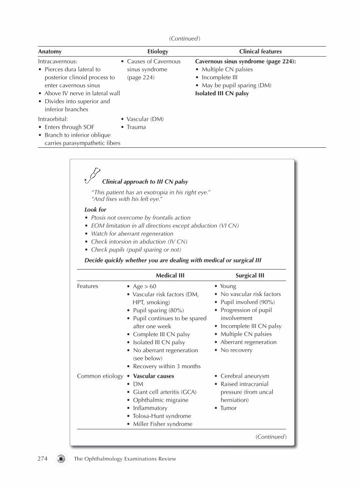
FA B1037 The Ophthalmology Examinations Review
274 The Ophthalmology Examinations Review
Anatomy Etiology Clinical features
Intracavernous:• Pierces dura lateral to
posterior clinoid process to enter cavernous sinus
• Above IV nerve in lateral wall • Divides into superior and
inferior branches
• Causes of Cavernous sinus syndrome(page 224)
Cavernous sinus syndrome (page 224): • Multiple CN palsies• Incomplete III• May be pupil sparing (DM)Isolated III CN palsy
Intraorbital:• Enters through SOF • Branch to inferior oblique
carries parasympathetic fi bers
• Vascular (DM)• Trauma
(Continued )
Clinical approach to III CN palsy
“This patient has an exotropia in his right eye.”“And fi xes with his left eye.”
Look for• Ptosis not overcome by frontalis action• EOM limitation in all directions except abduction (VI CN)• Watch for aberrant regeneration• Check intorsion in abduction (IV CN)• Check pupils (pupil sparing or not)
Decide quickly whether you are dealing with medical or surgical III
Medical III Surgical III
Features • Age > 60• Vascular risk factors (DM,
HPT, smoking)• Pupil sparing (80%)• Pupil continues to be spared
after one week• Complete III CN palsy• Isolated III CN palsy• No aberrant regeneration
(see below)• Recovery within 3 months
• Young• No vascular risk factors• Pupil involved (90%)• Progression of pupil
involvement• Incomplete III CN palsy• Multiple CN palsies• Aberrant regeneration• No recovery
Common etiology • Vascular causes• DM• Giant cell arteritis (GCA)• Ophthalmic migraine• Infl ammatory• Tolosa-Hunt syndrome• Miller Fisher syndrome
• Cerebral aneurysm• Raised intracranial
pressure (from uncal herniation)
• Tumor
(Continued )
B1037_Ch-06.indd 274B1037_Ch-06.indd 274 12/22/2010 7:01:39 PM12/22/2010 7:01:39 PM

B1037 The Ophthalmology Examinations Review FA
Section 6: Neuroophthalmology 275
“The management of III CN palsy involves treating the underlying cause, and addressing the diplopia.”
Q How do You Manage III CN Palsy?
1. Underlying cause• Ligation of aneurysm (endoscopic/surgical)• Optimize control of diabetes/hypertension/
hyperlipidemia if the etiology of III CN palsy is vascular
2. Diplopia• Non-surgical: Fresnel prisms, uniocular
occlusion • Strabismus surgery (at least 6 months after onset,
when all spontaneous improvement has ceased)
Medical III Surgical III
Evaluation • History of DM and HPT• Check BP (HPT)• Headache (GCA, ophthalmic
migraine)• Painful III CN palsy
(DM, migraine, Tolosa-Hunt syndrome)
• Ataxia, arefl exia(Miller Fisher)
• Check fundus for papilledema uncal herniation
• Examine neurologically
• History of head injury• History of headache,
nausea and vomiting (raised intracranialpressure)
• History of HPT (aneurysm)
Investigations • CBC, ESR• Fasting blood sugar level• VDRL, FTA• Autoimmune markers
• CT scan (CNS bleed, meningioma,stroke, trauma)
(Continued )
Exam tips:• There are two ways to remember etiology:
• Anatomical classifi cation is good for answering essay question.• Medical/surgical III classifi cation is good for viva/clinical exams.
B1037_Ch-06.indd 275B1037_Ch-06.indd 275 12/22/2010 7:01:39 PM12/22/2010 7:01:39 PM

FA B1037 The Ophthalmology Examinations Review
276 The Ophthalmology Examinations Review
Q Tell me about Aberrant Regeneration.
“Aberrant regeneration of CN usually follows damage of the nerve by TRAUMA or TUMOR.”“There are recognized syndromes involving III, VII and IX CN.”
Aberrant Regeneration of CN
1. Aberrant regeneration of III CN palsy:• Lid gaze dyskinesis:
• Elevation of lid on adduction (inverse Duane’s syndrome)
• Elevation of lid on depression (pseudo Von Graefe’s sign)
• Pupil gaze dyskinesis:• Constriction on adduction (pseudo Argyll
Robertson pupil)• Constriction on depression
2. Aberrant regeneration of VII CN palsy
(crocodile’s tears):• Synkinesis between salivation fi bers and
lacrimation fi bers• Tearing when eating
3. Aberrant regeneration of IX CN palsy
(Frey’s syndrome):• Synkinesis between salivation fi bers
and sympathetic fi bers• Flushing and sweating when eating
B1037_Ch-06.indd 276B1037_Ch-06.indd 276 12/22/2010 7:01:39 PM12/22/2010 7:01:39 PM

B1037 The Ophthalmology Examinations Review FA
Section 6: Neuroophthalmology 277
TOPIC 3
SIXTH CRANIALNERVE PALSY
Q Opening Question: Tell me About VI CN Palsy.
“VI CN palsy is a common neuroophthalmic diagnostic problem.”“The causes can be divided into….”
Anatomy Etiology Clinical features
Nuclear:• Mid-pons, ventral to
fl oor of fourth ventricle• VII CN fasciculus
curves around VI CN nucleus
• Vascular (stroke)• Demyelination (multiple
sclerosis)• Tumors• Encephalitis
Nuclear VI syndrome and gaze palsy:• Abducens nucleus
(ipsilateral LR palsy)• PPRF (ipsilateral failure of
horizontal gaze)• Facial nucleus (ipsilateral VII
CN palsy)
Fasciculus:• Leaves brainstem ventrally at
ponto medullary junction
• Raymond’s syndrome• Millard Gubler syndrome • Foville’s syndrome
Crossed syndrome:• Pyramidal tract (VI CN palsies
and contralateral hemiparesis)• Pyramidal tract and VII
nucleus (VI, VII CN palsy and contralateral hemiparesis)
• V, VII nucleus, sympathetic, PPRF (V, VI, VII CN palsy, gaze palsy and Horner’s syndrome)
Basilar:• Enters prepontine basilar cistern• Passes upwards close to base
of skull, crossed by anterior inferior cerebellar artery
• Pierces dura below clinoids• Angles over petrous
temporal bone• Passes through Dorello’s canal
and enters cavernous sinus
Causes of cerebellopontine angle syndrome (page 225):• Vestibular schwannoma• Raised intracranial pressure
(false localizing sign)• Nasopharyngeal CA• Basal skull fracture• Clivus meningioma• Gradenigo’s syndrome
Cerebellopontine angle syndrome (page 225)
(Continued )
Overall yield: ⍟ ⍟ ⍟ ⍟ ⍟ Clinical exam: ⍟ ⍟ ⍟ ⍟ ⍟ Viva: ⍟ Essay: ⍟ MCQ: ⍟ ⍟
B1037_Ch-06.indd 277B1037_Ch-06.indd 277 12/22/2010 7:01:39 PM12/22/2010 7:01:39 PM

FA B1037 The Ophthalmology Examinations Review
278 The Ophthalmology Examinations Review
Clinical Notes
1. Isolated medical VI palsy• Same workup as for medical III CN palsy
(page 228)• Recovery within 4–6 months
2. Six causes of “pseudo VI CN palsy”• Myasthenia gravis• Thyroid eye disease• Duane’s syndrome• Medial wall fracture• Esotropia (longstanding)• Convergence spasm
Anatomy Etiology Clinical features
Intracavernous:• Below III and IV CN• Close to internal carotid artery
in the middle of the sinus • Carries sympathetic branches
from paricarotid plexus
• Causes of Cavernous sinus syndrome (page 224)
Cavernous sinus syndrome (page 224)
Intraorbital:• Enters orbit through SOF within
annulus of Zinn to innervate LR
• Vascular (DM)• Trauma
Isolated VI CN palsy
3. Bilateral VI CN palsy• Nuclear:
• CVA• MS• Tumor• Encephalitis (Wernicke’s syndrome)
• Basilar:• False localizing sign• Clivus meningioma
• Intracavernous (big four in pathology):• Infl ammation (Tolosa-Hunt)• Vascular• Tumor (nasopharyngeal CA)• Trauma (carotid-cavernous sinus fi stula)
Clinical approach to VI CN palsy
”This patient has a right esotropia with abduction weakness.”
Look for other CN• EOM (III)• Check intorsion in abduction (IV)• Check pupils (II)
I’ll like to• Check fundus for papilledema (false localizing, pseudotumor)• Examine neurologically:
• Contralateral hemiparesis (Raymond’s syndrome)• VII CN and contralateral hemiparesis (Millard Gubler syndrome)• Horizontal gaze palsy, V, VII, Horner’s syndrome (Foville’s syndrome)• V, VII, VIII, cerebellar signs (cerebellopontine angle tumor — vestibular
schwannoma)• Check ears for otitis media (Gradenigo’s syndrome) and battle sign (petrous
bone fracture)• Refer to ENT to rule out nasopharyngeal CA
(Continued )
B1037_Ch-06.indd 278B1037_Ch-06.indd 278 12/22/2010 7:01:40 PM12/22/2010 7:01:40 PM

B1037 The Ophthalmology Examinations Review FA
Section 6: Neuroophthalmology 279
Q What are Causes of VI CN Palsy in Children?
“VI CN palsy is a common neuroophthalmic diagnostic problem in children.”“First, a squint (ET, Duane’s syndrome) must be excluded.”“The causes can be divided into….”
1. Congenital• Isolated idiopathic• Moebius syndrome
2. Acquired• Trauma• Infection:
• Gradenigo’s syndrome (otitis media, V, VI, VII, VIII CN palsies)
• Tumor:• Pontine glioma• Metastasis form neuroblastoma
Q What is the Surgical Management of VI CN Palsy?
“Surgical intervention for VI CN palsy is undertaken at least six months after onset, when all spontaneous improvement has ceased.”“The choice of surgery depends on the residual abduction power of LR….”
1. Poor residual LR power• Hummelsheim procedure• Jensen procedure
2. Some residual LR power• Ipsilateral MR recession and LR resection
+/− contralateral MR recession
Exam tips:• Do not confuse raised intracranial pressure causing uncal herniation (III CN
palsy) with false localizing sign (VI CN palsy).
B1037_Ch-06.indd 279B1037_Ch-06.indd 279 12/22/2010 7:01:40 PM12/22/2010 7:01:40 PM

FA B1037 The Ophthalmology Examinations Review
B1037_Ch-06.indd 280B1037_Ch-06.indd 280 12/22/2010 7:01:40 PM12/22/2010 7:01:40 PM
This page intentionally left blankThis page intentionally left blank

B1037 The Ophthalmology Examinations Review FA
Section 6: Neuroophthalmology 281
TOPIC 4
NEUROLOGICAL APPROACH TO PTOSIS
Decide Quickly — If
1. Senile aponeurotic ptosis• Describe:
• High lid crease• Atrophic lid tissues and tarsal plate• Deep UL sulcus
• Test:• Ptosis in downgaze — more severe• EOM full and pupils normal• Levator function — usually good
2. Congenital ptosis• Describe:
• Absent lid crease• Visual axis blocked (if blocked, risk of
amblyopia)• Test:
• Ptosis in downgaze — lid lag present
• EOM (SR rectus weakness) • Bell’s refl ex• Marcus Gunn Jaw Winking• Levator function — usually poor• Check the VA and refraction of patient
3. III CN palsy• Severe, complete ptosis• Eye is out and down• Dilated pupils• See CN III approach (page 228)
4. Horner’s syndrome• Mild ptosis, overcome by looking up• Miosis• Enophthalmos• See Horner’s approach (page 243)
Clinical approach to ptosis
Describe• Unilateral/bilateral• Total/severe/mild
• Shine torchlight to look at visual axis • Decide whether anisocoria is present now!
• If present, either III CN palsy or Horner’s syndrome• If absent, either myasthenia gravis, congenital ptosis or senile ptosis
• Overaction of frontalis• Lid crease, lid sulcus, lid mass• Eye position (squints)• Head tilt
(Continued )
O
Overall yield: ⍟ ⍟ ⍟ ⍟ Clinical exam: ⍟ ⍟ ⍟ ⍟ ⍟ Viva: ⍟ ⍟ Essay: ⍟ ⍟ MCQ: ⍟ ⍟
B1037_Ch-06.indd 281B1037_Ch-06.indd 281 12/22/2010 7:01:40 PM12/22/2010 7:01:40 PM

FA B1037 The Ophthalmology Examinations Review
282 The Ophthalmology Examinations Review
(Continued )
“This patient has a mild unilateral ptosis in the right lid.”“Associated with smaller pupils in the right eye….”
or
“This patient has severe bilateral ptosis in both lids, covering the visual axis….”“There is no anisocoria noted….”
Test• EOM
• Upgaze• Ptosis overcome by frontalis (Horner’s syndrome)• Limitation in upgaze (III CN palsy)
• Downgaze (lid lag in congenital ptosis vs aponeurotic, aberrant III CN regeneration — “pseudo-von Graefes sign”)
• Sidegaze (III CN palsy, aberrant III CN regeneration – “reverse Duane’s syndrome”)
• Myasthenia gravis tests — fatigability in upgaze, Cogan’s lid twitch, “eyelash” sign, “eyepeek” sign
• Lagophthalmos and Bell’s (important for management purposes)• Pupils• Marcus Gunn Jaw Winking sign
I’ll like to• Measure the degree of ptosis and levator function (important for
management purposes)• Perform an SLE and fundal examination• Perform myasthenia gravis tests
Exam tips:• One of the most common clinical examination cases.• See also the ptosis chapter in the oculoplastic section (page 291), III CN
palsy (page 228) and Horner’s syndrome (page 243).
B1037_Ch-06.indd 282B1037_Ch-06.indd 282 12/22/2010 7:01:40 PM12/22/2010 7:01:40 PM

B1037 The Ophthalmology Examinations Review FA
Section 6: Neuroophthalmology 283
Overall yield: ⍟ ⍟ ⍟ ⍟ Clinical exam: ⍟ ⍟ ⍟ ⍟ Viva: ⍟ ⍟ Essay: ⍟ ⍟ MCQ: ⍟ ⍟ ⍟
TOPIC 5
MYASTHENIA GRAVIS
“Myasthenia gravis is a systemic neurological disorder.”“Caused by a disorder occurring at the neuromuscular junctions.”“It has both systemic and ocular features.”
Q Opening Question: What are the Features of Myasthenia Gravis (MG)?
Myasthenia Gravis
1. Classifi cation:• Ocular:
• 60% of MG patients present with ocular features; 90% will have some ocular involvement in course of disease
• Prognosis (important):• 40% remain ocular• 10% remission• 50% progress to generalized MG
• Generalized:• Mild• Severe acute (respiratory)• Severe chronic
2. Natural history:• Labile phase • Slow progressive phase• Refractory phase
3. Ocular features (in 90%):• Ptosis:
• Fatigable:• Enhanced with light and passive elevation
of contralateral lid• Asymmetrical• Variable (different time of day)• Shifting (left and right eyes)• Cogan’s lid twitch• Lid hopping/fl uttering
• Ophthalmoplegia:• Pupils normal
• Not consistent with single CN palsy (mimic any of CN palsy)
• MR fi rst muscle to be involved (therefore need to exclude internuclear ophthalmoplegia)
• Saccadic abnormalities: Hypermetric and hypometric saccades
• Accommodative fatigue• Orbicularis oculi weakness:
• “Eyelash” sign (eyelash cannot be “buried” by forcible lid closure)
• “Eye peek” sign (lids drift open slowly after closure)
4. Diagnosis:• Tensilon test (80% sensitivity)• Electromyogram (EMG)
• Repetitive stimulation at 3 mHz (50–90% sensitivity)• Look for decremental amplitude with
repetitive stimulation• Single fi ber EMG (80–95% sensitivity)
• Look for variability between individual muscle fi bers within a motor unit
• Anticholinesterase antibody (70–90% sensitivity): No correlation with disease severity
• Anti-skeletal muscle antibody:• Increased in females and patients with
thymomas• Increased risk of bulbar involvement
Exam tips:• A differential diagnosis for almost any neuroophthalmic condition.
B1037_Ch-06.indd 283B1037_Ch-06.indd 283 12/22/2010 7:01:41 PM12/22/2010 7:01:41 PM

FA B1037 The Ophthalmology Examinations Review
284 The Ophthalmology Examinations Review
Notes• Osserman’s grading
I: Ocular II: Generalized III: Fulminant and crisisIV: Late progressive V: Muscle atrophy
Q How do You Perform the Tensilon Test?
“The tensilon test is a diagnostic test for MG with sensitivity of 80%.”
Tensilon Test
1. Preparation:• Edrophonium hydrochloride:
• Anti-acetylcholinesterase (e.g. increases acetylcholine effects at neuromuscular junction)
• One vial 10 mg/ml• Dilute to 10 ml (concentration: 1 mg/ml)
• Precaution:• Atropine 0.5–1 mg on standby
• Consider giving IM 0.4mg 15 min before test• Resuscitation equipment on standby
2. Injection (rule of 2s):• IV 2 mg, watch for 2 min• If no response, inject another 2 mg, watch for
further 2 min• If still no response, inject remaining 6 mg,
watch for 2 min again
3. Endpoint:• Most respond within 20–45 seconds• Objective endpoints must be determined:
• Ptosis (measured before and after)• Ophthalmoplegia (Hess chart or
Lancaster Red-Green Test)
4. Side effects:• Increased salivation• Sweating• Perioral fasciculation• Nausea• Hypotension, bradycardia, arrhythmia • Bronchospasm
5. False positive result: Eaton-Lambert syndrome,
Guillain-Barre syndrome, amyotrophic lateral
sclerosis
Exam tips:• One of few procedures in neuroophthalmology you need to know well.
Q How to Manage a Patient with MG?
“MG is usually co-managed with a neurologist.”“Need to treat both the systemic condition and the ocular complications.”
Management of MG
1. Systemic manifestations:• Anti-acetylcholinesterase (pyridostigmine/
mestinon):• 30 mg bid dose
• Thymectomy:• Better for generalized MG than ocular MG
• Immunosuppressive therapy:• Prednisolone:
• Given to reduce dose of pyridostigmine
• 65% remission, 30% improvement• Azathioprine
B1037_Ch-06.indd 284B1037_Ch-06.indd 284 12/22/2010 7:01:41 PM12/22/2010 7:01:41 PM

B1037 The Ophthalmology Examinations Review FA
Section 6: Neuroophthalmology 285
2. Ocular complications:• Lid crutches for ptosis• Prisms for ophthalmoplegia
Drugs to Avoid in MG• Aminoglycosides (gentamicin)• Neuromuscular blocker (curare,
suxamethonium)• Chlorpromazine
Notes• Causes of lid retraction in myasthenia gravis:
• Contralateral lid retraction when there is unilateral ptosis• Cogan’s lid twitch• Thyroid eye disease
Q What are the Drugs to Avoid in MG?
• Respiratory depressants (morphine)• Procainamide• Penicillamine
Exam tips:• Remember “ABCD” and “P”!
• Plasmapheresis and IV immunoglobulin — indications:• Myasthenic crisis:
B1037_Ch-06.indd 285B1037_Ch-06.indd 285 12/22/2010 7:01:41 PM12/22/2010 7:01:41 PM

FA B1037 The Ophthalmology Examinations Review
B1037_Ch-06.indd 286B1037_Ch-06.indd 286 12/22/2010 7:01:41 PM12/22/2010 7:01:41 PM
This page intentionally left blankThis page intentionally left blank

B1037 The Ophthalmology Examinations Review FA
Section 6: Neuroophthalmology 287
• Motor imbalance• Congenital nystagmus• Spasmas nutans• Latent nystagmus• Periodic alternating nystagmus• Convergence-retraction nystagmus• Downbeat nystagmus• Upbeat nystagmus• See-saw nystagmus• Ataxic nystagmus
• Sensory deprivation• Nystagmoid movements:
• Ocular fl utter and opsoclonus• Ocular bobbing• Before and after thymectomy• Awaiting response to immunosuppression
• Total body irradiation
Nystagmus
1. Defi nition:• Ocular oscillation which is ....
• Rhythmic in nature and biphasic with at least one slow phase
• The slow phase is abnormal and the fast phase is corrective
• The direction is named after the fast phase
2. Classifi cation:• Clinical:
• Primary position or gaze evoked• Pendular (two slow phases) or jerk (one quick
phase, one slow phase)• Horizontal, vertical, rotatory or mixed• Conjugate (same in both eyes) or dissociated
• Etiological:• Physiological:
• End-point nystagmus• Optokinetic nystagmus• Vestibular nystagmus
TOPIC 6
NYSTAGMUS
Q Opening Question: Tell me about Nystagmus.
“Nystagmus can be defi ned as….”“A simple classifi cation is….”
Exam tips:• Nystagmus is a diffi cult topic. You need to remember the basic principles
and certain types of nystagmus.
Notes• Physiological nystagmus:
• End-point nystagmus• Optokinetic nystagmus• Vestibular nystagmus
Overall yield: ⍟ ⍟ Clinical exam: ⍟ Viva: ⍟ ⍟ Essay: MCQ: ⍟ ⍟
B1037_Ch-06.indd 287B1037_Ch-06.indd 287 12/22/2010 7:01:41 PM12/22/2010 7:01:41 PM

FA B1037 The Ophthalmology Examinations Review
288 The Ophthalmology Examinations Review
Clinical approach to nystagmus
”This patient has a nystagmus.”
Describe• Direction — horizontal/vertical/rotational• Waveform — pendular/jerk (direction of fast phase)• Amplitude — large/small (effect of gaze position on amplitude)• Rest — primary position (at rest/gaze evoked)• Frequency — fast/slow
Exam tips:• Remember “DWARF.”
“There are several distinct ocular features of congenital nystagmus.”
Q What are the Features of Congenital Nystagmus?
Congenital Nystagmus
1. Classifi cation:• Primary/idiopathic• Secondary• Anterior segment disorders
• Corneal opacity, cataract, glaucoma• Visible posterior segment disorders
• ROP, optic atrophy, coloboma, macular scar• Normal looking eyes:
• Leber’s amaurosis• Achromatopsia• Cone dystrophy• Congenital stationary night blindness
2. Ocular features:• Starts at birth or 2–3 months after birth (1)• No oscillopsia (2)
• Nystagmus is:• Binocular (3)• Horizontal (4)• Conjugate (5)• Uniplanar (6)
• Associated with:• Head tilt (7)• Titubation (8)• Null point (9)• Dampens on convergence but increases on
fi xation (10)• Diminishes in darkness/during sleep/when
eye is covered (11)• Latent nystagmus may be present (12)• Paradoxical OKN response (13)
Exam tips:• A favorite question. Remember all the 13 points but DO NOT say, “There are
13 features….” in case you cannot remember all of them!
B1037_Ch-06.indd 288B1037_Ch-06.indd 288 12/22/2010 7:01:42 PM12/22/2010 7:01:42 PM

B1037 The Ophthalmology Examinations Review FA
Section 6: Neuroophthalmology 289
Clinical approach to congenital nystagmus
“On inspection, this young patient has a pendular nystagmus in the primary position.”
Describe features• Binocular (3), horizontal (4), conjugate (5)• Associated head tilt (7), titubation tremors (8)
Check in different directions of gaze • Uniplanar in all directions of gaze (6)• Null point (7)• Dampens on convergence, but increases on fi xation (8)• Cover one eye, observe for latent jerk nystagmus in contralateral eye (11)
“This patient has congenital nystagmus.”
I’d like to • Ask patient for history of onset of nystagmus (1) and whether patient has
symptoms of oscillopsia (2)• Test for paradoxical response to OKN drum (13)?
Q How would You Manage this Patient?
“My management will be conservative.”“I’ll do the following….”
• Refract and prescribe glasses (reading is not impaired)
• Prescribe contact lens if glasses are not suitable
• Give base-out prism to induce convergence
• I will offer surgery only if there is an abnormal head posture (AHP) which is stable:• Horizontal AHP: Kestenbaum procedure• Vertical AHP: Recess and Resect vertical recti
Q How do You Differentiate a Peripheral from a Central Cause of Vestibular Nystagmus?
“There are several distinct features which will help in differentiating peripheral from central cause of vestibular nystagmus.”
Peripheral Central
Nystagmus • Unidirectional, away from site of lesion• May be associated with a rotatory
component
• Multidirectional or unidirectional,towards side of lesion
Association • Dampens on fi xation• Rarely lasts more than 3 weeks• Marked vertigo• Tinnitus/deafness
• No dampening• May be permanent• Mild vertigo• No tinnitus/deafness
Location • Vestibular nerve• Labyrinth
• Cerebellum• Brainstem
Vestibular Nystagmus
B1037_Ch-06.indd 289B1037_Ch-06.indd 289 12/22/2010 7:01:42 PM12/22/2010 7:01:42 PM

FA B1037 The Ophthalmology Examinations Review
290 The Ophthalmology Examinations Review
Q Tell me about the Optokinetic Response.
“Optokinetic (OKN) nystagmus is induced by looking at the rotation of a striped drum — the OKN drum.”“There is the initial pursuit eye movement following the direction of the rotation….”“This is followed by the saccade corrective movement in the opposite direction….”
Use of OKN
1. Diagnosis of congenital nystagmus (paradoxical
response)
2. Detect internuclear ophthalmoplegia (rotate
drum in direction of eye with adduction failure)
(see page 253)3. Detect Parinaud’s syndrome (rotate drum
downwards to elicit convergence retraction nystagmus) (see page 244)
4. Differentiate organic from nonorganic blindness (see page 285)
5. Differentiate vascular from neoplastic causein patient with homonymous hemianopia
(see page 255):• If vascular, lesion is usually confi ned to
occipital lobe (OKN response is symmetrical)• If neoplastic, lesion may extend to parietal lobe
(OKN response is asymmetrical)
B1037_Ch-06.indd 290B1037_Ch-06.indd 290 12/22/2010 7:01:42 PM12/22/2010 7:01:42 PM

B1037 The Ophthalmology Examinations Review FA
Section 6: Neuroophthalmology 291
TOPIC 7
PUPILS
Possible Clinical Cases
1. Afferent defects• Clinical problem: RAPD• Optic nerve and tract
lesions
2. Efferent defects• Clinical problem: Anisocoria• Parasympathetic (III CN palsy) or sympathetic
(Horner’s syndrome)
3. Light near dissociation• Clinical problem: Reaction to accommodation
but not to light
• Causes:• Unilateral:
• Tonic pupil• Aberrant III CN regeneration• Herpes zoster ophthalmicus• Afferent conduction defect
• Bilateral:• Argyll Robertson pupil• Dorsal midbrain syndrome• Dystrophia myotonica• Diabetes• Chronic alcoholism
Q Opening Question: What are the Anatomical Pathways of the Pupil Refl exes?
“There are three important pupil refl exes, with different anatomical pathways for each.”
Pupil Pathways
1. Light refl ex pathway • 1st order: Retina (ganglion cells) — optic
nerve — optic tract — bypasses lateral geniculate body (LGB)
• 2nd order: Pretectal nucleus• 3rd order: Edinger-Westphal nucleus• 4th order: Ciliary ganglion — short ciliary
nerves — iris constrictor pupillae — pupil constriction
2. Sympathetic pupillary pathway• 1st order: Hypothalamus — brainstem• 2nd order: C8 to T2 spinal cord • 3rd order: Superior cervical ganglion —
pericarotid plexus — ophthalmic division of V
CN — nasociliary nerve — long ciliary nerve — iris dilator pupillae — pupil dilation
3. Accommodation refl ex• Not well defi ned, the orders are an
approximation only (important to emphasize this in the beginning):
• 1st order: Retina (ganglion cells) — optic nerve — optic tract
• 2nd order: LGB — optic radiation• 3rd order: Visual cortex — visual association
areas — internal capsule — brainstem• 4th order: Oculomotor nucleus (MR nucleus
and Edinger-Westphal nucleus) — pupil constriction and convergence
Overall yield: ⍟ ⍟ ⍟ ⍟ Clinical exam: ⍟ ⍟ ⍟ ⍟ ⍟ Viva: ⍟ ⍟ ⍟ Essay: ⍟ ⍟ MCQ: ⍟ ⍟
B1037_Ch-06.indd 291B1037_Ch-06.indd 291 12/22/2010 7:01:42 PM12/22/2010 7:01:42 PM

FA B1037 The Ophthalmology Examinations Review
292 The Ophthalmology Examinations Review
Q What are the Causes of Anisocoria?
“Causes of anisocoria depend on which pupil is the abnormal one….”
Anisocoria
1. Dilated (mydriatic) pupil abnormal • III CN palsy• Tonic pupil:
• Holmes Adie syndrome• Pharmacological mydriasis • Iris abnormalities
• Trauma
2. Constricted (miosed) pupil abnormal • Horner’s syndrome:
• Brainstem stroke
• Pancoast syndrome• Cluster headache
• Argyll Robertson pupil• Pharmacological• Iris abnormalities:
• Posterior synechiae• Pontine hemorrhage
Q What is the Marcus Gunn pupil?
“The Marcus Gunn pupil is also known as the relative afferent papillary defect.”“It is elicited with the swinging torchlight test.”“There is paradoxical dilation of the pupil when the torchlight is swung from the contralateral eye to the affected eye.”
Marcus Gunn Pupil
1. Etiology:• Optic nerve lesion (most important)• Other possible sites:
• Extensive retinal damage• Dense macular lesion• Optic chiasma/tract (RAPD in contralateral
eye because nasal retina larger than temporal)
• Dorsal midbrain (RAPD in contralateral eye)
2. Grading (Bell et al. Clinical grading of relative
afferent pupillary defects. Arch Ophthalmol
1993;111:938–942):• Grade I: Weak constriction then greater
dilatation
• Grade II: No initial constriction, stall, then dilatation
• Grade III: Immediate dilatation • Grade IV: Immediate dilatation following
prolonged illumination of the good eye for six seconds
• Grade V: Immediate dilatation with no secondary constriction
Exam tips:• One of the most important defi nitions asked in exams.
Q What is Tonic Pupil? What is the Holmes Adie Pupil? What is the Holmes Adie Syndrome?
Tonic Pupil
1. Clinical features:• Light-near-dissociation (response to
accommodation better than to light)• Dilated pupil (pupil small in longstanding Adies)• Slow constriction and dilatation• Constriction in segments (bag of worms)
• Asymmetrical accommodation
2. Investigation:• 0.1% pilocarpine (constriction due to
denervation supersensitivity)
3. Site of lesion:• Ciliary ganglion or short ciliary nerves
B1037_Ch-06.indd 292B1037_Ch-06.indd 292 12/22/2010 7:01:42 PM12/22/2010 7:01:42 PM

B1037 The Ophthalmology Examinations Review FA
Section 6: Neuroophthalmology 293
4. Etiology:• Primary = Holmes Adie pupil:
• Monocular (80%)• Female (80%)• Age 20–40• Arefl exia (= Holmes Adie syndrome)
• Secondary:• Syphilis (bilateral tonic pupil)• DM• Trauma, surgery• Degenerative
Exam tips:• Tonic pupil ≠ Holmes Adie pupil ≠ Holmes Adie syndrome.
Q What is Horner’s Syndrome?
“Horner’s syndrome is a neurological syndrome caused by lesion in the sympathetic pathway in the head and neck.”
Horner’s Syndrome
1. Clinical features:• Miosis• Ptosis, inverse ptosis• Enophthalmos• Heterochromia• Ocular hypotony• Anhidrosis (if lesion is below superior cervical
ganglion)
2. Investigation:• Cocaine 4–10%
• Blocks reuptake of norepinephrine → pupillary dilatation
• Confi rmation test• Affected pupil does not dilate
• Hydroxyamphetamine 1%• Promotes release of norepinephrine from
terminal axon • Differentiates between central/preganglionic
from postganglionic• Postganglionic lesion (3rd order) pupil fails to
dilate• Phenylephrine 1%
• Denervation supersensitivity• Differentiates central/preganglionic from
postganglionic• Postganglionic lesion pupil dilates more
widely
• Apraclonidine 0.5%• α1 and α2 receptor agonist• Reverses anisocoria in both central/
preganglionic and postganglionic Horner’s syndrome
3. Site and etiology:• Central (fi rst order):
• Brainstem CVA• Trauma• Spinal cord tumor• MS• Syringomyelia• Lateral medullary (Wallenburg) syndrome
• Preganglionic (second order):• Pancoast tumor• Thyroid CA• Vertebral metastasis• Subclavian/aortic/common carotid
aneurysm • Trauma• Aortic/common carotid dissection
• Postganglionic (third order)• Internal carotid dissection• Cluster headache• Cavernous sinus syndrome (Raeder’s
syndrome)• Nasopharyngeal carcinoma
Exam tips:• The pharmacological tests for Horner’s syndrome are one of the favorite
exam questions.• A preganglionic or second order Horner’s syndrome is important because of
the possibility of Pancoast tumor. Therefore, differentiation of central/preganglionic from postganglionic is important.
B1037_Ch-06.indd 293B1037_Ch-06.indd 293 12/22/2010 7:01:43 PM12/22/2010 7:01:43 PM

FA B1037 The Ophthalmology Examinations Review
294 The Ophthalmology Examinations Review
Notes• Causes of acquired Horner’s syndrome in children:
• Neuroblastoma• Thyroglossal cyst• Branchial cyst
Q What is the Argyll Robertson Pupil?
Argyll Robertson Pupil
1. Clinical features:• Light near dissociation (response to
accommodation better than to light)• Constricted (miosed) pupil• Speed of constriction and dilatation is normal • Bilateral pupil involvement common
2. Investigation:• Cocaine 4–10% (affected pupil does
not dilate)
3. Site of lesion:• Dorsal midbrain (pretectal interneurons to
Edinger-Westphal nucleus involved, ventrally located accommodative refl ex neurons being spared)
4. Etiology:• Syphilis
• Pupil signs:• AR pupil or tonic pupil• Pupillary irregularity (iritis)• Poor dilation to atropine
• Other ocular signs (page 328):• Interstitial keratitis• Optic atrophy• Chorioretinitis
• DM• MS• Alchoholism• Trauma, surgery• Aberrant III CN regeneration
Tonic pupil Argyll Robertson pupil
Demographics YoungFemale
OldMale
Pupil DilatedUnilateralSlow reaction to light and accommodation
MiosedBilateralNormal reaction to both
Common cause Holmes Adie pupil or syndrome Syphilis
Comparison Between Tonic Pupil and Argyll Robertson Pupil
Exam tips:• Argyll Robertson pupil ≠ tonic pupil.• Argyll Robertson pupil ≠ syphilis.
B1037_Ch-06.indd 294B1037_Ch-06.indd 294 12/22/2010 7:01:43 PM12/22/2010 7:01:43 PM

B1037 The Ophthalmology Examinations Review FA
Section 6: Neuroophthalmology 295
Q What is the Dorsal Midbrain Syndrome/Parinaud’s Syndrome?
Dorsal Midbrain Syndrome
1. Clinical features:• Light-near-dissociation (response to
accommodation better than to light)• Lid retraction (Collier’s sign)• Supranuclear gaze palsy (normal vestibular
ocular refl ex and Bell’s refl ex)• Convergence retraction nystagmus• Spasm of convergence• Spasm of accommodation• Skew deviation
2. Investigation:• MRI brainstem to exclude lesion on dorsal
midbrain
3. Site of lesion:• Dorsal midbrain (pretectal interneurons to
Edinger-Westphal nucleus involved, ventrally located accommodative refl ex neurons being spared, similar to AR pupil)
4. Etiology (by age group):• Hydrocephalus (infant)• Pinealoma (10 years)• Head injury (20 years)• AV malformation (30 years)• MS (40 years)• Vascular (50 years)• Degenerative (Wernicke’s) (60 years)
Exam tips:• Remember the seven classical signs and seven classical causes!
Clinical approach to pupils “Please examine this patient’s pupils”
Describe“On general inspection, there is ptosis/exodeviation.”“Please look at the distant (fi xation target).” “I would like to examine this patient’s pupils fi rst in the light and then in the dark.”• Greater anisocoria in light — dilated pupil abnormalities (III CN, Holmes
Adie….)• Greater anisocoria in dark — constricted pupil abnormalities (Horner’s….)
Perform light refl ex• Direct refl ex (consensual refl ex)• RAPD
Decide quickly which scenario
1. RAPD• Check EOM (INO, other CN)
I’ll like to check• Fundus (optic disc atrophy, retinal lesions)• VA, VF, color vision
2. Dilated pupil unreactive to light, anisocoria more pronounced in light• Ptosis/divergent squint
• Check EOM• Watch for lid retraction (inverse Duane’s syndrome or lid lag (pseudo-
von Graefe’s sign) from aberrant III CN regeneration)
(Continued )
B1037_Ch-06.indd 295B1037_Ch-06.indd 295 12/22/2010 7:01:43 PM12/22/2010 7:01:43 PM

FA B1037 The Ophthalmology Examinations Review
296 The Ophthalmology Examinations Review
(Continued )
“This patient has a complete III nerve palsy.”
I’ll like to check• Fundus (papilledema)• Examine patient neurologically for long tract signs• No ptosis/no divergent squint
• Check EOM • “I’d like to examine this patient under slit lamp.”
• Irregularity of pupils/vermiform movement (Holmes Adie)• Posterior synechiae (posterior synechiae)• Rupture sphincter/iris damage (traumatic mydriasis)
“This patient has tonic pupil.”
I’d like to• Examine fellow eye (Holmes Adie)• Check tendon refl exes• Perform pharmacological tests (0.1% pilocarpine)• Ask for history of trauma, eyedrop use (can also use 1%
pilocarpine to confi rm)
3. Small pupil, both reactive to light, anisocoria more pronounced in the dark• Mild ptosis, inverse ptosis• Enophthalmos• Flushing, anhidrosis• Check EOM — ptosis overcome by frontalis
“This patient has Horner’s syndrome.”
I’d like to• Check IOP (hypotony)• Confi rm diagnosis by performing pharmacological tests (cocaine,
hydroxyamphetamine)• Examine systematically and neurologically for:
• Neck scars, neck mass (trauma, thyroid CA, lymph nodes)• Clubbing, hypothenar wasting, fi nger abduction weakness (Pancoast
tumor)• III, IV, V CN palsies (Cavernous sinus syndrome, Raeder’s syndrome)• VIII, IX, X CN palsies crossed hemiesthesia, cerebellar signs (lateral
medullary syndrome)• Ask for history of pain and headache (cluster headache, carotid artery
dissection)
4. Small pupil, unreactive to light• Light-near-dissociation
“This patient has Argyll Robertson’s pupil.”
I’ll like to• Ask for history of DM, HPT, sarcoidosis, syphilis
B1037_Ch-06.indd 296B1037_Ch-06.indd 296 12/22/2010 7:01:43 PM12/22/2010 7:01:43 PM

B1037 The Ophthalmology Examinations Review FA
Section 6: Neuroophthalmology 297
TOPIC 8
OPTIC NEUROPATHIES
“The causes of optic neuropathy can be divided into….”
Q Opening Question No. 1: What are the Causes of Optic Neuropathy?
Optic Neuropathy
1. Congenital• Hereditary optic neuropathy
2. Acquired• Optic neuritis (retrobulbar, papillitis,
neuroretinitis):• Demyelinating• Postinfectious• Autoimmune diseases (systemic lupus)• Idiopathic
• Ischemic optic neuropathy (anterior ischemic optic neuropathy, posterior ischemic optic neuropathy):
• Arteritic (giant cell arteritis)• Nonarteritic (atherosclerotic)• Autoimmune diseases (systemic lupus)• Others (hypotension, hypovolemia)
• Compressive optic neuropathy (tumors)• Infi ltrative/granulomatous optic neuropathy
(sarcoidosis, lymphoma, leukemia)• Traumatic optic neuropathy• Toxic optic neuropathy• Radiation optic neuropathy
Exam tips:• Remember the causes of optic neuropathy as “NIGHT TICS” (Neuritis,
Ischemic, Granulomatous, Hereditary, Traumatic, Toxic, Irradiation and Compressive).
• But classify it as “congenital vs acquired.”
Q How do You Differentiate Optic Neuritis From Anterior Ischemic Optic Neuropathy (AION) and Compressive Optic Neuropathy?
Optic neuritis Non-arteritic AION Arteritic AIONCompressiveoptic neuropathy
Presentation 1. Age of onset 2. Gender 3. Onset
4. Pain
• 20–40• Female• Acute and
progressive• Yes
• 40–65• Male = Female• Dramatic sudden
onset• No
• 70–80• Female• Dramatic
sudden onset• Yes
• Any age• Male = Female• Gradual
and progressive• No
(Continued )
Overall yield: ⍟ ⍟ ⍟ ⍟ ⍟ Clinical exam: ⍟ ⍟ ⍟ ⍟ ⍟ Viva: ⍟ ⍟ ⍟ Essay: ⍟ ⍟ MCQ: ⍟ ⍟ ⍟
B1037_Ch-06.indd 297B1037_Ch-06.indd 297 12/22/2010 7:01:44 PM12/22/2010 7:01:44 PM

FA B1037 The Ophthalmology Examinations Review
298 The Ophthalmology Examinations Review
Optic neuritis Non-arteritic AION Arteritic AIONCompressiveoptic neuropathy
5. Other features • MS symptoms (e.g. Uhthoff’s phenomenon)
• Diabetes, hypertension, atherosclerosis risk factors
• Small, crowded disc
• Amaurosis fugax• Giant cell arteritis
symptoms (80%)• Jaw claudication• Neck pain• CRP > 2.45 mg/dl • ESR ≥ 47 mm/h• Scalp
tenderness• Polymyalgia rheumatica• Malaise/
weight loss
• Headache, nauseaand vomiting
Signs
6. VA • 6/18–6/60 • Severe loss (70%)• Mild loss (30%)
• HM — NPL (50%)
• May be normal
7. Bilateral involvement
• Rare in adults• May occur in
children• May alternate
between left and right eyes
• Unilateral• Second eye
may be involvedlater on
• Common • Rare
8. Pupil • RAPD • RAPD • RAPD • Normal
9. Fundus • Normal or • Papillitis (pink)
• Disc swelling(sectoral, pale)
• Disc swelling(chalky white disc edema)
• Cilioretinal artery occlusion
• Disc swelling (diffuse)
• Optociliary shunt (meningioma, optic nerve glioma)
10. Color vision • Dramatic loss, disproportional to VA loss
• Loss proportionalto VA loss
Investigation
11. VF • Diffuse (50%)• Central (10%)
• Inferior nasal sectoral defect
• Inferior altitudinal defect
• Enlarged blind-spot
12. Blood • Normal • ESR raised • ESR markedly raised• C-reactive protein
markedly raised (more sensitivethan ESR)
(Continued )
(Continued )
B1037_Ch-06.indd 298B1037_Ch-06.indd 298 12/22/2010 7:01:44 PM12/22/2010 7:01:44 PM

B1037 The Ophthalmology Examinations Review FA
Section 6: Neuroophthalmology 299
Optic neuritis Non-arteritic AION Arteritic AIONCompressiveoptic neuropathy
13. FFA • Mild leakage at disc margins
• Moderate leakage
• Severe leakage• Filling defect
seen (decrease capillary andchoroidal perfusion)
• Severe leakage
14. VEP • Latency increase
• (Myelination abnormality)
• Amplitudedecrease
• (axonalabnormality)
• Amplitudedecrease
• (Axonal abnormality)
• Amplitude decrease
• (Axonal abnormality)
15. Other investigations
• MRI • Temporal arterybiopsy
Prognosis • 100% recover• 75% to 6/9
• 30% recover• 30% deteriorate• 30% stay the same
• Very poor prognosis
• 65% blind in both eyes within a few weeks if left untreated
• Good if compressive lesion removed
(Continued)
Exam tips:• The three most important and common causes of optic neuropathy.• Remember that there are fi ve differentiating symptoms, fi ve differentiating
signs and fi ve differentiating investigations.• Read treatment of non-arteritic AION from Ischemic Optic Neuropathy
Decompression Trial Research Group (JAMA 1995;273:625–632).
Q How would You Perform a Temporal Artery Biopsy?
“Temporal artery biopsy is the “gold standard” test for making a diagnosis of temporal arteritis.”“I would commence corticosteroid therapy prior to temporal artery biopsy if the biopsy cannot be performed immediately.”“I would perform temporal artery biopsy ideally within three days of commencing corticosteroid therapy, and preferably not after one week of commencing steroids.”
Procedure
1. Identify branch of superfi cial temporal artery by
palpation or Doppler ultrasound
2. Local anesthesia: Lignocaine 1–2% and adrenaline
3. Incise skin over the artery with a No. 15 scalpel
until subcutaneous fat is seen
4. Blunt dissection of space below fat but above
superfi cial temporalis muscle fascia with small
curved artery forceps (superfi cial temporal
artery runs within the fascia), avoiding injury to
the facial nerve
B1037_Ch-06.indd 299B1037_Ch-06.indd 299 12/22/2010 7:01:44 PM12/22/2010 7:01:44 PM

FA B1037 The Ophthalmology Examinations Review
300 The Ophthalmology Examinations Review
5. Carefully dissect superfi cial temporal artery
from fascia with Wescott scissors
6. Tie both ends of segment to be excised with 4/0
silk
7. At least 3cm of artery is excised (because of skip
lesions)
8. Cauterize cut ends of artery
9. Close subcutaneous tissue with 5/0 interrupted
vicryl suture
10. Close skin with interrupted 6/0 or 7/0 prolene
Complications• Bleeding• Damage to branches of the facial nerve• Failure to identify artery (especially if non-
pulsatile)• Scalp necrosis• Stroke (rare)
Q Opening Question No. 2: What are the Causes of Optic Atrophy?
“Optic atrophy can be either unilateral or bilateral.”
Unilateral optic atrophy Bilateral optic atrophy Evaluation
Congenital (not a common cause)1. Hereditary optic neuropathy • Dominant (Kjer) • Recessive (Behr, DIDMOAD) • Mitochondrial — Leber’s optic
neuropathy
Congenital1. Hereditary optic neuropathy Family history
Acquired 1. Old ischemic optic neuropathy2. Compressive optic
neuropathy/pituitary tumor3. Infi ltrative optic neuropathy: • Sarcoidosis • Malignancies (lymphomas,
optic nerve tumors) 4. Old optic neuritis5. Traumatic optic neuropathy6. Radiation optic neuropathy7. Chronic glaucoma8. Toxic optic neuropathy: • TB drugs (ethambutol, isoniazid,
streptomycin), • Chloramphenicol, digitalis,
chloroquine • Toxins (lead, arsenic,
methanol) • Thiamine, vitamin B2, B6,
B12, niacin, folate defi ciency • Tobacco-alcohol toxicity9. Others — PRP, retinitis pigmentosa
Acquired 1. Pituitary tumor2. Chronic papilledema
(secondary OA)3. Toxic optic neuropathy 4. Consecutive ischemic optic
neuropathy5. Consecutive optic neuritis6. Radiation optic neuropathy7. Chronic glaucoma
CT scan Drug history, anemiaSerum vitamin levelsESRHistory of MS, VDRLFTAHistory of nasopharyngealCAIOP, VF
B1037_Ch-06.indd 300B1037_Ch-06.indd 300 12/22/2010 7:01:44 PM12/22/2010 7:01:44 PM

B1037 The Ophthalmology Examinations Review FA
Section 6: Neuroophthalmology 301
Exam tips:• Very similar answer to causes of optic neuropathy but this question requires a
more ”clinical” approach.• Causes are listed slightly differently for unilateral vs bilateral optic atrophy.
The common ones are in bold.
Clinical approach to optic atrophy
“The most obvious abnormality is a pale optic disc.”
Comment on• Sectoral pallor, altitudinal pallor, bow tie, cupping
I’ll like to• Test for RAPD• Check EOM for other CN involvement• Examine the fellow eye
If unilateral, think of• Old optic neuritis (internuclear ophthalmoplegia, other CN palsies)• AION (vascular risk factors)• Compressive optic neuropathy (headache, nausea, vomiting, optociliary
shunt, Foster Kennedy syndrome)• Traumatic optic neuropathy (history of trauma)• Radiation (history of DXT)
If bilateral, think of• Pituitary tumors (bitemporal VF defect)• Consecutive optic neuritis• Toxic optic neuropathy (ethambutol and other drugs)• Hereditary optic neuropathy (Leber’s optic neuropathy)
Q Opening Question No. 3: What are the Causes of Optic Disc Swelling?
“Optic disc swelling can be either unilateral or bilateral.”“The causes are either congenital or acquired.”
Unilateral disc swelling Bilateral disc swelling
Acquired1. Optic neuritis2. Ischemic optic neuropathy3. Compressive optic neuropathy4. Infi ltrative optic neuropathy5. Traumatic optic neuropathy6. Toxic optic neuropathy7. Radiation optic neuropathy
Acquired1. Papilledema
• Space occupying lesion• Idiopathic intracranial hypertension• Malignant HPT
2. Pseudopapilledema• Drusen• Congenital optic disc anomaly
3. Consecutive ischemic optic neuropathy
(Continued )
B1037_Ch-06.indd 301B1037_Ch-06.indd 301 12/22/2010 7:01:44 PM12/22/2010 7:01:44 PM

FA B1037 The Ophthalmology Examinations Review
302 The Ophthalmology Examinations Review
Unilateral disc swelling Bilateral disc swelling
PLUS8. Ocular disease — central retinal
vein occlusion, posterior uveitis, posterior scleritis
9. Orbital disease — pseudotumor, thyroid eye disease
Congenital (not as common as acquired)1. Hereditary optic neuropathy
4. Consecutive optic neuritis5. Compressive optic neuropathy6. Toxic optic neuropathy7. Diabetic papillopathy (may be
unilateral)
PLUS7. Ocular disease — posterior uveitis,
posterior scleritis8. Orbital disease — pseudotumor,
thyroid eye disease
Congenital (not as common as acquired)1. Hereditary optic neuropathy
(Continued )
Exam tips:• Unilateral disc swelling — cause of optic neuropathy plus ocular and orbital
diseases.
B1037_Ch-06.indd 302B1037_Ch-06.indd 302 12/22/2010 7:01:45 PM12/22/2010 7:01:45 PM

B1037 The Ophthalmology Examinations Review FA
Section 6: Neuroophthalmology 303
Overall yield: ⍟ ⍟ ⍟ ⍟ ⍟ Clinical exam: ⍟ ⍟ ⍟ ⍟ Viva: ⍟ ⍟ ⍟ Essay: ⍟ ⍟ MCQ: ⍟ ⍟ ⍟
TOPIC 9
OPTIC NEURITIS AND MULTIPLE SCLEROSIS
Q Opening Question No. 1: What is the Anatomy of the Optic Nerve?
“Optic neuritis is the second cranial nerve.”“It is divided into four segments.”
*From posterior ciliary arteries
Segment Length Diameter Blood supply
Intraocular • Optic disc • Prelaminar • Laminar • Postlaminar
1 mm 1.5 × 1.75 mm • Retinal arterioles from CRA• Peripapillary choroidal vessels*• Circle of Zinn Haller*• Pial branches*
Intraorbital 25 mm 3 mm Pial branches of CRA
Intracanicular 4–10 mm 3 mm Ophthalmic artery branches
Intracranial10 mm
7 mmInternal carotid artery and ophthalmic artery branches
Exam tips:• A “gift” question and one of the favorite anatomy questions. Answer it well.
Q Opening Question No. 2: Tell me About Optic Neuritis.
“Optic neuritis is an acute infl ammatory optic nerve disease.”
Optic Neuritis
1. Classifi cation• Etiological:
• Idiopathic/demyelination• Secondary to:
• Autoimmune diseases• Infectious diseases (e.g. syphilis, viral
infection)
• Anatomical• Sarcoidosis• Retrobulbar neuritis:
• Common variant in adults• Frequently associated with multiple
sclerosis
B1037_Ch-06.indd 303B1037_Ch-06.indd 303 12/22/2010 7:01:45 PM12/22/2010 7:01:45 PM

FA B1037 The Ophthalmology Examinations Review
304 The Ophthalmology Examinations Review
• Papillitis:• Common variant and usually bilateral in
children• Neuroretinitis:
• Associated with viral infection, cat scratch disease, Lyme’s disease, syphilis
• Not associated with multiple sclerosis
• Usually self-limiting and resolves within 6–12 months
2. Clinical features and investigations (see page 248)
3. Prognosis (see below)
4. Treatment (see below)
Notes• Signs of optic nerve dysfunction:
• Decrease in VA• RAPD• Dyschromatopsia (usually red-green)• Diminished light-brightness and contrast sensitivity• Visual fi eld defects
Q What is the Prognosis of a Patient with Optic Neuritis Diagnosed Two Days Ago?
Prognosis
1. Recovery• Onset within a few days, maximal impairment
within 1–2 weeks• Recovery begins within 2–3 weeks, maximal at
6 months• Almost 100% have some recovery• Full recovery in 75%
2. Recurrence• No recurrence in 75%
3. Risk of MS• MS develops in 75% of women (35% in men)• Risk factors:
• Patient:• Age 20–40• Female sex
• White race• Family history of MS• Winter onset
• Ocular:• History of Uhthoff’s phenomenon• FFA leakage around disc margin• Recurrence of optic neuritis• Optic neuritis in fellow eye
• Systemic:• History of nonspecifi c neurological
symptoms• HLA DR2• CSF oligoclonal bands• MRI periventricular lesions (≥ 3, each
≥ 3 mm in size)• If MRI normal, risk of developing MS only
16% in 5 years
Exam tips:• “3R” for “recovery, recurrence and risk of MS.”• 3/4 rule for the chance of each outcome.• Risk factors for MS: 4 patient factors, 4 ocular factors, 4 systemic factors.
B1037_Ch-06.indd 304B1037_Ch-06.indd 304 12/22/2010 7:01:45 PM12/22/2010 7:01:45 PM

B1037 The Ophthalmology Examinations Review FA
Section 6: Neuroophthalmology 305
Q What are the Main Findings of the Optic Neuritis Treatment Trial (ONTT)?
“The ONTT is a multicenter trial to evaluate treatment of optic neuritis with steroids.”“There were 457 patients enrolled.”“The patients were randomized to three treatment regimes.”
Treatment Regimes
1. IV steroids• 3 days IV methylprednisolone (1g/day) plus
11 days of oral prednisolone (1mg/kg/day)
2. Oral steroids• 14 days of oral prednisolone
3. Placebo• 14 days of oral placebo
Results
1. Recovery• IV steroids vs placebo: Faster recovery, but fi nal
VA same, although color vision, contrast sensitivity and VF better
• Oral steroids vs placebo: no difference
2. Recurrence• IV steroids vs placebo: No difference• Oral steroids vs placebo: Higher recurrence
3. Risk of MS• IV steroids vs placebo: Lower risk in fi rst two
years but same after that• Oral steroids vs placebo: No difference• MRI important predictor of MS (≥ 3, each ≥
3 mm or more in size increases risk by 12 times)
Q What are the Main Findings of the Controlled High Risk Subjects Avonex Multiple Sclerosis Prevention Study (CHAMPS)?
“The CHAMPS is a prospective randomized study designed to determine whether the effect of early treatment with interferon beta-1a (Avonex) delayed the risk of developing clinically defi nite multiple sclerosis.”“There were 383 patients enrolled, who had their fi rst demyelineating event (including optic neuritis, myelitis, brainstem-cerebellar syndromes) and had at least two MRI white matter abnormalities characteristic of MS.”“The patients were randomized to two treatment regimes.”
Treatment Regimes
Group 1: ONTT protocol of IV and oral steroids followed by weekly intramuscular 30µg interferon beta-1aGroup 2: ONTT protocol of IV and oral steroids followed by weekly intramuscular injection of placebo
Results
1. Interferon beta-1a reduces the three-year likelihood of conversion to clinically defi nite MS
2. MRI fi ndings:
• Reduction in volume of brain lesions in interferon beta-1a group
• Fewer new/enlarging lesions and fewer gadolinium enhancing lesions in interferon beta-1a group
Trial terminated because of clear benefi t of treatment over placebo.Benefi t of interferon beta-1a also shown in other trials (including Rebif), and continues over a 10-year period of observation (CHAMPIONS study)
• Side effects of interferon beta-1a:Depression, fl uid-like symptoms, liver toxicity, anaphylaxis
Exam tips:• An easy way to commit to memory the effects of each type of treatment on
the “3Rs” of prognosis.• One of the few big trials you are expected to know well. N Engl J Med
1992; 581–88, Surv Ophthalmol 1998;43:291, Arch Ophthalmol 1997;115:1545–1552.
B1037_Ch-06.indd 305B1037_Ch-06.indd 305 12/22/2010 7:01:45 PM12/22/2010 7:01:45 PM

FA B1037 The Ophthalmology Examinations Review
306 The Ophthalmology Examinations Review
Q Tell me about Multiple Sclerosis.
“Multiple sclerosis is an idiopathic demyelination disorder of the CNS.”“MS does not involve the peripheral nervous system.”“The CNS lesions are separated in TIME and SPACE.”“Diagnosis is therefore made when there are two or more different neurological events occurring at different times.”
• Two-thirds will have INO in course of disease
• Clinical features (see below)• Horizontal gaze palsy• One-and-a-half syndrome• Ocular dysmetria• Dorsal midbrain syndrome• Skew deviation• Nystagmus
• Isolated CN involvement• Paroxysmal eye movement disorders
• Rare:• Intermediate uveitis• Retinal periphlebitis
3. Investigations• Lumbar puncture:
• Oligoclonal bands• Leukocytosis• IgG > 15% total protein
• VEP:• Increase in latency typically exceeds
decrease in amplitude• MRI:
• Periventricular and corpus callosum plaques• Acute lesions may enhance with gadolinium
Multiple Sclerosis
1. Systemic features• Hemisphere:
• Dementia• Hemiparesis, dysphasia, hemianopia
• Brain stem:• Dysarthria, dysphagia• Nystagmus, ataxia, diplopia
• Spinal cord:• Motor• Sensory• Bladder, bowel and sexual disturbances
• Transient disturbances:• Lhermitte’s sign• Uhthoff’s phenomenon• Trigeminal neuralgia
2. Ocular features• Sensory (hemisphere lesions)
• Optic neuritis:• One-third of MS will present with optic
neuritis• Two-thirds will have optic neuritis in
course of disease• Risk of MS with optic neuritis (see above)
• Posterior visual system lesions (VF defects)• Motor (brainstem lesions)
• Gaze abnormalities:• Internuclear ophthalmoplegia:
• One-third of MS will present with INO
Q Tell me about Internuclear Ophthalmoplegia.
“INO is motor abnormality caused by lesions in the medial longitudinal fasciculus (MLF).”
1. Classical feature (triad)• Failure of adduction of ipsilateral eye
(e.g. side of MLF lesion)• Ataxic nystagmus of contralateral eye on
abduction• Normal convergence (posterior INO)
2. Other clinical features • Horizontal:
• Slowing of saccades in adducting eye• Horizontal nystagmus of adducting eye
• Vestibuloocular refl ex (VOR) impaired (note: VOR is not intact because lesion is not supranuclear)
• Abnormal convergence (note: Cogan’s anterior INO, implies lesion extents to midbrain convergence center)
• Manifest exotropia (wall-eyed bilateral INO or WEBINO)
• Vertical:• Vertical nystagmus• Vertical pursuit impaired
B1037_Ch-06.indd 306B1037_Ch-06.indd 306 12/22/2010 7:01:46 PM12/22/2010 7:01:46 PM

B1037 The Ophthalmology Examinations Review FA
Section 6: Neuroophthalmology 307
Clinical approach to INO
“The most obvious abnormality is a failure of adduction of the right eye.” “With a horizontal nystagmus seen in the left eye.”
Examine• Exclude one-and-a-half syndrome (failure of abduction of ipsilateral eye and
adduction of contralateral eye)• Vestibulo-ocular refl ex/Doll’s eye refl ex (should be impaired) • Convergence (if impaired, implies Cogan’s anterior INO)• Vertical movements
I’d like to• Examine pupils for RAPD (old optic neuritis infl amed optic nerve)• Examine fundus (optic atrophy)• Examine neurological system (signs of MS or stroke)• Ask for history of trauma
• Vertical VOR impaired• Upgaze maintenance impaired
3. Investigations• OKN drum:
• Slowing of saccades in adducting eye (to elicit, rotate drum in direction of ipsilateral MLF lesion)
• ENG (electronystagmogram):• Reduction of peak velocity of adduction
4. Etiology• MS (40%)• CVA (40%)• Others (tumor, trauma, infection)
Exam tips:• There are 10 clinical features of INO (not three).
Exam tips:• An extremely common clinical exam case. Usually asked to examine the
ocular movements.• Remember to look at the adducting eye when testing for horizontal
movements!
Q What are the Supranuclear Disorders of Ocular Motility?
“Supranuclear disorders of ocular motility are characterized by the absence of diplopia, and the presence of normal vestibulo-ocular refl exes.”
1. Horizontal gaze palsies:• Parapontine reticular formation (PPRF) lesions:
Ipsilateral horizontal gaze palsy• Medial longitudinal (MLF) lesions: Internuclear
ophthalmoplegia• PPRF and MLF lesions: “One-and-a-half
syndrome”
2. Vertical gaze palsies:• Parinaud dorsal midbrain syndrome• Progressive supranuclear palsy (Steele-
Richardson-Olszewski syndrome)
B1037_Ch-06.indd 307B1037_Ch-06.indd 307 12/22/2010 7:01:46 PM12/22/2010 7:01:46 PM

FA B1037 The Ophthalmology Examinations Review
B1037_Ch-06.indd 308B1037_Ch-06.indd 308 12/22/2010 7:01:46 PM12/22/2010 7:01:46 PM
This page intentionally left blankThis page intentionally left blank

B1037 The Ophthalmology Examinations Review FA
Section 6: Neuroophthalmology 309
TOPIC 10
VISUAL FIELD DEFECTS IN NEUROOPHTHALMOLOGY
Q Opening Question: What are the Ocular Manifestations of a Meningioma Compressing the Left Optic Tract?
• OKN asymmetry (defect when drum rotates towards left)
• Dominant parietal lobe (Gerstmann syndrome)• Aphasia/agraphia/acalculia• Left-right disorientation
• Non-dominant parietal lobe• Neglect/inattention• Impaired constructional ability• Dyscalculia
5. Left temporal lobe• Incongruous right upper homonymous
hemianopia• Formed visual hallucination• Auditory hallucinations• May be complicated by motor seizures
6. Left visual cortex• Congruous right homonymous hemianopia• OKN symmetry• Unformed visual hallucination • Other features of visual cortex involvement
(see stroke, migraine and other vascular disorders, page 266)
Lesion in Left Side of the Visual Pathway
1. Left optic nerve• Decrease in VA in left eye• Decrease in color vision in left eye• RAPD in left eye• Optic disc swelling or atrophy in left eye• Dense central/diffuse scotoma in left eye
2. Optic chiasm• Bitemporal hemianopia (classic)• Dense central/diffuse scotoma (optic nerve)• Incongruous homonymous hemianopia (optic
tract)• Bitemporal central scotoma (macular fi bers)• Junctional scotoma (junction of optic nerve and chiasma)• Other features of chiasmal syndrome
(see pituitary disorders, page 260)
3. Left optic tract• Incongruous right homonymous quadrantanopia• RAPD in right eye
4. Left parietal lobe• Incongruous right lower homonymous
quadrantanopia• Pursuit defect to left• Hemiparesis or hemianesthesia in right side of
body
Overall yield: ⍟ ⍟ ⍟ Clinical exam: ⍟ ⍟ ⍟ ⍟ Viva: ⍟ ⍟ Essay: ⍟ MCQ: ⍟ ⍟
B1037_Ch-06.indd 309B1037_Ch-06.indd 309 12/22/2010 7:01:46 PM12/22/2010 7:01:46 PM

FA B1037 The Ophthalmology Examinations Review
310 The Ophthalmology Examinations Review
Clinical approach to visual fi eld examination
“Please examine this patient’s visual fi eld.”
Common clinical syndromes• Bitemporal hemianopia• Homonymous hemianopia• Bitemporal or homonymous quadrantanopia• Altitudinal hemianopia
Preliminary instructions“I am going to test the area that you can see.”“Please cover your left eye and look at my nose.”“Is there any part of my face that is not clear?”“How many fi ngers are there?”“Now look at my face and do not move your eyes….”
Quadrant testing“How many fi ngers are there?”
Neglect testing“How many fi ngers are there altogether?”
Central scotoma testingTest fellow eye“This patient has bitemporal hemianopia, my clinical diagnosis is a pituitary lesion.”
I’d like to• Check EOM (see-saw nystagmus, CN palsies)• Check fundus (bow-tie atrophy, papilledema)• Ask history of diplopia (nonparetic), metamorphopsia, visual hallucination• Look for features of hypersecretion from an adenoma
• Growth hormone (acromegaly) • Prolactin (history of amenorrhea, galactorrhea, infertility in females or
impotence in males)• ACTH (Cushing’s syndrome)
• Assess for etiology of pituitary lesion — ask for history of• Trauma• Radiation• Shock, blood loss during pregnancy (pituitary apoplexy)• Adrenalectomy for Cushing’s (Nelson’s syndrome)• Secondaries to pituitary, infi ltrative lesions (TB, sarcoidosis)
Or
“This patient has right homonymous hemianopia, my clinical diagnosis is a left postchiasmal lesion.”
I’d like to• Check fundus (optic atrophy, papilledema)• Perform full Humphrey VF to assess congruity of lesion• Left optic tract
• Incongruous right homonymous quadrantanopia• RAPD in right eye
(Continued )
B1037_Ch-06.indd 310B1037_Ch-06.indd 310 12/22/2010 7:01:46 PM12/22/2010 7:01:46 PM

B1037 The Ophthalmology Examinations Review FA
Section 6: Neuroophthalmology 311
(Continued )• Left parietal lobe
• Incongruous right lower homonymous quadrantanopia• Check EOM (failure of pursuit to left)• Check for right hemiparesis or hemianesthesia• Assess reading (alexia) and writing (agraphia)• OKN asymmetry (move drum towards left)
• Left temporal lobe• Incongruous right upper homonymous hemianopia• Formed visual hallucination
• Left occipital lobe• Congruous right homonymous hemianopia• Assess visual attention (inattention) and visual recognition (agnosia)• OKN symmetry• Unformed visual hallucination
Exam tips:• Alternate question can be “What are the ocular manifestations of stroke
involving the right optic radiation?”• Few important principles to remember
1. VF defect becomes more congruous as you move further back along the visual pathway.
2. Visual hallucination is formed in temporal lobe lesions vs unformed for cortex lesions.
3. OKN asymmetry indicates that a lesion has involved the parietal lobe (usually a tumor) as compared with OKN symmetry in a pure cortex lesion (usually a stroke) (page 240).
B1037_Ch-06.indd 311B1037_Ch-06.indd 311 12/22/2010 7:01:47 PM12/22/2010 7:01:47 PM

FA B1037 The Ophthalmology Examinations Review
B1037_Ch-06.indd 312B1037_Ch-06.indd 312 12/22/2010 7:01:47 PM12/22/2010 7:01:47 PM
This page intentionally left blankThis page intentionally left blank

B1037 The Ophthalmology Examinations Review FA
Section 6: Neuroophthalmology 313
Overall yield: ⍟ ⍟ ⍟ ⍟ Clinical exam: ⍟ ⍟ ⍟ ⍟ Viva: ⍟ ⍟ ⍟ Essay: ⍟ ⍟ MCQ: ⍟ ⍟ ⍟ ⍟
TOPIC 11
PITUITARY AND CHIASMAL DISORDERS
1. Anatomy• Situated in sella turcica of sphenoid bone• Anterior lobe:
• Secretes ACTH, GH, Prolactin, FSH, LH and TSH
• Posterior lobe:• “Secretes” ADH and oxytocin (actual
hormones produced from hypothalamus)• Relationship with chiasma:
• Central:• 80%• Classic “chiasmal syndrome” features
(see below)• Optic chiasmal VF defect: Bitemporal
hemianopia, involving the superior fi elds fi rst
• Prefi xed (chiasma is anterior to pituitary gland):• 10%• Optic tract features • Optic tract VF defect: Incongruous
homonymous hemianopia• Macular involvement: Bitemporal central
scotoma• Postfi xed (chiasma is posterior to pituitary
gland):• 10%
Q Opening Question: Tell me about the Pituitary Gland.
• Optic nerve features (RAPD, color vision, etc)
• Optic nerve VF defect: Dense central/diffuse scotoma
• Optic nerve and chiasmal junction: Junctional scotoma (von Willebrand’s knee)
2. Spectrum of pituitary disorders• Tumors:
• Pituitary adenoma• Craniopharyngioma• Meningioma• Others:
• Chordoma• Nasopharyngeal CA• Rathke’s pouch cyst• Secondaries
• Vascular (aneurysm)• Infl ammation (Tolosa-Hunt syndrome,
meningitis)• Demyelination (MS)• Trauma, surgery and DXT• Non-neoplastic masses: Fibrous dysplasia,
sphenoidal sinus mucoceles, arachnoid cysts
B1037_Ch-06.indd 313B1037_Ch-06.indd 313 12/22/2010 7:01:47 PM12/22/2010 7:01:47 PM

FA B1037 The Ophthalmology Examinations Review
314 The Ophthalmology Examinations Review
Q Tell me about the Pituitary Adenomas.
“Pituitary adenomas are benign tumors of the pituitary gland.”“They can be classifi ed as either secreting or nonsecreting, which can be subdivided into chromophobes, acidophils….”“Or classifi ed as either microadenoma or macroadenoma, defi ned as….”“The clinical presentation is that a combination of local mass effects and systemic endocrine effects.”
Classifi cation Hormones Clinical features
Secreting (75%) Chromophobes (50%)
Prolactin • Infertility-amenorrhea-galactorrhea in women (like a lactation state, except you cannot get pregnant!)
• Hypogonadism, impotence, sterility, decreased libido, gynecomastia and galactorrhea in men
Acidophils (20%) GH • Acromegaly in adults (see below)• Gigantism in children
Basophils (5%) ACTHFSH and TSH
• Cushing’s disease (causing Cushing’s syndrome)
• FSH and TSH tumors are extremely rare
Nonsecreting (25%)
Microadenoma Less than 10 mm in diameter
• Usually that of secreting adenomas• Nonsecreting microadenomas are not
discovered!
Macroadenoma More than 10 mm in diameter
• Mass effects• Usually nonsecreting in nature
1. Localized mass effects• Chiasmal syndrome (see below)• Compression of other adjacent structure:
• Cavernous sinus (CN palsies)• Pituitary gland (hypopituitarism)• Raised intracranial pressure (papilledema)
2. Endocrine effects• Hypersecretion
3. Management• Investigation:
• Skull XR (note: In practice, skull XR is not very useful):• Expansion or ballooning of fossa• Erosion of clinoid• “Double fl oor” sign (asymmetrical fossa
expansion)• CT scan/MRI
• Endocrine evaluation: Prolactin, FSH, TSH, GH
• Treatment:• Factors to consider:
• Age• Presenting problem (vision, mass effect,
endocrine effect)• Size and stage of tumor
• Surgery (transphenoidal, transethmoidal, craniotomy)
• Bromocriptine for prolactinomas (increases prolactin inhibition factor)/somatostatin for GH tumors
• Radiotherapy (complications include radiation optic neuropathy and panhypopituitarism):• Gamma-knife stereotactic radiotherapy
B1037_Ch-06.indd 314B1037_Ch-06.indd 314 12/22/2010 7:01:47 PM12/22/2010 7:01:47 PM

B1037 The Ophthalmology Examinations Review FA
Section 6: Neuroophthalmology 315
Exam tips:• The clinical presentation is often a combination of either: Endocrine effects
(from secreting tumors) or mass effects (from macroadenomas).• Most ophthalmologists end up seeing macroadenomas with mass effects on
the chiasma.• To aid memory (not exactly accurate!), microadenomas = secreting
adenomas = endocrine effects; macroadenomas = nonsecreting adenomas = mass effects.
Q What are the Ocular Manifestations of a Pituitary Adenoma Pressing on the Chiasma?
Clinical Features of the Chiasmal Syndrome• VF defects (depending on location of chiasma):
• Bitemporal hemianopia involving superior fi elds fi rst (classic VF defect)
• Incongruous homonymous hemianopia (optic tract)
• Bitemporal central scotoma (macular fi bers)• Dense central/diffuse scotoma (optic nerve)• Junctional scotoma (junction of optic nerve
and chiasma)• Optic atrophy (spectrum of changes):
• Normal looking disc
• Temporal pallor (papillomacular bundle)• Bow tie atrophy• Dense optic atrophy
• Hemifi eld slip (nonparetic diplopia)• Postfi xation blindness• Visual hallucination• See-saw nystagmus
Exam tips:• The “chiasmal syndrome” is an important syndrome and bitemporal
hemianopia is but one of fi ve different VF defects.
Q What are the Ocular Features of Acromegaly?
Acromegaly• Angioid streaks• Chiasma syndrome • Retinopathy (DM and HPT retinopathy)
• Optic atrophy, papilledema• Muscle enlargement
Exam tips:• Remember the clinical features as “ACROM.”
Q What is Pituitary Apoplexy?
Pituitary Apoplexy• Infarction of pituitary gland• Tumor outgrows blood supply or tumor
compresses hypophyseal portal vessels
• Presents with hyperacute chiasmal syndrome
• Treatment: High dose steroids/surgery
B1037_Ch-06.indd 315B1037_Ch-06.indd 315 12/22/2010 7:01:47 PM12/22/2010 7:01:47 PM

FA B1037 The Ophthalmology Examinations Review
316 The Ophthalmology Examinations Review
• Children often present with panhypopituitarism secondary to hypothalamic dysfunction (dwarfi sm, delayed sexual development, obesity), while adults often present with visual impairment and visual fi eld defects
3. Diagnosis• CT scan/MRI (suprasellar calcifi cation in 70%)
4. Treatment: Surgical excision +/− postoperative
radiotherapy• Recurrence common and requires lifelong
follow-up
Q Tell me about Craniopharyngioma.
“Craniopharyngioma is an intracranial tumor arising from remnants of Rathke’s pouch.”
Craniopharyngioma
1. Histological features• Solid component with squamous epithelium
and calcifi cation• Cystic component with greenish fl uid
2. Clinical presentation — depends on growth of
tumor• Superiorly into ventricles (most common,
hydrocephalus and raised intracranial pressure)• Anteriorly to frontal lobe (dementia)• Anteroinferiorly to optic nerve and chiasma
(chiasmal syndrome)• Posteroinferiorly to hypothalamus and pituitary
gland (diabetes insipidus and hypopituitarism)
Notes• Differential diagnoses of bitemporal visual fi eld defects:
• Chiasmal lesions• Dermatochalasis of upper lids• Tilted discs• Optic nerve colobomas• Nasal retinitis pigmentosa• Nasal retinoschisis• Functional visual loss
Q What is the Empty Sellar Syndrome?
“The empty sellar syndrome is a neurological condition in which the subarachnoid space extends into the sella, remodeling the bone and enlarging the sella.”
Empty Sellar Syndrome
1. Classifi cation• Primary:
• Common, 25% of autopsies• Transfer of CSF pressure through a
congenitally large opening in diaphragm sella• Risk factors: Multiparous women, elderly
atherosclerotic patients, idiopathic intracranial hypertension
• Secondary:• Pituitary surgery• DXT• Pituitary apoplexy (need to exclude
concomitant pituitary adenoma)
2. Clinical features• VA usually normal
• Decrease VA rare• Due to herniation of suprasellar contents (e.g.
optic nerve) into sella or vascular compromise
• VF defects:• Binasal (classically)• Bitemporal, altitudinal and generalized
constriction of VF possible• Headache• Elevated prolactin levels
3. Diagnosis• CT scan/MRI
B1037_Ch-06.indd 316B1037_Ch-06.indd 316 12/22/2010 7:01:47 PM12/22/2010 7:01:47 PM

B1037 The Ophthalmology Examinations Review FA
Section 6: Neuroophthalmology 317
TOPIC 12
PAPILLEDEMA and INTRACRANIAL TUMORS
Q Opening Question: What are the Clinical Features of Papilledema?
“Papilledema is bilateral optic nerve head swelling secondary to raised intracranial pressure. It can be divided into four stages….”“Clinical features are related to either vascular or mechanical changes of the optic disc….”
Papilledema
Stage Vision Optic disc vascular changes Optic disc mechanical changes
Early • VA normal • Hyperemia of margins• Loss of spontaneous venous
pulsation (absent in 20% of normal individuals)
• Indistinct disc margins
• Blurring of margins (superior and inferior margin fi rst)
• Edema of peripapillary nerve fi ber layer
Established • Transient visual disturbances
• VA normal or impaired
• Enlarged blind spot
• Venous tortuosity and dilation• Peripapillary fl ame-shaped
hemorrhage• Cotton wool spots• Hard exudates• Obscuration of small vessels
traversing disc
• Elevated disc • Obliteration of cup• Retinal/choroidal folds
Longstanding • VA impaired• Constricted VF
• Vascular changes resolves • “Champagne cork” appearance• Optociliary shunts• Drusen-like crystalline deposits
Atrophic • VA severely impaired
• Secondary optic atrophy
Overall yield: ⍟ ⍟ ⍟ ⍟ Clinical exam: ⍟ ⍟ ⍟ ⍟ Viva: ⍟ ⍟ ⍟ Essay: ⍟ ⍟ MCQ: ⍟ ⍟
B1037_Ch-06.indd 317B1037_Ch-06.indd 317 12/22/2010 7:01:48 PM12/22/2010 7:01:48 PM

FA B1037 The Ophthalmology Examinations Review
318 The Ophthalmology Examinations Review
Q What are the Ocular Features of Idiopathic Intracranial Hypertension?
“Idiopathic Intracranial Hypertension (IIH) is a neurological syndrome of raised intracranial pressure in the absence of….”
Idiopathic Intracranial Hypertension
1. Defi nition (important)• Raised intracranial pressure
• > 250 mm water (need lumbar puncture) — lateral decubitus position
• In absence of (triad):• Space occupying lesion (need CT scan)• Hydrocephalus (need CT scan)• Abnormal CSF (need lumbar puncture)
2. Etiology• Associated with obesity• Exclude:
• Sagittal sinus thrombosis (most important to exclude. Need MRV to diagnose)
• Metabolic disorders (Cushing’s disease, Addison’s, hypoparathyroidism)
• Vitamin A toxicity (see page 413) and lead poisoning
• Drugs (steroids, nalidixic acid, amiodarone, tetracycline)
3. Clinical features• Headache (90%)• Visual loss:
• Transient (70%) or persistent (30%)• Variable (different time of day)• Shifting (left and right eyes)• Loss of contrast sensitivity• VF loss (essentially like VF defects in
papilledema):• Enlargement of blind spot and constriction
of VF• Other VF defects (nasal defects, central
defects)• Tinnitis (60%)
• IIH has been called “otitic hydrocephalus” precisely because of this symptom
• Others (photopsia, retrobulbar pain, diplopia)
4. Investigations• CT scan/MRI:
• Normal looking ventricles• Small ventricles
• Empty sella (see page 261)• Venous sinus thrombosis
• Lumbar puncture• Metabolic/endocrine evaluation• Perimetry
5. Prognosis• Spontaneous remission (3–12 months)• May be remitting and relapsing or develop into
chronic condition• Recurrence (10%)• Visual loss risk factors:
• Vascular disease (HPT, DM, anemia)• Raised IOP• Recent weight gain
6. Treatment• Conservative: Lose weight, discontinue inciting
drug, treat systemic disorders• Indications for treatment:
• Severe symptoms (headache)• Visual loss• High CSF pressure
• Repeat lumbar puncture:• 25% remit after fi rst lumbar puncture• However, CSF usually replenishes within 1–2
hours• Diuretics (acetazolamide):
• Oral diamox 500 mg bid for 4–6 weeks• Topiramate (anticonvulsant that also inhibits
carbonic anhydrase)• If there is no response, consider
• Steroids:• Dexamethasone 4 mg 6 hourly for 1–2 weeks • If no response, consider
• Optic nerve sheath decompression or ventriculo/lumbo — peritoneal shunt
• Stenting of transverse sinus
B1037_Ch-06.indd 318B1037_Ch-06.indd 318 12/22/2010 7:01:48 PM12/22/2010 7:01:48 PM

B1037 The Ophthalmology Examinations Review FA
Section 6: Neuroophthalmology 319
Q What are the Ocular Signs of a Meningioma? A Pinealoma?
“The ocular features of a meningioma include general signs due to raised intracranial pressure.”“And focal signs depending on where the meningioma is….”
Ocular Signs of Intracranial Tumors
1. General (raised intracranial pressure)• Symptoms:
• Visual blurring (transient or persistent)• Diplopia
• Signs:• Papilledema and optic atrophy• Foster Kennedy syndrome
• Triad of optic atrophy in one eye, papilledema in other eye and anosmia
• VI CN palsy (false localizing sign)• III CN palsy (uncal herniation)
2. Focal• Supratentorial (mainly sensory)• Suprasellar/sphenoidal ridge/midbrain (both
sensory and motor)• Infratentorial (mainly motor)
Exam tips:• The ocular features of ANY brain tumor can be divided into general signs due
to raised intracranial pressure (see papilledema above) and focal signs, depending on location of tumor.
• For neuroophthalmology and strokes (see page 255), for migraines and other vascular disorders (see page 265).
Q What are the Principles in the Management of Brain Tumors?
Principles of Management
1. Histological diagnosis• Access via burr hole or craniotomy• Guidance via free hand or stereotactic
technique
2. Curative excision for benign tumors
(e.g. meningioma)
3. Palliative excision for malignant tumors
(e.g. glioblastoma)
4. Adjunctive therapy (e.g. DXT, chemotherapy)
B1037_Ch-06.indd 319B1037_Ch-06.indd 319 12/22/2010 7:01:48 PM12/22/2010 7:01:48 PM

FA B1037 The Ophthalmology Examinations Review
B1037_Ch-06.indd 320B1037_Ch-06.indd 320 12/22/2010 7:01:48 PM12/22/2010 7:01:48 PM
This page intentionally left blankThis page intentionally left blank

B1037 The Ophthalmology Examinations Review FA
Section 6: Neuroophthalmology 321
TOPIC 13
STROKES, MIGRAINES & OTHER VASCULAR
DISORDERS
Q Opening Question: What are the Ocular Manifestations of Strokes?
“Strokes cause a variety of different ocular syndromes.”“Depending on the arterial system and location of the stroke….”“The ocular features can be either sensory or motor….”
Vertebrobasilar System Stroke
1. Vertebral or basilar artery• Complete brainstem infarct:
• CN involvement:• III, IV, VI CN palsies (ophthalmoplegia)• V CN palsy (loss of corneal refl ex)• VII CN palsy (lagophthalmos)• VIII CN palsy (nystagmus)
• PPRF, convergence center, MLF involvement:• Conjugate gaze palsies• INO, WEBINO (see page 253)• One-and-a-half syndrome (see page 254)• Parinaud’s syndrome (see page 244)
• Sympathetic involvement:• Horner’s syndrome
• Midbrain:• Crossed syndromes (Weber’s syndrome,
Benedikt’s syndrome and others)• Pons:
• Crossed syndromes (Millard Gubler and others)
• Medulla:• Lateral medullary syndrome (posterior inferior
cerebellar artery):• V CN palsy and spinothalamic tract
involvement (crossed hemianesthesia)• VIII CN palsy (vertigo and nystagmus)
• IX and X CN palsy (dysarthria and dysphagia)
• Sympathetic involvement (Horner’s syndrome)
• Cerebellar involvement (ataxia and other cerebellar signs)
2. Posterior cerebral artery • Lateral geniculate body (posterior choroidal
artery):• Incongruous homonymous hemianopia • Homonymous sectoranopia (wedge-shaped)
• Anterior visual cortex (see below):• Congruous homonymous hemianopia with
macular sparing• Others
3. Cerebellar arteries • Cerebellar involvement (see nystagmus, page 238)
Carotid System Stroke
1. Internal carotid artery• Amaurosis fugax (see below and page 201)• Venous stasis retinopathy (page 202)• Ischemic optic neuropathy (page 247)• Central retinal artery occlusion (page 193)• Anterior segment ischemia (page 202)
Overall yield: ⍟ ⍟ ⍟ ⍟ Clinical exam: ⍟ ⍟ Viva: ⍟ ⍟ ⍟ Essay: ⍟ ⍟ ⍟ MCQ: ⍟ ⍟
B1037_Ch-06.indd 321B1037_Ch-06.indd 321 12/22/2010 7:01:48 PM12/22/2010 7:01:48 PM

FA B1037 The Ophthalmology Examinations Review
322 The Ophthalmology Examinations Review
2. Anterior cerebral artery • Hemialexia (page 283)
3. Anterior choroidal artery • Important branch of internal carotid artery• Causes full blown CVA (hemiplegia,
hemianesthesia)• Involves different parts of the visual pathway:
• Optic tract:• Incongruous homonymous hemianopia
• Lateral geniculate body (compare with posterior choroidal artery involvement above):• Homonymous superior and inferior
sectoranopia• Optic radiation:
• Incongruous homonymous hemianopia
4. Middle cerebral artery • Involves different parts of cortex and visual
pathway (anterior to posterior):
• Frontal eye fi elds:• Conjugate gaze palsy (saccade defect)
• Parietal eye fi elds:• Conjugate gaze palsy (pursuit defect)
• Posterior parietal regions:• Alexia and agraphia (see page 283)
• Lateral geniculate body:• Homonymous sectoranopia
• Optic radiation:• Incongruous homonymous hemianopia
• Posterior visual cortex:• Congruous homonymous central fi eld
defect (“macular VF defect”)• Balint’s syndrome:
• Ocular apraxia (cannot move eyes on command but can move spontaneously!)
• Visual inattention (eyes wander around)
Exam tips:• Knowledge of the anatomy of the blood supply to the brain is important.• Alternate questions can be, “What happens when the basilar artery is
blocked?” or “What are the ocular manifestations of middle cerebral artery stroke?”
Q What are the Ocular Signs in Visual Cortex Lesions?
“The ocular manifestations of visual cortex lesions depend on the area and extent of the involvement.”“The signs are either predominantly anterior cortex or posterior cortex.”“And the ocular features can be either VF defects or various psychosomatic syndromes….”
Visual Cortex
1. Visual fi eld defects• Congruous homonymous hemianopia with
macular sparing (anterior cortex, posterior cerebral artery)
• Congruous homonymous central fi eld defect/“macular VF defect” (posterior cortex, middle cerebral artery)
• Temporal crescent unilateral VF defect (most anterior portion of visual cortex)• This is the only monocular VF defect in the
visual pathway posterior to the chiasma!• Others:
• Bilateral homonymous hemianopia with macular sparing (bilateral anterior cortex)
• Bilateral homonymous altitudinal defect:• Inferior (bilateral involvement of superior
cortex)
• Superior (bilateral involvement of inferior cortex)
• Checkerboard defect (homonymous anopia, superior defect in one eye, inferior defect in other)
2. Psychosomatic syndromes• Cortical blindness (page 283)• Anton’s syndrome (cortical blindness plus
denial of blinding)• Riddoch’s phenomenon (can see moving targets
but not stationary targets)• Balint’s syndrome (ocular apraxia, visual
inattention)• OKN asymmetry (see OKN, page 240)• Unformed visual hallucinations (see visual
hallucinations, page 282)
B1037_Ch-06.indd 322B1037_Ch-06.indd 322 12/22/2010 7:01:48 PM12/22/2010 7:01:48 PM

B1037 The Ophthalmology Examinations Review FA
Section 6: Neuroophthalmology 323
Exam tips:• An alternate question can be, “What are the ocular features of a meningioma
impinging on the visual cortex?”• Differentiate signs of the anterior visual cortex (supplied by posterior
cerebral artery) from those of the posterior visual cortex, where the macular representation is localized (supplied by middle cerebral artery).
• Note that macular area is supplied by the middle cerebral artery!
Q What is Amaurosis Fugax?
“Amaurosis fugax is an ocular transient ischemic attack (TIA).”“The etiology is usually….”
Amaurosis Fugax
1. Most common ocular TIA (transient ischemic
attack)
2. Due to cholesterol/platelet emboli from carotid
atheroma
3. Clinical notes• Visual loss:
• Transient, lasting less than 10 minutes to 2 hours (never more than 24 hours by TIA defi nition)
• Starts in the central VF, expanding outwards or altitudinal (curtain like effect)
• Contralateral hemiplegia (12.5%)• Carotid bruit (20%)
4. Treatment and prognosis• Carotid ultrasound• Carotid endarterectomy (NASCET results):
• Carotid stenosis < 50% (no benefi t)
• Carotid stenosis > 70% (eight-year benefi t)• Carotid stenosis 51 to 69% (consider this
only if other risk factors are present)• Carotid artery stenting:
• Carotid Revascularization Endarterectomy vs Stenting Trial (CREST) (N Engl J Med;2010: Epub):• Risk of stroke, MI or death similar between
stenting and endarterectomy groups• During periprocedural period, higher risk
of stroke with stenting, and higher risk of MI with endarterectomy
• European International Carotid Stenting Study (Lancet 2010;375:985–997):• Stents had a higher rate of complications
compared to endarterectomy (stroke, myocardial infarction, death)
• Commonest cause of death: Cardiac causes (not stroke!)
Exam tips:• This is an important differential diagnosis of sudden visual loss and an
extremely common problem referred to by ophthalmologists in emergency room settings “requiring decisions made on the spot (see page 201).”
• Read results of the North American Symptomatic Carotid Endarterectomy Trial (NASCET) N Engl J Med 1998;339:1415–25.
Q Tell me about Migraines.
“Migraine is a neurovascular disorder.”“It can be classifi ed into….”“The ocular features can be either sensory or motor…..”
Migraine
1. Classifi cation• Without aura:
• Common migraine (= classic migraine without the aura)
• With aura:• Classic migraine• Migraine equivalent
• Aura without headache
B1037_Ch-06.indd 323B1037_Ch-06.indd 323 12/22/2010 7:01:49 PM12/22/2010 7:01:49 PM

FA B1037 The Ophthalmology Examinations Review
324 The Ophthalmology Examinations Review
• Ophthalmoplegic migraine • Painful III, IV and V CN palsies
• Retinal migraine• Photopsia
• Basilar migraine• III, IV and V CN palsies• Fortifi cation spectra
• Cluster headache (variant of migraine)
2. Ocular features of classic migraine • Sensory
• Excitatory:• Fortifi cation spectra:
• Zig-zag patterns
• Gradual build up• Starts in central VF, expanding outwards
to peripheral VF• Photopsia • Metamorphopsia
• Inhibitory:• Blurring of vision• Amaurosis fugax• Scotoma
• Others:• Photophobia
• Motor:• III, IV and V CN palsies
Direct CCF Indirect CCF
Classifi cation • Barrow’s Type I (internal carotid artery to cavernous sinus)
• Type II (meningeal branches of internal carotid artery to cavernous sinus)
• Type III (meningeal branches of external carotid artery to cavernous sinus)
• Type IV (meningeal branches of both internal and external carotid artery to cavernous sinus)
Etiology • Head trauma with base of skull fracture (young men)
• Spontaneous rupture from atherosclerosis (postmenopausal hypertensive women)
• Congenital malformation (associated with Ehlers-Danlos or pseudoexanthoma)
• Spontaneous rupture from atherosclerosis (postmenopausal hypertensive women)
Clinical features • Acute pulsatile proptosis (with thrill and bruit)
• Severe EOM impairment (abolished by carotid compression)
• Anterior segment: • Engorged corkscrew episcleral vessels • Glaucoma (from increased episcleral
venous pressure, orbital congestion, secondary angle closure, neovascular glaucoma from CRVO)
• Anterior segment ischemia
• Slowly progressive proptosis (with thrill and bruit)
• Mild EOM impairment (VI CN palsy)• Subtle anterior segment signs • Dilated cockscrew episcleral vessels • Raised IOP • Increased ocular pulsation
Exam tips:• This is important because the migraine is one of the most common neurologi-
cal conditions and often presents with ocular symptoms fi rst.
Q Tell me about Carotid Cavernous Fistula.
“Carotid cavernous fi stula (CCF) is an arteriovenous fi stula which connects the carotid artery and the cavernous sinus.”“It can be divided into….”
(Continued )
B1037_Ch-06.indd 324B1037_Ch-06.indd 324 12/22/2010 7:01:49 PM12/22/2010 7:01:49 PM

B1037 The Ophthalmology Examinations Review FA
Section 6: Neuroophthalmology 325
Direct CCF Indirect CCF
• Posterior segment: • CRVO • Optic nerve head swelling (ON
compression)
• Posterior segment: • Features of venous congestion
Blinding complications
• Glaucoma• Exposure keratopathy• Optic nerve compression• Ocular ischemia
Investigation • CT scan (“What are CT scan features?”): • Proptosis • Distended superior ophthalmic vein • Enlarged EOM • Bowing of cavernous sinus• Orbital Doppler • Dilated superior ophthalmic vein • Reversal of fl ow• Carotid angiogram • Indicated if surgery considered
• Similar
Indications for treatment
• Blinding complications• Severe diplopia• Severe bruit
Treatment • Most fi stula close spontaneously• Interventional radiology (embolization): • Balloon, glue, sphere • Complications (stroke in 5%, failure of
procedure common)• Surgery (progressive carotid artery ligation)
• Most fi stula close spontaneously
(Continued)
Exam tips:• Common differential diagnosis of unilateral proptosis (page 307).
Notes• Glaucoma in CCF:
• Increased episcleral venous pressure ishemia → secondaryopen angle glaucoma
• Anterior segment ischemia → neovascular glaucoma• Congestion of uveal tissues → angle closure glaucoma
B1037_Ch-06.indd 325B1037_Ch-06.indd 325 12/22/2010 7:01:49 PM12/22/2010 7:01:49 PM

FA B1037 The Ophthalmology Examinations Review
B1037_Ch-06.indd 326B1037_Ch-06.indd 326 12/22/2010 7:01:49 PM12/22/2010 7:01:49 PM
This page intentionally left blankThis page intentionally left blank

B1037 The Ophthalmology Examinations Review FA
Section 6: Neuroophthalmology 327
TOPIC 14
NEUROOPHTHALMIC MANIFESTATIONS OF
CEREBRAL ANEURYSMS
Q Opening Question: Tell me about Cerebral Aneurysms.
“Cerebral aneurysms are saccular or fusiform dilatations of intracranial arteries.”
Cerebral Aneurysm
1. Prevalence and presentation• 1–6% of population in autopsies, of which
20–30% have multiple aneurysms• Presentation:
• 90% present acutely with subarachnoid hemorrhage from ruptured aneurysm
• 10% present with chronic mass effects:• Headache is most common symptom• The median time of signs due to mass
effect to aneurysm rupture is 14 days (therefore important to diagnose aneurysm early to prevent devastating subarachnoid hemorrhage)
2. Etiology• Primary/idiopathic • Hypertension• Others (rare):
• Connective tissue disorders (Ehlers-Danlos syndrome, polycystic kidneys)
• Bacterial/fungal aneurysm• Traumatic
3. Location• Anterior to the Circle of Willis (70%):
• Anterior cerebral artery, anterior communicating artery, internal carotid artery at bifurcation and posterior communicating artery
• Supraclinoid:• Sensory: Optic nerve, chiasma and tract
involved
• Infraclinoid:• Motor: III, IV and VI CN involved:
• Anterior (V1 CN involved)• Middle (V1 and V2 CN involved)• Posterior (V1, V2 and V3 CN involved)
• Middle cerebral artery (20%)• Posterior to Circle of Willis (10%)
4. Ocular features • Acute with subarachnoid hemorrhage:
• Increased intracranial pressure (see page 262)• Terson syndrome
• Chronic with mass effects:• Motor (70%):
• Infraclinoid aneurysms• Painful, incomplete, pupil-involved III CN
palsy (60% of aneurysms will have III CN involvement)
• Multiple CN involvement (IV, VI and V, depending on location)
• Sensory (20%):• Supraclinoid aneurysms• Chiasmal syndrome (see page 260)
• Mixed motor and sensory (10%):• Cavernous sinus carotid artery aneurysms • Triad of pulsatile proptosis (with bruit),
conjunctival injection and VI CN palsy (see carotid cavernous fi stula, page 268)
Overall yield: ⍟ ⍟ Clinical exam: Viva: ⍟ ⍟ Essay: ⍟ MCQ: ⍟ ⍟
B1037_Ch-06.indd 327B1037_Ch-06.indd 327 12/22/2010 7:01:49 PM12/22/2010 7:01:49 PM

FA B1037 The Ophthalmology Examinations Review
328 The Ophthalmology Examinations Review
Exam tips:• Ophthalmologists have an important role in detecting cerebral aneurysms
because 70% are anterior to the Circle of Willis and are present with ocular sensory and motor signs.
• Remember the 70:20:10 rule, which describes the location of aneurysm and the clinical features.
Q How would You Manage a Patient Suspected of Having a Cerebral Aneurysm?
Management of Cerebral Aneurysm
1. Investigations• CT scan:
• Sensitivity = 60%• If subarachnoid hemorrhage has occurred,
sensitivity increases to 90%• MRI:
• Sensitivity = 80%• MR angiogram, sensitivity = 90%
• Carotid angiogram:• Gold standard, sensitivity = 95%• Need four vessel angiograms to see both
anterior and posterior circulations
2. Treatment• Medical:
• Control of BP• Antivasospasm• Anticonvulsant• Antiedema (dexamethasone)
• Surgical:• Clipping of aneurysm• Proximal ligation of parent artery
• Interventional radiology: Embolization of aneurysm with coils
B1037_Ch-06.indd 328B1037_Ch-06.indd 328 12/22/2010 7:01:49 PM12/22/2010 7:01:49 PM

B1037 The Ophthalmology Examinations Review FA
Section 6: Neuroophthalmology 329
TOPIC 15
NEUROCUTANEOUS SYNDROMES
Q Opening Question: What is Neurofi bromatosis?
“Neurofi bromatosis (NFM) is one of the neurocutaneous syndromes.” “With systemic and ocular features.”
Classical Neurofi bromatosis
1. Hereditary pattern• AD (with incomplete penetrance and
expressitivity)• Chromosome 17 mutation
2. Skin features• Café au lait spots• Neurofi broma• Plexiform neurofi broma• Axillary freckles
3. CNS features• Neural tumors in brain, spinal cord, CN and
peripheral nerves (neurofi broma, glioma, schwannoma)
4. Ocular features• Orbital features:
• Lid plexiform neurofi broma and ptosis
• Nonaxial pulsatile proptosis (congenital defect of sphenoid bone with spheno-orbital-encephalocele)
• Axial nonpulsatile proptosis (optic nerve glioma)
• Nonaxial nonpulsatile proptosis (other orbital nerve tumors, e.g. neurilemmoma)
• Ocular features:• Prominent corneal nerves• Lisch nodules• Ectropion uvea• Glaucoma — 50% have unilateral UL
neurofi broma/facial hemiatrophy• Choroidal hamartomas
5. Others • Skeletal abnormalities:
• Short stature• Scoliosis• Macrocephaly• Facial hemiatrophy
• Childhood malignancies• Hypertension from pheochromocytoma
6. Diagnostic criteria for neurofi bromatosis type I
(NF-1) (≥ 2 of the following)• ≥ 6 cafe au lait spots• ≥ 2 neurofi broma or ≥ 1 plexiform neurofi broma• Axillary/inguinal freckles• Optic nerve/chiasmal glioma• ≥ 2 lisch nodules• Osseous lesions e.g. sphenoid dysplasia• First degree relative with NF-1
Exam tips:• Remember that ALL the syndromes have the three cardinal sites of involve-
ment: Skin, CNS and eye(s).
Overall yield: ⍟ ⍟ ⍟ ⍟ Clinical exam: ⍟ ⍟ ⍟ ⍟ Viva: ⍟ ⍟ Essay: ⍟ MCQ: ⍟ ⍟ ⍟ ⍟
B1037_Ch-06.indd 329B1037_Ch-06.indd 329 12/22/2010 7:01:49 PM12/22/2010 7:01:49 PM

FA B1037 The Ophthalmology Examinations Review
330 The Ophthalmology Examinations Review
Notes• “What are the possible mechanisms?”
• Obstruction of outfl ow by neurofi broma• Angle abnormality• Angle closure from ciliary body neurofi broma
Q What is the Difference between Neurofi bromatosis Type I and II?
Type II or Central NFM • Rarer• Chromosomal 22 mutation• Less café au lait spots• Ocular features include:
• Bilateral vestibular schwannomas (V, VI, VII, VIII palsies)
• Posterior subcapsular cataracts• Combined hamartomas of retina and RPE• No risk of glaucoma, no Lisch nodules
• Diagnostic criteria for neurofi bromatosis type II (NF-2):• Bilateral vestibular schwannomas or• First degree relative with NF-2 and either
unilateral vestibular schwannoma or any two of the following:• Meningioma• Glioma• Schwannoma• Juvenile posterior subcapsular lenticular
opacities/juvenile cortical cataract
Clinical approach to neurofi bromatosis
“This patient has multiple nodules on his face and neck.”
Look for• Lid neurofi broma and ptosis (plexiform neurofi broma)• Anterior segment:
• Prominent corneal nerves• Lisch nodules• Ectropion uvea
• Proptosis:• Pulsatile, no bruit (spheno-orbital encephalocele)• Nonpulsatile (optic nerve glioma)
“This patient has neurofi bromatosis.”
I’d like to• Check IOP and gonioscopy (glaucoma)• Check pupils for RAPD (optic nerve glioma, meningioma) and then dilate
the pupils to….• Examine fundus (optic atrophy, papilledema, optociliary shunts, choroidal
hamartomas)• Examine systemically for other features of NFM:
• Skin (café au lait, axillary freckles, plexiform neurofi bromas, neurofi bromas on other parts of body)
• Skeletal (short stature, scoliosis, macrocephaly, hemiatrophy of face)• Neurologically (CNS tumors)• BP (HPT from pheochromocytoma)
• Examine family members
B1037_Ch-06.indd 330B1037_Ch-06.indd 330 12/22/2010 7:01:50 PM12/22/2010 7:01:50 PM

B1037 The Ophthalmology Examinations Review FA
Section 6: Neuroophthalmology 331
Q What is Tuberous Sclerosis?
“Tuberous sclerosis is one of the neurocutaneous syndromes.”“With systemic and ocular features.”“The classical triad consists of: Mental handicap, epilepsy and adenoma sebaceum.”
Tuberous Sclerosis
1. Hereditary pattern• AD • Chromosome 9 mutation
2. Skin features• Adenoma sebaceum• Ash leaf spots• Shagreen patches• Café au lait• Skin tags
3. CNS features• Astrocytic hamartoma:
• Epilepsy
• Hydrocephalus• Mental handicap
4. Ocular features• Retinal astrocytoma• Hypopigmented iris and fundal lesions
5. Others • Visceral hamartoma:
• Kidneys• Heart• Subungal areas
Q What is the Sturge-Weber Syndrome?
“Sturge-Weber syndrome is one of the neurocutaneous syndromes.”“With systemic and ocular features.”
Sturge-Weber Syndrome
1. Hereditary pattern• None (like Wyburn Mason syndrome)
2. Skin features• Nevus fl ammeus/cavernous hemangioma/port
wine stain:• First and second divisions of V CN• Hypertrophy of face
3. CNS features• Angioma of meninges (“Which layer is
involved?” Answer: Pial) and brain:• Ipsilateral to side of facial angioma• Parietal and occipital lobe• May calcify and show up in Skull XR as “tram
track sign”
• Epilepsy, hemiparesis and hemianopia
4. Ocular features• Glaucoma:
• 30% of patients• Ipsilateral to side of facial angioma• Higher risk if upper lid involved
• Choroidal hemangioma:• 40% of patients• Ipsilateral to side of facial angioma• Diffuse type of choroidal hemangioma (not
circumscribed type of choroidal hemangioma)
• Episcleral, iris and ciliary body hemangiomas
Clinical approach to Sturge Weber syndrome
“This patient has a port wine stain….”“In the distribution of the 1st and 2nd divisions of the Trigeminal nerve.”
Look for• Hypertrophy of face on side of hemangioma• UL involvement — ptosis• Episcleral hemangioma
(Continued )
B1037_Ch-06.indd 331B1037_Ch-06.indd 331 12/22/2010 7:01:50 PM12/22/2010 7:01:50 PM

FA B1037 The Ophthalmology Examinations Review
332 The Ophthalmology Examinations Review
Ataxia Telangiectasia
1. Hereditary pattern• AR • Chromosome 11 mutation
2. Skin features• Cutaneous telangiectasia
3. CNS features• Cerebellar ataxia• Mental handicap
Exam tips:• Note that cavernous hemangioma (Sturg Weber syndrome) is not the same as
the benign capillary hemangioma (page 310).
Q What is Ataxia Telangiectasia?
“Ataxia telangiectasia is one of the neurocutaneous syndromes.”“With systemic and ocular features.”
Notes• “What are the mechanisms of glaucoma?”
• Raised episcleral venous pressure• Angle abnormally• Ciliary body angioma
(Continued )
• Iris, ciliary body hemangioma (lens subluxation)• Trabeculectomy
I’ll like to• Exclude glaucoma (30%), check IOP and look at optic disc • Perform gonioscopy (hemangioma, angle anomaly, increased episcleral
venous pressure)• Check fundus for diffuse choroidal hemangioma (40%)• Examine patient neurologically (hemangiomas of brain)
4. Ocular features• Conjunctival telangiectasia• Oculomotor defects:
• Nystagmus• Oculomotor apraxia• Strabismus
B1037_Ch-06.indd 332B1037_Ch-06.indd 332 12/22/2010 7:01:50 PM12/22/2010 7:01:50 PM

B1037 The Ophthalmology Examinations Review FA
Section 6: Neuroophthalmology 333
Q What is the Von Hippel-Lindau Syndrome?
“Von Hippel-Lindau syndrome is one of the neurocutaneous syndromes.”“With systemic and ocular features.”
Von Hippel-Lindau Syndrome
1. Hereditary pattern• AD • Chromosome 3 mutation
2. Skin features (not prominent)• Café au lait• Melanocytic nevi
3. CNS features• Hemangioblastoma
• Cerebellum• Brainstem• Spinal cord• May cause polycythemia
4. Ocular features• Capillary hemangioma of retina
• Treatment: Laser photocoagulation (small lesions), cryotherapy (peripheral lesions), PDT, IVTA, intravitreal anti-VEGF
• Hypertensive retinopathy (pheochromocytoma)• Papilledema (CNS hemangioblastoma)
5. Others• Visceral tumors:
• Cysts of kidney, pancreas, liver, epididymis, ovary and lungs
• Hypernephroma• Pheochromocytoma
Q What is Incontinentia Pigmenti?
“Incontinentia pigmenti is one of the neurocutaneous syndromes.”“With systemic and ocular features.”
Incontinentia Pigmenti
1. Hereditary pattern• Sex linked dominant (one of only few disease)
2. Skin features• Stage 1: Erythema and bullae at extremities• Stage 2: Wart-like changes• Stage 3: Hyperpigmented macules in “christmas
tree” pattern on trunk
3. CNS features• Epilepsy• Mental retardation• Hydrocephalus
4. Ocular features• Proliferative retinopathy (like retinopathy of
prematurity)
Notes• Ocular conditions with X-linked inheritance• X-linked dominant:
• Incontinentia pigmenti• Alport’s syndrome• Aicardi syndrome
• X-linked recessive:• Norrie’s disease• Juvenile retinoschisis• Choroideremia• Ocular albinism• Lowe syndrome
B1037_Ch-06.indd 333B1037_Ch-06.indd 333 12/22/2010 7:01:51 PM12/22/2010 7:01:51 PM

FA B1037 The Ophthalmology Examinations Review
334 The Ophthalmology Examinations Review
Q What is Wyburn Mason Syndrome?
“Wyburn Mason syndrome is one of the neurocutaneous syndromes.”“With systemic and ocular features.”
Wyburn Mason Syndrome
1. Hereditary pattern• None (like Sturg Weber syndrome)
2. Skin features (not prominent)
3. CNS features• Arteriovenous malformation in CNS:
• Epilepsy
• Hemiparesis• Mental retardation
4. Ocular features• Racemose angioma
B1037_Ch-06.indd 334B1037_Ch-06.indd 334 12/22/2010 7:01:51 PM12/22/2010 7:01:51 PM

B1037 The Ophthalmology Examinations Review FA
Section 6: Neuroophthalmology 335
TOPIC 16
HEAD INJURY
Q Opening Question: What are the Ocular Signs in Head Injury?
“Ocular signs are important in head injuries because they have immediate localizing and prognostic values.”“They can be divided into….”
Ocular Signs of Head Injury
1. Visual pathway signs:• Retina and optic disc:
• Papilledema• Purtscher’s retinopathy
• Optic nerve:• Traumatic optic neuropathy (see below)
• Optic chiasma:• Infrequent, usually from frontal contusion
• Retrochiasmal:• Homonymous hemianopia (secondary to
occipital ischemia)
2. Motor signs:• III, IV and VI CN palsies, conjugate gaze palsies,
INO:• Diffi cult to diagnose• Observe spontaneous eye movements• Oculocephalic refl ex may help
3. Pupillary signs:• Fixed dilated pupil:
• Transtentorial/uncal herniation (III CN palsy)
• Traumatic III CN palsy• Traumatic mydriasis• Orbital blow out fracture
• Small pupil:• Horner’s syndrome• Traumatic miosis• Pontine hemorrhage• Hutchinson’s pupil (early stages of
transtentorial herniation)
4. Late signs:• Subdural hematoma:
• Late III CN palsy (transtentorial herniation)• Late VI CN palsy (raised intracranial pressure)
• Aberrant III CN regeneration• Carotid cavernous fi stula• Late Horner’s syndrome
Q How do You Manage a Patient with Severe Head Injury?
“The aim of management is to limit the extent of the primary damage and to prevent secondary brain damage.”
Management of Head Injury
1. Primary brain damage:• Open laceration• Contusion• Diffuse axonal injury• Brainstem injury
2. Secondary brain damage:• Hemorrhage
• Extradural:• Lucid interval • Trauma to temporal bone area/pterion
area with rupture of middle meningeal artery
• III CN palsy on ipsilateral side (herniation of uncus on same side)
Overall yield: ⍟ ⍟ Clinical exam: Viva: ⍟ ⍟ Essay: ⍟ MCQ: ⍟ ⍟
B1037_Ch-06.indd 335B1037_Ch-06.indd 335 12/22/2010 7:01:51 PM12/22/2010 7:01:51 PM
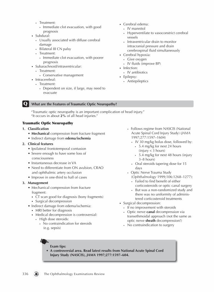
FA B1037 The Ophthalmology Examinations Review
336 The Ophthalmology Examinations Review
• Treatment:• Immediate clot evacuation, with good
prognosis• Subdural:
• Usually associated with diffuse cerebral damage
• Bilateral III CN palsy• Treatment:
• Immediate clot evacuation, with poorer prognosis
• Subarachnoid/intraventricular:• Treatment:
• Conservative management• Intracerebral:
• Treatment:• Dependent on size, if large, may need to
evacuate
• Cerebral edema:• IV mannitol• Hyperventilate to vasoconstrict cerebral
vessels• Intraventricular drain to monitor
intracranial pressure and drain cerebrospinal fl uid simultaneously
• Cerebral hypoxia:• Give oxygen • IV fl uids (improve BP)
• Infection:• IV antibiotics
• Epilepsy:• Antiepileptics
Q What are the Features of Traumatic Optic Neuropathy?
“Traumatic optic neuropathy is an important complication of head injury.”“It occurs in about 2% of all head injuries.”
Traumatic Optic Neuropathy
1. Classifi cation• Mechanical compression from fracture fragment• Indirect damage from edema/ischemia
2. Clinical features• Ipsilateral frontotemporal contusion• Severe enough to have some loss of
consciousness• Instantaneous decrease in VA • Need to differentiate from ON avulsion, CRAO
and ophthalmic artery occlusion• Improve in one-third to half of cases
3. Management• Mechanical compression from fracture
fragment:• CT scan good for diagnosis (bony fragments)• Surgical decompression
• Indirect damage from edema/ischemia:• MRI better for diagnosis• Medical decompression is controversial:
• High dose steroids:• No contraindication for steroids
(e.g. sepsis)
• Follows regime from NASCIS (National Acute Spinal Cord Injury Study) (JAMA 1997;277:1597–1604)• IV 30 mg/kg bolus dose, followed by:
• 5.4 mg/kg for next 24 hours (injury < 3 hours)
• 5.4 mg/kg for next 48 hours (injury 3–8 hours)
• Oral steroids tapering dose for 15 days
• Optic Nerve Trauma Study (Ophthalmology 1999;106:1268–1277):• Failed to fi nd benefi t of either
corticosteroids or optic canal surgery • But was a non-randomized study and
there was no uniformity of adminis-tered corticosteroid treatments
• Surgical decompression:• If no improvement with steroids• Optic nerve canal decompression via
transethmoidal approach (not the same as optic nerve sheath decompression!)
• No contraindication to surgery
Exam tips:• A controversial area. Read latest results from National Acute Spinal Cord
Injury Study (NASCIS), JAMA 1997;277:1597–604.
B1037_Ch-06.indd 336B1037_Ch-06.indd 336 12/22/2010 7:01:51 PM12/22/2010 7:01:51 PM

B1037 The Ophthalmology Examinations Review FA
Section 6: Neuroophthalmology 337
Overall yield: ⍟ ⍟ Clinical exam: Viva: ⍟ ⍟ Essay: ⍟ ⍟ ⍟ MCQ: ⍟ ⍟
TOPIC 17
COMA, DISORDERS OF HIGHER FUNCTIONS & PSYCHIATRIC DISEASES
Q Opening Question: What are the Ocular Features Seen in Patients in Coma?
“The neuroophthalmic signs include the eyelids, pupil, fundus and ocular motility.”
Neuroophthalmic Signs in Coma
1. Eyelid signs• Eye opening:
• Glasgow Coma Scale• Spontaneous, to speech, to pain
• Eye closure:• Closure (intact lower pons)• Tone of closure proportional to depth of coma• Asymmetrical closure (VII CN palsy on one side)
• Blinking:• Spontaneous blinking (intact reticular system)• Refl ex blinking:
• To light or threat (intact anterior visualpathway, brainstem and VII CN)
• To sound (intact VIII and VII CN)• To corneal refl ex (intact V and VII CN)
2. Pupillary signs• Fixed, dilated pupil:
• Unilateral (III CN palsy with transtentorialherniation)
• Bilateral (atropine poisoning, barbituratepoisoning, severe hypoxia-ischemia)
• Marcus Gunn pupil (optic nerve or chiasmdamage, pituitary apoplexy)
• Small pupil:• Unilateral (Horner’s syndrome)• Bilateral (pontine hemorrhage, opiates
poisoning, severe metabolic damage,thalamic and basal ganglia damage)
3. Fundus• Papilledema (space-occupying lesion)• Retinal hemorrhage (subarachnoid hemorrhage)
4. Eye motility• Spontaneous eye movement:
• Roving, bobbing, ping-pong movements(brainstem damage)
• Sustained conjugate eye deviation:• Horizontal deviation (“What are the possible
causes?”)• Ipsilateral hemispheric lesion:
• Oculocephalic refl ex/caloric stimulationpositive
• Associated contralateral hemiparesis• Contralateral pontine lesion:
• Oculocephalic refl ex/caloric stimulationnegative
• Associated ipsilateral hemiparesis• Contralateral thalamic lesion (also known
as the “wrong way deviation”)• Downward deviation:
• Dorsal midbrain lesion• Upward deviation:
• Hypoxia-ischemia• Sustained disconjugate eye deviation:
• III, IV, VI CN palsies, internuclearophthalmoplegia
• Skew deviation (brainstem lesion)
B1037_Ch-06.indd 337B1037_Ch-06.indd 337 12/22/2010 7:01:51 PM12/22/2010 7:01:51 PM

FA B1037 The Ophthalmology Examinations Review
338 The Ophthalmology Examinations Review
Q What are the Doll’s Eye Refl ex and Caloric Tests?
“The Doll’s eye refl ex is a head rotation test for the oculocephalic refl ex.”“Caloric stimulation is a similar test of the oculocephalic refl ex.”
Oculocephalic Refl ex
1. Anatomical pathway• Afferent: Labyrinth — VIII CN — gaze centers in
brainstem• Efferent: Medial longitudinal fasciculus – III, IV,
VI CN
2. Normal response• Rotation of head to one side (Doll’s eye test):
• Conjugate movement of eye to other side
• Cold water in ear to one side (caloric test):• Conjugate movement of eye to same side • Nystagmus to opposite side (COWS)
3. Patient with coma and normal response• Metabolic coma• Barbiturate poisoning
4. Patient with coma and abnormal response• Indicates brainstem damage
Exam tips:• This is an uncommon question, but is still an important neurological test.• Remember the mnemonic “COWS” for cold water stimulation and nystagmus
response: Cold Opposite, Warm Same.
Q Tell me about the Psychiatric Conditions with Ocular Manifestations.
“Psychiatric diseases are associated with a variety of different ocular manifestations.”
Psychiatric Conditions with Ocular Manifestations
1. Visual symptoms in patients with psychiatric
disorders• Visual hallucinations from traditional
psychiatric diseases (e.g. schizophrenia)• Palinopsia:
• Persistence of image after its removal• Causes:
• Lesion in non-dominant parieto-occipital lobe
• Drugs (cocaine abuse)• Metabolic (hyperglycemia)
• Release hallucinations following visual loss:• Crossed modality hallucinations (e.g. “hear”
a vision)• Perceive various images in blind fi eld• Associated with various VF defects
2. Charles Bonnet syndrome • Visual hallucination in patients with visual
impairment • First described in cataract-induced blindness• Variable onset• Duration episodic or continuous
• Images of humans, animals, fl owers• Usually colorful, well-defi ned, bright and rich
in theme• Individual’s emotional response: Surprise,
indifference, curiosity, but NOT fear
3. Psychiatric consequences of visual loss • In children, can lead to:
• Developmental delay• Associated hearing loss• Grieving process
• In adults, can lead to:• Grieving process• Depression• Personality change• Communication problems
4. Drug complication• Ophthalmic complications of psychiatric
drugs:• Chlorpromazine (cataract and retinal toxicity)• Thioridazine (retinal toxicity)• Anticholinergics (acute angle closure
glaucoma)• Lithium (nystagmus)
B1037_Ch-06.indd 338B1037_Ch-06.indd 338 12/22/2010 7:01:52 PM12/22/2010 7:01:52 PM

B1037 The Ophthalmology Examinations Review FA
Section 6: Neuroophthalmology 339
• Psychiatric complications of ophthalmic drugs:• Beta-blockers (depression, fatigue,
hallucinations)• Topical anti-cholinergics (tachycardia,
transient delirium)
• Diamox (depression, decreased libido) • Drug interactions:
• Beta-blockers and phenothiazines (increased levels of both)
• Effect of epinephrine prolonged in patients with tricyclic-antidepressants
Q What is Visual Hallucination?
“Visual hallucination is visual perception without retinal stimulus.”“It can be divided into physiological and pathological, and unformed or formed….”
Visual Hallucination
1. Physiological • Unformed hallucination:
• Entoptic phenomenon (phosphenes, lightning streaks of Moore’s)
• Formed hallucination:• Hypnagogic (occurs when person is falling
asleep)• Hypnopompic (occurs when person is
waking up)
2. Pathological • Unformed hallucination:
• Migraine
• Epilepsy (occipital lobe)• Optic neuritis• Retinal detachment
• Formed hallucination:• Epilepsy (temporal lobe)• Drugs (barbiturate, LSD, levodopa)• Alcohol
• Release hallucination:• Charles Bonnet syndrome
Q Tell me about Alexia.
“Alexia is the inability to read.”“It can be divided into….”
Alexia
1. Classifi cation • With agraphia:
• Inability to read or write • Site of lesion: Left angular gyrus• Associated with Gerstmann syndrome
• Without agraphia:• Able to write but unable to read what he/she
has written!
• Site of lesion: Left occipital lobe (i.e. pure visual sensory lesion)
• Associated with right homonymous hemianopia
• Hemialexia:• Site of lesion: Splenium of corpus callosum
2. Etiology• Stroke, tumor, trauma
Q Tell me about Metamorphopsia.
“Metamorphosia is the distortion of shape or size of objects.”“It can be divided into….”
Metamorphopsia
1. Peripheral causes • Macular edema and central serous retinopathy• Epiretinal membrane• Chiasmal syndrome (hemifi eld slip) (page 260)
2. Central causes (occipital and temporal lobe) • Migraine• Epilepsy• Drug intoxication
B1037_Ch-06.indd 339B1037_Ch-06.indd 339 12/22/2010 7:01:52 PM12/22/2010 7:01:52 PM

FA B1037 The Ophthalmology Examinations Review
340 The Ophthalmology Examinations Review
Q What is Cortical Blindness?
“Cortical blindness is decreased vision secondary to bilateral retrogeniculate lesions.”
• 25% of patients with unilateral occipital CVA develop contralateral CVA resulting in cortical blindness within four years
3. Etiology• Vascular (stroke, severe hypotension, post
angiography)• Infection (meningitis, encephalitis)• Demyelination (multiple sclerosis)• Tumors• Trauma
Cortical Blindness
1. Clinical features• Decreased VA may be mild to NPL• Decreased VA symmetrical in both eyes• Normal fundi, normal pupils• Anton’s syndrome — denial of blindness
(page 267)• Various degrees of dementia, memory loss
2. Location• Bilateral retrogeniculate lesions• Unilateral retrogeniculate lesions do not lead to
cortical blindness
B1037_Ch-06.indd 340B1037_Ch-06.indd 340 12/22/2010 7:01:52 PM12/22/2010 7:01:52 PM

B1037 The Ophthalmology Examinations Review FA
Section 6: Neuroophthalmology 341
Overall yield: ⍟ Clinical exam: ⍟ Viva: ⍟ Essay: ⍟ MCQ: ⍟ ⍟
TOPIC 18
OTHER NEUROOPHTHALMIC
PROBLEMS
“There are several clues to differentiate organic vs nonorganic blindness….”
Q Opening Question: How do You Tell if a Blind Patient is Malingering/Has a Nonorganic Cause?
Nonorganic Blindness
1. Clues:• Walks with normal gait• Wears sunglasses in darkened room• Avoids “looking” at doctor when talking• Normal pupils and normal anterior and
posterior segment examination
2. Differentiating from total blindness:• Evoke lid/eye movements with visual stimuli
(a blind person should have no movements):• Visual threat• OKN drum• Mirror test of Troost’s (movement of eye with
rotation of mirror)• Test proprioception (should be normal in a blind
person):• “Index fi nger” test (“point your index fi ngers
at each other”)• “Sign your name” test
• Visual evoked potential
3. Differentiating from partial blindness:• Look for discrepancies in vision tests:
• Failing to improve linearly with increasing target size or decreasing target distance
• Improvement with lens of minimal optical power
• Normal or incongruous results on testing stereopsis, color vision, contrast sensitivity
• OKN response at maximum distance• Four prism diopter lens (conjugate movement in
direction of apex of prism) • Refraction with fogging (for unilateral poor
vision)• Visual fi eld: Tunnel vision (failure for the visual
fi eld to become wider with increasing distance from the patient)
B1037_Ch-06.indd 341B1037_Ch-06.indd 341 12/22/2010 7:01:52 PM12/22/2010 7:01:52 PM

FA B1037 The Ophthalmology Examinations Review
342 The Ophthalmology Examinations Review
Q Congenital Optic Disc Abnormality
Clinical approach to optic disc coloboma
“On examination of the optic disc, there is an inferonasal defect seen.”“Otherwise the retina looks normal, there is no retinal detachment seen.”
Look for• Obvious dysmorphic features (trisomy 13, 18)• Choanal atresia (CHARGE syndrome)
“This patient has optic disc coloboma.”
I’d like to• Examine anterior segment for:
• Post embryotoxon• Posterior lenticonus• Lens and iris coloboma
• EOM for squint and nystagmus• Systemically for other features:
• Cardiac• Neurologically
Clinical approach to optic disc drusen
“On examination of the fundus, the most obvious abnormality is an optic disc swelling.”“However, the disc has a waxy, yellowish, lumpy appearance.”“There is no optic cup.”“The blood vessels are normal looking, not tortuous or dilated and elevated from the disc.”“There is no associated rim hemorrhages seen.”“This appearance is consistent with a diagnosis of optic disc drusen.”
Look for• Retinitis pigmentosa• Angioid streaks• Fellow eye (bilateral)• Obvious systemic features (neurofi bromatosis, tuberous sclerosis,
pseudoxanthoma elasticum, Paget’s disease)
I’ll like to confi rm my diagnosis with• B-scan under low gain (acoustically solid optic disc)• FFA (autofl uorescence, no vessel leakage)
B1037_Ch-06.indd 342B1037_Ch-06.indd 342 12/22/2010 7:01:52 PM12/22/2010 7:01:52 PM

B1037 The Ophthalmology Examinations Review FA
Section 6: Neuroophthalmology 343
Clinical approach to optic disc pit
“On examination of the fundus, the most obvious abnormality is a round defect at the temporal edge of the optic disc.”“The disc margin is otherwise distinct and has a normal optic cup.”“The blood vessels are normal looking.” “This appearance is consistent with a diagnosis of optic disc pit.”
Look for• Central serous retinopathy
I’d like to perform a• VF to look for enlarged blind spot
B1037_Ch-06.indd 343B1037_Ch-06.indd 343 12/22/2010 7:01:52 PM12/22/2010 7:01:52 PM

B1037 The Ophthalmology Examinations Review FA
Section 7
Oculoplastic and
Orbital Diseases
B1037_Ch-07.indd 345B1037_Ch-07.indd 345 12/24/2010 2:44:15 PM12/24/2010 2:44:15 PM

FA B1037 The Ophthalmology Examinations Review
B1037_Ch-07.indd 346B1037_Ch-07.indd 346 12/24/2010 2:44:16 PM12/24/2010 2:44:16 PM
This page intentionally left blankThis page intentionally left blank

B1037 The Ophthalmology Examinations Review FA
Section 7: Oculoplastic and Orbital Diseases 347
Overall yield: ⍟ ⍟ ⍟ Clinical exam: Viva: ⍟ ⍟ Essay: ⍟ MCQ: ⍟ ⍟ ⍟ ⍟
Q Opening Question No. 1: What is the Anatomy of the Eyelid?
TOPIC 1
THE EYELIDS AND ORBIT
“The eyelid is divided into the upper lid and lower lid.”“They each have anterior and posterior lamellas separated by the orbital septum.”
Eyelid Anatomy
1. Anterior lamella• Skin
• Thinnest skin in body• No subcutaneous fat
• Orbicularis oculi:• Three portions:
• Orbital • Palpebral:
• Preseptal• Pretarsal
• Lacrimal (Horner’s muscle)• Nerve supply: VII CN (temporal and
zygomatic branches)
2. Orbital septum• Extension of periosteum from orbital rim to
tarsus
• Separates preaponeurotic fat pad from levator and lower lid retractors
3. Posterior lamella• Tarsal plate:
• Fibrous skeleton of lids• Meibomian glands are embedded within
structure• 25 mm × 10 mm in upper lid but only
25 mm × 4 mm in lower lid (therefore upper lid tarsus can be used for lid grafts)
• Tarsal conjunctiva:• Tightly adherent to tarsus
Exam tips:• One of the more common basic science questions in examinations.
Q Opening Question No. 2: Tell me about the Levator Palpebrae Superioris.
“The levator muscle is an important extraocular muscle in the superior orbit.”“The main function is in raising the upper lid.”
Levator Palpebrae Superioris
1. Anatomy• 4 cm long, ending 10 mm behind orbital
septum to extend as an aponeurosis• Aponeurosis fuses with septum 4 mm above
tarsus• Identifi ed by pre-aponeurotic fat pad
• Muller’s muscle arises from posterior layer of levator inferior to Whitnall’s ligament and inserts on superior tarsal border
2. Origin • Lesser wing of sphenoid
B1037_Ch-07.indd 347B1037_Ch-07.indd 347 12/24/2010 2:44:16 PM12/24/2010 2:44:16 PM

FA B1037 The Ophthalmology Examinations Review
348 The Ophthalmology Examinations Review
• Pretarsal orbicularis
4. Nerve supply• III CN (upper division)
3. Insertion (four classical sites) • Skin crease• Medial and lateral palpebral ligaments
(including Whitnall’s ligament)• Anterior surface of tarsal plate (lower 1/3, NOT
upper 1/3!)
Exam tips:• Probably the most important oculoplastic muscle.• Note the importance of the number 4.
Q What is the Physiology of the Blinking Refl ex?
“There are three types of blinking.”
Blinking
1. Voluntary blinking• Palpebral and orbital portion of orbicularis
oculi
2. Refl ex• Stimuli:
• Sensory stimuli• Optical stimuli
• Palpebral portion of orbicularis oculi
3. Spontaneous/involuntary• Absent until about 3 months of life• No stimuli needed• Rate: 12/min• Amplitude: 9.5 mm (slightly less than palpebral
aperture)• Duration: 0.3 s (less than a second)• Palpebral portion of orbicularis oculi
Q Opening Question No. 3: Tell me about the Anatomy of the Orbit.
Anatomy of Orbit
1. Gross anatomy• Pyramidal-shaped, with base anteriorly and
apex posteriorly• 30 ml volume• Medial and lateral wall of orbit are 45° to each
other• Medial walls of the two orbits are parallel to
each other, while lateral walls are perpendicular to each other
• Orbital axis 22.5ο to sagittal plane
2. Bony orbit• Medial wall (lacrimal, maxilla, ethmoid, body of
sphenoid)• Floor (maxilla, zygomatic, palatine)• Lateral (zygomatic, greater wing of sphenoid)• Roof (frontal, lesser wing of sphenoid)
B1037_Ch-07.indd 348B1037_Ch-07.indd 348 12/24/2010 2:44:17 PM12/24/2010 2:44:17 PM

B1037 The Ophthalmology Examinations Review FA
Section 7: Oculoplastic and Orbital Diseases 349
Overall yield: ⍟ ⍟ ⍟ ⍟ ⍟ Clinical exam: ⍟ ⍟ ⍟ ⍟ Viva: ⍟ ⍟ ⍟ Essay: ⍟ ⍟ MCQ: ⍟ ⍟ ⍟ ⍟
TOPIC 2
PTOSIS
Q Opening Question No. 1: What are the Types of Ptosis?
“Ptosis is defi ned as an abnormally low position of the upper lid in respect to the globe.””It can be divided into congenital or acquired forms….”
Classifi cation of Ptosis
1. Congenital• Levator maldevelopment (see below)
2. Acquired• Neurogenic:
• III CN palsy• Horner’s syndrome• Marcus Gunn Jaw Winking syndrome• Myasthenia gravis
• Myogenic:• Chronic progressive external
ophthalmoplegia (CPEO)
• Muscular dystrophies• Aponeurotic:
• Senile ptosis• Post-surgery• Post-trauma
• Mechanical:• Lid mass• Scarring
Exam tips:• See also ptosis in the neuro-ophthalmology section (page 233).• The causes of acquired ptosis are listed from proximal (nerves,
neuromuscular junction) to distal (muscles, lids).
Q Opening Question No. 2: Tell me about Congenital Ptosis.
“Congenital ptosis is defi ned as an abnormally low position of the upper lid with respect to the globe.”“Occurring at birth or soon after birth….”
Classifi cation of Congenital Ptosis
1. Primary• Levator maldevelopment (see below)
2. Secondary • Neurogenic:
• Marcus Gunn Jaw Winking• III CN misdirection• Congenital Horner’s syndrome• Myasthenia gravis
• Myogenic:• Blepharophimosis
• Congenital fi brosis• Chronic progressive external
ophthalmoplegia (CPEO)• Aponeurotic:
• Post-trauma (birth trauma from forceps delivery)
• Mechanical:• Lid mass• Scarring
B1037_Ch-07.indd 349B1037_Ch-07.indd 349 12/24/2010 2:44:17 PM12/24/2010 2:44:17 PM

FA B1037 The Ophthalmology Examinations Review
350 The Ophthalmology Examinations Review
Exam tips:• The causes are classifi ed exactly like that of acquired ptosis.
Q What are the Differences Between Congenital and Senile Ptosis?
Primary Congenital Ptosis vs Aponeurotic Senile Ptosis
Congenital ptosis Senile ptosis
Age Young Old
Laterality Usually unilateral (75%) Usually bilateral but may be asymmetrical
Severity Severe Milder
Upper lid crease Absent High*
On downgaze Lid lag Ptosis is worse*
Levator function Poor Good*
Other signs Superior rectus weakness (10%) Thinning of eyelids*Deep upper lid sulcus*
Treatment Brow suspension usually needed Aponeurosis repair
*Five cardinal features of aponeurotic ptosis
Q Tell me about Marcus Gunn Jaw Winking Syndrome.
“The MG Jaw Winking syndrome is an uncommon cause of congenital ptosis.”
Marcus Gunn Jaw Winking Syndrome
1. Synketic innervation of levator and pterygoid
muscle by V CN
2. Movement of jaw (to opposite side) leads to
retraction or wink
3. Frequently associated with monocular elevation
defi cit (double levator palsy), anisometropia,
amblyopia and superior rectus weakness
4. Treatment depends on severity of ptosis vs degree
of winking:• For severe winking (synkinetic excursion
> 2 mm), consider complete extirpation of levator (including transaction of medial and lateral horns) plus brow suspension
• For mild winking (synkinetic excursion < 2 mm), mullerectomy and levator resection may be suffi cient
Q Tell me about the Blepharophimosis Syndrome.
“Blepharophimosis is a rare cause of congenital ptosis.”
Blepharophimosis Syndrome
1. AD inheritance
2. Defi ned as a narrowing of horizontal palpebral
aperture (note: NOT vertical!)
3. Five classical features:• Ptosis with poor levator function and absent lid
crease (note: Just like other congenital ptosis)
• Telecanthus (note: Defi ned as medial canthal distance > half of interpupillary distance, due to laxity of medical canthal tendons)
• Epicanthus inversus• Lateral ectropion of lower eyelid (note: One
of only a few causes of lower lid ectropion)• Hypoplasia of nasal bridge and orbital rim
B1037_Ch-07.indd 350B1037_Ch-07.indd 350 12/24/2010 2:44:17 PM12/24/2010 2:44:17 PM

B1037 The Ophthalmology Examinations Review FA
Section 7: Oculoplastic and Orbital Diseases 351
• Other features:• High arched palate• Protruding ears• Cardiac defects• Infertility (mainly in females, therefore
transmitted by males only)
4. Treatment depends on severity of ptosis, other lid
problems and presence of amblyopia• Treat amblyopia
• Correct lid defects fi rst (telecanthus and epicanthus inversus) at age 3–4 years
• Correct ptosis later (bilateral brow suspension) 6 months later
• Correct lateral ectropian in teens
Clinical approach to congenital ptosis
“This young boy has a right ptosis….”
Look for• Visual axis blockage (potential for amblyopia)• Check levator function (determine type of operation)• Check EOM (aberrant III CN, SR weakness)• Jaw movement (Marcus Gunn Jaw Wink)• Bell’s Refl ex (determine extent of correction)• Refraction (astigmatism?)• Iris color (congenital Horner’s syndrome)
Exam tips:• Do not forget to test for Jaw Winking!
Q How do You Manage a Patient with Ptosis?
“The management of a patient with ptosis depends on….” “They can be conservative (eyelid crutches) or surgical….”
Management of Ptosis
1. Factors to consider:• Cause of ptosis• Severity of ptosis• Levator function
2. Type of surgery:• Levator function good (> 10 mm):
• Ptosis severe (> 2 mm) — aponeurotic repair
• Ptosis mild (< 2 mm) — Fasanella servat (tarsomullerectomy)
• Levator function moderate (4 to 10 mm):• Levator resection
• Levator function poor (< 4 mm):• Brow suspension
B1037_Ch-07.indd 351B1037_Ch-07.indd 351 12/24/2010 2:44:18 PM12/24/2010 2:44:18 PM

FA B1037 The Ophthalmology Examinations Review
352 The Ophthalmology Examinations Review
Q What are Complications of Ptosis Surgery?
“The common postoperative complications are corneal exposure and either over or undercorrection….” “Other complications include….”
Complications of Ptosis Surgery
1. Corneal exposure
2. Over and undercorrection
3. Contour defects: Lateral droop and medial fl are
4. Less common complications:• Lash ptosis and entropion
• Lash eversion and ectropion• Conjunctival prolapse• Contralateral ptosis• Orbital hemorrhage (rare)
B1037_Ch-07.indd 352B1037_Ch-07.indd 352 12/24/2010 2:44:18 PM12/24/2010 2:44:18 PM

B1037 The Ophthalmology Examinations Review FA
Section 7: Oculoplastic and Orbital Diseases 353
Overall yield: ⍟ ⍟ ⍟ Clinical exam: ⍟ ⍟ Viva: ⍟ ⍟ ⍟ Essay: ⍟ ⍟ MCQ: ⍟ ⍟ ⍟ ⍟
TOPIC 3
ENTROPIAN AND ECTROPIAN
Q Opening Question No. 1: Tell me about Entropian.
“Entropian is an inversion of the eyelid.””It can be divided into….”
Classifi cation of Entropian
1. Congenital• Rare, associated with congenital epiblepharon
2. Acquired • Involutional • Cicatricial, e.g. trachoma:
• Infectious, e.g. trachoma
• Noninfectious, e.g. ocular cicatricial pemphigoid
• Stevens-Johnson syndrome, chemical injury, chronic blepharitis
• Acute spastic:• Spasm of orbicularis oculi (ocular irritation or
essential blepharospasm)
Notes• “What are the pathogenic mechanisms? How do you test for them?”• Five classic mechanisms:
• Overriding of preseptal to pretarsal orbicularis oculi (test by closure of eyelids)
• Horizontal lid laxity (test by pulling lid away from globe and watching lids “snap” back)
• Weakness of lower lid retractors (test by downgaze to see position of lower lid)
• Tarsal plate atrophy (test by palpation of tarsal plate)• Atrophy of retrobulbar fat leading to relative enophthalmos
B1037_Ch-07.indd 353B1037_Ch-07.indd 353 12/24/2010 2:44:18 PM12/24/2010 2:44:18 PM

FA B1037 The Ophthalmology Examinations Review
354 The Ophthalmology Examinations Review
Q How do You Manage a Patient with Entropian?
“The management of a patient with entropian depends on • Cause of entropian• Severity of entropian • Length of cure required and • Specifi c pathogenic mechanisms….”
“They can be conservative or surgical….”
Management of Entropian
1. Involutional entropian• Temporary cure required — transverse lid
everting sutures • Long term cure required:
• No excess horizontal laxity — Weis procedure (transverse lid split and everting sutures)
• Excess horizontal laxity — Quickert’s procedure (Weis procedure plus horizontal lid shortening)
• Recurrence of entropian after Weis or Quickert’s procedure — Jones procedure (plication of lower lid retractors)
2. Cicatricial entropian• Management of trichiasis/distichiasis:
• Epilation
• Electrolysis• Cryotherapy• Lash excision
• Mild — anterior lamellar repositioning with lid split at gray line
• Moderate — anterior • Severe — rotation of terminal tarso-conjunctiva
and posterior lamellar graft or advancement
3. Congenital entropian• Hotz procedure (tarsal fi xation of pretarsal skin
and orbicularis)
4. Acute spastic • Conservative (taping of lids, eyelid everting
sutures, Botox injection)
Exam tips:• Need to know basic surgical steps for each entropian operation. Prepare your
own surgical notes!
Q Opening Question No. 2: Tell me about Ectropian.
“Ectropian is an eversion of the eyelid.””It can be divided into….”
Classifi cation of Ectropian
1. Congenital• Rare condition, associated with
blepharophimosis or congenital ichthyosis
2. Acquired • Involutional
• Cicatricial• Infectious • Noninfectious (e.g. SJS)
• Paralytic (see facial nerve palsy — page 302)
Exam tips:• Very similar classifi cation to entropian. Substitute “acute spastic” in entro-
pion for “paralytic” in ectropian.
B1037_Ch-07.indd 354B1037_Ch-07.indd 354 12/24/2010 2:44:18 PM12/24/2010 2:44:18 PM

B1037 The Ophthalmology Examinations Review FA
Section 7: Oculoplastic and Orbital Diseases 355
Notes• “What are the mechanisms?”
• Weakness of pretarsal orbicularis oculi (test by closure of eyelids)• Horizontal lid laxity (test by pulling lid away from globe and watching
lid “snap” back)• Tarsal plate atrophy (test by palpation)• Laxity of medical and lateral canthal ligaments
Q How do You Manage a Patient with Ectropian?
“The management of a patient with ectropian depends on• Cause of the ectropian• Extent of ectropian (medial or entire lid) and • Whether horizontal lid laxity is present or not….”
“They can be conservative or surgical….”
Management of Ectropian
1. Involutional ectropian• Medial lid involvement only:
• No horizontal lid laxity — medial conjunctivoplasty (excision of a diamond of tarso-conjunctiva)
• Mild horizontal lid laxity — Lazy-T procedure (medial conjunctivoplasty plus full thickness lid excision)
• Severe horizontal lid laxity — medial canthal tendon plication
• Entire lid:• Mild:
• No excess skin — Bick’s procedure (horizontal lid shortening)
• Excess skin — Kuhnt-Szymanowski procedure (Bick’s procedure plus blepharoplasty)
• Moderate — lateral tarsal strip procedure• Severe — wedge resection with lateral tarsal
strip
2. Cicatricial ectropian• Mild — Z-plasty• Severe — Skin grafts or fl aps
3. Congenital ectropian• Skin grafts
4. Paralytic • See facial nerve palsy (page 302)
B1037_Ch-07.indd 355B1037_Ch-07.indd 355 12/24/2010 2:44:19 PM12/24/2010 2:44:19 PM

FA B1037 The Ophthalmology Examinations Review
B1037_Ch-07.indd 356B1037_Ch-07.indd 356 12/24/2010 2:44:19 PM12/24/2010 2:44:19 PM
This page intentionally left blankThis page intentionally left blank

B1037 The Ophthalmology Examinations Review FA
Section 7: Oculoplastic and Orbital Diseases 357
Overall yield: ⍟ ⍟ ⍟ Clinical exam: ⍟ ⍟ Viva: ⍟ ⍟ ⍟ Essay: ⍟ ⍟ MCQ: ⍟ ⍟ ⍟ ⍟ ⍟
TOPIC 4
LID TUMORS
Q Opening Question: Tell me about Eyelid Tumors.
“Eyelid tumors can be classifi ed into benign and malignant tumors….”“Malignant eyelid tumors can be further classifi ed into….”
Classifi cation of Malignant Tumors
1. Primary• Basal cell CA (BCC)• Squamous cell CA (SCC)• Sebaceous gland CA • Malignant melanoma• Kaposi’s sarcoma
2. Secondary• Lymphoma• Maxillary CA• Others
Clinical Approach to Classifi cation
1. Pigmented eyelid mass• BCC• Nevus• Malignant melanoma• Nevus of Ota
2. Non-pigmented eyelid mass• Epithelial:
• Papilloma — sessile, pedunculated• Keratoacanthoma — central crater with
keratin plug• Actinic keratosis — rough, dry scales• Seborrheic keratosis — greasy, brown, friable• BCC — shiny• SCC — crusting, erosions, fi ssures
• Subepithelial:• Solid:
• Sebaceous gland CA• Meibomian cyst
• Cystic:• Cyst of Moll• Cyst of Zeiss• Sebaceous cyst
Q How do You Manage a Patient Who Presents with a 6-Month History of Slowly Growing Eyelid Lump?
Clinical Approach
1. History• Demographics:
• Older age, white race • Risk factors:
• Prior skin CA• Excessive sun exposure• Previous radiation, burns, arsenic
treatment• Tumor characteristics:
• Growth, change in size
• Pain• Discharge, bleeding• Change in color
2. Examination of tumor• Size and shape• Destruction of eyelid margin architecture• Loss of cilia• Ulceration• Telangiectasia
B1037_Ch-07.indd 357B1037_Ch-07.indd 357 12/24/2010 2:44:19 PM12/24/2010 2:44:19 PM

FA B1037 The Ophthalmology Examinations Review
358 The Ophthalmology Examinations Review
• Proptosis• EOM• CN • Regional lymph nodes• Systemic features
• Loss of fi ne cutaneous wrinkles• Palpable induration well beyond margin
3. Other examinations• Punctal involvement• Fixation to deep tissue and bone
Clinical approach to lid mass
“On inspection, there is a nodular, shiny mass at the lower lid.”
Describe• Size “Measuring about 1 cm in diameter”• Color “Pearly-white color”• Margin “With distinct margins”• Ulceration “There is a central ulcer with rolled borders”• Areas of pigmentation “There are patches of pigmentation”• Telangiectasia, bleeding, crusting “However, no telangiectasia, bleeding or
crusting can be seen”• Eyelid margin architecture, loss of cilia, punctal involvement
“This patient has a basal cell CA.”
I’d like to• Examine tumor under slit lamp• Check lymph nodes• Ask for duration and change in size of lid mass, history of occupational
sun exposure, prior DXT
Q Tell me about Basal Cell Carcinoma.
“BCC is the most common human malignancy….”“Classically, there are three types….”
Basal Cell Carcinoma
1. Epidemiology • 90% of BCC located in head and neck region• 90% of malignant eyelid tumors• Most common site: Lower eyelid, followed by
medial canthus, upper eyelid and lateral canthus (note: Sequence is like extraocular muscle involvement sequence in thyroid eye disease!)
• Worst prognosis: Medial canthus (due to infi ltration into lacrimal system)
2. Classifi cation • Nodular:
• Shiny translucent nodule• Pearly white appearance
• Ulcerative:• Rolled border
• Rodent ulcer• Sclerosing:
• Multicentric involvement• Chronic blepharitis
3. Histology• Nodular and ulcerative:
• Nests and cords of proliferating epidermal basal cells
• Palisade of nuclei at edge of tumor• Cracking artifact (artifactious separation
between tumor and stroma)• Collagen deposition in dermis
• Sclerosing• Branching cords penetrate into dermis, like
tentacles (“indian fi le” arrangement)• Striking dermal fi brosis• Diffi cult to see edge of tumor
B1037_Ch-07.indd 358B1037_Ch-07.indd 358 12/24/2010 2:44:19 PM12/24/2010 2:44:19 PM

B1037 The Ophthalmology Examinations Review FA
Section 7: Oculoplastic and Orbital Diseases 359
Exam tips:• The most common human malignancy!
Q Tell me about Squamous Cell Carcinoma (SCC).
“SCC is the second most common eyelid malignancy….”“It can arise de novo or from a precancerous lesion….”
Squamous Cell Carcinoma
1. Epidemiology • 5–10% of eyelid malignancy• Worse prognosis as compared with BCC
2. Classifi cation • Arise de novo• Precancerous lesion:
• Actinic keratosis• Bowen’s disease
3. Histology• Arise from prickle cell layer• Dysplastic cells• Multinucleated giant cells• Well to moderate to poorly differentiated• Intradermal keratin pearls (keyword)
“Sebaceous cell CA is a rare malignancy of the eyelid….”“There are two types….”
Q Tell me about Sebaceous Cell CA.
Sebaceous Cell Carcinoma
1. Epidemiology • 1–5% malignant eyelid tumors
Most common site: Upper eyelid (“Why?” More meibomian glands in upper lid!)
• Arise from:• Meibomian glands• Glands of Zeiss• Sebaceous glands of eyebrow and caruncle
• Worst prognosis of three classical eyelid tumors. Mortality in 30%
2. Classifi cation • Nodular:
• Looks like chalazion• Superfi cial spreading:
• Pagetoid spread within epithelium of palpebral, forniceal and bulbar conjunctiva
(therefore looks like chronic blepharitis, chronic conjunctivitis or superior limbic keratitis!)
3. Histology• Cords and lobules of poorly differentiated
infi ltrative sebaceous cells• Cells have a foamy appearance (contain
fat — fresh histological specimen must be sent for special fat stains)
• Types of growth:• Lobular pattern• Papillary pattern• Comedo-acinar pattern• Combined
Notes• “What are the bad prognostic features?”
• Site: Upper lid involvement• Site: 10 mm or more• Duration: 6 months or more• Origin: Meibomian as compared with Zeiss• Type: Superfi cial spreading type (pagetoid spread)• Grade: Undifferentiated
B1037_Ch-07.indd 359B1037_Ch-07.indd 359 12/24/2010 2:44:19 PM12/24/2010 2:44:19 PM

FA B1037 The Ophthalmology Examinations Review
360 The Ophthalmology Examinations Review
• Breslow (thickness)
3. Suspicious nevus (ABCDE)• Asymmetry• Borders irregular• Color mottled• Diameter large• Enlargement over time
Malignant Melanoma
1. Classifi cation • Nodular• Superfi cial spreading• Arising from lentigo maligna
2. Staging• Clark (fi ve levels of invasion: Level 1: Epidermis,
Level 5: Subcutaneous)
Q Tell me about Malignant Melanoma.
“Malignant melanomas are rare eyelid malignancies….”“There are three types….”
Size of defect Upper lid Lower lid
Less than a third of eyelid margin • Direct closure• Consider lateral cantholysis
• Direct closure• Consider lateral cantholysis
A third to half of eyelid margin • Tenzel semicircular fl ap • Tenzel semicircular fl ap
More than half of eyelid margin • Bridging grafts: Cutler-Beard procedure (full thickness lower lid advancement)
• Hughes procedure (posterior lamellar tarsoconjunctival fl ap form upper lid and anterior lamellar skin graft)
• Hewes procedure (lateral tarsal transposition of tarso-conjunctiva) combined with anterior lamellar skin graft
• If vertical extent of defect > 5 mm, then use Mustarde cheek rotation procedure (anterior lamellar skin fl ap with posterior lamellar mucosal graft)
Q What are the Principles in the Treatment of Malignant Eyelid Tumors?
“The management of malignant eyelid tumors involves multiple modalities….”“We can use surgery, radiotherapy, chemotherapy and cryotherapy….”
Principles of SurgeryNo. 1: Remove as much tumor as possible, preserving
as much normal tissue as possibleNo. 2: Keep 3 mm margin of normal tissueNo. 3: Use either frozen section or Moh’s technique
(serial frozen section during surgery)No. 4: If tumor is > 4 mm from margin and not fi xed
to tarsal plate, can consider partial thickness excision of tumor with direct closure of margins
No. 5: If tumor is < 4mm from margin or fi xed to tarsal plate, need full thickness lid excision
No. 6: Reconstruct both anterior and posterior lamella separately (posterior lamella may not be needed if less than half of lid involved)
No. 7: Reconstruct either anterior or posterior lamella with a graft and the other layer with a fl ap (keep blood supply)
No. 8: Aim to provide stable eyelid margin and smooth internal surface
B1037_Ch-07.indd 360B1037_Ch-07.indd 360 12/24/2010 2:44:20 PM12/24/2010 2:44:20 PM

B1037 The Ophthalmology Examinations Review FA
Section 7: Oculoplastic and Orbital Diseases 361
Exam tips:• Keep basic principles in mind and remember one surgery approach.• Remember to give your own scenario, “For example, if a patient has a small
lower lid tumor….”
Q What are the Types of Lid Grafts Available?
Lid Grafts
1. Anterior lamellar skin graft• Skin upper lid or lower lid of either the same or
fellow eye• Retroauricular skin (full thickness)• Supraclavicular skin • Inner arm skin
2. Posterior lamellar mucosal graft• Tarsal plate + conjunctiva of upper lid or lower
lid of either the same or fellow eye
• Hard palate (keratinized epithelium — bandage contact lens needed in early postoperative period and better suited to lower lid use)
• Buccal mucosa• Ear cartilage• Perichondrium
B1037_Ch-07.indd 361B1037_Ch-07.indd 361 12/24/2010 2:44:20 PM12/24/2010 2:44:20 PM

FA B1037 The Ophthalmology Examinations Review
B1037_Ch-07.indd 362B1037_Ch-07.indd 362 12/24/2010 2:44:20 PM12/24/2010 2:44:20 PM
This page intentionally left blankThis page intentionally left blank

B1037 The Ophthalmology Examinations Review FA
Section 7: Oculoplastic and Orbital Diseases 363
Overall yield: ⍟ ⍟ ⍟ ⍟ Clinical exam: ⍟ ⍟ ⍟ Viva: ⍟ ⍟ ⍟ Essay: ⍟ ⍟ ⍟ MCQ: ⍟ ⍟ ⍟ ⍟
TOPIC 5
FACIAL NERVE PALSY
Q Opening Question: Tell me about Facial Nerve Palsy.
“Facial nerve palsy is a common neurological condition.”“The causes can be divided into….”
Classifi cation of Facial Nerve Palsy
1. Upper motor neuron (supranuclear causes)• CVA (different CVA syndromes)• Tumors
2. Lower motor neuron (nuclear and infranuclear causes)• Idiopathic:
• Bell’s palsy• Infectious:
• Herpes zoster (Ramsay Hunt syndrome)• Acute or chronic otitis media• Others: Syphilis, mumps, meningitis
• Tumors:• Parotid gland tumors
• Cerebellopontine angle tumors (acoustic neuroma, nasopharyngeal CA)
• Others: Sarcoma, leukemia• Trauma:
• Temporal bone fracture• Facial trauma
• Vascular:• Pontine stroke
• Metabolic:• DM, uremia
Exam tips:• A common examination topic. Alternate questions may be, “What is the
anatomy of the facial nerve?” and “Why are the upper facial muscles spared in supranuclear facial nerve palsies?”
Notes• “What is Bell’s palsy?”
• Sir Charles Bell was the founder of Royal College of Surgeons of Edinburgh and described the anatomy of the VII CN pathway
• Most common cause of lower motor neuron VII CN palsy• Etiology: Controversial, either ischemia, viral infection or demyelination• Prognosis:
• 75% spontaneous full recovery• 25% recovery incomplete with aberrant regeneration:
• Crocodile tears: Tearing at mealtimes, due to synkinetic innervation of submandibular and lacrimal gland
(Continued )
B1037_Ch-07.indd 363B1037_Ch-07.indd 363 12/24/2010 2:44:20 PM12/24/2010 2:44:20 PM

FA B1037 The Ophthalmology Examinations Review
364 The Ophthalmology Examinations Review
• Reverse jaw winking: “Twitching mouth with attempted blinking, due to synkinetic innervation of orbicularis and mouth muscles”
• Treatment:• Oral steroids initiated within the fi rst week associated with a better
outcome• May be combined with oral antivirals
“The management of a patient with facial nerve palsy depends on….”• Age of patient (younger patients tolerate exposure better)• Prognosis for recovery:
• Cause of the palsy • Duration of treatment needed (e.g. Bell’s palsy will recover)
• Risk of exposure:• Lagophthalmos• Bell’s phenomenon• Corneal sensation• Dry eye
“They can be conservative or surgical….”
Q How do You Manage a Patient with Facial Nerve Palsy?
Management of Facial Nerve Palsy
1. Temporary treatment required for acute corneal
symptoms• Conservative:
• Artifi cial tears and ointments• Taping of lids at night• External gold weights• Botox• Moist chamber goggles
• Surgical:• Tarsorrhaphy
2. Permanent treatment required:• Surgery aims to manage:
• Exposure
• Lower lid paralytic ectropian• Brow ptosis
• Exposure:• Punctal occlusion• Medial canthoplasty• Lateral tarsorrhaphy • Gold weights (implanted)• Mullerectomy/levator recession
• Ectropian present:• No MCT laxity: Medial canthoplasty• MCT laxity: Medial wedge resection
• May be combined with lateral tarsal strip• Fascia lata sling for extreme cases
Clinical approach to facial nerve palsy
”This patient has a right facial asymmetry.”
Look for• Facial nerve paralysis:
• Brow ptosis• Loss of forehead wrinkle• Ectropian• Loss of nasolabial fold• Drooping of outer angle of mouth• Asymmetry of blink refl ex
(Continued )
(Continued )
B1037_Ch-07.indd 364B1037_Ch-07.indd 364 12/24/2010 2:44:20 PM12/24/2010 2:44:20 PM

B1037 The Ophthalmology Examinations Review FA
Section 7: Oculoplastic and Orbital Diseases 365
• Corneal exposure and tearing• Esodeviation (VI CN palsy)
Examine• Eye closure (lagophthalmos, Bell’s palsy)• EOM (VI CN)• Hearing (VIII CN)• Corneal sensation (V CN)• Check of cause of palsy (neck scars, parotid mass, vesicles on ears)
I’ll like to• Check for hyperacusis, taste on anterior 2/3 of tongue • Check fundus for papilledema • Examine neurologically:
• VII and contralat hemiparesis (Millard Gubler syndrome)• VII and gaze palsy, V, VIII, Horner’s syndrome (Foville’s syndrome)• VII, V, VIII, cerebellar signs (cerebellopontine angle tumor —
nasopharyngeal CA)• Examine slit lamp for evidence of corneal exposure• Refer to ENT to rule out nasopharyngeal CA
(Continued )
B1037_Ch-07.indd 365B1037_Ch-07.indd 365 12/24/2010 2:44:21 PM12/24/2010 2:44:21 PM

FA B1037 The Ophthalmology Examinations Review
B1037_Ch-07.indd 366B1037_Ch-07.indd 366 12/24/2010 2:44:21 PM12/24/2010 2:44:21 PM
This page intentionally left blankThis page intentionally left blank

B1037 The Ophthalmology Examinations Review FA
Section 7: Oculoplastic and Orbital Diseases 367
Overall yield: ⍟ ⍟ ⍟ ⍟ ⍟ Clinical exam: ⍟ ⍟ ⍟ ⍟ ⍟ Viva: ⍟ ⍟ ⍟ ⍟ Essay: ⍟ ⍟ ⍟ ⍟ MCQ: ⍟ ⍟ ⍟ ⍟
TOPIC 6
THYROID EYE DISEASE
Q Opening Question No. 1: What is Dysthyroid or Thyroid Eye Disease (TED)?
“TED is a chronic infl ammatory disease of the eye which usually occurs….” “In patients with systemic thyroid disease….”“Commonly in middle aged women….”“It is believed to be autoimmune in nature….”“The systemic features include….”“The ocular features include….”
Q Opening Question No. 2: What are the Ocular Signs in Thyroid Eye Disease?
“The eye signs of thyroid eye disease can be classifi ed into….”
Ocular Features of Thyroid Eye Disease
1. Extraocular• Proptosis• Lid signs:
• Lid retraction, lid lag, lid swelling, lid pigmentation
• Restrictive myopathy
2. Intraocular• Anterior segment:
• Conjunctival injection and chemosis• Superior limbic keratitis• Dry eyes• Exposure keratopathy• Episcleritis/scleritis• Glaucoma
• Posterior segment:• Choroidal folds
• Macular edema• Optic nerve swelling
3. Signs of disease activity (modifi ed Mourits score)• Lid edema (0–1)• Lid injection (0–1)• Conjunctival chemosis (0–2)• Conjunctival injection (0–2)• Pain on eye movements (0–1)• Retro-orbital pain at rest (0–1):
• Total score of four or more is indication of active disease
• Active disease needs treatment for control of infl ammatory process fi rst
Exam tips:• This is a very common question, but always poorly answered! Many candi-
dates get stuck with describing in detail the different eyelid signs. You should quickly cover the entire spectrum of manifestations before concentrating on one aspect.
B1037_Ch-07.indd 367B1037_Ch-07.indd 367 12/24/2010 2:44:21 PM12/24/2010 2:44:21 PM

FA B1037 The Ophthalmology Examinations Review
368 The Ophthalmology Examinations Review
Blinding Complications of TED
1. ON compression2. Severe exposure keratopathy
Q What are the Blinding Complications of TED?
3. Glaucoma (rare)
Q What are the Prognostic Factors for TED?
1. Age of patient (older patients — poorer prognosis)2. Race (Asian patients have higher incidence of
optic neuropathy)3. Nature of disease onset (acute onset worse)4. Thyroid function:
• Thyroid dysfunction (hyper or hypothyroid) associated with poorer prognosis
• Pretibial myxedema• High autoantibody levels
5. Smoking (important!)
Q What is the Pathology of TED?
Pathology of TED
1. Acute phase• Hypertrophy of extraocular muscles
(accumulation of glycosaminoglycans — keyword in TED pathology)
• Increase in infl ammatory cells
• Proliferation of other tissues (fat, connective tissue, lacrimal gland)
2. Chronic phase• Fibrosis of muscles• Increase in chronic infl ammatory cells
Q How do You Manage a Patient with TED?
“The management of TED involves a team approach, with the general systemic condition managed by the physician.”“The specifi c ocular management will depend on several factors.”
• Activity of disease• Nature and severity of ocular involvement• Stability of disease• Thyroid status and general health of patient
“When surgery is indicated, the sequence of surgery is: 1) Orbital decompression, 2) strabismus surgery, and fi nally 3) lid surgery.”“In patients with….”
Management of TEDActive disease:
• Immunosuppression is the key• NSAIDs rarely effective• High-dose pulsed intravenous steroids are the
treatment of choice:• Oral steroids less effective
• Methotrexate may be used for long-term maintenance/avoidance of side effects of steroids
• Orbital radiation may be used if steroids contraindicated
1. Mild TED with lid and soft tissue involvement
only (80%)• Conservative treatment:
• Tear replacement and lubricants for dry eyes and mild exposure keratopathy
• Sunglasses for photophobia• Sleep with head elevated and diuretics for lid
swelling • Chemical sympathectomy (adrenergic
blocking agents, e.g. reserpine, propranol)• Topical steroids for superior limbic keratitis
B1037_Ch-07.indd 368B1037_Ch-07.indd 368 12/24/2010 2:44:21 PM12/24/2010 2:44:21 PM
B1037 The Ophthalmology Examinations Review FA
Section 7: Oculoplastic and Orbital Diseases 369
• Antiglaucoma medication for raised IOP• Monitor patient at regular intervals:
• VA and clinical examination • Visual fi elds• Hess chart, binocular fi elds
• Lid surgery is performed only when restrictive myopathy and proptosis are corrected (key principle in the management of TED):• Mild lid retraction (< 2mm): Mullerectomy• Moderate retraction (2–4 mm): Levator
recession with LCT lengthening• Severe retraction (> 4mm): Lid spacer (hard
palate, dermis-fat graft, auricular cartilage)• Other lid pathology that may require surgery:
Epiblepharon, fat prolapsed and dermatochalasis
2. Moderate TED where restrictive myopathy
predominates (15%)• Conservative:
• Correct with prisms• Botox injections
• What are the indications for surgery?• Diplopia in primary gaze or downgaze• Stable myopathy for six months• No evidence of acute congestive TED
• Type of surgery:• IR recession• Adjustable squint surgery
3. Severe TED with severe proptosis and ON
compression (5%)• What are the indications for orbital
decompression?• ON compression• Severe exposure keratopathy• Severe proptosis with choroidal folds and
macular edema• Cosmesis (less common)
• Involves medical decompression, surgical decompression or radiotherapy
• Types of surgical decompression:• Two wall:
• Floor and posterior portion of medial wall• Three wall:
• Two wall plus lateral wall• Four wall:
• Three wall plus sphenoidal bone at apex• Complications:
• Retrobulbar hemorrhage/soft tissue edema — potential for ON compression
• Strabismus, hypoglobus• Lid position changes
Exam tips:• Again, this is a very common question. You need to quickly cover all aspects
of the management before going into specifi c details.
Notes• “What is chemical sympathectomy?”• Indicated in several situations in the management of TED:
• Temporary relief while waiting for spontaneous correction or surgical intervention
• Subacute lid retraction of less than six months’ duration• Diagnostic test to assess role of Muller’s muscle in lid disease
B1037_Ch-07.indd 369B1037_Ch-07.indd 369 12/24/2010 2:44:21 PM12/24/2010 2:44:21 PM

FA B1037 The Ophthalmology Examinations Review
370 The Ophthalmology Examinations Review
Q What are the Causes of Lid Retraction?
“TED is the most common cause of lid retraction, but other causes include….”
Causes of Lid Retraction
1. “M” causes• Myasthenia Gravis (contralateral ptosis, Cogan’s
lid twitch)• Myotonic (hyperkalemia, hypokalemia,
dystrophia myotonica) • Marcus Gunn Jaw Winking syndrome• Metabolic diseases (uremia, cirrhosis)
2. “P” causes• Parinaud’s syndrome (Collier’s sign)• Parkinson’s disease (progressive supranuclear
palsy)• Ptosis of opposite eye• Palsy (aberrant III CN regeneration)
Exam tips:• Causes are “M” and “P.”
Q What are the Mechanisms of Lid Retraction in TED? What are the Types of Surgery Available to Correct Lid Retraction?
Pathophysiology of lid retraction in TED Surgery to correct lid retraction
Contraction/fi brosis of levator palpebrae superioris (LPS)LPS recessionBlepharoplastyLateral tarsorrhaphy
Contraction/fi brosis of inferior rectus (IR) with secondary overaction of LPS IR recession
Sympathetic overaction with overstimulation of Muller’s muscle Mullerectomy
Proptosis Orbital decompression
Q Why does Ptosis Sometimes Occur in TED?
Ptosis in TED• LPS aponeurosis dehiscence (aponeurotic
ptosis)• Associated myasthenia gravis
• CN III palsy (orbital apex compression)• Pseudoptosis (proptosis in fellow eye)
Clinical approach to proptosis
Thyroid eye disease (TED)
Inspection (eight features to describe)
“On inspection, this middle aged lady has….”
• Unilateral or bilateral proptosis• Axial in nature (use torchlight to look at light refl ex, then look at proptosis
from behind patient)(Continued )
B1037_Ch-07.indd 370B1037_Ch-07.indd 370 12/24/2010 2:44:22 PM12/24/2010 2:44:22 PM

B1037 The Ophthalmology Examinations Review FA
Section 7: Oculoplastic and Orbital Diseases 371
• Attentive gaze (Kocher’s sign) and infrequent blinking (Stellwag’s sign)• Squint• Fullness of eyelids (Enoth’s sign)• Lid retraction (Dalrymple’s sign) • Chemosis or conjunctival injection• Goitre (ask patient to swallow)
Test lid and EOM
“Please follow this target, move your eyes, do not move your head.”
• Test downgaze fi rst to look for lid lag (Von Graefe’s sign)• Then test for upgaze to look for restriction in upgaze• Test all other EOM• Do not forget to test convergence (Mobius sign)• May also see lower lid lag on testing upgaze (Griffi th’s sign)• Close lids to look for lagophthalmos and Bell’s refl ex• Test pupils
Palpation
“I’m going to gently touch your eyes, let me know if you feel any pain.”
• Orbital rim• Pulsation/thrill
Look for systemic features
• Hands: Pulse, sweat, tremor, acropachy• Thyroid goitre• Pretibial myxedema
I’d like to
• Objectively measure the proptosis• Examine the anterior segment under slit lamp for: Chemosis, superior limbic
keratitis, exposure keratopathy, keratoconjunctivitis sicca• Check IOP in primary and upgaze position• Check fundus for: Disc pallor or swelling, choroidal folds• Test VA, color vision, VF, Hess test• Investigate systemic complications: Thyroid function test, etc.
Nonthyroid eye disease
Inspection• Unilateral or bilateral• Axial or nonaxial proptosis (use torchlight, then look from behind)• Fullness of eyelids laterally (lacrimal gland) • Conjunctival injection (pseudotumor, CCF), cockscrew vessels (CCF)
Test lid and EOM• EOM• Lagophthalmos• Pupils
Palpation• Lacrimal mass
(Continued )
(Continued)
B1037_Ch-07.indd 371B1037_Ch-07.indd 371 12/24/2010 2:44:22 PM12/24/2010 2:44:22 PM

FA B1037 The Ophthalmology Examinations Review
372 The Ophthalmology Examinations Review
• Orbital rim• Globe retropulsion• Pulsation/thrill
Others (ABC)• Auscultate for bruit (CCF)• Bend down (varix, lymphangioma)• Check lymph nodes
I’d like to check• Fundus for: Disc pallor, optociliary shunts, choroidal folds
Exam tips:• One of the most common clinical ocular examinations [the others being:
Ptosis (page 233, pupils (page 245) and extraocular movements (page 223)].• The KEY is to make a quick decision as to whether the proptosis is related to
TED or nonthyroid eye disease.
Exam tips:• Common exam causes include: Carotid cavernous fi stula (CCF), cavernous
hemangioma and lacrimal gland tumors.
Q What is Grave’s Disease?
Grave’s Disease
1. Defi nition• Grave’s disease is an autoimmune systemic
disease and is the most common cause of hyperthyroidism
2. Pathophysiology of Grave’s disease• Lymphocytes → TSH receptor antibody
(TRAB) → bind to TSH receptor in thyroid gland → goitre and hyperthyroidism
3. Clinical features• Three cardinal features:
• Hyperthyroidism• Pretibial myxedema• TED
4. Sequence of presentation• 20% TED → hyperthyroidism• 40% TED and hyperthyroidism present
simultaneously• 40% hyperthyroidism → TED
5. Prevalence• 30% of patients with hyperthyroidism have TED
6. Investigation• Thyroxine levels• TRAB• TSI (thyroid stimulating immunoglobulin):
• Correlates well with bioactivity of eye disease
Q How do You Manage a Patient with Grave’s Disease?
Management of Grave’s Disease
1. Medical• Carbimazole:
• Blocks all T4 production• 30–40 mg everyday or twice a day
• 3–12 weeks until patient is euthyroidic:• Decremental regimen — 15 mg every-
day → 10 mg every day maintenance
(Continued )
B1037_Ch-07.indd 372B1037_Ch-07.indd 372 12/24/2010 2:44:22 PM12/24/2010 2:44:22 PM

B1037 The Ophthalmology Examinations Review FA
Section 7: Oculoplastic and Orbital Diseases 373
• Block and replace — maintain at 30–40 mg everyday plus supplemental thyroxine
• Advantages: Tasteless, cheap, small risk of teratogenicity
• Side effects: Skin rashes, loss of hair, neutropenia
• Propylthiouracil — indications:• Allergy to carbimazole• Pregnancy• T3 thyrotoxicosis• But bitter, expensive and not as effi cacious
• Prognosis:• Treat for one year:
• 50% relapse after one year• 70% still have abnormal TRAB
• On treatment:• 90% of lid retraction improvement• 30% of restrictive myopathy improvement• Only rarely does proptosis improve
2. Surgical treatment• Indications:
• Failed medical treatment or relapse • Allergic to medicine
• Large goitre• Prognosis:
• 60% become euthyroidic• 30% become hypothyroidic• 10% will relapse
3. Radioiodine (I131)• Indications:
• Failed medical treatment or relapse• Allergic to medicine• Contraindication to surgery (elderly, cardiac
disease) • Prognosis:
• Euthyroidic within three months• 50% become hyperthyroidic after one year• Incidence of hypothyroidism: 4% per year
(therefore patient needs T4 replacement for life)
• Disadvantages:• Worsens TED (need prednisolone to control
infl ammation)• Acute bout of thyroiditis with release of
T4 (in elderly, need to make sure patient is euthyroidic fi rst)
• Risk of gastric cancer
B1037_Ch-07.indd 373B1037_Ch-07.indd 373 12/24/2010 2:44:22 PM12/24/2010 2:44:22 PM

FA B1037 The Ophthalmology Examinations Review
B1037_Ch-07.indd 374B1037_Ch-07.indd 374 12/24/2010 2:44:23 PM12/24/2010 2:44:23 PM
This page intentionally left blankThis page intentionally left blank

B1037 The Ophthalmology Examinations Review FA
Section 7: Oculoplastic and Orbital Diseases 375
Overall yield: ⍟ ⍟ ⍟ ⍟ Clinical exam: ⍟ ⍟ ⍟ Viva: ⍟ ⍟ ⍟ Essay: ⍟ ⍟ ⍟ MCQ: ⍟ ⍟ ⍟ ⍟
TOPIC 7
PROPTOSIS & ORBITAL TUMORS
Q Opening Question: How do You Differentiate a Capillary from Cavernous Hemangioma?
Capillary hemangioma Cavernous hemangioma
Pathology • Vascular hamartoma. Abnormal growth of blood vessels, with varying degrees of endothelial proliferation
• Benign, encapsulated tumor consisting of large, endothelial-lined channels, vascular walls with smooth muscles and stroma
Demographics • Infant• Females > males• Most common benign orbital tumor in
childhood• Progressive slow growth in fi rst year of life• Spontaneous involution by age 5–7
• Adult 20–30 years• Usually woman• Most common benign orbital tumor in
adults• Progressive slow growth throughout life
Others • Associated with dermal hemangioma and deep visceral capillary tumors (Kasabach-Merritt syndrome — hemangioma, anemia and thrombocytopenia, Maffucci syndrome)
• May be associated with Sturge-Weber syndrome (page 274)
Presentation • Superfi cial hemangioma — hemangioma confi ned to dermis, single or multiple
• Deep hemangioma — posterior to orbital septum, present with nonaxial proptosis that increases in size with valsalva maneuver or crying
• Combined superfi cial and deep• Poor vision: Amblyopia from ptosis
(superfi cial) or astigmatism (deep)
• Deep hemangioma — presents with axial proptosis from intraconal tumor
• Usually presents with hyperopia
CT scan • Either intra- or extraconal mass• Moderate to poorly defi ned margins
• Well-encapsulated intraconal tumor • No bony erosion• Enhances with IV contrast
Angiography • Multiple feeder arteries and draining veins (therefore hemodynamically rapid)
• No feeding arteries or veins. Staining in late arterial phase (low fl ow lesion)
(Continued )
B1037_Ch-07.indd 375B1037_Ch-07.indd 375 12/24/2010 2:44:23 PM12/24/2010 2:44:23 PM

FA B1037 The Ophthalmology Examinations Review
376 The Ophthalmology Examinations Review
Capillary hemangioma Cavernous hemangioma
Management • Indications for removal: • Systemic complications: • High output cardiac failure • Kasabach-Merritt syndrome• Ocular complications: • Amblyopia (from astigmatism,
anisometropia, strabismus and ptosis) • Proptosis (ON compression and exposure
keratopathy) • Tissue necrosis
• Mainly observation in absence of symptoms
• Treatment options: • Systemic steroids • Intralesional steroids • Antifi brinolytic agents (aminocaproate,
tranexamic acid) in Kasabach-Merritt syndrome (2 Rs, 2 Ts)
• Angiographic embolization • RadiotherapySurgical excision — diffi cult
(Continued )
Q How do You Differentiate a Lymphangioma from an Orbital Varix?
Orbital lymphangioma Orbital varix
Pathology • Isolated vascular choristoma• Various types of tortuous vessels
containing blood or clear fl uid
• Dilated venous outfl ow channels, with well-defi ned endothelial lined channels containing blood
• Non-encapsulated• Similar to cavernous hemangioma but
with more smooth muscle in vessel walls
Demographics • Early childhood • Late middle age
Presentation • Superfi cial lymphangioma — transilluminable cystic lesion beneath skin of eye lid
• Deep lymphangioma — nonaxial proptosis that does not increase in size with valsalva maneuver. Acute episodes of spontaneous hemorrhage in tumor
• Combined superfi cial and deep
• Deep varix — intermittent proptosis and pain with exertion. Increases in size with valsalva maneuver
• Superfi cial varix — swelling of lids and conjunctiva
• Combined superfi cial and deep
CT scan • Low-density cyst-like mass• Enhancement of• Enlargement of bony orbit
• Abnormally dilated irregular veins• Mulitlobular lesions (hemorrhage)
(Continued)
B1037_Ch-07.indd 376B1037_Ch-07.indd 376 12/24/2010 2:44:23 PM12/24/2010 2:44:23 PM

B1037 The Ophthalmology Examinations Review FA
Section 7: Oculoplastic and Orbital Diseases 377
Orbital lymphangioma Orbital varix
Venography • No arterial or venous connection • Venous connection may be present
Management • Surgical removal• Prognosis is guarded because lesion is
large, friable, infi ltrate normal orbital tissue, not encapsulated and bleeds easily
• Surgical removal• Prognosis is also guarded because lesion
is friable and bleeds easily and excision may be incomplete
(Continued)
“Lacrimal gland tumors are common causes of nonaxial proptosis.”“Classifi ed according to epithelial or nonepithelial origin.”“Benign or malignant.”“The clinical presentation is….”“The pathological features include….”
Exam tips:• An easy way to remember is to compare the features of lymphangioma with
those of capillary hemangioma (both occur in childhood and have similar clinical). Then compare the features of varix with those of cavernous hemangioma.
Q Tell me about Lacrimal Gland Tumors.
Classifi cation, Clinical Features and Management
Type Frequency Tumor Clinical features Management
50% epithelial 50% benign • Pleomorphic adenoma
• Older patients• Chronic presentation• Painless• Nonaxial proptosis• CT scan — affect
usually orbital lobe with pressure changes in bony orbit
• Excision biopsy (keyword)
• Lateral orbitotomy
• Malignant transformation — 10%
50% malignant • 50% adenoid cystic carcinoma, of which 50% is basoloid variant (worst prognosis)
• Other malignant tumors (50%) include: Pleomorphic adenocarcinoma, mucoepidermoid CA, monomorphic adenocarcinoma
• Younger• More acute history• Pain (perineural
spread — keyword)• CT scan — usually
affect orbital lobe with destructive changes in bony orbit
• Incisional biopsy (keyword)
• Orbital exenteration
• Mid facial resection
• Radiotherapy
(Continued)
B1037_Ch-07.indd 377B1037_Ch-07.indd 377 12/24/2010 2:44:23 PM12/24/2010 2:44:23 PM

FA B1037 The Ophthalmology Examinations Review
378 The Ophthalmology Examinations Review
Type Frequency Tumor Clinical features Management
50% nonepithelial
50% benign • Infl ammation (dacryoadenitis)
• Signs of orbital infl ammatory disease
• Antibiotics• Steroids
50% malignant • Lymphoma • Older• Acute history• Pain• Bilateral tumor
common• CT scan — affect both
orbital and palpebral lobe and molds to the shape of globe
• Radiotherapy• Chemotherapy
(Continued)
Exam tips:• Famous “Rule of 50” (refers to new cases in tertiary oncology referral center).
In the general ophthalmology setting, closer to 80:20 nonepithelial:epithelial ratio.
Q What is the Pathology of Pleomorphic Adenoma? Adenoid Cystic Carcinoma?
Pathology of Lacrimal Gland Tumors
1. Pleomorphic adenoma• Involves orbital lobe• Mixture of epithelial tissues (nests/tubules in
two layers) and stromal tissues (connective tissues, cartilage, bone, myxoid tissues) (hence the term “pleomorphic”!)
• Pseudocapsule from compression of normal tissue by slow growing tumor
2. Adenoid cystic carcinoma• Involves orbital lobe• Classic type: Swiss cheese appearance
(keyword)• Other varieties: Basaloid variant (worst
prognosis), comedo acinar, sclerosing, tubular• No capsule (invades surrounding tissue,
including nerves hence “perineural spread”)
Q Tell me about Lymphoma Involving the Eye.
“Lymphoma is a malignant lymphoproliferative disease which can affect the ocular structures in a number of ways….”“Orbital lymphoma is a spectrum of disease which can range from….”
Lymphoma and the Eye
1. Orbital lymphoma
2. Anterior segment• Conjunctival lymphoma• Cornea (crystalline keratopathy)• Uveitis (masquerade syndrome)
3. Posterior segment• Vitritis (masquerade syndrome)• Subretinal infi ltrate• Primary intraocular lymphoma
B1037_Ch-07.indd 378B1037_Ch-07.indd 378 12/24/2010 2:44:23 PM12/24/2010 2:44:23 PM

B1037 The Ophthalmology Examinations Review FA
Section 7: Oculoplastic and Orbital Diseases 379
Types Histology Prognosis
Orbital infl ammatory disease(pseudotumor)
• Hypocellular lymphoid lesion• Mature lymphocytes• Mixture of different cells (polyclonal
proliferation)• Fibrous stroma
Benign
Benign reactive lymphoid hyperplasia
• Hypercellular lesion• Mature (T cell) lymphocytes• Reactive stroma• Patternless or follicular
20% become malignant
Atypical lymphoid hyperplasia • Hypercellular lesion• Borderline maturity• Diffuse or follicular pattern
30% become malignant
Malignant lymphoma • Hypercellular lesion• Immature malignant (B cell) cells• Monomorphous (monoclonal proliferation)• Follicular (10%) or diffuse (90%) pattern• Commonest type is MALT lymphoma
Malignant50% associated with systemic lymphoma (20% with preceding history of lymphoma)
Orbital Lymphoma
Q Tell me about Rhabdomyosarcoma.
“Rhabdomyosarcoma is the most common primary malignant tumor of the orbit in children.”“It is a tumor of connective tissues that has the capacity to differentiate towards muscle….” (Note: Does not arise from extraocular muscles!)
Rhabdomyosarcoma
1. Histology• Embryonal:
• Most common• Undifferentiated connective tissues
• Alveolar:• Most aggressive• Fibrovascular strands fl oating freely in
alveolar spaces• Pleomorphic:
• Best prognosis but rarest• Most differentiated and pathologically looks
like muscles• Usually in older individuals• (Note: Very much like pleomorphic adenoma
of lacrimal gland!)• Botyroid:
• Rare variant of embryonal
• Not primarily found in orbit, usually invades orbit from paranasal sinus
• Grape-like form
2. Clinical presentation• First decade (7–8 years)• Rapid progressive proptosis• Severe infl ammatory reaction:
• Main differential diagnoses in children: Chloroma, Ewing’s sarcoma, neuroblastoma, orbital cellulitis
• Nonaxial proptosis (mass in upper orbit)
3. Management• Radiotherapy• Chemotherapy (vincristine, dactinomycin,
cyclophosphamide)• Exenteration
B1037_Ch-07.indd 379B1037_Ch-07.indd 379 12/24/2010 2:44:23 PM12/24/2010 2:44:23 PM

FA B1037 The Ophthalmology Examinations Review
380 The Ophthalmology Examinations Review
Exam tips:• Remember the four histological subtypes: Alveolar stands for aggressive,
while pleomorphic has the best prognosis and behaves like pleomorphic adenoma of the lacrimal gland.
Q How do You Differentiate an Optic Nerve Glioma from Meningioma?
ON glioma ON meningioma
Gross pathology • Fusiform enlargement of ON • Expansile intraneural or invasive
perineural form (usually seen associated with neurofi bromatosis)
• Tubular enlargement of ON
Histopathology • Growth pattern: Intrinsic or extrinsic• Arises from glial tissue (astrocytes,
oligodendrocytes, ependymal cells)• Spindle-shaped cells• Rosenthal fi bers (keyword)• Microcystic degeneration• Meninges show reactive hyperplasia,
dura is normal
• Arises from meninges (arachnoid layer — meningo epithelial cells)
• Plump cells arranged in whorl-like pattern
• Psammoma bodies (keyword)• Intranuclear cytoplasmic inclusions• Dural invasion
Demographics • Young girls (2–6 years) • Late middle age women
Presentation • Axial proptosis occurs early• VA decrease occurs early• EOM normal• Optociliary shunt uncommon• Associated with Type I
neurofi bromatosis in 20–49% (keyword)
• Axial proptosis occurs late• VA decrease occurs late, may have gaze
evoked amaurosis• EOM impaired• Optociliary shunt (keyword) common• Associated with neurofi bromatosis
uncommon
CT scan/MRI • Fusiform enlargement (keyword)• Isodense to bone• Enlarged ON canal• Chiasmal involvement may be present• Mucinous degeneration• No central lucency (optic nerve)
• Tubular enlargement (keyword)• Hyperdense to bone (calcifi cation)• Normal ON canal• Sphenoidal bone hyperostosis (keyword)• ON sheath enhancement on MRI
(keyword)
Management • Conservative treatment if VA good • Surgical removal if VA poor and life
threatening• Radiotherapy
• Conservative treatment if VA good • Surgical removal if VA poor and life
threatening• Radiotherapy
B1037_Ch-07.indd 380B1037_Ch-07.indd 380 12/24/2010 2:44:23 PM12/24/2010 2:44:23 PM
B1037 The Ophthalmology Examinations Review FA
Section 7: Oculoplastic and Orbital Diseases 381
Overall yield: ⍟ ⍟ Clinical exam: Viva: ⍟ ⍟ Essay: ⍟ ⍟ ⍟ MCQ: ⍟ ⍟ ⍟
TOPIC 8
EPIPHORA
Q Opening Question: What are the Causes of Epiphora in Adults?
Functional classifi cation Etiology
Hypersecretion (1) 1. Entropian, ectropian2. Trichiasis3. Keratoconjunctivitis sicca4. Corneal/conjunctival diseases
Obstruction • Canalicular • Complete (2) • Partial (3) • Nasolacrimal duct (NLD) • Complete (4) • Partial (5)
1. Congenital — atresia2. Acquired • Involutional • Infection — canaliculitis, dacryocystitis • Trauma • Tumor
Lacrimal pump failure (6) 1. Facial nerve palsy • Combination of ectropion, punctal eversion, exposure keratopathy
and pump failure
“Epiphora can be divided into….”
Q How do You Evaluate a Patient with Epiphora?
Functional Evaluation
1. History• Onset• Watery or mucoid discharge• Medical history (sinus disease, previous trauma,
previous dacryocystitis)• Surgical or radiation history
2. Slit lamp exam• Lid position (entropian, ectropian)• Puncta (position, infl ammatory changes)• Tear fi lm• Cornea and conjunctiva• Dye disappearance test
3. Syringing/irrigation with saline
• Soft stop• Diagnosis: Complete canaliculi block (2)
• Refl ex from upper canaliculi — common canaliculi block
• Refl ex from lower canaliculi — lower canaliculi block
• Hard stop • If no saline enters nose/mucoid refl ux into
puncta:• Diagnosis: Complete NLD block (4)
• If saline enters nose:• Possibilities: Hypersecretion (1), partial
obstruction (3) or (5), lacrimal pump (6)• Proceed with Jones primary dye tests
B1037_Ch-07.indd 381B1037_Ch-07.indd 381 12/24/2010 2:44:24 PM12/24/2010 2:44:24 PM

FA B1037 The Ophthalmology Examinations Review
382 The Ophthalmology Examinations Review
4. Jones primary dye test (largely of historical value)
• Positive (dye in nose):• Diagnosis: Hypersecretion (1)
• Negative:• Possibilities: Partial obstruction (3) or (5),
lacrimal pump failure (6)• Proceed with Jones secondary dye test
5. Jones secondary dye test
• Positive (dye in nose after fl ushing):• Diagnosis: Partial obstruction NLD (5)
• Negative:• Diagnosis: Partial obstruction canaliculi (3),
lacrimal pump failure (6)
Q How do You Perform a Dacryocystorhinostomy (DCR)?
“DCR is a tear drainage surgical procedure to bypass an NLD obstruction.” “Creating an anastomosis between the lacrimal sac and the middle meatus.” “Through an artifi cial bony ostium.”
DCR
1. Indications• Persistent and symptomatic epiphora form NLD
obstruction• Chronic and recurrent dacryocystitis• Mucocele• Congenital NLD obstruction
2. Preoperative preparation• Assess nose for deviated septum /polyp:
• Consider ENT referral and X-ray to assess nasal septum
• Consider dacryocystogram (to look for stone, tumor and anatomy)
• Assess for bleeding tendencies:• Check BP and platelet levels • Stop warfarin/aspirin
3. Intraoperative procedure• GA (hypotensive anesthesia or LA — less
bleeding postoperatively):• Consider reverse-Trendelenburg position• LA injection to skin (lignocaine, marcaine,
adrenaline, wydase) 15 min before operation
• Cocaine 4% nasal pack• Skin incised:
• With #15 Bard Parker blade • 10 mm medial to medial canthus and
10–15 mm long incision • Blunt dissection of orbicularis oculi by blunt
dissecting scissors down to periosteum• Divide anterior limb of medial canthal
tendon • Periosteum incised:
• With Rollet periosteal elevator (or #11 Bard Parker) medial to anterior lacrimal crest
• Periosteum and lacrimal sac refl ected with lacrimal retractor to expose operation site at lacrimal fossa
• Osteotomy created:• With Traquair’s periosteal elevator at anterior
lacrimal crest • Osteotomy enlarged with Kerrison punch to
create 10 mm hole nasal mucosa exposed• Bowman probe inserted into canaliculi to
lacrimal sac to assess if common canalicular obstruction is present
• Lacrimal sac incised:• With #12 Bard Parker blade creating vertical
incision on medial wall of sac • Anterior and posterior fl aps created with right
angled scissors (Weib’s scissor)• Nasal mucosa incised:
• After injection with lignocaine and adrenaline (to decrease bleeding)
• Posterior fl ap sutured to posterior lacrimal sac fl ap with 6/0 vicryl
• Anterior fl ap sutured• Management of canalicular obstruction:
• Canalicular obstruction > 10 mm from punctum — lacrimal trephination to clear obstruction followed by intubation
• Canalicular obstruction < 10 mm from punctum — carunculectomy with insertion of Jones (pyrex) tube
• Closure:• Reattach medial canthal tendon• Close skin with 7/0 silk• Pack nose with antibiotic soaked gauze• Silicon tube insertion (Bodkin intubation)
4. Postoperative care• Monitor carefully for bleeding (from anterior
ethmoidal artery)• Tell patient not to blow nose• Skin suture removed by fi fth day• Syringing at six months• Remove tube (if present) at six months
B1037_Ch-07.indd 382B1037_Ch-07.indd 382 12/24/2010 2:44:24 PM12/24/2010 2:44:24 PM

B1037 The Ophthalmology Examinations Review FA
Section 7: Oculoplastic and Orbital Diseases 383
Exam tips:• One of the commoner surgical procedures asked in exams. The defi nition of
the procedure is “half the battle won.”
Notes• What are the indications for intubation after DCR?
• Associated canalicular obstruction• Repeat DCR• Sever bleeding during operation• Shrunken and scarred lacrimal sac found during operation
B1037_Ch-07.indd 383B1037_Ch-07.indd 383 12/24/2010 2:44:24 PM12/24/2010 2:44:24 PM

FA B1037 The Ophthalmology Examinations Review
B1037_Ch-07.indd 384B1037_Ch-07.indd 384 12/24/2010 2:44:24 PM12/24/2010 2:44:24 PM
This page intentionally left blankThis page intentionally left blank

B1037 The Ophthalmology Examinations Review FA
Section 7: Oculoplastic and Orbital Diseases 385
Overall yield: ⍟ ⍟ Clinical exam: Viva: ⍟ ⍟ Essay: MCQ: ⍟
TOPIC 9
ENUCLEATION, EVISCERATION & OTHER
ORBITAL SURGERIES
“Enucleation is the removal of the entire globe, including the sclera and cornea.”
Q Opening Question: What are the Indications for Enucleation?
Indications
1. Malignant tumors (e.g. retinoblastoma, choroidal melanoma)
2. Painful blind eye (e.g. advanced glaucoma)3. Severe ocular trauma (prevention of sympathetic
ophthalmia)
4. Blind eye with opaque media in which CA cannot be ruled out
5. Deformed phthisiscal eye in which CA cannot be ruled out
Q How do You Perform an Enucleation?
Enucleation
1. GA (or LA), clean, drape and speculum
2. Inject subconjunctival LA (lignocaine with
adrenaline)• 360° peritomy• Separate conjunctiva from Tenon’s and Tenon’s
from sclera with blunt dissection scissors
3. Identify MR with squint hook• Suture MR with double armed 6/0 vicryl 3 mm
behind insertion, and hold suture with artery forceps
• Divide MR 1 mm behind insertion• Suture MR insertion with 4/O silk (suture to
hold the globe)• Repeat for IR, LR, SR, then IO and SO
4. Lift and abduct the globe to stretch ON• Engage ON with curved artery forceps, by
strumming ON
• Do not clamp/crush ON if enucleation performed for retinoblastoma
• Cut ON with right angled scissors placed above the forceps
5. Pack socket with 2.5 mm wide ribbon gauze to
secure hemostasis
6. Insert implant either within Tenon’s capsule or
behind posterior Tenon’s
7. Close anterior Tenon’s capsule layer with 4/0
vicryl• Suture rectus muscles to fornix • Close conjunctiva with 6/0 vicryl• Place prosthesis conformer to maintain fornix
B1037_Ch-07.indd 385B1037_Ch-07.indd 385 12/24/2010 2:44:24 PM12/24/2010 2:44:24 PM

FA B1037 The Ophthalmology Examinations Review
386 The Ophthalmology Examinations Review
Q What are the Indications for Evisceration?
“Evisceration is the removal of the contents of the globe, leaving the sclera and ON.”
Evisceration
1. Indication• Endophthalmitis (less orbital contamination, less
risk of intracranial spread)
2. Advantages over enucleation • Less disruption of orbital anatomy• Better motility for prosthesis
• Technically simpler procedure• Better for endophthalmitis (reduces risk of
dissemination of infection)
3. Disadvantages• Risk of sympathetic ophthalmia not decreased• Not indicated for tumors
Q How do You Perform an Evisceration?
Evisceration
1. GA (LA), clean, drape and speculum
2. Corneal incised• With Beaver blade from 3 to 9 o’clock• Hold cornea with Jayles forceps and cut off
entire cornea with corneal scissors• Retract sclera at 12-, 5- and 9-o’clock with
Kilner’s hooks
3. Insert evisceration scoop between sclera and
uvea and scoop out intraocular contents • Send contents for culture• Remove uveal remnants with cellulose sponge
4. Closure• Pack scleral shell with adrenaline-soaked ribbon
gauze for fi ve min • Wash with 100% alcohol (destroy residual
uveal tissue), followed by gentamicin• Pack with ribbon gauze again• Apply pressure bandage for 24–48 hours
5. Allow sclera to granulate (healing by secondary
intention)
Q Tell me about Orbital Implants.
“Orbital implants are used to replace globe volume after enucleation or evisceration.”
Orbital Implants• Ideal implant
• Replace volume• Enhance motility• Good cosmesis • Easy to insert, stable and promote healing
• Material• Inert (non-integrating):
• Glass, silicon, plastic, methyl methacrylate• Bioreactive (integrating)• Hydroxyapatite, porous polyethylene• ‘Bio-eye’: Hydroxyapatite coated with
polyethylene with differential resorption rates:• Does not need wrap
• Anterior coat dissolves more slowly to allow conjunctival healing
• Posterior coat dissolves more quickly for bio-integration
• Size• 16 mm = 2 cm3 volume• 18 mm = 3 cm3 volume
• Ball covered with donor sclera/autogenous fascia• For attachment of recti muscles
• Implanted within Tenon’s capsule or behind post Tenon’s capsule
• May be drilled to create peg for ocular prosthesis
B1037_Ch-07.indd 386B1037_Ch-07.indd 386 12/24/2010 2:44:24 PM12/24/2010 2:44:24 PM

B1037 The Ophthalmology Examinations Review FA
Section 7: Oculoplastic and Orbital Diseases 387
Q What are the Complications of Postenucleation Socket Syndrome (PESS)?
Postenucleation Socket Syndrome
1. Infection
2. Contracture of fornices (conjunctival defi ciency)
3. Implant-related• Prominent upper lid sulcus (small implant)• Exposure of implant• Extrusion of implant• Giant papillary conjunctivitis
4. Lid problems• Anophthalmic ptosis• Anophthalmic ectropion• Lash margin entropion
Management of PESS
Directed at correcting underlying mechanisms in following order:
• Prosthesis modifi cation:• Frequently able to correct most
defi ciencies — least invasive
• Volume defi ciency:• Secondary orbital implants• Dermis fat grafts
• Conjunctival defi ciency:• Buccal mucosal grafts• Autologous conjunctival grafts
• Lid malpositions:• Correct residual malposition after correction
of volume and conjunctival defi ciencies
Q What is Exenteration?
“Exenteration is the removal of the globe and parts of the orbit.”
Exenteration
1. Types of exenteration• Subtotal:
• Periorbital tissue, eyelids and apex left behind• Total:
• All intraorbital tissue removed• Extended:
• All intraorbital tissue plus adjacent bony structures (wall and sinus) removed
2. Indications• Destructive orbital malignancies• Destructive intraocular tumors with extension
into orbit• Malignant lacrimal gland tumors• Orbital sarcomas• Fulminating fungal infection
B1037_Ch-07.indd 387B1037_Ch-07.indd 387 12/24/2010 2:44:25 PM12/24/2010 2:44:25 PM

B1037 The Ophthalmology Examinations Review FA
Section 8
Uveitis, SystemicDiseases AND Tumors
B1037_Ch-08.indd 389B1037_Ch-08.indd 389 12/24/2010 2:46:55 PM12/24/2010 2:46:55 PM

FA B1037 The Ophthalmology Examinations Review
B1037_Ch-08.indd 390B1037_Ch-08.indd 390 12/24/2010 2:46:56 PM12/24/2010 2:46:56 PM
This page intentionally left blankThis page intentionally left blank

B1037 The Ophthalmology Examinations Review FA
Section 8: Uveitis, Systemic Diseases and Tumors 391
“Uveitis is an infl ammation of the uveal tract….” “Infl ammation of the iris is referred to as iritis, the ciliary body referred to as cyclitis and the choroid referred to as choroiditis.”“It can be classifi ed in a few ways….”
Q Opening Question: What is Uveitis?
TOPIC 1
INTRODUCTION TO UVEITIS
TOTOTOTOOPIPIPIPIP CCCC 1111
Classifi cation of Uveitis
1. Anatomical• Anterior, intermediate, posterior and panuveitis
2. Pathological• Granulomatous vs nongranulomatous uveitis• Clinical • Infectious (e.g. bacterial, viral, fungal, parasitic)
• Noninfectious (e.g. known systemic associations, no known systemic associations)
• Masquerade (e.g. neoplastic, nonneoplastic)“Common causes of uveitis can be classifi ed based on the anatomical location of the uveitis.”
Q What are the Common Causes of Uveitis?
Etiology of Uveitis (by Anatomical Classifi cation According to the International Uveitis Study Group (IUSG))
1. Anterior uveitis (iritis, iridocyclitis and anterior
cyclitis)• Idiopathic• HLA-B27 related uveitis (e.g. anklyosing
spondylitis, Reiter’s syndrome, infl ammatory bowel disease and psoriatic arthritis)
• Fuch’s heterochromic iridocyclitis, Posner- Schlossman syndrome
• Viral e.g. herpetic• Juvenile rheumatoid arthritis
2. Intermediate uveitis (pars planitis, posterior
cyclitis, hyalitis and basal retinochoroiditis)• Pars planitis
3. Posterior (focal, multifocal, or diffuse choroiditis,
chorioretinitis, retinochoroiditis, neurouveitis)• Infective
• Viral e.g. Acute Retinal Necrosis (ARN) or Progressive Outer Retinal Necrosis (PORN)
• Fungal• Parasitic e.g. toxoplasmosis, toxocara
• Noninfective• Collagen vascular diseases e.g. SLE, PAN,
Wegener’s• Retinochoroidopathies
• Masquerade
4. Panuveitis (diffuse)• See below
Overall yield: ⍟ ⍟ ⍟ Clinical exam: ⍟ ⍟ Viva: ⍟ ⍟ ⍟ Essay: ⍟ ⍟ MCQ: ⍟ ⍟ ⍟
B1037_Ch-08.indd 391B1037_Ch-08.indd 391 12/24/2010 2:46:56 PM12/24/2010 2:46:56 PM

FA B1037 The Ophthalmology Examinations Review
392 The Ophthalmology Examinations Review
Exam tips:• There are many ways to answer this question. Decide on one and remember it.
• Infective:• TB• Syphilis• Leprosy• Toxoplasmosis• Lyme disease• Brucellosis• Viral
Q What are the Causes of Granulomatous Uveitis?
“The causes of granulomatous uveitis can be divided into infective, noninfective and masquerade syndromes of granulomatous uveitis.”
• Noninfective:• Vogt-Koyanagi-Harada syndrome (VKH)• Sympathetic ophthalmia• Sarcoidosis• Phacoantigenic uveitis
• Masquerade:• Lymphoma/leukaemias• Ocular metastases
Exam tips:• This is one of the most important LISTS to remember in uveitis.• It is especially useful in the clinical examination. If you see “mutton fat”
keratic precipitates, think of this list!• Remember the causes in groups (TB, syphilis and leprosy) and (VKH and
sympathetic ophthalmia).
Q How do You Manage a Patient with Uveitis?
“The management involves a comprehensive history, physical examination, appropriate investigations and treatment.”
Management of Uveitis
1. History• Symptoms of uveitis (redness, pain,
photophobia, blurring of vision, etc.)• Systemic review• Examination
• Ocular examination:• Anterior uveitis:
• AC cells and fl are, presence of hypopyon (severity), fi brin
• Keratic precipitates (small, medium size, mutton fat)
• Posterior synechiae and peripheral anterior synechiae, iris nodules
• Complications (cataract, glaucoma, band keratopathy, phthsis bulbi)
• Intermediate uveitis:• Snowfl akes and snowbanks• Vitritis
• Posterior uveitis:• Cystoid macular edema• Choroiditis, retinitis, vasculitis• Optic neuritis
• Systemic examination (heart, skin, joints, etc.)
2. Investigations • Blood (directed by clinical evaluation):
• FBC (eosinophilia for parasites), ESR• VDRL, FTA• Autoimmune markers (ANA, RF, anti-double
stranded DNA, ENA, HLA B27)• Calcium, serum ACE levels (sarcoidosis)• Toxoplasma serology and other TORCH
screen (toxoplasma, rubella, CMV, hepatitis B, HIV)
• Urine:• 24-hour urine calcium (sarcoidosis) • Culture (Bechet’s disease, Reiter’s syndrome)
B1037_Ch-08.indd 392B1037_Ch-08.indd 392 12/24/2010 2:46:56 PM12/24/2010 2:46:56 PM

B1037 The Ophthalmology Examinations Review FA
Section 8: Uveitis, Systemic Diseases and Tumors 393
• Radiological:• CXR (TB, sarcoidosis, histoplasmosis), CT
chest• Spine and sacroiliac joints X-ray (ankylosing
spondylitis)• X-ray of other joints (rheumatoid arthritis,
juvenile rheumatoid arthritis)• Skull X-ray for cerebral calcifi cations
(toxoplasmosis)• Skin tests:
• Mantoux tests • Intradermal injection of tuberculin purifi ed
protein derivative• Inject fi ve tuberculin units in 0.1 ml to
produce a wheal of 6–10 mm size on forearm
• Look for induration 48–72 hours later• Positive if induration > 10 mm, indicates
previous infection with TB or immunization for TB
• Anergy: May indicate immunosuppression (AIDS) or sarcoidosis
• TB quantiferon:• “In-vitro”, quantitative Mantoux test• Useful in areas with high levels of TB
endemicity• Measures IFN production in response to
Mantoux antigens in sample of patient’s blood
• Able to distinguish BCG immunization from TB infection
• Cannot distinguish recent/active and past infection
• Pathergy test for Behcet’s disease:• Increased dermal sensitivity to needle trauma
(increase leukotactic response)• Only 10% of Behcet’s patients respond • Intradermal needle puncture• Look for pustule 24–36 hours later
• Kveim test for sarcoidosis:• Similar to Mantoux test• Intradermal injection of sarcoid tissue (from
spleen of another patient with sarcoidosis)• Look for sarcoid granuloma four weeks later
3. Medications• Mydriatic and cycloplegic agents• Immunodulating:
• Corticosteroids (topical, periocular, systemic, intravitreal)
• NSAIDS• Immunosuppressives (e.g. cyclosporin,
methotrexate, azathioprine)• Newer medications (biologic agents):
• Etanercept, infl iximab, IVIG, interferon alfa-2b
Exam tips:• The skin tests for sarcoidosis and Behcet’s disease are almost never used, but
frequently asked in the examinations!
Notes• “When do you need to investigate for a specifi c cause?”
• Suggestive systemic features• Recurrent uveitis• Bilateral uveitis• Severe uveitis• Posterior uveitis• Young age of onset
B1037_Ch-08.indd 393B1037_Ch-08.indd 393 12/24/2010 2:46:57 PM12/24/2010 2:46:57 PM

FA B1037 The Ophthalmology Examinations Review
394 The Ophthalmology Examinations Review
Panuveitis/Posterior Uveitis
1. Granulomatous• Infective:
• TB, syphilis, leprosy and others• Noninfective:
• Sympathetic ophthalmia (previous trauma or surgery in other eye) and VKH
• Sarcoidosis and others• Masquerade
2. Nongranulomatous• Infective:
• Endophthalmitis (severe hypopyon, previous surgery)
• Noninfective:• Behcet’s disease (severe hypopyon, no
surgery, mention other features)• Candida (immunosuppression)• Toxoplasmosis• Lymphoma
• Masquerade
Exam tips:• The causes of panuveitis, posterior uveitis and vasculitis are nearly IDENTICAL.• The granulomatous vs nongranulomatous classifi cation list is very handy
here.
Q Tell me about Phthsis Bulbi.
“There are three overlapping stages in phthsis bulbi….”
Phthsis Bulbi
• Atrophic bulbi without shrinkage:• Initially size and shape of globe maintained• Continuous loss of nutritional support• Lens becomes cataractous• Serous detachment and atrophy of retina• Anterior and posterior synechiae formation,
leading to an increase in IOP• Atrophic bulbi with shrinkage:
• Ciliary body dysfunction leads to drop in IOP• AC collapse with corneal edema, pannus and
vascularization
• Globe becomes smaller and square-shaped (maintained by four recti muscle)
• Atrophic bulbi with disorganization (phthsis bulbi):• Size of globe decreases from 24–26 mm to
16–19 mm• Disorganization of ocular contents• Dystrophic calcifi cation of Bowman’s layer,
lens and retina• Sclera thickens• Bone replaces uveal tract
Q What are the Possible Causes of Panuveitis?
“Panuveitis can be divided into granulomatous or nongranulomatous conditions.”“They can be either infective or noninfective in origin.”
B1037_Ch-08.indd 394B1037_Ch-08.indd 394 12/24/2010 2:46:57 PM12/24/2010 2:46:57 PM

B1037 The Ophthalmology Examinations Review FA
Section 8: Uveitis, Systemic Diseases and Tumors 395
Overall yield: ⍟ ⍟ ⍟ ⍟ Clinical exam: Viva: ⍟ ⍟ ⍟ ⍟ Essay: ⍟ ⍟ MCQ: ⍟ ⍟ ⍟ ⍟ ⍟
Q Opening Question: What are the Ocular Complications of AIDS?
“Ocular complications develop in 75% of patients with AIDS.” “They can be divided into….”
TOPIC 2
SYSTEMIC INFECTIOUS DISEASES AND THE EYE I
Ocular Complications of AIDS
1. AIDS: Disease related manifestations • Posterior: Microangiopathy
• 70% of patients with AIDS• Microaneurysms, hemorrhages, cotton wool
spots• Lesions are:
• Transient• Smaller and multifocal• Located in the posterior pole
• Anterior • Ocular adnexa:
• Molluscum contagiosum• Neuroophthalmic:
• Optic neuritis and optic atrophy• CN palsies• Cortical blindness
2. Opportunistic infections• Viral
• CMV retinitis (see below)• Herpes zoster:
• Progressive outer retinal necrosis (PORN)
• Parasitic• Toxoplasmosis (relatively uncommon)• Pneumocystis carinii choroiditis
• Fungi• Cryptococcal choroiditis:
• Presents with optic neuritis and meningitis• Candida retinitis
• Bacteria:• TB• Syphilis
3. Neoplasia• Kaposi’s sarcoma of eyelid, conjunctiva and orbit• Lymphoma • Squamous cell CA
4. Treatment related• Cidofovir, Rifabutin may cause uveitis
5. Immune reconstitution uveitis• Mediated by recovery of immune responses
specifi c to residual cytomegalovirus antigen located in the eye
• On commencement of HAART therapy
Exam tips:• Remember “AIDS” complications as Angiopathy, Infections Diseases and
Sarcomas.• Remember the “BIG 8” opportunistic infections.
B1037_Ch-08.indd 395B1037_Ch-08.indd 395 12/24/2010 2:46:57 PM12/24/2010 2:46:57 PM

FA B1037 The Ophthalmology Examinations Review
396 The Ophthalmology Examinations Review
Notes• “How do you differentiate AIDS microangiopathy from CMV retinitis?” In
microangiopathy:• Patient is usually asymptomatic• CD4 levels are normal (200–500 cells/µl)
Notes• “How do you distinguish PORN from CMV retinitis and acute retinal
necrosis (ARN)?” Four characteristics of PORN:• Absence of infl ammation (unlike ARN)• Early posterior pole involvement (unlike ARN)• Multifocal (unlike CMV)• Rapid progression (unlike CMV)
Notes• “How do you differentiate toxoplasmosis in AIDS from the typical
toxoplasmosis that occurs in immunocompetent patients?” In patients with AIDS, toxoplasmosis is:• More severe• Bilateral • Multifocal• Not necessarily confi ned to the posterior pole• Not adjacent to old scars• Associated with CNS involvement• Requires treatment for life
Q Tell me about CMV Retinitis.
“CMV retinitis is an important ocular complication of AIDS.”“Developing in about 50% of patients in the past….”“The clinical manifestations can be divided into central and peripheral retinitis.”
CMV Retinitis
1. Clinical features• 50% of patients with AIDS (declining now with
better treatment)• Classifi cation:
• Indolent/granular• Fulminant/hemorrhagic
• Central• Dense, white, well-demarcated areas of
retinal necrosis• Retinal hemorrhages along edge or within
areas of necrosis• “Cheese and ketchup” appearance
• Lack of infl ammatory signs (like presumed ocular histoplasmosis syndrome)
• Peripheral• More common than central type• Foveal sparing granular retinal necrosis
2. Natural history• Relentless progression (like a “bush fi re”)• Retinal detachment • Retinal atrophy• Resolution• Recurrence
B1037_Ch-08.indd 396B1037_Ch-08.indd 396 12/24/2010 2:46:57 PM12/24/2010 2:46:57 PM

B1037 The Ophthalmology Examinations Review FA
Section 8: Uveitis, Systemic Diseases and Tumors 397
3. Treatment• Zone of involvement important and determines
urgency of treatment:• Zone 1: Within 3,000µm of fovea/1,500µm of
optic disc (vision-threatening)• Zone 2: Up to vortex vein entrances• Zone 3: Peripheral vortex vein entrances
• All drugs inhibit DNA polymerase• Ganciclovir (IV, oral and intraocular):
• 80% response• Induction phase with IV, followed by
maintenance with oral (poor bioavailability), IV and/or intravitreal
• Major complication is bone marrow suppression
• Valganciclovir: • Oral prodrug of ganciclovir• Good bioavailability• Useful for oral prophylaxis but main
limitation of cost (very expensive)• Foscarnet (IV, oral and intraocular):
• Major complication is nephrotoxicity• Cidofovir:
• Main complications are uveitis and nephrotoxicity
• Not for intravitreal use – causes severe permanent hypotony
• Response to treatment suggested by:• Decreasing size of lesions• Decreasing activity of lesions
Exam tips:• One of the most important ocular complications of IADS. Remember the
natural history is “5R”s.
Q What are the Ocular Features of Syphilis?
“Ocular involvement in syphilis is not common.”“Usually occurs in secondary and tertiary stages.”
Ocular Syphilis
1. Congenital syphilis• Keratouveitis (interstitial keratitis, may be
bilateral)• Salt and pepper fundus
2. Primary syphilis• Systemic: Chancre on genitalia• Eye chancre (Conj chancre)
3. Secondary• Systemic: Skin rash, fever, weight loss,
arthralgias• Orbit and eyelids:
• Eyelid rash• Orbital periostitis• Dacryocystitis• Dacryoadenitis• Madarosis
• Anterior segment:• Conjunctivitis• Interstitial keratitis• Episcleritis, scleritis• Uveitis
• Iritis roseate (dilated iris capillaries)• Iritis papulosa (iris papules)• Iritis nodosa (iris nodules)
• Posterior segment:• Chorioretinitis• Neuroretinitis• Retinal vasculitis
• Neuroophthalmic:• Optic neuritis and neuroretinitis• CN palsies
4. Tertiary• Systemic:
• Granulomas form when immune system unable to clear the organism
• Gummas produce a chronic infl ammatory state in the body with mass-effects upon the local anatomy
• Main features: Neurosyphilis and cardiovascular syphilis
• Anterior segment fi ndings similar to secondary syphilis (intersitial keratitis, uveitis etc)• Lens subluxation
• Neuroophthalmic:• Pupils:
• Argyll Robertson pupil• Tonic pupils• Horner’s syndrome• RAPD (optic atrophy)
B1037_Ch-08.indd 397B1037_Ch-08.indd 397 12/24/2010 2:46:58 PM12/24/2010 2:46:58 PM

FA B1037 The Ophthalmology Examinations Review
398 The Ophthalmology Examinations Review
• Others:• CN palsies• Ptosis
• Nystagmus• VF defects
Exam tips:• Primary syphilis — conjunctiva, secondary syphilis = anterior and posterior
segments and tertiary syphilis = neuroophthalmic lesions (i.e. involvement moves deeper with each stage).
Q What are the Ocular Features of TB?
• Gumma of ocular structures“Ocular involvement in TB is rare, but TB is one of the commonest causes of uveitis in Asia.”“It can occur at the anterior segment or posterior segment or can be a neuroophthalmic involvement or a result of complications from treatment.”
Ocular TB
1. Anterior segment• Eyelids (blepharitis, meibomitis)• Lacrimal gland and system (dacryoadenitis,
dacryocystitis)• Orbital periostitis, cellulitis• Follicular conjunctivitis• Phlyctenulosis• Conjunctiva nodules (tuberculomas)• Interstitial keratitis• Episcleritis/scleritis• Uveitis (granulomatous)
2. Posterior segment• Choroiditis (choroidal tubercles)• Retinitis• Vasculitis• Vitreous hemorrhage (Eale’s disease)
3. Neuroophthalmic• Optic neuritis, optic atrophy• CN palsies• Internuclear ophthalmoplegia
4. Treatment related• Ethambutol and others (pages 249 and 414)
Exam tips:• Most of the lesions are immune-related.
Q What are the Ocular Features of Leprosy?
“Ocular involvement in leprosy can be divided into….”
Ocular Leprosy
1. Leprosy mycobacteria favor cooler parts of the
body (e.g. skin). Ocular manifestations are thus
primarily periocular or in the anterior segment
2. Eyelid and lacrimal gland• Eyelid
• Madarosis• Trichiasis, distichiasis, entropion, ectropion• Lepromatous nodules, thickening of skin
• Lacrimal system• Dacryocystitis and nasolacrimal duct
obstruction
3. Cornea and sclera• Interstitial keratitis• Exposure keratopathy (VII CN palsy)• Neurotrophic keratopathy• Band keratopathy
B1037_Ch-08.indd 398B1037_Ch-08.indd 398 12/24/2010 2:46:58 PM12/24/2010 2:46:58 PM

B1037 The Ophthalmology Examinations Review FA
Section 8: Uveitis, Systemic Diseases and Tumors 399
• Thickened corneal nerves• Pannus and scarring• Episcleritis, scleritis
4. Intraocular • Granulomatous uveitis:
• Iris atrophy, iris pearls, nodular iris lepromas • Pupils:
• Occlusio/seclusio pupillae
• Corectopia, polycoria• Miosis (sympathetic nerves are preferentially
involved)• Anisocoria, secrease response to light
• Cataract and glaucoma
5. Neuroophthalmic• Optic neuritis • CN palsies
Exam tips:• Most of the signs involve the eyelids and the anterior segment.
Notes• “What are the possible mechanisms of pannus and scarring?” Combination of:
• Lid lesions• Interstitial keratitis• Exposure keratopathy• Neurotrophic keratopathy • Secondary infective keratitis
Q Tell me about Lyme’s Disease.
“Lyme’s disease is caused by the bacteria Borrelia burgdorferi.” “Transmitted through the Ixodes sp. tick.”“There are three classical stages….”“There are systemic and ocular symptoms in each stage.”
Lyme’s Disease
1. Stage 1• Systemic (localized disease):
• Erythema migrans rash• Fever, headache, arthralgia, myalgia (fl u like
symptoms)• Ocular (anterior segment):
• Follicular conjunctivitis• Periorbital edema
2. Stage 2• Systemic (disseminated disease):
• Heart (arrhythmia, myocarditis)• CNS• Skin• Joints
• Ocular (posterior segment):• Granulomatous uveitis• Intermediate uveitis• Retinal vasculitis• Choroiditis
3. Stage 3• Systemic (immune-related):
• Arthritis• Ocular (anterior segment):
• Episcleritis• Interstitial keratitis• Orbital myositis
B1037_Ch-08.indd 399B1037_Ch-08.indd 399 12/24/2010 2:46:58 PM12/24/2010 2:46:58 PM

FA B1037 The Ophthalmology Examinations Review
400 The Ophthalmology Examinations Review
Exam tips:• Surprisingly, this uncommon condition is one of the favorite exam questions!
Remember that ocular involvement goes from the anterior segment (Stage 1) to the posterior segment (Stage 2) and back to the anterior segment (Stage 3) again!
B1037_Ch-08.indd 400B1037_Ch-08.indd 400 12/24/2010 2:46:58 PM12/24/2010 2:46:58 PM

B1037 The Ophthalmology Examinations Review FA
Section 8: Uveitis, Systemic Diseases and Tumors 401
Overall yield: ⍟ ⍟ Clinical exam: Viva: ⍟ ⍟ Essay: ⍟ MCQ: ⍟ ⍟ ⍟
Q Opening Question: What are the Ocular Complications of Toxocariasis?
“Ocular complications of toxocariasis can be divided into three different presentations depending on the age of infection.”
TOPIC 3
SYSTEMIC INFECTIOUS DISEASES AND THE EYE II
Syndrome Age of presentation Clinical features Differential diagnoses
Chronic endophthalmitis Child (2–9) LeukocoriaPanuveitis
Retinoblastoma(Endophytic type)
Posterior pole granuloma Teenager (4–14) Localized mass at posterior pole
Retinoblastoma(Exophytic type)
Peripheral granuloma Adult (6–40) Pseudoesotropia Dragged discRetinal detachment
Exam tips:• Important differential diagnosis of dragged disc and leukocoria (page 400).
Dragged Disc
1. Proliferative vascular diseases• Cicatricial ROP• Proliferative DM retinopathy• Sickle cell retinopathy• Coat’s disease
2. Infectious• Toxocariasis
Q What are the Causes of Dragged Disc?
“The commonest cause is due to advanced cicatricial ROP.”
3. Developmental disorders• FEVR (Familial exudative vitreoretinopathy)• Combined hamartoma of retina and RPE• Incontinentia pigmenti• Norrie’s disease
B1037_Ch-08.indd 401B1037_Ch-08.indd 401 12/24/2010 2:46:59 PM12/24/2010 2:46:59 PM

FA B1037 The Ophthalmology Examinations Review
402 The Ophthalmology Examinations Review
Q Tell me about Presumed Ocular Histoplasmosis Syndrome (POHS).
“POHS is caused by fungal infection by Histoplasma capsulatum.”“The organism is acquired by inhalation and may spread by the blood stream to the choroid.”“Associated with HLA-B7.”
Clinical Features of POHS
1. Atrophic “histo” spots• Yellow-white in color• Half disc diameter in size• Asymptomatic unless macula is involved
2. Peripapillary atrophy
3. Subretinal neovascular membrane (SRNVM)• Usually develops adjacent to “histo” spot
4. No vitreous involvement • One of few “white dot syndromes” NOT to have
vitreous involvement
Exam tips:• Important differential diagnosis of “white dot syndromes” (page 353) and
SRNVM (page 176).
Notes• “What are the other important causes of peripapillary atrophy?”
• Myopic degeneration (page 179) • Vogt-Koyanagi-Harada syndrome (page 350)
Q Tell me about Masquerade Syndromes.
Masquerade Syndromes
1. Nonneoplastic masquerade syndromes• Ocular ischemic syndrome• Chronic retinal detachment• Endophthalmitis
2. Neoplastic masquerade syndromes• CNS/systemic lymphoma
• Leukemia• Uveal lymphoid proliferations• Nonlymphoid malignancies (e.g. melanoma,
retinoblastoma)• Metastases
B1037_Ch-08.indd 402B1037_Ch-08.indd 402 12/24/2010 2:46:59 PM12/24/2010 2:46:59 PM

B1037 The Ophthalmology Examinations Review FA
Section 8: Uveitis, Systemic Diseases and Tumors 403
Overall yield: ⍟ ⍟ ⍟ ⍟ Clinical exam: ⍟ ⍟ ⍟ Viva: ⍟ ⍟ ⍟ Essay: ⍟ MCQ: ⍟ ⍟ ⍟ ⍟
Q Opening Question: Tell me about Toxoplasmosis.
“Toxoplasmosis is a common parasitic infection with systemic and ocular manifestations.”“It is caused by Toxoplasma gondii.”“The cat is the defi nite host and other organisms, including humans, are the intermediate hosts.”“Ocular manifestations can be divided into congenital, acute acquired and recurrent….”
TOPIC 4
TOXOPLASMOSIS AND THE EYE
Toxoplasmosis
1. Life cycle• Sporocyst:
• Excreted in cat’s feces• Human infection: Ingestion from soil
• Bradyzoite:• Encysted in tissues (including retina)• Human infection: Ingestion from beef and
other meat • Tachyzoite:
• Active proliferative form, responsible for tissue destruction and infl ammation
• Human infection: Transplacental spread (from mother to fetus)
2. Clinical manifestations• Congenital toxoplasmosis (see below)• Acute acquired toxoplasmosis (not common)
• Immunocompetent:• Fever• Lymphadenopathy• Rash
• Immunosuppressed:• CNS and systemic manifestation
• Recurrent (congenital or acquired) toxoplasmosis• Primary lesion is an inner retinitis
• Anterior segment• Granulomatous OR nongranulomatous
uveitis• Posterior segment (ALL structures in posterior
segment are involved):• Superfi cial necrotizing retinitis:
• Commonest form• Distinguishing features (see toxoplasmo-
sis in AIDS for comparison, page 327):• Unilateral• Focal • Posterior pole• Adjacent to scar• Vitreous haze (“headlight in the fog”)• Local vasculitis around lesion
• Deep retinitis:• Yellow distinct lesion with no vitritis
(deeper than superfi cial retinitis)• Outer retinitis:
• Multifocal punctate white lesions• Choroid involvement:
• Massive granuloma• Optic nerve involvement:
• Papillitis secondary to retinitis and choroiditis next to ON
• Vessel involvement:• Vasculitis and vascular occlusions
B1037_Ch-08.indd 403B1037_Ch-08.indd 403 12/24/2010 2:46:59 PM12/24/2010 2:46:59 PM

FA B1037 The Ophthalmology Examinations Review
404 The Ophthalmology Examinations Review
Congenital Toxoplasmosis
1. Transmitted through placenta via tachyzoites
2. Severity depends on duration of gestation at time
of maternal infection
3. Systemic features• CNS:
• Epilepsy• Intracranial calcifi cation• Hydrocephalus
• Fever• Visceral organ involvement
• Hepatosplenomegaly
Exam tips:• The life cycle can be remembered by sporocyst = soil, bradyzoite = beef,
tachyzoite = transplacental spread.• The majority of clinical features are in the posterior segment (all posterior
segment structures involved i.e. retina, choroids, ON, vessels).
Q What are the Features of Congenital Toxoplasmosis?
“Congenital toxoplasmosis has systemic and ocular features.”
4. Ocular features• Bilateral chorioretinal scars:
• Usually situated at macula• May cause squint• Should be differentiated from Best macular
dystrophy (which also causes bilateral macula scars)
• Optic atrophy• Others:
• Microphthalmos• Cornea scars• Iris scars• Cataract
Q How do You Manage a Patient with Ocular Toxoplasmosis?
“The management of ocular toxoplasmosis depends on the patient’s immune status and severity of ocular involvement.”“In general, if the patient is not immunosuppressed, most ocular lesions do not need treatment….”“The indications for treatment are….”
Management of Ocular Toxoplasmosis
1. Natural history (note the number “3”)• Resolution
• Three months• Recurrence
• 50% recurrence rate within 3 years• Number of recurrence per person = 3
2. Indications for treatment• Patient factors:
• Immunosuppression (AIDS)• Severity of ocular involvement:
• Location of lesion: Macula, papillomacular bundle or around the ON
• Size of lesion: > 1 disc diameter• Severe infl ammation: Severe vitritis, cystoid
macular edema• Complications: Tractional RD, epiretinal
membrane
3. Treatment• Manage uveitis and associated complications
(e.g. RD)• Systemic therapy:
• Clindamycin:• Major complication is pseudomembranous
colitis• Sulphur drugs:
• Sulphadiazine• Co-trimoxazole (septrin)
• Pyrimethamine (folic acid antagonist):• Major complication is anemia (need to
moniter blood counts and add oral folic acid supplements)
• Spiramycin:• Indicated for pregnancy (spiramycin
concentrated in the placenta)
B1037_Ch-08.indd 404B1037_Ch-08.indd 404 12/24/2010 2:46:59 PM12/24/2010 2:46:59 PM

B1037 The Ophthalmology Examinations Review FA
Section 8: Uveitis, Systemic Diseases and Tumors 405
• Systemic steroids:• Indicated for severe vitritis (avoid in AIDS)
• Atovaquone:• Cysticidal but no clinical superiority
demonstrated so far
• Azithromycin• Treatment regimes:
• Typically given as ‘triple-therapy’: Pyrimethamine + sulphadiazine + steroids
• May also be given as ‘quadruple therapy’: Triple therapy + clindamycin
Exam tips:• A common mistake is to jump straight into the myriad of drugs available.
Most do not need treatment.
Clinical approach to toxoplasmosis scar
“On examination of this patient’s fundus….”“There is a solitary, round, pigmented, punched out retinal scar.”“Located temporal to the macula, measuring one disc diameter in size.”
Look for• Vitritis overlying scar• Satellite lesion• Surrounding perivascular sheathing• Disc hyperemia
I’d like to• Perform SLE (granulomatous or nongranulomatous uveitis, cataract)• Examine fellow eye
Frequent question: Would you treat this patient?
B1037_Ch-08.indd 405B1037_Ch-08.indd 405 12/24/2010 2:46:59 PM12/24/2010 2:46:59 PM

FA B1037 The Ophthalmology Examinations Review
B1037_Ch-08.indd 406B1037_Ch-08.indd 406 12/24/2010 2:47:00 PM12/24/2010 2:47:00 PM
This page intentionally left blankThis page intentionally left blank

B1037 The Ophthalmology Examinations Review FA
Section 8: Uveitis, Systemic Diseases and Tumors 407
Overall yield: ⍟ ⍟ ⍟ ⍟ Clinical exam: ⍟ ⍟ Viva: ⍟ ⍟ ⍟ ⍟ Essay: ⍟ MCQ: ⍟ ⍟ ⍟ ⍟
Q Opening Question: What Arthritic Diseases are Associated with Uveitis?
“There are three groups of arthritic conditions commonly associated with uveitis.”
TOPIC 5
ARTHRITIS AND THE EYE
Spectrum of Arthritic Diseases Associated with Uveitis
1. Spondyloarthropathies/seronegative arthritis• Ankylosing spondylitis• Reiter’s syndrome• Psoriatic arthritis• Infl ammatory bowel disease
2. Rheumatoid arthritis/juvenile rheumatoid
arthritis/seropositive arthritis
3. Others• Systemic lupus and other connective tissue
diseases • Behcet’s disease
Exam tips:• See also connective tissue disease and the eye (page 342).
Q What are Spondyloarthropathies?
“Spondyloarthropathies or seronegative arthritis are a group of arthritic conditions.”“With systemic clinical features that involves the axial skeleton and extraarticular features.”“And characteristic uveitis with intense fi brinous reaction.”
Spondyloarthropathy/Ankylosing Spondylitis
1. Systemic features• Men (peak 20–40 years)• Blood tests:
• Rheumatic factor negative (therefore called seronegative)
• ANA usually negative• HLA-B27 usually positive
• Family history common• Arthropathy:
• Axial skeleton infl ammation:• Spinal pain worse at night and with rest,
better with activity (compare this with mechanical spinal disorders — pain worse with activity)
• Sacroiliatis:• Buttock pain alternating from one side to
another• Extraarticular features
2. Uveitis (note: Six features)• Acute• Anterior• Unilateral, alternating • Nongranulomatous• Response to steroids• Recurrent
B1037_Ch-08.indd 407B1037_Ch-08.indd 407 12/24/2010 2:47:00 PM12/24/2010 2:47:00 PM

FA B1037 The Ophthalmology Examinations Review
408 The Ophthalmology Examinations Review
Important Facts About HLA-B27
1. HLA (Human leukocyte antigen) • Iso/allo antigen found on surface of cells• Differentiate one individual from another• Basis for graft rejection and blood transfusion
reaction• Genotype found on chromosome 6, region
called MHC (major histocompatibility complex)
2. Prevalence of HLA-B27• General population: 8%• Acute anterior uveitis: 45%• Psoriatic arthritis,
infl ammatory bowel disease: 60%
Exam tips:• These features are useful to remember because they apply to ankylosing
spondylitis and most of the other spondyloarthropathies!
• Reiter’s syndrome: 75%• Ankylosing spondylitis: 85%• Ankylosing spondylitis and
anterior uveitis: 95%
3. Relative risk of AS with HLA-B27 = 90
4. What are the indications for HLA testing?• To determine the cause of acute, unilateral,
anterior uveitis (e.g. not useful in chronic, bilateral, posterior uveitis)
• To exclude other diseases• To predict prognosis for recurrence• To predict risk of spondyloarthropathy
Q What is HLA-B27?
“HLA stands for human leukocyte antigen.”“HLA-B27 refers to a specifi c antigen.”“Commonly found in the population….”
HLA DiseasesRelative risk
(Normal person = 1)
HLA-A29 Birdshot retinochoroidopathy 97
HLA-B27Ankylosing spondylitisOther spondyloarthropathies
9010
HLA-B5 Behcet’s syndrome —
HLA-B7Presumed ocular histoplasmosissyndrome
—
HLA-DR4Juvenile DMVogt-Koyanagi-Harada syndrome
—
Reiter’s Syndrome
1. Systemic features• Young men• Blood tests:
• Rheumatic factor negative (seronegative)
Common HLA Associations
Q Tell me about Reiter’s Syndrome.
“Reiter’s syndrome is a spondyloarthropathy or seronegative arthritis.”“With the characteristic TRIAD of urethritis, conjunctivitis and arthritis.”“The systemic clinical features are….”
• ANA usually negative• HLA-B27 usually positive
• Urethritis or dysentery:• Nonspecifi c• Sterile
B1037_Ch-08.indd 408B1037_Ch-08.indd 408 12/24/2010 2:47:00 PM12/24/2010 2:47:00 PM

B1037 The Ophthalmology Examinations Review FA
Section 8: Uveitis, Systemic Diseases and Tumors 409
• Arthropathy• Acute arthritis (knees or ankles)• Extraarticular features:
• Painless mouth ulcers (compare this with Behcet’s disease, page 350)
• Skin rash (keratoderma blennorrhagica) • Penile erosions (circinate balanitis)• Cardiovascular problems
2. Ocular • Conjunctivitis:
• Bilateral papillary conjunctivitis• Sequence of events: Urethritis, followed by
conjunctivitis and arthritis• Keratitis• Uveitis:
• Acute anterior uveitis
Q Tell me about Psoriatic Arthritis.
“Psoriatic arthritis is a seronegative arthritis.”“With characteristic skin rash and ocular features.”
Psoriatic Arthritis
1. Systemic features• Both sexes• Blood tests:
• Rheumatic factor negative (seronegative)• ANA usually negative• HLA-B27 usually positive
• Skin rash:• Chronic scaling and plaques, “red with
silvery scales”• Bilateral but asymmetrical
• Arthritis:• 10% of psoriasis:
• Hand joints• Extraarticular features:
• Nail changes
2. Ocular • 10% of those with psoriatic arthritis (i.e. only
1% of all psoriasis patients will have eye signs)• Conjunctivitis, keratitis, uveitis
Q Tell me about Infl ammatory Bowel Disease.
“Infl ammatory bowel disease (IBD) is a systemic condition, classically divided into Crohn’s diseaseand ulcerative colitis.”“With characteristic gastrointestinal features and ocular features.”
Infl ammatory Bowel Disease
1. Systemic features• Gastrointestinal:
• Crohn’s disease:• Whole gastrointestinal tract, especially
small bowels• Segmental, skip lesions• Transmural• Risk of perforation
• Ulcerative colitis (compare this with Crohn’s colitis):• Rectum and colon• Continuous lesions• Confi ned to mucosa• Risk of CA colon
• Arthritis:• Typical spondyloarthropathy features
• Others:• Hepatobiliary complications• Skin rash• Renal complications
2. Ocular • Primary:
• Uveitis in 10% (more common in ulcerative colitis than Crohn’s disease)
• Conjunctivitis, keratitis, scleritis• Secondary:
• Hypovitaminosis (page 413)
Q What are the Different Gastrointestinal Diseases that have Prominent Ocular Manifestations?
“Gastrointestinal diseases are associated with a variety of ocular manifestations.”
Gastrointestinal Diseases and the Eye
1. Corneal complications• Primary biliary cirrhosis and Wilson’s disease
• Kayser-Fleischer ring• Corneal arcus
B1037_Ch-08.indd 409B1037_Ch-08.indd 409 12/24/2010 2:47:00 PM12/24/2010 2:47:00 PM
FA B1037 The Ophthalmology Examinations Review
410 The Ophthalmology Examinations Review
2. Uveitis• IBD (Crohn’s disease and ulcerative colitis)• Reiter’s• Whipple’s disease
3. Retina complications• Familial polyposis coli
• Congenital hypertrophy of RPE (CHRPE)
• Pancreatitis• Purtscher’s retinopathy
• Liver diseases, chronic diarrhoea• Vitamin A defi ciency and night blindness
Systemic(Still’s disease)
Polyarticular(fi ve or more joints)
Pauciarticular (four or fewer joints) Late onset type
PauciarticularEarly onset type
Frequency 20% 40%(20% RF positive,20% RF negative)
20% 20%
Salient features Systemic disease(fever, rash,hepatosplenomegaly) Uveitis rare
Resembles RA inadults, main problemis severe arthritis
Resemblesankylosingspondylitis
Highest uveitis rateUncommon systemic or arthriticcomplications
Demographics More common inboysEarly to latechildhood
More common ingirlsEarly to latechildhood
More common in boysLate childhood
More common in girlsEarly childhood
Arthritis Any joints Any joints, but small joints frequent (hand, fi ngers)
Sacroiliac and hip joints
Large joints (knee, ankle, elbow)
Uveitis Rare Uncommon Common (10–20%) Very common (20–40%)
Rheumatoid factor (RF) and HLA-B27
Negative RFNegative HLA-B27Negative ANA
50% positive RF Negative HLA-B27 50% positive ANA
Negative RF75% positive HLA-B27Negative ANA
Negative RFNegative HLA-B27 75% positive ANA
Frequency of ophthalmic follow-up required
Yearly 6–9 monthly 4 months 3 months
Q Tell me about Juvenile Rheumatoid Arthritis.
“Juvenile rheumatoid arthritis (JRA) is a systemic condition in children.”“It is classically divided into three types….”“With characteristic systemic and ocular features in each type….”
B1037_Ch-08.indd 410B1037_Ch-08.indd 410 12/24/2010 2:47:00 PM12/24/2010 2:47:00 PM

B1037 The Ophthalmology Examinations Review FA
Section 8: Uveitis, Systemic Diseases and Tumors 411
Complications
• Complications are frequent but usually minimally symptomatic, hence need for regular follow-up:
• Glaucoma
• Band keratopathy
• Cataract
• CME
Exam tips:• Risk of uveitis = pauciarticular, early onset and ANA positive JRA.
Exam tips:• See also arthritis and eye (above), skin and eye (page 415), renal diseases and
eye (page 210), cardiovascular diseases and eye (page 200), and cancer and the eye (page 360).
B1037_Ch-08.indd 411B1037_Ch-08.indd 411 12/24/2010 2:47:01 PM12/24/2010 2:47:01 PM

FA B1037 The Ophthalmology Examinations Review
B1037_Ch-08.indd 412B1037_Ch-08.indd 412 12/24/2010 2:47:01 PM12/24/2010 2:47:01 PM
This page intentionally left blankThis page intentionally left blank

B1037 The Ophthalmology Examinations Review FA
Section 8: Uveitis, Systemic Diseases and Tumors 413
Overall yield: ⍟ ⍟ ⍟ Clinical exam: ⍟ ⍟ Viva: ⍟ ⍟ ⍟ Essay: ⍟ ⍟ MCQ: ⍟ ⍟ ⍟ ⍟
Q Opening Question: What are the Ocular Features of Rheumatoid Arthritis (RA)?
“Rheumatoid arthritis (RA) is a chronic infl ammatory disease of the joints.”“The ocular manifestations can be divided into those affecting the anterior segment, those affecting the posterior segment, and those due to treatment.”
TOPIC 6
CONNECTIVE TISSUE DISEASES AND THE EYE
Ocular Manifestations of Rheumatoid Arthritis
1. Cornea• Keratoconjunctivitis sicca (plus xerostomia =
Sjogren’s syndrome)• Peripheral keratitis (note: Most important ocular
manifestation, 4 types, 2 central, 2 peripheral):• Sclerosing keratitis• Acute stromal keratitis• Peripheral corneal thinning • Peripheral corneal melting
• Filamentary keratitis• Microbial keratitis
2. Sclera • Episcleritis and RA nodules• Scleritis
3. Posterior segment and beyond• Venous stasis retinopathy• CN palsies• Orbital apex syndrome• Abnormal EOG• Cortical blindness
4. Treatment complications• Steroids, gold, chloroquine
Exam tips:• RA affects mainly the anterior segment (cornea and sclera).
Q What are the Systemic Effects of Rheumatoid Arthritis (RA)?
“RA is a chronic multisystem infl ammatory disease.”“Characterized by symmetrical arthritis, synovial infl ammation, cartilage and bone destruction.”
Systemic Manifestations of RA
1. Diagnosis (American Rheumatological
Association criteria: Needs four out of the seven
points below for diagnosis. 1–4 must last > six
weeks)• Arthritis of three or more joints: Soft tissue
swelling and fl uid observed by physician
• Arthritis of hand joints (wrist, metacarpal (MCP) or proximal interphalangeal (PIP) joints)
• Symmetrical swelling of same joint area (wrist, MCP, PIP)
• Morning stiffness lasting > one hour before improvement
B1037_Ch-08.indd 413B1037_Ch-08.indd 413 12/24/2010 2:47:01 PM12/24/2010 2:47:01 PM

FA B1037 The Ophthalmology Examinations Review
414 The Ophthalmology Examinations Review
• Rheumatoid nodules• Serum RF• Radiographic features of RA
2. Manifestations • Arthritis:
• Symmetrical infl ammatory polyarthropathy• Wrist, metacarpophalangeal (MCP), proximal
interphalangeal (PIP) joints affected• Sparing of distal interphalangeal (DIP)
joints• Swan-neck deformity (hyperextension of
PIP)• Boutonniere’s sign (fl exion deformity of PIP)
• Skin/subcutaneous:• RA nodules• Vasculitis
• Cardiovascular and respiratory:• Pleurisy and pleural effusion• Fibrosing alveolitis
• Neurological:• Atlantoaxial subluxation• Peripheral neuropathy and mononeuritis
multiplex• Entrapment neuropathies
• Hematological:• Anemia• Neutropenia (plus splenomegaly and leg
ulcers = Felty’s syndrome), • Renal:
• Amyloidosis
3. Treatment• Drugs:
• NSAIDS• Steroids• Immunosuppressants
• Physiotherapy, occupational therapy• Surgery
Ocular Manifestations of Systemic Lupus Erythematosus
1. Post segment (note: Clinical features are nearly
IDENTICAL to that in hypertensive retinopathy)• Retinal hemorrhages• Cotton wool spots (because arterioles
preferentially affected)• Hard exudate• Arteriolar narrowing• Venous engorgement• BRVO/BRAO/CRVO/CRAO• Disc edema
2. Anterior segment• Cornea — punctuate epithelial keratitis,
keratoconjunctivitis sicca, peripheral ulceration• Sclera — episcleritis/scleritis• Anterior uveitis
Q What are the Ocular Features of Systemic Lupus Erythematosus?
“Systemic lupus erythematosus is a multisystem autoimmune disease.”“Commonly affecting young women.”“Common systems involved include skin, blood vessels and CNS.”“Ocular features are most commonly seen in the posterior segment.”
3. Neurological• Sensory:
• Optic neuritis, anterior ischemic optic neuropathy
• Pupil abnormalities• Homonymous hemianopia• Cortical blindness
• Motor:• Ptosis• Nystagmus• III & IV CN palsy • Gaze palsy• Inter-nuclear ophthalmoplegia
4. Treatment (similar to RA)
Exam tips:• Systemic lupus erythematosus affects mainly the posterior segment (compared
to RA).
B1037_Ch-08.indd 414B1037_Ch-08.indd 414 12/24/2010 2:47:01 PM12/24/2010 2:47:01 PM

B1037 The Ophthalmology Examinations Review FA
Section 8: Uveitis, Systemic Diseases and Tumors 415
Systemic Manifestations of Systemic Lupus Erythematosus
1. Diagnosis (4 or more of 11 features)• Malar rash• Discoid rash• Photosensitivity• Mucosal ulcers• Arthritis
• Serositis• Renal involvement• Neurological involvement• Hematological involvement• Anti-DNA antibodies, anti-Sm antibody• ANA
Q What are the Systemic Effects of Systemic Lupus Erythematosus?
“Systemic lupus erythematosus is a multisystem autoimmune disease.”“Characterized by involvement of skin, joints, cardiovascular and neurological systems.”
Q What are the Ocular Features of Wegener’s Granulomatosis?
“Wegener’s granulomatosis is a multisystem infl ammatory disease of unknown etiology.”“The ocular manifestations can be divided into orbital, anterior segment, posterior segment, neurological and treatment related.”
Ocular Manifestations of Wegener’s Granulomatosis
1. Orbit• Orbital infl ammatory disease (pseudotumor)• Sinusitis leading to orbital abscess• NLD obstruction
2. Anterior segment• Conjunctivitis• Episcleritis/scleritis (necrotizing)• Keratitis — peripheral ulceration• Uveitis
3. Posterior segment • Vasculitis (CRAO, BRAO, cotton wool spots)• Hypertensive retinopathy
4. Neurological• Anterior ischemic optic neuropathy• CN palsies
5. Treatment• Immunosuppression (cyclophosphamide)
Q What are the Systemic Effects of Wegener’s Granulomatosis?
“Wegener’s granulomatosis is a multisystem infl ammatory disease of unknown etiology.”“With primary involvement of lungs, vessels and kidneys.”
Systemic Manifestations of Wegener’s Granulomatosis
1. Diagnosis (classic diagnostic TRIAD)• Respiratory tract (necrotizing granuloma of
lungs)• Vasculitis• Nephritis
2. Investigations (to investigate the classic
diagnostic TRIAD)• Respiratory tract (CXR)• Vasculitis (serum C-ANCA levels)• Nephritis (urine exam)
Exam tips:• Wegener’s granulomatosis affects mainly the orbit.
B1037_Ch-08.indd 415B1037_Ch-08.indd 415 12/24/2010 2:47:01 PM12/24/2010 2:47:01 PM

FA B1037 The Ophthalmology Examinations Review
416 The Ophthalmology Examinations Review
“Systemic sclerosis is a multisystem disease of unknown etiology.”“It mainly involves the skin and blood vessels.”“There are both systemic involvement and ocular involvement.”“The systemic features include….”“The ocular manifestations can be divided into….”
“PAN is a multisystem vasculitis of unknown etiology.”“It mainly involves medium size and small vessels.”“There are both systemic involvement and ocular involvement.”“The systemic features include….”“The ocular manifestations can be divided into….”
Exam tips:• If the question is “What are the features…?”, do not forget to mention the
systemic features FIRST.• PAN affects mainly the blood vessels.
Q What are the Features of Systemic Sclerosis?
Ocular Manifestations of PAN
1. Anterior segment• Episcleritis/scleritis• Keratitis — peripheral ulceration• Interstitial keratitis (one of the few systemic
causes of interstitial keratitis)
2. Posterior segment • Vasculitis (CRAO, BRAO, cotton wool spots)• Hypertensive retinopathy
3. Neurological • Anterior ischemic optic neuropathy
Exam tips:• Systemic sclerosis affects mainly the eyelids and skin.
Q What are the Features of Polyarteritis Nodosa (PAN)?
• CN palsies
4. Treatment
Systemic Manifestations of PAN
1. General features• Nephritis• Cardiovascular (myocardial infarct)• Bowel infarction• Skin (vasculitic lesions)• Arthritis• Neurological (peripheral neuropathy)
Ocular Manifestations of Systemic Sclerosis
1. Lids• Lagophthalmos• Punctal ectropion and epiphora• Linear scleroderma
2. Anterior segment• Keratoconjunctivitis sicca
3. Posterior segment • Hypertensive retinopathy
Systemic Manifestations of Systemic Sclerosis
1. General features• Skin:
• Sclerodermatous skin changes, “bird-like” facies• Raynaud’s phenomenon• Calcinosis
• Sclerodactyl• Nailfold infarcts• Telangiectasia
• Bowel (esophageal fi brosis)• Nephritis• Cardiovascular (serositis)• Respiratory (fi brosis)
2. CREST syndrome (a more benign form of systemic
sclerosis)• Calcinosis• Raynaud’s phenomenon• Esophageal• Sclerodactyl• Telangiectasia
B1037_Ch-08.indd 416B1037_Ch-08.indd 416 12/24/2010 2:47:01 PM12/24/2010 2:47:01 PM

B1037 The Ophthalmology Examinations Review FA
Section 8: Uveitis, Systemic Diseases and Tumors 417
Overall yield: ⍟ ⍟ ⍟ Clinical exam: ⍟ ⍟ Viva: ⍟ ⍟ ⍟ Essay: ⍟ ⍟ MCQ: ⍟ ⍟ ⍟
Q Opening Question: What are the Clinical Features of Sarcoidosis?
TOPIC 7
SPECIFIC UVEITIS SYNDROMES I
“Sarcoidosis is an idiopathic systemic condition.”“Characterized pathologically by presence of noncaseating granuloma.”“Affecting the lungs and other organs.”
Sarcoidosis
1. Pathology• Noncaseating granuloma
2. Systemic features• Acute presentation:
• Young adult• Lung:
• Stage 1: Bilateral hilar lymphadenopathy• Stage 2: Bilateral hilar lymphadenopathy
and reticulo nodular parenchymal infi ltrates
• Stage 3: Reticulo nodular parenchymal infi ltrates alone
• Stage 4: Progressive pulmonary fi brosis• Erythema nodosum rash• Parotid enlargement:
• Plus VII CN palsy and anterior uveitis = Heerfordt’s syndrome
• Acute unilateral nongranulomatous anterior uveitis
• Insidious onset:• Older adult• Nonspecifi c (weight loss, fever)• Lung, skin, joints, CNS, CVS, renal
involvement, hepatosplenomegaly and lymphadenopathy
• Chronic bilateral granulomatous panuveitis
3. Ocular features• 30% of patients • Orbit and lids:
• Granuloma
• Lupus pernio (sarcoid rash near eyelid margin)
• Anterior segment:• Acute unilateral nongranulomatous anterior
uveitis OR chronic bilateral granulomatous panuveitis
• Posterior segment:• Vitritis (snowballs)• Retinitis • Vasculitis (“candle wax” appearance, BRVO,
neovascularization)• ON involvement
4. Investigation (STEPWISE APPROACH, from
noninvasive to invasive)• Step 1:
• CXR• Serum angiotensin-converting enzyme (ACE)
levels (monocytes secretes ACE in sarcoidosis)• Serum and urinary calcium levels
• Step 2:• Chest CT or MRI• Gallium scan of head, neck and chest• Lung function tests
• Step 3:• Lung and lymph node biopsy• Lacrimal gland and conjunctival biopsy
• Step 4:• Bronchoalveolar lavage
B1037_Ch-08.indd 417B1037_Ch-08.indd 417 12/24/2010 2:47:02 PM12/24/2010 2:47:02 PM

FA B1037 The Ophthalmology Examinations Review
418 The Ophthalmology Examinations Review
Notes• “What is a noncaseating granuloma?”• Consists of:• Epitheloid cells (derived from monocytes, macrophages)• Giant cells (Langhans type):
• Schaumann’s body; cytoplasmic inclusion• Asteroid inclusion body (acidophilic, star-shaped)
Q What is Fuch’s Uveitis Syndrome? How is it Different from Posner-Schlossman Syndrome?
“Fuch’s uveitis is a common idiopathic uveitis with distinct clinical features.”“Posner-Schlossman syndrome is also an idiopathic uveitis characterized by recurrent attacks of glaucoma.”“There are several features which help distinguish the two conditions.”“The role of infectious agents, particularly rubella, toxoplasmosis, and CMV, in the pathogenesis of these forms of uveitis is currently under investigation.”
Fuch’s uveitis Posner-Schlossman syndrome
Age and sex • Middle age to elderly• Females more common
• Young to middle age• Males
Presentation • Asymptomatic, sometimes with blurring of vision
• Acute blurring of vision and halos • Acute pain
Keratic precipitates: • Diffuse• Well-defi ned• Small, stellate-shaped• White-gray in color
• Inferior half of corneal endothelium• May be confl uent• Larger• Colorless• May disappear with steroid
treatment
IOP • Mid-20s • High 30–40s
Other features • AC activity mild• Iris:
• Heterochromia iridis• Iris atrophy (moth-eaten pattern at pupil
border)• Iris nodules• No posterior or peripheral anterior synechiae• Rubeosis (fi ne): Amsler’s sign — acute
hyphema following ocular decompression such as paracentesis
• Cataract• Retina:
• Pigmentation/scarring (hence possible role for rubella/toxoplasmosis)
• No CME• Glaucoma (chronic POAG):
• Common (40%) and important cause of visual loss in FHI
• Gonioscopy:• Rubeosis• Bleeding 180o opposite site of AC
paracentesis (Amsler’s sign)
• Very similar to Fuch’s uveitis• Less iris atrophy and heterochromia• Long term risk of glaucoma up
to 40%
B1037_Ch-08.indd 418B1037_Ch-08.indd 418 12/24/2010 2:47:02 PM12/24/2010 2:47:02 PM

B1037 The Ophthalmology Examinations Review FA
Section 8: Uveitis, Systemic Diseases and Tumors 419
Clinical approach to Fuch’s heterochromic uveitis
“On examination of this patient’s anterior segment….” “There are gray white keratic precipitates scattered diffusely throughoutthe endothelium.”“The keratic precipitates are well-defi ned, small, stellate-shaped andnonconfl uent in nature.”
Look for• Cornea (should be clear)• AC activity (mild)• Iris:
• Iris atrophy (moth eaten pattern at pupil border)• No posterior or peripheral anterior synechiae• Pupil is dilated but reactive• Rubeosis
• Cataract• Fellow eye iris:
• Affected eye’s iris is hypochromia
I’ll like to• Check IOP• Perform gonioscopy (rubeosis)
Q What are the Causes of Iris Heterochromia?
“Iris hypochromia or iris hyperchromia can be either congenital or acquired.”
Congenital Acquired
Hypochromia • Congenital Horner’s syndrome• Waardenburg’s syndrome• Hirschsprung’s disease• Facial hemiatrophy (Parry-Romberg
syndrome)
• Uveitis (Fuch’s, Posner-Schlossman, HZV, HSV, leprosy)
• Glaucoma (pseudoexfoliation, pigmen-tary dispersion, post angle closure glaucoma)
• Post trauma/surgery• Juvenile xanthogranuloma
Hyperchromia • Oculodermal/ocular melanosis• Sector iris pigment epithelial harmatoma
• Uveitis (Fuch’s)• Glaucoma (pigmentary dispersion)• Iridocorneal endothelial syndrome
(ICE)• Diffuse pigmentation (siderosis, argyrosis,
chalosis, hemosiderosis)• Iris tumors (nevus, melanomas)
Exam tips:• Do not confuse Fuch’s uveitis with Fuch’s endothelial dystrophy (page 113).• The comparison between Fuch’s uveitis and Posner-Schlossman syndrome is
clinically important because it is often diffi cult to tell the two apart in daily practice.
B1037_Ch-08.indd 419B1037_Ch-08.indd 419 12/24/2010 2:47:02 PM12/24/2010 2:47:02 PM

FA B1037 The Ophthalmology Examinations Review
B1037_Ch-08.indd 420B1037_Ch-08.indd 420 12/24/2010 2:47:03 PM12/24/2010 2:47:03 PM
This page intentionally left blankThis page intentionally left blank

B1037 The Ophthalmology Examinations Review FA
Section 8: Uveitis, Systemic Diseases and Tumors 421
Overall yield: ⍟ ⍟ ⍟ Clinical exam: ⍟ Viva: ⍟ ⍟ Essay: ⍟ ⍟ MCQ: ⍟ ⍟ ⍟
Q Opening Question: What is Behcet’s Disease?
“Behcet’s disease is an idiopathic multisystem disorder.”“With characteristic, systemic clinical features and uveitis.”
TOPIC 8
SPECIFIC UVEITIS SYNDROMES II
Behcet’s Disease
1. Pathology• Associated with HLA-B5• Obliterative vasculitis with fi brinoid
degeneration• Type III hypersensitivity
2. Systemic features• Young men of Japanese, Asian or Mediterranean
origin• Diagnostic criteria (fi ve features: Oral
ulceration plus any two of the other four, International Study Group for Behcet’s)• Oral ulceration:
• Painful and recurrent• At least three times in last one year• 99% of cases
• Genital ulceration
• Skin lesions:• Erythema nodosum• Papular, pustular or nodular rash
• Positive pathergy test (page 325)• Eye lesions
• Other systemic features (NOT part of diagnostic criteria):• Arthritis• Thrombophlebitis• Gastrointestinal lesions• Cardiovascular involvement (myocardial
infarct)• CNS involvement (stroke)
3. Ocular features • 70% of patients• Severe bilateral nongranulomatous panuveitis
(iritis, retinitis, vitritis, vasculitis)
Q Tell me about Vogt-Koyanagi-Harada Syndrome.
“Vogt-Koyanagi-Harada syndrome is an idiopathic multisystem disorder.”“With characteristic systemic clinical features and uveitis.”
Vogt-Koyanagi-Harada Syndrome
1. Men of Japanese or Oriental origin
2. Systemic features• Triad of:
• Skin lesions, triad of:• Alopecia• Poliosis• Vitiligo
• CNS lesions, triad of:• Encephalopathy• Meningeal irritation • CSF pleocytosis
• Auditory symptoms, triad of:• Vertigo• Tinnitus• Deafness
B1037_Ch-08.indd 421B1037_Ch-08.indd 421 12/24/2010 2:47:03 PM12/24/2010 2:47:03 PM

FA B1037 The Ophthalmology Examinations Review
422 The Ophthalmology Examinations Review
3. Uveitis • Bilateral granulomatous panuveitis• Acute, triad of “D”s:
• Detachment of retina (multifocal choroiditis and exudative RD)
• Disc swelling• Dalen-Fuchs nodules (infl ammatory cells in
RPE and Bruch’s membrane)• Chronic, triad of “P”s:
• Pigmentary changes and scarring (“pseudo” retinitis pigmentosa)
• Peripapillary atrophy
• Pigment epithelial atrophy (sunset glow fundus)
4. Imaging features• B-scan: Choroidal thickening• FFA: Starry sky pattern, diffuse choroidal
leakage, exudative RD• ICG: Hypofl uorescent dark areas
Diagnostic criteria formulated by Read et al. Revised diagnostic criteria or VKH disease: A Report of an International Committee on Nomenclature. Am J Ophthalmol 2001;131:647–652.
Complete Vogt-Koyanagi-Harada disease (criteria 1 to 5 must be present)• No history of penetrating ocular trauma or surgery preceding the initial onset of uveitis• No clinical or laboratory evidence suggestive of other ocular disease entities• Bilateral ocular involvement (a or b must be met, depending on the stage of disease when the patient is examined)
• Early manifestations of disease• There must be evidence of a diffuse choroiditis (with or without anterior uveitis, vitreous infl ammatory
reaction, or optic disk hyperemia), which may manifest as one of the following:• Focal areas of subretinal fl uid, or• Bullous serous retinal detachments
• With equivocal fundus fi ndings; both of the following must be present as well• Focal areas of delay in choroidal perfusionv, multifocal areas of pinpoint leakage, large placoid areas of hyperfl uorescence, pooling within subretinal fl uid, and optic nerve staining (listed in order of sequential appearance) by fl uorescein angiography, and• Diffuse choroidal thickening, without evidence of posterior scleritis by ultrasonography
• Late manifestations of disease• History suggestive of prior presence of fi ndings from 3a, and either both (2) and (3) below, or multiple signs
from (3):• Ocular depigmentation (either of the following manifestations is suffi cient):• Sunset glow fundus, or• Sugiura sign
• Other ocular signs:• Nummular chorioretinal depigmented scars, or• Retinal pigment epithelium clumping and/or migration, or• Recurrent or chronic anterior uveitis
• Neurological/auditory fi ndings (may have resolved by time of examination)• Meningismus (malaise, fever, headache, nausea, abdominal pain, stiffness of the neck and back, or a combination of these factors; headache alone is not suffi cient to meet defi nition of meningismus, however), or• Tinnitus, or• Cerebrospinal fl uid pleocytosis
• Integumentary fi nding (not preceding onset of central nervous system or ocular disease)• Alopecia, or• Poliosis, or• Vitiligo
Incomplete Vogt-Koyanagi-Harada disease (criteria 1 to 3 and either 4 or 5 must be present):• No history of penetrating ocular trauma or surgery preceding the initial onset of uveitis, and• No clinical or laboratory evidence suggestive of other ocular disease entities, and• Bilateral ocular involvement• Neurologic/auditory fi ndings; as defi ned for complete Vogt-Koyanagi-Harada disease above, or• Integumentary fi ndings; as defi ned for complete Vogt-Koyanagi-Harada disease above.
Diagnostic Criteria for Vogt-Koyanagi-Harada Disease
(Continued )
B1037_Ch-08.indd 422B1037_Ch-08.indd 422 12/24/2010 2:47:03 PM12/24/2010 2:47:03 PM

B1037 The Ophthalmology Examinations Review FA
Section 8: Uveitis, Systemic Diseases and Tumors 423
Sympathetic Ophthalmia
1. Exciting eye: Penetrating injury or intraocular
surgery
2. Sympathizing eye: Fellow eye
3. Clinical features• Onset: Two weeks to one year after
initial event• Earliest symptom: Decreased accommodation
(ciliary body involvement)• Earliest sign: Retrolental cells• Bilateral granulomatous panuveitis
Clinical approach to Vogt-Koyanagi-Harada’s disease
“On examination of this patient’s fundus, there are areas of atrophy and pigmentation seen.”
Look for• Vitritis• Disc hyperemia • Peripapillary atrophy• Multifocal areas of exudative RD• Dalen-Fuchs nodules• Pigmentary changes in periphery• Sunset glow fundus• Skin: Alopecia, vitiligo, poliosis, perilimbal vitiligo
I’ll like to• Check the anterior segment (granulomatous uveitis, cataract)• Check IOP• Examine fellow eye• Ask for history of vertigo, tinnitus, deafness• Examine patient neurologically
Q What are the Clinical Features of Sympathetic Ophthalmia? How does it Differ from VKH?
“Sympathetic ophthalmia is a rare granulomatous panuveitis.”“With characteristic clinical features.”
• Acute, triad of “D”s:• Detachment of retina (multifocal choroiditis
and exudative RD)• Disc swelling• Dalen-Fuch’s nodules (infl ammatory cells in
RPE and Bruch’s membrane)• Chronic, triad of “P”s:
• Pigmentary changes and scarring (“pseudo” retinitis pigmentosa)
• Peripapillary atrophy• Pigment epithelial atrophy (sunset glow fundus)
(Continued )
Exam tips:• Remember the different “TRIADS.”• VKH may be subdivided into: Vogt-Koyanagi’s syndrome (anterior uveitis and
skin lesions) and Harada’s syndrome (posterior uveitis and CNS lesions).
Probable Vogt-Koyanagi-Harada disease (isolated ocular disease; criteria 1 to 3 must be present):• No history of penetrating ocular trauma or surgery preceding the initial onset of uveitis• No clinical or laboratory evidence suggestive of other ocular disease entities• Bilateral ocular involvement as defi ned for complete Vogt-Koyanagi-Harada disease above
B1037_Ch-08.indd 423B1037_Ch-08.indd 423 12/24/2010 2:47:03 PM12/24/2010 2:47:03 PM

FA B1037 The Ophthalmology Examinations Review
424 The Ophthalmology Examinations Review
VKH Sympathetic ophthalmia
Demographics • 20–50 years• Asians and blacks
• Younger • No racial preference
History of trauma or surgery
• Uncommon • Common
Clinical features • Skin changes• CNS changes• Hearing changes
• Uncommon• Uncommon• Uncommon
Pathological features • Involvement of choriocapillaris • Choriocapillaris spared(“sympathize” with choriocapillaris)
Q What is Intermediate Uveitis?
“Intermediate uveitis is an uncommon idiopathic uveitis.”“With characteristic clinical features.”
Intermediate Uveitis
1. Classifi cation• Primary• Secondary:
• Sarcoidosis• Retinitis pigmentosa• Multiple sclerosis• TB, syphilis, Lyme’s disease, toxocara
2. Clinical features of idiopathic type• Young adult• Bilateral involvement• Quiet anterior segment and no primary posterior
pole involvement (note: Uveitis is “intermediate”)
Exam tips:• Usually compared closely with VKH because the ocular features are
IDENTICAL.
• Vitritis with snowballs and snowbanking• Periphlebitis anterior to equator• Two spectrums:
• Pars planitis: Snowbanking prominent• Cyclitis: No snowbanking
• Complications:• Secondary anterior segment involvement
(cataract)• Secondary posterior pole involvement
(cystoid macula edema, RD)
B1037_Ch-08.indd 424B1037_Ch-08.indd 424 12/24/2010 2:47:03 PM12/24/2010 2:47:03 PM

B1037 The Ophthalmology Examinations Review FA
Section 8: Uveitis, Systemic Diseases and Tumors 425
Q What are the White Dot Syndromes?
“The white dot syndromes are a group of idiopathic posterior uveitis.”“They have overlapping clinical features.”
APMPPE MEWDS PICMultifocal choroiditis Birdshot Serpiginous
Age • Young • Young • Young • Young • Middle age
• Middle age
Sex preference • None • Females • Females • Females • Females • None
Clinical features
• Bilateral• Subacute• Flu-like
illness• Creamy
lesions
• Unilateral• Acute• Flu-like
illness• Tiny
granular lesions
• Enlarged blind spot
• No vitritis/ anterior uveitis
• Myopia common
• Small lesions
• Severe vitritis/ anterior uveitis
• Bilateral• Chronic• Indistinct
lesions half disc diameter
• Radiate from disc
• HLA-B29 (99%)
• Bilateral• Chronic• Amoeboid
“punched out” lesions
• Radiate from disc
• APMPPE: Acute posterior multifocal placoid pigment epitheliopathy
• MEWDS: Multiple evanescent white dot syndrome• PIC: Punctate inner choroidopathy• Multifocal choroiditis: Multifocal choroiditis with
panuveitis• Birdshot: Birdshot retinochoroidopathy• Serpiginous: Serpiginous choroidopathy
Exam tips:• One of the most diffi cult topics to remember in ophthalmology!• The fi rst step is to compare and contrast the syndromes in groups of two:
APMPPE and MEWDS, PIC with multifocal choroiditis and birdshot with serpiginous. The fi rst four occur in young adults, the last two in middle-aged adults.
• The next step is to identify the salient associations in each.
B1037_Ch-08.indd 425B1037_Ch-08.indd 425 12/24/2010 2:47:03 PM12/24/2010 2:47:03 PM

FA B1037 The Ophthalmology Examinations Review
B1037_Ch-08.indd 426B1037_Ch-08.indd 426 12/24/2010 2:47:04 PM12/24/2010 2:47:04 PM
This page intentionally left blankThis page intentionally left blank

B1037 The Ophthalmology Examinations Review FA
Section 8: Uveitis, Systemic Diseases and Tumors 427
Overall yield: ⍟ ⍟ ⍟ Clinical exam: ⍟ ⍟ ⍟ Viva: ⍟ ⍟ ⍟ Essay: ⍟ ⍟ MCQ: ⍟ ⍟ ⍟
Q Opening Question: What are the Possible Diagnoses of a Pigmented Conjunctival Lesion?
“Possible differential diagnoses include….”
TOPIC 9
ANTERIOR SEGMENT TUMORS
Racial melanosis
Oculodermal melanosis Nevus PAM
Malignantmelanoma
Age of onset and race
• Child• Pigmented
races
• Young adult• Pigmented
race
• Young adult• White
• Middle-aged • White
• Middle-aged • White
Laterality • Bilateral • Unilateral • Unilateral • Unilateral • Unilateral
Clinical features
• Limbal and interpalpebral region
• Epithelial• Static
• Subepithelial (sclera orepisclera)
• Adjacent dermal pigmentation (nevus of Ota)
• Bulbar conjunctiva
• Sharply demarcated
• Inclusion cysts
• Multifocal• Any part of
conjunctiva• No cysts• “Wax and
wane” in appearance
• Pigmented nodule
• Can be non-pigmented
• Limbus• Fixed to
underlying• Spontaneous
bleeding
Other associations
• None • Dermal pigmentation on shoulder blades(nevus of Ito)
• Uveal melanoma
• No risk of conjunctival melanoma
• Risk of glaucoma
• High risk of conjunctival melanoma (palpebral and fonix nevus, nevus straddling cornea, enlarging nevus)
• High risk of conjuncti-val melanoma (50% risk if biopsy shows atypia)
• Arise from PAM (50%), nevus (25%) and de novo (25%)
(Continued )
B1037_Ch-08.indd 427B1037_Ch-08.indd 427 12/24/2010 2:47:04 PM12/24/2010 2:47:04 PM

FA B1037 The Ophthalmology Examinations Review
428 The Ophthalmology Examinations Review
Racial melanosis
Oculodermal melanosis Nevus PAM
Malignantmelanoma
Treatment • None • Follow-up for melanoma
• Manage glaucoma
• Local excision with bare sclera
• Local excisional biopsy and cryothera-py to sclera
• Wide margin local excision
• Lamellarkerato-sclerectomy with lamellar keratoplasty
• Cryotherapy• Topical MMC• Exenteration
and chemotherapy
Iris Nodule
1. Tumors• Benign:
• Iris nevus • Iridocorneal endothelial syndrome (ICE)• Oculodermal melanosis (nevus of Ota)
Clinical approach to nevus of Ota
“There is an area of subepithelial melanosis….”“Associated with pigmentation of the lids and face.““In the distribution of the fi rst and second divisions of the trigeminal nerve.”
Look for• Proptosis (orbital melanoma)• Iris pigmentation/melanosis/melanoma• Lens subluxation (ciliary body melanoma)• Trabeculectomy (glaucoma operation)• Optic disc (cupping)
I’d like to• Check IOP, gonioscopy (angle pigmentation)• Examine the fundus for choroidal melanoma• Examine patient’s back for nevus of Ito
• Malignant:• Primary:
• Maligant melanoma• Leiomyoma• Leukemia
• Secondary
Q What are the Differential Diagnoses of Iris Nodules?
“The main causes can be divided into tumors and non-tumor lesions….”
(Continued )
Exam tips:• Fairly common clinical examination case.
PAM: Primary acquired melanosis
B1037_Ch-08.indd 428B1037_Ch-08.indd 428 12/24/2010 2:47:04 PM12/24/2010 2:47:04 PM

B1037 The Ophthalmology Examinations Review FA
Section 8: Uveitis, Systemic Diseases and Tumors 429
2. Non-tumor conditions• Infection/infl ammation:
• Granulomatous uveitis (Koeppe and Busacca nodules)
• Fungal endophthalmitis• Trauma:
• Inclusion cyst• Retained IOFB
• Developmental:• Neurofi bromatosis (Lisch’s nodule)
• What is the histology? Nevus cells• Down’s syndrome (Brushfi eld spots)
• What is the histology? Areas of normal stroma surrounded by ring of hypoplasia
• Juvenile xanthogranuloma• What is the histology? Granulomatous
lesion with lipid fi lled histiocytes and Touton giant cells
Exam tips:• The suggestive features of malignant melanoma can be remembered by the
mnemonic “RIPPLE.”
Notes• “What are the suggestive features of malignancy?”
• Rubeosis• IOP increase• Pupil distortion• Photograph documentation of growth• Lens opacity• Ectropian uvea
Clinical approach to iris nodule
“This patient has a pigmented iris nodule at the 9 o’clock position.”“Measuring about 2 mm in size.”
Look for• New vessels, pupil distortion, ectropian uvea, lens opacity (melanoma)• Keratic precipitates and AC cells (Koeppe or Busccada nodules)• Iris atrophy (ICE syndrome)• Conjunctival subepithelial melanosis (nevus of Ota)• Systemic features (neurofi bromatosis, Down’s syndrome)
I’ll like to• Check IOP (ICE syndrome, melanoma) and perform gonioscopy• Ask for a history of trauma (traumatic inclusion cyst) and use of pilocarpine
(iris cyst)• Examine patient systemically (neurofi bromatosis)
B1037_Ch-08.indd 429B1037_Ch-08.indd 429 12/24/2010 2:47:04 PM12/24/2010 2:47:04 PM

FA B1037 The Ophthalmology Examinations Review
430 The Ophthalmology Examinations Review
Q Tell me about Tumors of the Ciliary Body.
“The most important ciliary body tumor is ciliary body melanoma.”“Other tumors can be divided into tumors arising from either the pigmented and nonpigmented epithelium.”
Tumors of the Ciliary Body
1. Ciliary body melanoma• 15% of uveal melanomas• Anterior segment signs:
• Dilated episcleral vessels (“sentinel vessels”)• Cataract and subluxed lens• Uveitis• Glaucoma
• Posterior segment signs:• Retinal detachment
2. Tumors of ciliary epithelium• Arising from pigmented epithelium:
• Benign adenoma• Hyperplasia
• Arising from nonpigmented epithelium:• Congenital
• Medulloepithelioma:
• Present in childhood• Clinical presentation: Ciliary body mass,
raised IOP, subluxed lens and cataract• May be mistaken for retinoblastoma• Histology: Flexner-Wintersteiner and
Homer Wright rosettes can be seen• Glioneuroma (rare)
• Acquired:• Fuchs’ adenoma (pseudoadenomatous
hyperplasia) (rare)• Benign adenoma (rare)• Adenocarcinoma (rare)
3. Others• Leiomyoma• Hemangioma
Exam tips:• Remember only ciliary body melanoma and medulloepithelioma. The others
are extremely rare.
Notes• “What are the other causes of giant cells?”
• Infections: TB (Langhans type), syphilis, leprosy• Noninfectious diseases: Sarcoidosis (Langhans type), foreign body
B1037_Ch-08.indd 430B1037_Ch-08.indd 430 12/24/2010 2:47:05 PM12/24/2010 2:47:05 PM

B1037 The Ophthalmology Examinations Review FA
Section 8: Uveitis, Systemic Diseases and Tumors 431
Overall yield: ⍟ ⍟ ⍟ Clinical exam: ⍟ Viva: ⍟ ⍟ ⍟ ⍟ Essay: ⍟ ⍟ MCQ: ⍟ ⍟ ⍟ ⍟
Q Opening Question: What are the Possible Diagnoses of a Choroidal Mass?
“Possible causes include tumors and non-tumor lesions.”
TOPIC 10
POSTERIOR SEGMENT TUMORS
Choroidal Mass
1. Tumors• Choroidal melanoma• Secondaries:
• Bilateral, history of malignancy elsewhere• Choroidal nevus:
• Unilateral, fl at, drusens located within lesion• Choroidal hemangioma:
• High internal refl ectivity on B-scan• Congenital hypertrophy of RPE:
• Flat, lacunae located within lesion• Melanocytoma of optic disc:
• Jet black lesion at optic disc
2. Non-tumor lesions• Choroidal and retinal detachment• AMD with disciform scar:
• Bilateral, drusens in both eyes, FFA diagnostic• Exudative maculopathy:
• FFA useful• Posterior scleritis:
• Anterior segment signs, systemic history, FFA useful
Exam tips:• See also retinoblastoma (page 395).
Q What are the Causes of Choroidal Folds?
“Possible causes include extrinsic compression, intramural lesions, ocular hypotony and idiopathic choroidal folds.”
Choroidal Folds
Mechanisms Etiology
Extrinsic compression • Tumors (intraconal/extraconal)• Thyroid eye disease and pseudotumor • Retinal detachment surgery (scleral buckle)
(Continued )
B1037_Ch-08.indd 431B1037_Ch-08.indd 431 12/24/2010 2:47:05 PM12/24/2010 2:47:05 PM

FA B1037 The Ophthalmology Examinations Review
432 The Ophthalmology Examinations Review
Clinical Features of Choroidal Melanoma
1. Risk factors• White race (rare in blacks and pigmented race)• Oculodermal melanosis (nevus of Ota)• Neurofi bromatosis• Nevus
2. Clinical features• Age 50–60 years• Choroidal melanoma (IDENTICAL to
pathological features, see below):• Pigmented or nonpigmented mass• Break through Bruch’s membrane
(mushroom-shaped)• Secondary exudative RD• Orange pigment within lesion
(lipofuscin) • Choroidal folds
• Anterior segment signs:• Uveitis (masquerade syndrome)
• Cataract• Glaucoma
• Systemic metastasis:• Liver (most common)• Lung (second most common)
3. Investigation• Diagnosis
• B-scan • FFA • CT scan:
• Extraocular extension• MRI:
• Hyperintense to vitreous (T1 weighted fi lm)• Hypointense to vitreous (T2 weighted fi lm)
• Phosphorus-32 uptake (differentiate from hemangioma)
• Intraocular fi ne needle biopsy
Mechanisms Etiology
Intramural • Choroidal tumors• Uveal effusion syndrome• Posterior scleritis• Optic nerve disorders (optic neuritis, tumors)• Chorioretinal scars
Intraocular (ocularhypotony)
• Post traumatic (rupture, cyclodialysis)• Post surgical (trabeculectomy, wound leak)• Uveitis
Idiopathic • Usually in hypermetropic males with good VA• Spontaneous resolution
(Continued )
Q What are the Clinical Features of Choroidal Melanoma?
“Choroidal melanoma is the most common primary intraocular malignant tumor in adults.”“They present with a variety of clinical features and are sometimes diffi cult to diagnose.”
Notes• “What are the features of the nevus that suggest malignant transformation?”
• Presence of lipofuscin within the nevus (instead of drusens)• Location near the optic disc• More than 2 mm thick• Associated with retinal complications (e.g. RD)
Notes• “What are the mechanisms of glaucoma?”
• Direct invasion of angles• Release of pigments clogging the trabecular meshwork• Rubeosis at the angles
B1037_Ch-08.indd 432B1037_Ch-08.indd 432 12/24/2010 2:47:05 PM12/24/2010 2:47:05 PM

B1037 The Ophthalmology Examinations Review FA
Section 8: Uveitis, Systemic Diseases and Tumors 433
Notes• “What are the B-scan features of choroidal melanoma? B-scan shows a
collar button shaped mass with… (fi ve key features).”:• Highly refl ective anterior border of tumor• Acoustic hollowness (low internal refl ectivity on A-scan)• Choroidal excavation• Orbital shadowing• Extraocular extension
Notes• “What are the FFA features of choroidal melanoma?”
• Hyperfl uorescence (window defect from RPE destruction) or hypofl uorescence (masking from lipofuscin deposition)
• Double circulation (this is usually not seen in secondaries)
Q What are the Pathological Features of Choroidal Melanoma?
“The pathology of choroidal melanoma can be described in terms of gross pathology and histopathology.”
Pathology of Choroidal Melanoma
1. Gross pathology• Pigmented or nonpigmented mass• Breaks through Bruch’s membrane (mushroom-
shaped)• Secondary exudative RD• Orange pigment within lesion (lipofuscin) • Choroidal folds
2. Histopathology• Callender classifi cation
• Spindle A:• Cigar-shaped • Slender nuclei with basophilic line• No nucleolus
• Spindle B:• Oval-shaped, larger • Oval nuclei • Prominent nucleolus• Syncytium
• Epitheloid:• Large oval or round
• Round nuclei• Prominent nucleolus• Polymorphism, varied pigmentation,
mitotic fi gures• Mixed:
• Combination• Modifi ed Callender classifi cation:
• Spindle cell nevus = Spindle cell A (15-year mortality: < 5%)
• Spindle cell melanoma = Spindle cell B (15-year mortality: 25%)
• Epithelioid (15-year mortality: 75%)• Mixed (15-year mortality: 50%)
• ISDNA classifi cation (inverse of standard deviation of nucleoli area):• Newer classifi cation using pleomorphism of
cells as a guide• More objective quantifi cation of risk
Exam tips:• One of the most common pathology questions in the exams. The gross
pathology is IDENTICAL to the clinical features of the melanoma itself (see above).
B1037_Ch-08.indd 433B1037_Ch-08.indd 433 12/24/2010 2:47:06 PM12/24/2010 2:47:06 PM

FA B1037 The Ophthalmology Examinations Review
434 The Ophthalmology Examinations Review
Q What are the Treatment Options for Choroidal Melanoma?
“The treatment is still being evaluated and should be individualized to the patient.”“The factors to consider are….”“The options include….”
Treatment of Choroidal Melanoma
1. Factors to consider• VA of involved eye and fellow eye• Size, location and extent of tumor• Presence of metastasis• General health and age of patient
2. General principles• Large tumor (larger than 15 mm diameter
and 5 mm thickness)• Enucleation
• Indicated especially if:• Eye has poor visual prognosis• Tumor has extended to the anterior
segment• No systemic metastasis is detected• Patient is of good general health
• Pre-enucleation radiotherapy affords no additional benefi t (COMS)
• Bimodal incidence of death, initially at 2 years and later at 10 years
• Small tumor (less than 10 mm diameter and 3 mm thickness)• Laser photocoagulation
• Indicated especially if:• Eye has good visual potential
• Tumor is situated away from the fovea• No systemic metastasis is detected• No subretinal fl uid
• Plaque radiotherapy may be considered• Close observation for growth before initiating
treatment can be considered, especially if tumor is in less favorable location (macula, peripapillary) — small tumor arm of COMS showed slow progression without excess mortality with observation
• Medium-sized tumor (between 10–15 mm diameter and 3–5 mm thickness)• Most controversial• Plaque radiotherapy vs enucleation (this is
the primary objective of COMS):• Latest data suggests no difference in
survival• Plaque radiotherapy saves eye but does not
preserve vision
3. Other treatment options• Partial lamellar sclerectomy (for anterior tumors)• Exenteration• Chemotherapy• Radiotherapy
Exam tips:• Read the latest from COMS (Collaborative Ocular Melanoma Study) http://
www.jhu.edu/wctb/coms/in.
Notes • “What is the Zimmerman hypothesis?”
• Early peak in mortality due to increased metastasis after enucleation in the fi rst two years of treatment
B1037_Ch-08.indd 434B1037_Ch-08.indd 434 12/24/2010 2:47:06 PM12/24/2010 2:47:06 PM

B1037 The Ophthalmology Examinations Review FA
Section 8: Uveitis, Systemic Diseases and Tumors 435
Q What are the Ocular Manifestations of Systemic Malignancies?
“Systemic malignancies can affect the eye in one of four ways….”
Systemic Malignancies and the Eye
1. Spread to the eye• Orbit (fairly common)• Iris (rare)• Choroid (most common):
• 10 times more common than orbit• Primary tumor:
• Breast CA in women (patient usually provides previous history of breast CA)
• Lung CA in men (patient usually have no history of lung CA)
• Clinical features:• Posterior pole (most common site)• Bilateral and multiple• Poorly defi ned borders• Not elevated or pigmented
2. Spread to the CNS• Papilledema and other neuroophthalmic
features (page 262)
3. PARANEOPLASTIC syndrome (Cancer Associated
Retinopathy (CAR))• Usually associated with lung CA (small cell CA)• Rapid loss of VA• Normal looking fundus (there may be slight
narrowing of arterioles)• Severely reduced ERG• High serum levels of a particular 23 kD
antibody (specifi c for a protein similar to recovering)
4. Complications from TREATMENT• Chemotherapy drugs
Q Tell me about the Combined Hamartoma of Retinal and RPE.
“The combined hamartoma of retinal and the RPE has distinct clinical features….”
Combined Hamartoma of Retinal and RPE
1. Clinical features• Males• Childhood• Hamartoma
• Mossy gray-green lesion• Epiretinal membrane over hamartoma with
subsequent fi brosis • Vessel tortuosity
2. Associations• Differential diagnoses for retinoblastoma (page
401)• Differential diagnoses for dragged disc (page
332)• Associated with neurofi bromatosis type II (page
273)
Exam tips:• There are three signifi cant ocular associations.
B1037_Ch-08.indd 435B1037_Ch-08.indd 435 12/24/2010 2:47:06 PM12/24/2010 2:47:06 PM

FA B1037 The Ophthalmology Examinations Review
B1037_Ch-08.indd 436B1037_Ch-08.indd 436 12/24/2010 2:47:07 PM12/24/2010 2:47:07 PM
This page intentionally left blankThis page intentionally left blank

B1037 The Ophthalmology Examinations Review FA
Section 8: Uveitis, Systemic Diseases and Tumors 437
Overall yield: ⍟ ⍟ ⍟ Clinical exam: Viva: ⍟ ⍟ ⍟ Essay: ⍟ MCQ: ⍟ ⍟
TOPIC 11
IMMUNOSUPPRESSIVE THERAPY, STEROIDS AND
ATROPINE
Q Opening Question No. 1: Tell me about the Types of Immunosuppressive Therapy.
“Immunosuppressive therapy can be classifi ed into four different groups….”
Classifi cation of Immunosuppressive Therapy
1. Hormones• Steroids (see below)
2. Alkylating agents• Cyclophosphamide:
• Nitrogen mustard derivative• Reacts with guanine forming DNA cross-
linkages — non cell cycle specifi c• Drug of choice for Wegener’s granulomatosis,
Mooren’s ulcer and ocular cicatricial pemphigoid
3. Antimetabolites — target DNA production and
hence cell-cycle-specifi c• Folate antagonists — methotrexate:
• Folate analogue• Inhibits conversion of folate into
tetrahydrofolate• Purine analogues — azathioprine:
• Activited to 6-mercaptopurine • Incorporated into DNA causing false protein
coding
• Drug of choice for thyroid eye disease• Pyrimidine analogues — 5 fl uorouracil (5FU):
• See section on glaucoma (page 84)• Mycophenolic acid — mycophenolate mofetil:
• Metabolized to mycophenolic acid• Inhibitor of inosine monophosphate
dehydrogenase (involved in guanine synthesis)
4. Natural products• T-cell specifi c immunosuppressive
agent — cyclosporin:• Fungal product• Modulates T-cell function
• Antibiotics — mitomycin C (MMC):• See section on glaucoma (page 84)
B1037_Ch-08.indd 437B1037_Ch-08.indd 437 12/24/2010 2:47:07 PM12/24/2010 2:47:07 PM

FA B1037 The Ophthalmology Examinations Review
438 The Ophthalmology Examinations Review
Q Opening Question No. 2: What are the Indications for Immunosuppressive Therapy in Ophthalmology?
“Immunosuppressive therapy is useful in ophthalmology in two broad categories….”
Indications of Immunosuppressive Therapy
1. Infl ammatory/immune diseases• Cornea
• Peripheral ulcerative keratitis:• Mooren’s ulcer• Rheumatoid arthritis, systemic lupus,
Wegener’s granulomatosis, polyarteritis nodosa
• Ocular surface diseases (ocular cicatricial pemphigoid, Stevens-Johnson syndrome)
• Scleritis• Uveitis
• Behcet’s disease• Pars planitis
• Vogt-Koyanagi-Harada• Sympathetic ophthalmia• Sarcoidosis
• Orbit:• Thyroid eye disease• Infl ammatory orbital disease (pseudotumor)
• Retinitis/vasculitis• Optic neuritis
2. Adjunctive to eye operations• High risk penetrating keratoplasty• High risk glaucoma surgery
Q What are the Complications of Immunosuppressive Therapy?
“The complications can be divided into general complications and those specifi c to certain agents….”
Complications of Immunosuppressive Agents
1. General• Bone marrow suppression• Increased infection• Alopecia• Carcinogenesis (skin, lymphoma)
2. Specifi c• Cyclosporin and cyclophosphamide group:
• Renal toxicity (cyclosporin)• Hemorrhagic cystitis (cyclophosphamide)• Hirsutism, gingivitis (cyclosporin)
• Azathioprine and methotrexate group:• Hepatotoxicity (azathioprine, methotrexate,
cyclosporin)• Gastrointestinal disturbance (azathioprine,
methotrexate)• Azoospermia (azathioprine)• Rash/fever (azathioprine, methotrexate)
Q Opening Question No. 3: What are the Indications for Steroid Therapy in Ophthalmology?
“Steroid therapy is used in ophthalmology either via a topical, periocular or systemic route.”“Topical therapy are used for two broad categories of diseases….”
Indications of Steroid Therapy
1. Topical• Infl ammatory/immune diseases:
• Conjunctival diseases:• Atopic/allergic conjunctivitis
• Cornea:• Marginal keratitis and other peripheral
ulcerative keratitis• Specifi c keratitis (nummular keratitis,
Thygeson’s keratitis, interstitial keratitis, HSV stromal necrosis)
• Ocular surface diseases (ocular cicatricial pemphigoid, Stevens-Johnson syndrome)
• Scleritis• Uveitis• Glaucoma:
• Acute angle closure glaucoma• Adjunctive to eye operations
• Cataract and other intraocular surgeries (trabeculectomy, etc)
B1037_Ch-08.indd 438B1037_Ch-08.indd 438 12/24/2010 2:47:07 PM12/24/2010 2:47:07 PM

B1037 The Ophthalmology Examinations Review FA
Section 8: Uveitis, Systemic Diseases and Tumors 439
• Post-refractive surgery
2. Periocular• Cataract and other intraocular surgeries
(trabeculectomy, etc)• Post-refractive surgery
3. Systemic• Infl ammatory/immune diseases:
• Cornea:• Peripheral ulcerative keratitis• Ocular surface diseases (ocular cicatricial
pemphigoid, Stevens-Johnson syndrome)• Graft failure
• Scleritis• Uveitis:
• Behcet’s disease• Pars planitis• Vogt-Koyanagi-Harada• Sympathetic ophthalmia• Sarcoidosis
• Orbit:• Thyroid eye disease• Infl ammatory orbital disease
(pseudotumor)• Retinitis/vasculitis• Optic nerve:
• Optic neuritis• Anterior ischemic optic neuropathy
associated with giant cell arteritis• Adjunctive to eye operations:
• High risk penetrating keratoplasty• High risk glaucoma surgery
4. Intravitreal• Given as 4 mg dose (0.1 ml of 40 mg/ml
suspension)• Infl ammatory/immune diseases:
• Uveitis:• Behcet’s disease• Pars planitis• Vogt-Koyanagi-Harada• Sympathetic ophthalmia• Sarcoidosis
• Adjunctive to eye operations:• Highlight vitreous for anterior/posterior
vitrectomy• Vascular diseases:
• CME• CNV• CSME• Retinal vein occlusions
Exam tips:• Listen to the question, is it “steroid therapy” or “topical steroid therapy?”• The indications for “systemic steroid therapy” are IDENTICAL to that for
“immunosuppressive therapy.”
Q What are the Complications of Steroids?
“The complications of steroid therapy can be divided into ocular and systemic complications.”“Topical therapy is usually associated with ocular complications while systemic therapy can be associated with both ocular and systemic complications….”
Complications of Steroid Therapy
1. Ocular• Cataract (posterior subcapsular type)• Raised IOP (see steroid responder, page 62)• Exacerbation of infection (bacterial keratitis,
fungal keratitis, HSV)
2. Systemic• Cardiac complications (arrythmias, heart failure)• Ulcer (gastric ulcer)
• Suppression of hypothalamic — pituitary — adrenal axis (shock)
• Hypertension, hirsutism, Hepatic (liver failure)• Ischemic necrosis of femur and osteoporosis• Neutropenia and infection• Growth problems in children• S for Psychosis
B1037_Ch-08.indd 439B1037_Ch-08.indd 439 12/24/2010 2:47:07 PM12/24/2010 2:47:07 PM

FA B1037 The Ophthalmology Examinations Review
440 The Ophthalmology Examinations Review
Exam tips:• Listen to the question, is it “steroid therapy” or “topical steroid therapy?”• There are three big ocular complications.• The mnemonic for systemic complications is “CUSHINGS.”
Notes• “How do steroids cause cataract?”
• Binding of steroids to lens proteins• Disulphide bond formation• Increased glucose concentration in lens• Increased cation permeability• Decreased G6PD activity
Notes• “How do steroids cause glaucoma?”
• Increased glycosaminoglycans in trabecular meshwork• Inhibition of phagocytic activity of meshwork cells• Inhibition of prostaglandins
Q When is Atropine Needed in Ophthalmology?
“Atropine is a cholinergic receptor blocker, specifi cally a muscarinic antagonist.”“It is used topically for diagnosis and treatment, as well as systemically….”
Atropine Indications
1. Diagnostic• Mydriasis for vitreoretinal surgery:• Prolonged duration, onset 30–40 min, duration
10–14D• Cycloplegic refraction:
• Indicated for poor response to cyclopentolate or in cases of excessive accommodation
2. Therapeutic• Uveitis• Glaucoma:
• Infl ammatory glaucoma• Neovascular glaucoma• Malignant glaucoma
• Cataract (posterior subcapsular type):• In situations when surgery is contraindicated
or patient refused surgery• Amblyopia:
• Penalization technique (give atropine to the good eye)
3. Systemic use• Inhibit oculo-cardiac refl ex during orbital/squint
surgery• Standby for tensilon test in myasthenia gravis
(page 236)
B1037_Ch-08.indd 440B1037_Ch-08.indd 440 12/24/2010 2:47:07 PM12/24/2010 2:47:07 PM

B1037 The Ophthalmology Examinations Review FA
Section 8: Uveitis, Systemic Diseases and Tumors 441
Notes• “Why use atropine for uveitis?”
• Decrease pain and ciliary spasm• Prevent posterior synechiae• Stabilize blood-aqueous barrier
Q What are the Complications of Atropine Use?
“Atropine is a cholinergic receptor blocker, specifi cally a muscarinic antagonist.”“It has both local and systemic complications.”
Complications of Atropine
1. Local• Transient stinging effect• Conjunctival irritation, hyperemia, follicular
conjunctivitis• Acute ACG• Visual blurring from mydriasis and cycloplegia• Amblyopia in children
2. Systemic• Flushing (red as a beetroot)• Dryness (dry as a bone)
• Fever (hot as a hare)• Headache, dysarthria, ataxia, hallucination,
amnesia (mad as a hatter)• Bladder distension, decreased gastrointestinal
mobility (bloated as a barrow)• Tachycardia, dysarrhythmia• Hypotension, respiratory depression, coma and
death
B1037_Ch-08.indd 441B1037_Ch-08.indd 441 12/24/2010 2:47:08 PM12/24/2010 2:47:08 PM

B1037 The Ophthalmology Examinations Review FA
Section 9
Squints AND
Pediatric Eye Diseases
B1037_Ch-09.indd 443B1037_Ch-09.indd 443 12/22/2010 8:23:25 PM12/22/2010 8:23:25 PM

FA B1037 The Ophthalmology Examinations Review
B1037_Ch-09.indd 444B1037_Ch-09.indd 444 12/22/2010 8:23:27 PM12/22/2010 8:23:27 PM
This page intentionally left blankThis page intentionally left blank

B1037 The Ophthalmology Examinations Review FA
Section 9: Squints and Pediatric Eye Diseases 445
Assessment of Strabismus
1. Light refl ection tests:• Hirshberg’s test• Krimsky’s test• Bruckner’s test
2. Cover tests:• Cover and uncover test• Alternate cover test• Simultaneous prism cover test• Alternate prism cover test
3. Dissimilar image tests:• The Maddox wing• The Maddox rod• The Hess test/Lee’s screen
4. Binocular single vision tests:• Base out prism test• Worth’s four dot test• Bagolini striated glasses• Synoptophore• Stereopsis tests (Titmus test, TNO random dot
tests)
Overall yield: ⍟ ⍟ ⍟ ⍟ Clinical exam: Viva: ⍟ ⍟ ⍟ ⍟ Essay: MCQ: ⍟ ⍟ ⍟
TOPIC 1
ASSESSMENT OF STRABISMUS
Q Opening Question: What Clinical Tests are Used in the Assessment of Strabismus?
Exam tips:• Alternate questions are “How do you perform the cover and uncover tests?”
and “What is the Maddox rod?”
NotesCommon scenarios in orthoptics examination:
1. Intermittent XT2. Esotropia3. Duane’s syndrome4. Fourth nerve palsy5. Brown’s syndrome6. Monocular elevation defi cit
B1037_Ch-09.indd 445B1037_Ch-09.indd 445 12/22/2010 8:23:27 PM12/22/2010 8:23:27 PM

FA B1037 The Ophthalmology Examinations Review
446 The Ophthalmology Examinations Review
Q What are the Light Refl ection Tests?
Light Refl ection Tests
1. Hirshberg’s test:• Detects gross heterotropias• Based on Purkinje Sanson image no. 1• Look at symmetry of light refl ex• Normal refl ex:
• Just nasal to center of pupil• Abnormal refl ex:
• Border of pupil (15° or 30 prism D)• In between border and limbus (30° or
60 prism D)• Limbus (45° or 90 prism D)
• What are the possible differential diagnoses of an abnormal Hirshberg’s test?• Strabismus (tropia)
• Eccentric fi xation• Large (positive) angle kappa — congenital or
acquired (ROP)• Nonseeing eye
2. Krimsky’s test:• Place prism in front of fi xating eye until light
refl ex is symmetrical
3. Bruckner’s test:• Use direct ophthalmolscope• Look at symmetry of red refl ex• Brighter refl ex comes from deviated eye
Exam tips:• Describe each test as simply as possible.
Q What are the Cover Tests?
Cover Tests
1. Cover-uncover test:• Cover component:
• Detects heterotropias• Cover straight eye• Look at uncovered deviated eye (movement
indicates tropia)• Uncover component:
• Detects heterophorias• Uncover straight eye• Look at uncovered eye for deviation and
refi xation (movement indicates phoria in this eye)
2. Alternate cover-uncover test:• Detects heterophorias• Alternate cover and uncover both eyes
• Look at uncovered eye for movement (movement indicates phoria in that eye)
3. Simultaneous prism cover test:• Measures heterotropias• Simultaneous cover of one eye and placing
prism over the other until no movement
4. Alternate prism cover test:• Measures total deviation (heterotropias
and phorias)• Prism over deviated eye and alternate cover
each eye until no movement
Q What are the Dissimilar Image Tests?
Dissimilar Image Tests
1. The Maddox wing:• Measures heterophorias• Dissociates two eyes for near fi xation:
• Right eye sees white vertical arrow and red horizontal arrow
• Left eye sees vertical and horizontal row of numbers
• Patient asked which number arrow pointing at
2. The Maddox rod:• Measures heterophorias
B1037_Ch-09.indd 446B1037_Ch-09.indd 446 12/22/2010 8:23:27 PM12/22/2010 8:23:27 PM

B1037 The Ophthalmology Examinations Review FA
Section 9: Squints and Pediatric Eye Diseases 447
• Dissociates two eyes for distance fi xation• Consists of series of fused high power cylinder
red rods• Coverts white spot of light into red line
perpendicular to axis of rods• Rods placed in front of deviated eye and patient
asked to locate position of red line in relation to white spot of light:• If red line is temporal to light, indicates
esophoria (EP)• If red line is nasal to light, indicates
exophoria (XP)• To estimate degree of squint, place prisms until
red line is at center of white spot of light
3. Hess test/Lee’s screen:• Principle: Herring’s law of equal and
simultaneous innervation of yoke muscle• Dissociates two eyes for distance fi xation• Hess test: Dissociates two eyes with red and
green fi lters• Lee’s screen: Dissociates two eyes with mirror• Interpretation of Hess chart/Lee’s screen:
• Smaller fi eld is from abnormal eye (eye with limited movement)
• Larger fi eld is from normal eye (outward displacement indicates overaction in that direction)
• Equal size fi eld indicates no deviation or equal deviation
• Narrow fi eld indicates mechanical restriction of movements in opposing directions (blow out fracture)
• Sloping fi eld indicates A or V pattern (NOT torsion)
• When do you perform a Hess test? • To differentiate ET from paretic squint (e.g. VI
CN palsy)• Paretic squints • Thyroid eye disease• Myasthenia gravis (tensilon test)• Blow out fracture
• When is the Hess test contraindicated?• Patients who have monocular suppression• Patients who do not have normal retinal
correspondence
Q What are the Tests for Binocular Single Vision (BSV)?
BSV Tests
1. The base out prism test:• Place prism base out over eye • This displaces retinal image and initiates eye
movement in direction of apex• Examiner looks for corrective movement of eye• No movement indicates scotoma/suppression in
that eye • Four prism D base out prism will not induce
corrective movement in eye with microtropia
2. The Worth’s four dot test:• Test of BSV• Dissociates two eyes for distance fi xation• Consists of box with 4 dots (1 red, 1 white and
2 green).• Patient wears glasses with red lens in right eye
and green lens in left• Interpretation:
• If 4 lights are seen, indicates normal fusion• If 4 lights are seen in presence of manifest
squint, indicates anomalous retinal correspondence (ARC)
• If 2 lights are seen, indicates left suppression
• If 3 lights are seen, indicates right suppression
• If 5 lights are seen, indicates diplopia (uncompensated ET/XT)
3. Bagolini striated glasses:• Test of BSV• Consists of glasses with fi ne striations orientated
at 45° to each other• Converts point of light into a line perpendicular
to striations (like Maddox rod) but principle is based on interference and diffraction of light (not refraction as in Maddox rod)
• Patient wears glasses and sees point of light• Interpretation:
• If lines cross at center, indicates normal fusion
• If lines cross at center in presence of squint, indicates ARC
• If one of the lines is missing, indicates left or right suppression
• If lines do not cross at center (point of light), indicates diplopia
B1037_Ch-09.indd 447B1037_Ch-09.indd 447 12/22/2010 8:23:27 PM12/22/2010 8:23:27 PM

FA B1037 The Ophthalmology Examinations Review
448 The Ophthalmology Examinations Review
4. The synoptophore:• Test of BSV• Dissociates two eyes for both near and distance
fi xation
• Instrument: two cylindrical tubes with pictures are inserted at end of each tube
Notes• “What are the uses of the synoptophore?”
• Determine 3 grades of BSV• Measure objective and subjective angle of deviation• Measure angle kappa• Measure primary and secondary deviation• Assessment of retinal correspondence• Measurement of IPD• Assessment of anisokonia
Therapeutic use (treatment of suppression, ARC, accommodative ET, intermittent tropias and phorias)
Q What are the Tests for Stereopsis?
“Stereopsis tests can be divided into….”
Stereopsis
1. True 3-dimensional tests:• Frisby plates:
• Stereopsis test (600 — 15 seconds)• Consists of 3 clear plastic plates consisting of
4 squares with hidden circle in 1 of them• Plates are of varying thickness and tests can
be varied by distance• Patient asked to pick square with circle in it
2. Dissociated 2-dimensional tests:• Titmus test:
• Stereopsis test (3000 — 40 seconds)• Consists of three components: Fly, circle,
animal• Needs polaroid glasses
• TNO random dot:• Stereopsis test (1900 — 15 seconds)
• Consists of 7 plates with various obvious and hidden shapes (squares, dots)
• Needs red green glasses• No monocular clues (better than Titmus)
• Lang test:• Stereopsis test (1200 — 200 seconds)• Consists of plates with various hidden objects
(moon, sun) • No need for glasses (built-in cylindrical
elements)• No monocular clues
• Mentor BVAT:• Distance stereopsis test
B1037_Ch-09.indd 448B1037_Ch-09.indd 448 12/22/2010 8:23:27 PM12/22/2010 8:23:27 PM

B1037 The Ophthalmology Examinations Review FA
Section 9: Squints and Pediatric Eye Diseases 449
Overall yield: ⍟ ⍟ ⍟ ⍟ Clinical exam: Viva: ⍟ ⍟ ⍟ ⍟ Essay: ⍟ MCQ: ⍟ ⍟ ⍟ ⍟
TOPIC 2
BINOCULAR SINGLE VISION
Q Opening Question: Tell me about Fixation.
“Fixation is a monocular visual phenomenon.”“In which the image of an object is focused on the fovea.”
Fixation
1. Axes in fi xation:• Optical axis (anatomical axis) = line passing
through center of cornea that bisects globe into two equal halves (OR = line passing through center of cornea and lens; OR = line that joins all Purkinje images)
• Pupillary axis = line perpendicular to corneal center, which passes center of pupil
• Visual axis = line from object of regard to fovea• Angle kappa = between VISUAL and PUPILLARY
axis, subtended at anterior nodal point• Positive if Hirschberg light refl ex is displaced
nasally• Negative if displaced temporally• Normal: Up to 5° in adults/10° in infants
• Angle lambda = between VISUAL and PUPILLARY axes, but subtended at pupil entrance
• Angle alpha = between OPTICAL and PUPILLARY axes
2. Abnormalities of fi xation:• If development in fi xation is disturbed,
two possible consequences • Nystagmus:
• Appears at 3–4 months
• Eccentric fi xation:• A monocular phenomenon, whereby an eye
fi xates upon a target with a nonfoveal area• Develops if there is an early-onset macular
pathology and is also a very rare complication of strabismus
• Compare with anomalous retinal correspondence (see below)
3. Tests of fi xation:• Gross testing:
• Occlude one eye and test fi xation pattern of the other with target (e.g. cover-uncover test)
• Visioscopy:• Performed with a direct ophthalmoscope,
where examiner observes retinal position of projected target when viewed by fi xating patient
• Haidinger brushes:• Entoptic phenomenon appreciated only by a
macular area with its center located at fovea• Patient sees rotating Maltese cross when
stimulated with a rotating plane-polarized blue light. If eccentric fi xation present, patient will be unable to localize “hub” of cross correctly
Exam tips:• BSV is an extremely diffi cult subject.• Defi nitions for BSV, fusion, retinal correspondence, the horoptor, Panum’s
space and stereopsis must be committed to memory. Use keywords for each.
B1037_Ch-09.indd 449B1037_Ch-09.indd 449 12/22/2010 8:23:27 PM12/22/2010 8:23:27 PM

FA B1037 The Ophthalmology Examinations Review
450 The Ophthalmology Examinations Review
Q What is Binocular Single Vision?
“Binocular single vision (BSV) is a binocular acquired phenomenon.” “Whereby separate and similar images seen by two eyes are perceived as one.”“The prerequisites of BSV are….”“There are three grades of BSV.”
BSV
1. Prerequisites of BSV:• Clear visual axis in both eyes, with normal
function of visual pathways • Straight eyes — within 8 prism D horizontally
(motor fusion)
• Ability of cortex to integrate images (sensory fusion)
2. Grades of BSV (Worth’s three ascending levels):• Simultaneous perception• Fusion• Stereopsis
Q What is Simultaneous Perception?
“Simultaneous perception is the appreciation of two separate and dissimilar images being projected to the same position in space.”“Occurs in two sets of circumstances.”
Simultaneous Perception
1. Appreciation of dissimilar images (fi rst grade of
BSV):• Two dissimilar images appear to be projected to
same area
• Involves fovea in one eye and peri-foveal area in the other (synoptophore — bird in cage)
2. Appreciation of similar images too disparate to
fuse leading to diplopia
Q What is the Cyclopean Eye?
“This is a hypothetical “single eye” situated between the two eyes.”“An image which falls on the fovea in the two eyes is perceived to come from a straight ahead position.” “This direction is the subjective visual direction from the cyclopean eye.”
Q What is Fusion?
“Fusion is a binocular phenomenon where separate images are perceived as one due to stimulation of corresponding retinal areas in the two eyes.”“This is associated with a 2-dimensional localization of object in space.” “There are two types of fusion: Motor and sensory.”
Fusion
1. Motor fusion:• A vergence movement designed to allow
objects to stimulate corresponding retinal areas (reduce horizontal, vertical or torsional disparity of the retinal image)
• Strength of motor fusion = fusional amplitude (in prism D)
• Experiments have shown that vergence precision is not necessary for fusion and
stereopsis. Up to 2.5 degree of vergence error can be tolerated
2. Sensory fusion:• The appreciation of two separate images located
on the retina as a single unifi ed percept• Strength of sensory fusion = fi xation disparity• Similar foveal images of up to 14 minutes of
ARC are fusible
Distant: Convergence 15 Divergence 6 Vertical 2.5
Near: Convergence 25 Divergence 15 Vertical 2.5
B1037_Ch-09.indd 450B1037_Ch-09.indd 450 12/22/2010 8:23:28 PM12/22/2010 8:23:28 PM

B1037 The Ophthalmology Examinations Review FA
Section 9: Squints and Pediatric Eye Diseases 451
Q What is Retinal Correspondence?
“Retinal correspondence is a phenomenon in which retinal areas….”
Retinal Correspondence
1. Retinal correspondence• Retinal areas in two eyes share a common
visual direction and therefore project to same position in space and are connected to approximately the same area in visual cortex
2. Normal retinal correspondence
• When these retinal areas bear identical relationship with fovea
3. Anomalous retinal correspondence• When they do not share same relationship with
fovea
Q What is the Horoptor?
“Horoptor is an imaginary surface in space.”“All points of which will stimulate corresponding retinal points.” “All points will therefore be projected to same position in space.”
Horoptor
1. Each fi xating point determines a specifi c
horoptor.
2. All points located just off horopter will stimulate
non-corresponding retinal points, but images can
still be perceived singly as long as they are
located within Panum’s space
3. Horoptor’s surface is a torus, derived from
experiments
4. Vieth-Muller circle is an imaginary circle derived
from mathematical formulae, all points which
will stimulate corresponding retinal points, with
circle passing through optical centers of each eye
Q What is Panum’s Space?
“Panum’s space is an imaginary volume in space surrounding the horoptor.”“Within which objects will be seen singly, although they may stimulate non-corresponding retinal areas.”
Panum’s Space
1. Points falling outside of Panum’s space are not
fusible and will lead to physiological diplopia,
which is then physiologically suppressed in
nondominant eye
2. Panum’s space widens out toward periphery:• To match the increasing coarseness of
peripheral vision
• To prevent bothersome peripheral diplopia• To help facilitate cyclofusion
3. Not a fi xed space; it widens if the stimulus is:• Larger• Fuzzier• Slower moving
Q What is Stereopsis?
“Stereopsis is the binocular perception of depth.”“Occurs when separate but slightly dissimilar objects are seen by two eyes as one.”“Stereopsis is caused by horizontal retinal image disparity.”“In contrast to fusion, there is 3-dimensional localization of the object in space.”
Stereopsis
1. Prerequisites• Needs slight horizontal disparity (does not
occur for vertical/torsional disparity)• Images must be fusible (i.e. within Panum’s
space), BUT not all fusible images give stereo:
• Those pointing RIGHT on the horopter are not seen stereoscopically, as they project to corresponding retinal point with no horizontal disparity present!
B1037_Ch-09.indd 451B1037_Ch-09.indd 451 12/22/2010 8:23:28 PM12/22/2010 8:23:28 PM

FA B1037 The Ophthalmology Examinations Review
452 The Ophthalmology Examinations Review
• Retinal disparity must be large enough to prevent simple fusion, but not great enough for diplopia to occur
• Not possible beyond 700 m (insuffi cient image disparity)
• Cortically, must be able to have:• Binocular correlation (ability to determine
that two similar images come from sameobject)
• Disparity detection between these correlated images
2. Monocular clues to stereopsis• Apparent size (larger of two identical objects is
nearer)
• Overlay (nearer object will cover further one)• Aerial perspective (distant objects appear more
indistinct and less color-saturated)• Light shading• Geometric perspective (parallel lines converge
the further they are)• Relative velocity • Motion parallax (when observer’s head is
moved, closer object moves smaller amount in an opposite direction, while further objects move a larger amount in same direction)
Q What is Stereoacuity?
“Measure of the threshold of horizontal disparity required to perceive stereopsis.”
Stereoacuity
1. Smallest binocular disparity or parallax that can
be detected
2. Dependent on three parameters: Interpupillary
distance, object separation and object distance
3. Stereoacuity increases progressively as horoptor
is approached, but reaches 0 at horoptor (where
there is zero retinal image disparity)
4. Normal values:• Centrally: 20–40 sec of ARC• Peripherally: 200 sec of ARC• Maximal at about 0.25° off dead-center in the
foveola• Minimal beyond 15° eccentricity
Q What are the Differences between Fusion and Stereopsis?
“Both fusion and stereopsis are components of BSV….”
Comparison between Fusion and Stereopsis
Fusion Stereopsis
Image disparity • Eliminates disparity of retinal images. The less the disparity, the more ideal the fusion
• Based on existence of retinal image disparity
Motor component • Yes • Nostimuli • Horizontal, vertical and torsional visual
stimuli elicit a fusional response• Only horizontal disparity will elicit
stereopsisLocalization of object • In 2-dimensional space • In 3-dimensional spaceRange • All ranges of distances • Less effective as distance increases
B1037_Ch-09.indd 452B1037_Ch-09.indd 452 12/22/2010 8:23:28 PM12/22/2010 8:23:28 PM

B1037 The Ophthalmology Examinations Review FA
Section 9: Squints and Pediatric Eye Diseases 453
Q What are the Consequences of an Interruption of BSV? How do We Compensate for an Interruption of BSV?
BSV Abnormalities
1. Compensatory mechanisms • Physical adaptation:
• Abnormal head posture• Conscious closure of one eye
• Sensory adaptation:• Suppression (leads to amblyopia)• Anomalous retinal correspondence• Monofi xation syndrome (initially a
compensatory mechanism, later on becoming a BSV abnormality)
• Blind spot syndrome
2. BSV abnormalities• Absent BSV (e.g. congenital ET)• Impaired BSV (e.g. monofi xation syndrome)• Diplopia• Confusion
Q Tell me about Diplopia.
“Diplopia is an abnormality of BSV.”“Occurs when there is an acquired misalignment of visual axis (squint).”“Single object stimulates two non-corresponding retinal points.”“Object is therefore perceived to come from two different locations in subjective visual space.”
Diplopia
1. Does not occur in congenital squints
2. Usually there is stimulation of fovea in one eye
and a nonfoveal area in the other eye• One of two areas must project to a point outside
of Panum’s space:
3. Compensatory mechanism is peripheral
suppression (nonfoveal area)
4. Classifi ed as:• Crossed diplopia (XT) (note: “cross” = “X”)• Uncrossed diplopia (ET)
Q Tell me about Confusion.
“Confusion is an abnormality of BSV.”“Occurs when there is an acquired misalignment of visual axis (squint).”“Two objects stimulate corresponding retinal points.”“Two objects are therefore perceived to come from single location in subjective visual space.”
Confusion
1. Less common than diplopia
2. Does not occur in congenital squints
3. Usually there is stimulation of both foveas by
different objects in different locations
4. Compensatory mechanism is central suppression
Exam tips:• Confusion = corresponding retinal points = central suppression!
B1037_Ch-09.indd 453B1037_Ch-09.indd 453 12/22/2010 8:23:28 PM12/22/2010 8:23:28 PM
FA B1037 The Ophthalmology Examinations Review
454 The Ophthalmology Examinations Review
Q Tell me about Suppression.
“Suppression is a compensatory mechanism when there is an interruption of BSV.”“Visual sensation is prevented from reaching consciousness.”“Occurs when there is a misalignment of visual axis (squint).”“Adaptation to prevent diplopia and confusion.”
Suppression
1. Occurs mainly in children (congenital squints or
early acquired squints)
2. Physiological suppression (prevents physiological
diplopia) vs pathological suppression (squints)
3. Classifi ed as:• Central (prevent confusion) vs peripheral
(prevent diplopia)
• Monocular (higher risk of amblyopia) vs alternating
• Facultative (only when manifest squint is present) vs obligatory (all the time, higher risk of amblyopia)
Q Tell me about Monofi xation Syndrome (Microtropia).
“Monofi xation syndrome is an abnormality of BSV.”“Occurs when there is a small angle squint.”“The classical features include….”
Monofi xation Syndrome
1. Scenarios• Primary (small angle ET most common squint,
usually less than 8 prism D)• Secondary (treatment of ET with glasses or
surgery, anisometropia, macular lesions)
2. Differential diagnosis of unilateral decrease in VA
when no obvious squint is present
3. Variable features• Amblyopia is common• Central scotoma with peripheral fusion
capability
• Decreased stereopsis• May have ARC • May have central or eccentric fi xation
4. Alternate prism cover test measurement will
EXCEED simultaneous prism cover test
5. Treatment• Refractive correction of anisometropia• Occlusion to treat amblyopia• Poor prognosis for restoring binocular single
vision
Exam tips:• Fairly rare syndrome, but fairly common exam question.
B1037_Ch-09.indd 454B1037_Ch-09.indd 454 12/22/2010 8:23:28 PM12/22/2010 8:23:28 PM

B1037 The Ophthalmology Examinations Review FA
Section 9: Squints and Pediatric Eye Diseases 455
Overall yield: ⍟ ⍟ ⍟ Clinical exam: Viva: ⍟ ⍟ ⍟ ⍟ Essay: ⍟ MCQ: ⍟ ⍟ ⍟ ⍟
TOPIC 3
AMBLYOPIA
Q Opening Question: What is Amblyopia?
“Amblyopia is a unilateral or bilateral decrease in visual acuity.” “Caused by form vision deprivation or abnormal binocular interaction.” “No organic causes can be detected by the examination of the eye.” “In appropriate cases, is reversible by therapeutic measures.”
Classifi cation
1. Strabismic amblyopia• Most likely with constant tropias • Uncommon in intermittent XT
2. Amblyopia related to refractive errors• Ametropic:
• Due to either bilateral hyperopia (> 4–5 D) or bilateral myopia (> 6–7 D)
• More common in bilateral hyperopia • In bilateral amblyopia, high myopia less
likely as near objects will still be in focus due to accommodation
• Anisometropic:• Due to unequal refractive error between the
two eyes:• Anisohyperopia — difference in hyperopia
of > 1.5 D
• Anisomyopia — difference in myopia of> 3 D
• Meridonal:• Due to uncorrected astigmatism of > 1.5 D
3. Stimulus-deprivation amblyopia• Complete ptosis, corneal opacities, congenital
cataracts, other media opacities• Iatrogenic origin (occlusion amblyopia)
Q What are the Pathophysiological Changes in Amblyopia seen in Animal or Experimental Models?
Pathophysiological Changes of Amblyopia
1. Retina:• Reduction in spatial resolving powers of retinal
cells• Increased lateral inhibition between retinal
cone cells
2. Lateral geniculate nucleus — reduction in number
of cells of all six layers
3. Visual cortex — reduction in number of cortical
cells
B1037_Ch-09.indd 455B1037_Ch-09.indd 455 12/22/2010 8:23:28 PM12/22/2010 8:23:28 PM

FA B1037 The Ophthalmology Examinations Review
456 The Ophthalmology Examinations Review
Q What are the Clinical Features of Amblyopia?
Clinical Features of Amblyopia
1. Decreased VA commonly defi ned as loss of VA of
two or more lines on Snellen chart• VA paradoxically improves with neutral density
fi lter
2. Crowding phenomenon• Represents an abnormality of contour
interaction between point of fi xation and adjacent objects
• VA better for single optotypes than multiple optotypes (Sheridan Gardiner)
• VA better on grating tests (FPL)
3. Normal ocular exam and no RAPD
4. Eccentric fi xation
5. Decreased contrast sensitivity and decreased
brightness perception
6. Binocular suppression of amblyopic eye
7. Increased perception and reaction times
Q How do You Manage a 3-year-old Child with Amblyopia?
“The management of amblyopia will depend on the age of patient, cause of amblyopia and severity of amblyopia.”“First, we need to exclude…”
Management of Amblyopia
1. Exclude other organic causes of poor vision• Refractive errors, cataract, tumors
2. Remove obstacles to clear vision• Refractive correction, cataract surgery
3. Occlusion therapy (gold standard)• Amount of occlusion depends on age of patient,
cause of amblyopia and severity of amblyopia• Best achieved with adhesive patches• Practical guidelines:
• Patching should be started as soon as amblyopia is detected
• For part-time occlusion, duration of patching usually depends on age of child: 1 hour/day for each year of age
• Full-time occlusion should not exceed 1 week per year of age
• Patching should be continued till VA reaches and maintains a plateau for 3–6 months
• From full-time patching, decrease to half-time patching for a few months, then to several hours per day
• If no progress is made for 3 consecutive months, patching may be considered a failure
• Regular follow-up to ensure that vision remains stable
• Maintenance patching may be required until 9 years of age when visual system is assumed to have “matured”
4. Penalization • Usually reserved for patching failure or
noncompliance with patching • Pharmacologic:
• 1% atropine placed in good eye to blur eye for near vision
• Optical:• Degrades image in better eye to a degree
such that amblyopic eye has a competitive advantage at a given fi xation distance
• Undercorrecting the refractive error in better eye
5. Others — CAM visual stimulator, pleoptics
(unproven alternatives to patching)
6. Prevention• Education and awareness of primary care
physician • Vision screening programs essential in any
community• Red refl ex of every baby should be checked at
birth
Additional Management Principles
1. Strabismic amblyopia• Occlusion therapy should be instituted prior to
surgery:• Fixation behavior will be harder to determine
once eyes are surgically aligned
B1037_Ch-09.indd 456B1037_Ch-09.indd 456 12/22/2010 8:23:28 PM12/22/2010 8:23:28 PM

B1037 The Ophthalmology Examinations Review FA
Section 9: Squints and Pediatric Eye Diseases 457
• Optimal acuity may maximize chances of restoring binocular vision
• Parent motivation toward patching might be increased by visual reminder of strabismus
2. Amblyopia related to refractive errors• Correct refractive error fi rst before occlusion
therapy
• Part-time occlusion preferable if binocular interaction present, amblyopia is mild and child is in school
3. Stimulus deprivation amblyopia• Remove barriers to vision preferably within fi rst
6 weeks of life
Notes• “What if there is no response after 3 months of patching?” Consider
possible causes:• Wrong diagnosis• Noncompliance• Uncorrected refractive error• Failure to prescribe suffi cient treatment• Irreversible amblyopia
B1037_Ch-09.indd 457B1037_Ch-09.indd 457 12/22/2010 8:23:28 PM12/22/2010 8:23:28 PM

FA B1037 The Ophthalmology Examinations Review
B1037_Ch-09.indd 458B1037_Ch-09.indd 458 12/22/2010 8:23:29 PM12/22/2010 8:23:29 PM
This page intentionally left blankThis page intentionally left blank

B1037 The Ophthalmology Examinations Review FA
Section 9: Squints and Pediatric Eye Diseases 459
“Esotropia is a convergent misalignment of eyes.”“They can be divided according to age of onset….”
Overall yield: ⍟ ⍟ ⍟ Clinical exam: ⍟ ⍟ ⍟ ⍟ Viva: ⍟ ⍟ Essay: ⍟ ⍟ MCQ: ⍟ ⍟ ⍟ ⍟
TOPIC 4
ESOTROPIA
Q Opening Question: What are the Causes of Esotropia?
Causes of ET
1. Infantile ET (< 6 months)• Essential or congenital ET• Early accommodative ET• Duane’s syndrome Type 1• Mobius syndrome• VI CN palsy• Nystagmus blockage syndrome
2. Acquired ET (> 6 months)• Comitant ET:
• Accommodative ET• Sensory ET (disruption of BSV in children
usually before 2 years of age e.g. congenital cataract)
• Consecutive ET• Divergence insuffi ciency• Stress-induced ET• Cyclic ET
• Incomitant ET:• VI CN palsy• Thyroid eye disease• Medial wall fracture• Mobius syndrome
Q Tell me about Essential/Congenital Esotropia.
“Essential or congenital ET is a common convergent squint.”
Essential/Congenital ET
1. Clinical features• Presents at 6 months of birth• Family history common• Characteristic of ET:
• Large angle (> 30 prism D)• Stable• Angle at distance = near
• Normal refractive error (therefore not accommodative ET)
• Alternating fi xation in primary position but cross fi xation in side-gaze
• Need to exclude VI CN palsy (cover one eye, elicit Doll’s refl ex)
• Latent nystagmus, dissociated vertical deviation, IO overaction and asymmetrical OKN response may be present
• Poor potential for BSV
2. Management• Correct amblyopia• Timing of surgery: 6 months to 2 years (usually
before 1st birthday):• Angle of deviation stable over 2 visits, 3–4
weeks apart; no amblyopia; at least 2 cycloplegic refractions 3–4 weeks apart
• “Why not before 6 months?”• There is a chance of spontaneous recovery
before 6 months and angle is also smaller after 6 months
• “Why not after 2 years then?”• Loses stereopsis after 2 years
B1037_Ch-09.indd 459B1037_Ch-09.indd 459 12/22/2010 8:23:29 PM12/22/2010 8:23:29 PM

FA B1037 The Ophthalmology Examinations Review
460 The Ophthalmology Examinations Review
• Type of surgery• Bilateral MR recession with IO overaction
correction• Aim for 10 prism D of residual ET (allows
good peripheral fusion although central BSV is still impaired)
• Subsequent management• Manage amblyopia (develops in 40% of
congenital ET after surgery)
• Watch for:• Accommodative ET • Undercorrection (need further LR
resection)• IO overaction and DVD
Q What are Accommodative Esotropias?
“Accommodative esotropias are common types of convergent squints.”“Due to an overaction of the accommodative refl ex.”“There are three classical types.”
Accommodative ET
1. Classifi cation• Fully accommodative• Partially accommodative• Fully and partially accommodative with high
AC/A ratio
2. Clinical features• Presents at 2.5 years• 5 cardinal features common to all 3 types:
• Usually intermittent in onset early on then becomes constant
• Family history is common• May be precipitated by trauma or illness• Amblyopia is common• Diplopia is uncommon
• Fully accommodative ET:• ET fully corrected with glasses• Deviation same near and distance• Normal AC/A ratio• Surgery not necessary. Reassure patient that
the hypermetropia and ET will lessen as they grow older
• Partially accommodative:• ET not fully corrected with glasses• Deviation same near and distance• Normal AC/A ratio• Surgery as soon as possible to correct the
non-accommodative component. The patient will still require glasses post-surgery
• Fully and partially accommodative ET with high AC/A ratio:• Deviation greater for near compared to
distance• High AC/A ratio• Patient will require bifocals (in addition to
surgery for partially accommodative ET)
3. Management• Correct refractive error (hypermetropia):
• “Plus” bifocal component for nonrefractive accommodative ET
• Miotic therapy (ecothiopate or pilocarpine):• Temporary measure for children who are
noncompliant with glasses• Induces peripheral accommodation so that
less accommodative effort is needed by patient
• Side effects: Miosis, ciliary spasm, iris cysts, cataract, RD
• Correct amblyopia• Surgery:
• If ET is not fully corrected with spectacles
• Determine the amount of surgical correction• Standard:
• Target angle determined by residual distance ET that is not corrected with spectacles
• Undercorrection rate high: 25–30%• Augmented:
• Target angle determined by averaging near deviation with correction and near deviation without correction
• 7% overcorrection rate for distance• Prism adaptation:
• Target angle determined using prisms• Patient prescribed base-out prism for
residual ET after being prescribed full hypermetropic correction. Prisms are increased at 2 weekly intervals until ET has stabilized
B1037_Ch-09.indd 460B1037_Ch-09.indd 460 12/22/2010 8:23:29 PM12/22/2010 8:23:29 PM

B1037 The Ophthalmology Examinations Review FA
Section 9: Squints and Pediatric Eye Diseases 461
• Target angle determined by full prism-adapted angle
• 85% success rate, but time-consuming and costly
• Type of surgery:• Bilateral MR recession
• Recess-resect if amblyopia in one eye• Other considerations:
• Correct IO overaction• Correct V or A pattern
B1037_Ch-09.indd 461B1037_Ch-09.indd 461 12/22/2010 8:23:29 PM12/22/2010 8:23:29 PM

FA B1037 The Ophthalmology Examinations Review
B1037_Ch-09.indd 462B1037_Ch-09.indd 462 12/22/2010 8:23:29 PM12/22/2010 8:23:29 PM
This page intentionally left blankThis page intentionally left blank

B1037 The Ophthalmology Examinations Review FA
Section 9: Squints and Pediatric Eye Diseases 463
“Exotropias are divergent misalignment of eyes.”“The commonest cause is intermittent XT.”“Other causes include….”
Q Opening Question: What are the Causes of Exotropia?
Overall yield: ⍟ ⍟ ⍟ Clinical exam: ⍟ ⍟ ⍟ Viva: ⍟ ⍟ Essay: ⍟ ⍟ MCQ: ⍟ ⍟ ⍟ ⍟
TOPIC 5
EXOTROPIA
Causes of Exotropia
1. Congenital • Congenital XT: Present at birth, large and
constant angle, frequently associated with neurological anomalies
• Duane’s syndrome Type 2
2. Acquired• Comitant XT:
• Intermittent XT• Consecutive XT (after correction for ET)
• Sensory XT (disruption of BSV in children usually after 2 years of age)
• Convergence insuffi ciency • Incomitant XT:
• III CN palsy• Myasthenia gravis• Thyroid eye disease• INO
Q Tell me about Intermittent Exotropias.
“Intermittent XT is a common divergent squint.”“It can be divided into 3 types based on severity of XT for near vs far.”“And into 3 phases….”
Intermittent XT
1. Classifi cation:• Convergence insuffi ciency (worse for near,
needs MR resection or recess-resect)• Divergence excess (worse for distance, needs
LR recession):• Simulated excess (accommodative fusion
controls deviation at near)• True excess (diagnosed by adding “plus” 3D
lens at near to control for accommodation, or occluding one eye to relax the accommodation)
• Basic (near and distance same or within 10 ∆, needs LR recession)
2. Phases:• Phase 1 (intermittent XP at distance)• Phase 2 (XT at distance, XP at near)
• Phase 3 (XT at distance and near)
3. Clinical features:• Age of onset: 2 years• Precipitated by illness, bright light, day-
dreaming, fatigue• Goes through 3 phases • Temporal retinal hemisuppression when eyes
are deviated• Amblyopia not common• ARC and eccentric fi xation may be present
4. Management:• Correct refractive errors (myopia)• Correct amblyopia• Orthoptic treatment:
• Fusional exercise (pencil pushups, base-out prism)
B1037_Ch-09.indd 463B1037_Ch-09.indd 463 12/22/2010 8:23:29 PM12/22/2010 8:23:29 PM

FA B1037 The Ophthalmology Examinations Review
464 The Ophthalmology Examinations Review
• Diplopia awareness• Surgery:
• Indications (4 classic indications)• Increase angle of XT• Increase frequency of breakdown (i.e.
progressing from Phase 1 to 2)• Decreasing stereopsis• Abnormal head posture
• Determine the amount of surgical correction:• Basic and pseudo-divergence excess:
Correct angle of deviation for distance• Divergence excess: Correct ½ (angle of
deviation for distance + angle of deviation for near)
• Type of surgery:• Bilateral LR recession• If one eye amblyopic, recess LR and resect
MR of amblyopic eye• If angle of deviation is large, 3 muscle
surgery (bilateral LR recession + MR resection)
• If there is a V-pattern XT:• Bilateral LR recession and IO weakening• Bilateral LR recession and upward
transposition of LR (if there is no signifi cant IO overaction)
Notes
“What are the causes of abnormal head posture?”
• Ocular:• Strabismus• Nystagmus• Ptosis• Refractive error
• Non-ocular:• Torticollis• Unilateral hearing loss• Habitual
B1037_Ch-09.indd 464B1037_Ch-09.indd 464 12/22/2010 8:23:29 PM12/22/2010 8:23:29 PM

B1037 The Ophthalmology Examinations Review FA
Section 9: Squints and Pediatric Eye Diseases 465
Overall yield: ⍟ ⍟ Clinical exam: ⍟ ⍟ ⍟ ⍟ Viva: ⍟ ⍟ Essay: ⍟ MCQ: ⍟ ⍟ ⍟
TOPIC 6
VERTICAL SQUINTS AND OTHER MOTILITY
SYNDROMES
Q Opening Question: What are the Types of Vertical Squints?
Vertical Squints
1. SO and IO muscles• SO palsy• SO overaction• IO palsy• IO overaction
2. Multiple muscles• Congenital fi brosis syndrome
• Double elevator palsy• Dissociated vertical deviation (DVD)• A and V patterns
3. Others (III CN palsy, thyroid eye disease, blowout
fracture)
Q Tell me about Inferior Oblique Overaction.
“IO overaction is a common vertical squint.”“50% of patients with essential or congenital ET have IO overaction.”
Inferior Oblique Overaction
1. Introduction• Bilateral, but may be asymmetrical• Clinical scenarios:
• With horizontal squints • Paresis of one or both SO• Primary (uncommon)
• Signifi cance of IO overaction:• Affects cosmesis• Disruption of BSV• Contributes to large angle ET
2. Clinical features• V pattern — difference of > 15 prism D is
considered signifi cant• Upshoot of eye in adduction• Associated with SO underaction
3. Surgery• Grade +1:
• IO recession 8–10 mm• Grade +2:
• IO myomectomy• IO myotomy at insertion
• Grade +3:• Extirpation of IO muscle
• Grade +4:• Denervation• Anteriorization of IO tendon:
• Marshall Parks point: 3 mm posterior to IR insertion + 1 mm lateral
• Equivalent to 12–15 mm of IO recession• Can correct for DVD as well
B1037_Ch-09.indd 465B1037_Ch-09.indd 465 12/22/2010 8:23:30 PM12/22/2010 8:23:30 PM

FA B1037 The Ophthalmology Examinations Review
466 The Ophthalmology Examinations Review
Localization of IO During Surgery• Isolate LR and IR• IO is a pink tendon within white Tenon’s
capsule
Notes• “How do you differentiate IO overaction from DVD?” In IO overaction:
• Elevation of eye in adduction only (in DVD, in primary position and abduction as well)
• Hypotropia of fellow eye (in DVD, only hypertropia of affected eye)• Base-up prism over fellow eye will neutralize hypotropia (in DVD, only
base down prism over affected eye will correct hypotropia)
Q How do You Locate the IO Muscle During Surgery?
• Tubular/worm-like structure• Pull IO and feel tug at point of origin at orbital
rim
Notes
• Complications of IO surgery:1. Injury to the macula2. Injury to the posterior ciliary artery3. Breach of the posterior tenon space → fat prolapse, adhesions4. Injury to the vortex veins
Q What are the Advantages of an IO Myomectomy Compared to IO Recession?
Myomectomy Recession
Advantages • Easy visualization andtechnique
• Skilled assistant not needed• Consistent result• Lower risk of undercorrection
• Graded• Reversible potentially• Anteriorization for DVD
Disadvantages • Dilate pupils• Not reversible• Cannot be graded (all or none)• No benefi t for DVD
• More diffi cult• Need skilled assistant• Results less consistent
Q What is the Duane’s Syndrome?
“Duane’s syndrome is an ocular motility disorder.”“The main clinical feature is retraction of the globe on attempted adduction.”“It can be classifi ed into 3 types….”
Duane’s Syndrome
1. Classifi cation• Type 1:
• 60%
• Limitation in abduction• Can present as an ET
B1037_Ch-09.indd 466B1037_Ch-09.indd 466 12/22/2010 8:23:30 PM12/22/2010 8:23:30 PM

B1037 The Ophthalmology Examinations Review FA
Section 9: Squints and Pediatric Eye Diseases 467
• Type 2:• 15%• Limitation in adduction• Can present as an XT
• Type 3:• 25%• Limitation in both adduction and abduction • Usually orthophoric
2. Clinical features:• Females more common• Left eye in 60%, bilateral in 20%• Retraction of globe on adduction (sine qua non)
• Co-contraction of MR and LR• Associated with narrowing of palpebral fi ssure• “What is the underlying pathogenesis?”
Pontine dysgenesis with III CN innervating both MR and LR.
• Upshoot or downshoot (lease phenomenon, do not mistake for IO overaction!)
3. Ocular and systemic associations:• Ocular associations (8%):
• Ptosis• Epibulbar dermoids (associated Goldenhar
syndrome)
• Anisocoria• Persistent hyaloid artery• Myelinated nerve fi bers• Nystagmus• Lid and iris colobomas• Optic disc and foveal hypoplasia
• Systemic associations:• Agenesis of genitourinary system• Bone (vertebral column abnormalities,
syndactyly, polydactyly)• CNS (epilepsy)• Deafness (sensory neural deafness is the most
common association; 16% of all Duane’s syndrome)
• Dermatological (café au lait spot)• Wildervank syndrome (Duane’s syndrome,
deafness and Klippel-Fiel anomaly of spine)
4. Management:• Correct amblyopia• Indications for surgery:
• Abnormal head posture• Unacceptable upshoot or downshoot• Squint in primary position
• Liberal MR recession (may add LR recession)
Exam tips:• Systemic associations can be remember as ABCD.
Q What is Brown’s Syndrome?
“Brown’s syndrome is an ocular motility disorder.”“The main problem is pathology of the SO tendon.”“It can be either congenital or acquired….”
Brown’s Syndrome
1. Classifi cation• Congenital:
• Bilateral in 10%• Pathology: Short SO tendon, tight trochlea,
nodule on SO tendon• Acquired:
• Trauma• Tenosynovitis (rheumatoid arthritis)• Marfan’s syndrome• Acromegaly• Extraocular surgery (RD surgery)
2. Clinical features• Classical triad:
• Defective elevation in adduction (most important)
• Less severe defective elevation in midline• Normal elevation in abduction
• Vertical gaze triad:• No SO overaction (i.e. not IO palsy!)• V pattern• Hypotropia in primary position
• Additional triad:• Positive forced duction test• Downshoot in adduction• Widening of palpebral fi ssure on adduction
3. Management• Correct amblyopia• Spontaneous recovery common• Steroids (oral or injection into trochlear area)
B1037_Ch-09.indd 467B1037_Ch-09.indd 467 12/22/2010 8:23:30 PM12/22/2010 8:23:30 PM

FA B1037 The Ophthalmology Examinations Review
468 The Ophthalmology Examinations Review
• Indications for surgery:• Abnormal head posture• Squint (hypotropia) in primary position
• Diplopia in downgaze• SO tenotomy, silicon expander or chicken
suture
Exam tips:• Sometimes hard to differentiate form IO palsy.• The clinical features can be remembered in triads.
Notes
• Causes of V pattern strabismus:1. Brown’s syndrome2. IO overaction secondary to IV nerve palsy3. LR overaction4. SR underaction5. Craniofacial anomalies e.g. Crouzon syndrome
• Causes of A pattern strabismus:1. SO overaction2. LR underaction3. IR underaction
Brown’s syndrome IO palsy
Deviation in primary position • Slight • Signifi cant hypotropiaMuscle sequalae • Contralateral SR overaction • Ipsilateral SO overactionA or V pattern • V • ACompensatory head posture • Slight • Marked chin elevationForced duction test • Positive • Negative
B1037_Ch-09.indd 468B1037_Ch-09.indd 468 12/22/2010 8:23:30 PM12/22/2010 8:23:30 PM

B1037 The Ophthalmology Examinations Review FA
Section 9: Squints and Pediatric Eye Diseases 469
Overall yield: ⍟ ⍟ ⍟ Clinical exam: Viva: ⍟ ⍟ ⍟ Essay: ⍟ MCQ: ⍟ ⍟ ⍟
Q Opening Question: What are the Indications for Squint Surgeries?
TOPIC 7
STRABISMUS SURGERY
“In general, the indications for squint surgeries are….”
Indications for Squint Surgeries
1. Anatomical (largely a “cosmetic” indication):• Correct misalignment (large angle, increase
frequency of breakdown if intermittent)
2. Functional:• Restore BSV (if child is young enough)• Correct abnormal head posture• Treat diplopia and confusion
Q What are the Principles of Squint Surgeries?
“The principles of squint surgeries are….”
Principles of Squint Surgeries
1. Recess or resect? Recession is more forgiving
2. MR or LR? If deviation at near > at distance,
consider operation on MR. If distance > near,
consider LR
3. What are the indications of recess = resect
operation on 1 eye? • Constant squint in 1 eye
• Amblyopia in 1 eye• Previous surgery in 1 eye
4. How much to correct? • Recess 1 mm = 4 prism D• Vertical muscle surgery 1 mm = 3 prism D • Resect 1 mm = 2 prism D • Recession of MR more effective than LR
Recession Operation
1. GA
2. U shaped fornix-based conjunctival peritomy
3. Isolate LR:• Dissect Tenon’s capsule on either side of LR
muscle with Weskott scissors• Isolate LR muscle with squint hook• Clear off fascial sheath and ligaments with
sponge• Spread muscle using Stevens hook
Q How do You Perform a Recession (Resection) Operation?
“In a simple case of an XT with deviation worse at distance, I would perform a bilateral LR recession.”
4. Stitch two ends of muscle with 6/0 vicryl:• One partial and 2 full-thickness bites dividing
muscle into 3 parts• Clamp suture ends with Bulldog• For resection, measured distance to resect from
insertion
5. Cut muscle just anterior to stitches (for resection,
cut muscle at the desired site)
6. Measure distance of recession
B1037_Ch-09.indd 469B1037_Ch-09.indd 469 12/22/2010 8:23:30 PM12/22/2010 8:23:30 PM

FA B1037 The Ophthalmology Examinations Review
470 The Ophthalmology Examinations Review
7. Resuturing of LR:• Diathermize point of insertion to create ridge• Stitch each end of muscle to sclera or stitch to
insertion stump using a hangback technique
• For resection, stitch end to insertion stump
8. Close conjunctiva with 8/0 vicryl
Notes
• Strabismus surgery with forniceal incision• Advantages:
1. Cosmetically superior2. Less discomfort3. No trauma to limbal area (stem cells, tear fi lm disturbances etc)4. Obeys anatomical planes, hence less bleeding and scarring.
• Disadvantages:1. Adequate exposure diffi cult — needs good assistant2. Suitable only for younger patients (usually less than 45 years of age) as
Tenon’s layer is atrophic in older patients
Adjustable Squint Surgeries
1. Indications:• Adult squints:
• Best for rectus muscles• Best with recession (principle: Recess more
than necessary and adjust postoperatively)• Vertical squints• Thyroid eye disease• Blow out fractures
Q What are the Indications for Adjustable Squint Surgeries?
“In general, it is indicated in adult squints when a precise outcome is needed….”
• VI CN palsy• Reoperations
2. Contraindications:• Childhood squints• Patient unwilling to cooperate after operation• Oblique dysfunctions and DVD• Concomitant nystagmus
Q What are the Complications of Squint Surgeries?
“The complications can be divided into intraoperative, early and late postoperative complications….”“The most dangerous intraoperative complications are scleral perforation and malignant hyperthermia.”
Complications of Squint Surgeries
1. Intraoperative (“M”)• Malignant hyperthermia (see below)• Lost muscle:
• MR most common muscle lost• Muscle retracts into Tenon’s capsule and
usually ends up at apex• Slipped muscle:
• Slip within muscle capsule• Prevented by adequate suture placement• Management similar to lost muscle
• Scleral perforation:• Thinnest part of sclera (< 0.3 mm just
posterior to insertion)• Potential sequelae: RD, endophthalmitis,
vitreous hemorrhage• Usually end up with chorioretinal scar• Management:
• Stop operation and examine fundus• Consider cryotherapy at site of scar• Refer to retinal surgeon
B1037_Ch-09.indd 470B1037_Ch-09.indd 470 12/22/2010 8:23:30 PM12/22/2010 8:23:30 PM

B1037 The Ophthalmology Examinations Review FA
Section 9: Squints and Pediatric Eye Diseases 471
• Others:• Oculo-cardiac refl ex• Hemorrhage
2. Early postoperative (“A”)• Alignment:
• Most common complication • Under- or over-correction • Late misalignment caused by scarring, poor
fusion, poor vision, altered accommodation• Anterior segment ischemia:
• Operate on 3 or more recti• Adherence syndrome:
• Tenon’s capsule is violated• Allergic reaction • Infection:
• Mild conjunctivitis
• Preseptal cellulites/orbital cellulites• Endophthalmitis (missed perforation)
3. Late postoperative (“D”)• Diplopia:
• Can be early or late• Scenarios:
• In children, diplopia resolves because of new suppression scotoma or of fusion
• In adults, diplopia usually persists if squint is acquired after 10 years of age
• Management:• Prisms• Diplopia awareness • Reoperation (adjustable surgery)
• Droopy lids (ptosis)• Dellen and conjunctival cysts
Exam tips:• The complications can be remembered using the mnemonic “MAD.”• Intraoperative complications begin with “M” (muscle and malignant hyper-
thermia).• Early postoperative complications begin with “A” (anterior segment ischemia,
alignment, etc).• Late postoperative complications begin with “D” (diplopia, droopy lids, etc).
Notes• “How do you manage a lost muscle?”
• Stop operation (do not dig around frantically)• Microscopic exploration (look for suture ends within Tenon’s capsule)• Irrigate with saline and adrenaline (Tenon’s usually appears more white)• Watch for oculocardiac refl ex when structures are pulled• If muscle cannot be found, abandon search• Postoperatively, can try CT scan localization• May consider reoperation/muscle transposition surgery
Q How do You Manage Malignant Hyperthermia?
“Malignant hyperthermia is a medical emergency and requires immediate recognition and management.”
Malignant Hyperthermia
1. Mechanism of action:• Acute metabolic condition characterized by
extreme heat production• Inhalation anesthetics (e.g. halothane) and
muscle relaxants (succinlycholine) trigger following chain of events:• Increase free intracellular calcium
• Excess calcium binding to skeletal muscles initiates and maintains contraction
• Muscle contraction leads to anerobic metabolism, metabolic acidosis, lactate accumulation, heat production and cell breakdown
B1037_Ch-09.indd 471B1037_Ch-09.indd 471 12/22/2010 8:23:31 PM12/22/2010 8:23:31 PM

FA B1037 The Ophthalmology Examinations Review
472 The Ophthalmology Examinations Review
2. Clinical features:• More common in children• Isolated case or family history (AD inheritance)• Early signs:
• Tachycardia is earliest sign • Unstable BP• Tachypnea• Cyanosis• Dark urine• Trismus• Elevated carbon dioxide levels• Electrolyte imbalance• Renal failure• Cardiac failure and arrest• Disseminated intravascular coagulation
3. Management:• Stop triggering agents and fi nish surgery• Hyperventilate with 100% oxygen• Muscle relaxant (dantrolene)• Prevent hyperthermia:
• IV iced saline• Iced lavage of stomach, bladder, rectum• Surface cool with ice blanket
• Treat complications:• Sodium bicarbonate (metabolic acidosis)• Diuretics (renal failure)• Insulin (hyperkalemia)• Cardiac agents (cardiac arrhythmias)
Exam tips:• One of the few life-threatening conditions in ophthalmology you need to
know.
Q Tell me about Botulinum Toxin.
“Botulinum toxin or Botox is a toxin used for chemodenervation.”“The mechanism is believed to be….”“The indications in ophthalmology include either squint or lid disorders….”
Botulinum Toxin
1. Mechanism of action:• Purifi ed botulinum toxin A from Clostridium
botulinum• Permanent blockage of acetylcholine release
from nerve terminals• Injection with electromyographic guidance• After injection, Botox bound and internalized
within 24–48 hours• Paralysis of muscle within 48–72 hours• Recovery by sprouting of new nerve terminals;
paralysis recovers in 2 (squint) to 3 months (lid)
2. Indications:• Squint:
• VI CN palsy (weakening of antagonistic MR to prevent contracture)
• Small angle squints• Postoperative residual squint
• Assess possibility of postoperative diplopia before squint operation in adults
• When surgery is contraindicated• Part of transposition operation• Cyclic ET
• Lid disorders:• Essential blepharospasm• Hemifacial spasm
3. Complications:• Intraoperative:
• Scleral perforation• Retrobulbar hemorrhage
• Postoperative:• Temporary ptosis (common)• Vertical squints• Diplopia• Mydriasis
B1037_Ch-09.indd 472B1037_Ch-09.indd 472 12/22/2010 8:23:31 PM12/22/2010 8:23:31 PM

B1037 The Ophthalmology Examinations Review FA
Section 9: Squints and Pediatric Eye Diseases 473
Overall yield: ⍟ ⍟ ⍟ ⍟ ⍟ Clinical exam: Viva: ⍟ ⍟ ⍟ ⍟ ⍟ Essay: ⍟ ⍟ ⍟ ⍟ MCQ: ⍟ ⍟ ⍟ ⍟
Q Opening Question No. 1: Tell me about Retinoblastoma (RB).
TOPIC 8
RETINOBLASTOMA
“RB is a tumor of the primitive retinal cells.”
Epidemiology
1. RB is the most common primary, malignant,
intraocular tumor of childhood
2. Eighth most common childhood cancer
3. Second most common intraocular tumor (after
choroidal melanoma)
4. Incidence is 1 in 20,000 births (range 1 in 14,000
to 1 in 34,000)
5. No gender or racial variation
Q Opening Question No. 2: Tell me about the Genetics of Retinoblastoma.
“Retinoblastoma gene is a tumor suppressor gene, which is located on….”“RB can be divided into hereditary vs nonhereditary RB.”
Genetics of Retinoblastoma
1. RB gene (RB1)• Maps to chromosome 13 q14 (13 associated
with bad luck)• Produces RB protein (pRB) that binds various
cellular proteins to suppress cell growth• RB1 is a recessive oncogene at cellular level• Mutations of RB1 alleles result in cancer only in
developing retina; other cell types die by apoptosis in absence of RB1
• Primitive retinal cells disappear within fi rst few years of life so RB is seldom seen after 3 or 4 years of age
2. Knudson’s 2 hit hypothesis• Both alleles must be knocked out for tumor to
develop
3. Hereditary RB• The patient inherits one mutant allele from
parents and one normal allele which undergoes subsequent new mutation after conception
(one of Knudson’s 2 hits occur prior to conception)
• 40% of RB is hereditary type of RB• The risk of the Knudson’s second hit/new
mutation is extremely high (therefore RB is inherited as AD trait with 90% penetrance)
• There is risk of bilateral RB (as all cells have inherited one mutant allele)
• There is risk of non-ocular malignancies elsewhere (as all cells have one mutant allele)
• Age of presentation: 1 year
4. Nonhereditary RB• Both alleles are normal after fertilization, but two
or more subsequent spontaneous mutations inactivate both alleles (both of Knudson’s 2 hits occur after conception)
• 60% of RB is nonhereditary type of RB• No risk of bilateral RB• No risk of nonocular malignancies elsewhere • Age of presentation: 2 years
B1037_Ch-09.indd 473B1037_Ch-09.indd 473 12/22/2010 8:23:31 PM12/22/2010 8:23:31 PM

FA B1037 The Ophthalmology Examinations Review
474 The Ophthalmology Examinations Review
Distinguish Between
Hereditary (inherited RB gene) vs nonfamilial Bilateral vs Unilateral
Familial (positive family history) vs nonfamilial
Hereditary (40%)Nonhereditary (60%)
Bilateral (30%)Unilateral (70%):• 10–15% of unilateral cases are still
hereditary RB • Therefore absence of bilateral RB
does not rule out hereditary RB
Familial (6%)Nonfamilial (94%):• 25–30% of nonfamilial cases
are still hereditary RB (the rate of new mutation is high)
• Therefore a negative familyhistory does not rule out hereditary RB
Q How do You Counsel Parents with a Child with RB?
“Risk of RB depends on presence or absence of family history and whether tumor is unilateral or bilateral.”“If there is a positive family history, the risk to the next child is 40%.”“If there is no family history, but the tumor is bilateral, the risk to the next child is 6%.”“If there is no family history and the tumor is unilateral, the risk to the next child is only 1%.”
Genetic Counseling
Exam tips:• The FIRST of a few important topics in RB.• Be clear about percentages (see table below).• Distinguish between hereditary vs nonhereditary, familial vs nonfamilial and
unilateral vs bilateral RB. It is conceptually easiest to talk about hereditary vs nonhereditary RB.
Chance of the following people having a baby with RB:
Parent Affected child (patient) Normal sibling
Family historyNo family history • Bilateral • Unilateral
• 40%
• 6%• 1%
• 40%
• 40% • 8%
• 7%
• 1%• 1%
Q Opening Question No. 3: What is the Pathology of Retinoblastoma?
“RB is a tumor of the primitive retinal cells.”“Pathological it has distinct gross and microscopic features.”
Pathology
1. Originates from neuroretina (primitive cone cells)
2. Gross pathology• Endophytic tumor:
• Projects into vitreous cavity• White or pink• Cottage cheese appearance
• Dystrophic calcifi cation• Presents with endophthalmitis picture
• Exophytic tumor:• Grows into subretinal space• Presents with total retinal detachment
• Diffuse infi ltrative tumor:
B1037_Ch-09.indd 474B1037_Ch-09.indd 474 12/22/2010 8:23:31 PM12/22/2010 8:23:31 PM

B1037 The Ophthalmology Examinations Review FA
Section 9: Squints and Pediatric Eye Diseases 475
• Age of presentation: 6 years• Presents with uveitis, glaucoma
3. Histopathology• RB cells (5 features):
• Twice the size of lymphocytes with round or oval nuclei
• Hyperchromatic nuclei with little cytoplasm
• High mitotic activity• Necrosis• Calcifi cation
• Arrangement (Homer Wright, Flexner Wintersteiner rosettes and fl eurettes)
Exam tips:
Type ofarrangements Differentiation Features
Homer Wright • Neurobastic differentiation • Single row of columnar cells surrounding a central lumen
• Central lumen is tangle of neural fi laments• Can be seen in neuroblastoma and medulloblastoma
FlexnerWintersteiner
• Early retinal differentiation • Single row of columnar cells surrounding a central lumen with a refractile lining
• Cilia projects into lumen• Central lumen is subretinal space• Refractile lining is external limiting membrane• Can be seen in retinocytoma and pinealoblastoma
Fleurettes • Photoreceptor differentiation • Two rows of curvilinear cells • Inner cluster represents rod and cone inner segments• Outer cluster represents outer segment
Q What is a Retinocytoma?
“Pathological and genetically it shares many characteristics of RD.”Retinocytoma
1. Originates from neuroretina
2. Same genetic implications
3. Histopathology• Retinocytoma (5 features):
• Round or oval nuclei with even chromatin distribution
• More cytoplasm
• The SECOND of important topics in RB.• The 5 histological features of RB should be contrasted with the 5 features of
retinocytoma (see below).“Retinocytoma can be considered a benign variant of RB.”
• Low or no mitotic activity• No necrosis• Calcifi cation not common
• Arrangement (Flexner Wintersteiner rosettesand fl eurettes)
Exam tips:• A fairly common follow-up question to the pathology or genetics of RB.
B1037_Ch-09.indd 475B1037_Ch-09.indd 475 12/22/2010 8:23:31 PM12/22/2010 8:23:31 PM

FA B1037 The Ophthalmology Examinations Review
476 The Ophthalmology Examinations Review
Indications for Enucleation for RB
1. Large unilateral tumor• Large unilateral RB occupying more than 1/2 of
globe• Large unilateral RB with no visual potential
2. Associated complications• Massive vitreous seeds
Q How do You Manage a Patient with Retinoblastoma?
“The aims of management of RB are….”“This depends on a team approach involving….”
• Total retinal detachment• Iris neovascularization• Ciliary body involvement
3. Failure of other treatment
Exam tips:• Extremely diffi cult question. Answer with broad principles.• Do not get into details too quickly.
Exam tips:• Relatively “hot” topic for RB.• See Ophthalmology 1997; 104: 2101–2111.
Q Tell me about Chemotherapy for Retinoblastoma.
“The indications for chemotherapy in RB are….”“The current drugs under investigations include….”
Chemotherapy for RB
1. Indications• Curative:
• Chemoreduction for small- and medium-size tumors
• Vitreous/subretinal seeds (isolated local therapy is not good enough)
• Palliative:• Tumor cells crossed lamina cribrosa/
extraocular extension• Orbital recurrences• Metastasis
2. Drugs used• VEC (vincristine, etoposide, carboplatin)• VTC (tenoposide instead of etoposide)• Cyclosporin
3. Cycles• Four cycles:
• Small to medium-size tumors, 4–10 disc diameters, < 4 mm thick
• Seven to nine:• Larger tumors, vitreous seeds, RD, bone
marrow or orbital involvement
4. Response to chemotherapy• 80% remission at 3 years• RB tumors frequently become “multi-drug
resistant” and regrow after initial response• Related to expression of P-glycoprotein (P170)
(note: This is a relatively “hot” topic!)• Increased P170 correlated with therapeutic
failure in other tumors (neuroblastoma, rhabdomyosarcoma, leukemia, myeloma, lymphoma)
• Favorable response to chemotherapy:• Considerable shrinkage after 2 cycles• Reduced vascularization or avascular tumor• Calcifi cation (cottage-cheese appearance)• Disappearance or signifi cant clearance of
vitreous seeds• Resolution of extensive RD
• Unfavorable response to chemotherapy:• Little shrinkage or calcifi cation• Remains vascular or translucent (fi sh-fl esh
appearance)• Unchanged vitreous seeds
B1037_Ch-09.indd 476B1037_Ch-09.indd 476 12/22/2010 8:23:32 PM12/22/2010 8:23:32 PM
B1037 The Ophthalmology Examinations Review FA
Section 9: Squints and Pediatric Eye Diseases 477
Q Tell me about Second Cancers in Retinoblastoma.
“Second cancers are leading causes of death in patients with the hereditary type of RB.”“The incidence is….”“The common tumors include….”“The different modalities available include….”“Factors to consider are….”
Management of Retinoblastoma
1. Aims of management• First goal to save life• Second goal to save eye• Third goal to maximize vision
2. Team approach• Ophthalmologist• Pediatric oncologist and radiation oncologist • Geneticist • Ocular prosthetist• Medical social worker and RB support group
3. Treatment methods• Enucleation • External beam radiotherapy• Chemotherapy (e.g. chemoreduction, systemic
chemotherapy, subconjunctival chemoreduction, intrathecal cytosine arabinoside)
• Focal therapy (e.g. laser, cryotherapy, radioactive plaque, thermotherapy)
• Orbital exenteration
4. Trends• In the past, enucleation was standard treatment
for small tumors within globe and external beam radiotherapy was standard for large tumors extending out of globe
• Trend towards more conservative treatment for small to medium-size tumors
• Increasing use of chemotherapy followed by focal therapy for small tumors and plaque radiotherapy for medium-size tumors
5. Factors to consider• Tumor size and location• Bilateral or unilateral disease
• Visual potential of affected eye• Visual potential of unaffected eye • Associated ocular problems (e.g. RD, vitreous
hemorrhage, iris neovascularization, secondary glaucoma)
• Age and general health of child• Personal preferences of parents
6. Follow-up• Patients with treated RB and siblings at risk need
to be followed up indefi nitely• After initial treatment, re-examine patient 3–6
weeks later:• Active tumor on treatment requires follow-up
every 3 weeks• If tumor is obliterated, follow-up 6–12 weeks
later• 3-monthly until 2 years post-treatment, then 6
monthly until 6 years of age, then yearly for life
7. Risk of new or recurrent retinoblastoma• Risk of new RB decreases rapidly after 4 years of
age to negligible risk after 7 years of age• Risk of recurrence of treated RB negligible after
2 years of completed treatment (unrelated to patient’s age)
8. Prognosis • Location (most important factor):
• 95% 5-year survival if intraocular tumor• 5% 5-year survival with extraocular
extension/optic nerve involvement• Tumor size and grade• Iris rubeosis• Bilateral tumors (risk of second malignancy)• Age of patient (older is worse)
Notes
• Presenting features of retinoblastoma:
• Leukocoria (60%)• Strabismus (20%)• Secondary glaucoma• Masquerade syndrome/iris nodules
(Continued)
B1037_Ch-09.indd 477B1037_Ch-09.indd 477 12/22/2010 8:23:32 PM12/22/2010 8:23:32 PM

FA B1037 The Ophthalmology Examinations Review
478 The Ophthalmology Examinations Review
• Orbital infl ammation• Routine examination of a patient at risk of RB• Late presentation: Orbital invasion, metastases
(Continued)
Second Cancers in RB Patients
1. Incidence• Hereditary RB: 6% over lifetime• Hereditary RB with external beam radiotherapy:
Incidence 1% per year in fi eld of radiation (i.e. 30% in 30 years, 50% in 50 years)
• Average age of diagnosis: 13 years (note: Remember that RB gene is on chromosome 13!)
2. Type of tumors• Osteogenic sarcoma is the most common cancer
Q What are the Current Indications for Enucleation of Retinoblastoma?
“Enucleation remains the treatment of choice for large tumors.”“And in eyes with little or no potential vision.”
• Pineoblastoma, ectopic intracanial RB (trilateral RB) is common up to 2 years after diagnosis of RB
• Beyond 2 years after diagnosis of RB:• Bony and soft tissue sarcomas (Ewing’s tumor,
chondrosarcoma, rhabdomyosarcoma)• Skin tumors (malignant melanoma, sebaceous
cell CA, squamous cell CA) • Neuroblastoma, medulloblastoma, leukemia
Q What is the Reese-Ellsworth Classifi cation?
“Refers to a classifi cation which relates to VISUAL prognosis (not mortality).”“Based on size, number, location of tumor and vitreous involvement.”
Reese-Ellsworth Classifi cation• Group I (very favorable, cure rate 95%):
• Less than 4 DD• Solitary or multiple• Behind equator• No vitreous seeding
• Group II (favorable, 87%):• 4–10 DD• Solitary or multiple• Behind equator
• Group III (doubtful, 67%):• Larger than 10 DD• Anterior to equator
• Group IV (unfavorable, 50%):• Multiple, some larger than 10 DD• At ora
• Group V (very unfavorable, 34%):• Massive tumors involving 1/2 of retina• Vitreous seeding
NotesInternational classifi cation of RB (predicts those likely to be cured without need for enucleation or EBRT):Group A: Small intraretinal tumors (≤ 3 mm) away from foveola (> 3 mm) and disc (> 1.5 mm)Group B: All remaining discrete tumors confi ned to the retinaGroup C: Discrete local (≤ 1/4 of retina) disease with minimal subretinal or vitreous seedingGroup D: Diffuse disease with signifi cant vitreous or subretinal seedingGroup E: Presence of any one or more of these poor prognosis features:
• Tumor touching lens• Tumor anterior to anterior vitreous face, involving ciliary body or
anterior segment(Continued)
B1037_Ch-09.indd 478B1037_Ch-09.indd 478 12/22/2010 8:23:32 PM12/22/2010 8:23:32 PM
B1037 The Ophthalmology Examinations Review FA
Section 9: Squints and Pediatric Eye Diseases 479
• Diffuse infi ltrating RB• Neovascular glaucoma• Opaque media from hemorrhage• Tumor necrosis with aseptic orbital cellulitis• Phthisis bulbi
(Continued )
Q How do You Manage a Child with Leukocoria?
“In a child with leukocoria, the most important diagnosis to exclude is retinoblastoma.”“However, the other common diagnoses for leukocoria are….”“The management involves a complete history, ocular and systemic examination and appropriate investigations.”
Leukocoria
1. Causes of leukocoria• Retinoblastoma• Other common causes:
• Persistent hyperplastic primary vitreous (PHPV) (30% of cases)
• Coat’s disease (15%)• Toxocara (15%)• Congenital cataract
• Vascular diseases:• ROP• Incontinentia pigmenti
• Congenital/developmental anomalies:• Large coloboma• Retinal dysplasia• Juvenile retinoschisis• Norrie’s disease• Combined hamartoma of retina and RPE
• Other tumors:• Medulloepithelioma• Retinal astrocytoma
2. History• Age of presentation:
• Birth (PHPV)• 1–3 years (RB)• Preschool (Coat’s disease, toxocara)
• Sex:• Male (Coat’s disease, juvenile retinoschisis,
Norrie’s disease)• Female (incontinentia pigmenti)
• Pregnancy history:• Gestational age (ROP)• Maternal health (TORCH syndromes)
• Birth history:• Weight (ROP)• Trauma (congenital cataract, retinal
detachment, vitreous hemorrhage)• Oxygen exposure (ROP)
• Family history:• None (PHPV, Coat’s disease, toxocara)• AD (RB)• SLR (juvenile retinoschisis, Norrie’s disease)• AD/SLD (incontinentia pigmenti)
3. Examination • Unilateral (RB, PHPV, Coat’s disease, toxocara
and cataract)• Bilateral (RB, ROP, cataract, Norrie’s disease,
incontinentia pigmenti)• Normal size eye and no cataract (RB)• Microophthalmia or concomitant cataract
(PHPV)• Other ocular abnormalities (Norrie’s disease)
4. Investigation• Ultrasound:
• Acoustically solid tumor with high internal refl ectivity (RB)
• Calcifi cation (RB)• CT scans:
• Calcifi cation (RB)• Optic nerve, orbital and CNS involvement (RB)
• MRI:• Detect pinealoblastoma (RB)• Optic nerve involvement (RB)
Exam tips:• Relatively common essay question.• Remember NOT to focus solely on retinoblastoma.
B1037_Ch-09.indd 479B1037_Ch-09.indd 479 12/22/2010 8:23:32 PM12/22/2010 8:23:32 PM

B1037 The Ophthalmology Examinations Review FA
Section 10
MiscellaneousExamination Problems
B1037_Ch-10.indd 481B1037_Ch-10.indd 481 12/24/2010 4:03:00 PM12/24/2010 4:03:00 PM

FA B1037 The Ophthalmology Examinations Review
B1037_Ch-10.indd 482B1037_Ch-10.indd 482 12/24/2010 4:03:02 PM12/24/2010 4:03:02 PM
This page intentionally left blankThis page intentionally left blank

B1037 The Ophthalmology Examinations Review FA
Section 10: Miscellaneous Examination Problems 483
“The ocular manifestations can be divided into orbit, anterior and posterior segment and neurological.”
Q Opening Question: What are the Possible Manifestations of Blunt Ocular Trauma?
Overall yield: ⍟ ⍟ ⍟ ⍟ ⍟ Clinical exam: ⍟ ⍟ Viva: ⍟ ⍟ ⍟ ⍟ Essay: ⍟ ⍟ ⍟ ⍟ MCQ: ⍟ ⍟ ⍟ ⍟
TOPIC 1
OCULAR TRAUMA
Blunt Ocular Trauma
1. Orbital fracture
2. Anterior segment:• Hyphema• Iris and angles:
• Traumatic mydriasis, miosis• Angle recession, iridodialysis, cyclodialysis
• Lens:• Traumatic cataract (Vossius ring)• Lens subluxation/dislocation
3. Posterior segment:• Vitreous hemorrhage• Commotio retinae• Vitreous base avulsion
• Retinal breaks and detachment:• Retinal dialysis• U-shaped tears and operculated retinal holes
in the periphery• Giant retinal tear• Macular hole
• Choroidal rupture:• SRNVM
• Scleral rupture• Optic nerve avulsion
4. Neurological:• Traumatic optic neuropathy• SO palsy
Exam tips:• Because this problem is so “common” in daily clinical practice, candidates
are frequently not adequately prepared for this question in examinations!
Signs of Penetrating Ocular Trauma
1. Suggestive signs:• Deep lid laceration• Conjunctiva:
• Hemorrhage, laceration• Chemosis
• Iris and AC:• Iridocorneal adhesion• Iris defect• Shallow AC• Hypotony
Q What are the Signs of Penetrating Ocular Trauma?
• Localized cataract• Retinal tear/hemorrhage
2. Diagnostic:• Laceration with positive Siedal’s test • Exposed uvea, vitreous and retina at wound• Visualization of IOFB• XR diagnosis of IOFB
B1037_Ch-10.indd 483B1037_Ch-10.indd 483 12/24/2010 4:03:02 PM12/24/2010 4:03:02 PM

FA B1037 The Ophthalmology Examinations Review
484 The Ophthalmology Examinations Review
Principles of Management of Penetrating Injury
1. Manage life-threatening injuries fi rst
2. Assess severity and extent of penetrating injury
3. Tetanus vaccine
4. Exclude IOFB:• Suggestive features:
• History (projectile foreign body, hammering related activities)
• Examination: Visible entry wound, iris defect• Dilated fundal examination• XR orbit• B-scan• Consider CT scan
5. If IOFB is present:• Removal of IOFB indicated if injury is acute
(e.g. within 24–48 hours):• If patient presents much later (e.g. 7 days),
removal is indicated if:• Endophthalmitis is present• IOFB is toxic (e.g. copper, iron material)• IOFB is organic• Associated vitreous hemorrhage• IOFB is impacted onto retina• Secondary surgery is being considered (e.g.
RD surgery)• Otherwise, can consider leaving IOFB in situ
6. Exclude infection:• No obvious signs of infection
• Clean wound → prophylactic antibiotics (topical)
• Dirty wound → prophylactic antibiotics (topical and systemic)
• Endophthalmitis → therapeutic antibiotics (intravitreal, topical and systemic)
7. Restore globe integrity:• Principles:
• Surgical closure of wound• Minimal distortion of globe anatomy
• Procedure:• Eye shield to protect the eye before surgery
• Non-depolarizing anesthesia• Eyelids retracted with Jeffrey’s speculum (less
pressure on globes) or lid sutures• Swab for gram stain and cultures• Clean eye with chlorhexidine (rather than
iodine)• Assess extent of injury• AC paracentesis, re-form AC with
viscoelastic, reposit prolapsing uveal tissue if still viable
• If wound limited to the cornea:• Close wound with 10–0 nylon• Sutures should be longer and more widely
spaced near the limbus• Central corneal sutures are shorter and
more closely spaced• Sutures should be deep (~3/4 of corneal
thickness)• Water-tight closure essential (as wound
may leak when corneal edema resolves)• If wound involves both cornea and sclera:
• Conjunctival peritomy to determine posterior limits of scleral wound (may need to disinsert extraocular muscles)
• Appose limbus fi rst with 8/0 or 10/0 nylon• Suture corneal wound (see above)• Scleral wound sutured with 8/0 nylon• Conjunctiva sutured with 8/0 vicryl
• Assess for Siedel’s — ensure wounds are not leaking
• Subconjunctival and intracameral antibiotics• KIV bandage contact lens• Eye shield to protect eye
• Post-surgery:• Monitor closely for signs of infection• If no signs of infection, steroids can be started
cautiously
8. Assess secondary injuries and complications
and manage accordingly
Q How would You Manage a Patient with a Penetrating Injury?
“Management must be individualized….”“The principles of management are to manage life-threatening injuries fi rst, then assess severity of the eye injury, exclude IOFB and infection, restore globe integrity, and manage secondary injuries….”
B1037_Ch-10.indd 484B1037_Ch-10.indd 484 12/24/2010 4:03:04 PM12/24/2010 4:03:04 PM

B1037 The Ophthalmology Examinations Review FA
Section 10: Miscellaneous Examination Problems 485
Principles of Management of Penetrating Injury
1. Factors to consider:• Time of injury:
• Acute or late presentation• Assess site of IOFB:
• Anterior or posterior segment• Free-fl oating in vitreous or incarcerated with
tissues• Assess nature of IOFB:
• Organic or nonorganic• Inert or toxic
2. Exclude infection and secondary injuries• Cataract will cause poor view of posterior
segment• RD
3. Removal of IOFB indicated if injury acute
(e.g. 24–48 hours):• If patient presents much later (e.g. 7 days),
removal is indicated if:• Endophthalmitis is present• IOFB is toxic (e.g. copper, iron material)
Notes• Parameters used to classify ocular trauma developed by the Ocular Trauma
Classifi cation Group (Am J Ophthalmol 1998;125:565–56):• Type of injury• Grade of injury (visual acuity)• Pupil (presence of RAPD)• Zone of injury (location)
Notes• Ocular trauma score derived from:
• Initial VA• Globe rupture• Endophthalmitis• Perforating injury• Retinal detachment• RAPD
Q How would You Manage a Patient with a Penetrating Injury?
“Management of a patient with IOFB must be individualized….”“The principles of management are to assess time of injury, site and nature of IOFB, exclude other complications and decide on whether the IOFB needs to be removed….”
• IOFB is organic• Associated vitreous hemorrhage• IOFB is impacted onto retina• Secondary surgery is being considered (e.g.
RD surgery)
4. Type of surgery:• Small, free-fl oating metallic IOFB in vitreous →
removal with intraocular magnet• Large nonmetallic IOFB incarcerated in retina
→ vitrectomy, lensectomy and intraocular forceps:• A small rare earth magnet can be used to
mobilize foreign body from retinal surface
B1037_Ch-10.indd 485B1037_Ch-10.indd 485 12/24/2010 4:03:05 PM12/24/2010 4:03:05 PM

FA B1037 The Ophthalmology Examinations Review
486 The Ophthalmology Examinations Review
Clinical approach to ocular trauma
“This patient had ocular injury three months ago. Please examine him.”“There are periorbital and lid scars seen….”
Look for• Corneal injury — laceration scars, suture wounds, siderosis bulbi, blood
staining• AC depth — uneven (lens subluxation)• Iris — iridodialysis, traumatic mydriasis• Lens — phacodonesis, cataract• Vitreous in AC
I’d like to• Check IOP and perform a gonioscopy (angle recession, cyclodialysis)• Examine the pupils for RAPD (traumatic optic neuropathy)• Examine the fundus:
• Macular hole• Retinal breaks, detachment and dialysis• Choroidal rupture• Optic atrophy
• Check extraocular movements (SO palsy)
B1037_Ch-10.indd 486B1037_Ch-10.indd 486 12/24/2010 4:03:05 PM12/24/2010 4:03:05 PM

B1037 The Ophthalmology Examinations Review FA
Section 10: Miscellaneous Examination Problems 487
“Color vision is the ability to perceive and differentiate colors.”“It is the sensory response to stimulation of cones by light of wavelength 400–700 nm.”“The physiological basis is the relative absorption of different wavelengths by the three cones.”“Color itself can be described in terms of its hue, saturation and brightness….”
Q Opening Question: What is Color Vision?
Overall yield: ⍟ ⍟ ⍟ ⍟ Clinical exam: Viva: ⍟ ⍟ ⍟ ⍟ Essay: ⍟ MCQ: ⍟ ⍟ ⍟
TOPIC 2
COLOR VISION
Color Vision
1. Defi nition:• Sensory response to stimulation of cones by
light of wavelength 400–700 nm• Relative absorption of different wavelengths by
cone outer segment visual pigments
2. Two basic theories:• Trichromatic theory = selective wavelength
absorption• Three types of photolabile visual pigments:
• Short wavelength: Absorbed by “blue” cones• Middle wavelength: Absorbed by “green”
cones• Long wavelength: Absorbed by “red” cones
• Opponent color theory = stimulation and inhibition of different “receptive fi elds”• “Receptive fi elds” of color sensitive cells have
regions that compare intensity of:• Red vs green• Blue vs yellow
3. Description of color:• Hue (“color”): Refers to wavelength• Saturation: Refers to depth of color, purity or
richness of color• Brightness: Refers to intensity or radiant fl ux
Exam tips:• Extremely common question in the viva.
1. Color blindness• Congenital:
• 8% of all males and 0.5% all females• SLR inheritance• Red-green abnormality• Patients are not “aware” of wrong color• Bilateral and symmetrical between 2 eyes
• Acquired:• Males and females equally affected
• No inheritance pattern• Yellow-blue abnormality• Patients use incorrect color names or report
that color appearance of familial objects (e.g. apple) has changed
• Unilateral or asymmetrical between two eyes
Q What is Color Blindness?
“Color blindness can be divided into congenital vs acquired….”
B1037_Ch-10.indd 487B1037_Ch-10.indd 487 12/24/2010 4:03:06 PM12/24/2010 4:03:06 PM

FA B1037 The Ophthalmology Examinations Review
488 The Ophthalmology Examinations Review
• Anomalous trichromats: Require 3 colors, but in abnormal proportions
• Pathological: Based on loss or abnormality of cone pigments:• Loss of red sensitive cone: Protan defect• Loss of green sensitive cone: Deutan defect• Loss of blue/yellow sensitive cone: Tritan
defect
2. Classifi cation:• Clinical: Based on color matching:
• Trichromats: Require all 3 primary colors to match an arbitrary color (possess 3 normal cones)
• Dichromats: Require only 2 colors (loss of 1 type of cone)
• Monochromats: Cannot match any color (loss of 2 or 3 types of cones)
Q What is Achromatopsia?
“Achromatopsia is a congenital color blindness with absence of color discrimination.”“It can be divided into blue cone monochromatism of rod monochromatism….”
Achromatopsia
1. Types:• Blue cone monochromatism:
• Only blue sensitive cones present. Loss of both red and green cones. Because only one cone is present, there is no effective cone function
• SLR• Rod monochromatism:
• Loss of 3 cones (e.g. true achromatopsia/true color blindness)
• AR• Sees with shades of gray
2. Diagnosis:• Present with congenital nystagmus, poor VA and
photoaversion• ERG:
• Absence of cone responses• Rod ERG normal
• Dark adaptation test:• No cone plateau• No cone rod break
Q Tell me about Color Vision Tests.
“They can be divided into quantitative or qualitative tests….”
Color Vision Tests
1. Quantitative (both sensitive and specifi c)• Farnsworth-Munsell 100 hue test:
• Based on matching hues/color• Consists of 84 colored discs• Discs arranged in sequence (increasing levels
of hue) • Test is then scored• Difference in hues between adjacent tablets
is 1–4 nm• Accurate in classifying color defi ciency• Very sensitive• Time consuming and tiring
• Nagel’s anomaloscope:• Based on matching luminance or brightness• Good for congenital red-green color defects• Sensitive
2. Qualitative (more sensitive, but less specifi c):• Farnsworth 15 panel
• More rapid and convenient to use than 100 hue test
• 15 colored tablets• Hues more saturated than 100 hue test• Tablets arranged in sequence• Errors plotted very quickly on a simple
circular diagram to defi ne nature of color defi ciency
• Not very sensitive• Useful in judging practical signifi cance of
color defi ciency• Desaturated versions available to recognize
more subtle degrees of color defi ciency• Discriminates well between congenital and
acquired defects• Congenital defects:
• Very precise protan/deutan pattern• Acquired defects:
• Irregular pattern or errors• Shows tritan errors very clearly
• Pseudoisochromatic color plate test:• Examples: Ishihara/AO Hardy Rand Rittler
B1037_Ch-10.indd 488B1037_Ch-10.indd 488 12/24/2010 4:03:07 PM12/24/2010 4:03:07 PM

B1037 The Ophthalmology Examinations Review FA
Section 10: Miscellaneous Examination Problems 489
• Gross estimate of acquired color loss and central visual dysfunction
Ishihara Plates
1. Test in well-illuminated room
2. Held 75 cm from subject and perpendicular to
line of sight
3. Literate patients use plates 1–17• Answer given within 3 seconds
4. Illiterate patients use plate 18–24• Lines traced with a brush within 10 seconds
5. Results:• 13 plates correct: Normal color vision • < 9 plates correct: Defi cient color vision
Q Tell me about the Ishihara Plates.
“The Ishihara plates is a type of qualitative color vision test….”
• Only reads “12”: Total color blindness • Reads fi rst 7 plates (except “12”) incorrectly and
unable to read the rest: Red-green defi ciency • Reads “26” as 6 and “42” as 2: Protan defect • Reads “26” as 2 and “42” as 4: Deutan defect• Unable to read all plates, including “12”
(despite good VA): Suspect functional color blindness
• Quick, available, useful• Test congenital red-green defects
B1037_Ch-10.indd 489B1037_Ch-10.indd 489 12/24/2010 4:03:08 PM12/24/2010 4:03:08 PM

FA B1037 The Ophthalmology Examinations Review
B1037_Ch-10.indd 490B1037_Ch-10.indd 490 12/24/2010 4:03:09 PM12/24/2010 4:03:09 PM
This page intentionally left blankThis page intentionally left blank

B1037 The Ophthalmology Examinations Review FA
Section 10: Miscellaneous Examination Problems 491
Overall yield: ⍟ ⍟ ⍟ Clinical exam: Viva: ⍟ ⍟ ⍟ Essay: ⍟ MCQ: ⍟ ⍟ ⍟
TOPIC 3
LASERS IN OPHTHALMOLOGY
Lasers
1. Defi nition:• Laser = Light Amplifi cation by Stimulated
Emission of Radiation• Laser light is:
• Monochromatic (same wavelength)• Coherent (in phase)• Polarized (in one plane)• Collimated (in one direction and
nonspreading)• High energy
Q Opening Question: What is a Laser? What are the Basic Components of a Laser System?
“Laser stands for….”
2. Basic components:• Power source:
• Generate energy• Active medium:
• Special properties to emit photons• Chamber:
• Stores active medium• Mirrors at opposite ends to refl ect energy
back and forth (optical feedback)• One of the mirror partially transmits energy
Laser Classifi cation
1. Clinical effects• Photocoagulation (thermal effect):
• Temperature raised to 80°C• Coagulation of proteins• Clinical effect: Burn tissue• Example: Argon laser, Nd:YAG laser in
“continuous mode”
Exam tips:• See relevant sections in glaucoma (page 77) and retina (pages 178 and 190).
Q What are the Lasers available in Ophthalmology?
“Lasers can be classifi ed either by their clinical effects or by the active medium….”
• Photodisruption:• Temperature raised to 15,000°C• Interatomic forces are destroyed (electrons
stripped from atoms)• Plasma formation (fourth state of matter,
physical properties of gas, electrical properties of metal)
• Clinical effect: Cut tissue
B1037_Ch-10.indd 491B1037_Ch-10.indd 491 12/24/2010 4:03:09 PM12/24/2010 4:03:09 PM

FA B1037 The Ophthalmology Examinations Review
492 The Ophthalmology Examinations Review
• Example: Nd:YAG laser in “Q switched or mode locked”
• Photoablation:• No release of heat• Interatomic forces are destroyed (carbon-
carbon bonds are broken)• Clinical effect: Etch tissue• Example: Excimer
2. Active medium• Gas lasers:
• Argon, krypton, carbon dioxide• Solid state (crystals) lasers:
• Nd:YAG, holmium: YAG• Liquid lasers:
• Dye lasers• Others:
• Diode, excimer
B1037_Ch-10.indd 492B1037_Ch-10.indd 492 12/24/2010 4:03:10 PM12/24/2010 4:03:10 PM

B1037 The Ophthalmology Examinations Review FA
Section 10: Miscellaneous Examination Problems 493
Overall yield: ⍟ ⍟ Clinical exam: Viva: ⍟ ⍟ Essay: ⍟ MCQ: ⍟ ⍟ ⍟
Q Opening Question: What are the Associations between Vitamin and the Eye?
TOPIC 4
VITAMINS, ALCOHOL, DRUGS AND SKIN
“Vitamin defi ciency or excess causes eye diseases.”“Vitamins can also be used for treatment.”
Vitamins and the Eye
1. Defi ciency:• Vitamin A (see below)• Vitamin B:
• Optic neuropathy• Angular blepharoconjunctivitis (B2)• Gyrate atrophy (B6) (page 220)• Flame-shaped hemorrhage (B12)
• Vitamin C:• Subconjunctival hemorrhage
• Vitamin D:• Associated with proptosis
2. Excess:• Vitamin A:
• Benign intracranial hypertension (page 262)
• Vitamin D:• Band keratopathy (metastatic calcifi cation)
3. Treatment:• Vitamin A (abetalipoproteinemia in Bassen
Kornzweig syndrome) (page 215) • Vitamin B6 (gyrate atrophy, homocystinuria)• Vitamin C (chemical injury)• Vitamin E (abetalipoproteinemia in Bassen
Kornzweig syndrome, ROP)• Vitamin K (coagulation problem)
Q Tell me about Vitamin A Defi ciency.
“Vitamin A defi ciency is one of the common causes of blindness in the world.”
1. Role of vitamin A:• Precursor of photosensitive visual pigment• Outer segment turnover• Maintain conjunctival mucosa and corneal
stroma
2. Clinical features/WHO classifi cation:• XN: Night blindness• X1• X1A: Conjunctiva xerosis• X1B: Bitot’s spots
• X2: Corneal xerosis• X3
• X3A: Corneal ulceration/keratomalacia < 1/3 corneal surface
• X3B: Corneal ulceration/keratomalacia > 1/3 corneal surface
• XS: Corneal scarring• XF: Xerophthalmic fundus
B1037_Ch-10.indd 493B1037_Ch-10.indd 493 12/24/2010 4:03:11 PM12/24/2010 4:03:11 PM

FA B1037 The Ophthalmology Examinations Review
494 The Ophthalmology Examinations Review
Exam tips:• Vitamins affect both the front (conjunctiva/cornea) and the back (retina) of
the eye.• See also vitamin A cycle (page 145).
Q What Systemic Drugs Have Established Ocular Toxicities?
Classifi cation Site Syndrome Drugs
Anterior segment • Conjunctiva • Steven Johnson’ssyndrome
• Sulphonamides
• Cornea • Vortex keratopathy(page 117)
• Amiodarone• Chloroquine• Chlorpromazine• Tamoxifen• Indomethacin
• Band keratopathy • Vitamin D
• Angles • Glaucoma • Steroids
• Lens • Cataract • Steroids• Amiodarone• Chlorpromazine• Gold• Busulphan
Posterior segment • Retina • Retinotoxicity • Chloroquine (see below)
• Optic nerve • Optic neuropathy(page 248)
• Ethambutol, boniazid, streptomycin
• Alcohol• Chloroquine• Chloramphenicol• Digitalis• Tamoxifen• Chemotherapeutic agents
Neurological • BIHBenign intracranialhypertension (page 262)
• Steroid• Tetracycline• Nalidixic acid• Vitamin A
Q What are the Patterns of Retinal Toxicity with Systemic Drugs?
Retinal Toxicity
1. RPE/photoreceptor dysfunction• Chloroquine, hydroxychloroquine• Phenothiazines• Deferoxamine (treatment of thalassemia)
2. Vascular• Quinine• Oral contraceptive
3. Macular edema• Nicotinic acid (treatment of hyperlipidemia)• Oral contraceptive
4. Crystalline retinopathy• Tamoxifen• Canthaxanthine (used to enhance sun-tanning)• Methoxyfl urane (anesthetic agent)
5. Color vision• Digoxin
B1037_Ch-10.indd 494B1037_Ch-10.indd 494 12/24/2010 4:03:12 PM12/24/2010 4:03:12 PM

B1037 The Ophthalmology Examinations Review FA
Section 10: Miscellaneous Examination Problems 495
Effects of Alcohol
1. Indirect effects on the eye:• Risk factor for cardiovascular disease and ocular
ischemia (see page 200)• Risk factor for ocular trauma• Drug interactions
Q What are the Associations between Alcohol and the Eye?
2. Direct effects on the eye:• Toxic optic neuropathy (page 248)• Fetal alcohol syndrome
Q What are the Associations between Skin Disorders and the Eye?
Skin Disorders and the Eye
1. Acneiform disorders:• Acne rosacea
2. Bullous dermatoses:• Ocular pemphigus, ocular pemphigoid• Steven Johnson’s disease
3. Congenital skin disorders:• Albinism• Incontinentia pigmenti• Xeroderma pigmentosum• Congenital ichthyosis
4. Connective tissue disorders:• Systemic lupus
• Butterfl y rash, discoid lupus, photosensitivity, alopecia, telangiectasia
• Scleroderma
• Sclerodactyly, telangiectasia, Raynaud’s phenomenon, digital ulcers
• Rheumatoid arthritis• Rheumatoid nodules, vasculitic skin lesions,
generalized rash of Still’s disease• Sarcoidosis
• Erythema nodosum, lupus pernio, sarcoid granulomas
• Wegener’s granulomatosis• Purpura, hemorrhagic vesicles, gingival
hyperplasia
5. Dermatitis:• Atopic dermatitis
6. Infections:• Herpes simplex, herpes zoster, AIDS
Exam tips:• Remember “ABCD.”
Q Tell me about Atopic Dermatitis.
“Atopic dermatitis is a chronic infl ammatory dermatitis with skin and ocular manifestations.”
Atopic Dermatitis
1. Skin fi ndings:• Acute: Exudation, vesicles, crusting• Chronic: Lichenifi cation, pigmentation
2. Ocular fi ndings:• Blepharitis, conjunctivitis
• Vernal or atopic keratoconjunctivitis• Keratoconus • Cataract (anterior lamellar cataract)• RD (increased risk of RD after cataract
surgery)
B1037_Ch-10.indd 495B1037_Ch-10.indd 495 12/24/2010 4:03:13 PM12/24/2010 4:03:13 PM

FA B1037 The Ophthalmology Examinations Review
B1037_Ch-10.indd 496B1037_Ch-10.indd 496 12/24/2010 4:03:14 PM12/24/2010 4:03:14 PM
This page intentionally left blankThis page intentionally left blank

B1037 The Ophthalmology Examinations Review FA
Section 10: Miscellaneous Examination Problems 497
Overall yield: ⍟ Clinical exam: Viva: ⍟ Essay: ⍟ MCQ: ⍟
Q Opening Question: How would You Test a New Drug X in the Treatment of Glaucoma?
TOPIC 5
EPIDEMIOLOGY, PUBLIC HEALTH AND RESEARCH
METHODS
Steps in RCT
1. Background research into clinical problem:• Do you need a new drug? • What are the current therapies and how
effective are they? (e.g. effi cacy, side effects)• What are the characteristics of the ideal drug
(long-term effi cacy, side effects, costs, etc)
2. Defi ne study population:• Restricted population or generalized population?• Early glaucoma or advanced glaucoma?• New cases or those on previous follow-up?• Include patients with concomitant diseases
(e.g. DM)?
3. Randomization:• New drug vs placebo?• New drug vs established drugs?• Cross-over study design (drug and placebo are
exchanged in course of RCT)?
4. Masking:• Single (participants), double (participants and
investigators) or triple (participants, investigators and reviewers) masking?
• Unmasking protocol (when do you stop?)
5. Outcome:• VA?• Lower IOP?• Optic disc and VF changes?• Complications?
6. Exit protocol:• Failure defi nitions (when do you consider
whether a drug has worked or not worked?)• If it does not work, then what? How long do
you wait?• If it works better than old therapy, how much is
the effect?
7. Statistics:• Sample size and power issues• Randomization protocol• Statistical signifi cance
8. Other issues:• Ethics
Exam tips:• Candidates will be expected to know something about research methods and
randomized clinical trials (RCT).
B1037_Ch-10.indd 497B1037_Ch-10.indd 497 12/24/2010 4:03:14 PM12/24/2010 4:03:14 PM

FA B1037 The Ophthalmology Examinations Review
498 The Ophthalmology Examinations Review
Prevention Strategies
1. Primary prevention — prevent disease, usually
before occurrence of symptoms• Screening for DR and glaucoma
2. Secondary prevention — prevent progression of
disease, before occurrence of complications• Lower IOP in glaucoma
Q What is Primary Prevention?
3. Tertiary prevention (controversial) — prevent
effects of complications on morbidity (and
mortality)• PRP for PDR
Q What is Screening?
Screening
1. Defi nition:• Presumptive identifi cation of unrecognized
disease by application of tests which can be applied rapidly
• Screening for asymptomatic people• Raises ethical and societal issues (who to
screen? At what costs?)
2. Goals of screening:• Primary prevention if possible; usually this
cannot be achieved • Secondary prevention by improving outcome of
disease by early detection and treatment
3. Criteria for screening of a disease:• Important public health problem (high
prevalence, high rate of morbidity).• Natural history understood (asymptomatic latent
phase must be present)• Acceptable, effective and available treatment• Early detection has effect on treatment outcome
and natural history• Acceptable, reliable (repeatable) and valid (high
sensitivity and specifi city) screening test at reasonable cost
Q What are the Major Causes of Blindness Today? What are the Issues?
Disease Public health problemNatural history and
treatment Screening test
Glaucoma • 50–60 million • 5–6 million blind• Can measure glaucoma• Can treat risk factor
(IOP)
• Defi nition of glaucoma?• Natural history not well
understood• Natural history of ocular
hypertension (risk of glaucoma is 1%/year)
• Need for treatment?• Better outcome with
early treatment?• Medical vs surgical?
• Screening tests?• All screening test low
sensitivity and specifi city• Screening tests accept-
able?• Costs of screening?
(Continued )
B1037_Ch-10.indd 498B1037_Ch-10.indd 498 12/24/2010 4:03:15 PM12/24/2010 4:03:15 PM

B1037 The Ophthalmology Examinations Review FA
Section 10: Miscellaneous Examination Problems 499
Disease Public health problemNatural history and
treatment Screening test
Diabeticretinopathy
• Most common cause of blindness in 30–50years (workingpopulation):
• 2% ofpopulation in U.S. (50 million patients)
• 30% NPDR• 13% vision threatening
DR (6 million patients/12 million eyes need treatment)
• 24% million laser sessions or 500,000 per week
• Cost US$4.8 billion/year
• Natural history fairly clear
• Well-defi nedasymptomatic stage
• High risk factorsidentifi ed:• Duration of DM• Control of DM• HPT• Smoking is not risk
factor• Early treatment benefi cial • Cost savings for society
• What screening tests?1. DR photography —
dilated vs nondilated2. Direct fundoscopy —
dilated vs nondilated
3. FFA• Who should screen?
1. Ophthalmologists2. Internists3. Family physicians4. Optometrists
• How often to screen?• Training of family
physicians?
Amblyopia • Usually not refl ected in prevalence of blindness statistics
• No good prevalencedata on amblyopia
• 2% ofpopulation (estimate in white population)
• 15% of unilateral blindness
• Defi nition of amblyopia?• Different types of
amblyopia• Long term effectiveness
of amblyopia treatment not proven
• Optimal length of occlusion?
• No value in treatment after age 6 years
• Early treatment benefi cial (proven)
• Screening may be important because treatment ineffective
• School children too old?• No captive population
for preschool children• Vision screening tests
diffi cult in children under 3
Cataract • 15 million people blind from cataract
• Surgery is effective• Cost-effectiveness of
surgery?
• ICCE vs ECCE?• Cataract camps?• One eye vs two?
Trachoma • 5 million people blind • Natural history fairly clear
• Well-defi ned disease• Early treatment benefi cial• Treatment cheap
(Continued )
Exam tips:• Only certain issues are highlighted here. Refer to textbooks for details.
B1037_Ch-10.indd 499B1037_Ch-10.indd 499 12/24/2010 4:03:16 PM12/24/2010 4:03:16 PM

FA B1037 The Ophthalmology Examinations Review
B1037_Ch-10.indd 500B1037_Ch-10.indd 500 12/24/2010 4:03:17 PM12/24/2010 4:03:17 PM
This page intentionally left blankThis page intentionally left blank
B1037 The Ophthalmology Examinations Review FA
Section 10: Miscellaneous Examination Problems 501
Overall yield: ⍟ ⍟ ⍟ Clinical exam: Viva: ⍟ ⍟ ⍟ Essay: MCQ: ⍟ ⍟
Q Opening Question: What is Visual Acuity? What is Snellen VA?
“Visual acuity is the measurement of spatial resolution of the eye.”
TOPIC 6
LAST MINUTE PHYSIOLOGY
Visual Acuity
1. Minimum VISIBLE VA• Determines presence or absence of target• Threshold: 1 second of ARC
2. Minimum RESOLVABLE VA (ordinary VA)• Determines presence of identifying/
distinguishing feature in visible target• Threshold: 30 sec to 1 min of ARC• Snellen VA:
• Measures minimum resolvable visual acuity• In normal observer, the minimum resolvable
angle is 30 sec to 1 min of ARC• Snellen chart has letters that subtend 5 min
of ARC, each part (stroke) of the letter subtends 1 min of ARC
• Letters are made of different sizes and designated by distance at which letter subtends 5 min of ARC, e.g. letters on 20/20 line subtend 5 min of ARC when viewed at 20 feet
• Patient asked to recognize progressively smaller letters or forms
3. Minimum DISCRIMINABLE (hyperacuity)• Spatial distinctions can be made at lower than
ordinary VA• Determines positions of 2 or more visible
features relative to each other• Threshold: 2–10 sec of ARC
Q What is Contrast Sensitivity?
“Contrast sensitivity is the ability to detect slight changes in luminance/brightness.”
Contrast Sensitivity
1. Contrast is a variation of brightness of an object:• Dependent on difference in maximum
brightness and minimum brightness• A 100% contrast target is a target composed
of letters printed with perfectly black ink (i.e. totally nonrefl ecting) on perfectly white paper (100% refl ecting)
2. Snellen chart is a measure of VA under
approximately 100% contrast condition
3. Clinical measurement: Present targets of various
spatial frequencies and various peak contrast
B1037_Ch-10.indd 501B1037_Ch-10.indd 501 12/24/2010 4:03:17 PM12/24/2010 4:03:17 PM

FA B1037 The Ophthalmology Examinations Review
502 The Ophthalmology Examinations Review
• Increased spatial acuity• Increased temporal acuity
Q What is Visual Adaptation?
“Visual adaptation is phenomenon in which exposure of eye to light results in….”
• Decreased sensitivity• And exposure to darkness results in reverse
of above
Q Tell me about Toxoplasmosis.
“Dark adaptation is the measure of rod and cone sensitivity in darkness after exposure to light.”“Ability of visual system (both rod and cone mechanisms) to recover sensitivity following exposure to light.”
B1037_Ch-10.indd 502B1037_Ch-10.indd 502 12/24/2010 4:03:18 PM12/24/2010 4:03:18 PM

B1037 The Ophthalmology Examinations Review FA
Explanation of Terms 503
EXPLANATION OF TERMS
Overall yield:� = Rarely asked in any parts of the exams. Study only if you have enough time.�� = Uncommon question in some areas of the exams. Study to do well.��� = Common basic core knowledge. Expected to know principals of topic fairly well. ���� = Important and common. Expected to know topic with a fair amount of details
included.����� = Extremely important and common. Condition is usually sight threatening. Expected to
have in-depth knowle dge of topic. Poor answer in this topic will likely lead to failure in that question.
Clinical exam:� = Rare clinical condition of no signifi cance. Expected to have heard of condition.�� = Uncommon clinical condition. Expected to describe clinical signs.��� = Common clinical condition. Expected to come to diagnosis or differential diagnoses.���� = Important and common condition. Expected to diagnose condition with ease.����� = Extremely important and common. Expected to diagnose condition, exclude other
conditions, ask appropriate questions and initiate discussion.
Viva:� = Rarely asked.�� = Uncommon question. ��� = Basic core viva knowledge. Expected to know principles around the topic. ���� = Important and common. Expected to know topic reasonably well with a fair amount of
details.����� = Extremely important and common. Expected to know topic inside out. Poor answer in
this topic will likely lead to failure of that question.
Essay:� = Rarely asked. �� = Uncommon question. ��� = Basic core knowledge. Expected to know principles about the topic. ���� = Important and common. Expected to write full-length essay on topic.����� = Extremely important and common. Expected to write with enough details to get a good
score.
MCQ:� = Rare.�� = Uncommon. ��� = Common. ���� = Very common. ����� = Extremely common. High-yield facts for MCQ.
B1037_Explanation.indd 503B1037_Explanation.indd 503 12/30/2010 1:39:32 PM12/30/2010 1:39:32 PM

FA B1037 The Ophthalmology Examinations Review
B1037_Explanation.indd 504B1037_Explanation.indd 504 12/30/2010 1:39:33 PM12/30/2010 1:39:33 PM
This page intentionally left blankThis page intentionally left blank

B1037 The Ophthalmology Examinations Review FA
Common Abbreviations 505
COMMON ABBREVIATIONS
AC Anterior chamber
ACE Angiotensin-converting enzyme
ACG Angle closure glaucoma
AD Autosomal dominant
AMD Age-related macular degeneration
ANA Anti-nuclear antibody
AR Autosomal recessive
ARC Anomalous retinal correspondence
B scan B-mode ultrasonography
BIO Binocular indirect ophthalmolscopy
BP Blood pressure
BRAO Branch retinal artery occlusion
BRVO Branch retinal vein occlusion
CA Carcinoma
CBC Complete blood count
CDR Cup-disc ratio
CME Cystoid macular edema
CMV Cytomegalovirus
CN Cranial nerve
CNS Central nervous system
CRA Central retinal artery
CRAO Central retinal artery occlusion
CRV Central retinal vein
CRVO Central retinal vein occlusion
CSF Cerebrospinal fl uid
CT scan Computer tomographic scan
CVA Cerebrovascular accident
CXR Chest X-ray
B1037_Abbreviations.indd 505B1037_Abbreviations.indd 505 12/30/2010 1:39:14 PM12/30/2010 1:39:14 PM

506 The Ophthalmology Examinations Review
FA B1037 The Ophthalmology Examinations Review
DM Diabetes mellitus
DR Diabetic retinopathy
DRS Diabetic retinopathy study
DVD Dissociated vertical deviation
DXT Deep radiotherapy
ECCE Extracapsular cataract extraction
ECG Electrocardiogram
EOM Extraocular movements
EP Esophoria
ERG Electroretinogram
ERM Epiretinal membrane
ESR Erythrocyte sedimentation rate
ET Esotropia
ETDRS Early treatment for diabetic retinopathy study
FFA Fundal fl uorescein angiography
FTA Fluorescein treponemal antibody test for syphilis
GA General anesthesia
HM Hand movement
HPT Hypertension
HSV Herpes simplex virus
HVF Humphrey visual fi eld
HZV Herpes zoster virus
ICCE Intracapsular cataract extraction
IDDM Insulin dependent diabetes mellitus
INO Inter-nuclear ophthalmoplegia
IO Inferior oblique
IOFB Intraocular foreign body
IOL Intraocular lens implant
IOP Intraocular pressure
IR Inferior rectus
IV Intravenous
LA Local anesthesia
LPS Levator palpebrae superioris
LR Lateral rectus
MG Myasthenia gravis
MLF Medial longitudinal fasiculus
MR Medial rectus
MRI Magnetic resonance imaging
MS Multiple sclerosis
B1037_Abbreviations.indd 506B1037_Abbreviations.indd 506 12/30/2010 1:39:16 PM12/30/2010 1:39:16 PM

B1037 The Ophthalmology Examinations Review FA
Common Abbreviations 507
NIDDM Non-insulin dependent diabetes mellitus
NLD Nasolacrimal duct
NPDR Non-proliferative diabetic retinopathy
NPL No perception of light
NV New vessels
NVD New vessels at the disc
NVE New vessels elsewhere
OAG Open angle glaucoma
OCT Optical coherence comography
OKN Optokinetic
ON Optic nerve
PACG Primary angle closure glaucoma
PC Posterior chamber
PCO Posterior capsule opacifi cation
PCR Posterior capsule rupture
PDR Proliferative diabetic retinopathy
PI Peripheral iridotomy
PKP Penetrating keratoplasty
PMMA Polymethylmethacrylate
POAG Primary open angle glaucoma
POHS Presumed ocular histoplasmosis syndrome
PPRF Parapontine reticular formation
PRP Panretinal photocoagulation
PVD Posterior vitreous detachment
RA Rheumatoid arthritis
RAPD Relative afferent papillary defect
RB Retinoblastoma
RD Retinal detachment
RF Rheumatoid factor
ROP Retinopathy of prematurity
RP Retinitis pigmentosa
RPE Retinal pigment epithelium
SLE Slit lamp examination
SLR Sex linked recessive
SO Superior oblique
SR Superior rectus
SRF Subretinal fl uid
SRNVM Subretinal neovascular membrane
SXR Skull X-ray
B1037_Abbreviations.indd 507B1037_Abbreviations.indd 507 12/30/2010 1:39:16 PM12/30/2010 1:39:16 PM

FA B1037 The Ophthalmology Examinations Review
TB Tuberculosis
TRD Tractional retinal detachment
VA Visual acuity
VDRL Venereal disease research laboratory test for syphilis
VEGF Vascular endothelial growth factor
VEP Visual evoked potential
VF Visual fi eld
VH Vitreous hemorrhage
VKH Vogt Koyanagi Harada syndrome
VOR Vestibulocular refl ex
XP Exophoria
XR X-ray
XT Exotropia
B1037_Abbreviations.indd 508B1037_Abbreviations.indd 508 12/30/2010 1:39:16 PM12/30/2010 1:39:16 PM

Index 509
B1037 The Ophthalmology Examinations Review FA
INDEX
Aabducens nerve palsy 277aberrant regeneration of cranial nerve 276acanthamoeba keratitis 123, 124achromatopsia 262, 288, 488acne rosacea 129, 131, 495acromegaly 310, 314, 315, 467acute posterior multifocal placoid pigment
epitheliopathy (APMPPE) 117, 425acute retinal necrosis (ARN) 126, 391, 396adenoid cystic carcinoma
lacrimal gland 122, 363, 368, 371, 372, 377–380, 387, 398, 417
adjustable squint surgery 369age-related macular degeneration 209
drusens 181, 200, 209–211, 242, 431, 432
fl uorescein angiography 199macular photocoagulation study 211management 214subretinal neovascular membrane 402
AIDS 153, 236, 240, 252, 393, 395, 396, 403–405, 495
albinismocular 263
alcoholocular effect 241, 249, 250, 252
alexia 311, 322, 339Alport’s syndrome 11, 12, 111, 137, 217, 252, 260,
333amaurosis fugax 233, 241, 242, 298, 321, 323amblyopia 9, 12, 13, 26, 31, 66, 86, 151, 207, 281,
350, 351, 375, 376, 440, 441, 453–457, 459–461, 463, 467, 469, 499
Amsler grid 197, 214, 221amyloidosis 136, 137, 414
angle closure glaucoma. See also peripheral laser iridotomy 7, 56, 62, 71, 77–79, 84, 90, 126, 325, 338, 419, 438
angle kappa 446, 448, 449angle recession glaucoma 86aniridia 7, 11, 12, 41, 65, 68, 69, 139, 140, 252, 262ankylosing spondylitis 393, 407, 408, 410anomalous retinal correspondence (ARC) 193,
447–454, 463, 501anterior chamber depth
clinical assessment 63anterior ischemic optic neuropathy 58, 74, 223,
240, 242, 297, 414–416, 439anterior segment ischemia 126, 188, 189, 241, 243,
251, 321, 324, 325, 471anti-metabolite
glaucoma surgery 21, 39, 45, 103, 104, 438, 439indication 13, 16, 30, 56, 67, 181, 188, 251,
367, 386, 469Mooren’s ulcer 129–131, 166, 437, 438
anti-phospholipid syndrome 233Anton’s syndrome 322, 340aphakic glaucoma 86aqueous humor 3, 48, 51, 178argon laser trabeculoplasty 61, 83Argyll Robertson 223, 276, 291, 292, 294, 296, 397arthritis. See specifi c arthritis
uveitis syndrome 83, 417, 418, 421asteroid hyalosis 180, 223astigmatism 15, 30, 35, 40, 41, 111, 139, 152, 153,
157, 159, 160, 164–167, 188, 264, 351, 375, 376, 455
ataxia telangiectasia 332atopic dermatitis 12, 139, 495atropine use 441Axenfeld’s anomaly 67, 68
B1037_Index.indd 509B1037_Index.indd 509 12/24/2010 4:06:46 PM12/24/2010 4:06:46 PM

510 The Ophthalmology Examinations Review
FA B1037 The Ophthalmology Examinations Review
myopia 31pathophysiology 7, 8uveitis 31
cataract surgery 1, 13–15, 20, 21, 23, 25, 26, 29–32, 35, 36, 38–40, 83–85, 101, 102, 114, 166, 187, 456, 495
complication of cataract surgery 35diabetes 32extracapsular cataract extraction 15, 16glaucoma 35intracapsular cataract extraction 15–17irrigating solution 20phacoemulsifi cation 15viscoelastics. See viscoelastics
cavernous hemangiomaorbital 375
cavernous sinus syndrome 270, 274, 278, 293, 296central retinal artery occlusion 321central retinal vein occlusion 236, 302central serous retinopathy 200, 221, 339, 343cerebellopontine angle syndrome 271, 272, 277cerebral aneurysm 271, 274, 327, 328cerebrovascular accident. See strokechemical injury 113–115, 157, 169, 353, 493chemical sympathectomy
thyroid eye disease 368chiasmal syndrome 309, 313–316, 327, 339chloroquine toxicity 263choroidal folds
causes 431choroidal melanoma 81, 202, 385, 428, 431–434,
473clinical feature 432pathological feature 135–137, 377, 424, 432, 433treatment 434
choroidal tumortype 222
cicatricial conjunctivitis 90, 113, 115, 250ciliary body 4, 28, 40, 41, 47, 48, 51, 56, 62, 63,
78, 86, 90, 96, 97, 104, 178, 186, 189, 330–332, 391, 394, 423, 428, 430, 476, 478
ciliary body melanoma 41, 428, 430ciliary body tumor
type 430cloudy cornea at birth 65, 66CMV retinitis 200, 262, 395, 396color vision 197, 231, 295, 298, 305, 309, 313,
341, 371, 487–489, 494color blindness 487–489
coma 337, 338, 441combined hamartoma of the retina and RPE 479
Bbackground diabetic retinopathy. See diabetic
retinopathyBagolini striated glasses
test for binocular single vision 447binocular single vision 447, 449, 450, 454
Balint’s syndrome 322Bardet-Biedl syndrome 12, 260, 263basal cell carcinoma 358base out prism test
test for binocular single vision 447Bassen Kornzweig syndrome 259, 493Bechet’s syndrome 392Bell’s palsy 363–365Benedikt’s syndrome 321benign intracranial hypertension 493, 494binocular single vision 447, 449, 450, 454
tests 445–449biometry 13, 27, 28, 31bipolar cells of the retina 205birdshot retinochoroidopathy 425blepharophimosis syndrome 350blinking refl ex 337, 348blood ocular barriers 49botulinum toxin 472branch retinal artery occlusion 233branch retinal vein occlusion 235Brown’s syndrome 269, 270, 445, 467, 468Bruckner’s test
strabismus assessment 445, 446Brushfi eld spots
Down’s syndrome 7–9, 11, 12, 139, 140, 217, 429
bullous keratopathy 15, 24, 35, 36, 117, 118, 137, 138, 151, 155, 156, 159, 169, 192, 193
bull’s eye maculopathycauses 263
Ccaloric test 338cancer. See also tumor 153
ocular manifestation of systemic cancercapillary hemangioma
orbital 375cardiovascular disease 209, 241, 495carotid artery disease 241, 242carotid cavernous fi stula 324cataract. See also cataract surgery
cause 7congenital. See congenital cataractglaucoma 29
B1037_Index.indd 510B1037_Index.indd 510 12/24/2010 4:06:47 PM12/24/2010 4:06:47 PM

Index 511
B1037 The Ophthalmology Examinations Review FA
lamellar keratoplasty 131, 140, 157, 163, 428preoperative assessment 152procedure 151prognosis 155
corneal hypoesthesiacause 127
corneal plana 68corneal scar 117, 118, 159corneal ulcer. See also herpetic eye disease 121cortical blindness 249, 262, 322, 340, 395, 413,
414cover-uncover test
strabismus assessment 446, 449cranial nerve 269–273, 277, 303
multiple cranial nerve palsies 269sixth cranial nerve palsy 277facial nerve palsy 363third cranial nerve palsy 273
craniopharyngioma 271, 313, 316CREST syndrome 416Crohn’s disease 409, 410cryopexy. See retinal detachmentcrystalline keratopathy 143, 378cyclopean eye 450cyclophotocoagulation. See laser
Ddacryocystorhinostomy 382dark adaptation 198, 206, 262, 488, 502deafness 12, 133, 252, 259, 260, 263, 289, 421,
423, 467ocular condition 151, 233, 240, 254, 260, 333
diabetes mellitus. See also diabetic retinopathy 127, 260
cataract surgery 1, 13–15, 20, 21, 23, 25, 26, 29–32, 35, 36, 38–40, 83–85, 101, 102, 114, 166, 187, 456, 495
diabetic cataract, pathophysiology 8ocular manifestation 125, 131, 223, 309, 311,
315, 321, 322, 338, 398, 403, 409, 413–416, 435, 483, 495
diabetic retinopathy 32, 33, 39, 49, 81, 191, 218, 219, 223–225, 227–229, 249, 499
clinical feature 223clinical trial 104, 211, 212, 228, 239, 497laser photocoagulation 19, 183, 185, 186, 210,
211, 215, 218, 222, 237, 247, 251, 253–255, 333, 434
management 227pregnancy 91, 165, 225, 228, 230, 249, 310,
373, 404, 479
compressive optic neuropathy 74, 297–302confusion 91, 218, 453, 454, 469
visual 453congenital
cataract 7color blindness 487–489corneal abnormality 111glaucoma 65–68hereditary endothelial dystrophy 65, 137, 155hereditary stromal dystrophy 65stationary night blindness 217, 261, 262, 288
conjugate eye deviation 337conjunctival fl ap 101, 169connective tissue disease
ocular effect 241, 249, 250, 252contact lens 13, 14, 26, 28, 40, 42, 98, 102, 114,
118, 121, 123, 127, 138–140, 159–161, 167, 169, 199, 206, 230, 289, 361, 484
complication 160indication 159fi tting 161material 159
contrast sensitivity 166, 304, 305, 318, 341, 456, 501
cornea 9, 12, 17, 23, 27, 40, 41, 47, 51, 53, 61, 65–68, 79, 81, 98, 109–114, 117, 122, 127, 130, 131, 135–140, 144, 149, 151–155, 159, 163–166, 169, 192, 199, 206, 223, 249, 250, 378, 381, 385, 386, 398, 404, 413, 414, 419, 427, 438, 439, 449, 484, 494
anatomy 3, 32, 47–49, 109, 173, 175–177, 179, 197, 273, 274, 277, 278, 303, 313, 322, 347, 348, 363, 382, 386, 397, 484
function 3, 4, 12, 13, 32, 48, 49, 93, 99, 110, 157, 177, 178, 197, 198, 205, 206, 262, 281, 282, 347, 350, 351, 368, 371, 417, 437, 450, 488
nerve supply 48, 110, 347, 348corneal astigmatism 30, 40, 166, 167corneal biopsy 122, 169corneal dystrophy. See also specifi c dystrophy 135,
149, 151corneal gluing 169corneal graft 138, 140, 151, 153, 155–157
cause of graft failure 154complication 153cyclosporin 151, 157, 393, 437, 438, 476donor button 152graft rejection 154, 155, 157, 408indication 13, 16, 30, 56, 67, 181, 188, 251,
367, 386, 469
B1037_Index.indd 511B1037_Index.indd 511 12/24/2010 4:06:47 PM12/24/2010 4:06:47 PM

512 The Ophthalmology Examinations Review
FA B1037 The Ophthalmology Examinations Review
evisceration 385, 386exenteration 377, 379, 387, 428, 434, 477exotropia 463
cause 463intermittent 463
extracapsular cataract extraction. See cataract surgery
extraocular movementexamination 372
eyelidanatomy 347
Ffacial nerve palsy 354, 355, 363, 364, 381Farnsworth-Munsell test
for color vision 488fi ltering shunts in glaucoma surgery 105fi ve-fl uorouracil (5-FU). See anti-metabolite in
glaucoma surgeryfi xation 59, 152, 358, 449, 456fl eck retina syndrome 261Flexner Wintersteiner rosettes
retinoblastoma 11, 12, 65, 254, 255, 262, 385, 401, 402, 430, 431, 435, 473, 474, 476–479
fovea. See maculaFoville’s syndrome 277, 278, 365Frey’s syndrome 276Frisby plates
test for binocular single vision 448Fuch’s adenoma
ciliary body 4, 28, 40, 41, 47, 48, 51, 56, 62, 63, 78, 86, 90, 96, 97, 104, 178, 186, 189, 330–332, 391, 394, 423, 428, 430, 476, 478
Fuch’s endothelial dystrophy 29, 72, 118, 119, 137, 138, 141, 149, 419
Fuch’s uveitis 126, 418, 419fundal fl uorescein angiography 199
age-related macular degeneration 209central serous retinopathy 200, 221, 339, 343diabetic retinopathy 32, 33, 39, 49, 81, 191,
218, 219, 223–225, 227–229, 249, 499fundus albipunctata 262fundus fl avimaculatus 261, 265fungal keratitis 122–124, 155, 439fusion 447, 449–452, 460, 463, 471
Ggalactosemia 7, 8, 11, 12ganglion cells of the retina 175gastrointestinal disease 409giant cells 359, 418, 429, 430
vitrectomy 13, 36–38, 42, 87, 103, 152, 183, 185, 188, 191–194, 219, 221, 222, 227–229, 231, 247, 251, 439, 485
diplopiaexamination 453pathophysiological basis 454
dissociated vertical deviation 459, 465Doll’s eye refl ex 338Down’s syndrome 7–9, 11, 12, 139, 140, 217, 429dragged optic disc 401, 435dropped nucleus 35, 37, 191drug toxicity 234Drusens 181, 200, 209–211, 242, 431, 432
age-related macular degeneration 209optic disc 9, 31, 49, 55, 56, 69, 71, 73–75, 79,
82, 85, 87, 143, 148, 178, 197, 210, 222, 234, 247, 253, 257, 258, 263, 295, 301, 303, 309, 317, 332, 335, 342, 343, 397, 428, 431, 432, 467, 497
Duane’s syndrome 112, 269, 270, 276, 278, 279, 282, 295, 445, 459, 463, 466, 467
dysthyroid eye disease. See thyroid eye disease
Eectropian 11, 122, 364electrooculogram 205, 206electrophysiology 205
electroretinogram 205electrooculogram 205, 206visual evoked potential 205, 207, 341
electroretinogram 205empty sellar syndrome 316empty socket syndrome 387endophthalmitis 25, 35, 36, 38, 39, 67, 89, 100,
102, 105, 121, 153, 189, 191, 192, 213, 214, 219, 386, 394, 401, 402, 429, 471, 474, 484, 485
entoptic phenomenon 198, 339, 449entropian 122enucleation 118, 255, 385–387, 434, 476–478epidemiology
eye disease 52, 107, 125, 126, 214, 269, 271, 272, 278, 285, 302, 358, 367, 370–372, 431, 437–439, 443, 447, 459, 463, 465, 470, 493
epiphora 381, 382, 416epiretinal membrane 178, 191, 218, 219, 220, 235,
339, 404, 435esotropia 459, 460
accommodative 459cause 459congenital 459
B1037_Index.indd 512B1037_Index.indd 512 12/24/2010 4:06:48 PM12/24/2010 4:06:48 PM

Index 513
B1037 The Ophthalmology Examinations Review FA
hypertensive retinopathy 49, 249, 253, 254, 333, 414–416
hyphema 81, 85, 86, 97, 102, 104, 105, 163, 164, 251, 252, 418, 483
Iimmunosuppressive therapy 284, 437–439,incontinentia pigmenti 247, 333, 401, 479, 495,indocyanine green angiography 202infective keratitis. See corneal ulcerinferior oblique overaction 465inferior oblique palsy 465infl ammatory bowel disease 391, 407–409injury. See traumaintermediate uveitis 306, 391, 392, 399, 424,internuclear ophthalmoplegia 283, 290, 301, 306,
307, 337, 398interstitial keratitis 398, 399, 416, 438intracapsular cataract extraction. See cataract surgeryintraocular foreign body 191intraocular gas
vitreous substitute 192–194intraocular lens implant 23
anterior chamber 199, 234material 3, 8, 13, 17, 20, 23, 25, 31, 82, 83, 85,
105, 135–137, 159–161, 164, 176, 187, 210, 241, 386, 484, 485
posterior chamber 23, 51, 178power calculation 26scleral fi xated 25
intraocular pressure 51, 110, 118iridocorneal dysgenesis. See mesodermal dysgenesisiridocorneal endothelial syndrome (ICE syndrome) 87iris atrophy
cause 126iris heterochromia
cause 349iris nodule
cause 428clinical approach 429
iris tumor 419ischemic optic neuropathy 416, 439ishinara plate
color vision 487–489, 494
Jjuvenile rheumatoid arthritis 407juvenile xanthogranuloma 419, 429
KKasabach-Merritt syndrome 375, 376
giant papillary conjunctivitis 161, 387glaucoma. See open angle glaucoma, angle closure
glaucoma, secondary glaucoma and congenital glaucoma
Goldenhar syndrome 467Goldman 51–53, 58, 61
Goldman equation 51, 52Goldman goniolens 61Goldman perimeter 58Goldman tonometry 53
gonioscopy 31, 43, 56, 61, 62, 66, 69, 78, 79, 81, 83, 85, 87, 98, 225, 237, 238, 330, 332, 418, 419, 428, 429, 486
goniotomy 66, 67Gradenigo’s syndrome 277–279graft rejection. See corneal graftgranular corneal dystrophy 155granulomatous uveitis 392, 399, 423, 429Grave’s disease 249, 372gyrate atrophy 262, 264, 493
HHaidinger brushes 449head injury
ocular effect 241, 249, 250, 252heavy liquid
vitreous substitute 193, 194Heerfordt’s syndrome 417herpetic eye disease 125, 126Herring’s law 447Hess test
strabismus assessment 445Hirshberg’s test
strabismus assessment 445histoplasmosis 210, 261, 393, 396, 402, 408HIV infectionHolmes Adie syndrome 292, 293Homer Wright rosettes
retinoblastoma 11, 12, 65, 254, 255, 262,385, 401, 402, 430, 431, 435, 473, 474, 476–479
homocystinuria 41, 43, 493Horner’s syndrome 242, 270, 271, 277, 278, 281,
282, 291, 293, 294, 296, 321, 335, 337, 349, 351, 365, 397, 419
horoptor 449, 451, 452human leukocyte antigen (HLA)
association with ocular diseasehumphrey visual fi eld 58, 59Hunter’s syndrome 144Hurler’s syndrome 144
B1037_Index.indd 513B1037_Index.indd 513 12/24/2010 4:06:49 PM12/24/2010 4:06:49 PM

514 The Ophthalmology Examinations Review
FA B1037 The Ophthalmology Examinations Review
local anesthesia 299Lowe’s syndrome 7, 11, 12, 65, 67, 252Lyme’s disease 399, 424lymphangioma
orbital 376, 377lymphoma 271, 297, 300, 357, 378, 379, 392, 394,
395, 402, 438, 476
Mmacroaneurysm 253, 254macula 175, 178, 183, 189, 197, 203, 205, 206,
210, 219, 221, 222, 231, 234, 253, 255, 258, 261, 262, 264, 265, 402, 404, 405, 424, 434, 466
macular corneal dystrophy 135macular edema. See diabetic retinopathymacular hole 188, 191, 193, 203, 217, 220, 221,
234, 265, 483, 486macular pucker. See epiretinal membraneMaddox rod
strabismus assessment 445Maddox wing
strabismus assessment 445malignant glaucoma 40, 56, 78, 84, 96–98, 100,
102, 103, 178, 191, 231, 440malignant hyperthermia
during strabismus surgery 470malignant melanoma
conjunctiva 67, 101, 103, 113, 114, 131, 188, 250, 347, 354, 355, 359, 361, 376, 381, 385, 395, 398, 427, 470, 483, 484, 493, 494
eyelid 131, 337, 347, 350, 351, 353–355, 357–360, 367, 371, 387, 395, 397–399, 416, 417, 484
iris 428Marcus Gunn
jaw winking syndrome 349, 350, 370pupil 282
Marfan’s syndrome 32, 41–43, 111, 139, 140, 182, 467
medial longitudinal fasciculus. See internuclear ophthalmoplegia
medulloepitheliomaciliary 430
Meesmann’s syndrome 135megalocornea 12, 41, 111, 112melanosis
conjunctiva 429meningioma 249, 271, 275, 277, 278, 298, 309,
313, 319, 323, 330, 380mesodermal dysgenesis 69
Kearne-Sayre syndrome 258keratoconus 258, 263, 495Knudson’s two-hit hypothesis
retinoblastoma 473Krimsky’s test
strabismus assessment 445, 446
Llacrimal gland tumor type 377Lang test
test for binocular single vision 448laser 491
cyclophotocoagulation 96, 97diabetic retinopathy 499glaucoma 491iridoplasty 78, 95peripheral iridotomy 32, 95, 97refractive surgery 28, 163–165, 167, 439,trabeculoplasty 61, 83, 96
LASIK 163–166lateral medullary syndrome 271, 272, 296, 321lattice corneal dystrophy 127, 155lattice degeneration 182, 188, 217Laurence-Moon syndrome 260Leber’s congenital amaurosis 139, 260, 262, 263Lee’s screen
strabismus assessment 445lens 3
anatomy 3, 32, 47–49, 109, 173, 175–177, 179, 197, 273, 274, 277, 278, 303, 313, 322, 347, 348, 363, 382, 386, 397, 484
biochemistry 4embryology 4, 177, 179function 3, 4, 12, 13, 32, 48, 49, 93, 99, 110,
157, 177, 178, 197, 198, 205, 206, 262, 281, 282, 347, 350, 351, 368, 371, 417, 437, 450, 488
metabolism of glucose 4lens-induced glaucoma 42, 84leprosy 127, 133, 137, 141, 392, 394, 398, 419,
430leukemia
ocular effect 209leukocoria
approach 479levator palpebrae superioris 347, 370lid retraction 285, 295, 367, 369, 370, 371, 373limbus 16, 39, 47, 49, 63, 67, 81, 97, 101, 109,
110, 113, 121, 129, 152, 173, 214, 427, 446, 484
Lincoff’s rules to locate retinal breaks 185
B1037_Index.indd 514B1037_Index.indd 514 12/24/2010 4:06:50 PM12/24/2010 4:06:50 PM

Index 515
B1037 The Ophthalmology Examinations Review FA
Oguchi’s disease 262one-and-a-half syndrome 269, 306, 307, 321open angle glaucoma 71, 72, 82, 83, 257, 325
defi nition 71laser therapy 95, 103medical management 72normal tension glaucoma 74ocular hypertension 73, 75, 498principles of management 36–39, 242, 319,
484, 485surgical management 279, 351, 354, 355, 364trabeculectomy. See trabeculectomy
optic atrophycause 300
optic discchange in glaucoma 55coloboma 12, 41, 68, 69, 112, 179, 222, 288,
316, 342, 467, 479Drusens 181, 200, 209–211, 242, 431, 432pit 4, 222, 343
optic disc swelling. See papilledemaoptic nerve
anatomy 303optic nerve glioma 298, 329, 330, 380optic nerve meningioma 380optic neuritis 58, 200, 297–307, 339, 392, 395,
397–399, 414, 432, 438, 439optic neuropathy 58, 71, 73, 74, 77, 130, 223, 240,
242, 250, 252, 253, 297–302, 314, 321, 335, 336, 368, 414–416, 439, 483, 486, 493–495
compressive 74, 242, 297–302ischemic 58, 71, 74, 81, 90, 223–225, 230,
233, 235, 236, 238, 240–243, 250–253, 297, 299–301, 321, 323, 402, 414–416, 439
traumatic 7, 38, 39, 86, 96, 104, 105, 151, 155, 166, 194, 210, 221, 235, 296, 297, 300, 301, 327, 335, 336, 429, 432, 483, 486
optokinetic response 290orbit
anatomy 348orbital implant 386, 387
Ppanretinal photocoagulation. See laserPanum’s space 449, 451, 453panuveitis 391, 394, 401, 417, 421–423, 425papilledema 200, 201, 242, 243, 271, 272, 275,
278, 296, 300, 301, 310, 314, 315, 317–319, 330, 333, 335, 337, 365, 435
paraneoplastic syndrome 435parapontine reticular formation 307
metamorphopsia 198, 220, 222, 310, 324, 339microcornea 69, 111, 112microcystic (map-dot-fi ngerprint) corneal
dystrophy 135, 136migraine 71, 73, 74, 242, 271, 274, 275, 309, 319,
321, 323, 324, 339Millard Gubler syndrome 277, 278, 321, 365mitomycin C. See anti-metabolite in glaucoma surgerymonofi xation syndrome
visual 449Mooren’s ulcer 129, 130, 131, 166, 437, 438mucopolysaccharidosis 65, 144multifocal choroiditis 422, 423, 425multiple evanescent white dot syndrome
(MEWDS) 425multiple sclerosis 271, 273, 303–306, 340, 424myasthenia gravis 269, 272, 278, 281–283, 285,
349, 370, 440, 447, 463myopia. See also refractive surgery
cataract operation 26, 29, 30, 228, 230pathological myopia 210, 212, 217
Nnasolacrimal duct obstruction 9, 398neovascular glaucoma 33, 71, 81, 82, 96, 119, 138,
223, 224, 234, 236, 238, 250, 324, 325, 440, 479
neurocutaneous syndrome 329, 331–334neurofi bromatosis 141, 329, 330, 342, 380, 429,
432, 435nevus of Ito 427, 428nevus of Ota 357, 427–429, 432night blindness 217, 257, 261, 262, 264, 288, 410,
493nonorganic blindness 290, 341non-proliferative diabetic retinopathy. See diabetic
retinopathyNothnagel’s syndrome 273nystagmus 9, 68, 69, 262, 263, 269, 271, 287–290,
295, 306, 307, 310, 315, 321, 332, 338, 342, 398, 414, 449, 459, 464, 467, 470, 488
congenital 287–290, 488optokinetic 12, 287, 290vestibular 259, 271, 277, 278, 287, 289, 295,
330
Oocular ischemic syndrome 235, 241, 243, 402oculodermal melanosis
conjunctiva 427oculomotor nerve palsy 291
B1037_Index.indd 515B1037_Index.indd 515 12/24/2010 4:06:51 PM12/24/2010 4:06:51 PM

516 The Ophthalmology Examinations Review
FA B1037 The Ophthalmology Examinations Review
psoriasis 409psychiatric disorder 221, 338ptosis 19, 159, 169, 258, 259, 269, 271, 273, 274,
281–285, 293, 295, 296, 330, 331, 349–352, 364, 370, 372, 375, 376, 387, 398, 414, 455, 464, 467, 471, 472
congenital 282neurological approach 281senile 281, 349, 350surgery 352thyroid eye disease 52, 269, 271, 272, 278, 285,
302, 358, 367, 370, 371, 372, 431, 437, 438, 439, 447, 459, 463, 465, 470
punctate inner choroidopathy (PIC) 425pupil
Argyll Robertson 223, 276, 291, 292, 294, 296, 397
Holmes Adie syndrome 292, 293Horner’s syndrome 291–294, 296Marcus Gunn 281, 282, 292, 337, 349–351,
370Parinaud’s syndrome 290, 295, 321,
370
Rradiation retinopathy 250Raymond’s syndrome 277, 278recession surgery
strabismus 9, 13, 255, 263, 269, 271, 275, 332, 368, 369, 376, 445, 446, 449, 457, 464, 468–470, 477
Redman-Smith methodanterior chamber depth assessment 63
Reese-Ellsworth classifi cation 478retinoblastoma 11, 12, 65, 254, 255, 262,
385, 401, 402, 430, 431, 435, 473, 474, 476–479
refractive surgery 28, 163–165, 167, 439LASIK 163–166photorefractive keratectomy 40, 159, 163, 164,
167type 163
Refsum’s syndrome 259, 260Reis-Buckler’s syndrome 135Reiter’s syndrome 391, 392, 407, 408relative afferent papillary defect 292renal disease 8, 12, 91, 252
ocular effect 241, 249, 250, 252resection surgery for strabismus 469retina 173, 175, 176
anatomy 173
Parinaud’s syndrome 290, 295, 321, 370pars planitis 247, 391, 424, 438, 439penetrating keratoplasty. See corneal graftperipheral iridotomy 32, 95, 97peripheral ulcerative keratitis 129, 166, 438, 439Peter’s anomaly 66–68photorefractive keratectomy 40, 159, 163, 164, 167photostress test 198phthsis bulbi 159, 192, 392, 394pigment dispersion syndrome 62, 82–84pinealoma 295, 319pituitary gland 249, 313–316
adenoma 58, 249, 271, 310, 313–316, 331, 377–380, 430
anatomy 3, 32, 47–49, 109, 173, 175–177, 179, 197, 273, 274, 277, 278, 303, 313, 322, 347, 348, 363, 382, 386, 397, 484
apoplexy 310, 315, 316, 337visual fi eld defect 60, 309
plateau iris syndrome 56, 78pleomorphic adenoma
lacrimal gland 122, 363, 368, 371, 372, 377–380, 387, 398, 417
pneumoretinopexy 183, 189, 193polyarteritis nodosa 122, 129, 133, 147, 148, 233,
242, 416, 438Posner-Schlossman syndrome 83, 391, 418, 419posterior capsule opacifi cation 13, 35, 39posterior capsule rupture 35, 36posterior polymorphous dystrophy 137, 252posterior vitreous detachment 28, 177, 180potential acuity meter 32, 198pregnancy
ocular effect 241, 249, 250, 252primary acquired melanosis
conjunctiva 427primary angle closure glaucoma. See angle closure
glaucomaprimary open angle glaucoma. See open angle
glaucomaprogressive outer retinal necrosis (PORN) 391, 395,
396proliferative diabetic retinopathy. See diabetic
retinopathyproliferative vitreoretinopathy 178, 183, 188, 191,
247proptosis
cause 375clinical approach 370
pseudoexfoliation syndrome 32, 41, 71, 82, 83, 85, 126
B1037_Index.indd 516B1037_Index.indd 516 12/24/2010 4:06:51 PM12/24/2010 4:06:51 PM

Index 517
B1037 The Ophthalmology Examinations Review FA
scleritis 123, 125, 126, 130, 147–149, 302, 367, 397–399, 409, 413–416, 431, 432, 438, 439
sclerocornea 12, 65, 66, 68, 69, 112screening for eye disease 498, 499sebaceous cell carcinoma of the eyelid 359secondary glaucoma 67, 71, 74, 81, 83, 84, 103,
105, 245, 252, 477angle recession glaucoma 86aphakic glaucoma 86iridocorneal endothelial syndrome 87, 104, 105,
126, 419, 428lens-induced glaucoma 42, 84neovascular glaucoma 33, 71, 81, 82, 96, 119,
138, 223, 224, 234, 236, 238, 250, 324, 325, 440, 479
pigment dispersion syndrome 62, 82–84pseudoexfoliation syndrome 32, 41, 71, 82, 83,
85, 126serpiginous choroidopathy 425Shaffer’s angle classifi cation 62shallow anterior chamber
after cataract surgery 38–40, 495after glaucoma surgery 102
sickle cell retinopathy 401silicone oil 27, 183, 192, 193
silicone oil study 183simultaneous perception 450Spaeth’s angle classifi cation 63spondyloarthropathies 407, 408squamous cell carcinoma of the eyelid 359Stargardt’s disease 261, 263, 265stereoacuity 452stereopsis. See also binocular single vision 341, 445,
448–452, 454, 459, 464test 448
steroid response 72, 78, 114steroid use 7strabismus. See also specifi c type
assessment 13, 63, 102, 103, 167, 445surgery 469
stroke 213, 242, 243, 271, 273, 275, 277, 292, 300, 307, 309, 311, 321–323, 325, 339, 340, 363, 421, 501
Sturge-Weber syndrome 62, 65, 331, 375subluxed lens 11, 16, 41, 42, 63, 78, 85, 97, 430subretinal fl uid drainage. See retinal detachmentsubretinal neovascular membrane 402superior oblique palsy 465suppression
visual 454
bipolar cells 173, 175, 205ganglion cells 60, 173, 175, 197, 291macular. See macularetinal pigment epithelium 173, 176rods and cones 173, 257, 262visual pigment 173, 174, 198, 262, 487, 493Vitamin A cycle 174, 176, 494
retinal break 19, 37, 178, 181–183, 185, 186, 188, 189, 192, 193, 217, 221, 251, 483, 486
retinal correspondence 447–449, 451, 453retinal degeneration 144, 178, 181, 182, 259retinal detachment 185, 188, 189
complication 186cryotherapy 19, 96, 183, 186, 218, 219, 247,
255, 333, 354, 360, 428, 470, 477pneumoretinopexy 183, 189, 193principle 185scleral buckle 185, 187, 189, 251, 431subretinal fl uid drainage 186vitrectomy indication 188
retinal dystrophy 261retinal pigment epithelium 173, 176retinitis pigmentosa 29, 72, 139, 219, 235, 257,
258, 300, 316, 342, 422–424retinoblastoma 11, 12, 65, 254, 255, 262, 385, 401,
402, 430, 431, 435, 473, 474, 476–479classifi cation 478genetics 71, 259, 473, 475management 477pathology 135–137, 182, 183, 210, 250, 271,
278, 368, 369, 375, 376, 378, 380, 417, 421, 433, 449, 467, 474, 475
retinocytoma 475retinopathy of prematurity 245, 246, 333retinoschisis 182, 262, 316, 333, 479rhabdomyosarcoma 379, 476, 478rheumatoid arthritis 12, 122, 129, 147, 148, 391,
393, 407, 410, 413, 438, 467, 495juvenile-onset 410
Riddoch’s phenomenon 322Rieger’s anomaly and syndrome 68Roth’s spots 252
Ssarcoidosis 129, 133, 235, 236, 271, 296, 297, 300,
303, 310, 392–394, 417, 424, 430, 438, 439, 495
Scheie’s angle classifi cation 62Schnyder’s crystalline dystrophy 137, 143scleral buckle. See retinal detachment
B1037_Index.indd 517B1037_Index.indd 517 12/24/2010 4:06:52 PM12/24/2010 4:06:52 PM

518 The Ophthalmology Examinations Review
FA B1037 The Ophthalmology Examinations Review
tuberous sclerosis 331, 342tumor
brain 319choroidal 222, 432ciliary body 4, 28, 40, 41, 47, 48, 51, 56, 62,
63, 78, 86, 90, 96, 97, 104, 178, 186, 189, 330–332, 391, 394, 423, 428, 430, 476, 478
conjunctiva 67, 101, 103, 113, 114, 131, 188, 250, 347, 354, 355, 359, 361, 376, 381, 385, 395, 398, 427, 470, 483, 484, 493, 494
iris 419lacrimal gland 122, 363, 368, 371, 372, 377,
378–380, 387, 398, 417lid 357–361optic nerve 300orbital 19, 28, 122, 223, 233, 252, 270, 302,
324, 325, 329, 330, 335, 345, 347, 348, 350, 352, 367–372, 375–379, 385–387, 397–399, 413, 415, 428, 433, 438–440, 466, 471, 476–479, 483
Uulcerative colitis 409, 410Usher’s syndrome 259, 260uveitis. See also specifi c type 391–394
common causes 391investigation 392
VVan Herick’s method
anterior chamber depth assessment 63varix
orbital 376, 377Vieth-Muller circle 451viscoelastics 16, 17, 19, 20, 32, 37, 40visual acuity 12, 75, 214, 220, 455, 485, 501
assessment in children 11visual adaptation 502visual cortex 205, 291, 309, 321–323, 451, 455visual evoked potential 205, 207, 341visual fi eld 57–60, 309
bitemporal hemianopia 309, 310, 313, 315changes in glaucoma 55examination in neuroophthalmology 57humphrey visual fi eld 58, 59
visual hallucination 309–311, 315, 322, 338,339
vitaminsdefi ciency and excess 493Vitamin A 122, 174–176, 318, 410, 493, 494
suprachoroidal hemorrhage 35–37, 40, 83, 85, 102, 187, 192
sympathetic ophthalmia 96, 385, 386, 392, 394, 423, 424, 438, 439
synchisis scintillans 180synoptophore
test for binocular single vision 450syphilis 133, 147, 153, 258, 260, 271, 293, 294,
296, 303, 304, 363, 392, 394, 395, 397, 398, 424, 430
systemic lupus erythematosis 122systemic sclerosis 416
Ttensilon test 283, 284, 440, 447Terrien’s marginal degeneration 129–131, 157thyroid eye disease 52, 269, 271, 272, 278, 285,
302, 358, 367, 370, 371, 372, 431, 437, 438, 439, 447, 459, 463, 465, 470
clinical approach 370management 368ocular sign 8, 9, 294, 319, 322, 335, 367pathology 135–137, 182, 183, 210, 250, 271,
278, 368, 369, 375, 376, 378, 380, 417, 421, 433, 449, 467, 474, 475
Titmus testtest for binocular single vision 445
TNO random dot teststest for binocular single vision 445
tonometry 53, 75, 199toxocariasis 401toxoplasmosis 11, 12, 210, 262, 391–396, 403–405,
418, 502trabecular meshwork 47–49, 51, 52, 67, 84, 109,
432, 440trabeculectomy
clinical approach 84trabeculodialysis 67trabeculotomy
congenital glaucoma 11, 41, 65–68, 111, 141, 155, 217
tractional retinal detachment. See retinal detachmenttrauma
blunt 85, 86, 177, 299, 382, 385, 483chemical 4, 5, 113–115, 118, 122, 155, 157,
169, 180, 191, 250, 263, 353, 368, 369, 493penetrating 21, 85, 114, 122, 127, 131, 151,
166, 423, 438, 439, 483–485traumatic optic neuropathy 297, 300, 301, 335,
336, 483, 486tuberculosis 398
B1037_Index.indd 518B1037_Index.indd 518 12/24/2010 4:06:53 PM12/24/2010 4:06:53 PM

Index 519
B1037 The Ophthalmology Examinations Review FA
Von Hippel-Lindau syndrome 333vortex keratopathy 143, 149, 494
WWeber’s syndrome 273, 321Wegener’s granulomatosis 122, 129, 148, 271, 415,
437, 438, 495Weil Marchesani syndrome 43White dot syndrome 202, 219, 402, 425Wildervank syndrome 467Wilm’s tumor 68, 69Wilson’s disease 12, 144, 409Worth’s four-dot test
test for binocular single vision 445Wyburn Mason syndrome 331, 334
vitrectomyindication 191diabetic retinopathy 191retinal detachment 191
vitreous 177–180anatomy 177attachment 177, 178, 184, 189, 386embryology 179function 177
vitreous hemorrhagecause 235
vitreous substitute 192–194intraocular gas 189, 192, 194silicone oil 27, 183, 192, 193heavy liquid 192, 194
vitreous tap 38, 39, 103Vogt-Koyanagi-Harada syndrome 202, 210, 258,
392, 402, 408, 421
B1037_Index.indd 519B1037_Index.indd 519 12/24/2010 4:06:54 PM12/24/2010 4:06:54 PM