The brain tissue response to biodegradable poly(methylidene malonate 2.1.2)-based microspheres in...
12
Biomaterials 27 (2006) 4963–4974 The brain tissue response to biodegradable poly(methylidene malonate 2.1.2)-based microspheres in the rat Elvire Fournier a, ,1 , Catherine Passirani a , Nathalie Colin b , Serge Sagodira b , Philippe Menei c , Jean-Pierre Benoit a , Claudia N. Montero-Menei a a Inserm U646 (Inge´nierie de la vectorisation particulaire), 10 rue Andre´Bocquel, 49 100 Angers, France b Halisol, 46 rue Boissie`re, 75 116 Paris, France c De´partement de Neurochirurgie, CHU Angers, 4 rue Larrey, 49 100 Angers, France Received 4 January 2006; accepted 28 April 2006 Abstract The aim of this study was to follow the in vivo biodegradation as well as to appreciate the brain tissue response to poly(methylidene malonate 2.1.2) (PMM 2.1.2)-based microspheres implanted into the rat brain. Ninety-three adult Sprague–Dawley female rats were engaged in the study in which 54 underwent stereotactic implantation of blank g-sterilized PMM 2.1.2-based microspheres, prepared by an emulsion-extraction method. Twelve rats were implanted with the same 5-fluorouracil (5-FU)-loaded microspheres. Seventeen controls received the suspension medium alone (carboxymethylcellulose aqueous solution). The animals were sacrificed on post-operative days 1, 2, 8 and months 1, 2, 3, 6, 9, 12, 15 and 18. The brains were dissected, frozen, cut in a freezing microtome, and the slides were processed for immunohistological evaluation and scanning electron microscopy. During the first few days, the moderate inflammatory response to blank or loaded PMM 2.1.2 microspheres was largely a consequence of the mechanical trauma that occurs during surgery. The macrophagous-microglial reaction was similar to the one typically found following any damage in the CNS. There were also no differences in GFAP reactivity between the implanted animals and the controls. Blank microspheres began to degrade between 3 and 6 months, while 5-FU microspheres degraded between 8 days and 1 month. The polymer degradation generated in both cases a pronounced inflammatory and immunological reaction, leading to an important cell loss, a cerebral atrophy and to the death of several animals. PMM 2.1.2 was thus shown to be inadequate for intracerebral drug delivery. r 2006 Elsevier Ltd. All rights reserved. Keywords: Poly(methylidene malonate 2.1.2); Biocompatibility; 5-fluorouracil; Biodegradation; Microspheres; Rat brain 1. Introduction The cerebral tissue is protected by the blood brain barrier (BBB), which limits the entrance of hydrophilic molecules within the central nervous system (CNS). Hence, such compounds may require systemic administration at potentially toxic doses, in order to reach therapeutic levels in the brain. For improved delivery, different groups developed implan- table polymeric drug carriers able to assure a site-specific sustained release of a drug, in various regions of the CNS [1–7]. Some systems were especially applied to the delivery of anticancer drugs to the CNS in order to treat malignant brain tumors, and underwent clinical trials [8,9]. Before testing the therapeutic efficacy of a drug delivery device in clinical trials, researchers must first control the safety of the polymeric material and of its degradation products, in a convenient way [10]. Controlled release systems implanted in humans were tested before use in several biocompatibility studies performed in vivo in appropriate animal models [11–16]. New poly(methylidene malonate 2.1.2) (PMM 2.1.2)- based microspheres were developed in a previous study ARTICLE IN PRESS www.elsevier.com/locate/biomaterials 0142-9612/$ - see front matter r 2006 Elsevier Ltd. All rights reserved. doi:10.1016/j.biomaterials.2006.04.045 Corresponding author. E-mail addresses: [email protected], [email protected] (E. Fournier). 1 Present address: Serono Pharmaceutical Research Institute, 14 chemin des Aulx, 1228 Plan les Ouates, Geneva, Switzerland.
-
Upload
independent -
Category
Documents
-
view
0 -
download
0
Transcript of The brain tissue response to biodegradable poly(methylidene malonate 2.1.2)-based microspheres in...

ARTICLE IN PRESS
0142-9612/$ - se
doi:10.1016/j.bi
�CorrespondE-mail addr
elvire.fournier@1Present addr
des Aulx, 1228
Biomaterials 27 (2006) 4963–4974
www.elsevier.com/locate/biomaterials
The brain tissue response to biodegradablepoly(methylidene malonate 2.1.2)-based microspheres
in the rat
Elvire Fourniera,�,1, Catherine Passirania, Nathalie Colinb, Serge Sagodirab, Philippe Meneic,Jean-Pierre Benoita, Claudia N. Montero-Meneia
aInserm U646 (Ingenierie de la vectorisation particulaire), 10 rue Andre Bocquel, 49 100 Angers, FrancebHalisol, 46 rue Boissiere, 75 116 Paris, France
cDepartement de Neurochirurgie, CHU Angers, 4 rue Larrey, 49 100 Angers, France
Received 4 January 2006; accepted 28 April 2006
Abstract
The aim of this study was to follow the in vivo biodegradation as well as to appreciate the brain tissue response to poly(methylidene
malonate 2.1.2) (PMM 2.1.2)-based microspheres implanted into the rat brain. Ninety-three adult Sprague–Dawley female rats were
engaged in the study in which 54 underwent stereotactic implantation of blank g-sterilized PMM 2.1.2-based microspheres, prepared by
an emulsion-extraction method. Twelve rats were implanted with the same 5-fluorouracil (5-FU)-loaded microspheres. Seventeen
controls received the suspension medium alone (carboxymethylcellulose aqueous solution). The animals were sacrificed on post-operative
days 1, 2, 8 and months 1, 2, 3, 6, 9, 12, 15 and 18. The brains were dissected, frozen, cut in a freezing microtome, and the slides were
processed for immunohistological evaluation and scanning electron microscopy.
During the first few days, the moderate inflammatory response to blank or loaded PMM 2.1.2 microspheres was largely a consequence of
the mechanical trauma that occurs during surgery. The macrophagous-microglial reaction was similar to the one typically
found following any damage in the CNS. There were also no differences in GFAP reactivity between the implanted animals and the
controls. Blank microspheres began to degrade between 3 and 6 months, while 5-FU microspheres degraded between 8 days and 1 month.
The polymer degradation generated in both cases a pronounced inflammatory and immunological reaction, leading to an important cell
loss, a cerebral atrophy and to the death of several animals. PMM 2.1.2 was thus shown to be inadequate for intracerebral drug delivery.
r 2006 Elsevier Ltd. All rights reserved.
Keywords: Poly(methylidene malonate 2.1.2); Biocompatibility; 5-fluorouracil; Biodegradation; Microspheres; Rat brain
1. Introduction
The cerebral tissue is protected by the blood brain barrier(BBB), which limits the entrance of hydrophilic moleculeswithin the central nervous system (CNS). Hence, suchcompounds may require systemic administration at potentiallytoxic doses, in order to reach therapeutic levels in the brain.For improved delivery, different groups developed implan-
e front matter r 2006 Elsevier Ltd. All rights reserved.
omaterials.2006.04.045
ing author.
esses: [email protected],
serono.com (E. Fournier).
ess: Serono Pharmaceutical Research Institute, 14 chemin
Plan les Ouates, Geneva, Switzerland.
table polymeric drug carriers able to assure a site-specificsustained release of a drug, in various regions of the CNS[1–7]. Some systems were especially applied to the delivery ofanticancer drugs to the CNS in order to treat malignant braintumors, and underwent clinical trials [8,9]. Before testing thetherapeutic efficacy of a drug delivery device in clinical trials,researchers must first control the safety of the polymericmaterial and of its degradation products, in a convenient way[10]. Controlled release systems implanted in humans weretested before use in several biocompatibility studies performedin vivo in appropriate animal models [11–16].New poly(methylidene malonate 2.1.2) (PMM 2.1.2)-
based microspheres were developed in a previous study

ARTICLE IN PRESSE. Fournier et al. / Biomaterials 27 (2006) 4963–49744964
[17]. 5-fluorouracil (5-FU) was encapsulated as an idealcandidate for a site-specific sustained release, since it ishydrophilic and potentially toxic systemically. The effec-tiveness of these microspheres for an intratumoral che-motherapy approach against an established brain tumorwas demonstrated [18]. PMM 2.1.2 microspheres degradeslowly in vivo and could constitute an interesting long-termdelivery system for the treatment of neurodegenerativediseases. In this regard, the biocompatibility of thesemicrospheres with brain tissue should be explored.Biocompatibility data concerning PMM 2.1.2 nano ormicroparticles and their degradation products are availablein the literature, but their intracerebral behaviour has notbeen studied. The studies consisted mainly of cytotoxicityassays performed in vitro on different cell types but not oncells of the CNS [19–21]. One study was carried out in vivoin order to evaluate the short-term toxicity of intravenouslyinjected nanoparticles [22]. Thus, for an intracerebralapplication, biocompatibility was assessed on inadequatecells in vitro, and outside the CNS in vivo. Moreover, thelong-term biocompatibility of PMM 2.1.2 has never beenexplored.
Therefore, the goal of this study was to follow thebiodegradation of PMM 2.1.2-based microspheres im-planted into the rat striatum, and to evaluate the short-and long-term brain tissue responses to the implantation ofthe microspheres and to their degradation products. Forthis purpose, Sprague–Dawley female rats were injectedstereotactically with blank and loaded microspheres. Theywere thereafter sacrificed at different time points between 1day and 18 months, and their brains were compared tocontrols at each time point. Various immunostainings werecarried out in order to study the inflammatory andimmunological reactions, and scanning electron micro-scopy (SEM) was performed to observe the degradation ofthe microspheres.
2. Material and methods
2.1. Microsphere preparation and characterization
The microspheres were prepared using an emulsion/extraction method
previously described [17]. Briefly, PMM 2.1.2 (Mw 32,277) was dissolved
in methylene chloride (Prolabo, Paris, France) and an emulsion was
obtained by pouring the organic phase into a polyvinylalcohol aqueous
phase (2% w/v) (Rhodoviols 4/125, Prolabo, Paris, France) under
mechanical stirring. A large volume of deionized water was then added to
the emulsion, allowing the formation of microspheres, which resulted from
the solvent extraction. The particles were collected by filtration under
nitrogen pressure, washed with deionized water, frozen in liquid nitrogen
and freeze-dried. Finally, the particles were sterilized by g-irradiation at
25 kGy (Ionisos, Dagneux, France) and their size was determined by using
a Coulters counter (Multisizer, Coultronics, Margency, France).
The 5-FU microspheres were prepared in the same way. Before
dissolving the polymer (Mw 20,638), a suspension of 5-FU crystals was
formed in methylene chloride. Drug loading was determined by dissolving
microspheres in dimethylsulfoxide and reading by spectrophotometry the
optical density at 266 nm. The in vitro 5-FU release profile was carried out
in PBS pH 7.4 at 37 1C.
2.2. Animals
Sprague–Dawley female rats weighing 250–300 g were obtained from
the animal house of the Angers University Hospital. The animals were
kept in standard animal facilities with two rats/cage, and given free access
to food and water. They were housed in a temperature and humidity
controlled room with 12 h on–off light cycles.
2.3. Anesthesia
Animals were anesthetized with an intraperitoneal injection of
0.75–1.5ml/kg of a solution containing 2/3 ketamine (100mg/ml)
(Clorketams, Vetoquinol, Paris France) and 1/3 xylazine (20mg/ml)
(Rompuns, Bayer, Paris, France).
2.4. Microsphere implantation
For stereotactic implantation, the microspheres were sieved and a
suspension was obtained in a sterile aqueous medium consisting of 1.25%
(w/v) carboxymethylcellulose (CMC) (Cooper Pharmaceutique, France),
1% (w/v) polysorbate 80 (Prolabo, Paris, France) and 4% (w/v) mannitol
(Cooper Pharmaceutique, France). The suspension was sonicated before
use. Surgery was performed under aseptic conditions. The animals were
immobilized in a stereotaxic head frame (Lab Standard Stereotaxic,
Stoelting, Chicago, IL, USA) with the incisor bar set at �3.0mm. The
head was shaved, a midline incision was made in the scalp and a burr hole
was drilled at the following coordinates according to the atlas of Paxinos
and Watson [23]: 0.2mm posterior from Bregma, 3.0mm lateral from the
sagittal suture, and 5.0mm below dura.
Ninety-three animals were involved in the study. Fifty-four rats
underwent implantation of blank microspheres (2.5mg/10ml), 12 rats
received 5-FU microspheres (2.5mg/10ml) (10mg/kg 5-FU) and 17
controls received 10 ml of suspension medium.
All the injections were performed at a flow rate of 1ml/min, the cannula
was left in place for 4 additional minutes to avoid the suspension to be
expelled from the brain and the syringe was withdrawn very slowly
(0.5mm/min).
2.5. Histological examination
To evaluate the biodegradation and the biocompatibility of the
microspheres in the CNS, the animals were sacrificed on post-operative
days 1, 2, 8 and months 1, 2, 3, 6, 9, 12, 15 and 18. Until the 3rd month, 4
rats implanted with blank microspheres and 2 controls were sacrificed at
each time point. From the 6th month till the 18th month, 6 rats implanted
with blank microspheres and 3 controls were sacrificed at each time-point.
Rats implanted with 5-FU microspheres were sacrificed on post-operative
days 1, 2, 8, 30, 45 and 60. Two rats/point were sacrificed.
The brains were removed, frozen in isopentane cooled by liquid
nitrogen (1min, �40 1C) and stored at �80 1C. Frontal sections (20mm)
were performed in a freezing microtome (Leica, CM3050S, Nussloch,
Germany) at �20 1C throughout the whole implantation site. The sections
were recovered on slides and allowed to dry. The slides were kept at least
24 h at �20 1C before being processed for immunoperoxidase staining.
The sections were allowed to defrost at room temperature and were
then fixed with paraformaldehyde 4% (Sigma, Steinheim, Germany) for
15min at 4 1C and washed in PBS pH 7.4
The antibodies used are summarized in Table 1, and the immunohis-
tochemical procedure was carried out as follows. After washing in
phosphate-buffered saline (PBS), saturation of the non-specific sites was
performed with normal goat serum (Sigma, Steinheim, Germany) diluted
at 1/10 in PBS–BSA 4% (bovine serum albumin) (Sigma, Steinheim,
Germany) for 30min. Thereafter, sections were incubated with the first
antibody diluted in PBS–BSA 4% for 24 h at 4 1C (the anti-NOS antibody
was diluted in PBS–BSA containing 0.1% Triton). The incubation
with the second antibody, i.e. an anti-mouse or anti-rabbit (Vector

ARTICLE IN PRESS
Table 1
Antibodies used in the immunohistological procedure
Antibody Dilution Stained cells
Rabbit anti-glial fibrillary acidic (GFAP) protein polyclonal antibody
(Dako, Glostrup, Denmark)
1/200 Astrocytes
Mouse anti-rat CD11b monoclonal antibody (clone MRC OX42)
(Serotec, Blackthorn, Bicester, Bucks, UK)
1/100 Microglial cells, macrophages, dendritic cells, granulocytes
Mouse anti-rat ab T-cell receptor monoclonal antibody (BD
Biosciences Pharmingen, Heidelberg, Germany)
1/50 T lymphocytes
Mouse anti-rat CD4 monoclonal antibody (BD Biosciences
Pharmingen, Heidelberg, Germany)
1/50 MHC class II-restricted T cells (including most T helper
cells)
Mouse anti-rat CD8b monoclonal antibody (BD Biosciences
Pharmingen, Heidelberg, Germany)
1/50 MHC class I-restricted T cells (including most T
suppressor/cytotoxic cells)
Rabbit anti-nitric oxide synthase (NOS) type II polyclonal antibody
(BD Biosciences Pharmingen, Heidelberg, Germany)
1/100 Activated macrophages
Mouse anti-rat RT1B monoclonal antibody (clone OX6) (BD
Biosciences Pharmingen, Heidelberg, Germany)
1/100 Rat MHC class II antigens (B-lymphocytes, dendritic cells,
macrophages, activated microglial cells)
E. Fournier et al. / Biomaterials 27 (2006) 4963–4974 4965
Laboratories, Burlingame, USA) biotinylated antibody diluted at 1/100
was carried out for 1 h. To reduce the endogenous peroxidase activity, an
incubation was performed in PBS–H2O2 (Sigma, Steinheim, Germany)
0.3% for 15min. The amplification step was carried out with the
avidin–biotin–peroxidase complex (Vector Laboratories, Burlingame,
USA) diluted at 1/100 in PBS for 1 h. For the anti-GFAP antibody, the
amplification step was not necessary and the second antibody was
peroxidase conjugated (Sanofi Diagnostics Pasteur, Marnes-la-Coquette,
France). The enzymatic reaction was developed with a solution of PBS
containing 0.03% of diaminobenzidine (DAB) (Sigma, Steinheim,
Germany) and 0.03% of H2O2 for 6–8min.
Three rinses of 5min each were performed between all incubations.
Several slides were counterstained with haematoxylin at the end of the
procedure. Finally the slides were washed with deionized water and
mounted with gelatin jelly. Control sections without the primary antibody
and isotypic controls were performed to verify the labeling specificity.
2.6. Microscopy studies
Slides were examined with an Axioscope 2 optical microscope (Zeiss,
Jena, Germany). Several slides were also viewed by SEM (Jeol JSM
6301F, Tokyo, Japan). Those ones were not stained. They were just
defrosted and metallized before SEM observation.
3. Results
3.1. Microsphere preparation and characterization
Blank and loaded microspheres were perfectly spherical.Their drug loading was between 17% and 24% (w/w).Their in vitro release profile showed a first rapid release of60% 5-FU within 24 h (‘‘burst effect’’), and a followingslow progressive release of 5-FU, which was still ongoingafter 43 days.
3.2. Biodegradation
3.2.1. Blank microspheres
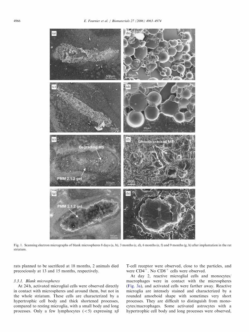
One day after implantation in the rat striatum, themicrospheres appeared as regular and smooth spheresgrouped in a cluster. At day 8, an identical image wasobserved (Fig. 1a and b), and after 3 months, microspheres
were still perfectly intact and in a cluster (Fig. 1c). Theirsurface was still totally smooth and no pores weredetectable (Fig. 1d).After 6 months, the microspheres began to degrade from
the periphery to the center of the implant site, as shown inFig. 1e. In the center, microspheres could still be observed(Fig. 1f), irregular in shape, smooth but cracked. Thosemicrospheres were surrounded by totally degraded parti-cles, which had taken a polymer gel appearance.After 9 months, only PMM 2.1.2 gel could be seen and
the microspheres had totally disappeared. The gel hadtaken the same volume in the hemisphere, compared to theinitial volume of the microspheres, which indicated that itwas not resorbed (Fig. 1g). With higher magnification,pieces of microspheres could be seen and appeared asgranular particles which seemed to ‘‘melt’’ (Fig. 1h).
3.2.2. 5-FU microspheres
5-FU microspheres degraded between day 8 and 30.They were intact at day 8, their surface was smooth andthey were grouped in a cluster like blank particles. Invarious microspheres, 5-FU crystals could be observed bymicroscopy with polarized light or with differentialinterferential contrast (DIC) (Fig. 2a). The microsphereswere totally degraded at day 30. The inoculum (Fig. 2b)was still visible and appeared as a polymer gel whichpersisted at least until day 60. With higher magnification,the polymer gel was composed of particles around 20 mm indiameter, whose surface was granular (Fig. 2c and d). Nocrystals were observed with optical microscopy (differentialinterferential contrast or polarized light).
3.3. Biocompatibility
None of the animals exhibited any overt behavioralchanges, obvious neurological deficits or weight loss,suggestive of systemic or localized toxicity from thesurgical procedure or from the microspheres. Among the
ARTICLE IN PRESS
Fig. 1. Scanning electron micrographs of blank microspheres 8 days (a, b), 3 months (c, d), 6 months (e, f) and 9 months (g, h) after implantation in the rat
striatum.
E. Fournier et al. / Biomaterials 27 (2006) 4963–49744966
rats planned to be sacrificed at 18 months, 2 animals diedprecociously at 13 and 15 months, respectively.
3.3.1. Blank microspheres
At 24 h, activated microglial cells were observed directlyin contact with microspheres and around them, but not inthe whole striatum. These cells are characterized by ahypertrophic cell body and thick shortened processes,compared to resting microglia, with a small body and longprocesses. Only a few lymphocytes (o5) expressing ab
T-cell receptor were observed, close to the particles, andwere CD4+. No CD8+ cells were observed.At day 2, reactive microglial cells and monocytes/
macrophages were in contact with the microspheres(Fig. 3a), and activated cells were farther away. Reactivemicroglia are intensely stained and characterized by arounded amoeboid shape with sometimes very shortprocesses. They are difficult to distinguish from mono-cytes/macrophages. Some activated astrocytes with ahypertrophic cell body and long processes were observed,

ARTICLE IN PRESS
Fig. 2. (a) Optical micrograph with differential interferential contrast (DIC) 8 days after implantation in the rat striatum; 5-FU crystal are visible in
various microspheres (white arrowheads); (b–d) scanning electron micrographs (SEM) of 5-FU microspheres one month after implantation in the rat
striatum; the PMM 2.1.2 gel is made of granular degrading particles (white arrows in Fig. 2c); a higher magnification shows the surface of one particle (2d).
E. Fournier et al. / Biomaterials 27 (2006) 4963–4974 4967
not directly in contact with the microspheres (Fig. 3b) butaround the macrophagous cells, as illustrated on 2 adjacentsections. A few CD4+ lymphocytes (o5) were still present.
At day 8, GFAP immunoreactivity was maximum andastrocytes formed a ring around the microspheres (Fig. 3d),directly in contact with them. Activated microglial cellswere still present, around the astrocyte ring (Fig. 3c), butno macrophagous cells could be seen. A few CD4+
lymphocytes (o5) were still present.At 1, 2 and 3 months, astrocytes only persisted as
cicatricial tracks. Activated microglial cells could stillappear at 1 month, and were perfectly quiescent at latertime points (Fig. 3e and f).
No lymphocytes could be observed. No iNOS+ cells orcells expressing MHC class II molecules were observed atthose time points.
Concerning the control animals, the reaction wascomparable to the implanted animals, with a macropha-gous/microglial reaction maximum at day 2, and anastrocytic one maximum at day 8. A few CD4+
lymphocytes (o5) were also observed at days 1, 2, and 8.The animals sacrificed after 6 months presented an
extended and pronounced inflammatory reaction in theimplantation site. Reactive microglial cells and macro-phagous cells were located around the inoculum, infiltrat-ing the polymer gel and the microspheres, and covering thewhole striatum (Fig. 4a–c). An important astrocyticreaction was also observed around the inoculum, coveringthe whole striatum, but not infiltrating the degradingpolymer and the microparticles (Fig. 4b–d). Many lym-phocytes expressing the ab T-cell receptor were detectedand were mainly CD8+ (Fig. 5a–c). CD8+ lymphocytes
were both located in blood vessels and in the tissuesurrounding the polymer gel (Fig. 5d). iNOS positive cellscould be clearly observed in contact with the microspheresand within the polymer gel, and corresponded to OX-42positive cells (Fig. 5e). MHC class II immunoreactivity wasvery strong around the implant site and was expressed byboth OX-42 cells and astrocytes (Fig. 5f).Irrespective of the antibodies tested, the control animals
at 6 months were perfectly similar to the controls at 3months (Fig. 4e and f).After 9, 12, 15 and 18 months, microspheres were totally
degraded. The tissue was very fragile, but immunostainingswere nevertheless performed. At those time points, aninflammatory and astrocytic reaction was still highlypronounced, and lymphocytes could also be observed.The brains of the animals killed after 9 months weremacroscopically normal. The brains of the animals killedafter 12 months were macroscopically normal, but tissuedestruction of the injected side was observed when cuttingthe brains. The brains of the animals killed after 15 (Fig. 6aand b) and 18 months (Fig. 6c and e) presented variousdegrees of tissue destruction. Macroscopically normalbrains with some tissue loss that was visible when cuttingthe brain or macroscopically atrophied brains with tissuedestruction could be observed. One animal at 18 monthshad lost half of one hemisphere (Fig. 6d). The controlswere normal.
3.3.2. 5-FU microspheres
From day 1 till day 8, the inflammatory and immuno-logical reactions were identical to the blank microspheresand to the controls. The microglial reaction was maximal

ARTICLE IN PRESS
Fig. 3. CD11b antigen visualized by OX-42 antibody (a, c, e) and astrocytes visualized by anti-GFAP antibody (b, d, f), 2 days (a, b), 8 days (c, d) and 3
months (e, f) after implantation of blank microspheres. Rounded cells observed at day 2 are reactive microglial cells (arrow); at day 8, cells with a
hypertrophic cell body and thick short processes are activated microglial cells (arrowhead); at 3rd month, microglial cells are quiescent, i.e. cells with a
small body and long processes; astrocytes with a hypertrophic body are activated at day 8, at a distance from the microspheres (arrow); they encircle the
implant site at day 8 (arrowhead), and are quiescent at 3 months.
E. Fournier et al. / Biomaterials 27 (2006) 4963–49744968
at day 2 with rounded reactive microglial cells andmonocytes directly in contact with the microspheres. Theastrocytic reaction was maximal at day 8 and consisted ofactivated astrocytes forming a ring around the implant site,in contact with the microspheres. After 30, 45 and 60 days,the image was comparable to the reaction obtained at 6months with blank microspheres (Fig. 7). Numerousmicroglial and macrophagous cells were observed aroundthe micropsheres and infiltrating the polymer gel. Activatedastrocytes were also numerous around the implant site, butdid not infiltrate the polymer gel.
4. Discussion
4.1. Biodegradation
Biodegradation of the microspheres was followed in vivoby SEC, and showed that blank microspheres started todegrade between 3 and 6 months. Indeed, they were
perfectly intact after 3 months, and were partially degradedafter 6 months. At this time point, in the center of theimplant site, individual microspheres could still be clearlydistinguished, with a smooth and cracked surface. At theperiphery of the implant site, no microspheres could beobserved. They appeared as a polymer gel composed ofsmall granular particles. At 9 months, only the polymer gelwas observed, which was still not resorbed at 18 months.5-FU microspheres degraded much more faster. They
were still intact at day 8, but appeared as a polymer gelafter only 1 month. The different degradation ratesobserved between blank and loaded microspheres weredue in part to the different polymer molecular weights usedin the formulation of the blank and loaded particles. Twodifferent molecular weights were used consequently to aprevious optimization work which showed that a lowermolecular weight could slow down the release of thedrug [17]. Thus, loaded microspheres were formulated witha lower PMM 2.1.2 molecular weight for in vivo

ARTICLE IN PRESS
Fig. 4. CD11b antigen visualized by OX-42 antibody (a, c) and astrocytes visualized by anti-GFAP antibody (b, d), 6 months after implantation of blank
microspheres. Reactive microglial cells infiltrate the polymer gel and the microspheres (arrow in c). Activated astrocytes do not infiltrate the polymer gel
(arrow in fig.d). Microglial cells (e) and astrocytes (f) are quiescent on the controls.
E. Fournier et al. / Biomaterials 27 (2006) 4963–4974 4969
experiments, and therefore degraded faster, as classicallyobserved in the literature. Moreover, 5-FU is a hydrophilicmolecule. Hence, water can easily dissolve 5-FU crystalslocated at the periphery of the microspheres, which leads tothe creation of pores allowing the penetration of water inthe particle core and the dissolution of deeper 5-FUcrystals. Water thus rapidly penetrates in the polymermatrix, and hydrolyzes the polymer.
4.2. Biocompatibility
During the first days post-implantation, both injectedanimals (blank or loaded microspheres) and controlsshowed a brain tissue reaction similar to the one observedfollowing a mechanical lesion to the central nervoussystem. The microglial/macrophagous reaction was max-imal at day 2, and the astrocytic reaction at day 8. FewCD4+ lymphocytes patrolling in the CNS could be
observed (o5). In the case of blank particles, theinflammatory reaction thereafter decreased until onemonth, and glial cells remained quiescent until micro-spheres began to degrade. In the case of 5-FU loadedparticles, glial cells followed the same evolution pattern atleast until day 8. These results are in accordance withprevious publications concerning various drug carriersimplanted in the striatum such as PLGA microspheres[15,24–26], poly(e-caprolactone), ethylcellulose or polystyr-ene microspheres [27], poly[bis(p-carboxyphenoxy)pro-pane] and sebacic acid implants [11–14,28] and2-hydroxyethyl methacrylate copolymer microcapsules[29,30]. For all those controlled release systems, biodegrad-able or not, the brain tissue reaction after implantationconsisted of a typical and mild foreign body reaction whichresolved between 1 and 2 months.In this study, when microspheres began to degrade, the
inflammatory reaction was reactivated. At 6 months, the

ARTICLE IN PRESS
Fig. 5. T-cell receptor (a, b), CD8b (c, d), iNOS (e) and RT1B (f) antigens visualized by anti-TCRab, anti-CD8b, anti-iNOS and OX-6 antibodies,
respectively, 6 months after implantation of blank microspheres. Most lymphocytes (arrows) stay around the polymer gel (a, b); CD8+ lymphocytes (d)
are observed in vessels (arrow) or extravasated (arrowhead). iNOS positive cells (arrow) are observed between the degrading microspheres (e). OX-6
positive cells are located around, in contact and between the degrading microspheres (f).
E. Fournier et al. / Biomaterials 27 (2006) 4963–49744970
blank microspheres were not fully degraded since severalmicrospheres could still be observed in the center of theimplant site. At the periphery, the degraded microspheresconsisted of small granular particles which seemed to fuseand/or melt and took the appearance of a gel. Microglialcells, astrocytes, CD8+ lymphocytes, iNOS and CMHclass II positive cells were numerous and surrounded theimplant site. Microglial cells and iNOS-positive cells wereeven infiltrating the polymer gel and the degrading micro-spheres in the center of the implant site. At 1 month, the5-FU microspheres were totally degraded and a similartissue reaction was observed. Control brain cells were allperfectly quiescent. Hence, polymer breakdown productsclearly induced a direct toxicological effect on thesurrounding tissue, since the tissue reaction was ‘‘normal’’as long as the microspheres stayed intact. Moreover, thedamage was irreversible since an important tissue destruc-tion was observed in all the animals sacrificed at later time
points, and could lead to a macroscopically visible cerebralatrophy, and/or to the death of the animals.PMM 2.1.2 structure is represented in Fig. 8. Two
degradation pathways were detailed in previous studies[19,20]. The first one consists of the hydrolysis of the estergroups in the side chains, generating soluble polymers,ethanol and, to a lesser extent, glycolic acid. Indeed, the esterhydrolysis leads to the formation of free carboxyl groups onthe polymer backbone which becomes more and morehydrophilic, until it is water soluble, as previously describedfor poly(alkyl cyanoacrylates) [31]. The second pathway isthe backbone degradation process occurring through areverse Knoevenagel’s reaction similar to that detailed forpoly(alkyl cyanoacrylates) by Leonard et al. [32]. Thosepolymers can degrade by hydrolytic scission of the polymerchain to form formaldehyde and an alkyl cyanoacetate. Thispathway was shown to be minor in the PMM 2.1.2degradation process, compared to ester hydrolysis [19].

ARTICLE IN PRESS
Fig. 6. Rat brains 15 months (a, b) and 18 months (c–e) after implantation of blank microspheres into the rat striatum, just after sacrifice (a, c, d) and
during the cutting (b–e) (St ¼ striatum; cc ¼ corpus callosum; CaPu ¼ caudate putamen).
E. Fournier et al. / Biomaterials 27 (2006) 4963–4974 4971
Some studies have highlighted the low in vitro [19–21]and in vivo [22] toxicity of PMM 2.1.2 nanoparticles,compared to poly(alkyl cyanoacrylates). Moreover, pre-degraded particles were shown to be 3–5 fold less toxicthan non degraded particles, and ethanol and glycolic acidwere not toxic for the cells at the concentrations tested,which were much higher than what could be expected fromthe total degradation of the nanoparticles.
In this study, a severe toxicity of the PMM 2.1.2 wasobserved, which appeared just after the polymer began todegrade. Based on the studies mentioned above, this resultwas not expected. Nevertheless, different points mightexplain our results. Firstly, the biocompatibility of abiomaterial consists on its ‘‘ability to perform with anappropriate host response in a specific application’’ [33].Hence, biocompatibility strongly depends on the adminis-tration site. PMM 2.1.2 nanoparticles previously tested invivo [22] were injected intravenously to mice and rats, andassessed for acute toxicity. Organs were examined only 48 hpost-injection. Thus, nanoparticles did not induce anyshort-term pathological reaction, but the long-term effects
were not monitored. Secondly, cells tested in vitro werefibroblasts or Caco-2 cells, which are less sensitive thanneurons and even glial cells. Indeed, for our application, amore convenient in vitro cytotoxicity testing schedulewould have used a primary neuronal culture also contain-ing astrocytes, oligodendrocytes and microglial cells, sincea material should be evaluated on the cells with whom itwill be in contact in vivo.The soluble polymers generated by the ester hydrolysis
might have been responsible for the direct toxicity on cells,since they are acidic entities which seemed to resorb veryslowly in the cerebral tissue. The polymer degradationcontinuously generated acidic polyanions slowly resorbingand constituting a permanent inflammatory stimulus.Thereafter, the inflammatory and immunological reactioncontributed to the cytotoxic damage caused to the cells.Indeed, several iNOS-positive cells were observed, indicat-ing an ultimate activation state of the macrophagous cells.iNOS is an inducible nitric oxide-synthase which is notexpressed by CNS cells under physiological conditions. It isinduced in non-neuronal cells during various types of CNS

ARTICLE IN PRESS
Fig. 7. CD11b (a), astrocytes (b) and T-cell receptor (c) antigens
visualized by OX-42, anti-GFAP and anti-TCRab antibodies, respectively,
1 month after implantation of 5-FU microspheres. Reactive microglial
cells (arrow) infiltrate the polymer gel (a); activated astrocytes and
lymphocytes (arrows) stay around the implant site (b, c).
CH2
R1
R2
n
O
O
O
O
CH3
R1 =
O
O
CH3
R2 =
Fig. 8. Poly(methylidene malonate 2.1.2) chemical structure.
E. Fournier et al. / Biomaterials 27 (2006) 4963–49744972
injuries or diseases. Cells expressing iNOS in this case aremacrophages, microglia and astrocytes [34]. This enzymeallows the synthesis of the nitric oxide free radical gaswhich contributes to the elimination of bacteria andpathogens. Excess amounts can damage host cells, causingneurotoxicity [35]. In this study, iNOS was not expressedjust after implantation, which indicated a mild foreignbody reaction. However, during polymer degradation, NOsynthesis was detected and might be responsible for cellulartoxicity. Moreover, the potential second degradation
pathway of the polymer may become more important inthe brain tissue, and thus lead to a larger production of thetoxic compound formaldehyde.While degrading, the polymer also induced a specific and
pronounced immunologic reaction with many lymphocytesand a strong expression of MHC class II molecules. Aspecific immune response is usually observed following theintroduction of a foreign antigen in the body. For T-cells tobe stimulated, the foreign agent must be presented to themon the surface of an appropriate cell (antigen presentingcell (APC)) with a MHC molecule [36]. Synthetic polymersdo not constitute peptidic or glycolipid antigens, and thuscannot be processed by APC and presented to T-cells orNK T-cells (natural killers). Hence, other mechanismsmight be involved in the reaction observed in this study. Afirst hypothesis consists of the modification of endogenousproteins by the polymer, via hydrophobic interactions.Once denatured, proteins are generally not recognized bythe immune system. They are endocytosed, presented byAPC to specific T-cells, and activated T-cell clones maythus possess specificity against self-proteins, which maylead to cell lysis and death [36,37]. This hypothesis is

ARTICLE IN PRESSE. Fournier et al. / Biomaterials 27 (2006) 4963–4974 4973
supported by the presence of CD8+ lymphocytes. Indeed,exogenous antigen are generally processed and presentedvia the MHC class II pathway, while endogenous antigensuse the MHC class I pathway, which activates the cytotoxicT-lymphocytes (CD8+) [38,39]. Thus, we can suppose thatthe polymer altered endogenous proteins, involving theMHC class I pathway and activating CD8+ T-cells.
A second hypothesis is that polymer breakdownproducts can bind to appropriate carrier molecules in thesurrounding tissue, and become antigenic. Besides, theslowly degrading polymer constitutes a persistent source ofreaction products, which may induce the development ofan immunological memory [40].
Concerning the tissue destruction observed at long timepoints, different mechanisms can be proposed: firstly,activated CD8+ lymphocytes may be specific of selfproteins, leading to cell lysis, as mentioned above.Secondly, if phagocytic cells fail to engulf the foreignmaterial, they can nevertheless get close enough to causedegranulation. Enzymes are thus released in the surround-ing tissue causing destruction and promoting the inflam-matory reaction. Finally, macrophagous cells mayphagocyte small particles of the degrading polymer. Ifphagolysosomes fail to destroy the particles, or if theparticles of polymer are toxic for the cells, macrophagescan die and release highly toxic products in the surround-ing tissue such as oxygenated radicals and powerfulenzymes [40].
5. Conclusion
Blank and loaded PMM 2.1.2-based microspheres wereimplanted in rat brain in order to evaluate their biode-gradation and their biocompatibility. Microspheres firstappeared as a promising system, since they exhibited a slowdegradation rate. Indeed, blank microspheres were stillperfectly intact at 3 months. However, when the polymerstarted to degrade, a strong inflammatory and immunolo-gical reaction developed, leading to an important cell loss,a cerebral atrophy and to the death of several animals.Polymer degradation products thus generated directtoxicological effect on the brain tissue, and an interestingspecific immunological response. Further experiments arerequired to determine the potential toxic compounds, andto elucidate the mechanisms involved in the immunologicalreaction.
Acknowledgments
The authors want to thank Dr. A. Clavreul (InsermU-646, Angers, France) for fruitful discussions, and Dr.R. Filmon and M. Lesourd (Service commun de micro-scopie electronique, Universite d’Angers, France) for SEManalysis.
Appendix A. Supplementary materials
Supplementary data associated with this article can befound in the online version at doi:10.1016/j.biomaterials.2006.04.045.
References
[1] Benoit JP, Faisant N, Venier-Julienne MC, Menei P. Development of
microspheres for neurological disorders: from basics to clinical
applications. J Control Release 2000;65:285–96.
[2] Menei P, Benoit JP, Boisdron-Celle M, Fournier D, Mercier P, Guy
G. Drug targeting into the central nervous system by stereotactic
implantation of biodegradable microspheres. Neurosurgery 1994;34:
1058–64.
[3] Brem H, Gabikian P. Biodegradable polymer implants to treat brain
tumors. J Control Release 2001;74:63–7.
[4] Gutman RL, Peacock G, Lu DR. Targeted drug delivery for brain
cancer treatment. J Control Release 2000;65:31–41.
[5] Gentile FT, Doherty EJ, Rein DH, Shoichet MS, Winn SR. Polymer
science for macroencapsulation of cells for central nervous system
transplantation. Reactive Polym 1995;25:207–27.
[6] Fung LK, Saltzman WM. Polymeric implants for cancer chemother-
apy. Adv Drug Deliv Rev 1997;26:209–30.
[7] Menei P, Montero-Menei C, Venier-Julienne MC, Benoit JP. Drug
delivery into the brain using poly(lactide-co-glycolide) microspheres.
Expert Opin Drug Deliv 2005;2:363–76.
[8] Brem H, Piantadosi S, Burger PC, Walker M, Selker R, Vick NA,
et al. Placebo-controlled trial of safety and efficacy of intraoperative
controlled delivery by biodegradable polymers of chemotherapy for
recurrent gliomas. Lancet 1995;345:1008–12.
[9] Menei P, Venier MC, Gamelin E, Saint-Andre JP, Hayek G, Jadaud
E, et al. Local and sustained delivery of 5-fluorouracil from
biodegradable microspheres for the radiosensitization of glioblasto-
ma: a pilot study. Cancer 1999;86:325–30.
[10] Fournier E, Passirani C, Montero-Menei C, Benoit JP. Biocompat-
ibility of implantable synthetic polymeric drug carriers: focus on
brain biocompatibility. Biomaterials 2003;24:3311–31.
[11] Brem H, Kader A, Epstein JI, Tamargo RJ, Domb A, Langer R,
et al. Biocompatibility of a biodegradable, controlled-release polymer
in the rabbit brain. Select Cancer Therap 1989;5:55–65.
[12] Brem H, Domb A, Lenartz D, Dureza C, Olivi A, Epstein JI. Brain
biocompatibility of a biodegradable controlled release polymer
consisting of anhydride copolymer of fatty acid dimer and sebacic
acid. J Control Release 1992;19:325–9.
[13] Tamargo RJ, Epstein JI, Reinhard CS, Chasin M, Brem H. Brain
biocompatibility of a biodegradable, controlled-release polymer in
rats. J Biomed Mater Res 1989;23:253–66.
[14] Olivi A, Kim KD, Benedetti L, Epstein JI, Pinn ML, Callegaro L,
et al. Ocular and brain biocompatibility of polymeric delivery
materials prepared from ester derivatives of hyaluronic acid. Clin
Mater 1993;13:163–8.
[15] Menei P, Daniel V, Montero-Menei C, Brouillard M, Pouplard-
Barthelaix A, Benoit JP. Biodegradation and brain tissue reaction to
poly(D,L-lactide-co-glycolide) microspheres. Biomaterials 1993;14:
470–8.
[16] Veziers J, Lesourd M, Jollivet C, Montero-Menei C, Benoit JP,
Menei P. Analysis of brain biocompatibility of drug-releasing
biodegradable microspheres by scanning and transmission electron
microscopy. J Neurosurg 2001;95:489–94.
[17] Fournier E, Passirani C, Colin N, Breton P, Sagodira S, Benoit JP.
Development of novel 5-FU-loaded poly(methylidene malonate
2.1.2)-based microspheres for the treatment of brain cancers. Eur
J Pharm Biopharm 2004;57:189–97.

ARTICLE IN PRESSE. Fournier et al. / Biomaterials 27 (2006) 4963–49744974
[18] Fournier E, Passirani C, Montero-Menei C, Colin N, Breton P,
Sagodira S, et al. Therapeutic effectiveness of novel 5-FU-loaded
poly(methylidene malonate 2.1.2)-based microspheres on F98 glioma-
bearing rats. Cancer 2003;97:2822–9.
[19] Lescure F, Seguin C, Breton P, Bourrinet P, Roy D, Couvreur P.
Preparation and characterization of novel poly(methylidene malonate
2.1.2.)-made nanoparticles. Pharm Res 1994;11:1270–7.
[20] Breton P, Roy D, Marchal-Heussler L, Seguin C, Couvreur P,
Lescure F. New poly(methylidene malonate 2.1.2) nanoparticles:
recent developments. In: Gregoriadis G, editor. Targeting of drugs.
New York, 1994. p. 161–172.
[21] Le Visage C, Couvreur P, Mysiakine E, Breton P, Bru N, Fattal E. In
vitro and in vivo evaluation of poly(methylidene malonate 2.1.2)
microparticles behavior for oral administration. J Drug Target
2001;9:141–53.
[22] Breton P, Seguin C, Roy D, Couvreur P, Lescure F. In vivo
evaluation of poly(methylidene malonate 2.1.2) nanoparticle toxicity.
In: Proceedings of the controlled release society 1994. p. 608–9.
[23] Paxinos G, Watson C. The rat brain in stereotaxic coordinates.
New York: Academic Press; 1986.
[24] Chen W, He J, Olson JJ, Lu DR. Carboplatin-loaded PLGA
microspheres for intracerebral implantation: In vivo characterization.
Drug Deliv 1997;4:301–11.
[25] Chen W, He J, Olson JJ, Lu DR. Direct intracerebral delivery of
carboplatin from PLGA microspheres against experimental malig-
nant glioma in rats. Drug Deliv 1998;5:101–10.
[26] Emerich DF, Tracy MA, Ward KL, Figueiredo M, Qian R, Henschel
C, et al. Biocompatibility of poly (DL-lactide-co-glycolide) micro-
spheres implanted into the brain. Cell Transp 1999;8:47–58.
[27] Menei P, Croue A, Daniel V, Pouplard-Barthelaix A, Benoit JP. Fate
and biocompatibility of three types of microspheres implanted into
the brain. J Biomed Mater Res 1994;28:1079–85.
[28] Brem H, Tamargo RJ, Olivi A, Pinn M, Weingart JD, Wharam M,
et al. Biodegradable polymers for controlled delivery of chemother-
apy with and without radiation therapy in the monkey brain.
J Neurosurg 1994;80:283–90.
[29] Campioni EG, Nobrega JN, Sefton MV. HEMA/MMMA micro-
capsule implants in hemiparkinsonian rat brain: biocompatibility
assessment using [3H]PK11195 as a marker for gliosis. Biomaterials
1998;19:829–37.
[30] Mokry J, Karbanova J, Lukas J, Paleckova V, Dvorankova B.
Biocompatibility of HEMA copolymers designed for treatment of
CNS diseases with polymer-encapsulated cells. Biotechnol Progr
2000;16:897–904.
[31] Lenaerts V, Couvreur P, Christiaens-Leyh D, Joiris E, Roland M,
Rollman B, et al. Degradation of poly(isobutyl cyanoacrylate)
nanoparticles. Biomaterials 1984;5:65–8.
[32] Leonard F, Kulkarni RK, Brandes G, Nelson J, Cameron JJ.
Synthesis and degradation of poly(alkyl alpha-cyanoacrylates).
J Appl Polym Sci 1966;10:259–72.
[33] Williams DF. Definitions in biomaterials. In: Williams DF, editor.
Progress in biomedical engineering. Amsterdam, 1987.
[34] Garcion E, Sindji L, Montero-Menei C, Andre C, Brachet P, Darcy
F. Expression of inducible nitric oxide synthase during rat brain
inflammation: regulation by 1,25-dihydroxyvitamin D3. Glia
1998;22:282–94.
[35] Lowenstein CJ, Dinerman JL, Snyder SH. Nitric oxide: a physiologic
messenger. Ann Intern Med 1994;120:227–37.
[36] Remes A, Williams DF. Immune response in biocompatibility.
Biomaterials 1992;13:731–43.
[37] Rihova B. Biocompatibility of biomaterials: Hemocompatibility,
immunocompatibility and biocompatibility of solid polymeric mate-
rials and soluble targetable polymeric carriers. Adv Drug Deliv Rev
1996;21:157–76.
[38] Kuby J. Antigen processing and presentation. In: Freeman
WH and Company, editors. Immunology. New York, 1994.
p. 239–50.
[39] Newman KD, Elamanchili P, Kwon GS, Samuel J. Uptake of
poly(D,L-lactic-co-glycolic acid) microspheres by antigen-presenting
cells in vivo. J Biomed Mater Res 2002;60:480–6.
[40] Williams DF. Tissue-biomaterial interactions. J Mater Sci 1987;22:
3421–45.