Regulation of sialic acid 9-O-acetylation during the growth and differentiation of murine...
10
Regulation of Sialic Acid 9-O-Acetylation during the Growth and Differentiation of Murine Erythroleukemia Cells* (Received for publication, September 3, 1996) Wei-Xing Shi, Roger Chammas‡, and Ajit Varki§ From the Glycobiology Program, UCSD Cancer Center, the Division of Cellular and Molecular Medicine, University of California, San Diego, La Jolla, California 92093 Sialic acids are typically found at the terminal posi- tion on vertebrate oligosaccharides. They are sometimes modified by an O-acetyl ester at the 9-position, poten- tially altering recognition of sialic acid by antibodies, lectins, and viruses. 9-O-Acetylation is known to be se- lectively expressed on gangliosides in melanoma cells and on N-linked chains in hepatocytes. Using a recently developed probe, we show here that in murine erythro- leukemia cells, this modification is selectively expressed on another class of oligosaccharides, O-linked chains carried on cell surface sialomucins. These cells also ex- press 9-O-acetylation on the ganglioside G D3 , but this modification appears to be undetectable on the cell sur- face. Increasing cell density in culture is associated with a decrease in cell surface 9-O-acetylation of sialomucins. This change correlates with the spontaneous differenti- ation toward a mature erythroid phenotype. This down- regulation upon differentiation and entry into the G 0 /G 1 stage of the cell cycle is confirmed by differentiation- inducing agents. In contrast, cells arrested in G 2 /M by the microtubule depolymerizing agent nocodazole show increased expression of cell surface 9-O-acetylated sia- lomucins (but not the 9-O-acetylated ganglioside). How- ever, the microtubule stabilizer taxol does not induce this increase, showing that the nocodazole effect is in- dependent of cell cycle stage. Indeed, direct analysis showed no correlation of 9-O-acetylation with cell cycle stage in rapidly growing cells, and shorter treatments with nocodazole also increased expression. Western blots of cell extracts confirmed that changes caused by differentiation and nocodazole are not due to redistri- bution of molecules from the cell surface. Indeed, follow- ing selective removal of 9-O-acetyl groups from the cell surface by a specific esterase, the recovery of expression is mediated by new synthesis rather than by redistribu- tion from an internal pool. Thus, 9-O-acetylation on these sialomucins appears to be primarily regulated by the rate of synthesis, and the increase with nocodazole treatment is likely due to the inhibition of turnover of cell surface molecules. These data show that 9-O-acety- lation of sialic acids in murine erythroleukemia cells is a highly regulated modification, being selectively ex- pressed in a cell type-specific manner on certain classes of oligosaccharides and differentially regulated with re- gard to subcellular localization and to the state of cellu- lar differentiation. The sialic acids (Sias) 1 are a family of 9-carbon monosaccha- rides usually found as the terminal units of animal oligosac- charides (1). The most common modifications of Sias are O- acetyl substitutions at the C-7 or C-9 positions (2, 3). Since O-acetyl esters at C-7 position spontaneously migrate to the C-9 position at physiological extracellular pH (4, 5), 9-O-acety- lated Sias predominate on cell surface glycoconjugates. Several observations show that these modifications can affect the phys- icochemical and biological properties of the parent molecule (2, 3), affecting the activity of microbial sialidases (6), the speci- ficity of sialyloligosaccharide recognition by viruses (7, 8), rec- ognition by mammalian lectins and monoclonal antibodies (9, 10), and potentially altering activation of the alternative com- plement pathway (11, 12). These modifications also show re- markable tissue-specific, molecule-specific, and developmen- tally regulated expression in a variety of systems. For example, structural studies of different O-acetylated gangliosides from melanoma cells have shown that the 9-O-acetyl group is invari- ably located on a particular terminal a2,8-linked Sia originat- ing from the b1,4-linked galactose of lactosylceramide (13, 14); in the same cell type, Sias on glycoproteins do not appear to be O-acetylated (15). In contrast, rat hepatocytes express 9-O- acetylation selectively on the Sia residues of N-linked sugar chains (16). Since the O-acetylation of Sias takes place after transfer of Sias to glycoconjugates (2, 3), these findings suggest that this reaction may be regulated in a molecule- and tissue- specific fashion. Other studies have shown O-acetylated Sias as distinct markers for human lymphocyte subsets (10, 17–21), and vari- able expression has been reported in human leukemic cells (22). Murine erythroleukemia (MEL) cells are virus-trans- formed erythroid precursors that rapidly proliferate in culture (23). These cell lines have served as classic models for in vitro differentiation along the erythrocyte pathway (23), which can be initiated by a number of agents and results in decreased cell size, restricted proliferative capacity, expression of mature erythrocyte antigens, and terminal differentiation (24 –26). Be- cause of these characteristics, MEL cells are useful to investi- gate the mechanisms by which proliferating transformed pre- cursor cells in a differentiation lineage withdraw from the cell division cycle and express the genes characteristic of the nor- * This research was supported by Grants RO1-GM32373 (to A. V.) and P01-CA5869 (to M. G. Farquhar). The costs of publication of this article were defrayed in part by the payment of page charges. This article must therefore be hereby marked “advertisement” in accordance with 18 U.S.C. Section 1734 solely to indicate this fact. ‡ Supported by Postdoctoral Fellowship 200015/94-0 from the Con- selho Nacional de Desenvolvimento Cientı ´fico e Tecnolo ´gico, Brasil. § To whom correspondence should be addressed. Tel.: 619-534-3296; Fax: 619-534-5611. 1 The abbreviations used are: Sia, sialic acid, type unspecified; mAb, monoclonal antibody; HPTLC, high performance thin layer chromatog- raphy; PBS, phosphate-buffered saline; CHE-Fc, chimeric protein made of InfCHE (influenza C hemagglutinin-esterase with the fusion peptide eliminated by mutation) and the Fc portion of human IgG 1 ; CHE-FcD, DFP-treated CHE-Fc (esterase activity irreversibly inactivated); HMBA, hexamethylene-bisacetamide; Me 2 SO, dimethyl sulfoxide; MEL, murine erythroleukemia cells; DFP, diisopropyl fluorophosphate; BSA, bovine serum albumin; OSGPase, O-sialoglycoprotease; PAGE, polyacrylamide gel electrophoresis; ELISA, enzyme-linked immunosor- bent assay; FITC, fluorescein isothiocyanate; PNGase, peptide-N-gly- cosidase F; BKG, background. THE JOURNAL OF BIOLOGICAL CHEMISTRY Vol. 271, No. 49, Issue of December 6, pp. 31517–31525, 1996 © 1996 by The American Society for Biochemistry and Molecular Biology, Inc. Printed in U.S.A. This paper is available on line at http://www-jbc.stanford.edu/jbc/ 31517 by guest on June 17, 2016 http://www.jbc.org/ Downloaded from
-
Upload
independent -
Category
Documents
-
view
0 -
download
0
Transcript of Regulation of sialic acid 9-O-acetylation during the growth and differentiation of murine...

Regulation of Sialic Acid 9-O-Acetylation during the Growth andDifferentiation of Murine Erythroleukemia Cells*
(Received for publication, September 3, 1996)
Wei-Xing Shi, Roger Chammas‡, and Ajit Varki§
From the Glycobiology Program, UCSD Cancer Center, the Division of Cellular and Molecular Medicine, University ofCalifornia, San Diego, La Jolla, California 92093
Sialic acids are typically found at the terminal posi-tion on vertebrate oligosaccharides. They are sometimesmodified by an O-acetyl ester at the 9-position, poten-tially altering recognition of sialic acid by antibodies,lectins, and viruses. 9-O-Acetylation is known to be se-lectively expressed on gangliosides in melanoma cellsand on N-linked chains in hepatocytes. Using a recentlydeveloped probe, we show here that in murine erythro-leukemia cells, this modification is selectively expressedon another class of oligosaccharides, O-linked chainscarried on cell surface sialomucins. These cells also ex-press 9-O-acetylation on the ganglioside GD3, but thismodification appears to be undetectable on the cell sur-face. Increasing cell density in culture is associated witha decrease in cell surface 9-O-acetylation of sialomucins.This change correlates with the spontaneous differenti-ation toward a mature erythroid phenotype. This down-regulation upon differentiation and entry into the G0/G1stage of the cell cycle is confirmed by differentiation-inducing agents. In contrast, cells arrested in G2/M bythe microtubule depolymerizing agent nocodazole showincreased expression of cell surface 9-O-acetylated sia-lomucins (but not the 9-O-acetylated ganglioside). How-ever, the microtubule stabilizer taxol does not inducethis increase, showing that the nocodazole effect is in-dependent of cell cycle stage. Indeed, direct analysisshowed no correlation of 9-O-acetylation with cell cyclestage in rapidly growing cells, and shorter treatmentswith nocodazole also increased expression. Westernblots of cell extracts confirmed that changes caused bydifferentiation and nocodazole are not due to redistri-bution of molecules from the cell surface. Indeed, follow-ing selective removal of 9-O-acetyl groups from the cellsurface by a specific esterase, the recovery of expressionis mediated by new synthesis rather than by redistribu-tion from an internal pool. Thus, 9-O-acetylation onthese sialomucins appears to be primarily regulated bythe rate of synthesis, and the increase with nocodazoletreatment is likely due to the inhibition of turnover ofcell surface molecules. These data show that 9-O-acety-lation of sialic acids in murine erythroleukemia cells isa highly regulated modification, being selectively ex-pressed in a cell type-specific manner on certain classesof oligosaccharides and differentially regulated with re-gard to subcellular localization and to the state of cellu-lar differentiation.
The sialic acids (Sias)1 are a family of 9-carbon monosaccha-rides usually found as the terminal units of animal oligosac-charides (1). The most common modifications of Sias are O-acetyl substitutions at the C-7 or C-9 positions (2, 3). SinceO-acetyl esters at C-7 position spontaneously migrate to theC-9 position at physiological extracellular pH (4, 5), 9-O-acety-lated Sias predominate on cell surface glycoconjugates. Severalobservations show that these modifications can affect the phys-icochemical and biological properties of the parent molecule (2,3), affecting the activity of microbial sialidases (6), the speci-ficity of sialyloligosaccharide recognition by viruses (7, 8), rec-ognition by mammalian lectins and monoclonal antibodies (9,10), and potentially altering activation of the alternative com-plement pathway (11, 12). These modifications also show re-markable tissue-specific, molecule-specific, and developmen-tally regulated expression in a variety of systems. For example,structural studies of different O-acetylated gangliosides frommelanoma cells have shown that the 9-O-acetyl group is invari-ably located on a particular terminal a2,8-linked Sia originat-ing from the b1,4-linked galactose of lactosylceramide (13, 14);in the same cell type, Sias on glycoproteins do not appear to beO-acetylated (15). In contrast, rat hepatocytes express 9-O-acetylation selectively on the Sia residues of N-linked sugarchains (16). Since the O-acetylation of Sias takes place aftertransfer of Sias to glycoconjugates (2, 3), these findings suggestthat this reaction may be regulated in a molecule- and tissue-specific fashion.Other studies have shown O-acetylated Sias as distinct
markers for human lymphocyte subsets (10, 17–21), and vari-able expression has been reported in human leukemic cells(22). Murine erythroleukemia (MEL) cells are virus-trans-formed erythroid precursors that rapidly proliferate in culture(23). These cell lines have served as classic models for in vitrodifferentiation along the erythrocyte pathway (23), which canbe initiated by a number of agents and results in decreased cellsize, restricted proliferative capacity, expression of matureerythrocyte antigens, and terminal differentiation (24–26). Be-cause of these characteristics, MEL cells are useful to investi-gate the mechanisms by which proliferating transformed pre-cursor cells in a differentiation lineage withdraw from the celldivision cycle and express the genes characteristic of the nor-
* This research was supported by Grants RO1-GM32373 (to A. V.)and P01-CA5869 (to M. G. Farquhar). The costs of publication of thisarticle were defrayed in part by the payment of page charges. Thisarticle must therefore be hereby marked “advertisement” in accordancewith 18 U.S.C. Section 1734 solely to indicate this fact.‡ Supported by Postdoctoral Fellowship 200015/94-0 from the Con-
selho Nacional de Desenvolvimento Cientıfico e Tecnologico, Brasil.§ To whom correspondence should be addressed. Tel.: 619-534-3296;
Fax: 619-534-5611.
1 The abbreviations used are: Sia, sialic acid, type unspecified; mAb,monoclonal antibody; HPTLC, high performance thin layer chromatog-raphy; PBS, phosphate-buffered saline; CHE-Fc, chimeric protein madeof InfCHE (influenza C hemagglutinin-esterase with the fusion peptideeliminated by mutation) and the Fc portion of human IgG1; CHE-FcD,DFP-treated CHE-Fc (esterase activity irreversibly inactivated);HMBA, hexamethylene-bisacetamide; Me2SO, dimethyl sulfoxide;MEL, murine erythroleukemia cells; DFP, diisopropyl fluorophosphate;BSA, bovine serum albumin; OSGPase, O-sialoglycoprotease; PAGE,polyacrylamide gel electrophoresis; ELISA, enzyme-linked immunosor-bent assay; FITC, fluorescein isothiocyanate; PNGase, peptide-N-gly-cosidase F; BKG, background.
THE JOURNAL OF BIOLOGICAL CHEMISTRY Vol. 271, No. 49, Issue of December 6, pp. 31517–31525, 1996© 1996 by The American Society for Biochemistry and Molecular Biology, Inc. Printed in U.S.A.
This paper is available on line at http://www-jbc.stanford.edu/jbc/ 31517
by guest on June 17, 2016http://w
ww
.jbc.org/D
ownloaded from

mal differentiated phenotype. Sia O-acetylation has also beenreported on murine erythrocytes (11, 27, 28) and on MEL cells(29). However, the class of sugar chains (gangliosides, N-linkedor O-linked) to which these O-acetyl groups were attached wasnot known. Recently, murine erythrocytes have been shown tobear ligands for sialoadhesin, a macrophage-specific I-type lec-tin known to selectively recognize the Siaa2–3Galb1–3GalNActerminal structure that can occur on either O-linked chains organgliosides (30). In this case, de-O-acetylation increased bind-ing (31), suggesting that at least a portion of the target struc-tures might be masked by O-acetyl groups.With the exception of certain ganglioside antigens recog-
nized by specific monoclonal antibodies (32–35), it was previ-ously not possible to detect 9-O-acetyl sialic acids on cell sur-faces without resorting to biochemical analyses. We and others(20, 36, 37) have suggested the use of the whole influenza Cvirion as a probe for this purpose. This virus expresses amembrane-bound hemagglutinin-esterase (CHE), which canboth detect and cleave 9-O-acetyl residues on sialic acids, re-gardless of the underlying oligosaccharide structure to whichthey are attached. However, the intact influenza C virions areunstable, are subject to steric hindrance in binding, and areimpractical for many applications. To address this deficiency,we created a recombinant soluble chimera of the influenza Chemagglutinin-esterase fused to the hinge and Fc regions ofhuman IgG1 (38). The purified molecule (CHE-Fc) retains thespecific 9-O-acetyl esterase activity of the parent viral glyco-protein (38). Irreversible inactivation of the enzyme activitywith DFP gives the derivative CHE-FcD, which serves as aspecific probe for detection of 9-O-acetylated Sias. Using theseprobes, we demonstrated the widespread but selective expres-sion of 9-O-acetylation in various rat tissues (39) and occur-rence of 9-O-acetyl groups masking sialylated ligands for theB-cell-specific lectin CD22 (10). We have now used these re-agents to explore the expression, regulation, and turnover of9-O-acetyl groups during growth and differentiation in MELcells.
EXPERIMENTAL PROCEDURES
Materials—Most of the materials used were obtained from Sigma.The following materials were obtained from other sources as indicated.Biotin-conjugated Lc-hydrazide and fluorescein isothiocyanate-conju-gated goat anti-mouse IgG Ab was from Pierce; proteinase K was fromLife Technologies, Inc.; alkaline phosphatase-conjugated streptavidinand alkaline phosphatase substrate package were from Life Technolo-gies, Inc.; peroxidase-conjugated goat anti-mouse IgG Ab and alkalinephosphatase-conjugated goat anti-human IgG Ab were from Bio-Rad;fluorescein isothiocyanate-conjugated goat anti-human IgG was fromCalTag Laboratories; propidium iodide was from Calbiochem; diisopro-pyl fluorophosphate (DFP) was from Aldrich; fetal calf serum was fromHyclone; peptide:N-glycosidase F was from Genzyme; high performancethin layer chromatography (HPTLC) plates (silica Gel-60 10 3 10 cm)was from Merck. Protein assays were determined with the bicincho-ninic acid protein assay reagent kit (Pierce) using BSA as a standard.The O-sialoglycoprotease enzyme was a kind gift from Dr. Alan Mellors,University of Guelph, Canada. All other chemicals were of reagentgrade or better and were obtained from commercial sources.Cell Lines and Monoclonal Antibodies—Murine erythroleukemia
(MEL) cells were obtained from Dr. George Palade, UCSD (24, 40), andcultured in RPMI 1640 with 10% heat-inactivated fetal calf serum.Human melanoma cells (Melur) were from David Cheresh, ScrippsResearch Institute, and were propagated in Dulbecco’s modified Eagle’smedium supplemented with 10% fetal calf serum. The mouse IgG3
anti-GD3 monoclonal antibody R-24 (American Type Culture Collec-tion), and the IgG3 anti-9-O-acetyl-GD3 monoclonal antibody 27A (M.Farquhar, UCSD) were used as the hybridoma supernatants.Chimeric CHE-Fc and CHE-FcD—The soluble chimeric protein
CHE-Fc, consisting of the extracellular domain of influenza C hemag-glutinin-esterase fused to Fc portion of human IgG1, was generated andcharacterized as described elsewhere (38). The modified form CHE-FcDwas generated by treating CHE-Fc with 1 mM diisopropyl fluorophos-phate to irreversibly inactivate the esterase as described (38). CHE-Fc
specifically releases 9-O-acetyl esters from Sias, whereas CHE-FcDspecifically recognizes and binds to 9-O-acetylated Sias (38).Extraction of Gangliosides from Cultured Cells—Washed cell pellets
were resuspended in 3 volumes of ice-cold deionized water, homoge-nized at 4 °C, and the homogenate was added dropwise to 10.6 volumesof methanol at room temperature under constant stirring. Chloroform(5.3 volumes) was then added to the suspension. After centrifugation,the supernatant was collected and adjusted to a final chloroform/meth-anol/water ratio of 4:8:5.6 (v/v/v). After phase separation, gangliosideswere enriched in the hydrophilic upper phase, which was dried down,resuspended in methanol, and kept at 220 °C until use.Extraction of Proteins from Cultured Cells—Washed cell pellets were
resuspended into a buffer containing 50 mM Tris (pH 7.5), 150 mMNaCl,1% Triton X-100, and protease inhibitors (1 mM EDTA, 50 mg/ml leu-peptin, and 4 mg/ml pepstatin) and incubated 10 min at room temper-ature with occasional mixing. After centrifugation, the supernatant wassaved for analysis.O-Sialoglycoprotease Treatments—Proteins (40-mg aliquots) from
MEL cells were incubated with 2.5 milliunits of enzyme in 100 mM
HEPES (pH 7.4), in the presence of BSA (stabilizer) for 2 h at 37 °C. Fortreatment of cell surface molecules, MEL cells (100 ml, 106 cells/ml) inHanks’ balanced saline solution, 1% fetal calf serum, 0.1% NaN3, and 10mM Neu2en5Ac (sialidase inhibitor) were incubated for 2 h at 37 °C inthe presence or absence of 5 milliunits ofO-sialoglycoprotease. The cellswere then washed with Hanks’ balanced saline solution,, 1% fetal calfserum, 0.1% NaN3 prior to immunostaining.Trypsin Treatment—Cells were washed three times with ice-cold
PBS and finally resuspended in 0.5 ml of PBS containing 0.1% NaN3
and 1% trypsin and incubated at 37 °C for 1 h. The trypsin was inacti-vated by adding an excess of medium containing fetal calf serum. Thecells were washed with staining buffer prior to immunostaining.PNGase F Treatment—Protein samples were denatured as described
(42) and treated with 1 milliunit of PNGase F and incubated overnightat 37 °C. The enzyme was thereafter heat-inactivated for 3 min at100 °C.Removal of 9-O-Acetyl Esters from Cell Surface Sias—2 3 106 MEL
cells were resuspended in 100 ml of PBS, containing 0.02% NaN3 (con-trol cells) or 100 ml of PBS, containing 0.02% NaN3 with 10 mg ofCHE-Fc. After incubation at 37 °C for 60 min, the cells were washed 33in ice-cold PBS and then stained for flow cytometry analysis as de-scribed below. For studies requiring pulse-chase analysis, the incuba-tions were done in Hanks’ balanced saline solution, and the cells wereplaced back in full culture medium for varying chase periods.SDS-PAGE and Western Blot Analysis—Proteins were separated by
SDS-PAGE in 7.5% polyacrylamide gels, transferred to Immobilon-Pmembranes (Millipore), and the blots incubated overnight at 4 °C withCHE FcD (10 mg/ml) in PBS, 1% BSA. Glycoproteins reacting with thechimera were colorimetrically detected with a goat anti-human IgGantibody conjugated with alkaline phosphatase.ELISA Plate Assays for 9-O-Acetyl Gangliosides—Total ganglioside
extracts prepared as above were studied by lipid ELISA as describedpreviously (15, 41). Lipids were applied to the plate in 45% methanol,allowed to dry, and then blocked with 5% BSA in PBS overnight. Theeffects of base on reactivity were assessed by treatment with 0.1 M
NaOH at 4 °C for 30 min. After treatment, the plates were extensivelywashed with PBS, blocked with 2% BSA in PBS for 1 h, and thenincubated with the mAbmixture for 2 h. After washing three times with1% BSA, a mixture of horseradish peroxidase-conjugated goat anti-mouse IgG and goat anti-human IgG antibodies was added (each at1:1000 dilutions) for 1 h. After washing, the reaction was developed asdescribed. Background levels determined with the secondary antibodyalone were subtracted in all cases.HPTLC Immuno-overlay of Gangliosides—Gangliosides were sepa-
rated on aluminum-backed Silica Gel-60 HPTLC plates; the plates wereplasticized and overlaid with antibodies as described previously (15,41). Horseradish peroxidase-conjugated goat anti-mouse IgG recogniz-ing mouse IgG 1:2000 (27A) was reacted with the plate for 1 h at 4 °C.After washing, the reaction was developed with diaminobenzidine.Treatment of MEL Cells with Biological Modulators—MEL cells were
routinely grown to ;90% confluency before splitting for passage. Cellswere used during asynchronous near-confluent growth or were arrestedat the G2/M stages of the cell cycle using nocodazole (1 mg/ml) or Taxol(1 mg/ml) for varying periods of time. Optimal enrichment of G2/M-arrested cells was obtained when the anti-microtubule agents wereadded to cells at 50% confluency. The percentage of G2/M cells wasdetermined by flow cytometric cell cycle analysis as described below.Synchronization of cells at the G1/S boundary was carried out by incu-bating near-confluent MEL cells with thymidine (5 mg/ml, 24 h). The
O-Acetylated Sialic Acids in Leukemia Cells31518
by guest on June 17, 2016http://w
ww
.jbc.org/D
ownloaded from
cells were then chased with normal medium for 8 h and then incubatedwith nocodazole for 10 h.Growth and Differentiation of MEL Cells—A culture of cells at var-
ious densities (2.5 3 105/ml to 4 3 106/ml) in complete medium wasstudied by measuring the extent of spontaneous differentiation, usingthe benzidine assay to measure hemoglobin accumulation, and DNAcontent to determine cell cycle phase status. Differentiation was in-duced by growing MEL cells (starting inoculum of 5 3 105 cells/ml) inthe presence of 2% Me2SO in complete medium for 3 days.Staining of MEL cells for Flow Cytometry Analysis—MEL cells were
washed three times with 1% BSA in PBS, and incubated with a mixtureof fluorescein isothiocyanate-conjugated goat anti-human IgG andCHE-FcD for 2 h at 4 °C. Control cells were incubated with similaramounts of human IgG1 in 1% BSA in PBS. Cells were washed once inPBS, fixed in 2% formaldehyde in PBS, and analyzed by flow cytometryusing a Becton Dickinson FACScan instrument. Intact cells were gatedbased on their forward and side scattering characteristics.Propidium Iodide Staining of MEL Cells and Flow Cytometry Anal-
ysis of Cell Cycle Status—Cells were washed and stained with CHE-FcDas described above. After fixing with 2% formaldehyde in PBS, the cellswere treated with 0.03% saponin at 37 °C for 15 min and stained withpropidium iodide (20 mg/ml) in PBS containing RNase A (40 mg/ml,Boehringer Mannheim) at 37 °C for 30 min. The DNA content wasmeasured using FACScan (Lysis program, Becton Dickinson).Sia Side Chain Oxidation and Labeling with Biotin Hydrazide—
Control and base-treated glycoproteins were incubated with freshlyprepared 2 mM sodium periodate in phosphate-buffered saline (pH 6.5)at 4 °C for 30 min in the dark. The resulting aldehydes were reactedwith an excess of biotin-labeled hydrazide at room temperature for 1 h.The labeled sialoglycoproteins were dialyzed against PBS (pH 7.0)overnight at 4 °C, separated by SDS-PAGE, transferred onto nitrocel-lulose, and analyzed using streptavidin-alkaline phosphatase develop-ing system.
RESULTS
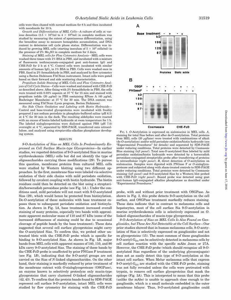
9-O-Acetylation of Sias on MEL Cells Is Predominantly Ex-pressed on Cell Surface Mucin-type Glycoproteins—In earlierstudies, we reported detection of 9-O-acetylated Sias in murineerythroleukemia (MEL) cells but did not identify the type ofoligosaccharides carrying these modifications (29). To pursuethis question, membrane proteins from cultured MEL cellswere studied by Western blotting using two different ap-proaches. In the first, membrane Sias were labeled via selectiveoxidation of their side chains with mild periodate oxidation,followed by covalent coupling with biotin hydrazide. The biotinresidues could then be detected on the blot using a streptavi-din/horseradish peroxidase probe (see Fig. 1A.). Under the con-ditions used, mild periodate will not react with 9-O-acetylatedSias (29), which would hence be protected from biotinylation.De-O-acetylation of these molecules with base treatment ex-poses them to subsequent periodate oxidation and biotinyla-tion. As shown in Fig. 1A, base treatment increased overallstaining of many proteins, especially two bands with approxi-mate apparent molecular mass of 110 and 87 kDa (some of theincreased diffuseness of staining could be due to occasionalcleavage of peptide bonds by the base treatment). These datasuggested that several cell surface glycoproteins might carrythe O-acetylated Sias. To confirm this, we probed other un-treated blots with the CHE-FcD probe which is specific forbinding to 9-O-acetylated Sias. As shown in Fig. 1B, majorbands fromMEL cells with apparent masses of 140, 110, and 90kDa carry 9-O-acetylated Sias. The staining of these bands bythe CHE-FcD probe is unaffected by prior PNGase F treatment(see Fig. 1B), indicating that the 9-O-acetyl groups are notcarried on the Sias of N-linked oligosaccharides. On the otherhand, their staining is markedly reduced by prior treatment ofthe membrane extract with O-sialoglycoprotease (OSGPase),an enzyme known to selectively proteolyze only mucin-typeglycoproteins that carry clustered O-linked oligosaccharides(42, 43). To confirm that the protein bands found by SDS-PAGErepresent cell surface 9-O-acetylation, intact MEL cells werestudied by flow cytometry for staining with the CHE-FcD
probe, with and without prior treatment with OSGPase. Asshown in Fig. 2, this probe detects 9-O-acetylation on the cellsurface, and OSGPase treatment markedly reduces staining.These data indicate that in contrast to melanoma cells andhepatocytes, most of the cell surface Sia 9-O-acetylation inmurine erythroleukemia cells is selectively expressed on O-linked oligosaccharides of mucin-type glycoproteins.9-O-Acetylation of Sias on MEL Cells Is Also Found on Gan-
gliosides, but These Are Not Detectable at the Cell Surface—Ourprior studies showed that in human melanoma cells, 9-O-acety-lation of Sias is selectively expressed on gangliosides and noton glycoproteins (15). The most common of these gangliosides9-O-acetyl-GD3 can be selectively detected in melanoma cells bycell surface reaction with the specific mAbs Jones or 27A.However, the CHE-FcD probe (which should recognize all 9-O-acetylated Sias regardless of the underlying glycoconjugate)does not as easily detect this type of 9-O-acetylation at theintact cell surface. When Melur melanoma cells that express9-O-acetyl-GD3 are studied with the CHE-FcD probe, stainingwas not fully revealed unless the cells were pretreated withtrypsin, to remove cell surface glycoproteins that mask theepitope (Fig. 3A). This is interpreted to mean that this probe(unlike the mAbs) is unable to approach close enough to theganglioside, which is a small molecule embedded in the outermembrane bilayer. Thus, 9-O-acetylated gangliosides could
FIG. 1. O-Acetylation is expressed on sialomucins in MEL cells. A,staining for total Sias before and after de-O-acetylation. Total proteinsfrom MEL cells (20 mg/lane) were treated with combinations of alkali(de-O-acetylation) and/or mild periodate oxidation/biotin hydrazide (see“Experimental Procedures” for details) and separated by SDS-PAGEunder reducing conditions. Total proteins were detected by CoomassieBlue staining (left panel). Total non-O-acetylated Sias labeled by mildperiodate oxidation/biotin hydrazide were detected by a horseradishperoxidase-conjugated streptavidin probe after transferring of proteinsto nitrocellulose (right panel). B, direct detection of O-acetylation onsialomucins. Samples were digested with PNGase F or O-sialoglyco-protease and subjected along with sham-treated controls to SDS-PAGEunder reducing conditions. Total proteins were detected by Coomassiestaining (left panel) and 9-O-acetylated Sias by a Western blot probedwith CHE-FcD (right panel). Bound probe was detected using goatanti-human IgG-conjugated alkaline phosphatase as described under“Experimental Procedures.”
O-Acetylated Sialic Acids in Leukemia Cells 31519
by guest on June 17, 2016http://w
ww
.jbc.org/D
ownloaded from
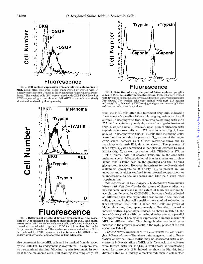
also be present in the MEL cells and be masked from detectionby the CHE-FcD by endogenous glycoproteins. To explore this,we re-examined staining following trypsin treatment. In con-trast to the melanoma cells, FcD staining was completely lost
from the MEL cells after this treatment (Fig. 3B), indicatingthe absence of accessible 9-O-acetylated gangliosides on the cellsurface. In keeping with this, there was no staining with mAb27A on flow cytometry analysis, even after trypsin treatment(Fig. 4, upper panels). However, upon permeabilization withsaponin, some reactivity with 27A was detected (Fig. 4, lowerpanels). In keeping with this, MEL cells (like melanoma cells)were found to contain the precursor GD3 as one of the majorgangliosides (detected by TLC with resorcinol spray and byreactivity with mAb R24, data not shown). The presence of9-O-acetyl-GD3 was confirmed in ganglioside extracts by lipidELISA (Fig. 5), as well by overlay with CHE-FcD or 27A onHPTLC plates (data not shown). Thus, unlike the case withmelanoma cells, 9-O-acetylation of Sias in murine erythroleu-kemia cells is found both on the glycolipid and the O-linkedglycoprotein fraction. However, in contrast to the O-acetylatedsialomucin glycoproteins, 9-O-acetyl-GD3 is present in lowamounts and is either confined to an internal compartment oris inaccessible to the antibodies and CHE-FcD, even aftertrypsinization.The Expression of Cell Surface 9-O-Acetylated Sialomucins
Varies with Cell Density—In the course of these studies, wenoticed some variations in the extent of MEL cell surface O-acetylation (detected by CHE-FcD) in batches of cells collectedon different days. The explanation was found in the fact thatcells grown at higher cell densities have marked reduction in9-O-acetylation (see Table I). When MEL cells are grown athigher densities, they spontaneously differentiate toward amature erythroid phenotype. Indeed, as shown in Table I, theloss of O-acetylation with increasing density seems to parallelthe appearance of hemoglobin expression, a known marker ofMEL cell differentiation. This change is also paralleled by anincrease in the proportion of cells in the G0/G1 phases of the cellcycle (see Table I).Induced Differentiation of MEL Cells Results in Loss of Sur-
face 9-O-Acetylation—The above data suggested that differen-tiation and/or cell cycle status may be associated with a de-crease in 9-O-acetylation of MEL cells. To check this, cultureswere treated with 2% Me2SO, a well-known differentiatingagent for these cells (23). Indeed, as shown in Fig. 6A, fullydifferentiated cells undergo a marked reduction in cell surface
FIG. 2. Cell surface expression of O-acetylated sialomucins inMEL cells. MEL cells were either sham-treated or treated with O-sialoglycoprotease (OSGPase) as described under “Experimental Proce-dures.” The washed cells (106) were stained with CHE-FcD followed byFITC-conjugated goat anti-human IgG (BKG 5 secondary antibodyalone) and analyzed by flow cytometry.
FIG. 3. Differential effects of trypsin treatment on the detec-tion of O-acetylated cell surface molecules in MEL and mela-noma cells. MEL or Melur melanoma cells (106) were either sham-treated or treated with trypsin at 37 °C for 1 h as described under“Experimental Procedures.” The washed cells were stained with CHE-FcD followed by FITC-conjugated goat anti-human IgG (BKG 5 sec-ondary antibody alone) and analyzed by flow cytometry.
FIG. 4. Detection of a cryptic pool of 9-O-acetylated ganglio-sides in MEL cells after permeabilization. MEL cells were treatedwith trypsin or saponin, respectively, as described under “ExperimentalProcedures.” The washed cells were stained with mAb 27A against9-O-acetyl GD3, followed by FITC-conjugated goat anti-mouse IgG. Dot-ted line, secondary antibody alone.
O-Acetylated Sialic Acids in Leukemia Cells31520
by guest on June 17, 2016http://w
ww
.jbc.org/D
ownloaded from

O-acetylation, to a nearly undetectable level. To rule out aneffect of Me2SO unrelated to the differentiation process, cul-tures were subjected to serum starvation, which also causescells to differentiate (as shown by hemoglobin accumulation).This was also accompanied by a significant loss of O-acetyla-tion, although not the extent seen with Me2SO (Fig. 6B). Sim-ilar effects were seen with hexamethylene-bisacetamide(HMBA), another inducer of differentiation (data not shown).Notably, none of these treatments was associated with a majorchange in total cell surface sialylation (detected by flow cytom-etry using wheat germ agglutinin-FITC, data not shown).Effects of Cell Cycle Stage upon the Expression of 9-O-Acety-
lation—Since all treatments that induce differentiation (highcell density, Me2SO, serum starvation, or HMBA) also arrestcells in G0/G1 of the cell cycle, the relationship of 9-O-acetyla-tion to cell cycle stage was analyzed in rapidly growing cellsthat are not committed to the differentiated phenotype. No lossof O-acetylation is seen in rapidly growing cells that are in theG1 phase (Fig. 7). Thus, the loss of O-acetylation seen upondifferentiation is not simply a consequence of entry into G0/G1.However, a small increase in cell surface 9-O-acetylation wasobserved when rapidly growing cells are in the G2/M phase(Fig. 7). To study this further, cells were also treated withnocodazole (or colcemid), to arrest them in the G2/M phase. Amore substantial increase of cell surface O-acetylation (.5times control) was observed after this treatment. However,nocodazole could also be causing this change by virtue of itsability to alter microtubule dynamics and membrane recycling.The MEL cells were also therefore treated with taxol, anotherG2/M phase blocker which has (in contrast to nocodazole) amicrotubule stabilizing effect. While taxol-treated cells werealso found to be blocked in G2/M to the same extent as thosetreated with nocodazole (data not shown), this was not accom-panied by such a marked increase in cell surface O-acetylation(Fig. 8B). Indeed, the very small increase seen could be ex-plained by the increase in cell size that occurs during the G2/Mphase of the cell cycle. In keeping with this, shorter treatmentswith nocodazole (3–6 h, too short to cause extensive accumu-lation in the M phase) still caused an increase in cell surface
9-O-acetylation (analyzed by flow cytometry, data not shown).The effects of cell cycle, nocodazole, and taxol on the cellular
content of 9-O-acetylated GD3 was also investigated. None ofthese appeared to have any effect on the content or the internallocalization of this form of 9-O-acetylated molecules (stainingwith mAb 27A, with and without saponin pretreatment andanalyzed by flow cytometry, data not shown). Thus, depolymer-izing microtubules affect the expression and localization of9-O-acetylated sialomucins but not of 9-O-acetylated GD3.Changes in Cell Surface 9-O-Acetylated Sialomucins after
Induced Differentiation or Nocodazole Treatment Are Not Dueto Redistribution—As indicated above, the changes in O-acety-lation seen with induced differentiation (decrease) or nocoda-zole treatment (increase) are not due to arrest in the G2/Mphase. Alternatively, that could be due to a redistribution of theO-acetylated sialomucins rather than changes in actual cellu-lar content. To rule out this possibility, a Western blot of totalcell glycoproteins from each situation was probed with CHE-FcD. As shown in Fig. 9, this approach confirmed a substantialdecrease of protein-associated 9-O-acetylation upon Me2SO-induced differentiation and a significant increase with nocoda-zole treatment, particularly in some high molecular weightsialoglycoproteins.Increase in 9-O-Acetylation Following Nocodazole Treatment
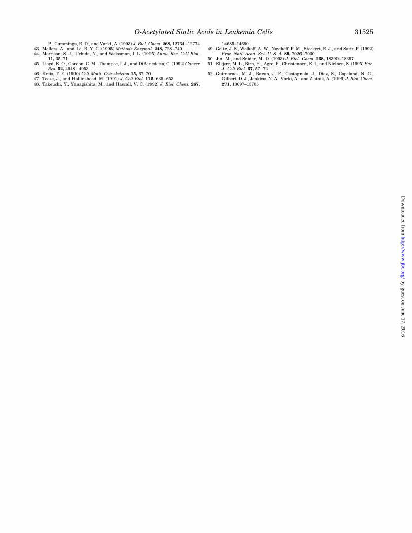
Requires New Synthesis and Is Due to Accumulation on the CellSurface—In the past, it was not possible to selectively remove9-O-acetyl groups from cell surface molecules and to follow thesubsequent fate of the Sias. Using the recombinant solubleinfluenza C 9-O-acetyl esterase (CHE-Fc), we have exploredconditions for complete removal of cell surface 9-O-acetylgroups from MEL cells. As shown in Fig. 10, complete removalis possible, and the cells can be returned to culture for a chaseperiod. As shown in the examples in Fig. 10A, 9-O-acetylationgradually returned to the cell surface over a period of 2–3 h.However, simultaneous treatment with nocodazole had no ef-fect upon the rate of recovery (Fig. 10B), indicating that thiscompound may not affect the rate of new synthesis of 9-O-acetylated Sias.To confirm that the restoration of cell surface 9-O-acetylation
after selective removal was due to new protein synthesis, westudied the effects of cycloheximide, a protein synthesis inhib-itor. As shown in Fig. 11, this treatment markedly blunted therestoration of expression of 9-O-acetylation. Taken together,the data indicate that the mucin molecules that were de-O-acetylated at the cell surface did not undergo re-O-acetylationand re-expression. Thus, the effects of nocodazole are mostlikely explained by the accumulation of newly synthesized mol-ecules on the cell surface, i.e. a decreased rate of internalizationand turnover.
DISCUSSION
Friend murine erythroleukemia cells (MEL cells) were one ofthe first cellular models where “normal” differentiation couldbe induced and recapitulated in vitro (23), as determined by theexpression of molecules such as the globins. Erythroid differ-entiation normally takes place in a defined microenvironmentin the bone marrow, where erythroblasts interact via cell sur-face glycoproteins with stromal cells (fibroblasts and macro-phages) and with extracellular matrix components (44). Thus,besides the coordinated expression of globin genes, controlledexpression of cell surface sialoglycoconjugates may character-ize the differentiated phenotype of erythrocytes. Murine eryth-rocytes also present O-acetylated sialoglycoconjugates on theircell surface (11, 27, 28). Although the precise function of thismodification is still an open question,O-acetylation of Sias mayalter the binding of some viruses and is predicted to affect theactivation of the alternative complement pathway.
FIG. 5. The presence of 9-O-acetyl-GD3 is confirmed in gangli-oside extracts from MEL cells. Gangliosides were extracted fromMEL cells as described under “Experimental Procedures.” Base-treatedor non-base-treated gangliosides were coated on ELISA plates andincubated with the mAb 27A or the CHE-FcD probe. Binding wasdetected using goat anti-mouse IgG or goat anti-human IgG-conjugatedhorseradish peroxidase, respectively, as described under “ExperimentalProcedures.” The data are the mean of triplicates.
O-Acetylated Sialic Acids in Leukemia Cells 31521
by guest on June 17, 2016http://w
ww
.jbc.org/D
ownloaded from

Here we have identified the nature of O-acetylated sialogly-coconjugates in MEL cells and studied the control of theirexpression upon differentiation. Both glycoproteins and glyco-lipids of MEL cells were found O-acetylated. Among glycopro-teins, several high molecular weight polypeptides were themajor O-acetylated species. These glycoconjugates were sensi-tive to O-sialoglycoprotease but not to PNGase F, thus identi-fying them as mucin-like molecules carrying clusters of O-linked chains. The identity of the polypeptides that carry thismodification needs to be investigated. Among glycolipids, MELcells contain both GD3 and its 9-O-acetylated form, but thelatter is a minor species that is present either in an internalcompartment or it is not readily accessible on the cell surface,as discussed for other cellular models (45). This is in contrast tohuman melanoma cell lines, which express both GD3 and 9-O-acetylated GD3 on the cell surface, but not 9-O-acetylated sia-loglycoproteins. Also, unlike the MEL cells, 9-O-acetylated GD3
was readily identified on the cell surface of Melur cells using
specific mAbs and becomes unmasked for CHE-FcD detectionafter cell surface trypsinization. On the other hand, MEL cellsdo not express any trypsin-resistant sialoglycoconjugate, and9-O-acetyl-GD3 could be observed only after treatment of cellswith saponin (which permeabilizes cells by extracting choles-terol from the plasma membrane). In separate work, we haveshown that 9-O-acetylation is found primarily on N-linked gly-coproteins of hepatocytes (16). Thus, 9-O-acetylation appears tobe differentially regulated in different cell types in a molecule-specific and location-specific manner.We next analyzed the expression of 9-O-acetylated sialomu-
cins on the cell surface of MEL cells during their growth anddifferentiation. At cell densities of .4 3 106/ml, we observed anarrest of cells in G0/G1, accompanied by spontaneous produc-tion of hemoglobin, and a marked reduction of 9-O-acetylation.Induced differentiation with serum starvation, Me2SO, orHMBA caused a more marked increase in hemoglobin accumu-lation and arrest in G0/G1 phases and was accompanied by amore marked loss of cell surface expression of 9-O-acetylation,without any accompanying major changes in the level of cellsurface sialylation. Since all of these treatments arrested cellsin the G0/G1 phase, we evaluated a possible cell cycle depend-ency of this Sia modification. Cells in G2/M displayed a small(;2-fold) increase of CHE-FcD reactivity over those in G0/G1.However, if one assumes that the cells in G2 phase woulddouble their volume before division, an actual increase of about60% in cell surface area is expected. To explore this, we induced
TABLE IEffect of cell culture density on cell cycle, differentiation, and cell surface 9-O-acetylation
MEL cells were seeded and grown to different densities in culture as indicated, and aliquots were studied for the various parameters indicated.
Cell density G0/G1a G2/M Hemoglobin-positiveb Relative cell surface
O-acetylationc
cell/ml % % % %
2.5 3 105 62 38 0 935 3 105 61 39 0 941 3 106 63 38 0 932 3 106 64 37 0 924 3 106 93 7 50 17
a Cells were stained with propidium iodide, and DNA content was analyzed by flow cytometry as described under “Experimental Procedures.”b Cells were stained for hemoglobin expression with benzidine as described under “Experimental Procedures.”c Cells were stained with CHE-FcD and studied by flow cytometry as described under “Experimental Procedures.” The mean fluorescence
intensity is presented, relative to control cells stained with secondary antibody alone.
FIG. 6. Effect of differentiation state on the expression of cellsurface 9-O-acetylated sialomucins in MEL cells. MEL cells weregrown in the presence of 2% Me2SO (DMSO) or serum-free media for 3days, and washed cells were stained with CHE-FcD and analyzed byflow cytometry as described under “Experimental Procedures” (BKG,secondary antibody alone).
FIG. 7. Relationship of cell cycle to expression of 9-O-acety-lated sialomucins in rapidly growing MEL cells. MEL cells weregrown in logarithmic growth phase and double-stained with CHE-FcDand propidium iodide as described under “Experimental Procedures.”Flow cytometry analysis was set up with FITC/propidium iodide fluo-rescence compensation. The G0/G1 and G2/M cells in the histogram weregated from analysis of a dot plot display of the double-stained MEL cells(not shown), (BKG, secondary antibody alone).
O-Acetylated Sialic Acids in Leukemia Cells31522
by guest on June 17, 2016http://w
ww
.jbc.org/D
ownloaded from
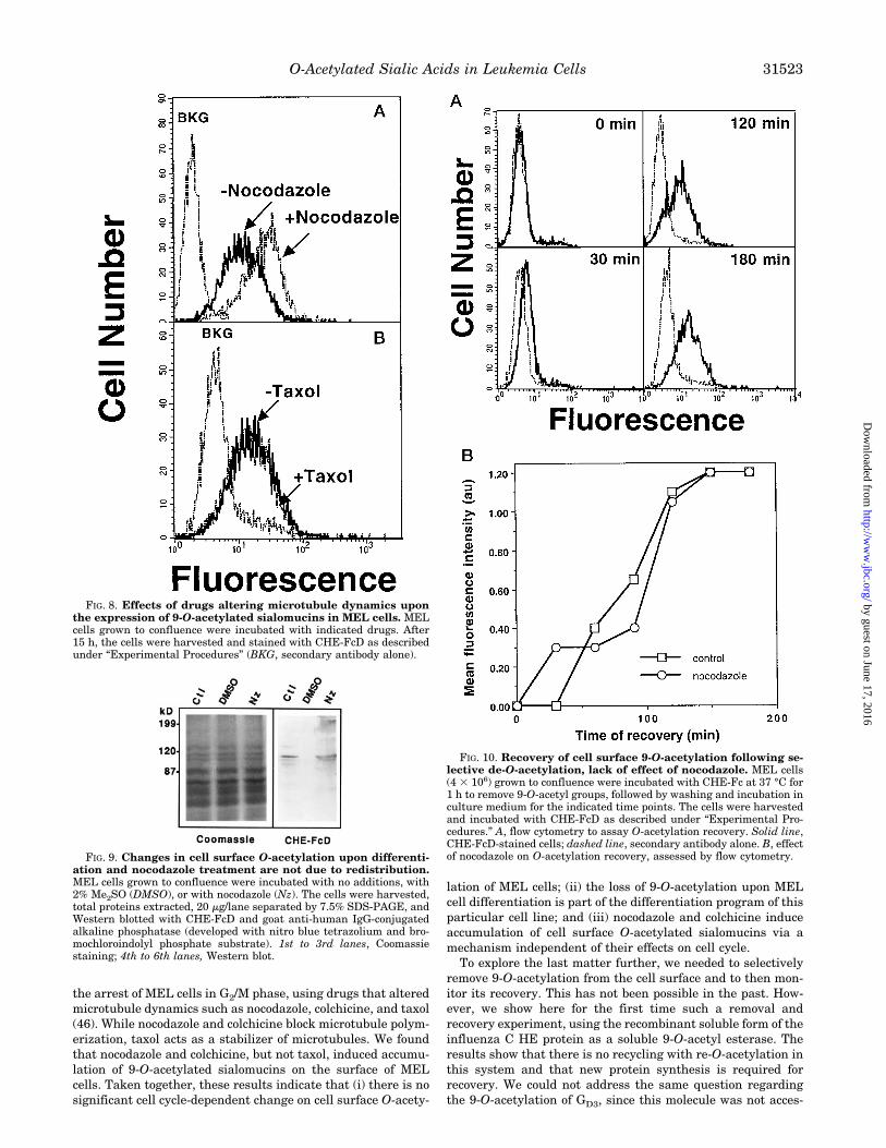
the arrest of MEL cells in G2/M phase, using drugs that alteredmicrotubule dynamics such as nocodazole, colchicine, and taxol(46). While nocodazole and colchicine block microtubule polym-erization, taxol acts as a stabilizer of microtubules. We foundthat nocodazole and colchicine, but not taxol, induced accumu-lation of 9-O-acetylated sialomucins on the surface of MELcells. Taken together, these results indicate that (i) there is nosignificant cell cycle-dependent change on cell surface O-acety-
lation of MEL cells; (ii) the loss of 9-O-acetylation upon MELcell differentiation is part of the differentiation program of thisparticular cell line; and (iii) nocodazole and colchicine induceaccumulation of cell surface O-acetylated sialomucins via amechanism independent of their effects on cell cycle.To explore the last matter further, we needed to selectively
remove 9-O-acetylation from the cell surface and to then mon-itor its recovery. This has not been possible in the past. How-ever, we show here for the first time such a removal andrecovery experiment, using the recombinant soluble form of theinfluenza C HE protein as a soluble 9-O-acetyl esterase. Theresults show that there is no recycling with re-O-acetylation inthis system and that new protein synthesis is required forrecovery. We could not address the same question regardingthe 9-O-acetylation of GD3, since this molecule was not acces-
FIG. 8. Effects of drugs altering microtubule dynamics uponthe expression of 9-O-acetylated sialomucins in MEL cells. MELcells grown to confluence were incubated with indicated drugs. After15 h, the cells were harvested and stained with CHE-FcD as describedunder “Experimental Procedures” (BKG, secondary antibody alone).
FIG. 9. Changes in cell surface O-acetylation upon differenti-ation and nocodazole treatment are not due to redistribution.MEL cells grown to confluence were incubated with no additions, with2% Me2SO (DMSO), or with nocodazole (Nz). The cells were harvested,total proteins extracted, 20 mg/lane separated by 7.5% SDS-PAGE, andWestern blotted with CHE-FcD and goat anti-human IgG-conjugatedalkaline phosphatase (developed with nitro blue tetrazolium and bro-mochloroindolyl phosphate substrate). 1st to 3rd lanes, Coomassiestaining; 4th to 6th lanes, Western blot.
FIG. 10. Recovery of cell surface 9-O-acetylation following se-lective de-O-acetylation, lack of effect of nocodazole. MEL cells(4 3 106) grown to confluence were incubated with CHE-Fc at 37 °C for1 h to remove 9-O-acetyl groups, followed by washing and incubation inculture medium for the indicated time points. The cells were harvestedand incubated with CHE-FcD as described under “Experimental Pro-cedures.” A, flow cytometry to assay O-acetylation recovery. Solid line,CHE-FcD-stained cells; dashed line, secondary antibody alone. B, effectof nocodazole on O-acetylation recovery, assessed by flow cytometry.
O-Acetylated Sialic Acids in Leukemia Cells 31523
by guest on June 17, 2016http://w
ww
.jbc.org/D
ownloaded from

sible for de-O-acetylation at the cell surface. Thus, the effects ofnocodazole and colcemid must be due to delay of normal cellsurface turnover via endocytosis. This interpretation is givencredence by earlier studies of the transferrin receptor, whichaccumulates on the cell surface after endocytosis blockage byagents like nocodazole (47–51).As mentioned above, murine erythrocytes present some 9-O-
acetylated sialoglycoconjugates on their surface (11, 27, 28).Thus, despite being a suitable model for differentiation regard-ing globin gene expression, MEL cells do not constitute a rep-resentative model for “normal” differentiation regarding cellsurface Sia O-acetylation. On the other hand, it represents avaluable system for general studies on the control of O-acety-lation of Sias, an issue that is still very poorly understood.O-Acetylation of sialoglycoconjugates depends on the activity oftwo opposing enzymatic systems, O-acetyltransferase(s) andSia-specific esterases (2, 3). Although the activity of O-acetyl-transferases could be detected in a series of different cellularsystems, the enzyme(s) have resisted all attempts to date forpurification or molecular cloning. At least two Sia-specific es-terases were identified (2, 3), one a cytosolic protein and theother one a lysosomal enzyme, which has recently been cloned(52). In preliminary studies, we have noted that during Me2SO-induced differentiation there is only a marginal increase ontotal esterase specific activity.2 Thus, loss of O-acetylation ondifferentiated MEL cells is more likely associated with a de-creased activity of O-acetyltransferase(s). Unfortunately, theactivity of the latter enzyme cannot be accurately measured incell extracts. An alternative, but less likely, explanation for lossof 9-O-acetylation upon differentiation would be down-regula-tion of a unique subset of sialomucins that are specific sub-strates for the O-acetyltransferase(s) expressed on growingMEL cells.
Analysis of the 9-O-acetylated Sias described so far in natureindicates that there may be separate O-acetyltransferase ac-tivities directed toward a2,6-linked Sias (e.g. for N-linked sia-loglycoproteins from hepatocytes) and toward a2,8-linked Sias(e.g. for GD3 in human melanoma cells). It is still not known ifa2,3-linked Sias can be 9-O-acetylated. Since MEL cells clearlyexpressed 9-O-acetyl-GD3, they must be expressing anO-acetyltransferase activity toward a2,8-linked Sias. The na-ture of the Sia linkage on the 9-O-acetylated sialomucins re-mains to be determined. Regardless, it is evident that theO-acetyltransferase(s) present in these cells show some speci-ficity, since they do not modify the Sias on the N-linked oligo-saccharides of cell surface sialoglycoproteins.
Acknowledgments—We thank Marilyn Farquhar, George Palade,Leland D. Powell, Steffen Thiel, and Sandra Diaz for helpful discus-sions and for their comments on the manuscript.
REFERENCES
1. Schauer, R. (ed) (1982) Sialic Acids: Chemistry, Metabolism and Function, CellBiology Monographs, Vol. 10, Springer-Verlag Inc., New York
2. Schauer, R. (1991) Glycobiology 1, 449–4523. Varki, A. (1992) Glycobiology 2, 25–404. Varki, A., and Diaz, S. (1984) Anal. Biochem. 137, 236–2475. Kamerling, J. P., Schauer, R., Shukla, A. K., Stoll, S., van Halbeek, H., and
Vliegenthart, J. F. G. (1987) Eur. J. Biochem. 162, 601–6076. Corfield, A. P., Sander-Wewer, M., Veh, R. W., Wember, M., and Schauer, R.
(1986) Biol. Chem. Hoppe-Seyler 367, 433–4397. Rogers, G. N., Herrler, G., Paulson, J. C., and Klenk, H.-D. (1986) J. Biol.
Chem. 261, 5947–59518. Vlasak, R., Krystal, M., Nacht, M., and Palese, P. (1987) Virology 160,
419–4259. Cheresh, D. A., Varki, A., Varki, N. M., Stallcup, W. B., Levine, J., and
Reisfeld, R. A. (1984) J. Biol. Chem. 259, 7453–745910. Sjoberg, E. R., Powell, L. D., Klein, A., and Varki, A. (1994) J. Cell Biol. 126,
549–56211. Varki, A., and Kornfeld, S. (1980) J. Exp. Med. 152, 532–54412. Shi, W.-X., Chammas, R., Varki, N. M., Powell, L., and Varki, A. (1996) J. Biol.
Chem. 271, 31526–3153213. Cheresh, D. A., Reisfeld, R. A., and Varki, A. (1984) Science 225, 844–84614. Thurin, J., Herlyn, M., Hindsgaul, O., Stromberg, N., Karlsson, K.-A., Elder,
D., Steplewski, Z., and Koprowski, H. (1985) J. Biol. Chem. 260,14556–14563
15. Manzi, A. E., Sjoberg, E. R., Diaz, S., and Varki, A. (1990) J. Biol. Chem. 265,13091–13103
16. Butor, C., Diaz, S., and Varki, A. (1993) J. Biol. Chem. 268, 10197–1020617. Kamerling, J. P., Makovitzky, J., Schauer, R., Vliegenthart, J. F. G., and
Wember, M. (1982) Biochim. Biophys. Acta 714, 351–35518. Kniep, B., Peter-Katalinic, J., Flegel, W., Northoff, H., and Rieber, E. P. (1992)
Biochem. Biophys. Res. Commun. 187, 1343–134919. Kniep, B., Flegel, W. A., Northoff, H., and Rieber, E. P. (1993) Blood 82,
1776–178620. Zimmer, G., Suguri, T., Reuter, G., Yu, R. K., Schauer, R., and Herrler, G.
(1994) Glycobiology 4, 343–35021. Kniep, B., Claus, C., Peter-Katalinic, J., Monner, D. A., Dippold, W., and
Nimtz, M. (1995) J. Biol. Chem. 270, 30173–3018022. Sen, G., Chowdhury, M., and Mandal, C. (1994)Mol. Cell. Biochem. 136, 65–7023. Marks, P. A., and Rifkind, R. A. (1978) Annu. Rev. Biochem. 47, 419–44824. Dolci, E. D., and Palade, G. E. (1989) J. Cell Sci. 93, 191–19725. Misawa, Y., and Shibuya, M. (1992) Oncogene 7, 919–92626. Okayama, M., Oguri, K., Yoshida, K., and Ohkita, T. (1991) J. Biol. Chem. 266,
3808–381927. Sarris, A. H., and Palade, G. E. (1979) J. Biol. Chem. 254, 6724–673128. Reuter, G., Vliegenthart, J. F. G., Wember, M., Schauer, R., and Howard, R. J.
(1980) Biochem. Biophys. Res. Commun. 94, 567–57229. Diaz, S., and Varki, A. (1985) Anal. Biochem. 150, 32–4630. Fraser, I. P., and Gordon, S. (1994) Eur. J. Cell Biol. 64, 217–22131. Kelm, S., Schauer, R., Manuguerra, J.-C., Gross, H.-J., and Crocker, P. R.
(1994) Glycoconj. J. 11, 576–58532. Levine, J., Beasley, L., and Stallcup, W. (1984) J. Neurosci. 4, 820–83133. Constantine-Paton, M., Blum, A. S., Mendez Otero, R., and Barnstable, C. J.
(1986) Nature 324, 459–46234. Blum, A. S., and Barnstable, C. J. (1987) Proc. Natl. Acad. Sci. U. S. A. 84,
8716–872035. Drazba, J., Pierce, M., and Lemmon, V. (1991) Dev. Biol. 145, 154–16336. Muchmore, E., and Varki, A. (1987) Science 236, 1293–129537. Zimmer, G., Reuter, G., and Schauer, R. (1992) Eur. J. Biochem. 204, 209–21538. Klein, A., Krishna, M., Varki, N. M., and Varki, A. (1994) Proc. Natl. Acad. Sci.
U. S. A. 91, 7782–778639. Wallick, S. C., Kabat, E. A., and Morrison, S. L. (1988) J. Exp. Med. 168,
1099–110940. Ulmer, J. B., and Palade, G. E. (1989) Proc. Natl. Acad. Sci. U. S. A. 86,
6992–699641. Sjoberg, E. R., Chammas, R., Ozawa, H., Kawashima, I., Khoo, K.-H., Morris,
H. R., Dell, A., Tai, T., and Varki, A. (1995) J. Biol. Chem. 270, 2921–293042. Norgard, K. E., Moore, K. L., Diaz, S., Stults, N. L., Ushiyama, S., McEver, R.2 R. Chammas and A. Varki, unpublished data.
FIG. 11. Effects of cycloheximide on the recovery of cell sur-face 9-O-acetylation following selective de-O-acetylation. MELcells grown to confluence were treated with CHE-Fc at 37 °C for 1 h toremove 9-O-acetyl groups, followed by washing and incubation in cul-ture medium in the presence or absence of cycloheximide (CHX) at37 °C for 3 h. The cells were harvested and stained with CHE-FcD asdescribed under “Experimental Procedures.” Solid line, CHE-FcD-stained cells; dashed line, secondary antibody alone.
O-Acetylated Sialic Acids in Leukemia Cells31524
by guest on June 17, 2016http://w
ww
.jbc.org/D
ownloaded from
P., Cummings, R. D., and Varki, A. (1993) J. Biol. Chem. 268, 12764–1277443. Mellors, A., and Lo, R. Y. C. (1995) Methods Enzymol. 248, 728–74044. Morrison, S. J., Uchida, N., and Weissman, I. L. (1995) Annu. Rev. Cell Biol.
11, 35–7145. Lloyd, K. O., Gordon, C. M., Thampoe, I. J., and DiBenedetto, C. (1992) Cancer
Res. 52, 4948–495346. Kreis, T. E. (1990) Cell Motil. Cytoskeleton 15, 67–7047. Tooze, J., and Hollinshead, M. (1991) J. Cell Biol. 115, 635–65348. Takeuchi, Y., Yanagishita, M., and Hascall, V. C. (1992) J. Biol. Chem. 267,
14685–1469049. Goltz, J. S., Wolkoff, A. W., Novikoff, P. M., Stockert, R. J., and Satir, P. (1992)
Proc. Natl. Acad. Sci. U. S. A. 89, 7026–703050. Jin, M., and Snider, M. D. (1993) J. Biol. Chem. 268, 18390–1839751. Elkjær, M. L., Birn, H., Agre, P., Christensen, E. I., and Nielsen, S. (1995) Eur.
J. Cell Biol. 67, 57–7252. Guimaraes, M. J., Bazan, J. F., Castagnola, J., Diaz, S., Copeland, N. G.,
Gilbert, D. J., Jenkins, N. A., Varki, A., and Zlotnik, A. (1996) J. Biol. Chem.271, 13697–13705
O-Acetylated Sialic Acids in Leukemia Cells 31525
by guest on June 17, 2016http://w
ww
.jbc.org/D
ownloaded from

Wei-Xing Shi, Roger Chammas and Ajit VarkiMurine Erythroleukemia Cells
-Acetylation during the Growth and Differentiation ofORegulation of Sialic Acid 9-
doi: 10.1074/jbc.271.49.315171996, 271:31517-31525.J. Biol. Chem.
http://www.jbc.org/content/271/49/31517Access the most updated version of this article at
Alerts:
When a correction for this article is posted•
When this article is cited•
to choose from all of JBC's e-mail alertsClick here
http://www.jbc.org/content/271/49/31517.full.html#ref-list-1
This article cites 51 references, 31 of which can be accessed free at
by guest on June 17, 2016http://w
ww
.jbc.org/D
ownloaded from