HDAC6 Modulates Cell Motility by Altering the Acetylation Level of Cortactin
28
HDAC6 Modulates Cell Motility by Altering the Acetylation Level of Cortactin Xiaohong Zhang 1,9 , Zhigang Yuan 1 , Yingtao Zhang 2 , Sarah Yong 1,9 , Alexis Salas- Burgos 3 , John Koomen 1 , Nancy Olashaw 1 , J. Thomas Parsons 4 , Xiang-Jiao Yang 5 , Sharon R. Dent 6 , Tso-Pang Yao 7 , William S. Lane 8 , and Edward Seto 1,* 1 Molecular Oncology Program, H. Lee Moffitt Cancer Center & Research Institute, Tampa, FL 33612 USA 2 Department of Pathology & Cell Biology, College of Medicine, University of South Florida, Tampa, FL 33612 USA 3 Departmento de Fisiopatologia, Facultad de Ciencias Biologicas, Universidad de Concepcion, Concepcion, Chile 4 Department of Microbiology, Health Sciences Center, University of Virginia, Charlottesville, VA 22903 USA 5 Molecular Oncology Group, Department of Medicine, McGill University Health Center, Montreal, Quebec, Canada 6 Department of Biochemistry and Molecular Biology, M.D. Anderson Cancer Center, Houston, TX 77030 USA 7 Department of Pharmacology and Cancer Biology, Duke University, Durham, NC 27710 USA 8 Microchemistry and Proteomics Analysis Facility, Harvard University, Cambridge, MA 02138 USA Summary Histone deacetylase 6 (HDAC6) is a tubulin-specific deacetylase that regulates microtubule- dependent cell movement. In this study, we identify the F-actin-binding protein, cortactin, as a HDAC6 substrate. We demonstrate that HDAC6 binds cortactin and that overexpression of HDAC6 leads to hypoacetylation of cortactin, while inhibition of HDAC6 activity leads to cortactin hyperacetylation. HDAC6 alters the ability of cortactin to bind F-actin by modulating a “charge patch” in its repeat region. Introduction of charge-preserving or charge-neutralizing mutations in this cortactin repeat region correlates with the gain or loss of F-actin binding ability, respectively. Cells expressing a charge-neutralizing cortactin mutant were less motile than control cells or cells expressing a charge-preserving mutant. These findings suggest that, in addition to its role in microtubule-dependent cell motility, HDAC6 influences actin-dependent cell motility by altering the acetylation status of cortactin, which, in turn, changes the F-actin binding activity of cortactin. *Correspondence: E-mail: [email protected]; phone 813-745-6754; fax 813-745-4907. 9 Present address: Department of Pathology & Cell Biology, College of Medicine, University of South Florida, Tampa, FL 33612 USA Publisher's Disclaimer: This is a PDF file of an unedited manuscript that has been accepted for publication. As a service to our customers we are providing this early version of the manuscript. The manuscript will undergo copyediting, typesetting, and review of the resulting proof before it is published in its final citable form. Please note that during the production process errors may be discovered which could affect the content, and all legal disclaimers that apply to the journal pertain. NIH Public Access Author Manuscript Mol Cell. Author manuscript; available in PMC 2009 May 20. Published in final edited form as: Mol Cell. 2007 July 20; 27(2): 197–213. doi:10.1016/j.molcel.2007.05.033. NIH-PA Author Manuscript NIH-PA Author Manuscript NIH-PA Author Manuscript
-
Upload
independent -
Category
Documents
-
view
2 -
download
0
Transcript of HDAC6 Modulates Cell Motility by Altering the Acetylation Level of Cortactin

HDAC6 Modulates Cell Motility by Altering the Acetylation Levelof Cortactin
Xiaohong Zhang1,9, Zhigang Yuan1, Yingtao Zhang2, Sarah Yong1,9, Alexis Salas-Burgos3, John Koomen1, Nancy Olashaw1, J. Thomas Parsons4, Xiang-Jiao Yang5, SharonR. Dent6, Tso-Pang Yao7, William S. Lane8, and Edward Seto1,*
1 Molecular Oncology Program, H. Lee Moffitt Cancer Center & Research Institute, Tampa, FL 33612 USA
2 Department of Pathology & Cell Biology, College of Medicine, University of South Florida, Tampa, FL33612 USA
3 Departmento de Fisiopatologia, Facultad de Ciencias Biologicas, Universidad de Concepcion, Concepcion,Chile
4 Department of Microbiology, Health Sciences Center, University of Virginia, Charlottesville, VA 22903USA
5 Molecular Oncology Group, Department of Medicine, McGill University Health Center, Montreal, Quebec,Canada
6 Department of Biochemistry and Molecular Biology, M.D. Anderson Cancer Center, Houston, TX 77030USA
7 Department of Pharmacology and Cancer Biology, Duke University, Durham, NC 27710 USA
8 Microchemistry and Proteomics Analysis Facility, Harvard University, Cambridge, MA 02138 USA
SummaryHistone deacetylase 6 (HDAC6) is a tubulin-specific deacetylase that regulates microtubule-dependent cell movement. In this study, we identify the F-actin-binding protein, cortactin, as aHDAC6 substrate. We demonstrate that HDAC6 binds cortactin and that overexpression of HDAC6leads to hypoacetylation of cortactin, while inhibition of HDAC6 activity leads to cortactinhyperacetylation. HDAC6 alters the ability of cortactin to bind F-actin by modulating a “chargepatch” in its repeat region. Introduction of charge-preserving or charge-neutralizing mutations in thiscortactin repeat region correlates with the gain or loss of F-actin binding ability, respectively. Cellsexpressing a charge-neutralizing cortactin mutant were less motile than control cells or cellsexpressing a charge-preserving mutant. These findings suggest that, in addition to its role inmicrotubule-dependent cell motility, HDAC6 influences actin-dependent cell motility by alteringthe acetylation status of cortactin, which, in turn, changes the F-actin binding activity of cortactin.
*Correspondence: E-mail: [email protected]; phone 813-745-6754; fax 813-745-4907.9Present address: Department of Pathology & Cell Biology, College of Medicine, University of South Florida, Tampa, FL 33612 USAPublisher's Disclaimer: This is a PDF file of an unedited manuscript that has been accepted for publication. As a service to our customerswe are providing this early version of the manuscript. The manuscript will undergo copyediting, typesetting, and review of the resultingproof before it is published in its final citable form. Please note that during the production process errors may be discovered which couldaffect the content, and all legal disclaimers that apply to the journal pertain.
NIH Public AccessAuthor ManuscriptMol Cell. Author manuscript; available in PMC 2009 May 20.
Published in final edited form as:Mol Cell. 2007 July 20; 27(2): 197–213. doi:10.1016/j.molcel.2007.05.033.
NIH
-PA Author Manuscript
NIH
-PA Author Manuscript
NIH
-PA Author Manuscript

IntroductionHistone deacetylases (HDACs) and histone acetyltransferases (HATs) are ubiquitouslyexpressed enzymes that primarily target core histones. The hyperacetylation of histonestypically enhances gene expression, while their deacetylation usually represses geneexpression. Many transcriptional co-activators are HATs and many transcriptional co-repressors are HDACs. Mammalian HDACs can be subdivided into the following three classes:class I (HDACs 1, 2, 3, and 8), class II (HDACs 4, 5, 6, 7, 9, and 10), and class III (SIRTs 1,2, 3, 4, 5, 6, 7). HDAC11 shares homology with both class I and class II HDACs.
In addition to histones, HDACs and HATs also target non-histone proteins. Some of these non-histone targets are transcription factors such as p53, GATA-1, E2F1, YY1, and MyoD.Importantly, the reversible acetylation of these proteins modifies their activities. Other non-histone HAT and HDAC substrates include proteins that regulate cell proliferation, survival,and motility. For example, PCAF acetylates the DNA end-joining protein Ku70, leading to anattenuation of Ku70 anti-apoptotic activity. p300 acetylates the tumor suppressor Rb andprevents Rb phosphorylation by cyclin-dependent kinases and blocks cell cycle progression.
One of the most extensively studied and best characterized non-histone HDAC substrates isthe cytoplasmic protein α-tubulin (Haggarty et al., 2003; Hubbert et al., 2002; Matsuyama etal., 2002; Zhang et al., 2003). HDAC6 associates with and deacetylates α-tubulin in vitro andin vivo. Cells overexpressing HDAC6 have more deacetylated α-tubulin than control cells andare more motile. Consistent with its effect on cell motility, HDAC6 is predominantly localizedto the cytoplasm. Interestingly, malignant mammary epithelial cells have a more pronouncedHDAC6 cytosolic localization than normal cells (Yoshida et al., 2004).
Cortactin, a protein originally identified as a substrate of the Src tyrosine kinase, also plays animportant role in regulating cell motility (Wu et al., 1991). It interacts with F-actin to promotepolymerization and branching. Cortactin can be found at areas of dynamic actin assembly, suchas at the leading edge of migrating cells (e.g., in lamellipodia and membrane ruffles) (Kaksonenet al., 2000; Uruno et al., 2001; Weaver et al., 2001; Wu and Parsons, 1993). The translocationof cortactin to the cell periphery requires the activation of the small GTPase Rac1 and leads tothe activation of the actin-nucleating complex Arp2/3 (Head et al., 2003; Uruno et al., 2001;Weaver et al., 2001; Weed et al., 1998). Overexpression of cortactin increases cell migration(Kowalski et al., 2005; Patel et al., 1998), while depletion of cortactin impairs it (Bryce et al.,2005). Cortactin contains an N-terminal acidic domain (NTA), six and a half tandem repeatsof a unique 37-amino acid sequence, and a C-terminal Src homology (SH3) domain (Wu andParsons, 1993). The repeat region of cortactin is both necessary and sufficient for F-actinbinding. An α-helical structure and a proline-rich region separate the repeat and the SH3domains. It is well-established that Src targets cortactin primarily at three residues (Tyr-421,Tyr-466, and Tyr-482 in mice), which are situated between the proline-rich region and the SH3domain (Huang et al., 1998).
Cortactin is abnormally expressed in many human tumors, often as a result of geneamplification. EMS1, the human gene that encodes for cortactin, is amplified in head and necksquamous cell carcinomas, and the extent of amplification is inversely proportional to thesurvival rate (Rodrigo et al., 2000). Cortactin expression correlates with the metastatic potentialof hepatocellular carcinomas (Chuma et al., 2004) and breast cancer cells injected into nudemice (Li et al., 2001). Evidence suggests that cortactin may regulate the formation ofpodosomes, structures that cells utilize to make contact with surfaces (Schuuring et al., 1993;Zhou et al., 2006; Tehrani et al., 2006). In invasive breast cancer cells, cortactin is present inmembrane protrusions, or invadopodia, which carry proteases that digest the extracellularmatrix (Bowden et al., 1999; Bowden et al., 2006). Collectively, these data demonstrate a strong
Zhang et al. Page 2
Mol Cell. Author manuscript; available in PMC 2009 May 20.
NIH
-PA Author Manuscript
NIH
-PA Author Manuscript
NIH
-PA Author Manuscript

link between cortactin expression and tumor invasiveness and metastasis. This link suggeststhe need for a greater understanding of the mechanisms by which cortactin activity is regulated.
In the present study, we show that the cortactin protein is acetylated. Importantly, we foundthat HDAC6 associates with and deacetylates cortactin in vitro and in vivo. Moreover, wedemonstrate that hyperacetylation of cortactin prevents its translocation to the cell periphery,blocks its association with F-actin, and impairs cell motility. Together, our findings uncovera pathway by which actin-dependent cell motility can be modulated by HDAC6 and provideadditional evidence for the importance of HDAC6 in the regulation of cell movement.
ResultsHDAC6 Associates with the Repeat Region of Cortactin
To identify HDAC6 cytoplasmic substrates, we prepared cytoplasmic extracts from a HeLaS3-derived cell line stably expressing Flag-tagged HDAC6. Flag-HDAC6 and associatedproteins were purified by anti-Flag immunoaffinity chromatography. As a control, mockpurifications were performed using extracts prepared from HeLa cells expressing an emptyFlag vector (Figure 1A). We found at least five polypeptides that specifically associated withFlag-tagged HDAC6. Tandem mass spectrometric analysis (MS/MS) of a 80–85 kDapolypeptide yielded the amino acid sequence LPSSPVYEDAASFK, which was identified ascortactin (Accession Q14247).
To determine if HDAC6 interacts with cortactin under normal physiologic conditions, co-immunoprecipitation of the endogenous proteins from a cytoplasmic extract was performed.As shown in Figure 1B, a significant fraction of cortactin could be co-precipitated with an anti-HDAC6 antibody but not with pre-immune serum. No cortactin was precipitated when theprimary antibody was omitted. Using bacterially expressed and highly purified GST-cortactinand histidine-tagged HDAC6 expressed and purified from Sf9 insect cells, we found thatHDAC6 interacts directly with cortactin in the absence of any other cellular proteins (Figure1C).
To demonstrate specificity for the HDAC6-cortactin interaction, we used adenoviral infectionto generate a HeLa cell line that overexpresses Flag-tagged HDAC5. HDAC5-containingprotein complexes were then immunoprecipitated from HeLa extracts using a Flag-specificantibody. The presence of cortactin in these immunoprecipitates was analyzed by Westernblotting. As shown in Figure 1D, the anti-Flag antibody specifically co-precipitated cortactinfrom cells expressing Flag-tagged HDAC6 but not from cells expressing Flag-tagged HDAC5.In a reciprocal experiment, an anti-cortactin antibody specifically co-precipitated Flag-taggedHDAC6, but not Flag-tagged HDAC5. Western blots verified the equivalent expression of theFlag-tagged HDAC6 and HDAC5 proteins in the infected cells. Further, overexpressed Flag-HDAC5 was active as determined by a comparison of acetylated histone H4 levels in Flag-HDAC5-overexpressing cells and in parental cells. Thus, Flag-HDAC5 appears to be a suitablenegative control. In similar experiments, cortactin was not shown to interact with any otherclass II HDACs (data not shown). This finding suggests that the cortactin-HDAC6 interactionis highly specific.
To identify the HDAC6-binding region of cortactin, we performed serial deletions from theN- or C-terminus. The truncated sequences were then Myc-tagged and expressed in HeLa cellsby transfection. These cells also expressed Flag-tagged HDAC6 as a result of adenoviralinfection. Lysates from these cells were subjected to immunoprecipitation using a Flag-specificantibody followed by Western blotting using a Myc-specific antibody. As shown in Figure 1E,neither an N-terminal cortactin fragment (containing the acidic domain, aa 1–84) nor a C-terminal cortactin fragment (containing the α-helix, the proline-rich region, and the SH3
Zhang et al. Page 3
Mol Cell. Author manuscript; available in PMC 2009 May 20.
NIH
-PA Author Manuscript
NIH
-PA Author Manuscript
NIH
-PA Author Manuscript

domain, aa 350–546) interacted with HDAC6. Results from GST pull-down assays furtherreveal that the repeat region alone binds HDAC6 (Figure S1). Therefore, the cortactin repeatregion, a region known to be responsible for interaction with F-actin (Wu and Parsons,1993), is necessary and sufficient for the interaction between cortactin and HDAC6.
To define the HDAC6 domain(s) required for interaction with cortactin, anti-Flagimmunoprecipitates prepared from cells expressing Myc-cortactin and either Flag-taggedwildtype (1–1215) or deletion mutants were subjected to Western blot analysis using anti-Mycantibodies. As shown in Figure 1F, full-length HDAC6 and a C-terminal HDAC6 deletionmutant (1–840) bind cortactin. In contrast, an HDAC6 mutant (1–503) lacking both the C-terminal and the second deacetylase domain (DAC2) completely lost its ability to bindcortactin. Further deletion analysis indicates that DAC2 alone does not bind HDAC6 (data notshown), suggesting that both DAC1 and DAC2 are necessary, and neither domain alone issufficient for cortactin binding.
HDAC6 Regulates the Acetylation of Cortactin in vivoThe association between cortactin and HDAC6 suggests that cortactin may be a deacetylationtarget of HDAC6. To determine if cortactin is acetylated, immunoprecipitated Myc-taggedcortactin was subjected to Western blotting using an anti-acetyl-lysine specific antibody. Asshown in Figure 2A, Myc-cortactin is acetylated in vivo. Similarly, endogenous cortactin wasalso found to be acetylated as assayed by high stringency immunoprecipitation of a whole cellextract either with an anti-acetyl-lysine antibody followed by Western blotting with an anti-cortactin antibody or anti-cortactin followed by anti-acetyl-lysine antibody (Figure 2B).Treatment of cells with the class I and II HDAC inhibitor, TSA, greatly increased the level ofacetylated Myc-tagged (Figure 2A) and endogenous (Figure 2B) cortactin.
To confirm that cortactin is acetylated, we raised a rabbit polyclonal antibody that specificallyrecognizes acetylated cortactin. Western blot analysis using this antibody revealed that HDACinhibition increased acetylation of both endogenous and overexpressed cortactin (Figure 2C).Interestingly, like TSA, treatment of cells with the class III deacetylase inhibitor nicotinamidealso resulted in an increase of cortactin acetylation. However, unlike TSA, cortactin acetylationwas unchanged in the presence of the potent HDAC inhibitor sodium butyrate, which does notaffect HDAC6 (Hubbert et al., 2002).
Next, using an alternative approach to demonstrate that cortactin is acetylated underphysiologic conditions, we immunopurified cortactin from a NIH3T3 whole cell extract,separated the products on a 2-dimensional gel, and analyzed the acetylated cortactin by Westernblot with an anti-acetyl-lysine antibody. As shown in Figure 2D, a significant fraction ofendogenous cortactin was present in an acetylated form.
To examine the possible deacetylation of cortactin by HDAC6, we co-expressed Myc-taggedcortactin and either Flag-tagged HDAC6 or Flag-tagged HDAC5 in HeLa cells. Myc-immunoprecipitates were then analyzed by Western blot using an acetyl-lysine-specificantibody. As shown in Figure 3A, overexpression of HDAC6 resulted in a reduction in thelevel of acetylated cortactin. The total level of cortactin was unaffected. In contrast, theoverexpression of HDAC5 or a catalytically defective HDAC6 did not alter levels of total oracetylated cortactin.
To verify that HDAC6 mediates the deacetylation of cortactin, we used an established A549cell line (HD6KD) in which HDAC6 expression is specifically knocked-down by a retrovirus-mediated RNAi system (Kawaguchi et al., 2003). Consistent with previous studies (Hubbertet al., 2002), HD6KD cells contained more acetylated tubulin than control A549 cells (Figure3B). More importantly, we found that HD6KD cells contained higher levels of acetylated
Zhang et al. Page 4
Mol Cell. Author manuscript; available in PMC 2009 May 20.
NIH
-PA Author Manuscript
NIH
-PA Author Manuscript
NIH
-PA Author Manuscript

cortactin than the control cells. A reciprocal experiment with a cell line stably overexpressingHDAC6 revealed that acetylated cortactin levels were markedly decreased in these cells ascompared to the negative control parental cells.
Since HDAC6 has been reported to interact with SIRT2 (North et al., 2003), we tested theeffect of SIRT2 on cortactin acetylation levels. As shown in Figure 3C, similar to HDAC6knockdown, SIRT2-knockdown resulted in an increase in cortactin acetylation suggesting that,in addition to HDAC6, SIRT2 might also regulate the state of cortactin acetylation.
Previous analysis of mouse tissues has shown that HDAC6 is highly expressed in testes(Seigneurin-Berny et al., 2001). In addition, we have found that HDAC6 is highly expressedin human ovarian tumor tissues (data not shown). We examined the expression of HDAC6 inthree different human ovarian cancer cell lines and found that HDAC6 expression in theOV2008 and SW626 cell lines was relatively high. On the other hand, the OVCAR3 cell linecontained lower levels of HDAC6 (Figure 3D). Importantly, the acetylation level of cortactinwas inversely proportional to the expression level of HDAC6. That is, endogenous cortactinwas highly acetylated in the OVCAR3 cell line, but not in the OV2008 or SW626 cell lines.These data strongly suggest that HDAC6 is a chief regulator of cortactin acetylation.
Cortactin is Acetylated/Deacetylated in its Repeat RegionTo examine the HAT-mediated acetylation of cortactin, Myc-tagged cortactin was co-expressed with either Flag-tagged PCAF or HA-tagged p300 in HeLa cells. Anti-Myc-immunoprecipitates prepared from these cells were then analyzed by Western blot using anacetyl-lysine-specific antibody. As shown in Figure 4A, PCAF expression increased the levelof acetylated cortactin in a dose-dependent manner (left panel). However, overexpression ofp300 or catalytic mutants of PCAF (Δ579–608 and Δ609–624) did not alter levels of acetylatedcortactin.
Although PCAF has been reported to be located both in the nucleus and cytoplasm of cells(Wong et al., 2004), most studies on PCAF so far have focused on its nuclear functions. Becausecortactin is a cytoplasmic protein, to rule out the possibility that PCAF acetylation of cortactinis a result of over-expression and consequently mis-localization, we re-examined thesubcellular localization of endogenous PCAF both by cellular protein fractionation and byimmunostaining. As shown in Figure S2, endogenous PCAF is indeed present in both thenuclear and cytoplasm of 293T and NIH3T3 cells, confirming the possibility that cortactincould be a true physiological substrate of PCAF.
To determine if PCAF directly acetylates cortactin, an in vitro acetylation assay was performedusing GST-cortactin and the catalytic domains of either PCAF or p300. Consistent with the invivo results, these experiments showed that PCAF, but not p300, can acetylate cortactin invitro (Figure 4B).
To map the region(s) of cortactin acetylated by PCAF in vitro, the following three GST-taggedcortactin fragments were prepared: the N-terminal acidic region (1–84), the repeat region (84–330), and the C-terminal region (331–546) (see diagram in Figure 1E). Together, thesefragments cover the entire cortactin protein sequence. As determined by in vitro acetylationassays, PCAF was able to acetylate the repeat region of cortactin, but not the N-terminal acidicor the C-terminal regions (Figure 4C). These data suggest that the repeat region of cortactin isthe primary site of acetylation.
To determine if the cortactin repeat region alone is sufficient to serve as a HDAC6 substrate,we infected HeLa cells that express Flag-(84–330) with adenoviruses that express either Flag-HDAC6 or GFP as control, prepared cell lysates, and assayed acetylation levels by
Zhang et al. Page 5
Mol Cell. Author manuscript; available in PMC 2009 May 20.
NIH
-PA Author Manuscript
NIH
-PA Author Manuscript
NIH
-PA Author Manuscript

immunoprecipitation with anti-Flag and Western blotting with anti-acetyl-lysine antibodies.As shown in Figure 4D, acetylation level of cortactin repeat region diminishes significantly inthe presence of overexpressed HDAC6.
Identification of Acetylated Lysines in CortactinTo identify the sites of acetylation on cortactin, in vitro acetylation assays were performedusing GST-cortactin, the PCAF catalytic domain, and acetyl CoA. To verify cortactinacetylation, an aliquot of each reaction mixture was analyzed by Western blotting using ananti-acetyl-lysine antibody (data not shown). The remainder of the reaction mixture wasresolved by SDS-PAGE, and the polypeptide band corresponding to cortactin was excised andanalyzed by LC tandem mass spectrometry (LC-MS/MS). Of the 50 lysines in cortactin, 11were found to be acetylated. Of these 11 acetyl-lysines, eight (K87, K161, K189, K198, K235,K272, K309, and K319) were present in the cortactin repeat region (Figure 5A). We alsomapped the in vivo sites of acetylation on cortactin by focusing on the repeat region. For theseanalyses, we transfected 293T cells with a plasmid encoding the Flag-tagged repeat region ofcortactin. To maximize acetylation, cells were treated with 400 ng/ml TSA for 12 h. Followingthis treatment, cellular extracts were prepared from these cells, and the extracts were subjectedto immunoprecipitation using a Flag-specific antibody. The resulting immunoprecipitates werethen resolved by SDS-PAGE, and the band corresponding to the cortactin repeat region wasexcised from the gel and analyzed by LC-MS/MS. Finally, using a similar strategy, weimmunopurified endogenous cortactin protein using anti-cortactin antibody and subjected thepurified product to LC-MS/MS analysis.
To assess their contributions to the overall acetylation status of cortactin, all eight of the lysinesthat were identified as the PCAF in vitro acetylation sites as well as K124 (a residue detectedboth in the purified Flag-tagged repeat region and endogenous cortactin) were mutated toglutamine. This mutant, referred to as 9KQ, was examined using an in vitro acetylation assay.As shown in Figure 5B, while PCAF effectively acetylated wildtype GST-cortactin, acetylationof the GST-9KQ mutant was nearly undetectable. Coomassie blue gel staining verified thatsimilar amounts of wildtype cortactin and 9KQ were present in both assays. Furthermore, thelysine to glutamine change does not affect the binding of cortactin to HDAC6 (Figure S3).Similarly, in transiently transfected HeLa cells, the 9KQ mutant was also much less efficientlyacetylated than wild-type cortactin (Figure 5C). Thus, the repeat region is most likely theprimary (if not the only) cortactin region acetylated in vitro and in vivo.
The secondary structure of the individual repeats of cortactin repeat region predicts an α-helicalregion within the carboxyl-half of each subunit (Wu and Parsons, 1993). Previous computermodeling of this helical region suggested that highly conserved lysine residues may bepositioned on the same face of the α-helices, a conformation that would contribute to apositively-charged helical surface. In our predicted model, all of the above-described acetylatedlysines in the repeat domain (with the exception of K189 and K319) are in a loop rather thanin the helical region (Figure 5D–F). Additionally, the acetylated lysines are present at two endsof the helices, a conformation that could result in the formation of two “charged patches”. Inits deacetylated state, this charged patch (or the positively-charged loop surface) of cortactinis likely to contribute to F-actin binding.
Acetylation of Cortactin Impedes its Interaction with F-ActinTo determine the effect of cortactin acetylation status on cortactin interactions with F-actin, F-actin co-sedimentation assays were performed. For these experiments, GST-fused cortactinrepeat regions were expressed in and purified from E. coli cells. This purified protein was thenincubated with polymerized rabbit muscle F-actin. As shown in Figure 6A, following F-actinsedimentation, the non-acetylated GST-tagged cortactin repeat region was bound to (i.e., co-
Zhang et al. Page 6
Mol Cell. Author manuscript; available in PMC 2009 May 20.
NIH
-PA Author Manuscript
NIH
-PA Author Manuscript
NIH
-PA Author Manuscript

sedimented) F-actin in vitro; whereas, immunopurified, PCAF-acetylated GST-taggedcortactin repeat region was not.
To examine this effect in more detail, we prepared two sets of GST-tagged cortactin mutants.In the first set, each lysine of the cortactin repeat region was individually mutated to glutamine.Although glutamine cannot be acetylated, its neutral charge mimics the acetylation of lysine.Each of these point mutants were able to bind to F-actin as efficiently as wild-type cortactin(Figure 6B). Likewise, a charge-preserving cortactin mutant in which all nine of the repeatregion lysines were mutated to arginine (9KR) was able to efficiently bind to F-actin. In sharpcontrast, a charge-neutralizing cortactin mutant in which all nine of the repeat region lysineresidues were mutated to glutamine (9KQ) was not able to bind F-actin. In the second set ofmutants, the lysines of the cortactin repeat region were progressively mutated to glutaminebeginning at either the amino or carboxyl end (Figure 6C). Mutation of less than three lysinesat either terminal end did not significantly alter the ability of cortactin to bind to F-actin.However, mutation of more than four of these lysines dramatically reduced the F-actin bindingactivity of cortactin; the more residues mutated, the less binding detected. The effect was notlimited to mutations at the N-terminal or the C-terminal end of cortactin, because mutationwithin the internal repeats (6KQ) resulted in similar decrease in F-actin binding when comparedto N6KQ or C6KQ (Figure 6D). Thus, the acetylation of multiple lysine residues of the cortactinrepeat region attenuates its actin-binding ability in vitro. This effect was also observed invivo using cells overexpressed with the cortactin repeat region. For example, HeLa cells treatedwith TSA had more acetylated Flag-tagged cortactin and more cortactin in the supernatant(i.e., not bound to F-actin) than did untreated cells (Figure 6E). Consistent with this finding,inhibition of HDAC6 either by treatment of cells with HDAC inhibitors or HDAC6 siRNAdecreased endogenous cortactin-F-actin association (Figure 6F).
Acetylation Causes Aberrant Cortactin Localization and Cell MotilityIn response to growth factor stimulation or small GTPase, Rac1 activation, cortactintranslocates from the cytosol to the cell periphery, where it interacts with and enhances theformation of F-actin (Weed et al., 1998; Weed et al., 2000). Because acetylation/deacetylationis a key determinate of cortactin binding to F-actin, we examined whether Rac1 has an effecton the level of cortactin acetylation. NIH3T3 cells were transfected with plasmids expressingMyc-cortactin and various amounts of constitutively active Rac1 (HA-Rac1G12V). Anti-Mycimmunoprecipitates prepared from these cells were subjected to Western blot analysis usingan anti-acetyl-lysine antibody. As shown in Figure 7A, consistent with our observation thatdeacetylated cortactin binds better to F-actin, Rac1 activation clearly resulted in cortactindeacetylation.
To further analyze whether the acetylation of cortactin affects its subcellular location, we co-transfected NIH3T3 cells with constitutively active Rac1 (HA-Rac1G12V) and either Flag-tagged wildtype, 9KQ mutant, or 9KR mutant cortactin. Cells were immunostained with anti-Flag and anti-HA antibodies followed by Alexa-594 and Alexa-488 conjugated secondaryantibodies. Consistent with previous studies, wildtype cortactin was present in the membraneruffles in cells expressing active Rac1. Similarly, the 9KR cortactin mutant, which is capableof F-actin binding, translocated to the cell periphery in the presence of active Rac1. In contrast,the 9KQ cortactin mutant, which is not capable of F-actin binding, remained cytoplasmic.Quantification of the percentage of cells expressing Rac1 and the wildtype, 9KQ, or 9KRcortactin that show a leading edge is presented in Table S1. Together, these results suggest thatthe acetylation of cortactin inhibits, while deacetylation may be required for, its Rac-mediatedtranslocation to the cell periphery.
It has been reported that growth factor-induced membrane ruffling is due to activation of Rac1(Kozma et al., 1995; Nobes and Hall, 1995). We found that the treatment of cells with EGF
Zhang et al. Page 7
Mol Cell. Author manuscript; available in PMC 2009 May 20.
NIH
-PA Author Manuscript
NIH
-PA Author Manuscript
NIH
-PA Author Manuscript

resulted in a decrease in cortactin acetylation (Figure 7B). To determine if HDAC6 is involvedin the Rac1-mediated translocation of cortactin, we stimulated serum-starved NIH3T3 cellswith EGF and monitored the subcellular localization of cortactin and HDAC6 byimmunofluorescence microscopy. We found that HDAC6 was translocated to the cell peripherytogether with cortactin upon EGF stimulation (Figure 7B). This result suggests that HDAC6interacts with and deacetylates cortactin at the cell periphery. Further analysis demonstratedthat, upon EGF stimulation, 9KR mutant translocates to the cell periphery suggesting that thecharge-preserving mutant could dislodge the HDAC6-cortactin association. Also, as can beseen in Figure S4, consistent with the notion that cortactin must be deacetylated in order totranslocate to the membrane ruffle or leading edges, the localization of the 9KQ mutant isunchanged in the presence of EGF.
A previous study has shown that overexpression of HDAC6 increases the chemotactic motilityof NIH3T3 cells (Hubbert et al., 2002). To confirm this result, transwell assays were performedwith 293T cells expressing either HDAC6 siRNA or control siRNA. The results clearly indicatethat chemotactic cell motility decreases in cells depleted of HDAC6 (Figure 7C). To examinethe effect of cortactin acetylation status on chemotactic cell migration, parental NIH3T3 cellsand NIH3T3 cells stably expressing either wildtype or mutated cortactins were seeded in theupper chamber of migration plates. As shown in Figure 7C, the percentage of motile cells wasdecreased among cultures expressing cortactin mutants that do not bind F-actin (N8KQ and9KQ) than in cells expressing either wildtype, N1KQ mutant, or 9KR mutant with intact F-actin binding capability. Western blot analyses indicate that cortactin mutants are expressedin comparable levels as wildtype cortactin (Figure S5). These results strongly argue thatcortactin deacetylation is critical for the regulation of cell migration.
In complementary experiments, we determined if cortactin mutants could rescue the phenotypecaused by cortactin knockdown. Cortactin expression was assessed by a Western blot (FigureS5). As expected, a HT1080 human fibrosarcoma cell line in which cortactin protein expressionwas reduced by more than 95% (cortactin KD; Bryce et al., 2005) exhibited slower migrationcompared to parental cells (control) (Figure 7D). Interestingly, the cell motility defect incortactin KD cells was effectively rescued by the introduction of wildtype or 9KR mutantcortactin, but not by the 9KQ mutant, underscoring the significance of deacetylation in cortactinfunction.
To further demonstrate that acetylation/deacetylation of cortactin affects cell motility, weperformed migration assays on three different ovarian cancer cell lines that were determinedto possess different levels of acetylated cortactin (Figure 3D). As shown in Figure 7E, OV2008and SW626 cells that express high levels of HDAC6 and contain hypoacetylated cortactin showfaster migration when compared to OVCAR3 that expresses low level of HDAC6 and containhyperacetylated cortactin. Next, we used siRNA to knock-down HDAC6 expression in SKOV3cells, an ovarian cancer cell line with high levels of HDAC6 protein (Figure 7F). We thencompared the migratory properties of the HDAC6 knock-down cells with the parental cellstreated with control siRNA. As expected, cells with partially depleted HDAC6 (HDAC6KD)displayed a slower migration phenotype.
Finally, using an alternative approach to assess the effects of acetylation/deacetylation ofcortactin on cell motility, we established an MDA-MB-231 cell line that stably expresses the9KQ mutant. Using a live-cell imaging technique, we then measured the actual distance andvelocity these cells traveled under random motility compared to the parental cell line. As shownin Figure 7G, movement velocity was significantly decreased in cells overexpressing the 9KQmutant. These results are consistent with the transwell assay results and unequivocally confirmthat cells that express the charge-neutralizing acetylation mutant of cortactin (9KQ) travel lessdistance and move slower than the parental cell line.
Zhang et al. Page 8
Mol Cell. Author manuscript; available in PMC 2009 May 20.
NIH
-PA Author Manuscript
NIH
-PA Author Manuscript
NIH
-PA Author Manuscript

DiscussionResults from previous studies have shown that HDAC6 associates with and regulates theacetylation of α-tubulin and Hsp90 (Bali et al., 2005; Haggarty et al., 2003; Hubbert et al.,2002; Kovacs et al., 2005; Matsuyama et al., 2002; Zhang et al., 2003). HDAC6 also functionsas an adaptor linking dynein motors to their aggregated protein cargos (Kawaguchi et al.,2003). Additionally, Seigneurin-Berny et al. (2001) and Boyault et al. (2006) found that themammalian homolog of yeast UFD3 (ubiquitin fusion degradation protein 3), as well as p97/VCP/Cdc48p, co-purifies with HDAC6 in mouse testes. They demonstrated that HDAC6 alsoefficiently interacts with ubiquitin and that this interaction led to the dissociation of HDAC6from this protein complex. Furthermore, Hook et al. (2002) reported that HDAC6 bindspolyubiquitin through its zinc finger and also co-purifies with de-ubiquitinating enzymes.Using multiple approaches, we have also identified an interaction between polyubiquitin andHDAC6 (data not shown). Unexpectedly, we also identified the F-actin-binding protein,cortactin, as an HDAC6-interacting protein. This protein, which co-purifies with cytoplasmicHDAC6, is a non-histone substrate of HDAC6.
Our results indicate that HDAC6 interacts with cortactin in vivo and that this interactionrequires the repeat region of cortactin. Similar to the HDAC6-β-tubulin interaction (Zhang etal., 2003), the association between HDAC6 and cortactin is direct, is mediated throughdeacetylase domains, and does not required deacetylase activities. However, unlike the β-tubulin-HDAC6 interaction in which only one deacetylase domain is sufficient to bindHDAC6, the association of cortactin with HDAC6 requires both deacetylase domains.
We found that PCAF acetylates cortactin and HDAC6 deacetylates it. It has been shownpreviously that the repeat region of cortactin mediates its interaction with F-actin, and that thisinteraction stimulates F-actin polymerization and branching (Uruno et al., 2001; Weaver et al.,2001; Wu and Parsons, 1993). PCAF was able to acetylate cortactin in vitro and promotecortactin acetylation when overexpressed in vivo, while the related HAT, p300, was not. Thecortactin repeat region was the primary site of PCAF-mediated acetylation in vitro and invivo. Using LC tandem mass spectrometry, we identified multiple lysine residues in this regionthat were acetylated. Whether endogenous PCAF is solely responsible for cortactin acetylationin vivo remains to be determined. Because no significant differences in acetylation of cortactinwere detected from PCAF−/− versus PCAF+/+ fibroblast cells, we currently favor the idea thatbesides PCAF, an additional acetyltransferase(s) may acetylate cortactin.
We showed that deacetylated cortactin accumulated in cells that overexpressed HDAC6 andthat acetylated cortactin accumulated in cells depleted of HDAC6. Also, in our studies, no otherectopically-expressed class II HDAC affected the acetylation level of cortactin (data notshown). These results strongly suggest that cortactin is a physiologic substrate of HDAC6, butwhether HDAC6 is the only biologically relevant cortactin deacetylase remains to bedetermined. HDAC6 is known to associate with SIRT2, an NAD-dependent class III HDACand a tubulin deacetylase (North et al., 2003). Therefore, it is possible that cortactin is asubstrate of, not only HDAC6, but also SIRT2. Consistent with this possibility, we found thattreatment of cells with the class III inhibitor nicotinamide or knockdown of SIRT2 with siRNAfurther enhances the acetylation of cortactin in TSA-treated cells. The possibility of more thanone class of HDACs targeting a single non-histone protein substrate is not without precedent.For example, both HDAC1 and SIRT1 deacetylate the tumor suppressor p53.
We found that PCAF-acetylated cortactin no longer binds F-actin in vitro. This loss of bindingactivity was dependent on the acetylation of multiple lysines in the cortactin repeat region. Ourresults suggest that these acetylated lysines alter a “charge patch” that reduces the affinity ofcortactin for F-actin in a graded manner. Once some threshold number of lysines becomes
Zhang et al. Page 9
Mol Cell. Author manuscript; available in PMC 2009 May 20.
NIH
-PA Author Manuscript
NIH
-PA Author Manuscript
NIH
-PA Author Manuscript

acetylated, the F-actin-binding affinity of cortactin is decreased to a level that inhibits thecortactin-F-actin interaction. This would suggest that the acetylation of particular lysine(s) inthe patch is of lesser importance than the total number of lysines that are acetylated.
Our computer model predicts two charge patches in the repeat region. The first region iscomprised of K124, K189, K198, and K272, and the second is comprised of K161, K309, andK319. Studies of histones H1 and H2A. Z in Tetrahymena have demonstrated a similarmechanism in which their functions are regulated by both acetylation- and phosphorylation-generated charge patches (Dou and Gorovsky, 2000; Ren and Gorovsky, 2001; Ren andGorovsky, 2003). The mechanism by which the charge of the cortactin repeat region affectsits interaction with F-actin is not known. One possibility is that a charge-inducedconformational change mediated by HDAC6-induced deactylation favors the accessibility ofthe cortactin repeat region for interaction with F-actin, while acetylation of this region inhibitsthis accessibility. A similar mechanism is observed for the ETS-1 transcription factor in whichits progressive phosphorylation shifts the protein from an active, DNA-binding conformationto a folded, inactive conformation (Pufall et al., 2005). An alternative, although not mutuallyexclusive, possibility is that positive charges in the cortactin repeat region are required forinteraction with the acidic domain of F-actin. By deacetylating cortactin, HDAC6 enhancesthe association between cortactin and F-actin as a result of increasing the positive charges inthe cortactin repeat region. The seven GF6/Y1GGacKF4/Y3GV6I1 motifs present in thecortactin repeat region share similarity with the acetylated lysine motif in histone H4(KGGacK). Therefore, the interaction between cortactin and F-actin may be regulated by amechanism similar to the one that modulates the interaction between histone H4 and cellularproteins.
We found that mutation of the acetylated lysines in the cortactin repeat region preventedcortactin translocation to the cell periphery (i.e., the site of dynamic actin assembly) in responseto activated Rac1. Because the phosphorylation of cortactin by Src and Src-related kinasesrequires the association of cortactin with F-actin (Head et al., 2003), we predict that acetylatedcortactin, which is not able to efficiently interact with F-actin, will not be efficientlyphosphorylated. In support of this hypothesis, the levels of Src-phosphorylated cortactin werereduced in cells ectopically expressing the 9KQ mutant cortactin relative to the levels in wild-type expressing cells (data not shown). However, further studies are needed to fully elucidatethe relationship between cortactin acetylation and Src-mediated cortactin phosphorylation.
In summary, our results demonstrate that the reversible acetylation of cortactin dynamicallyregulates actin-dependent cell mobility. Cell motility is important for many normal cellularprocesses, and dysregulated cell motility contributes to vascular disease, chronic inflammatorydisease, and tumor metastasis. Thus, our finding that the reversible acetylation of cortactinmodulates its actin-binding activity and affects cell migration has many far-reaching clinicalrelevancies.
Experimental ProceduresPlasmids, Recombinant Proteins, and Antibodies
Details of all plasmid constructions and sources of recombinant proteins and antibodies areprovided in the Supplementary Experimental Procedures.
Purification of HDAC6 ComplexesThe Flag-tagged HDAC6 insert from pBJ-HDAC6F was excised and ligated downstream ofthe HA sequence in pcDNA3.1(+)HA. The resultant plasmid was transfected into HeLa S3cells using Lipofectamine 2000 (Invitrogen), and neomycin-resistant colonies were selected
Zhang et al. Page 10
Mol Cell. Author manuscript; available in PMC 2009 May 20.
NIH
-PA Author Manuscript
NIH
-PA Author Manuscript
NIH
-PA Author Manuscript
in medium containing 400 μg/ml G418 for two weeks. Thirty-two colonies were screened forFlag-tagged HDAC6 expression by Western blotting with Flag- and HA-specific antibodies.One of the HDAC6-expressing clones was expanded and maintained in DMEM containing10% calf serum and 200 μg/ml G418.
Cytoplasmic extract was prepared from HeLa cells stably expressing Flag-tagged HDAC6using a standard protocol (Coligan et al., 1995). Affinity purification of Flag-tagged HDAC6-containing protein complexes using an anti-Flag antibody was performed according topreviously published methods (Ogryzko et al., 1998). Purified complexes were concentrated,resolved by SDS-PAGE, and analyzed by silver staining. A Coomassie blue-stained samplewas prepared in parallel, and bands corresponding to HDAC6-associated proteins were excisedand subjected to proteolysis with trypsin. Peptides from these mixtures were sequenced bymicrocapillary LC-MS/MS.
Immunoprecipitation and ImmunoblottingFor immunoprecipitations, cells were lysed in buffer (50 mM Tris-HCl [pH 7.5], 1 mM EDTA,1% NP-40, and protease inhibitor cocktail) containing either 500 mM NaCl (high stringency)or 150 mM NaCl (low stringency). Lysates were incubated with the indicated primaryantibodies for 12 h at 4°C. Immunocomplexes were collected, washed four times in lysis buffer,and resolved by SDS-PAGE. For immunoblotting, samples were transferred to nitrocellulosemembranes that were then probed with the indicated antibodies. Bound antibodies weredetected using a Chemiluminescent Detection Kit (Pierce).
GST Pull-Down AssayGST and GST-cortactin were expressed and purified from bacteria using standard methods.Equimolar amounts of the purified proteins were conjugated to glutathione-Sepharose beadsand incubated with purified histidine-tagged HDAC6 for 1 h at room temperature. Afterextensive washing, bound proteins were eluted and analyzed by Western blotting with anti-His6 antibodies. A full detail protocol is presented in the Supplementary ExperimentalProcedures.
Two-Dimensional Gel ElectrophoresisOne confluent NIH3T3 culture grown in a 150 mm dish was harvested and lysed in 500 μlRIPA buffer containing a cocktail of proteinase, phosphatase, and HDAC inhibitors. Lysateswere immunoprecipitated with 10 μg of anti-cortactin antibody and subjected to 2-D gelelectrophoresis described in the Supplementary Experimental Procedures.
In vitro Acetylation AssayBacterially-expressed GST-tagged cortactin was purified using standard procedures, and 5 μgof protein were incubated with 20 μM of 14C-acetyl CoA and either a fragment of PCAF (500ng) containing the HAT domain or the HAT fragment of p300 (5 units) in buffer comprised of50 mM Tris (pH 8), 0.1 mM EDTA, 1 mM DTT, 10% glycerol, 10 mM sodium butyrate, andprotease inhibitors. Reactions were allowed to proceed for 1 h at 30°C. Reaction products wereresolved by SDS-PAGE and visualized by autoradiography.
Tandem Mass Spectrometry Acetylation Site AnalysisGel slices containing acetylated cortactin were prepared and subjected to tandem massspectrometry analysis as described in the Supplementary Experimental Procedures.
Zhang et al. Page 11
Mol Cell. Author manuscript; available in PMC 2009 May 20.
NIH
-PA Author Manuscript
NIH
-PA Author Manuscript
NIH
-PA Author Manuscript

Three-Dimensional Modeling of CortactinDetails of three-dimensional modeling of cortactin are provided in the SupplementaryExperimental Procedures. Data are deposited in the Protein Data Bank (PDB ID 2F9X).
F-Actin Co-sedimentation AssayRabbit skeletal muscle actin (>99% pure; purchased from Cytoskeleton, Inc.) was polymerizedto generate F-actin, according to the manufacturers’ protocol. Typically, purified actin wasstored in G-buffer (20 mM Tris-HCl [pH 7.5], 0.2 mM ATP, 0.2 mM DTT, and 0.2 mMCaCl2). Actin was polymerized by adjusting the buffer condition to that of F-buffer (2 mMTris-HCl [pH 8], 0.2 mM DTT, 0.2 mM CaCl2, 50 mM KCl, 2 mM MgCl2, and 1 mM ATP).F-actin co-sedimentation assays were carried out according to a previously published protocol(Wu and Parsons, 1993). Briefly, cell lysates or recombinant proteins were incubated with 30μg of F-actin in 50 μl of F-actin binding buffer for 1 h at 4°C. Reaction mixtures were layeredwith 50 μl of 10% sucrose in F-actin binding buffer and were centrifuged at 64,000 rpm for 1h at 4°C. Supernatants were transferred to fresh tubes, and pellets were incubated in 50 μl ofG buffer for at least 1 h. Equal volumes of supernatant and pellet were analyzed by Westernblotting with the indicated antibodies.
Immunostaining and Fluorescence MicroscopyCells cultured on chamber slides (Chamber Slide System Lab-TekII) were washed with PBSand fixed in 4% paraformaldehyde for 10 min at room temperature. Fixation was terminatedwith 1% glycine in PBS. For permeabilization, the cells were subjected to several changes ofa 1% glycine/0.5% Triton X-100 solution at room temperature for 1 h. Cells were thenincubated in PBS containing 0.2% Triton X-100, 1% bovine serum albumin, and primaryantibody for 1 h. After washing in PBS containing 0.1% Tween, the cells were incubated for45 min with secondary antibody in PBS containing 1% bovine serum albumin. Finally, thecells were washed in PBS containing Tween and then PBS alone. The slides were dried andmounted with Vectashield mounting medium with DAPI.
Cell Migration AssayTranswell migration assays were performed using the CytoSelect™ 24 (8 μm, colorimetricformat) kit purchased from Cell Biolabs. A detail protocol is presented in the SupplementaryExperimental Procedures.
Live-Cell ImagingFor phase-contrast microscopy, 3 × 10⁴ cells/cm2 were plated on a MatTek coverslip andincubated overnight prior to imaging. Imaging was performed using a Nikon TE-2000-Sinverted wildfield microscope equipped with a Retigia 1600 CCD camera and IP Labs 3.6software from Scanlytics Inc. The images were taken with a 10× 0.25 NA PhI DL lens every5 min for a total of 205 min. The Image Pro Plus v. 5.0 software was used to measure actualdistance and velocity of cell motility. A 37°C re-circulating warm stage was used to maintaincell viability. To calculate the velocity, actual distance cells traveled by tracks (in pixcells)were measured. The sum of total distance was divided by cell number (64 for control and 76for 9KQ) and time (205 min). Since 115 pixcells equal 10 μm, our final velocity was calculatedin μm/cell/min. The Student t test was employed for statistical analysis.
Supplementary MaterialRefer to Web version on PubMed Central for supplementary material.
Zhang et al. Page 12
Mol Cell. Author manuscript; available in PMC 2009 May 20.
NIH
-PA Author Manuscript
NIH
-PA Author Manuscript
NIH
-PA Author Manuscript

AcknowledgmentsWe thank Stuart Schreiber, Alissa Weaver, Jerry Wu, and Xi Zhan for plasmids, cell lines, and antibodies; Wei Fu,Gui Gao, Michele Glozak, Mark Lloyd, Noreen Luetteke, Jason Phan, and Alejandro Villagra for helpful discussions;John Neveu and Renee Robinson for LC-MS/MS; Upstate Biotechnology (Millipore) for their help with generatingthe anti-Ac-cortactin antibody; and the Moffitt Cancer Center Core Facility for their technical support. This work wassupported by grants to E. S. from the NIH (GM58486, CA109699) and the Kaul Foundation.
ReferencesBali P, Pranpat M, Bradner J, Balasis M, Fiskus W, Guo F, Rocha K, Kumaraswamy S, Boyapalle S,
Atadja P, et al. Inhibition of histone deacetylase 6 acetylates and disrupts the chaperone function ofheat shock protein 90: a novel basis for antileukemia activity of histone deacetylase inhibitors. J BiolChem 2005;280:26729–26734. [PubMed: 15937340]
Bowden ET, Barth M, Thomas D, Glazer RI, Mueller SC. An invasion-related complex of cortactin,paxillin and PKCmu associates with invadopodia at sites of extracellular matrix degradation. Oncogene1999;18:4440–4449. [PubMed: 10442635]
Bowden ET, Onikoyi E, Slack R, Myoui A, Yoneda T, Yamada KM, Mueller SC. Co-localization ofcortactin and phosphotyrosine identifies active invadopodia in human breast cancer cells. Exp CellRes 2006;312:1240–1253. [PubMed: 16442522]
Boyault C, Gilquin B, Zhang Y, Rybin V, Garman E, Meyer-Klaucke W, Matthias P, Muller CW,Khochbin S. HDAC6-p97/VCP controlled polyubiquitin chain turnover. EMBO J 2006;25:3357–3366. [PubMed: 16810319]
Bryce NS, Clark ES, Leysath JL, Currie JD, Webb DJ, Weaver AM. Cortactin promotes cell motility byenhancing lamellipodial persistence. Curr Biol 2005;15:1276–1285. [PubMed: 16051170]
Chuma M, Sakamoto M, Yasuda J, Fujii G, Nakanishi K, Tsuchiya A, Ohta T, Asaka M, Hirohashi S.Overexpression of cortactin is involved in motility and metastasis of hepatocellular carcinoma. JHepatol 2004;41:629–636. [PubMed: 15464244]
Coligan, JE.; Dunn, BM.; Ploegh, HL.; Speicher, DW.; Wingfield, PT. Current Protocols in ProteinScience. New York: John Wiley & Sons, Inc; 1995.
Dou Y, Gorovsky MA. Phosphorylation of linker histone H1 regulates gene expression in vivo by creatinga charge patch. Mol Cell 2000;6:225–231. [PubMed: 10983971]
Haggarty SJ, Koeller KM, Wong JC, Grozinger CM, Schreiber SL. Domain-selective small-moleculeinhibitor of histone deacetylase 6 (HDAC6)-mediated tubulin deacetylation. Proc Natl Acad Sci USA2003;100:4389–4394. [PubMed: 12677000]
Head JA, Jiang D, Li M, Zorn LJ, Schaefer EM, Parsons JT, Weed SA. Cortactin tyrosine phosphorylationrequires Rac1 activity and association with the cortical actin cytoskeleton. Mol Biol Cell2003;14:3216–3229. [PubMed: 12925758]
Hook SS, Orian A, Cowley SM, Eisenman RN. Histone deacetylase 6 binds polyubiquitin through itszinc finger (PAZ domain) and copurifies with deubiquitinating enzymes. Proc Natl Acad Sci USA2002;99:13425–13430. [PubMed: 12354939]
Huang C, Liu J, Haudenschild CC, Zhan X. The role of tyrosine phosphorylation of cortactin in thelocomotion of endothelial cells. J Biol Chem 1998;273:25770–25776. [PubMed: 9748248]
Hubbert C, Guardiola A, Shao R, Kawaguchi Y, Ito A, Nixon A, Yoshida M, Wang XF, Yao TP. HDAC6is a microtubule-associated deacetylase. Nature 2002;417:455–458. [PubMed: 12024216]
Kaksonen M, Peng HB, Rauvala H. Association of cortactin with dynamic actin in lamellipodia and onendosomal vesicles. J Cell Sci 2000;113:4421–4426. [PubMed: 11082035]
Kawaguchi Y, Kovacs JJ, McLaurin A, Vance JM, Ito A, Yao TP. The deacetylase HDAC6 regulatesaggresome formation and cell viability in response to misfolded protein stress. Cell 2003;115:727–738. [PubMed: 14675537]
Kovacs JJ, Murphy PJ, Gaillard S, Zhao X, Wu JT, Nicchitta CV, Yoshida M, Toft DO, Pratt WB, YaoTP. HDAC6 Regulates Hsp90 Acetylation and Chaperone-Dependent Activation of GlucocorticoidReceptor. Mol Cell 2005;18:601–607. [PubMed: 15916966]
Kowalski JR, Egile C, Gil S, Snapper SB, Li R, Thomas SM. Cortactin regulates cell migration throughactivation of N-WASP. J Cell Sci 2005;118:79–87. [PubMed: 15585574]
Zhang et al. Page 13
Mol Cell. Author manuscript; available in PMC 2009 May 20.
NIH
-PA Author Manuscript
NIH
-PA Author Manuscript
NIH
-PA Author Manuscript

Kozma R, Ahmed S, Best A, Lim L. The Ras-related protein Cdc42Hs and bradykinin promote formationof peripheral actin microspikes and filopodia in Swiss 3T3 fibroblasts. Mol Cell Biol 1995;15:1942–1952. [PubMed: 7891688]
Li Y, Tondravi M, Liu J, Smith E, Haudenschild CC, Kaczmarek M, Zhan X. Cortactin potentiates bonemetastasis of breast cancer cells. Cancer Res 2001;61:6906–6911. [PubMed: 11559568]
Matsuyama A, Shimazu T, Sumida Y, Saito A, Yoshimatsu Y, Seigneurin-Berny D, Osada H, KomatsuY, Nishino N, Khochbin S, et al. In vivo destabilization of dynamic microtubules by HDAC6-mediated deacetylation. EMBO J 2002;21:6820–6831. [PubMed: 12486003]
Nobes CD, Hall A. Rho, rac, and cdc42 GTPases regulate the assembly of multimolecular focal complexesassociated with actin stress fibers, lamellipodia, and filopodia. Cell 1995;81:53–62. [PubMed:7536630]
North BJ, Marshall BL, Borra MT, Denu JM, Verdin E. The human Sir2 ortholog, SIRT2, is an NAD+-dependent tubulin deacetylase. Mol Cell 2003;11:437–444. [PubMed: 12620231]
Ogryzko VV, Kotani T, Zhang X, Schiltz RL, Howard T, Yang XJ, Howard BH, Qin J, Nakatani Y.Histone-like TAFs within the PCAF histone acetylase complex. Cell 1998;94:35–44. [PubMed:9674425]
Patel AS, Schechter GL, Wasilenko WJ, Somers KD. Overexpression of EMS1/cortactin in NIH3T3fibroblasts causes increased cell motility and invasion in vitro. Oncogene 1998;16:3227–3232.[PubMed: 9681820]
Pufall MA, Lee GM, Nelson ML, Kang HS, Velyvis A, Kay LE, McIntosh LP, Graves BJ. Variablecontrol of Ets-1 DNA binding by multiple phosphates in an unstructured region. Science2005;309:142–145. [PubMed: 15994560]
Ren Q, Gorovsky MA. Histone H2A. Z acetylation modulates an essential charge patch. Mol Cell2001;7:1329–1335. [PubMed: 11430834]
Ren Q, Gorovsky MA. The nonessential H2A N-terminal tail can function as an essential charge patchon the H2A. Z variant N-terminal tail. Mol Cell Biol 2003;23:2778–2789. [PubMed: 12665578]
Rodrigo JP, Garcia LA, Ramos S, Lazo PS, Suarez C. EMS1 gene amplification correlates with poorprognosis in squamous cell carcinomas of the head and neck. Clin Cancer Res 2000;6:3177–3182.[PubMed: 10955801]
Schuuring E, Verhoeven E, Litvinov S, Michalides RJ. The product of the EMS1 gene, amplified andoverexpressed in human carcinomas, is homologous to a v-src substrate and is located in cell-substratum contact sites. Mol Cell Biol 1993;13:2891–2898. [PubMed: 8474448]
Seigneurin-Berny D, Verdel A, Curtet S, Lemercier C, Garin J, Rousseaux S, Khochbin S. Identificationof components of the murine histone deacetylase 6 complex: link between acetylation andubiquitination signaling pathways. Mol Cell Biol 2001;21:8035–8044. [PubMed: 11689694]
Tehrani S, Faccio R, Chandrasekar I, Ross FP, Cooper JA. Cortactin has an essential and specific role inosteoclast actin assembly. Mol Biol Cell 2006;17:2882–2895. [PubMed: 16611741]
Uruno T, Liu J, Zhang P, Fan Y, Egile C, Li R, Mueller SC, Zhan X. Activation of Arp2/3 complex-mediated actin polymerization by cortactin. Nat Cell Biol 2001;3:259–266. [PubMed: 11231575]
Weaver AM, Karginov AV, Kinley AW, Weed SA, Li Y, Parsons JT, Cooper JA. Cortactin promotesand stabilizes Arp2/3-induced actin filament network formation. Curr Biol 2001;11:370–374.[PubMed: 11267876]
Weed SA, Du Y, Parsons JT. Translocation of cortactin to the cell periphery is mediated by the smallGTPase Rac1. J Cell Sci 1998;111:2433–2443. [PubMed: 9683637]
Weed SA, Karginov AV, Schafer DA, Weaver AM, Kinley AW, Cooper JA, Parsons JT. Cortactinlocalization to sites of actin assembly in lamellipodia requires interactions with F-actin and the Arp2/3complex. J Cell Biol 2000;151:29–40. [PubMed: 11018051]
Wong K, Zhang J, Awasthi S, Sharma A, Rogers L, Matlock EF, Van Lint C, Karpova T, McNally J,Harrod R. Nerve growth factor receptor signaling induces histone acetyltransferase domain-dependent nuclear translocation of p300/CREB-binding protein-associated factor and hGCN5acetyltransferases. J Biol Chem 2004;279:55667–55674. [PubMed: 15496412]
Wu H, Parsons JT. Cortactin, an 80/85-kilodalton pp60src substrate, is a filamentous actin-binding proteinenriched in the cell cortex. J Cell Biol 1993;120:1417–1426. [PubMed: 7680654]
Zhang et al. Page 14
Mol Cell. Author manuscript; available in PMC 2009 May 20.
NIH
-PA Author Manuscript
NIH
-PA Author Manuscript
NIH
-PA Author Manuscript

Wu H, Reynolds AB, Kanner SB, Vines RR, Parsons JT. Identification and characterization of a novelcytoskeleton-associated pp60src substrate. Mol Cell Biol 1991;11:5113–5124. [PubMed: 1922035]
Yoshida N, Omoto Y, Inoue A, Eguchi H, Kobayashi Y, Kurosumi M, Saji S, Suemasu K, Okazaki T,Nakachi K, et al. Prediction of prognosis of estrogen receptor-positive breast cancer with combinationof selected estrogen-regulated genes. Cancer Sci 2004;95:496–502. [PubMed: 15182430]
Zhang Y, Li N, Caron C, Matthias G, Hess D, Khochbin S, Matthias P. HDAC-6 interacts with anddeacetylates tubulin and microtubules in vivo. EMBO J 2003;22:1168–1179. [PubMed: 12606581]
Zhou S, Webb BA, Eves R, Mak AS. Effects of tyrosine phosphorylation of cortactin on podosomeformation in A7r5 vascular smooth muscle cells. Am J Physiol Cell Physiol 2006;290:C463–471.[PubMed: 16162656]
Zhang et al. Page 15
Mol Cell. Author manuscript; available in PMC 2009 May 20.
NIH
-PA Author Manuscript
NIH
-PA Author Manuscript
NIH
-PA Author Manuscript

Figure 1. HDAC6 Interacts with Cortactin(A) Silver-stained SDS-PAGE of the immunopurified Flag-tagged HDAC6-containingcomplexes. “Control” indicates an anti-Flag immunopurified sample prepared from HeLa cellsexpressing the empty Flag vector. (B) HeLa cell lysate was incubated with protein A-agarosealone (mock IP), preimmune serum (PI), or anti-HDAC6 antibody. Precipitates and total celllysate (input) were analyzed by Western blotting using an anti-cortactin antibody. (C) GSTand GST-cortactin coupled to Sepharose beads were incubated with purified His-HDAC6. Thebeads were washed extensively, and bound proteins were eluted and analyzed by Westernblotting with anti-His6 antibodies (top panel). Coomassie blue stain gels were used to assessthe quality and quantities of the purified proteins (middle and bottom panels). (D) HeLa cells
Zhang et al. Page 16
Mol Cell. Author manuscript; available in PMC 2009 May 20.
NIH
-PA Author Manuscript
NIH
-PA Author Manuscript
NIH
-PA Author Manuscript

were infected with adenoviruses encoding either Flag-tagged HDAC5 or Flag-tagged HDAC6.Whole cell lysates were immunoprecipitated with Flag-specific antibodies and Western blottedwith cortactin antibodies (top panel) or vice versa (upper middle panel). Levels of Flag-taggedHDAC5 and Flag-tagged HDAC6 were determined by Western blot analysis of cell extractsusing an anti-Flag antibody (lower middle panel). The ability of Flag-HDAC5 to deacetylatehistone H4 was assessed by Western blot analysis of cell extracts using an anti-Ac-H4 antibody.(E) Left panel, a schematic diagram (not drawn to scale) of cortactin and various cortactindeletion mutants. P-rich: proline-rich. NTA: N-terminal acidic domain. For simplicity, the Mycportions of the fusion proteins are not shown. Right panel, HeLa cells were infected withadenoviruses encoding Flag-tagged HDAC6 and were transfected with plasmids encodingMyc-tagged cortactin or cortactin deletion mutants. Anti-Flag immunoprecipitates wereWestern blotted with antibodies specific for Myc (right top panel). Levels of Flag-taggedHDAC6 (right middle panel) and Myc-tagged cortactin (right bottom panel) were determinedby Western blot analysis of cell extracts using antibodies specific for Flag or Myc, respectively.To maximize resolution of the different protein fragments, gels shown were prepared withdifferent percentages of polyacrylamide. (F) Left panel, a schematic diagram (not drawn toscale) of HDAC6 and two HDAC6 deletion mutants. DAC: deacetylase (catalytic) domain.SE14: Ser/Glu-containing tetradecapeptide repeats. HUB: HDAC6/USP3/BRAP2-likeubiquitin-binding zinc finger. For simplicity, the Flag portions of the fusion proteins are notshown. Right panel, HeLa cells were transfected with plasmids encoding Myc-tagged cortactinand either Flag-tagged wildtype or mutant HDAC6. Anti-Flag immunoprecipitates wereWestern blotted with anti-Myc (top panel) or anti-Flag (middle panel). Expression levels ofMyc-cortactin were assayed by Western blot with anti-Myc antibodies (bottom panel).
Zhang et al. Page 17
Mol Cell. Author manuscript; available in PMC 2009 May 20.
NIH
-PA Author Manuscript
NIH
-PA Author Manuscript
NIH
-PA Author Manuscript

Figure 2. Cortactin is Acetylated in vivo(A) HeLa cells transfected with either an empty vector or a vector encoding Myc-taggedcortactin were treated with ethanol (−) or 400 ng/ml TSA (+). Anti-Myc immunoprecipitateswere Western blotted using antibodies specific for acetyl-lysine (AcK) or Myc. Relative levelof acetylated cortactin: Ac-cortactin/cortactin = 0.33 (lane 2), 1.75 (lane 3). (B) HeLa cellswere untreated (bottom panel) or treated (top panel) with either ethanol (−) or 400 ng/ml TSA(+) for 12 h. Lysates were immunoprecipitated under high stringency conditions andimmunoprecipitates were Western blotted with the indicated antibodies. The same lysates weredirectly immunoblotted with an anti-cortactin antibody. (C) Left top panel, total extractsprepared from HeLa cells treated with ethanol (−) or 400 ng/ml TSA plus 20 mM nicotinamide
Zhang et al. Page 18
Mol Cell. Author manuscript; available in PMC 2009 May 20.
NIH
-PA Author Manuscript
NIH
-PA Author Manuscript
NIH
-PA Author Manuscript

(+) for 12 h were separated on SDS-PAGE and analyzed by Western blot with the anti-acetyl-cortactin antibody. The blot was stripped and re-probed with an anti-cortactin antibody. Leftbottom panel, HeLa cells transfected with plasmids expressing Flag-tagged cortactin or anempty parental vector were treated with ethanol (−) or 400 ng/ml TSA plus 20 mM nicotinamide(+) for 12 h. Cell lysates were immunoprecipitated with anti-Flag and Western blotted with ananti-acetyl-cortactin antibody or directly Western blotted with a Flag-specific antibody. Rightpanels, extracts prepared from cells treated with nicotinamide, sodium butyrate (NaB), and/orTSA were analyzed as before. Anti-Ac-H4 blot was performed to confirm that NaB was activein this system. Anti-cortactin and anti-β-actin Western blots were done as loading controls.(D) Top panel, anti-cortactin immunoprecipitates prepared from a NIH3T3 whole cell extractwas separated on a 2-dimensional gel and Western blotted with an anti-acetyl-lysine antibody.Bottom panel, the blot was stripped and re-probed with an anti-cortactin antibody.
Zhang et al. Page 19
Mol Cell. Author manuscript; available in PMC 2009 May 20.
NIH
-PA Author Manuscript
NIH
-PA Author Manuscript
NIH
-PA Author Manuscript
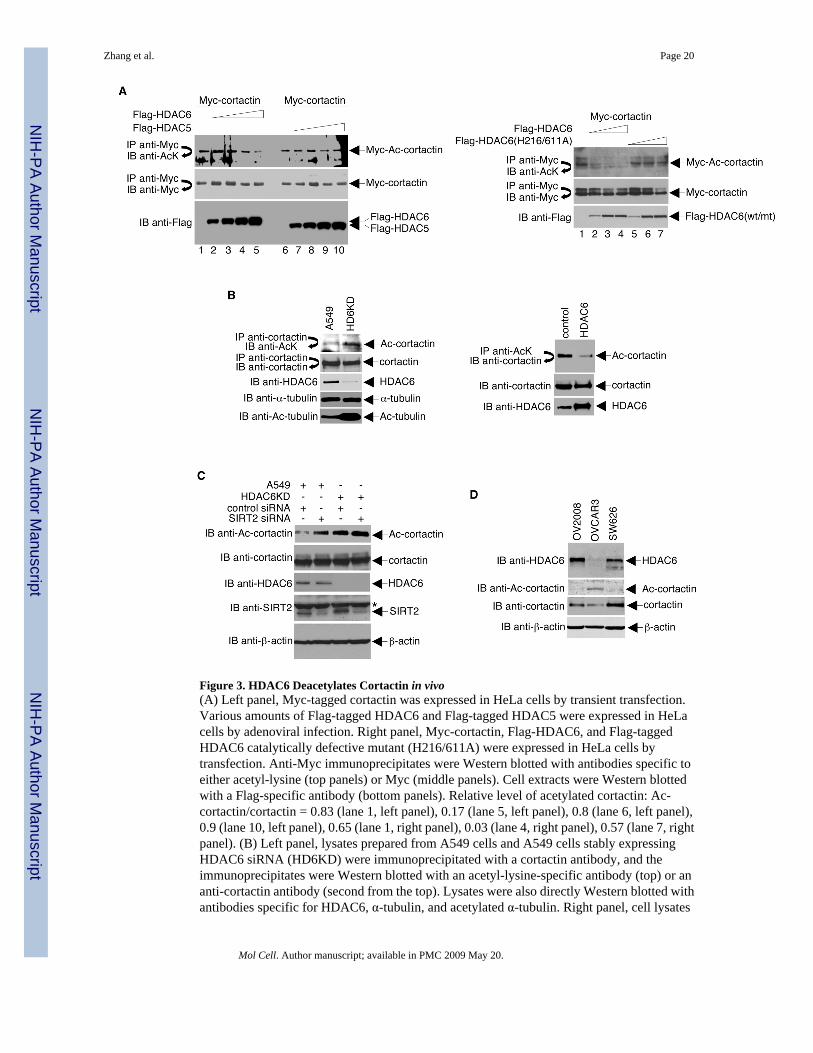
Figure 3. HDAC6 Deacetylates Cortactin in vivo(A) Left panel, Myc-tagged cortactin was expressed in HeLa cells by transient transfection.Various amounts of Flag-tagged HDAC6 and Flag-tagged HDAC5 were expressed in HeLacells by adenoviral infection. Right panel, Myc-cortactin, Flag-HDAC6, and Flag-taggedHDAC6 catalytically defective mutant (H216/611A) were expressed in HeLa cells bytransfection. Anti-Myc immunoprecipitates were Western blotted with antibodies specific toeither acetyl-lysine (top panels) or Myc (middle panels). Cell extracts were Western blottedwith a Flag-specific antibody (bottom panels). Relative level of acetylated cortactin: Ac-cortactin/cortactin = 0.83 (lane 1, left panel), 0.17 (lane 5, left panel), 0.8 (lane 6, left panel),0.9 (lane 10, left panel), 0.65 (lane 1, right panel), 0.03 (lane 4, right panel), 0.57 (lane 7, rightpanel). (B) Left panel, lysates prepared from A549 cells and A549 cells stably expressingHDAC6 siRNA (HD6KD) were immunoprecipitated with a cortactin antibody, and theimmunoprecipitates were Western blotted with an acetyl-lysine-specific antibody (top) or ananti-cortactin antibody (second from the top). Lysates were also directly Western blotted withantibodies specific for HDAC6, α-tubulin, and acetylated α-tubulin. Right panel, cell lysates
Zhang et al. Page 20
Mol Cell. Author manuscript; available in PMC 2009 May 20.
NIH
-PA Author Manuscript
NIH
-PA Author Manuscript
NIH
-PA Author Manuscript

were prepared from stably transfected HeLa cells expressing either the empty vector or highlevels of HDAC6 and used either in a high stringency immunoprecipitation with anti-acetyl-lysine-specific antibodies followed by Western blotting with anti-cortactin (top) or for Westernblotting with antibodies specific for either cortactin or HDAC6 (middle and bottom panels,respectively). (C) Four different sets of SIRT2 siRNA from the siGENOME SMART poolwere purchased from Dharmacon and used to knock-down SIRT2 expression in A549 orHDAC6KD cells. Protein expression levels and the effects on cortactin acetylation wereexamined by Western blotting with the indicated antibodies. * indicates a non-specific bandon the anti-SIRT2 Western blot. (D) Cell lysates prepared from three different ovarian cancercell lines subjected to Western blot analysis with the indicated antibodies.
Zhang et al. Page 21
Mol Cell. Author manuscript; available in PMC 2009 May 20.
NIH
-PA Author Manuscript
NIH
-PA Author Manuscript
NIH
-PA Author Manuscript

Figure 4. Cortactin is Acetylated Primarily in its Repeat Region(A) HeLa cells were transfected with Myc-tagged cortactin and various amounts of Flag-taggedPCAF, Flag-tagged PCAF catalytically-dead mutants, or HA-tagged p300. Anti-Mycimmunoprecipitates were Western blotted with antibodies specific for either acetyl-lysine orMyc. Levels of Flag-tagged PCAF and HA-tagged p300 were determined by Western blottingof cell extracts with antibodies specific for Flag and HA, respectively. To confirm that p300was active in this system, a Western blot was performed using the same extracts with anantibody specific for acetylated K382 of p53. (B) GST-cortactin was incubated with 14C-acetylCoA and recombinant PCAF or p300. Acetylation of GST-cortactin was visualized byautoradiography of SDS-PAGE gels (top panel). Levels of GST-cortactin were determined by
Zhang et al. Page 22
Mol Cell. Author manuscript; available in PMC 2009 May 20.
NIH
-PA Author Manuscript
NIH
-PA Author Manuscript
NIH
-PA Author Manuscript

Coomassie staining of SDS-PAGE gels (bottom panel). (C) Acetylation assays were performedusing purified GST-tagged cortactin fragments and PCAF. (D) HeLa cells were infected withadenoviruses encoding either GFP (−) or Flag-tagged HDAC6 (+). Whole cell lysates wereimmunoprecipitated with Flag-specific antibodies and Western blotted with antibodies againstacetylated lysines (top panel). The blot was stripped and re-probed with anti-Flag (middlepanel). A direct Western blot was performed to assess expression of Flag-HDAC6 (bottompanel).
Zhang et al. Page 23
Mol Cell. Author manuscript; available in PMC 2009 May 20.
NIH
-PA Author Manuscript
NIH
-PA Author Manuscript
NIH
-PA Author Manuscript

Figure 5. Identification of Acetylation Sites of Cortactin in vitro and in vivo(A) GST-tagged cortactin (in vitro acetylated by PCAF), Flag-tagged cortactin (expressed andpurified from HeLa cells), and endogenous cortactin immunopurified with anti-cortactinantibody were cleaved with trypsin and analyzed by ion trap mass spectrometry. Positions ofunambiguously identified acetylated lysine residues are listed. (B) In vitro acetylation assaysperformed using the catalytic domain of PCAF and GST-tagged (wildtype) or 9KQ mutantcortactin. Reaction products were resolved by SDS-PAGE. Acetylated proteins were visualizedby autoradiography (top panel), and total amounts of protein were monitored by Coomassiestaining (bottom panel). (C) Flag-tagged cortactin (wildtype) and Flag-tagged 9KQ mutantcortactin were expressed in HeLa cells by transient transfection. Anti-Flag immunoprecipitateswere Western blotted with antibodies specific to either acetyl-lysine or Flag. (D) Alignmentof the mouse cortactin repeats (residues 80 to 325). The green line represents the predictedtransmembrane zone, and the red bold letters represent the potential acetylated lysine residues.(E) Representation of the acetylated residues in balls (left) and sticks (center and right). Atomsin red belong to the lysine residues and atoms in blue to the acetyl groups. The molecule onthe right is colored according to atom type: carbon (green), nitrogen (blue), oxygen (red), andhydrogen (white). (F) Cartoon representations of both sides of the mouse cortactin model. Thecortactin repeats are in green and the SH3 motif in the carboxyl-terminal is in yellow. Theacetylated lysines are in the same color scheme described in E (left and center).
Zhang et al. Page 24
Mol Cell. Author manuscript; available in PMC 2009 May 20.
NIH
-PA Author Manuscript
NIH
-PA Author Manuscript
NIH
-PA Author Manuscript

Figure 6. Acetylation of Cortactin Reduces its Interaction with F-actin(A) GST-tagged cortactin (the repeat region) was acetylated by PCAF in vitro. Reactionmixtures were then incubated with mouse IgG or anti-acetyl-lysine antibodies. Precipitateswere resolved by SDS-PAGE. The band corresponding to the repeat region was eluted fromthe gel with 1% SDS. Eluates were incubated with F-actin, and co-sedimentation assays wereperformed. Supernatants (S) and pellets (P) were subjected to Western blotting with cortactin-specific antibodies. For lanes 1 and 2, co-sedimentation assays were performed usingunacetylated GST-tagged cortactin (the repeat region). (B, C, D) GST-tagged wildtype ormutant cortactin proteins were incubated with F-actin, and co-sedimentation assays wereperformed. Supernatants and pellets were subjected to Western blotting with an anti-GST
Zhang et al. Page 25
Mol Cell. Author manuscript; available in PMC 2009 May 20.
NIH
-PA Author Manuscript
NIH
-PA Author Manuscript
NIH
-PA Author Manuscript

antibody. Diagrams of the wildtype and mutant constructs are shown. (E) HeLa cells weretransfected with a plasmid encoding Flag-tagged cortactin (repeat region) and treated withethanol (vehicle control) or 400 ng/ml TSA for 12 h. Top panel, co-sedimentation assays wereperformed on cell lysates, and supernatants and pellets were subjected to Western blotting withan anti-Flag antibody. Bottom panel, Anti-Flag immunoprecipitates were Western blotted withan antibody specific for acetyl-lysine. (F) Similar co-sedimentation experiments wereperformed to assess endogenous cortactin-F-actin interactions.
Zhang et al. Page 26
Mol Cell. Author manuscript; available in PMC 2009 May 20.
NIH
-PA Author Manuscript
NIH
-PA Author Manuscript
NIH
-PA Author Manuscript

Figure 7. Acetylation of Cortactin Prevents its Localization to Membrane Ruffles and Inhibits CellMotility(A) Top panel, NIH3T3 cells were transfected with plasmids that express Myc-cortactin andvarious amounts of HA-Rac1G12V. Anti-Myc immunoprecipitates prepared from these cellswere assayed for cortactin acetylation by Western blotting with an anti-acetyl-lysine antibody.Immunoprecipitation efficiency and HA-Rac1G12V expression were monitored by Westernblotting with with anti-Myc and anti-HA, respectively. Bottom panel, NIH 3T3 cells weretransfected with plasmids encoding active Rac1 (HA-Rac1G12V) and either Flag-taggedwildtype, Flag-tagged 9KQ mutant, or Flag-tagged 9KR mutant cortactin. Twenty-four hourspost-transfection, cells were immunostained with antibodies or stained with 4′,6-diamidino-2-phenyl-indole and analyzed under a confocal microscope. (B) Top panel, NIH3T3 cells were
Zhang et al. Page 27
Mol Cell. Author manuscript; available in PMC 2009 May 20.
NIH
-PA Author Manuscript
NIH
-PA Author Manuscript
NIH
-PA Author Manuscript

serum-starved overnight and then treated with 20 ng/ml EGF. Lysates were analyzed byWestern blotting with anti-acetyl-cortactin antibodies. The blot was stripped and re-probedwith anti-cortactin antibodies. For controls, anti-phospho-ERK1/2 and anti-ERK1/2 Westernblots were performed to monitor the efficiency of EGF treatment. Bottom panels, NIH3T3 cellswere serum-starved overnight and then either mock-treated or treated with 10 ng/ml EGF for10 min. Endogenous HDAC6 and cortactin were detected using anti-HDAC6 and anti-cortactinspecific antibodies and Alexa-594- and Alexa-488-conjugated secondary antibodies.Flag-9KR and Flag-9KQ were detected with anti-Flag antibodies. (C) Left panels, 293T cellsstably expressing HDAC6 siRNA (HDAC6KD; generated by the OligoEngine retrovirus-mediated pSuper RNAi system) and 293T cells expressing control siRNA were serum-starvedovernight and assayed for migratory properties. Migratory cells were stained (top) andquantified at OD 560 nm following extraction (bottom). Right panels, parental NIH 3T3 cells(control) and NIH 3T3 cells stably expressing either wildtype or cortactin mutants were serum-starved overnight and assayed for migratory properties as in left panels. (D) Plasmidsexpressing wildtype or mutant cortactins were transfected into HT1080 cells depleted ofcortactin (cortactin KD). Stable polyclonal cell populations were selected and assayed for cellmigration activity as in (C). Control cells indicate parental untransfected HT1080 cells. (E)Three different ovarian cancer cell lines were assayed for cell migration activity as in (C). (F)Top left panel, Western blot analysis of HDAC6 expression in different ovarian cancer celllines. Bottom left panel, Western blots to assess HDAC6 knock-down in SKOV3 cells. Middleand right panels, SKOV3 cells transfected with either HDAC6 siRNA or control siRNA wereassayed for cell migration activity as in (C). (G) Data showing velocity of parental MDA-MB-231 cells (control) and a pool of stable cell clones overexpressing 9KQ mutant cortactinundergoing random motility on tissue-culture dishes. For MDA-MB-231 cells, n = 64; for 9KQstable clones, n = 76. *p < 0.0001 vs. MDA-MB-231 cells. Error bars denote the standarddeviation (SD).
Zhang et al. Page 28
Mol Cell. Author manuscript; available in PMC 2009 May 20.
NIH
-PA Author Manuscript
NIH
-PA Author Manuscript
NIH
-PA Author Manuscript