Regulation of Kruppel-like Factor 6 Tumor Suppressor Activity by Acetylation
11
Regulation of Kru ¨ ppel-like Factor 6 Tumor Suppressor Activity by Acetylation Dan Li, 1 Steven Yea, 1 Georgia Dolios, 2 John A. Martignetti, 2 Goutham Narla, 1 Rong Wang, 2 Martin J. Walsh, 3 and Scott L. Friedman 1 1 Division of Liver Diseases, Department of Medicine; and Departments of 2 Human Genetics and 3 Pediatrics, Mount Sinai School of Medicine, New York, New York Abstract Kru ¨ppel-like factor 6 (KLF6) is a zinc finger transcription factor and tumor suppressor that is inactivated in a number of human cancers by mutation, allelic loss, and/or promoter methylation. A key mechanism of growth inhibition by wild- type KLF6 is through p53-independent up-regulation of p21 WAF1/cip1 (CDKN1A), which is abrogated in several tumor-derived mutants. Here we show by chromatin immu- noprecipitation that transactivation of p21 WAF1/cip1 by KLF6 occurs through its direct recruitment to the p21 WAF1/cip1 promoter and requires acetylation by histone acetyltransfer- ase activity of either cyclic AMP–responsive element binding protein–binding protein or p300/CBP-associated factor. Direct lysine acetylation of KLF6 peptides can be shown by mass spectrometry. A single lysine-to-arginine point muta- tion (K209R) derived from prostate cancer reduces acetyla- tion of KLF6 and abrogates its capacity to up-regulate endogenous p21 WAF1/cip1 and reduce cell proliferation. These data indicate that acetylation may regulate KLF6 function, and its loss in some tumor-derived mutants could contribute to its failure to suppress growth in prostate cancer. (Cancer Res 2005; 65(20): 9216-25) Introduction Kru ¨ppel-like factor 6 (KLF6) is an ubiquitously expressed zinc finger protein belonging to the KLF family of transcription factors (1, 2). These proteins are DNA-binding transcriptional regulators that serve myriad roles in differentiation and development (3, 4). All KLF members possess a distinct NH 2 terminus activation domain and a highly conserved COOH terminus zinc finger DNA-binding domain that interacts with ‘‘GC box’’ or ‘‘CACC’’ DNA motifs in responsive promoters (3, 4). In contrast to the conserved DNA- binding domain among KLFs, the divergent activation domain of each family member accounts for their diverse biological activities. KLF6 has broad activity in regulating cell growth, tissue injury, and differentiation. Its transcriptional targets include a placental glycoprotein (5), HIV-1 (6), collagen a 1 (I) (2), transforming growth factor h1 (TGFh1), types I and II TGFh receptors (7), nitric oxide synthase (8), and urokinase type plasminogen activator (9). It is also an immediate early gene up-regulated in hepatic stellate cells during acute liver injury (1) and during the differentiation of preadipocytes into adipocytes (10), suggesting a generalized function of KLF6 in different biological contexts. KLF6 has recently been identified as a tumor suppressor gene that is inactivated in primary prostate (1, 11), colon (12) nasopharyngeal (13), glial (14), and hepatocellular (15) cancers and is down-regulated in lung and prostate cancers (16, 17), Decreased KLF6 mRNA expression correlates with clinical outcome in prostate cancer (17). Somatic loss of heterozygosity (LOH) and DNA mutations result in functional deletion of the KLF6 gene in f60% of prostate and 45% of colon tumors (1, 11, 12). KLF6 promoter silencing by methylation also has been uncovered in esophageal cancer cell lines (18). One mechanism by which KLF6 reduces cell proliferation is through up-regulation of p21 WAF1/cip1 , a key cyclin-dependent kinase (cdk) inhibitor. This induction does not require p53, a well- established transactivator of p21 WAF1/cip1 , because p21 WAF1/cip1 induction by KLF6 is preserved in p53-null cells (1). The molecular requirements for p21 WAF1/cip1 up-regulation by KLF6 have not been defined, and information about transcriptional coactivators within KLF6 transcriptional complexes is limited. Heterologous interac- tion of KLF6 with KLF4 or Sp1 is required for keratin (19) or endoglin gene expression (20), respectively, but potential interact- ing proteins in that context have not be explored. Coactivators, specifically histone acetyltransferases (HAT), contribute to the transcriptional activity of other tumor suppres- sors (e.g., p53), where recruitment of HATs is vital to its function (21). HATs are chromatin-modifying proteins that directly regulate transcription through interaction with transcription factors (22), including p53 (23, 24), pRb (25), and BRCA1 (26), as well as the zinc finger transcription factors Sp1 (27) and EKLF (28, 29). Altered interactions between HATs and transcription factors may contrib- ute to tumorigenesis. For example, disruption of the p300-p53 interaction may underlie the mechanism by which the viral oncoprotein E1A induces cell transformation (23, 24, 30). In vivo , HATs function as part of large protein complexes that share a highly conserved acetyl-CoA binding motif but have different substrate specificities. Among the best studied HATs, cyclic AMP–responsive element binding protein–binding protein (CBP) and p300/CBP-associated factor (PCAF) modulate gene transcription through the acetylation of specific lysine residues on chromatin, and function as coactivators for a number of trans- cription factors regulating cell growth and development (31–34). In addition, acetylation of nonhistone proteins has emerged as a novel mechanism of posttranslational modification (35, 36). Our identification of a KLF6 lysine-to-arginine mutation in primary prostate cancer raised the prospect that mutation of an acetylation substrate site might contribute to the loss of growth suppressive activity through the abrogation of p21 WAF1/cip1 trans- activation. In the present study, we have explored the role of CBP and PCAF in up-regulating p21 WAF1/cip1 gene expression by KLF6. Loss of lysine through mutation impairs KLF6’s capacity to transactivate the p21 WAF1/cip1 promoter or up-regulate endogenous Requests for reprints: Scott L. Friedman, Mount Sinai School of Medicine, Box 1123, 1425 Madison Avenue, Room 11-70C, New York, NY 10029-6574. Phone: 212-659- 9501; Fax: 212-849-2574; E-mail: [email protected]. I2005 American Association for Cancer Research. doi:10.1158/0008-5472.CAN-05-1040 Cancer Res 2005; 65: (20). October 15, 2005 9216 www.aacrjournals.org Research Article Research. on July 7, 2015. © 2005 American Association for Cancer cancerres.aacrjournals.org Downloaded from
Transcript of Regulation of Kruppel-like Factor 6 Tumor Suppressor Activity by Acetylation

Regulation of Kruppel-like Factor 6 Tumor Suppressor
Activity by Acetylation
Dan Li,1Steven Yea,
1Georgia Dolios,
2John A. Martignetti,
2Goutham Narla,
1
Rong Wang,2Martin J. Walsh,
3and Scott L. Friedman
1
1Division of Liver Diseases, Department of Medicine; and Departments of 2Human Genetics and 3Pediatrics,Mount Sinai School of Medicine, New York, New York
Abstract
Kruppel-like factor 6 (KLF6) is a zinc finger transcriptionfactor and tumor suppressor that is inactivated in a numberof human cancers by mutation, allelic loss, and/or promotermethylation. A key mechanism of growth inhibition by wild-type KLF6 is through p53-independent up-regulation ofp21WAF1/cip1 (CDKN1A), which is abrogated in severaltumor-derived mutants. Here we show by chromatin immu-noprecipitation that transactivation of p21WAF1/cip1 by KLF6occurs through its direct recruitment to the p21WAF1/cip1
promoter and requires acetylation by histone acetyltransfer-ase activity of either cyclic AMP–responsive element bindingprotein–binding protein or p300/CBP-associated factor.Direct lysine acetylation of KLF6 peptides can be shown bymass spectrometry. A single lysine-to-arginine point muta-tion (K209R) derived from prostate cancer reduces acetyla-tion of KLF6 and abrogates its capacity to up-regulateendogenous p21WAF1/cip1 and reduce cell proliferation. Thesedata indicate that acetylation may regulate KLF6 function,and its loss in some tumor-derived mutants could contributeto its failure to suppress growth in prostate cancer. (CancerRes 2005; 65(20): 9216-25)
Introduction
Kruppel-like factor 6 (KLF6) is an ubiquitously expressed zincfinger protein belonging to the KLF family of transcription factors(1, 2). These proteins are DNA-binding transcriptional regulatorsthat serve myriad roles in differentiation and development (3, 4). AllKLF members possess a distinct NH2 terminus activation domainand a highly conserved COOH terminus zinc finger DNA-bindingdomain that interacts with ‘‘GC box’’ or ‘‘CACC’’ DNA motifs inresponsive promoters (3, 4). In contrast to the conserved DNA-binding domain among KLFs, the divergent activation domain ofeach family member accounts for their diverse biological activities.KLF6 has broad activity in regulating cell growth, tissue injury,
and differentiation. Its transcriptional targets include a placentalglycoprotein (5), HIV-1 (6), collagen a1(I) (2), transforming growthfactor h1 (TGFh1), types I and II TGFh receptors (7), nitric oxidesynthase (8), and urokinase type plasminogen activator (9). It isalso an immediate early gene up-regulated in hepatic stellate cellsduring acute liver injury (1) and during the differentiation ofpreadipocytes into adipocytes (10), suggesting a generalizedfunction of KLF6 in different biological contexts.
KLF6 has recently been identified as a tumor suppressor genethat is inactivated in primary prostate (1, 11), colon (12)nasopharyngeal (13), glial (14), and hepatocellular (15) cancersand is down-regulated in lung and prostate cancers (16, 17),Decreased KLF6 mRNA expression correlates with clinical outcomein prostate cancer (17). Somatic loss of heterozygosity (LOH) andDNA mutations result in functional deletion of the KLF6 gene inf60% of prostate and 45% of colon tumors (1, 11, 12). KLF6promoter silencing by methylation also has been uncovered inesophageal cancer cell lines (18).One mechanism by which KLF6 reduces cell proliferation is
through up-regulation of p21WAF1/cip1, a key cyclin-dependentkinase (cdk) inhibitor. This induction does not require p53, a well-established transactivator of p21WAF1/cip1, because p21WAF1/cip1
induction by KLF6 is preserved in p53-null cells (1). The molecularrequirements for p21WAF1/cip1 up-regulation by KLF6 have not beendefined, and information about transcriptional coactivators withinKLF6 transcriptional complexes is limited. Heterologous interac-tion of KLF6 with KLF4 or Sp1 is required for keratin (19) orendoglin gene expression (20), respectively, but potential interact-ing proteins in that context have not be explored.Coactivators, specifically histone acetyltransferases (HAT),
contribute to the transcriptional activity of other tumor suppres-sors (e.g., p53), where recruitment of HATs is vital to its function(21). HATs are chromatin-modifying proteins that directly regulatetranscription through interaction with transcription factors (22),including p53 (23, 24), pRb (25), and BRCA1 (26), as well as the zincfinger transcription factors Sp1 (27) and EKLF (28, 29). Alteredinteractions between HATs and transcription factors may contrib-ute to tumorigenesis. For example, disruption of the p300-p53interaction may underlie the mechanism by which the viraloncoprotein E1A induces cell transformation (23, 24, 30).In vivo , HATs function as part of large protein complexes that
share a highly conserved acetyl-CoA binding motif but havedifferent substrate specificities. Among the best studied HATs,cyclic AMP–responsive element binding protein–binding protein(CBP) and p300/CBP-associated factor (PCAF) modulate genetranscription through the acetylation of specific lysine residues onchromatin, and function as coactivators for a number of trans-cription factors regulating cell growth and development (31–34). Inaddition, acetylation of nonhistone proteins has emerged as a novelmechanism of posttranslational modification (35, 36).Our identification of a KLF6 lysine-to-arginine mutation in
primary prostate cancer raised the prospect that mutation of anacetylation substrate site might contribute to the loss of growthsuppressive activity through the abrogation of p21WAF1/cip1 trans-activation. In the present study, we have explored the role of CBPand PCAF in up-regulating p21WAF1/cip1 gene expression by KLF6.Loss of lysine through mutation impairs KLF6’s capacity totransactivate the p21WAF1/cip1promoter or up-regulate endogenous
Requests for reprints: Scott L. Friedman, Mount Sinai School of Medicine, Box1123, 1425 Madison Avenue, Room 11-70C, New York, NY 10029-6574. Phone: 212-659-9501; Fax: 212-849-2574; E-mail: [email protected].
I2005 American Association for Cancer Research.doi:10.1158/0008-5472.CAN-05-1040
Cancer Res 2005; 65: (20). October 15, 2005 9216 www.aacrjournals.org
Research Article
Research. on July 7, 2015. © 2005 American Association for Cancercancerres.aacrjournals.org Downloaded from

p21WAF1/cip1. These findings suggest that acetylation of KLF6 playsa critical role in its regulation of target gene expression. Given itsrole as a tumor suppressor, loss of KLF6 acetylation may beassociated with tumorigenesis.
Materials and Methods
Expression plasmids. PCIneo-KLF6 (human; previously known as
‘‘Zf9’’) expression plasmid was constructed as previously described (1).HA-CBP was a gift from Dr. R.H. Goodman, Vollum Institute, Oregon
Health Sciences University, Portland, OR (37). PCI-PCAF plasmid was a gift
from Dr. Yoshihiro Nakatani, Department of Cancer Biology, Dana-Farber
Cancer Institute, Boston, MA (38). p21WAF1/cip1 promoter-luciferaseconstruct was a gift from Dr. Toru Ouchi, Department of Oncological
Sciences, Mount Sinai School of Medicine, New York, NY (39). pRL-TK
Vector used as a control for transfection efficiency was from Dual-luciferase Reporter Assay System (Promega, Madison, WI).
Site-directed mutagenesis. A lysine-to-arginine point mutant of KLF6
(pCIneoK209R) was constructed using Quick-Change site-directed mutagen-
esis kit (Stratagene, La Jolla, CA), as described below. pCIneo-KLF6 (human)was used as the template for the mutagenesis. The primers used for
mutagenesis were K209R sense, 5V-CCACTTTAACGGCTGCAGGAGAGTTTA-CACCAAAAGC-3V and K209R antisense, 5V-GCTTTTGGTGTAAACTC-TCCTGCAGCCGTTAAAGTGG-3V. All mutated constructs were sequencedon both strands to verify these mutations and to confirm that no other
alterations were introduced.
Cell culture. Prostate cancer 3 (PC3M) cells, 293 cells, and 293T cells
were obtained from the American Type Culture Collection (Manassas, VA).Cells were grown in DMEM supplemented with 10% fetal bovine serum
(Hyclone, Logan, UT), 100 units/mL penicillin and 100 units/mL
streptomycin, and 2 mmol/L L-glutamine (Life Technologies, Gaithersburg,MD). Treatment with trichostatin A (Sigma-Aldrich, St. Louis, MO) and
suberoylaniline hydroxamic acid (Biomol, Plymouth Meeting, PA) was
used at a final concentration of 0.5 Amol/L. Cell were recovered 8 to 12
hours following treatment with individual histone deacetylase (HDAC)inhibitors.
Transfection and luciferase reporter assays. Transient transfectionswere done using Lipofectamine 2000 reagent (Invitrogen, Carlsbad, CA). For
analyzing transfected protein using Western blot, PC3M cells cultured in10-cm plates (Corning, Acton, MA) were transfected with 10 Ag pCIneo
empty vector, pCIneoKLF6, or the pCIneoK209R mutant; cells were lysed 36
hours later for Western blot analysis. For luciferase assay, PC3 cells weretransfected with pCIneo-KLF6 (1 Ag), HA-CBP (5 Ag), pCl-PCAF (5 Ag),respectively or together, as indicated in text, together with the p21WAF1/cip1
promoter-luciferase construct. Five nanograms of pRL-TK plasmid (Prom-
ega) were cotransfected in each transfection as a control for transfectionefficiency. Forty-eight hours after tranfection, cells were washed thrice with
cold PBS and cell lysates prepared using Dual-luciferase reporter assay
system (Promega). The luciferase activity in 10 AL lysate was determined
using Dual-luciferase reporter assay system and luminometer (DynexTechnologies, Worthing. West Sussex, United Kingdom). Transfection
efficiency was normalized by Renilla luciferase activity measured concur-
rently in the same lysate. Stable cell lines were generated by cotransfectingthe appropriate expression constructs (pCIneo empty vector, KLF6 full
length, and KLF6 full length with K209R mutation) with a puromycin
expressing plasmid in a 10:1 ratio. Transfected cells were selected with
2.5 Ag/mL of puromycin, and pooled clones of cells were used in subsequentexperiments.
Chromatin immunoprecipitation assays. Chromatin immunoprecip-
itation assays were done as previously described (40) with minor
modifications described here. Briefly, 293 cells (4 � 106) were transfectedwith pCIneoKLF6 or pSG4 + Sp1 (gift from R. Tijan, University of
California at Berkeley and G. Gill, Harvard University, Boston, MA). Thirty-
six hours after transfection, cells were cross-linked with 1% formaldehyde
for 10 minutes at 37jC followed by cell lysis and sonication of DNA into200- to 1,000-bp fragments. Proteins cross-linked to DNA were
immunoprecipitated with 10 Ag anti-Zf9/KLF6 antibody (R-173; Santa
Cruz Biotechnology, Santa Cruz, CA) or anti-Sp1 antibody (UpstateBiotechnology, Charlottesville, VA) and 60 AL salmon sperm DNA/protein
A agarose beads. Antisera against histone H3 (Upstate Biotechnology) was
used as an input control for chromatin in each assay as shown. The
protein A agarose/antibody/protein complexes were washed extensivelyand eluted following the manufacturer’s instruction. The cross-linking was
reversed by heating at 65jC for 4 hours and proteins were digested by
proteinase K for 1 hour at 45jC. DNA was recovered by phenol/
chloroform extraction and ethanol precipitation in the presence of 10 Ag/mL yeast tRNA carrier and used as a template for PCR reactions. Genomic
sequence primers encompassing the p21WAF1/cip1 promoter region �150 to
�3 bp upstream of transcriptional start site were used to amplify
immunoprecipitated DNA as template: �150F, 5V-GCTGGGCAGCCAG-GAGCCTG-3V; �3R, 5V-CTGCTCACACCTCAGCTGGC-3V.
Immunoprecipitation and Western blotting. For coimmunoprecipi-
tation assays, 293T cells were transfected with 20 Ag of plasmid DNA.
Thirty-six hours after transfection, cells were washed twice with cold PBS
and lysed on ice for 20 minutes using immunoprecipitation lysis buffer
containing 0.5% NP40, 50 mmol/L Tris-HCl (pH 7.4), 120 mmol/L NaCl,
and protease inhibitors (Complete Protease Inhibitor Cocktail Tablets,
Roche, Nutley, NJ). The cell lysate was precipitated for 30 minutes at
14,000 � g at 4jC. The supernatant was immunoprecipitated at 4jC for 1
hour using 4 Ag of anti-Zf9/KLF6 antibody (R-173; Santa Cruz
Biotechnology), or 4 Ag of anti-HA antibody (Mount Sinai Hybridoma
Center, New York, NY) with 50 AL of protein-G beads (Pierce, Rockford,
IL), or using 50 AL of M2 anti-agarose (Sigma, St. Louis, MO). For
coimmunoprecipitation of endogenous KLF6 and CBP, monoclonal anti-
KLF6 antibody was cross-linked to agarose beads using ImmunoPure
Protein G IgG Plus Orientation Kit (Pierce). NIH 3T3 cells (1 � 108) were
lysed and precipitated, as described above. The supernatant was
immunoprecipitated using cross-linked anti-KLF6 agarose. The beads
were subsequently washed thrice with 800 AL immunoprecipitation wash
buffer [0.5% NP40, 50 mmol/L Tris-HCl (pH 7.4), and 500 mmol/L NaCl],
solubilized in Laemmli sample buffer (Sigma) containing 5% h-mercap-
toethanol, boiled, and separated by SDS-PAGE followed by Western
blotting as described below. As a negative control, immunoprecipitation
was done using control rabbit or mouse IgG (Sigma).For Western blotting, cell extracts were harvested in radioimmunopre-
cipitation assay buffer (Santa Cruz Biotechnology, standard protocol).
Protein samples (30 Ag per sample) were separated on SDS-polyacrylamidegel (6% for CBP and 10% for KLF6 and PCAF) and transferred to a
nitrocellulose membrane (Bio-Rad, Hercules, CA). The membranes were
blocked in 5% dried milk in 10 mmol/L Tris-HCl (pH 8.0), 150 mmol/L NaCl,
and 0.1% Tween 20 (1� TBS-T) for 1 hour at room temperature. Themembrane was incubated with primary antibody: anti-Zf9/KLF6 (R-173),
1:2,000 (Santa Cruz Biotechnology); anti-p21WAF1/cip1 (H-164), 1:250 (Santa
Cruz Biotechnology); anti-CBP-NT, 1:1,000 (Upstate Biotechnology); anti-
PCAF, 1:1,000 (Upstate Biotechnology); anti-acetyl-lysine antibody, 1:1,000(Cell Signaling, Beverly, MA). Secondary antibody (horseradish peroxidase–
conjugated anti-rabbit or anti-mouse IgG (Amersham Biosciences, Piscat-
away, NJ) was used according to manufacturer’s instruction at 1:2,000dilution followed by enhanced chemiluminescence protocol (Amersham
Pharmacia).
In vitro protein acetyltransferase assay. Protein acetyltransferase
reactions were done with in vitro translated KLF6 protein and synthesizedby using the TNT in vitro transcription and translation system (Promega)
either in the absence of radioisotope or in the presence of [14C]-leucine
(50 ACi/mmol, Perkin Elmer, Boston, MA). For acetyltransferase reactions,f1 Ag of in vitro translated KLF6 and 100 ng of immunoprecipitatedHA or FLAG-tagged proteins of CBP (wild type), CBP (HAT�; kind gifts
from Dr. R.H. Goodman), or PCAF (wild type) were incubated with
[14C]-acetyl-CoA (55 ACi/mmol, New England Nuclear) for 1 hour at 30jC.After acetyltransferase reactions, KLF6 protein was immunoprecipitatedwith anti-Zf9/KLF6 antibody (R-173, Santa Cruz Biotechnology). The
entire reaction mixture was separated by 10% SDS-PAGE. Gels containing
[14C]-labeled proteins were stained and fixed with 10% glacial aceticacid and 40% methanol for 1 hour, soaked in Amplify (Amersham
Acetylation Regulates KLF6 Tumor Suppressor
www.aacrjournals.org 9217 Cancer Res 2005; 65: (20). October 15, 2005
Research. on July 7, 2015. © 2005 American Association for Cancercancerres.aacrjournals.org Downloaded from

Pharmacia) for 40 minutes, and exposed to X-ray film for autoradiographyfor f10 minutes.
Analysis of proliferation. Proliferation was determined by estimating3H-thymidine incorporation. BPH1 and PC3M cell lines stably expressing
the appropriate expression vectors were plated at a density of 50,000 cellsper well in 12-well dishes. Twenty-four hours after plating, 1 ACi/mL 3H-
thymidine (Amersham Biosciences) was added. After 2 hours, cells were
washed four times with ice-cold PBS and fixed in methanol for
30 minutes at 4jC. After methanol removal and cell drying, cells weresolubilized in 0.25% sodium hydroxide/0.25% SDS. After neutralization
with hydrochloric acid (1 N), disintegrations per minute were estimated
by liquid scintillation counting. This process was repeated at 48 and
72 hours.Tumor samples. The preparation of tumor samples was as previously
described (1). Briefly, 5-Am sections stained with H&E were used as anaccurate histologic reference for normal and tumor-derived tissue.Microdissection was done on sequential 20-Am sections and DNAsubsequently extracted (Ambion paraffin block isolation kit, Austin, TX).DNA was isolated by proteinase K digestion overnight at 37jC incubatorfollowed by heat inactivation at 95jC for 10 minutes. KLF6 sequenceanalysis of tumor samples was done as previously described (1).
Peptide acetylation assay. Four peptides were synthesized using
commercially available resources (Invitrogen) covering the majority oflysine residues within the KLF6 molecule. The peptides’ sequences are as
follows: (a) h68-87 ILAREKKEESELKISSSPPE, (b) h115-134 SSEELSP-
TAKFTSDPIGEVL, (c) h204-223 FNGCRKVYTKSSHLKAHQRT, and (d)h248-267 TRHFRKHTGAKPFKCSHCDR. For peptide acetylation assay, 5 ng
of peptide were incubated at 30jC for 1.5 hours with 5 ng CBP or PCAF, 10 ALof 1 mmol/L acetyl CoA, 5 AL of 0.1 mol/L sodium butyrate, in the presence
or absence of 10 AL 3H-acetyl CoA (0.5 ACi/AL, Amersham Pharmacia), in1� HAT assay buffer (Upstate Biotechnology). Following the reaction, the
mixture was analyzed by scintillation counting to confirm 3H incorporation
followed by mass spectrometry (MS) analysis. To assess 3H incorporation,
5 AL of reaction mixture were spotted onto a small square of filter paperfollowed by washing with 50 mmol/L Na2HPO4 (pH 9.0), then the filter paper
was placed into scintillation fluid overnight for counting the next day.
Mass spectrometry analysis. KLF6 peptides and their acetylationproducts (1 pmol) were isolated and purified using Poros 20 R2 beads(Applied Biosystems, Foster City, CA) and ZipTipC18 pipette tip (Millipore,Bedford, MA) following the manufacturer’s protocol. Molecular masses ofthe synthetic peptides before and after acetylation reaction were accuratelymeasured by matrix-assisted laser desorption ionization-MS (MALDI-MS)using a QSTAR XL hybrid quadrupole time-of-flight mass spectrometer(Applied Biosystems). a-Cyano-4-hydroxy-cinnamic acid was used as matrixfor sample preparation. To determine the acetylation site(s), fragmentspectra of peaks corresponding to acetylated peptides were collected andanalyzed by MALDI tandem MS (MS/MS) experiment using the same massspectrometer.
Results
KLF6 targets the p21WAF1/cip1 (CDKN1A) locus and isassociated with hyperacetylation of histone H3. To determineif KLF6 interacts directly with the endogenous p21WAF1/cip1
(CDKN1A) promoter, we used chromatin immunoprecipitation(Fig. 1). PCR reactions were carried out using primers encompassing�150 to �3 bp upstream of the start site of p21WAF1/cip1
transcription as shown (Fig. 1A), which contains multiple GC boxespredicted by sequence homology to interact with KLF6 and relatedfamily members. Chromatin immunoprecipitation analysis con-firmed that KLF6 binds to this region of the endogenous p21WAF1/cip1
promoter, establishing p21WAF1/cip1 as a transcriptional target ofKLF6 (Fig. 1B). Interestingly, we were unable to show the binding ofSp1 to the same region despite reports to the contrary based onelectromobility shift assay (41). To confirm the ability of the Sp1antibody to recognize Sp1 in the chromatin immunoprecipitation
assay, a positive control was used to verify Sp1 on other loci (data notshown). Furthermore, ectopic expression of KLF6 showed increasedrecruitment of acetylated histone H3 encompassing the regionbetween�150 to�3 of the KLF6 promoter when compared with thecontrol expression vector (Fig. 1C).KLF6 interacts with CBP and PCAF in vivo. Because CBP and
PCAF interact with critical cellular proteins, including p53 (21) andE2F1 (42) leading to altered function, we examined whether KLF6interacts with CBP and/or PCAF using a coimmunoprecipitationassay. As shown in Fig. 2A (top), CBP was associatedwith KLF6 whenwhole cell lysate of 293T cells expressing Flag-tagged KLF6 and HA-tagged CBP was immunoprecipitated using anti-Flag antibody andblotted with anti-HA antibody; this result was confirmed byreciprocal coimmunoprecipitation (Fig. 2A). To show the interactionbetween endogenous KLF6 and CBP, coimmunoprecipitation was
Figure 1. KLF6 is directly associated with the p21WAF1/cip1 (CDKN1A )locus and deposition of acetylated histone H3. A, schematic representation ofp21WAF1/cip1 promoter. PCR for chromatin immunoprecipitation (ChIP) assaywas done using primers (small arrows ) positioned at �150 and �3 bpupstream of transcriptional start site of p21WAF1/cip1 promoter. This GC-richregion contains five putative binding sites for KLF6 as well as for Sp1, all of whichare between �102 and �30 bp region, as shown in (A ). B, chromatinimmunoprecipitation assay at p21WAF1/cip1 promoter in 293 cells. Anti-KLF6antibody (aKLF6) was used for immunoprecipitation. Anti-histone H3(aHistone H3) antibody was used as a positive control. The binding of Sp1 top21waf1/cip1 promoter is also examined using anti-Sp1 antibody (aSp1).C, acetylation of the p21WAF1/cip1 (CDKN1A ) locus within the gene promoterwas monitored upon transfection with a KLF6 expression vector by chromatinimmunoprecipitation analysis. Input levels of chromatin for immunoprecipitationreactions were monitored for total histone H3. HDAC inhibitors trichostatin A(TSA ) and suberoylanilide hydroxamic acid (SAHA ) at a final concentration of0.5 Amol/L were used to compare acetylation of histone H3.
Cancer Research
Cancer Res 2005; 65: (20). October 15, 2005 9218 www.aacrjournals.org
Research. on July 7, 2015. © 2005 American Association for Cancercancerres.aacrjournals.org Downloaded from

done from a lysate of 1 � 108 NIH 3T3 cells using a monoclonalanti-KLF6 antibody cross-linked to protein G agarose. Western blotusing anti-CBP antibody revealed that CBP was coimmunoprecipi-tated with KLF6 (Fig. 2B). The interaction between KLF6 and PCAFwas also shown using a similar approach (Fig. 2C).CBP and PCAF synergize with KLF6 in transactivating
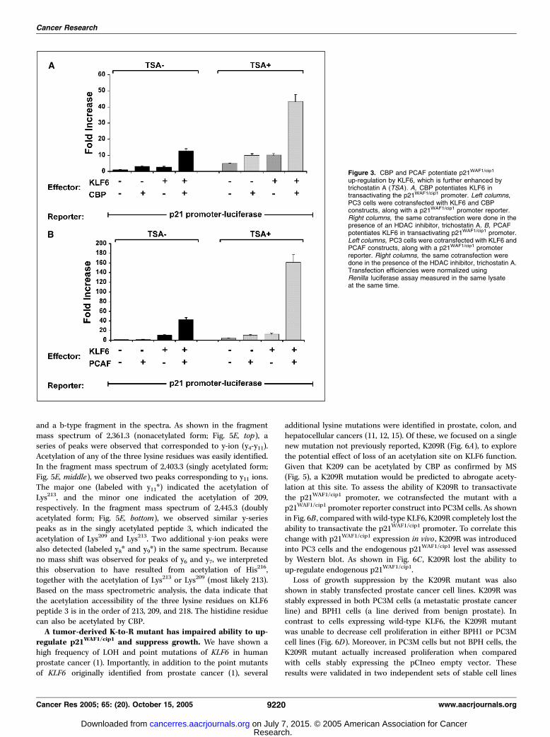
p21WAF1/cip1. To establish the functional significance of the inter-action between KLF6 and CBP and/or PCAF, we explored whetherCBP and PCAF potentiated the transactivation of p21WAF1/cip1 byKLF6 (Fig. 3). As shown in Fig. 3A , KLF6 transactivated thep21WAF1/cip1 (CDKN1A) promoter 3-fold, which was furtherincreased to 12-fold in the presence of CBP. When a HDAC inhi-bitor, trichostatin A was added, the transactivation of p21WAF1/cip1
by KLF6 was further increased, with the maximal transactivation>40-fold achieved in the presence of CBP plus trichostatin A.In addition, we also examined the synergistic effects of KLF6 and
PCAF in driving p21WAF1/cip1 transactivation (Fig. 3B). In the pres-ence of PCAF, the transactivation of p21WAF1/cip1 by KLF6 wasenhanced 14-fold. Again, trichostatin A maximally increased thesynergistic effects of KLF6 and PCAF. These results suggest thefunctional involvement of CBP and PCAF in KLF6-mediatedp21WAF1/cip1 up-regulation. Considering the roles of CBP and PCAFas acetyltransferases, these results suggested that enhanced acety-lation of KLF6 by trichostatin A promoted the transactivation ofp21WAF1/cip1 promoter. Previous studies showed that trichostatin Aitself enhances transactivation of the p21WAF1/cip1 promoter (41).This is consistent with the results shown in Fig. 3, in which theluciferase activity in empty vector control was also increased inthe presence of trichostatin A. It is likely that this effect is due to theenhanced acetylation by trichostatin A on endogenous transcriptionfactors that transactivate the exogenous p21WAF1/cip1 promoterconstruct.KLF6 is acetylated by CBP and PCAF, in vitro and in vivo .
The data above suggested that KLF6 and either CBP or PCAF
functionally interact on the p21WAF1/cip1 promoter and raisedthe possibility that acetylation of KLF6 by either these twoHATs promoted p21WAF1/cip1 transactivation. To test to thishypothesis, we did an in vitro acetylation assay. As shown inFig. 4A , KLF6 was acetylated in vitro by both CBP and PCAF butnot by a CBP mutant lacking the HAT domain. To verify thatKLF6 was also acetylated in vivo , we immunoprecipitated KLF6from 293T cells and blotted with an anti-acetylated lysine anti-body (Fig. 4B).Determination of key acetylation sites on KLF6. To determine
which lysine residues on KLF6 are acetylated, we used syntheticKLF6 peptide fragments covering the majority of lysine residues ofKLF6 (Fig. 5A). Two peptides were located in the transactivationdomain (peptides 1 and 2) and the other two were located in theDNA-binding domain (peptides 3 and 4). HAT assay in vitro wasdone using 3H-labeled acetyl-CoA to identify acetylated residues(Fig. 5B and C). When KLF6 peptides were subjected to acetylationby CBP, a 4.5-fold increase in counts per minute (CPM) in peptide3 and 2.7-fold increase in CPM in peptide 4 were observed(Fig. 5B). On the other hand, no significant increase in cpm wasobserved when KLF6 peptides were subjected to acetylation byPCAF (Fig. 5C). These data indicate that lysine residues in the DNA-binding domain of KLF6 are acetylation sites for CBP.Molecular mass measurement of the KLF6 peptides by MS also
confirmed that molecular mass shifting resulted from the acetyla-tion (42 amu per acetylation). Molecular mass spectra of peptide 3before and after acetylation by CBP (Fig. 5D) indicated that all ofthe three lysine residues in peptide 3 could be acetylated, but themajority form was singly acetylated followed by a doublyacetylated form. To identify the acetylation site(s) on peptide 3,molecular ions corresponding to nonacetylated, singly, and doublyacetylated peptide 3 were analyzed by fragment mass analysisusing MS/MS (Fig. 5E). To simplify data interpretation, we onlylabeled those peaks corresponding to y-series peptide fragments
Figure 2. KLF6 interacts with CBP and PCAF in vivo . A, KLF6 interacts with CBP. 293T cells were cotransfected with FLAG-tagged KLF6 and HA-tagged CBPconstructs. Thirty-six hours after transfection, cells were lysed and coimmunoprecipitated followed by SDS-PAGE/Western analysis. Top, cell lysates wereimmunoprecipitated with anti-FLAG antibody. Anti-HA antibody was used in a Western blot. Bottom, cell lysates were immunoprecipitated with anti-HA antibody.Anti-FLAG antibody was used in Western blot. B, KLF6 interacts with CBP in vivo . Lysate of 1 � 108 NIH 3T3 cells was immunoprecipitated with agarose-linkedanti-KLF6 antibody to pull down endogenous KLF6. Following SDS-PAGE, anti-CBP antibody was used in Western blot to detect the endogenous CBP that wascoimmunoprecipitated with KLF6. C, KLF6 interacts with PCAF. 293T cells were cotransfected with KLF6 and FLAG-tagged PCAF constructs. Thirty-six hours aftertransfection, cells were lysed and coimmunoprecipitated followed by SDS-PAGE/Western analysis. Top, cell lysates were immunoprecipitated with anti-FLAG antibody.Anti-KLF6 antibody was used in Western blot. Bottom, cell lysates were immunoprecipitated with anti-KLF6 antibody. Anti-PCAF antibody was used in Western blot.
Acetylation Regulates KLF6 Tumor Suppressor
www.aacrjournals.org 9219 Cancer Res 2005; 65: (20). October 15, 2005
Research. on July 7, 2015. © 2005 American Association for Cancercancerres.aacrjournals.org Downloaded from
and a b-type fragment in the spectra. As shown in the fragmentmass spectrum of 2,361.3 (nonacetylated form; Fig. 5E, top), aseries of peaks were observed that corresponded to y-ion (y4-y11).Acetylation of any of the three lysine residues was easily identified.In the fragment mass spectrum of 2,403.3 (singly acetylated form;Fig. 5E, middle), we observed two peaks corresponding to y11 ions.The major one (labeled with y11*) indicated the acetylation ofLys213, and the minor one indicated the acetylation of 209,respectively. In the fragment mass spectrum of 2,445.3 (doublyacetylated form; Fig. 5E, bottom), we observed similar y-seriespeaks as in the singly acetylated peptide 3, which indicated theacetylation of Lys209 and Lys213. Two additional y-ion peaks werealso detected (labeled y8* and y9*) in the same spectrum. Becauseno mass shift was observed for peaks of y6 and y7, we interpretedthis observation to have resulted from acetylation of His216,together with the acetylation of Lys213 or Lys209 (most likely 213).Based on the mass spectrometric analysis, the data indicate thatthe acetylation accessibility of the three lysine residues on KLF6peptide 3 is in the order of 213, 209, and 218. The histidine residuecan also be acetylated by CBP.A tumor-derived K-to-R mutant has impaired ability to up-
regulate p21WAF1/cip1 and suppress growth. We have shown ahigh frequency of LOH and point mutations of KLF6 in humanprostate cancer (1). Importantly, in addition to the point mutantsof KLF6 originally identified from prostate cancer (1), several
additional lysine mutations were identified in prostate, colon, andhepatocellular cancers (11, 12, 15). Of these, we focused on a singlenew mutation not previously reported, K209R (Fig. 6A), to explorethe potential effect of loss of an acetylation site on KLF6 function.Given that K209 can be acetylated by CBP as confirmed by MS(Fig. 5), a K209R mutation would be predicted to abrogate acety-lation at this site. To assess the ability of K209R to transactivatethe p21WAF1/cip1 promoter, we cotransfected the mutant with ap21WAF1/cip1 promoter reporter construct into PC3M cells. As shownin Fig. 6B , compared with wild-type KLF6, K209R completely lost theability to transactivate the p21WAF1/cip1 promoter. To correlate thischange with p21WAF1/cip1 expression in vivo , K209R was introducedinto PC3 cells and the endogenous p21WAF1/cip1 level was assessedby Western blot. As shown in Fig. 6C , K209R lost the ability toup-regulate endogenous p21WAF1/cip1.Loss of growth suppression by the K209R mutant was also
shown in stably transfected prostate cancer cell lines. K209R wasstably expressed in both PC3M cells (a metastatic prostate cancerline) and BPH1 cells (a line derived from benign prostate). Incontrast to cells expressing wild-type KLF6, the K209R mutantwas unable to decrease cell proliferation in either BPH1 or PC3Mcell lines (Fig. 6D). Moreover, in PC3M cells but not BPH cells, theK209R mutant actually increased proliferation when comparedwith cells stably expressing the pCIneo empty vector. Theseresults were validated in two independent sets of stable cell lines
Figure 3. CBP and PCAF potentiate p21WAF1/cip1
up-regulation by KLF6, which is further enhanced bytrichostatin A (TSA ). A, CBP potentiates KLF6 intransactivating the p21WAF1/cip1 promoter. Left columns,PC3 cells were cotransfected with KLF6 and CBPconstructs, along with a p21WAF1/cip1 promoter reporter.Right columns, the same cotransfection were done in thepresence of an HDAC inhibitor, trichostatin A. B, PCAFpotentiates KLF6 in transactivating p21WAF1/cip1 promoter.Left columns, PC3 cells were cotransfected with KLF6 andPCAF constructs, along with a p21WAF1/cip1 promoterreporter. Right columns, the same cotransfection weredone in the presence of the HDAC inhibitor, trichostatin A.Transfection efficiencies were normalized usingRenilla luciferase assay measured in the same lysateat the same time.
Cancer Research
Cancer Res 2005; 65: (20). October 15, 2005 9220 www.aacrjournals.org
Research. on July 7, 2015. © 2005 American Association for Cancercancerres.aacrjournals.org Downloaded from

for both BPH1 and PC3M cells. Collectively, these data suggestthat the acetylation at K209 is necessary for KLF6 to up-regulatep21WAF1/cip1 gene expression, which in turn can inhibit cellularproliferation.
Discussion
KLF6 has been established as a tumor suppressor gene involvedin key intracellular pathways in a number of human cancers (1, 12,14, 15, 43, 44). Among several target genes transcriptionallyregulated by KLF6, p21WAF1/cip1 has been particularly relevant toprostate cancer given p21WAF1/cip1’s central role in growthregulation, especially as a mediator of p53-stimulated cell cyclearrest. The acetylation status of the human p21WAF1/cip1 (CDKN1A)locus has been extensively characterized in cellular contexts thatare tied to cancer development (45). These activities requirespecific GC-rich elements (46) in the p21WAF1/cip1 promoter, whichare consensus target sequences for KLF6 binding (1). However,little is known about either the biochemical requirements fortranscriptional activity of KLF6, or the complexes through whichKLF6 regulates target gene activity.An important goal in studying KLF6 has been to understand
the physiologic significance of protein complexes that modify itsfunction in regulating the activity of its target genes. Recently, weshowed that KLF6 sequesters cyclin D1 to reduce cyclin D/cdk4interactions and enhance the phosphorylation of pRb, therebypromoting G1 cell cycle arrest (47). This activity alters theequilibrium of p21WAF1/cip1 levels, with induction and titrationonto cdk2 complexes and further growth suppression (47). These
findings suggest that covalent modifications may differentiallyregulate KLF6 activity.Despite clear evidence that KLF6 transcriptionally activates
p21WAF1/cip1 (1, 12), the biochemical mechanisms underlying thisobservation have not been clarified. Here, we establish that KLF6activity is associated with increased complexing of acetylatedhistones within the p21WAF1/cip1 (CDKN1A) locus. Furthermore, weshow that KLF6 interacts with the CBP/p300 complex and itsacetylation by CBP contributes to normal KLF6 activity. Moreover,MS shows acetylation of a specific lysine residue of KLF6 that ismutated in a primary prostate tumor. Collectively, the findingsprovide evidence that acetylation of KLF6 is biologically significant,and its dysregulation in human cancer may contribute to loss ofKLF6’s growth suppressive activity.Our findings also provide an important functional link between
protein acetyltransferases and KLF6 by showing that KLF6 recruitsCBP and PCAF to the p21WAF1/cip1 locus. In contrast to the evidencethat KLF6 occupies the p21WAF1/cip1 promoter using chromosomalimmunoprecipitation, the lack of promoter occupation by Sp1 wasunexpected. One potential explanation may be the weak affinity ofdifferent antisera used against Sp1 from cross-linked material todetect the protein in these experiments. Alternatively, Sp1 may re-present a low-abundance protein involved primarily in stimulatedrather than basal expression of p21WAF1/cip1. This possibility issupported by studies showing that p21WAF1/cip1 transactivation bySp1 is greatly enhanced by TGFh signaling, for example (48).Although our study does not exclude the possible participation ofSp1-like factors in regulating the expression of p21WAF1/cip1 (49), itmay indicate a greater role for KLF6 than Sp1 in regulatingp21WAF1/cip1 within this specific cellular context. Additionally, Sp1may use different GC boxes in the p21WAF1/cip1 promoter fromthose used by KLF6 under our experimental conditions. In othercontexts, there may be cooperation between KLF6 and Sp1, assuggested by their transcriptional synergy in regulating theexpression of TGFh and of endoglin, a TGFh-binding protein (20).Thus, there may be a precise stoichiometry between KLF6 and Sp1that is promoter and context specific in regulating their targetgenes.The identification of several point mutations of KLF6 in human
cancer affecting lysine residues led us to explore the potential roleof lysine modification in regulating KLF6 activity. These haveincluded mutations in prostate (K186R; ref. 1), colon (K74R; ref.12), and hepatocellular carcinoma (K182R; ref. 15). Here we testedthe functional activity of an additional prostate cancer–derivedmutant of KLF6, K209R, for its capacity to both transactivate thep21waf1/cip1 promoter, and to function as a substrate for proteinacetyltransferase activity by CBP. Our findings indicate thatacetylation of a specific Lys209 was closely linked with CBP-dependent activation of endogenous p21WAF1/cip1 in vitro .Moreover, its mutation to arginine as seen in prostate cancerabrogated p21WAF1/cip1 transactivation. Interestingly, this lysine iswithin the peptide consensus sequence for acetylation of p53 byp300/CBP (21, 50, 51), consistent with recent models proposed foracetylation of p53 (21). However, despite ample evidence thatacetylation of p53 regulates several of its functions (52), therelative biological significance of p53 acetylation still remainsuncertain. Because p53 has been one of the most thoroughlystudied molecules that is covalently modified (52), more recentefforts have focused on the interdependence of its differentcovalent modifications, including phosphorylation, acetylation,methylation, ubiquitination, and sumoylation, and how they affect
Figure 4. KLF6 is acetylated in vitro and in vivo . A, KLF6 is acetylated in vitroby CBP and PCAF. Equivalent molar amount of CBP (wild type), CBP (HAT�),and PCAF (wild type) were immunoprecipitated from cells expressing HA-taggedCBP, HA-tagged CBP (HAT�), and FLAG-tagged PCAF (wild type). In vitrotranslated KLF6 was incubated with immunoprecipitation-purified CBP (wild type),CBP (HAT�), and PCAF (wild type), together with [14C]-acetyl CoA. KLF6 wassynthesized with [14C]-leucine in vitro and immunoprecipitated as a control.Immunoprecipitated products were resolved on SDS/PAGE followed byautoradiography. B, KLF6 is acetylated in vivo . 293T cells expressingFLAG-tagged KLF6 were immunoprecipitated with anti-FLAG agarose, followedby SDS-PAGE and Western blot analysis. Immunoprecipitated KLF6 wasdetected by anti-acetylated lysine antibody. Moreover, using lysates from thesame cell line, FLAG-tagged KLF6 was immunoprecipitated by anti-acetylatedlysine antibody and detected by anti-FLAG antibody using Western blot.
Acetylation Regulates KLF6 Tumor Suppressor
www.aacrjournals.org 9221 Cancer Res 2005; 65: (20). October 15, 2005
Research. on July 7, 2015. © 2005 American Association for Cancercancerres.aacrjournals.org Downloaded from
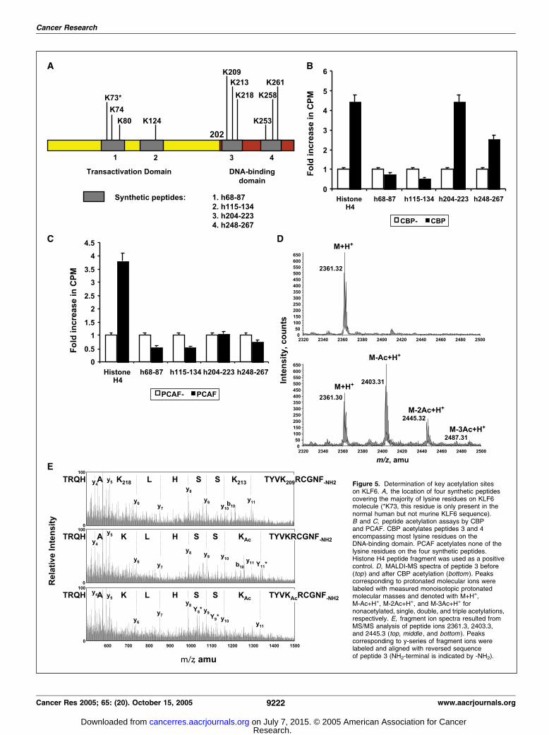
Figure 5. Determination of key acetylation siteson KLF6. A, the location of four synthetic peptidescovering the majority of lysine residues on KLF6molecule (*K73, this residue is only present in thenormal human but not murine KLF6 sequence).B and C, peptide acetylation assays by CBPand PCAF. CBP acetylates peptides 3 and 4encompassing most lysine residues on theDNA-binding domain. PCAF acetylates none of thelysine residues on the four synthetic peptides.Histone H4 peptide fragment was used as a positivecontrol. D, MALDI-MS spectra of peptide 3 before(top ) and after CBP acetylation (bottom ). Peakscorresponding to protonated molecular ions werelabeled with measured monoisotopic protonatedmolecular masses and denoted with M+H+,M-Ac+H+, M-2Ac+H+, and M-3Ac+H+ fornonacetylated, single, double, and triple acetylations,respectively. E, fragment ion spectra resulted fromMS/MS analysis of peptide ions 2361.3, 2403.3,and 2445.3 (top, middle , and bottom ). Peakscorresponding to y-series of fragment ions werelabeled and aligned with reversed sequenceof peptide 3 (NH2-terminal is indicated by -NH2).
Cancer Research
Cancer Res 2005; 65: (20). October 15, 2005 9222 www.aacrjournals.org
Research. on July 7, 2015. © 2005 American Association for Cancercancerres.aacrjournals.org Downloaded from

p53’s interaction with other cellular and viral factors, therebyaltering protein p53 levels (52–55).Acetylation of specific lysine residues by different acetyltrans-
ferases may lead to divergent functional outcomes. For example,acetylation of pRb at specific lysine residues by p300 has been
linked to inhibition of cell cycle progression (25), whereas acety-lation of pRb by PCAF of key Lys873/874 near the COOH terminuscorrelates with cell differentiation (56). These divergent outcomesmay also reflect differences between p300 and PCAF in promotingcell differentiation in vivo (57). It seems possible that similar, subtle
Figure 6. A lysine (K) to arginine (R ) mutant (K209R) identified from prostate cancer has impaired ability to up-regulate p21WAF1/cip1 expression. A, a KLF6 mutation(K209R) affecting an acetylation site in primary prostate cancer. Sequencing chromatogram of DNA derived from microdissected primary prostate cancer is shown, alongwith normal sequence from the surrounding unaffected sequence. B, K209 tumor mutant has reduced ability to transactivate p21WAF1/cip1 promoter. Wild-type KLF6and K209R were cotransfected with a p21WAF1/cip1 promoter reporter, respectively. Empty expression vector was transfected as a control for baseline luciferase activity.Transfection efficiencies were normalized using Renilla luciferase assay measured in the same lysate at the same time. C, K209R tumor mutant has reduced ability toup-regulate endogenous p21WAF1/cip1. Wild-type KLF6 and K209R were transfected into PC3 cells, respectively. Thirty-six hours after transfection, cells were lysed andloaded onto SDS-PAGE followed by Western blot analysis. Anti-p21WAF1/cip1 antibody was used to detect the expression of endogenous p21WAF1/cip1 protein. The proteinexpression of transfected KLF6 and mutants constructed was detected by anti-KLF6 antibody. Tubulin was blotted as a control for protein loading. D, impaired growthsuppression by K209R mutant in stable prostate cell lines. Both PC3M and BPH cell lines were stably transfected with either pCI-neo empty vector, KLF6 wild type,or K209R mutant as described in Materials and Methods, and cell proliferation was assessed by estimating 3H-thymidine incorporation at 24, 48, and 72 hours afterreplating equal numbers of cells. The differences were statistically significant, as indicated by Ps. 1, P<0.05; 111, P<0.01.
Acetylation Regulates KLF6 Tumor Suppressor
www.aacrjournals.org 9223 Cancer Res 2005; 65: (20). October 15, 2005
Research. on July 7, 2015. © 2005 American Association for Cancercancerres.aacrjournals.org Downloaded from

differences in KLF6 acetylation may also underlie functionaldifferences as well, a possibility that merits further study. Interes-tingly, rodent and human KLF6 carry the consensus R-K-X-X-T-Ksequence at residues 208 to 213, which is as a substrate site forp300/CBP in nuclear factor-nB (relA)–mediated inflammation andinteraction with InBa (58). Thus far, KLF5 and KLF13 are the onlyKLFs reported to be regulated by acetylation (59, 60).In summary, our data establish a role of KLF6 in recruiting a
specific coactivator complex containing CBP and PCAF to promotetranscriptional activation of p21WAF1/cip1 and provide evidencethat KLF6 serves as a substrate for CBP HAT activity. Combinedwith the loss of growth suppressive activity of a tumor-derived
lysine mutant of KLF6, these findings point to acetylation as a keymodification in regulating its growth- and tumor-suppressiveactivities.
AcknowledgmentsReceived 3/29/2005; revised 7/31/2005; accepted 8/8/2005.
Grant support: Department of Defense grants DAMD17-03-1-0100 (S.L. Friedman),DAMD17-02-1-0720, and DAMD17-03-1-0129 (J.A. Martignetti); NIH grants DK37340(S.L. Friedman), CA088325 (R. Wang), HL67099, and CA98552 (M.J. Walsh); BendheimFoundation (S.L. Friedman); and Howard Hughes Medical Student Research Fellow-ships (S. Yea and G. Narla).
The costs of publication of this article were defrayed in part by the payment of pagecharges. This article must therefore be hereby marked advertisement in accordancewith 18 U.S.C. Section 1734 solely to indicate this fact.
References
1. Narla G, Heath KE, Reeves HL, et al. KLF6, a candidatetumor suppressor gene mutated in prostate cancer.Science 2001;294:2563–6.
2. Ratziu V, Lalazar A, Wong L, et al. Zf9, a Kruppel-liketranscription factor up-regulated in vivo during earlyhepatic fibrosis. Proc Natl Acad Sci U S A 1998;95:9500–5.
3. Turner J, Crossley M. Mammalian Kruppel-like tran-scription factors: more than just a pretty finger. TrendsBiochem Sci 1999;24:236–40.
4. Bieker JJ. Kruppel-like factors: three fingers in manypies. J Biol Chem 2001;276:34355–8.
5. Koritschoner NP, Bocco JL, Panzetta-Dutari GM,Dumur CI, Flury A, Patrito LC. A novel human zincfinger protein that interacts with the core promoterelement of a TATA box-less gene. J Biol Chem 1997;272:9573–80.
6. Suzuki T, Yamamoto T, Kurabayashi M, Nagai R, YazakiY, Horikoshi M. Isolation and initial characterization ofGBF, a novel DNA-binding zinc finger protein that bindsto the GC-rich binding sites of the HIV-1 promoter. JBiochem (Tokyo) 1998;124:389–95.
7. Kim Y, Ratziu V, Choi SG, et al. Transcriptionalactivation of transforming growth factor h1 and itsreceptors by the Kruppel-like factor Zf9/core promoter-binding protein and Sp1. Potential mechanisms forautocrine fibrogenesis in response to injury. J Biol Chem1998;273:33750–8.
8. Warke VG, Nambiar MP, Krishnan S, et al. Transcrip-tional activation of the human inducible nitric-oxidesynthase promoter by Kruppel-like factor 6. J Biol Chem2003;278:14812–9.
9. Kojima S, Hayashi S, Shimokado K, et al. Transcrip-tional activation of urokinase by the Kruppel-like factorZf9/COPEB activates latent TGF-h1 in vascular endo-thelial cells. Blood 2000;95:1309–16.
10. Inuzuka H, Wakao H, Masuho Y, Muramatsu MA, TojoH, Nanbu-Wakao R. cDNA cloning and expressionanalysis of mouse zf9, a Kruppel-like transcription factorgene that is induced by adipogenic hormonal stimulationin 3T3-L1 cells. Biochim Biophys Acta 1999;1447:199–207.
11. Chen C, Hyytinen ER, Sun X, et al. Deletion,mutation, and loss of expression of KLF6 in humanprostate cancer. Am J Pathol 2003;162:1349–54.
12. Reeves HL, Narla G, Oginbiyi O, et al. Kruppel-likeFactor 6 (KLF6) is a tumor suppressor gene frequentlyinactivated in colorectal cancer. Gastroenterology2004;126:1090–103.
13. Chen HK, Liu XQ, Lin J, Chen TY, Feng QS, Zeng YX.Mutation analysis of KLF6 gene in human nasopharyn-geal carcinomas. Ai Zheng 2002;21:1047–50.
14. Jeng YM, Hsu HC. KLF6, a putative tumor suppressorgene, is mutated in astrocytic gliomas. Int J Cancer2003;105:625–9.
15. Kremer-Tal S, Reeves HL, Narla G, et al. Frequentinactivation of the tumor suppressor Kruppel-like factor6 (KLF6) in hepatocellular carcinoma. Hepatology2004;40:1047–52.
16. Ito G, Uchiyama M, Kondo M, et al. Kruppel-likefactor 6 is frequently down-regulated and induces
apoptosis in non-small cell lung cancer cells. CancerRes 2004;64:3838–43.
17. Glinsky GV, Glinskii AB, Stephenson AJ, HoffmanRH, Gerald WL. Gene expression profiling predictsclinical outcome of prostate cancer. J Clin Invest 2004;113:913–23.
18. Yamashita K, Upadhyay S, Osada M, et al. Pharma-cologic unmasking of epigenetically silenced tumorsuppressor genes in esophageal squamous cell carcino-ma. Cancer Cell 2002;2:485–95.
19. Okano J, Opitz OG, Nakagawa H, Jenkins TD,Friedman SL, Rustgi AK. The Kruppel-like transcrip-tional factors Zf9 and GKLF coactivate the humankeratin 4 promoter and physically interact. FEBS Lett2000;473:95–100.
20. Botella LM, Sanchez-Elsner T, Sanz-Rodriguez F,et al. Transcriptional activation of endoglin and trans-forming growth factor-h signaling components bycooperative interaction between Sp1 and KLF6: theirpotential role in the response to vascular injury. Blood2002;100:4001–10.
21. Barlev NA, Liu L, Chehab NH, et al. Acetylation ofp53 activates transcription through recruitment ofcoactivators/histone acetyltransferases. Mol Cell 2001;8:1243–54.
22. Narlikar GJ, Fan HY, Kingston RE. Cooperationbetween complexes that regulate chromatin structureand transcription. Cell 2002;108:475–87.
23. Lill NL, Grossman SR, Ginsberg D, DeCaprio J,Livingston DM. Binding and modulation of p53 byp300/CBP coactivators. Nature 1997;387:823–7.
24. Avantaggiati ML, Ogryzko V, Gardner K, Giordano A,Levine AS, Kelly K. Recruitment of p300/CBP in p53-dependent signal pathways. Cell 1997;89:1175–84.
25. Chan HM, Krstic-Demonacos M, Smith L,Demonacos C, La Thangue NB. Acetylation controlof the retinoblastoma tumour-suppressor protein.Nat Cell Biol 2001;3:667–74.
26. Pao GM, Janknecht R, Ruffner H, Hunter T, VermaIM. CBP/p300 interact with and function as transcrip-tional coactivators of BRCA1. Proc Natl Acad Sci U S A2000;97:1020–5.
27. Doetzlhofer A, Rotheneder H, Lagger G, et al. Histonedeacetylase 1 can repress transcription by binding toSp1. Mol Cell Biol 1999;19:5504–11.
28. Matsumoto N, Laub F, Aldabe R, et al. Cloning thecDNA for a new human zinc finger protein defines agroup of closely related Kruppel-like transcriptionfactors. J Biol Chem 1998;273:28229–37.
29. Zhang W, Kadam S, Emerson BM, Bieker JJ. Site-specific acetylation by p300 or CREB binding proteinregulates erythroid Kruppel-like factor transcriptionalactivity via its interaction with the SWI-SNF complex.Mol Cell Biol 2001;21:2413–22.
30. Scolnick DM, Chehab NH, Stavridi ES, et al. CREB-binding protein and p300/CBP-associated factor aretranscriptional coactivators of the p53 tumor suppres-sor protein. Cancer Res 1997;57:3693–6.
31. Goodman RH, Smolik S. CBP/p300 in cell growth,transformation, and development. Genes Dev 2000;14:1553–77.
32. Janknecht R, Hunter T. Transcription. A growingcoactivator network. Nature 1996;383:22–3.
33. Kouzarides T. Acetylation: a regulatory modificationto rival phosphorylation? EMBO J 2000;19:1176–9.
34. Shikama N, Lee CW, France S, et al. A novel cofactorfor p300 that regulates the p53 response. Mol Cell1999;4:365–76.
35. Boyes J, Byfield P, Nakatani Y, Ogryzko V. Regulationof activity of the transcription factor GATA-1 byacetylation. Nature 1998;396:594–8.
36. Liu L, Scolnick DM, Trievel RC, et al. p53 sitesacetylated in vitro by PCAF and p300 are acetylatedin vivo in response to DNA damage. Mol Cell Biol1999;19:1202–9.
37. Lundblad JR, Kwok RP, Laurance ME, Harter ML,Goodman RH. Adenoviral E1A-associated protein p300as a functional homologue of the transcriptional co-activator CBP. Nature 1995;374:85–8.
38. Jiang H, Lu H, Schiltz RL, et al. PCAF interacts withtax and stimulates tax transactivation in a histoneacetyltransferase-independent manner. Mol Cell Biol1999;19:8136–45.
39. Ouchi T, Monteiro AN, August A, Aaronson SA,Hanafusa H. BRCA1 regulates p53-dependent geneexpression. Proc Natl Acad Sci U S A 1998;95:2302–6.
40. Nishio H, Walsh MJ. CCAAT displacement protein/cut homolog recruits G9a histone lysine methyltransfer-ase to repress transcription. Proc Natl Acad Sci U S A2004;101:11257–62.
41. Gartel AL, Tyner AL. Transcriptional regulation of thep21(WAF1/CIP1) gene. Exp Cell Res 1999;246:280–9.
42. Martinez-Balbas MA, Bauer UM, Nielsen SJ, Brehm A,Kouzarides T. Regulation of E2F1 activity by acetylation.EMBO J 2000;19:662–71.
43. Kimmelman AC, Qiao RF, Narla G, et al.Suppression of glioblastoma tumorigenicity by theKruppel-like transcription factor KLF6. Oncogene2004;23:5077–83.
44. Wang SP, Chen XP, Qiu FZ. A candidate tumorsuppressor gene mutated in primary hepatocellularcarcinoma: Kruppel-like factor 6. Zhonghua Wai Ke ZaZhi 2004;42:1258–61.
45. Fang JY, Lu YY. Effects of histone acetylation andDNA methylation on p21(WAF1) regulation. World JGastroenterol 2002;8:400–5.
46. Archer SY, Hodin RA. Histone acetylation and cancer.Curr Opin Genet Dev 1999;9:171–4.
47. Benzeno S, Narla G, Allina J, et al. Cyclin-dependentkinase inhibition by the KLF6 tumor suppressor proteinthrough interaction with cyclin D1. Cancer Res 2004;64:3885–91.
48. Pardali K, Kurisaki A, Moren A, ten Dijke P, KardassisD, Moustakas A. Role of Smad proteins and trans-cription factor Sp1 in p21(Waf1/Cip1) regulation bytransforming growth factor-h. J Biol Chem 2000;275:29244–56.
49. Xiao H, Hasegawa T, Isobe K. Both Sp1 and Sp3 areresponsible for p21waf1 promoter activity induced byhistone deacetylase inhibitor in NIH3T3 cells. J CellBiochem 1999;73:291–302.
Cancer Research
Cancer Res 2005; 65: (20). October 15, 2005 9224 www.aacrjournals.org
Research. on July 7, 2015. © 2005 American Association for Cancercancerres.aacrjournals.org Downloaded from

50. Mujtaba S, He Y, Zeng L, et al. Structuralmechanism of the bromodomain of the coactivatorCBP in p53 transcriptional activation. Mol Cell 2004;13:251–63.
51. Wang YH, Tsay YG, Tan BC, Lo WY, Lee SC.Identification and characterization of a novel p300-mediated p53 acetylation site, lysine 305. J Biol Chem2003;278:25568–76.
52. Haupt Y, Robles AI, Prives C, Rotter V. Deconstruc-tion of p53 functions and regulation. Oncogene 2002;21:8223–31.
53. Minamoto T, Buschmann T, Habelhah H, et al.Distinct pattern of p53 phosphorylation in humantumors. Oncogene 2001;20:3341–7.
54. Nakamura S, Roth JA, Mukhopadhyay T. Multiplelysine mutations in the C-terminus of p53 make itresistant to degradation mediated by MDM2 but notby human papillomavirus E6 and induce growthinhibition in MDM2-overexpressing cells. Oncogene2002;21:2605–10.
55. Chao C, Hergenhahn M, Kaeser MD, et al. Celltype- and promoter-specific roles of Ser18 phosphor-ylation in regulating p53 responses. J Biol Chem 2003;278:41028–33.
56. Nguyen DX, Baglia LA, Huang SM, Baker CM,McCance DJ. Acetylation regulates the differentiation-specific functions of the retinoblastoma protein. EMBO J2004;23:1609–18.
57. Puri PL, Sartorelli V, Yang XJ, et al. Differential rolesof p300 and PCAF acetyltransferases in muscle differ-entiation. Mol Cell 1997;1:35–45.
58. Chen LF, Mu Y, Greene WC. Acetylation of RelA atdiscrete sites regulates distinct nuclear functions of NF-nB. EMBO J 2002;21:6539–48.
59. Matsumura T, Suzuki T, Aizawa K, et al. Thedeacetylase HDAC1 negatively regulates the cardiovas-cular transcription factor Kruppel-like factor 5 throughdirect interaction. J Biol Chem 2005;280:12123–9.
60. Song CZ, Keller K, Chen Y. Stamatoyannopoulos G.Functional interplay between CBP and PCAF in acetyla-tion and regulation of transcription factor KLF13 activity.J Mol Biol 2003;329:207–15.
Acetylation Regulates KLF6 Tumor Suppressor
www.aacrjournals.org 9225 Cancer Res 2005; 65: (20). October 15, 2005
Research. on July 7, 2015. © 2005 American Association for Cancercancerres.aacrjournals.org Downloaded from

2005;65:9216-9225. Cancer Res Dan Li, Steven Yea, Georgia Dolios, et al. Activity by AcetylationRegulation of Krüppel-like Factor 6 Tumor Suppressor
Updated version
http://cancerres.aacrjournals.org/content/65/20/9216
Access the most recent version of this article at:
Cited articles
http://cancerres.aacrjournals.org/content/65/20/9216.full.html#ref-list-1
This article cites 60 articles, 29 of which you can access for free at:
Citing articles
http://cancerres.aacrjournals.org/content/65/20/9216.full.html#related-urls
This article has been cited by 9 HighWire-hosted articles. Access the articles at:
E-mail alerts related to this article or journal.Sign up to receive free email-alerts
Subscriptions
Reprints and
To order reprints of this article or to subscribe to the journal, contact the AACR Publications
Permissions
To request permission to re-use all or part of this article, contact the AACR Publications
Research. on July 7, 2015. © 2005 American Association for Cancercancerres.aacrjournals.org Downloaded from