Tumor suppressor microRNA-27a in colorectal carcinogenesis and progression by targeting SGPP1 and...
11
Tumor Suppressor MicroRNA-27a in Colorectal Carcinogenesis and Progression by Targeting SGPP1 and Smad2 Yonghua Bao 1. , Zhiguo Chen 2. , Yongchen Guo 3. , Yansheng Feng 4 , Zexin Li 5 , Wenliang Han 6 , Jianguo Wang 5 , Weixing Zhao 2 , Yunjuan Jiao 2 , Kai Li 2 , Qian Wang 1 , Jiaqi Wang 2 , Huijuan Zhang 2 , Liang Wang 2,7 , Wancai Yang 2,8 * 1 Department of Immunology, Xinxiang Medical University, Xinxiang, China, 2 Department of Pathology, Xinxiang Medical University, Xinxiang, China, 3 Department of Laboratory Medicine, Xinxiang Medical University, Xinxiang, China, 4 Department of Pathophysiology, Xinxiang Medical University, Xinxiang, China, 5 Department of Surgery, the First Affiliated Hospital, Xinxiang Medical University, Weihui, China, 6 Department of Gastroenterology, Xinxiang Central Hospital, Xinxiang Medical University, Xinxiang, China, 7 Department of Pathology, Medical College of Wisconsin, Milwaukee, Wisconsin, United States of America, 8 Department of Pathology, University of Illinois at Chicago, Chicago, Illinois, United States of America Abstract The aberrant expression of microRNAs (miRNAs) is associated with colorectal carcinogenesis, but the underlying mechanisms are not clear. This study showed that the miRNA-27a (miR-27a) was significantly reduced in colorectal cancer tissues and colorectal cancer cell lines, and that the reduced miR-27a was associated with distant metastasis and colorectal cancer clinical pathological stages–miR-27a was lower at stages III/IV than that at stage II. Bioinformatic and systemic biological analysis predicted several targets of miR-27a, among them SGPP1 and Smad2 were significantly affected. SGPP1 and Smad2 at mRNA and protein levels were negatively correlated with miR-27a in human colorectal cancer tissues and cancer cell lines. Increased miR-27a significantly repressed SGPP1 and Smad2 at transcriptional and translational levels. Functional studies showed that increasing miR-27a inhibited colon cancer cell proliferation, promoted apoptosis and attenuated cell migration, which were also linked to downregulation of p-STAT3 and upregulation of cleaved caspase 3. In vivo, miR-27a inhibited colon cancer cell growth in tumor-bearing mice. Taken together, this study has revealed miR-27a as a tumor suppressor and has identified SGPP1 and Smad2 as novel targets of miR-27a, linking to STAT3 for regulating cancer cell proliferation, apoptosis and migration in colorectal cancer. Therefore, miR-27a could be a useful biomarker for monitoring colorectal cancer development and progression, and also could have a therapeutic potential by targeting SGPP1, Smad2 and STAT3 for colorectal cancer therapy. Citation: Bao Y, Chen Z, Guo Y, Feng Y, Li Z, et al. (2014) Tumor Suppressor MicroRNA-27a in Colorectal Carcinogenesis and Progression by Targeting SGPP1 and Smad2. PLoS ONE 9(8): e105991. doi:10.1371/journal.pone.0105991 Editor: Alfons Navarro, University of Barcelona, Spain Received May 16, 2014; Accepted July 26, 2014; Published August 28, 2014 Copyright: ß 2014 Bao et al. This is an open-access article distributed under the terms of the Creative Commons Attribution License, which permits unrestricted use, distribution, and reproduction in any medium, provided the original author and source are credited. Data Availability: The authors confirm that all data underlying the findings are fully available without restriction. All relevant data are within the paper and its Supporting Information files. Funding: This work was supported in part by the grant from the National Natural Science Foundation of China (grant #91229115 and 81272251), a grant for the Innovative Team of Science and Technology from the Department of Education, Henan Province, China and Doctor Research Fund (#100820 and 505011) and Startup Fund from Xinxiang Medical University, China. The funders had no role in study design, data collection and analysis, decision to publish, or preparation of the manuscript. Competing Interests: The authors have declared that no competing interests exist. * Email: [email protected] . These authors contributed equally to this work. Introduction Colorectal cancer is one of the most common malignant diseases worldwide, but the causes of colorectal carcinogenesis and progression are largely unknown. Numerous studies have revealed that genetic and epigenetic changes and oncogenic signaling activation are the major causes of malignant transformation and progression. In recent years, the epigenetic alterations, in particular, the aberrant expression of microRNAs (miRNAs), have been shown critical roles in cancer formation, metastasis, and response to cancer therapy [1–6]. miRNAs are a novel class of small noncoding RNAs that typically inhibit the translation and stability of messenger RNAs (mRNAs) by binding to the 39-untranslated regions (39-UTR) of their target mRNAs [7]. miRNAs have 19–22 nucleotides and are found in all multi-cellular eukaryotic cells. miRNAs have important roles in various biological and pathological processes, such as development, cell proliferation, differentiation, apoptosis, inflammation, stress response and migration [1–3]. Increasing evidences have suggested that miRNAs are deregulated or upregulated in all types of cancers, acting either as tumor suppressors (e.g. miR-34, miR-15/16, let-7, miR 200 family) or as oncogenes (e.g. miR-155, miR-222/221, miR-17-5p, miR-21) [1,3,8], in which the miRNAs play key roles in important aspects of tumorigenesis, such as cancer initiation, differentiation, growth and progression [3,5,8], mainly by interfering with the expression PLOS ONE | www.plosone.org 1 August 2014 | Volume 9 | Issue 8 | e105991
-
Upload
independent -
Category
Documents
-
view
0 -
download
0
Transcript of Tumor suppressor microRNA-27a in colorectal carcinogenesis and progression by targeting SGPP1 and...
Tumor Suppressor MicroRNA-27a in ColorectalCarcinogenesis and Progression by Targeting SGPP1 andSmad2Yonghua Bao1., Zhiguo Chen2., Yongchen Guo3., Yansheng Feng4, Zexin Li5, Wenliang Han6,
Jianguo Wang5, Weixing Zhao2, Yunjuan Jiao2, Kai Li2, Qian Wang1, Jiaqi Wang2, Huijuan Zhang2,
Liang Wang2,7, Wancai Yang2,8*
1 Department of Immunology, Xinxiang Medical University, Xinxiang, China, 2 Department of Pathology, Xinxiang Medical University, Xinxiang, China, 3 Department of
Laboratory Medicine, Xinxiang Medical University, Xinxiang, China, 4 Department of Pathophysiology, Xinxiang Medical University, Xinxiang, China, 5 Department of
Surgery, the First Affiliated Hospital, Xinxiang Medical University, Weihui, China, 6 Department of Gastroenterology, Xinxiang Central Hospital, Xinxiang Medical University,
Xinxiang, China, 7 Department of Pathology, Medical College of Wisconsin, Milwaukee, Wisconsin, United States of America, 8 Department of Pathology, University of
Illinois at Chicago, Chicago, Illinois, United States of America
Abstract
The aberrant expression of microRNAs (miRNAs) is associated with colorectal carcinogenesis, but the underlyingmechanisms are not clear. This study showed that the miRNA-27a (miR-27a) was significantly reduced in colorectal cancertissues and colorectal cancer cell lines, and that the reduced miR-27a was associated with distant metastasis and colorectalcancer clinical pathological stages–miR-27a was lower at stages III/IV than that at stage II. Bioinformatic and systemicbiological analysis predicted several targets of miR-27a, among them SGPP1 and Smad2 were significantly affected. SGPP1and Smad2 at mRNA and protein levels were negatively correlated with miR-27a in human colorectal cancer tissues andcancer cell lines. Increased miR-27a significantly repressed SGPP1 and Smad2 at transcriptional and translational levels.Functional studies showed that increasing miR-27a inhibited colon cancer cell proliferation, promoted apoptosis andattenuated cell migration, which were also linked to downregulation of p-STAT3 and upregulation of cleaved caspase 3. Invivo, miR-27a inhibited colon cancer cell growth in tumor-bearing mice. Taken together, this study has revealed miR-27a asa tumor suppressor and has identified SGPP1 and Smad2 as novel targets of miR-27a, linking to STAT3 for regulating cancercell proliferation, apoptosis and migration in colorectal cancer. Therefore, miR-27a could be a useful biomarker formonitoring colorectal cancer development and progression, and also could have a therapeutic potential by targetingSGPP1, Smad2 and STAT3 for colorectal cancer therapy.
Citation: Bao Y, Chen Z, Guo Y, Feng Y, Li Z, et al. (2014) Tumor Suppressor MicroRNA-27a in Colorectal Carcinogenesis and Progression by Targeting SGPP1 andSmad2. PLoS ONE 9(8): e105991. doi:10.1371/journal.pone.0105991
Editor: Alfons Navarro, University of Barcelona, Spain
Received May 16, 2014; Accepted July 26, 2014; Published August 28, 2014
Copyright: � 2014 Bao et al. This is an open-access article distributed under the terms of the Creative Commons Attribution License, which permits unrestricteduse, distribution, and reproduction in any medium, provided the original author and source are credited.
Data Availability: The authors confirm that all data underlying the findings are fully available without restriction. All relevant data are within the paper and itsSupporting Information files.
Funding: This work was supported in part by the grant from the National Natural Science Foundation of China (grant #91229115 and 81272251), a grant for theInnovative Team of Science and Technology from the Department of Education, Henan Province, China and Doctor Research Fund (#100820 and 505011) andStartup Fund from Xinxiang Medical University, China. The funders had no role in study design, data collection and analysis, decision to publish, or preparation ofthe manuscript.
Competing Interests: The authors have declared that no competing interests exist.
* Email: [email protected]
. These authors contributed equally to this work.
Introduction
Colorectal cancer is one of the most common malignant
diseases worldwide, but the causes of colorectal carcinogenesis and
progression are largely unknown. Numerous studies have revealed
that genetic and epigenetic changes and oncogenic signaling
activation are the major causes of malignant transformation and
progression. In recent years, the epigenetic alterations, in
particular, the aberrant expression of microRNAs (miRNAs),
have been shown critical roles in cancer formation, metastasis, and
response to cancer therapy [1–6].
miRNAs are a novel class of small noncoding RNAs that
typically inhibit the translation and stability of messenger RNAs
(mRNAs) by binding to the 39-untranslated regions (39-UTR) of
their target mRNAs [7]. miRNAs have 19–22 nucleotides and are
found in all multi-cellular eukaryotic cells. miRNAs have
important roles in various biological and pathological processes,
such as development, cell proliferation, differentiation, apoptosis,
inflammation, stress response and migration [1–3]. Increasing
evidences have suggested that miRNAs are deregulated or
upregulated in all types of cancers, acting either as tumor
suppressors (e.g. miR-34, miR-15/16, let-7, miR 200 family) or
as oncogenes (e.g. miR-155, miR-222/221, miR-17-5p, miR-21)
[1,3,8], in which the miRNAs play key roles in important aspects
of tumorigenesis, such as cancer initiation, differentiation, growth
and progression [3,5,8], mainly by interfering with the expression
PLOS ONE | www.plosone.org 1 August 2014 | Volume 9 | Issue 8 | e105991
of target genes involved in cell cycle, apoptosis, cell migration and
invasion, angiogenesis. Using a miRNA array profile we have
found that miRNA were differential expressed in colonic epithelial
cells of a colorectal cancer mouse model, the Muc2 gene knockout
mice [9]. One of the most changed miRNAs was miRNA-27a
(miR-27a).
MiR-27a is located at chromosome 19 [10]. Its expression levels
and biological functions in cancers are controversial. For instance,
several studies have reported that miR-27a acts as an oncogene,
whose expression is upregulated in breast cancers [11,12], colon
cancer cell lines [13–15], and in hepatocellular adenocarcinoma
cells [16], and that the increased expression of miR-27a is
associated with breast cancer progression and poor outcomes
[11,12]. Several studies have also observed that miR-27a exhibited
oncogenic activity by directly suppressing ZBTB10/RINZF
expression [10,17], resulting in upregulation of transcription
factor specificity protein (Sp), vascular endothelial growth factor
(VEGF) and VEGF receptor 1 (VEGFR1). In another hand, miR-
27a has also shown tumor suppressor roles, such as miR-27a is
downregulated in esophageal cancers [18], oral squamous cell
carcinoma [19], acute leukemia [20], and in non-small cell lung
cancer (NSCLC) [21]. In NSCLC, miR-27a directly targets MET
and EGFR 39 UTR, leading to reduced expression of MET and
EGFR [21].
This study was to determine the expression of miR-27a and
association with colorectal cancer formation, progression and the
underlying mechanisms. We found that miR-27a was significantly
reduced in human colorectal cancer tissues and in colorectal
cancer cell lines. Using the approaches of miRNA array, systemic
biology, in vitro manipulating expression of miR-27a and in vivotumor-bearing mouse model, we found that miR-27a acted as a
tumor suppressor in colorectal cancer, which was through
targeting SGPP1 and Smad2.
Materials and Methods
Ethics StatementThe animal care and use were approved by the Institutional
Animal Care and Use Committee of Xinxiang Medical University
and University of Illinois at Chicago, and human samples
collection and use were approved by the Institutional Review
Board of Xinxiang Medical University. All patients gave informed
consent in written.
Muc2 mouse colonic epithelia cells collection and miRNAarray
As reported previously [9,22], the Muc22/2 and Muc2+/+mice were generated by crossmating the Muc2+/2 mice, and the
Muc22/2 mice spontaneously developed colorectal tumors at age
about 3 months. Thus, mouse colonic epithelial cells were
collected from 3-month aged Muc2+/+ and Muc22/2 mice,
respectively. Four mice from each group were used. The total
RNAs were extracted for miRNA array analysis. The miRNA
array was performed in the Genomic Facility of University of
Chicago (Chicago, Illinois). Affymetrix GeneChip miRNA Arrays
version 3.0 was used for miRNA profile. The detailed experimen-
tal design, detailed protocol and data analysis could be accessed at
Gene Expression Omnibus (GEO) (Access #GSE56577) and as
published recently by us [23]. The animal use protocol was
approved by both the University of Illinois at Chicago and
Xinxiang Medical University Animal Care Committee.
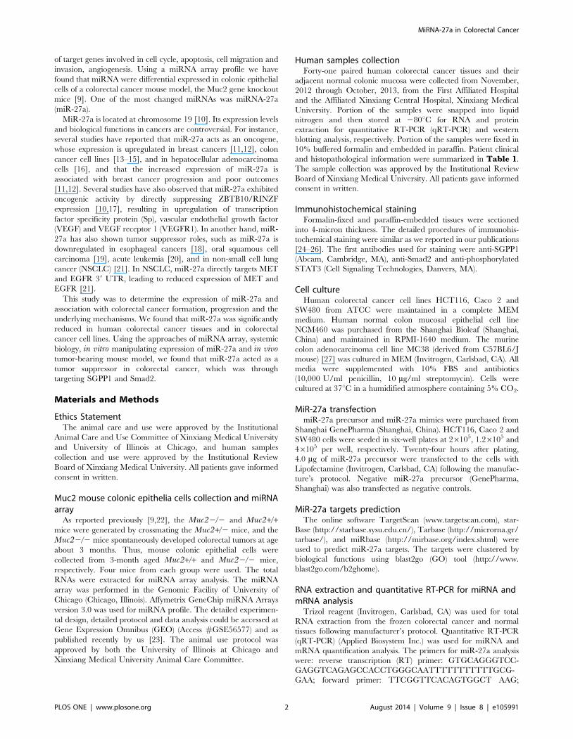
Human samples collectionForty-one paired human colorectal cancer tissues and their
adjacent normal colonic mucosa were collected from November,
2012 through October, 2013, from the First Affiliated Hospital
and the Affiliated Xinxiang Central Hospital, Xinxiang Medical
University. Portion of the samples were snapped into liquid
nitrogen and then stored at 280uC for RNA and protein
extraction for quantitative RT-PCR (qRT-PCR) and western
blotting analysis, respectively. Portion of the samples were fixed in
10% buffered formalin and embedded in paraffin. Patient clinical
and histopathological information were summarized in Table 1.
The sample collection was approved by the Institutional Review
Board of Xinxiang Medical University. All patients gave informed
consent in written.
Immunohistochemical stainingFormalin-fixed and paraffin-embedded tissues were sectioned
into 4-micron thickness. The detailed procedures of immunohis-
tochemical staining were similar as we reported in our publications
[24–26]. The first antibodies used for staining were anti-SGPP1
(Abcam, Cambridge, MA), anti-Smad2 and anti-phosphorylated
STAT3 (Cell Signaling Technologies, Danvers, MA).
Cell cultureHuman colorectal cancer cell lines HCT116, Caco 2 and
SW480 from ATCC were maintained in a complete MEM
medium. Human normal colon mucosal epithelial cell line
NCM460 was purchased from the Shanghai Bioleaf (Shanghai,
China) and maintained in RPMI-1640 medium. The murine
colon adenocarcinoma cell line MC38 (derived from C57BL6/J
mouse) [27] was cultured in MEM (Invitrogen, Carlsbad, CA). All
media were supplemented with 10% FBS and antibiotics
(10,000 U/ml penicillin, 10 mg/ml streptomycin). Cells were
cultured at 37uC in a humidified atmosphere containing 5% CO2.
MiR-27a transfectionmiR-27a precursor and miR-27a mimics were purchased from
Shanghai GenePharma (Shanghai, China). HCT116, Caco 2 and
SW480 cells were seeded in six-well plates at 26105, 1.26105 and
46105 per well, respectively. Twenty-four hours after plating,
4.0 mg of miR-27a precursor were transfected to the cells with
Lipofectamine (Invitrogen, Carlsbad, CA) following the manufac-
ture’s protocol. Negative miR-27a precursor (GenePharma,
Shanghai) was also transfected as negative controls.
MiR-27a targets predictionThe online software TargetScan (www.targetscan.com), star-
Base (http://starbase.sysu.edu.cn/), Tarbase (http://microrna.gr/
tarbase/), and miRbase (http://mirbase.org/index.shtml) were
used to predict miR-27a targets. The targets were clustered by
biological functions using blast2go (GO) tool (http://www.
blast2go.com/b2ghome).
RNA extraction and quantitative RT-PCR for miRNA andmRNA analysis
Trizol reagent (Invitrogen, Carlsbad, CA) was used for total
RNA extraction from the frozen colorectal cancer and normal
tissues following manufacturer’s protocol. Quantitative RT-PCR
(qRT-PCR) (Applied Biosystem Inc.) was used for miRNA and
mRNA quantification analysis. The primers for miR-27a analysis
were: reverse transcription (RT) primer: GTGCAGGGTCC-
GAGGTCAGAGCCACCTGGGCAATTTTTTTTTTTGCG-
GAA; forward primer: TTCGGTTCACAGTGGCT AAG;
MiRNA-27a in Colorectal Cancer
PLOS ONE | www.plosone.org 2 August 2014 | Volume 9 | Issue 8 | e105991
internal control snord47 primers were: RT primer:
GTGCAGGGTCCGAGGTCAGAGCCACCTGGGCAAT
TTTTTTTTTTaacctc; forward primer: CGCCAATGATG-
TAATGATTCTG; Universal reverse primer: CAGTGCA-
GGGTCCGAGGT. Universal Taqman probe: 56-FAM/CA-
GAGCCAC/ZEN/CTGGGCAATTT/3IABkFQ. The primers
for SGPP1 mRNA analysis were: forward primer: TGGTCCTC
CTCACCTATGGC; reverse primer: CTAGAGAACACCAG-
CAGGGA. The primers for Smad2 mRNA analysis were: forward
primer: AACAGAACTTCCGCCTCTGG; reverse primer:
GGAGGTGGCGTTT CTGGAAT. The primers for internal
control GAPDH mRNA were: forward primer: GTCAAGGCT-
GAGAACGGGAA; reverse primer: AAATGAGCCCCAG-
CCTTCTC.
Dual luciferase report construction and transfectionThe full length of 39-UTR of SGPP1 and Smad2 were cloned
from human genomic DNA and inserted into psiCHECK-2 vector
(Promega, Madison, WI), to generate psiCHECK-2-39-UTR-
SGPP1 and psiCHECK-2-39-UTR-Smad2 luciferase reporter
system, respectively. Twenty-four hours before transfection,
1.26104 cells were seeded in a 96-well plate. In brief, 10 pmol
Table 1. The correlation between miR-27a levels and clinicopathological features.
Clinicopathologic Variables N miR-27a expression P value
Low (n) High (n)
Gender
Male 23 12 11
Female 17 7 10 .0.05
Age
#60 15 7 8
.60 25 12 13 .0.05
Smoking
Yes 10 5 5
No 30 15 15 .0.05
Drinking
Yes 7 3 4
No 33 17 16 .0.05
Colitis
Absence 20 11 9
Presence 20 9 11 .0.05
Tumor size
#5 24 11 13
.5 16 8 8 .0.05
Lymphatic metastasis
Yes 14 8 6
No 26 11 15 .0.05
Lymphatic number
Zero 26 11 15
Multiple ($1) 14 8 6 .0.05
Distant metastasis
Yes 12 9 3
No 28 10 18 0.037
TNM stages
Stage II 11 5 6
Stage III/IV 29 14 15 .0.05
Clinical stages
1–2 21 5 16
3–4 19 14 5 0.00016
Differentiation
Low/Moderate 12 7 5
High 28 13 15 .0.05
N, number of patients in this study; n, number of patients in each group.doi:10.1371/journal.pone.0105991.t001
MiRNA-27a in Colorectal Cancer
PLOS ONE | www.plosone.org 3 August 2014 | Volume 9 | Issue 8 | e105991
of miR-27a mimics or negative miRNA mimics (negative control)
was co-transfected into cells with 100 ng of psiCHECK-2-39-
UTR-SGPP1 or psiCHECK-2-39-UTR-Smad2, respectively, us-
ing DharmaFect Duo reagent (Dharmacon, Lafayette, CO, USA).
Luciferase assay was performed 24 h after transfection by the
dual-luciferase reporter assay system (Promega, Madison, WI). For
each sample, firefly luciferase activity was normalized to Renilla
luciferase activity and the inhibition of miR-27a on SGPP1 39-
UTR and Smad2 39-UTR was normalized to the control mimics.
Cell proliferation assayThe MTS assay was used as a cytotoxicity assay for the
HCT116 cells transfection with miR-27a using the Lipofectamine
2000 (Invtrogen, Carlsbad, CA). Briefly, 16105 cells were seeded
into 96-well plate transfected with 0.2 mg of miR-27a precursor
with 0.5 ml Lipofectamine (Invitrogen, Carlsbad, CA). After 24, 48
and 72 hours, cell proliferation was determined by MTS assay (3-
(4,5-dimethylthiazol-2-yl) 5-(3-carboxymethoxyphenyl)-2-(4-sulfo-
phenyl)-2H-tetrazolium) according to the manufacturer’s protocol
(CellTiter 96 Non-Radioactive Cell Proliferation Assay Kit,
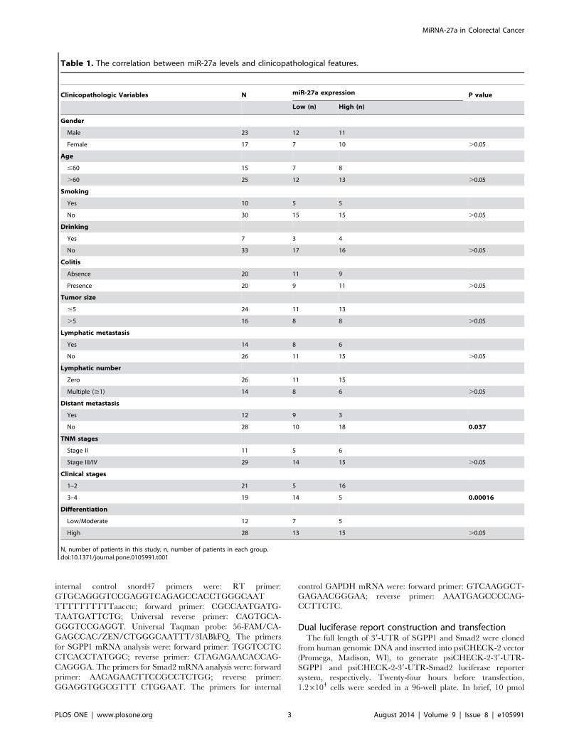
Figure 1. miR-27a was down-regulated in Muc22/2 colonic epithelial cells and human colorectal cancers. A, Cluster analysis of miRNAsexpression profiles in mouse colonic epithelial cells from Muc22/2 mice and Muc2+/+ mice (4 mice each group). Upregulated expression wasindicated in red, whereas downregulated expression was coded in green. B, miR-27a was frequently downregulated in human colorectal cancers (30of 41 cancers exhibited downregulation and 11 cases showed upregulation, in comparison to the adjacent normal tissues). Expression levels of miR-27a were normalized to the corresponding levels of SNORD44. Data were analyzed using a DDCt approach and expressed as log2 fold change (DDCt[cancer/normal]). C, Differential expression of miR-27a at stage II and stage III/IV cancers. The difference was significant using Mann Whitney test (*p,0.0001). D, miR-27a expression was reduced in colorectal cancer cell lines, compared to the immortalized normal colon epithelial cell (NCM460).doi:10.1371/journal.pone.0105991.g001
MiRNA-27a in Colorectal Cancer
PLOS ONE | www.plosone.org 4 August 2014 | Volume 9 | Issue 8 | e105991
Promega Corporation, Madison, WI). The remaining viable cells
with MTS uptake were determined by measuring the optical
density at 570 nm using an enzyme-linked immunosorbent assay
reader (Molecular Devices, Sunnyvale, CA). Values shown were
mean +/2 standard deviation. At least three measurements were
read, and the experiments were conducted 3 times independently.
Apoptosis analysisTo detect apoptosis, the HCT116 cells were transfected with
miR-27a precursor or negative control miRNA precursor. After 48
hours, the cells were harvested and fixed with 70% ethanol
followed by propidium iodide (P.I.) and Annexin V (Invitrogen,
Carlsbad, CA) staining. Cells were then counted by flow cytometry
(FACScan, BD Biosciences, San Jose, CA) for apoptosis analysis.
Usually, about 10,000 cells were counted. The percentage of the
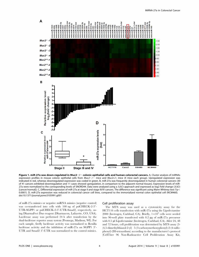
Figure 2. miR-27a targeted SGPP1 and Smad2. A, There was one miR-27a binding site at SGPP1 39-UTR, and there were two miR-27a bindingsites at Smad2 39-UTR. B, miRNA-27a suppressed SGPP1 and Smad2 reporter activities assayed by Dual Luciferases in HCT116 cells. (NC, negativecontrol). C, miRNA-27a inhibited SGPP1 and Smad2 mRNA expression in HCT116 cells. D, miR-27a suppressed SGPP1 and Smad2 protein in coloncancer cells HCT116 and SW480.doi:10.1371/journal.pone.0105991.g002
MiRNA-27a in Colorectal Cancer
PLOS ONE | www.plosone.org 5 August 2014 | Volume 9 | Issue 8 | e105991
apoptosis was calculated by dividing the total cells by apoptotic
cells.
Wound healing assayAs reported previously [26], the HCT116 cells transiently
transfected with miR-27a precursor or negative control miRNA
precursor were seeded in a 100-mm Petri dish. A wound was made
by scratching on the Petri dish bottom, followed by another 48
hours growth.
ImmunoblottingFor immunoblotting, the human colon cancer cells HCT116
and SW480 were collected 72 hours after transfection with miR-
27a precursor or negative control miRNA precursor. Cells were
lysed using 1x RIPA buffer (Upstate Biotechnology, Lake Placid,
NY) containing a protease inhibitor cocktail (Sigma, St. Louis,
MO). After cell lysis, 45 mg of protein was loaded on a 10% SDS
gel followed by transfer to PVDF membrane. Antibodies against
SGPP1 (Abcam, Cambridge, MA), Smad2, Stat3, phosphorylated
Stat3 and Caspase 3 (Cell Signaling Technologies, Danvers, MA),
and b-actin (Sigma, St. Louis, MO) were used. Secondary
antibody was purchased from Santa Cruz Biotechnology (Santa
Cruz, CA). The detected signals were visualized by an enhanced
chemiluminescence kit (Beyotime Institute of Biotechnology,
Haimen, Jiangsu, China), as recommended by the manufacturer.
Tumor-bearing (Xenografts) studyAs reported recently [26], 1.56105 murine colon adenocarci-
noma cells MC38 re-suspended in 150 ml PBS were injected
subcutaneously into the flank of the normal C57B/l6 mice at age
about 8 weeks (5 mice per group). Both MC38 cell line and the
mice were in C57BL/6 background and no rejection occurred.
The animals were maintained in a pathogen-free barrier facility
and closely monitored by animal facility staff. The grown tumors
(xenografts) were measured every 3 day starting 21 days post
inoculation of MC38 cells using caliper as length x width x width/
2 (mm3). 6.26 mg of miR-27a precursor or negative miRNA
(GenePharma, Shanghai, China) mixed with 1.6 ml transfection
reagent Lipofectamine 2000 (Invitrogen) in 50 ml PBS were
injected into the tumors every 3 days, for total of 3 times. 30
days after inoculation, the animals were sacrificed and the
xenografts were isolated, the weight (gram) and volume (mm3) of
the xenografts were determined. All procedures were conducted
according to the Animal Care and Use guideline approved by
Xinxiang Medical University Animal Care Committee.
Results
MiR-27a expression was reduced in Muc22/2 mousecolonic epithelial cells, in human colorectal cancer tissuesand colorectal cancer cells
Our previous studies have demonstrated that Muc22/2 mice
spontaneously developed colorectal cancers and the carcinogenesis
is linked to chronic inflammation [9,22,28,29]. Interestingly,
chronic colitis is frequently seen at the early age of Muc22/2mice, and colorectal tumors are frequently seen after 3 months of
age. Thus, the 3 months could be a borderline of the transition
from chronic inflammation to tumor formation. To indentify the
potent miRNAs involving in the malignant transformation, we
isolated colonic epithelial cells from the age of 3 months mice,
extracted RNA and performed miRNA profile. As shown in
Figure 1A, miRNA profile showed differential expression of
miRNAs in both Muc22/2 and Muc2+/+ mouse colonic
epithelial cells. Based on potential biology roles in proliferation
and inflammation, miRNA profile and published literatures, miR-
27a was chosen for further studies. Since genetic deficiency of the
Muc2 gene in mice causes colorectal cancer formation, the
decreased expression of miR-27a in colonic epithelial cells could
be involved in the carcinogenesis.
To determine the role of miR-27a in colonic epithelial cell
malignant transformation, miR-27a expression levels were deter-
mined in human colorectal cancer and their adjacent tissues. In
comparison with the adjacent normal colon mucosa, 73% (30/41)
of colorectal cancer tissues shown reduced miR-27a expression,
and only 27% (11/41) colorectal cancers exerted upregulated
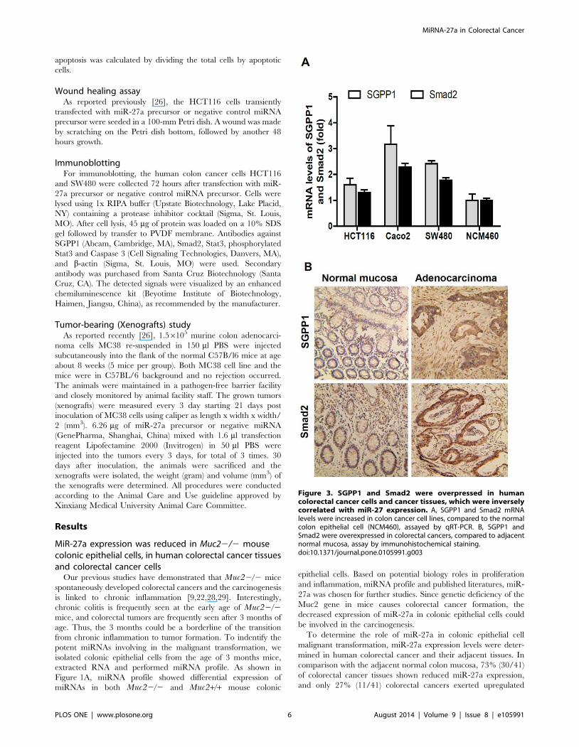
Figure 3. SGPP1 and Smad2 were overpressed in humancolorectal cancer cells and cancer tissues, which were inverselycorrelated with miR-27 expression. A, SGPP1 and Smad2 mRNAlevels were increased in colon cancer cell lines, compared to the normalcolon epithelial cell (NCM460), assayed by qRT-PCR. B, SGPP1 andSmad2 were overexpressed in colorectal cancers, compared to adjacentnormal mucosa, assay by immunohistochemical staining.doi:10.1371/journal.pone.0105991.g003
MiRNA-27a in Colorectal Cancer
PLOS ONE | www.plosone.org 6 August 2014 | Volume 9 | Issue 8 | e105991
expression of miR-27a, the difference was significant (Figure 1B,
p,0.01). More importantly, the reduced expression of miR-27a
was also associated with colorectal cancer pathological stages –
miR-27a levels were more downregulated at stages III/IV than
those at stage II (Figure 1C, p,0.0001). In addition, reduced
miR-27a was correlated with distant metastasis (Table 1).
The expression levels of miR-27a were further determined in
human colorectal cancer cell lines. As shown in Figure 1D, miR-
27a expression was downregulated 95%, 90% and 52% in
HCT116, Caco-2 and SW480 cells, respectively, compared to
the immortalized normal human colon epithelial cell NCM460.
miR-27a targets predictionAbove findings strongly suggested miR-27a was frequently
downregulated in colorectal cancers. Therefore, it is essential to
determine whether the reduction of miR-27a is involved in
colorectal cancer formation and progression, and it is warranted to
identify miR-27a targets and reveal the underlying molecular
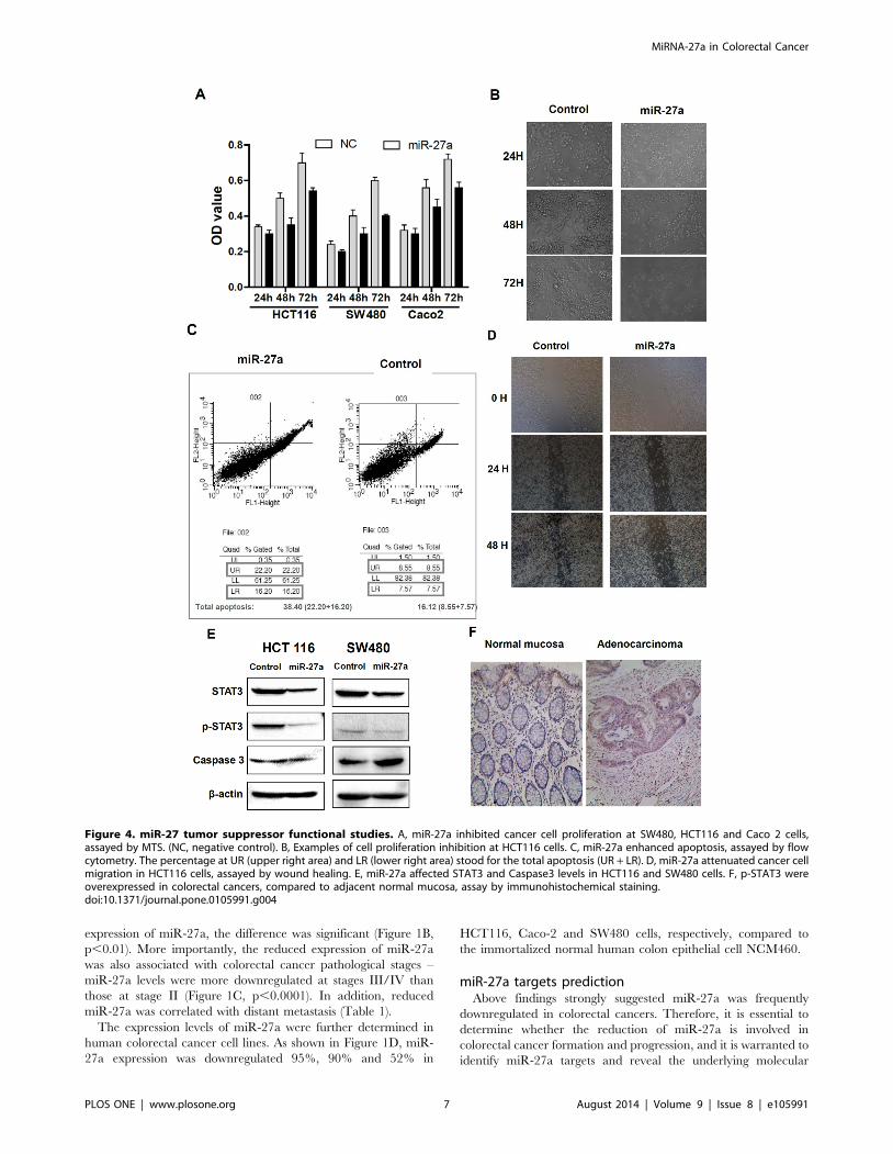
Figure 4. miR-27 tumor suppressor functional studies. A, miR-27a inhibited cancer cell proliferation at SW480, HCT116 and Caco 2 cells,assayed by MTS. (NC, negative control). B, Examples of cell proliferation inhibition at HCT116 cells. C, miR-27a enhanced apoptosis, assayed by flowcytometry. The percentage at UR (upper right area) and LR (lower right area) stood for the total apoptosis (UR + LR). D, miR-27a attenuated cancer cellmigration in HCT116 cells, assayed by wound healing. E, miR-27a affected STAT3 and Caspase3 levels in HCT116 and SW480 cells. F, p-STAT3 wereoverexpressed in colorectal cancers, compared to adjacent normal mucosa, assay by immunohistochemical staining.doi:10.1371/journal.pone.0105991.g004
MiRNA-27a in Colorectal Cancer
PLOS ONE | www.plosone.org 7 August 2014 | Volume 9 | Issue 8 | e105991
mechanisms. Previous studies have demonstrated that ZBTB10/
RINZF is a direct target of oncogenic miR-27a in breast cancers
[10,17] and colon cancer cell lines [15], but this could not be the
case in the colorectal cancer tissues because herein miR-27a
seemed to be a tumor suppressor in colorectal cancers. We
employed multiple tools to predict novel targets of miR-27a.
Supplemental Figure S1A (Supplemental materials) showed 346
potent targets at different signaling pathways, among them, the
categories of cell cycle, cell death, cell differentiation, cell growth
and proliferation and cellular homeostasis, etc, could be more
relevant to cancer development and progression. We further
narrowed down the targets using GO program, and the targets at
the categories of cellular process, single organism process,
biological regulation and metabolic process, etc, exhibited stronger
scores, meaning more relevant to miR-27a-associated functions in
cancer formation and progression (Supplemental Materials Figure
S1B).
SGPP1 and Smad2 were the targets of miR-27aAmong the hundreds of targets, the two novel targets of miR-
27a, Sphingosine-1-phosphate phosphatase 1 (SGPP1) and
Smad2, were chosen for further studies. SGPP1 is a catalyze of
Sphingosine-1-phosphate (S1P), the later is a bioactive sphingo-
lipid metabolite that regulates diverse biologic processes [30,31],
and recent report showed that SIP is linked to signal transducer
and activator of transcription 3 (STAT3) activation and the
development of colitis-associated colorectal cancer [31]. But the
biological role of SGPP1 on carcinogenesis is not clear. Smad 2 is
a member of Smad family and is associated with cell growth and
proliferation through transforming growth factor b (TGF-b)
signaling pathway [32,33].
Genomic alignment showed that 39-UTR of SGPP1 and Smad2
have one or two miR-27a binding sites (Figure 2A). To determine
whether miR-27a could repress SGPP1 and Smad2 expression by
targeting its binding site at 39-UTR in SGPP1 and Smad2, the
PCR products containing full length of 39-UTR with intact target
site of miR-27a recognition sequences were inserted into the
luciferase reporter vector. The plasmids were transfected into
HCT116 cells that had been transfected with control miRNA
mimics or miR-27a mimics, and the luciferase reporter activities
were measured. As shown in Figure 2B, SGPP1 reporter activities
were reduced about 65% and both of the Smad2 reporter activities
(Smad2-a and Smad2-b) were reduced about 90%, respectively.
These finding confirmed that SGPP1 and Smad2 were the targets
of miR-27a.
The repression of SGPP1 and Smad2 expression by miR-27a
were determined by qRT-PCR and immunoblotting. As shown in
Figure 2C, SGPP1 and Smad2 mRNA levels were repressed about
60% and 50% by miR-27a in HCT116 cells, respectively,
consistent with the repression on their reporter luciferase activities.
SGPP1 and Smad2 protein levels were also downregulated by
miR-27a on both HCT116 and SW480 colorectal cancer cells
(Figure 2D).
SGPP1 and Smad2 were inversely correlated with miR-27a in colorectal cancer
As SGPP1 and Smad2 were likely the targets of miR-27a, we
then determined the expression levels of SGPP1 and Smad2 in
human colorectal cancers and cancer cell lines. As shown in
Figure 3A, SGPP1 mRNA levels were 1.6-, 3.2- and 2.4-fold in
HCT116, Caco-2 and SW480 cells, respectively, than that in the
normal colonic epithelial cell NCM460 cells. Similar as the
expression levels of SGPP1, Smad2 mRNA levels were also
upregulated in HCT116, Caco-2 and SW480 cells, showing 1.4-,
2.3- and 1.8-fold higher than that in the normal colonic epithelia
NCM460 cells (Figure 3A). Both SGPP1 and Smad2 mRNA levels
were inversely correlated with the miR-27a levels at these human
colorectal cancer cells (Figure 1D).
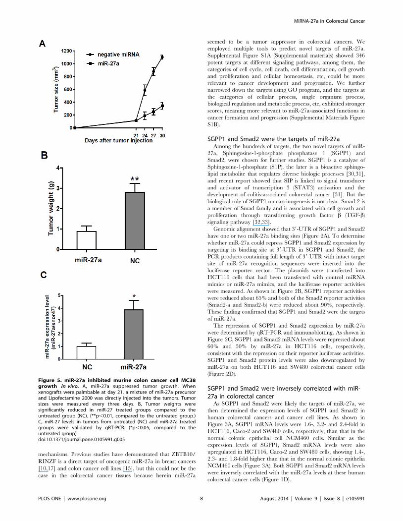
Figure 5. miR-27a inhibited murine colon cancer cell MC38growth in vivo. A, miR-27a suppressed tumor growth. Whenxenografts were palmbable at day 21, a mixture of miR-27a precursorand Lipofectamine 2000 was directly injected into the tumors. Tumorsizes were measured every three days. B, Tumor weights weresignificantly reduced in miR-27 treated groups compared to theuntreated group (NC). (**p,0.01, compared to the untreated group.)C, miR-27 levels in tumors from untreated (NC) and miR-27a treatedgroups were validated by qRT-PCR. (*p,0.05, compared to theuntreated group).doi:10.1371/journal.pone.0105991.g005
MiRNA-27a in Colorectal Cancer
PLOS ONE | www.plosone.org 8 August 2014 | Volume 9 | Issue 8 | e105991
The expression of SGPP1 and Smad2 were then evaluated in
human colorectal cancers by immunohistochemical staining.
Compared to the adjacent normal colon mucosa, both SGPP1
and Smad2 expression were much higher at colorectal adenocar-
cinomas (Figure 3B), negatively correlated with miR-27a levels in
colorectal cancers in which miR-27a was frequently reduced
(Figure 1B).
MiR-27a inhibited cell proliferation, enhanced apoptosisand attenuated cancer cell migration
To determine miR-27a tumor suppressing functions, miR-27a
precursor were transfected into the HCT116 cell, and the effects
on cell proliferation, apoptosis and migration were analyzed. As
shown in Figure 4A and 4B, increasing miR-27a significantly
inhibited cancer cell proliferations after 48 and 72 h at HCT116,
Caco-2 and SW480 cells, although the effects at 24 h post
transfection were not changed. In addition, increasing miR-27a
significantly enhanced cancer cell apoptosis (45% at miR-27a
groups versus 15% at vector control group, p,0.01). The
examples of apoptosis changes were shown at Figure 4C.
Moreover, increased miR-27a significantly attenuated HCT116
cancer cell migration at 24 and 48 h after transfection, assayed by
wound healing (Figure 4D).
To determine the mechanisms of miR-27a mediated inhibition
of cell proliferation and migration and enhancement of apoptosis,
we determined the changes of STAT3 and Caspase3. We found
that besides the downregulation of total STAT3 by miR-27a, the
phosphorylated STAT-3 (p-STAT3) was also dramatically re-
pressed by miR-27a (Figure 4E). Since miR-27a was reduced in
human colorectal cancers, we determined whether p-STAT3 was
increased in colorectal cancers. P-STAT was indeed increased in
colorectal cancers (Figure 4F).
MiR-27a inhibited cancer growth in miceTo determine miR-27a tumor suppressing effects in vivo, we
transplanted murine colon cancer cells MC38 into wild-type
C57Bl/6 mice and injected a mixture of miR-27a precursor and
Lipofectamine 2000 into the tumors when the tumors were
palmable at day 21 post inoculation. Compared to vector control,
miR-27a treatment significantly inhibited cancer cell growth in
mice, in terms of significant reduction of tumor sizes and weight
(Figure 5A, 5B). qRT-PCR showed that miR-27a level was still at
a higher level in the tumors isolated from mouse xenografts
(Figure 5C).
Discussion
It is well known that the functions and target genes of a miRNA
are tissue- and cancer-type specific. Functional studies have shown
that miR-27a has shown both oncogenic and tumor suppressive
functions in different cell lines and human cancer tissues. For
example, miR-27a directly suppresses ZBTB10/RINZF expres-
sion [10,17]. and in turn upregulates VEGF and VEGF receptor
in the cancers of breast [11,12] and colon [13–15], and
overexpression of miR-27a is associated with poor outcomes
[11,12]. Whereas, in the cancers of esophagus [18], oral cavity
[19], lung [21], and head and neck [34], miR-27a is downreg-
ulated, and miR-27a directly targets MET and EGFR and
suppresses their expression in lung cancer [21]. In this study, we
found that miR-27a was significantly downregulated in colorectal
cancers and colorectal cancer cells. Importantly, the downregu-
lated miR-27a was also associated with colorectal cancer
pathological stages and distant metastasis, showing tumor
suppressor roles in colorectal cancer. First, in vitro studies showed
that increased expression of miR-27a inhibited colorectal cancer
cell proliferation, promoted apoptosis and attenuated cancer cell
migration. Second, in a tumor-bearing mouse model, a direct
injection of miR-27a to tumor suppressed tumor growth. These
findings strongly suggested that miR-27a could be used as a
biomarker to monitor cancer development and progression, and
could be used as a potential therapeutic target and even a potential
therapeutic agent for colorectal cancer.
The mechanistic studies further showed that miR-27a-mediated
tumor suppressor could be through targeting SGPP1, Smad2 and
STAT3. Previous studies have demonstrated that Apc mutation
and Wnt/b-catenin signaling activation, and chronic inflamma-
tion in colon, are the two major causes for colorectal cancer
formation [35–37]. The studies from us have shown that genetic
deletion of the Muc2 gene is sufficient to cause chronic colitis and
rectal inflammation at early stage, and leads to colorectal cancer
formation at late stage [9,22,29,38]. The malignant transforma-
tion is linked to the activation of inflammatory signaling and
upregulation of cytokines [22,38]. And the chronic inflammation
facilitated Apc-mutation-caused gastrointestinal tumor formation
in the Apc/Muc2 double gene knockout mouse model of
colorectal cancer [22]. In fact, other studies have demonstrated
that the transcription factor STAT3 also plays a critical role in
inflammation-associated colorectal cancer formation [31,39–42].
During the transition of chronic intestinal inflammation to colitis-
associated cancer, STAT3 could be persistently activated by
sphingosine-1-phosphate (S1P) produced by upregulation of
sphingosine kinase 1 (SphK1), which was linked to production of
the multifunctional NF-kB-regulated cytokine IL-6, and conse-
quently upregulating of the S1P receptor, S1PR1 [31]. While, the
balance of S1P levels are regulated by sphingosine-1-phosphate
phosphatase 1 (SGPP1) [30].
Using comprehensive approaches, including qRT-PCR, immu-
blotting and in situ immunohistochemical staining, we showed
that the expression of SGPP1 at mRNA and protein levels were
upregulated in colorectal cancers and colorectal cancer cell lines,
which were inversely correlated with the expression of miR-27a.
Both dual luciferase assay and increasing expression of miR-27a
further showed the inhibitory effects of miR-27a on SGPP1. Thus,
this is the first to reveal that SGPP1 is a potent direct target of
miR-27a, although the evidence of direction regulatory interaction
is needed for further investigation. Limited studies have demon-
strated that SGPP1 catalyzes the degradation of S1P via salvage
and recycling of sphingosine into long-chain ceramides, and that
aberrant expression of SGPP1 is associated with idiopathic
pulmonary fibrosis, and pulmonary fibrosis [30]. However, the
biological functions for SGPP1 in carcinogenesis are largely
unknown and worthy further elucidative.
Smad2 is a member of the Smad family and is a key element in
TGF-b signaling. Therefore, accompanying actions of TGF-b in
different circumstances, e.g., in regulating development and
differentiation in physiological cell process, and in facilitating cell
growth and migration and angiogenesis in cancers, Smad2 exerts
dual functions as tumor suppressor or oncogene [32,33,43,44]. In
the current study, we found that, similar as SGPP1, Samd2
expression at mRNA and protein levels was upregulated in
colorectal cancers and cell lines, exhibiting oncogenic phenotypes;
moreover, Smad2 was repressed at transcriptional and transla-
tional levels by miR-27a, suggesting the direct target of miR-27a, a
novel finding that has not been reported previously. A few recent
studies have reported that Smad2, similar as Smad3 and Smad4, is
mutant in cancers [43], but Smad2 plays differential roles from
Smad3 and Samd4 in TGF-b signaling [33]. The mutation
frequency in colorectal cancer and whether the mutation of
MiRNA-27a in Colorectal Cancer
PLOS ONE | www.plosone.org 9 August 2014 | Volume 9 | Issue 8 | e105991
Smad2 leads to stability of SMAD2 protein in the colorectal
cancer is not clear and warrants further investigation. Previous
studies have also shown therapeutic role of Smad2, that blocking
Smad2 could suppress TGF-b-induced tumorigenesis, epithelial-
mesenchymal transition (EMT), cell motility, and invasion [45],
indicating that targeting miR-27a/Smad2 could have a great
impact on developing a novel strategy for colorectal cancer
therapy. The new signal pathway of miR-27a-Smad2-TGF-bcould also contribute to the inhibitory role of miR-27a on cancer
cell migration (Figure 4D), invasion and metastasis, detailed
mechanism is under investigation.
In summary, we have demonstrated that miR-27a is frequently
downregulated in colorectal cancer, and the reduced miR-27a is
correlated with cancer distant metastasis and histopathological
stages, and thus, miR-27a acts as a tumor suppressor. Both in vivoand in vitro studies have identified SGPP1 and Smad2 as two
novel targets of miR-27a, which is linked to STAT3 to regulate
cancer cell proliferation, apoptosis and migration. Therefore,
miR-27a could be a useful biomarker for colorectal cancer
development and progression, and also could have a therapeutic
potential targeting SGPP1, Smad2 and Stat3 for colorectal cancer
therapy.
Supporting Information
Figure S1 Prediction of miR-27a targets. A. The targets
were categorized by biological process (filtered by sequences
numbers, cutoff = 5.0). B, The targets were clustered into multiple
categories by biological process level 2 using Go Oncology tool.
(TIF)
Acknowledgments
We would like to thank Professor Deming Gou at Shenzhen University
(Shenzhen, China) for providing reagents for miRNA qRT-PCT, and
thank Professor Huanliang Liu (Sun Yat-sen University, Guangzhou,
China) for providing reagents.
Author Contributions
Conceived and designed the experiments: WY YB. Performed the
experiments: YB ZC YG YF YJ KL QW Jiaqi Wang HZ. Analyzed the
data: YB YG LW WY. Contributed reagents/materials/analysis tools: ZL
WH Jianguo Wang WZ. Contributed to the writing of the manuscript: YB
WY.
References
1. Ventura A, Jacks T (2009) MicroRNAs and cancer: short RNAs go a long way.
Cell 136: 586–591.
2. Farazi TA, Spitzer JI, Morozov P, Tuschl T (2011) miRNAs in human cancer.
The Journal of pathology 223: 102–115.
3. Di Leva G, Garofalo M, Croce CM (2014) MicroRNAs in Cancer. Annual
review of pathology 9: 287–314.
4. Garofalo M, Leva GD, Croce CM (2014) MicroRNAs as Anti-Cancer Therapy.
Current pharmaceutical design Jan 28. [Epub ahead of print].
5. White NM, Fatoohi E, Metias M, Jung K, Stephan C, et al. (2011) Metastamirs:
a stepping stone towards improved cancer management. Nature reviews 8: 75–
84.
6. Volinia S, Calin GA, Liu CG, Ambs S, Cimmino A, et al. (2006) A microRNA
expression signature of human solid tumors defines cancer gene targets.
Proceedings of the National Academy of Sciences of the United States of
America 103: 2257–2261.
7. Bartel DP (2004) MicroRNAs: genomics, biogenesis, mechanism, and function.
Cell 116: 281–297.
8. Lages E, Ipas H, Guttin A, Nesr H, Berger F, et al. (2012) MicroRNAs:
molecular features and role in cancer. Frontiers in bioscience (Landmark edition)
17: 2508–2540.
9. Velcich A, Yang W, Heyer J, Fragale A, Nicholas C, et al. (2002) Colorectal
cancer in mice genetically deficient in the mucin Muc2. Science 295: 1726–
1729.
10. Mertens-Talcott SU, Chintharlapalli S, Li X, Safe S (2007) The oncogenic
microRNA-27a targets genes that regulate specificity protein transcription
factors and the G2-M checkpoint in MDA-MB-231 breast cancer cells. Cancer
research 67: 11001–11011.
11. Tang W, Yu F, Yao H, Cui X, Jiao Y, et al. (2013) miR-27a regulates
endothelial differentiation of breast cancer stem like cells. Oncogene Jun 10.
doi:10.1038/onc.2013.214. [Epub ahead of print].
12. Tang W, Zhu J, Su S, Wu W, Liu Q, et al. (2012) MiR-27 as a prognostic
marker for breast cancer progression and patient survival. PloS one 7: e51702.
13. Zanetti KA, Haznadar M, Welsh JA, Robles AI, Ryan BM, et al. (2012) 39-UTR
and functional secretor haplotypes in mannose-binding lectin 2 are associated
with increased colon cancer risk in African Americans. Cancer research 72:
1467–1477.
14. Chintharlapalli S, Papineni S, Abdelrahim M, Abudayyeh A, Jutooru I, et al.
(2009) Oncogenic microRNA-27a is a target for anticancer agent methyl 2-
cyano-3,11-dioxo-18beta-olean-1,12-dien-30-oate in colon cancer cells. Inter-
national journal of cancer 125: 1965–1974.
15. Pathi SS, Jutooru I, Chadalapaka G, Sreevalsan S, Anand S, et al. (2011) GT-
094, a NO-NSAID, inhibits colon cancer cell growth by activation of a reactive
oxygen species-microRNA-27a: ZBTB10-specificity protein pathway. Mol
Cancer Res 9: 195–202.
16. Huang S, He X, Ding J, Liang L, Zhao Y, et al. (2008) Upregulation of miR-23a
approximately 27a approximately 24 decreases transforming growth factor-beta-
induced tumor-suppressive activities in human hepatocellular carcinoma cells.
International journal of cancer 123: 972–978.
17. Scott GK, Mattie MD, Berger CE, Benz SC, Benz CC (2006) Rapid alteration
of microRNA levels by histone deacetylase inhibition. Cancer research 66:
1277–1281.
18. Zhu L, Wang Z, Fan Q, Wang R, Sun Y (2014) microRNA-27a functions as a
tumor suppressor in esophageal squamous cell carcinoma by targeting KRAS.
Oncology reports 31: 280–286.
19. Venkatesh T, Nagashri MN, Swamy SS, Mohiyuddin SM, Gopinath KS, et al.
(2013) Primary microcephaly gene MCPH1 shows signatures of tumor
suppressors and is regulated by miR-27a in oral squamous cell carcinoma. PloS
one 8: e54643.
20. Scheibner KA, Teaboldt B, Hauer MC, Chen X, Cherukuri S, et al. (2012)
MiR-27a functions as a tumor suppressor in acute leukemia by regulating 14-3-
3theta. PloS one 7: e50895.
21. Acunzo M, Romano G, Palmieri D, Lagana A, Garofalo M, et al. (2013) Cross-
talk between MET and EGFR in non-small cell lung cancer involves miR-27a
and Sprouty2. Proceedings of the National Academy of Sciences of the United
States of America 110: 8573–8578.
22. Yang K, Popova NV, Yang WC, Lozonschi I, Tadesse S, et al. (2008)
Interaction of Muc2 and Apc on Wnt signaling and in intestinal tumorigenesis:
potential role of chronic inflammation. Cancer research 68: 7313–7322.
23. Bao Y, Guo Y, Li Z, Fang W, Yang Y, et al. (2014) MicroRNA profiling in
Muc2 knockout mice of colitis-associated cancer model reveals epigenetic
alterations during chronic colitis malignant transformation. PloS One 9(6):
e99132.
24. Tong C, Yin Z, Song Z, Dockendorff A, Huang C, et al. (2007) c-Jun NH2-
terminal kinase 1 plays a critical role in intestinal homeostasis and tumor
suppression. Am J Pathol 171: 297–303.
25. Bi X, Tong C, Dockendorff A, Bancroft L, Gallagher L et al. (2008) Genetic
deficiency of decorin causes intestinal tumor formation through disruption of
intestinal cell maturation. Carcinogenesis 29: 1435–1440.
26. Bi X, Pohl NM, Qian Z, Yang GR, Gou Y, et al. (2012) Decorin-mediated
inhibition of colorectal cancer growth and migration is associated with E-
cadherin in vitro and in mice. Carcinogenesis 33: 326–330.
27. Rosenberg SA, Spiess P, Lafreniere R (1986) A new approach to the adoptive
immunotherapy of cancer with tumor-infiltrating lymphocytes. Science 233:
1318–1321.
28. Yang W, Velcich A, Lozonschi I, Liang J, Nicholas C, et al. (2005) Inactivation
of p21WAF1/cip1 Enhances Intestinal Tumor Formation in Muc22/2 Mice.
Am J Pathol 166: 1239–1246.
29. Van der Sluis M, De Koning BA, De Bruijn AC, Velcich A, Meijerink JP, et al.
(2006) Muc2-deficient mice spontaneously develop colitis, indicating that MUC2
is critical for colonic protection. Gastroenterology 131: 117–129.
30. Johnson KR, Johnson KY, Becker KP, Bielawski J, Mao C, et al. (2003) Role of
human sphingosine-1-phosphate phosphatase 1 in the regulation of intra- and
extracellular sphingosine-1-phosphate levels and cell viability. The Journal of
biological chemistry 278: 34541–34547.
31. Liang J, Nagahashi M, Kim EY, Harikumar KB, Yamada A, et al. (2013)
Sphingosine-1-phosphate links persistent STAT3 activation, chronic intestinal
inflammation, and development of colitis-associated cancer. Cancer cell 23:
107–120.
32. Yang J, Wahdan-Alaswad R, Danielpour D (2009) Critical role of Smad2 in
tumor suppression and transforming growth factor-beta-induced apoptosis of
prostate epithelial cells. Cancer research 69: 2185–2190.
MiRNA-27a in Colorectal Cancer
PLOS ONE | www.plosone.org 10 August 2014 | Volume 9 | Issue 8 | e105991

33. Brown KA, Pietenpol JA, Moses HL (2007) A tale of two proteins: differential
roles and regulation of Smad2 and Smad3 in TGF-beta signaling. Journal ofcellular biochemistry 101: 9–33.
34. Wu X, Bhayani MK, Dodge CT, Nicoloso MS, Chen Y, et al. (2013)
Coordinated targeting of the EGFR signaling axis by microRNA-27a*.Oncotarget 4: 1388–1398.
35. Vogelstein B, Papadopoulos N, Velculescu VE, Zhou S, Diaz LA, Jr., et al.(2013) Cancer genome landscapes. Science 339: 1546–1558.
36. Grivennikov SI, Greten FR, Karin M (2010) Immunity, inflammation, and
cancer. Cell 140: 883–899.37. Antonioli L, Blandizzi C, Pacher P, Hasko G (2013) Immunity, inflammation
and cancer: a leading role for adenosine. Nat Rev Cancer 13: 842–857.38. Petersson J, Schreiber O, Hansson GC, Gendler SJ, Velcich A, et al. (2011)
Importance and regulation of the colonic mucus barrier in a mouse model ofcolitis. American journal of physiology 300: G327–333.
39. Danese S, Mantovani A (2010) Inflammatory bowel disease and intestinal
cancer: a paradigm of the Yin-Yang interplay between inflammation and cancer.Oncogene 29: 3313–3323.
40. Grivennikov S, Karin E, Terzic J, Mucida D, Yu GY, et al. (2009) IL-6 and
Stat3 are required for survival of intestinal epithelial cells and development of
colitis-associated cancer. Cancer cell 15: 103–113.
41. Bromberg J, Wang TC (2009) Inflammation and cancer: IL-6 and STAT3
complete the link. Cancer cell 15: 79–80.
42. Nguyen AV, Wu YY, Liu Q, Wang D, Nguyen S, et al. (2013) STAT3 in
epithelial cells regulates inflammation and tumor progression to malignant state
in colon. Neoplasia (New York, NY 15: 998–1008.
43. Fleming NI, Jorissen RN, Mouradov D, Christie M, Sakthianandeswaren A, et
al. (2013) SMAD2, SMAD3 and SMAD4 mutations in colorectal cancer.
Cancer research 73: 725–735.
44. Horiguchi K, Shirakihara T, Nakano A, Imamura T, Miyazono K, et al. (2009)
Role of Ras signaling in the induction of snail by transforming growth factor-
beta. The Journal of biological chemistry 284: 245–253.
45. Zhao BM, Hoffmann FM (2006) Inhibition of transforming growth factor-beta1-
induced signaling and epithelial-to-mesenchymal transition by the Smad-binding
peptide aptamer Trx-SARA. Molecular biology of the cell 17: 3819–3831.
MiRNA-27a in Colorectal Cancer
PLOS ONE | www.plosone.org 11 August 2014 | Volume 9 | Issue 8 | e105991