MicroRNA as Biomarkers in Cancer Diagnostics and Therapy
168
MicroRNA as Biomarkers in Cancer Diagnostics and Therapy Lorenzo F. Sempere www.mdpi.com/journal/ijms Edited by Printed Edition of the Special Issue Published in International Journal of Molecular Sciences International Journal of Molecular Sciences
-
Upload
khangminh22 -
Category
Documents
-
view
0 -
download
0
Transcript of MicroRNA as Biomarkers in Cancer Diagnostics and Therapy

MicroRNA as Biomarkers in Cancer Diagnostics and Therapy
Lorenzo F. Sempere
www.mdpi.com/journal/ijms
Edited by
Printed Edition of the Special Issue Published in
International Journal of Molecular Sciences
International Journal of
Molecular Sciences

MicroRNA as Biomarkers in CancerDiagnostics and Therapy


MicroRNA as Biomarkers in CancerDiagnostics and Therapy
Special Issue Editor
Lorenzo F. Sempere
MDPI • Basel • Beijing • Wuhan • Barcelona • Belgrade

Special Issue Editor
Lorenzo F. Sempere
Michigan State University,
USA
Editorial Office
MDPI
St. Alban-Anlage 66
4052 Basel, Switzerland
This is a reprint of articles from the Special Issue published online in the open access journal
International Journal of Molecular Sciences (ISSN 1422-0067) from 2018 to 2019 (available at: https:
//www.mdpi.com/journal/ijms/special issues/miR biomarkers cancer)
For citation purposes, cite each article independently as indicated on the article page online and as
indicated below:
LastName, A.A.; LastName, B.B.; LastName, C.C. Article Title. Journal Name Year, Article Number,
Page Range.
ISBN 978-3-03921-249-1 (Pbk)
ISBN 978-3-03921-250-7 (PDF)
Cover image courtesy of Lorenzo F. Sempere.
c© 2019 by the authors. Articles in this book are Open Access and distributed under the Creative
Commons Attribution (CC BY) license, which allows users to download, copy and build upon
published articles, as long as the author and publisher are properly credited, which ensures maximum
dissemination and a wider impact of our publications.
The book as a whole is distributed by MDPI under the terms and conditions of the Creative Commons
license CC BY-NC-ND.

Contents
About the Special Issue Editor . . . . . . . . . . . . . . . . . . . . . . . . . . . . . . . . . . . . . . vii
Lorenzo F. Sempere
Celebrating 25 Years of MicroRNA Research: From Discovery to Clinical ApplicationReprinted from: Int. J. Mol. Sci. 2019, 20, 1987, doi:10.3390/ijms20081987 . . . . . . . . . . . . . . 1
Francesca Pezzuto, Luigi Buonaguro, Franco Maria Buonaguro and Maria Lina Tornesello
The Role of Circulating Free DNA and MicroRNA in Non-Invasive Diagnosis of HBV- andHCV-Related Hepatocellular CarcinomaReprinted from: Int. J. Mol. Sci. 2018, 19, 1007, doi:10.3390/ijms19041007 . . . . . . . . . . . . . . 6
Yi-An Chang, Shun-Long Weng, Shun-Fa Yang, Chih-Hung Chou, Wei-Chih Huang,
Siang-Jyun Tu, Tzu-Hao Chang, Chien-Ning Huang, Yuh-Jyh Jong and Hsien-Da Huang
A Three–MicroRNA Signature as a Potential Biomarker for the Early Detection of Oral CancerReprinted from: Int. J. Mol. Sci. 2018, 19, 758, doi:10.3390/ijms19030758 . . . . . . . . . . . . . . . 23
Aelita Konstantinell, Dag H. Coucheron, Baldur Sveinbjørnsson and Ugo Moens
MicroRNAs as Potential Biomarkers in Merkel Cell CarcinomaReprinted from: Int. J. Mol. Sci. 2018, 19, 1873, doi:10.3390/ijms19071873 . . . . . . . . . . . . . . 40
Jaqueline Carvalho de Oliveira, Gabriela Molinari Roberto, Mirella Baroni, Karina Bezerra Salomao, Julia Alejandra Pezuk and Marıa Sol Brassesco
MiRNA Dysregulation in Childhood Hematological CancerReprinted from: Int. J. Mol. Sci. 2018, 19, 2688, doi:10.3390/ijms19092688 . . . . . . . . . . . . . . 57
Monika Drobna, Bronisława Szarzy nska-Zawadzka, Patrycja Daca-Roszak, Maria Kosmalska, Roman Jaksik, Michał Witt and Małgorzata Dawidowska
Identification of Endogenous Control miRNAs for RT-qPCR in T-Cell Acute Lymphoblastic LeukemiaReprinted from: Int. J. Mol. Sci. 2018, 19, 2858, doi:10.3390/ijms19102858 . . . . . . . . . . . . . . 87
Grzegorz Hibner, Małgorzata Kimsa-Furdzik and Tomasz Francuz
Relevance of MicroRNAs as Potential Diagnostic and Prognostic Markers in Colorectal CancerReprinted from: Int. J. Mol. Sci. 2018, 19, 2944, doi:10.3390/ijms19102944 . . . . . . . . . . . . . . 104
Paola Ulivi, Matteo Canale, Alessandro Passardi, Giorgia Marisi, Martina Valgiusti,
Giovanni Luca Frassineti, Daniele Calistri, Dino Amadori and Emanuela Scarpi
Circulating Plasma Levels of miR-20b, miR-29b and miR-155 as Predictors of BevacizumabEfficacy in Patients with Metastatic Colorectal CancerReprinted from: Int. J. Mol. Sci. 2018, 19, 307, doi:10.3390/ijms19010307 . . . . . . . . . . . . . . . 130
Trine Møller, Jaslin P James, Kim Holmstrøm, Flemming B Sørensen, Jan Lindebjerg and
Boye S Nielsen
Co-Detection of miR-21 and TNF-α mRNA in Budding Cancer Cells in Colorectal CancerReprinted from: Int. J. Mol. Sci. 2019, 20, 1907, doi:10.3390/ijms20081907 . . . . . . . . . . . . . . 142
v


About the Special Issue Editor
Lorenzo F. Sempere is originally from Elche, a sunny city in southeastern Spain with one of largest
palm tree groves in the world. He obtained his B.S. in Biochemistry at Universidad Miguel Hernandez
and trained in the laboratory of Victor Ambros, co-discoverer of microRNAs, at Geisel School of
Medicine at Dartmouth College. Dr. Sempere initiated his translational cancer research in the
laboratory of Charles Cole, funded by a Susan G. Komen for the Cure postdoctoral fellowship, and
continued to climb the academic ladder at Dartmouth in the laboratory of Murray Korc, funded by
a Laurie and Paul MacCaskill PanCAN-AACR Career Development award. Dr. Sempere started his
independent laboratory as a research track faculty first at Geisel School of Medicine, and then at Van
Andel Research Institute. Dr. Sempere joined Michigan State University in 2018 as Assistant Professor
of Radiology in the tenure-track system and as faculty member of the campus-wide Precision Health
Program. Dr. Sempere has worked in the field of microRNAs since their discovery in 2001. He has
experience and expertise as an author, reviewer, and journal editor in diverse areas of microRNA
research, including evolutionary and developmental biology, molecular and cellular biology, and
immunology and cancer biology.
vii


International Journal of
Molecular Sciences
Editorial
Celebrating 25 Years of MicroRNA Research: FromDiscovery to Clinical Application
Lorenzo F. Sempere
Department of Radiology, Precision Health Program, Michigan State University, East Lansing, MI 48824, USA;[email protected]; Tel.: +1(517)355-3982
Received: 19 April 2019; Accepted: 21 April 2019; Published: 23 April 2019
In 1993, the Ambros lab reported the cloning and developmental function of lin-4, the firstmicroRNA [1]. This short non-coding RNA was regarded as an oddity of the mighty little roundwormCaenorhabditis elegans, controlling gene expression by binding to partially complementary sites on the3′ UTR of target mRNAs and inhibiting translation [1–3]. The discovery of let-7, also in C. elegans,reinforced this molecular oddity and highlighted the power of forward genetics in this modelorganism [4]. These two small temporal RNAs control developmental timing of larval transition inC. elegans and other Caenorhabditis species [5]. This restricted classification was short-lived as theevolutionary conservation of let-7 [6] and the shared enzymatic machinery for processing of endogenousshort non-coding RNAs and exogenous double-stranded RNA substrates for RNA interference [7]strongly suggested a broadly co-opted regulatory mechanism of short non-coding RNAs in animalevolution. Indeed, let-7 and miR-125 (lin-4 paralog) family members have been implicated in temporalidentity in the fly brain, and are likely involved in temporal cell fate decision in vertebrates [5]. Over thelast 20 years, microRNAs have been identified in many animal species. Some of these microRNAs arephylum-, order-, genus-, or even species-specific [8–10]. The size of the microRNome and complexityof animal body plans and organ systems suggest a role of microRNAs in cell fate determination anddifferentiation [8,9]. More than 2000 sequences have been proposed to represent unique microRNAgenes in humans with an increasing number of mechanistic roles in developmental, physiological, andpathological processes [10,11].
MicroRNA are short non-coding regulatory RNAs. The mature and biologically active formis about 18–25 nucleotides long. This mature sequence binds to and guides an Ago-containing,RNA-induced, multi-protein silencing complex to partially complementary sites on the mRNA oftarget genes [12]. Due to this partial complementarity, a single microRNA can regulate the expressionof many, if not hundreds, of target genes. Thus, dysregulation of a few key microRNAs can have aprofound global effect on gene expression and molecular programs of a cell. Conversely, restoration ofbaseline microRNA activity can also have profound effects to reverse a pathological process [12,13].This great potential for clinical intervention captured the interest and imagination of researchers inmany fields. However, very few fields have been as prolific as the field of cancer research. This islargely due to early studies by Carlo Croce and colleagues that linked microRNAs with cancer just acouple of years after the discovery of let-7. In 2002, Calin et al. showed that chromosomal deletionof miR-15 and miR-16 was a frequent event in B-cell chronic lymphocytic leukemia [14]. Soon after,Calin et al. also made the tantalizing observation that many microRNA loci are located at fragilesites, breakpoint regions, or frequently altered regions (e.g., deletion or amplification) in the cancergenome [15]. These seminal papers, along with a technological explosion of high-throughput detectionplatforms, led several groups to extensively profile microRNA expression in healthy and tumortissues. Altered microRNA expression has been associated with diagnostic and prognostic indicatorsin many cancer types. Different mechanisms have been reported for this dysregulation, includingchromosomal deletion or amplification of a microRNA gene, epigenetic and transcriptional regulation,and mutation in the enzymes responsible for microRNA processing, export, or silencing [12,13]. Despite
Int. J. Mol. Sci. 2019, 20, 1987; doi:10.3390/ijms20081987 www.mdpi.com/journal/ijms1

Int. J. Mol. Sci. 2019, 20, 1987
the overwhelming number of diagnostic and prognostic studies, the current impact of microRNA-basedassays is very limited in clinical practice. Some microRNA-based assays have reached the clinic in theform of laboratory developed tests, and several on-going clinical trials propose the use of microRNAsas biomarkers for early disease detection, guiding treatment selection, monitoring disease progression,or other specific clinical endpoints.
This Special Issue celebrates the 25th anniversary of the discovery of the first microRNA. It providesbut a glimpse of the large body of literature of microRNA biology in cancer research. This Special Issuecontains four original research studies [16–19] and four review papers [20–23] with a disease focus onspecific hematologic or solid tumors. Collectively, these papers highlight state-of-the-art approachesand methodologies for microRNA detection in tissue, blood, and other body fluids for biomarkerapplications from early cancer detection to prognosis and treatment response. These papers alsoaddress some of the challenges for clinical implementation. Pezzuto et al. provide a comprehensivereview of blood-based detection of cell-free DNA and microRNAs for early detection of hepatitisviruses–related liver cancer [21]. Chang et al. report on a plasma-based 3-microRNA signaturefor early detection of oral cancer [17]. Konstantinell et al. provide a comprehensive review ofextracellular vesicle-based and tissue-based detection of microRNA for diagnostic and prognosticapplications in Merkel cell carcinoma [22]. de Oliveira et al. provide a comprehensive review oftissue-based and cell-based detection of microRNAs for diagnostic and prognostic applications inchildhood hematological cancers [23]. Drobna et al. describe a methodological advancement ofmost suitable endogenous microRNA controls for cell-based microRNA detection in T-cell acutelymphoblastic leukemia [18]. Hibner et al. provide a comprehensive review [20], while Moller et al. [19]and Ulivi et al. [16] present specific studies of microRNA biomarkers for diagnostic and prognosticapplications in colorectal cancer. Below is a brief summary and highlights of each of these articles.
Liver cancer is the third leading cause of cancer-related death in the world. Hepatocellularcarcinoma (HCC) represents 85% of all liver cancer cases and generally has a poor prognosis due tolate presentation. Chronic infection of Hepatitis B or Hepatitis C viruses are a major risk factor fordevelopment of HCC. Pezzuto et al. review blood-based detection assays that would complementand improve current diagnostic tools based on various imaging modalities and plasma levels ofalpha-fetoprotein [21]. Pezzuto et al. describe the utility of detecting circulating cell-free DNA,and cell-free or extracellular vesicle-loaded microRNAs and long non-coding RNAs. While DNAmutations or altered DNA methylation pattern in circulating DNA or altered circulating microRNAlevels can detect HCC tumors, altered levels of some microRNAs such as miR-122 can reflect moreclosely the effect of viral infection on malignant transformation of hepatocytes. Of the discussedblood-based markers, Pezzuto et al. highlight altered circulating levels of miR-122 and let-7, andRASSF1A hypermethylation in circulating free-DNA as the most promising biomarkers for diagnosticapplication. As in HCC, patients that present with late stage oral cancer, which can be as high as 50%of all cases in some countries, have a much worse prognosis than early stage cases. Oral squamouscell carcinoma (OSCC) is the most common type of oral cancer. Prevalence and risk factors vary withgeographic location. In South and Southeast Asia and Taiwan, betel quid chewing is a major riskfactor. Chang et al. performed RNA sequencing from plasma of healthy controls, early stage patientswith oral leukoplakia (OL, precursor lesion associated with OSCC progression) and late stage patientsdiagnosed with OSCC to identify differentially expressed microRNAs. Selected microRNAs werefurther studied in a training set of 72 samples and validated in an independent set of 178 samples ofTaiwanese patients. From these analyses, Chang et al. identified a 3-microRNA signature (miR-150-5p,miR-222-3p, miR-423-5p) that could accurately separate OL from OSCC cases and may have clinicalutility for early detection of OSCC. As in HCC, viral infection is a major risk factor in Merkel cellcarcinoma (MCC), which is a rare but aggressive type of skin cancer. About 80% of MCC tumors areinfected with viral DNA of Merkel cell polyomavirus. Konstantinell et al. provide a comprehensivereview on the host and viral microRNA expression in MCC tissue samples and present their originaldata profiling microRNA expression in extracellular vesicles secreted by MCC cell lines [22].
2

Int. J. Mol. Sci. 2019, 20, 1987
MicroRNA expression and function have been extensively studied during normal hematopoiesisand in several hematological malignancies, including different types of leukemias and lymphomas.Childhood leukemia and lymphomas have certain features that distinguish them from adult counterpartconditions such as mutational spectrum, cell of origin and location, and cellular context andmicroenvironment. de Oliveira et al. provide a comprehensive review of microRNA dysregulationin childhood leukemia and lymphomas and contrast their differences with adult counterpartconditions [23]. Childhood leukemias and lymphomas represent about 30% and 15% of all pediatrictumors, respectively. de Oliveira et al. systematically review the literature for each leukemia andlymphoma cancer types, with an emphasis on major types, including acute lymphoblastic leukemia(ALL), acute myeloid leukemia and Burkitt lymphoma. In addition to detailed text descriptions,de Oliveira’s colorful figures provide informative and concise graphical summaries of microRNAdysregulation associated with diagnostically and therapeutically relevant criteria in each conditionsuch as chromosomal rearrangement and treatment response to a specific drug regimen. While deOliveira et al. focus their review on microRNA detection in cancer cells or tumor tissues and itsdiagnostic and prognostic implications, the authors also discuss emerging studies on circulatingmicroRNAs in acute lymphoblastic leukemia and other conditions.
Reliable endogenous controls for normalization of microRNA expression in cells or tissues orfor circulating microRNA levels are an important consideration to maximize accuracy of biomarkerreadout. Identifying such controls is technically challenging because microRNA expression is celltype-, context-, and disease-dependent. Drobna et al. describe a strategy to identify endogenousmicroRNA controls for adult T-cell ALL using a reverse-transcription quantitative PCR (RT-qPCR)assay [18]. Drobna et al. performed RNA sequencing analysis on sorted T cells from 34 T-cell ALLcases and from bone marrow of five healthy controls. Using an algorithm to identify microRNA withstable expression across the samples, the authors selected 10 microRNAs for further evaluation byRT-qPCR assay; most of these 10 microRNAs had been previously suggested to serve as appropriatecontrols in other tissue types or disease conditions. Drobna et al. propose three microRNAs (let-7a-5p,miR-16-5p, miR-25-3p) as optimal endogenous controls for evaluation of T-cell ALL samples.
Colorectal cancer (CRC) is the second leading cause of cancer-related death in the world. Withover a million new cases and over half a million deaths yearly, clinical management of colorectal canceris an important and worldwide health problem. Hibner et al. provide a comprehensive review ofblood-based and tissue-based studies that utilize a single microRNA or a microRNA signature to findassociation with diagnostic and prognostic indicators in CRC [20]. Hibner et al. devote individualsections to microRNAs frequently associated with CRC in multiple independent studies, includingmiR-21, miR-29b, miR-34a, and miR-155. Although these sections focus on diagnostic and prognosticapplications, Hibner et al. also report on specific targets of these miRNAs and their potential applicationfor therapeutic intervention. RT-qPCR assay is the preferred method for miRNA expression analysisin most of these CRC studies as well as in other studies described above. The study by Ulivi et al.exemplifies the robustness of RT-qPCR for detection of circulating miRNAs [16]. Ulivi et al. analyzedplasma levels of miR-20b, miR-29b, and miR-155 in a cohort of 52 metastatic CRC cases treated withbevacizumab-containing chemotherapy regimen. Higher circulating levels of these three microRNAsin plasma collected before treatment were associated with longer progression-free and overall survival.The authors only analyzed these microRNAs individually, thus it will be interesting to see if this3-miRNA signature would provide a stronger prognostic signal. Intriguingly, comparison of circulatinglevels of these microRNAs before treatment and after 1 month of treatment showed that cases withincreased levels of miR-155 after treatment are associated with shorter progression-free and overallsurvival. The authors discuss mechanisms for the timing and opposite outcome based on circulatingmiR-155 levels. However, RT-qPCR assay has limitations and is technically challenged to apply formiRNA detection in specific cell types that compose the tumor mass. Møller et al. 2019 describeelegant and robust methodology for in situ co-detection of microRNAs, mRNAs, and non-codingRNA molecules in tumor tissues, combining locked nucleic acid chemistry for microRNA probes and
3

Int. J. Mol. Sci. 2019, 20, 1987
RNAscope® technology for mRNA probes [19]. This in situ co-detection assay enables characterizationof RNA expression at single cell resolution providing biologically relevant information of the celltype(s) that present with altered regulation of miR-21 expression in a particular tumor. Previousstudies by this group and others have shown that miR-21 expression is predominantly upregulated andcarries prognostic value in cancer-associated fibroblasts (CAFs) more than in cancer cells (reviewed inHibner et al. [20]). Curiously, Møller et al. [19] report upregulation of miR-21 in a discrete set of cancercells, budding cells, in addition to CAFs in a panel of colorectal cancer cases. Budding cancer cells aresingle or a small cluster of cells at the invading front that pinched off or detach from the main tumormass. Co-detection of miR-21 and TNF-α mRNA expression did not indicate a regulatory relationbetween this pro-fibrotic and pro-survival microRNA and this pro-inflammatory cytokine in buddingcancer cells. Nonetheless, upregulation of miR-21 expression suggests a potential role in the survivaland/or migration of budding cells.
I would like to thank the authors for their valuable contributions to this Special Issue. I alsowould like to thank editorial staffmembers, especially Meredith Liu, and anonymous reviewers whoimproved the presentation and content of this Special Issue. I hope readers find this Special Issuean accessible reference to keep abreast of recent findings, methodologies, and approaches related tomicroRNA biology in cancer research and its potential applications in cancer medicine.
Conflicts of Interest: The author declares no conflict of interest.
References
1. Lee, R.C.; Feinbaum, R.L.; Ambros, V. The C. elegans heterochronic gene lin-4 encodes small RNAs with
antisense complementarity to lin-14. Cell 1993, 75, 843–854. [CrossRef]
2. Wightman, B.; Ha, I.; Ruvkun, G. Posttranscriptional regulation of the heterochronic gene lin-14 by lin-4
mediates temporal pattern formation in C. elegans. Cell 1993, 75, 855–862. [CrossRef]
3. Moss, E.G.; Lee, R.C.; Ambros, V. The cold shock domain protein LIN-28 controls developmental timing in
C. elegans and is regulated by the lin-4 RNA. Cell 1997, 88, 637–646. [CrossRef]
4. Reinhart, B.J.; Slack, F.J.; Basson, M.; Pasquinelli, A.E.; Bettinger, J.C.; Rougvie, A.E.; Horvitz, H.R.; Ruvkun, G.
The 21-nucleotide let-7 RNA regulates developmental timing in Caenorhabditis elegans. Nature 2000, 403,
901–906. [CrossRef]
5. Sokol, N.S. Small temporal RNAs in animal development. Curr. Opin. Genet. Dev. 2012, 22, 368–373.
[CrossRef] [PubMed]
6. Pasquinelli, A.E.; Reinhart, B.J.; Slack, F.; Martindale, M.Q.; Kuroda, M.I.; Maller, B.; Hayward, D.C.; Ball, E.E.;
Degnan, B.; Muller, P.; et al. Conservation of the sequence and temporal expression of let-7 heterochronic
regulatory RNA. Nature 2000, 408, 86–89. [CrossRef] [PubMed]
7. Grishok, A.; Pasquinelli, A.E.; Conte, D.; Li, N.; Parrish, S.; Ha, I.; Baillie, D.L.; Fire, A.; Ruvkun, G.; Mello, C.C.
Genes and mechanisms related to RNA interference regulate expression of the small temporal RNAs that
control C. elegans developmental timing. Cell 2001, 106, 23–34. [CrossRef]
8. Sempere, L.F.; Cole, C.N.; McPeek, M.A.; Peterson, K.J. The Phylogenetic Distribution of Metazoan microRNAs:
Insights into Evolutionary Complexity and Constraint. J. Exp. Zool B Mol. Dev. Evol. 2006, 306, 575–588.
[CrossRef] [PubMed]
9. Heimberg, A.M.; Sempere, L.F.; Moy, V.N.; Donoghue, P.C.; Peterson, K.J. MicroRNAs and the advent of
vertebrate morphological complexity. Proc. Natl. Acad. Sci. USA 2008, 105, 2946–2950. [CrossRef] [PubMed]
10. Kozomara, A.; Birgaoanu, M.; Griffiths-Jones, S. miRBase: From microRNA sequences to function. Nucleic
Acids Res. 2019, 47, D155–D162. [CrossRef] [PubMed]
11. Fromm, B.; Billipp, T.; Peck, L.E.; Johansen, M.; Tarver, J.E.; King, B.L.; Newcomb, J.M.; Sempere, L.F.;
Flatmark, K.; Hovig, E.; Peterson, K.J. A Uniform System for the Annotation of Vertebrate microRNA Genes
and the Evolution of the Human microRNAome. Annu. Rev. Genet. 2015, 49, 213–242. [CrossRef] [PubMed]
12. Di Leva, G.; Garofalo, M.; Croce, C.M. MicroRNAs in cancer. Annu. Rev. Pathol. 2014, 9, 287–314. [CrossRef]
13. Rupaimoole, R.; Slack, F.J. MicroRNA therapeutics: Towards a new era for the management of cancer and
other diseases. Nat. Rev. Drug Discov. 2017, 16, 203–222. [CrossRef] [PubMed]
4

Int. J. Mol. Sci. 2019, 20, 1987
14. Calin, G.A.; Dumitru, C.D.; Shimizu, M.; Bichi, R.; Zupo, S.; Noch, E.; Aldler, H.; Rattan, S.; Keating, M.;
Rai, K.; et al. Frequent deletions and down-regulation of micro- RNA genes miR15 and miR16 at 13q14 in
chronic lymphocytic leukemia. Proc. Natl. Acad. Sci. USA 2002, 99, 15524–15529. [CrossRef]
15. Calin, G.A.; Sevignani, C.; Dumitru, C.D.; Hyslop, T.; Noch, E.; Yendamuri, S.; Shimizu, M.; Rattan, S.;
Bullrich, F.; Negrini, M.; Croce, C.M. Human microRNA genes are frequently located at fragile sites and
genomic regions involved in cancers. Proc. Natl. Acad. Sci. USA 2004, 101, 2999–3004. [CrossRef]
16. Ulivi, P.; Canale, M.; Passardi, A.; Marisi, G.; Valgiusti, M.; Frassineti, G.; Calistri, D.; Amadori, D.; Scarpi, E.
Circulating Plasma Levels of miR-20b, miR-29b and miR-155 as Predictors of Bevacizumab Efficacy in
Patients with Metastatic Colorectal Cancer. Int. J. Mol. Sci. 2018, 19, 307. [CrossRef]
17. Chang, Y.; Weng, S.; Yang, S.; Chou, C.; Huang, W.; Tu, S.; Chang, T.; Huang, C.; Jong, Y.; Huang, H. A
Three–MicroRNA Signature as a Potential Biomarker for the Early Detection of Oral Cancer. Int. J. Mol. Sci.
2018, 19, 758. [CrossRef]
18. Drobna, M.; Szarzynska-Zawadzka, B.; Daca-Roszak, P.; Kosmalska, M.; Jaksik, R.; Witt, M.; Dawidowska, M.
Identification of Endogenous Control miRNAs for RT-qPCR in T-Cell Acute Lymphoblastic Leukemia. Int. J.
Mol. Sci. 2018, 19, 2858. [CrossRef] [PubMed]
19. Møller, T.; James, J.P.; Holmstrøm, K.; Sørensen, F.B.; Lindebjerg, J.; Nielsen, B.S. Co-Detection of miR-21
and TNF-α mRNA in Budding Cancer Cells in Colorectal Cancer. Int. J. Mol. Sci. 2019, 20, 1907. [CrossRef]
[PubMed]
20. Hibner, G.; Kimsa-Furdzik, M.; Francuz, T. Relevance of MicroRNAs as Potential Diagnostic and Prognostic
Markers in Colorectal Cancer. Int. J. Mol. Sci. 2018, 19, 2944. [CrossRef]
21. Pezzuto, F.; Buonaguro, L.; Buonaguro, F.; Tornesello, M. The Role of Circulating Free DNA and MicroRNA
in Non-Invasive Diagnosis of HBV- and HCV-Related Hepatocellular Carcinoma. Int. J. Mol. Sci. 2018, 19,
1007.
22. Konstantinell, A.; Coucheron, D.; Sveinbjørnsson, B.; Moens, U. MicroRNAs as Potential Biomarkers in
Merkel Cell Carcinoma. Int. J. Mol. Sci. 2018, 19, 1873. [CrossRef] [PubMed]
23. Carvalho de Oliveira, J.; Molinari Roberto, G.; Baroni, M.; Bezerra Salomão, K.; Alejandra Pezuk, J.; Sol
Brassesco, M. MiRNA Dysregulation in Childhood Hematological Cancer. Int. J. Mol. Sci. 2018, 19, 2688.
[CrossRef] [PubMed]
© 2019 by the author. Licensee MDPI, Basel, Switzerland. This article is an open access
article distributed under the terms and conditions of the Creative Commons Attribution
(CC BY) license (http://creativecommons.org/licenses/by/4.0/).
5

International Journal of
Molecular Sciences
Review
The Role of Circulating Free DNA and MicroRNA inNon-Invasive Diagnosis of HBV- and HCV-RelatedHepatocellular Carcinoma
Francesca Pezzuto, Luigi Buonaguro, Franco Maria Buonaguro and Maria Lina Tornesello *
Molecular Biology and Viral Oncology Unit, Istituto Nazionale Tumori IRCCS “Fondazione G. Pascale”,80131 Napoli, Italy; [email protected] (F.P.); [email protected] (L.B.);[email protected] (F.M.B.)* Correspondence: [email protected]; Tel.: +39-081-590-3609
Received: 23 February 2018; Accepted: 24 March 2018; Published: 28 March 2018
Abstract: Hepatocellular carcinoma (HCC) is the third and the fifth leading cause of cancer relateddeath worldwide in men and in women, respectively. HCC generally has a poor prognosis,with a very low 5-year overall survival, due to delayed diagnosis and treatment. Early tumourdetection and timely intervention are the best strategies to reduce morbidity and mortality in HCCpatients. Histological evaluation of liver biopsies is the gold standard for cancer diagnosis, althoughit is an invasive, time-consuming and expensive procedure. Recently, the analysis of circulatingfree DNA (cfDNA) and RNA molecules released by tumour cells in body fluids, such as bloodserum, saliva and urine, has attracted great interest for development of diagnostic assays based oncirculating liver cancer molecular biomarkers. Such “liquid biopsies” have shown to be useful forthe identification of specific molecular signatures in nucleic acids released by cancer cells, such asgene mutations and altered methylation of DNA as well as variations in the levels of circulatingmicroRNAs (miRNAs) and long non-coding RNAs (lncRNAs). Body fluids analysis may representa valuable strategy to monitor liver disease progression in subjects chronically infected with hepatitisviruses or cancer relapse in HCC treated patients. Several studies showed that qualitative andquantitative assays evaluating molecular profiles of circulating cell-free nucleic acids could besuccessfully employed for early diagnosis and therapeutic management of HCC patients. This reviewdescribes the state of art on the use of liquid biopsy for cancer driver gene mutations, deregulatedDNA methylation as well as miRNA levels in HCC diagnosis.
Keywords: liquid biopsy; early diagnosis; circulating free DNA; microRNA; hepatocellularcarcinoma; hepatitis B virus; hepatitis C virus; long non coding RNA
1. Introduction
Primary liver cancer represents the sixth most common and deadly tumour in the world with782,000 new cases and 746,000 deaths in 2012 [1]. Hepatocellular carcinoma (HCC) is the majorhistological subtype accounting for 85% of all liver cancer cases worldwide [2–4]. The major riskfactors for the development of HCC are hepatitis B (HBV) and hepatitis C (HCV) chronic infectionswhich have been found to be associated with 56% and 20% of cases, respectively [5]. HBV-related HCCis more frequent (67%) than HCV-related HCC (12%) in less developed countries, while HBV-relatedHCC is less frequent (23%) than HCV-related tumours (44%) in more developed countries [5]. The HBVand HCV viral proteins along with biological and environmental co-factors promote chronic insult tohepatocytes, accumulation of genetic damages and epigenetic deregulation, which cause over the time,the hepatic damage, cirrhosis, fibrosis and cancer [6].
Int. J. Mol. Sci. 2018, 19, 1007; doi:10.3390/ijms19041007 www.mdpi.com/journal/ijms6
Int. J. Mol. Sci. 2018, 19, 1007
The diagnosis of liver cancer is generally performed by imaging techniques, such asultrasonography, computed tomography and magnetic resonance tomography, in combination withthe dosage of plasmatic alpha-fetoprotein (AFP) and histological analysis of tissue biopsies [7].The diagnostic imaging methods have the advantage of not being invasive and the disadvantageof insufficient sensitivity for detection of HCC nodules smaller than one cm [8]. The measurement ofAFP in the blood, which is one of the most widely used screening tests to diagnose HCC, has a limitedsensitivity and specificity given that some liver nodules may not release AFP, and patients with chronicactive hepatitis or liver cirrhosis may have high levels of AFP [9]. To date, liver tissue biopsy isconsidered the gold standard for HCC diagnosis but has drawbacks of invasiveness, is effective whenthe nodule has reached considerable dimensions and carries the risk of neoplastic cells diffusion [10].
The treatment options for HCC include surgical resection, transarterial chemoembolization,radiofrequency ablation, high-intensity focused ultrasound, targeted molecular therapy such assorafenib and more rarely liver transplantation. The success of these treatments could be seriouslyimproved by early cancer detection and effective post-treatment monitoring [11].
Recent studies have shown that specific biomarkers of cancer cells are detectable in the bodyfluids such as blood serum, urine and saliva, which for this reason have been termed “liquidbiopsies”. The blood serum contains detectable amounts of circulating free DNA (cfDNA) rangingfrom 1 to 500 ng/mL, showing the mutational spectrum of the tumour cell DNA [11]. In addition,cfDNA fragments have the same methylation profile as the original tumour DNA, suggesting thepossibility of analyzing the cfDNA methylation status for monitoring tumour growth. Many tumourcells, including liver cancer cells, release specific microRNAs (miRNAs) and long non-codingRNAs (lncRNAs) in the bloodstream, either as free molecules or entrapped in vesicles such asexosomes [12–14]. Such molecules may represent important biomarkers of tumour development.
We performed a systematic review of published studies to investigate the state of art on theemployment of screening tests based on circulating liver biomarkers for diagnosis and prognosis ofHCC associated with different aetiologies (Table 1).
Published data were searched in Medline using the terms (“hepatocellular” OR “Liver” AND“Cancer”) AND (“liquid biopsy” OR “blood” OR “plasma” OR “serum” OR “urine”) AND (“circulatingfree DNA”) AND (“microRNA OR miRNA”) AND (“DNA mutations”) AND (“DNA methylation”)AND (“microsatellite instability”) AND (“microRNA” OR “miRNA”) AND (“long non codingRNA” OR “lncRNA”) AND (“extracellular vesicles” OR “exosomes”). The search was updated on28 January 2018.
2. Circulating Free DNA
Circulating free DNA was first described by Mandel and Metais in 1948 [15]. Thirty years later,Leon et al. observed that the amount of cfDNA was higher in cancer patients compared to healthycontrols and that its concentration in the serum further increased after radiation therapy [16–18].Nowadays, it is recognized that cfDNA originates mainly from the activity of macrophages or otherscavenger cells which engulf apoptotic and necrotic tumour cells and release digested tumour DNAinto the blood stream [19,20]. Because the length of digested DNA molecules is around 160 bp,the recovery and analysis of cfDNA requires highly sensitive techniques [21].
Qualitative and quantitative analysis of cfDNA as a diagnostic and prognostic parameter in cancerpatients has been studied by many groups. Piciocchi et al. observed higher levels of cfDNA amongpatients affected by HCC, cirrhosis and HCV-related chronic hepatitis compared to healthy subjects,and the increase was directly correlated to the disease status and reduced patients’ survival [22].Other studies, however, reported wide inter-subject variations in cfDNA levels, showing sometimesoverlapping values between malignant and benign diseases or healthy controls. In addition, patientsaffected by some non-oncologic pathologies such as autoimmune diseases are also characterized byincreased levels of cfDNA in the peripheral blood, making this parameter not specific for cancerdiagnosis [23,24].
7

Int. J. Mol. Sci. 2018, 19, 1007
Table 1. Summary of published articles retrieved from Pubmed on the role of somatic mutations andmethylation in non-invasive diagnosis in liver cancer.
DNA Alterations GeneTissue
BiopsiesN Cases (%)
CfDNAN Cases (%)
Method 2 Ref.
Single nucleotide mutations CTNNB1 0 6/48 (12.5) ddPCR [25]CTNNB1 11/41 (26.8) 4/41 (9.7) MiSeq [26]
TERT promoter 5/41 (12.2) 11/48 (22.9) ddPCR [25]TERT promoter 29/41 (70.7) 2/41 (4.9) MiSeq [26]
TP53 1/41 (2.4) 7/48 (14.6) ddPCR [25]TP53 27/41 (65.8) 2/41 (4.9) MiSeq [26]
Hypermethylation APC NA 1 49/72 (68.1) MSRE-qPCR [27]APC NA 36/98 (36.7) Methylight [17]BVES NA 29/98 (29.6) Methylight [17]ELF 22/34 (64.7) 18/31 (58.1) MSP [28]
GSTP1 NA 40/72 (55.6) MSRE-qPCR [27]GSTP1 23/34 (67.6) 12/31 (38.7) MSP [28]GSTP1 23/26 (88.5) 16/32 (50.0) MSP [29]GSTP1 NA 17/98 (17.3) Methylight [17]
HOXA9 NA 20/98 (20.4) Methylight [17]P16 16/22 (72.7) 13/22 (59.1) MSP [30]P16 25/34 (73.5) 13/31 (41.9) MSP [28]
RASSF1A 5/5 (100) 59/63 (93.6) MSRE, RT-PCR [31]RASSF1A NA 51/98 (52.0) Methylight [17]RASSF1A NA 47/72 (65.3) MSRE-qPCR [27]RASSF1A 32/34 (94.1) 16/31 (51.6) MSP [28]RASSF1A NA 77/105 (73.3) MSP [32]RASSF1A 37/40 (92.5) 17/40 (42.5) MSP [33]
SFRP1 NA 40/72 (55.6) MSRE-qPCR [27]SOCS3 23/48 (47.9) 34/119 (28.6) MSP [34]TGR5 NA 77/160 (48.1) MSP [35]TIMP3 NA 11/98 (11.2) Methylight [17]
Hypomethylation LINE-1 NA 70/105 (66.7) MSP [32]1 NA, information not available in the article; 2 ddPCR = digital droplet PCR; MiSeq = next generation sequencingmethod; MSRE = methylation sensitive restriction enzyme digestion; MSP = methylation specific PCR; Methylight =multiplex PCR assay; qPCR = quantitative PCR; RT-PCR = Real Time PCR.
Moreover, the different methodologies of cfDNA extraction may bias its quantification given thatdifferent extraction kits with variable recovery efficiencies can hamper the measurement of the realcfDNA levels in the blood serum [36,37].
Several studies analyzed cfDNA integrity as a parameter of the disease status although withcontrasting results [38]. Huang et al. reported low integrity of cfDNA in a cohort of Chinese HCCpatients, mainly related to HBV infection, compared to patients with benign liver disease and healthysubjects [39]. Interestingly, the cfDNA integrity test had a sensitivity, specificity and accuracy of43.4%, 100% and 60%, respectively, in the detection of HCC [39]. The high efficacy of cfDNA integrityas a diagnostic marker was achieved by the improved sensitivity of PCR protocols based on shortamplicons targeting the notably short tumour derived DNA fragments [40,41]. Conversely, Wang et al.reported that the increased cfDNA integrity was associated with cancer, and measurement of thisparameter may be useful for cancer detection [42]. Accordingly, two other studies observed that cfDNAintegrity was significantly higher in HCC patients compared to HBV- and HCV-positive patients andhealthy controls [41,43]. Elshimali et al. also observed that cfDNA integrity was associated withtumour size, TNM stage, vascular invasion, lymph node involvement, distant metastasis and poorsurvival [36].
The majority of cancer types are characterized by distinctive somatic mutations which can beidentified in the DNA released by cancer cells and, in combination with the measurement of cfDNAlevels, may provide valuable clinical information, Figure 1 [44]. Several methodologies, mainly basedon the polymerase chain reaction (PCR) technique, have been used to detect tumour-related known
8

Int. J. Mol. Sci. 2018, 19, 1007
mutations by specific probes in cfDNA including the amplification refractory mutation system (ARMS)PCR, single-strand conformation polymorphism (SSCP), mutant enriched (ME) PCR, mutant allelespecific amplification (MASA), pyrophosphorolysis-activated polymerization allele specific (PAP-A)PCR, and restriction fragment length polymorphism (RFLP-PCR) [45]. In addition, novel methodsbased on digital technology have been introduced in cfDNA analysis such as the droplet digital PCR(ddPCR). This technique is based on a droplet generating system, and BEAMing, involving the use ofbeads, emulsions, amplification, magnetics, and microfluidics digital PCR [46–49]. All such PCR-basedtechniques are very sensitive but have the disadvantage of generating false positive results when thetarget DNA has a low copy number. Next generation sequencing (NGS) is widely used to analyzelarge genomic regions on cfDNA and to detect, besides the known tumour related mutations, the lesscommon but clinically relevant variations. However, NGS with its high degree of sensitivity mayoriginate false positive results which require careful validation of all steps involved in the experimentalprocedures including blood collection, cfDNA extraction, library preparation, sequencing and variantcallings [50].
Figure 1. Schematic representation of the liquid biopsy as tool for the analysis of circulating DNAs andRNAs released from apoptotic or necrotic cancer cells into the blood stream.
Many studies have been published on the detection of tumour-specific somatic mutations incfDNA of various cancer types [51]. A significant association has been reported between tumour stageand cancer-related genetic alterations, such as nucleotide changes in TP53, KRAS, APC and allelicimbalances, in the blood of patients affected by breast, ovarian, pancreatic, colorectal cancer and oralcarcinoma as well as HCC [52–54]. Cancer driver mutations in TP53 and CTNNB1 genes as well as inthe TERT promoter region have been frequently identified in tumour tissues of HCC patients [55,56].These mutations have been also detected in the peripheral blood of liver cancer patients. Particularly,Huang et al. analyzed the mutational profile of TP53 (c.747 G > T), CTNNB1 (c.121A > G, c.133 T > C),and TERT promoter (−124 C > T) in 48 HCC cases by digital droplet PCR assay and found that 56.3% ofpatients had at least one mutation in cfDNA and 22.2% had concordant mutations in tumour DNA andcfDNA [57]. Liao et al. investigated the mutational profile of these three genes in a cohort of ChineseHCC patients and identified TERT, CTNNB1 and TP53 mutations in 4.9%, 9.8% and 4.9%, respectively,of serum samples [26]. Interestingly, one patient had the CTNNB1 mutation (c.122 C > T) in cfDNAbut not in the primary tumour DNA, suggesting that circulating DNA fragments originated fromdifferent tumour nodules with heterogeneous DNA alterations [26]. However, discordant mutationsbetween the DNA from primary tumour and cfDNA could also indicate the occurrence of false positiveresults generated by highly sensitive techniques and repeated experiments are needed to rule out sucha possibility.
9

Int. J. Mol. Sci. 2018, 19, 1007
Dietary exposure to aflatoxin B1 (AFB1) in Asia and Africa, in association with HBV infection,has shown to increase the risk of HCC. The AFB1-related HCC patients frequently have distinctivemutations in TP53 gene, such as the G to T transversion at codon 249 causing the arginine substitutionto serine (R249S) [58]. Jiao et al. identified the TP53 R249S mutation in 7.3% of HCC from Hispanicpatients living in South Texas but not among 218 HCC non-Hispanic patients and not in 96 subjectswith advanced fibrosis or cirrhosis living in the same region, suggesting that AFB-1 exposure mayhave occurred only in the Hispanic population [59]. They observed that patients with TP53 R249Smutations were significantly younger and had a lower overall survival. In Gambia, a country withhigh exposure to AFB-1, the TP53 R249S mutation has been identified in 35% of HCC biopsies andin 42% of plasma samples from HCC patients with a concordance of 88.5% between tumour tissuesand matched plasma [60]. Moreover, Huang et al. [61] studied the intra tumour genetic heterogeneityin relation to the type of mutations identified in cfDNA fragments by analyzing a large panel ofmutations in HCC driver genes, comprising TP53, CTNNB1, PIK3CA and ARID1A. They observedthat cfDNA might provide a higher genome profiling potential than a single tumour specimen usinghighly sensitive deep sequencing technology [61]. More recently, Cohen et al. developed a bloodassay able to diagnose the most common mutations in eight cancer types, including HCC, throughthe analysis of circulating proteins, such as CA19-9, HGF, OPN, TIMP-1, CA-125, CEA, MPO andPRL as well as of genetic alterations in cfDNA, such as the mutations in TP53, CTNNB1, CDKN2A,PTEN and KRAS genes [62]. This a combined test, based on Luminex bead immunoassay technology,showed 100% sensitivity for the detection of cancer lesions in the early stages [62]. Cai et al. performeda whole exome sequencing analysis of DNA extracted from paired biopsies and plasma samples offour HCC patients and showed that 96.9% of the tissue mutations could be also detected in cfDNA [63].Such results strongly suggest that the analysis of cfDNA could overcome tumour heterogeneity withuneven distribution of mutations in different nodules and could allow rapid evaluation of therapeuticresponses in the longitudinal monitoring of treated patients [63]. Furthermore, they found that thevaline-to-methione substitution at codon 174 in Hck tyrosine kinase, a recurrent metastasis relatedmutation, could promote the migration and invasion of HCC cells [63].
High levels of HBV DNA in the blood serum have shown to be a strong risk factor for HCConset. Chen et al. observed that an elevated HBV DNA level (≥10,000 copies/mL) in the serum isa predictor of HCC independently from HBeAg, alanine aminotransferase level and liver cirrhosis [64].Moreover, circulating HBV DNA has been suggested to be an early indicator of the success or failureof transarterial chemoembolisation [65].
3. DNA Methylation
DNA methylation is one of the most common epigenetic mechanisms used by the cells tocontrol gene expression. It consists of the addition of methyl groups at CpG dinucleotides whichare concentrated at specific clusters defined as CpG islands [66]. DNA methylation is usuallya repressive mechanism causing specific gene silencing and allele inactivation of the X-chromosome.Aberrant methylation of normally unmethylated 5′-CpG-rich regions in cancer cells leads to therepression of several genes coding for factors involved in DNA damage repair, cell cycle regulationand apoptosis [67]. HBV infection has shown to affect the methylation of several genes includingRas association domain family 1 isoform A (RASSF1A), Glutathione S-Transferase Pi 1 (GSTP1),Cyclin Dependent Kinase Inhibitor 2A (p16[INK4A]), E-cadherin (CDH1) and Cyclin DependentKinase Inhibitor 1A (p21[WAF1/CIP1]) genes, while HCV infection has been associated withaberrant methylation of Adenomatous Polyposis Coli (APC), Suppressor of Cytokine Signaling 1(SOCS-1), Growth Arrest and DNA Damage Inducible Beta (Gadd45β), O-6-Methylguanine-DNAMethyltransferase (MGMT) and Signal Transducer and Activator of Transcription 1 (STAT1)genes [68,69]. Hypermethylation of the RASSF1A gene is frequently observed in HCC [27,33,67].Chan et al. found the RASSF1A gene hyper methylated in 93% of the sera of HBV-related HCC patientsand in 58% of the sera of HBV chronic infected patients suggesting that RASSF1A hypermethylation
10

Int. J. Mol. Sci. 2018, 19, 1007
could represent an early event in HCC pathogenesis [31]. Other studies reported that patients with highRASSF1A methylation at diagnosis or one year after tumour resection show generally poor disease-freesurvival, suggesting that RASSF1A methylation could be a good cancer prognostic marker [27,32,33].Conversely, Hui-Chen et al. failed to find RASSF1A gene methylation in plasma of Taiwanese HCCpatients although it was hypermethylated in tumour biopsies [70]. Dong et al. reported that severalgenes, such as RASSF1A, APC, Blood Vessel Epicardial Substance (BVES), Homeobox A9 (HOXA9),GSTP1, and Tissue Inhibitor of Metallopeptidase Inhibitor 3 (TIMP3), were hypermethylated incancer biopsies of 343 HCC patients but only RASSF1A, BVES, and HOXA9 gene promoters werefound significantly hypermethylated also in the sera of these patients [17]. In addition, in this study,the sensitivity of RASSF1A hypermethylation in the serum was higher than AFP (≥20 ng/L) indistinguishing HCC from HBV chronic infected patients [17].
The promoter region of GSTP1, encoding for Glutathione S-transferase P1, has been found to behyper methylated in about 50% of cancer tissues including HCC [71,72]. The aberrant methylation ofGSTP1 has been shown to be associated with HCC progression [1], and to be more frequent in tumourscharacterized by capsular invasion and metastasis [73]. Several studies suggested GSTP1 methylationas a diagnostic marker for HCC reporting a sensitivity of 50–75% and a specificity of 70–91% witha performance superior to that of APC or RASSF1 genes [28]. The meta-analysis conducted by Liu et al.analyzing the methylation status of GSTP1 in 646 HCC tissues, APC in 592, and SOCS1 in 512 HCCtissues showed a strong correlation between the hypermethylation of such genes and the risk ofHCC and suggested such epigenetic alterations as promising biomarkers for HCC development [74].Huang et al. analyzed the methylation status of GSTP1, RASSF1A, APC and Secreted Frizzled RelatedProtein 1 (SFRP1) genes in plasma samples of 72 patients with HCC and 37 subjects with benign liverdiseases showing that RASSF1A methylation was positively correlated with tumour size, while GSTP1methylation was associated with elevated AFP levels in the serum, and SFRP1 methylation wasmore common in females [27]. The authors also found that hypermethylation of all these genes hada sensitivity of 84.7% in the detection of HCC [27]. Wang et al. reported that the methylation status ofGSTP1 contributes to hepatic carcinogenesis since this gene has been found hypermethylated in theserum of 50% of HCC patients and in 37.5% of cirrhotic patients [29].
Other hypermethylated genes detected in the plasma of HCC patients are CDKN2A,which encodes for p16, an inhibitor of cyclin D-dependent kinases, and SOCS3, which encodesfor the cytokine signaling 3 suppressor [30,34]. Moreover, Han et al. found that G Protein-Coupled BileAcid Receptor 1 (TGR5), a membrane-bound receptor with a crucial role in regulating bile homeostasisand glucose metabolism, is aberrantly methylated in HCC and could have a diagnostic value ofAFP in the discrimination of HCC from HBV chronic infected patients [35]. TGR5 acts as tumoursuppressor gene, in fact its activation greatly inhibits the proliferation and migration of human livercancer cells in vitro while the deficiency of TGR5 enhances chemical-induced liver carcinogenesis [35].Recently, other genes have been found hypermethylated in HCC, such as AKR1B1, GRASP, MAP9,NXPE3, RSPH9, SPINT2, STEAP4, ZNF154, VIM and FBLN1 genes [75,76]. On the other hand,an elevated level of hypomethylated LINE1 Type Transposase Domain Containing 1 (LINE-1) inthe serum has been associated with tumour progression, invasiveness and poor prognosis in HCCpatients [77–79]. Liu et al. found that LINE-1 was hypomethylated in 66.7% of sera from HCC patientsand was associated with HBsAg positivity, tumour size, AFP levels and poor survival [32]. Importantly,measurement of LINE-1 hypomethylation and RASSF1A promoter hypermethylation was found tobe significantly correlated with early recurrence and poor prognosis in HCC patients after curativeresection [32].
4. Microsatellite Instability
Microsatellites are short, highly repeated DNA sequences commonly present in the eukaryoticgenomes [80]. Loss and length alteration of microsatellite regions are frequent events in the neoplasticprocess, suggesting their possible employment in the tumour diagnosis. The comparative genomic
11

Int. J. Mol. Sci. 2018, 19, 1007
hybridization (CGH) technique has enabled the study of some microsatellite alterations in HCCgenomes such as those affecting chromosome 8p, 17p and 19p deletions, which might cause HCCmetastasis [81]. Moreover, two microsatellite markers located on the chromosome 8p, namely D8S258and D8S264, have been found to be associated with increased cfDNA levels and involved in HCCprogression, metastasis and reduced survival [82]. Pang et al. observed microsatellite instability andloss of heterozygosity of D8S277, D8S298, and D8S1771 located on chromosome 8p in the plasma DNAof HCC patients [83].
The analysis of 109 microsatellite markers, representing 24 chromosomal arms, in 21 cases of HCC,six cholangiocarcinoma and 27 chronic hepatitis or cirrhosis cases performed by Chang et al. showedat least one loss of heterozygosity in the cfDNA of approximately 76% of HCC patients. None of thecholangiocarcinoma patients exhibited loss of heterozygosity, suggesting that microsatellite markersmight be appropriate for differential diagnosis of primary liver cancers [84]. Interestingly, 71.4% ofHCC patients with AFP levels below 20 ng/mL showed loss of heterozygosity in the microsatelliteregions, suggesting that this factor is an early marker of tumour development [84].
5. Circulating MicroRNAs
MiRNAs are short non-coding RNAs which regulate gene expression through their bindingto the 3′UTR of mRNAs and consequent degradation or translational repression of targetedgene transcripts [85]. Deregulation of miRNAs levels in the cells plays an important role intumour development.
Numerous miRNAs have shown to be associated with HCC on the basis of their differentialexpression in tumour versus non-tumour liver tissues such as the miR-122, miR-200a, miR-21, miR-223,let-7f, and miR-155 [85]. The role of circulating miR-122 and let-7 in the early diagnosis of HCC wassuggested by the observation that their levels in the sera of HBV positive patients with dysplasticnodules and of early stage HCC patients had a sensitivity comparable to AFP testing [57]. Moreover,the hyper expression of let-7f in the serum has been shown to correlate with tumour size above 5 cm indiameter and with early recurrence [86]. miR223-3p and miR-125b-5p also were evaluated as goodbiomarkers in HBV-positive HCC [87]. Zheng et al. analyzed the serum levels of miR-125-5p in120 patients with HCC, 91 with chronic HBV and 164 healthy controls, observing increased expressionin liver fibrosis but not in HCC. Low serum levels of miR-125a-5p in HCC patients were correlatedwith a poor prognosis [88].
miR-122 has been shown to have a major role in HCV-related HCC. Zekri et al, using a panel ofmiR-122, miR-885-5p, and miR-29b in association with AFP testing, obtained a high diagnostic accuracyfor early detection of HCC in a normal population, while using a panel of miR-122, miR-885-5p,miR-221, and miR-22 with AFP, obtained a high diagnostic accuracy for early detection of HCC incirrhotic patients [89]. In addition, Qu et al., testing for miR-143 and miR-215 in association withAFP, showed a good efficiency in HCC diagnosis [90]. Okajima et al. analyzed the expressionof four oncogenic miRNAs, namely miR-151, miR-155, miR-191 and miR-224, in the plasma of107 HCC patients and 75 healthy volunteers. They observed that miR-224 was highly expressedin HCC tissues and plasma, but the levels decreased significantly following surgery, suggesting thatmiR-224 reflects tumour dynamic [91]. Similarly, miR-500 has been found to be largely expressedin sera of HCC patients and decreased to normal levels after surgery [92]. The expression profilesof miR-21 showed contrasting results. Ge et al. and Zhuang et al. observed down-regulation ofmiR-21 in HCC [86,93], while Zhou et al. and Amr et al. found hyper expression of miR-21 in HCCpatients [94,95]. Recently, Ding et al. performed a meta-analysis including 24 studies and concludedthat the high expression levels of miR-21, as well as miR-122 and miR-199, are highly specific forthe diagnosis of HCC [96]. Despite its expression levels, miR-21 has been found to be involved intumour cell migration, invasion and in metastasis [94,95]. Other miRNAs have been found to beassociated with the development of metastasis, including miR-182, which is able to down-regulate
12

Int. J. Mol. Sci. 2018, 19, 1007
metastasis suppressor 1 [97], and miR-331–3P, targeting the PH domain and leucine-rich repeat proteinphosphatase [98].
miR-16 is down-regulated in the serum of HCC patients, and the low expression is correlated withsome clinical features such as platelets, prothrombin time and bilirubin [86]. miR-30e and miR-223have also been found at significantly lower levels in the sera of HCC patients compared to chronic liverdiseases patients and healthy volunteers [99]. In addition, miR-26a and miR-101 are deregulated in theserum of HCC patients and could be used as biomarkers in combination with AFP testing to obtaina better sensitivity than AFP alone [93]. Yin et al. found that miR-199a-3p have high specificity andgood predictive value in the diagnosis of early-stage alcohol-related HCC cases [100]. Zhan et al. foundthat patients with high levels of circulating miR-210 are resistant to trans-arterial chemoembolizationtreatment and have generally poor survival [101]. The levels of circulating miR-106b showed highsensitivity and specificity in differentiating HCC patients from chronic liver diseases or healthy subjects,denoting its clinical relevance [12].
6. Long Non-Coding RNA
Long non-coding RNAs (lncRNA) have been defined as transcripts longer than 200 nucleotidesthat are not translated into proteins and are largely expressed in various tissues [102]. They are alsoinvolved in multiple tumour processes including proliferation, apoptosis, invasion and metastasisthrough chromatin remodelling, epigenetic modifications, and gene regulation. Many previousstudies showed that lncRNAs might be used as biomarkers in cancers [11]. Among these, the longintergenic non-protein coding RNA 974 (Linc00974) has been shown to be increased in the serum ofHCC patients in comparison to the cytokeratin 19 fragment (CYFRA 21-1) and is useful as a tumourmarker to improve the prognosis of HCC patients [103]. In vitro studies showed that Linc00974 causesproliferation and metastasis of HCC cells by interacting with keratin 19 (KRT19) [103]. In addition,the overexpression of lncRNA SPRY4-IT1 has been shown to promote tumour cell proliferationand invasion through the activation of the histone-lysine N-methyltransferase enzyme EZH2 [104].Accordingly, Jing et al. observed that SPRY4-IT1 levels were significantly upregulated in HCC biopsiescompared to the adjacent non-tumour tissues and that the amount of SPRY4-IT1 was significantlyhigher in the plasma collected in pre-surgery compared to that withdrawn in post-surgery [105].The lncRNA MALAT1 has been demonstrated to regulate Zinc finger E-box-binding homeobox 1(ZEB1) expression, promoting HCC development [106]. The evaluation of MALAT1 in peripheralblood and HCC tissues showed that there was a progressive and significant increase of MALAT1levels in the plasma of patients with increasing severity of disease. On the other hand, plasmaMALAT1 levels were significantly low in HCC patients with hepatitis B infection [106]. The circulatinglncRNA-CTBP has been shown to have high sensitivity and specificity for discriminating HCC fromhealthy controls and from cirrhotic patients [107]. Weidong et al. identified three circulating lncRNAs,LINC00152, RP11-160H22.5 and XLOC014172, which, combined with the dosage of AFP, could bepotential biomarkers of HCC development both in cirrhotic patients and healthy subjects [108].
7. Extracellular Vesicles
Extracellular vesicles are membrane-derived structures, released by cells into their microenvironment,which are classified into exosomes, microvesicles and apoptotic bodies, based on their biogenesis, size,and membrane markers [109]. Exosomes are the smallest subtype, with a diameter of 100–150 nm, and areformed by the fusion of multivesicular bodies and the plasma membrane [14]. Microvesicles have a largerdiameter, approximately of 100–1000 nm, and derive from the cell membrane. Apoptotic bodies have thelargest diameter, ranging from 1 to 5 µm, and are formed by the aggregation of apoptotic cells [14,110].Secretion of extracellular vesicles in the body fluids is a common mechanism of cell homeostasis, thus thevesicles content can reflect the disease-associated cellular changes [13]. An enhancement of extracellularvesicle secretion is frequently observed in the serum of patients affected by alcoholic liver diseaseor by early stage fibrosis associated with chronic HBV or HCV infection. One of the molecules
13

Int. J. Mol. Sci. 2018, 19, 1007
highly enriched within the extracellular vesicles released by HCC cells is the lncRNA TUC339 [111].Moreover, cirrhotic patients with chronic HBV or HCV infection have an increased amount of AnnexinV+, EpCAM+, ASGPR1+ and CD133+ microvesicles [112]. The number CD4+ and CD8+ microvesicleshas also been found to increase in patients with liver diseases due to chronic inflammation andelevated number of T-cells in the injured liver [113]. Since multiple diseases are associated withthe activation of inflammatory cells, the quantification of inflammatory cell-derived extracellularvesicles is not specific to liver pathology. However, the detection of asialoglycoprotein receptor 1(ASGPR1), a hepatocyte-specific receptor, of EpCAM/CD133, markers of liver progenitor cells, and ofcytokeratin-18 (CK18), a marker of hepatocytes and cholangiocytes, could help to define the hepaticorigin of such vesicles [114].
Exosomes contain a wide range of biological molecules, including proteins, lipids and nucleicacids, which are markers of tumour onset and progression [19]. Tumour cells release numerousexosomes that are involved in intercellular communication, angiogenesis, metastasis, drug andradiotherapy resistance [115]. Kogure et al. identified 134 different types of miRNAs in Hep3B cellline-derived exosomes and found 55 miRNAs were over expressed more than 4-fold in the exosomescompared with the donor cells [116]. The expression levels of 25 of these miRNAs were increased up to166-fold, 30 miRNAs were decreased up to 113-fold and importantly, 11 miRNAs were only detectedin exosomes [116]. Wei et al. identified nine miRNAs differentially expressed in the SMMC-7721 livercancer cell line expressing VPS4, a protein involved in endosomal transport, and derived exosomes.Particularly, six tumour suppressor miRNAs (miR-122-5p, miR-33a-5p, miR-34a-5p, miR-193a-3p,miR-16-5p, and miR-29b-3p) were significantly up-regulated in exosomes secreted by SMMC-7721expressing Vps4 versus those produced by SMMC-7721 negative forVps4 [117].
Exosomes have shown to vehicle miRNAs into cells and to alter biological functions by targetingspecific genes. Lou et al. found that miR-122 could be transported to HCC cells via exosomes and couldregulate the target genes resulting in the improvement of HCC cell sensitivity to chemotherapeuticdrugs [118]. Exosomal miR-718 has shown to regulate the homeobox B8 gene expression and toinhibit the differentiation of liver HCC cells [119]. Patients with low numbers of exosomes positive formiR-718 in the serum showed higher probability of tumour recurrence after liver transplantation [119].Moreover exosomal miRNA content could be useful for the differential diagnosis of liver diseases.In fact, the number of exosomes containing miR-18a, miR-221, miR-222 and miR-224 in the serumof patients with HCC has been found to be significantly higher than that in patients with hepatitisand cirrhosis, whereas the presence of miR-101, miR-106b, miR-122 and miR-195 was found to besignificantly reduced in HCC [120]. Shi et al. observed reduced levels of exosomal miR-638 in theserum of HCC patients and a negative association with tumour size, vascular infiltration, TNM stageand poor prognosis [121].
Sohn et al. observed that levels of exosomal miR-18a, miR-221, miR-222 and miR-224 in theserum were significantly higher in patients with HCC than those with chronic hepatitis B or withcirrhosis [120].
Wang et al. [122] analyzed the expression level of exosomal miR-21 in the serum and foundsignificantly higher levels in patients with HCC than in those with chronic hepatitis or healthyvolunteers. High levels of miR-21 correlated with cirrhosis and advanced tumour stages. Interestingly,they found high levels of miR-21 both in sera and in exosomes; however, exosomal miR-21 expressionshowed better sensitivity compared to the circulating free molecules [122]. Exosomal miR-665 levelshave been found to be significantly over expressed in tumours with large size (>5 cm), local invasion,advanced clinical stage (stage III/IV) and reduced survival [123]. The expression of miR-939, miR-595and miR-519d was shown to differentiate cirrhotic patients with and without HCC while miR-939 andmiR-595 have been shown to be independent predicting factors for HCC [124].
Exosomal miRNAs are emerging as mediators of the interaction between mast cells and tumourcells. Xiong et al. observed that mast cells are able to block HCC cell metastasis by inhibiting the
14

Int. J. Mol. Sci. 2018, 19, 1007
ERK1/2 pathway through the transfer of the exosomal miRNAs into HCC cells, thus providing newinsights for the biological therapy of HCV-related HCC [125].
Exosomes have the potential to be employed in target therapy. In fact, the transfer of miR-142and miR-223 from human macrophages to liver cancer cells by exosomes has been shown to inhibitthe proliferation of tumour cells [126]. Moreover, Ma et al. showed that bone marrow-derivedmesenchymal stem cells showed significant anti-tumour activity after their sensitization with HCCcell-derived exosomes and inhibited the proliferation of HCC cells, suggesting that sensitization withcancer cell-derived exosomes may be a novel therapeutic strategy [1].
8. Conclusions
Several genetic and epigenetic alterations have shown to contribute to tumour developmentand progression. During neoplastic process, tumour heterogeneity progressively increases, making itextremely difficult to obtain good response to therapeutic treatments. Early diagnosis and dynamictumour monitoring represent crucial factors to improve the clinical outcome of malignant tumoursincluding HCC. cfDNA, circulating miRNA and epigenetic alterations are a good source of informationfor tumour diagnosis. Although HCC driver mutations, such as those in TERT promoter, CTNNB1and TP53 genes, have been widely observed in tissue biopsies, they have been rarely found in cfDNAprobably due to the low fraction of circulating mutated molecules or to the lack in sensitivity of mostmethodologies. Pre-analytical parameters, such as blood storage and processing, also affect cfDNAintegrity and recovery yield [36].
Recently, the development of new technologies such as ddPCR, able to detect one mutant copyin a background of 20,000 wild type molecules, is opening new perspectives in the detection ofmutant cfDNA [127,128]. The ddPCR method has wide employment also in miRNA detection since itallows absolute miRNA quantification and is not affected by variations caused by samples and PCRamplification efficiency [127].
Some molecular alterations, such as miR-122 and let-7 expression or RASSF1A hypermethylation,provided good diagnostic results when used alone or in combination with AFP dosage [17,57,89].Conversely, miR-21 levels [86,93–95] and cfDNA integrity [12,42,43,57] show contrasting trends andunderlie the need for further investigations.
Although the number of studies evaluating biomarkers in liquid biopsies as diagnostic tools forcancer detection is progressively increasing, very few of them have demonstrated solid diagnosticperformance. The Early Detection Research Network, administered by the Cancer Biomarkers ResearchGroup in the Division of Cancer Prevention of the US National Cancer Institute, proposed that thedevelopment of biomarkers for cancer diagnosis must undergo five phases, but most of the studies arestill in the early phases [129].
Standardization among different laboratories in collecting, storage and analytic methods is a keyfactor to ensure consistency in clinical application. All the genetic and epigenetic alterations proposedas good tumour markers by the studies described above need further analyses on larger cohorts inorder to validate them as HCC diagnostic and prognostic biomarkers. It is likely that in the futuresome of these biomarkers will be employed, alone or in combination with other already establishedassays (i.e., AFP), to improve the accuracy in the diagnosis of the medical practice.
Acknowledgments: Francesca Pezzuto is the recipient of a research fellowship awarded by FIRE/AISF ONLUS(Fondazione Italiana per la Ricerca in Epatologia) http://www.fondazionefegato.it/.
Author Contributions: Francesca Pezzuto, Luigi Buonaguro, Franco Maria Buonaguro and Maria Lina Torneselloconceived and wrote the review article.
Conflicts of Interest: The authors have declared no conflicts of interest.
15

Int. J. Mol. Sci. 2018, 19, 1007
References
1. Ferlay, J.; Soerjomataram, I.; Dikshit, R.; Eser, S.; Mathers, C.; Rebelo, M.; Parkin, D.M.; Forman, D.; Bray, F.
Cancer incidence and mortality worldwide: Sources, methods and major patterns in GLOBOCAN 2012.
Int. J. Cancer 2015, 136, E359–E386. [CrossRef] [PubMed]
2. Perz, J.F.; Armstrong, G.L.; Farrington, L.A.; Hutin, Y.J.; Bell, B.P. The contributions of hepatitis B virus and
hepatitis C virus infections to cirrhosis and primary liver cancer worldwide. J. Hepatol. 2006, 45, 529–538.
[CrossRef] [PubMed]
3. Okuda, K.; Nakanuma, Y.; Miyazaki, M. Cholangiocarcinoma: Recent progress. Part 1: Epidemiology and
etiology. J. Gastroenterol. Hepatol. 2002, 17, 1049–1055. [CrossRef] [PubMed]
4. Bosetti, C.; Turati, F.; La, V.C. Hepatocellular carcinoma epidemiology. Best Pract. Res. Clin. Gastroenterol.
2014, 28, 753–770. [CrossRef] [PubMed]
5. Maucort-Boulch, D.; de Martel, C.; Franceschi, S.; Plummer, M. Fraction and incidence of liver cancer
attributable to hepatitis B and C viruses worldwide. Int. J. Cancer 2018, 10. [CrossRef] [PubMed]
6. El-Serag, H.B. Epidemiology of viral hepatitis and hepatocellular carcinoma. Gastroenterology 2012, 142,
1264–1273. [CrossRef] [PubMed]
7. Tang, A.; Cruite, I.; Mitchell, D.G.; Sirlin, C.B. Hepatocellular carcinoma imaging systems: Why they exist,
how they have evolved, and how they differ. Abdom. Radiol. 2018, 43, 3–12. [CrossRef] [PubMed]
8. Bolondi, L.; Cillo, U.; Colombo, M.; Craxi, A.; Farinati, F.; Giannini, E.G.; Golfieri, R.; Levrero, M.;
Pinna, A.D.; Piscaglia, F.; et al. Position paper of the Italian Association for the Study of the Liver (AISF):
The multidisciplinary clinical approach to hepatocellular carcinoma. Dig. Liver Dis. 2013, 45, 712–723.
[CrossRef] [PubMed]
9. Gamil, M.; Alboraie, M.; El-Sayed, M.; Elsharkawy, A.; Asem, N.; Elbaz, T.; Mohey, M.; Abbas, B.; Mehrez, M.;
Esmat, G. Novel scores combining AFP with non-invasive markers for prediction of liver fibrosis in chronic
hepatitis C patients. J. Med. Virol. 2018, 10. [CrossRef] [PubMed]
10. Attwa, M.H.; El-Etreby, S.A. Guide for diagnosis and treatment of hepatocellular carcinoma. World J. Hepatol.
2015, 7, 1632–1651. [CrossRef] [PubMed]
11. Tang, J.C.; Feng, Y.L.; Guo, T.; Xie, A.Y.; Cai, X.J. Circulating tumor DNA in hepatocellular carcinoma: Trends
and challenges. Cell Biosci. 2016, 6, 32. [CrossRef] [PubMed]
12. Jiang, L.; Li, X.; Cheng, Q.; Zhang, B.H. Plasma microRNA might as a potential biomarker for hepatocellular
carcinoma and chronic liver disease screening. Tumour. Biol. 2015, 36, 7167–7174. [CrossRef] [PubMed]
13. Raposo, G.; Stoorvogel, W. Extracellular vesicles: Exosomes, microvesicles, and friends. J. Cell Biol. 2013, 200,
373–383. [CrossRef] [PubMed]
14. Thery, C.; Zitvogel, L.; Amigorena, S. Exosomes: Composition, biogenesis and function. Nat. Rev. Immunol.
2002, 2, 569–579. [CrossRef] [PubMed]
15. Mandel, P.; Metais, P. Les acides nucléiques du plasma sanguin chez l’homme. C. R. Seances Soc. Biol. Fil.
1948, 142, 241–243. [PubMed]
16. Leon, S.A.; Shapiro, B.; Sklaroff, D.M.; Yaros, M.J. Free DNA in the serum of cancer patients and the effect of
therapy. Cancer Res. 1977, 37, 646–650. [PubMed]
17. Dong, X.; Hou, Q.; Chen, Y.; Wang, X. Diagnostic Value of the Methylation of Multiple Gene Promoters in
Serum in Hepatitis B Virus-Related Hepatocellular Carcinoma. Dis. Markers 2017, 2017. [CrossRef] [PubMed]
18. Sozzi, G.; Conte, D.; Leon, M.; Ciricione, R.; Roz, L.; Ratcliffe, C.; Roz, E.; Cirenei, N.; Bellomi, M.; Pelosi, G.;
et al. Quantification of free circulating DNA as a diagnostic marker in lung cancer. J. Clin. Oncol. 2003, 21,
3902–3908. [CrossRef] [PubMed]
19. Yin, C.Q.; Yuan, C.H.; Qu, Z.; Guan, Q.; Chen, H.; Wang, F.B. Liquid Biopsy of Hepatocellular Carcinoma:
Circulating Tumor-Derived Biomarkers. Dis. Markers 2016, 2016. [CrossRef] [PubMed]
20. Breitbach, S.; Tug, S.; Simon, P. Circulating cell-free DNA: An up-coming molecular marker in exercise
physiology. Sports Med. 2012, 42, 565–586. [CrossRef] [PubMed]
21. Snyder, M.W.; Kircher, M.; Hill, A.J.; Daza, R.M.; Shendure, J. Cell-free DNA Comprises an In Vivo
Nucleosome Footprint that Informs Its Tissues-Of-Origin. Cell 2016, 164, 57–68. [CrossRef] [PubMed]
22. Piciocchi, M.; Cardin, R.; Vitale, A.; Vanin, V.; Giacomin, A.; Pozzan, C.; Maddalo, G.; Cillo, U.; Guido, M.;
Farinati, F. Circulating free DNA in the progression of liver damage to hepatocellular carcinoma. Hepatol.
Int. 2013, 7, 1050–1057. [CrossRef] [PubMed]
16

Int. J. Mol. Sci. 2018, 19, 1007
23. Truszewska, A.; Foroncewicz, B.; Paczek, L. The role and diagnostic value of cell-free DNA in systemic lupus
erythematosus. Clin. Exp. Rheumatol. 2017, 35, 330–336. [PubMed]
24. Zhang, S.; Lu, X.; Shu, X.; Tian, X.; Yang, H.; Yang, W.; Zhang, Y.; Wang, G. Elevated plasma cfDNA may
be associated with active lupus nephritis and partially attributed to abnormal regulation of neutrophil
extracellular traps (NETs) in patients with systemic lupus erythematosus. Intern. Med. 2014, 53, 2763–2771.
[CrossRef] [PubMed]
25. Huang, A.; Zhang, X.; Zhou, S.L.; Cao, Y.; Huang, X.W.; Fan, J.; Yang, X.R.; Zhou, J. Detecting Circulating
Tumor DNA in Hepatocellular Carcinoma Patients Using Droplet Digital PCR Is Feasible and Reflects
Intratumoral Heterogeneity. J. Cancer 2016, 7, 1907–1914. [CrossRef] [PubMed]
26. Liao, W.; Yang, H.; Xu, H.; Wang, Y.; Ge, P.; Ren, J.; Xu, W.; Lu, X.; Sang, X.; Zhong, S.; et al. Noninvasive
detection of tumor-associated mutations from circulating cell-free DNA in hepatocellular carcinoma patients
by targeted deep sequencing. Oncotarget 2016, 7, 40481–40490. [CrossRef] [PubMed]
27. Huang, Z.H.; Hu, Y.; Hua, D.; Wu, Y.Y.; Song, M.X.; Cheng, Z.H. Quantitative analysis of multiple methylated
genes in plasma for the diagnosis and prognosis of hepatocellular carcinoma. Exp. Mol. Pathol. 2011, 91,
702–707. [CrossRef] [PubMed]
28. Huang, W.; Li, T.; Yang, W.; Chai, X.; Chen, K.; Wei, L.; Duan, S.; Li, B.; Qin, Y. Analysis of DNA methylation
in plasma for monitoring hepatocarcinogenesis. Genet. Test. Mol. Biomark. 2015, 19, 295–302. [CrossRef]
[PubMed]
29. Wang, J.; Qin, Y.; Li, B.; Sun, Z.; Yang, B. Detection of aberrant promoter methylation of GSTP1 in the tumor
and serum of Chinese human primary hepatocellular carcinoma patients. Clin. Biochem. 2006, 39, 344–348.
[CrossRef] [PubMed]
30. Wong, I.H.; Lo, Y.M.; Zhang, J.; Liew, C.T.; Ng, M.H.; Wong, N.; Lai, P.B.; Lau, W.Y.; Hjelm, N.M.; Johnson, P.J.
Detection of aberrant p16 methylation in the plasma and serum of liver cancer patients. Cancer Res. 1999, 59,
71–73. [PubMed]
31. Chan, K.C.; Lai, P.B.; Mok, T.S.; Chan, H.L.; Ding, C.; Yeung, S.W.; Lo, Y.M. Quantitative analysis of circulating
methylated DNA as a biomarker for hepatocellular carcinoma. Clin. Chem. 2008, 54, 1528–1536. [CrossRef]
[PubMed]
32. Liu, Z.J.; Huang, Y.; Wei, L.; He, J.Y.; Liu, Q.Y.; Yu, X.Q.; Li, Z.L.; Zhang, J.; Li, B.; Sun, C.J.; et al. Combination
of LINE-1 hypomethylation and RASSF1A promoter hypermethylation in serum DNA is a non-invasion
prognostic biomarker for early recurrence of hepatocellular carcinoma after curative resection. Neoplasma
2017, 64, 795–802. [CrossRef] [PubMed]
33. Yeo, W.; Wong, N.; Wong, W.L.; Lai, P.B.; Zhong, S.; Johnson, P.J. High frequency of promoter hypermethylation
of RASSF1A in tumor and plasma of patients with hepatocellular carcinoma. Liver Int. 2005, 25, 266–272.
[CrossRef] [PubMed]
34. Wei, L.; Huang, Y.; Zhao, R.; Zhang, J.; Liu, Q.; Liang, W.; Ding, X.; Gao, B.; Li, B.; Sun, C.; et al. Detection
of promoter methylation status of suppressor of cytokine signaling 3 (SOCS3) in tissue and plasma from
Chinese patients with different hepatic diseases. Clin. Exp. Med. 2017, 18, 79–87. [CrossRef] [PubMed]
35. Han, L.Y.; Fan, Y.C.; Mu, N.N.; Gao, S.; Li, F.; Ji, X.F.; Dou, C.Y.; Wang, K. Aberrant DNA methylation of
G-protein-coupled bile acid receptor Gpbar1 (TGR5) is a potential biomarker for hepatitis B Virus associated
hepatocellular carcinoma. Int. J. Med. Sci. 2014, 11, 164–171. [CrossRef] [PubMed]
36. Elshimali, Y.I.; Khaddour, H.; Sarkissyan, M.; Wu, Y.; Vadgama, J.V. The clinical utilization of circulating
cell free DNA (CCFDNA) in blood of cancer patients. Int. J. Mol. Sci. 2013, 14, 18925–18958. [CrossRef]
[PubMed]
37. Page, K.; Guttery, D.S.; Zahra, N.; Primrose, L.; Elshaw, S.R.; Pringle, J.H.; Blighe, K.; Marchese, S.D.; Hills, A.;
Woodley, L.; et al. Influence of plasma processing on recovery and analysis of circulating nucleic acids.
PLoS ONE 2013, 8, e77963. [CrossRef] [PubMed]
38. Chen, K.; Zhang, H.; Zhang, L.N.; Ju, S.Q.; Qi, J.; Huang, D.F.; Li, F.; Wei, Q.; Zhang, J. Value of circulating
cell-free DNA in diagnosis of hepatocelluar carcinoma. World J. Gastroenterol. 2013, 19, 3143–3149. [CrossRef]
[PubMed]
39. Huang, A.; Zhang, X.; Zhou, S.L.; Cao, Y.; Huang, X.W.; Fan, J.; Yang, X.R.; Zhou, J. Plasma Circulating
Cell-free DNA Integrity as a Promising Biomarker for Diagnosis and Surveillance in Patients with
Hepatocellular Carcinoma. J. Cancer 2016, 7, 1798–1803. [CrossRef] [PubMed]
17

Int. J. Mol. Sci. 2018, 19, 1007
40. Andersen, R.F.; Spindler, K.L.; Brandslund, I.; Jakobsen, A.; Pallisgaard, N. Improved sensitivity of circulating
tumor DNA measurement using short PCR amplicons. Clin. Chim. Acta 2015, 439, 97–101. [CrossRef]
[PubMed]
41. Jiang, P.; Chan, C.W.; Chan, K.C.; Cheng, S.H.; Wong, J.; Wong, V.W.; Wong, G.L.; Chan, S.L.; Mok, T.S.;
Chan, H.L.; et al. Lengthening and shortening of plasma DNA in hepatocellular carcinoma patients.
Proc. Natl. Acad. Sci. USA 2015, 112, E1317–E1325. [CrossRef] [PubMed]
42. Wang, B.G.; Huang, H.Y.; Chen, Y.C.; Bristow, R.E.; Kassauei, K.; Cheng, C.C.; Roden, R.; Sokoll, L.J.;
Chan, D.W.; Shih, I. Increased plasma DNA integrity in cancer patients. Cancer Res. 2003, 63, 3966–3968.
[PubMed]
43. Chen, H.; Sun, L.Y.; Zheng, H.Q.; Zhang, Q.F.; Jin, X.M. Total serum DNA and DNA integrity: Diagnostic
value in patients with hepatitis B virus-related hepatocellular carcinoma. Pathology 2012, 44, 318–324.
[CrossRef] [PubMed]
44. Frattini, M.; Gallino, G.; Signoroni, S.; Balestra, D.; Lusa, L.; Battaglia, L.; Sozzi, G.; Bertario, L.; Leo, E.;
Pilotti, S.; et al. Quantitative and qualitative characterization of plasma DNA identifies primary and recurrent
colorectal cancer. Cancer Lett. 2008, 263, 170–181. [CrossRef] [PubMed]
45. Polivka, J., Jr.; Pesta, M.; Janku, F. Testing for oncogenic molecular aberrations in cell-free DNA-based liquid
biopsies in the clinic: Are we there yet? Expert Rev. Mol. Diagn. 2015, 15, 1631–1644. [CrossRef] [PubMed]
46. Beaver, J.A.; Jelovac, D.; Balukrishna, S.; Cochran, R.; Croessmann, S.; Zabransky, D.J.; Wong, H.Y.; Toro, P.V.;
Cidado, J.; Blair, B.G.; et al. Detection of cancer DNA in plasma of patients with early-stage breast cancer.
Clin. Cancer Res. 2014, 20, 2643–2650. [CrossRef] [PubMed]
47. Higgins, M.J.; Jelovac, D.; Barnathan, E.; Blair, B.; Slater, S.; Powers, P.; Zorzi, J.; Jeter, S.C.; Oliver, G.R.;
Fetting, J.; et al. Detection of tumor PIK3CA status in metastatic breast cancer using peripheral blood.
Clin. Cancer Res. 2012, 18, 3462–3469. [CrossRef] [PubMed]
48. Dawson, S.J.; Tsui, D.W.; Murtaza, M.; Biggs, H.; Rueda, O.M.; Chin, S.F.; Dunning, M.J.; Gale, D.; Forshew, T.;
Mahler-Araujo, B.; et al. Analysis of circulating tumor DNA to monitor metastatic breast cancer. N. Engl. J.
Med. 2013, 368, 1199–1209. [CrossRef] [PubMed]
49. Yung, T.K.; Chan, K.C.; Mok, T.S.; Tong, J.; To, K.F.; Lo, Y.M. Single-molecule detection of epidermal growth
factor receptor mutations in plasma by microfluidics digital PCR in non-small cell lung cancer patients.
Clin. Cancer Res. 2009, 15, 2076–2084. [CrossRef] [PubMed]
50. Malapelle, U.; Pisapia, P.; Rocco, D.; Smeraglio, R.; di Spirito, M.; Bellevicine, C.; Troncone, G. Next generation
sequencing techniques in liquid biopsy: Focus on non-small cell lung cancer patients. Transl. Lung Cancer
Res. 2016, 5, 505–510. [CrossRef] [PubMed]
51. Lu, J.L.; Liang, Z.Y. Circulating free DNA in the era of precision oncology: Pre- and post-analytical concerns.
Chronic Dis. Transl. Med. 2016, 2, 223–230. [CrossRef] [PubMed]
52. Pisapia, P.; Pepe, F.; Smeraglio, R.; Russo, M.; Rocco, D.; Sgariglia, R.; Nacchio, M.; De Luca, C.; Vigliar, E.;
Bellevicine, C.; et al. Cell free DNA analysis by SiRe® next generation sequencing panel in non small cell
lung cancer patients: Focus on basal setting. J. Thorac. Dis. 2017, 9, S1383–S1390. [CrossRef] [PubMed]
53. Lodrini, M.; Sprussel, A.; Astrahantseff, K.; Tiburtius, D.; Konschak, R.; Lode, H.N.; Fischer, M.; Keilholz, U.;
Eggert, A.; Deubzer, H.E. Using droplet digital PCR to analyze MYCN and ALK copy number in plasma
from patients with neuroblastoma. Oncotarget 2017, 8, 85234–85251. [CrossRef] [PubMed]
54. Garcia, J.; Dusserre, E.; Cheynet, V.; Bringuier, P.P.; Brengle-Pesce, K.; Wozny, A.S.; Rodriguez-Lafrasse, C.;
Freyer, G.; Brevet, M.; Payen, L.; et al. Evaluation of pre-analytical conditions and comparison of the
performance of several digital PCR assays for the detection of major EGFR mutations in circulating DNA
from non-small cell lung cancers: The CIRCAN_0 study. Oncotarget 2017, 8, 87980–87996. [CrossRef]
[PubMed]
55. Pezzuto, F.; Buonaguro, L.; Buonaguro, F.M.; Tornesello, M.L. Frequency and geographic distribution of
TERT promoter mutations in primary hepatocellular carcinoma. Infect. Agents Cancer 2017, 12, 27. [CrossRef]
[PubMed]
56. Tornesello, M.L.; Buonaguro, L.; Izzo, F.; Buonaguro, F.M. Molecular alterations in hepatocellular carcinoma
associated with hepatitis B and hepatitis C infections. Oncotarget 2016, 7, 25087–25102. [CrossRef] [PubMed]
57. Hung, C.H.; Hu, T.H.; Lu, S.N.; Kuo, F.Y.; Chen, C.H.; Wang, J.H.; Huang, C.M.; Lee, C.M.; Lin, C.Y.; Yen, Y.H.;
et al. Circulating microRNAs as biomarkers for diagnosis of early hepatocellular carcinoma associated with
hepatitis B virus. Int. J. Cancer 2016, 138, 714–720. [CrossRef] [PubMed]
18

Int. J. Mol. Sci. 2018, 19, 1007
58. Tornesello, M.L.; Buonaguro, L.; Tatangelo, F.; Botti, G.; Izzo, F.; Buonaguro, F.M. Mutations in TP53, CTNNB1
and PIK3CA genes in hepatocellular carcinoma associated with hepatitis B and hepatitis C virus infections.
Genomics 2013, 102, 74–83. [CrossRef] [PubMed]
59. Jiao, J.; Niu, W.; Wang, Y.; Baggerly, K.A.; Ye, Y.; Wu, X.; Davenport, D.; Almeda, J.L.; Betancourt-Garcia, M.M.;
Forse, R.A.; et al. Prevalence of Aflatoxin-associated TP53R249S mutation in Hepatocellular Carcinoma in
Hispanics in South Texas. Cancer Prev. Res. 2017, 11, 103–112. [CrossRef] [PubMed]
60. Szymanska, K.; Lesi, O.A.; Kirk, G.D.; Sam, O.; Taniere, P.; Scoazec, J.Y.; Mendy, M.; Friesen, M.D.; Whittle, H.;
Montesano, R.; et al. Ser-249TP53 mutation in tumour and plasma DNA of hepatocellular carcinoma patients
from a high incidence area in the Gambia, West Africa. Int. J. Cancer 2004, 110, 374–379. [CrossRef] [PubMed]
61. Huang, A.; Zhao, X.; Yang, X.R.; Li, F.Q.; Zhou, X.L.; Wu, K.; Zhang, X.; Sun, Q.M.; Cao, Y.; Zhu, H.M.;
et al. Circumventing intratumoral heterogeneity to identify potential therapeutic targets in hepatocellular
carcinoma. J. Hepatol. 2017, 67, 293–301. [CrossRef] [PubMed]
62. Cohen, J.D.; Li, L.; Wang, Y.; Thoburn, C.; Afsari, B.; Danilova, L.; Douville, C.; Javed, A.A.; Wong, F.;
Mattox, A.; et al. Detection and localization of surgically resectable cancers with a multi-analyte blood test.
Science 2018. [CrossRef] [PubMed]
63. Cai, Z.X.; Chen, G.; Zeng, Y.Y.; Dong, X.Q.; Lin, M.J.; Huang, X.H.; Zhang, D.; Liu, X.L.; Liu, J.F. Circulating
tumor DNA profiling reveals clonal evolution and real-time disease progression in advanced hepatocellular
carcinoma. Int. J. Cancer 2017, 141, 977–985. [CrossRef] [PubMed]
64. Chen, C.J.; Yang, H.I.; Su, J.; Jen, C.L.; You, S.L.; Lu, S.N.; Huang, G.T.; Iloeje, U.H. Risk of hepatocellular
carcinoma across a biological gradient of serum hepatitis B virus DNA level. JAMA 2006, 295, 65–73.
[CrossRef] [PubMed]
65. Su, Y.W.; Huang, Y.W.; Chen, S.H.; Tzen, C.Y. Quantitative analysis of plasma HBV DNA for early evaluation
of the response to transcatheter arterial embolization for HBV-related hepatocellular carcinoma. World J.
Gastroenterol. 2005, 11, 6193–6196. [CrossRef] [PubMed]
66. Barros, S.P.; Offenbacher, S. Epigenetics: Connecting environment and genotype to phenotype and disease.
J. Dent. Res. 2009, 88, 400–408. [CrossRef] [PubMed]
67. Schagdarsurengin, U.; Wilkens, L.; Steinemann, D.; Flemming, P.; Kreipe, H.H.; Pfeifer, G.P.; Schlegelberger, B.;
Dammann, R. Frequent epigenetic inactivation of the RASSF1A gene in hepatocellular carcinoma. Oncogene
2003, 22, 1866–1871. [CrossRef] [PubMed]
68. Rongrui, L.; Na, H.; Zongfang, L.; Fanpu, J.; Shiwen, J. Epigenetic mechanism involved in the HBV/HCV-related
hepatocellular carcinoma tumorigenesis. Curr. Pharm. Des. 2014, 20, 1715–1725. [CrossRef] [PubMed]
69. Iyer, P.; Zekri, A.R.; Hung, C.W.; Schiefelbein, E.; Ismail, K.; Hablas, A.; Seifeldin, I.A.; Soliman, A.S.
Concordance of DNA methylation pattern in plasma and tumor DNA of Egyptian hepatocellular carcinoma
patients. Exp. Mol. Pathol. 2010, 88, 107–111. [CrossRef] [PubMed]
70. Wu, H.C.; Yang, H.I.; Wang, Q.; Chen, C.J.; Santella, R.M. Plasma DNA methylation marker and
hepatocellular carcinoma risk prediction model for the general population. Carcinogenesis 2017, 38, 1021–1028.
[CrossRef] [PubMed]
71. Lambert, M.P.; Paliwal, A.; Vaissiere, T.; Chemin, I.; Zoulim, F.; Tommasino, M.; Hainaut, P.; Sylla, B.;
Scoazec, J.Y.; Tost, J.; et al. Aberrant DNA methylation distinguishes hepatocellular carcinoma associated
with HBV and HCV infection and alcohol intake. J. Hepatol. 2011, 54, 705–715. [CrossRef] [PubMed]
72. Gurioli, G.; Martignano, F.; Salvi, S.; Costantini, M.; Gunelli, R.; Casadio, V. GSTP1 methylation in cancer:
A liquid biopsy biomarker? Clin. Chem. Lab. Med. 2018. [CrossRef] [PubMed]
73. Qu, Z.; Jiang, Y.; Li, H.; Yu, D.C.; Ding, Y.T. Detecting abnormal methylation of tumor suppressor genes
GSTP1, P16, RIZ1, and RASSF1A in hepatocellular carcinoma and its clinical significance. Oncol. Lett. 2015,
10, 2553–2558. [CrossRef] [PubMed]
74. Liu, M.; Cui, L.H.; Li, C.C.; Zhang, L. Association of APC, GSTP1 and SOCS1 promoter methylation with
the risk of hepatocellular carcinoma: A meta-analysis. Eur. J. Cancer Prev. 2015, 24, 470–483. [CrossRef]
[PubMed]
75. Yamada, N.; Yasui, K.; Dohi, O.; Gen, Y.; Tomie, A.; Kitaichi, T.; Iwai, N.; Mitsuyoshi, H.; Sumida, Y.;
Moriguchi, M.; et al. Genome-wide DNA methylation analysis in hepatocellular carcinoma. Oncol. Rep. 2016,
35, 2228–2236. [CrossRef] [PubMed]
19

Int. J. Mol. Sci. 2018, 19, 1007
76. Holmila, R.; Sklias, A.; Muller, D.C.; Degli, E.D.; Guilloreau, P.; Mckay, J.; Sangrajrang, S.; Srivatanakul, P.;
Hainaut, P.; Merle, P.; et al. Targeted deep sequencing of plasma circulating cell-free DNA reveals Vimentin
and Fibulin 1 as potential epigenetic biomarkers for hepatocellular carcinoma. PLoS ONE 2017, 12, e0174265.
[CrossRef] [PubMed]
77. Zhou, J.; Shi, Y.H.; Fan, J. Circulating cell-free nucleic acids: Promising biomarkers of hepatocellular
carcinoma. Semin. Oncol. 2012, 39, 440–448. [CrossRef] [PubMed]
78. Ramzy, I.I.; Omran, D.A.; Hamad, O.; Shaker, O.; Abboud, A. Evaluation of serum LINE-1 hypomethylation
as a prognostic marker for hepatocellular carcinoma. Arab J. Gastroenterol. 2011, 12, 139–142. [CrossRef]
[PubMed]
79. Tangkijvanich, P.; Hourpai, N.; Rattanatanyong, P.; Wisedopas, N.; Mahachai, V.; Mutirangura, A. Serum
LINE-1 hypomethylation as a potential prognostic marker for hepatocellular carcinoma. Clin. Chim. Acta
2007, 379, 127–133. [CrossRef] [PubMed]
80. Nawroz, H.; Koch, W.; Anker, P.; Stroun, M.; Sidransky, D. Microsatellite alterations in serum DNA of head
and neck cancer patients. Nat. Med. 1996, 2, 1035–1037. [CrossRef] [PubMed]
81. Zhang, L.H.; Qin, L.X.; Ma, Z.C.; Ye, S.L.; Liu, Y.K.; Ye, Q.H.; Wu, X.; Huang, W.; Tang, Z.Y. Allelic imbalance
regions on chromosomes 8p, 17p and 19p related to metastasis of hepatocellular carcinoma: Comparison
between matched primary and metastatic lesions in 22 patients by genome-wide microsatellite analysis.
J. Cancer Res. Clin. Oncol. 2003, 129, 279–286. [PubMed]
82. Ren, N.; Qin, L.X.; Tu, H.; Liu, Y.K.; Zhang, B.H.; Tang, Z.Y. The prognostic value of circulating plasma DNA
level and its allelic imbalance on chromosome 8p in patients with hepatocellular carcinoma. J. Cancer Res.
Clin. Oncol. 2006, 132, 399–407. [CrossRef] [PubMed]
83. Pang, J.Z.; Qin, L.X.; Ren, N.; Ye, Q.H.; Ying, W.D.; Liu, Y.K.; Tang, Z.Y. Microsatellite alterations of circulating
DNA in the plasma of patients with hepatocellular carcinoma. Zhonghua Yi Xue Za Zhi 2006, 86, 1662–1665.
[PubMed]
84. Chang, Y.C.; Ho, C.L.; Chen, H.H.; Chang, T.T.; Lai, W.W.; Dai, Y.C.; Lee, W.Y.; Chow, N.H. Molecular
diagnosis of primary liver cancer by microsatellite DNA analysis in the serum. Br. J. Cancer 2002, 87,
1449–1453. [CrossRef] [PubMed]
85. Romano, G.; Veneziano, D.; Acunzo, M.; Croce, C.M. Small non-coding RNA and cancer. Carcinogenesis 2017,
38, 485–491. [CrossRef] [PubMed]
86. Ge, W.; Yu, D.C.; Li, Q.G.; Chen, X.; Zhang, C.Y.; Ding, Y.T. Expression of serum miR-16, let-7f, and miR-21
in patients with hepatocellular carcinoma and their clinical significances. Clin. Lab. 2014, 60, 427–434.
[CrossRef] [PubMed]
87. Giray, B.G.; Emekdas, G.; Tezcan, S.; Ulger, M.; Serin, M.S.; Sezgin, O.; Altintas, E.; Tiftik, E.N. Profiles of
serum microRNAs; miR-125b-5p and miR223-3p serve as novel biomarkers for HBV-positive hepatocellular
carcinoma. Mol. Biol. Rep. 2014, 41, 4513–4519. [CrossRef] [PubMed]
88. Zheng, J.; Zhou, Z.; Xu, Z.; Li, G.; Dong, P.; Chen, Z.; Lin, D.; Chen, B.; Yu, F. Serum microRNA-125a-5p, a
useful biomarker in liver diseases, correlates with disease progression. Mol. Med. Rep. 2015, 12, 1584–1590.
[CrossRef] [PubMed]
89. Zekri, A.N.; Youssef, A.S.; El-Desouky, E.D.; Ahmed, O.S.; Lotfy, M.M.; Nassar, A.A.; Bahnassey, A.A. Serum
microRNA panels as potential biomarkers for early detection of hepatocellular carcinoma on top of HCV
infection. Tumour. Biol. 2016, 37, 12273–12286. [CrossRef] [PubMed]
90. Qu, K.Z.; Zhang, K.; Li, H.; Afdhal, N.H.; Albitar, M. Circulating microRNAs as biomarkers for hepatocellular
carcinoma. J. Clin. Gastroenterol. 2011, 45, 355–360. [CrossRef] [PubMed]
91. Okajima, W.; Komatsu, S.; Ichikawa, D.; Miyamae, M.; Kawaguchi, T.; Hirajima, S.; Ohashi, T.; Imamura, T.;
Kiuchi, J.; Arita, T.; et al. Circulating microRNA profiles in plasma: Identification of miR-224 as a novel
diagnostic biomarker in hepatocellular carcinoma independent of hepatic function. Oncotarget 2016, 7,
53820–53836. [CrossRef] [PubMed]
92. Yamamoto, Y.; Kosaka, N.; Tanaka, M.; Koizumi, F.; Kanai, Y.; Mizutani, T.; Murakami, Y.; Kuroda, M.;
Miyajima, A.; Kato, T.; et al. MicroRNA-500 as a potential diagnostic marker for hepatocellular carcinoma.
Biomarkers 2009, 14, 529–538. [CrossRef] [PubMed]
93. Zhuang, C.; Jiang, W.; Huang, D.; Xu, L.; Yang, Q.; Zheng, L.; Wang, X.; Hu, L. Serum miR-21, miR-26a and
miR-101 as potential biomarkers of hepatocellular carcinoma. Clin. Res. Hepatol. Gastroenterol. 2016, 40,
386–396. [CrossRef] [PubMed]
20

Int. J. Mol. Sci. 2018, 19, 1007
94. Zhou, L.; Yang, Z.X.; Song, W.J.; Li, Q.J.; Yang, F.; Wang, D.S.; Zhang, N.; Dou, K.F. MicroRNA-21 regulates
the migration and invasion of a stem-like population in hepatocellular carcinoma. Int. J. Oncol. 2013, 43,
661–669. [CrossRef] [PubMed]
95. Amr, K.S.; Ezzat, W.M.; Elhosary, Y.A.; Hegazy, A.E.; Fahim, H.H.; Kamel, R.R. The potential role of miRNAs
21 and 199-a in early diagnosis of hepatocellular carcinoma. Gene 2016, 575, 66–70. [CrossRef] [PubMed]
96. Ding, Y.; Yan, J.L.; Fang, A.N.; Zhou, W.F.; Huang, L. Circulating miRNAs as novel diagnostic biomarkers in
hepatocellular carcinoma detection: A meta-analysis based on 24 articles. Oncotarget 2017, 8, 66402–66413.
[CrossRef] [PubMed]
97. Wang, J.; Li, J.; Shen, J.; Wang, C.; Yang, L.; Zhang, X. MicroRNA-182 downregulates metastasis suppressor 1
and contributes to metastasis of hepatocellular carcinoma. BMC Cancer 2012, 12, 227. [CrossRef] [PubMed]
98. Chang, R.M.; Yang, H.; Fang, F.; Xu, J.F.; Yang, L.Y. MicroRNA-331-3p promotes proliferation and metastasis
of hepatocellular carcinoma by targeting PH domain and leucine-rich repeat protein phosphatase. Hepatology
2014, 60, 1251–1263. [CrossRef] [PubMed]
99. Bhattacharya, S.; Steele, R.; Shrivastava, S.; Chakraborty, S.; Di Bisceglie, A.M.; Ray, R.B. Serum miR-30e and
miR-223 as Novel Noninvasive Biomarkers for Hepatocellular Carcinoma. Am. J. Pathol. 2016, 186, 242–247.
[CrossRef] [PubMed]
100. Yin, J.; Hou, P.; Wu, Z.; Wang, T.; Nie, Y. Circulating miR-375 and miR-199a-3p as potential biomarkers for
the diagnosis of hepatocellular carcinoma. Tumour. Biol. 2015, 36, 4501–4507. [CrossRef] [PubMed]
101. Zhan, M.; Li, Y.; Hu, B.; He, X.; Huang, J.; Zhao, Y.; Fu, S.; Lu, L. Serum microRNA-210 as a predictive
biomarker for treatment response and prognosis in patients with hepatocellular carcinoma undergoing
transarterial chemoembolization. J. Vasc. Interv. Radiol. 2014, 25, 1279–1287. [CrossRef] [PubMed]
102. Wong, C.M.; Tsang, F.H.; Ng, I.O. Non-coding RNAs in hepatocellular carcinoma: Molecular functions and
pathological implications. Nat. Rev. Gastroenterol. Hepatol. 2018, 15, 137–151. [CrossRef] [PubMed]
103. Tang, J.; Zhuo, H.; Zhang, X.; Jiang, R.; Ji, J.; Deng, L.; Qian, X.; Zhang, F.; Sun, B. A novel biomarker Linc00974
interacting with KRT19 promotes proliferation and metastasis in hepatocellular carcinoma. Cell Death Dis.
2014, 5, e1549. [CrossRef] [PubMed]
104. Zhou, M.; Zhang, X.Y.; Yu, X. Overexpression of the long non-coding RNA SPRY4-IT1 promotes tumor cell
proliferation and invasion by activating EZH2 in hepatocellular carcinoma. Biomed. Pharmacother. 2017, 85,
348–354. [CrossRef] [PubMed]
105. Jing, W.; Gao, S.; Zhu, M.; Luo, P.; Jing, X.; Chai, H.; Tu, J. Potential diagnostic value of lncRNA SPRY4-IT1 in
hepatocellular carcinoma. Oncol. Rep. 2016, 36, 1085–1092. [CrossRef] [PubMed]
106. Konishi, H.; Ichikawa, D.; Yamamoto, Y.; Arita, T.; Shoda, K.; Hiramoto, H.; Hamada, J.; Itoh, H.; Fujita, Y.;
Komatsu, S.; et al. Plasma level of metastasis-associated lung adenocarcinoma transcript 1 is associated
with liver damage and predicts development of hepatocellular carcinoma. Cancer Sci. 2016, 107, 149–154.
[CrossRef] [PubMed]
107. El-Tawdi, A.H.; Matboli, M.; Shehata, H.H.; Tash, F.; El-Khazragy, N.; Azazy, A.; Abdel-Rahman, O.
Evaluation of Circulatory RNA-Based Biomarker Panel in Hepatocellular Carcinoma. Mol. Diagn. Ther. 2016,
20, 265–277. [CrossRef] [PubMed]
108. Yuan, W.; Sun, Y.; Liu, L.; Zhou, B.; Wang, S.; Gu, D. Circulating LncRNAs Serve as Diagnostic Markers for
Hepatocellular Carcinoma. Cell. Physiol. Biochem. 2017, 44, 125–132. [CrossRef] [PubMed]
109. Lambrecht, J.; Verhulst, S.; Mannaerts, I.; Reynaert, H.; van Grunsven, L.A. Prospects in non-invasive
assessment of liver fibrosis: Liquid biopsy as the future gold standard? Biochim. Biophys. Acta 2018.
[CrossRef] [PubMed]
110. Sato-Kuwabara, Y.; Melo, S.A.; Soares, F.A.; Calin, G.A. The fusion of two worlds: Non-coding RNAs and
extracellular vesicles—Diagnostic and therapeutic implications (Review). Int. J. Oncol. 2015, 46, 17–27.
[CrossRef] [PubMed]
111. Kogure, T.; Yan, I.K.; Lin, W.L.; Patel, T. Extracellular Vesicle-Mediated Transfer of a Novel Long Noncoding
RNA TUC339: A Mechanism of Intercellular Signaling in Human Hepatocellular Cancer. Genes Cancer 2013,
4, 261–272. [CrossRef] [PubMed]
112. Julich-Haertel, H.; Urban, S.K.; Krawczyk, M.; Willms, A.; Jankowski, K.; Patkowski, W.; Kruk, B.;
Krasnodebski, M.; Ligocka, J.; Schwab, R.; et al. Cancer-associated circulating large extracellular vesicles in
cholangiocarcinoma and hepatocellular carcinoma. J. Hepatol. 2017, 67, 282–292. [CrossRef] [PubMed]
21

Int. J. Mol. Sci. 2018, 19, 1007
113. Kornek, M.; Lynch, M.; Mehta, S.H.; Lai, M.; Exley, M.; Afdhal, N.H.; Schuppan, D. Circulating microparticles
as disease-specific biomarkers of severity of inflammation in patients with hepatitis C or nonalcoholic
steatohepatitis. Gastroenterology 2012, 143, 448–458. [CrossRef] [PubMed]
114. Rautou, P.E.; Bresson, J.; Sainte-Marie, Y.; Vion, A.C.; Paradis, V.; Renard, J.M.; Devue, C.; Heymes, C.;
Letteron, P.; Elkrief, L.; et al. Abnormal plasma microparticles impair vasoconstrictor responses in patients
with cirrhosis. Gastroenterology 2012, 143, 166–176. [CrossRef] [PubMed]
115. Pan, J.H.; Zhou, H.; Zhao, X.X.; Ding, H.; Li, W.; Qin, L.; Pan, Y.L. Role of exosomes and exosomal microRNAs
in hepatocellular carcinoma: Potential in diagnosis and antitumour treatments (Review). Int. J. Mol. Med.
2018, 41, 1809–1816. [CrossRef] [PubMed]
116. Kogure, T.; Lin, W.L.; Yan, I.K.; Braconi, C.; Patel, T. Intercellular nanovesicle-mediated microRNA transfer:
A mechanism of environmental modulation of hepatocellular cancer cell growth. Hepatology 2011, 54,
1237–1248. [CrossRef] [PubMed]
117. Wei, J.X.; Lv, L.H.; Wan, Y.L.; Cao, Y.; Li, G.L.; Lin, H.M.; Zhou, R.; Shang, C.Z.; Cao, J.; He, H.; et al. Vps4A
functions as a tumor suppressor by regulating the secretion and uptake of exosomal microRNAs in human
hepatoma cells. Hepatology 2015, 61, 1284–1294. [CrossRef] [PubMed]
118. Lou, G.; Song, X.; Yang, F.; Wu, S.; Wang, J.; Chen, Z.; Liu, Y. Exosomes derived from miR-122-modified
adipose tissue-derived MSCs increase chemosensitivity of hepatocellular carcinoma. J. Hematol. Oncol. 2015,
8, 122. [CrossRef] [PubMed]
119. Sugimachi, K.; Matsumura, T.; Hirata, H.; Uchi, R.; Ueda, M.; Ueo, H.; Shinden, Y.; Iguchi, T.; Eguchi, H.;
Shirabe, K.; et al. Identification of a bona fide microRNA biomarker in serum exosomes that predicts
hepatocellular carcinoma recurrence after liver transplantation. Br. J. Cancer 2015, 112, 532–538. [CrossRef]
[PubMed]
120. Sohn, W.; Kim, J.; Kang, S.H.; Yang, S.R.; Cho, J.Y.; Cho, H.C.; Shim, S.G.; Paik, Y.H. Serum exosomal
microRNAs as novel biomarkers for hepatocellular carcinoma. Exp. Mol. Med. 2015, 47, e184. [CrossRef]
[PubMed]
121. Shi, M.; Jiang, Y.; Yang, L.; Yan, S.; Wang, Y.G.; Lu, X.J. Decreased levels of serum exosomal miR-638 predict
poor prognosis in hepatocellular carcinoma. J. Cell. Biochem. 2017. [CrossRef] [PubMed]
122. Wang, H.; Hou, L.; Li, A.; Duan, Y.; Gao, H.; Song, X. Expression of serum exosomal microRNA-21 in human
hepatocellular carcinoma. Biomed. Res. Int. 2014, 2014. [CrossRef] [PubMed]
123. Qu, Z.; Wu, J.; Wu, J.; Ji, A.; Qiang, G.; Jiang, Y.; Jiang, C.; Ding, Y. Exosomal miR-665 as a novel minimally
invasive biomarker for hepatocellular carcinoma diagnosis and prognosis. Oncotarget 2017, 8, 80666–80678.
[CrossRef] [PubMed]
124. Fornari, F.; Ferracin, M.; Trere, D.; Milazzo, M.; Marinelli, S.; Galassi, M.; Venerandi, L.; Pollutri, D.; Patrizi, C.;
Borghi, A.; et al. Circulating microRNAs, miR-939, miR-595, miR-519d and miR-494, Identify Cirrhotic
Patients with HCC. PLoS ONE 2015, 10, e0141448. [CrossRef] [PubMed]
125. Xiong, L.; Zhen, S.; Yu, Q.; Gong, Z. HCV-E2 inhibits hepatocellular carcinoma metastasis by stimulating
mast cells to secrete exosomal shuttle microRNAs. Oncol. Lett. 2017, 14, 2141–2146. [CrossRef] [PubMed]
126. Aucher, A.; Rudnicka, D.; Davis, D.M. MicroRNAs transfer from human macrophages to hepato-carcinoma
cells and inhibit proliferation. J. Immunol. 2013, 191, 6250–6260. [CrossRef] [PubMed]
127. Hindson, C.M.; Chevillet, J.R.; Briggs, H.A.; Gallichotte, E.N.; Ruf, I.K.; Hindson, B.J.; Vessella, R.L.; Tewari, M.
Absolute quantification by droplet digital PCR versus analog real-time PCR. Nat. Methods 2013, 10, 1003–1005.
[CrossRef] [PubMed]
128. Pinheiro, L.B.; Coleman, V.A.; Hindson, C.M.; Herrmann, J.; Hindson, B.J.; Bhat, S.; Emslie, K.R. Evaluation
of a droplet digital polymerase chain reaction format for DNA copy number quantification. Anal. Chem.
2012, 84, 1003–1011. [CrossRef] [PubMed]
129. Srivastava, S. Cancer biomarker discovery and development in gastrointestinal cancers: Early detection
research network—A collaborative approach. Gastrointest. Cancer Res. 2007, 1, S60–S63. [PubMed]
© 2018 by the authors. Licensee MDPI, Basel, Switzerland. This article is an open access
article distributed under the terms and conditions of the Creative Commons Attribution
(CC BY) license (http://creativecommons.org/licenses/by/4.0/).
22

International Journal of
Molecular Sciences
Article
A Three–MicroRNA Signature as a PotentialBiomarker for the Early Detection of Oral Cancer
Yi-An Chang 1,2,†, Shun-Long Weng 3,4,5,†, Shun-Fa Yang 6,7, Chih-Hung Chou 1,8,
Wei-Chih Huang 1,8, Siang-Jyun Tu 8, Tzu-Hao Chang 9, Chien-Ning Huang 7,10,
Yuh-Jyh Jong 1,11,12 and Hsien-Da Huang 1,8,*
1 Department of Biological Science and Technology, National Chiao Tung University,Hsinchu City 300, Taiwan; [email protected] (Y.-A.C.); [email protected] (C.-H.C.);[email protected] (W.-C.H.); [email protected] (Y.-J.J.)
2 Department of Medical Research, Hsinchu Mackay Memorial Hospital, Hsinchu City 300, Taiwan3 Department of Medicine, Mackay Medical College, New Taipei City 252, Taiwan; [email protected] Department of Obstetrics and Gynaecology, Hsinchu Mackay Memorial Hospital, Hsinchu City 300, Taiwan5 Mackay Junior College of Medicine, Nursing and Management College, Taipei 112, Taiwan6 Department of Medical Research, Chung Shan Medical University Hospital, Taichung 402, Taiwan;
[email protected] Institute of Medicine, Chung Shan Medical University, Taichung 402, Taiwan; [email protected] Institute of Bioinformatics and Systems Biology, National Chiao Tung University, Hsinchu City 300, Taiwan;
[email protected] Graduate Institute of Biomedical Informatics, Taipei Medical University, Taipei 110, Taiwan;
[email protected] Department of Internal Medicine, Division of Endocrinology and Metabolism,
Chung Shan Medical University Hospital, Taichung 402, Taiwan11 Graduate Institute of Clinical Medicine, College of Medicine, Kaohsiung Medical University,
Kaohsiung 807, Taiwan12 Departments of Pediatrics and Laboratory Medicine, Kaohsiung Medical University Chung-Ho Memorial Hospital,
Kaohsiung 807, Taiwan* Correspondence: [email protected]; Tel.: +886-3-571-2121 (ext. 56952)† These authors contributed equally to this work.
Received: 18 January 2018; Accepted: 3 March 2018; Published: 7 March 2018
Abstract: Oral squamous cell carcinoma (OSCC) is often diagnosed at a late stage and may bemalignantly transformed from oral leukoplakia (OL). This study aimed to identify potential plasmamicroRNAs (miRNAs) for the early detection of oral cancer. Plasma from normal, OL, and OSCCpatients were evaluated. Small RNA sequencing was used to screen the differently expressedmiRNAs among the groups. Next, these miRNAs were validated with individual samples byquantitative real-time polymerase chain reaction (qRT-PCR) assays in the training phase (n = 72) andvalidation phase (n = 178). The possible physiological roles of the identified miRNAs were furtherinvestigated using bioinformatics analysis. Three miRNAs (miR-222-3p, miR-150-5p, and miR-423-5p)were identified as differentially expressed among groups; miR-222-3p and miR-423-5p negativelycorrelated with T stage, lymph node metastasis status, and clinical stage. A high diagnostic accuracy(Area under curve = 0.88) was demonstrated for discriminating OL from OSCC. Bioinformaticsanalysis reveals that miR-423-5p and miR-222-3p are significantly over-expressed in oral cancertissues and involved in various cancer pathways. The three-plasma miRNA panel may be useful tomonitor malignant progression from OL to OSCC and as potential biomarkers for early detection oforal cancer.
Keywords: miRNA; biomarker; oral cancer; leukoplakia; early diagnosis
Int. J. Mol. Sci. 2018, 19, 758; doi:10.3390/ijms19030758 www.mdpi.com/journal/ijms23

Int. J. Mol. Sci. 2018, 19, 758
1. Introduction
Oral squamous cell carcinoma (OSCC) is the most common type (84–97%) of oral cancer [1].In South and Southeast Asia and Taiwan, the major risk factor is betel quid chewing. The five-yearsurvival rates for early- and late-stage oral cancer are approximately 82% and 20%, respectively [1].Unfortunately, around 50% of oral cancer patients present at an advanced stage (TNM III or IV) [2,3],signifying the importance of early diagnosis. Malignant transformation of mucosal lesions predisposeto oral cancer. The World Health Organization (WHO) defined these lesions as “potentially malignantdisorders (PMD)”. In Taiwan, oral leukoplakia (OL) is the most common oral precancerous lesion [4]and usually transforms into OSCC after five years. Betel quid chewing, alcohol consumption andsmoking habits have been indicated to increase the risk of malignant transformation [5,6]. The rate ofdysplastic or malignant transformation is between 15.6% and 39.2% [7–9]. Thus, the assessment andfollow-up of OL should be focused for the early detection of OSCC.
MicroRNAs (miRNAs) are a large family of about 22-nucleotide-long, non-coding, single-strandedRNA molecules that interact with target sequences to degrade or repress translation [9]. They havealso been documented to have roles in all of the cancer hallmarks [10] by acting as oncogenes ortumor suppressor genes. Recent studies have revealed the association of miRNA deregulation andits role in OSCC. The most reported possible players in the tumorigenesis of OSCC were miR-21,miR-221, miR-184, miR-133a, miR-375, and let-7b [11]. Besides, over expression of miR-21, miR-181,and miR-345 is associated with the malignant transformation of OL [12]. Three-miRNA signatures(miR-129-5p, miR-339-5p, and miR-31-3p) have been identified as mediators in the initiation andprogression of non-malignant to aggressive type of OL [13]. These studies were based on tissueexpression, which might not be a practical tool for clinical diagnosis.
Circulating miRNA is an ideal biomarker for the diagnosis and assessment of disease progressionand metastasis [14], due to its stability in the extracellular environment and accessibility in variousbody fluids [15,16]. In previous studies, circulating miRNAs were identified by comparing normalpatients to those with OSCC. However, if PMD patients are not included, miRNAs might not help tomonitor the transformation from PMD to OSCC, thus missing out on the opportunity to diagnose earlyOSCC. Moreover, the reference miRNAs for normalization of quantitative real-time polymerase chainreaction (qRT-PCR) data used in past studies, such as RNU-6B, have been variably expressed as inserum and plasma [17]; this might question the reliability of qPCR data.
In the present study, we aimed to identify circulating miRNAs as potential biomarkers for theearly detection of OSCC. Moreover, suitable reference miRNA for OL and OSCC populations were alsoinvestigated. We collected plasma from normal, OL, and OSCC patients and designed a three phasestudy to investigate potential biomarkers. In the screening phase, miRNA expression was profiled bysmall RNA sequencing platform in order to identify differentially expressed miRNAs and referencemiRNAs. Subsequently, we used qRT-PCR assays to confirm miRNA expression in individual samplesand refined the number of miRNAs. Potential miRNA biomarkers were accessed by an independentcohort in the validation phase. Our results provided a three-miRNA panel for discrimination betweennormal, OL, and OSCC patients.
2. Results
2.1. Characteristics of Study Subjects
We recruited 250 participants (including 72 and 178 participants in training and validation phases,respectively) (Table 1). All of the OSCC patients were free from distant metastasis. There was no ageor gender distribution difference between the screening/training phase (p = 0.960, 0.453, respectively)and validation phase (p = 0.130, 0.877, respectively).
24

Int. J. Mol. Sci. 2018, 19, 758
Table 1. Clinical characteristics of study subjects.
Screening and Training Phase (n = 72) Validation Phase (n = 178)
Variables Normal (%) OL (%) OSCC (%) Normal (%) OL (%) OSCC (%)
Number 20 20 32 50 46 82
Age (mean ± SD) 52.05 ± 12.78 52.20 ± 12.53 52.20 ± 9.03 52.86 ± 14.06 48.35 ± 12.11 53.79 ± 11.25
SexMale 20 (100.0) 18 (90.0) 31 (96.8) 48 (96.0) 44 (95.6) 80 (97.5)Female 0 (0.0) 2 (10.0) 1 (3.2) 2 (4.0) 2 (4.4) 2 (2.5)
SmokingNon-smoker 4 (20.0) 6 (30.0) 1 (3.2) 2 (4.0) 6 (13.0) 7 (8.5)Former smoker 9 (45.0) 11 (55.0) 12 (37.5) 15 (30.0) 12 (26.1) 16 (19.5)Current smoker 7 (35.0) 3 (15.0) 19 (59.3) 33 (66.0) 28 (60.9) 59 (72.0)
BQ chewingNon-BQ 11 (55.0) 6 (30.0) 3 (9.4) 27 (54.0) 12 (26.1) 7 (8.5)Former BQ
chewing6 (30.0) 10 (50.0) 25 (78.1) 15 (30.0) 28 (60.9) 63 (76.8)
CurrentBQ-chewing
3 (15.0) 4 (20.0) 4 (12.5) 8 (16.0) 6 (13.0) 12 (14.7)
Alcoholconsumption
Non-drinker 5 (25.0) 9 (45.0) 11 (34.4) 20 (40.0) 20 (43.5) 22 (26.8)Former drinker 10 (50.0) 9 (45.0) 13 (40.6) 22 (44.0) 23 (50.0) 36 (43.9)Current drinker 5 (25.0) 2 (10.0) 8 (25.0) 8 (16.0) 3 (6.5) 24 (29.3)
StageI 14 (43.8) 32 (39.0)II 0 (0.0) 15 (18.3)III 0 (0.0) 11 (13.4)IV 18 (56.2) 24 (29.3)
T stageT1 14 (43.8) 33 (40.2)T2 3 (9.4) 24 (29.3)T3 0 (0.0) 4 (4.9)T4 15 (46.8) 21 (25.6)
N stageN0 19 (59.4) 60 (73.2)N1 5 (15.6) 11 (13.4)N2 8 (25.0) 11 (13.4)
Abbreviations: OL = oral leukoplakia; OSCC = oral squamous cell carcinoma; BQ = betel quid; SD = standard deviation.
2.2. Expression Profiling of miRNAs by Small RNA-seq
As illustrated in Figure 1, small RNA-seq analysis was performed on three pooled samplesto identify the differentially expressed miRNA. NGS raw data was uploaded and submitted toa public repository Gene Expression Omnibus (GEO) database (GSE104440). Small RNA reads werehighly qualified (Figure 2) for subsequent analysis. Differentially expressed miRNA (Figure 3A)were identified according to the criteria detailed in Figure 1. Total of 14 miRNAs (Figure 3B) werefound to be deregulated between normal/OL, OL/OSCC, or OSCC/normal groups. Among the threesets of comparisons, nine candidate miRNA were deregulated in any two sets and were selected forsubsequent confirmation.
We tried to identify potential reference miRNA across normal, OL, and OSCC patients forrelative quantification by qRT-PCR. Potential reference miRNA were identified if they met the criteria,as illustrated in Figure 1. Four miRNA (miR-130b-3p, miR-221-3p, miR-101-3p, and miR-16-5p) wereselected for further analysis.
25

Int. J. Mol. Sci. 2018, 19, 758
Figure 1. Study design. Abbreviations: RPM = reads per million; qRT-PCR = quantitative reversetranscription polymerase chain reaction; ROC = receiver operating characteristics.
Figure 2. Cont.
26

Int. J. Mol. Sci. 2018, 19, 758
Figure 2. Quality of small RNA sequencing data. (A) Read length distribution indicates that all the sampleswith a peak in read length 21–23 of miRNA length. (B) More than 95% reads were mapped to referencegenome. (C) More than 44% of mapped reads were miRNAs.
Figure 3. Cont.
27

Int. J. Mol. Sci. 2018, 19, 758
Figure 3. miRNA profiling by small RNA sequencing. (A) Each point indicates the expression differenceof a single miRNA between specified groups. The dotted line represents the cut-off value of expressiondifference and expression level while the solid line indicates no discrepant expression among groups.(B) Fourteen deregulated miRNAs. The miRNA expression level (log2 transformed RPM value) ispresented. The color legend from red to white means the expression value from 6 to 14, and the lightblue line presents a histogram of the expression value.
2.3. Selection of Suitable Reference miRNAs
The expression levels of four reference miRNAs were confirmed by qRT-PCR in 72 individualsamples (Figure 1). Four miRNAs were detected among 72 samples with median Ct <30 and wereconsidered suitable. By analyzing the Ct values, miR-101-3p revealed differential expression amonggroups (p = 0.001) and was excluded (Supplementary Table S1). Stability was investigated by RefFinder,which is an online tool integrating four programs (http://150.216.56.64/referencegene.php); lowervalues indicate greater stability. As a result, miR-130b-3p and miR-221-3p were combined and selectedas the reference miRNA set (Supplementary Table S1).
2.4. Investigation of the Six Candidate miRNAs in Training Phase
Three out of nine candidate miRNAs were excluded, owing to the relatively low expression levels(Ct > 30) in pooled samples. The remaining six miRNAs (miR-let-7e-5p, miR-222-3p, miR-423-5p,miR-150-5p, miR-125a-5p, and miR-100-5p) were investigated in individual samples.
The relative expression level of each miRNA among groups was obtained by normalization withreference miRNA set (mean Ct) by comparative Ct method. Significant miRNAs were chosen accordingto the criteria listed in Figure 1. No significantly different miRNA levels of miR-125a-5p and miR-100-5p
28

Int. J. Mol. Sci. 2018, 19, 758
were observed (Figure 4A) between groups. In addition, miR-let-7e-5p displayed an inconsistent trendwith NGS profiling data (Supplementary Figure S1). Taken together, three miRNAs (miR-222-3p,miR-423-5p, and miR-150-5p) were considered significant for the next validation.
Figure 4. Expression of candidate miRNA in different data sets. (A) Abundance of miR-let-7e-5p,miR-125a-5p, miR-100-5p, miR-150-5p, miR-222-3p, and miR-423-5p in plasma of subjects in trainingphase (n = 72). Expression of significant miRNA (miR-222-3p, miR-423-5p, and miR-150-5p) in plasmafrom validation phase (n = 178) (B) and from all subjects (n = 250) (C). Significance of two-sided p-valuesis indicated as follows: * p < 0.05, ** p < 0.01, *** p < 0.001, and **** p < 0.001 (Mann–Whitney test).
29

Int. J. Mol. Sci. 2018, 19, 758
2.5. Validation of Three Significant miRNA with an Independent Cohort
An independent cohort was used to validate the expression levels of the three miRNAs (Figure 1).We found that miR-222-3p was found to be significantly down regulated in OL patients when comparedto that in normal and OSCC patients (p < 0.0001), whereas miR-423-5p and miR-150-5p increasedin OSCC patients as compared to those in normal and OL patients (p < 0.001, Figure 4B). Similarresults were observed when analyzing all of the samples (Figure 4C) from training and validationphases. These results reveal that the three miRNA could be potential biomarkers for the diagnosis ofOL and OSCC.
2.6. Correlations between miRNA Signature and Clinical Parameters
Spearman rank analysis showed that miR-222-3p and miR-423-5p negatively correlated withclinical stage, lymph node metastasis status, and T stage (Table A1). For OSCC patients, miR-222-3p,and miR-423-5p significantly down regulated when tumors spread to lymph node (p = 0.026, 0.019,respectively), and gradually declined with tumor progression (Figure 5A). Although miR-150-5pdid not correlate with the node metastasis and tumor progression, decreased the expression level atlate-stage tumor was observed (Figure 5A). These findings imply that miR-222 and miR-423-5p couldbe predictors for tumor progression.
In non-cancer patients, betel quid chewing was shown to have negative correlation with miR-222-3plevel (Table A1). A significant difference was observed in miR-222-3p level between non-chewers and formerchewers (p = 0.001); long-term smokers (>10 years); and, non-smokers (p = 0.032) (Figure 5B). However,the miRNA abundance neither correlated with (Table A1) nor showed difference among patients withdifferent drinking habits.
Figure 5. Expression of miR-222-3p, miR-423-5p, and miR-150-5p in different groups of patients.(A) OSCC patients (n = 114) were categorized according to clinical stage, lymph node metastasis statusindicates the presence [N(+)] and absence [N(−)] of metastasis and T stage. (B) Normal and oralleukoplakia (OL) patients (n = 136) were classified by the habit of smoking and betel nut chewing.Current smokers were further divided into two groups according to the history of smoking. Significanceof two-sided p-values is indicated as follows: * p < 0.05, ** p < 0.01 (Mann–Whitney test).
30

Int. J. Mol. Sci. 2018, 19, 758
2.7. Logistic Regression Analysis of miRNA Biomarkers
For all of the patients, regression analysis was conducted to determine the diagnostic efficacy ofthree miRNA signatures. In model 1, we set the normal group as the reference category. The relativerisks (RRs) of these miRNAs for OL were shown in Table 2. These results reveal that miR-222-3p andmiR-150-5p were independently associated with OL, whereas miR-150-5p and miR-423-5p associatedwith OSCC. Similarly, we found that all three miRNAs were significant independent predictors ofOSCC when the OL group was treated as the reference category (Table 2). In addition, multivariatelogistic regression analysis adjusted for sex, age, smoking, and betel quid chewing habits werealso performed. As with univariate logistic regression analysis, these independent associations stillremained significant (Table 2).
Furthermore, logistic regression analysis was used to conduct a risk score analysis to identifythe best combinations of miRNAs to predict OL and OSCC. The combination of miR-222-3p andmiR-150-5p, miR-150-5p, and miR-423-5p, produced the best model to predict OL and OSCC,respectively. A combination of the three miRNAs enabled distinction between OL and OSCC (Table 2).
Table 2. Regression analysis.
Univariate Mulitivariate
Relative Risk p-Value Relative Risk p-Value
Model 1OL
miR-222-3p 0.205 (0.123−0.344) <0.001 0.212 (0.127−0.357) <0.001miR-150-5p 1.114 (1.027−1.210) 0.010 1.124 (1.032−1.223) 0.007miR-423-5p 0.880 (0.673−1.150) 0.349 0.897 (0.682−1.180) 0.437miR panel a 1.348 (1.233−1.474) <0.001 1.361 (1.238−1.496) <0.001
OSCCmiR-222-3p 1.038 (0.858−1.256) 0.699 1.114 (0.898−1.383) 0.324miR-150-5p 1.189 (1.083−1.306) <0.001 1.198 (1.079−1.330) 0.001miR-423-5p 1.466 (1.182−1.817) <0.001 1.599 (1.238−2.066) <0.001miR panel b 1.377 (1.198−1.584) <0.001 1.386 (1.189−1.615) <0.001
Model 2OSCC
miR-222-3p 2.915 (2.087−4.072) <0.001 3.014 (2.102−4.321) <0.001miR-150-5p 1.038 (1.003−1.075) 0.035 1.048 (1.007−1.091) 0.020miR-423-5p 1.581 (1.238−2.021) <0.001 1.601 (1.236−2.075) <0.001miR panel c 1.455 (1.308−1.617) <0.001 1.448 (1.292−1.623) <0.001
The reference category was normal and OL patients in mode 1 and model 2, respectively. a miR222-3p andmiR-150-5p; b miR-150-5p and miR-423-5p; c miR-222-3p, miR-150-5p, and miR-423-5p.
2.8. Diagnostic Performance of miRNA Signature
Receiver operating characteristic (ROC) analysis was applied on the miRNAs in all of thesubjects (Figure 6) after being adjusted by the multivariate model. We also evaluated the diagnosticvalue of the combined miRNA panel. The combination of miRNAs panel increased the AUC whenindividually when compared with any of the miRNAs (Figure 6). The area under curve (AUC) forcombined miRNA panel was 0.959 (miR-150-5p/miR-222-3p, 95% CI, 0.927–0.991, p < 0.0001) and 0.749(miR-150-5p/miR-423-5p, 95% CI, 0.678–0.819, p < 0.0001) for OL and OSCC patients, respectively.The three miRNA combined panel for distinguishing OL from OSCC patients yielded an AUC valueof 0.916 (95% CI, 0.874–0.957, p < 0.0001).
We observed lower miRNA expression at different stages of OSCC; therefore, we consideredwhether these miRNAs may discriminate between OL and early (stage I) OSCC. The three-miRNApanel yielded an AUC value of 0.917 (95% CI, 0.861–0.973, p < 0.0001) (Figure 6). Accordingly,these data suggest that different combinations of the three miRNAs serve as potential biomarkers for
31

Int. J. Mol. Sci. 2018, 19, 758
OL and OSCC. Importantly, the three-miRNA panel helped detection of transformation from OL toearly malignancy.
Figure 6. ROC analysis for individual miRNA and combined panel. ROC curve was generated byanalyzing samples from all patients. The combined panels were generated by linear combinationof values of each miRNA. AUC = area under the ROC curve. p-Values were calculated usingMann–Whitney test.
2.9. Bioinformatics Analysis of miR-222-3p, miR-423-5p, and miR-150-5p
Circulating miRNA could originate from tumor cells. To elucidate this, we analyzed the miRNAexpression profiles of solid tissue of head and neck cancer from The Cancer Genome Atlas (TCGA,http://cancergenome.nih.gov/). Tissue miR-222-3p and miR-423-5p levels were significantly up-regulatedin OSCC patients (Supplementary Figure S2) when compared to those in normal (p < 0.0001). Furthermore,tissue miR-222-3p was down regulated in OSCC with lymph node metastasis, similar to the levelsobserved in OSCC plasma. Although tissue miR-150-5p was not found to be differently expressedbetween normal and OSCC patients, a gradual decrease was observed in miR-150-5p with tumorgrowth (Supplementary Figure S2).
Putative predicted target genes of miRNA and experimentally validated miRNA-gene interactionswere included for pathway enrichment analysis. The identified pathways (Table 3) reveal theinvolvement of these miRNAs in cancer-related pathways, such as Wnt, PI3K-Akt, MAPK, and Ras
32

Int. J. Mol. Sci. 2018, 19, 758
signaling pathway. Among these pathways, Wnt signaling pathway, significant in head and neckcancer, was the most enriched pathway. IPA analysis also indicated that Wnt signaling was the oneof the most enriched pathways (Supplementary Figure S3) and cancer was the top enriched disease(Supplementary Table S2). These results provided a clue of the roles played by these miRNAs in OSCC.
Table 3. The top 10 most enriched KEGG pathway with the targetome of three miRNAs.
Term No. of Genes p-Value Fold Enrichment
Wnt signaling pathway 16 4.69 × 10⁻⁶ 4.22Pathways in cancer 26 6.07 × 10⁻⁵ 2.41Hepatitis B 14 1.55 × 10⁻⁴ 3.51Axon guidance 13 1.70 × 10⁻⁴ 3.72Sphingolipid signalingpathway
11 1.58 × 10⁻³ 3.33
HTLV-I infection 17 1.66 × 10⁻³ 2.42PI3K-Akt signalingpathway
20 2.71 × 10⁻³ 2.11
Proteoglycans in cancer 14 3.19 × 10⁻³ 2.55FoxO signaling pathway 11 3.57 × 10⁻³ 2.99Rap1 signaling pathway 14 4.79 × 10⁻³ 2.42MicroRNAs in cancer 17 4.84 × 10⁻³ 2.17
KEGG: Kyoto Encyclopedia of Genes and Genomes.
3. Discussion
To date, histopathology remains as the golden standard for reporting cancer risk of PMD.The invasiveness of histopathology leads to poor compliance for patients and is impossible to beused for monitoring the disease progression. Thus, non-invasive tools, such as tolonium chloride ortoluidine blue dye, Oral CDx brush biopsy and latest optical systems (e.g., Vizilite and Velscope) weredeveloped to detect precancer lesions [18]. Unfortunately, morphological finding only indicates themalignant potential (dysplasia) of a given lesion at that time, whereas subtle molecular changes canbe detected before the morphological changes. Therefore, molecular biomarkers for detection of oralcancers have been developed and extended to point of care tests (e.g., IL-8 and IL-8 mRNA) [19,20].
This study aims to identify plasma miRNAs as biomarkers for early detection of OSCC. We found thatmiR-130b-3p and miR-221-3p were the most suitable reference miRNAs in this population. The expressionlevels of three miRNAs, miR-222-3p, miR-150-5p, and miR-423-3p, were found to be different betweengroups. For non-cancer patients, miR-222-3p correlated with betel chewing, whereas miR-222-3p andmiR-423-5p were associated with tumor progression and lymph node metastasis. Among these miRNAs,the combination of miR-150-5p/miR-222-3p and miR-150-5p/miR-423-5p best discriminated normal fromOL and OSCC, respectively. Importantly, we demonstrated that a three-miRNA panel can be used in OLpatients for early detection of OSCC.
Several studies demonstrated the differential miRNA levels between the plasma of OSCC andnormal [21–23]; Yang et al., 2011a [24]. However, little is known about circulating miRNA in OL.Only one study [25] revealed that salivary miR-31 was lower in OL compared to that in OSCC.The miRNA alteration we observed in this study differs from that in previous reports. Recent studieshave indicated some pre-analytical and analytical factors causing these problems, e.g., sample type,extraction methods, and measurement platforms [26,27]. Most studies used RNU-6 and miR-16as reference genes for the relative quantification of target miRNA. Unfortunately, RNU-6 unstablyexpressed in plasma and serum [17], whereas miR-16 was affected by hemolysis [28]. Instead of usingRNU-6 and miR-16, we identified miR-221-3p and miR-130b-3p as suitable miRNA for our study.These factors might contribute to the different findings we observed.
33

Int. J. Mol. Sci. 2018, 19, 758
In the screening phase, we used a small RNA sequencing platform than using microarray, which iswidely used in previous studies. With this technique, it is possible to profile miRNA without knowingthe sequence of miRNA beforehand and is powerful for miRNA discovery [29]. The application ofNGS to measure miRNAs in serum/plasma is still in its early phase. To our knowledge, there was onlyone study utilized NGS strategy to profile plasma miRNA in the field of oral oncology and identifiedplasma miRNA biomarkers to monitor OSCC recurrence in patients after surgery [30].
It was indicated that miR-222 was co-transcribed in a cluster with miR-221, and the expression ofthese two miRNAs were shown to be highly correlated in OSCC [31]. Most recently, down regulationof miR-221/222 was shown to promote apoptosis in OSCC cells [32]. The expression levels of referencemiRNA sets, miR-130b, and miR-221-3p, were used to calculate the relative expression level ofmiR-222-3p in this study. Therefore, not only the correlations between miR-221-3p and miR-222-3pbut also the independence of the reference miRNA sets were examined. We found that the referencemiRNA sets were not correlated with any of the three identified miRNAs. Moreover, the high correlatedexpression level between miR-221 and miR-222 demonstrated in previous tissue or cell line studieswere not observed in the present study ( = −0.221, p = 0.061).
The limitation of this study is the limited source of tissue samples, especially for OL and earlystage OSCC. Therefore, we investigated the tissue expression levels of the three identified miRNAsby analyzing the HNSC data set from TCGA. Among them, miR-222-3p was confirmed to havehigher expression level in OSCC. A previous study indicated increased miR-222 expression that wasfound in 40% OSCC and was correlated with tumor growth [31]. We found that tissue and plasmamiR-222-3p was down regulated in OSCC if lymph node metastasis were present. A previous studyalso demonstrated that miR-222-3p contributed to metastasis in tongue cancer by targeting matrixmetalloproteinase 1 and manganese superoxide dismutase 2. Ectopic transfection of miR-222-3presulted in aberrant decrease in cell invasion and migration [33]. Other studies suggested thatmiR-222 affected cell growth, invasive and apoptotic abilities by targeting to PUMA in OSCC [34,35].Our analysis also revealed that tissue miR-150-5p declined with tumor progression. The expression ofvascular endothelial growth factor A, which is the target gene of miR-150-5p [36], was significantlyassociated with the tumor stage [37]. In OSCC, increased expression of miR-423-5p was demonstratedin plasma and tissues; however, another study reported a down-regulation in miR-423 in OSCCtissues [38]. Nonetheless, these findings suggest that the identified miRNA in this study acts asan oncomiR during tumor development.
Among the three identified miRNAs, we observed the expressions of miR-222-3p and miR-150-5pdid not match the results from TCGA analysis. Up-regulation of miR-222-3p in OSCC was found intissue but not in plasma when compared to normal. On the other hand, the up-regulation of miR-150-5pin OSCC was only found in plasma. Regarding the consistency of expression level between circulatingmiRNA and tissue miRNA, only a limited number of studies addressed this issue. Findings werecontroversial: some researchers described a similar trend of alteration, both in circulating and tissuemiRNAs [39,40], whereas others observed the inconsistency between cellular miRNA and circulatingmiRNA [41–44]. Moreover, Pigati et al. [45] suggested the existence of a cellular selection mechanismfor miRNA release and indicated that the extracellular and cellular miRNA profiles differ.
Pathway enrichment analysis also revealed possible functions of the identified miRNAs.Our results suggest that Wnt signaling pathway was the most enriched pathway. The deregulation ofWnt signaling pathway promoted the development and progression of oral cancer; it is also associatedwith prognosis in OSCC. Of note, β-catenin, which is a downstream mediator of Wnt signalingpathway, was demonstrated to be involved in oral malignant transformation. In dysplastic oral tissuesor cancer tissues, β-catenin translocated form membrane to cytoplasm or nucleus [46]. A recent studyrevealed up-regulation in MAPK, ERK, JNK, IL-6/STAT3, WNT, TGFβ, and glucocorticoid receptorsignaling to be the possible driving force behind the early stages of OSCC tumorigenesis [47]. Takentogether, our results suggest that the three identified miRNAs might play important roles in the early
34

Int. J. Mol. Sci. 2018, 19, 758
stage OSCC development. However, the underlying mechanisms are beyond the scope of this studyand will be further investigated in the future.
In conclusion, we identified three plasma miRNA for the detection of OL and OSCC by integrationof small RNA sequencing and qRT-PCR platforms. By different combination of these three miRNA,OL and OSCC could be diagnosed. The three-miRNA panel demonstrated a high diagnostic value fordiscriminating OL from OSCC, and could be useful in the follow-up of OL and early detection of OSCC.Our results provide the basis of applying circulating miRNA to monitor malignant transformation ofOL and could be extended to other PMD to benefit OSCC patients.
4. Materials and Methods
4.1. Clinical Samples
Two hundred and fifty patients (70 normal, 66 OL, and 114 OSCC) at the Chung Shan MedicalUniversity Hospital, Taiwan, were recruited between 2013 and 2016. Ethics approval for this study wasobtained from the Institutional Review Board of Chung Shan Medical University Hospital (CSMUHNo.: CS13214-1, 28 November 2014). Patients’ blood was drawn within two weeks after the diagnosiswas confirmed. The whole blood samples were collected in EDTA tubes from each patient afterobtaining written informed consent. Plasma was separated by centrifuged at 3000× g within two hoursafter the blood was drawn. RNA extraction from clinical samples was described in the SupplementaryMaterials and Methods.
4.2. Small RNA Library Preparation and Sequencing
Library was constructed by TruSeq Small RNA Preparation Kits (Illumina Inc., San Diego, CA, USA),according to the manufacturer’s instructions. Library was size selected by 6% TBE PAGE gels to removeexcess adapter dimmers. The final library size was confirmed by Agilent tape station 2200 (HSD1000 assay).Subsequently, indexed libraries were quantified by KAPA Library Quantification Kit (Kapa Biosystems,Wilmington, MA, USA), and 2 nM of library sample was subjected to NextSeq 500 (Illumina) for clustergeneration and sequencing.
4.3. Small RNA Sequencing Analysis
Reads of small RNA sequencing were trimmed and processed before mapping to human genome.Detailed information is provided in the Supplementary Materials and Methods.
4.4. miRNA Quantification by qRT-PCR Assays
Plasma miRNA was reverse transcribed using miScript II RT Kit (Qiagen) according to themanufacturer’s manual. Subsequent qPCR quantification was performed using miScript SYBR GreenPCR Kit (Qiagen) on Rotor-Gene Q (Qiagen) instrument. Each sample was analyzed in triplicate.The C. elegans synthetic mir-39 spike-in control was used to normalize and evaluate technical variationin RNA extraction experiment as previously described [48].
4.5. Statistical Analysis
Differences in clinical characteristics among patients were compared using χ2 test and one-wayANOVA for categorical and continuous variables, respectively. The Mann–Whitney U test was used tocompare different miRNAs levels between groups, and data were presented as means ± 95% confidenceinterval (CI). Spearman rank correlation test was used to determine the association between miRNAsand clinical parameters. ROC curves of individual miRNAs were constructed to obtain the optimalcutoff for the detection of OL and OSCC. The risk score analysis is described in SupplementaryMaterials and Methods. Statistical analysis was performed using the GraphPad Prism 6 (GraphPadSoftware, Inc., La Jolla, CA, USA) or SPSS software version 22.0, (SPSS Inc., Chicago, IL, USA).A p-value < 0.05 was considered statistically significant.
35

Int. J. Mol. Sci. 2018, 19, 758
4.6. Bioinformatics Analysis
The miRNA expression profiles of solid tissue from head and neck cancer were analyzed andcompared to those of plasma in our observations. In addition, pathway enrichment analysis wasconducted to discover potential functional roles of identified miRNAs. Further information is providedin the Supplementary Materials and Methods.
Supplementary Materials: Supplementary materials can be found at www.mdpi.com/1422-0067/19/3/758/s1.
Acknowledgments: We would like to acknowledge Chun-Yi Chuang (ENT physician) and Chih-Yu Peng (oralsurgeon) for their contributions in collecting the clinical samples and providing clinical assistance. This work wassupported by the Ministry of Health and Welfare (MOHW-106-TDU-B-212-144005, MOHW-107-TDU-B-212-114025)and the Ministry of Science and Technology, Taiwan (MOST-106-2319-B-400-001 and MOST-106-2633-B-009-001).
Author Contributions: Yi-An Chang, Shun-Long Weng and Shun-Fa Yang conceived and planned the experiments.Yi-An Chang carried out the experiments and statistical analysis. Chih-Hung Chou, Wei-Chih Huang, Siang-Jyun Tuand Tzu-Hao Chang planned and carried out the bioinformatics analysis. Shun-Fa Yang and Chien-Ning Huangcontributed to sample preparation. Yi-An Chang and Shun-Long Weng took the lead in writing the manuscript.Yuh-Jyh Jong helped supervise the project. Hsien-Da Huang was in charge of overall direction. All authors providedcritical feedback and helped shape the research, analysis and manuscript.
Conflicts of Interest: The authors declare no conflict of interest.
Abbreviations
OSCC Oral squamous cell carcinomaOL Oral leukoplakiamiRNAs MicroRNAsqRT-PCR Quantitative real-time polymerase chain reactionPMD Potentially malignant disordersROC Receiver operating characteristicAUC Area under curveTCGA The Cancer Genome Atlas
Appendix A
Table A1. Spearman correlation analysis.
mir-222-3p mir-423-5p mir-150-5p
Variables p-Value p-Value p-Value
Age 0.105 NS 0.037 NS −0.052 NSGender 0.039 NS 0.004 NS 0.103 NS
Betel chewing status −0.241 0.005 −0.149 NS −0.099 NSSmoking status −0.161 NS 0.034 NS 0.001 NSAlcohol status −0.121 NS 0.086 NS −0.178 NSClinical stage −0.201 0.032 −0.237 0.011 −0.116 NS
T stage −0.220 NS −0.276 0.003 −0.156 NSLymph node
metastasis−0.222 0.018 −0.220 0.019 0.012 NS
NS: Not significant.
References
1. Corso, G.D.; Villa, A.; Tarsitano, A.; Gohel, A. Current trends in oral cancer: A systematic review.
Cancer Cell Microenviron. 2016, 3. [CrossRef]
2. Lo, W.L.; Kao, S.Y.; Chi, L.Y.; Wong, Y.K.; Chang, R.C. Outcomes of oral squamous cell carcinoma in Taiwan
after surgical therapy: Factors affecting survival. J. Oral Maxillofac. Surg. 2003, 61, 751–758. [CrossRef]
3. Chen, Y.K.; Huang, H.C.; Lin, L.M.; Lin, C.C. Primary oral squamous cell carcinoma: An analysis of 703 cases in
southern Taiwan. Oral Oncol. 1999, 35, 173–179. [CrossRef]
36

Int. J. Mol. Sci. 2018, 19, 758
4. Kao, S.Y.; Chu, Y.W.; Chen, Y.W.; Chang, K.W.; Liu, T.Y. Detection and screening of oral cancer and
pre-cancerous lesions. J. Chin. Med. Assoc. 2009, 72, 227–233. [CrossRef]
5. Jeng, J.H.; Chang, M.C.; Hahn, L.J. Role of areca nut in betel quid-associated chemical carcinogenesis: Current
awareness and future perspectives. Oral Oncol. 2001, 37, 477–492. [CrossRef]
6. Yeh, C.Y.; Lin, C.L.; Chang, M.C.; Chen, H.M.; Kok, S.H.; Chang, S.H.; Kuo, Y.S.; Hahn, L.J.; Chan, C.P.;
Lee, J.J.; et al. Differences in oral habit and lymphocyte subpopulation affect malignant transformation of
patients with oral precancer. J. Formos. Med. Assoc. 2016, 115, 263–268. [CrossRef] [PubMed]
7. Neville, B.W.; Day, T.A. Oral cancer and precancerous lesions. CA Cancer J. Clin. 2002, 52, 195–215. [CrossRef]
[PubMed]
8. Mortazavi, H.; Baharvand, M.; Mehdipour, M. Oral potentially malignant disorders: An overview of more
than 20 entities. J. Dent Res. Dent Clin. Dent Prospect. 2014, 8, 6–14.
9. Zhang, B.; Pan, X.; Cobb, G.P.; Anderson, T.A. microRNAs as oncogenes and tumor suppressors. Dev. Biol.
2007, 302, 1–12. [CrossRef] [PubMed]
10. Hanahan, D.; Weinberg, R.A. Hallmarks of cancer: The next generation. Cell 2011, 144, 646–674. [CrossRef]
[PubMed]
11. Gorenchtein, M.; Poh, C.F.; Saini, R.; Garnis, C. MicroRNAs in an oral cancer context—from basic biology to
clinical utility. J. Dent. Res. 2012, 91, 440–446. [CrossRef] [PubMed]
12. Cervigne, N.K.; Reis, P.P.; Machado, J.; Sadikovic, B.; Bradley, G.; Galloni, N.N.; Pintilie, M.; Jurisica, I.;
Perez-Ordonez, B.; Gilbert, R.; et al. Identification of a microRNA signature associated with progression of
leukoplakia to oral carcinoma. Hum. Mol. Genet. 2009, 18, 4818–4829. [CrossRef] [PubMed]
13. Maimaiti, A.; Abudoukeremu, K.; Tie, L.; Pan, Y.; Li, X. MicroRNA expression profiling and functional
annotation analysis of their targets associated with the malignant transformation of oral leukoplakia. Gene
2015, 558, 271–277. [CrossRef] [PubMed]
14. Krysan, K.; Kusko, R.; Grogan, T.; O’Hearn, J.; Reckamp, K.L.; Walser, T.C.; Garon, E.B.; Lenburg, M.E.;
Sharma, S.; Spira, A.E.; et al. PGE2-driven expression of c-Myc and oncomiR-17-92 contributes to apoptosis
resistance in NSCLC. Mol. Cancer Res. 2014, 12, 765–774. [CrossRef] [PubMed]
15. Mitchell, P.S.; Parkin, R.K.; Kroh, E.M.; Fritz, B.R.; Wyman, S.K.; Pogosova-Agadjanyan, E.L.; Peterson, A.;
Noteboom, J.; O’Briant, K.C.; Allen, A.; et al. Circulating microRNAs as stable blood-based markers for
cancer detection. Proc. Natl. Acad. Sci. USA 2008, 105, 10513–10518. [CrossRef] [PubMed]
16. Ng, E.K.; Chong, W.W.; Jin, H.; Lam, E.K.; Shin, V.Y.; Yu, J.; Poon, T.C.; Ng, S.S.; Sung, J.J. Differential
expression of microRNAs in plasma of patients with colorectal cancer: A potential marker for colorectal
cancer screening. Gut 2009, 58, 1375–1381. [CrossRef] [PubMed]
17. Wang, K.; Yuan, Y.; Cho, J.H.; McClarty, S.; Baxter, D.; Galas, D.J. Comparing the MicroRNA spectrum
between serum and plasma. PLoS ONE 2012, 7, e41561. [CrossRef] [PubMed]
18. Messadi, D.V. Diagnostic aids for detection of oral precancerous conditions. Int. J. Oral Sci. 2013, 5, 59–65.
[CrossRef] [PubMed]
19. Khan, R.S.; Khurshid, Z.; Asiri, F.Y.I. Advancing Point-of-Care (PoC) Testing Using Human Saliva as Liquid
Biopsy. Diagnostics 2017, 7, 39. [CrossRef] [PubMed]
20. Torrente-Rodriguez, R.M.; Campuzano, S.; Ruiz-Valdepenas Montiel, V.; Gamella, M.; Pingarron, J.M.
Electrochemical bioplatforms for the simultaneous determination of interleukin (IL)-8 mRNA and IL-8
protein oral cancer biomarkers in raw saliva. Biosens. Bioelectron. 2016, 77, 543–548. [CrossRef] [PubMed]
21. Lu, Y.C.; Chang, J.T.; Huang, Y.C.; Huang, C.C.; Chen, W.H.; Lee, L.Y.; Huang, B.S.; Chen, Y.J.; Li, H.F.;
Cheng, A.J. Combined determination of circulating miR-196a and miR-196b levels produces high sensitivity
and specificity for early detection of oral cancer. Clin. Biochem. 2015, 48, 115–121. [CrossRef] [PubMed]
22. Liu, C.J.; Tsai, M.M.; Tu, H.F.; Lui, M.T.; Cheng, H.W.; Lin, S.C. miR-196a overexpression and miR-196a2
gene polymorphism are prognostic predictors of oral carcinomas. Ann. Surg. Oncol. 2013, 20, S406–S414.
[CrossRef] [PubMed]
23. Hung, P.S.; Liu, C.J.; Chou, C.S.; Kao, S.Y.; Yang, C.C.; Chang, K.W.; Chiu, T.H.; Lin, S.C. miR-146a enhances
the oncogenicity of oral carcinoma by concomitant targeting of the IRAK1, TRAF6 and NUMB genes.
PLoS ONE 2013, 8, e79926. [CrossRef] [PubMed]
24. Liu, C.J.; Kao, S.Y.; Tu, H.F.; Tsai, M.M.; Chang, K.W.; Lin, S.C. Increase of microRNA miR-31 level in plasma
could be a potential marker of oral cancer. Oral Dis. 2010, 16, 360–364. [CrossRef] [PubMed]
37

Int. J. Mol. Sci. 2018, 19, 758
25. Liu, C.J.; Lin, S.C.; Yang, C.C.; Cheng, H.W.; Chang, K.W. Exploiting salivary miR-31 as a clinical biomarker
of oral squamous cell carcinoma. Head Neck 2012, 34, 219–224. [CrossRef] [PubMed]
26. Tiberio, P.; Callari, M.; Angeloni, V.; Daidone, M.G.; Appierto, V. Challenges in using circulating miRNAs as
cancer biomarkers. Biomed Res. Int. 2015, 2015, 731479. [CrossRef] [PubMed]
27. He, Y.; Lin, J.; Kong, D.; Huang, M.; Xu, C.; Kim, T.K.; Etheridge, A.; Luo, Y.; Ding, Y.; Wang, K. Current State
of Circulating MicroRNAs as Cancer Biomarkers. Clin. Chem. 2015, 61, 1138–1155. [CrossRef] [PubMed]
28. Kirschner, M.B.; Kao, S.C.; Edelman, J.J.; Armstrong, N.J.; Vallely, M.P.; van Zandwijk, N.; Reid, G.
Haemolysis during sample preparation alters microRNA content of plasma. PLoS ONE 2011, 6, e24145.
[CrossRef] [PubMed]
29. Moldovan, L.; Batte, K.E.; Trgovcich, J.; Wisler, J.; Marsh, C.B.; Piper, M. Methodological challenges in
utilizing miRNAs as circulating biomarkers. J. Cell Mol. Med. 2014, 18, 371–390. [CrossRef] [PubMed]
30. Yan, Y.; Wang, X.; Veno, M.T.; Bakholdt, V.; Sorensen, J.A.; Krogdahl, A.; Sun, Z.; Gao, S.; Kjems, J. Circulating
miRNAs as biomarkers for oral squamous cell carcinoma recurrence in operated patients. Oncotarget 2017, 8,
8206–8214. [CrossRef] [PubMed]
31. Yang, C.J.; Shen, W.G.; Liu, C.J.; Chen, Y.W.; Lu, H.H.; Tsai, M.M.; Lin, S.C. miR-221 and miR-222 expression
increased the growth and tumorigenesis of oral carcinoma cells. J. Oral Pathol. Med. 2011, 40, 560–566.
[CrossRef] [PubMed]
32. Zhou, L.; Jiang, F.; Chen, X.; Liu, Z.; Ouyang, Y.; Zhao, W.; Yu, D. Downregulation of miR-221/222 by a
microRNA sponge promotes apoptosis in oral squamous cell carcinoma cells through upregulation of PTEN.
Oncol. Lett. 2016, 12, 4419–4426. [CrossRef] [PubMed]
33. Liu, X.; Yu, J.; Jiang, L.; Wang, A.; Shi, F.; Ye, H.; Zhou, X. MicroRNA-222 regulates cell invasion by targeting
matrix metalloproteinase 1 (MMP1) and manganese superoxide dismutase 2 (SOD2) in tongue squamous
cell carcinoma cell lines. Cancer Genom. Proteom. 2009, 6, 131–139.
34. Jiang, F.; Zhao, W.; Zhou, L.; Liu, Z.; Li, W.; Yu, D. MiR-222 targeted PUMA to improve sensitization of UM1
cells to cisplatin. Int. J. Mol. Sci. 2014, 15, 22128–22141. [CrossRef] [PubMed]
35. Jiang, F.; Zhao, W.; Zhou, L.; Zhang, L.; Liu, Z.; Yu, D. miR-222 regulates the cell biological behavior of oral
squamous cell carcinoma by targeting PUMA. Oncol. Rep. 2014, 31, 1255–1262. [CrossRef] [PubMed]
36. Yu, Z.Y.; Bai, Y.N.; Luo, L.X.; Wu, H.; Zeng, Y. Expression of microRNA-150 targeting vascular endothelial
growth factor-A is downregulated under hypoxia during liver regeneration. Mol. Med. Rep. 2013, 8, 287–293.
[CrossRef] [PubMed]
37. Naruse, T.; Kawasaki, G.; Yanamoto, S.; Mizuno, A.; Umeda, M. Immunohistochemical study of VEGF
expression in oral squamous cell carcinomas: Correlation with the mTOR-HIF-1alpha pathway. Anticancer Res
2011, 31, 4429–4437. [PubMed]
38. Roy, R.; Singh, R.; Chattopadhyay, E.; Ray, A.; Sarkar, N.D.; Aich, R.; Paul, R.R.; Pal, M.; Roy, B. MicroRNA
and target gene expression based clustering of oral cancer, precancer and normal tissues. Gene 2016, 593,
58–63. [CrossRef] [PubMed]
39. Brase, J.C.; Johannes, M.; Schlomm, T.; Falth, M.; Haese, A.; Steuber, T.; Beissbarth, T.; Kuner, R.; Sultmann, H.
Circulating miRNAs are correlated with tumor progression in prostate cancer. Int. J. Cancer 2011, 128,
608–616. [CrossRef] [PubMed]
40. Zhu, C.; Ren, C.; Han, J.; Ding, Y.; Du, J.; Dai, N.; Dai, J.; Ma, H.; Hu, Z.; Shen, H.; et al. A five-microRNA
panel in plasma was identified as potential biomarker for early detection of gastric cancer. Br. J. Cancer 2014,
110, 2291–2299. [CrossRef] [PubMed]
41. Cabibi, D.; Caruso, S.; Bazan, V.; Castiglia, M.; Bronte, G.; Ingrao, S.; Fanale, D.; Cangemi, A.; Calo, V.;
Listi, A.; et al. Analysis of tissue and circulating microRNA expression during metaplastic transformation of
the esophagus. Oncotarget 2016, 7, 47821–47830. [CrossRef] [PubMed]
42. Jo, P.; Azizian, A.; Salendo, J.; Kramer, F.; Bernhardt, M.; Wolff, H.A.; Gruber, J.; Grade, M.; Beissbarth, T.;
Ghadimi, B.M.; et al. Changes of Microrna Levels in Plasma of Patients with Rectal Cancer during
Chemoradiotherapy. Int. J. Mol. Sci. 2017, 18, 1140. [CrossRef] [PubMed]
43. Molina-Pinelo, S.; Suarez, R.; Pastor, M.D.; Nogal, A.; Marquez-Martin, E.; Martin-Juan, J.; Carnero, A.;
Paz-Ares, L. Association between the miRNA signatures in plasma and bronchoalveolar fluid in respiratory
pathologies. Dis. Markers 2012, 32, 221–230. [CrossRef] [PubMed]
38

Int. J. Mol. Sci. 2018, 19, 758
44. Brunet Vega, A.; Pericay, C.; Moya, I.; Ferrer, A.; Dotor, E.; Pisa, A.; Casalots, A.; Serra-Aracil, X.; Oliva, J.C.;
Ruiz, A.; et al. microRNA expression profile in stage III colorectal cancer: Circulating miR-18a and miR-29a
as promising biomarkers. Oncol. Rep. 2013, 30, 320–326. [CrossRef] [PubMed]
45. Pigati, L.; Yaddanapudi, S.C.; Iyengar, R.; Kim, D.J.; Hearn, S.A.; Danforth, D.; Hastings, M.L.; Duelli, D.M.
Selective release of microRNA species from normal and malignant mammary epithelial cells. PLoS ONE
2010, 5, e13515. [CrossRef] [PubMed]
46. Shiah, S.G.; Shieh, Y.S.; Chang, J.Y. The Role of Wnt Signaling in Squamous Cell Carcinoma. J. Dent. Res.
2016, 95, 129–134. [CrossRef] [PubMed]
47. Makarev, E.; Schubert, A.D.; Kanherkar, R.R.; London, N.; Teka, M.; Ozerov, I.; Lezhnina, K.; Bedi, A.;
Ravi, R.; Mehra, R.; et al. In silico analysis of pathways activation landscape in oral squamous cell carcinoma
and oral leukoplakia. Cell Death Discov. 2017, 3, 17022. [CrossRef] [PubMed]
48. Kroh, E.M.; Parkin, R.K.; Mitchell, P.S.; Tewari, M. Analysis of circulating microRNA biomarkers in plasma
and serum using quantitative reverse transcription-PCR (qRT-PCR). Methods 2010, 50, 298–301. [CrossRef]
[PubMed]
© 2018 by the authors. Licensee MDPI, Basel, Switzerland. This article is an open access
article distributed under the terms and conditions of the Creative Commons Attribution
(CC BY) license (http://creativecommons.org/licenses/by/4.0/).
39

International Journal of
Molecular Sciences
Review
MicroRNAs as Potential Biomarkers in MerkelCell Carcinoma
Aelita Konstantinell, Dag H. Coucheron, Baldur Sveinbjørnsson and Ugo Moens *
Molecular Inflammation Research Group, Department of Medical Biology, The Arctic University of Norway,N-9037 Tromsø, Norway; [email protected] (A.K.); [email protected] (D.H.C.);[email protected] (B.S.)* Correspondence: [email protected]; Tel.: +47-77-644-622
Received: 10 May 2018; Accepted: 22 June 2018; Published: 26 June 2018
Abstract: Merkel cell carcinoma (MCC) is a rare and aggressive type of skin cancer associated witha poor prognosis. This carcinoma was named after its presumed cell of origin, the Merkel cell, whichis a mechanoreceptor cell located in the basal epidermal layer of the skin. Merkel cell polyomavirusseems to be the major causal factor for MCC because approximately 80% of all MCCs are positive forviral DNAs. UV exposure is the predominant etiological factor for virus-negative MCCs. IntracellularmicroRNA analysis between virus-positive and virus-negative MCC cell lines and tumor sampleshave identified differentially expressed microRNAs. Comparative microRNA profiling has also beenperformed between MCCs and other non-MCC tumors, but not between normal Merkel cells andmalignant Merkel cells. Finally, Merkel cell polyomavirus encodes one microRNA, but its expressionin virus-positive MCCs is low, or non-detectable or absent, jeopardizing its biological relevance intumorigenesis. Here, we review the results of microRNA studies in MCCs and discuss the potentialapplication of microRNAs as biomarkers for the diagnosis, progression and prognosis, and treatmentof MCC.
Keywords: exosomes; extracellular microRNA; large T-antigen; protein-miRNA complex; smallt-antigen
1. MicroRNAs
MicroRNAs (miRNAs) are ~18–24 nucleotides long, non-coding RNA molecules encoded by thegenomes of viruses, protists, plants and animals [1]. The human genome may code for more than3000 miRNAs [2,3]. MiRNAs are produced through multiple processes of larger precursor transcriptsreferred to as primary miRNAs, which are generated by RNA polymerase II or RNA polymerase III.Primary miRNAs transcribed from genome DNAs are cleaved into precursor miRNAs, which havea short hairpin structure, and subsequently, are exported from the nucleus to the cytoplasm. Lastly,the duplex RNA is processed by degrading one of the strands (the passenger strand) and leaving theother strand as a mature guide miRNA [4,5]. It is also possible to have arm switching, in which themature guide sequence from a pre-miRNA may shift from one arm to the other in different tissues [6].In addition to the mature miRNA, isoforms (isomiRs) are also produced that are variants of the maturemiRNA. Numerous studies have demonstrated that these isomiRs have functional importance [7,8].
MiRNAs not only reside intracellularly, but also can be released from cells in extracellular vesicles,such as exosomes (vesicles with a characteristic size of ~30–150 nm in diameter), and in apoptoticbodies [9–11]. Moreover, extracellular miRNAs in complex with proteins have been described [12].These circulating miRNAs can be taken up by recipient cells, and in this way, play a role in intercellularcommunication [13]. It is estimated that approximately 10% of secreted miRNAs are encapsulated inextracellular vesicles, whereas 90% are secreted in a vesicle–free state as complexes with proteins [14].Plasma from healthy blood donors and media from cell cultures were shown to contain miRNAs
Int. J. Mol. Sci. 2018, 19, 1873; doi:10.3390/ijms19071873 www.mdpi.com/journal/ijms40

Int. J. Mol. Sci. 2018, 19, 1873
associated with the protein argonaute 2 (Ago2), and Ago2-miRNA complexes were stable for atleast two months at room temperature. It is not known whether these Ago2-miRNA complexes arebyproducts of dying or dead cells or if they are actively released from living cells [12,15]. Recently,neuropilin-1 was identified as a receptor for Ago2-miRNA complexes, suggesting a selective uptakeof this protein-miRNA by target cells [16]. Other proteins that have been reported to be associatedwith extracellular miRNAs are high-density lipoproteins (HDL) and nucleophosmin 1 (NPM1) [17–19].HDL-miRNAs exhibit a distinct expression pattern in relation to different pathological conditionsand may thus have biomarker potentials [17,18]. Extracellular miRNAs in complex with NPM1 weredetected in a serum-free medium of HepG2 (human hepatocellular), A549 (human lung carcinoma),T98 (human glioblastoma) and BSEA2B (normal human bronchial epithelium) cells [19]. Zernecke andco-workers found that endothelial cell-derived apoptotic bodies generated during atherosclerosis wereenriched in miR-126 [20]. Apoptotic bodies have been reported in Merkel cell carcinoma (MCC) [21],but the presence of miRNAs has not been investigated thus far.
Mature miRNAs inhibit gene expression at the posttranscriptional level by binding to complementarysequences in mRNA targets, which prevent their translation or induce their degradation [22]. However,miRNAs can also activate gene expression by binding to target sequences in promoters [23]. MiRNAscan interfere with numerous cellular processes, including cell proliferation, differentiation, development,apoptosis, angiogenesis, metabolism, and immune responses [24–29]. An aberrant expression of miRNAsis involved in pathogenic processes, including cancer [30–35].
Many human miRNAs are expressed in a cell-type, cellular-process, and disease-specific manner.Moreover, miRNAs are relatively stable. This makes miRNAs relevant as biomarkers for physiologicaland pathogenic processes. For cancer, in particular, interest in identifying circulating miRNAs asprognostic and diagnostic markers is growing. Both mature miRNA and isomiR profiles may be usedas biomarkers [36].
2. Merkel Cell Carcinoma
MCC is an aggressive type of cancer as trabecular cell carcinoma of the skin, which was firstdescribed by Cyril Toker in 1972 [37]. Later, he showed that the cellular origin of this cancer was Merkelcells; hence, these tumors were renamed MCC. Merkel cells were originally described as Tastzellernor touch cells in the skin by Frederick Sigmund Merkel in 1875 (for a recent review, see [38]), and arelocated in the basal layer of the skin (in particular, around hair follicles) and mucosa. They serve asmechanoreceptors for gentle touch stimulation, and are associated with afferent sensory nerves toform the Merkel cell-neurite complex. The exact origin of Merkel cells remains controversial. It hasbeen suggest that these cells originate from one of the neurocrest derivatives [39–41], keratinocytes,epidermal fibroblasts, early B cells or hair follicle stem cells [42–45].
MCC is associated with a poor prognosis, as more than one-third of patients die from thedisease compared to ~15% for malignant melanoma. Approximately half of MCC patients withadvanced diseases survive for nine months or less [46,47]. The highest worldwide incidence of MCCsis found in Australia (1.6 cases per 100,000 persons), followed by Northern America (0.6/100,000)and Europe (~0.3/100,000). The higher incidence in Australia is attributed to high year-round UVexposure [37,47–50]. The median age at diagnosis is roughly 75 years old, while only 12% of MCCpatients are younger than 60 years of age [51]. MCC mostly presents on sun-exposed areas, such as thehead and neck and the extremities, and can also occur on the buttocks, oral mucosa, the penis andvulva [52–54].
UV light exposure is a major factor for MCC, but immune deficiencies, fair skin, age (immunesenescence), association with other cancer, and chronic inflammation can also be contributingfactors [47,49,55]. In 2008, a novel virus was identified in eight out of 10 MCC samples [56]. This viruswas named Merkel cell polyomavirus (MCPyV), and has subsequently been detected in 80% of allexamined MCC samples [57,58]. The oncogenic potentials of this virus are predominantly attributedto two of its viral proteins: large T-antigen (LTAg) and small t-antigen (STAg). Similar to the LTAg and
41
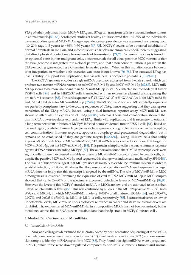
Int. J. Mol. Sci. 2018, 19, 1873
STAg of other polyomaviruses, MCPyV LTAg and STAg can transform cells in vitro and induce tumorsin animal models [59–66]. Serological studies of healthy adults showed that ~40–85% of the individualshave antibodies against MCPyV. An age-dependence seroprevalence was measured, increasing from~10–20% (age 1–5 years) to ~80% (>70 years) [67–73]. MCPyV seems to be a normal inhabitant ofdermal fibroblasts in the skin, and infectious virus particles are chronically shed, thereby suggestingthat direct physical contact may be one mode of transmission [74,75]. Whereas the virus is found inan episomal state in non-malignant cells, a characteristic for all virus-positive MCC tumors is thatthe viral genome is integrated into a clonal pattern, and that a non-sense mutation is present in theLTAg-encoding gene encoding a C-terminal truncated protein. Whether this mutation occurs before orafter integration, or whether both scenarios can occur is not known [76–78]. The truncated LTAg haslost its ability to support viral replication, but has retained its oncogenic potentials [61,79–81].
The MCPyV genome encodes a single miRNA precursor expressed from the late strand, which canproduce two mature miRNAs referred to as MCV-miR-M1-5p and MCV-miR-M1-3p [82,83]. MCV-miR-M1-5p seems to be more abundant than MCV-miR-M1-3p in MCPyV-infected neuroectodermal tumorPFSK-1 cells [84], and in HEK293T cells transfected with an expression plasmid encompassing thepre-miR-M1 sequence [85]. The seed sequence is 5′-CUGGAAG-3′ or 5′-GGAAGAA-3′ for MCV-miR1-5pand 5′-UGCUGGA3′- for MCV-miR-M1-3p [82–84]. The MCV-miR-M1-5p and MCV-miR-3p sequencesare perfectly complementary to the coding sequences of LTAg, hence suggesting that they can represstranslation of the LTAg mRNA. Indeed, using a dual-luciferase reporter assay, MCV-miR-M1 wasshown to attenuate the expression of LTAg [82,86], whereas Theiss and collaborators showed thatthis miRNA down-regulates expression of LTAg, limits viral replication, and is necessary to establisha long-term persistent infection of MCPyV-infected neuroectodermal tumor PFSK-1 cells [84]. Based onthe seed region, predicted human target genes include genes encoding proteins involved in transcription,cell communication, immune response, apoptosis, autophagy and proteasomal degradation, but itremains to be established if they are genuine targets [82,83,86]. Using HEK293 cells that stablyexpress MCV-miR-M1-5p or MCV-miR-M1-3p, SP100 mRNA was verified as a bona fide target forMCV-miR-M1-5p, but not MCV-miR-M1-3p [86]. This protein is implicated in the innate immune responseagainst dsDNA viruses, including MCPyV [87]. The authors also found that CXCL8 transcript levels weresignificantly different expressed in stably expressing MCV-miR-M1 cells compared to control cells, butdespite the putative MCV-miR-M1-3p seed sequence, this change was indirect and mediated by SP100 [86].The results of this work suggest that MCPyV uses its miRNA to evade the immune system in order toestablish infection, but it also illustrates that the presence of a putative miRNA seed sequence in a targetmRNA does not imply that this transcript is targeted by the miRNA. The role of MCV-miR-M1 in MCCtumorigenesis is less clear. Examining the expression of viral miRNA MCV-miR-M1-5p in MCC samplesshowed that up to 29–80% of the specimens expressed detectable levels of MCV-miR-M1-5p [82,83].However, the levels of this MCPyV-encoded miRNA in MCCs are low, and are estimated to be less than0.005% of total miRNA levels [82]. This was confirmed by studies in the MCPyV-positive MCC cell linesWaGa and MKL-1, in which MCV-miR-M1 made up 0.001% of all mature miRNAs [84], and 0.0067%,0.007%, and 0.0025% of MKL-1a, MKL-1b, MKL-1c cells, respectively [85]. Because its absence or low orundetectable levels, MCV-miR-M1-5p’s biological relevance in cancer and its value as biomarkers aredoubtful. The expression of MCV-miR-M1-3p in virus-positive MCCs has not been examined, but asmentioned above, this miRNA is even less abundant than the 5p strand in MCPyV-infected cells.
3. Merkel Cell Carcinoma and MicroRNAs
3.1. Intracellular MicroRNAs
Ning and colleagues determined the microRNAome by next-generation sequencing of three MCCs,one melanoma, one squamous cell carcinoma (SCC), one basal cell carcinoma (BCC) and one normalskin sample to identify miRNAs specific to MCC [88]. They found that eight miRNAs were upregulatedin MCC, while three were downregulated compared to non-MCC cutaneous tumors and normal
42
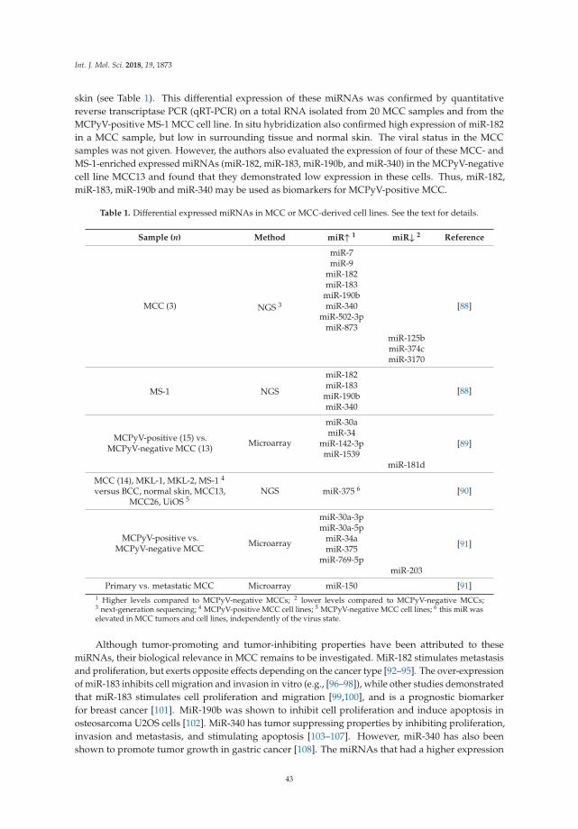
Int. J. Mol. Sci. 2018, 19, 1873
skin (see Table 1). This differential expression of these miRNAs was confirmed by quantitativereverse transcriptase PCR (qRT-PCR) on a total RNA isolated from 20 MCC samples and from theMCPyV-positive MS-1 MCC cell line. In situ hybridization also confirmed high expression of miR-182in a MCC sample, but low in surrounding tissue and normal skin. The viral status in the MCCsamples was not given. However, the authors also evaluated the expression of four of these MCC- andMS-1-enriched expressed miRNAs (miR-182, miR-183, miR-190b, and miR-340) in the MCPyV-negativecell line MCC13 and found that they demonstrated low expression in these cells. Thus, miR-182,miR-183, miR-190b and miR-340 may be used as biomarkers for MCPyV-positive MCC.
Table 1. Differential expressed miRNAs in MCC or MCC-derived cell lines. See the text for details.
Sample (n) Method miR↑ 1 miR↓ 2 Reference
MCC (3) NGS 3
miR-7
[88]
miR-9miR-182miR-183
miR-190bmiR-340
miR-502-3pmiR-873
miR-125bmiR-374cmiR-3170
MS-1 NGS
miR-182
[88]miR-183
miR-190bmiR-340
MCPyV-positive (15) vs.MCPyV-negative MCC (13)
Microarray
miR-30a
[89]miR-34
miR-142-3pmiR-1539
miR-181d
MCC (14), MKL-1, MKL-2, MS-1 4
versus BCC, normal skin, MCC13,MCC26, UiOS 5
NGS miR-375 6 [90]
MCPyV-positive vs.MCPyV-negative MCC
Microarray
miR-30a-3p
[91]
miR-30a-5pmiR-34amiR-375
miR-769-5pmiR-203
Primary vs. metastatic MCC Microarray miR-150 [91]1 Higher levels compared to MCPyV-negative MCCs; 2 lower levels compared to MCPyV-negative MCCs;3 next-generation sequencing; 4 MCPyV-positive MCC cell lines; 5 MCPyV-negative MCC cell lines; 6 this miR waselevated in MCC tumors and cell lines, independently of the virus state.
Although tumor-promoting and tumor-inhibiting properties have been attributed to thesemiRNAs, their biological relevance in MCC remains to be investigated. MiR-182 stimulates metastasisand proliferation, but exerts opposite effects depending on the cancer type [92–95]. The over-expressionof miR-183 inhibits cell migration and invasion in vitro (e.g., [96–98]), while other studies demonstratedthat miR-183 stimulates cell proliferation and migration [99,100], and is a prognostic biomarkerfor breast cancer [101]. MiR-190b was shown to inhibit cell proliferation and induce apoptosis inosteosarcoma U2OS cells [102]. MiR-340 has tumor suppressing properties by inhibiting proliferation,invasion and metastasis, and stimulating apoptosis [103–107]. However, miR-340 has also beenshown to promote tumor growth in gastric cancer [108]. The miRNAs that had a higher expression
43

Int. J. Mol. Sci. 2018, 19, 1873
in MCPyV-negative MCCs, compared to virus-positive tumors, include miR-125b, miR-374c andmiR-3170 [88]. MiR-125b can suppress and promote cancer progression, and further down-regulateγδ T cell activation and cytotoxicity [109], as well as cells involved in anti-tumor surveillance [110].MiR-374c was identified as a novel miRNA in cervical tumors, and miR-3170 was first identified in themiRNAome of melanoma, which has also been found in breast cancer tumors [111,112]. A possiblerole of these two miRNAs in cancer has not been elucidated, nor has the plausible involvement inMCC been solved.
A comparison of the microRNAome of MCPyV-positive and MCPyV-negative MCCs revealedthat approximately 2.5- to 5-fold higher levels of miR-30a, miR-34a, miR-142-3p and miR-1539 invirus-positive MCCs and 3.5-fold higher levels of miR-181d in virus-negative MCCs [89]. MiR-30ahas a dual role in cancer and can act as an oncogene or an onco-suppressor in different cancers, andseveral of its target genes have been identified (reviewed in [113]). Although miR-34a is a tumorsuppressor [114], Veija et al. speculated that an over-expression of miR-34a could decrease p53expression, thereby interfering with apoptosis, angiogenesis and DNA repair [89]. MiR-142-3pcan inhibit cell proliferation and invasion, but high levels have also been correlated with cancerprogression [115]. MiR-181d can act as a tumor suppressor [116], but the down-regulation of miR-181dresulted in a decreased proliferation and migration of pancreatic cancer cells [117]. To the best ofour knowledge, the role of miR-1539 has not been investigated. The prognostic value of miR-30a,miR-142-3p, miR-1539 and miR-181d is jeopardized, because qRT-PCR validation demonstrated thatonly miR-34a was significantly under-expressed in virus-negative MCCs compared to virus-positiveMCC samples [89]. Whether any of these miRNAs contribute to MCC tumorigenesis remains tobe established.
Deep sequencing of RNA purified from normal skin (n = 5), BCC (n = 5), MCC (n = 14), andMCPyV-negative (MCC13, MCC26, UiOS) and MCPyV-positive (MKL-1, MKL-2; MS-1) MCC cellsshowed that miR-375 is specific for MCC [90]. The miR-375 concentrations were 60-fold higher in theMCC group than in the non-MCC (normal skin and BCC) group. The enrichment of miR-375 seems tobe independent of the viral state, because elevated miR-375 levels were found in both virus-negativeand virus-positive tumors and tumor cell lines. Of the five skin samples that were examined, three wereMCPyV positive, one was virus negative and one was not tested. Of the five BCC samples, four werevirus negative and one was not tested. Similarly, since no increased miR-375 levels were found in thevirus-positive non-MCC samples, the presence of the virus seems not to affect the expression of miR-375.Although not discussed by the authors, there was a tendency for higher expression levels of miR-9 andmiR-188 in MCC samples. MiR-188 suppresses proliferation in different cancers [118–121]. MiR-9 canstimulate or inhibit cell proliferation and metastasis depending on the type of cancer, whereas highexpression levels in most cancers are associated with poor survival of the patients, except for ovariancancer patients, in which an inverse correlation was found [122,123]. MiR-375 has been describedas a tumor suppressor known to impede cell proliferation, to prevent cancer cell migration, and toinhibit autophagy, thereby generating an antitumor effect in liver cancer [124–129]. Therefore, it seemssurprising that this miRNA is over-expressed in MCC. Nonetheless, an over-expression of miR-375has also been implied in prostate carcinogenesis and disease progression, while an up-regulationof miR-375 is associated with a poor prognosis in pediatric acute myeloid leukemia [130,131], thusindicating a dual role for miR-375 in cancer. Moreover, miR-375 was shown to inhibit autophagy inhepatocellular carcinoma [132], but whether this role of miR-375 is of importance in MCC is unknown.
A comparison of the intracellular miRNA expression profiles in 10 MCPyV-negative and16 MCPyV-positive MCCs by a miRNA microarray-based method identified 36 over-expressed and20 under-expressed miRNAs in virus-positive MCCs compared to virus-negative MCCs [91]. Amongthese, a significant over-expression of miR-30a-3p, miR-30a-5p, miR-34a, miR-375 and miR-769-5p,and a significant under-expression of miR-203, were confirmed by qRT-PCR. A putative role ofmiR-30a, miR-34a and miR-375 in oncogenesis was described above. MiR-769 expression wasstrongly increased in human melanoma cells and clinical tissues compared with their corresponding
44

Int. J. Mol. Sci. 2018, 19, 1873
controls. The over-expression of miR-769 promoted cell proliferation in the human melanoma cellline A375 [133]. MiR-769 may exert these functions by targeting glycogen synthase kinase 3B, whilea similar mechanism may be operational in MCC oncogenesis. It is not known whether MCPyV LTAgand/or STAg stimulate the expression of miR-30a-3p, miR-30a-5p, miR-34a, miR-375 and miR-769-5p.The possible involvement of miR-203 in MCC oncogenesis was examined by over-expressing miR-203in three MCPyV-negative MCC cell lines [91]. This resulted in reduced cell growth, more cells inG1 and less in the G2 phase, but no apparent effect on apoptosis compared to cells transfected withmiRNA mimic control. Moreover, survivin expression was reduced. The over-expression of miR-203in the MCPyV-positive WaGa MCC cell line had no significant effect on cell proliferation, cell cycleprogression and survivin expression levels. These results suggest that miR-203 only regulates survivinexpression in virus-negative MCCs, but not in MCPyV-positive MCCs, in which LTAg seems torepress survivin expression by sequestering pRb [134]. The same group also examined differentiallyexpressed miRNAs in primary and metastatic MCC tumors [91]. They found that 92 miRNAs wereover-expressed in metastasis compared to primary tumors. The four most up-regulated miRNAs weremiR-150, miR-142-3p, miR-483-5p and miR-630, but qRT-PCR validation revealed that only miR-150was significantly overexpressed.
Xie et al. found that miR-375 was specifically over-expressed in MCPyV-positive MCCs, whileRenswick et al. reported that miR-375 was specific for MCC, independent of the viral state inthe tumors [90,91]. The discrepancy in these results may be explained by the differences in MCCsamples that were examined, or because different methods (next-generation sequencing versus miRNAmicroarray) were used.
The role of MCPyV on miRNA expression in non-small cell lung cancer (NSCLC) was investigatedby Lasithiotaki and co-workers [135]. The expression of miR-21, miR-145, miR-146a, miR-155, miR-302c,miR-367 and miR-376c was examined by qRT-PCR in MCPyV-positive and MCPyV-negative NSCLC.MiR-21 and miR-376c were up-regulated, whereas miR-145 was down-regulated in virus-positiveNSCLC (n = 8) compared to virus-negative NSCLC (n = 16). MiR-21 and miR-376c expressionlevels were also higher in virus-positive NSCLC versus adjacent healthy tissue samples (n = 10;5 MCPyV-positive and 5 MCPyV-negative), while miR-145 levels in MCPyV-negative NSCLC washigher than in control samples. To the best of our knowledge, none of the miRNAs investigatedby Lasithiotaki et al. have been described in MCC, except miR-146a which was enriched ~8-fold inexosomes derived from MCPyV-negative MCC13 and MCC26 cell lines compared to virus-positiveMKL-1 and MKL-2 cell lines (A.K., D.H.C., B.S., U.M., University of Tromsø, Norway, 2018).
3.2. Extracellular MicroRNAs and Merkel Cell Carcinoma
The presence of extracellular miRNA-protein complexes secreted by MCC cell lines or in MCCpatients has not been examined thus far. Likewise, the occurrence of miRNAs in apoptotic bodieshas not been investigated although apoptotic bodies have been reported in MCC [21]. We haveapplied next-generation sequencing to examine the microRNAome in exosomes purified from theMCPyV-negative MCC13 and MCC26 and the MCPyV-positive MKL-1 and MKL-2 MCC cell lines. Onaverage, there were 20.4 million reads per sample (three independent exosome samples of each cellline), with the number of miRNAs per sample varying approximately between 200 and 400. Of thepreviously identified intracellular miRNAs identified in MCC samples of MCC cell lines (Table 1), ourpreliminary results confirmed the presence of miR-30a, miR-125b, mi-183, miR-190b and miR-375 inexosomes. MCV-miR-M1 was not detected in any of our samples (A.K., D.H.C., B.S., U.M., Universityof Tromsø, Norway, 2018).
4. MicroRNAs as Biomarkers and Therapeutic Targets in Merkel Cell Carcinoma
MiRNAs are key components of cells in both normal and pathogenic states. The miRNAexpression pattern of normal cells versus malignant cells differs, and cancer-cell-specific miRNAs arebeing used as biomarkers in different cancers [136–141]. Exosomal miRNAs have become attractive
45

Int. J. Mol. Sci. 2018, 19, 1873
cancer biomarkers because exosomes are easily obtainable from body fluids, such as blood and urinewithout the requirement of a biopsy sample of the tumor. Exosomal miRNAs in plasma or urine areused as biomarkers for different malignancies, including melanoma, breast, colon, prostate, renal andgastric cancer [142–148]. Only a few studies have examined the miRNAome of MCC and miRNAs arenot yet used as biomarkers. One of the pitfalls of using MCC-derived miRNAs as biomarkers is thatthe miRNA expression pattern of normal Merkel cells has not been determined because these cells arerare. Hence, it is not known whether the MCC-derived miRNAs are specific for the malignant cellsor also expressed by non-malignant Merkel cells. Another problem is the lack of common miRNAsamong the MCC expressed miRNAs identified so far by independent studies. MiR-30a, miR-34 andmiR-375 were reported by Renwick et al. and Xie et al., but not by others (Table 1) [90,91]. We foundmiR-30 and miR-375 in exosomes derived from MCC cell lines, but it is not known whether thesemiRNAs are also present in non-transformed Merkel cells. MiR-30a is expressed in different cell types,including normal dermal fibroblasts, keratinocytes and endothelial cells [149–151], whereas miR-375 ispresent in normal epithelial, pituitary and pancreatic β-cells [152–154], jeopardizing the value of thesemiRNAs as specific biomarkers for MCC. Additional tumor specimens must be investigated in orderto isolate MCC-specific miRNAs and their potential use as biomarkers should be verified.
MiRNAs may also be used to determine the viral state in the MCC. Levels of miR-30a and miR-34were increased in MCPyV-positive MCCs compared to MCPyV-negative [89,91]; hence, these miRNAsmay be applied to distinguish between virus-positive and virus-negative cancers. MiR-375 was foundto be a specific miRNA for virus-positive MCCs [90,91], but this could not be confirmed by others whofound this miRNA in both MCPyV-positive and MCPyV-negative MCC [89] and in exosomes of bothvirus-positive and virus-negative MCC cell lines (our unpublished results). Whether miR-375 can beused as a hallmark for MCPyV-positive MCCs needs further investigation.
As for diagnostic purposes, real-time PCR methods with primers against exosomal miRNAsspecific for MCPyV-positive MCCs could replace the commonly used PCR with MCPyV sequence-specific on DNA extracted from a tumor sample. The advantage of real-time PCR on a circulatingmiRNA is that body fluids, such as blood or urine, are readily accessible sources and more convenientfor the MCC patient to obtain than using a biopsy, and that the number of miRNA molecules canbe estimated.
Another criterion for a useful biomarker is that it can predict the outcome of the disease. Studiesby Xie et al. found that higher levels of miR-150 were associated with a worse prognosis of MCC [91].Quantifying MCC-specific miRNA levels may also provide information on the disease progressionand the efficiency of treatment in the case of miRNA-target therapy. There is a need for new andimproved therapy of MCC patients. As of today, MCC treatment includes surgery, radiotherapy andchemotherapy. In a few cases, a spontaneous regression of primary and metastatic MCC has beenreported (see e.g., [155–158]). Recently, immunotherapy based on blocking the PD1-PDL-1 pathwayby either anti-PD1 antibodies (pembrolizumab, nivolumab) or anti-PDL-1 antibodies (avelumab) hasdemonstrated favorable responses, with a six-month progression-free survival in 40–85%, and evencomplete resolution of the tumors in some patients [159–167]. Avelumab became the first Food andDrug Administration-approved drug for the treatment of MCC (https://www.fda.gov/newsevents/newsroom/pressannouncements/ucm548278.htm, 10 May 2018). Treatment with the anti-CTLA-4antibody ipilimumab has also shown beneficial effects in metastatic MCC [168,169]. However, notall patients have a positive response, so the development of additional therapies is necessary. Drugsagainst MCC-specific miRNAs or their target transcripts can supplement immunotherapy. Clinicaltrials with miRNAs against some pathological conditions except MCC have been initiated [170–172].One of the miRNA-based clinical trials includes miR-34, which is expressed in MCC [89,91].
Finally, miRNAs may also help in solving the enigma of the origin of Merkel cells. MiRNAsignatures may be an alternative to immunhistochemical staining. This requires the identification ofcell-specific miRNAs of neural crest cells, keratinocytes, epidermal fibroblasts, early B cells and hairfollicle stem cells, cells that have been suggested to be the origin of Merkel cells (see Section 2).
46

Int. J. Mol. Sci. 2018, 19, 1873
In conclusion, multi-center microRNAome studies on a large number of MCC samples or biofluidsof patients are required to identify valuable miRNA biomarkers. These data should be linked toparameters, such as the clinical features of the patient, the stage of the tumor (primary or metastatic),viral states and LTAg and STAg expression, age and gender of the patient. MiRNA profiling may beused in determining the prognosis and progression of the disease, and monitoring the response totherapy (Figure 1). Exosomes have been found in a number of biological fluids including plasma,urine, breast milk, semen, cerebrospinal fluid and saliva [173]. Exosomal miRNAs, in addition tointracellular miRNAs, may therefore be easily accessible biomarkers.
Figure 1. Detection of MCC-specific miRNAs from tumor biopsies or from body fluids. (A) Thepresence of intracellular or/and extracellular MCC-specific miRNAs is examined by qRT-PCR usingspecific primers. Intracellular miRNAs are amplified from a total RNA isolated from MCC tumortissue, while extracellular miRNAs are amplified from a RNA extracted from purified exosomesor from the extracellular environment. The biogenesis of a miRNA is shown. A pre-miRNA istransported from the nucleus to the cytoplasm, and when processed to mature miRNA, it bindstarget mRNA (step 1). Pre-miRNAs and miRNAs can also be enclosed in vesicles and excreted inexosomes (step 2) or other extracellular vesicles (step 3). Pre-miRNAs and miRNAs can also releasefrom the cell in complex with RNA-binding proteins, such as Argonaut 2 or nucleophosmin-1, or incomplex with high-density lipoproteins (step 4); (B) circulating exosomes are purified from body fluids(e.g., blood, urine, lymphatic fluid, saliva) and a total RNA is extracted. MCC-specific miRNAs aresubsequently detected by qRT-PCR applying specific primers, next-generation sequencing (NGS),microarray or nCounter.
5. Future Challenges
MiRNAs can be used as reliable biomarkers in several cancers [137–141], and miRNA-basedcancer therapy is being developed and tested [170,171]. As outlined above, little research has beendone on MCC-specific miRNAs. It is reasonable to wonder whether there is any clinical value formiRNA in MCC and if so, what could it be?
• MCC-specific miRNAs as biomarkers still have a long way to go. Consensus intracellular MCCmiRNAs have not yet been identified, and circulating miRNAs have not been investigated.
47

Int. J. Mol. Sci. 2018, 19, 1873
We are currently studying the exosome miRNAome of MCC cell lines with the aim of identifyingMCC-specific extracellular miRNAs. Cell–cell communication is important in the tumormicroenvironment and one way of communication is exosomes [174]. Thus, analyzing themiRNAome of exosomes may provide clues on how tumor cells promote survival, growth andmetastasis by modulating the tumor microenvironment [175].
• The intracellular microRNAome or circulating miRNA may be used to discriminate betweenMCPyV-negative and MCPyV-positive MCCs. So far, unambiguous miRNAs that allowdistinguishing between virus-negative and virus-positive tumors have not yet been described.
• Can MCC-specific miRNAs be used as therapeutic targets? The biological importance of miRNAsin MCC oncogenesis is incompletely understood. In fact, most of the currently reported miRNAsin MCC have dual functions (oncogenic or tumor suppressive roles) in other cell systems, so thattargeting their expression may be a double-edged sword. The exact contributing role of miRNAsin MCC is required to design efficient and specific therapies.
• Affordable and easy laboratory tests based on these MCC-specific miRNA biomarkers should bedeveloped to improve the diagnosis, prognosis, and progression of this cancer.
Author Contributions: A.K., D.H.C, B.S. and U.M. wrote the paper.
Funding: Studies in our laboratory were funded by the Olav and Erna Aakre Foundation for Cancer (Tromsø,Norway) and Raagholt Stiftelsen (Oslo, Norway). The publication charges for this article have been funded bya grant from the publication fund of UiT the Artic University of Norway.
Acknowledgments: We greatly acknowledge Roy Lyså for preparing Figure 1.
Conflicts of Interest: The authors declare no conflicts of interest.
References
1. He, L.; Hannon, G.J. MicroRNAs: Small RNAs with a big role in gene regulation. Nat. Rev. Genet. 2004, 5,
522–531. [CrossRef] [PubMed]
2. Kozomara, A.; Griffiths-Jones, S. miRBase: Annotating high confidence microRNAs using deep sequencing
data. Nucleic Acids Res. 2014, 42, D68–D73. [CrossRef] [PubMed]
3. Londin, E.; Loher, P.; Telonis, A.G.; Quann, K.; Clark, P.; Jing, Y.; Hatzimichael, E.; Kirino, Y.; Honda, S.;
Lally, M.; et al. Analysis of 13 cell types reveals evidence for the expression of numerous novel primate- and
tissue-specific microRNAs. Proc. Natl. Acad. Sci. USA 2015, 112, E1106–E1115. [CrossRef] [PubMed]
4. Kim, V.N.; Han, J.; Siomi, M.C. Biogenesis of small RNAs in animals. Nat. Rev. Mol. Cell Biol. 2009, 10,
126–139. [CrossRef] [PubMed]
5. Ha, M.; Kim, V.N. Regulation of microRNA biogenesis. Nat. Rev. Mol. Cell Biol. 2014, 15, 509–524. [CrossRef]
[PubMed]
6. Marco, A.; Ninova, M.; Griffiths-Jones, S. Multiple products from microRNA transcripts. Biochem. Soc. Trans.
2013, 41, 850–854. [CrossRef] [PubMed]
7. Neilsen, C.T.; Goodall, G.J.; Bracken, C.P. IsomiRs—The overlooked repertoire in the dynamic microRNAome.
Trends Genet. 2012, 28, 544–549. [CrossRef] [PubMed]
8. Tan, G.C.; Dibb, N. IsomiRs have functional importance. Malays. J. Pathol. 2015, 37, 73–81. [PubMed]
9. Lunavat, T.R.; Cheng, L.; Kim, D.K.; Bhadury, J.; Jang, S.C.; Lasser, C.; Sharples, R.A.; Lopez, M.D.; Nilsson, J.;
Gho, Y.S.; et al. Small RNA deep sequencing discriminates subsets of extracellular vesicles released by
melanoma cells—Evidence of unique microRNA cargos. RNA Biol. 2015, 12, 810–823. [CrossRef] [PubMed]
10. Zhang, J.; Li, S.; Li, L.; Li, M.; Guo, C.; Yao, J.; Mi, S. Exosome and exosomal microRNA: Trafficking, sorting,
and function. Genom. Proteom. Bioinform. 2015, 13, 17–24. [CrossRef] [PubMed]
11. Turchinovich, A.; Tonevitsky, A.G.; Burwinkel, B. Extracellular miRNA: A Collision of Two Paradigms.
Trends Biochem. Sci. 2016, 41, 883–892. [CrossRef] [PubMed]
12. Turchinovich, A.; Weiz, L.; Langheinz, A.; Burwinkel, B. Characterization of extracellular circulating
microRNA. Nucleic Acids Res. 2011, 39, 7223–7233. [CrossRef] [PubMed]
13. Hannafon, B.N.; Ding, W.Q. Intercellular communication by exosome-derived microRNAs in cancer. Int. J.
Mol. Sci. 2013, 14, 14240–14269. [CrossRef] [PubMed]
48

Int. J. Mol. Sci. 2018, 19, 1873
14. Kai, K.; Dittmar, R.L.; Sen, S. Secretory microRNAs as biomarkers of cancer. Semin. Cell Dev. Biol. 2018, 78,
22–36. [CrossRef] [PubMed]
15. Arroyo, J.D.; Chevillet, J.R.; Kroh, E.M.; Ruf, I.K.; Pritchard, C.C.; Gibson, D.F.; Mitchell, P.S.; Bennett, C.F.;
Pogosova-Agadjanyan, E.L.; Stirewalt, D.L.; et al. Argonaute2 complexes carry a population of circulating
microRNAs independent of vesicles in human plasma. Proc. Natl. Acad. Sci. USA 2011, 108, 5003–5008.
[CrossRef] [PubMed]
16. Prud’homme, G.J.; Glinka, Y.; Lichner, Z.; Yousef, G.M. Neuropilin-1 is a receptor for extracellular miRNA
and AGO2/miRNA complexes and mediates the internalization of miRNAs that modulate cell function.
Oncotarget 2016, 7, 68057–68071. [CrossRef] [PubMed]
17. Vickers, K.C.; Palmisano, B.T.; Shoucri, B.M.; Shamburek, R.D.; Remaley, A.T. MicroRNAs are transported
in plasma and delivered to recipient cells by high-density lipoproteins. Nat. Cell Biol. 2011, 13, 423–433.
[CrossRef] [PubMed]
18. Niculescu, L.S.; Simionescu, N.; Sanda, G.M.; Carnuta, M.G.; Stancu, C.S.; Popescu, A.C.; Popescu, M.R.;
Vlad, A.; Dimulescu, D.R.; Simionescu, M.; et al. MiR-486 and miR-92a Identified in Circulating HDL
Discriminate between Stable and Vulnerable Coronary Artery Disease Patients. PLoS ONE 2015, 10, e0140958.
[CrossRef] [PubMed]
19. Wang, K.; Zhang, S.; Weber, J.; Baxter, D.; Galas, D.J. Export of microRNAs and microRNA-protective protein
by mammalian cells. Nucleic Acids Res. 2010, 38, 7248–7259. [CrossRef] [PubMed]
20. Zernecke, A.; Bidzhekov, K.; Noels, H.; Shagdarsuren, E.; Gan, L.; Denecke, B.; Hristov, M.;
Koppel, T.; Jahantigh, M.N.; Lutgens, E.; et al. Delivery of microRNA-126 by apoptotic bodies induces
CXCL12-dependent vascular protection. Sci. Signal. 2009, 2, ra81. [CrossRef] [PubMed]
21. Mori, Y.; Hashimoto, K.; Tanaka, K.; Cui, C.Y.; Mehregan, D.R.; Stiff, M.A. A study of apoptosis in Merkel
cell carcinoma: An immunohistochemical, ultrastructural, DNA ladder, and TUNEL labeling study. Am. J.
Dermatopathol. 2001, 23, 16–23. [CrossRef] [PubMed]
22. Iwakawa, H.O.; Tomari, Y. The Functions of MicroRNAs: MRNA Decay and Translational Repression.
Trends Cell Biol. 2015, 25, 651–665. [CrossRef] [PubMed]
23. Place, R.F.; Li, L.C.; Pookot, D.; Noonan, E.J.; Dahiya, R. MicroRNA-373 induces expression of genes with
complementary promoter sequences. Proc. Natl. Acad. Sci. USA 2008, 105, 1608–1613. [CrossRef] [PubMed]
24. Wilfred, B.R.; Wang, W.X.; Nelson, P.T. Energizing miRNA research: A review of the role of miRNAs in lipid
metabolism, with a prediction that miR-103/107 regulates human metabolic pathways. Proc. Natl. Acad.
Sci. USA 2008, 105, 1608–1613. [CrossRef] [PubMed]
25. Stefani, G.; Slack, F.J. Small non-coding RNAs in animal development. Nat. Rev. Mol. Cell Biol. 2008, 9,
219–230. [CrossRef] [PubMed]
26. Shi, Y.; Jin, Y. MicroRNA in cell differentiation and development. Sci. China C Life Sci. 2009, 52, 205–511.
[CrossRef] [PubMed]
27. O’Connell, R.M.; Rao, D.S.; Chaudhuri, A.A.; Baltimore, D. Physiological and pathological roles for
microRNAs in the immune system. Nat. Rev. Immunol. 2010, 10, 111–122. [CrossRef] [PubMed]
28. Subramanian, S.; Steer, C.J. MicroRNAs as gatekeepers of apoptosis. J. Cell. Physiol. 2010, 223, 289–298.
[CrossRef] [PubMed]
29. Landskroner-Eiger, S.; Moneke, I.; Sessa, W.C. miRNAs as modulators of angiogenesis. Cold Spring Harb.
Perspect. Med. 2013, 3, a006643. [CrossRef] [PubMed]
30. Zhao, Y.; Srivastava, D. A developmental view of microRNA function. Trends Biochem. Sci. 2007, 32, 189–197.
[CrossRef] [PubMed]
31. Visone, R.; Croce, C.M. MiRNAs and cancer. Am. J. Pathol. 2009, 174, 1131–1138. [CrossRef] [PubMed]
32. Ameres, S.L.; Zamore, P.D. Diversifying microRNA sequence and function. Nat. Rev. Mol. Cell Biol. 2013, 14,
475–488. [CrossRef] [PubMed]
33. Vidigal, J.A.; Ventura, A. The biological functions of miRNAs: Lessons from in vivo studies. Trends Cell Biol.
2015, 25, 137–147. [CrossRef] [PubMed]
34. Bracken, C.P.; Scott, H.S.; Goodall, G.J. A network-biology perspective of microRNA function and
dysfunction in cancer. Nat. Rev. Genet. 2016, 17, 719–732. [CrossRef] [PubMed]
35. Lou, W.; Liu, J.; Gao, Y.; Zhong, G.; Chen, D.; Shen, J.; Bao, C.; Xu, L.; Pan, J.; Cheng, J.; et al. MicroRNAs in
cancer metastasis and angiogenesis. Oncotarget 2017, 8, 115787–115802. [CrossRef] [PubMed]
49

Int. J. Mol. Sci. 2018, 19, 1873
36. Telonis, A.G.; Magee, R.; Loher, P.; Chervoneva, I.; Londin, E.; Rigoutsos, I. Knowledge about the presence
or absence of miRNA isoforms (isomiRs) can successfully discriminate amongst 32 TCGA cancer types.
Nucleic Acids Res. 2017, 45, 2973–2985. [CrossRef] [PubMed]
37. Schrama, D.; Ugurel, S.; Becker, J.C. Merkel cell carcinoma: Recent insights and new treatment options.
Curr. Opin. Oncol. 2012, 24, 141–149. [CrossRef] [PubMed]
38. Erovic, I.; Erovic, B.M. Merkel cell carcinoma: The past, the present, and the future. J. Skin Cancer 2013,
2013, 929364. [CrossRef] [PubMed]
39. Godlewski, J.; Kowalczyk, A.; Kozielec, Z.; Pidsudko, Z.; Kmiec, A.; Siedlecka-Kroplewska, K. Plasticity
of neuropeptidergic neoplasm cells in the primary and metastatic Merkel cell carcinoma. Folia Histochem.
Cytobiol. 2013, 51, 168–173. [CrossRef] [PubMed]
40. Visscher, D.; Cooper, P.H.; Zarbo, R.J.; Crissman, J.D. Cutaneous neuroendocrine (Merkel cell) carcinoma:
An immunophenotypic, clinicopathologic, and flow cytometric study. Mod. Pathol. 1989, 2, 331–338.
[PubMed]
41. Love, J.E.; Thompson, K.; Kilgore, M.R.; Westerhoff, M.; Murphy, C.E.; Papanicolau-Sengos, A.;
McCormick, K.A.; Shankaran, V.; Vandeven, N.; Miller, F.; et al. CD200 Expression in Neuroendocrine
Neoplasms. Am. J. Clin. Pathol. 2017, 148, 236–242. [CrossRef] [PubMed]
42. Jankowski, M.; Kopinski, P.; Schwartz, R.; Czajkowski, R. Merkel cell carcinoma: Is this a true carcinoma?
Exp. Dermatol. 2014, 23, 792–794. [CrossRef] [PubMed]
43. Sauer, C.M.; Haugg, A.M.; Chteinberg, E.; Rennspiess, D.; Winnepenninckx, V.; Speel, E.J.; Becker, J.C.;
Kurz, A.K.; Zur Hausen, A. Reviewing the current evidence supporting early B-cells as the cellular origin of
Merkel cell carcinoma. Crit. Rev. Oncol. Hematol. 2017, 116, 99–105. [CrossRef] [PubMed]
44. Tilling, T.; Moll, I. Which are the cells of origin in merkel cell carcinoma? J. Skin Cancer 2012, 2012, 680410.
[CrossRef] [PubMed]
45. Sunshine, J.C.; Jahchan, N.S.; Sage, J.; Choi, J. Are there multiple cells of origin of Merkel cell carcinoma?
Oncogene 2018, 37, 1409–1416. [CrossRef] [PubMed]
46. Tai, P. Merkel cell cancer: Update on biology and treatment. Curr. Opin. Oncol. 2008, 20, 196–200. [CrossRef]
[PubMed]
47. Becker, J.C.; Stang, A.; DeCaprio, J.A.; Cerroni, L.; Lebbe, C.; Veness, M.; Nghiem, P. Merkel cell carcinoma.
Nat. Rev. Dis. Primers 2017, 3, 17077. [CrossRef] [PubMed]
48. Agelli, M.; Clegg, L.X.; Becker, J.C.; Rollison, D.E. The etiology and epidemiology of merkel cell carcinoma.
Curr. Probl. Cancer 2010, 34, 14–37. [CrossRef] [PubMed]
49. Kaae, J.; Hansen, A.V.; Biggar, R.J.; Boyd, H.A.; Moore, P.S.; Wohlfahrt, J.; Melbye, M. Merkel cell carcinoma:
Incidence, mortality, and risk of other cancers. J. Natl. Cancer Inst. 2010, 102, 793–801. [CrossRef] [PubMed]
50. Stang, A.; Becker, J.C.; Nghiem, P.; Ferlay, J. The association between geographic location and incidence of
Merkel cell carcinoma in comparison to melanoma: An international assessment. Eur. J. Cancer 2018, 94,
47–60. [CrossRef] [PubMed]
51. Harms, K.L.; Healy, M.A.; Nghiem, P.; Sober, A.J.; Johnson, T.M.; Bichakjian, C.K.; Wong, S.L. Analysis of
Prognostic Factors from 9387 Merkel Cell Carcinoma Cases Forms the Basis for the New 8th Edition AJCC
Staging System. Ann. Surg. Oncol. 2016, 23, 3564–3571. [CrossRef] [PubMed]
52. Tomic, S.; Warner, T.F.; Messing, E.; Wilding, G. Penile Merkel cell carcinoma. Urology 1995, 45, 1062–1065.
[CrossRef]
53. Roy, S.; Das, I.; Nandi, A.; Roy, R. Primary Merkel cell carcinoma of the oral mucosa in a young adult male:
Report of a rare case. Indian J. Pathol. Microbiol. 2015, 58, 214–216. [CrossRef] [PubMed]
54. Nguyen, A.H.; Tahseen, A.I.; Vaudreuil, A.M.; Caponetti, G.C.; Huerter, C.J. Clinical features and treatment of
vulvar Merkel cell carcinoma: A systematic review. Gynecol. Oncol. Res. Pract. 2017, 4, 2. [CrossRef] [PubMed]
55. Oram, C.W.; Bartus, C.L.; Purcell, S.M. Merkel cell carcinoma: A review. Cutis 2016, 97, 290–295. [PubMed]
56. Feng, H.; Shuda, M.; Chang, Y.; Moore, P.S. Clonal integration of a polyomavirus in human Merkel cell
carcinoma. Science 2008, 319, 1096–1100. [CrossRef] [PubMed]
57. Coursaget, P.; Samimi, M.; Nicol, J.T.; Gardair, C.; Touze, A. Human Merkel cell polyomavirus: Virological
background and clinical implications. APMIS 2013, 121, 755–769. [CrossRef] [PubMed]
58. Liu, W.; MacDonald, M.; You, J. Merkel cell polyomavirus infection and Merkel cell carcinoma. Curr. Opin.
Virol. 2016, 20, 20–27. [CrossRef] [PubMed]
50

Int. J. Mol. Sci. 2018, 19, 1873
59. Baez, C.F.; Brandao Varella, R.; Villani, S.; Delbue, S. Human Polyomaviruses: The Battle of Large and Small
Tumor Antigens. Virology 2017, 8. [CrossRef] [PubMed]
60. Wendzicki, J.A.; Moore, P.S.; Chang, Y. Large T and small T antigens of Merkel cell polyomavirus. Curr. Opin.
Virol. 2015, 11, 38–43. [CrossRef] [PubMed]
61. Demetriou, S.K.; Ona-Vu, K.; Sullivan, E.M.; Dong, T.K.; Hsu, S.W.; Oh, D.H. Defective DNA repair and
cell cycle arrest in cells expressing Merkel cell polyomavirus T antigen. Int. J. Cancer 2012, 131, 1818–1827.
[CrossRef] [PubMed]
62. Cheng, J.; Rozenblatt-Rosen, O.; Paulson, K.G.; Nghiem, P.; DeCaprio, J.A. Merkel cell polyomavirus large T
antigen has growth-promoting and inhibitory activities. J. Virol. 2013, 87, 6118–6126. [CrossRef] [PubMed]
63. Verhaegen, M.E.; Mangelberger, D.; Harms, P.W.; Vozheiko, T.D.; Weick, J.W.; Wilbert, D.M.; Saunders, T.L.;
Ermilov, A.N.; Bichakjian, C.K.; Johnson, T.M.; et al. Merkel cell polyomavirus small T antigen is oncogenic
in transgenic mice. J. Investig. Dermatol. 2015, 135, 1415–1424. [CrossRef] [PubMed]
64. Spurgeon, M.E.; Cheng, J.; Bronson, R.T.; Lambert, P.F.; DeCaprio, J.A. Tumorigenic activity of merkel cell
polyomavirus T antigens expressed in the stratified epithelium of mice. Cancer Res. 2015, 75, 1068–1079.
[CrossRef] [PubMed]
65. Shuda, M.; Guastafierro, A.; Geng, X.; Shuda, Y.; Ostrowski, S.M.; Lukianov, S.; Jenkins, F.J.; Honda, K.;
Maricich, S.M.; Moore, P.S.; et al. Merkel Cell Polyomavirus Small T Antigen Induces Cancer and Embryonic
Merkel Cell Proliferation in a Transgenic Mouse Model. PLoS ONE 2015, 10, e0142329. [CrossRef] [PubMed]
66. Verhaegen, M.E.; Mangelberger, D.; Harms, P.W.; Eberl, M.; Wilbert, D.M.; Meireles, J.; Bichakjian, C.K.;
Saunders, T.L.; Wong, S.Y.; Dlugosz, A.A. Merkel Cell Polyomavirus Small T Antigen Initiates Merkel Cell
Carcinoma-like Tumor Development in Mice. Cancer Res. 2017, 77, 3151–3157. [CrossRef] [PubMed]
67. Kean, J.M.; Rao, S.; Wang, M.; Garcea, R.L. Seroepidemiology of human polyomaviruses. PLoS Pathog. 2009,
5, e1000363. [CrossRef] [PubMed]
68. Tolstov, Y.L.; Pastrana, D.V.; Feng, H.; Becker, J.C.; Jenkins, F.J.; Moschos, S.; Chang, Y.; Buck, C.B.; Moore, P.S.
Human Merkel cell polyomavirus infection II. MCV is a common human infection that can be detected by
conformational capsid epitope immunoassays. Int. J. Cancer 2009, 125, 1250–1256. [CrossRef] [PubMed]
69. Pastrana, D.V.; Tolstov, Y.L.; Becker, J.C.; Moore, P.S.; Chang, Y.; Buck, C.B. Quantitation of human
seroresponsiveness to Merkel cell polyomavirus. PLoS Pathog. 2009, 5, e1000578. [CrossRef] [PubMed]
70. Viscidi, R.P.; Rollison, D.E.; Sondak, V.K.; Silver, B.; Messina, J.L.; Giuliano, A.R.; Fulp, W.; Ajidahun, A.;
Rivanera, D. Age-specific seroprevalence of Merkel cell polyomavirus, BK virus, and JC virus. Clin. Vaccine
Immunol. 2011, 18, 1737–1743. [CrossRef] [PubMed]
71. Nicol, J.T.; Robinot, R.; Carpentier, A.; Carandina, G.; Mazzoni, E.; Tognon, M.; Touze, A.; Coursaget, P.
Age-specific seroprevalences of merkel cell polyomavirus, human polyomaviruses 6, 7, and 9, and
trichodysplasia spinulosa-associated polyomavirus. Clin. Vaccine Immunol. 2013, 20, 363–368. [CrossRef]
[PubMed]
72. Zhang, C.; Liu, F.; He, Z.; Deng, Q.; Pan, Y.; Liu, Y.; Zhang, C.; Ning, T.; Guo, C.; Liang, Y.; et al. Seroprevalence
of Merkel cell polyomavirus in the general rural population of Anyang, China. PLoS ONE 2014, 9, e106430.
[CrossRef] [PubMed]
73. Antonsson, A.; Neale, R.E.; O’Rourke, P.; Wockner, L.; Michel, A.; Pawlita, M.; Waterboer, T.; Green, A.C.
Prevalence and stability of antibodies to thirteen polyomaviruses and association with cutaneous squamous
cell carcinoma: A population-based study. Clin. Virol. 2018, 101, 34–37. [CrossRef] [PubMed]
74. Schowalter, R.M.; Pastrana, D.V.; Pumphrey, K.A.; Moyer, A.L.; Buck, C.B. Merkel cell polyomavirus and
two previously unknown polyomaviruses are chronically shed from human skin. Cell Host Microbe 2010, 7,
509–515. [CrossRef] [PubMed]
75. Martel-Jantin, C.; Pedergnana, V.; Nicol, J.T.; Leblond, V.; Tregouet, D.A.; Tortevoye, P.; Plancoulaine, S.;
Coursaget, P.; Touze, A.; Abel, L.; et al. Merkel cell polyomavirus infection occurs during early childhood
and is transmitted between siblings. J. Clin. Virol. 2013, 58, 288–291. [CrossRef] [PubMed]
76. Stakaityte, G.; Wood, J.J.; Knight, L.M.; Abdul-Sada, H.; Adzahar, N.S.; Nwogu, N.; Macdonald, A.;
Whitehouse, A. Merkel cell polyomavirus: Molecular insights into the most recently discovered human
tumour virus. Cancers 2014, 6, 1267–1297. [CrossRef] [PubMed]
77. Arora, R.; Chang, Y.; Moore, P.S. MCV and Merkel cell carcinoma: A molecular success story. Curr. Opin.
Virol. 2012, 2, 489–498. [CrossRef] [PubMed]
51

Int. J. Mol. Sci. 2018, 19, 1873
78. Chang, Y.; Moore, P.S. Merkel cell carcinoma: A virus-induced human cancer. Annu. Rev. Pathol. 2012, 7,
123–144. [CrossRef] [PubMed]
79. Houben, R.; Adam, C.; Baeurle, A.; Hesbacher, S.; Grimm, J.; Angermeyer, S.; Henzel, K.; Hauser, S.; Elling, R.;
Brocker, E.B.; et al. An intact retinoblastoma protein-binding site in Merkel cell polyomavirus large T antigen
is required for promoting growth of Merkel cell carcinoma cells. Int. J. Cancer 2012, 130, 847–856. [CrossRef]
[PubMed]
80. Hesbacher, S.; Pfitzer, L.; Wiedorfer, K.; Angermeyer, S.; Borst, A.; Haferkamp, S.; Scholz, C.J.; Wobser, M.;
Schrama, D.; Houben, R. RB1 is the crucial target of the Merkel cell polyomavirus Large T antigen in Merkel
cell carcinoma cells. Oncotarget 2016, 7, 32956–32968. [CrossRef] [PubMed]
81. Shuda, M.; Kwun, H.J.; Feng, H.; Chang, Y.; Moore, P.S. Human Merkel cell polyomavirus small T antigen is
an oncoprotein targeting the 4E-BP1 translation regulator. J. Clin. Investig. 2011, 121, 3623–3634. [CrossRef]
[PubMed]
82. Seo, G.J.; Chen, C.J.; Sullivan, C.S. Merkel cell polyomavirus encodes a microRNA with the ability to
autoregulate viral gene expression. Virology 2009, 383, 183–187. [CrossRef] [PubMed]
83. Lee, S.; Paulson, K.G.; Murchison, E.P.; Afanasiev, O.K.; Alkan, C.; Leonard, J.H.; Byrd, D.R.; Hannon, G.J.;
Nghiem, P. Identification and validation of a novel mature microRNA encoded by the Merkel cell
polyomavirus in human Merkel cell carcinomas. J. Clin. Virol. 2011, 52, 272–275. [CrossRef] [PubMed]
84. Theiss, J.M.; Gunther, T.; Alawi, M.; Neumann, F.; Tessmer, U.; Fischer, N.; Grundhoff, A. A Comprehensive
Analysis of Replicating Merkel Cell Polyomavirus Genomes Delineates the Viral Transcription Program
and Suggests a Role for mcv-miR-M1 in Episomal Persistence. PLoS Pathog. 2015, 11, e1004974. [CrossRef]
[PubMed]
85. Chen, C.J.; Cox, J.E.; Azarm, K.D.; Wylie, K.N.; Woolard, K.D.; Pesavento, P.A.; Sullivan, C.S. Identification
of a polyomavirus microRNA highly expressed in tumors. Virology 2015, 476, 43–53. [CrossRef] [PubMed]
86. Akhbari, P.; Tobin, D.; Poterlowicz, K.; Roberts, W.; Boyne, J.R. MCV-miR-M1 targets the host-cell immune
response resulting in the attenuation of neutrophil chemotaxis. J. Investig. Dermatol. 2018. [CrossRef] [PubMed]
87. Neumann, F.; Czech-Sioli, M.; Dobner, T.; Grundhoff, A.; Schreiner, S.; Fischer, N. Replication of Merkel
cell polyomavirus induces reorganization of promyelocytic leukemia nuclear bodies. J. Gen. Virol. 2016, 97,
2926–2938. [CrossRef] [PubMed]
88. Ning, M.S.; Kim, A.S.; Prasad, N.; Levy, S.E.; Zhang, H.; Andl, T. Characterization of the Merkel Cell
Carcinoma miRNome. J. Skin Cancer 2014, 2014, 289548. [CrossRef] [PubMed]
89. Veija, T.; Sahi, H.; Koljonen, V.; Bohling, T.; Knuutila, S.; Mosakhani, N. miRNA-34a underexpressed in
Merkel cell polyomavirus-negative Merkel cell carcinoma. Virchows Arch. 2015, 466, 289–295. [CrossRef]
[PubMed]
90. Renwick, N.; Cekan, P.; Masry, P.A.; McGeary, S.E.; Miller, J.B.; Hafner, M.; Li, Z.; Mihailovic, A.; Morozov, P.;
Brown, M.; et al. Multicolor microRNA FISH effectively differentiates tumor types. J. Clin. Investig. 2013,
123, 2694–2702. [CrossRef] [PubMed]
91. Xie, H.; Lee, L.; Caramuta, S.; Hoog, A.; Browaldh, N.; Bjornhagen, V.; Larsson, C.; Lui, W.O. MicroRNA
expression patterns related to merkel cell polyomavirus infection in human merkel cell carcinoma. J. Investig.
Dermatol. 2014, 134, 507–517. [CrossRef] [PubMed]
92. Segura, M.F.; Hanniford, D.; Menendez, S.; Reavie, L.; Zou, X.; Alvarez-Diaz, S.; Zakrzewski, J.; Blochin, E.;
Rose, A.; Bogunovic, D.; et al. Aberrant miR-182 expression promotes melanoma metastasis by repressing
FOXO3 and microphthalmia-associated transcription factor. Proc. Natl. Acad. Sci. USA 2009, 106, 1814–1819.
[CrossRef] [PubMed]
93. Tang, L.; Chen, F.; Pang, E.J.; Zhang, Z.Q.; Jin, B.W.; Dong, W.F. MicroRNA-182 inhibits proliferation through
targeting oncogenic ANUBL1 in gastric cancer. Oncol. Rep. 2015, 33, 1707–1716. [CrossRef] [PubMed]
94. Feng, Y.A.; Liu, T.E.; Wu, Y. microRNA-182 inhibits the proliferation and migration of glioma cells through
the induction of neuritin expression. Oncol. Lett. 2015, 10, 1197–1203. [CrossRef] [PubMed]
95. Zhang, X.; Ma, G.; Liu, J.; Zhang, Y. MicroRNA-182 promotes proliferation and metastasis by targeting
FOXF2 in triple-negative breast cancer. Oncol. Lett. 2017, 14, 4805–4811. [CrossRef] [PubMed]
96. Lowery, A.J.; Miller, N.; Dwyer, R.M.; Kerin, M.J. Dysregulated miR-183 inhibits migration in breast cancer
cells. BMC Cancer 2010, 10, 502. [CrossRef] [PubMed]
97. Miao, F.; Zhu, J.; Chen, Y.; Tang, N.; Wang, X.; Li, X. MicroRNA-183-5p promotes the proliferation, invasion
and metastasis of human pancreatic adenocarcinoma cells. Oncol. Lett. 2016, 11, 134–140. [CrossRef] [PubMed]
52
Int. J. Mol. Sci. 2018, 19, 1873
98. Yang, X.; Wang, L.; Wang, Q.; Li, L.; Fu, Y.; Sun, J. MiR-183 inhibits osteosarcoma cell growth and invasion by
regulating LRP6-Wnt/beta-catenin signaling pathway. Biochem. Biophys. Res. Commun. 2018, 496, 1197–1203.
[CrossRef] [PubMed]
99. Ren, L.H.; Chen, W.X.; Li, S.; He, X.Y.; Zhang, Z.M.; Li, M.; Cao, R.S.; Hao, B.; Zhang, H.J.; Qiu, H.Q.; et al.
MicroRNA-183 promotes proliferation and invasion in oesophageal squamous cell carcinoma by targeting
programmed cell death. Br. J. Cancer 2014, 111, 2003–2013. [CrossRef] [PubMed]
100. Ruan, H.; Liang, X.; Zhao, W.; Ma, L.; Zhao, Y. The effects of microRNA-183 promots cell proliferation and
invasion by targeting MMP-9 in endometrial cancer. Biomed. Pharmacother. 2017, 89, 812–818. [CrossRef]
[PubMed]
101. Song, C.; Zhang, L.; Wang, J.; Huang, Z.; Li, X.; Wu, M.; Li, S.; Tang, H.; Xie, X. High expression of
microRNA-183/182/96 cluster as a prognostic biomarker for breast cancer. Sci. Rep. 2016, 6, 24502.
[CrossRef] [PubMed]
102. Kang, M.; Xia, P.; Hou, T.; Qi, Z.; Liao, S.; Yang, X. MicroRNA-190b inhibits tumor cell proliferation and
induces apoptosis by regulating Bcl-2 in U2OS osteosarcoma cells. Die Pharm. 2017, 72, 279–282. [CrossRef]
103. Huang, K.; Tang, Y.; He, L.; Dai, Y. MicroRNA-340 inhibits prostate cancer cell proliferation and metastasis
by targeting the MDM2-p53 pathway. Oncol. Rep. 2016, 35, 887–895. [CrossRef] [PubMed]
104. Yuan, J.; Ji, H.; Xiao, F.; Lin, Z.; Zhao, X.; Wang, Z.; Zhao, J.; Lu, J. MicroRNA-340 inhibits the proliferation
and invasion of hepatocellular carcinoma cells by targeting JAK1. Biochem. Biophys. Res. Commun. 2017, 483,
578–584. [CrossRef] [PubMed]
105. Maskey, N.; Li, D.; Xu, H.; Song, H.; Wu, C.; Hua, K.; Song, J.; Fang, L. MicroRNA-340 inhibits invasion and
metastasis by downregulating ROCK1 in breast cancer cells. Oncol. Lett. 2017, 14, 2261–2267. [CrossRef]
[PubMed]
106. Arivazhagan, R.; Lee, J.; Bayarsaikhan, D.; Kwak, P.; Son, M.; Byun, K.; Salekdeh, G.H.; Lee, B. MicroRNA-340
inhibits the proliferation and promotes the apoptosis of colon cancer cells by modulating REV3L. Oncotarget
2018, 9, 5155–5168. [CrossRef] [PubMed]
107. Qu, F.; Wang, X. microRNA-340 induces apoptosis by downregulation of BAG3 in ovarian cancer SKOV3
cells. Pharmazie 2017, 72, 482–486. [CrossRef] [PubMed]
108. Yin, G.; Zhou, H.; Xue, Y.; Yao, B.; Zhao, W. MicroRNA-340 promotes the tumor growth of human gastric
cancer by inhibiting cyclin G2. Oncol. Rep. 2016, 36, 1111–1118. [CrossRef] [PubMed]
109. Yin, H.; Sun, Y.; Wang, X.; Park, J.; Zhang, Y.; Li, M.; Yin, J.; Liu, Q.; Wei, M. Progress on the relationship
between miR-125 family and tumorigenesis. Exp. Cell Res. 2015, 339, 252–260. [CrossRef] [PubMed]
110. Zou, C.; Zhao, P.; Xiao, Z.; Han, X.; Fu, F.; Fu, L. Gammadelta T cells in cancer immunotherapy. Oncotarget
2017, 8, 8900–8909. [CrossRef] [PubMed]
111. Stark, M.S.; Tyagi, S.; Nancarrow, D.J.; Boyle, G.M.; Cook, A.L.; Whiteman, D.C.; Parsons, P.G.; Schmidt, C.;
Sturm, R.A.; Hayward, N.K. Characterization of the Melanoma miRNAome by Deep Sequencing. PLoS ONE
2010, 5, e9685. [CrossRef] [PubMed]
112. Persson, H.; Kvist, A.; Rego, N.; Staaf, J.; Vallon-Christersson, J.; Luts, L.; Loman, N.; Jonsson, G.; Naya, H.;
Hoglund, M.; et al. Identification of new microRNAs in paired normal and tumor breast tissue suggests
a dual role for the ERBB2/Her2 gene. Cancer Res. 2011, 71, 78–86. [CrossRef] [PubMed]
113. Yang, X.; Chen, Y.; Chen, L. The Versatile Role of microRNA-30a in Human Cancer. Cell. Physiol. Biochem.
2017, 41, 1616–1632. [CrossRef] [PubMed]
114. Farooqi, A.A.; Tabassum, S.; Ahmad, A. MicroRNA-34a: A Versatile Regulator of Myriads of Targets in
Different Cancers. Int. J. Mol. Sci. 2017, 18, E2089. [CrossRef] [PubMed]
115. Shrestha, A.; Mukhametshina, R.T.; Taghizadeh, S.; Vasquez-Pacheco, E.; Cabrera-Fuentes, H.; Rizvanov, A.;
Mari, B.; Carraro, G.; Bellusci, S. MicroRNA-142 is a multifaceted regulator in organogenesis, homeostasis,
and disease. Dev. Dyn. 2017, 246, 285–290. [CrossRef] [PubMed]
116. Wang, X.F.; Shi, Z.M.; Wang, X.R.; Cao, L.; Wang, Y.Y.; Zhang, J.X.; Yin, Y.; Luo, H.; Kang, C.S.; Liu, N.; et al.
MiR-181d acts as a tumor suppressor in glioma by targeting K-ras and Bcl-2. J. Cancer Res. Clin. Oncol. 2012,
138, 573–584. [CrossRef] [PubMed]
117. Zhang, G.; Liu, D.; Long, G.; Shi, L.; Qiu, H.; Hu, G.; Hu, G.; Liu, S. Downregulation of microRNA-181d had
suppressive effect on pancreatic cancer development through inverse regulation of KNAIN2. Tumour Biol.
2017, 39. [CrossRef] [PubMed]
53
Int. J. Mol. Sci. 2018, 19, 1873
118. Zhang, H.; Qi, S.; Zhang, T.; Wang, A.; Liu, R.; Guo, J.; Wang, Y.; Xu, Y. miR-188-5p inhibits tumour
growth and metastasis in prostate cancer by repressing LAPTM4B expression. Oncotarget 2015, 6, 6092–6104.
[CrossRef] [PubMed]
119. Wang, L.; Liu, H. microRNA-188 is downregulated in oral squamous cell carcinoma and inhibits proliferation
and invasion by targeting SIX1. Tumour Biol. 2016, 37, 4105–4113. [CrossRef] [PubMed]
120. Li, N.; Shi, H.; Zhang, L.; Li, X.; Gao, L.; Zhang, G.; Shi, Y.; Guo, S. MiR-188 Inhibits Glioma Cell Proliferation
and Cell Cycle Progression through Targeting ss-catenin. Oncol. Res. 2017. [CrossRef]
121. Peng, Y.; Shen, X.; Jiang, H.; Chen, Z.; Wu, J.; Zhu, Y.; Zhou, Y.; Li, J. MiR-188-5p suppresses gastric cancer
cell proliferation and invasion via targeting ZFP91. Oncol. Res. 2018. [CrossRef] [PubMed]
122. Yuva-Aydemir, Y.; Simkin, A.; Gascon, E.; Gao, F.B. MicroRNA-9: Functional evolution of a conserved small
regulatory RNA. RNA Biol. 2011, 8, 557–564. [CrossRef] [PubMed]
123. Sun, H.; Shao, Y.; Huang, J.; Sun, S.; Liu, Y.; Zhou, P.; Yang, H. Prognostic value of microRNA-9 in cancers:
A systematic review and meta-analysis. Oncotarget 2016, 7, 67020–67032. [CrossRef] [PubMed]
124. Chang, Y.; Lin, J.; Tsung, A. Manipulation of autophagy by MIR375 generates antitumor effects in liver
cancer. Autophagy 2012, 8, 1833–1834. [CrossRef] [PubMed]
125. Shi, Z.C.; Chu, X.R.; Wu, Y.G.; Wu, J.H.; Lu, C.W.; Lu, R.X.; Ding, M.C.; Mao, N.F. MicroRNA-375 functions
as a tumor suppressor in osteosarcoma by targeting PIK3CA. Tumour Biol. 2015, 36, 8579–8584. [CrossRef]
[PubMed]
126. Cui, F.; Wang, S.; Lao, I.; Zhou, C.; Kong, H.; Bayaxi, N.; Li, J.; Chen, Q.; Zhu, T.; Zhu, H. miR-375 inhibits
the invasion and metastasis of colorectal cancer via targeting SP1 and regulating EMT-associated genes.
Oncol. Rep. 2016, 36, 487–493. [CrossRef] [PubMed]
127. Xu, L.; Wen, T.; Liu, Z.; Xu, F.; Yang, L.; Liu, J.; Feng, G.; An, G. MicroRNA-375 suppresses human colorectal
cancer metastasis by targeting Frizzled 8. Oncotarget 2016, 7, 40644–40656. [CrossRef] [PubMed]
128. Osako, Y.; Seki, N.; Kita, Y.; Yonemori, K.; Koshizuka, K.; Kurozumi, A.; Omoto, I.; Sasaki, K.; Uchikado, Y.;
Kurahara, H.; et al. Regulation of MMP13 by antitumor microRNA-375 markedly inhibits cancer cell
migration and invasion in esophageal squamous cell carcinoma. Int. J. Oncol. 2016, 49, 2255–2264. [CrossRef]
[PubMed]
129. Wei, R.; Yang, Q.; Han, B.; Li, Y.; Yao, K.; Yang, X.; Chen, Z.; Yang, S.; Zhou, J.; Li, M.; et al. microRNA-375
inhibits colorectal cancer cells proliferation by downregulating JAK2/STAT3 and MAP3K8/ERK signaling
pathways. Oncotarget 2017, 8, 16633–16641. [CrossRef] [PubMed]
130. Wang, Z.; Hong, Z.; Gao, F.; Feng, W. Upregulation of microRNA-375 is associated with poor prognosis in
pediatric acute myeloid leukemia. Mol. Cell. Biochem. 2013, 383, 59–65. [CrossRef] [PubMed]
131. Costa-Pinheiro, P.; Ramalho-Carvalho, J.; Vieira, F.Q.; Torres-Ferreira, J.; Oliveira, J.; Goncalves, C.S.;
Costa, B.M.; Henrique, R.; Jeronimo, C. MicroRNA-375 plays a dual role in prostate carcinogenesis.
Clin. Epigenet. 2015, 7, 42. [CrossRef] [PubMed]
132. Liu, L.; Liao, J.Z.; He, X.X.; Li, P.Y. The role of autophagy in hepatocellular carcinoma: Friend or foe.
Oncotarget 2017, 8, 57707–57722. [CrossRef] [PubMed]
133. Qiu, H.J.; Lu, X.H.; Yang, S.S.; Weng, C.Y.; Zhang, E.K.; Chen, F.C. MiR-769 promoted cell proliferation in
human melanoma by suppressing GSK3B expression. Biomed. Pharmacother. 2016, 82, 117–123. [CrossRef]
[PubMed]
134. Arora, R.; Shuda, M.; Guastafierro, A.; Feng, H.; Toptan, T.; Tolstov, Y.; Normolle, D.; Vollmer, L.L.; Vogt, A.;
Domling, A.; et al. Survivin is a therapeutic target in Merkel cell carcinoma. Sci. Transl. Med. 2012, 4, 133ra56.
[CrossRef] [PubMed]
135. Lasithiotaki, I.; Tsitoura, E.; Koutsopoulos, A.; Lagoudaki, E.; Koutoulaki, C.; Pitsidianakis, G.;
Spandidos, D.A.; Siafakas, N.M.; Sourvinos, G.; Antoniou, K.M. Aberrant expression of miR-21, miR-376c
and miR-145 and their target host genes in Merkel cell polyomavirus-positive non-small cell lung cancer.
Oncotarget 2017, 8, 112371–112383. [CrossRef] [PubMed]
136. Hayes, J.; Peruzzi, P.P.; Lawler, S. MicroRNAs in cancer: Biomarkers, functions and therapy. Trends Mol. Med.
2014, 20, 460–469. [CrossRef] [PubMed]
137. Lan, H.; Lu, H.; Wang, X.; Jin, H. MicroRNAs as potential biomarkers in cancer: Opportunities and challenges.
Biomed. Res. Int. 2015, 2015, 125094. [CrossRef] [PubMed]
54

Int. J. Mol. Sci. 2018, 19, 1873
138. Larrea, E.; Sole, C.; Manterola, L.; Goicoechea, I.; Armesto, M.; Arestin, M.; Caffarel, M.M.; Araujo, A.M.;
Araiz, M.; Fernandez-Mercado, M.; et al. New Concepts in Cancer Biomarkers: Circulating miRNAs in
Liquid Biopsies. Int. J. Mol. Sci. 2016, 17, E627. [CrossRef] [PubMed]
139. Barger, J.F.; Rahman, M.A.; Jackson, D.; Acunzo, M.; Nana-Sinkam, S.P. Extracellular miRNAs as biomarkers
in cancer. Food Chem. Toxicol. 2016, 98, 66–72. [CrossRef] [PubMed]
140. Moretti, F.; D’Antona, P.; Finardi, E.; Barbetta, M.; Dominioni, L.; Poli, A.; Gini, E.; Noonan, D.M.;
Imperatori, A.; Rotolo, N.; et al. Systematic review and critique of circulating miRNAs as biomarkers
of stage I-II non-small cell lung cancer. Oncotarget 2017, 8, 94980–94996. [CrossRef] [PubMed]
141. Wang, H.; Peng, R.; Wang, J.; Qin, Z.; Xue, L. Circulating microRNAs as potential cancers biomarkers:
The advantage and disavantage. Clin. Epigenet. 2018, 10, 59. [CrossRef] [PubMed]
142. Huang, X.; Yuan, T.; Liang, M.; Du, M.; Xia, S.; Dittmar, R.; Wang, D.; See, W.; Costello, B.A.; Quevedo, F.; et al.
Exosomal miR-1290 and miR-375 as prognostic markers in castration-resistant prostate cancer. Oncol. Rep.
2016, 35, 887–895. [CrossRef] [PubMed]
143. Pfeffer, S.R.; Grossmann, K.F.; Cassidy, P.B.; Yang, C.H.; Fan, M.; Kopelovich, L.; Leachman, S.A.; Pfeffer, L.M.
Detection of Exosomal miRNAs in the Plasma of Melanoma Patients. J. Clin. Med. 2015, 4, 2012–2027.
[CrossRef] [PubMed]
144. Imamura, T.; Komatsu, S.; Ichikawa, D.; Miyamae, M.; Okajima, W.; Ohashi, T.; Kiuchi, J.; Nishibeppu, K.;
Kosuga, T.; Konishi, H.; et al. Low plasma levels of miR-101 are associated with tumor progression in gastric
cancer. Oncotarget 2017, 8, 106538–106550. [CrossRef] [PubMed]
145. Zhao, Q.; Deng, S.; Wang, G.; Liu, C.; Meng, L.; Qiao, S.; Shen, L.; Zhang, Y.; Lu, J.; Li, W.; et al. A direct
quantification method for measuring plasma MicroRNAs identified potential biomarkers for detecting
metastatic breast cancer. Oncotarget 2016, 7, 21865–21874. [CrossRef] [PubMed]
146. Foj, L.; Ferrer, F.; Serra, M.; Arevalo, A.; Gavagnach, M.; Gimenez, N.; Filella, X. Exosomal and Non-Exosomal
Urinary miRNAs in Prostate Cancer Detection and Prognosis. Prostate 2017, 77, 573–583. [CrossRef] [PubMed]
147. Wang, J.; Yan, F.; Zhao, Q.; Zhan, F.; Wang, R.; Wang, L.; Zhang, Y.; Huang, X. Circulating exosomal
miR-125a-3p as a novel biomarker for early-stage colon cancer. Sci. Rep. 2017, 7, 4150. [CrossRef] [PubMed]
148. Butz, H.; Nofech-Mozes, R.; Ding, Q.; Khella, H.W.Z.; Szabo, P.M.; Jewett, M.; Finelli, A.; Lee, J.;
Ordon, M.; Stewart, R.; et al. Exosomal MicroRNAs Are Diagnostic Biomarkers and Can Mediate Cell-Cell
Communication in Renal Cell Carcinoma. Eur. Urol. Focus 2016, 2, 210–218. [CrossRef] [PubMed]
149. Jiang, Q.; Lagos-Quintana, M.; Liu, D.; Shi, Y.; Helker, C.; Herzog, W.; le Noble, F. miR-30a regulates
endothelial tip cell formation and arteriolar branching. Hypertension 2013, 62, 592–598. [CrossRef] [PubMed]
150. Alsaleh, G.; Francois, A.; Philippe, L.; Gong, Y.Z.; Bahram, S.; Cetin, S.; Pfeffer, S.; Gottenberg, J.E.;
Wachsmann, D.; Georgel, P.; et al. MiR-30a-3p negatively regulates BAFF synthesis in systemic sclerosis and
rheumatoid arthritis fibroblasts. PLoS ONE 2014, 9, e111266. [CrossRef] [PubMed]
151. Muther, C.; Jobeili, L.; Garion, M.; Heraud, S.; Thepot, A.; Damour, O.; Lamartine, J. An expression screen for
aged-dependent microRNAs identifies miR-30a as a key regulator of aging features in human epidermis.
Aging 2017, 9, 2376–2396. [CrossRef] [PubMed]
152. Kapsimali, M.; Kloosterman, W.P.; de Bruijn, E.; Rosa, F.; Plasterk, R.H.; Wilson, S.W. MicroRNAs show
a wide diversity of expression profiles in the developing and mature central nervous system. Genome Biol.
2007, 8, R173. [CrossRef] [PubMed]
153. Lu, T.X.; Lim, E.J.; Wen, T.; Plassard, A.J.; Hogan, S.P.; Martin, L.J.; Aronow, B.J.; Rothenberg, M.E. MiR-375
is downregulated in epithelial cells after IL-13 stimulation and regulates an IL-13-induced epithelial
transcriptome. Mucosal Immunol. 2012, 5, 388–396. [CrossRef] [PubMed]
154. Eliasson, L. The small RNA miR-375—A pancreatic islet abundant miRNA with multiple roles in endocrine
beta cell function. Mol. Cell. Endocrinol. 2017, 456, 95–101. [CrossRef] [PubMed]
155. Branch, S.; Maloney, K.; Purcell, S.M. Spontaneous regression of Merkel cell carcinoma. Cutis 2018, 101,
301–305. [PubMed]
156. Ahmadi Moghaddam, P.; Cornejo, K.M.; Hutchinson, L.; Tomaszewicz, K.; Dresser, K.; Deng, A.; O’Donnell, P.
Complete Spontaneous Regression of Merkel Cell Carcinoma After Biopsy: A Case Report and Review of
the Literature. Am. J. Dermatopathol. 2016, 38, e154–e158. [CrossRef] [PubMed]
157. Cirillo, F. Spontaneous Regression of Primitive Merkel Cell Carcinoma. Rare Tumors 2015, 7, 5961. [CrossRef]
[PubMed]
55

Int. J. Mol. Sci. 2018, 19, 1873
158. Walsh, N.M. Complete spontaneous regression of Merkel cell carcinoma (1986–2016): A 30 year perspective.
J. Cutan. Pathol. 2016, 43, 1150–1154. [CrossRef] [PubMed]
159. Mantripragada, K.; Birnbaum, A. Response to Anti-PD-1 Therapy in Metastatic Merkel Cell Carcinoma
Metastatic to the Heart and Pancreas. Cureus 2015, 7, e403. [CrossRef] [PubMed]
160. Nghiem, P.T.; Bhatia, S.; Lipson, E.J.; Kudchadkar, R.R.; Miller, N.J.; Annamalai, L.; Berry, S.; Chartash, E.K.;
Daud, A.; Fling, S.P.; et al. PD-1 Blockade with Pembrolizumab in Advanced Merkel-Cell Carcinoma. N.
Engl. J. Med. 2016, 374, 2542–2552. [CrossRef] [PubMed]
161. Kaufman, H.L.; Russell, J.; Hamid, O.; Bhatia, S.; Terheyden, P.; D’Angelo, S.P.; Shih, K.C.; Lebbe, C.;
Linette, G.P.; Milella, M.; et al. Avelumab in patients with chemotherapy-refractory metastatic Merkel cell
carcinoma: A multicentre, single-group, open-label, phase 2 trial. Lancet Oncol. 2016, 17, 1374–1385. [CrossRef]
162. Walocko, F.M.; Scheier, B.Y.; Harms, P.W.; Fecher, L.A.; Lao, C.D. Metastatic Merkel cell carcinoma response
to nivolumab. J. Immunother. Cancer 2016, 4, 79. [CrossRef] [PubMed]
163. Thiem, A.; Kneitz, H.; Schummer, P.; Herz, S.; Schrama, D.; Houben, R.; Goebeler, M.; Schilling, B.;
Gesierich, A. Coincident Metastatic Melanoma and Merkel Cell Carcinoma with Complete Remission
on Treatment with Pembrolizumab. Acta Derm. Venereol. 2017, 97, 1252–1254. [CrossRef] [PubMed]
164. Zhao, C.; Tella, S.H.; Del Rivero, J.; Kommalapati, A.; Ebenuwa, I.; Gulley, J.; Strauss, J.; Brownell, I.
Anti-PD-L1 Treatment Induced Central Diabetes Insipidus. J. Clin. Endocrinol. Metab. 2018, 103, 365–369.
[CrossRef] [PubMed]
165. Kaufman, H.L.; Russell, J.S.; Hamid, O.; Bhatia, S.; Terheyden, P.; D’Angelo, S.P.; Shih, K.C.; Lebbe, C.;
Milella, M.; Brownell, I.; et al. Updated efficacy of avelumab in patients with previously treated metastatic
Merkel cell carcinoma after >/=1 year of follow-up: JAVELIN Merkel 200, a phase 2 clinical trial.
J. Immunother. Cancer 2018, 6, 7. [CrossRef] [PubMed]
166. Eshghi, N.; Lundeen, T.F.; MacKinnon, L.; Avery, R.; Kuo, P.H. 18F-FDG PET/CT for Monitoring Response
of Merkel Cell Carcinoma to the Novel Programmed Cell Death Ligand 1 Inhibitor Avelumab. Clin. Nucl.
Med. 2018, 43, e142–e144. [CrossRef] [PubMed]
167. Topalian, S.L.; Bhatia, S.; Hollebecque, A.; Awada, A.; de Boer, J.P.; Kudchadkar, R.R.; Goncalves, A.;
Delord, J.-P.; Martens, U.M.; Picazo, J.M.L.; et al. Non-comparative, open-label, multiple cohort, phase 1/2
study to evaluate nivolumab (NIVO) in patients with virus-associated tumors (CheckMate 358): Efficacy
and safety in Merkel cell carcinoma (MCC). Cancer Res. 2017, 77. [CrossRef]
168. Winkler, J.K.; Dimitrakopoulou-Strauss, A.; Sachpekidis, C.; Enk, A.; Hassel, J.C. Ipilimumab has efficacy
in metastatic Merkel cell carcinoma: A case series of five patients. Eur. Acad. Dermatol. Venereol. 2017, 31,
e389–e391. [CrossRef] [PubMed]
169. Williams, B.A.; Hardee, M.E.; Hutchins, L.F.; Shalin, S.; Gao, L. A case of Merkel cell carcinoma treatment
with anti-CTLA-4 antibody (Ipilimumab). J. Clin. Case Rep. 2015, 5, 12. [CrossRef]
170. Shah, M.Y.; Ferrajoli, A.; Sood, A.K.; Lopez-Berestein, G.; Calin, G.A. microRNA Therapeutics in Cancer—An
Emerging Concept. EBioMedicine 2016, 12, 34–42. [CrossRef] [PubMed]
171. Rupaimoole, R.; Slack, F.J. MicroRNA therapeutics: Towards a new era for the management of cancer and
other diseases. Nat. Rev. Drug Discov. 2017, 16, 203–222. [CrossRef] [PubMed]
172. Chakraborty, C.; Sharma, A.R.; Sharma, G.; Doss, C.G.P.; Lee, S.S. Therapeutic miRNA and siRNA: Moving
from Bench to Clinic as Next Generation Medicine. Mol. Ther. Nucleic Acids 2017, 8, 132–143. [CrossRef]
[PubMed]
173. Verma, M.; Lam, T.K.; Hebert, E.; Divi, R.L. Extracellular vesicles: Potential applications in cancer diagnosis,
prognosis, and epidemiology. BMC Clin. Pathol. 2015, 15, 6. [CrossRef] [PubMed]
174. Maia, J.; Caja, S.; Strano Moraes, M.C.; Couto, N.; Costa-Silva, B. Exosome-Based Cell-Cell Communication
in the Tumor Microenvironment. Front. Cell Dev. Biol. 2018, 6, 18. [CrossRef] [PubMed]
175. Graner, M.W.; Schnell, S.; Olin, M.R. Tumor-derived exosomes, microRNAs, and cancer immune suppression.
Semin. Immunopathol. 2018. [CrossRef] [PubMed]
© 2018 by the authors. Licensee MDPI, Basel, Switzerland. This article is an open access
article distributed under the terms and conditions of the Creative Commons Attribution
(CC BY) license (http://creativecommons.org/licenses/by/4.0/).
56
International Journal of
Molecular Sciences
Review
MiRNA Dysregulation in ChildhoodHematological Cancer
Jaqueline Carvalho de Oliveira 1, Gabriela Molinari Roberto 2, Mirella Baroni 2,
Karina Bezerra Salomão 2, Julia Alejandra Pezuk 3 and María Sol Brassesco 4,*
1 Department Genetics, Federal University of Paraná, 80060-000 Paraná, Brazil; [email protected] Department of Pediatrics, Ribeirão Preto School of Medicine, University of São Paulo,
14049-900 Ribeirão Preto, Brazil; [email protected] (G.M.R.);[email protected] (M.B.); [email protected] (K.B.S.)
3 Programa de Pós-graduação em Farmácia, Anhanguera University of São Paulo, UNIAN/SP,05145-200 São Paulo, Brazil; [email protected]
4 Departamento de Biologia, Faculty of Philosophy, Sciences and Letters at Ribeirão Preto,University of São Paulo, 14040-901 Ribeirão Preto, Brazil
* Correspondence: [email protected]; Tel.: +55-16-3315-9144; Fax: +55-16-3315-4886
Received: 20 July 2018; Accepted: 8 September 2018; Published: 10 September 2018
Abstract: For decades, cancer biology focused largely on the protein-encoding genes that haveclear roles in tumor development or progression: cell-cycle control, apoptotic evasion, genomeinstability, drug resistance, or signaling pathways that stimulate growth, angiogenesis, or metastasis.MicroRNAs (miRNAs), however, represent one of the more abundant classes of cell modulatorsin multicellular organisms and largely contribute to regulating gene expression. Many of the~2500 miRNAs discovered to date in humans regulate vital biological processes, and their aberrantexpression results in pathological and malignant outcomes. In this review, we highlight what hasbeen learned about the roles of miRNAs in some of the most common human pediatric leukemiasand lymphomas, along with their value as diagnostic/prognostic factors.
Keywords: miRNA; cancer; children; leukemia; lymphoma; review
1. Introduction
Following the first microRNA (miRNA) discovery in 1993 [1], a constantly increasing number ofmiRNAs have been described and investigated. In 2002, the first miRNA dysregulation associated withhuman disease revealed deletion of miR-15 and miR-16 as a frequent event in chronic lymphocyticleukemia patients [2]. A year later, another couple of miRNAS, miR-143 and miR-145, were describedas downregulated in colon adenocarcinoma [3].
In 2004, Calin and colleagues mapped 186 miRNAs and found that over 50% of miRNA genesare located at cancer-associated genomic regions or in fragile sites, and that those located in deletedregions are generally downregulated in cancer samples [4]. In the same year, let-7 was associated withshortened postoperative survival in lung cancer [5] and it was identified as a specific miRNA profile inB cell chronic lymphocytic leukemia [6].
Thenceforth, many miRNAs have been identified to have an essential role in human carcinogenesisand progression. Several reports have shown that miRNAs are central in cancer pathways by actingas “oncomiRs” or “tumor-suppressive miRNAs” and are often related to apoptosis, cell proliferation,angiogenesis, metastasis, and drug resistance [7,8]. Furthermore, miRNA regulation is dependenton the expression of its multiple mRNA targets, which are not always constitutively expressed;consequently, a unique miRNA may have different effects under diverse conditions [9].
Int. J. Mol. Sci. 2018, 19, 2688; doi:10.3390/ijms19092688 www.mdpi.com/journal/ijms57

Int. J. Mol. Sci. 2018, 19, 2688
The biological behavior of pediatric tumors is heterogeneous with several aspects distinguishingthem from their adult counterparts, including their location, cellular environment, cell of origin,and genetic mutations. From treatment perspectives, they are also heterogeneous, ranging fromgenerally curable low-grade tumors to highly aggressive forms that are usually fatal. Consequently,the identification of molecular markers that can effectively predict prognosis, and might contribute tothe development of new therapeutic approaches, is still needed.
Like in adults, dysregulation of miRNAs is a hallmark in childhood cancer. Herein, we willcompile current information about the role of miRNAs in the biology of leading hematological cancersin a pediatric setting.
2. Leukemias
Leukemia, a cancer of the bone marrow (BM) that hampers normal hematopoiesis, is the mostcommon childhood malignancy, accounting for about 30% of all pediatric cancer. The two majorsubtypes seen in children are acute lymphoblastic leukemia (ALL), and acute myeloid leukemia(AML) [10,11], though a small fraction may present chronic myeloid leukemia (CML) and juvenilemyelomonocytic leukemia (JMML) [12,13].
2.1. Acute Lymphoblastic Leukemia
ALL represents 80% of all leukemia types in children [10] and in recent decades clinicians haveseen a significant improvement in event-free survival (EFS) rates, currently exceeding 80% in developednations [14]. This advancement was notably facilitated by multiagent chemotherapy regimens andrisk-adapted therapy, where the study of laboratory-based outcome variables consents the allocationof treatment [14,15].
In 2007, Mi and colleagues [16] showed that miRNA signatures could accurately discriminate ALLfrom AML. This study, although samples from adult and pediatric patients were analyzed indiscriminately,was the first suggestion of miRNAs’ involvement in childhood leukemia. Thereafter, many researchgroups also utilized miRNA-expression analyses and proved this strategy to be useful in the refinementof ALL classification schemes. Nowadays, disruption of miRNA expression and function in ALL is themost broadly studied and well-characterized among pediatric leukemias (Figure 1).
Differentially expressed miRNAs in childhood ALL (cALL) were firstly described in 2009 [17].Examining 40 newly diagnosed pre-B ALL samples, miR-222, miR-339, and miR-142-3p werefound overexpressed, along with the downregulation of miR-451 and miR-373 when comparedto normal cells [17]. Additionally, a subsequent report [18] examined miRNA profiles in pediatricALL samples in comparison to normal CD34+ cells, and gave evidence of the upregulation ofmiR-128a, miR-142, miR-150, miR-181, miR-30e-5p, miR-193, miR-34b, miR-365, miR-582, and miR-708,and the downregulation of miR-100, miR-125b, miR-99a, miR-196b, and miR-let-7e. Later on, severalstudies reported dysregulated miRNA expression in pediatric ALL samples compared to normalcells. For example, a number of publications described increased expression of miR-21, miR-34,miR-128, miR-142, miR-146a, miR-181b, miR-195, and miR-708 [19–23], and decreased levels of miR-18,miR-181a, miR-99a, miR-100, miR-145, let-7, and miR-196b in ALL cells [19,21,24,25].
The biological heterogeneity and distinct-lineage origins of ALL are well-established [26].Such heterogeneity is also reflected with respect to miRNA expression profiles, for example, miR-18ais lower in childhood ALL when compared to the adult counterparts [24]. Among the dysregulationof miRNAs evinced in multiples studies, specific miRNA profiles have been described for specificALL subtypes [18]. The same group [27] later identified unique miRNA expression patterns foreach pediatric ALL subtype and measured the expression levels of 397 miRNAs in samples from81 patients. The authors were able to differentiate many of the major subtypes of ALL, such asT-cell, MLL-rearranged, ETV6/RUNX1-positive, E2A/PBX1-positive, and hyperdiploid. However,conclusive evidence for discriminative miRNA expression was not found in BCR/ABL positive and“B-other groups”.
58

Int. J. Mol. Sci. 2018, 19, 2688
Moreover, the downregulation of miR-let-7b (~70-fold) in MLL-rearranged ALL; miR-100 inETV6/RUNX1-positive; miR-17-3p, miR-17-5p, miR-29c-3p, miR-92a-3p, miR-214-3p, miR-214-5p,miR-708 in T-ALL; and miR-31, miR-24, miR-708, and miR-128 were associated with PAX5-deletedALL [18,20,28,29]. Contrariwise, higher expression of miR-125b, miR-196, miR-223, and miR-708 werefound in patients with ETV6/RUNX1 translocation, and miR-24 and miR-542 were associated withPAR1 deletion, ALL [27,29,30] increased miR-181b and miR-128a in MLL-rearranged [20,27,31], highermiR-100 and miR-21 in B-ALL, and higher miR-196b in T-ALL [19,32–34]. Moreover, a difference onmiRNA expression was found when infant and childhood T-ALL were compared [35].
The clinical importance of miRNA profiling was also verified by the description of an associationwith treatment resistance and EFS. Specific miRNA profiles were described for several commonlyused drugs (Figure 2). A study published in 2015 by Hamzeh et al. showed that miRNA-relateddysregulated pathways were associated to resistance to asparaginase (L_ASP), daunorubicin (DAUNO),prednisolone (PREDS), and vincristine (VIN) [36]. Several miRNAs have been attributed anassociation with leukemia treatment resistance, such as miR-34 [37,38], miR-128b, and miR-223 [39].Zhang et al. [23] described an miRNA signature (miR-18a, miR-532, miR-218, miR-625, miR-193a,miR-638, miR-550, and miR-633) that was able to predict prednisone (PRED) response in childhoodALL patients. Later, in a separate study involving cALL samples, it was shown that the expressionprofiles of the same group of miRNAs could similarly be used to predict early response tothis glucocorticoid [40]. Furthermore, miR-124 was upregulated in prednisone and glucocorticoidresistance [41]. Alternatively, it was shown that prednisolone significantly increased miR-16-1 andmiR-15a expression [42]. Additionally, it has been demonstrated that the restoration of miR-128b andmiR-221 co-operatively sensitizes MLL/AF4(+) ALL cell lines to glucocorticoids [43], while exogenousexpression of miR-335 in ALL cells renders cells to PREDS-mediated apoptosis [44].
Comparatively, in an attempt to elucidate miRNA signatures that indicate sensitivity to otherchemotherapeutics used in ALL treatment, 397 miRNA were verified by Schotte and colleagues [27].From those, 17 were related to resistance to one or more drugs. Among them, miR-99a, miR-100,miR-125b, and miR-126 were associated with VIN and DAUNO resistance, while miR-625 wasassociated with VIN and PREDS resistance. The expression of miR-125b, together with miR-99a and/ormiR-100 overexpression, is also linked to vincristine resistance [45,46], while the overexpression ofmiR-652-3p increases sensitivity to vincristine and cytarabin (CYT) [47]. Recently, downregulationmiR-326 was associated with multidrug resistance [48].
Furthermore, miR-3117-3p polymorphism has been associated with vincristine-inducedneurotoxicity [49]. Moreover, miR-5189, miR-595, miR-6083, and some polymorphisms related tomiRNA can also be linked to methotrexate (MTX) response, and miR-1206 can be used to predict MTXtoxicity [50–52].
On the other hand, the association of distinct miRNA expression patterns in relation to riskstratification in childhood ALL has been scrutinized in the literature. A report by Zhang et al. [23]alluded to the association of miRNA expression with prognostic parameters such Central Nervoussystem (CNS) relapse, specific risk category, and disease recurrence. More than 20% of patients withCNS relapse showed a threefold increase of miR-7, miR-198, and miR-633, and a decrease of miR-126,miR-345, miR-222, and miR-551a at a one-year follow-up. Some of these findings were later confirmedin a report by Xu and colleagues [40].
The expression of some miRNAs can be used to monitor disease progression, such as with miR-128,miR-146a, miR-155, miR-181a, and miR-195 [21]. In addition, miR-210 has been proposed as a goodprognostic factor and a useful predictor of drug sensitivity [53]. Moreover, a systematic investigationby Schotte et al. [27] verified a correlation with the probability of disease-free survival (DFS) andexpression levels of 31 distinct miRNAs. Among those, 14 miRNAs were considered independentprognostic factors that allowed the distinction of a group of patients with favorable expression profilesand a five-year DFS of 89.4 ± 7% from those with less favorable miRNA profiles, with a five-year DFSrate of 60.8 ± 12%.
59

Int. J. Mol. Sci. 2018, 19, 2688
In parallel, Han et al. [54], using panel of matched samples (diagnosis/remission anddiagnosis/relapse), described the altered expression of miR-223, miR-23a, let-7g, miR-181, miR-708,and miR-130b in relapsed samples, and miR-27a, miR-223, miR-23a, miR-181, and miR-128b in samplestaken at remission. MiRNAs expression associated with shorter EFS or other clinical markers in cALLwere also described [39,53,55–58]. An association of miR-708, miR-223, and miR-27a with relapse-freesurvival (RFS) was also demonstrated, as well as a prediction for relapse in patients with alteredexpression of miR-210 and for miR-143/miR-182 [59,60]. Moreover, the low expression of miR-151-5pand miR-451, high expression of miR-1290, or a combination of all three predicted inferior RFS [61].
Figure 1. Dysregulated microRNAs (miRNAs) in childhood acute lymphoblastic leukemia.Hyperexpressed and hypoexpressed miRNAs in acute lymphoblastic leukemia within cellular (B-cellor T-cell) or molecular (ETV6/RUNX1+, PAX5-deleted, MLL-rearranged, or PAR1-deleted) subgroupsare denoted in green and red, respectively.
Figure 2. Dysregulated miRNAS that are associated with treatment response. Several miRNAs havebeen attributed an association with differential responses to dexamethasone (DEXA), daunorrubicin(DAUNO), L-asparaginase (L-ASP), methotrexate (MTX), prednisolone (PREDS), cytarabin (CYT),vincristine (VIN), or prednisone (PRED).
60

Int. J. Mol. Sci. 2018, 19, 2688
2.2. Acute Myeloid Leukemia
Acute myeloid leukemia (AML) is the second most common type of pediatric leukemia,representing 17% of all hematological cancer in children [10] and five-year survival estimates ofapproximately 65% in developed countries [62]. MiRNA dysregulation has also been described in thispathology, though to a much lower extent when compared to ALL (Figure 3).
The first analysis of miRNA signatures in childhood AML (cAML) was also described by Mi andcolleagues [16]. Nonetheless, adult and pediatric patients were analyzed indiscriminately and, amongthe 93 AML cases analyzed, only 17 were under 18 years old (nine patients under 12 years-old).
In 2009, samples from patients diagnosed with AML showed low levels of miR-34b, while in vitroexogenous expression of this miRNA caused cell-cycle abnormalities, reduced anchorage-independentgrowth, and altered CREB (cAMP response element-binding protein) target gene expression, suggestingsuppressor potential [63]. Furthermore, in 2013, the same group demonstrated the hypermethylationof miR-34b promoter in AML [63].
Hypermethylation of the miR-663 promoter was also observed in another pediatric AML cohortand, consequently, a significantly lower expression of this miRNA was observed compared to normalbone-marrow control samples [64].
Several research groups have widely explored the study of individual miRNAs in AML.Emmrich et al. [65] observed that miR-582 and miR-9 are downregulated in t(8;21) AML; miR-500aand miR-192/194 are downregulated in AML with inv (16); and miR-181a, miR-1331, and miR-126 aredownregulated while miR-187 is increased in MLL-rearranged AML. In 2017, Obulkasim et al. [66]published a signature prolife of 47 miRNAs to distinguished different AML cytogenetic subtypes.Moreover, miR-155 was proposed as a potential diagnostic biomarker for all AML, whereas miR-196bis specific for subgroups M4–M5 [67,68]. Another study stated that high miR-155 expression is alsoan adverse prognostic factor in pediatric NK-AML and is associated with worse EFS and overallsurvival (OS) [69].
In addition, miR-193b-3p was described as downregulated and proposed as an independentindicator for poor prognosis in pediatric AML, independent of patient age or genetics [70]. MiR-146bwas described as an independent poor prognostic factor, while high expression of miR-181c andmiR-4786 appeared to be favorable factors [71]. High expression of miR-196b in diagnostic marrowsamples of pediatric AML was also associated with an unfavorable outcome [72].
Upregulation of miR-100 and miR-375 was also correlated with poor RFS OS [73],and downregulation of miR-29a was associated with advanced clinical features and poor prognosisof pediatric patients [74]. More, recently, an miRNA-based predictor of poststandard inductionchemotherapy outcome in cAML was created to identify EFS in children with AML, and it offers thepotential for improved patient stratification and management [75].
On the other hand, Danen-van Oorschot and colleagues [76] showed high levels of miR-196a and-b expression in pediatric patients carrying MLL fissions, NPM1 mutations, or FLT3/ITD. In contrast,CEBPA-mutated cases presented low expression of miR-196a and -b. Alternatively, high expression ofmiR-155 was also observed in FLT3/ITD and NPM1-mutated cases, while downregulation of miR-29awas mostly detected in MLL-rearranged samples [76]. Moreover, the miR-106b~25 cluster has shownto be upregulated in relapse pediatric AML with MLL rearrangements [77].
Another comprehensive overview of miRNA expression showed that samples with core-bindingfactor AML and promyelocytic leukemia differed from each other and could be distinguished fromMLL-rearranged AML subtypes by differentially expressed miRNAs that included miR-126, -146a,-181a/b, -100, and miR-125b [78].
MiR-99a was also found highly expressed in pediatric-onset AML, while significantlyunderexpressed during complete remission. Additionally, in vitro studies suggested a potentialoncogenic role [79]. Moreover, forced expression of miR-9 reduced leukemic growth and inducedmonocytes differentiation of t(8;21) AML cell lines in vitro and in vivo, being characterized as atumor-suppressor miRNA that acts in a strict cell context-dependent manner [65].
61

Int. J. Mol. Sci. 2018, 19, 2688
In parallel, many miRNAs have been related to AML by regulating cell proliferation, includingdownregulation of miR-122 as an aggressive progression marker [80], and miR-181a as a regulator ofG1/S transition [81]. Others, like miR-126 and miR-182, were highly expressed in AML cell lines andinhibition of miR-126 significantly induced cell death through apoptosis [82,83]
2.3. Chronic Myeloid Leukemia
Chronic myeloid leukemia (CML) is a rare childhood hematological malignancy representingaround 3% of all leukemias, with an annual incidence of one per million children and young peopleaged <15 years [84].
The characteristic reciprocal translocation t(9;22)(q34;q11) that leads to the formation of BCR/ABL
chimeric oncoprotein is present in 90–95% of childhood CML [85]. In the era of therapy with specifictyrosine kinase inhibitors, the two-year survival among children with CML is 81–89%; nonetheless,age-group analysis evidenced that risk of death was three times higher for children younger than fiveyears versus those aged 10–14 years [86].
Independent of age, many miRNAs have been described as active regulators of ABL1 andBCR/ABL. For example, it was demonstrated that ABL1 is a direct target of miR-203. This miRNAis silenced in CML and its restoration reduces ABL1 and BCR/ABL1 expression, decreasing cellgrowth. Additionally, one of the molecular mechanisms of imatinib is the demethylation of miR-203in BCR/ABL-positive leukemia cells [87,88]. Nevertheless, the specific role of miRNAs in pediatricCML has been little explored. In 2013, miR-99a expression levels were evaluated in eight CML patients(including four samples before therapy and four samples with complete remission) and 12 pediatriccontrols. Although with a small number of patients, miR-99a expression was significantly increased insamples collected at diagnosis and decreased in samples after treatment [79].
A more recent study, aiming to evaluate profibrotic changes in childhood CML, analyzed16 pediatric and 16 adult CML samples with and without fibrosis (each n = 8), as well as18 non-neoplastic controls. Fiber accumulation in BM represents an adverse prognostic factor inadult CML, but, in children, this event is unknown. Nonetheless, among many gene-expressionprofiles investigated, two were miRNAs: miR-10 (previously associated with CML) and miR-146b(previously associated with fibrosis). MiR-10 was not associated with disease subtypes, fibrosis,or age. MiR-146b, on the other hand, showed lower expression levels in most pediatric samples whencompared to adult counterparts, but no clear associations were found [89]. Other studies evaluatingmiRNA pathways specifically in childhood CML were not found.
2.4. Juvenile Myelomonocytic Leukemia
Juvenile myelomonocytic leukemia (JMML) is a rare myeloid progenitor disorder that occurs inyoung children with an annual incidence of as much as 1.2 per million children, accounting for lessthan 3% of all childhood hematologic malignancies [90].
Patients with JMML respond poorly to chemotherapy and have poor prognosis. The EFSis between 24–54% after hematopoietic stem-cell transplantation and is less than 10% withouttransplant [91]. Somatic defects in RAS, NF1, PTPN11, or CBL are detected in 85% of patients,evidencing RAS/MAPK-pathway (Rat Sarcoma virus /mitogen-activated protein kinase) activation asan important mechanism in JMML pathogenesis [92].
Aiming to evaluate miRNAs’ role in JMML, miR-let-7a-1/miR-let-7f-1 and the 3′UTR of NRAS orKRAS were sequenced in BM cells from 10 JMML patients. RAS is a known target of miR-let-7 but,in this report, there was no evidence of any mutations in let-7 or in let-7-binding sites that might leadto its upregulation in JMML [93]. On the other hand, reduced levels of most members of the miR-let-7miRNA family were evidenced in a novel fetal-like subgroup of JMML patients with LIN28B proteinoverexpression [94].
Analyses of 20 JMML samples by Ripperger and coworkers through comparative genomichybridization found two patients with an almost identical partial gain of chromosome 8, suggesting
62

Int. J. Mol. Sci. 2018, 19, 2688
8p11.21q11.21 as a critical region. This band includes 31 protein-coding genes and two noncodingRNAs among which miR-486 is a known regulator of phosphatase and tensin homolog (PTEN) andthe transcription factor forkhead box O1 (FOXO1) [95].
In 2013, downregulation of miR-34b was described in JMML patients (n = 17) [63], but thisassociation was not confirmed by Liu et al., analyzing a bigger JMML cohort (n = 47) [96]. Nonetheless,these authors described high expression levels of miR-183 (13.8 vs. 4.2, p < 0.001) with significantlinear correlation with monocyte percentage and in samples with PTPN11 mutations [96]. MiR-223 andmiR-15a were also found upregulated in JMML BM harboring PTPN11 mutations (11 from 19 analyzedpatients), but not those without PTPN11 defects [97].
More recently, distinctive miRNA signatures associated with the PTPN11, KRAS, and NRAS
molecular subtypes of JMML were also described. From a panel of miRNAs, miR-630, miR-3195,miR-575, miR-4508, miR-224-5p, miR-320e, miR-494, miR-548ai, miR-222-3p, miR-23a-3p, and miR-338-3pwere found upregulated, while miR-150-5p, let-7g-5p, miR-1260a, let-7a-5p, miR-4454, miR-148a-3p,miR-146b-5p, miR-342-3p, let-7f-5p, miR-26a-5p, let-7d-5p, miR-30b-5p, miR-29b-3p, and miR-29a-3p weredescribed as downregulated. Of note, miR-150-5p was found to target STAT5b (Signal transducer andactivator of transcription 5b), and its induced overexpression in mononuclear cells from JMML patientsdecreased proliferation rates [98].
Figure 3. Dysregulated miRNAS in childhood acute myeloid leukemia. Hyperexpressed andhypoexpressed miRNAs in acute lymphoblastic myeloid and cellular/molecular subgroups are denotedin green and red, respectively.
3. Lymphomas
Lymphomas stricto sensu comprise any neoplasm of the lymphatic tissue. In the pediatric setting,lymphomas represent the third most common malignancy.
The World Health Organization (WHO) groups lymphomas by cell type and defining phenotypic,molecular, or cytogenetic characteristics [99]. Basically, there are two main categories of lymphoma,Hodgkin (HL) and non-Hodgkin lymphoma (NHL). HL most commonly affects adolescents andaccounts for 4–7% of overall childhood cancer [100], while NHL is more frequently diagnosed inchildren younger than 15 years of age and represents 7–10% of pediatric malignancies [101,102].NHL has a wide range of histological appearances and clinical features at presentation and, despiteclassification refinement, some groups remain heterogeneous. Nonetheless, Burkitt lymphoma (BL),
63

Int. J. Mol. Sci. 2018, 19, 2688
diffuse large B-cell lymphoma (DLBCL), primary mediastinal large B-cell lymphoma (PMLBCL),anaplastic large cell lymphoma (ALCL), and lymphoblastic lymphoma (LL) comprise childhoodNHLs [103].
Over the last decade, the pattern of miRNA expression of different types of pediatric lymphomashas been extensively studied. A substantial number of miRNAs have been described as dysregulatedand contributing to better classifying this type of tumor, though, for many, their roles in tumordevelopment are still unclear (Figure 4). Moreover, the lack of information about miRNAs in someforms, such as PMLBCL and classical HL, is evident.
3.1. Burkitt Lymphoma
BL represents about 30% of all pediatric NHL and is considered a highly aggressive tumor [102].The study of miRNA in BL is usually focused on the establishment of miRNA profiles and theunderstanding of tumor transformation and progression. On this regard, miRNA expression coulddiscriminate BL from other lymphomas, pediatric from adult samples, and Epstein–Barr virus (EBV)BL-positive from negative cases [104–108].
In 2004, miR-155 was the first described as deregulated in lymphoma. This miRNA is encoded bythe human BIC gene and it was found overexpressed in pediatric BL. Metzler et al. [102] suggested thatthis RNA could be acting in co-operation with c-Myc on B-cell transformation. MiR-155 expressioninduces polyclonal expansion in B-cells, favoring the occurrence of secondary mutations and leading tofull transformation [109]. This miRNA has as a direct target SHIP1 (Src homology-2 domain-containinginositol 5-phosphatase 1), whose suppression in hematopoietic cells leads to mieloproliferativediseases [110]. In addition, miR-155 showed low expression in pediatric BL, inversely associated withthe downregulation of the nuclear interactor of ARF (ADP-ribosylation factor) and Mdm2 (murinedouble minute 2) (NIAM, the protein-coding transcript splice variant of TBRG1 locus), a protein with atumor-suppressor function [111].
It was demonstrated that miR-155 was expressed only in BL EBV-positive cases, which account for70% of all pediatric BL [112,113]. Secreted vesicles (exosomes) from EBV-positive Raji cells could delivermiR-155 to other recipient cell lines, such as retinal-pigment epithelial cells (ARPE-19), and miR-155increased transcriptional and translational levels of VEGF-A in ARPE-19 cells [114].
Other studies have also demonstrated a close association between EBV infection and miRNAdysregulation in BL. High levels of miR-155 and miR-146a were found in response to the viral latentmembrane protein-1 (LMP1) through NF-κB (nuclear factor kappa B) modulation, although the precisemechanism is still unclear [115,116]. LMP1 seems to stabilize BIC mRNA via p38/MAPK, and theLMP1–BIC axis contributes to EBV-induced lymphomagenesis [117]. Additionally, LMP1 inducesmiR-34a expression, leading to EBV-transformed cell growth [118]. However, a tumor-suppressoreffect of LMP1 has also been described through the upregulation of miR-29b, which represses TCL1
(T-cell leukemia/lymphoma protein 1) and leads to tumor cell-proliferation reduction [119].Epstein–Barr nuclear antigen 1 (EBNA1) also has a role on BL development by miR-127 induction.
This miRNA impairs B-cell differentiation by decreasing BLIMP-1 (PR domain zinc finger protein 1)and XBP-1 (X-box binding protein 1) expression, which leads to BCL-6 overexpression and IRF-4(interferon regulatory factor 4) downregulation [120]. The presence of the EBV virus determines aprofile of miRNAs in pediatric and adults BLs, therefore 28 miRNAs were differentially expressed inpositive EBV cases, including EBV-encoded and host miRNAs [107].
During EBV infection, the virus-control miRNAs expression of host cells and expressed twoclusters of miRNAs. A member of the miR-BART cluster, EBV-BART-6-3p target interleukin-6receptor (IL-6R) and impair the immune system [121,122]. Synergistically with EBV-BASRT-6-3p,host cellular miR-142 and miR-197 targeted and reduced expression of IL-6R in Ramos BL celllines [123,124]. Therefore, miRNA detection could be a specific and sensitive tool to recognize EBVvestiges once EBV-negative samples classified by immunohistochemistry demonstrated the presenceof EBV-miRNAs, suggesting that EBV might contribute to lymphomagenesis [125].
64

Int. J. Mol. Sci. 2018, 19, 2688
Among the dysregulated miRNAs in BL, there are also those regulated by the NF-κB pathway andthose regulated by c-Myc, transcription factors that regulate cell proliferation, growth, and apoptosis.Among these, miR-23a, miR-26a, miR-29b, miR-30d, miR-146a, miR-146b-5p, miR-155, and miR-221were found statistically significantly downregulated in BL compared to other lymphomas [126].In Raji cells, miR-520a was associated with the regulation of the AKT1 (v-akt murine thymoma viraloncogene homolog 1) and NF-κB signaling pathways, and mimics of this miRNA inhibited growthand proliferation, and promoted apoptosis [127].
Alterations on c-Myc expression or function of are one of the most frequent abnormalities inhuman malignancy; in BL, the recurrent t(14;18) chromosomal translocation juxtaposes this oncogene tothe regulatory elements of the immunoglobulin resulting in the constitutive expression of c-Myc [128].This activation promotes the expression of cluster miR-17–92 [129], which has a causative role inlymphomagenesis, regulating proapoptotic proteins and cell-cycle regulators [130]. In the p53-mutatedBL cell line Raji, c-Myc is an alternative target of Inauhzin (INZ) via miRNA pathways, includingmiR-24 and miR-34a. INZ is a small molecule that activates p53 and inhibits tumor growth [131].In fact, De Falco et al. (2015) showed four miRNAs (miR-29a, miR-29b, miR-513a-5p, and miR-628-3p)differentially expressing between MYC translocation-positive and negative BL. These miRNAs targetsare involved in gene expression, proliferation, and DNA modification. In MYC translocation-negative,overexpression of DNA methyltransferase (DNMT) was associated with hypoexpression of the miR-29family [132]. Association between high expression of DNMT1 and decrease in the miR-29 family wasobserved in a pediatric cohort (n = 71), suggesting a methylation control of has-miR-29 [133].
Analysis of known Myc-targeted miRNAs demonstrated significant association between BL withMyc translocation in a cohort composed by 61% of pediatric BL [104]. Those miRNAs includedan upregulated cluster (miR-17-92 and its paralogs miR-18b, miR20b, miR-106a), and a set ofdownregulated miRNAs (miR-23a, miR-29c, miR-29b, miR150, miR146a). Upregulated miRNAs in BLwere expressed at significantly lower levels in normal B cells, T cells and stromal cells, but were notedin BL cell lines (Daudi and Raji). BL cases with high expression of miR-17-92 cluster members showedsignificantly repression of these target genes in BL [104]. The investigation of the miR-17–92 cluster(miR-17, miR-19a, miR-19b, miR-20, and miR-92 a) expression in pediatric BL showed correlationbetween the upregulation of miR-17 and miR-20a with a lack of proapoptotic BIM (Bcl-2-like protein11) expression. MiR-17 was a predictor of shortened OS, and inhibition of this miRNA in Daudi cellsinduced BIM expression [133].
MiR-26a and miR-28, on the other hand, were found underexpressed in BL. Their ectopicexpression reduces proliferation, impairs cell-cycle progression, and increases apoptosis by targetingdifferent proteins [134,135]. Another important c-Myc target, miR-150, which targets MYB and survivin,was found downregulated in BL. Overexpression of this miRNA in BL cells reduced proliferation ratesand increased apoptosis [136,137].
Moreover, miR-181b, which is located in the intron of the FAMLF (familial acute myelogenousleukemia related factor) gene, showed an inverse correlation with FAMLF expression, and aninteraction with its 5′UTR. Downregulation of FAMLF by miR-181b inhibited cell viability and arrestedcell cycle in Raji BL cells [138,139].
Differential miRNA profiles have also been described in endemic Burkitt Lymphoma (eBL),an aggressive germinal center CG cell cancer that represents a subdivision of BL with high incidence inpediatric patients in equatorial Africa. eBL is associated with EBV and Plasmodium falciparum malariacoinfection, and shows c-Myc overexpression [140]. An integrative analysis compared normal germinalcenter (GC) B cells with eBL and evidenced 49 miRNAs with differential gene expression. Of these,27 miRNAs were downregulated (including let-7 family members) and 22 upregulated (among themmiR-17–92 cluster) in eBL samples. Enrichment of pathways showed the interaction of these miRNAswith marked tumor suppression (PTEN, AXIN1, ATM, NLK), and important proto-oncogenes andtumor-promoting genes as MYC [141].
65

Int. J. Mol. Sci. 2018, 19, 2688
A comparison between eBL jaw- and abdominal-tumor biopsies showed no discernible clusteringbased on tumor-site designation. MiR-10a-5p was the only miRNA with differential expression, and itwas lower in jaw eBL compared to abdominal, and it presented reduced expression in nonsurvivorpatients. MiR-10a-5p could target 473 genes, and enrichment of pathways showed its importancein cancer, focal adhesion, EPV infection, and apoptosis pathways [142]. Therefore, in pediatricBL, EBV infection and c-Myc translocation promote lymphomagenesis through the deregulation ofseveral miRNAs.
3.2. Diffuse Large B-Cell Lymphoma
DLBCL represents 10% to 20% of childhood lymphomas and it is more frequently found inchildren older than 10 years of age [128]. This tumor is divided in two distinct subtypes accordingto the cell of origin: the activated B cell-like (ABC) and the germinal center B cell-like (GCB) [143],though most pediatric DLBCL patients are diagnosed with the GCB form [144].
The clinical presentation of pediatric DLBCL and BL are very similar. Currently, they arerecognized as two different entities; however, in the pediatric group, there is a significant overlap offeatures resulting in a group of unclassifiable lymphomas. Nonetheless, through microarray technology,different research groups were able to define a collection of distinct miRNAs that constitutes a DLBCLsignature [106,145]. This analysis also enables us to differentiate ABC from GCB-subtypes.
Ten miRNAs (miR-146b, miR-146a, miR-21, miR-155, miR-500, miR-222, miR-221, miR-363,miR-574, and miR-574*) were found to be more upregulated in ABC than in the GCB lymphomatype, suggesting that the high levels of these miRNAs are not due to tumor malignancy but associatedwith the cell of origin [146,147]. MiR-155 was one of the first miRNAs found upregulated inABC-DLBCL [147,148]. Its aberrant expression seems to be a consequence of an autocrine stimulationby TNFα (tumor necrosis factor alfa) rather than chromosomal translocations like in BL tumors [149].Initially, no correlation with prognosis was found when all DLBCL tumors were considered [145,150];however, when only the ABC group was examined, high miR-155 expression was associated withbetter survival rates. In this case, the five-year survival probability changed from 15% for patients withlow miR-155 to 53% for patients with high miR-155. Moreover, higher expression of miR-222 was alsoassociated with inferior overall and progression-free survival [150].
Other studies later indicated miR-155 and miR-146a as potential diagnostic and prognosticindicators in DLBCL. Patients with low expression of these miRNAs were associated with highcomplete remission, high overall response rate, and better five-year OS when patients were treatedwith the R-CHOP protocol (rituximab, cyclophosphamide, doxorubicin, VIN, and PREDS) [151].Furthermore, in DLBCL patients, higher expression of miR-28, miR-214, miR-339*, and miR-5586was associated with better outcome, while upregulation of miR-324 was associated with poorprognosis [152].
Although ABC-DLBCL miRNA signatures have been better studied, the expression of fewmiRNAs was also associated with GCB-DLBCL, such as the amplification of the 17–92 cluster [153]and high levels of miR-106a and miR-181b [154].
Additionally, the role of some of these miRNAs in DLBCL development has been elucidatedover the last few years. MiR-125a and miR-25b, for example, are overexpressed in DLBCL and targetTNFαIP3, an NF-κB negative regulator. They participate in a positive self-regulatory loop wheremiR-125 is also regulated by NF-κB, what is probably an important mechanism to keep the constitutiveactivation of the NF-κB pathway in DLBCL pathogenesis [155]. In addition, miR-34a repression wasdescribed to cause high-grade transformation of B-cell lymphoma by altering FOXP1 (Forkhead BoxP1) expression [156]. Interestingly, mice treated with miR-34a mimics results in a 95% reduction inDLBCL tumor growth due to its strong proapoptotic properties, suggesting an alternative therapeuticstrategy [157].
66

Int. J. Mol. Sci. 2018, 19, 2688
3.3. Primary Mediastinal Large B-Cell Lymphoma
PMLBCL was first described in the 1980s and is considered a distinct clinicopathologic entity ofDLBCL [158]. PMLBCL is characterized by a rapidly growing mediastinal mass that arises from maturethymic medulla B-cells, frequently accompanied by local invasiveness and occasionally with distantmetastasis. Uncommon, but not rare, this clinicopathological entity occurs more often in young adultfemales [159] and constitutes 2–3% of all N and 6–10% of all diffuse large-cell lymphomas [160,161].
Recently, a large population-based study was able to estimate the incidence of PMLBCL. Based onslightly more than 400 patients in the United States, the annual incidence rate was estimated at 0.4 permillion. Females had significantly higher incidence than males (ratio 3:1) and the peak of occurrencewas recognized at 30–39 years. The five-year survival rate of 85% and prognosis were also reducedwith advancing age [162].
In patients aged <18 years (22/451 cases), PMLBCL incidence was 4.9% [162]. Other reports,in which young patients showed inferior outcomes, compared adult counterparts or children withother B-NHL histological subtypes, with EFS rates ranging between 70% and 80% [163–167].
In the literature, there are few studies on miRNA expression profiles in PMLBCL. The first reportwas given by Kluiver et al. [168], who showed positivity for BIC and miR-155 in one cell line and eightPMLBCL samples derived from a tissue bank. Later, Iqbal et al. [104] described an miRNA signaturethat allowed the distinction between PMLBCL from other DLBCL, including upregulation of miR-193band miR-365, and underexpression of miR-629, miR-423-5p, and miR-15a. Higher levels of miR-92awere also described as a classifier of PMBLBCL [169]. Moreover, a recent study by Malpeli et al. [170]showed that the polycistron miR-17–92 cluster, miR-29 family, miR-150, and miR-497 had the highestpower of discrimination between B-cell NHL types, though only eight PMLBCL samples were included.Nonetheless, none of these studies gave any specific details about pediatric samples, and the mean ageof patients was always reported above 27 years old.
3.4. Anaplastic Large-Cell Lymphoma
Anaplastic large-cell lymphoma (ALCL) is an intermediate grade NHL and accounts forapproximately 10% of pediatric NHL [171]. Most pediatric ALCL present the chromosomaltranslocation t(2;5) (p23;q35). In 80% of cases, that translocation results in the expression of a fusiongene called NPM–ALK that encodes a potent oncogenic tyrosine kinase [172].
Several miRNAs have been described as promoting this neoplasia and they seem to expressand act differently in ALK+ and ALK− tumors and cell lines [173,174]. The suggested signature forALK+ cells includes seven miRNA, five of them being upregulated (miR-512*, miR-886, miR-886*,miR-708, and miR-135b) and two downregulated (miR-146a and miR-155). High expression of miR-886and miR-886* seems to be related with higher AKT expression, since treatment with AKT inhibitorsleads to a reduction on these miRNAs levels. It has been shown that miR-886 might act deregulatingapoptosis by targeting the proapoptotic gene BAX [173]. Furthermore, miR-16 is downregulated inAKT+, resulting in VEGF expression, tumor growth, and angiogenesis [175].
Besides the above-mentioned miRNAs, the 17–92 cluster has also been found overexpressed inAKT+ ALCL [176]. These miRNAs are transcriptionally regulated by STAT3, a major substrate forALK, and promote survival and growth of this tumor. Among the known targets of this cluster, BIMand TGFβRII have been described. An autoregulatory loop between STAT3 and miR-17–92 was alsocharacterized, suggesting an involvement of this cluster in the pathogenesis of this tumor [177].
Conversely, miR-155 showed low expression in ALCL ALK+ tumors and cell lines, and itsinhibition is mediated by methylation. SR278 transfection (pediatric ALCL ALK-positive cells) withpre-miR-155 reduced expression levels of miR-155 targets (C/EBPβ, SOCS1) by binding sites in their3′-UTR. The action of miR-155 in the immune system was demonstrated through reducing IL-8 andIL-22 transcript levels [178]. C/EBPβ downregulation evidenced the role of this transcription factorin miRNA regulation, mainly miR-181a*, miR-181, and miR-203. MiR-181a showed low expression
67

Int. J. Mol. Sci. 2018, 19, 2688
in ALK+ ALCL cases; this miRNA coordinates T-cell differentiation and modulates TCR antigenexpression, being involved in innate and adaptive immune response [174].
MiR-29a was found remarkably reduced in ALK+ when compared to ALK-ALCL, where itregulates MCL-1, contributing with apoptosis blockage [179]. Moreover, ALK knockdown results inincreased miR-96 levels, while miR-96 overexpression leads to a reduction in ALK protein levelsand decreases cell viability and growth, reinforcing the hypothesis that ALK sustains its ownexpression by exerting a reciprocal negative feedback loop that hinders the expression of miRNAs [180].ALK-positive cells showed low levels of miR-146a, miR-29c, miR-29b, miR-29a, miR-22, miR-101,miR-150, and miR-125b, while miR-20b was upregulated [176].
The translocation and activity of NPM-ALK are responsible for miR-150 and miR-125b silencingin cell lines, mediated by DNMT1-dependent activity [181,182]. Inhibition of DNMT1 binding to theMIR125B1 promoter decreased BAK1 expression, an miR-125b target. Mir-125b repression and increaseof BAK1 is correlated with early relapse in human ALK+ ALCL biopsies [182]. Conversely, miR-101is found downregulated in both types of ALCL, but, because it targets the mammalian/mechanistictarget of the rapamycin (mTOR) pathway, its forced expression only affects ALK+ cell growth [176].
3.5. Lymphoblastic Lymphoma
LL is a rare neoplasm of immature cells committed to the B (B-LBL)- or T-cell lineage (T-LBL)that accounts for approximately 2% of all lymphomas. The annual incidence in children (<15 years) is3.6 per 100,000, which is then reduced to 0.8 in people older than 25 years old [183]. Studies about therole of miRNAs on this form of lymphoma are scarce and are summarized as follows.
3.5.1. B-Cell Lymphoblastic Lymphoma
B-LBL typically affects children younger than six years, but is also encountered in older childrenand in adult populations [184]. B-LBL tumor cells are virtually always positive for B-cell markers CD19,CD79a, and CD22, and may be associated with the presence of leukemia rearrangements such as thoseinvolving ETV6, MLL, or ABL1 [183]. Thus, even though lymph nodes and extranodal sites, such asskin, bone, and soft tissue, are frequently involved, this rare NHL is considered a lymphomatousvariant of ALL and is often treated with leukemia-like regimens [183].
As a result, over the last years, high priority has been given to the identification ofbiological/prognostic features of T-LBL to allow either risk stratification or treatment planning.
3.5.2. T-Cell Lymphoblastic Lymphoma
T-LBL represents 30% of pediatric NHL [185]. The downregulation of miR-193b in T-LBLwas first associated with the activation of the GLI/hedgehog pathway promoting cell survival andproliferation by enhancing SMO (smoothened) expression [186]. Later, this miRNA with miR-196bwere found involved in the regulation of the PDGF (platelet-derived growth factor) signaling pathway.In addition, miR-221 was specifically found upregulated in T-LBL directly targeting CDKN1B,a cell-cycle regulator [187]. MiR-22, miR-125a, and miR-125b were also identified as upregulatedin T-LBL, and seem to have a role on the maintenance of hematopoietic cells contributing to theirproliferation and self-renewal abilities [187].
In a cohort with 52% of T-LBL pediatric samples, upregulation of miR-17 and miR-19 and positiveMYC protein was associated with unfavorable prognosis. MYC is known to regulate the miR-17–92cluster. Cox proportional hazard models showed that miR-17, miR-19, and MYC overexpression wereindependent poor prognostic factors [188]. MiR-241 is upregulated in T-LBL tissue and a direct targetof a long noncoding RNA MEG3 (maternally expressed 3). Overexpression of MEG3 inhibits tumorgrowth in vitro and in vivo [189].
Downregulation of miR-374b was associated with worse overall survival and increased risk inT-LBL samples. MiR-374b inhibited proliferation and promoted apoptosis in a pediatric T-LBL cellline (SUP-T1) by repressing AKT1 and Wnt-16 [190]. Moreover, upregulation of miR-221-3p and
68

Int. J. Mol. Sci. 2018, 19, 2688
miR-222-3p, and downregulation of miR-203a and miR-205-5p, miR-200a-3p, and miR-375 have shownto play important roles in T-LBLs by dysregulating in the CDKN1C/E2F1/TP53 axis [191].
Figure 4. Dysregulated miRNAS in childhood lymphomas. Upside and downside arrows designatehyperexpressed and hypoexpressed miRNAs, respectively; Burkitt lymphoma (BL) (includingEpstein–Bar virus and t(14;18) positive cases), diffuse large B-cell lymphoma (DLBCL) (includingactivated B-cell-like and germinal center B-cell-like subtypes), anaplastic large-cell lymphoma (ALCL)(including the ALK+ subtype), and lymphoblastic T-cell lymphoma (T-LBL).
3.6. Hodgkin’s Lymphoma
Hodgkin lymphoma (HL) is characterized by multinucleated giant cells (Hodgkin/Reed-Sternbergcells, H/RS) or large mononuclear cell variants (lymphocytic and histiocytic cells) (representing 1% ofthe tumor) in a background of inflammatory cells that include lymphocytes, histiocytes, neutrophils,eosinophils, plasma cells, and fibroblasts [192]. The annual incidence of HL is 2–3 cases per 100,000 inEurope and the USA, with a bimodal peak, with young adults aged 15–34 being the most affected,followed by those aged 60 and older [193]. HL accounts for 5% to 6% of all childhood cancer and isone of the most curable forms [194]. The five-year EFS in childhood and adolescence exceeds 90% forpatients with early-stage and 70% to 80% for those with advanced-stage disease [195].
Over the last decade, efforts have been made in order to identify miRNAs as biomarkers for therefinement of diagnosis and therapy of HL; even so, information is still limited. Some miRNAs havebeen described as dysregulated in adult samples (mean age 29 years old) by different groups [196–198].MiR-25, miR-30a/d, miR-26b, miR-182, miR-186, miR-140*, and miR-125a [199], or miR-34a-5p,miR-146a-5p, miR-93-5p, miR-20a-5p, miR-339-3p, miR-324-3p, miR-372, miR-127-3p, miR-155-5p,miR-320a, and miR-370 [200], for instance, have been described as upregulated in tumor samples.Concomitantly, miR-23a, miR-122, miR-93, and miR-144 [199], miR-582-3p, miR-525-3p, miR-448,miR-512-3p, miR-642a-5p, miR-876-5p, miR-532-3p, miR-654-5p, miR-128, miR-145-5p, miR-15b-5p,miR-328, and miR-660-5p were designated as downregulated [200]. Other studies that are based ona limited number of samples have no information about age [201], used data miming [202], or arecentered on different cell lines that are all of adult origin [203–206]. Thus, so far, there is no informationabout dysregulated miRNAs in the pediatric setting.
4. MiRNAs in Clinics
4.1. Circulating MiRNAs as Biomarkers
The pursuit for noninvasive tools for the diagnosis and management of cancer has longencouraged the interest of researchers into the field of circulating nucleic acids. Compelling evidence
69

Int. J. Mol. Sci. 2018, 19, 2688
has shown that genetic and epigenetic cancer markers are also measureable in the plasma and serumof cancer patients and may be useful as a tool for early detection, diagnosis, and follow-up [207,208].
Recently, extracellular circulating miRNAs were detected in secreted membrane vesicles(exosomes), blood serum, and other body fluids. This discovery suggests that miRNAs play a role inintracellular communication in both a paracrine and endocrine manner [209]. Dysregulated expressionof miRNAs is implicated in tumorigenesis; therefore, functional characterization of these miRNAs incancer has received more attention in identifying promising diagnostic and/or prognostic biomarkers.On this regard, MiRNAs are ideal candidates due to their unique expression patterns associated withdisease-stage stability and their stability in plasma, easy detection, and recovery [210].
After the first description of circulating miRNAs in lymphoma patients, a significant increase inthe number of studies appeared [207], but the amount of research of pediatric cases is smaller thantheir adult counterparts.
In ALL, a few circulating miRNAs were recently described, and among them miR-146a. Significanthigher median levels of miR-100, miR-196a, and miR-146a were reported in blood samples of affectedchildren compared to controls, but the diagnostic efficacy for miR-146a analysis presented superiorsensitivity and specificity [211]. These same authors recently reported the significant increasedexpression of circulating miR-125b-1 and low levels of miR-203 in serum samples from untreated newlydiagnosed children with ALL (n = 43), as detected by quantitative RT-PCR analysis [212]. They alsoshowed higher levels of miR-125b-1 in T-ALL samples as compared to other ALL phenotypes [212].More recently, circulating miR-652-3p was found downregulated in serum from ALL patients andlevels reported as restored when patients attained in complete remission [47].
In AML patients, Fayyad-Kazan et al. [213] analyzed serum samples from a large cohort of newlydiagnosed patients and compared them to normal samples from adult donors. After a two-phaseselection and validation process, let-7d, miR-150, miR-339, and miR-342 were found downregulated,while let-7b and miR-523 were upregulated AML compared to control sera [213]. Other results haverevealed the presence of two miRNAs, miR-150 and miR-342, significantly downregulated in theplasma of AML patients at diagnosis when compared to healthy controls [214]. Moreover, highserum miR-335 levels were associated with shorter RFS and OS. Furthermore, serum miR-335 andcytogenetic risk were identified as independent prognostic factors for both RFS and OS, suggestingmiR-335 as a promising biomarker for pediatric AML [214,215]. Conversely, Zhao and colleagues [216]showed that miR-144 was markedly reduced in both the peripheral blood and bone marrow of AMLpatients. A similar pattern is commonly observed, miR-34a underexpression in AML patients withintermediate/poor risk cytogenetic and the M5 subtype [217]. More recently, low levels of miR-370and miR-195 were described in sera of pediatric AML patients and associated with classification M7subtype, unfavorable karyotype, and shorter RFS and OS [218,219].
In lymphoma, Lawrie et al. [207] showed miR-155, miR-210, and miR-21 in high levels in serumfrom DLBCL patients compared with healthy controls’ sera. Moreover, high miR-21 expression wasassociated with RFS [207], which was later confirmed by Chen et al. [220] in an independent cohort;thus far, it is the only circulating miRNA in DLBC that has shown consistent results and is nowconsidered a biomarker for diagnosis [221].
More recently, the high levels of miR-155 and miR-22 in plasma from DLBCL patients wereassociated with shorter overall survival [222,223], while high levels of circulating miR-125b andmiR-130a further demonstrated that they were involved in the recurrence, progression, and R-CHOPresistance [224]. Additionally, Khare et al. [199] described increased plasma levels of miR-124 andmiR-532-5p, and decreased levels of miR-425, miR-141, miR-145, miR-197, miR-345, miR-424, miR-128,and miR-122 in plasma samples from patients with DLBCL through small-RNA sequencing.
For other lymphoma types, information is restricted to adult cases. MiR-221 has been described aselevated in plasma samples from T-LBL patients, with higher levels associated with a poorer long-termoutcome [225]. Overexpression of miR-21 and miR-23a, on the other hand, has been associated withstaging, WBC, upregulated serum lactate dehydrogenase (LDH) level, and tumor size ≥6 cm in
70

Int. J. Mol. Sci. 2018, 19, 2688
BL, while miR-125b expression had an association with staging and upregulated serum LDH [226].Additionally, a more recent study identified miR-25, miR-30a/d, miR-26b, miR-182, miR-186, miR-140*,and miR-125a to be upregulated, while miR-23a, miR-122, miR-93, and miR-144 were downregulatedin HL [199].
4.2. Prognostic Use of MiRNAs
Despite substantial advancement in research and medicine, cancer remains a major public-healthproblem in our society. Thus, the utility of miRNA expression analysis as diagnostic and prognosticmolecular markers is strongly supported. For example, analysis of 217 miRNAs from 334 samplesincluding multiple human cancers provided expression signatures more accurate for cancer-subtypeclassification than expression-profiling of all known mRNAs does [227]. Additionally, as seen inprevious sections, many miRNA dysregulations have also been associated with treatment response(Figure 2). Thereafter, many research groups have shown aberrant-expression profiles of miRNAsin a broad variety of human malignant cancers, especially hematological cancer (Tables S1 and S2).Furthermore, miRNA analysis has some benefits because these molecules are highly resistant todegradation and their expression levels can be obtained in a few hours with small biological samples [9].
4.3. Therapeutic Use of MiRNAs
The progress of miRNAs analysis as molecular markers creates hope for personalized cancertreatments by miRNA modulation. Unfortunately, miRNAs are still not druggable, and clinics arefar from reality due to a variety of challenges. Nonetheless, local delivery through encapsulation inlipidic or polymer nanoparticles, or ultrasound-mediated microbubble formulations and hyaluronicacid (HA)/protamine sulfate (PS) interpolyelectrolyte complexes, has shown promising results inmice models [228–233]. Viral vectors for delivering miRNAs into cells have also been widely used inpreclinical studies; nonetheless, their safety remains controversial mainly because of lack of safety(i.e., lentivirus), off-target effects, or immune responses [234].
So far, only two strategies have entered clinical trials, though none of them involved hematologiccancer. Miravirsen (Satnaris Pharma®), a locked nucleic acid-modified DNA antisense oligonucleotidethat targets the liver-specific miR-122 has demonstrated antiviral activity against hepatitis C and,at phase 2, no dose-limiting adverse events [233,235]. More recently, Mirna Therapeutics Inc.began treating patients with advanced solid tumors with MRX34®, a liposomal injection carryingencapsulated miR-34 showing acceptable safety and evidence of antitumor activity in a subset ofpatients, despite some liposome-related toxicities [236].
5. Concluding Remarks
Targeted therapies have distinctly transformed the treatment of cancer over the past decade.The utility of miRNA-expression analysis as diagnostic and prognostic molecular markers is stronglysupported and, in the near future, it may impact the treatment of hematological cancer.
Supplementary Materials: Supplementary materials can be found at http://www.mdpi.com/1422-0067/19/9/2688/s1.
Author Contributions: J.C.d.O., G.M.R., M.B., K.B.S., J.A.P. and M.S.B. wrote and organized the manuscript. M.S.B.created the figures, and edited and finalized the manuscript. All authors read and approved the final manuscript.
Funding: This research received no external funding.
Conflicts of Interest: The authors declare no conflict of interest.
Abbreviations
ABC Activated B-cell-likeALCL Anaplastic Large-Cell LymphomaALL Acute Lymphoblastic Leukemia
71

Int. J. Mol. Sci. 2018, 19, 2688
AML Acute Myeloid LeukemiaBL Burkitt LymphomaB-LBL B-cell Lymphoblastic LymphomacALL Childhood ALLCML Chronic Myeloid LeukemiaCNS Central nervous SystemCYT CytarabinDAUNO DaunorrubicinDEXA DexamethasoneDFS Disease-Free SurvivalDLBCL Diffuse Large B-cell lymphomaeBL Endemic Burkitt LymphomaEBNA1 Epstein–Barr nuclear antigen 1EBV Epstein–Barr virusEFS Event-Free SurvivalGCB Germinal center B cell-likeHL Hodgkin LymphomaINZ InauhzinJMML Juvenile Myelomonocytic LeukemiaL-ASP L-asparaginaseLDH Lactate dehydrogenaseLL Lymphoblastic LymphomamiR Micro RNAMiRNAs Micro RNAsMTX MethotrexateNHL Non-Hodgkin LymphomaOS Overall SurvivalPMLBCL Primary Mediastinal Large B Cell LymphomaPRED PrednisonePREDS PrednisoloneRFS Relapse-Free SurvivalRT-PCR Reverse Transcription Polymerase Chain ReactionT-LBL T-cell Lymphoblastic LymphomaVIN VincristineWHO World Health Organization
References
1. Lee, R.C.; Feinbaum, R.L.; Ambros, V. The C. elegans heterochronic gene lin-4 encodes small RNAs with
antisense complementarity to lin-14. Cell 1993, 75, 843–854. [CrossRef]
2. Calin, G.A.; Dumitru, C.D.; Shimizu, M.; Bichi, R.; Zupo, S.; Noch, E.; Aldler, H.; Rattan, S.;
Keating, M.; Rai, K.; et al. Nonlinear partial differential equations and applications: Frequent deletions and
down-regulation of micro-RNA genes miR15 and miR16 at 13q14 in chronic lymphocytic leukemia. Proc. Natl.
Acad. Sci. USA 2002, 99, 15524–15529. [CrossRef] [PubMed]
3. Michael, M.Z.; O’ Connor, S.M.; van Holst Pellekaan, N.G.; Young, G.P.; James, R.J. Reduced accumulation of
specific microRNAs in colorectal neoplasia. Mol. Cancer Res. 2003, 1, 882–891. [PubMed]
4. Calin, G.A.; Sevignani, C.; Dumitru, C.D.; Hyslop, T.; Noch, E.; Yendamuri, S.; Shimizu, M.; Rattan, S.;
Bullrich, F.; Negrini, M.; et al. Human microRNA genes are frequently located at fragile sites and genomic
regions involved in cancers. Proc. Natl. Acad. Sci. USA 2004, 101, 2999–3004. [CrossRef] [PubMed]
5. Takamizawa, J.; Konishi, H.; Yanagisawa, K.; Tomida, S.; Osada, H.; Endoh, H.; Harano, T.; Yatabe, Y.;
Nagino, M.; Nimura, Y.; et al. Reduced Expression of the let-7 MicroRNAs in Human Lung Cancers in
Association with Shortened Postoperative Survival Advances in Brief Reduced Expression of the let-7
MicroRNAs in Human Lung Cancers in Association with Shortened Postoperative Survival. Cancer Res.
2004, 64, 3753–3756. [CrossRef] [PubMed]
72

Int. J. Mol. Sci. 2018, 19, 2688
6. Calin, G.A.; Liu, C.-G.; Sevignani, C.; Ferracin, M.; Felli, N.; Dumitru, C.D.; Shimizu, M.; Cimmino, A.;
Zupo, S.; Dono, M.; et al. MicroRNA profiling reveals distinct signatures in B cell chronic lymphocytic
leukemias. Proc. Natl. Acad. Sci. USA 2004, 101, 11755–11760. [CrossRef] [PubMed]
7. MacDonagh, L.; Gray, S.G.; Finn, S.P.; Cuffe, S.; O’Byrne, K.J.; Barr, M.P. The emerging role of microRNAs in
resistance to lung cancer treatments. Cancer Treat. Rev. 2015, 41, 160–169. [CrossRef] [PubMed]
8. Sun, X.; Charbonneau, C.; Wei, L.; Chen, Q.; Terek, R.M. miR-181a Targets RGS16 to Promote
Chondrosarcoma Growth, Angiogenesis, and Metastasis. Mol. Cancer Res. 2015, 13, 1347–1357. [CrossRef]
[PubMed]
9. Kong, Y.W.; Ferland-McCollough, D.; Jackson, T.J.; Bushell, M. microRNAs in cancer management.
Lancet Oncol. 2012, 13, e249–e258. [CrossRef]
10. Metayer, C.; Milne, E.; Clavel, J.; Infante-Rivard, C.; Petridou, E.; Taylor, M.; Schüz, J.; Spector, L.G.;
Dockerty, J.D.; Magnani, C.; et al. The Childhood Leukemia International Consortium. Cancer Epidemiol.
2013, 37, 336–347. [CrossRef] [PubMed]
11. Amitay, E.L.; Keinan-Boker, L. Breastfeeding and Childhood Leukemia Incidence: A Meta-analysis and
Systematic Review. JAMA Pediatr. 2015, 169, e151025. [CrossRef] [PubMed]
12. Madhusoodhan, P.P.; Carroll, W.L.; Bhatla, T. Progress and Prospects in Pediatric Leukemia. Curr. Probl.
Pediatr. Adolesc. Health Care 2016, 46, 229–241. [CrossRef] [PubMed]
13. Seth, R.; Singh, A. Leukemias in Children. Indian J. Pediatr. 2015, 82, 817–824. [CrossRef] [PubMed]
14. Pui, C.-H.; Evans, W.E. A 50-year journey to cure childhood acute lymphoblastic leukemia. Semin. Hematol.
2013, 50, 185–196. [CrossRef] [PubMed]
15. Iacobucci, I.; Papayannidis, C.; Lonetti, A.; Ferrari, A.; Baccarani, M.; Martinelli, G. Cytogenetic and molecular
predictors of outcome in acute lymphocytic leukemia: Recent developments. Curr. Hematol. Malig. Rep. 2012,
7, 133–143. [CrossRef] [PubMed]
16. Mi, S.; Lu, J.; Sun, M.; Li, Z.; Zhang, H.; Neilly, M.B.; Wang, Y.; Qian, Z.; Jin, J.; Zhang, Y.; et al. MicroRNA
expression signatures accurately discriminate acute lymphoblastic leukemia from acute myeloid leukemia.
Proc. Natl. Acad. Sci. USA 2007, 104, 19971–19976. [CrossRef] [PubMed]
17. Ju, X.; Li, D.; Shi, Q.; Hou, H.; Sun, N.; Shen, B. Differential microRNA expression in childhood B-cell
precursor acute lymphoblastic leukemia. Pediatr. Hematol. Oncol. 2009, 26, 1–10. [CrossRef] [PubMed]
18. Schotte, D.; Chau, J.C.K.; Sylvester, G.; Liu, G.; Chen, C.; van der Velden, V.H.J.; Broekhuis, M.J.C.;
Peters, T.C.J.M.; Pieters, R.; den Boer, M.L. Identification of new microRNA genes and aberrant microRNA
profiles in childhood acute lymphoblastic leukemia. Leukemia 2009, 23, 313–322. [CrossRef] [PubMed]
19. De Oliveira, J.C.; Scrideli, C.A.; Brassesco, M.S.; Morales, A.G.; Pezuk, J.A.; Queiroz, R.D.P.; Yunes, J.A.;
Brandalise, S.R.; Tone, L.G. Differential MiRNA expression in childhood acute lymphoblastic leukemia and
association with clinical and biological features. Leuk. Res. 2012, 36, 293–298. [CrossRef] [PubMed]
20. De Oliveira, J.C.; Scrideli, C.A.; Brassesco, M.S.; Yunes, J.A.; Brandalise, S.R.; Tone, L.G. MiR-708-5p is
differentially expressed in childhood acute lymphoblastic leukemia but not strongly associated to clinical
features. Pediatr. Blood Cancer 2015, 62, 177–178. [CrossRef] [PubMed]
21. Duyu, M.; Durmaz, B.; Gunduz, C.; Vergin, C.; Yilmaz Karapinar, D.; Aksoylar, S.; Kavakli, K.; Cetingul, N.;
Irken, G.; Yaman, Y.; et al. Prospective evaluation of whole genome microRNA expression profiling in
childhood acute lymphoblastic leukemia. Biomed. Res. Int. 2014, 2014, 967585. [CrossRef] [PubMed]
22. Panagal, M.; Senthil, R.S.K.; Sivakurunathan, P.; Biruntha, M.; Karthigeyan, M.; Gopinathe, V.; Sivakumare, P.;
Sekar, D. MicroRNA21 and the various types of myeloid leukemia. Cancer Gene Ther. 2018, 25, 161–166.
[CrossRef] [PubMed]
23. Zhang, H.; Luo, X.-Q.; Zhang, P.; Huang, L.-B.; Zheng, Y.-S.; Wu, J.; Zhou, H.; Qu, L.-H.; Xu, L.; Chen, Y.-Q.
MicroRNA Patterns Associated with Clinical Prognostic Parameters and CNS Relapse Prediction in Pediatric
Acute Leukemia. PLoS ONE 2009, 4, e7826. [CrossRef] [PubMed]
24. Mosakhani, N.; Missiry, M.E.; Vakkila, E.; Knuutila, S.; Vakkila, J. Low Expression of miR-18a as a
Characteristic of Pediatric Acute Lymphoblastic Leukemia. J. Pediatr. Hematol. Oncol. 2017, 39, 585–588.
[CrossRef] [PubMed]
25. Nabhan, M.; Louka, M.L.; Khairy, E.; Tash, F.; Ali-Labib, R.; El-Habashy, S. MicroRNA-181a and its target
Smad 7 as potential biomarkers for tracking child acute lymphoblastic leukemia. Gene 2017, 628, 253–258.
[CrossRef] [PubMed]
73

Int. J. Mol. Sci. 2018, 19, 2688
26. Fulci, V.; Colombo, T.; Chiaretti, S.; Messina, M.; Citarella, F.; Tavolaro, S.; Guarini, A.; Foà, R.; Macino, G.
Characterization of B- and T-lineage acute lymphoblastic leukemia by integrated analysis of MicroRNA and
mRNA expression profiles. Genes. Chromosom. Cancer 2009, 48, 1069–1082. [CrossRef] [PubMed]
27. Schotte, D.; De Menezes, R.X.; Moqadam, F.A.; Khankahdani, L.M.; Lange-Turenhout, E.; Chen, C.; Pieters, R.;
Den Boer, M.L. MicroRNA characterize genetic diversity and drug resistance in pediatric acute lymphoblastic
leukemia. Haematologica 2011, 96, 703–711. [CrossRef] [PubMed]
28. He, Z.; Liao, Z.; Chen, S.; Li, B.; Yu, Z.; Luo, G.; Yang, L.; Zeng, C.; Li, Y. Downregulated miR-17, miR-29c,
miR-92a and miR-214 may be related to BCL11B overexpression in T cell acute lymphoblastic leukemia.
Asia Pac. J. Clin. Oncol. 2018. [CrossRef] [PubMed]
29. Krzanowski, J.; Madzio, J.; Pastorczak, A.; Tracz, A.; Braun, M.; Tabarkiewicz, J.; Pluta, A.; Młynarski, W.;
Zawlik, I. Selected miRNA levels are associated with IKZF1 microdeletions in pediatric acute lymphoblastic
leukemia. Oncol. Lett. 2017, 14, 3853–3861. [CrossRef] [PubMed]
30. Gefen, N.; Binder, V.; Zaliova, M.; Linka, Y.; Morrow, M.; Novosel, A.; Edry, L.; Hertzberg, L.; Shomron, N.;
Williams, O.; et al. Hsa-mir-125b-2 is highly expressed in childhood ETV6/RUNX1 (TEL/AML1) leukemias
and confers survival advantage to growth inhibitory signals independent of p53. Leukemia 2010, 24, 89–96.
[CrossRef] [PubMed]
31. Labib, H.A.; Elantouny, N.G.; Ibrahim, N.F.; Alnagar, A.A. Upregulation of microRNA-21 is a poor prognostic
marker in patients with childhood B cell acute lymphoblastic leukemia. Hematology 2017, 22, 392–397.
[CrossRef] [PubMed]
32. Mavrakis, K.J.; Van Der Meulen, J.; Wolfe, A.L.; Liu, X.; Mets, E.; Taghon, T.; Khan, A.A.; Setty, M.; Setti, M.;
Rondou, P.; et al. A cooperative microRNA-tumor suppressor gene network in acute T-cell lymphoblastic
leukemia (T-ALL). Nat. Genet. 2011, 43, 673–678. [CrossRef] [PubMed]
33. Mansour, M.R.; Sanda, T.; Lawton, L.N.; Li, X.; Kreslavsky, T.; Novina, C.D.; Brand, M.; Gutierrez, A.;
Kelliher, M.A.; Jamieson, C.H.M.; et al. The TAL1 complex targets the FBXW7 tumor suppressor by activating
miR-223 in human T cell acute lymphoblastic leukemia. J. Exp. Med. 2013, 210, 1545–1557. [CrossRef]
[PubMed]
34. Kumar, V.; Palermo, R.; Talora, C.; Campese, A.F.; Checquolo, S.; Bellavia, D.; Tottone, L.; Testa, G.; Miele, E.;
Indraccolo, S.; et al. Notch and NF-kB signaling pathways regulate miR-223/FBXW7 axis in T-cell acute
lymphoblastic leukemia. Leukemia 2014, 28, 2324–2335. [CrossRef] [PubMed]
35. Doerrenberg, M.; Kloetgen, A.; Hezaveh, K.; Wössmann, W.; Bleckmann, K.; Stanulla, M.; Schrappe, M.;
McHardy, A.C.; Borkhardt, A.; Hoell, J.I. T-cell acute lymphoblastic leukemia in infants has distinct genetic
and epigenetic features compared to childhood cases. Genes Chromosom. Cancer 2017, 56, 159–167. [CrossRef]
[PubMed]
36. Mesrian Tanha, H.; Mojtabavi Naeini, M.; Rahgozar, S.; Moafi, A.; Honardoost, M.A. Integrative
computational in-depth analysis of dysregulated miRNA-mRNA interactions in drug-resistant pediatric
acute lymphoblastic leukemia cells: An attempt to obtain new potential gene-miRNA pathways involved in
response to treatment. Tumour Biol. 2016, 37, 7861–7872. [CrossRef] [PubMed]
37. Bereza, W.; Szczepanek, J.; Laskowska, J.; Tretyn, A. New Candidate Genes for Lack of Sensitivity to Therapy
in Pediatric Leukemias. Curr. Cancer Drug Targets 2017, 17, 333–343. [CrossRef] [PubMed]
38. Cao, L.; Wang, N.; Pan, J.; Hu, S.; Zhao, W.; He, H.; Wang, Y.; Gu, G.; Chai, Y. Clinical significance of
microRNA-34b expression in pediatric acute leukemia. Mol. Med. Rep. 2016, 13, 2777–2784. [CrossRef]
[PubMed]
39. Nemes, K.; Csóka, M.; Nagy, N.; Márk, Á.; Váradi, Z.; Dankó, T.; Kovács, G.; Kopper, L.; Sebestyén, A.
Expression of Certain Leukemia/Lymphoma Related microRNAs and its Correlation with Prognosis in
Childhood Acute Lymphoblastic Leukemia. Pathol. Oncol. Res. 2015, 21, 597–604. [CrossRef] [PubMed]
40. Xu, C.; Zheng, Y.; Lian, D.; Ye, S.; Yang, J.; Zeng, Z. Analysis of microRNA expression profile identifies novel
biomarkers for non-small cell lung cancer. Tumori 2015, 101, 104–110. [CrossRef] [PubMed]
41. Liang, Y.-N.; Tang, Y.-L.; Ke, Z.-Y.; Chen, Y.-Q.; Luo, X.-Q.; Zhang, H.; Huang, L.-B. MiR-124 contributes to
glucocorticoid resistance in acute lymphoblastic leukemia by promoting proliferation, inhibiting apoptosis
and targeting the glucocorticoid receptor. J. Steroid Biochem. Mol. Biol. 2017, 172, 62–68. [CrossRef] [PubMed]
42. Azimi, A.; Hagh, M.; Yousefi, B.; Rahnama, M.; Khorrami, A.; Heydarabad, M.; Najafpour, M.; Hallajzadeh, J.;
Ghahremani, A. The Effect of Prednisolone on miR 15a and miR16-1 Expression Levels and Apoptosis in
Acute Lymphoblastic Leukemia Cell Line: CCRF-CEM. Drug Res. 2016, 66, 432–435. [CrossRef] [PubMed]
74

Int. J. Mol. Sci. 2018, 19, 2688
43. Kotani, A.; Ha, D.; Hsieh, J.; Rao, P.K.; Schotte, D.; den Boer, M.L.; Armstrong, S.A.; Lodish, H.F. miR-128b
is a potent glucocorticoid sensitizer in MLL-AF4 acute lymphocytic leukemia cells and exerts cooperative
effects with miR-221. Blood 2009, 114, 4169–4178. [CrossRef] [PubMed]
44. Yan, J.; Jiang, N.; Huang, G.; Tay, J.L.-S.; Lin, B.; Bi, C.; Koh, G.S.; Li, Z.; Tan, J.; Chung, T.-H.; et al. Deregulated
MIR335 that targets MAPK1 is implicated in poor outcome of paediatric acute lymphoblastic leukaemia.
Br. J. Haematol. 2013, 163, 93–103. [CrossRef] [PubMed]
45. Umerez, M.; Garcia-Obregon, S.; Martin-Guerrero, I.; Astigarraga, I.; Gutierrez-Camino, A.; Garcia-Orad, A.
Role of miRNAs in treatment response and toxicity of childhood acute lymphoblastic leukemia.
Pharmacogenomics 2018, 19, 361–373. [CrossRef] [PubMed]
46. Akbari Moqadam, F.; Lange-Turenhout, E.A.M.; Ariës, I.M.; Pieters, R.; den Boer, M.L. MiR-125b, miR-100
and miR-99a co-regulate vincristine resistance in childhood acute lymphoblastic leukemia. Leuk. Res. 2013,
37, 1315–1321. [CrossRef] [PubMed]
47. Jiang, Q.; Lu, X.; Huang, P.; Gao, C.; Zhao, X.; Xing, T.; Li, G.; Bao, S.; Zheng, H. Expression of miR-652-3p
and Effect on Apoptosis and Drug Sensitivity in Pediatric Acute Lymphoblastic Leukemia. Biomed. Res. Int.
2018, 2018, 1–10. [CrossRef] [PubMed]
48. Ghodousi, E.S.; Rahgozar, S. MicroRNA-326 and microRNA-200c: Two novel biomarkers for diagnosis and
prognosis of pediatric acute lymphoblastic leukemia. J. Cell. Biochem. 2018, 119, 6024–6032. [CrossRef]
[PubMed]
49. Gutierrez-Camino, Á.; Umerez, M.; Martin-Guerrero, I.; García de Andoin, N.; Santos, B.; Sastre, A.;
Echebarria-Barona, A.; Astigarraga, I.; Navajas, A.; Garcia-Orad, A. Mir-pharmacogenetics of Vincristine
and peripheral neurotoxicity in childhood B-cell acute lymphoblastic leukemia. Pharmacogenom. J. 2017.
[CrossRef] [PubMed]
50. Gutierrez-Camino, A.; Oosterom, N.; den Hoed, M.A.H.; Lopez-Lopez, E.; Martin-Guerrero, I.; Pluijm, S.M.F.;
Pieters, R.; de Jonge, R.; Tissing, W.J.E.; Heil, S.G.; et al. The miR-1206 microRNA variant is associated with
methotrexate-induced oral mucositis in pediatric acute lymphoblastic leukemia. Pharmacogenet. Genom. 2017,
27, 303–306. [CrossRef] [PubMed]
51. Wang, S.-M.; Zeng, W.-X.; Wu, W.-S.; Sun, L.-L.; Yan, D. Association between MTHFR microRNA binding
site polymorphisms and methotrexate concentrations in Chinese pediatric patients with acute lymphoblastic
leukemia. J. Gene Med. 2017, 19, e2990. [CrossRef] [PubMed]
52. Iparraguirre, L.; Gutierrez-Camino, A.; Umerez, M.; Martin-Guerrero, I.; Astigarraga, I.; Navajas, A.;
Sastre, A.; Garcia de Andoin, N.; Garcia-Orad, A. MiR-pharmacogenetics of methotrexate in childhood B-cell
acute lymphoblastic leukemia. Pharmacogenet. Genom. 2016, 26, 517–525. [CrossRef] [PubMed]
53. Mei, Y.; Gao, C.; Wang, K.; Cui, L.; Li, W.; Zhao, X.; Liu, F.; Wu, M.; Deng, G.; Ding, W.; et al. Effect of
microRNA-210 on prognosis and response to chemotherapeutic drugs in pediatric acute lymphoblastic
leukemia. Cancer Sci. 2014, 105, 463–472. [CrossRef] [PubMed]
54. Han, B.-W.; Feng, D.-D.; Li, Z.-G.; Luo, X.-Q.; Zhang, H.; Li, X.-J.; Zhang, X.-J.; Zheng, L.-L.; Zeng, C.-W.;
Lin, K.-Y.; et al. A set of miRNAs that involve in the pathways of drug resistance and leukemic stem-cell
differentiation is associated with the risk of relapse and glucocorticoid response in childhood ALL.
Hum. Mol. Genet. 2011, 20, 4903–4915. [CrossRef] [PubMed]
55. Ohyashiki, J.H.; Umezu, T.; Kobayashi, C.; Hamamura, R.S.; Tanaka, M.; Kuroda, M.; Ohyashiki, K. Impact
on cell to plasma ratio of miR-92a in patients with acute leukemia: In vivo assessment of cell to plasma ratio
of miR-92a. BMC Res. Notes 2010, 3, 347. [CrossRef] [PubMed]
56. Wang, Y.; Li, Z.; He, C.; Wang, D.; Yuan, X.; Chen, J.; Jin, J. MicroRNAs expression signatures are associated
with lineage and survival in acute leukemias. Blood Cells. Mol. Dis. 2010, 44, 191–197. [CrossRef] [PubMed]
57. Rodriguez-Otero, P.; Román-Gómez, J.; Vilas-Zornoza, A.; José-Eneriz, E.S.; Martín-Palanco, V.; Rifón, J.;
Torres, A.; Calasanz, M.J.; Agirre, X.; Prosper, F. Deregulation of FGFR1 and CDK6 oncogenic pathways
in acute lymphoblastic leukaemia harbouring epigenetic modifications of the MIR9 family. Br. J. Haematol.
2011, 155, 73–83. [CrossRef] [PubMed]
58. Kaddar, T.; Chien, W.W.; Bertrand, Y.; Pages, M.P.; Rouault, J.P.; Salles, G.; Ffrench, M.; Magaud, J.P.
Prognostic value of miR-16 expression in childhood acute lymphoblastic leukemia relationships to normal
and malignant lymphocyte proliferation. Leuk. Res. 2009, 33, 1217–1223. [CrossRef] [PubMed]
75

Int. J. Mol. Sci. 2018, 19, 2688
59. Mei, Y.; Li, Z.; Zhang, Y.; Zhang, W.; Hu, H.; Zhang, P.; Wu, M.; Huang, D. Low miR-210 and CASP8AP2
expression is associated with a poor outcome in pediatric acute lymphoblastic leukemia. Oncol. Lett. 2017,
14, 8072–8077. [CrossRef] [PubMed]
60. Piatopoulou, D.; Avgeris, M.; Drakaki, I.; Marmarinos, A.; Xagorari, M.; Baka, M.; Pourtsidis, A.; Kossiva, L.;
Gourgiotis, D.; Scorilas, A. Clinical utility of miR-143/miR-182 levels in prognosis and risk stratification
specificity of BFM-treated childhood acute lymphoblastic leukemia. Ann. Hematol. 2018, 97, 1169–1182.
[CrossRef] [PubMed]
61. Avigad, S.; Verly, I.R.; Lebel, A.; Kordi, O.; Shichrur, K.; Ohali, A.; Hameiri-Grossman, M.; Kaspers, G.J.;
Cloos, J.; Fronkova, E.; et al. miR expression profiling at diagnosis predicts relapse in pediatric precursor
B-cell acute lymphoblastic leukemia. Genes Chromosom. Cancer 2016, 55, 328–339. [CrossRef] [PubMed]
62. Pulte, D.; Gondos, A.; Brenner, H. Trends in 5- and 10-year Survival After Diagnosis with Childhood
Hematologic Malignancies in the United States, 1990–2004. JNCI J. Natl. Cancer Inst. 2008, 100, 1301–1309.
[CrossRef] [PubMed]
63. Pigazzi, M.; Manara, E.; Bresolin, S.; Tregnago, C.; Beghin, A.; Baron, E.; Giarin, E.; Cho, E.-C.; Masetti, R.;
Rao, D.S.; et al. MicroRNA-34b promoter hypermethylation induces CREB overexpression and contributes
to myeloid transformation. Haematologica 2013, 98, 602–610. [CrossRef] [PubMed]
64. Yan-Fang, T.; Jian, N.; Jun, L.; Na, W.; Pei-Fang, X.; Wen-Li, Z.; Dong, W.; Li, P.; Jian, W.; Xing, F.; et al.
The promoter of miR-663 is hypermethylated in Chinese pediatric acute myeloid leukemia (AML).
BMC Med. Genet. 2013, 14, 74. [CrossRef] [PubMed]
65. Emmrich, S.; Katsman-Kuipers, J.E.; Henke, K.; Khatib, M.E.; Jammal, R.; Engeland, F.; Dasci, F.; Zwaan, C.M.;
den Boer, M.L.; Verboon, L.; et al. miR-9 is a tumor suppressor in pediatric AML with t(8;21). Leukemia 2014,
28, 1022–1032. [CrossRef] [PubMed]
66. Obulkasim, A.; Katsman-Kuipers, J.E.; Verboon, L.; Sanders, M.; Touw, I.; Jongen-Lavrencic, M.; Pieters, R.;
Klusmann, J.-H.; Zwaan, C.M.; Heuvel-Eibrink, M.M.; et al. Classification of pediatric acute myeloid
leukemia based on miRNA expression profiles. Oncotarget 2017, 8, 33078–33085. [CrossRef] [PubMed]
67. Yan, W.; Xu, L.; Sun, Z.; Lin, Y.; Zhang, W.; Chen, J.; Hu, S.; Shen, B. MicroRNA biomarker identification for
pediatric acute myeloid leukemia based on a novel bioinformatics model. Oncotarget 2015, 6, 26424–26436.
[CrossRef] [PubMed]
68. Xu, L.-H.; Guo, Y.; Cen, J.-N.; Yan, W.-Y.; He, H.-L.; Niu, Y.-N.; Lin, Y.-X.; Chen, C.-S.; Hu, S.-Y. Overexpressed
miR-155 is associated with initial presentation and poor outcome in Chinese pediatric acute myeloid
leukemia. Eur. Rev. Med. Pharmacol. Sci. 2015, 19, 4841–4850. [PubMed]
69. Ramamurthy, R.; Hughes, M.; Morris, V.; Bolouri, H.; Gerbing, R.B.; Wang, Y.-C.; Loken, M.R.; Raimondi, S.C.;
Hirsch, B.A.; Gamis, A.S.; et al. miR-155 expression and correlation with clinical outcome in pediatric AML:
A report from Children’s Oncology Group. Pediatr. Blood Cancer 2016, 63, 2096–2103. [CrossRef] [PubMed]
70. Bhayadia, R.; Krowiorz, K.; Haetscher, N.; Jammal, R.; Emmrich, S.; Obulkasim, A.; Fiedler, J.; Schwarzer, A.;
Rouhi, A.; Heuser, M.; et al. Endogenous Tumor Suppressor microRNA-193b: Therapeutic and Prognostic
Value in Acute Myeloid Leukemia. J. Clin. Oncol. 2018, 36, 1007–1016. [CrossRef] [PubMed]
71. Zhu, R.; Zhao, W.; Fan, F.; Tang, L.; Liu, J.; Luo, T.; Deng, J.; Hu, Y. A 3-miRNA signature predicts prognosis
of pediatric and adolescent cytogenetically normal acute myeloid leukemia. Oncotarget 2017, 8, 38902–38913.
[CrossRef] [PubMed]
72. Xu, L.; Guo, Y.; Yan, W.; Cen, J.; Niu, Y.; Yan, Q.; He, H.; Chen, C.-S.; Hu, S. High level of miR-196b at newly
diagnosed pediatric acute myeloid leukemia predicts a poor outcome. EXCLI J. 2017, 16, 197–209. [CrossRef]
[PubMed]
73. Wang, Z.; Hong, Z.; Gao, F.; Feng, W. Upregulation of microRNA-375 is associated with poor prognosis in
pediatric acute myeloid leukemia. Mol. Cell. Biochem. 2013, 383, 59–65. [CrossRef] [PubMed]
74. Zhu, C.; Wang, Y.; Kuai, W.; Sun, X.; Chen, H.; Hong, Z. Prognostic value of miR-29a expression in pediatric
acute myeloid leukemia. Clin. Biochem. 2013, 46, 49–53. [CrossRef] [PubMed]
75. Lim, E.L.; Trinh, D.L.; Ries, R.E.; Wang, J.; Gerbing, R.B.; Ma, Y.; Topham, J.; Hughes, M.; Pleasance, E.;
Mungall, A.J.; et al. MicroRNA Expression-Based Model Indicates Event-Free Survival in Pediatric Acute
Myeloid Leukemia. J. Clin. Oncol. 2017, 35, 3964–3977. [CrossRef] [PubMed]
76. Danen-van Oorschot, A.A.; Kuipers, J.E.; Arentsen-Peters, S.; Schotte, D.; de Haas, V.; Trka, J.; Baruchel, A.;
Reinhardt, D.; Pieters, R.; Michel Zwaan, C.; et al. Differentially expressed miRNAs in cytogenetic and
76

Int. J. Mol. Sci. 2018, 19, 2688
molecular subtypes of pediatric acute myeloid leukemia. Pediatr. Blood Cancer 2012, 58, 715–721. [CrossRef]
[PubMed]
77. Verboon, L.J.; Obulkasim, A.; de Rooij, J.D.E.; Katsman-Kuipers, J.E.; Sonneveld, E.; Baruchel, A.; Trka, J.;
Reinhardt, D.; Pieters, R.; Cloos, J.; et al. MicroRNA-106b~25 cluster is upregulated in relapsed;-rearranged
pediatric acute myeloid leukemia. Oncotarget 2016, 7, 48412–48422. [CrossRef] [PubMed]
78. Daschkey, S.; Röttgers, S.; Giri, A.; Bradtke, J.; Teigler-Schlegel, A.; Meister, G.; Borkhardt, A.; Landgraf, P.
MicroRNAs Distinguish Cytogenetic Subgroups in Pediatric AML and Contribute to Complex Regulatory
Networks in AML-Relevant Pathways. PLoS ONE 2013, 8, e56334. [CrossRef] [PubMed]
79. Zhang, L.; Li, X.; Ke, Z.; Huang, L.; Liang, Y.; Wu, J.; Zhang, X.; Chen, Y.; Zhang, H.; Luo, X. MiR-99a
may serve as a potential oncogene in pediatric myeloid leukemia. Cancer Cell Int. 2013, 13, 110. [CrossRef]
[PubMed]
80. Yang, J.; Yuan, Y.; Yang, X.; Hong, Z.; Yang, L. Decreased expression of microRNA-122 is associated with an
unfavorable prognosis in childhood acute myeloid leukemia and function analysis indicates a therapeutic
potential. Pathol. Res. Pract. 2017, 213, 1166–1172. [CrossRef] [PubMed]
81. Liu, X.; Liao, W.; Peng, H.; Luo, X.; Luo, Z.; Jiang, H.; Xu, L. miR-181a promotes G1/S transition and cell
proliferation in pediatric acute myeloid leukemia by targeting ATM. J. Cancer Res. Clin. Oncol. 2016, 142,
77–87. [CrossRef] [PubMed]
82. Ding, Q.; Wang, Q.; Ren, Y.; Zhu, H.Q.; Huang, Z. MicroRNA-126 attenuates cell apoptosis by targeting
TRAF7 in acute myeloid leukemia cells. Biochem. Cell Biol. 2018. [CrossRef] [PubMed]
83. Sharifi, M.; Fasihi-Ramandi, M.; Sheikhi, A.; Moridnia, A.; Saneipour, M. Apoptosis induction in acute
promyelocytic leukemia cells through upregulation of CEBPα by miR-182 blockage. Mol. Biol. Res. Commun.
2018, 7, 25–33. [CrossRef] [PubMed]
84. Chen, Y.; Wang, H.; Kantarjian, H.; Cortes, J. Trends in chronic myeloid leukemia incidence and survival in
the United States from 1975 to 2009. Leuk. Lymphoma 2013, 54, 1411–1417. [CrossRef] [PubMed]
85. De la Fuente, J.; Baruchel, A.; Biondi, A.; de Bont, E.; Dresse, M.-F.; Suttorp, M.; Millot, F.; International
BFM Group (iBFM). Study Group Chronic Myeloid Leukaemia Committee Managing children with chronic
myeloid leukaemia (CML). Br. J. Haematol. 2014, 167, 33–47. [CrossRef] [PubMed]
86. Karalexi, M.A.; Baka, M.; Ryzhov, A.; Zborovskaya, A.; Dimitrova, N.; Zivkovic, S.; Eser, S.; Antunes, L.;
Sekerija, M.; Zagar, T.; et al. Survival trends in childhood chronic myeloid leukaemia in Southern-Eastern
Europe and the United States of America. Eur. J. Cancer 2016, 67, 183–190. [CrossRef] [PubMed]
87. Shibuta, T.; Honda, E.; Shiotsu, H.; Tanaka, Y.; Vellasamy, S.; Shiratsuchi, M.; Umemura, T. Imatinib induces
demethylation of miR-203 gene: An epigenetic mechanism of anti-tumor effect of imatinib. Leuk. Res. 2013,
37, 1278–1286. [CrossRef] [PubMed]
88. Faber, J.; Gregory, R.I.; Armstrong, S.A. Linking miRNA regulation to BCR-ABL expression: The next
dimension. Cancer Cell 2008, 13, 467–469. [CrossRef] [PubMed]
89. Hussein, K.; Stucki-Koch, A.; Göhring, G.; Kreipe, H.; Suttorp, M. Increased megakaryocytic proliferation,
pro-platelet deposition and expression of fibrosis-associated factors in children with chronic myeloid
leukaemia with bone marrow fibrosis. Leukemia 2017, 31, 1540–1546. [CrossRef] [PubMed]
90. Hasle, H.; Kerndrup, G.; Jacobsen, B.B. Childhood myelodysplastic syndrome in Denmark: Incidence and
predisposing conditions. Leukemia 1995, 9, 1569–1572. [PubMed]
91. Yoshimi, A.; Kojima, S.; Hirano, N. Juvenile Myelomonocytic Leukemia. Pediatr. Drugs 2010, 12, 11–21.
[CrossRef] [PubMed]
92. Stieglitz, E.; Taylor-Weiner, A.N.; Chang, T.Y.; Gelston, L.C.; Wang, Y.-D.; Mazor, T.; Esquivel, E.; Yu, A.;
Seepo, S.; Olsen, S.R.; et al. The genomic landscape of juvenile myelomonocytic leukemia. Nat. Genet. 2015,
47, 1326–1333. [CrossRef] [PubMed]
93. Steinemann, D.; Tauscher, M.; Praulich, I.; Niemeyer, C.M.; Flotho, C.; Schlegelberger, B. Mutations in the
let-7 binding site—A mechanism of RAS activation in juvenile myelomonocytic leukemia? Haematologica
2010, 95, 1616. [CrossRef] [PubMed]
94. Helsmoortel, H.H.; Bresolin, S.; Lammens, T.; Cavé, H.; Noellke, P.; Caye, A.; Ghazavi, F.; de Vries, A.;
Hasle, H.; Labarque, V.; et al. LIN28B overexpression defines a novel fetal-like subgroup of juvenile
myelomonocytic leukemia. Blood 2016, 127, 1163–1172. [CrossRef] [PubMed]
95. Ripperger, T.; Tauscher, M.; Praulich, I.; Pabst, B.; Teigler-Schlegel, A.; Yeoh, A.; Göhring, G.;
Schlegelberger, B.; Flotho, C.; Niemeyer, C.M.; et al. Constitutional trisomy 8p11.21-q11.21 mosaicism:
77

Int. J. Mol. Sci. 2018, 19, 2688
A germline alteration predisposing to myeloid leukaemia. Br. J. Haematol. 2011, 155, 209–217. [CrossRef]
[PubMed]
96. Liu, Y.L.; Lensing, S.Y.; Yan, Y.; Cooper, T.M.; Loh, M.L.; Emanuel, P.D. Deficiency of CREB and over
expression of miR-183 in juvenile myelomonocytic leukemia. Leukemia 2013, 27, 1585–1588. [CrossRef]
[PubMed]
97. Mulero-Navarro, S.; Sevilla, A.; Roman, A.C.; Lee, D.-F.; D’Souza, S.L.; Pardo, S.; Riess, I.; Su, J.; Cohen, N.;
Schaniel, C.; et al. Myeloid Dysregulation in a Human Induced Pluripotent Stem Cell Model of PTPN11
-Associated Juvenile Myelomonocytic Leukemia. Cell Rep. 2015, 13, 504–515. [CrossRef] [PubMed]
98. Leoncini, P.P.; Bertaina, A.; Papaioannou, D.; Flotho, C.; Masetti, R.; Bresolin, S.; Menna, G.; Santoro, N.;
Zecca, M.; Basso, G.; et al. MicroRNA fingerprints in juvenile myelomonocytic leukemia (JMML) identified
miR-150-5p as a tumor suppressor and potential target for treatment. Oncotarget 2016, 7, 55395–55408.
[CrossRef] [PubMed]
99. Campo, E.; Swerdlow, S.H.; Harris, N.L.; Pileri, S.; Stein, H.; Jaffe, E.S. The 2008 WHO classification of
lymphoid neoplasms and beyond: Evolving concepts and practical applications. Blood 2011, 117, 5019–5032.
[CrossRef] [PubMed]
100. Dinand, V.; Arya, L.S. Epidemiology of childhood Hodgkins disease: Is it different in developing countries?
Indian Pediatr. 2006, 43, 141–147. [PubMed]
101. Gualco, G.; Klumb, C.E.; Barber, G.N.; Weiss, L.M.; Bacchi, C.E. Pediatric lymphomas in Brazil. Clinics 2010,
65, 1267–1277. [CrossRef] [PubMed]
102. Metzler, M.; Wilda, M.; Busch, K.; Viehmann, S.; Borkhardt, A. High expression of precursor microRNA-
155/BIC RNA in children with Burkitt lymphoma. Genes Chromosom. Cancer 2004, 39, 167–169. [CrossRef]
[PubMed]
103. Sandlund, J.T. Non-Hodgkin Lymphoma in Children. Curr. Hematol. Malig. Rep. 2015, 10, 237–243. [CrossRef]
[PubMed]
104. Iqbal, J.; Shen, Y.; Huang, X.; Liu, Y.; Wake, L.; Liu, C.; Deffenbacher, K.; Lachel, C.M.; Wang, C.; Rohr, J.; et al.
Global microRNA expression profiling uncovers molecular markers for classification and prognosis in
aggressive B-cell lymphoma. Blood 2015, 125, 1137–1145. [CrossRef] [PubMed]
105. Ambrosio, M.R.; Mundo, L.; Gazaneo, S.; Picciolini, M.; Vara, P.S.; Sayed, S.; Ginori, A.; Bello, G.L.;
Del Porro, L.; Navari, M.; et al. MicroRNAs sequencing unveils distinct molecular subgroups of plasmablastic
lymphoma. Oncotarget 2017, 8, 107356–107373. [CrossRef] [PubMed]
106. Deffenbacher, K.E.; Iqbal, J.; Sanger, W.; Shen, Y.; Lachel, C.; Liu, Z.; Liu, Y.; Lim, M.S.; Perkins, S.L.;
Fu, K.; et al. LYMPHOID NEOPLASIA Molecular distinctions between pediatric and adult mature B-cell
non-Hodgkin lymphomas identified through genomic profiling. Blood 2012, 119, 3757–3766. [CrossRef]
[PubMed]
107. Navari, M.; Etebari, M.; De Falco, G.; Ambrosio, M.R.; Gibellini, D.; Leoncini, L.; Piccaluga, P.P. The presence
of Epstein-Barr virus significantly impacts the transcriptional profile in immunodeficiency-associated Burkitt
lymphoma. Front. Microbiol. 2015, 6, 556. [CrossRef] [PubMed]
108. Hezaveh, K.; Kloetgen, A.; Bernhart, S.H.; Mahapatra, K.D.; Lenze, D.; Richter, J.; Haake, A.; Bergmann, A.K.;
Brors, B.; Burkhardt, B.; et al. ICGC MMML-Seq Project Alterations of microRNA and microRNA-regulated
messenger RNA expression in germinal center B-cell lymphomas determined by integrative sequencing
analysis. Haematologica 2016, 101, 1380–1389. [CrossRef] [PubMed]
109. Costinean, S.; Zanesi, N.; Pekarsky, Y.; Tili, E.; Volinia, S.; Heerema, N.; Croce, C.M. Pre-B cell proliferation
and lymphoblastic leukemia/high-grade lymphoma in E(mu)-miR155 transgenic mice. Proc. Natl. Acad.
Sci. USA 2006, 103, 7024–7029. [CrossRef] [PubMed]
110. O’Connell, R.M.; Chaudhuri, A.A.; Rao, D.S.; Baltimore, D. Inositol phosphatase SHIP1 is a primary target of
miR-155. Proc. Natl. Acad. Sci. USA 2009, 106, 7113–7118. [CrossRef] [PubMed]
111. Slezak-Prochazka, I.; Kluiver, J.; de Jong, D.; Smigielska-Czepiel, K.; Kortman, G.; Winkle, M.; Rutgers, B.;
Koerts, J.; Visser, L.; Diepstra, A.; et al. Inhibition of the miR-155 target NIAM phenocopies the growth
promoting effect of miR-155 in B-cell lymphoma. Oncotarget 2016, 7, 2391–2400. [CrossRef] [PubMed]
112. Kluiver, J.; Haralambieva, E.; de Jong, D.; Blokzijl, T.; Jacobs, S.; Kroesen, B.-J.; Poppema, S.;
van den Berg, A. Lack of BIC and microRNA miR-155 expression in primary cases of Burkitt lymphoma.
Gene. Chromosom. Cancer 2006, 45, 147–153. [CrossRef] [PubMed]
78

Int. J. Mol. Sci. 2018, 19, 2688
113. Chabay, P.A.; Preciado, M.V. EBV primary infection in childhood and its relation to B-cell lymphoma
development: A mini-review from a developing region. Int. J. Cancer 2013, 133, 1286–1292. [CrossRef]
[PubMed]
114. Yoon, C.; Kim, J.; Park, G.; Kim, S.; Kim, D.; Hur, D.Y.; Kim, B.; Kim, Y.S. Delivery of miR-155 to retinal
pigment epithelial cells mediated by Burkitt’s lymphoma exosomes. Tumor Biol. 2016, 37, 313–321. [CrossRef]
[PubMed]
115. Motsch, N.; Pfuhl, T.; Mrazek, J.; Barth, S.; Grässer, F.A. Epstein-Barr virus-encoded latent membrane protein
1 (LMP1) induces the expression of the cellular microRNA miR-146a. RNA Biol. 2007, 4, 131–137. [CrossRef]
[PubMed]
116. Cameron, J.E.; Yin, Q.; Fewell, C.; Lacey, M.; McBride, J.; Wang, X.; Lin, Z.; Schaefer, B.C.; Flemington, E.K.
Epstein-Barr virus latent membrane protein 1 induces cellular MicroRNA miR-146a, a modulator of
lymphocyte signaling pathways. J. Virol. 2008, 82, 1946–1958. [CrossRef] [PubMed]
117. Rahadiani, N.; Takakuwa, T.; Tresnasari, K.; Morii, E.; Aozasa, K. Latent membrane protein-1 of Epstein-Barr
virus induces the expression of B-cell integration cluster, a precursor form of microRNA-155, in B lymphoma
cell lines. Biochem. Biophys. Res. Commun. 2008, 377, 579–583. [CrossRef] [PubMed]
118. Forte, E.; Salinas, R.E.; Chang, C.; Zhou, T.; Linnstaedt, S.D.; Gottwein, E.; Jacobs, C.; Jima, D.; Li, Q.-J.;
Dave, S.S.; et al. The Epstein-Barr virus (EBV)-induced tumor suppressor microRNA MiR-34a is growth
promoting in EBV-infected B cells. J. Virol. 2012, 86, 6889–6898. [CrossRef] [PubMed]
119. Anastasiadou, E.; Boccellato, F.; Vincenti, S.; Rosato, P.; Bozzoni, I.; Frati, L.; Faggioni, A.; Presutti, C.;
Trivedi, P. Epstein-Barr virus encoded LMP1 downregulates TCL1 oncogene through miR-29b. Oncogene
2010, 29, 1316–1328. [CrossRef] [PubMed]
120. Onnis, A.; Navari, M.; Antonicelli, G.; Morettini, F.; Mannucci, S.; De Falco, G.; Vigorito, E.; Leoncini, L.
Epstein-Barr nuclear antigen 1 induces expression of the cellular microRNA hsa-miR-127 and impairing B-cell
differentiation in EBV-infected memory B cells. New insights into the pathogenesis of Burkitt lymphoma.
Blood Cancer J. 2012, 2, e84. [CrossRef] [PubMed]
121. Kanda, T.; Miyata, M.; Kano, M.; Kondo, S.; Yoshizaki, T.; Iizasa, H. Clustered microRNAs of the Epstein-Barr
virus cooperatively downregulate an epithelial cell-specific metastasis suppressor. J. Virol. 2015, 89,
2684–2697. [CrossRef] [PubMed]
122. Ambrosio, M.R.; Navari, M.; Di Lisio, L.; Leon, E.A.; Onnis, A.; Gazaneo, S.; Mundo, L.; Ulivieri, C.;
Gomez, G.; Lazzi, S.; et al. The Epstein Barr-encoded BART-6-3p microRNA affects regulation of cell growth
and immuno response in Burkitt lymphoma. Infect. Agent. Cancer 2014, 9, 12. [CrossRef] [PubMed]
123. Zhou, L.; Bu, Y.; Liang, Y.; Zhang, F.; Zhang, H.; Li, S. Epstein-Barr Virus (EBV)-BamHI-A Rightward
Transcript (BART)-6 and Cellular MicroRNA-142 Synergistically Compromise Immune Defense of Host
Cells in EBV-Positive Burkitt Lymphoma. Med. Sci. Monit. 2016, 22, 4114–4120. [CrossRef] [PubMed]
124. Zhang, Y.-M.; Yu, Y.; Zhao, H.-P. EBV-BART-6-3p and cellular microRNA-197 compromise the immune
defense of host cells in EBV-positive Burkitt lymphoma. Mol. Med. Rep. 2017, 15, 1877–1883. [CrossRef]
[PubMed]
125. Mundo, L.; Ambrosio, M.R.; Picciolini, M.; Lo Bello, G.; Gazaneo, S.; Del Porro, L.; Lazzi, S.; Navari, M.;
Onyango, N.; Granai, M.; et al. Unveiling Another Missing Piece in EBV-Driven Lymphomagenesis:
EBV-Encoded MicroRNAs Expression in EBER-Negative Burkitt Lymphoma Cases. Front. Microbiol. 2017,
8, 229. [CrossRef] [PubMed]
126. Lenze, D.; Leoncini, L.; Hummel, M.; Volinia, S.; Liu, C.G.; Amato, T.; De Falco, G.; Githanga, J.; Horn, H.;
Nyagol, J.; et al. The different epidemiologic subtypes of Burkitt lymphoma share a homogenous micro RNA
profile distinct from diffuse large B-cell lymphoma. Leukemia 2011, 25, 1869–1876. [CrossRef] [PubMed]
127. Wang, X.; Wang, P.; Zhu, Y.; Zhang, Z.; Zhang, J.; Wang, H. MicroRNA-520a attenuates proliferation of
Raji cells through inhibition of AKT1/NF-κB and PERK/eIF2α signaling pathway. Oncol. Rep. 2016, 36,
1702–1708. [CrossRef] [PubMed]
128. Allen, C.E.; Kelly, K.M.; Bollard, C.M. Pediatric lymphomas and histiocytic disorders of childhood.
Pediatr. Clin. North Am. 2015, 62, 139–165. [CrossRef] [PubMed]
129. O’Donnell, K.A.; Wentzel, E.A.; Zeller, K.I.; Dang, C.V.; Mendell, J.T. c-Myc-regulated microRNAs modulate
E2F1 expression. Nature 2005, 435, 839–843. [CrossRef] [PubMed]
79

Int. J. Mol. Sci. 2018, 19, 2688
130. Jin, H.Y.; Oda, H.; Lai, M.; Skalsky, R.L.; Bethel, K.; Shepherd, J.; Kang, S.G.; Liu, W.-H.; Sabouri-Ghomi, M.;
Cullen, B.R.; et al. MicroRNA-17~92 plays a causative role in lymphomagenesis by coordinating multiple
oncogenic pathways. EMBO J. 2013, 32, 2377–2391. [CrossRef] [PubMed]
131. Jung, J.H.; Liao, J.-M.; Zhang, Q.; Zeng, S.; Nguyen, D.; Hao, Q.; Zhou, X.; Cao, B.; Kim, S.-H.; Lu, H.
Inauhzin(c) inactivates c-Myc independently of p53. Cancer Biol. Ther. 2015, 16, 412–419. [CrossRef]
[PubMed]
132. De Falco, G.; Ambrosio, M.R.; Fuligni, F.; Onnis, A.; Bellan, C.; Rocca, B.J.; Navari, M.; Etebari, M.; Mundo, L.;
Gazaneo, S.; et al. Burkitt lymphoma beyond MYC translocation: N-MYC and DNA methyltransferases
dysregulation. BMC Cancer 2015, 15, 668. [CrossRef] [PubMed]
133. Robaina, M.C.; Mazzoccoli, L.; Arruda, V.O.; Reis, F.R.D.S.; Apa, A.G.; de Rezende, L.M.M.; Klumb, C.E.
Deregulation of DNMT1, DNMT3B and miR-29s in Burkitt lymphoma suggests novel contribution for
disease pathogenesis. Exp. Mol. Pathol. 2015, 98, 200–207. [CrossRef] [PubMed]
134. Sander, S.; Bullinger, L.; Klapproth, K.; Fiedler, K.; Kestler, H.A.; Barth, T.F.E.; Möller, P.; Stilgenbauer, S.;
Pollack, J.R.; Wirth, T. MYC stimulates EZH2 expression by repression of its negative regulator miR-26a.
Blood 2008, 112, 4202–4212. [CrossRef] [PubMed]
135. Ristau, J.; Staffa, J.; Schrotz-King, P.; Gigic, B.; Makar, K.W.; Hoffmeister, M.; Brenner, H.; Ulrich, A.;
Schneider, M.; Ulrich, C.M.; et al. Suitability of Circulating miRNAs as Potential Prognostic Markers in
Colorectal Cancer. Cancer Epidemiol. Biomark. Prev. 2014, 23, 2632–2637. [CrossRef] [PubMed]
136. Chen, S.; Wang, Z.; Dai, X.; Pan, J.; Ge, J.; Han, X.; Wu, Z.; Zhou, X.; Zhao, T. Re-expression of microRNA-150
induces EBV-positive Burkitt lymphoma differentiation by modulating c-Myb in vitro. Cancer Sci. 2013, 104,
826–834. [CrossRef] [PubMed]
137. Wang, M.; Yang, W.; Li, M.; Li, Y. Low expression of miR-150 in pediatric intestinal Burkitt lymphoma.
Exp. Mol. Pathol. 2014, 96, 261–266. [CrossRef] [PubMed]
138. Chen, W.L.; Luo, D.F.; Gao, C.; Ding, Y.; Wang, S.Y. The consensus sequence of FAMLF alternative splice
variants is overexpressed in undifferentiated hematopoietic cells. Braz. J. Med. Biol. Res. 2015, 48, 603–609.
[CrossRef] [PubMed]
139. Li, J.G.; Ding, Y.; Huang, Y.M.; Chen, W.L.; Pan, L.L.; Li, Y.; Chen, X.L.; Chen, Y.; Wang, S.Y.; Wu, X.N. FAMLF
is a target of miR-181b in Burkitt lymphoma. Braz. J. Med. Biol. Res. 2017, 50. [CrossRef] [PubMed]
140. Hecht, J.L.; Aster, J.C. Molecular Biology of Burkitt’s Lymphoma. J. Clin. Oncol. 2000, 18, 3707–3721.
[CrossRef] [PubMed]
141. Oduor, C.I.; Kaymaz, Y.; Chelimo, K.; Otieno, J.A.; Ong’echa, J.M.; Moormann, A.M.; Bailey, J.A. Integrative
microRNA and mRNA deep-sequencing expression profiling in endemic Burkitt lymphoma. BMC Cancer
2017, 17, 761. [CrossRef] [PubMed]
142. Oduor, C.I.; Movassagh, M.; Kaymaz, Y.; Chelimo, K.; Otieno, J.; Ong’echa, J.M.; Moormann, A.M.; Bailey, J.A.
Human and Epstein-Barr Virus miRNA Profiling as Predictive Biomarkers for Endemic Burkitt Lymphoma.
Front. Microbiol. 2017, 8, 501. [CrossRef] [PubMed]
143. Alizadeh, A.A.; Eisen, M.B.; Davis, R.E.; Ma, C.; Lossos, I.S.; Rosenwald, A.; Boldrick, J.C.; Sabet, H.; Tran, T.;
Yu, X.; et al. Distinct types of diffuse large B-cell lymphoma identified by gene expression profiling. Nature
2000, 403, 503–511. [CrossRef] [PubMed]
144. Oschlies, I.; Klapper, W.; Zimmermann, M.; Krams, M.; Wacker, H.-H.; Burkhardt, B.; Harder, L.; Siebert, R.;
Reiter, A.; Parwaresch, R. Diffuse large B-cell lymphoma in pediatric patients belongs predominantly to the
germinal-center type B-cell lymphomas: A clinicopathologic analysis of cases included in the German BFM
(Berlin-Frankfurt-Munster) Multicenter Trial. Blood 2006, 107, 4047–4052. [CrossRef] [PubMed]
145. Roehle, A.; Hoefig, K.P.; Repsilber, D.; Thorns, C.; Ziepert, M.; Wesche, K.O.; Thiere, M.; Loeffler, M.;
Klapper, W.; Pfreundschuh, M.; et al. MicroRNA signatures characterize diffuse large B-cell lymphomas and
follicular lymphomas. Br. J. Haematol. 2008, 142, 732–744. [CrossRef] [PubMed]
146. Lawrie, C.H.; Soneji, S.; Marafioti, T.; Cooper, C.D.O.; Palazzo, S.; Paterson, J.C.; Cattan, H.; Enver, T.;
Mager, R.; Boultwood, J.; et al. Microrna expression distinguishes between germinal center B cell-like and
activated B cell-like subtypes of diffuse large B cell lymphoma. Int. J. Cancer 2007, 121, 1156–1161. [CrossRef]
[PubMed]
147. Eis, P.S.; Tam, W.; Sun, L.; Chadburn, A.; Li, Z.; Gomez, M.F.; Lund, E.; Dahlberg, J.E. Accumulation of
miR-155 and BIC RNA in human B cell lymphomas. Proc. Natl. Acad. Sci. USA 2005, 102, 3627–3632.
[CrossRef] [PubMed]
80

Int. J. Mol. Sci. 2018, 19, 2688
148. Rai, D.; Karanti, S.; Jung, I.; Dahia, P.L.M.; Aguiar, R.C.T. Coordinated expression of microRNA-155 and
predicted target genes in diffuse large B-cell lymphoma. Cancer Genet. Cytogenet. 2008, 181, 8–15. [CrossRef]
[PubMed]
149. Pedersen, I.M.; Otero, D.; Kao, E.; Miletic, A.V.; Hother, C.; Ralfkiaer, E.; Rickert, R.C.; Gronbaek, K.; David, M.
Onco-miR-155 targets SHIP1 to promote TNFalpha-dependent growth of B cell lymphomas. EMBO Mol. Med.
2009, 1, 288–295. [CrossRef] [PubMed]
150. Jung, I.; Aguiar, R.C.T. MicroRNA-155 expression and outcome in diffuse large B-cell lymphoma. Br. J. Haematol.
2009, 144, 138–140. [CrossRef] [PubMed]
151. Zhong, H.; Xu, L.; Zhong, J.-H.; Xiao, F.; Liu, Q.; Huang, H.-H.; Chen, F.-Y. Clinical and prognostic significance
of miR-155 and miR-146a expression levels in formalin-fixed/paraffin-embedded tissue of patients with
diffuse large B-cell lymphoma. Exp. Ther. Med. 2012, 3, 763–770. [CrossRef] [PubMed]
152. Lim, E.L.; Trinh, D.L.; Scott, D.W.; Chu, A.; Krzywinski, M.; Zhao, Y.; Robertson, A.G.; Mungall, A.J.;
Schein, J.; Boyle, M.; et al. Comprehensive miRNA sequence analysis reveals survival differences in diffuse
large B-cell lymphoma patients. Genome Biol. 2015, 16, 18. [CrossRef] [PubMed]
153. Lenz, G.; Wright, G.W.; Emre, N.C.T.; Kohlhammer, H.; Dave, S.S.; Davis, R.E.; Carty, S.; Lam, L.T.;
Shaffer, A.L.; Xiao, W.; et al. Molecular subtypes of diffuse large B-cell lymphoma arise by distinct genetic
pathways. Proc. Natl. Acad. Sci. USA 2008, 105, 13520–13525. [CrossRef] [PubMed]
154. Tan, L.P.; Wang, M.; Robertus, J.-L.; Schakel, R.N.; Gibcus, J.H.; Diepstra, A.; Harms, G.; Peh, S.-C.;
Reijmers, R.M.; Pals, S.T.; et al. miRNA profiling of B-cell subsets: Specific miRNA profile for germinal center
B cells with variation between centroblasts and centrocytes. Lab. Investig. 2009, 89, 708–716. [CrossRef]
[PubMed]
155. Kim, S.-W.; Ramasamy, K.; Bouamar, H.; Lin, A.-P.; Jiang, D.; Aguiar, R.C.T. MicroRNAs miR-125a and
miR-125b constitutively activate the NF-κB pathway by targeting the tumor necrosis factor alpha-induced
protein 3 (TNFAIP3, A20). Proc. Natl. Acad. Sci. USA 2012, 109, 7865–7870. [CrossRef] [PubMed]
156. Craig, V.J.; Cogliatti, S.B.; Imig, J.; Renner, C.; Neuenschwander, S.; Rehrauer, H.; Schlapbach, R.; Dirnhofer, S.;
Tzankov, A.; Müller, A. Myc-mediated repression of microRNA-34a promotes high-grade transformation of
B-cell lymphoma by dysregulation of FOXP1. Blood 2011, 117, 6227–6236. [CrossRef] [PubMed]
157. Craig, V.J.; Tzankov, A.; Flori, M.; Schmid, C.A.; Bader, A.G.; Müller, A. Systemic microRNA-34a delivery
induces apoptosis and abrogates growth of diffuse large B-cell lymphoma in vivo. Leukemia 2012, 26,
2421–2424. [CrossRef] [PubMed]
158. Lichtenstein, A.K.; Levine, A.; Taylor, C.R.; Boswell, W.; Rossman, S.; Feinstein, D.I.; Lukes, R.J. Primary
mediastinal lymphoma in adults. Am. J. Med. 1980, 68, 509–514. [CrossRef]
159. Martelli, M.; Ferreri, A.; Di Rocco, A.; Ansuinelli, M.; Johnson, P.W.M. Primary mediastinal large B-cell
lymphoma. Crit. Rev. Oncol. Hematol. 2017, 113, 318–327. [CrossRef] [PubMed]
160. Cazals-Hatem, D.; Lepage, E.; Brice, P.; Ferrant, A.; d’Agay, M.F.; Baumelou, E.; Brière, J.; Blanc, M.;
Gaulard, P.; Biron, P.; et al. Primary mediastinal large B-cell lymphoma. A clinicopathologic study of
141 cases compared with 916 nonmediastinal large B-cell lymphomas, a GELA ("Groupe d’Etude des
Lymphomes de l’Adulte") study. Am. J. Surg. Pathol. 1996, 20, 877–888. [CrossRef] [PubMed]
161. Falini, B.; Venturi, S.; Martelli, M.; Santucci, A.; Pileri, S.; Pescarmona, E.; Giovannini, M.; Mazza, P.;
Martelli, M.F.; Pasqualucci, L.; et al. Mediastinal large B-cell lymphoma: Clinical and immunohistological
findings in 18 patients treated with different third-generation regimens. Br. J. Haematol. 2008, 89, 780–789.
[CrossRef]
162. Liu, P.-P.; Wang, K.-F.; Xia, Y.; Bi, X.-W.; Sun, P.; Wang, Y.; Li, Z.-M.; Jiang, W.-Q. Racial patterns of patients
with primary mediastinal large B-cell lymphoma. Medicine 2016, 95, e4054. [CrossRef] [PubMed]
163. Lones, M.A.; Perkins, S.L.; Sposto, R.; Kadin, M.E.; Kjeldsberg, C.R.; Wilson, J.F.; Cairo, M.S. Large-cell
lymphoma arising in the mediastinum in children and adolescents is associated with an excellent outcome:
A Children’s Cancer Group report. J. Clin. Oncol. 2000, 18, 3845–3853. [CrossRef] [PubMed]
164. Seidemann, K.; Tiemann, M.; Lauterbach, I.; Mann, G.; Simonitsch, I.; Stankewitz, K.; Schrappe, M.;
Zimmermann, M.; Niemeyer, C.; Parwaresch, R.; et al. NHL Berlin-Frankfurt-Münster Group Primary
mediastinal large B-cell lymphoma with sclerosis in pediatric and adolescent patients: Treatment and results
from three therapeutic studies of the Berlin-Frankfurt-Münster Group. J. Clin. Oncol. 2003, 21, 1782–1789.
[CrossRef] [PubMed]
81

Int. J. Mol. Sci. 2018, 19, 2688
165. Gerrard, M.; Waxman, I.M.; Sposto, R.; Auperin, A.; Perkins, S.L.; Goldman, S.; Harrison, L.; Pinkerton, R.;
McCarthy, K.; Raphael, M.; et al. French-American-British/Lymphome Malins de Burkitt 96 (FAB/LMB
96) International Study Committee Outcome and pathologic classification of children and adolescents with
mediastinal large B-cell lymphoma treated with FAB/LMB96 mature B-NHL therapy. Blood 2013, 121,
278–285. [CrossRef] [PubMed]
166. Burkhardt, B.; Oschlies, I.; Klapper, W.; Zimmermann, M.; Woessmann, W.; Meinhardt, A.; Landmann, E.;
Attarbaschi, A.; Niggli, F.; Schrappe, M.; et al. Non-Hodgkin’s lymphoma in adolescents: Experiences in
378 adolescent NHL patients treated according to pediatric NHL-BFM protocols. Leukemia 2011, 25, 153–160.
[CrossRef] [PubMed]
167. Cairo, M.S.; Sposto, R.; Gerrard, M.; Auperin, A.; Goldman, S.C.; Harrison, L.; Pinkerton, R.; Raphael, M.;
McCarthy, K.; Perkins, S.L.; et al. Advanced stage, increased lactate dehydrogenase, and primary site,
but not adolescent age (≥15 years), are associated with an increased risk of treatment failure in children and
adolescents with mature B-cell non-Hodgkin’s lymphoma: Results of the FAB LMB 96 study. J. Clin. Oncol.
2012, 30, 387–393. [CrossRef] [PubMed]
168. Kluiver, J.; Poppema, S.; de Jong, D.; Blokzijl, T.; Harms, G.; Jacobs, S.; Kroesen, B.-J.; van den Berg, A. BIC
and miR-155 are highly expressed in Hodgkin, primary mediastinal and diffuse large B cell lymphomas.
J. Pathol. 2005, 207, 243–249. [CrossRef] [PubMed]
169. Romero, M.; Gapihan, G.; Castro-Vega, L.J.; Acevedo, A.; Wang, L.; Li, Z.W.; Bouchtaoui, M.E.; Di Benedetto, M.;
Ratajczak, P.; Feugeas, J.-P.; et al. Primary mediastinal large B-cell lymphoma: Transcriptional regulation by
miR-92a through FOXP1 targeting. Oncotarget 2017, 8, 16243–16258. [CrossRef] [PubMed]
170. Malpeli, G.; Barbi, S.; Tosadori, G.; Greco, C.; Zupo, S.; Pedron, S.; Brunelli, M.; Bertolaso, A.; Scupoli, M.T.;
Krampera, M.; et al. MYC-related microRNAs signatures in non-Hodgkin B-cell lymphomas and their
relationships with core cellular pathways. Oncotarget 2018, 9, 29753–29771. [CrossRef] [PubMed]
171. Burkhardt, B.; Zimmermann, M.; Oschlies, I.; Niggli, F.; Mann, G.; Parwaresch, R.; Riehm, H.; Schrappe, M.;
Reiter, A.; BFM Group. The impact of age and gender on biology, clinical features and treatment outcome
of non-Hodgkin lymphoma in childhood and adolescence. Br. J. Haematol. 2005, 131, 39–49. [CrossRef]
[PubMed]
172. Morris, S.W.; Kirstein, M.N.; Valentine, M.B.; Dittmer, K.G.; Shapiro, D.N.; Saltman, D.L.; Look, A.T. Fusion
of a kinase gene, ALK, to a nucleolar protein gene, NPM, in non-Hodgkin’s lymphoma. Science 1994, 263,
1281–1284. [CrossRef] [PubMed]
173. Liu, C.; Iqbal, J.; Teruya-Feldstein, J.; Shen, Y.; Dabrowska, M.J.; Dybkaer, K.; Lim, M.S.; Piva, R.; Barreca, A.;
Pellegrino, E.; et al. MicroRNA expression profiling identifies molecular signatures associated with anaplastic
large cell lymphoma. Blood 2013, 122, 2083–2092. [CrossRef] [PubMed]
174. Steinhilber, J.; Bonin, M.; Walter, M.; Fend, F.; Bonzheim, I.; Quintanilla-Martinez, L. Next-generation
sequencing identifies deregulation of microRNAs involved in both innate and adaptive immune response in
ALK+ ALCL. PLoS ONE 2015, 10, e0117780. [CrossRef] [PubMed]
175. Dejean, E.; Renalier, M.H.; Foisseau, M.; Agirre, X.; Joseph, N.; de Paiva, G.R.; Al Saati, T.; Soulier, J.;
Desjobert, C.; Lamant, L.; et al. Hypoxia-microRNA-16 downregulation induces VEGF expression in
anaplastic lymphoma kinase (ALK)-positive anaplastic large-cell lymphomas. Leukemia 2011, 25, 1882–1890.
[CrossRef] [PubMed]
176. Merkel, O.; Hamacher, F.; Laimer, D.; Sifft, E.; Trajanoski, Z.; Scheideler, M.; Egger, G.; Hassler, M.R.;
Thallinger, C.; Schmatz, A.; et al. Identification of differential and functionally active miRNAs in both
anaplastic lymphoma kinase (ALK)+ and ALK− anaplastic large-cell lymphoma. Proc. Natl. Acad. Sci. USA
2010, 107, 16228–16233. [CrossRef] [PubMed]
177. Spaccarotella, E.; Pellegrino, E.; Ferracin, M.; Ferreri, C.; Cuccuru, G.; Liu, C.; Iqbal, J.; Cantarella, D.;
Taulli, R.; Provero, P.; et al. STAT3-mediated activation of microRNA cluster 17~92 promotes proliferation
and survival of ALK-positive anaplastic large cell lymphoma. Haematologica 2014, 99, 116–124. [CrossRef]
[PubMed]
178. Merkel, O.; Hamacher, F.; Griessl, R.; Grabner, L.; Schiefer, A.-I.; Prutsch, N.; Baer, C.; Egger, G.; Schlederer, M.;
Krenn, P.W.; et al. Oncogenic role of miR-155 in anaplastic large cell lymphoma lacking the t(2;5) translocation.
J. Pathol. 2015, 236, 445–456. [CrossRef] [PubMed]
179. Cile Desjobert, C.; Ne Renalier, M.-H.; Bergalet, J.; Dejean, E.; Joseph, N.; Kruczynski, A.; Soulier, J.;
Espinos, E.; Meggetto, F.; Rome Cavaillé, J.; et al. MiR-29a down-regulation in ALK-positive anaplastic large
82

Int. J. Mol. Sci. 2018, 19, 2688
cell lymphomas contributes to apoptosis blockade through MCL-1 overexpression. Blood 2011. [CrossRef]
[PubMed]
180. Vishwamitra, D.; Li, Y.; Wilson, D.; Manshouri, R.; Curry, C.V.; Shi, B.; Tang, X.M.; Sheehan, A.M.; Wistuba, I.I.;
Shi, P.; et al. MicroRNA 96 is a post-transcriptional suppressor of anaplastic lymphoma kinase expression.
Am. J. Pathol. 2012, 180, 1772–1780. [CrossRef] [PubMed]
181. Hoareau-Aveilla, C.; Valentin, T.; Daugrois, C.; Quelen, C.; Mitou, G.; Quentin, S.; Jia, J.; Spicuglia, S.;
Ferrier, P.; Ceccon, M.; et al. Reversal of microRNA-150 silencing disadvantages crizotinib-resistant
NPM-ALK(+) cell growth. J. Clin. Investig. 2015, 125, 3505–3518. [CrossRef] [PubMed]
182. Congras, A.; Caillet, N.; Torossian, N.; Quelen, C.; Daugrois, C.; Brousset, P.; Lamant, L.; Meggetto, F.;
Hoareau-Aveilla, C. Doxorubicin-induced loss of DNA topoisomerase II and DNMT1- dependent
suppression of MiR-125b induces chemoresistance in ALK-positive cells. Oncotarget 2018, 9, 14539–14551.
[CrossRef] [PubMed]
183. Cortelazzo, S.; Ferreri, A.; Hoelzer, D.; Ponzoni, M. Lymphoblastic lymphoma. Crit. Rev. Oncol. Hematol.
2017, 113, 304–317. [CrossRef] [PubMed]
184. Shankland, K.R.; Armitage, J.O.; Hancock, B.W. Non-Hodgkin lymphoma. Lancet 2012, 380, 848–857.
[CrossRef]
185. Burkhardt, B. Paediatric lymphoblastic T-cell leukaemia and lymphoma: One or two diseases? Br. J. Haematol.
2009, 149, 653–668. [CrossRef] [PubMed]
186. González-Gugel, E.; Villa-Morales, M.; Santos, J.; Bueno, M.J.; Malumbres, M.; Rodríguez-Pinilla, S.M.;
Piris, M.Á.; Fernández-Piqueras, J. Down-regulation of specific miRNAs enhances the expression of the
gene Smoothened and contributes to T-cell lymphoblastic lymphoma development. Carcinogenesis 2013, 34,
902–908. [CrossRef] [PubMed]
187. Mussolin, L.; Holmes, A.B.; Romualdi, C.; Sales, G.; D’Amore, E.S.G.; Ghisi, M.; Pillon, M.; Rosolen, A.;
Basso, K. An aberrant microRNA signature in childhood T-cell lymphoblastic lymphoma affecting CDKN1B
expression, NOTCH1 and growth factor signaling pathways. Leukemia 2014, 28, 1909–1912. [CrossRef]
[PubMed]
188. Xi, Y.; Li, J.; Zhang, P.; Bai, W.; Gao, N.; Bai, W.; Zhang, Y.; Wu, Y.; Ning, Y. Upregulation of miRNA-17
and miRNA-19 is associated with unfavorable prognosis in patients with T-cell lymphoblastic lymphoma.
Exp. Mol. Pathol. 2015, 99, 297–302. [CrossRef] [PubMed]
189. Fan, F.-Y.; Deng, R.; Yi, H.; Sun, H.-P.; Zeng, Y.; He, G.-C.; Su, Y. The inhibitory effect of MEG3/miR-214/AIFM2
axis on the growth of T-cell lymphoblastic lymphoma. Int. J. Oncol. 2017, 51, 316–326. [CrossRef] [PubMed]
190. Qian, D.; Chen, K.; Deng, H.; Rao, H.; Huang, H.; Liao, Y.; Sun, X.; Lu, S.; Yuan, Z.; Xie, D.; et al.
MicroRNA-374b Suppresses Proliferation and Promotes Apoptosis in T-cell Lymphoblastic Lymphoma
by Repressing AKT1 and Wnt-16. Clin. Cancer Res. 2015, 21, 4881–4891. [CrossRef] [PubMed]
191. López-Nieva, P.; Fernández-Navarro, P.; Vaquero-Lorenzo, C.; Villa-Morales, M.; Graña-Castro, O.;
Cobos-Fernández, M.Á.; López-Lorenzo, J.L.; Llamas, P.; González-Sanchez, L.; Sastre, I.; et al. RNA-Seq
reveals the existence of a CDKN1C-E2F1-TP53 axis that is altered in human T-cell lymphoblastic lymphomas.
BMC Cancer 2018, 18, 430. [CrossRef] [PubMed]
192. Kelly, K.M. Hodgkin lymphoma in children and adolescents: Improving the therapeutic index. Blood 2015,
126, 2452–2458. [CrossRef] [PubMed]
193. Meti, N.; Esfahani, K.; Johnson, N. The Role of Immune Checkpoint Inhibitors in Classical Hodgkin
Lymphoma. Cancers 2018, 10, 204. [CrossRef] [PubMed]
194. Bair, S.M.; Mato, A.; Svoboda, J. Immunotherapy for the Treatment of Hodgkin Lymphoma: An Evolving
Paradigm. Clin. Lymphoma Myeloma Leuk. 2018, 18, 380–391. [CrossRef] [PubMed]
195. Sherief, L.M.; Elsafy, U.R.; Abdelkhalek, E.R.; Kamal, N.M.; Elbehedy, R.; Hassan, T.H.; Sherbiny, H.S.;
Beshir, M.R.; Saleh, S.H. Hodgkin lymphoma in childhood: Clinicopathological features and therapy
outcome at 2 centers from a developing country. Medicine 2015, 94, e670. [CrossRef] [PubMed]
196. Navarro, A.; Diaz, T.; Martinez, A.; Gaya, A.; Pons, A.; Gel, B.; Codony, C.; Ferrer, G.; Martinez, C.;
Montserrat, E.; et al. Regulation of JAK2 by miR-135a: Prognostic impact in classic Hodgkin lymphoma.
Blood 2009, 114, 2945–2951. [CrossRef] [PubMed]
197. Navarro, A.; Gaya, A.; Martinez, A.; Urbano-Ispizua, A.; Pons, A.; Balague, O.; Gel, B.; Abrisqueta, P.;
Lopez-Guillermo, A.; Artells, R.; et al. MicroRNA expression profiling in classic Hodgkin lymphoma. Blood
2008, 111, 2825–2832. [CrossRef] [PubMed]
83

Int. J. Mol. Sci. 2018, 19, 2688
198. Huang, X.; Zhou, X.; Wang, Z.; Li, F.; Liu, F.; Zhong, L.; Li, X.; Han, X.; Wu, Z.; Chen, S.; et al. CD99
triggers upregulation of miR-9-modulated PRDM1/BLIMP1 in Hodgkin/Reed-Sternberg cells and induces
redifferentiation. Int. J. Cancer 2012, 131, E382–E394. [CrossRef] [PubMed]
199. Khare, D.; Goldschmidt, N.; Bardugo, A.; Gur-Wahnon, D.; Ben-Dov, I.Z.; Avni, B. Plasma microRNA
profiling: Exploring better biomarkers for lymphoma surveillance. PLoS ONE 2017, 12, e0187722. [CrossRef]
[PubMed]
200. Paydas, S.; Acikalin, A.; Ergin, M.; Celik, H.; Yavuz, B.; Tanriverdi, K. Micro-RNA (miRNA) profile in
Hodgkin lymphoma: Association between clinical and pathological variables. Med. Oncol. 2016, 33, 34.
[CrossRef] [PubMed]
201. Gibcus, J.H.; Kroesen, B.-J.; Koster, R.; Halsema, N.; de Jong, D.; de Jong, S.; Poppema, S.; Kluiver, J.;
Diepstra, A.; van den Berg, A. MiR-17/106b seed family regulates p21 in Hodgkin’s lymphoma. J. Pathol.
2011, 225, 609–617. [CrossRef] [PubMed]
202. Zhu, M.; Xu, Z.; Wang, K.; Wang, N.; Zhu, M.; Wang, S. MicroRNA and gene networks in human Hodgkin’s
lymphoma. Mol. Med. Rep. 2013, 8, 1747–1754. [CrossRef] [PubMed]
203. Yuan, Y.; Kluiver, J.; Koerts, J.; de Jong, D.; Rutgers, B.; Abdul Razak, F.R.; Terpstra, M.; Plaat, B.E.;
Nolte, I.M.; Diepstra, A.; et al. miR-24-3p Is Overexpressed in Hodgkin Lymphoma and Protects Hodgkin
and Reed-Sternberg Cells from Apoptosis. Am. J. Pathol. 2017, 187, 1343–1355. [CrossRef] [PubMed]
204. Leucci, E.; Zriwil, A.; Gregersen, L.H.; Jensen, K.T.; Obad, S.; Bellan, C.; Leoncini, L.; Kauppinen, S.;
Lund, A.H. Inhibition of miR-9 de-represses HuR and DICER1 and impairs Hodgkin lymphoma tumour
outgrowth in vivo. Oncogene 2012, 31, 5081–5089. [CrossRef] [PubMed]
205. Van Vlierberghe, P.; De Weer, A.; Mestdagh, P.; Feys, T.; De Preter, K.; De Paepe, P.; Lambein, K.;
Vandesompele, J.; Van Roy, N.; Verhasselt, B.; et al. Comparison of miRNA profiles of microdissected
Hodgkin/Reed-Sternberg cells and Hodgkin cell lines versus CD77+ B-cells reveals a distinct subset of
differentially expressed miRNAs. Br. J. Haematol. 2009, 147, 686–690. [CrossRef] [PubMed]
206. Gibcus, J.H.; Tan, L.P.; Harms, G.; Schakel, R.N.; de Jong, D.; Blokzijl, T.; Möller, P.; Poppema, S.; Kroesen, B.-J.;
van den Berg, A. Hodgkin lymphoma cell lines are characterized by a specific miRNA expression profile.
Neoplasia 2009, 11, 167–176. [CrossRef] [PubMed]
207. Lawrie, C.H.; Gal, S.; Dunlop, H.M.; Pushkaran, B.; Liggins, A.P.; Pulford, K.; Banham, A.H.; Pezzella, F.;
Boultwood, J.; Wainscoat, J.S.; et al. Detection of elevated levels of tumour-associated microRNAs in serum
of patients with diffuse large B-cell lymphoma. Br. J. Haematol. 2008, 141, 672–675. [CrossRef] [PubMed]
208. Pezuk, J.A. The Importance of Circulating miRNAs and Its Limitation on the Clinic. Hum. J. Rev. Artic.
Novemb. Ijsrm. Hum. 2017, 8, 278–283.
209. Stamatopoulos, B.; Van Damme, M.; Crompot, E.; Dessars, B.; El Housni, H.; Mineur, P.; Meuleman, N.;
Bron, D.; Lagneaux, L. Opposite Prognostic Significance of Cellular and Serum Circulating MicroRNA-150
in Patients with Chronic Lymphocytic Leukemia. Mol. Med. 2015, 21, 123–133. [CrossRef] [PubMed]
210. Jones, K.; Nourse, J.P.; Keane, C.; Bhatnagar, A.; Gandhi, M.K. Plasma MicroRNA Are Disease Response
Biomarkers in Classical Hodgkin Lymphoma. Clin. Cancer Res. 2014, 20, 253–264. [CrossRef] [PubMed]
211. Swellam, M.; El-Khazragy, N. Clinical impact of circulating microRNAs as blood-based marker in childhood
acute lymphoblastic leukemia. Tumor Biol. 2016, 37, 10571–10576. [CrossRef] [PubMed]
212. Swellam, M.; Hashim, M.; Mahmoud, M.S.; Ramadan, A.; Hassan, N.M. Aberrant Expression of Some
Circulating miRNAs in Childhood Acute Lymphoblastic Leukemia. Biochem. Genet. 2018, 56, 283–294.
[CrossRef] [PubMed]
213. Fayyad-Kazan, H.; Bitar, N.; Najar, M.; Lewalle, P.; Fayyad-Kazan, M.; Badran, R.; Hamade, E.; Daher, A.;
Hussein, N.; ElDirani, R.; et al. Circulating miR-150 and miR-342 in plasma are novel potential biomarkers
for acute myeloid leukemia. J. Transl. Med. 2013, 11, 31. [CrossRef] [PubMed]
214. Lin, X.; Wang, Z.; Zhang, R.; Feng, W. High serum microRNA-335 level predicts aggressive tumor progression
and unfavorable prognosis in pediatric acute myeloid leukemia. Clin. Transl. Oncol. 2015, 17, 358–364.
[CrossRef] [PubMed]
215. Guo, H.-Q.; Huang, G.-L.; Guo, C.-C.; Pu, X.-X.; Lin, T.-Y. Diagnostic and prognostic value of circulating
miR-221 for extranodal natural killer/T-cell lymphoma. Dis. Mark. 2010, 29, 251–258. [CrossRef]
216. Zhao, Q.; Li, J.; Chen, S.; Shen, K.; Ai, G.; Dai, X.; Xie, B.; Shi, Y.; Jiang, S.; Feng, J.; et al. Decreased miR-144
expression as a non-invasive biomarker for acute myeloid leukemia patients. Pharmazie 2017, 72, 232–235.
[CrossRef] [PubMed]
84

Int. J. Mol. Sci. 2018, 19, 2688
217. Huang, Y.; Zou, Y.; Lin, L.; Ma, X.; Chen, H. Identification of serum miR-34a as a potential biomarker in
acute myeloid leukemia. Cancer Biomark. 2018, 22, 799–855. [CrossRef] [PubMed]
218. Lin, X.; Wang, Z.; Wang, Y.; Feng, W. Serum MicroRNA-370 as a potential diagnostic and prognostic
biomarker for pediatric acute myeloid leukemia. Int. J. Clin. Exp. Pathol. 2015, 8, 14658–14666. [PubMed]
219. Hong, Z.; Zhang, R.; Qi, H. Diagnostic and prognostic relevance of serum miR-195 in pediatric acute myeloid
leukemia. Cancer Biomark. 2018, 21, 269–275. [CrossRef] [PubMed]
220. Chen, W.; Wang, H.; Chen, H.; Liu, S.; Lu, H.; Kong, D.; Huang, X.; Kong, Q.; Lu, Z. Clinical significance and
detection of microRNA-21 in serum of patients with diffuse large B-cell lymphoma in Chinese population.
Eur. J. Haematol. 2014, 92, 407–412. [CrossRef] [PubMed]
221. Lopez-Santillan, M.; Larrabeiti-Etxebarria, A.; Arzuaga-Mendez, J.; Lopez-Lopez, E.; Garcia-Orad, A.
Circulating miRNAs as biomarkers in diffuse large B-cell lymphoma: A systematic review. Oncotarget
2018, 9, 22850–22861. [CrossRef] [PubMed]
222. Ahmadvand, M.; Eskandari, M.; Pashaiefar, H.; Yaghmaie, M.; Manoochehrabadi, S.; Khakpour, G.;
Sheikhsaran, F.; Montazer Zohour, M. Over expression of circulating miR-155 predicts prognosis in diffuse
large B-cell lymphoma. Leuk. Res. 2018, 70, 45–48. [CrossRef] [PubMed]
223. Marchesi, F.; Regazzo, G.; Palombi, F.; Terrenato, I.; Sacconi, A.; Spagnuolo, M.; Donzelli, S.; Marino, M.;
Ercolani, C.; Di Benedetto, A.; et al. Serum miR-22 as potential non-invasive predictor of poor clinical
outcome in newly diagnosed, uniformly treated patients with diffuse large B-cell lymphoma: An explorative
pilot study. J. Exp. Clin. Cancer Res. 2018, 37, 95. [CrossRef] [PubMed]
224. Yuan, W.X.; Gui, Y.X.; Na, W.N.; Chao, J.; Yang, X. Circulating microRNA-125b and microRNA-130a
expression profiles predict chemoresistance to R-CHOP in diffuse large B-cell lymphoma patients. Oncol. Lett.
2016, 11, 423–432. [CrossRef] [PubMed]
225. Beta, M.; Krishnakumar, S.; Elchuri, S.V.; Salim, B.; Narayanan, J. A comparative fluorescent beacon-based
method for serum microRNA quantification. Anal. Sci. 2015, 31, 231–235. [CrossRef] [PubMed]
226. Li, J.; Zhai, X.-W.; Wang, H.-S.; Qian, X.-W.; Miao, H.; Zhu, X.-H. Circulating MicroRNA-21, MicroRNA-23a,
and MicroRNA-125b as Biomarkers for Diagnosis and Prognosis of Burkitt Lymphoma in Children.
Med. Sci. Monit. 2016, 22, 4992–5002. [CrossRef] [PubMed]
227. Babar, I.A.; Cheng, C.J.; Booth, C.J.; Liang, X.; Weidhaas, J.B.; Saltzman, W.M.; Slack, F.J. Nanoparticle-based
therapy in an in vivo microRNA-155 (miR-155)-dependent mouse model of lymphoma. Proc. Natl. Acad.
Sci. USA 2012, 109, E1695–E1704. [CrossRef] [PubMed]
228. Tivnan, A.; Orr, W.S.; Gubala, V.; Nooney, R.; Williams, D.E.; McDonagh, C.; Prenter, S.; Harvey, H.;
Domingo-Fernández, R.; Bray, I.M.; et al. Inhibition of neuroblastoma tumor growth by targeted delivery
of microRNA-34a using anti-disialoganglioside GD2 coated nanoparticles. PLoS ONE 2012, 7, e38129.
[CrossRef] [PubMed]
229. Hsu, S.-H.; Yu, B.; Wang, X.; Lu, Y.; Schmidt, C.R.; Lee, R.J.; Lee, L.J.; Jacob, S.T.; Ghoshal, K. Cationic lipid
nanoparticles for therapeutic delivery of siRNA and miRNA to murine liver tumor. Nanomedicine 2013, 9,
1169–1180. [CrossRef] [PubMed]
230. Gill, S.-L.; O’Neill, H.; McCoy, R.J.; Logeswaran, S.; O’Brien, F.; Stanton, A.; Kelly, H.; Duffy, G.P. Enhanced
delivery of microRNA mimics to cardiomyocytes using ultrasound responsive microbubbles reverses
hypertrophy in an in-vitro model. Technol. Health Care 2014, 22, 37–51. [CrossRef] [PubMed]
231. Liu, J.; Dang, L.; Li, D.; Liang, C.; He, X.; Wu, H.; Qian, A.; Yang, Z.; Au, D.W.T.; Chiang, M.W.L.; et al.
A delivery system specifically approaching bone resorption surfaces to facilitate therapeutic modulation of
microRNAs in osteoclasts. Biomaterials 2015, 52, 148–160. [CrossRef] [PubMed]
232. Wang, S.; Cao, M.; Deng, X.; Xiao, X.; Yin, Z.; Hu, Q.; Zhou, Z.; Zhang, F.; Zhang, R.; Wu, Y.; et al. Degradable
hyaluronic acid/protamine sulfate interpolyelectrolyte complexes as miRNA-delivery nanocapsules for
triple-negative breast cancer therapy. Adv. Healthc. Mater. 2015, 4, 281–290. [CrossRef] [PubMed]
233. Janssen, H.L.A.; Reesink, H.W.; Lawitz, E.J.; Zeuzem, S.; Rodriguez-Torres, M.; Patel, K.; van der Meer, A.J.;
Patick, A.K.; Chen, A.; Zhou, Y.; et al. Treatment of HCV infection by targeting microRNA. N. Engl. J. Med.
2013, 368, 1685–1694. [CrossRef] [PubMed]
234. Simonson, B.; Das, S. MicroRNA Therapeutics: The Next Magic Bullet? Mini Rev. Med. Chem. 2015, 15,
467–474. [CrossRef] [PubMed]
85

Int. J. Mol. Sci. 2018, 19, 2688
235. Cai, C.-K.; Zhao, G.-Y.; Tian, L.-Y.; Liu, L.; Yan, K.; Ma, Y.-L.; Ji, Z.-W.; Li, X.-X.; Han, K.; Gao, J.; et al. miR-15a
and miR-16-1 downregulate CCND1 and induce apoptosis and cell cycle arrest in osteosarcoma. Oncol. Rep.
2012, 28, 1764–1770. [CrossRef] [PubMed]
236. Beg, M.S.; Brenner, A.J.; Sachdev, J.; Borad, M.; Kang, Y.-K.; Stoudemire, J.; Smith, S.; Bader, A.G.; Kim, S.;
Hong, D.S. Phase I study of MRX34, a liposomal miR-34a mimic, administered twice weekly in patients with
advanced solid tumors. Investig. New Drugs 2017, 35, 180–188. [CrossRef] [PubMed]
© 2018 by the authors. Licensee MDPI, Basel, Switzerland. This article is an open access
article distributed under the terms and conditions of the Creative Commons Attribution
(CC BY) license (http://creativecommons.org/licenses/by/4.0/).
86

International Journal of
Molecular Sciences
Article
Identification of Endogenous Control miRNAs forRT-qPCR in T-Cell Acute Lymphoblastic Leukemia
Monika Drobna 1, Bronisława Szarzynska-Zawadzka 1, Patrycja Daca-Roszak 1,
Maria Kosmalska 1, Roman Jaksik 2, Michał Witt 1 and Małgorzata Dawidowska 1,*
1 Institute of Human Genetics, Polish Academy of Sciences, 60-479 Poznan, Poland;[email protected] (M.D.); [email protected] (B.S.-Z.);[email protected] (P.D.-R.); [email protected] (M.K.);[email protected] (M.W.)
2 Department, Silesian University of Technology, 44-100 Gliwice, Poland; [email protected]* Correspondence: [email protected]; Tel.: +48-61-65-79158
Received: 6 August 2018; Accepted: 18 September 2018; Published: 20 September 2018
Abstract: Optimal endogenous controls enable reliable normalization of microRNA (miRNA)expression in reverse-transcription quantitative PCR (RT-qPCR). This is particularly important whenmiRNAs are considered as candidate diagnostic or prognostic biomarkers. Universal endogenouscontrols are lacking, thus candidate normalizers must be evaluated individually for each experiment.Here we present a strategy that we applied to the identification of optimal control miRNAs forRT-qPCR profiling of miRNA expression in T-cell acute lymphoblastic leukemia (T-ALL) and innormal cells of T-lineage. First, using NormFinder for an iterative analysis of miRNA stabilityin our miRNA-seq data, we established the number of control miRNAs to be used in RT-qPCR.Then, we identified optimal control miRNAs by a comprehensive analysis of miRNA stability inmiRNA-seq data and in RT-qPCR by analysis of RT-qPCR amplification efficiency and expressionacross a variety of T-lineage samples and T-ALL cell line culture conditions. We then showed the utilityof the combination of three miRNAs as endogenous normalizers (hsa-miR-16-5p, hsa-miR-25-3p,and hsa-let-7a-5p). These miRNAs might serve as first-line candidate endogenous controls forRT-qPCR analysis of miRNAs in different types of T-lineage samples: T-ALL patient samples, T-ALLcell lines, normal immature thymocytes, and mature T-lymphocytes. The strategy we present isuniversal and can be transferred to other RT-qPCR experiments.
Keywords: miRNA (microRNA); T-cell acute lymphoblastic leukemia (T-ALL); normalization ofmiRNA expression in RT-qPCR; endogenous controls; reference genes; tissue analysis; cell lines
1. Introduction
1.1. Background
MicroRNAs (miRNAs) belong to the class of small noncoding RNAs, serving as negative regulatorsof gene expression at the posttranscriptional level [1,2]. They bind to 3′ untranslated regions (3′ UTRs)of their target mRNAs, leading to translational repression and thus gene silencing [3]. Aberrantexpression of miRNAs contributes to diseases, including malignancies. In cancer, miRNAs may serveas proto-oncogenes, which target and silence tumor-suppressor genes (these miRNAs attain oncogenicproperties when overexpressed), and as tumor-suppressor miRNAs, which negatively regulate theexpression of oncogenes (the suppressive function of these miRNAs is lost or diminished due totheir downregulation) [4]. Research on cancer-related miRNAs provides insight into the molecularpathogenesis of cancer and forms the basis for using miRNAs as diagnostic and prognostic markers
Int. J. Mol. Sci. 2018, 19, 2858; doi:10.3390/ijms19102858 www.mdpi.com/journal/ijms87

Int. J. Mol. Sci. 2018, 19, 2858
and even as therapeutic targets. Thus, intracellular and extracellular (circulating exosomal) miRNAsare extensively studied in various cancer types [5–7], including hematologic malignancies [8–10].
The successful implementation of miRNAs as biomarkers in standardized diagnostic andtreatment-stratification strategies depends on many factors, including the choice of a reliable methodfor miRNA profiling and an optimal strategy for normalization of miRNA expression [11,12].
1.2. Challenges of Normalization in RT-qPCR-Based miRNA Profiling
Despite a rapid increase in the use of high-throughput deep sequencing of themiRNA-transcriptome (miRNA-seq), RT-qPCR still remains the gold standard of miRNA profiling,used either as a primary method for expression analysis or for the validation of miRNA-seqresults [13–15]. In contrast to miRNA-seq, which, due to a large number of features, allows for theuse of distribution-based normalization techniques, RT-qPCR often requires the choice of appropriateendogenous controls to normalize miRNA expression levels. An optimal endogenous normalizer (EN)should be a gene or a combination of genes exhibiting stable and relatively abundant expression acrossall samples examined by RT-qPCR, regardless of their tissue of origin, the preanalytical procedures, orthe time points analyzed [16].
The use of miRNAs (and not other types of RNAs) as ENs is currently the most commonlyadvocated strategy in RT-qPCR-based miRNA profiling [9,17–20]. The length of miRNA moleculesexcludes the possibility of using housekeeping gene transcripts, which are standard ENs for mRNAexpression. Differences in length between miRNAs and mRNAs affect isolation yield, reversetranscription, and amplification efficiencies, while these should be similar for ENs and the studiedtranscripts [11]. Other small RNAs, such as small nuclear RNA (snRNA) and small nucleolarRNA (snoRNA), have frequently been used as endogenous controls in miRNA studies [8,9,21–23].Yet, the length of these small RNAs is also different from miRNAs (60–200 nt for snoRNAs [24] and150 nt for snRNAs [25], as compared to 20–24 for miRNAs [1]). miRNAs also differ structurally.Unlike other small RNA types, miRNAs contain 5′-phosphate and 3′-hydroxyl groups at their ends [1].For these reasons, some commercially available technologies for RT-qPCR experiments, based onhydrolysis probes, implement solutions that hamper the use of ENs other than miRNAs. For example,the TaqMan Advanced miRNA technology provided by Thermo Fisher Scientific makes it possible toreverse-transcribe only the miRNA fraction during cDNA synthesis, excluding other small RNA types.
However, the question remains as to which miRNAs will serve as optimal ENs in a particularexperiment. Universal endogenous control miRNAs are lacking, due to a large variation of miRNAexpression profiles across various cell and tissue types [18]. This problem is particularly valid inresearch on miRNAs as potential cancer biomarkers. Therefore, instead of aiming to identify universalEN miRNAs, the focus should be on applying a universal methodology for the identification of optimalENs for each particular study. Such a methodology should allow for a comprehensive evaluation ofstability (using in silico and wet-lab approaches) of candidate ENs, selected and tested in a pilot phase,preceding the RT-qPCR expression analysis [12].
Here we present a strategy we applied for the identification of optimal EN miRNAs for miRNAprofiling in T-cell acute lymphoblastic leukemia (T-ALL), an aggressive and highly heterogeneoustype of hematologic cancer [26,27]. The study was driven by the shortage of data on comprehensiveassessment of miRNAs as optimal ENs, specifically for cells of T-lineage.
1.3. miRNA Expression Profiling in T-Cell Acute Lymphoblastic Leukemia
In recent years, miRNAs have become an object of growing interest in the field of T-ALL research,due to their involvement in oncogenesis and their potential as candidate biomarkers [28,29]. Most ofthe data on miRNA expression in T-ALL patients and T-ALL cell lines comes from RT-qPCR-basedstudies [13,30,31], with only a few studies exploiting miRNA-seq technology [32,33].
One of the essential challenges in T-ALL research is the choice of a proper control material.The use of thymocytes separated from the thymus gland is widely approved [34–36] but technically
88

Int. J. Mol. Sci. 2018, 19, 2858
demanding. Thus, other types of cells are often used as controls, including CD34+-enriched cells [37,38]or mature T-lymphocytes from peripheral blood or bone marrow [39,40]. Additionally, T-ALL cell linesserving as an in vitro model of this disease are often used for miRNA expression profiling. Importantly,miRNA expression in cell lines might be affected by culturing conditions, such as time of cultureor medium composition [41,42]. Considering the diversity of cell and tissue types used in miRNAresearch in T-ALL, identifying stably expressed miRNAs to be used as optimal ENs for RT-qPCR ishighly necessary.
The endogenous control miRNAs we analyzed can serve as the first-line choice for those dealingwith miRNA expression in cells of T-lineage: T-ALL patient samples, T-ALL cell lines, normalthymocytes, and normal mature T-lymphocytes. The workflow we applied for the identificationof optimal EN miRNAs was successfully used to validate our miRNA-seq results. Additionally,the strategy is universal and can be adapted to other RT-qPCR experiments for miRNA profiling incancer samples and cell lines, and is potentially transferable to the identification of cancer biomarkers,cancer diagnostics, and therapy.
2. Results
Here we present a strategy for the identification of miRNAs as optimal endogenous normalizersfor RT-qPCR miRNA expression profiling used in T-lymphoid cells. The results of an miRNA-seqexperiment were the starting point of our study, including 34 T-ALL samples and 5 samples ofnormal mature T-lymphocytes from bone marrow used as controls [43]. Based on miRNA-seq results,we identified a set of miRNAs that were differentially expressed between T-ALL samples and controls,including known and new potential oncogenic and tumor-suppressive miRNAs. We selected severalof these miRNAs for validation by RT-qPCR, using TaqMan Advanced miRNA Assays (Thermo FisherScientific, Waltham, MA USA). To identify optimal EN miRNAs, we applied a stepwise strategy forthe selection and evaluation of candidate ENs. The scheme of the workflow with respect to the type ofanalysis and material used in each step is shown in Figure 1.
Figure 1. Workflow diagram illustrating strategy for identification of endogenous normalizermicroRNAs (miRNAs) for RT-qPCR. BM, bone marrow; EN, endogenous normalizer; miRNA-seq,miRNA sequencing; T-ALL, T-cell acute lymphoblastic leukemia; thymo, thymocytes.
89

Int. J. Mol. Sci. 2018, 19, 2858
2.1. Selection of Candidate Endogenous Normalizer miRNAs (Step 1)
In order to test how many reference miRNAs should be used, we created an iterative algorithmbased on NormFinder (further referred to as iterative analysis of stability), which we used to selectthe best set of up to 100 miRNAs from our miRNA-seq data (Figure 1, Step 1). First, we conducted astandard NormFinder analysis and selected the most stable miRNAs out of a total of 1503 expressed inthe analyzed cells. Then, we averaged the signals of the selected miRNAs with each of the remainingN - 1 miRNAs individually, each time repeating the NormFinder analysis. Based on that, we selectedthe best miRNA pair in terms of the stability index. This process was repeated by adding an additionalmiRNA from the remaining pool to the previously selected pair until we obtained a set of 100 uniquemiRNAs, which is already beyond any practical application in small-scale experiments such asRT-qPCR. The file including the code for this iterative algorithm, written in R programming language,is available in the Supplementary Materials (IterativeStability_v1.0.R).
In Figure 2 we present the plots of the average NormFinder scores obtained for all sets in eachiteration, and the minimum score that describes the best miRNA set of specific size. By addingadditional miRNAs, we were able to reduce the score in general. However, the minimal score showsthat the gain of incorporating one additional miRNA very quickly becomes negligible, being thehighest for the first three iterations. Thus, in our dataset we show that the use of three miRNAs asENs is reasonable, both for a fair representation of stably expressed miRNAs and for the cost of anRT-qPCR experiment.
Figure 2. NormFinder stability scores relative to the number of miRNAs tested. For visualizationpurposes, we limited the plot to the results of the first 50 iterations. Beyond that point, no clear changescould be observed in the linear scale.
In our experimental setting, the most optimal combination of three miRNAs identified by thisiterative analysis of ENs was hsa-miR-1301-3p, hsa-miR-185-5p, and hsa-miR-30d-5p. Their stabilityscores according to NormFinder and mean read counts in miRNA-seq are shown in SupplementaryTable S1. Due to relatively low read counts for two of these (hsa-miR-1301-3p and hsa-miR-185-5p),we decided to aim for three EN miRNAs in our RT-qPCR experiments, but searched for additionalsuitable candidates.
To accomplish this, we compared the list of the most stably expressed miRNAs in ourmiRNA-seq data with miRNAs recommended as suitable ENs for TaqMan Advanced miRNA Assays(www.thermofisher.com/advancedmirna). In addition, we searched through literature data regardingdifferent tissues, including those of malignant origin [44–48]. Importantly, no such data regardingT-ALL and cells of T-lineage specifically are available so far. Thus, by integrating our miRNA-seq
90

Int. J. Mol. Sci. 2018, 19, 2858
results with the recommendations for TaqMan assays and literature review, we selected 10 candidateEN miRNAs based on their stability scores and mean read counts in miRNA-seq, as illustrated inFigure 1, Step 1 and presented in Table 1. These 10 candidates were further comprehensively evaluatedby RT-qPCR (Figure 1, Step 2).
Table 1. Candidate endogenous normalizer miRNAs.
Candidate ENmiRNAs
Criteria for Selection
miRNA NameStability in miRNA-Seq
Thermo Fisher Scientific Recommendation Literature DataStability Score * Mean Read Count
hsa-let-7a-5p 0.25 330,280 – [44]hsa-miR-30d-5p 0.25 98,732 – –hsa-miR-92a-3p 0.31 630,854 Suitable endogenous control for tissue samples –hsa-miR-93-5p 0.36 9838 Suitable endogenous control [45]hsa-let-7f-5p 0.37 326,028 – –
hsa-miR-25-3p 0.45 108,466 Suitable endogenous control [45]
hsa-miR-26a-5p 0.5 176,895Suitable endogenous control for breast and
heart tissue–
hsa-miR-21-5p 0.5 116,490 Suitable endogenous control for tissue samples –hsa-miR-16-5p 0.52 5746 Suitable endogenous control [46–48]hsa-let-7g-5p 0.56 194,895 Suitable endogenous control –
* Stability score according to NormFinder software. The stability score value is inversely proportional to the stabilityof gene expression.
2.2. Evaluation of Candidate Endogenous Normalizer miRNAs in RT-qPCR (Step 2)
Ten candidate EN miRNAs and three overexpressed miRNAs were tested by RT-qPCR foramplification efficiency of the assays and expression stability (Figure 1, Steps 2a and 2b, respectively).The slope and amplification efficiency data are shown in Table 2. Out of 13 assays, 11 exhibitedamplification efficiency in the desired range of 90–110%, except for hsa-let-7f-5p and hsa-miR-181a-5p,with amplification efficiencies of 89% and 81%, respectively.
Table 2. Amplification efficiency of miRNA assays.
miRNA NameTaqMan
Advanced miRNAAssay Name
Standard Curve AmplificationEfficiency (%)
Slope R2 (Correlation Coefficient)
Candidate EN miRNAs
hsa-let-7a-5p 478575_mir −3.292 0.991 101hsa-miR-30d-5p 478606_mir −3.371 0.996 98hsa-miR-92a-3p 477827_mir −3.549 0.994 91hsa-miR-93-5p 478210_mir −3.371 0.996 98hsa-let-7f-5p 478578_mir −3.626 0.985 89
hsa-miR-25-3p 477994_mir −3.533 0.994 92hsa-miR-26a-5p 477995_mir −3.558 0.997 91hsa-miR-21-5p 477975_mir −3.516 0.994 92hsa-miR-16-5p 477860_mir −3.582 0.997 90hsa-let-7g-5p 478580_mir −3.338 0.959 99
Selected miRNAs Overexpressed in T-ALL vs. Control
hsa-miR-181a-5p 477857_mir −3.872 0.966 81hsa-miR-128-3p 477892_mir −3.406 0.99 97hsa-miR-20b-5p 477804_mir −3.570 0.997 91
Mean raw Cq values and mean standard deviation (SD) across biological groups representingdifferent types of material (T-ALL patient samples, T-ALL cell lines, normal mature T-lymphocytes ofbone marrow, and normal thymocytes) are shown in Table 3. Mean raw Cq values and mean SD valuesobtained for T-ALL cell lines analyzed with the application of different culture types and conditions
91

Int. J. Mol. Sci. 2018, 19, 2858
are shown in Supplementary Table S2. The variability of Cq values across all samples with respect tothe type of material analyzed is presented for all candidate EN miRNAs in Figure 3.
Table 3. Mean raw Cq and standard deviation (SD) values for candidate endogenous normalizermiRNA across analyzed samples with respect to biological groups.
miRNA
AllSamples
T-ALLSamples
Normal BMT-Lymphocytes
ThymocytesT-ALL Cell
Linesp-adj
Cq SD Cq SD Cq SD Cq SD Cq SD
hsa-miR-92a-3p 21.54 1.68 21.38 1.61 20.37 0.92 23.22 1.78 21.65 1.53 0.173hsa-miR-16-5p 22.58 1.66 22.18 1.42 23.61 1.79 22.89 2.42 23.40 1.82 0.192hsa-miR-25-3p 23.00 1.64 22.70 1.51 24.11 1.76 22.60 1.90 23.67 1.83 0.240hsa-let-7a-5p 23.08 1.91 22.66 1.83 23.98 1.99 24.22 2.15 23.73 1.89 0.240
hsa-miR-26a-5p 23.17 2.07 22.48 1.42 23.57 2.21 24.51 2.70 24.99 2.50 0.028hsa-let-7f-5p 24.15 1.96 23.69 1.77 24.26 2.07 24.88 2.20 25.43 2.15 0.192
hsa-miR-93-5p 24.89 1.55 24.76 1.54 26.10 1.64 25.31 1.77 24.63 1.47 0.308hsa-let-7g-5p 25.46 1.95 24.98 1.72 25.18 2.12 26.55 2.46 26.82 2.02 0.163
hsa-miR-21-5p 26.16 2.20 25.55 1.61 26.62 2.36 28.13 2.19 27.58 2.80 0.064hsa-miR-30d-5p 26.18 1.49 25.91 1.38 26.98 1.62 26.83 1.90 26.59 1.56 0.308
Cq and SD values represent mean values across biological replicates. Significance of Cq differences betweenbiological groups was tested with one-way ANOVA and Benjamini and Hochberg correction to adjust for multipletesting (p-adj). BM, bone marrow.
Figure 3. Overview of Cq values obtained by RT-qPCR for all samples with respect to the type ofsample. BM, mature T-lymphocytes from normal bone marrow; thymocytes, normal precursors of T-cells.Dots represent mean raw Cq values for technical replicates of individual samples. Boxes correspond tothe interquartile range (IQR) for each miRNA. Lines inside boxes indicate median Cq values. Candidateendogenous control miRNAs are ranked from left to right, according to increasing IQR value.
Next, for these 10 candidate EN miRNAs, we analyzed expression stability by RT-qPCR across allsamples using the RefFinder tool (http://leonxie.esy.es/RefFinder) (Figure 1, Step 2b) [49]. This onlineopen source tool integrates four algorithms for expression stability assessment: NormFinder [50],geNorm [16], BestKeeper [51], and a comparative Delta CT method [52]. Thus, RefFinder generates acomprehensive ranking of candidate ENs. The comprehensive stability ranking is shown in Table 4.The individual ranks generated by each algorithm separately are shown in Supplementary Tables
92

Int. J. Mol. Sci. 2018, 19, 2858
S3–S6. The overlap between four stability-testing algorithms is presented in a Venn diagram in Figure 4.Out of the 10 candidate ENs, we finally selected three miRNAs: hsa-miR-16-5p, hsa-miR-25-3p,and hsa-let-7a-5p. The selection was based on the highest stability according to the RefFinder tool(Table 4), high overlapping between the four algorithms (Figure 4), and relatively high expression (lowmean Cq values; equivalent to CT values in Delta CT method) across all samples (Table 3 and Figure 3).
Table 4. RefFinder comprehensive ranking score of miRNA expression stability.
miRNA Name Comprehensive Ranking Stability Score
hsa-miR-16-5p 2.11hsa-miR-30d-5p 2.71hsa-miR-25-3p 2.99hsa-let-7g-5p 3.98hsa-let-7a-5p 4.36
hsa-miR-93-5p 4.53hsa-let-7f-5p 4.58
hsa-miR-92a-3p 5.66hsa-miR-21-5p 8.74hsa-miR-26a-5p 10
The stability score value is inversely proportional to the stability of gene expression.
Figure 4. Venn diagram presenting the overlap between the five most stable candidate miRNAsindicated by each of four stability-testing algorithms.
2.3. Testing the Utility of Selected Candidate Endogenous Normalizer miRNAs (Step 3)
To test for the applicability of the three selected EN miRNAs, we used them in the RT-qPCRvalidation of our miRNA-seq results (Figure 1, Step 3). For the purposes of this study, we report thevalidation results for three miRNAs with an already-reported role in T-ALL biology: hsa-miR-128-3p,hsa-miR-181a-5p, and hsa-miR-20b-5p. We identified these miRNAs to be overexpressed in T-ALL (seeTable 5), which is in line with their reported role as oncogenic miRNAs in this disease. In the RT-qPCR,we used the same samples that were used in miRNA-seq. Normalized relative expression levels ofhsa-miR-20b-5p, hsa-miR-128-3p, and hsa-miR-181a-5p in patient samples and in normal controls arepresented in Figure 5. For all three miRNAs tested by RT-qPCR, we observed statistically significantoverexpression in T-ALL samples. The comparison of logarithmic fold change and p-values obtainedby miRNA-seq and RT-qPCR is shown in Table 5.
93

Int. J. Mol. Sci. 2018, 19, 2858
Figure 5. Normalized relative expression levels of miRNAs overexpressed in T-ALL vs. controls.(A) hsa-miR-20b-5p, (B) hsa-miR-181a-5p, and (C) hsa-miR-128-3p in patients (34 T-ALL samples) andin controls (5 samples of mature T-lymphocytes obtained from the bone marrow of healthy donors).Dots represent relative gene expression in individual samples. Upper and lower edges of boxescorrespond to first (Q1) and third (Q3) quartiles, respectively. Lines inside boxes indicate medianexpression values. Whiskers extend to the smallest and largest observations within the 1.5-timesinterquartile range (IQR) from the box.
Table 5. Validation of expression levels for selected miRNAs with oncogenic role in T-ALL.
miRNA NamemiRNA-Seq RT-qPCR
Log2 Fold Change p-Value Log2 Fold Change p-Value
hsa-miR-128-3p 2.814 <0.001 2.373 <0.001hsa-miR-181a-5p 2.362 <0.001 5.951 <0.001hsa-miR-20b-5p 3.522 <0.001 1.329 <0.001
To explain the possible reason for discrepancies between log2 fold changes of both methods, moststriking in the case of hsa-miR-181a-5p, we tested for similarities between miRNA sequences. These maycause the nonspecific binding of primers and probes used in RT-qPCR. We performed a computationalanalysis of the global similarity between mature miRNA sequences, their seed sequences, and thePearson’s correlation of expression based on our miRNA-seq results. The outcome of this analysis forhsa-miR-181a-5p is presented in Supplementary Table S7. The similarity of hsa-miR-181a-5p and itsisoforms (isomiRs) was considerable, which may cause nonspecific annealing of primers and probes.It is noteworthy that the mean read counts of hsa-miR-181b-5p and hsa-miR-181d-5p in our miRNA-seqresults were high enough (see Supplementary Table S7) to produce potential bias in the quantificationof hsa-miR-181a-5p by RT-qPCR and explain the higher fold change value than that in miRNA-seq.
Regardless of the fold change discrepancies, the direction of expression changes and thestatistical significance were consistent between both methods for all three miRNAs (see Table 5).Thus, the overexpression of hsa-miR-20b-5p, hsa-miR-128-3p, and hsa-miR-181a-5p in T-ALL patientsrelative to normal controls that we observed in the miRNA-seq experiment was successfully validatedby RT-qPCR with the use of the selected EN miRNAs.
94

Int. J. Mol. Sci. 2018, 19, 2858
Taken together, the combination of criteria we adopted (i.e., comprehensively tested stability ofexpression and relative abundance of expression) allowed us to identify three miRNAs (hsa-miR-16-5p,hsa-miR-25-3p, and hsa-let-7a-5p) as optimal endogenous normalizers for RT-qPCR experiments inT-ALL samples.
3. Discussion
The choice of appropriate endogenous controls to normalize expression levels is one of the keyfactors greatly influencing the results of RT-qPCR expression profiling [53]. Optimal endogenousnormalizers are crucial for the comparison of miRNA expression levels from different RT-qPCRexperiments. Such interexperimental reproducibility of expression data is particularly important whenmiRNAs are considered as candidate biomarkers to be implemented in standardized diagnostic ortreatment stratification procedures [54].
3.1. Strategy for the Identification of Optimal Endogenous Normalizer miRNAs for RT-qPCR
Here we present the strategy used for the identification of optimal endogenous normalizer (EN)miRNAs for RT-qPCR miRNA profiling in cells of T-lineage. The study design and experimentalworkflow are presented in Figure 1. The starting point of our study was the results of miRNAtranscriptome profiling with use of next-generation sequencing (miRNA-seq) performed in pediatricT-ALL samples and normal controls [43]. First, using an iterative analysis of the stability of candidateENs, we established three as an applicable number of miRNAs to be combined as ENs. Based onthe expression stability in our miRNA-seq data, we selected 10 candidate EN miRNAs. Out of these,seven were recommended by Thermo Fisher Scientific as endogenous controls for TaqMan AdvancedmiRNA Assays (www.thermofisher.com/advancedmirna). Four miRNAs were also reported in theliterature as suitable endogenous controls for different cancer samples [44–48]. To get insight intothe potential utility of these candidate EN miRNAs in other experimental settings relative to cells ofT-lineage, we included in our study four types of material commonly used in this research: primaryT-ALL samples, normal mature T-lymphocytes from bone marrow, immature thymocyte samples,and six T-ALL cell lines. We also included varying cell line culture conditions for a more in-depthanalysis of miRNA stability. Next, we analyzed the expression of these candidate EN miRNAs byRT-qPCR, and with the use of four algorithms we assessed the expression stability across all sampleswith respect to different material types. We selected three EN miRNAs (hsa-miR-16-5p, hsa-miR-25-3p,and hsa-let-7a-5p) exhibiting stable and abundant expression across all samples under study andsuccessfully used them in the RT-qPCR validation of our miRNA-seq results.
Note that two of the three miRNAs we identified as the most optimal ENs for T-lineagecells, hsa-miR-25-3p and hsa-miR-16-5p, were among those proposed as universal ENs for TaqManAdvanced miRNA Assays (www.thermofisher.com/advancedmirna). These two miRNAs werealso reported in the literature as optimal ENs used for miRNA expression profiling in cancercells. Specifically, hsa-miR-25-3p was shown to be stably expressed in several human cancer celllines, including cervical, breast, and colorectal cancer, acute lymphoblastic leukemia, and testicularembryonal carcinoma [45]. hsa-miR-16-5p was reported to be a suitable EN for miRNA expressionprofiling in malignant, benign, and normal breast tissues [44], and was also used as an EN forthe profiling of exosomal miRNA expression in blood serum of patients with breast and gastriccancer [46–48]. The third miRNA we identified as an optimal EN, hsa-let-7a-5p, was reported to besuitable for miRNA expression analysis in human breast cancer [44]. Here we show that these threemiRNAs are also optimal ENs for miRNA profiling in T-ALL.
Below, we discuss several important aspects of the selection of optimal normalizers for miRNAexpression in light of the proposed strategy.
95

Int. J. Mol. Sci. 2018, 19, 2858
3.2. Number of miRNAs to Be Used as Endogenous Normalizers in RT-qPCR
There are several approaches to the normalization of expression data in RT-qPCR, such as the useof a single endogenous control gene, the geometric mean of expression of several endogenous controls,and the global mean of expression of all transcripts under study. Below, we discuss the issue of thenumber of endogenous normalizers to be used in RT-qPCR.
One of the approaches to normalization in RT-qPCR is the use of a single endogenous controlgene. However, this approach may generate a large normalization error [16]. Therefore, normalizationagainst a geometric mean of expression of several validated endogenous control genes is a moreappropriate approach and greatly reduces the error rate [53,55].
Another possible strategy for RT-qPCR normalization is the use of a global mean expressionvalue of all miRNAs under study (or genes, in the case of mRNA profiling) [56–58]. This approachwas demonstrated by Mestdagh et al. to be accurate and reliable for miRNA profiling in RT-qPCRexperiments [56]. In this study, the stability of a global mean was higher than the stability of commonlyused endogenous control small RNAs. However, this approach is not suitable for every dataset,especially those in which a significant proportion of miRNAs show a positive correlation of signalintensities, which will limit the effectiveness of the approach. Additionally, a limitation of this methodis the need to analyze a large and unbiased group of examined miRNAs or genes [56]. Another optionfor normalization of miRNA expression is a method based on the use of weight mean of miRNAexpression to generate an artificial endogenous control used to calculate ∆Cq values. The standarddeviation of miRNA expression across all samples is used as a measure of stability, and the expressionof each miRNA is weighted by its stability [59]. Yet, the utility of these approaches is limited to high-and medium-throughput expression profiling experiments like RT-qPCR-based arrays, and they arenot suitable for small numbers of studied miRNAs or genes. Thus, the valid question is, what numberof miRNAs should be used as ENs in a particular experiment?
To address this question, we created an algorithm for an iterative analysis of miRNA expressionstability (Supplementary Material, IterativeStability_v1.0.R) and applied it to our miRNA-seq data.Several aspects should be considered when translating findings from miRNA-seq to an RT-qPCRexperimental setting. The stability of miRNAs as measured by miRNA-seq depends not only onthe intersample variability in miRNA concentrations, but also on the accuracy of the measurementmethod. The accuracy depends on the RNA isolation, the library preparation, the process ofsequencing, and the data processing algorithms, including the data standardization strategy andits assumptions [17]. Furthermore, the expression of low-level miRNAs might be significantly affectedby the sampling process, caused by the limited capacity of the flow-cell used in the sequencing process,as in RNA-seq [60]. For these reasons, even the most stable miRNAs will show significant variancebetween samples, caused by noise of technical origin. In order to reduce this effect, it is often beneficialto combine several endogenous reference RNAs for the normalization of expression data in RT-qPCR.The goal is to establish the number and optimal set of miRNAs to be combined. Using our miRNA-seqdata, we selected the best k-number of miRNAs. However, in other experiments, a k + 1 numberof combined miRNAs might provide a more stable signal. In general, in this iterative approach thestability is expected to increase as k increases, until we reach the point at which we start to incorporatedifferentially expressed miRNAs. However, in most cases, this will happen long after we exceed thepractical limits of the number of miRNAs that can be used as reference in RT-qPCR experiments.
In our iterative analysis of miRNA expression stability, we demonstrated that the use of morethan three EN miRNAs could provide only a limited increase of stability. The advantage of combiningmore miRNAs is likely disproportionate in view of additional cost. The use of three miRNAs seems togive a reasonable balance between good stability for endogenous normalization and cost-effectivenessof the study.
96

Int. J. Mol. Sci. 2018, 19, 2858
3.3. Expression Stability Relative to Sample Type and Culture Conditions
In our study, we analyzed the expression stability of 10 candidate EN miRNAs in several typesof material commonly used in research on T-lineage cells, both normal and malignant. We includedT-ALL samples, normal thymocytes, normal mature T-lymphocytes, and six T-ALL cell lines, culturedin varying conditions (as presented in Figure 1 and described in detail in the Materials section). We didnot observe significant differences in the expression levels of candidate EN miRNAs between differenttypes of biological material tested, as presented in Table 3. This observation also applies to T-ALL cellline samples cultured in different conditions, as presented in Supplementary Table S2. The variabilityof expression of these miRNAs is rather sample-dependent, as illustrated in Supplementary Figure S1.
We also demonstrated that our three selected EN miRNAs were suitable for miRNA profilingacross different T-ALL cell lines and varying culture conditions by evaluating the relative expressionof hsa-miR-128-3p in these samples in reference to normal bone marrow T-lymphocytes. As shown inSupplementary Figure S2, the hsa-miR-128-3p expression levels were very close in the MOLT-4 cellscultured with and without antibiotic. Similarly, the time of culture (early vs. late passage analyzed inthe case of the CCRF-CEM cell line) did not affect the expression level of this miRNA. We observedthe difference in fold change in one of the three independent cultures of Jurkat cells and, naturally,differences of fold change values across different cell lines. Nevertheless, using our three EN miRNAs,we showed that hsa-miR-128-3p was overexpressed in all six T-ALL cell lines, which is in line withliterature data [35,61].
3.4. RT-qPCR Validation of miRNA-Seq Results
One of the discrepancies between miRNA-seq and RT-qPCR results when used for validationpurposes is the difference of fold change values observed for differentially expressed miRNAs [62].This can be explained by the fact that the operating principles for the two methods of quantificationare different. miRNA-seq is based on the read count number of a particular transcript, whereasRT-qPCR is based on hybridization with primers and hydrolysis probe and amplification. The designof amplification primers and probes is particularly challenging in the case of miRNAs due to the factthat mature miRNA sequences are short. In combination with the high similarity between sequencesof isomiRs, this limits the options for the optimal design of a miRNA assay to ensure the highestsensitivity and specificity [17]. These factors can potentially lead to nonspecific hybridization andeventually to biased RT-qPCR results as compared to miRNA-seq [62]. In our experimental setting,the difference in the fold change values was most striking in hsa-miR-181a-5p, as shown in Table 5.Based on the analysis of similarity between mature miRNA sequences, their seed sequences, and thePearson’s correlation of expression in our miRNA-seq data, we concluded that this discrepancy mightbe attributed to high similarity of hsa-miR-181a-5p and its isomiRs, as presented in SupplementaryTable S7.
Nevertheless, the direction of expression change and statistical significance were concordantbetween the two methods for all three miRNAs, which proves the positive validation of miRNA-seqfindings with RT-qPCR using our EN miRNAs.
4. Materials and Methods
4.1. Materials
Bone marrow samples of 34 pediatric T-ALL patients and 5 age-related healthy donors werecollected in Polish pediatric hemato-oncology centers. The study was approved by the EthicsCommittee of the Medical University of Silesia (KNW/0022/KB1/153/I/16/17, 3 Octorber 2017).Informed consent was obtained in accordance with the Declaration of Helsinki. T-ALL cells andnormal T-lymphocytes were selected from the samples by immunomagnetic separation. Four RNAsamples obtained from CD34+ and CD4+CD8+ normal thymocyte subsets were a kind gift fromProf. Pieter van Vlierberghe (Center for Medical Genetics Ghent, Ghent University, Belgium).
97

Int. J. Mol. Sci. 2018, 19, 2858
The samples were obtained and processed as described previously [32]. Six T-ALL cell lines—Jurkat,DND-41, CCRF-CEM, BE-13, P12-Ichikawa, and MOLT-4—were purchased from the Leibniz InstituteDSMZ–German Collection of Microorganisms and Cell Cultures. To gain insight into miRNA stabilityunder different culture conditions, we applied culture/harvesting settings as follows. All cell lineswere harvested for RNA extraction at the fifth (early) passage. Additionally, CCRF-CEM cells were alsoharvested in the twentieth (late) passage. MOLT-4 cells were harvested after culture in antibiotic-freemedium as well as after standard antibiotic-including culture, applied to all cell lines. The Jurkat cellline was harvested after 3 independent cultures in standard conditions. Details of T-ALL and bonemarrow sample preparation, T-ALL cell line culture conditions, and RNA isolation are included in theSupplementary Materials and Methods.
4.2. miRNA-Seq
miRNA-seq was conducted by Exiqon (NGS Services Exiqon, Denmark) using NextSeq500Illumina, with 10 million reads per sample, read length: 51 bp single-end; reference annotation:GRCh37. Raw sequencing reads were adapter-trimmed using Cutadapt (version 1.11) and alignedwith Bowtie (version 1.2.2) to a modified version of miRBase (version 21) created according to themiRge specifications [63]. Differentially expressed miRNAs were selected using edgeR [64].
4.3. Reverse-Transcription and RT-qPCR Amplification Conditions
Total RNA, including miRNA fraction, was reverse-transcribed using an adapter-based TaqManAdvanced miRNA cDNA Synthesis Kit (Thermo Fisher Scientific, Waltham, MA, USA) withpreamplification step according to the manufacturer’s protocol. Predesigned hydrolysis probe-basedTaqMan Advanced miRNA Assays and TaqMan Fast Advanced Master Mix (Thermo Fisher Scientific)were used. The TaqMan assay IDs for the miRNAs are shown in Table 2. The reactions were performedusing a 7900HT Fast Real-Time PCR System with 96-well block module (Applied Biosystems, currentlypart of Thermo Fisher Scientific, Waltham, MA, USA). All RT-qPCR reactions were performed in3 technical replicates for each sample.
4.4. Amplification Efficiency
Standard curve analysis was applied to test the amplification efficiency of all assays (candidateEN miRNAs and miRNAs overexpressed in T-ALL). Standard curves were generated by 10-fold serialdilutions (100–10−5) of cDNA, reverse-transcribed from RNA obtained from the DND-41 cell line.
4.5. Analysis of Gene Expression
For the relative quantification of gene expression, a comparative CT method (∆∆ CT) was used [65].Statistical analysis was performed with Data Assist Software (v. 3.01; Thermo Fisher Scientific, Waltham,MA USA). The significance of differences in expression level between patients and controls was testedusing a 2-tailed Student′s t-test, with p < 0.05 indicating statistical significance. Results are presentedas boxplots. Whenever relevant, CT values are further referred to as Cq values, in accordance with theMinimum Information for Publication of Quantitative Real-Time PCR Experiments guidelines [11].Significance of Cq differences between biological groups representing different types of biologicalmaterial used in the study were tested with one-way ANOVA and Benjamini and Hochberg correctionto adjust for multiple testing (p-adj).
4.6. Analysis of Expression Stability
NormFinder software (https://moma.dk/normfinder-software) [50] was used to assess thestability of expression across all samples in miRNA-seq results. For stability testing by RT-qPCR,the Cq data collected for each candidate EN miRNA for each sample were used as input data forthe RefFinder tool (http://leonxie.esy.es/RefFinder), integrating 4 algorithms for the assessment of
98
Int. J. Mol. Sci. 2018, 19, 2858
expression stability: NormFinder [50], geNorm [16], BestKeeper [51], and a comparative Delta CT
method [52]. Stability ranks were generated comprehensively and individually from the 4 independentstability-assessing algorithms.
4.7. Analysis of the Similarity of Mature miRNA Sequences
Mature miRNA sequences were downloaded from miRBase v.21 and compared pairwise using theNeedleman–Wunsch global alignment algorithm, as implemented in the pairwise Alignment functionof the Biostrings R package (version 2.48). Seed sequences, defined as the nucleotides at positions 2–7of the mature miRNA sequences, were compared separately, retaining only information about idealmatches of the compared sequences. Expression levels between individual miRNAs were comparedpairwise using Pearson’s correlation coefficient.
5. Conclusions
Herein we present a strategy for the identification of miRNAs as optimal endogenous normalizersfor the RT-qPCR profiling of miRNA expression in T-ALL cells and normal cells of T-lineage.Our strategy includes a computational approach to assess the applicable number of miRNAs to beused for the normalization of RT-qPCR results. The identification of optimal endogenous normalizerswas based on a comprehensive analysis of miRNA expression stability in both miRNA-seq dataand RT-qPCR, complemented by an analysis of RT-qPCR amplification efficiency and an analysis ofexpression across a variety of sample types and cell culture conditions. We showed the utility of thecombination of three endogenous normalizers (hsa-miR-16-5p, hsa-miR-25-3p, and hsa-let-7a-5p) forsuccessful validation of our miRNA-seq results.
The panel of 10 miRNAs, particularly the three miRNAs we identified as optimal endogenousnormalizers, might serve as the first-line choice for those dealing with RT-qPCR expression analysis ofmiRNAs in different types of T-lineage samples: patient samples, T-ALL cell lines, normal thymocytes,and mature T-lymphocytes. So far, no data have been available on the comprehensive evaluation ofcandidate endogenous normalizer miRNAs regarding T-ALL and normal T-cells. Thus, our results fillthis research gap, while the strategy we propose, including the algorithm we created for the assessmentof expression stability, might be used more universally and transferred to other experimental settingsconcerning miRNA profiling with the use of RT-qPCR.
Supplementary Materials: Supplementary materials can be found at http://www.mdpi.com/1422-0067/19/10/2858/s1.
Author Contributions: M.D. (Małgorzata Dawidowska) conceived the study and designed the experiments. M.D.(Monika Drobna), B.S.-Z., and M.K. carried out the experiments. M.D. (Monika Drobna) and P.D.-R. performedstatistical analysis and relevant graphics. R.J. carried out the bioinformatics analysis. M.D. (Monika Drobna) andM.D. (Małgorzata Dawidowska) interpreted the results of the experiments and drafted the manuscript. M.W.was in charge of overall direction and funding. M.D. (Małgorzata Dawidowska) supervised the study and wasin charge of the final version of the manuscript. All authors critically revised the manuscript and approved thesubmitted version.
Funding: This research was funded by grants from the National Science Centre, Poland(2014/15/B/NZ2/03394 and 2017/25/N/NZ2/01132), and the National Centre of Research and Development(STRATEGMED3/304586/5/NCBR/2017). Bronisława Szarzynska-Zawadzka received financial resources aspart of financing of a doctoral scholarship from the National Science Center (ETIUDA scholarship number:2017/24/T/NZ5/00359).
Acknowledgments: The authors wish to acknowledge Prof. Pieter van Vlierberghe (Center for MedicalGenetics Ghent, Ghent University, Belgium), who kindly provided RNA samples obtained from CD34+ andCD4+CD8+ normal thymocyte subsets. The authors wish to thank Paweł Sledzinski for technical assistance indata visualization.
Conflicts of Interest: The authors declare no conflict of interest.
99

Int. J. Mol. Sci. 2018, 19, 2858
Abbreviations
BM bone marrow
Cq
quantification cycle ( nomenclature in accordance with Minimum Information forPublication of Quantitative Real-Time PCR Experiments, MIQE guidelines); Cq isequivalent to CT (threshold cycle) which is only used, when referred to ∆∆ CT methodimplemented in Data Assist Software version 3.01 (Thermo Fisher Scientific) orComparative Delta CT method; these methods has retained their original names
EN endogenous normalizerIQR interquartile rangeisomiR miRNA isoformmiRNA-seq next-generation sequencing of miRNA-transcriptomeQ1 first quartileQ3 third quartileSD standard deviationsnoRNA small nucleolar RNAsnRNA small nuclear RNAT-ALL T-cell acute lymphoblastic leukemiathymo thymocytes
References
1. MacFarlane, L.A.; Murphy, P.R. MicroRNA: Biogenesis, Function and Role in Cancer. Curr. Genom. 2010, 11,
537–561. [CrossRef] [PubMed]
2. Bartel, D.P. MicroRNAs: Target Recognition and Regulatory Functions. Cell 2009, 136, 215–233. [CrossRef]
[PubMed]
3. Krol, J.; Loedige, I.; Filipowicz, W. The widespread regulation of microRNA biogenesis, function and decay.
Nat. Rev. Genet. 2010, 11, 597–610. [CrossRef] [PubMed]
4. Esquela-Kerscher, A.; Slack, F.J. Oncomirs-microRNAs with a role in cancer. Nat. Rev. Cancer 2006, 6, 259–269.
[CrossRef] [PubMed]
5. Ogata-Kawata, H.; Izumiya, M.; Kurioka, D.; Honma, Y.; Yamada, Y.; Furuta, K.; Gunji, T.; Ohta, H.;
Okamoto, H.; Sonoda, H.; et al. Circulating Exosomal microRNAs as Biomarkers of Colon Cancer. PLoS ONE
2014, 9, e92921. [CrossRef] [PubMed]
6. Wang, J.; Yan, F.; Zhao, Q.; Zhan, F.; Wang, R.; Wang, L.; Zhang, Y.; Huang, X. Circulating exosomal
miR-125a-3p as a novel biomarker for early-stage colon cancer. Sci. Rep. 2017, 7, 4150. [CrossRef] [PubMed]
7. Bhome, R.; Del Vecchio, F.; Lee, G.-H.; Bullock, M.D.; Primrose, J.N.; Sayan, A.E.; Mirnezami, A.H. Exosomal
microRNAs (exomiRs): Small molecules with a big role in cancer. Cancer Lett. 2018, 420, 228–235. [CrossRef]
[PubMed]
8. Hornick, N.I.; Huan, J.; Doron, B.; Goloviznina, N.A.; Lapidus, J.; Chang, B.H.; Kurre, P. Serum Exosome
MicroRNA as a Minimally-Invasive Early Biomarker of AML. Sci. Rep. 2015, 5, 11295. [CrossRef] [PubMed]
9. Elhamamsy, A.R.; El Sharkawy, M.S.; Zanaty, A.F.; Mahrous, M.A.; Mohamed, A.E.; Abushaaban, E.A.
Circulating miR-92a, miR-143 and miR-342 in Plasma are Novel Potential Biomarkers for Acute Myeloid
Leukemia. Int. J. Mol. Cell. Med. 2017, 6, 77–86. [CrossRef] [PubMed]
10. Manier, S.; Liu, C.-J.; Avet-Loiseau, H.; Park, J.; Shi, J.; Campigotto, F.; Salem, K.Z.; Huynh, D.; Glavey, S.V.;
Rivotto, B.; et al. Prognostic role of circulating exosomal miRNAs in multiple myeloma. Blood 2017, 129,
2429–2436. [CrossRef] [PubMed]
11. Bustin, S.A.; Benes, V.; Garson, J.A.; Hellemans, J.; Huggett, J.; Kubista, M.; Mueller, R.; Nolan, T.; Pfaffl, M.W.;
Shipley, G.L.; et al. The MIQE guidelines: Minimum information for publication of quantitative real-time
PCR experiments. Clin. Chem. 2009, 55, 611–622. [CrossRef] [PubMed]
12. Marabita, F.; de Candia, P.; Torri, A.; Tegnér, J.; Abrignani, S.; Rossi, R.L. Normalization of circulating
microRNA expression data obtained by quantitative real-time RT-PCR. Brief. Bioinform. 2016, 17, 204–212.
[CrossRef] [PubMed]
100

Int. J. Mol. Sci. 2018, 19, 2858
13. Mavrakis, K.J.; Wolfe, A.L.; Oricchio, E.; Palomero, T.; de Keersmaecker, K.; McJunkin, K.; Zuber, J.; James, T.;
Chang, K.; Khan, A.A.; et al. Genome-wide RNA-mediated interference screen identifies miR-19 targets in
Notch-induced T-cell acute lymphoblastic leukaemia. Nat. Cell Biol. 2010, 12, 372. [CrossRef] [PubMed]
14. Kroh, E.M.; Parkin, R.K.; Mitchell, P.S.; Tewari, M. Analysis of circulating microRNA biomarkers in plasma
and serum using quantitative reverse transcription-PCR (qRT-PCR). Methods 2010, 50, 298–301. [CrossRef]
[PubMed]
15. Farr, R.J.; Januszewski, A.S.; Joglekar, M.V.; Liang, H.; McAulley, A.K.; Hewitt, A.W.; Thomas, H.E.;
Loudovaris, T.; Kay, T.W.H.; Jenkins, A.; et al. A comparative analysis of high-throughput platforms for
validation of a circulating microRNA signature in diabetic retinopathy. Sci. Rep. 2015, 5, 10375. [CrossRef]
[PubMed]
16. Vandesompele, J.; De Preter, K.; Pattyn, F.; Poppe, B.; Van Roy, N.; De Paepe, A.; Speleman, F. Accurate
normalization of real-time quantitative RT-PCR data by geometric averaging of multiple internal control
genes. Genome Biol. 2002, 3, 12. [CrossRef]
17. Chugh, P.; Dittmer, D.P. Potential pitfalls in microRNA profiling. Wiley Interdiscip. Rev. RNA 2012, 3, 601–616.
[CrossRef] [PubMed]
18. Schwarzenbach, H.; da Silva, A.M.; Calin, G.; Pantel, K. Data Normalization Strategies for MicroRNA
Quantification. Clin. Chem. 2015, 61, 1333–1342. [CrossRef] [PubMed]
19. Gee, H.E.; Buffa, F.M.; Camps, C.; Ramachandran, A.; Leek, R.; Taylor, M.; Patil, M.; Sheldon, H.; Betts, G.;
Homer, J.; et al. The small-nucleolar RNAs commonly used for microRNA normalisation correlate with
tumour pathology and prognosis. Br. J. Cancer 2011, 104, 1168–1177. [CrossRef] [PubMed]
20. Lamba, V.; Ghodke-Puranik, Y.; Guan, W.; Lamba, J.K. Identification of suitable reference genes for hepatic
microRNA quantitation. BMC Res. Notes 2014, 7, 129. [CrossRef] [PubMed]
21. Serafin, A.; Foco, L.; Blankenburg, H.; Picard, A.; Zanigni, S.; Zanon, A.; Pramstaller, P.P.; Hicks, A.A.;
Schwienbacher, C. Identification of a set of endogenous reference genes for miRNA expression studies in
Parkinson’s disease blood samples. BMC Res. Notes 2014, 7, 715. [CrossRef] [PubMed]
22. Correia, N.C.; Melao, A.; Povoa, V.; Sarmento, L.; de Cedron, M.G.; Malumbres, M.; Enguita, F.J.; Barata, J.T.
microRNAs regulate TAL1 expression in T-cell acute lymphoblastic leukemia. Oncotarget 2016, 7, 8268–8281.
[CrossRef] [PubMed]
23. Ortega, M.; Bhatnagar, H.; Lin, A.P.; Wang, L.; Aster, J.C.; Sill, H.; Aguiar, R.C.T. A microRNA-mediated
regulatory loop modulates NOTCH and MYC oncogenic signals in B- and T-cell malignancies. Leukemia
2015, 29, 968–976. [CrossRef] [PubMed]
24. Scott, M.S.; Ono, M. From snoRNA to miRNA: Dual function regulatory non-coding RNAs. Biochimie 2011,
93, 1987–1992. [CrossRef] [PubMed]
25. Delpu, Y.; Larrieu, D.; Gayral, M.; Arvanitis, D.; Dufresne, M.; Cordelier, P.; Torrisani, J. Noncoding RNAs:
Clinical and Therapeutic Applications. In Drug Discovery in Cancer Epigenetics; Academic Press: Boston, MA,
USA, 2016; Chapter 12; pp. 305–326. ISBN 978-0-12-802208-5.
26. Belver, L.; Ferrando, A. The genetics and mechanisms of T cell acute lymphoblastic leukaemia.
Nat. Rev. Cancer 2016, 16, 494–507. [CrossRef] [PubMed]
27. Van Vlierberghe, P.; Pieters, R.; Beverloo, H.B.; Meijerink, J.P.P. Molecular-genetic insights in paediatric T-cell
acute lymphoblastic leukaemia. Br. J. Haematol. 2008, 143, 153–168. [CrossRef] [PubMed]
28. Wallaert, A.; Durinck, K.; Taghon, T.; Van Vlierberghe, P.; Speleman, F. T-ALL and thymocytes: A message of
noncoding RNAs. J. Hematol. Oncol. 2017, 10, 17. [CrossRef] [PubMed]
29. Drobna, M.; Szarzynska-Zawadzka, B.; Dawidowska, M. T-cell acute lymphoblastic leukemia from miRNA
perspective: Basic concepts, experimental approaches, and potential biomarkers. Blood Rev. 2018. [CrossRef]
[PubMed]
30. Mets, E.; Van der Meulen, J.; Van Peer, G.; Boice, M.; Mestdagh, P.; Van de Walle, I.; Lammens, T.; Goossens, S.;
De Moerloose, B.; Benoit, Y.; et al. MicroRNA-193b-3p acts as a tumor suppressor by targeting the MYB
oncogene in T-cell acute lymphoblastic leukemia. Leukemia 2015, 29, 798–806. [CrossRef] [PubMed]
31. Correia, N.C.; Durinck, K.; Leite, A.P.; Ongenaert, M.; Rondou, P.; Speleman, F.; Enguita, F.J.; Barata, J.T.
Novel TAL1 targets beyond protein-coding genes: Identification of TAL1-regulated microRNAs in T-cell
acute lymphoblastic leukemia. Leukemia 2013, 27, 1603–1606. [CrossRef] [PubMed]
101

Int. J. Mol. Sci. 2018, 19, 2858
32. Wallaert, A.; Van Loocke, W.; Hernandez, L.; Taghon, T.; Speleman, F.; Van Vlierberghe, P. Comprehensive
miRNA expression profiling in human T-cell acute lymphoblastic leukemia by small RNA-sequencing.
Sci. Rep. 2017, 7, 7901. [CrossRef] [PubMed]
33. Schotte, D.; Moqadam, F.A.; Lange-Turenhout, E.A.M.; Chen, C.; van Ijcken, W.F.J.; Pieters, R.; den Boer, M.L.
Discovery of new microRNAs by small RNAome deep sequencing in childhood acute lymphoblastic
leukemia. Leukemia 2011, 25, 1389–1399. [CrossRef] [PubMed]
34. Mavrakis, K.J.; Van der Meulen, J.; Wolfe, A.L.; Liu, X.P.; Mets, E.; Taghon, T.; Khan, A.A.; Setti, M.;
Rondou, P.; Vandenberghe, P.; et al. A cooperative microRNA-tumor suppressor gene network in acute T-cell
lymphoblastic leukemia (T-ALL). Nat. Genet. 2011, 43, 673. [CrossRef] [PubMed]
35. Mets, E.; Van Peer, G.; Van der Meulen, J.; Boice, M.; Taghon, T.; Goossens, S.; Mestdagh, P.; Benoit, Y.;
De Moerloose, B.; Van Roy, N.; et al. MicroRNA-128-3p is a novel oncomiR targeting PHF6 in T-cell acute
lymphoblastic leukemia. Haematologica 2014, 99, 1326–1333. [CrossRef] [PubMed]
36. Sanghvi, V.R.; Mavrakis, K.J.; Van der Meulen, J.; Boice, M.; Wolfe, A.L.; Carty, M.; Mohan, P.; Rondou, P.;
Socci, N.D.; Benoit, Y.; et al. Characterization of a set of tumor suppressor microRNAs in T cell acute
lymphoblastic leukemia. Sci. Signal. 2014, 7, ra111. [CrossRef] [PubMed]
37. Oliveira, L.H.; Schiavinato, J.L.; Fraguas, M.S.; Lucena-Araujo, A.R.; Haddad, R.; Araujo, A.G.;
Dalmazzo, L.F.; Rego, E.M.; Covas, D.T.; Zago, M.A.; et al. Potential roles of microRNA-29a in the molecular
pathophysiology of T-cell acute lymphoblastic leukemia. Cancer Sci. 2015, 106, 1264–1277. [CrossRef]
[PubMed]
38. Coskun, E.; von der Heide, E.K.; Schlee, C.; Kuhnl, A.; Gokbuget, N.; Hoelzer, D.; Hofmann, W.K.; Thiel, E.;
Baldus, C.D. The role of microRNA-196a and microRNA-196b as ERG regulators in acute myeloid leukemia
and acute T-lymphoblastic leukemia. Leuk. Res. 2011, 35, 208–213. [CrossRef] [PubMed]
39. Lv, M.; Zhang, X.; Jia, H.; Li, D.; Zhang, B.; Zhang, H.; Hong, M.; Jiang, T.; Jiang, Q.; Lu, J.; et al. An oncogenic
role of miR-142-3p in human T-cell acute lymphoblastic leukemia (T-ALL) by targeting glucocorticoid
receptor-alpha and cAMP/PKA pathways. Leukemia 2012, 26, 769–777. [CrossRef] [PubMed]
40. Nemes, K.; Csoka, M.; Nagy, N.; Mark, A.; Varadi, Z.; Danko, T.; Kovacs, G.; Kopper, L.; Sebestyen, A.
Expression of Certain Leukemia/Lymphoma Related microRNAs and its Correlation with Prognosis in
Childhood Acute Lymphoblastic Leukemia. Pathol. Oncol. Res. 2015, 21, 597–604. [CrossRef] [PubMed]
41. Ikari, J.; Smith, L.M.; Nelson, A.J.; Iwasawa, S.; Gunji, Y.; Farid, M.; Wang, X.Q.; Basma, H.;
Feghali-Bostwick, C.; Liu, X.D.; et al. Effect of culture conditions on microRNA expression in primary
adult control and COPD lung fibroblasts in vitro. In Vitro Cell. Dev. Biol.-Anim. 2015, 51, 390–399. [CrossRef]
[PubMed]
42. Wagner, W.; Horn, P.; Castoldi, M.; Diehlmann, A.; Bork, S.; Saffrich, R.; Benes, V.; Blake, J.; Pfister, S.;
Eckstein, V.; et al. Replicative Senescence of Mesenchymal Stem Cells: A Continuous and Organized Process.
PLoS ONE 2008, 3, e2213. [CrossRef] [PubMed]
43. Dawidowska, M.; Szarzynska-Zawadzka, B.; Jaksik, R.; Drobna, M.; Kosmalska, M.; Łukasz, S.;
Szczepanskiand, T.; Witt, M. miRNA Profiling in Pediatric T-ALL with Use of Next-Generation Sequencing:
Focus on T-ALL Pathobiology and Heterogeneity. Blood 2017, 130, 1443.
44. Davoren, P.A.; McNeill, R.E.; Lowery, A.J.; Kerin, M.J.; Miller, N. Identification of suitable endogenous
control genes for microRNA gene expression analysis in human breast cancer. BMC Mol. Biol. 2008, 9, 76.
[CrossRef] [PubMed]
45. Das, M.K.; Andreassen, R.; Haugen, T.B.; Furu, K. Identification of Endogenous Controls for Use in miRNA
Quantification in Human Cancer Cell Lines. Cancer Genom. Proteom. 2016, 13, 63–68.
46. Muller, V.; Gade, S.; Steinbach, B.; Loibl, S.; von Minckwitz, G.; Untch, M.; Schwedler, K.; Lubbe, K.; Schem, C.;
Fasching, P.A.; et al. Changes in serum levels of miR-21, miR-210, and miR-373 in HER2-positive breast
cancer patients undergoing neoadjuvant therapy: A translational research project within the Geparquinto
trial. Breast Cancer Res. Treat. 2014, 147, 61–68. [CrossRef] [PubMed]
47. Song, J.N.; Bai, Z.G.; Han, W.; Zhang, J.; Meng, H.; Bi, J.T.; Ma, X.M.; Han, S.W.; Zhang, Z.T. Identification of
Suitable Reference Genes for qPCR Analysis of Serum microRNA in Gastric Cancer Patients. Dig. Dis. Sci.
2012, 57, 897–904. [CrossRef] [PubMed]
48. McDermott, A.M.; Kerin, M.J.; Miller, N. Identification and Validation of miRNAs as Endogenous Controls
for RQ-PCR in Blood Specimens for Breast Cancer Studies. PLoS ONE 2013, 8, e83718. [CrossRef] [PubMed]
102

Int. J. Mol. Sci. 2018, 19, 2858
49. Xie, F.L.; Xiao, P.; Chen, D.L.; Xu, L.; Zhang, B.H. miRDeepFinder: A miRNA analysis tool for deep
sequencing of plant small RNAs. Plant Mol. Biol. 2012, 80, 75–84. [CrossRef] [PubMed]
50. Andersen, C.L.; Jensen, J.L.; Orntoft, T.F. Normalization of real-time quantitative reverse transcription-PCR
data: A model-based variance estimation approach to identify genes suited for normalization, applied to
bladder and colon cancer data sets. Cancer Res. 2004, 64, 5245–5250. [CrossRef] [PubMed]
51. Pfaffl, M.W.; Tichopad, A.; Prgomet, C.; Neuvians, T.P. Determination of stable housekeeping genes,
differentially regulated target genes and sample integrity: BestKeeper—Excel-based tool using pair-wise
correlations. Biotechnol. Lett. 2004, 26, 509–515. [CrossRef] [PubMed]
52. Silver, N.; Best, S.; Jiang, J.; Thein, S.L. Selection of housekeeping genes for gene expression studies in human
reticulocytes using real-time PCR. BMC Mol. Biol. 2006, 7, 33. [CrossRef] [PubMed]
53. Derveaux, S.; Vandesompele, J.; Hellemans, J. How to do successful gene expression analysis using real-time
PCR. Methods 2010, 50, 227–230. [CrossRef] [PubMed]
54. Henry, N.L.; Hayes, D.F. Cancer biomarkers. Mol. Oncol. 2012, 6, 140–146. [CrossRef] [PubMed]
55. Thellin, O.; Zorzi, W.; Lakaye, B.; De Borman, B.; Coumans, B.; Hennen, G.; Grisar, T.; Igout, A.; Heinen, E.
Housekeeping genes as internal standards: Use and limits. J. Biotechnol. 1999, 75, 291–295. [CrossRef]
56. Mestdagh, P.; Van Vlierberghe, P.; De Weer, A.; Muth, D.; Westermann, F.; Speleman, F.; Vandesompele, J.
A novel and universal method for microRNA RT-qPCR data normalization. Genome Biol. 2009, 10, R64.
[CrossRef] [PubMed]
57. Kang, K.; Peng, X.; Luo, J.; Gou, D. Identification of circulating miRNA biomarkers based on global
quantitative real-time PCR profiling. J. Anim. Sci. Biotechnol. 2012, 3, 4. [CrossRef] [PubMed]
58. Chang, K.H.; Mestdagh, P.; Vandesompele, J.; Kerin, M.J.; Miller, N. MicroRNA expression profiling to
identify and validate reference genes for relative quantification in colorectal cancer. BMC Cancer 2010, 10, 173.
[CrossRef] [PubMed]
59. Qureshi, R.; Sacan, A. A novel method for the normalization of microRNA RT-PCR data. BMC Med. Genom.
2013, 6 (Suppl. 1), S14. [CrossRef]
60. McIntyre, L.M.; Lopiano, K.K.; Morse, A.M.; Amin, V.; Oberg, A.L.; Young, L.J.; Nuzhdin, S.V. RNA-seq:
Technical variability and sampling. BMC Genom. 2011, 12, 293. [CrossRef] [PubMed]
61. Yamada, N.; Noguchi, S.; Kumazaki, M.; Shinohara, H.; Miki, K.; Naoe, T.; Akao, Y. Epigenetic regulation of
microRNA-128a expression contributes to the apoptosis-resistance of human T-cell leukaemia Jurkat cells by
modulating expression of Fas-associated protein with death domain (FADD). Biochim. Biophys. Acta Mol.
Cell Res. 2014, 1843, 590–602. [CrossRef] [PubMed]
62. Git, A.; Dvinge, H.; Salmon-Divon, M.; Osborne, M.; Kutter, C.; Hadfield, J.; Bertone, P.; Caldas, C.
Systematic comparison of microarray profiling, real-time PCR, and next-generation sequencing technologies
for measuring differential microRNA expression. RNA 2010, 16, 991–1006. [CrossRef] [PubMed]
63. Baras, A.S.; Mitchell, C.J.; Myers, J.R.; Gupta, S.; Weng, L.-C.; Ashton, J.M.; Cornish, T.C.; Pandey, A.;
Halushka, M.K. miRge-A Multiplexed Method of Processing Small RNA-Seq Data to Determine MicroRNA
Entropy. PLoS ONE 2015, 10, e0143066. [CrossRef] [PubMed]
64. Robinson, M.D.; McCarthy, D.J.; Smyth, G.K. edgeR: A Bioconductor package for differential expression
analysis of digital gene expression data. Bioinformatics 2010, 26, 139–140. [CrossRef] [PubMed]
65. Schmittgen, T.D.; Livak, K.J. Analyzing real-time PCR data by the comparative C(T) method. Nat. Protoc.
2008, 3, 1101–1108. [CrossRef] [PubMed]
© 2018 by the authors. Licensee MDPI, Basel, Switzerland. This article is an open access
article distributed under the terms and conditions of the Creative Commons Attribution
(CC BY) license (http://creativecommons.org/licenses/by/4.0/).
103

International Journal of
Molecular Sciences
Review
Relevance of MicroRNAs as Potential Diagnostic andPrognostic Markers in Colorectal Cancer
Grzegorz Hibner *, Małgorzata Kimsa-Furdzik and Tomasz Francuz
Department of Biochemistry, School of Medicine in Katowice, Medical University of Silesia in Katowice, St.Medyków 18, 40-752 Katowice, Poland; [email protected] (M.K.-F.); [email protected] (T.F.)* Correspondence: [email protected]; Tel.: +48-32-252-5088
Received: 3 July 2018; Accepted: 25 September 2018; Published: 27 September 2018
Abstract: Colorectal cancer (CRC) is currently the third and the second most common cancer in menand in women, respectively. Every year, more than one million new CRC cases and more than halfa million deaths are reported worldwide. The majority of new cases occur in developed countries.Current screening methods have significant limitations. Therefore, a lot of scientific effort isput into the development of new diagnostic biomarkers of CRC. Currently used prognosticmarkers are also limited in assessing the effectiveness of CRC therapy. MicroRNAs (miRNAs)are a promising subject of research especially since single miRNA can recognize a variety ofdifferent mRNA transcripts. MiRNAs have important roles in epigenetic regulation of basiccellular processes, such as proliferation, apoptosis, differentiation, and migration, and may serveas potential oncogenes or tumor suppressors during cancer development. Indeed, in a largevariety of human tumors, including CRC, significant distortions in miRNA expression profileshave been observed. Thus, the use of miRNAs as diagnostic and prognostic biomarkers in cancer,particularly in CRC, appears to be an inevitable consequence of the advancement in oncology andgastroenterology. Here, we review the literature to discuss the potential usefulness of selectedmiRNAs as diagnostic and prognostic biomarkers in CRC.
Keywords: colorectal; cancer; microRNA; biomarkers
1. Introduction
Colorectal cancer (CRC) accounts for about 10% of all cancer cases worldwide. The latest GlobalCancer Observatory data from 2012 estimates that nearly 1.4 million new cases of CRC and over694,000 deaths were reported. CRC is more common in developed countries, with over 65% of cases.In Europe, about 400,000 new patients are diagnosed with CRC each year, and more than 200,000die annually. The majority of CRCs occur in patients over the age of 50, with more than 75%being those over 60 years old. The risk of CRC increases with age and is 1.5–2 times higher inmen than in women. Over the past three decades, in the male population, there has been a steadyincrease in mortality. In the female population, the increase in mortality stabilized in the mid-1990sand since then, mortality has remained relatively constant [1]. Currently used screening methods,including the fecal occult blood test (FOBT), stool DNA test, double-contrast barium enema (DCBE),and colonoscopy, have significant limitations. Colonoscopy is an invasive procedure that carriesthe risk of bowel perforation. Additionally, due to the nature of this test, some patients deny itsimplementation. Furthermore, the other tests mentioned are limited by either insufficient sensitivity orhigh percentage of false positive results. Therefore, currently, the development of new biomarkers forCRC screening tests is the subject of intensive research. However, most of the results of recent studiesrequire confirmation on larger groups of patients before being implemented in clinical practice [2].
Currently, tumor-node-metastasis (TNM) classification is the primary prognostic marker inCRC therapy. The TNM system describes the size of the primary tumor, the degree of invasion of
Int. J. Mol. Sci. 2018, 19, 2944; doi:10.3390/ijms19102944 www.mdpi.com/journal/ijms104
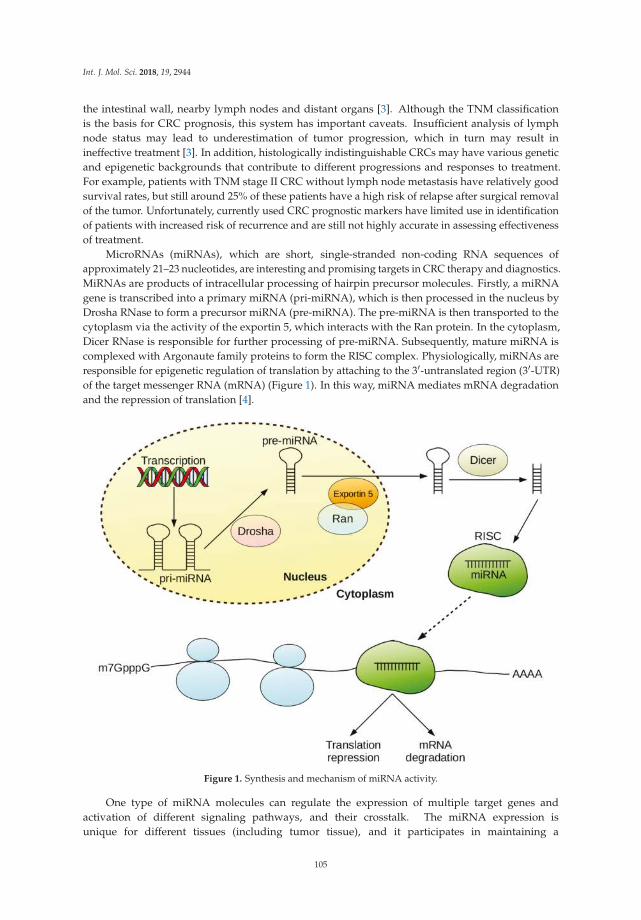
Int. J. Mol. Sci. 2018, 19, 2944
the intestinal wall, nearby lymph nodes and distant organs [3]. Although the TNM classificationis the basis for CRC prognosis, this system has important caveats. Insufficient analysis of lymphnode status may lead to underestimation of tumor progression, which in turn may result inineffective treatment [3]. In addition, histologically indistinguishable CRCs may have various geneticand epigenetic backgrounds that contribute to different progressions and responses to treatment.For example, patients with TNM stage II CRC without lymph node metastasis have relatively goodsurvival rates, but still around 25% of these patients have a high risk of relapse after surgical removalof the tumor. Unfortunately, currently used CRC prognostic markers have limited use in identificationof patients with increased risk of recurrence and are still not highly accurate in assessing effectivenessof treatment.
MicroRNAs (miRNAs), which are short, single-stranded non-coding RNA sequences ofapproximately 21–23 nucleotides, are interesting and promising targets in CRC therapy and diagnostics.MiRNAs are products of intracellular processing of hairpin precursor molecules. Firstly, a miRNAgene is transcribed into a primary miRNA (pri-miRNA), which is then processed in the nucleus byDrosha RNase to form a precursor miRNA (pre-miRNA). The pre-miRNA is then transported to thecytoplasm via the activity of the exportin 5, which interacts with the Ran protein. In the cytoplasm,Dicer RNase is responsible for further processing of pre-miRNA. Subsequently, mature miRNA iscomplexed with Argonaute family proteins to form the RISC complex. Physiologically, miRNAs areresponsible for epigenetic regulation of translation by attaching to the 3′-untranslated region (3′-UTR)of the target messenger RNA (mRNA) (Figure 1). In this way, miRNA mediates mRNA degradationand the repression of translation [4].
Figure 1. Synthesis and mechanism of miRNA activity.
One type of miRNA molecules can regulate the expression of multiple target genes andactivation of different signaling pathways, and their crosstalk. The miRNA expression isunique for different tissues (including tumor tissue), and it participates in maintaining a
105

Int. J. Mol. Sci. 2018, 19, 2944
differentiated cellular state. Therefore, disorders of miRNA expression may shift the cell to theundifferentiated proliferative phenotype. MiRNA expression disorders may be caused by a varietyof molecular processes, such as deletions, amplifications, mutations within the miRNA locus,epigenetic silencing, or abnormal regulation of transcription factor activity. It is estimatedthat miRNAs are responsible for the regulation of translation of about 30–60% of humangenes [5,6]. Because miRNAs recognize many different mRNA transcripts, these moleculesare important in epigenetic regulation of basic cell processes, such as proliferation, apoptosis,differentiation, and migration. Therefore, miRNAs may act as potential oncogenes or suppressorsin tumor development processes. Indeed, for many different human cancers, including CRC,significant abnormalities in miRNA expression have been observed. In CRCs, the altered expressionof a large number of miRNAs associated with the development, progression and response to thetreatment of this cancer was observed [7–9]. Changes in miRNA expression are associated with morefrequent metastases, promotion of tumor mass growth, and increased malignancy of tumor cells.MiRNA expression is also associated with the risk of recurrence, response to the therapeutic regimen,and survival time in different cancers. Therefore, miRNA profiling may be a new and valuabletool in the diagnosis and prognosis of many types of cancer, including CRC. However, knowledgein this area still remains fragmented, and previous studies were conducted only on small groupsof patients. In addition, the vast majority of studies conducted so far have evaluated the expression ofmiRNAs only in tumor tissues. However, miRNA expression profiling in tumor tissue has significantdisadvantages. The heterogeneity of tumor cells produces variable results. Obtaining reliable datain this case is problematic and requires the isolation of individual cancer cells, e.g., using the lasermicrodissection method. This method, however, is costly, requires specialized equipment and istherefore not commonly used in clinical practice. Furthermore, studying tumor tissues does notallow for the assessment of changes in miRNA expression during anti-cancer therapy. Alternatively,choosing serum or blood plasma as the test material is a more practical and cheaper approach tomiRNA profiling. Stool is also suitable for miRNA expression analysis. In both cases, the materialis readily available, which makes it possible to identify potential biomarkers at any stage duringand after the therapy, e.g., for early detection of cancer recurrence. MiRNA in plasma and serum isencapsulated in exosomes, which makes it highly stable. Therefore, the material does not require anyspecial storage or protection operations prior to analysis. Potential applications of miRNA expressionanalysis for diagnostic, therapeutic and prognostic purposes are summarized in Table 1. The use ofmiRNAs as diagnostic and prognostic markers in neoplastic diseases, particularly in CRC, appears tobe an inevitable consequence of advances in clinical oncology and gastroenterology.
Table 1. Possibilities and advantages of using miRNA for diagnostic, therapeutic and prognosticpurposes in cancer diseases.
Possible Applications of miRNA:
Predictors of response to therapy and prognostic biomarkersUse of miRNA as a drug—modulation of gene expressionPrediction and detection of metastases or non-invasive tumor phenotypesCancer diagnostics—detection of tumor specific miRNA signatures
Advantages of Using miRNA:
Easy detection in various biological materials (serum/plasma, cerebrospinal fluid, faeces)High stability of miRNA moleculesAbility to determine specific types of cancer, and predict response to therapy and prognosis based on miRNAexpression profilePotential for use as antagonists in cancer therapy
106

Int. J. Mol. Sci. 2018, 19, 2944
2. MiRNAs as Diagnostic Biomarkers
Circulating miRNAs are stable due to encapsulation in exosomes [10], but the mechanism of theirformation and secretion has not yet been fully understood. There is increasing evidence that miRNAscontained in exosomes may be involved in the exchange of information between distant tissues [11].Serum miRNA expression is known to be altered in CRC patients and current studies attempt toinvestigate the correlation of the expression of certain types of miRNAs, both in serum and tumor tissue.Due to minimal invasiveness of miRNA harvesting procedures, circulating miRNAs may be potentiallyused as diagnostic biomarkers of various cancers, including CRC. A significant limitation of miRNAexpression analysis is its poor utility in diagnosis, due to non-specificity and high variability ofexpression of a single miRNA type. Therefore, recent studies have attempted to evaluate the expressionof miRNA sets. Such studies were conducted using microarray technology as well as quantitativereal-time reverse transcriptase polymerase chain reaction (qRT-PCR) [12]. Insufficient sensitivityresulting from low concentrations of miRNA in serum or plasma of the patients is the principallimitation of a microarray experiment. QRT-PCR is characterized by better sensitivity, but evaluationof expression of numerous miRNAs using this method is a difficult and time-consuming task.Next-generation sequencing (NGS) is a novel and promising method that may be applied to evaluatethe expression profiles of many miRNAs simultaneously [13]. Moreover, novel isolation techniquesallow obtaining more miRNA for analysis, which, in combination with specialized NGS protocols,can increase the utility of this method in diagnostics of cancer, including CRC in the near future.
Some non-invasive screening methods used in CRC diagnosis are based on stool testing.Endogenous miRNAs encapsulated in exosomes are protected against RNases, in contrast tomRNAs or proteins that are rapidly degraded. For this reason, the detection of miRNA in stoolis relatively easier. However, in order to ensure sensitivity and replicability, appropriate protocols,including material preparation, extraction, and quantitative analysis of miRNA, are required inthis case [14]. Stool tests allow for earlier detection of tumor cells and most tumor markers,as compared to peripheral blood tests. Therefore, stool miRNA assays may be useful in detectingprecancerous lesions [15]. Stool miRNA purification kits are commercially available, making it possibleto obtain high-quality and high-purity nucleic acids for further analysis. However, methods thatuse stool miRNA molecules as biomarkers of CRC are still in their infancy. Although manyrecent studies indicate that stool miRNA tests have higher specificity, higher sensitivity andhigher reproducibility than peripheral blood assays, no particular stool test has yet passed thepreclinical phase. Therefore, it is necessary to carry out further studies and validation of methodsbased on miRNA derived from this material.
3. MiRNAs as Prognostic Biomarkers and Therapeutic Agents
MiRNA molecules also appear to be promising prognostic biomarkers, as has been shown so farin many preclinical and clinical studies [16–22]. The profiling of miRNA expression for prognosticpurposes has been demonstrated in many human tumors, including: colorectal, pancreatic, ovarian,breast cancer and glioblastoma [15,23–25]. Since one type of miRNA molecule can influence theregulation of expression of many different genes, the use of these molecules in anti-cancer therapyalso seems promising. Currently, there are two potential strategies: (1) the inhibition of oncogenicmiRNAs and (2) the activation of suppressive miRNAs. Both strategies can be effective, as shownin preclinical studies. Direct inhibition involves antisense oligonucleotides used to sequester agiven miRNAs. Modified antisense oligonucleotides used to inhibit miRNA in vivo are often referredto in the literature as antagomirs [26]. For example, a study conducted by Lanford et al. [27] publishedin Science in 2010 showed that the use of anti-miR-122 in chimpanzees chronically infected withhepatitis C virus (HCV) contributes to an improvement in liver disease. Currently, anti-miR-122 isin phase II clinical trials of HCV therapy in humans, and this miRNA-based therapy may possiblybe included in clinical treatments in the coming years. The use of miRNA antagonists seems to be apromising form of therapy, as evidenced by the successful treatment of patients with chronic HCV
107

Int. J. Mol. Sci. 2018, 19, 2944
infection [28]. Additionally, miRNAs can also be inhibited indirectly using a variety of chemicalcompounds [29]. Moreover, there are also studies on genetic knockout of miRNAs in cancer cells.These studies can provide valuable information about the role of miRNAs in cancer and contributeto the development of novel anti-cancer strategies. For example, Shi et al. [30] showed in mousemodel that knockout of oncogenic miR-21 causes an attenuated proliferation of colitis-associated CRC.In turn, Jiang and Hermeking [31] and Rokavec et al. [32] performed studies on suppressive miR-34 inmouse model. In these studies, the authors showed that genetic knockout of miR-34a and miR-34b/c
can contribute to CRC progression.
4. MiR-21
One of the most intensively studied miRNA molecules is miR-21, which is often overexpressedin CRC [23,33]. MiR-21 lowers the expression of several different suppressor genes that influencevarious biological functions, such as proliferation, adhesion, angiogenesis, migration, metabolism,and apoptosis [34]. Therefore, aberrant miR-21 has potentially oncogenic properties. It is worthnoting that some colorectal polyps transform into malignant tumors as a result of successive,consecutive genetic events. Many studies have shown that miR-21 is associated with the progressionof polyps into malignant tumor, and that expression of this miRNA may be increased in CRC [35,36].One study evaluated the expression of miR-21 in different stages of CRC in 39 surgically removedtumors and 34 polyps after endoscopic resection. Using in situ hybridization (ISH) of nucleic acids,expression of miR-21 was shown to be increased in non-malignant polyps in comparison withcontrols and was highest in advanced CRC tumors and also in adjacent stromal fibroblasts [36].In another study, Bastaminejad et al. [37], using the qRT-PCR method, investigated the expressionlevel of miR-21 in serum and stool samples from 40 patients with CRC and 40 healthy controls.The expression level of this miRNA was significantly up-regulated in serum (12.1-fold) and stool(10.0-fold) in CRC patients, compared to the control group. The sensitivity and specificity of serummiR-21 expression level were found to be 86.05% and 72.97%, respectively, and the sensitivity andspecificity of stool miR-21 expression were 86.05% and 81.08%, respectively. The expression levelof miR-21 was able to significantly distinguish CRC stages III–IV from stages I–II (according tothe American Joint Committee on Cancer (AJCC) TNM staging system) in stool samples with asensitivity and a specificity of 88.1% and 81.6%, respectively, and in serum samples with a sensitivityand a specificity of 88.10% and 73.68%, respectively. Significantly increased miR-21 expression wasalso demonstrated in stool samples of 88 CRC patients compared to control group of 101 healthyvolunteers [38]. Similar results were obtained in 29 patients with CRC and eight healthy patients [39].Therefore, the expression of this miRNA in tumor tissues as well as in serum and stool may be apotential and minimally invasive diagnostic biomarker of CRC.
MiR-21 overexpression is closely related to proliferation and lymph node metastases in CRC,which are important prognostic factors in this type of cancer. Analysis of the expression of miR-21derived from CRC tissues may also be helpful in prognosis. Fukushima et al. [40] assessed theprognostic value of miR-21 in a group of 306 CRC patients. The authors found that high miR-21expression was correlated with low overall survival (OS), as well as low disease-free survival (DFS) inCRC patients in Dukes stages B, C, and D [40]. In another study, the prognostic value of miR-21 wasalso considered in patients classified in the TNM staging system. After tumor tissues from 301 patientswith varying degrees of CRC were investigated, a statistically significant correlation between miR-21expression and prognosis was observed [23]. Moreover, high expression of this miRNA was associatedwith low OS. Oue et al. [23] demonstrated that miR-21 expression in tumor tissues is significantlyincreased in patients with tumors infiltrating adjacent organs (T4), compared to patients with tumorslimited to the colon (T1–T3). The study also presented a similar relationship in patients with regionallymph node metastases present (N1) compared to patients without cancer in the lymph nodes (N0).Furthermore, high miR-21 expression in CRC patients was correlated with insensitivity to 5-fluorouracil(5-FU) treatment. Oue et al. [23] analyzed the expression of miR-21 in German (stage II, n = 145) and
108

Int. J. Mol. Sci. 2018, 19, 2944
Japanese (stage I-IV, n = 156) cohorts of patients using the qRT-PCR method. MiR-21 overexpressionwas associated with poor prognosis in both Japanese (stage II/III) and German patients (stage II).These correlations were not dependent on other clinical data in a multivariate model. In addition,the use of adjuvant chemotherapy did not benefit patients with high miR-21 expression in both cohorts.Similar correlations were also obtained in other studies [16,19,41–43]. Moreover, Schetter et al. [16],using the ISH method, observed a high expression level of miR-21 in colonic epithelial cells intumor tissues compared to adjacent non-tumor tissues. In turn, Nielsen et al. [42] detected miR-21expression mainly in stromal fibroblasts adjacent to tumors. On the other hand, Xia et al. [44] showedin a meta-analysis of miR-21 expression profiles of 1174 CRC tissue samples that overexpressionof this miRNA is associated with low OS, but there was no correlation with the carcinoembryonicantigen (CEA) level. Additionally, Chen et al. [45] and Peng et al. [46] in their meta-analysis studiesshowed that miR-21 expression in tumor tissues is associated with poor DFS and OS in CRC patients.However, Chen et al. [45] revealed the significant correlation between miR-21 expression and poor OSin studies based on the qRT-PCR analysis but not the fluorescence in situ hybrydization (FISH) method.Nonetheless, a few previous studies showed that a higher miR-21 expression level detected with theuse of ISH method is associated with poor recurrence-free survival of CRC patients in stage II [41,47].Moreover, in these studies, miR-21 expression was also detected mainly in stromal fibroblasts adjacentto tumors and only in a few samples in cancer cells. All studies discussed above indicated that theanalysis of miR-21 expression in tumor tissues may be a potential, but certainly not an ideal biomarkerof CRC prognosis.
The prognostic value of miR-21 expression in blood and stool of CRC patients is also the subjectof intensive research. Kanaan et al. [48] observed significantly increased plasma levels of miR-21in CRC patients. In turn, Toiyama et al. [33] evaluated the expression of miR-21 in a cell culturemedium from two different CRC lines, in serum collected from 12 CRC patients and 12 healthyvolunteers separately, and confirmed that this miRNA belongs to the secretory group of the miRNAs.The same research group subsequently measured miR-21 expression in 246 blood samples fromCRC patients, 53 healthy volunteers, and 43 colorectal polyps. They also compared the expressionof miR-21 in serum and tumor tissues in 166 paired samples. Statistically significant increase inserum miR-21 expression was observed in patients with benign polyps and in those with CRC.Moreover, a decrease in the expression of this miRNA in serum was observed in patients after surgicalremoval of the tumor [33]. Furthermore, many studies showed that expression of miR-21 both intissue and serum samples of CRC patients is associated with lower OS and DFS [33,49]. However,Chen et al. [45] showed no significant association of serum miR-21 expression with poor OS of CRCpatients in their meta-analysis studies (421 patients). In another study, it was shown that the increasein miR-21 expression in stool of CRC patients may be correlated with TNM classification [50]. Similarly,Bastaminejad et al. [37] revealed that increased expression of miR-21 was associated with AJCCTNM staging, more related with III and IV compared to I and II stages, in both serum and stoolsamples of 40 CRC patients. The above-mentioned studies show that miR-21 expression is associatedwith tumor size, metastases and low patient survival. Therefore, the expression of this miRNA intumor tissues as well as in serum and stool may be a potential and minimally invasive prognosticbiomarker of CRC.
5. MiR-29 Family
Another promising potential CRC biomarker is the miR-29 family, which includes threerelated miRNAs: miR-29a, miR-29b, and miR-29c. This family is associated with variousmolecular functions, such as the regulation of cell proliferation, cell senescence and tumor metastasis.Therefore, these molecules can participate in carcinogenesis and progression. It has been shownthat the expression of miRNAs belonging to this family is altered in many different cancers [51].Wang et al. [52] performed a study in 114 patients with CRC—56 subjects without metastasis and 58with liver metastasis, which are commonly found in this type of cancer. The authors demonstrated
109

Int. J. Mol. Sci. 2018, 19, 2944
that the expression of miR-29a in serum of patients with liver metastasis was significantly increased.In addition, a significantly increased expression of miR-29a was also observed in patients in stage T4,compared to T2. The authors concluded that miR-29a enables the early detection of liver metastasisin CRC [52]. In addition to early metastasis detection, miR-29a was also tested for potential useas a diagnostic biomarker for CRC. Huang et al. [53], using qRT-PCR, studied the expression of 12miRNAs (miR-17-3p, miR-25, miR-29a, miR-92a, miR-134, miR-146a, miR-181d, miR-191, miR-221,miR-222, miR-223, and miR-320a) in the plasma of patients with advanced stages of colorectal neoplasia(CRC and advanced adenomas), compared to a group of healthy volunteers. It was shown that theexpression of two miRNAs, miR-29a and miR-92a, can have a significant diagnostic value in CRC.For miR-29a, the area under the curve (AUC) was 0.844, while for miR-92a, the AUC was 0.838in differentiating patients with CRC from healthy volunteers. The utility of both miRNAs wasalso demonstrated in differentiation of advanced adenomas and normal tissues. In this case, theAUC value for miR-29a was 0.769, while for miR-92a, it was 0.749. Overall, a receiver operatingcharacteristic (ROC) analysis for both miRNAs showed an AUC of 0.883 (sensitivity = 83.0% andspecificity = 84.7%) in differentiation of CRC and an AUC of 0.773 (sensitivity = 73.0% and specificity =79.7%) in differentiation of advanced adenomas. Similarly, Al-Sheikh et al. [54] revealed up-regulationof miR-29 and miR-92, and down-regulation of miR-145 and miR-195 in 20 CRC patients (both in tissueand plasma) compared to controls. The above-mentioned results suggest that the determination ofmiR-29a and miR-92a expression in plasma may be a novel and potential biomarker in the diagnosisof CRC.
MiR-29b is also the member of miR-29 family. This miRNA inhibits proliferation and inducesapoptosis in CRC cells. MiR-29b mediates the inhibition of the epithelial–mesenchymal transition.In many tumors that originate from epithelium, including CRC, the epithelial–mesenchymal transitionis considered to be a key processes in initiation of metastasis. Li et al. [55] showed decreased expressionof miR-29b in tissue and plasma samples obtained from CRC patients compared to controls. In addition,Basati et al. [56] showed down-regulation of miR-29b and miR-194 in serum samples obtained from 55CRC patients compared to controls. Moreover, these authors showed a negative correlation betweenthese miRNA expression and TNM stages.
MiR-29 is also a potential prognostic biomarker of CRC. Tang et al. [57] analyzed the expressionof miR-29a and KLF4 mRNA in 85 tumor tissues of CRC patients and CRC cell lines using theqRT-PCR method. It was shown that reduced expression of KLF4 mRNA is associated with the presenceof metastasis. Moreover, increased miR-29a expression indicated presence of metastasis and worsenedprognosis of patients with CRC. It is known that KLF4 is a target of miR-29a and that it acts to inhibitmetastasis by reducing MMP-2 and increasing E-cadherin expression [57]. The study mentionedabove showed that high expression of miR-29a is associated with metastasis and poor prognosis.The predictive value of miR-29a was also shown in stage II CRC. Weissmann-Brenner et al. [58]performed studies on 110 CRC patients (51 with stage I cancer and 59 patients with stage IIcancer according to the AJCC TNM staging system) using miRNA microarrays and verified themicroarray results using the qRT-PCR technique afterwards. RNA was extracted from formalin-fixedparaffin-embedded tumor tissues. The authors defined a poor prognosis as a recurrence of the diseasewithin 36 months of surgery. Patients with a good prognosis (n = 87; 79%) and a poor prognosis(n = 23; 21%) were compared. There were no statistically significant differentially expressed miRNAsbetween good- and poor-prognosis stage I CRC patients, among the set of 903 analyzed miRNAs.On the other hand, the expression of miR-29a was significantly higher in stage II CRC patients withgood prognosis compared to the poor-prognosis group. High expression of this miRNA was associatedwith longer DFS in both univariate and multivariate analysis. In case of miR-29a expression, a positivepredictive value (PPV) for non-recurrence of the disease was 94% (two cases out of 31). Differencesin the miR-29a expression were confirmed using qRT-PCR, and this method showed the effect ofoverexpression of this miRNA on prolonging of the DFS. This study demonstrated a significantassociation of the miR-29a expression level with the risk of CRC recurrence in stage II patients.
110

Int. J. Mol. Sci. 2018, 19, 2944
For the patients in stage I, no such correlation was demonstrated. Moreover, significantly decreasedexpressions of miR-29a and miR-29c were reported in tumor tissues in 43 early-recurrence patientscompared to the control group [59]. Although increased expressions of both miR-29a and miR-29cwere associated with better outcome after 12 months of therapy, the authors suggested that onlymiR-29a may be used as a predictor marker for an early recurrence of the disease. The low PPVof miR-29c in this case may result from short follow-up time of the patients and the small studygroup [59]. In another study on 245 patients by Inoue et al. [60], the expression of miR-29b level intumor tissues was used to divide the patients into two groups. The reference value was the medianexpression of this miRNA. The authors concluded that higher expression of miR-29b is associatedwith higher five-year DFS and OS values. Analysis of the severity of the disease showed that themiR-29b expression reflects the five-year DFS and has a significant prognostic value, but only inpatients with stage III CRC. In addition, the low level of miR-29b expression was also a predictor oflymph node metastasis. These findings confirmed the prognostic value of this miRNA in stage IIICRC patients. In another study, Yuan et al. [61] studied the expression of miR-29b in tumor tissueand adjacent normal mucosa samples of 41 patients using the qRT-PCR method. The authors found asignificant decrease in the expression of this miRNA in CRC and concluded that the level of miR-29bmay be associated with the size of a tumor, clinical status and lymph node metastasis. Basati et al. [56]studied 55 serum samples of CRC patients and revealed that lower expression of miR-29b is correlatedwith poor prognosis. In turn, Ulivi et al. [62] showed that higher miR-29b level in plasma samples isassociated with longer progression-free survival (PFS) and OS of metastatic CRC patients treated withbevacizumab-based chemotherapy. As indicated by numerous studies, analysis of changes of miR-29expression may be helpful in assessing an early recurrence and evaluating DFS in CRC patients.
6. MiR-34 Family
There are also studies on miR-34, a group of miRNAs that includes miR-34a, miR-34b andmiR-34c. These molecules show suppressor properties and are regulated by p53 protein and DNAhypermethylation. The miR-34 group influences various processes that occur in tumor cells such asdifferentiation, drug resistance and metastasis [63]. For example, miR-34a overexpression inhibitsNOTCH signaling and suppresses symmetric cell division, which prevents expansion of the CRC stemcell niche [64]. Wu et al. [65] studied the possibility of using miR-34 as a potential diagnostic biomarkerfor CRC. The authors showed abnormal methylation of miR-34a, miR-34b, and miR-34c genes in tissueand stool samples of 82 CRC patients. In turn, Aherne et al. [66] reported higher expression of miR-34ain plasma samples obtained from CRC patients compared to controls.
MiR-34 expression has also been found to be useful for CRC prognosis. The usefulness ofmiR-34 as a biomarker of a recurrence of the disease in two independent groups of 268 CRC patientswas evaluated. It was shown that the miR-34a expression in CRC tissues is directly proportionalto DFS, and therefore, this molecule may be a good prognostic factor in assessing the risk of therecurrence of CRC. In addition, the expression of miR-34a was significantly higher in patients withhigh expression of p53 compared to those with low expression of this protein. The authors suggestedthat miR-34 inhibits the growth and invasiveness of CRC in p53-dependent manner, which allowsthis miRNA to be used as a potential biomarker for a recurrence in patients with stage II and stage IIICRC [67]. The PAR2 receptor also plays an important role in the progression of CRC. Previous reportshave indicated that miR-34a expression is inhibited by PAR2, which results in increased synthesisof cyclin D1 and transforming growth factor β (TGF-β) in CRC cells [68]. Furthermore, silencing ofmiR-34a expression through promoter methylation in CRC tissues is associated with the occurrence ofmetastases [69]. In other studies, a lower expression of this miRNA was observed in some patientswith CRC in tissue [70] and serum/plasma samples [71], which suggests that miR-34a may play a rolein the progression of this cancer. Li et al. [72] showed that lower expression of miR-34a in CRC tissues iscorrelated with the lymph node metastasis and TNM stage. Zhang et al. [73] performed studies on 103CRC tissue samples and showed that miR-34a expression was down-regulated compared to adjacent
111

Int. J. Mol. Sci. 2018, 19, 2944
normal mucosa samples with the use of ISH technique. Moreover, these authors indicated that the lowerexpression of this molecule is correlated with more distant metastasis and shorter OS time. Studies onthe effect of the miR-34 group on the prognosis of CRC have also been performed. The purpose of thesestudies was to determine the relationship between the miR-34b and miR-34c expression in CRC tissuesand the development of this disease. Samples were obtained from 159 American and 113 Chinesepatients with stages I–IV CRC. Using the qRT-PCR method, Hiyoshi et al. [74] showed an increasedexpression of miR-34b and miR-34c in advanced CRC, which was associated with poor prognosis inboth study groups. Similarly to miR-34a, the expressions of miR-34b and miR-34c are also regulatedby p53 protein at the transcriptional level. The results of all of these studies show that miR-34 may bean interesting prognostic tool and may be used to assess the risk of a recurrence in patients with CRC.
Furthermore, recent studies have determined the possibility of using miR-34 as a potentialtherapeutic agent. A low level of this miRNA was observed in DLD-1 CRC cell line that wasresistant to 5-FU. Restoration of the miR-34a expression caused the sensitization of cells to 5-FUtreatment and resulted in an inhibition of cell growth [75]. Moreover, Sun et al. [76] showed thatmiR-34a expression was down-regulated in blood samples of CRC patients after oxaliplatin-basedchemotherapy. The negative correlation between miR-34a and TGF-β/SMAD4 expression is also noted.The authors suggested that miR-34a and TGF-β/SMAD4 expression changes can lead to activation ofmacroautophagy and oxaliplatin resistance in CRC cells.
7. MiR-124
MiR-124 is known to inhibit cell proliferation, metastasis and invasion in CRC. This miRNA notonly down-regulates rho-associated protein kinase 1 (ROCK1) [77], which functions as an oncogene,but also inhibits the activity of cyclin-dependent kinase 4 (CDK4), which is responsible for cell cycleprogression at the G1/S checkpoint [78]. Studies have shown that, in CRC cells, the expressionof miR-124 is silenced through DNA methylation [79,80]. Since miR-124 gene is more likely to bemethylated in CRCs compared to other tumors, DNA methylation status of this miRNA may be usedas a specific marker for CRC. Harada et al. [79] performed the detection of DNA methylation in bowellavage fluid for CRC screening. These authors analyzed DNA methylation status in a total of 508patients—56 with CRC, 53 with advanced adenoma, 209 with minor polyp, and 190 healthy individuals.Three genes showed the greatest sensitivity for CRC detection (miR-124-3, LOC386758, and SFRP1)after training set analysis (n = 345). A scoring system based on the methylation of these three genesachieved 82% sensitivity and 79% specificity, and the AUC was 0.834. These results were subsequentlyvalidated in an independent test set (n = 153; AUC = 0.808). In another study, Xi et al. [77] investigatedthe expression of ROCK1 and miR-124 in CRC patients using 68 paired tissue specimens (38 cases ofnon-metastatic CRC and 30 cases of metastatic CRC). The use of qRT-PCR revealed that expressionof miR-124 was higher in normal compared with CRC tissues, and in non-metastatic compared tometastatic CRC tissues. In contrast, ROCK1 was significantly overexpressed in CRC compared withcontrol tissues and between metastatic tissues and non-metastatic CRC tissues. The above-mentionedresults suggest that miR-124, as a tumor suppressor, may play a role in tumor growth and metastasis.Recently, the miR-124a level has also been studied in 40 patients with ulcerative colitis (withoutcolorectal cancer), four patients with CRC or inflammatory dysplasia, eight patients with CRC (withoutinflammatory background), and 12 healthy volunteers. It was found that miR-124a-1, miR-124a-2,and miR-124a-3 genes are methylated in tumor tissues. The authors suggested that the methylation ofmiR-124a-3 occurring during oncogenesis in patients with ulcerative colitis can be used to evaluate theindividual’s risk of developing cancer [81].
MiR-124 may also be a prognostic marker for CRC. Wang et al. [82] studied 96 tumor tissuesamples and showed that down-regulated expression of miR-124 is correlated with poor OS and DFS inCRC patients. Moreover, Jinushi et al. [83] showed that higher expression of miR-124-5p (both in plasmaand tissues) was associated with better prognosis of CRC patients. In turn, Slattery et al. [84] performedstudies in 1893 patients with CRC. The authors found that miR-124-3p belongs to infrequently
112

Int. J. Mol. Sci. 2018, 19, 2944
highly expressed miRNAs in tumor tissues and showed that up-regulation of miR-124-3p can worsenprognosis of these CRC patients.
8. MiR-130b
The direct target of miR-130b is the PPARγ receptor, of which inhibition results in the regulationof the expression of cadherin E, vascular endothelial growth factor (VEGF) and phosphatase and tensinhomolog (PTEN). Since tissue miR-130b overexpression was observed in stage III and IV CRCs, it issuggested that miR-130b-PPARγ signaling may play a significant role in increasing tumor malignancy.Furthermore, evaluating the expression of proteins in this pathway, including miR-130b, may bea promising prognostic biomarker in CRC [85]. Finally, a study performed in 53 cancer andnon-cancerous samples [86] suggested miR-130a as a good biomarker of CRC because of correlationwith TNM staging and lymph node metastasis.
9. MiR-155
The sequence of this miRNA is located in a non-coding region of MIR155 host gene (BIC).Altered expression of miR-155 has been observed in many different tumors, and is associatedwith severity of disease, progression, and response to treatment. Interestingly, Sempere et al. [87]using the ISH method observed that miR-155 expression is detected mainly in tumor-infiltratingimmune cells. Lv et al. [88] examined the possibility of using serum miR-155 expression as adiagnostic tool. Using qRT-PCR, they measured the expression levels of miR-155 in 146 CRC patientsand 60 healthy controls. Serum miR-155 was up-regulated in CRC patients compared with the matchedhealthy controls. Moreover, ROC curve analysis indicated that miR-155 is a suitable marker fordiscriminating CRC patients from healthy controls, with an AUC of 0.776. Therefore, this molecule canbe used as a potential tumor biomarker in the diagnosis of CRC.
MiR-155 can also play an important role in CRC prognosis. Shibuya et al. [43] showed that patientswith an increased expression of miR-155 in tumor tissues were characterized by shorter OS and DFS,compared to those with lower expression of this miRNA. In another study, multivariate analysis alsodemonstrated a relationship between the level of miR-155 expression and poor prognosis in CRC,depending on the severity of the disease. The control group consisted of 60 healthy volunteers,while the experimental group consisted of 146 patients. The authors did not observe changes in themiR-155 serum level in patients with stage I CRC, but reported a statistically significant overexpressionof this miRNA in patients with stages II–IV of the disease [88]. In another study, the serum CEAlevel and miR-155 expression were measured in tissues that were obtained before and after surgeryof 84 CRC patients. It is well known that CEA is used for determining the prognosis, evaluating theeffectiveness of therapy and monitoring the recurrence of CRC. In this study, the miR-155 expressionwas observed to be significantly increased in patients with CRC. This is associated with metastases anda recurrence of the disease [89]. Therefore, the evaluation of miR-155 expression in association withserum CEA may provide additional diagnostic information and enable a more accurate assessmentof the risk of the metastasis of CRC. A statistically significant increase in miR-155 expression intumor tissues, compared to normal samples, was also observed by Zhang et al. [90] in a study of76 patients with CRC. In addition, the authors observed a correlation between miR-155 expressionand lymph node and distant metastases and disease progression. Moreover, they observed thatmiR-155 overexpression inhibited E-cadherin expression and positively regulated zinc finger E-boxbinding homeobox 1 protein (ZEB-1), which resulted in an increased cell migration and metastases.Similarly, Qu et al. [91] revealed association of miR-155-5p expression in tumor tissues with location,grade of tumor, TNM stage and distant metastasis. Ulivi et al. [62] in multivariate analysis also showedthat increased expression of circulating miR-155 is associated with shorter PFS and OS in metastaticCRC patients treated with bevacizumab-based chemotherapy. The above-mentioned observationsindicate that miR-155 may play a role in the development and metastasis of CRC.
113

Int. J. Mol. Sci. 2018, 19, 2944
10. MiR-224
Recent reports have revealed that miR-224 may influence many processes that are associated withtumor cell growth and development, such as proliferation, growth, differentiation and apoptosis [92].Some groups have investigated the expression of miR-224 in CRC patients. Zhu et al. [93] foundsignificantly lower miR-224 levels in feces from CRC patients than these from normal volunteers in theirretrospective analysis of miR-224 levels in fecal samples from 80 CRC patients and 51 normal controls.The authors suggested that the miR-224 expression level in feces can be used for screening and earlydiagnosis of CRC.
MiR-224 is also a potential prognostic biomarker in CRC. Zhang et al. [94] evaluated theclinical and pathological information of 40 patients with a recurrence and 68 patients without arecurrence within three years after a surgical intervention. Moreover, using the qRT-PCR and Westernblot methods, the authors analyzed samples from all 108 patients with stages I and II CRC. They showedthat miR-224 is involved in the regulation of SMAD4 protein, which is involved in cell signaling.SMAD4, together with other proteins from this family, forms a DNA-binding complex that actsas a transcription factor. Furthermore, a significant increase in miR-224 expression in CRC tissueswas observed in the study and this change was associated with a higher risk of a recurrence and ashorter DFS. In another study, Ling et al. [95] showed that miR-224 is an activator of metastasis andthat the target of this miRNA is SMAD4. The authors concluded that an evaluation of miR-224 alone oran evaluation of miR-224 together with SMAD4 may be an independent prognostic marker in patientswith CRC. MiR-224 expression in tumor tissues and its association with the survival of patients wereevaluated in 449 CRC patients. In this study, the patients were divided into two groups. These twogroups were characterized by low and high levels of miR-224 expression, respectively. A shorterOS and survival time without metastases were observed in patients with miR-224 overexpression.Moreover, Wang et al. [96] showed an inverse correlation between SMAD4 and miR-224 expressionin tumor tissues of 40 CRC patients. These authors also revealed that miR-224 can regulate USP3expression and its higher expression is associated with poor prognosis. In turn, Liao et al. [97] observeda statistically significant increase in the expression of this miRNA in tumor tissues of patients with anaggressive CRC phenotype and poor prognosis. In another study, miR-224 expression was evaluatedin 79 patients with CRC and 18 healthy volunteers. The authors observed a significant inhibitionof miR-224 expression in tumor tissues. Since the molecular target of miR-224 is CDC42, a lowerexpression of this miRNA reduces tumor cells migration. In general, the study indicated an importantrole of miR-224 in inhibiting migration of CRC cells. The authors concluded that miR-224 may be apromising biomarker in evaluating development of CRC [98]. Furthermore, Zhang et al. [99] showed intheir meta-analysis that the increased level of miR-224-5p is correlated with poor OS of CRC patients.
11. MiR-378
MiR-378 is known to play a significant role in development of different types of cancer, includingCRC. Current studies have shown that miR-378 is overexpressed in CRC cells and its targets includeFUS-1 and SUFU suppressor genes [100,101]. In addition, miR-378 is involved in tumor progressionby promoting cell survival, migration and angiogenesis [102]. Significant differences in the levelof blood and tumor miR-378 expression between oncological and healthy subjects were observed.Zanutto et al. [103] analyzed miRNA expression in serum samples from 65 CRC patients and 70healthy volunteers, and found significantly increased miR-378 levels in CRC patients compared tothe control group. At the same time, the authors observed a statistically significant decrease in theexpression of this miRNA after the surgical removal of the tumor. Similar results were obtained inpatients who had no recurrence of the disease within four to six months after surgery. The resultssuggest that serum miR-378 levels may be useful not only for differentiating CRC patients fromhealthy subjects, but that miR-378 is synthesized in the tumor tissue and its concentration is associatedwith tumor mass and possible recurrence. In turn, Wang et al. [104] qualified miR-378 as a tumorsuppressor after analyzing miRNA expression in 47 CRC samples that were matched with normal
114

Int. J. Mol. Sci. 2018, 19, 2944
tissue samples. In another study, Zhang et al. [105] observed an inhibition of miR-378 expression in 84CRC samples compared to normal mucosal samples. Similarly, Zeng et al. [106] showed lower miR-378expression in 27 CRC samples compared to paired adjacent normal samples. There have also beenstudies that showed an association of a reduced expression of miR-378 in cancer tissues with increasedtumor size, metastasis and shorter OS in patients with CRC [105,107]. The above-mentioned resultssuggest that miR-378 may play an important role in carcinogenesis and may have clinical value as apotential biomarker of CRC.
12. Other miRNAs
Many recent studies attempted to identify other miRNAs in tumor tissues or plasma/serumsamples as the potential diagnostic, prognostic or predictive biomarkers of CRC, e.g., miR-17,miR-19a, miR-20a [108], miR-22 [109], miR-24-3p [110], miR-26a, miR-26b [111], miR-9 [112–114],miR-106a [115], miR-122, miR-200 [116], miR-125a-3p [117], miR-126-3p [118], miR-139-3p [119],miR-139-5p [120], miR-148a, miR-625-3p [121], miR-181a, miR-181b [122], miR-181c [123],miR-181d [124], miR-193a-3p [125], miR-200c [126,127], miR-196b-5p [126], miR-223 [108,113], miR-375,miR-760 [112], miR-506, miR-4316 [128], miR-1290 [129]. In addition, Kiss et al. [130] using microarrayand qRT-PCR techniques revealed that miR-92b-3p, miR-3156-5p, miR-10a-5p, miR-125a-5p maybe used as predictive biomarkers of response to bevacizumab/FOLFOX therapy of CRC patientswith metastasis. Similarly, Fiala et al. [118] showed that miR-126-3p expression is correlated withresponse to bevacizumab/FOLFOX treatment of CRC patients with metastasis. On the other hand,some studies showed only altered expression of various miRNAs in CRC tissues/serum samplescompared to controls [131,132]. The significance of miRNAs as diagnostic and prognostic biomarkersin CRC is summarized in Table 2.
Table 2. The significance of miRNAs as diagnostic and prognostic biomarkers of colorectal cancer.
MiRNABiomarker
TypeRegulation in
CRCSource ofmiRNA
CohortSize
Correlation/Differentiation
Detection Method Authors
miR-17 diagnostic up-regulation serum n = 190 control vs. CRC qRT-PCR Zekri et al. [108]
miR-17-5p diagnostic up-regulation serum n = 39 control vs. CRC qRT-PCR Fu et al. [114]
miR-19a diagnostic up-regulation serum n = 190 control vs. CRC qRT-PCR Zekri et al. [108]
miR-20a diagnostic up-regulation serum n = 190 control vs. CRC qRT-PCR Zekri et al. [108]
miR-21 diagnostic up-regulationplasma,tissues
n = 116 control vs. CRCmicrofluidic array
technology,qRT-PCR
Kanaan et al. [48]
miR-21 diagnostic up-regulation tissues n = 73control vs. CRC
vs. polypsISH
Yamamichi et al.[36]
miR-21 diagnostic up-regulationserum,stool
n = 80 TNM stages qRT-PCRBastaminejad et al.
[37]
miR-21 diagnostic up-regulationstool,
tissuesn = 246
control vs. CRCvs. polyps
qRT-PCR Wu et al. [38]
miR-21 diagnostic up-regulation stool n = 55 TNM stagesmiRNA
microarrays,qRT-PCR
Ahmed et al. [50]
miR-21 diagnostic up-regulationstool,
tissuesn = 37
control vs. CRCvs. adenomas
qRT-PCR, miRNAmicroarrays
Link et al. [39]
miR-21diagnostic,prognostic
up-regulationserum,tissues
n = 279
TNM stages,tumor size,
distantmetastasis, poor
survival
qRT-PCR Toiyama et al. [33]
miR-21 prognostic up-regulation tissues n = 301 poor survival qRT-PCR Oue et al. [23]
miR-21 prognostic up-regulation tissues n = 306
liver metastasis,Dukes’ stage,shorter OS,shorter DFS
qRT-PCRFukushima et al.
[40]
115

Int. J. Mol. Sci. 2018, 19, 2944
Table 2. Cont.
MiRNABiomarker
TypeRegulation in
CRCSource ofmiRNA
CohortSize
Correlation/Differentiation
Detection Method Authors
miR-21 prognostic up-regulation tissues n = 197poor survival,
poor therapeuticoutcome
miRNAmicroarrays,
qRT-PCR, ISHSchetter et al. [16]
miR-21 prognostic up-regulation tissues n = 46 shorter DFS qRT-PCR Kulda et al. [19]
miR-21 prognostic up-regulation tissues n = 520
inferiorrecurrence-freecancer-specific
survival
ISHKjaer-Frifeldt et al.
[41]
miR-21 prognostic up-regulation tissues n = 234 shorter DFS ISH Nielsen et al. [42]
miR-21 prognostic up-regulation tissues n = 156liver metastasis,
shorter OS,shorter DFS
qRT-PCR Shibuya et al. [43]
miR-21 prognostic up-regulation tissues n = 277 shorter RFS ISH Kang et al. [47]
miR-22 prognostic down-regulation tissues n = 193 shorter OS qRT-PCR Li et al. [109]
miR-24-3p prognostic up-regulation tissues n = 268shorter OS,shorter DFS
qRT-PCR Kerimis et al. [110]
miR-26a/b prognostic down-regulation tissues n = 58 shorter OS qRT-PCR Li et al. [111]
miR-29 diagnostic up-regulationtissues,plasma
n = 40 control vs. CRC qRT-PCR Al-Sheikh et al. [54]
miR-29a diagnostic up-regulation plasma n = 209 control vs. CRC qRT-PCR Huang et al. [53]
miR-29a diagnostic down-regulation stool n = 131 control vs. CRC qRT-PCR Zhu et al. [93]
miR-29a diagnostic up-regulation serum n = 74 liver metastasis qRT-PCR Wang and Gu [52]
miR-29a prognostic down-regulation tissues n = 110 poor prognosismiRNA
microarrays,qRT-PCR
Weissmann-Brenneret al. [58]
miR-29b diagnostic down-regulationtissues,plasma
n = 600 control vs. CRC qRT-PCR Li et al. [55]
miR-29bdiagnostic,prognostic
down-regulation serum n = 110 TNM stages qRT-PCR Basati et al. [56]
miR-29b prognostic down-regulation tissues n = 245shorter OS,shorter DFS
qRT-PCR Inoue et al. [60]
miR-29b prognostic down-regulation plasma n = 52shorter OS,shorter PFS
qRT-PCR Ulivi et al. [62]
miR-34a diagnostic up-regulation plasma n = 185polyps vs.
advanced cancerqRT-PCR Aherne et al. [66]
miR-34adiagnostic,prognostic
methylationtissues,stool
n = 142lymph
metastasis,control vs. CRC
methylation-specificPCR
Wu et al. [65]
miR-34a prognostic down-regulation tissues n = 268shorter DFS,recurrence
qRT-PCR Gao et al. [67]
miR-34a prognostic methylation tissues n = 94 liver metastasismethylation-specific
PCRSiemens et al. [69]
miR-34a prognostic down-regulation tissues n = 176 poor prognosis qRT-PCR Li et al. [72]
miR-34a prognostic down-regulation tissues n = 103distant
metastasisqRT-PCR, ISH Zhang et al. [73]
miR-34a/b/c prognostic up-regulation tissues n = 272 poor prognosis qRT-PCR Hiyoshi et al. [74]
miR-34b/c diagnostic methylationtissues,stool
n = 142 control vs. CRCmethylation-specific
PCRWu et al. [65]
miR-92a diagnostic up-regulationtissues,stool,
plasman = 907 control vs. CRC qRT-PCR Chang et al. [113]
miR-92adiagnostic,prognostic
up-regulation serum n = 91control vs. CRC,
TNM stages,poor prognosis
qRT-PCR Elshafei et al. [112]
miR-92a-3p diagnostic up-regulation serum n = 39 control vs. CRC qRT-PCR Fu et al. [114]
miR-106a diagnostic up-regulationtissues,plasma
n = 84 control vs. CRC qRT-PCR He et al. [115]
miR-122 prognostic up-regulation plasma n = 543shorter RFS,shorter OS
qRT-PCRMaierthaler et al.
[116]
116

Int. J. Mol. Sci. 2018, 19, 2944
Table 2. Cont.
MiRNABiomarker
TypeRegulation in
CRCSource ofmiRNA
CohortSize
Correlation/Differentiation
Detection Method Authors
miR-124 prognostic down-regulation tissues n = 96shorter OS,shorter DFS
qRT-PCR Wang et al. [82]
miR-124-3p prognostic up-regulation tissuesn =
1893
increasedlikelihood of
dyingmiRNA microarrays Slattery et al. [84]
miR-124-5p prognostic down-regulation tissues n = 71 shorter OS qRT-PCR Jinushi et al. [83]
miR-125a-3p diagnostic down-regulation tissues n = 35 control vs. CRC qRT-PCR Liang et al. [117]
miR-126-3p prognostic down-regulation tissues n = 63shorter OS,shorter PFS
qRT-PCR Fiala et al. [118]
miR-126-5p prognostic down-regulation tissues n = 63shorter OS,shorter PFS
qRT-PCR Fiala et al. [118]
miR-130b prognostic up-regulation tissues n = 80 poor prognosis miRNA microarrays Colangelo et al. [85]
miR-139-3p diagnostic down-regulationtissues,serum
n = 249 control vs. CRC qRT-PCR Ng et al. [119]
miR-139-5p prognostic up-regulationtissues,serum
n = 433 shorter RFSmiRNA
microarrays,qRT-PCR
Miyoshi et al. [120]
miR-148a prognostic down-regulation tissues n = 54 shorter PFS qRT-PCRBaltruskeviciene et
al. [121]
miR-150 diagnostic down-regulation plasma n = 185adenomas vs.
advanced cancerqRT-PCR Aherne et al. [66]
miR-155 prognostic up-regulation serum n = 206shorter OS,shorter PFS
qRT-PCR Lv et al. [88]
miR-155 prognostic up-regulation tissues n = 156shorter OS,shorter DFS
qRT-PCR Shibuya et al. [43]
miR-155 prognostic up-regulation tissues n = 84 poor prognosis qRT-PCRHongliang et al.
[89]
miR-155 prognostic up-regulation tissues n = 76lymph node and
distantmetastases
qRT-PCR Zhang et al. [90]
miR-155-5p prognostic up-regulation tissues n = 372 metastasis qRT-PCR Qu et al. [91]
miR-155-5p prognostic down-regulation plasma n = 52shorter OS,shorter PFS
qRT-PCR Ulivi et al. [62]
miR-181c prognostic up-regulation tissues n = 147 shorter RFSmiRNA
microarrays,qRT-PCR
Yamazaki et al.[123]
miR-181d prognostic up-regulation tissues n = 40 metastasis qRT-PCR Guo et al. [124]
miR-193a-3p prognostic down-regulation tissues n = 96 shorter OSmiRNA
microarrays,qRT-PCR
Lin et al. [125]
miR-196-5p prognostic down-regulation tissues n = 48 shorter OSmiRNA
microarrays,qRT-PCR
Li et al. [126]
miR-200 prognostic down-regulation plasma n = 543shorter RFS,shorter OS
qRT-PCRMaierthaler et al.
[116]
miR-200c prognostic down-regulation tissues n = 48 shorter OSmiRNA
microarrays,qRT-PCR
Li et al. [126]
miR-200c prognostic up-regulation serum n = 90 shorter OS qRT-PCR Tayel et al. [127]
miR-223 diagnostic down-regulation stool n = 131 control vs. CRC qRT-PCR Zhu et al. [93]
miR-223 diagnostic up-regulation serum n = 190 control vs. CRC qRT-PCR Zekri et al. [108]
miR-223 diagnostic up-regulationtissues,stool,
plasman = 907 control vs. CRC qRT-PCR Chang et al. [113]
miR-224 diagnostic down-regulation stool n = 131 control vs. CRC qRT-PCR Zhu et al. [93]
miR-224 prognostic up-regulation tissues n = 108 shorter DFS qRT-PCR Zhang et al. [94]
miR-224 prognostic up-regulation tissues n = 621 shorter OSmiRNA
microarrays,qRT-PCR
Ling et al. [95]
117

Int. J. Mol. Sci. 2018, 19, 2944
Table 2. Cont.
MiRNABiomarker
TypeRegulation in
CRCSource ofmiRNA
CohortSize
Correlation/Differentiation
Detection Method Authors
miR-224 prognostic up-regulation tissues n = 110 shorter OS qRT-PCR Liao et al. [97]
miR-375diagnostic,prognostic
down-regulation serum n = 91control vs. CRC,
TNM stages,poor prognosis
qRT-PCR Elshafei et al. [112]
miR-378 diagnostic up-regulation plasma n = 65 control vs. CRC qRT-PCR Zanutto et al. [103]
miR-378 prognostic down-regulation tissues n = 84 shorter OS qRT-PCR Zhang et al. [105]
miR-378a-3p prognostic down-regulation tissues n = 96 shorter OS qRT-PCR Li et al. [107]
miR-378a-5p prognostic down-regulation tissues n = 96 shorter OS qRT-PCR Li et al. [107]
miR-506 diagnostic up-regulation plasma n = 126 control vs. CRC qRT-PCRKrawczyk et al.
[128]
miR-625-3p diagnostic down-regulation tissues n = 54 control vs. CRC qRT-PCRBaltruskeviciene et
al. [121]
miR-664-3p prognostic down-regulation tissues n = 63shorter OS,shorter PFS
qRT-PCR Fiala et al. [118]
miR-760diagnostic,prognostic
down-regulation serum n = 91control vs. CRC,
TNM stages,poor prognosis
qRT-PCR Elshafei et al. [112]
miR-1290 prognostic up-regulation tissues n = 291shorter OS,shorter DFS
miRNAmicroarrays,
qRT-PCRYe et al. [129]
miR-4316 diagnostic up-regulation plasma n = 126 control vs. CRC qRT-PCRKrawczyk et al.
[128]
RFS—relapse-free survival.
13. MiRNA Panels
More researchers are focusing on finding miRNA sets that may be used as potential diagnosticor prognostic markers due to the fact that the expression of a single miRNA may not havesufficient specificity and sensitivity to distinguish CRC stages or CRC patients from healthy controls.Recently, interesting study was performed by Pan et al. [133], in which the expression level of 30miRNAs in plasma samples was analyzed with the use of qRT-PCR. These authors showed thatanalysis of plasma expression level of five miRNAs, such as miR-15b, miR-17, miR-21, miR-26b,and miR-145, together with CEA, can improve diagnostic accuracy of CRC (AUC = 0.85 in thetraining cohort, AUC = 0.818 in the validation cohort). In turn, Guo et al. [134] selected a 5-miRNAset (miR-1246, miR-202-3p, miR-21-3p, miR-1229-3p, and miR-532-3p) from 528 miRNAs in serumand revealed that these miRNA panels have good sensitivity and specificity to distinguish CRCpatients from colorectal adenoma patients (AUC = 0.951, sensitivity = 94.4%, specificity = 84.7%)and healthy controls (AUC = 0.960, sensitivity = 91.6%, specificity = 91.7%). Zhu et al. [135]showed that a 3-serum miRNA set (miR-19a-3p, miR-21-5p, and miR-425-5p) can be useful indiagnosis of CRC (AUC = 0.886 in the training phase, AUC = 0.768 in the validation phase,AUC = 0.783 in the combined training and validation phases, and AUC = 0.830 in the externalvalidation phase). Wang et al. [136] also evaluated a 3-plasma miRNA set (miR-409-3p, miR-7,and miR-93) with diagnostic potential for CRC patients in Dukes stages A–D (AUC = 0.866in the training phase, sensitivity = 91%, specificity = 88%; AUC = 0.897 in the validation phase,sensitivity = 82%, specificity = 89%) and also for non-metastatic CRC patients in Dukes stages A and B(AUC = 0.809 in the training phase, sensitivity = 85%, specificity = 88%; AUC = 0.892 in the validationphase, sensitivity = 82%, specificity = 89%), and for metastatic CRC patients in Dukes stages C and D(AUC = 0.917 in the training phase, sensitivity = 96%, specificity = 88%; AUC = 0.865 in the validationphase, sensitivity = 91%, specificity = 88%). Similar results were obtained in CRC patients whencompared to age-matched healthy controls (for CRC patients in Dukes stages A–D: AUC = 0.894,sensitivity = 90%, specificity = 96%; in Dukes stages A and B: AUC = 0.850, sensitivity = 85%,
118

Int. J. Mol. Sci. 2018, 19, 2944
specificity = 96%; and in Dukes stages C and D: AUC = 0.937, sensitivity = 95%, specificity = 96%).In turn, Kanaan et al. [137] observed that a 3-plasma miRNA set (miR-431, miR-15b, and miR-139-3p)can distinguish stage IV CRC from controls with high specificity and sensitivity (AUC = 0.896,sensitivity = 93%, specificity = 74%). They also found that a 5-miRNA set (miR-331, miR-15b, miR-21,miR-142-3p, and miR-339-3p) may be used to distinguish colorectal adenoma patients from CRCpatients (AUC = 0.856, sensitivity = 91%, specificity = 69%), and CRC patients may be distinguishedfrom healthy controls with the use of a 2-miRNA set (miR-431, and miR-139-3p) (AUC = 0.829,sensitivity = 91%, specificity = 57%). Wikberg et al. [138] used a 4-plasma miRNA set (miR-18a,miR-21, miR-22, and miR-25) to diagnose CRC (in all CRC stages, AUC = 0.93, sensitivity = 81% and67% at 80% and 90% specificity, respectively; in CRC stages I–II, AUC = 0.92, sensitivity = 88% and73% at 80% and 90% specificity, respectively; and in CRC stages, III–IV AUC = 0.85, sensitivity =68% and 57% at 80% and 90% specificity, respectively). It is worth noticing that miR-21 is presentin majority of miRNA sets discussed above. Additionally, Wikberg et al. [138] showed that miR-21expression was increasing during three years before CRC diagnosis. Interestingly, Yang et al. [139]revealed that a 6-miRNA set (miR-7, miR-93, miR-195, miR-141, miR-494, and let-7b) in tumor tissuestogether with six clinicopathological factors (the Union for International Cancer Control (UICC) stage,location, type of tumor, vascular invasion, perineural invasion, and lymph node metastasis) can beused as potential prognostic markers of CRC recurrence within 12 months after surgery (AUC = 0.948,sensitivity = 89.4%, specificity = 88.9%). Moreover, based on literature data, Liu et al. [140] found63 miRNAs, which can have diagnostic value for CRC. Then, using qRT-PCR technique, the authorsanalyzed the expression of five miRNAs: miR-21, miR-29a, miR-92a, miR-125b and miR-223 in serumsamples obtained from 85 CRC patients and revealed that expression levels of these miRNAs wereup-regulated in CRC samples compared to healthy controls. However, there were no differences inexpression levels between TNM stages. The authors suggested that analysis of these five miRNAstogether has higher diagnostic value than expression analysis of a single miRNA (AUC = 0.952,sensitivity = 84.7%, specificity = 98.7%). Interestingly, the authors also compared diagnostic valueof CEA and CA19-9 with gas chromatography-mass spectrometry metabolomic data. The resultsdiscussed above show that miRNA panels in general have better sensitivity and similar specificitywhen compared to CRC screening tests currently used in clinical practice, such as FOBT (sensitivity= 64.3% (range: 35.6–86%), specificity = 90.1% (range: 89.3–90.8%)) and fecal immunochemical tests(FITs) (sensitivity = 81.8% (range: 47.8–96.8%), specificity = 96.9% (range: 96.4–97.4%)) [141,142].The significance of miRNA panels as diagnostic and prognostic biomarkers in CRC is summarized inTable 3.
Table 3. The significance of miRNA panels as diagnostic and prognostic biomarkers of colorectal cancer.
MiRNAPanel
BiomarkerType
Regulation inCRC
Source ofmiRNA
CohortSize
Correlation/Differentiation
DetectionMethod
Authors
miR-15bmiR-17miR-21
miR-26bmiR-145
diagnostic up-regulation plasma n = 280control vs.
CRCqRT-PCR
Pan et al.[133]
miR-1246miR-202-3pmiR-21-3pmiR-1229-3pmiR-532-3p
diagnostic
up-regulation(miR-1246,
miR-1229-3p,miR-532-3p)
down-regulation(miR-202-3p,miR-21-3p)
serum n = 575
control vs.CRC vs.
colorectaladenomas
qRT-PCRGuo et al.
[134]
miR-19a-3pmiR-21-5pmiR-425-5p
diagnostic up-regulation serum n = 334control vs.
CRCqRT-PCR
Zhu et al.[135]
119

Int. J. Mol. Sci. 2018, 19, 2944
Table 3. Cont.
MiRNAPanel
BiomarkerType
Regulation inCRC
Source ofmiRNA
CohortSize
Correlation/Differentiation
DetectionMethod
Authors
miR-409-3pmiR-7
miR-93diagnostic
up-regulation(miR-409-3p)
down-regulation(miR-7, miR-93)
plasma n = 241control vs.
CRC
miRNAmicroarrays,
qRT-PCR
Wang et al.[136]
miR-431miR-15b
miR-139-3pdiagnostic up-regulation plasma n = 87
control vs.stage IV CRC
microfluidic arraytechnology,qRT-PCR
Kanaanet al. [137]
miR-431miR-139-3p
diagnostic up-regulation plasma n = 87control vs.
CRC
microfluidic arraytechnology,qRT-PCR
Kanaanet al. [137]
miR-331miR-15bmiR-21
miR-142-3pmiR-339-3p
diagnostic up-regulation plasma n = 87colorectal
adenomas vs.CRC
microfluidic arraytechnology,qRT-PCR
Kanaanet al. [137]
miR-18amiR-21miR-22miR-25
diagnostic
up-regulation(miR-18a, miR-21,
miR-25)down-regulation
(miR-22)
plasma n = 201control vs.
CRCsemi-quantitative
RT-PCRWikberg
et al. [138]
miR-7miR-93miR-195miR-141miR-494
let-7b
prognostic
up-regulation(miR-7, miR-141,
miR-494)down-regulation(miR-93, miR-195,
let-7b))
tissues n = 104
non-earlyrelapsed CRC
vs. earlyrelapsed CRC
qRT-PCRYang et al.
[139]
miR-21miR-15bmiR-29amiR-92a
miR-125bmiR-223
diagnostic up-regulation serum n = 163control vs.
CRCqRT-PCR
Liu et al.[140]
14. Conclusions
Our paper presents the latest reports on the diagnostic and prognostic values of selected miRNAsin CRC. Although the number of published papers that describe miRNAs as potential biomarkers forCRC has increased significantly over the past decade, clinical knowledge still remains fragmented.Only two miRNAs (miR-21 and miR-29) have been described in more detail in many previous studies.However, it is necessary to conduct further prospective validation studies before translating theknowledge into clinical use. Most of the current findings are from preliminary studies, which are oftennot free of methodological limitations such as small sample size, lack of detailed patient information,untested replicability, and statistical errors. It is also worth noticing that using the expression of asingle miRNA as a diagnostic or prognostic biomarker of CRC is often limited due to insufficientspecificity and sensitivity. Currently, many groups of researchers are investigating miRNA panels asCRC biomarkers, which appears to be a more promising strategy than the use of single miRNA tests.The development of panels containing many miRNA biomarkers seems to be essential and may enablemore accurate diagnoses and prognoses of CRC in the future. However, the cost–benefit issue isalso important in this case. In addition, for every potential miRNA biomarker, it is necessary tounderstand its molecular and biological functions as well as the mechanisms that are associated withits regulation. Understanding these processes is key to clinical application and identification of newtherapeutic targets.
Author Contributions: Conceptualization, G.H. and M.K.-F.; Investigation, G.H. and M.K.-F.; Writing-OriginalDraft Preparation, G.H. and M.K.-F.; Writing-Review & Editing, T.F.; Visualization, G.H. and M.K.-F.; Supervision,T.F.; Funding Acquisition, T.F. All authors approved the final version of the article before submission.
120

Int. J. Mol. Sci. 2018, 19, 2944
Funding: This research was funded by Medical University of Silesia (grant number: KNW-1-002/K/7/0).
Conflicts of Interest: The authors declare no conflict of interest.
Abbreviations
3′-UTR 3′-untranslated region5-FU 5-fluorouracilAJCC American Joint Committee on CancerAUC area under the curveBIC MIR155 host geneCDK6 cyclin-dependent kinase 6CEA carcinoembryonic antigenCRC colorectal cancerDCBE double-contrast barium enemaDFS disease-free survivalFISH fluorescence in situ hybrydizationFOBT fecal occult blood testHCV hepatitis C virusISH in situ hybridizationmiRNAs microRNAsmRNA messenger RNANGS next-generation sequencingOS overall survivalPFS progression-free survivalPPV positive predictive valuePTEN phosphatase and tensin homologRFS relapse-free survivalqRT-PCR quantitative real-time reverse transcriptase polymerase chain reactionROC receiver operating characteristicROCK1 rho-associated protein kinase 1TGF-β transforming growth factor βTNM tumor-node-metastasisUICC Union for International Cancer ControlVEGF vascular endothelial growth factorZEB-1 zinc finger E-box binding homeobox 1 protein
References
1. Global Cancer Observatory. Available online: http://gco.iarc.fr/today/home (accessed on 16 June 2018).
2. Maida, M.; Macaluso, F.S.; Ianiro, G.; Mangiola, F.; Sinagra, E.; Hold, G.; Maida, C.; Cammarota, G.;
Gasbarrini, A.; Scarpulla, G. Screening of colorectal cancer: Present and future. Expert Rev. Anticancer Ther.
2017, 17, 1131–1146. [CrossRef] [PubMed]
3. Wolpin, B.M.; Mayer, R.J. Systemic treatment of colorectal cancer. Gastroenterology 2008, 134, 1296–1310.
[CrossRef] [PubMed]
4. Tétreault, N.; De Guire, V. miRNAs: Their discovery, biogenesis and mechanism of action. Clin. Biochem.
2013, 46, 842–845. [CrossRef] [PubMed]
5. Lewis, B.P.; Burge, C.B.; Bartel, D.P. Conserved seed pairing, often flanked by adenosines, indicates that
thousands of human genes are microRNA targets. Cell 2005, 120, 15–20. [CrossRef] [PubMed]
6. Friedman, R.C.; Farh, K.K.-H.; Burge, C.B.; Bartel, D.P. Most mammalian mRNAs are conserved targets of
microRNAs. Genome Res. 2009, 19, 92–105. [CrossRef] [PubMed]
7. Volinia, S.; Calin, G.A.; Liu, C.-G.; Ambs, S.; Cimmino, A.; Petrocca, F.; Visone, R.; Iorio, M.; Roldo, C.;
Ferracin, M.; et al. A microRNA expression signature of human solid tumors defines cancer gene targets.
Proc. Natl. Acad. Sci. USA 2006, 103, 2257–2261. [CrossRef] [PubMed]
121

Int. J. Mol. Sci. 2018, 19, 2944
8. Lu, J.; Getz, G.; Miska, E.A.; Alvarez-Saavedra, E.; Lamb, J.; Peck, D.; Sweet-Cordero, A.; Ebert, B.L.;
Mak, R.H.; Ferrando, A.A.; et al. MicroRNA expression profiles classify human cancers. Nature 2005, 435,
834–838. [CrossRef] [PubMed]
9. Tan, W.; Liu, B.; Qu, S.; Liang, G.; Luo, W.; Gong, C. MicroRNAs and cancer: Key paradigms in molecular
therapy. Oncol. Lett. 2018, 15, 2735–2742. [CrossRef] [PubMed]
10. Chaffer, C.L.; Weinberg, R.A. A perspective on cancer cell metastasis. Science 2011, 331, 1559–1564. [CrossRef]
[PubMed]
11. Ahlquist, D.A. Molecular detection of colorectal neoplasia. Gastroenterology 2010, 138, 2127–2139. [CrossRef]
[PubMed]
12. Muniyappa, M.K.; Dowling, P.; Henry, M.; Meleady, P.; Doolan, P.; Gammell, P.; Clynes, M.; Barron, N.
MiRNA-29a regulates the expression of numerous proteins and reduces the invasiveness and proliferation
of human carcinoma cell lines. Eur. J. Cancer 2009, 45, 3104–3118. [CrossRef] [PubMed]
13. Eminaga, S.; Christodoulou, D.C.; Vigneault, F.; Church, G.M.; Seidman, J.G. Quantification of microRNA
expression with next-generation sequencing. Curr. Protoc. Mol. Biol. 2013. [CrossRef] [PubMed]
14. Slaby, O.; Svoboda, M.; Fabian, P.; Smerdova, T.; Knoflickova, D.; Bednarikova, M.; Nenutil, R.; Vyzula, R.
Altered expression of miR-21, miR-31, miR-143 and miR-145 is related to clinicopathologic features of
colorectal cancer. Oncology 2007, 72, 397–402. [CrossRef] [PubMed]
15. Pfeffer, U.; Romeo, F.; Noonan, D.M.; Albini, A. Prediction of breast cancer metastasis by genomic profiling:
Where do we stand? Clin. Exp. Metastasis 2009, 26, 547–558. [CrossRef] [PubMed]
16. Schetter, A.J.; Leung, S.Y.; Sohn, J.J.; Zanetti, K.A.; Bowman, E.D.; Yanaihara, N.; Yuen, S.T.; Chan, T.L.;
Kwong, D.L.W.; Au, G.K.H.; et al. MicroRNA expression profiles associated with prognosis and therapeutic
outcome in colon adenocarcinoma. JAMA 2008, 299, 425–436. [CrossRef] [PubMed]
17. Wang, C.-J.; Zhou, Z.-G.; Wang, L.; Yang, L.; Zhou, B.; Gu, J.; Chen, H.-Y.; Sun, X.-F. Clinicopathological
significance of microRNA-31, -143 and -145 expression in colorectal cancer. Dis. Markers 2009, 26, 27–34.
[CrossRef] [PubMed]
18. Koga, Y.; Yasunaga, M.; Takahashi, A.; Kuroda, J.; Moriya, Y.; Akasu, T.; Fujita, S.; Yamamoto, S.; Baba, H.;
Matsumura, Y. MicroRNA expression profiling of exfoliated colonocytes isolated from feces for colorectal
cancer screening. Cancer Prev. Res. 2010, 3, 1435–1442. [CrossRef] [PubMed]
19. Kulda, V.; Pesta, M.; Topolcan, O.; Liska, V.; Treska, V.; Sutnar, A.; Rupert, K.; Ludvikova, M.; Babuska, V.;
Holubec, L.; et al. Relevance of miR-21 and miR-143 expression in tissue samples of colorectal carcinoma
and its liver metastases. Cancer Genet. Cytogenet. 2010, 200, 154–160. [CrossRef] [PubMed]
20. Sundaram, P.; Hultine, S.; Smith, L.M.; Dews, M.; Fox, J.L.; Biyashev, D.; Schelter, J.M.; Huang, Q.;
Cleary, M.A.; Volpert, O.V.; et al. p53-responsive miR-194 inhibits thrombospondin-1 and promotes
angiogenesis in colon cancers. Cancer Res. 2011, 71, 7490–7501. [CrossRef] [PubMed]
21. Wang, X.; Wang, J.; Ma, H.; Zhang, J.; Zhou, X. Downregulation of miR-195 correlates with lymph node
metastasis and poor prognosis in colorectal cancer. Med. Oncol. 2012, 29, 919–927. [CrossRef] [PubMed]
22. Wu, X.; Somlo, G.; Yu, Y.; Palomares, M.R.; Li, A.X.; Zhou, W.; Chow, A.; Yen, Y.; Rossi, J.J.; Gao, H.; et al.
De novo sequencing of circulating miRNAs identifies novel markers predicting clinical outcome of locally
advanced breast cancer. J. Transl. Med. 2012, 10, 42. [CrossRef] [PubMed]
23. Oue, N.; Anami, K.; Schetter, A.J.; Moehler, M.; Okayama, H.; Khan, M.A.; Bowman, E.D.; Mueller, A.;
Schad, A.; Shimomura, M.; et al. High miR-21 expression from FFPE tissues is associated with poor survival
and response to adjuvant chemotherapy in colon cancer. Int. J. Cancer 2014, 134, 1926–1934. [CrossRef]
[PubMed]
24. Zhao, G.; Zhang, J.; Shi, Y.; Qin, Q.; Liu, Y.; Wang, B.; Tian, K.; Deng, S.; Li, X.; Zhu, S.; et al. MiR-130b is a
prognostic marker and inhibits cell proliferation and invasion in pancreatic cancer through targeting STAT3.
PLoS ONE 2013, 8, e73803. [CrossRef]
25. Niyazi, M.; Zehentmayr, F.; Niemöller, O.M.; Eigenbrod, S.; Kretzschmar, H.; Schulze-Osthoff, K.; Tonn, J.-C.;
Atkinson, M.; Mörtl, S.; Belka, C. MiRNA expression patterns predict survival in glioblastoma. Radiat. Oncol.
2011, 6, 153. [CrossRef] [PubMed]
26. Krützfeldt, J.; Rajewsky, N.; Braich, R.; Rajeev, K.G.; Tuschl, T.; Manoharan, M.; Stoffel, M. Silencing of
microRNAs in vivo with “antagomirs”. Nature 2005, 438, 685–689. [CrossRef]
122

Int. J. Mol. Sci. 2018, 19, 2944
27. Lanford, R.E.; Hildebrandt-Eriksen, E.S.; Petri, A.; Persson, R.; Lindow, M.; Munk, M.E.; Kauppinen, S.;
Ørum, H. Therapeutic silencing of microRNA-122 in primates with chronic hepatitis C virus infection.
Science 2010, 327, 198–201. [CrossRef] [PubMed]
28. Janssen, H.L.A.; Reesink, H.W.; Lawitz, E.J.; Zeuzem, S.; Rodriguez-Torres, M.; Patel, K.; van der Meer, A.J.;
Patick, A.K.; Chen, A.; Zhou, Y.; et al. Treatment of HCV infection by targeting microRNA. N. Engl. J. Med.
2013, 368, 1685–1694. [CrossRef] [PubMed]
29. Young, D.D.; Connelly, C.M.; Grohmann, C.; Deiters, A. Small molecule modifiers of microRNA miR-122
function for the treatment of hepatitis C virus infection and hepatocellular carcinoma. J. Am. Chem. Soc.
2010, 132, 7976–7981. [CrossRef] [PubMed]
30. Shi, C.; Yang, Y.; Xia, Y.; Okugawa, Y.; Yang, J.; Liang, Y.; Chen, H.; Zhang, P.; Wang, F.; Han, H.; et al.
Novel evidence for an oncogenic role of microRNA-21 in colitis-associated colorectal cancer. Gut 2016, 65,
1470–1481. [CrossRef] [PubMed]
31. Jiang, L.; Hermeking, H. miR-34a and miR-34b/c Suppress Intestinal Tumorigenesis. Cancer Res. 2017, 77,
2746–2758. [CrossRef] [PubMed]
32. Rokavec, M.; Öner, M.G.; Li, H.; Jackstadt, R.; Jiang, L.; Lodygin, D.; Kaller, M.; Horst, D.; Ziegler, P.K.;
Schwitalla, S.; et al. IL-6R/STAT3/miR-34a feedback loop promotes EMT-mediated colorectal cancer invasion
and metastasis. J. Clin. Investig. 2014, 124, 1853–1867. [CrossRef] [PubMed]
33. Toiyama, Y.; Takahashi, M.; Hur, K.; Nagasaka, T.; Tanaka, K.; Inoue, Y.; Kusunoki, M.; Boland, C.R.; Goel, A.
Serum miR-21 as a diagnostic and prognostic biomarker in colorectal cancer. J. Natl. Cancer Inst. 2013, 105,
849–859. [CrossRef] [PubMed]
34. Wu, Y.; Song, Y.; Xiong, Y.; Wang, X.; Xu, K.; Han, B.; Bai, Y.; Li, L.; Zhang, Y.; Zhou, L. MicroRNA-21
(Mir-21) Promotes Cell Growth and Invasion by Repressing Tumor Suppressor PTEN in Colorectal Cancer.
Cell. Physiol. Biochem. 2017, 43, 945–958. [CrossRef] [PubMed]
35. Nana-Sinkam, S.P.; Fabbri, M.; Croce, C.M. MicroRNAs in cancer: Personalizing diagnosis and therapy.
Ann. N. Y. Acad. Sci. 2010, 1210, 25–33. [CrossRef] [PubMed]
36. Yamamichi, N.; Shimomura, R.; Inada, K.; Sakurai, K.; Haraguchi, T.; Ozaki, Y.; Fujita, S.; Mizutani, T.;
Furukawa, C.; Fujishiro, M.; et al. Locked nucleic acid in situ hybridization analysis of miR-21 expression
during colorectal cancer development. Clin. Cancer Res. 2009, 15, 4009–4016. [CrossRef] [PubMed]
37. Bastaminejad, S.; Taherikalani, M.; Ghanbari, R.; Akbari, A.; Shabab, N.; Saidijam, M. Investigation of
MicroRNA-21 Expression Levels in Serum and Stool as a Potential Non-Invasive Biomarker for Diagnosis of
Colorectal Cancer. Iran. Biomed. J. 2017, 21, 106–113. [CrossRef] [PubMed]
38. Wu, C.W.; Ng, S.S.M.; Dong, Y.J.; Ng, S.C.; Leung, W.W.; Lee, C.W.; Wong, Y.N.; Chan, F.K.L.; Yu, J.; Sung, J.J.Y.
Detection of miR-92a and miR-21 in stool samples as potential screening biomarkers for colorectal cancer
and polyps. Gut 2012, 61, 739–745. [CrossRef] [PubMed]
39. Link, A.; Balaguer, F.; Shen, Y.; Nagasaka, T.; Lozano, J.J.; Boland, C.R.; Goel, A. Fecal MicroRNAs as novel
biomarkers for colon cancer screening. Cancer Epidemiol. Biomark. Prev. 2010, 19, 1766–1774. [CrossRef]
[PubMed]
40. Fukushima, Y.; Iinuma, H.; Tsukamoto, M.; Matsuda, K.; Hashiguchi, Y. Clinical significance of microRNA-21
as a biomarker in each Dukes’ stage of colorectal cancer. Oncol. Rep. 2015, 33, 573–582. [CrossRef] [PubMed]
41. Kjaer-Frifeldt, S.; Hansen, T.F.; Nielsen, B.S.; Joergensen, S.; Lindebjerg, J.; Soerensen, F.B.; dePont
Christensen, R.; Jakobsen, A.; Danish Colorectal Cancer Group. The prognostic importance of miR-21
in stage II colon cancer: A population-based study. Br. J. Cancer 2012, 107, 1169–1174. [CrossRef] [PubMed]
42. Nielsen, B.S.; Jørgensen, S.; Fog, J.U.; Søkilde, R.; Christensen, I.J.; Hansen, U.; Brünner, N.; Baker, A.;
Møller, S.; Nielsen, H.J. High levels of microRNA-21 in the stroma of colorectal cancers predict short
disease-free survival in stage II colon cancer patients. Clin. Exp. Metastasis 2011, 28, 27–38. [CrossRef]
[PubMed]
43. Shibuya, H.; Iinuma, H.; Shimada, R.; Horiuchi, A.; Watanabe, T. Clinicopathological and prognostic value
of microRNA-21 and microRNA-155 in colorectal cancer. Oncology 2010, 79, 313–320. [CrossRef] [PubMed]
44. Xia, X.; Yang, B.; Zhai, X.; Liu, X.; Shen, K.; Wu, Z.; Cai, J. Prognostic role of microRNA-21 in colorectal
cancer: A meta-analysis. PLoS ONE 2013, 8, e80426. [CrossRef] [PubMed]
45. Chen, Z.; Liu, H.; Jin, W.; Ding, Z.; Zheng, S.; Yu, Y. Tissue microRNA-21 expression predicted recurrence
and poor survival in patients with colorectal cancer—A meta-analysis. Oncotargets Ther. 2016, 9, 2615–2624.
[CrossRef]
123

Int. J. Mol. Sci. 2018, 19, 2944
46. Peng, Q.; Zhang, X.; Min, M.; Zou, L.; Shen, P.; Zhu, Y. The clinical role of microRNA-21 as a promising
biomarker in the diagnosis and prognosis of colorectal cancer: A systematic review and meta-analysis.
Oncotarget 2017, 8, 44893–44909. [CrossRef] [PubMed]
47. Kang, W.K.; Lee, J.K.; Oh, S.T.; Lee, S.H.; Jung, C.K. Stromal expression of miR-21 in T3-4a colorectal cancer
is an independent predictor of early tumor relapse. BMC Gastroenterol. 2015, 15, 2. [CrossRef] [PubMed]
48. Kanaan, Z.; Rai, S.N.; Eichenberger, M.R.; Roberts, H.; Keskey, B.; Pan, J.; Galandiuk, S. Plasma miR-21:
A potential diagnostic marker of colorectal cancer. Ann. Surg. 2012, 256, 544–551. [CrossRef] [PubMed]
49. Tsukamoto, M.; Iinuma, H.; Yagi, T.; Matsuda, K.; Hashiguchi, Y. Circulating Exosomal MicroRNA-21 as a
Biomarker in Each Tumor Stage of Colorectal Cancer. Oncology 2017, 92, 360–370. [CrossRef] [PubMed]
50. Ahmed, F.E.; Ahmed, N.C.; Vos, P.W.; Bonnerup, C.; Atkins, J.N.; Casey, M.; Nuovo, G.J.; Naziri, W.;
Wiley, J.E.; Mota, H.; et al. Diagnostic microRNA markers to screen for sporadic human colon cancer in stool:
I. Proof of principle. Cancer Genom. Proteom. 2013, 10, 93–113.
51. Wang, Y.; Zhang, X.; Li, H.; Yu, J.; Ren, X. The role of miRNA-29 family in cancer. Eur. J. Cell Biol. 2013, 92,
123–128. [CrossRef] [PubMed]
52. Wang, L.-G.; Gu, J. Serum microRNA-29a is a promising novel marker for early detection of colorectal liver
metastasis. Cancer Epidemiol. 2012, 36. [CrossRef] [PubMed]
53. Huang, Z.; Huang, D.; Ni, S.; Peng, Z.; Sheng, W.; Du, X. Plasma microRNAs are promising novel biomarkers
for early detection of colorectal cancer. Int. J. Cancer 2010, 127, 118–126. [CrossRef] [PubMed]
54. Al-Sheikh, Y.A.; Ghneim, H.K.; Softa, K.I.; Al-Jobran, A.A.; Al-Obeed, O.; Mohamed, M.A.V.; Abdulla, M.;
Aboul-Soud, M.A.M. Expression profiling of selected microRNA signatures in plasma and tissues of Saudi
colorectal cancer patients by qPCR. Oncol. Lett. 2016, 11, 1406–1412. [CrossRef] [PubMed]
55. Li, L.; Guo, Y.; Chen, Y.; Wang, J.; Zhen, L.; Guo, X.; Liu, J.; Jing, C. The Diagnostic Efficacy and Biological
Effects of microRNA-29b for Colon Cancer. Technol. Cancer Res. Treat. 2016, 15, 772–779. [CrossRef] [PubMed]
56. Basati, G.; Razavi, A.E.; Pakzad, I.; Malayeri, F.A. Circulating levels of the miRNAs, miR-194, and miR-29b,
as clinically useful biomarkers for colorectal cancer. Tumour Biol. 2016, 37, 1781–1788. [CrossRef] [PubMed]
57. Tang, W.; Zhu, Y.; Gao, J.; Fu, J.; Liu, C.; Liu, Y.; Song, C.; Zhu, S.; Leng, Y.; Wang, G.; et al. MicroRNA-29a
promotes colorectal cancer metastasis by regulating matrix metalloproteinase 2 and E-cadherin via KLF4. Br.
J. Cancer 2014, 110, 450–458. [CrossRef] [PubMed]
58. Weissmann-Brenner, A.; Kushnir, M.; Lithwick Yanai, G.; Aharonov, R.; Gibori, H.; Purim, O.; Kundel, Y.;
Morgenstern, S.; Halperin, M.; Niv, Y.; et al. Tumor microRNA-29a expression and the risk of recurrence in
stage II colon cancer. Int. J. Oncol. 2012, 40, 2097–2103. [CrossRef] [PubMed]
59. Kuo, T.-Y.; Hsi, E.; Yang, I.-P.; Tsai, P.-C.; Wang, J.-Y.; Juo, S.-H.H. Computational analysis of mRNA expression
profiles identifies microRNA-29a/c as predictor of colorectal cancer early recurrence. PLoS ONE 2012, 7,
e31587. [CrossRef] [PubMed]
60. Inoue, A.; Yamamoto, H.; Uemura, M.; Nishimura, J.; Hata, T.; Takemasa, I.; Ikenaga, M.; Ikeda, M.;
Murata, K.; Mizushima, T.; et al. MicroRNA-29b is a Novel Prognostic Marker in Colorectal Cancer.
Ann. Surg. Oncol. 2015, 22 (Suppl. 3), S1410–S1418. [CrossRef] [PubMed]
61. Yuan, L.; Zhou, C.; Lu, Y.; Hong, M.; Zhang, Z.; Zhang, Z.; Chang, Y.; Zhang, C.; Li, X. IFN-γ-mediated
IRF1/miR-29b feedback loop suppresses colorectal cancer cell growth and metastasis by repressing IGF1.
Cancer Lett. 2015, 359, 136–147. [CrossRef] [PubMed]
62. Ulivi, P.; Canale, M.; Passardi, A.; Marisi, G.; Valgiusti, M.; Frassineti, G.L.; Calistri, D.; Amadori, D.;
Scarpi, E. Circulating Plasma Levels of miR-20b, miR-29b and miR-155 as Predictors of Bevacizumab Efficacy
in Patients with Metastatic Colorectal Cancer. Int. J. Mol. Sci. 2018, 19, 307. [CrossRef] [PubMed]
63. Misso, G.; Di Martino, M.T.; De Rosa, G.; Farooqi, A.A.; Lombardi, A.; Campani, V.; Zarone, M.R.; Gullà, A.;
Tagliaferri, P.; Tassone, P.; et al. Mir-34: A new weapon against cancer? Mol. Ther. Nucleic Acids 2014, 3, e194.
[CrossRef] [PubMed]
64. Bu, P.; Chen, K.-Y.; Chen, J.H.; Wang, L.; Walters, J.; Shin, Y.J.; Goerger, J.P.; Sun, J.; Witherspoon, M.;
Rakhilin, N.; et al. A microRNA miR-34a-regulated bimodal switch targets Notch in colon cancer stem cells.
Cell Stem Cell 2013, 12, 602–615. [CrossRef] [PubMed]
65. Wu, X.; Song, Y.-C.; Cao, P.-L.; Zhang, H.; Guo, Q.; Yan, R.; Diao, D.-M.; Cheng, Y.; Dang, C.-X. Detection
of miR-34a and miR-34b/c in stool sample as potential screening biomarkers for noninvasive diagnosis of
colorectal cancer. Med. Oncol. 2014, 31. [CrossRef] [PubMed]
124

Int. J. Mol. Sci. 2018, 19, 2944
66. Aherne, S.T.; Madden, S.F.; Hughes, D.J.; Pardini, B.; Naccarati, A.; Levy, M.; Vodicka, P.; Neary, P.;
Dowling, P.; Clynes, M. Circulating miRNAs miR-34a and miR-150 associated with colorectal cancer
progression. BMC Cancer 2015, 15, 329. [CrossRef] [PubMed]
67. Gao, J.; Li, N.; Dong, Y.; Li, S.; Xu, L.; Li, X.; Li, Y.; Li, Z.; Ng, S.S.; Sung, J.J.; et al. miR-34a-5p suppresses
colorectal cancer metastasis and predicts recurrence in patients with stage II/III colorectal cancer. Oncogene
2015, 34, 4142–4152. [CrossRef] [PubMed]
68. Ma, Y.; Bao-Han, W.; Lv, X.; Su, Y.; Zhao, X.; Yin, Y.; Zhang, X.; Zhou, Z.; MacNaughton, W.K.; Wang, H.
MicroRNA-34a mediates the autocrine signaling of PAR2-activating proteinase and its role in colonic cancer
cell proliferation. PLoS ONE 2013, 8, e72383. [CrossRef] [PubMed]
69. Siemens, H.; Neumann, J.; Jackstadt, R.; Mansmann, U.; Horst, D.; Kirchner, T.; Hermeking, H. Detection of
miR-34a promoter methylation in combination with elevated expression of c-Met and β-catenin predicts
distant metastasis of colon cancer. Clin. Cancer Res. 2013, 19, 710–720. [CrossRef] [PubMed]
70. Tazawa, H.; Tsuchiya, N.; Izumiya, M.; Nakagama, H. Tumor-suppressive miR-34a induces senescence-like
growth arrest through modulation of the E2F pathway in human colon cancer cells. Proc. Natl. Acad. Sci. USA
2007, 104, 15472–15477. [CrossRef] [PubMed]
71. de Almeida, A.L.N.R.; Bernardes, M.V.A.A.; Feitosa, M.R.; Peria, F.M.; da Tirapelli, D.P.C.; da Rocha, J.J.R.;
Feres, O. Serological under expression of microRNA-21, microRNA-34a and microRNA-126 in colorectal
cancer. Acta Cir. Bras. 2016, 31 (Suppl. 1), 13–18. [CrossRef] [PubMed]
72. Li, C.; Wang, Y.; Lu, S.; Zhang, Z.; Meng, H.; Liang, L.; Zhang, Y.; Song, B. MiR-34a inhibits colon cancer
proliferation and metastasis by inhibiting platelet-derived growth factor receptor α. Mol. Med. Rep. 2015, 12,
7072–7078. [CrossRef] [PubMed]
73. Zhang, X.; Ai, F.; Li, X.; Tian, L.; Wang, X.; Shen, S.; Liu, F. MicroRNA-34a suppresses colorectal cancer
metastasis by regulating Notch signaling. Oncol. Lett. 2017, 14, 2325–2333. [CrossRef] [PubMed]
74. Hiyoshi, Y.; Schetter, A.J.; Okayama, H.; Inamura, K.; Anami, K.; Nguyen, G.H.; Horikawa, I.; Hawkes, J.E.;
Bowman, E.D.; Leung, S.Y.; et al. Increased microRNA-34b and -34c predominantly expressed in stromal
tissues is associated with poor prognosis in human colon cancer. PLoS ONE 2015, 10, e0124899. [CrossRef]
[PubMed]
75. Akao, Y.; Noguchi, S.; Iio, A.; Kojima, K.; Takagi, T.; Naoe, T. Dysregulation of microRNA-34a expression
causes drug-resistance to 5-FU in human colon cancer DLD-1 cells. Cancer Lett. 2011, 300, 197–204. [CrossRef]
[PubMed]
76. Sun, C.; Wang, F.-J.; Zhang, H.-G.; Xu, X.-Z.; Jia, R.-C.; Yao, L.; Qiao, P.-F. miR-34a mediates oxaliplatin
resistance of colorectal cancer cells by inhibiting macroautophagy via transforming growth factor-β/Smad4
pathway. World J. Gastroenterol. 2017, 23, 1816–1827. [CrossRef] [PubMed]
77. Xi, Z.-W.; Xin, S.-Y.; Zhou, L.-Q.; Yuan, H.-X.; Wang, Q.; Chen, K.-X. Downregulation of rho-associated
protein kinase 1 by miR-124 in colorectal cancer. World J. Gastroenterol. 2015, 21, 5454–5464. [CrossRef]
[PubMed]
78. Feng, T.; Xu, D.; Tu, C.; Li, W.; Ning, Y.; Ding, J.; Wang, S.; Yuan, L.; Xu, N.; Qian, K.; et al. MiR-124 inhibits
cell proliferation in breast cancer through downregulation of CDK4. Tumour Biol. 2015, 36, 5987–5997.
[CrossRef] [PubMed]
79. Harada, T.; Yamamoto, E.; Yamano, H.; Nojima, M.; Maruyama, R.; Kumegawa, K.; Ashida, M.; Yoshikawa, K.;
Kimura, T.; Harada, E.; et al. Analysis of DNA methylation in bowel lavage fluid for detection of colorectal
cancer. Cancer Prev. Res. 2014, 7, 1002–1010. [CrossRef] [PubMed]
80. Faber, C.; Kirchner, T.; Hlubek, F. The impact of microRNAs on colorectal cancer. Virchows Arch. Int. J. Pathol.
2009, 454, 359–367. [CrossRef] [PubMed]
81. Ueda, Y.; Ando, T.; Nanjo, S.; Ushijima, T.; Sugiyama, T. DNA methylation of microRNA-124a is a potential
risk marker of colitis-associated cancer in patients with ulcerative colitis. Dig. Dis. Sci. 2014, 59, 2444–2451.
[CrossRef] [PubMed]
82. Wang, M.-J.; Li, Y.; Wang, R.; Wang, C.; Yu, Y.-Y.; Yang, L.; Zhang, Y.; Zhou, B.; Zhou, Z.-G.; Sun, X.-F.
Downregulation of microRNA-124 is an independent prognostic factor in patients with colorectal cancer.
Int. J. Colorectal Dis. 2013, 28, 183–189. [CrossRef] [PubMed]
83. Jinushi, T.; Shibayama, Y.; Kinoshita, I.; Oizumi, S.; Jinushi, M.; Aota, T.; Takahashi, T.; Horita, S.;
Dosaka-Akita, H.; Iseki, K. Low expression levels of microRNA-124-5p correlated with poor prognosis
in colorectal cancer via targeting of SMC4. Cancer Med. 2014, 3, 1544–1552. [CrossRef] [PubMed]
125

Int. J. Mol. Sci. 2018, 19, 2944
84. Slattery, M.L.; Pellatt, A.J.; Lee, F.Y.; Herrick, J.S.; Samowitz, W.S.; Stevens, J.R.; Wolff, R.K.; Mullany, L.E.
Infrequently expressed miRNAs influence survival after diagnosis with colorectal cancer. Oncotarget 2017, 8,
83845–83859. [CrossRef] [PubMed]
85. Colangelo, T.; Fucci, A.; Votino, C.; Sabatino, L.; Pancione, M.; Laudanna, C.; Binaschi, M.; Bigioni, M.;
Maggi, C.A.; Parente, D.; et al. MicroRNA-130b promotes tumor development and is associated with poor
prognosis in colorectal cancer. Neoplasia 2013, 15, 1086–1099. [CrossRef] [PubMed]
86. Chen, W.; Tong, K.; Yu, J. MicroRNA-130a is upregulated in colorectal cancer and promotes cell growth and
motility by directly targeting forkhead box F2. Mol. Med. Rep. 2017, 16, 5241–5248. [CrossRef] [PubMed]
87. Sempere, L.F.; Preis, M.; Yezefski, T.; Ouyang, H.; Suriawinata, A.A.; Silahtaroglu, A.; Conejo-Garcia, J.R.;
Kauppinen, S.; Wells, W.; Korc, M. Fluorescence-based codetection with protein markers reveals distinct
cellular compartments for altered MicroRNA expression in solid tumors. Clin. Cancer Res. 2010, 16, 4246–4255.
[CrossRef] [PubMed]
88. Lv, Z.; Fan, Y.; Chen, H.; Zhao, D. Investigation of microRNA-155 as a serum diagnostic and prognostic
biomarker for colorectal cancer. Tumour Biol. 2015, 36, 1619–1625. [CrossRef] [PubMed]
89. Hongliang, C.; Shaojun, H.; Aihua, L.; Hua, J. Correlation between expression of miR-155 in colon cancer and
serum carcinoembryonic antigen level and its contribution to recurrence and metastasis forecast. Saudi Med. J.
2014, 35, 547–553. [PubMed]
90. Zhang, G.-J.; Xiao, H.-X.; Tian, H.-P.; Liu, Z.-L.; Xia, S.-S.; Zhou, T. Upregulation of microRNA-155 promotes
the migration and invasion of colorectal cancer cells through the regulation of claudin-1 expression.
Int. J. Mol. Med. 2013, 31, 1375–1380. [CrossRef] [PubMed]
91. Qu, Y.-L.; Wang, H.-F.; Sun, Z.-Q.; Tang, Y.; Han, X.-N.; Yu, X.-B.; Liu, K. Up-regulated miR-155-5p promotes
cell proliferation, invasion and metastasis in colorectal carcinoma. Int. J. Clin. Exp. Pathol. 2015, 8, 6988–6994.
[PubMed]
92. Wang, Y.; Lee, C.G.L. Role of miR-224 in hepatocellular carcinoma: A tool for possible therapeutic
intervention? Epigenomics 2011, 3, 235–243. [CrossRef] [PubMed]
93. Zhu, Y.; Xu, A.; Li, J.; Fu, J.; Wang, G.; Yang, Y.; Cui, L.; Sun, J. Fecal miR-29a and miR-224 as the noninvasive
biomarkers for colorectal cancer. Cancer Biomark. Sect. Dis. Markers 2016, 16, 259–264. [CrossRef] [PubMed]
94. Zhang, G.-J.; Zhou, H.; Xiao, H.-X.; Li, Y.; Zhou, T. Up-regulation of miR-224 promotes cancer cell proliferation
and invasion and predicts relapse of colorectal cancer. Cancer Cell Int. 2013, 13, 104. [CrossRef] [PubMed]
95. Ling, H.; Pickard, K.; Ivan, C.; Isella, C.; Ikuo, M.; Mitter, R.; Spizzo, R.; Bullock, M.; Braicu, C.; Pileczki, V.;
et al. The clinical and biological significance of MIR-224 expression in colorectal cancer metastasis. Gut 2016,
65, 977–989. [CrossRef] [PubMed]
96. Wang, Z.; Yang, J.; Di, J.; Cui, M.; Xing, J.; Wu, F.; Wu, W.; Yang, H.; Zhang, C.; Yao, Z.; et al. Downregulated
USP3 mRNA functions as a competitive endogenous RNA of SMAD4 by sponging miR-224 and promotes
metastasis in colorectal cancer. Sci. Rep. 2017, 7, 4281. [CrossRef] [PubMed]
97. Liao, W.-T.; Li, T.-T.; Wang, Z.-G.; Wang, S.-Y.; He, M.-R.; Ye, Y.-P.; Qi, L.; Cui, Y.-M.; Wu, P.; Jiao, H.-L.; et al.
microRNA-224 promotes cell proliferation and tumor growth in human colorectal cancer by repressing
PHLPP1 and PHLPP2. Clin. Cancer Res. 2013, 19, 4662–4672. [CrossRef] [PubMed]
98. Ke, T.-W.; Hsu, H.-L.; Wu, Y.-H.; Chen, W.T.-L.; Cheng, Y.-W.; Cheng, C.-W. MicroRNA-224 suppresses
colorectal cancer cell migration by targeting Cdc42. Dis. Markers 2014. [CrossRef] [PubMed]
99. Zhang, L.; Huang, L.-S.; Chen, G.; Feng, Z.-B. Potential Targets and Clinical Value of MiR-224-5p in Cancers
of the Digestive Tract. Cell. Physiol. Biochem. 2017, 44, 682–700. [CrossRef] [PubMed]
100. Mosakhani, N.; Sarhadi, V.K.; Borze, I.; Karjalainen-Lindsberg, M.-L.; Sundström, J.; Ristamäki, R.;
Osterlund, P.; Knuutila, S. MicroRNA profiling differentiates colorectal cancer according to KRAS status.
Genes Chromosomes Cancer 2012, 51. [CrossRef] [PubMed]
101. Wang, Y.X.; Zhang, X.Y.; Zhang, B.F.; Yang, C.Q.; Chen, X.M.; Gao, H.J. Initial study of microRNA expression
profiles of colonic cancer without lymph node metastasis. J. Dig. Dis. 2010, 11, 50–54. [CrossRef] [PubMed]
102. Chen, L.; Xu, S.; Xu, H.; Zhang, J.; Ning, J.; Wang, S. MicroRNA-378 is associated with non-small cell lung
cancer brain metastasis by promoting cell migration, invasion and tumor angiogenesis. Med. Oncol. 2012, 29,
1673–1680. [CrossRef] [PubMed]
103. Zanutto, S.; Pizzamiglio, S.; Ghilotti, M.; Bertan, C.; Ravagnani, F.; Perrone, F.; Leo, E.; Pilotti, S.; Verderio, P.;
Gariboldi, M.; et al. Circulating miR-378 in plasma: A reliable, haemolysis-independent biomarker for
colorectal cancer. Br. J. Cancer 2014, 110, 1001–1007. [CrossRef] [PubMed]
126

Int. J. Mol. Sci. 2018, 19, 2944
104. Wang, Z.; Ma, B.; Ji, X.; Deng, Y.; Zhang, T.; Zhang, X.; Gao, H.; Sun, H.; Wu, H.; Chen, X.; et al.
MicroRNA-378-5p suppresses cell proliferation and induces apoptosis in colorectal cancer cells by targeting
BRAF. Cancer Cell Int. 2015, 15. [CrossRef] [PubMed]
105. Zhang, G.-J.; Zhou, H.; Xiao, H.-X.; Li, Y.; Zhou, T. MiR-378 is an independent prognostic factor and inhibits
cell growth and invasion in colorectal cancer. BMC Cancer 2014, 14, 109. [CrossRef] [PubMed]
106. Zeng, M.; Zhu, L.; Li, L.; Kang, C. miR-378 suppresses the proliferation, migration and invasion of colon
cancer cells by inhibiting SDAD1. Cell. Mol. Biol. Lett. 2017, 22. [CrossRef] [PubMed]
107. Li, H.; Dai, S.; Zhen, T.; Shi, H.; Zhang, F.; Yang, Y.; Kang, L.; Liang, Y.; Han, A. Clinical and biological
significance of miR-378a-3p and miR-378a-5p in colorectal cancer. Eur. J. Cancer 2014, 50, 1207–1221.
[CrossRef] [PubMed]
108. Zekri, A.-R.N.; Youssef, A.S.E.-D.; Lotfy, M.M.; Gabr, R.; Ahmed, O.S.; Nassar, A.; Hussein, N.; Omran, D.;
Medhat, E.; Eid, S.; et al. Circulating Serum miRNAs as Diagnostic Markers for Colorectal Cancer. PLoS
ONE 2016, 11, e0154130. [CrossRef] [PubMed]
109. Li, B.; Li, B.; Sun, H.; Zhang, H. The predicted target gene validation, function, and prognosis studies of
miRNA-22 in colorectal cancer tissue. Tumour Biol. 2017, 39, 1010428317692257. [CrossRef] [PubMed]
110. Kerimis, D.; Kontos, C.K.; Christodoulou, S.; Papadopoulos, I.N.; Scorilas, A. Elevated expression of
miR-24-3p is a potentially adverse prognostic factor in colorectal adenocarcinoma. Clin. Biochem. 2017, 50,
285–292. [CrossRef] [PubMed]
111. Li, Y.; Sun, Z.; Liu, B.; Shan, Y.; Zhao, L.; Jia, L. Tumor-suppressive miR-26a and miR-26b inhibit cell
aggressiveness by regulating FUT4 in colorectal cancer. Cell Death Dis. 2017, 8, e2892. [CrossRef] [PubMed]
112. Elshafei, A.; Shaker, O.; Abd El-Motaal, O.; Salman, T. The expression profiling of serum miR-92a, miR-375,
and miR-760 in colorectal cancer: An Egyptian study. Tumour Biol. 2017, 39, 1010428317705765. [CrossRef]
[PubMed]
113. Chang, P.-Y.; Chen, C.-C.; Chang, Y.-S.; Tsai, W.-S.; You, J.-F.; Lin, G.-P.; Chen, T.-W.; Chen, J.-S.; Chan, E.-C.
MicroRNA-223 and microRNA-92a in stool and plasma samples act as complementary biomarkers to increase
colorectal cancer detection. Oncotarget 2016, 7, 10663–10675. [CrossRef] [PubMed]
114. Fu, F.; Jiang, W.; Zhou, L.; Chen, Z. Circulating Exosomal miR-17-5p and miR-92a-3p Predict Pathologic
Stage and Grade of Colorectal Cancer. Transl. Oncol. 2018, 11, 221–232. [CrossRef] [PubMed]
115. He, Y.; Wang, G.; Zhang, L.; Zhai, C.; Zhang, J.; Zhao, X.; Jiang, X.; Zhao, Z. Biological effects and clinical
characteristics of microRNA-106a in human colorectal cancer. Oncol. Lett. 2017, 14, 830–836. [CrossRef]
[PubMed]
116. Maierthaler, M.; Benner, A.; Hoffmeister, M.; Surowy, H.; Jansen, L.; Knebel, P.; Chang-Claude, J.; Brenner, H.;
Burwinkel, B. Plasma miR-122 and miR-200 family are prognostic markers in colorectal cancer. Int. J. Cancer
2017, 140, 176–187. [CrossRef] [PubMed]
117. Liang, L.; Gao, C.; Li, Y.; Sun, M.; Xu, J.; Li, H.; Jia, L.; Zhao, Y. miR-125a-3p/FUT5-FUT6 axis mediates
colorectal cancer cell proliferation, migration, invasion and pathological angiogenesis via PI3K-Akt pathway.
Cell Death Dis. 2017, 8, e2968. [CrossRef] [PubMed]
118. Fiala, O.; Pitule, P.; Hosek, P.; Liska, V.; Sorejs, O.; Bruha, J.; Vycital, O.; Buchler, T.; Poprach, A.; Topolcan, O.;
et al. The association of miR-126-3p, miR-126-5p and miR-664-3p expression profiles with outcomes of
patients with metastatic colorectal cancer treated with bevacizumab. Tumour Biol. 2017, 39, 1010428317709283.
[CrossRef] [PubMed]
119. Ng, L.; Wan, T.M.-H.; Man, J.H.-W.; Chow, A.K.-M.; Iyer, D.; Chen, G.; Yau, T.C.-C.; Lo, O.S.-H.; Foo, D.C.-C.;
Poon, J.T.-C.; et al. Identification of serum miR-139-3p as a non-invasive biomarker for colorectal cancer.
Oncotarget 2017, 8, 27393–27400. [CrossRef] [PubMed]
120. Miyoshi, J.; Toden, S.; Yoshida, K.; Toiyama, Y.; Alberts, S.R.; Kusunoki, M.; Sinicrope, F.A.; Goel, A.
MiR-139-5p as a novel serum biomarker for recurrence and metastasis in colorectal cancer. Sci. Rep. 2017, 7,
43393. [CrossRef] [PubMed]
121. Baltruskeviciene, E.; Schveigert, D.; Stankevicius, V.; Mickys, U.; Zvirblis, T.; Bublevic, J.; Suziedelis, K.;
Aleknavicius, E. Down-regulation of miRNA-148a and miRNA-625-3p in colorectal cancer is associated with
tumor budding. BMC Cancer 2017, 17, 607. [CrossRef] [PubMed]
127

Int. J. Mol. Sci. 2018, 19, 2944
122. Gu, X.; Jin, R.; Mao, X.; Wang, J.; Yuan, J.; Zhao, G. Prognostic value of miRNA-181a/b in colorectal cancer:
A meta-analysis. Biomark. Med. 2018, 12, 299–308. [CrossRef] [PubMed]
123. Yamazaki, N.; Koga, Y.; Taniguchi, H.; Kojima, M.; Kanemitsu, Y.; Saito, N.; Matsumura, Y. High expression
of miR-181c as a predictive marker of recurrence in stage II colorectal cancer. Oncotarget 2017, 8, 6970–6983.
[CrossRef] [PubMed]
124. Guo, X.; Zhu, Y.; Hong, X.; Zhang, M.; Qiu, X.; Wang, Z.; Qi, Z.; Hong, X. miR-181d and c-myc-mediated
inhibition of CRY2 and FBXL3 reprograms metabolism in colorectal cancer. Cell Death Dis. 2017, 8, e2958.
[CrossRef] [PubMed]
125. Lin, M.; Duan, B.; Hu, J.; Yu, H.; Sheng, H.; Gao, H.; Huang, J. Decreased expression of miR-193a-3p is
associated with poor prognosis in colorectal cancer. Oncol. Lett. 2017, 14, 1061–1067. [CrossRef] [PubMed]
126. Li, W.; Chang, J.; Tong, D.; Peng, J.; Huang, D.; Guo, W.; Zhang, W.; Li, J. Differential microRNA expression
profiling in primary tumors and matched liver metastasis of patients with colorectal cancer. Oncotarget 2017,
8, 35783–35791. [CrossRef] [PubMed]
127. Tayel, S.I.; Fouda, E.A.M.; Gohar, S.F.; Elshayeb, E.I.; El-Sayed, E.H.; El-Kousy, S.M. Potential role of
MicroRNA 200c gene expression in assessment of colorectal cancer. Arch. Biochem. Biophys. 2018, 647, 41–46.
[CrossRef] [PubMed]
128. Krawczyk, P.; Powrózek, T.; Olesinski, T.; Dmitruk, A.; Dziwota, J.; Kowalski, D.; Milanowski, J.
Evaluation of miR-506 and miR-4316 expression in early and non-invasive diagnosis of colorectal cancer.
Int. J. Colorectal Dis. 2017, 32, 1057–1060. [CrossRef] [PubMed]
129. Ye, L.; Jiang, T.; Shao, H.; Zhong, L.; Wang, Z.; Liu, Y.; Tang, H.; Qin, B.; Zhang, X.; Fan, J. miR-1290 Is a
Biomarker in DNA-Mismatch-Repair-Deficient Colon Cancer and Promotes Resistance to 5-Fluorouracil by
Directly Targeting hMSH2. Mol. Ther. Nucleic Acids 2017, 7, 453–464. [CrossRef] [PubMed]
130. Kiss, I.; Mlcochová, J.; Soucková, K.; Fabian, P.; Poprach, A.; Halamkova, J.; Svoboda, M.; Vyzula, R.; Slaby, O.
MicroRNAs as outcome predictors in patients with metastatic colorectal cancer treated with bevacizumab in
combination with FOLFOX. Oncol. Lett. 2017, 14, 743–750. [CrossRef] [PubMed]
131. Chang, J.; Huang, L.; Cao, Q.; Liu, F. Identification of colorectal cancer-restricted microRNAs and their target
genes based on high-throughput sequencing data. Oncotargets Ther. 2016, 9, 1787–1794. [CrossRef]
132. Koduru, S.V.; Tiwari, A.K.; Hazard, S.W.; Mahajan, M.; Ravnic, D.J. Exploration of small RNA-seq data for
small non-coding RNAs in Human Colorectal Cancer. J. Genom. 2017, 5, 16–31. [CrossRef] [PubMed]
133. Pan, C.; Yan, X.; Li, H.; Huang, L.; Yin, M.; Yang, Y.; Gao, R.; Hong, L.; Ma, Y.; Shi, C.; et al. Systematic
literature review and clinical validation of circulating microRNAs as diagnostic biomarkers for colorectal
cancer. Oncotarget 2017, 8, 68317–68328. [CrossRef] [PubMed]
134. Guo, S.; Zhang, J.; Wang, B.; Zhang, B.; Wang, X.; Huang, L.; Liu, H.; Jia, B. A 5-serum miRNA panel for the
early detection of colorectal cancer. Oncotargets Ther. 2018, 11, 2603–2614. [CrossRef] [PubMed]
135. Zhu, M.; Huang, Z.; Zhu, D.; Zhou, X.; Shan, X.; Qi, L.-W.; Wu, L.; Cheng, W.; Zhu, J.; Zhang, L.; et al.
A panel of microRNA signature in serum for colorectal cancer diagnosis. Oncotarget 2017, 8, 17081–17091.
[CrossRef] [PubMed]
136. Wang, S.; Xiang, J.; Li, Z.; Lu, S.; Hu, J.; Gao, X.; Yu, L.; Wang, L.; Wang, J.; Wu, Y.; et al. A plasma microRNA
panel for early detection of colorectal cancer. Int. J. Cancer 2015, 136, 152–161. [CrossRef] [PubMed]
137. Kanaan, Z.; Roberts, H.; Eichenberger, M.R.; Billeter, A.; Ocheretner, G.; Pan, J.; Rai, S.N.; Jorden, J.;
Williford, A.; Galandiuk, S. A plasma microRNA panel for detection of colorectal adenomas: A step toward
more precise screening for colorectal cancer. Ann. Surg. 2013, 258, 400–408. [CrossRef] [PubMed]
138. Wikberg, M.L.; Myte, R.; Palmqvist, R.; van Guelpen, B.; Ljuslinder, I. Plasma miRNA can detect colorectal
cancer, but how early? Cancer Med. 2018, 7, 1697–1705. [CrossRef] [PubMed]
139. Yang, I.-P.; Tsai, H.-L.; Miao, Z.-F.; Huang, C.-W.; Kuo, C.-H.; Wu, J.-Y.; Wang, W.-M.; Juo, S.-H.H.; Wang, J.-Y.
Development of a deregulating microRNA panel for the detection of early relapse in postoperative colorectal
cancer patients. J. Transl. Med. 2016, 14. [CrossRef] [PubMed]
140. Liu, H.-N.; Liu, T.-T.; Wu, H.; Chen, Y.-J.; Tseng, Y.-J.; Yao, C.; Weng, S.-Q.; Dong, L.; Shen, X.-Z. Serum
microRNA signatures and metabolomics have high diagnostic value in colorectal cancer using two novel
methods. Cancer Sci. 2018, 109, 1185–1194. [CrossRef] [PubMed]
128

Int. J. Mol. Sci. 2018, 19, 2944
141. Wong, C.K.W.; Fedorak, R.N.; Prosser, C.I.; Stewart, M.E.; van Zanten, S.V.; Sadowski, D.C. The sensitivity
and specificity of guaiac and immunochemical fecal occult blood tests for the detection of advanced colonic
adenomas and cancer. Int. J. Colorectal Dis. 2012, 27, 1657–1664. [CrossRef] [PubMed]
142. Barzi, A.; Lenz, H.-J.; Quinn, D.I.; Sadeghi, S. Comparative effectiveness of screening strategies for colorectal
cancer. Cancer 2017, 123, 1516–1527. [CrossRef] [PubMed]
© 2018 by the authors. Licensee MDPI, Basel, Switzerland. This article is an open access
article distributed under the terms and conditions of the Creative Commons Attribution
(CC BY) license (http://creativecommons.org/licenses/by/4.0/).
129

International Journal of
Molecular Sciences
Article
Circulating Plasma Levels of miR-20b, miR-29b andmiR-155 as Predictors of Bevacizumab Efficacy inPatients with Metastatic Colorectal Cancer
Paola Ulivi 1,*, Matteo Canale 1, Alessandro Passardi 2, Giorgia Marisi 1, Martina Valgiusti 2,
Giovanni Luca Frassineti 2, Daniele Calistri 1, Dino Amadori 2 and Emanuela Scarpi 3
1 Biosciences Laboratory, Istituto Scientifico Romagnolo per lo Studio e la Cura dei Tumori (IRST) IRCCS,47014 Meldola, Italy; [email protected] (M.C.); [email protected] (G.M.);[email protected] (D.C.)
2 Department of Medical Oncology, Istituto Scientifico Romagnolo per lo Studio e la Cura dei Tumori (IRST)IRCCS, 47014 Meldola, Italy; [email protected] (A.P.); [email protected] (M.V.);[email protected] (G.L.F.); [email protected] (D.A.)
3 Unit of Biostatistics and Clinical Trials, Istituto Scientifico Romagnolo per lo Studio e la Cura deiTumori (IRST) IRCCS, 47014 Meldola, Italy; [email protected]
* Correspondence: [email protected]; Tel.: +39-0543-739277; Fax: +39-0543-739221
Received: 3 January 2018; Accepted: 18 January 2018; Published: 20 January 2018
Abstract: Targeting angiogenesis in the treatment of colorectal cancer (CRC) is a common strategy,for which potential predictive biomarkers have been studied. miRNAs are small non-coding RNAsinvolved in several processes including the angiogenic pathway. They are very stable in biologicalfluids, which turns them into potential circulating biomarkers. In this study, we considered a caseseries of patients with metastatic (m) CRC treated with a bevacizumab (B)-based treatment, enrolledin the prospective multicentric Italian Trial in Advanced Colorectal Cancer (ITACa). We then analyzeda panel of circulating miRNAs in relation to the patient outcome. In multivariate analysis, circulatingbasal levels of hsa-miR-20b-5p, hsa-miR-29b-3p and hsa-miR-155-5p resulted in being significantlyassociated with progression-free survival (PFS) (p = 0.027, p = 0.034 and p = 0.039, respectively) andoverall survival (OS) (p = 0.044, p = 0.024 and p = 0.032, respectively). We also observed that anincrease in hsa-miR-155-5p at the first clinical evaluation was significantly associated with shorterPFS (HR 3.03 (95% CI 1.06–9.09), p = 0.040) and OS (HR 3.45 (95% CI 1.18–10.00), p = 0.024), with PFSand OS of 9.5 (95% CI 6.8–18.7) and 15.9 (95% CI 8.4–not reached), respectively, in patients with anincrease ≥30% of hsa-miR-155-5p and 22.3 (95% CI 10.2–25.5) and 42.9 (24.8–not reached) months,respectively, in patients without such increase. In conclusion, our results highlight the potentialusefulness of circulating basal levels of hsa-miR-20b-5p, hsa-miR-29b-3p and hsa-miR-155-5p inpredicting the outcome of patients with mCRC treated with B. In addition, the variation of circulatinghsa-miR-155-5p could also be indicative of the patient survival.
Keywords: plasma; miRNAs; colorectal cancer; bevacizumab
1. Introduction
Targeting angiogenesis has been the standard of treatment in metastatic colorectal cancer (mCRC)for more than a decade, and novel anti-angiogenic agents are emerging each year. However, despiteimprovements in our understanding of the molecular biology of colorectal cancer (CRC), there are stillno validated biomarkers for anti-angiogenic treatment. According to randomized clinical trials [1–3],bevacizumab (B), a monoclonal antibody against the vascular endothelial growth factor A, is widelyused in combination with the chemotherapeutic regimen in a number of countries. Although several
Int. J. Mol. Sci. 2018, 19, 307; doi:10.3390/ijms19010307 www.mdpi.com/journal/ijms130

Int. J. Mol. Sci. 2018, 19, 307
biomarkers have been studied and hypothesized to be useful for patient selection, none of these haveyet been validated for use in clinical practice [4–6].
MicroRNAs (miRNAs) are a class of small non-coding RNAs approximately 18–25 nucleotideslong with an important role in regulating gene expression. Expression patterns of miRNAscorrelate with specific clinical pathological parameters in different cancer subtypes, suggesting thatmiRNAs could be potential biomarkers on the basis of tumor origin, histology, aggressiveness orchemosensitivity [7]. It has been reported that miRNAs may regulate the angiogenic process byexerting pro-angiogenic or anti-angiogenic effects [8–14]. Specific tumor tissue miRNAs have beenshown to be predictive of the effectiveness of B in CRC patients [15]. The nature of miRNAs rendersthem particularly stable in biological fluids such as serum and plasma, making them potentiallyideal circulating biomarkers for diagnosis, prognosis and as predictors of response to treatment [7,16].With regard to this last characteristic, a miRNA signature composed of eight circulating miRNAshas been found to significantly correlate with overall survival (OS) in patients with glioblastomatreated with B [17], whereas a six-circulating miRNA signature has proven prognostic in patientswith advanced non-small cell lung cancer treated with B plus erlotinib followed by platinum-basedchemotherapy (CT) [18].
A previous study analyzed the role of circulating miR-126 in relation to outcome in patientstreated with CT plus B. The authors demonstrated that an increased level of miR-126 from baseline tothe first clinical evaluation was associated with a lack of benefit to treatment, concluding that it couldrepresent a resistance mechanism to B [19].
In this study, we evaluated a panel of circulating miRNAs, including miR-126, selected on thebasis of their role in the angiogenic process, as bio-markers of the treatment with bevacizumab. To thisaim, we determined miRNA plasma levels in a case series of patients treated with a B-based CT,enrolled into the prospective multicentric randomized phase III study “Italian Trial in AdvancedColorectal Cancer” (ITACa) [20].
2. Results
2.1. Case Series
The clinical pathological characteristics of patients are shown in Table 1.Median age was 65 years (range 37–83 years), and about two thirds (35 patients, 67.3%) were
male, in a good performance status and in an advanced stage of the disease. The tumor localizationwas mainly the colon (71.1%), compared to the rectum (28.9%), and equally distributed as left- andright-sided tumors. A total of 48.1% had a RAS mutation (21 patients were KRAS mutated and fourwere NRAS mutated), and 11.5% had a BRAF mutation.
Table 1. Patient characteristics (n = 52).
Patient Characteristics No. (%)
Median age, years (range) 65 (37–83)
GenderMale 35 (67.3)
Female 17 (32.7)
Performance Status (ECOG)0 44 (84.6)
1–2 8 (15.4)
Stage at DiagnosisI–III 12 (23.1)IV 40 (76.9)
Tumor LocalizationColon 37 (71.1)
Rectum 15 (28.9)Left-sided 27 (55.1)
Right-sided 22 (44.9)
131

Int. J. Mol. Sci. 2018, 19, 307
Table 1. Cont.
Patient Characteristics No. (%)
Grading1–2 25 (59.5)
3 17 (40.5)Missing 10 (19.0)
CT RegimenFOLFOX4 27 (51.9)FOLFIRI 25 (48.1)
Prior Cancer TherapySurgery 40 (76.9)
Radiotherapy 4 (7.7)Adjuvant CT 9 (17.3)
RAS StatusWild type 27 (51.9)Mutated 25 (48.1)
BRAF StatusWild type 46 (88.5)Mutated 6 (11.5)
ECOG, Eastern Cooperative Oncology Group; CT: chemotherapy; FOLFOX4, folinic acid, 5-fluorouracil andoxaliplatin; FOLFIRI, folinic acid, 5-fluorouracil and irinotecan.
2.2. Baseline Circulating miRNAs in Relation to Clinical Pathological Characteristics of Patients
By GeNorm analysis, two miRNAs (hsa-miR-484 and hsa-miR-223-3p) resulted in being the morestable and were used for the normalization analysis together with the spike in cel-miR-39-3p.
Baseline circulating levels of some miRNAs were significantly associated with the clinicalpathological characteristics of patients. Of the analyzed miRNAs, three (hsa-miR-199a-5p, hsa-miR-335-5p and hsa-miR-520d-3p) were significantly upregulated in left-sided compared to right-sidedlesions (Table 2) and two were significantly correlated with RAS status.
Table 2. miRNAs significantly correlated with tumor localization.
miRNALeft-Sided Right-Sided
pMedian Value (Range)
hsa-miR-199a-5p 3188 (0.5–149,395) 1960.5 (0.47–48,761) 0.034hsa-miR-335-5p 6574.5 (1493–1,332,286) 3214 (2.14–40,038) 0.006
hsa-miR-520d-3p 5087 (3.09–2,831,724) 1505 (0.49–48,452) 0.008
In particular, hsa-miR-21-5p was significantly downregulated in both KRAS and NRAS mutatedpatients. Conversely, hsa-miR-221-3p was significantly upregulated in RAS mutated patients (Table 3).
Table 3. miRNAs significantly correlated with RAS status.
miRNA
KRAS
p
NRAS
pMedian Value (Range) Median Value (Range)
Wild Type Mutated Wild Type Mutated
hsa-miR-21-5p1424
(0.57–4627)1.71
(0.53–3594)0.019
1558(0.57–4627)
1011(0.53–3594)
0.008
hsa-miR-221-3p1163
(0.03–5499)1878
(0.58–34,375)0.050
1122(0.03–5499)
1866(0.58–34,375)
0.010
2.3. Response to Therapy and Prognosis in Relation to Clinical Pathological Characteristics of Patients
Overall, an objective response rate (ORR) of 62.7% was observed. Progression-free (PFS) andoverall survival (OS) were 9.7 months (95% confidence interval (CI) 8.1–14.1) and 22.7 months (95% CI
132

Int. J. Mol. Sci. 2018, 19, 307
13.1–28.8), respectively. No correlation was found between response to therapy and clinical pathologicalcharacteristics of patients. Conversely, performance and BRAF statuses were significantly associatedwith both PFS and OS. In particular, a hazard ratio (HR) of 2.32 (95% CI 1.06–5.08), p = 0.036, and HRof 3.27 (95% CI 1.45–7.41), p = 0.004, were observed for performance status in relation to PFS andOS, respectively, whereas HR of 3.41 (95% CI 1.35–8.59), p = 0.009, and HR of 3.62 (95% CI 1.45–9.07),p = 0.006, were observed for BRAF status in relation to PFS and OS, respectively. Age, dichotomizedon the basis of the median value, was significantly associated with PFS (HR 2.11 (95% CI 1.15–3.89),p = 0.016) but not with OS. Moreover, CT regimen was associated with OS: HR 1.99 (95% CI 1.05–3.79),p = 0.035 (Table S1).
2.4. Baseline Circulating miRNAs in Relation to Response to Therapy and Patient Prognosis
With regard to response to therapy, only hsa-miR-17-5p resulted in being significantly correlated,with an odds ratio (OR) of 0.87 (95% CI 0.77–0.99).
In univariate analysis, two miRNAs, hsa-miR-20b-5p and hsa-miR424-5p, were significantlyassociated with PFS and OS. In particular, HR of 0.931 (95% CI 0.880–0.986, p = 0.014) and of 0.932 (95%CI 0.869–0.999, p = 0.048) were observed for PFS. With regard to OS, HR of 0.922 (95% CI 0.869–0.978,p = 0.007) and of 0.891 (95% CI 0.827–0.960, p = 0.002) were observed. miRNA hsa-miR-29b-3p resultedin being significantly correlated with PFS (HR 0.868 (95% CI 0.796–0.948), p = 0.002), but not with OS(p = 0.070). In addition, hsa-miR-155-5p was borderline associated with PFS and OS (p = 0.078 and0.065, respectively) (Table S2).
In multivariate analysis, considering miRNAs levels as continuous variables, hsa-miR-20b-5p,hsa-miR-29b-3p and hsa-miR-155-5p resulted in being significantly associated with PFS (p = 0.027,p = 0.034 and p = 0.039, respectively) and OS (p = 0.044, p = 0.024 and p = 0.032, respectively) (Table 4).
Table 4. Multivariate analysis of PFS and OS.
BaselinePFS OS
HR (95% CI) p HR (95% CI) p
has-miR-20b-5p 0.922 (0.847–0.989) 0.035 0.930 (0.850–0.995) 0.046has-miR-29b-3p 0.854 (0.728–0.997) 0.045 0.872 (0.753–0.991) 0.039has-miR-424-5p 0.968 (0.877–1.069) 0.517 0.936 (0.838–1.046) 0.242has-miR-155-5p 0.927 (0.863–0.997) 0.040 0.917 (0.850–0.990) 0.026
ECOG PS (1–2 vs. 0) 1.206 (0.424–3.433) 0.725 1.838 (0.667–5.060) 0.239BRAF (mutated vs. wild type) 3.574 (1.075–11.882) 0.038 3.628 (1.063–12.378) 0.040
Age, years (≥65 vs. <65) 2.207 (0.987–4.935) 0.054 1.478 (0.650–3.364) 0.351
Setting the median value as the cutoff, statistically-significant differences were seen in terms ofPFS and OS for the three miRNAs. In particular, significantly longer PFS and OS were observed forpatients with circulating miRNA values over the cutoff (Table 5 and Figure 1).
Table 5. Univariate analysis of PFS and OS in relation to miRNA cutoff values.
PFS No. Patients No. EventsMedian PFS
(Months) (95% CI)p HR (95% CI) p
hsa-miR-20b-5p<1293 26 24 8.1 (5.0–12.5) 1.00≥1293 26 20 14.0 (9.4–21.3) 0.008 0.44 (0.24–0.82) 0.010
hsa-miR-29b-3p<3138 25 23 8.2 (5.0–12.4) 1.00≥3138 27 21 14.9 (9.1–21.3) 0.021 0.50 (0.27–0.91) 0.024
hsa-miR-155-5p<0.73 32 30 8.3 (6.1–9.7) 1.00≥0.73 20 14 16.0 (10.2–23.0) 0.007 0.42 (0.22–0.81) 0.009
133

Int. J. Mol. Sci. 2018, 19, 307
Table 5. Cont.
OS No. Patients No. EventsMedian OS
(Months) (95% CI)p HR (95% CI) p
hsa-miR-20b-5p<1293 26 23 11.6 (8.2–23.4) 1.00≥1293 26 17 28.8 (19.3–42.9) 0.004 0.40 (0.21–0.77) 0.005
hsa-miR-29b-3p<3138 25 22 15.5 (6.8–24.8) 1.00≥3138 27 18 31.7 (13.9–47.1) 0.005 0.40 (0.21–0.78) 0.007
hsa-miR-155-5p<0.73 32 27 13.5 (8.2–23.4) 1.00≥0.73 20 13 31.6 (21.8–42.9) 0.024 0.47 (0.24–0.92) 0.028
0.00
0.20
0.40
0.60
0.80
1.00
PFS
0 3 6 9 12 15 18 21 24
months
0.00
0.20
0.40
0.60
0.80
1.00
OS
0 6 12 18 24 30 36 42
months
hsa-miR-20b-5p
0.00
0.20
0.40
0.60
0.80
1.00
PFS
0 3 6 9 12 15 18 21 24
months
0.00
0.20
0.40
0.60
0.80
1.00
OS
0 6 12 18 24 30 36 42
months
hsa-miR-29b-3p
0.20
0.40
0.60
0.80
1.00
PFS
0 3 6 9 12 15 18 21 24
months
hsa-miR-155-5p
0.20
0.40
0.60
0.80
1.00
OS
0 6 12 18 24 30 36 42
months
p = 0.008 p = 0.004
p = 0.021p = 0.005
p = 0.007
p = 0.024
a b
c d
e f
Figure 1. PFS and OS of basal circulating levels of hsa-miR-20b-5p (a,b), hsa-miR-29b-5p (c,d) andhsa-miR-155-5p (e,f). Dashed lines represent patients with miRNA values greater than the medianvalue, whereas continuous lines represent patients with miRNA values lower than the median value.
134

Int. J. Mol. Sci. 2018, 19, 307
2.5. Circulating miRNAs’ Variations during Treatment in Relation to Patient Outcome
Variations of miRNAs at the first clinical evaluation with respect to baseline were analyzed.Differences in miRNAs variation were observed in relation to the clinical pathological characteristics ofpatients. In particular, different variations were observed in relation to left- or right-sided tumors forhsa-miR-16-5p (p = 0.049), hsa-miR-221-3p (p = 0.011), hsa-miR-29b-3p (p = 0.015) and hsa-miR-335-5p(p = 0.026). Variations of hsa-miR-221-3p were also associated with the BRAF status (p = 0.049).Moreover, hsa-miR-194-5p variations were significantly associated with the KRAS status (p = 0.040).
We analyzed the variation of circulating miRNA expression at the first clinical evaluation inrelation to response to treatment and PFS and OS. No significant associations were observed betweenmiRNA variations and response to therapy. Conversely, we observed that an increase of hsa-miR-155-5pwas significantly associated with shorter PFS (HR 3.03 (95% CI 1.06–9.09), p = 0.040) and OS (HR 3.45(95% CI 1.18–10.00), p = 0.024), with PFS and OS of 9.5 (95% CI 6.8–18.7) and 15.9 (95% CI 8.4–not reached), respectively, in patients with an increase ≥30% of hsa-miR-155-5p and 22.3 (95%CI 10.2–25.5) and 42.9 (24.8–not reached) months, respectively, in patients without such increase(Figure 2). An increase of hsa-miR-24-3p was also associated with a significantly shorter PFS (HR 2.22(95% CI 0.99–5.00), p = 0.053), and OS (HR 2.13 (95% CI 0.89–5.00), p = 0.087).
0.00
0.20
0.40
0.60
0.80
1.00
PFS
0 3 6 9 12 15 18 21 24
months
<30% variation
30% variation
0.00
0.20
0.40
0.60
0.80
1.00
OS
0 6 12 18 24 30 36 42
months
<30% variation
30% variation
hsa-mir-155-5p
p=0.040 p=0.024
a b
Figure 2. PFS (a) and OS (b) of patients with an increase ≥ or <30% of circulating hsa-miR-155-5p at thefirst clinical evaluation. Dashed lines represent patients with miRNA values greater than the medianvalue, whereas continuous lines represent patients with miRNA values lower than the median value.
3. Discussion
In this study, we found that specific circulating miRNAs are associated with prognosis in mCRCpatients treated with CT plus B. Baseline circulating levels of hsa-miR-20b-5p, hsa-miR-29b-3p andhsa-miR-155-5p were significantly correlated with PFS and OS. Patients with higher baseline levels ofthe three miRNAs showed longer PFS and OS, suggesting that they could be involved in pathwayspotentially correlated with the angiogenic pathway and, as a consequence, with B efficacy. It hasbeen demonstrated that hsa-miR-29b is capable of repressing tumor angiogenesis, invasion andmetastasis, by targeting metalloproteinase-2 (MMP2) [21]. Similarly, another study has demonstratedthat miR-29b in non-small cell lung cancer models could suppress cells proliferation, migration andinvasion by targeting the 3’-UTR of MMP2 and PTEN mRNA [22]. More recently, it has been reportedthat hsa-miR-29b suppresses tumor growth through simultaneously inhibiting angiogenesis andtumorigenesis by targeting Akt3 [23]. These findings are in agreement with our results, suggestingthat patients with higher levels of hsa-miR-29b could have a greater benefit from B as both exert ananti-angiogenic effect. Although little evidence is available on the correlation between miR-20b and
135

Int. J. Mol. Sci. 2018, 19, 307
the angiogenic process, a recent study reported the role of hsa-miR-20b in regulating proliferationand senescence of endothelial cells, through the involvement of RBL1 [24]. We also observed thatpatients with a high basal level of hsa-miR-155-5p had a better outcome and that patients with a risein the level of this type of miRNA at the first clinical evaluation (i.e., after one month of treatment)had a considerable shorter PFS and OS. Of the many studies on the role of hsa-miR-155-5p in theangiogenic process [25–27], some have reported a role in the process of hypoxia [25,28], showingthat hsa-miR-155 contributes to controlling hypoxia-inducible factor 1-alpha (HIF-1α) expressionand promotes angiogenesis under hypoxia condition. The association between high basal levels ofhsa-miR-155-5p and better outcome of patients treated with B is consistent with the link betweenthis miRNA and angiogenesis and inflammation processes [27,29], both targets of the drug. On theother hand, the induction of circulating hsa-miR-155 after treatment with B could indicate a process ofdrug resistance due to the stimulation of angiogenesis that could contrast with the B activity and thatcould be in line with the poor prognosis of such patients. We also observed a less evident associationbetween the induction of circulating hsa-miR-24-3p and a worse outcome. It has been shown thatthe endothelial nitric oxide synthase (eNOS) gene is one of the targets of hsa-miR-24-3p and thathsa-miR-24-3p inhibition increases eNOS protein expression [30,31] with a consequent role in theangiogenic process. In contrast with previous findings [19], our study did not reveal any correlationbetween miR-126 circulating levels and patient outcome.
We also showed that baseline circulating miRNA levels differed with respect to patient clinicalpathological characteristics, in particular tumor location and RAS status. As defined previously [32],left-sided tumors (originating in the splenic flexure, descending colon, sigmoid colon, rectum orone-third of the transverse colon) derive from the embryonic hindgut, whereas right-sided tumors(originating in the appendix, cecum, ascending colon, hepatic flexure or two-thirds of the transversecolon) derive from the embryonic midgut. The hsa-miR-199a-5p, hsa-miR-335-5p and hsa-miR-520d-3pmiRNAs were significantly more upregulated in patients with left-sided than right-sided lesions,reflecting the different tumor biology. As all three miRNAs act as tumor suppressors [33–36],their overexpression in left-sided tumors could partially explain the better outcome of this group ofpatients. These differences in circulating miRNA expression with respect to tumor side agree withour recent report indicating different gene expression profiles, inflammatory indexes and responsesto B in patients with left- and right-sided tumors [37]. Furthermore, miRNAs has-miR-21-5p andhsa-miR-221-3p were found to be significantly correlated with RAS status. Decreased hsa-miR-21-5pand increased hsa-miR-221-3p were observed in RAS mutated patients with respect to RAS wt patients.Although it has been demonstrated that these miRNAs are involved in the angiogenic process, we didnot observe a correlation between hsa-miR-21-5p or hsa-miR-221-3p and response to bevacizumab.
This study has some limitations. First, the sample size was small, making it necessary to confirmresults in a larger case series. Moreover, the lack of a control group treated with chemotherapy alonedid not permit us to understand whether miRNAs were of prognostic value or predictive of responseto B. Finally, we restricted our analysis to a specific panel of miRNAs on the basis of literature results,but cannot exclude that other miRNAs may play a role in response to B.
4. Materials and Methods
4.1. Case Series
This study included patients enrolled in the ITACa clinical trial [17], randomized to be treatedwith first-line CT (FOLFOX4 or FOLFIRI) only or CT plus B. Fifty-two patients in the CT + B arm,whose biological material was available, were analyzed for this study. All patients were characterizedfor RAS and BRAF status by MassARRAY (Sequenom, San Diego, CA, USA) using the MyriapodColon status (Diatech Pharmacogenetics, Jesi, Italy) as the routine diagnostic procedure. Consentingpatients underwent periodic blood sampling: peripheral blood samples were collected at baseline(before treatment began), at the first evaluation (after around 2 months) and when progressive disease
136

Int. J. Mol. Sci. 2018, 19, 307
(PD) was documented. All patients were assessed for response, PFS and OS according to RECIST(Response Evaluation Criteria In Solid Tumors) criteria Version 1.1. Tumor response was evaluatedevery 2 months by CT scan. Responders included patients with a complete response (CR) and apartial response (PR). Non-responders included patients with stable disease (SD) or PD. The studyprotocol was approved by the Local Ethics Committee (Comitato Etico Area Vasta e Istituto ScientificoRomagnolo per lo Studio e la Cura dei Tumori, no. 674) on 19 September 2007. All patients gaveinformed consent before blood sample collection.
4.2. miRNA Selection
A review of the literature was made for selecting the panel of miRNAs for analysis in plasma.The miRNAs were selected on the basis of their role in the angiogenic process, especially in CRC,and evidence of their possible determination in plasma or serum [38–40]. Twenty-three miRNAswere selected for analysis as follows: has-miR-107, hsa-miR-126-3hashsa-miR-145-5p, hsa-miR-194-5p,hsa-miR-199a-5p, hsa-miR-200b-3p, hsa-miR-20b-5p, hsa-miR-21-5p, hsa-miR-210-3p, hsa-miR-221-3p,hsa-miR-223-3p, hsa-miR-24-3p, hsa-miR-27a-3p, hsa-miR-29b-3p, hsa-miR-335-5p, hsa-miR-424-5p,hsa-miR-484, hsa-miR-497-5p, hsa-miR-520d-3p, hsa-miR-92a-3p, hsa-miR-17-5p, hsa-miR-155-5p,hsa-miR-16-5p. Moreover, cel-miR-39-3p was used as a spike-in for exogenous normalization.
4.3. Circulating miRNA Expression Analysis
Plasma was obtained from peripheral blood collected in EDTA-tubes, after centrifugation at3000 rpm for 15 min. Plasma samples were stored at −80 ◦C until miRNA extraction. miRNAswere extracted from 400 µL of plasma using the miRVANA PARIS kit (Thermofisher, Monza, Italy).The 24 selected miRNAs were then spotted in array custom plates. For the selection of housekeeping(HSK) miRNAs, results were analyzed by GeNorm software (v. 3.2) to evaluate the stablest miRNAs.Assays were run on a 7500 Real-Time PCR System (Thermofisher). The reactions were initiated at95 ◦C for 5 min followed by 40 cycles of 95 ◦C for 15 s and 60 ◦C for 1 min. All reactions, including theno template controls, were run in duplicate. Data were analyzed using Expression suite software v1.1(Thermofisher) according to the ∆∆Ct method.
4.4. Statistical Analysis
The aim of this analysis was to examine the association between baseline circulating miRNAexpression levels and PFS, OS and ORR in the ITACa case series and to evaluate their modificationduring CT + B therapy. The primary objective of the ITACa trial was PFS. Secondary efficacy endpointswere ORR and OS. PFS was calculated as the time from the date of randomization to the date of thefirst documented evidence of PD (per investigator assessment), last tumor assessment or death inthe absence of disease progression. Patients submitted to curative metastasectomy were censoredat the time of surgery. OS was calculated as the time from the date of randomization to the dateof death from any cause or last follow-up. Descriptive statistics were used to describe patients.The relationship between baseline miRNA expression and clinical pathological factors was evaluatedusing a nonparametric ranking statistic test (median test). The median value of variation in thecase series (30%) was set as the cutoff point. Time-to-event data (PFS, OS) were described using theKaplan–Meier method and compared using the log rank test (significance level of 5%). Ninety-fivepercent confidence intervals (95% CI) were calculated using nonparametric methods. Estimated HRand their 95% CI were calculated by the Cox regression model. The multivariate Cox regressionmodel was used to select the most useful prognostic markers of all the miRNAs used (consideredas continuous variables) adjusting for clinical pathological characteristics of patients statisticallysignificant at univariate analysis. Circulating basal levels of miRNAs were dichotomized into “high”or “low” according to median values [41]. We also conducted landmark analyses to reduce possibleconfounding by time on treatment by assessing the impact of miRNA level change from baseline to firsttumor evaluation (about 2 months of onset of the treatment protocol) of survival outcomes. Patients
137

Int. J. Mol. Sci. 2018, 19, 307
who were still alive and not lost to follow-up at the landmark time were divided into two categories,i.e., patients who had progressed and patients who had not progressed by that time. PFS and OS afterthe landmark time were computed using the Kaplan–Meier curves. Logistic regression models wereused to assess OR and their 95% CI in order to evaluate the association between miRNA baselinelevels and ORR (CR + PR). All p-values were based on two-sided testing, and statistical analyses wereperformed using SAS statistical software Version 9.4 (SAS Institute, Cary, NC, USA).
5. Conclusions
This study showed that circulating higher levels of hsa-miR-20b-5p, hsa-miR-29b-3p andhsa-miR-155-5p at baseline are associated with a better prognosis in mCRC patients treated withB-based CT. Measuring these miRNAs before treatment could be helpful in selecting the patients whoare more likely to benefit from the drug. The increase in circulating hsa-miR-155-5p after one monthof treatment is associated with much shorter PFS and OS, suggesting that the determination of thismiRNA during treatment could give important information for monitoring the drug response. Theseresults should be confirmed and verified in an independent case series before being translated into theclinical setting.
Supplementary Materials: Supplementary materials can be found at www.mdpi.com/1422-0067/19/1/307/s1.
Acknowledgments: The authors would like to thank Veronica Zanoni for editorial assistance.
Author Contributions: Paola Ulivi conceived of and designed the experiments. Alessandro Passardi, Martina Valgiustiand Giovanni Luca Frassineti were responsible for data collection. Matteo Canale and Giorgia Marisi performedthe experiments. Paola Ulivi and Emanuela Scarpi analyzed the data and wrote the manuscript. Dino Amadoriand Daniele Calistri critically revised the manuscript for important intellectual content. All authors read andapproved the final version for submission.
Conflicts of Interest: The authors declare no conflict of interest.
Abbreviations
B bevacizumabCT chemotherapyeNOS nitric oxide synthasemCRC metastatic colorectal cancermiRNA microRNAMMP2 metalloproteinase-2ORR objective response rateOS overall survivalPFS progression-free survival
References
1. Hochster, H.S.; Hart, L.L.; Ramanathan, R.K.; Childs, B.H.; Hainsworth, J.D.; Cohn, A.L.; Wong, L.;
Fehrenbacher, L.; Abubakr, Y.; Saif, M.W.; et al. Safety and Efficacy of Oxaliplatin and Fluoropyrimidine
Regimens with or without Bevacizumab as First-Line Treatment of Metastatic Colorectal Cancer: Results of
the TREE Study. J. Clin. Oncol. 2008, 26, 3523–3529. [CrossRef] [PubMed]
2. Saltz, L.B.; Clarke, S.; Diaz-Rubio, E.; Scheithauer, W.; Figer, A.; Wong, R.; Koski, S.; Lichinitser, M.; Yang, T.S.;
Rivera, F.; et al. Bevacizumab in Combination with Oxaliplatin-Based Chemotherapy as First-Line Therapy in
Metastatic Colorectal Cancer: A Randomized Phase III Study. J. Clin. Oncol. 2008, 26, 2013–2019. [CrossRef]
[PubMed]
3. Hurwitz, H.; Fehrenbacher, L.; Novotny, W.; Cartwright, T.; Hainsworth, J.; Heim, W.; Berlin, J.; Baron, A.;
Griffing, S.; Holmgren, E.; et al. Bevacizumab Plus Irinotecan, Fluorouracil, and Leucovorin for Metastatic
Colorectal Cancer. N. Engl. J. Med. 2004, 350, 2335–2342. [CrossRef] [PubMed]
138

Int. J. Mol. Sci. 2018, 19, 307
4. Gonzalez-Vacarezza, N.; Alonso, I.; Arroyo, G.; Martinez, J.; De Andres, F.; LLerena, A.; Estevez-Carrizo, F.
Predictive Biomarkers Candidates for Patients with Metastatic Colorectal Cancer Treated with Bevacizumab-
Containing Regimen. Drug Metab. Pers. Ther. 2016, 31, 83–90. [CrossRef] [PubMed]
5. Marien, K.M.; Croons, V.; Martinet, W.; De Loof, H.; Ung, C.; Waelput, W.; Scherer, S.J.; Kockx, M.M.; De
Meyer, G.R. Predictive Tissue Biomarkers for Bevacizumab-Containing Therapy in Metastatic Colorectal
Cancer: An Update. Expert Rev. Mol. Diagn. 2015, 15, 399–414. [CrossRef] [PubMed]
6. Cidon, E.U.; Alonso, P.; Masters, B. Markers of Response to Antiangiogenic Therapies in Colorectal Cancer:
Where are we Now and what should be Next? Clin. Med. Insights Oncol. 2016, 10, 41–55. [CrossRef]
[PubMed]
7. Inamura, K.; Ishikawa, Y. MicroRNA in Lung Cancer: Novel Biomarkers and Potential Tools for Treatment.
J. Clin. Med. 2016, 5. [CrossRef] [PubMed]
8. Fish, J.E.; Santoro, M.M.; Morton, S.U.; Yu, S.; Yeh, R.F.; Wythe, J.D.; Ivey, K.N.; Bruneau, B.G.; Stainier, D.Y.;
Srivastava, D. MiR-126 Regulates Angiogenic Signaling and Vascular Integrity. Dev. Cell. 2008, 15, 272–284.
[CrossRef] [PubMed]
9. Ota, T.; Doi, K.; Fujimoto, T.; Tanaka, Y.; Ogawa, M.; Matsuzaki, H.; Kuroki, M.; Miyamoto, S.; Shirasawa, S.;
Tsunoda, T. KRAS Up-Regulates the Expression of miR-181a, miR-200c and miR-210 in a Three-Dimensional-
Specific Manner in DLD-1 Colorectal Cancer Cells. Anticancer Res. 2012, 32, 2271–2275. [PubMed]
10. Wang, X.; Wang, J.; Ma, H.; Zhang, J.; Zhou, X. Downregulation of miR-195 Correlates with Lymph Node
Metastasis and Poor Prognosis in Colorectal Cancer. Med. Oncol. 2012, 29, 919–927. [CrossRef] [PubMed]
11. Wang, C.J.; Stratmann, J.; Zhou, Z.G.; Sun, X.F. Suppression of microRNA-31 Increases Sensitivity to 5-FU at
an Early Stage, and Affects Cell Migration and Invasion in HCT-116 Colon Cancer Cells. BMC Cancer 2010,
10, 616. [CrossRef] [PubMed]
12. Kanaan, Z.; Rai, S.N.; Eichenberger, M.R.; Barnes, C.; Dworkin, A.M.; Weller, C.; Cohen, E.; Roberts, H.;
Keskey, B.; Petras, R.E.; et al. Differential microRNA Expression Tracks Neoplastic Progression in
Inflammatory Bowel Disease-Associated Colorectal Cancer. Hum. Mutat. 2012, 33, 551–560. [CrossRef]
[PubMed]
13. Hur, K.; Toiyama, Y.; Takahashi, M.; Balaguer, F.; Nagasaka, T.; Koike, J.; Hemmi, H.; Koi, M.; Boland, C.R.;
Goel, A. MicroRNA-200c Modulates Epithelial-to-Mesenchymal Transition (EMT) in Human Colorectal
Cancer Metastasis. Gut 2013, 62, 1315–1326. [CrossRef] [PubMed]
14. Xu, K.; Liang, X.; Shen, K.; Sun, L.; Cui, D.; Zhao, Y.; Tian, J.; Ni, L.; Liu, J. miR-222 Modulates Multidrug
Resistance in Human Colorectal Carcinoma by Down-Regulating ADAM-17. Exp. Cell Res. 2012, 318,
2168–2177. [CrossRef] [PubMed]
15. Boisen, M.K.; Dehlendorff, C.; Linnemann, D.; Nielsen, B.S.; Larsen, J.S.; Osterlind, K.; Nielsen, S.E.;
Tarpgaard, L.S.; Qvortrup, C.; Pfeiffer, P.; et al. Tissue microRNAs as Predictors of Outcome in Patients
with Metastatic Colorectal Cancer Treated with First Line Capecitabine and Oxaliplatin with or without
Bevacizumab. PLoS ONE 2014, 9, e109430. [CrossRef] [PubMed]
16. Inamura, K. Diagnostic and Therapeutic Potential of MicroRNAs in Lung Cancer. Cancers (Basel) 2017, 9.
[CrossRef] [PubMed]
17. Hayes, J.; Thygesen, H.; Gregory, W.; Westhead, D.R.; French, P.J.; Van Den Bent, M.J.; Lawler, S.E.; Short, S.C.
A Validated microRNA Profile with Predictive Potential in Glioblastoma Patients Treated with Bevacizumab.
Mol. Oncol. 2016, 10, 1296–1304. [CrossRef] [PubMed]
18. Joerger, M.; Baty, F.; Fruh, M.; Droege, C.; Stahel, R.A.; Betticher, D.C.; von Moos, R.; Ochsenbein, A.; Pless, M.;
Gautschi, O.; et al. Circulating microRNA Profiling in Patients with Advanced Non-Squamous NSCLC
Receiving bevacizumab/erlotinib Followed by Platinum-Based Chemotherapy at Progression (SAKK 19/05).
Lung Cancer 2014, 85, 306–313. [CrossRef] [PubMed]
19. Hansen, T.F.; Carlsen, A.L.; Heegaard, N.H.; Sorensen, F.B.; Jakobsen, A. Changes in Circulating
microRNA-126 during Treatment with Chemotherapy and Bevacizumab Predicts Treatment Response
in Patients with Metastatic Colorectal Cancer. Br. J. Cancer 2015, 112, 624–629. [CrossRef] [PubMed]
20. Passardi, A.; Nanni, O.; Tassinari, D.; Turci, D.; Cavanna, L.; Fontana, A.; Ruscelli, S.; Mucciarini, C.;
Lorusso, V.; Ragazzini, A.; et al. Effectiveness of Bevacizumab Added to Standard Chemotherapy in
Metastatic Colorectal Cancer: Final Results for First-Line Treatment from the ITACa Randomized Clinical
Trial. Ann. Oncol. 2015, 26, 1201–1207. [CrossRef] [PubMed]
139

Int. J. Mol. Sci. 2018, 19, 307
21. Fang, J.H.; Zhou, H.C.; Zeng, C.; Yang, J.; Liu, Y.; Huang, X.; Zhang, J.P.; Guan, X.Y.; Zhuang, S.M.
MicroRNA-29b Suppresses Tumor Angiogenesis, Invasion, and Metastasis by Regulating Matrix
Metalloproteinase 2 Expression. Hepatology 2011, 54, 1729–1740. [CrossRef] [PubMed]
22. Wang, H.; Guan, X.; Tu, Y.; Zheng, S.; Long, J.; Li, S.; Qi, C.; Xie, X.; Zhang, H.; Zhang, Y. MicroRNA-29b
Attenuates Non-Small Cell Lung Cancer Metastasis by Targeting Matrix Metalloproteinase 2 and PTEN.
J. Exp. Clin. Cancer Res. 2015, 34. [CrossRef] [PubMed]
23. Li, Y.; Cai, B.; Shen, L.; Dong, Y.; Lu, Q.; Sun, S.; Liu, S.; Ma, S.; Ma, P.X.; Chen, J. MiRNA-29b Suppresses
Tumor Growth through Simultaneously Inhibiting Angiogenesis and Tumorigenesis by Targeting Akt3.
Cancer Lett. 2017, 397, 111–119. [CrossRef] [PubMed]
24. Wong, P.F.; Jamal, J.; Tong, K.L.; Khor, E.S.; Yeap, C.E.; Jong, H.L.; Lee, S.T.; Mustafa, M.R.; Abubakar, S.
Deregulation of hsa-miR-20b Expression in TNF-α-Induced Premature Senescence of Human Pulmonary
Microvascular Endothelial Cells. Microvasc. Res. 2017, 114, 26–33. [CrossRef] [PubMed]
25. Yang, D.; Wang, J.; Xiao, M.; Zhou, T.; Shi, X. Role of Mir-155 in Controlling HIF-1α Level and Promoting
Endothelial Cell Maturation. Sci. Rep. 2016, 6, 35316. [CrossRef] [PubMed]
26. Pankratz, F.; Bemtgen, X.; Zeiser, R.; Leonhardt, F.; Kreuzaler, S.; Hilgendorf, I.; Smolka, C.; Helbing, T.;
Hoefer, I.; Esser, J.S.; et al. MicroRNA-155 Exerts Cell-Specific Antiangiogenic but Proarteriogenic Effects
during Adaptive Neovascularization. Circulation 2015, 131, 1575–1589. [CrossRef] [PubMed]
27. Kong, W.; He, L.; Richards, E.J.; Challa, S.; Xu, C.X.; Permuth-Wey, J.; Lancaster, J.M.; Coppola, D.; Sellers, T.A.;
Djeu, J.Y.; et al. Upregulation of miRNA-155 Promotes Tumour Angiogenesis by Targeting VHL and is
Associated with Poor Prognosis and Triple-Negative Breast Cancer. Oncogene 2014, 33, 679–689. [CrossRef]
[PubMed]
28. Robertson, E.D.; Wasylyk, C.; Ye, T.; Jung, A.C.; Wasylyk, B. The Oncogenic MicroRNA hsa-miR-155–5p
Targets the Transcription Factor ELK3 and Links it to the Hypoxia Response. PLoS ONE 2014, 9, e113050.
[CrossRef] [PubMed]
29. Wan, J.; Xia, L.; Xu, W.; Lu, N. Expression and Function of miR-155 in Diseases of the Gastrointestinal Tract.
Int. J. Mol. Sci. 2016, 17. [CrossRef] [PubMed]
30. Meloni, M.; Marchetti, M.; Garner, K.; Littlejohns, B.; Sala-Newby, G.; Xenophontos, N.; Floris, I.;
Suleiman, M.S.; Madeddu, P.; Caporali, A.; et al. Local Inhibition of microRNA-24 Improves Reparative
Angiogenesis and Left Ventricle Remodeling and Function in Mice with Myocardial Infarction. Mol. Ther.
2013, 21, 1390–1402. [CrossRef] [PubMed]
31. Marisi, G.; Scarpi, E.; Passardi, A.; Nanni, O.; Ragazzini, A.; Valgiusti, M.; Casadei Gardini, A.; Neri, L.;
Frassineti, G.; Amadori, D.; et al. Circulating VEGF and eNOS Variations as Predictors of Outcome in
Metastatic Colorectal Cancer Patients Receiving Bevacizumab. Sci. Rep. 2017, 7. [CrossRef] [PubMed]
32. Tejpar, S.; Stintzing, S.; Ciardiello, F.; Tabernero, J.; van Cutsem, E.; Beier, F.; Esser, R.; Lenz, H.J.;
Heinemann, V. Prognostic and Predictive Relevance of Primary Tumor Location in Patients with RAS
Wild-Type Metastatic Colorectal Cancer: Retrospective Analyses of the CRYSTAL and FIRE-3 Trials.
JAMA Oncol. 2017, 3, 194–201. [CrossRef] [PubMed]
33. Sun, Z.; Zhang, Z.; Liu, Z.; Qiu, B.; Liu, K.; Dong, G. MicroRNA-335 Inhibits Invasion and Metastasis of
Colorectal Cancer by Targeting ZEB2. Med. Oncol. 2014, 31. [CrossRef] [PubMed]
34. Sun, D.; Han, S.; Liu, C.; Zhou, R.; Sun, W.; Zhang, Z.; Qu, J. Microrna-199a-5p Functions as a Tumor
Suppressor Via Suppressing Connective Tissue Growth Factor (CTGF) in Follicular Thyroid Carcinoma.
Med. Sci. Monit. 2016, 22, 1210–1217. [CrossRef] [PubMed]
35. Yang, X.; Lei, S.; Long, J.; Liu, X.; Wu, Q. MicroRNA-199a-5p Inhibits Tumor Proliferation in Melanoma by
Mediating HIF-1α. Mol. Med. Rep. 2016, 13, 5241–5247. [CrossRef] [PubMed]
36. Tsuno, S.; Wang, X.; Shomori, K.; Hasegawa, J.; Miura, N. Hsa-miR-520d Induces Hepatoma Cells to Form
Normal Liver Tissues Via a Stemness-Mediated Process. Sci. Rep. 2014, 4, 3852. [CrossRef] [PubMed]
37. Ulivi, P.; Scarpi, E.; Chiadini, E.; Marisi, G.; Valgiusti, M.; Capelli, L.; Casadei Gardini, A.; Monti, M.;
Ruscelli, S.; Frassineti, G.L.; et al. Right- vs. Left-Sided Metastatic Colorectal Cancer: Differences in Tumor
Biology and Bevacizumab Efficacy. Int. J. Mol. Sci. 2017, 18. [CrossRef] [PubMed]
38. Hong, L.; Han, Y.; Zhou, Y.; Nita, A. Angiogenesis-Related microRNAs in Colon Cancer. Expert Opin.
Biol. Ther. 2013, 13, 77–84. [CrossRef] [PubMed]
140

Int. J. Mol. Sci. 2018, 19, 307
39. Muhammad, S.; Kaur, K.; Huang, R.; Zhang, Q.; Kaur, P.; Yazdani, H.O.; Bilal, M.U.; Zheng, J.; Zheng, L.;
Wang, X.S. MicroRNAs in Colorectal Cancer: Role in Metastasis and Clinical Perspectives. World J. Gastroenterol.
2014, 20, 17011–17019. [CrossRef] [PubMed]
40. Gallach, S.; Calabuig-Farinas, S.; Jantus-Lewintre, E.; Camps, C. MicroRNAs: Promising New Antiangiogenic
Targets in Cancer. Biomed. Res. Int. 2014, 2014, 878450. [CrossRef] [PubMed]
41. Altman, D.G.; McShane, L.M.; Sauerbrei, W.; Taube, S.E. Reporting Recommendations for Tumor Marker
Prognostic Studies (REMARK): Explanation and Elaboration. PLoS Med. 2012, 9, e1001216. [CrossRef]
[PubMed]
© 2018 by the authors. Licensee MDPI, Basel, Switzerland. This article is an open access
article distributed under the terms and conditions of the Creative Commons Attribution
(CC BY) license (http://creativecommons.org/licenses/by/4.0/).
141

International Journal of
Molecular Sciences
Article
Co-Detection of miR-21 and TNF-αmRNAin Budding Cancer Cells in Colorectal Cancer
Trine Møller 1, Jaslin P James 1, Kim Holmstrøm 1, Flemming B Sørensen 2,3, Jan Lindebjerg 2,4,5
and Boye S Nielsen 1,*
1 Bioneer A/S, Hørsholm, Kogle Allé 2, 2970 Hørsholm, Denmark; [email protected] (T.M.);[email protected] (J.P.J.); [email protected] (K.H.)
2 Danish Colorectal Cancer Center South, Vejle Hospital, Part of Lillebaelt Hospital, Beriderbakken 4,7100 Vejle, Denmark; [email protected] (F.B.S.); [email protected] (J.L.)
3 University Institute of Pathology, Aarhus University Hospital, Palle Juul-Jensens Boulevard 99,8200 Aarhus N, Denmark
4 Department of Pathology, Vejle Hospital, Part of Lillebaelt Hospital, Beriderbakken 4, 7100 Vejle, Denmark5 Institute of Regional Health Research, University of Southern Denmark, Winsløwparken 19,3,
5000 Odense C, Denmark* Correspondence: [email protected]
Received: 31 January 2019; Accepted: 15 April 2019; Published: 17 April 2019
Abstract: MicroRNA-21 (miR-21) is upregulated in many cancers including colon cancers and is aprognostic indicator of recurrence and poor prognosis. In colon cancers, miR-21 is highly expressed instromal fibroblastic cells and more weakly in a subset of cancer cells, particularly budding cancer cells.Exploration of the expression of inflammatory markers in colon cancers revealed tumor necrosis factoralpha (TNF-α) mRNA expression at the invasive front of colon cancers. Surprisingly, a majority ofthe TNF-α mRNA expressing cells were found to be cancer cells and not inflammatory cells. BecausemiR-21 is positively involved in cell survival and TNF-α promotes necrosis, we found it interesting toanalyze the presence of miR-21 in areas of TNF-α mRNA expression at the invasive front of coloncancers. For this purpose, we developed an automated procedure for the co-staining of miR-21,TNF-α mRNA and the cancer cell marker cytokeratin based on analysis of frozen colon cancer tissuesamples (n = 4) with evident cancer cell budding. In all four cases, TNF-α mRNA was seen in a smallsubset of cancer cells at the invasive front. Evaluation of miR-21 and TNF-α mRNA expression wasperformed on digital slides obtained by confocal slide scanning microscopy. Both co-expression andlack of co-expression with miR-21 in the budding cancer cells was noted, suggesting non-correlatedexpression. miR-21 was more often seen in cancer cells than TNF-α mRNA. In conclusion, we reportthat miR-21 is not linked to expression of the pro-inflammatory cytokine TNF-α mRNA, but thatmiR-21 and TNF-α both take part in the cancer expansion at the invasive front of colon cancers.We hypothesize that miR-21 may protect both fibroblasts and cancer cells from cell death directed byTNF-α paracrine and autocrine activity.
Keywords: colorectal cancer; confocal slide scanning microscopy; inflammation; interleukin-1β,microRNA; miR-21; TNF-α; tumor budding cells
1. Introduction
MicroRNAs are short regulatory RNAs that are formed as inactive precursors with hairpin-likestructure from which a 3p and 5p strand containing 19–23 nucleotides are generated after cleavage byendonucleases [1–3]. The released 3p and 5p strand have different functions, but often only one ofthe strands associates with the RNA-induced silencing complex (RISC). The microRNA:RISC binds tounique complementary RNA sequences often located in the untranslated 3′ end of a mRNA and leads
Int. J. Mol. Sci. 2019, 20, 1907; doi:10.3390/ijms20081907 www.mdpi.com/journal/ijms142

Int. J. Mol. Sci. 2019, 20, 1907
to de-stabilization or degradation of the individual target mRNA. microRNA-21 (miR-21) is one ofmost consistently upregulated microRNAs in cancer tissue including colorectal cancer [4–7]. miR-21is expressed predominantly in stromal fibroblast-like cells, but is seen also in populations of cancercells [7,8]. In colon cancer tissue, we recently described the presence of miR-21 in budding cancercells [9]. Budding cancer cells are de-differentiated cancer cells that have detached from the cohesive,more differentiated, part of the colon cancer and are located as single cells or small clusters of cancercells at the invasive front [10]. The prevalence of budding cancer cells in tumor microenvironmentis associated with increased metastasis and poor prognosis [11,12]. The function of miR-21 in coloncancer fibroblasts and cancer cells is not clear, but its regulatory roles are likely different in different cellpopulations. miR-21 is involved in fibrosis as part of the transforming growth factor (TGF-β) inducedfibrosis pathway [13], and targets programmed cell death-4 (PDCD4) to sustain cell survival, cancercell invasion and metastasis [14]. miR-21 is also upregulated in inflammatory bowel disease [15–17],in which expression is seen in thus far uncharacterized inflammatory cells [16].
Inflammation is an inherent part of colorectal cancer progression. Several inflammatory cytokinesare upregulated in cancer tissue, including tumor necrosis factor alpha (TNF-α), a potent cytokine thatcauses necrosis and inflammation [18–20] and promotes cancer [21]. Increased levels of TNF-α areassociated with metastatic disease in several cancer types including colorectal cancer [22–24]. TNF-α isproduced in cells as a type II transmembrane protein arranged in stable homo-trimers [25]. SolubleTNF-α is generated by proteolytic cleavage by TNF-α converting enzyme (TACE) that forms theactive soluble homo-trimeric cytokine. TNF-α acts through TNF-α receptors (TNFR) to induce cellsignaling. Interestingly, Cottonham et al. [26] showed that TNF-α in cooperation with TGF-β positivelyregulates the expression of miR-21 in colorectal cancer cells, and that the cells in an organoid modelincrease their motility and invasiveness. In addition, Qiu et al. [27] found that miR-21 expressionwas positively correlated with TNF-α in oral cancer cells and controlled proliferation and apoptosis,and Chen et al. [28] found that miR-21 depletion inhibited secretion of TNF-α in a rat model of diabeticnephropathy. Thus, several studies indicate an interplay between miR-21 and TNF-α putatively, of bothparacrine and autocrine nature. If miR-21 regulates TNF-α protein expression, it would be expected tooccur through an indirect pathway since TNF-α mRNA is not considered a direct target of miR-21according to the microRNA target database (miRDB) (www.mirdb.org).
To study expression of individual molecules in clinical tissue samples, we have established in situhybridization (ISH) technologies for microRNA and mRNA. Localization of the mRNA transcriptsshow the origin of protein synthesis in situ and thereby reveals the cellular expression in complextissue structures. During recent years, new ISH technologies have been developed. For long RNAs,branched DNA (bDNA) probes (RNAscope) have allowed detection of mRNA expression withhigh specificity and high sensitivity [29]. The method allows detection of single molecules that arevisible as single dots in cells and tissues and has been developed for both automated and manualprocedures [30,31]. For microRNAs, the limited size of 19–23 nucleotides makes locked nucleic acid(LNA) probes advantageous [32]. LNA probes have been used for detection of multiple microRNAsboth in manual and automated assays, on paraffin and frozen tissue sections, and in single-plex anddu-plex applications [7,33–36].
This study was undertaken to explore expression of miR-21 and TNF-α mRNA at the invasivefront of colon cancers to elucidate interplay between miR-21 and TNF-α. To do this, we developed acombined microRNA and mRNA ISH assay that allows the use of LNA probes and bDNA probes onthe same section as well as enables immunohistochemical detection of cellular markers.
2. Results
In this study, we performed in situ expression analyses on sections from both formalin-fixed,paraffin embedded (FFPE) samples and frozen (cryo-embedded) samples. The FFPE samples were usedfor chromogenic single-plex analysis and the frozen samples were used for the subsequent combinedmulti-plex fluorescence analyses.
143
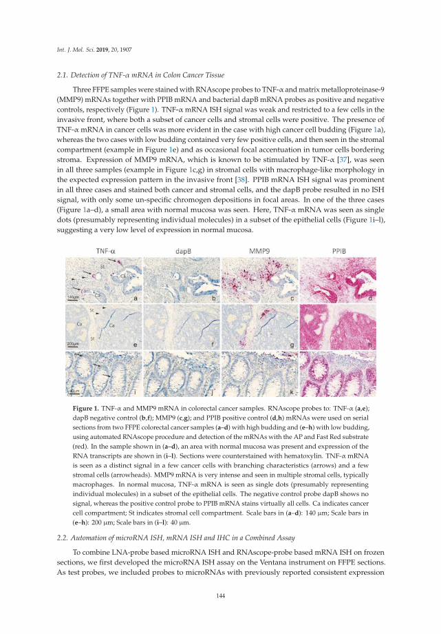
Int. J. Mol. Sci. 2019, 20, 1907
2.1. Detection of TNF-α mRNA in Colon Cancer Tissue
Three FFPE samples were stained with RNAscope probes to TNF-α and matrix metalloproteinase-9(MMP9) mRNAs together with PPIB mRNA and bacterial dapB mRNA probes as positive and negativecontrols, respectively (Figure 1). TNF-α mRNA ISH signal was weak and restricted to a few cells in theinvasive front, where both a subset of cancer cells and stromal cells were positive. The presence ofTNF-α mRNA in cancer cells was more evident in the case with high cancer cell budding (Figure 1a),whereas the two cases with low budding contained very few positive cells, and then seen in the stromalcompartment (example in Figure 1e) and as occasional focal accentuation in tumor cells borderingstroma. Expression of MMP9 mRNA, which is known to be stimulated by TNF-α [37], was seenin all three samples (example in Figure 1c,g) in stromal cells with macrophage-like morphology inthe expected expression pattern in the invasive front [38]. PPIB mRNA ISH signal was prominentin all three cases and stained both cancer and stromal cells, and the dapB probe resulted in no ISHsignal, with only some un-specific chromogen depositions in focal areas. In one of the three cases(Figure 1a–d), a small area with normal mucosa was seen. Here, TNF-α mRNA was seen as singledots (presumably representing individual molecules) in a subset of the epithelial cells (Figure 1i–l),suggesting a very low level of expression in normal mucosa.
Figure 1. TNF-α and MMP9 mRNA in colorectal cancer samples. RNAscope probes to: TNF-α (a,e);dapB negative control (b,f); MMP9 (c,g); and PPIB positive control (d,h) mRNAs were used on serialsections from two FFPE colorectal cancer samples (a–d) with high budding and (e–h) with low budding,using automated RNAscope procedure and detection of the mRNAs with the AP and Fast Red substrate(red). In the sample shown in (a–d), an area with normal mucosa was present and expression of theRNA transcripts are shown in (i–l). Sections were counterstained with hematoxylin. TNF-α mRNAis seen as a distinct signal in a few cancer cells with branching characteristics (arrows) and a fewstromal cells (arrowheads). MMP9 mRNA is very intense and seen in multiple stromal cells, typicallymacrophages. In normal mucosa, TNF-α mRNA is seen as single dots (presumably representingindividual molecules) in a subset of the epithelial cells. The negative control probe dapB shows nosignal, whereas the positive control probe to PPIB mRNA stains virtually all cells. Ca indicates cancercell compartment; St indicates stromal cell compartment. Scale bars in (a–d): 140 µm; Scale bars in(e–h): 200 µm; Scale bars in (i–l): 40 µm.
2.2. Automation of microRNA ISH, mRNA ISH and IHC in a Combined Assay
To combine LNA-probe based microRNA ISH and RNAscope-probe based mRNA ISH on frozensections, we first developed the microRNA ISH assay on the Ventana instrument on FFPE sections.As test probes, we included probes to microRNAs with previously reported consistent expression
144

Int. J. Mol. Sci. 2019, 20, 1907
patterns [33,39], including miR-126 in endothelial cells, miR-21 in stromal cells and miR-17 in cancercells (Figure 2). The microRNA ISH assay on the Ventana instrument was found to be equally or moresensitive than the Tecan assay [7] and was found suitable for unambiguous detection of microRNAs.The microRNA staining procedure with peroxidase and TSA substrate detection [34] was then combinedwith RNAscope procedure, using the AP (red kit) for mRNA detection and immunohistochemistry forcell marker detection on frozen sections from a colorectal cancer. Initially, miR-17 was combined withautomated TNF-α and IL-1β mRNA RNAscope probe and cytokeratin immunofluorescence (Figure 3).The combined staining revealed localization of miR-17 in the cancer cells as expected, and TNF-αmRNA was, in this case, mostly seen in CK-positive cancer cells located at the invasive front (Figure 3).In contrast, IL-1β mRNA ISH signal was limited to stromal cells in focal areas close to cancer cellde-differentiation and budding (Figure 3). miR-17 expression was lost in the cancer cells at the outerinvasive front including in the budding cancer cells (Figure 3) [39].
Figure 2. Automation of miR-21, miR-17 and miR-126 in a Ventana instrument. LNA probes to:miR-21 (a); miR-17 (b); and miR-126 (c) were used on FFPE colon cancer samples in a Tecan Genepaintinstrument (a–c) and a Ventana Discovery Ultra instrument (a’–c’), using AP detection and NBT-BCIPsubstrate (blue). The sections were counterstained with nuclear fast red. At optimized experimentalconditions, the Ventana and Tecan instruments showed identical staining patterns. miR-21 in stromalfibroblastic cells (a,a’); miR-17 in cancer cells (b,b’); and miR-126 in endothelial cells (c,c’). Scale bars in(a–c) are representative for (a’–c’).
145

Int. J. Mol. Sci. 2019, 20, 1907
Figure 3. Combination of LNA ISH, RNAscope and IHC (AP procedure). Colorectal cancer sectionsfrom a cryo-embedded sample were stained for miR-17 using an LNA probe, and either TNF-αor IL-1β using RNAscope probes, and cytokeratin using immunohistochemistry. miR-17 (white in(a,b,e,f)) is seen in tumor cells in differentiated cancer cells (cohesive structures) and is absent in thede-differentiated cancer cells in areas with budding and/or branching. TNF-α mRNA (red in (a,c))is seen in CK-positive cancer cells primarily located at the invasive front (arrows in (a–d)) and inCK-negative cells (arrowheads in (a–d)). IL-1β mRNA is seen CK-negative stromal cells (arrowheadsin (e–h)). The cytokeratin staining (green in (a,d,e,h)) is seen in cancer cells including the differentiatedcells of the cohesive structures and de-differentiated cells in branching and budding cancer cells.The CK staining was sub-optimal in this case—the miR-17 positive cancer cells are poorly stained withcytokeratin, and there is a dubious bleed-through of the red signal into the white channel. The bluefluorescence signal is nuclear DAPI counterstain in all images. Scale bars are indicated in lower leftcorner of each panel.
2.3. Co-Localization Analyses of TNF-α and miR-21
To evaluate co-localization of miR-21 and TNF-α mRNA in budding cancer cells, we performedthe combined assay, detecting miR-21, TNF-α mRNA and CK, on four frozen colorectal cancer sampleswith evident budding characteristics. As a negative control, the scramble LNA probe was combinedwith dapB RNAscope and cytokeratin (CK) immunofluorescence. We changed the RNAscope detectionassay to being HRP-based, using rhodamine substrate, to avoid masking effects of the Fast Redchromogen. In all four cases, TNF-α mRNA was seen in a subset of CK-positive cancer cells in theinvasive front, whereas expression in (CK-negative) stromal cells was a rare event (see Figure 4).The TNF-α mRNA ISH signal was often seen in cancer cells with a branching appearance, i.e., cellsthat show initial outgrowth from the cohesive adenocarcinoma structure (Figure 4g,k), whereas thefront-runner budding cancer cells were often TNF-α mRNA negative (Figure 4g,k). miR-21 was asexpected seen in stromal fibroblastic cells, and subsets of cancer cells were also positive, includingsubsets of budding cancer cells (Figure 4f,j). No ISH signal was observed with the scramble probe andthe dapB probe (Figure 4d), and the background fluorescence signal was limited to typical endogenousautofluorescence (Figure 4d). The combined stained slides were scanned using a confocal slide scannerwith a 20× objective. Examples from the other three cases are shown in the Supplementary Materials.At 20×magnification, the TNF-α mRNA signal appeared as a rather diffuse signal. Hence, some areasof interest were scanned using a 40× water objective. At this magnification, the individual spots fromthe RNAscope assay were clearly visible (Figure 5).
146

Int. J. Mol. Sci. 2019, 20, 1907
Figure 4. TNF-α mRNA and miR-21 in budding cancer cells in colorectal cancer (HRP procedure).The colon cancer tissue sample (cryo-embedded) is shown after H&E staining at low (a) and highmagnification (b), and after combined fluorescence staining and subsequent confocal slide scanningmicroscopy at low magnification (c). The tissue section shows miR-21 (white in (c,d,e,l,m)) or scramble(white in (h,i)), TNF-α mRNA (red in (c,d,f,l,n)) or dapB mRNA (red in (h,j)) and CK (green in(c,d,g,h,k,l,o)). Probes to miR-21 and scramble probe were incubated on serial sections (d–g, and h–k)respectively. Arrows in (d–h) indicate the same cancer cell population in the two sections that arepositive with the miR-21 and TNF-α probes and negative with the scramble and dapB probes. Arrowsin (l–o) indicate miR-21 and TNF-α positive cancer cells and the arrowhead indicate a TNF-α positivestromal cell. Sections were scanned using a Pannoramic confocal slide scanner at 20× to give theoverview image (c) and the framed areas are shown in (d–g,l–o) at high magnification (digital zoom),and two frames are shown Figure 5. Arrows in (a,c) indicate direction of invasion. Ca, cancer area; N,normal area; St, stroma area. Scale bars are indicated in lower left corner of each panel.
147

Int. J. Mol. Sci. 2019, 20, 1907
Figure 5. Confocal slide scanning at 40×. The same section as in Figure 4 here scanned using a 40×water objective. The two areas (a–f) show typical TNF-α mRNA expression at cancer cell branchingwith miR-21 expression in TNF-α-positive cells and neighboring TNF-α-positive signal in the stroma(arrowheads in (c,f)). Note that miR-21 positive stromal cells are TNF-α-negative. The RNAscope ISHsignal is here evident as single dots representing individual mRNA molecules (arrows in (c,f)). Scalebars are indicated in lower left corner of each panel.
Since expression of TNF-α mRNA was very restricted compared to miR-21, we evaluated theTNF-α positive cancer cells in the four cases for miR-21 positivity within the same cell. In two cases,virtually all TNF-α positive cells were also miR-21-positive, whereas in the other two cases only 20% orless were miR-21 positive. TNF-α positive cells located in the stroma were generally miR-21 negative.In one of the cases, we noted that a TNF-α mRNA positive cancer cell was often associated withmiR-21 in neighboring cancer cells and stromal cells. However, stromal fibroblastic cells located in thevicinity of TNF-α positive cancer cells did generally not show differences in the miR-21 expressionpatterns compared to areas without TNF-α mRNA signal. These observations suggest that miR-21 isnot co-regulated with the TNF-α mRNA, neither within the same cell nor in neighboring cells.
3. Discussion
This study was undertaken to address if in situ localization analyses can help to clarifyinterplay between miR-21 and the pro-inflammatory cytokine TNF-α during colon cancer progression.We developed a fully automated combined in situ hybridization and immunohistochemistry assay thatallowed use of LNA probe technology and RNAscope technology together with immunofluorescenceon the same tissue section. Using CK immunofluorescence, all cancer cells at the invasive front ofthe colorectal cancers were visualized. Co-localization of the TNF-α mRNA in the CK positive cellsindicated that this potent pro-inflammatory cytokine is expressed in a small subset of cancer cellslocated at the invasive front. We found that miR-21 also co-localizes in some, but not all, of these cancercells, suggesting that miR-21 is not co-regulated with TNF-α mRNA, and that the presence of TNF-αmRNA does not lead to suppression of miR-21 expression. In normal mucosa, TNF-α mRNA wasnoted as a few single dots representing very low expression and miR-21 was only occasionally seen innormal mucosa [7], indicating that the cancer or associated inflammation may be required to induce
148

Int. J. Mol. Sci. 2019, 20, 1907
expression of the two transcripts. We speculate that miR-21 in TNF-α mRNA positive cancer cells maysuppress an autocrine effect of TNF-α to mediate cell death, e.g., via PDCD4 [40].
We previously developed a microRNA ISH method on a Tecan Genepaint instrument [7],which provides highly reproducible staining results; however, we found that RNAscope was notfeasible for this instrument due to reagent consumption and consequently high cost. On the other hand,fully automated RNAscope assay procedures, e.g., on a Ventana instrument [30], have been developedby the vendors (ACDbio and Roche) and implemented in our Lab. To combine microRNA ISH withRNAscope, we therefore first established a microRNA ISH procedure on the Ventana instrument. Usingthe AP-based staining method, and similar reagents developed previously on the Tecan instrument,we obtained identical results with a slightly improved sensitivity and signal-to-noise ratio. Becausepre-treatment procedures, required for microRNA ISH and RNAscope assays, were not compatiblefor FFPE samples, we developed the combined assay on frozen tissue, which also has the advantageof having better RNA integrity if processed for cryo-protection immediately after resection andappropriate storage. The combination of the three techniques into a triple fluorescence assay canpotentially have many applications, including co-localization studies of a microRNA and its targetmRNA and protein.
We investigated four cases of colorectal cancer that all included the invasive tumor front andprevalent cancer cell budding. In each of the four cases, multiple events of TNF-α mRNA expressionand miR-21 expression in cancer cells could be evaluated. Because the same localization pattern wasnoted for TNF-α mRNA in the four cases, and also that the four cases showed the similar miR-21localization pattern, we assume that our findings are representative for such adenocarcinomas despitethe limited number of cases. The invasive growth pattern, however, varied with respect to the cancercell structures, the presence of inflammation and fibrosis. In all four cases, the localization of TNF-αmRNA was similar, thus TNF-α positive cancer cells were often located at branching points of thecohesive adenocarcinoma structures located toward the invasive front or in detached de-differentiatedbudding cancer cells or in larger islands of cancer cells. TNF-α mRNA positive cancer cells were onlyoccasionally seen in the central areas of the tumors. The characteristic TNF-α mRNA expression patternwas seen in both paraffin and cryo-embedded samples, and the same pattern was obtained with boththe AP- and HRP-based RNAscope kits. Differences in staining intensity and prevalence of positivecells may be related to biological differences or differences in mRNA integrity. For specificity evaluationof the RNAscope assay, we included reference probes, of which, most importantly, the negative controlprobe resulted in the lack of ISH signal except from background. RNAscope probes to mRNAs forMMP9, PPIB and IL-1β all showed the expected expression patterns. Taken together, the observationssuggest that the ISH signal obtained with the TNF-α probe is representing the genuine TNF-α mRNA.To our knowledge, this is the first study to show expression of TNF-α mRNA in budding andbranching colon cancer cells in situ. The particular localization of the TNF-α mRNA expressing cellssuggests a role in aggressive cancer cell invasion. In support of this observation, Li et al. [41] measuredincreased TNF-α mRNA levels in total RNA isolated from micro-dissected colorectal cancer buddingcells and the surrounding microenvironment. In addition, TNF-α has been found to be involved inbranching morphogenesis in in vivo models, e.g., during rat mammary gland development facilitatedby MMP9 [42,43], suggesting that the cancer cells re-establish cellular mechanism used during earlyorgan development. It is also of particular interest that TNF-α was found to induce an invasivephenotype in ovarian epithelial cystic structures [44]. In the latter study, TNF-α was added to thegrowth medium with the cystic structures, suggesting a paracrine mechanism inducing invasion,which would be expected if the source of TNF-α was inflammatory cells, including TNF-α-loadedneutrophilic granulocytes that may not carry the TNF-α mRNA. The observations in our study suggestthat TNF-α also mediate an autocrine mechanism that contributes to cancer invasion. A larger sampleset would be required to address if the expression of TNF-α mRNA by ISH or the co-expression withmiR-21 is of prognostic relevance.
149

Int. J. Mol. Sci. 2019, 20, 1907
Digital slides obtained by confocal slide scanning microscopy allowed systematic evaluationof individual cells. We evaluated TNF-α mRNA and CK positive cells in each of the four casesfor the presence of miR-21. The miR-21 positive fraction varied from 20% to 100%, suggesting nogeneral correlation. In one of the cases, we noted that a TNF-α mRNA positive cancer cell was oftenassociated with miR-21 in neighboring cancer cells and stromal cells. This observation could suggestthat TNF-α stimulates miR-21 in various cells in the local neighborhood independent of the celltype. TNF-α has been reported to upregulate miR-21 expression in a variety of cell types, includinghuman renal epithelial cells [45] and in Caco-2 cells used in an intestinal barrier model [46]. In astudy by Xu et al. [47], it was observed that the expression of miR-21 increased gradually with lowconcentrations of TNF-α, while being suppressed at high concentrations. Thus, TNF-α is likely tohave an effect on miR-21 expression, but in a complex tissue with many different cells and differentsignaling pathways interacting, distinct correlation patterns can be difficult to decipher. This mayexplain why no obvious correlation between TNF-α mRNA in cancer cells and stromal miR-21 wasobserved. Such patterns, may be better characterized in early stage cancers or in appropriate in vitrocell models. Conversely, miR-21 may control TNF-α expression. Ando et al. [48] found that a miR-21mimic upregulated TNF-α in T-cells and Zhang et al. [49] reported that miR-21 directly targets TNF-αin bronchial epithelial cells. Future studies may disclose whether miR-21 and TNF-α are cooperating,competing or completely independent during cancer progression.
In conclusion, we report that miR-21 and TNF-α mRNA both are expressed at the invasive frontof colon cancers and are co-localized in a subset of budding cancer cells and cells located at branchingpoints and in clusters of cancer cells. Our attempts to clarify potential interactions between miR-21 andTNF-α did not result in consistent co-expression or converse expression patterns. If miR-21 expressionis suppressed by TNF-α, therapeutic use of Infliximab would increase miR-21, which would promotetumor progression. Therefore, better understanding of the TNF-α/miR-21 interplay is highly warranted.
4. Material and Methods
4.1. Tissue Material
Three FFPE and four frozen colon adenocarcinoma tissue samples (Table 1) were obtainedfrom Asterand (BioIVT, W Sussex, UK). The tissue samples were selected according to resectionyear (2015–2017), a well-defined invasive area and, for the frozen samples, also evident tumor cellbudding and high RNA integrity number (RIN) values (>7). Evaluation of tissue sections stainedwith hematoxylin and eosin showed that all cases were moderately differentiated adenocarcinomas.The Asterand/BioIVT tissue samples were obtained according to the ethical principles defined inthe Nuremburg Code, on the recommendations to consider when sourcing human biospecimens(https://info.bioivt.com/biospecimen-sourcing-white-paper).
Table 1. Tissue samples.
Case Procurement Type Differentiation Budding ISH Method
1 FFPE-A Ad Mod 1 (BD1) Chromogen2 FFPE-B Ad Mod 2 (BD1) Chromogen3 FFPE-C Ad Mod 10 (BD3) Chromogen4 Cryo-A Ad Mod 12 (BD3) Fluorescence5 Cryo-B Ad Mod 6 (BD2) Fluorescence6 Cryo-C Ad Mod 6 (BD2) Fluorescence7 Cryo-D Ad Mod 5 (BD2) Fluorescence
Ad, adenocarcinoma; BD, budding density (according to guidelines issued by International Tumor BuddingConsensus Conference, April 2016); FFPE, formaldehyde fixed, paraffin embedded; Mod, moderately differentiated.Budding scores: BD1: 0–4, BD2: 5–9, BD3: >10 buds per 0.785 mm2 field of view.
150

Int. J. Mol. Sci. 2019, 20, 1907
4.2. Probes for in situ Hybridization
Double-labeled locked nucleic acid (LNA™) probes were obtained from Qiagen (Exiqon-Qiagen,Hilden, Germany) as double digoxigenin-labeled LNA:DNA chimeric oligos. The microRNAantisense probes had approximately 30% of DNA replaced by LNA to increase the bindingaffinity (RNA Tm): miR-21-5p (TCAACATCAGTCTGATAAGCTA, RNA Tm = 83 ◦C), miR-17-5p(TACCTGCACTGTAAGCACTTT, RNA Tm = 89 ◦C), miR-126-3p (CATTATTACTCACGGTACGA,RNA Tm = 84 ◦C), and scramble (ATGTAACACGTCTATACGCCCA, RNA Tm = 86 ◦C). The microRNAprobes recognize both the mature forms as well as precursor forms. RNAscope probes, or branchedDNA (bDNA) probes, are designed to have high specificity and high sensitivity (www.acdbio.com).The high specificity is obtained by design of two antisense DNA oligonucleotides, also called double-Zprobes, to bind adjacent sequences as pairs on the target sequence. The high sensitivity is partlybased on design of typically up to 20 pairs for individual mRNA targets. The following RNAscopeprobes were obtained from ACD, Biotechne (Newark, CA): TNF-α (Tumor necrosis factor, targetregion: 67–1079, 13 zz pairs), IL-1β (Interleukin-1, target region: 2–1319, 20 zz pairs), MMP9 (matrixmetalloproteinase-9, target region: 93–1422, 30 zz pairs), dapB (a Bacillus subtilis gene, 414–862, 10 zzpairs), and PPIB (Cyclophilin B, 139–989, 16 zz pairs).
4.3. Automated Chromogenic LNA ISH
MicroRNA ISH was performed using LNA probes on 5-µm-thick FFPE sections essentially asdescribed previously [7], in which the specificity of the ISH staining was also analyzed. In brief, sectionswere deparaffinized and proteinase-K treated. Probes were added to the sections and incubated atconcentrations and hybridization temperatures described earlier [7,39]. The probes were detectedwith alkaline phosphatase (AP) conjugated anti-DIG antibodies and stained with NBT-BCIP substratefor 30–60 min. In the Ventana instrument (Roche, Basel, Switzerland), protease-1 was used for tissuepre-treatment and probes were incubated at 2nM (miR-21) or 10nM (miR-17 and miR-126).
4.4. Automated Chromogenic RNAscope
RNAscope ISH was performed on 5-µm-thick FFPE sections essentially as described byAnderson et al. [30] using the AP (red) kit at recommended experimental conditions for the VentanaDiscovery Ultra instrument. In this setup, we used Amp5 for 32 min (MMP9, IL-1β) or 60 min (TNF-α,dapB, PPIB).
4.5. Automated Combined ISH and IHC
Ten-micrometer-thick sections were obtained and fixed overnight in 4% paraformaldehyde.Staining of microRNA in frozen tissue sections has previously been described [35], usingfluorescence detection with horse-radish-peroxidase (HRP)-conjugated anti-DIG antibodies andTSA-Cy5 substrate [34] (Roche). RNAscope ISH was performed as recommended by the manufacturerusing the AP kit (Fast red substrate, Figure 3) or the HRP kit (TSA-rhodamine substrate, Figure 4,Figure 5 and Figure S1) in a Ventana Discovery Ultra instrument (Roche) [30]. Blocking of HRP inbetween the LNA assay and the RNAscope assay was done using the Discovery inhibitor for 10 min(Ventana, Roche Diagnostics). For cytokeratin immunofluorescence, the AE1/3 mouse monoclonalantibody (Dako-Agilent, Glostrup, Denmark) was used at 1:500 and detected with Alexa-488 conjugatedanti-mouse Ig (Jackson Immunoresearch, West Grove, PA, USA). The negative control slides used asreference included replacement of the miR-21 probe with the scramble probe and replacement of theTNF-α probe with the dapB probe, whereas the anti-cytokeratin was retained, and was performed oncryo-samples A, B and C. Here, the miR-21 probe was incubated at 10 and 20 nM and the scrambleprobe at 20 nM. Despite a difference in staining intensity, there was no difference in the cellularstaining pattern of the miR-21 signal when comparing the miR-21 probe concentration of 10 and 20 nM.In Figure 4, Figure 5 and Figure S1, the images were acquired from slides incubated with 20 nM probe
151

Int. J. Mol. Sci. 2019, 20, 1907
concentration equal to the scramble probe. Sections were mounted with a DAPI-containing anti-fadesolution, ProLong Gold (Thermo Fisher Scientific, Waltham, MA, USA). The detailed procedure isproprietary of Bioneer A/S (Bioneer, Hørsholm, Denmark).
4.6. Slide Scanning and IMAGE Acquisition
All chromogen stained slides were scanned on a Zeiss Axioscan equipped with a 20× objective.All images in Figures 1 and 2 were obtained from such digital slides. Confocal slide scanningwas performed using a Pannoramic confocal scanner (3DHISTECH Ltd., Budapest, Hungary),which provides confocal images made by LED light and structured illumination technology. The use ofthis slide scanner has previously been described in detail for use on sections stained for miR-21 and twosubsequent immunoperoxidase stainings [9]. The scanner was equipped with a 20× objective (NA = 0.8,Zeiss, Oberkochen, Germany) and a 40×water immersion objective (NA = 1.2, C-Apochromat (W),Zeiss, Oberkochen, Germany). For the current study, the following LED light sources were appliedin the excitations: DAPI 390/22 nms, 520 mW, Cy3 555/28 nms, 370 mW, Cy5 635/22 nms, 510 mW,FITC 475/28, 530 mW. We used one dual pass filter (for FITC and Cy5, custom designed by 3DHisTech) and two single pass filters (TRITC/rhodamine and DAPI, both Semrock, New York, NY, USA).Digital slides were obtained with manually adjusted settings with regards to exposure time, digitalgain and excitation intensity. The image acquisition settings for all 4 fluorophores were set at fullexcitation intensity if not otherwise stated. Image acquisition parameters were: miR-21 (Cy5, exposuretime varying from 20–40 ms with digital gain 1), TNF-α mRNA (rhodamine, 20 ms, with 15–30%of full excitation intensity), CK immunofluorescence (FITC, 74–174 ms, digital gain 2), and nuclearcounterstain (DAPI, 176–325 ms, digital gain 2). The settings applied to the individual cases werealso used on the respective negative control sections incubated with scramble LNA probe and dapBmRNA RNAscope probe (cryo samples A, B, and C). Seven confocal layers of 1 µm were obtained andassembled into one extended focus layer, from which we obtained all fluorescence images in this paper.
5. Conclusions
In this study we developed a combined microRNA and mRNA ISH assay that allows the useof LNA probes and RNAscope probes on the same section as well as enables immunohistochemicaldetection of cellular markers. We explored expression of miR-21 and TNF-α mRNA at the invasive frontof four colon cancers in order to elucidate expression interplay between miR-21 and TNF-α, and foundthat they are both expressed at the invasive front of colon cancers and are often co-detected in buddingcancer cells and cancer cells considered to be part of branching events, however, the co-localizationanalysis of the four cases did not show consistent co-expression or converse expression patterns.
Supplementary Materials: Supplementary materials can be found at http://www.mdpi.com/1422-0067/20/8/1907/s1. Figure S1. Confocal slide scanning of sections submitted to automated combined staining of miR-21, TNF-αmRNA and CK. The three panels in this supplementary figure show miR-21 and TNF-α mRNA expression in3 different colorectal cancer cases with varying invasion patterns (cases cryo-B, cryo-C and cryo-D, Table 1).Examples are shown at low (a–d) and high (e–h) magnification, and in a, the framed area is depicted in e–h.In example cryo-B, TNF-α mRNA is seen in foci of cancer cells also positive for miR-21. In example cryo-C, TNF-αmRNA is prevalent and seen in multiple cancer cells that are generally weakly stained for miR-21 or miR-21negative, whereas miR-21 is prevalent in the stromal cells. In example cryo-D, miR-21 is seen in a few TNF-αmRNA expressing branching cancer cells. For Cryo-B and Cryo-C, in a serial tissue section, the miR-21 probewas replaced with an LNA scramble probe, and the TNF-α mRNA probe was replaced with the dapB mRNARNAscope probe, that both show virtually no staining (magnification in e–h is identical to i–l).
Author Contributions: Data curation, J.P.J., F.B.S., J.L. and B.S.N.; Formal analysis, F.B.S. and J.L.; Fundingacquisition, K.H.; Investigation, T.M. and B.S.N.; Methodology, T.M. and B.S.N.; Project administration,B.S.N.; Resources, T.M.; Validation, B.S.N.; Visualization, J.P.J. and B.S.N.; Writing—original draft, B.S.N.;and Writing—review and editing, J.P.J., K.H., F.B.S. and J.L.
Funding: This research received no external funding.
Acknowledgments: This study was supported by The Danish Agency for Science and Higher Education.
Conflicts of Interest: The authors declare no conflict of interest.
152

Int. J. Mol. Sci. 2019, 20, 1907
References
1. Ambros, V. microRNAs: Tiny regulators with great potential. Cell 2001, 107, 823–826. [PubMed]
2. Winter, J.; Jung, S.; Keller, S.; Gregory, R.I.; Diederichs, S. Many roads to maturity: microRNA biogenesis
pathways and their regulation. Nat. Cell Biol. 2009, 11, 228–234. [PubMed]
3. Bartel, D.P. MicroRNAs: Genomics, biogenesis, mechanism, and function. Cell 2004, 116, 281–297. [PubMed]
4. Krichevsky, A.M.; Gabriely, G. miR-21: A small multi-faceted RNA. J. Cell Mol. Med. 2009, 13, 39–53.
5. Volinia, S.; Calin, G.A.; Liu, C.G.; Ambs, S.; Cimmino, A.; Petrocca, F.; Visone, R.; Iorio, M.; Roldo, C.;
Ferracin, M.; et al. A microRNA expression signature of human solid tumors defines cancer gene targets.
Proc. Natl. Acad. Sci. USA 2006, 103, 2257–2261. [PubMed]
6. Schetter, A.J.; Leung, S.Y.; Sohn, J.J.; Zanetti, K.A.; Bowman, E.D.; Yanaihara, N.; Yuen, S.T.; Chan, T.L.;
Kwong, D.L.; Au, G.K.; et al. MicroRNA expression profiles associated with prognosis and therapeutic
outcome in colon adenocarcinoma. JAMA 2008, 299, 425–436. [PubMed]
7. Nielsen, B.S.; Jorgensen, S.; Fog, J.U.; Sokilde, R.; Christensen, I.J.; Hansen, U.; Brunner, N.; Baker, A.;
Moller, S.; Nielsen, H.J. High levels of microRNA-21 in the stroma of colorectal cancers predict short
disease-free survival in stage II colon cancer patients. Clin. Exp. Metastasis 2011, 28, 27–38. [PubMed]
8. Nielsen, B.S.; Balslev, E.; Poulsen, T.S.; Nielsen, D.; Moller, T.; Mortensen, C.E.; Holmstrom, K.; Hogdall, E.
miR-21 Expression in Cancer Cells may Not Predict Resistance to Adjuvant Trastuzumab in Primary Breast
Cancer. Front. Oncol. 2014, 4, 207. [PubMed]
9. Knudsen, K.N.; Lindebjerg, J.; Kalmar, A.; Molnar, B.; Sorensen, F.B.; Hansen, T.F.; Nielsen, B.S. miR-21
expression analysis in budding colon cancer cells by confocal slide scanning microscopy. Clin. Exp. Metastasis
2018, 35, 819–830. [PubMed]
10. Zlobec, I.; Lugli, A. Epithelial mesenchymal transition and tumor budding in aggressive colorectal cancer:
Tumor budding as oncotarget. Oncotarget 2010, 1, 651–661.
11. Hase, K.; Shatney, C.; Johnson, D.; Trollope, M.; Vierra, M. Prognostic value of tumor “budding” in patients
with colorectal cancer. Dis. Colon Rectum. 1993, 36, 627–635.
12. Rogers, A.C.; Winter, D.C.; Heeney, A.; Gibbons, D.; Lugli, A.; Puppa, G.; Sheahan, K. Systematic review and
meta-analysis of the impact of tumour budding in colorectal cancer. Br. J. Cancer 2016, 115, 831–840.
13. Patel, V.; Noureddine, L. MicroRNAs and fibrosis. Curr. Opin. Nephrol. Hypertens. 2012, 21, 410–416.
[PubMed]
14. Asangani, I.A.; Rasheed, S.A.; Nikolova, D.A.; Leupold, J.H.; Colburn, N.H.; Post, S.; Allgayer, H.
MicroRNA-21 (miR-21) post-transcriptionally downregulates tumor suppressor Pdcd4 and stimulates
invasion, intravasation and metastasis in colorectal cancer. Oncogene 2008, 27, 2128–2136. [PubMed]
15. Yang, Y.; Ma, Y.; Shi, C.; Chen, H.; Zhang, H.; Chen, N.; Zhang, P.; Wang, F.; Yang, J.; Yang, J.; et al.
Overexpression of miR-21 in patients with ulcerative colitis impairs intestinal epithelial barrier function
through targeting the Rho GTPase RhoB. Biochem. Biophys. Res. Commun. 2013, 434, 746–752. [PubMed]
16. Thorlacius-Ussing, G.; Schnack Nielsen, B.; Andersen, V.; Holmstrom, K.; Pedersen, A.E. Expression and
Localization of miR-21 and miR-126 in Mucosal Tissue from Patients with Inflammatory Bowel Disease.
Inflamm. Bowel Dis. 2017, 23, 739–752. [PubMed]
17. Chapman, C.G.; Pekow, J. The emerging role of miRNAs in inflammatory bowel disease: A review.
Therap. Adv. Gastroenterol. 2015, 8, 4–22.
18. Carswell, E.A.; Old, L.J.; Kassel, R.L.; Green, S.; Fiore, N.; Williamson, B. An endotoxin-induced serum factor
that causes necrosis of tumors. Proc. Natl. Acad. Sci. USA 1975, 72, 3666–3670.
19. Old, L.J. Tumor necrosis factor (TNF). Science 1985, 230, 630–633.
20. Arnott, C.H.; Scott, K.A.; Moore, R.J.; Hewer, A.; Phillips, D.H.; Parker, P.; Balkwill, F.R.; Owens, D.M.
Tumour necrosis factor-alpha mediates tumour promotion via a PKC alpha- and AP-1-dependent pathway.
Oncogene 2002, 21, 4728–4738.
21. Balkwill, F. Tumor necrosis factor or tumor promoting factor? Cytokine Growth Factor Rev. 2002, 13, 135–141.
[PubMed]
22. Guadagni, F.; Ferroni, P.; Palmirotta, R.; Portarena, I.; Formica, V.; Roselli, M. TNF/VEGF cross-talk in chronic
inflammation-related cancer initiation and progression: An early target in anticancer therapeutic strategy.
In Vivo 2007, 21, 147–161. [PubMed]
153

Int. J. Mol. Sci. 2019, 20, 1907
23. Grimm, M.; Lazariotou, M.; Kircher, S.; Hofelmayr, A.; Germer, C.T.; von Rahden, B.H.; Waaga-Gasser, A.M.;
Gasser, M. Tumor necrosis factor-alpha is associated with positive lymph node status in patients with
recurrence of colorectal cancer-indications for anti-TNF-alpha agents in cancer treatment. Cell. Oncol. 2011,
34, 315–326.
24. Ham, B.; Fernandez, M.C.; D’Costa, Z.; Brodt, P. The diverse roles of the TNF axis in cancer progression and
metastasis. Trends Cancer Res. 2016, 11, 1–27. [PubMed]
25. Qu, Y.; Zhao, G.; Li, H. Forward and Reverse Signaling Mediated by Transmembrane Tumor Necrosis
Factor-Alpha and TNF Receptor 2: Potential Roles in an Immunosuppressive Tumor Microenvironment.
Front. Immunol. 2017, 8, 1675. [PubMed]
26. Cottonham, C.L.; Kaneko, S.; Xu, L. miR-21 and miR-31 converge on TIAM1 to regulate migration and
invasion of colon carcinoma cells. J. Biol. Chem. 2010, 285, 35293–35302.
27. Qiu, Y.F.; Wang, M.X.; Meng, L.N.; Zhang, R.; Wang, W. MiR-21 regulates proliferation and apoptosis of oral
cancer cells through TNF-alpha. Eur. Rev. Med. Pharmacol. Sci. 2018, 22, 7735–7741.
28. Chen, X.; Zhao, L.; Xing, Y.; Lin, B. Down-regulation of microRNA-21 reduces inflammation and podocyte
apoptosis in diabetic nephropathy by relieving the repression of TIMP3 expression. Biomed. Pharmacother.
2018, 108, 7–14.
29. Player, A.N.; Shen, L.P.; Kenny, D.; Antao, V.P.; Kolberg, J.A. Single-copy gene detection using branched
DNA (bDNA) in situ hybridization. J. Histochem. Cytochem. 2001, 49, 603–612.
30. Anderson, C.M.; Zhang, B.; Miller, M.; Butko, E.; Wu, X.; Laver, T.; Kernag, C.; Kim, J.; Luo, Y.; Lamparski, H.;
et al. Fully Automated RNAscope In Situ Hybridization Assays for Formalin-Fixed Paraffin-Embedded
Cells and Tissues. J. Cell. Biochem. 2016, 117, 2201–2208.
31. Wang, H.; Su, N.; Wang, L.C.; Wu, X.; Bui, S.; Nielsen, A.; Vo, H.T.; Luo, Y.; Ma, X.J. Dual-color ultrasensitive
bright-field RNA in situ hybridization with RNAscope. Methods Mol. Biol. 2014, 1211, 139–149.
32. Thomsen, R.; Nielsen, P.S.; Jensen, T.H. Dramatically improved RNA in situ hybridization signals using
LNA-modified probes. RNA 2005, 11, 1745–1748. [PubMed]
33. Jorgensen, S.; Baker, A.; Moller, S.; Nielsen, B.S. Robust one-day in situ hybridization protocol for detection
of microRNAs in paraffin samples using LNA probes. Methods 2010, 52, 375–381.
34. Nielsen, B.S.; Holmstrom, K. Combined microRNA in situ hybridization and immunohistochemical detection
of protein markers. In Target Identification and Validation in Drug Discovery; Moll, J., Colombo, R., Eds.;
Humana Press: New York, NY, USA, 2013; pp. 353–365.
35. Nielsen, B.S.; Moller, T.; Holmstrom, K. Chromogen detection of microRNA in frozen clinical tissue samples
using LNA probe technology. Methods Mol. Biol. 2014, 1211, 77–84.
36. Sempere, L.F.; Preis, M.; Yezefski, T.; Ouyang, H.; Suriawinata, A.A.; Silahtaroglu, A.; Conejo-Garcia, J.R.;
Kauppinen, S.; Wells, W.; Korc, M. Fluorescence-based codetection with protein markers reveals distinct
cellular compartments for altered MicroRNA expression in solid tumors. Clin. Cancer Res. 2010, 16, 4246–4255.
[PubMed]
37. Leber, T.M.; Balkwill, F.R. Regulation of monocyte MMP-9 production by TNF-alpha and a tumour-derived
soluble factor (MMPSF). Br. J. Cancer 1998, 78, 724–732.
38. Nielsen, B.S.; Timshel, S.; Kjeldsen, L.; Sehested, M.; Pyke, C.; Borregaard, N.; Dano, K. 92 kDa type IV
collagenase (MMP-9) is expressed in neutrophils and macrophages but not in malignant epithelial cells in
human colon cancer. Int. J. Cancer 1996, 65, 57–62.
39. Knudsen, K.N.; Nielsen, B.S.; Lindebjerg, J.; Hansen, T.F.; Holst, R.; Sorensen, F.B. microRNA-17 is the
most up-regulated member of the miR-17-92 cluster during early colon cancer evolution. PLoS ONE 2015,
10, e0140503.
40. Fluckiger, A.; Dumont, A.; Derangere, V.; Rebe, C.; de Rosny, C.; Causse, S.; Thomas, C.; Apetoh, L.;
Hichami, A.; Ghiringhelli, F.; et al. Inhibition of colon cancer growth by docosahexaenoic acid involves
autocrine production of TNFalpha. Oncogene 2016, 35, 4611–4622. [PubMed]
41. Li, H.; Zhong, A.; Li, S.; Meng, X.; Wang, X.; Xu, F.; Lai, M. The integrated pathway of TGFbeta/Snail with
TNFalpha/NFkappaB may facilitate the tumor-stroma interaction in the EMT process and colorectal cancer
prognosis. Sci. Rep. 2017, 7, 4915. [PubMed]
42. Varela, L.M.; Ip, M.M. Tumor necrosis factor-alpha: A multifunctional regulator of mammary gland
development. Endocrinology 1996, 137, 4915–4924. [PubMed]
154
Int. J. Mol. Sci. 2019, 20, 1907
43. Lee, P.P.; Hwang, J.J.; Murphy, G.; Ip, M.M. Functional significance of MMP-9 in tumor necrosis factor-induced
proliferation and branching morphogenesis of mammary epithelial cells. Endocrinology 2000, 141, 3764–3773.
[PubMed]
44. Kwong, J.; Chan, F.L.; Wong, K.K.; Birrer, M.J.; Archibald, K.M.; Balkwill, F.R.; Berkowitz, R.S.; Mok, S.C.
Inflammatory cytokine tumor necrosis factor alpha confers precancerous phenotype in an organoid model of
normal human ovarian surface epithelial cells. Neoplasia 2009, 11, 529–541. [PubMed]
45. Zarjou, A.; Yang, S.; Abraham, E.; Agarwal, A.; Liu, G. Identification of a microRNA signature in renal
fibrosis: Role of miR-21. Am. J. Physiol. Renal Physiol. 2011, 301, F793–F801. [PubMed]
46. Zhang, L.; Shen, J.; Cheng, J.; Fan, X. MicroRNA-21 regulates intestinal epithelial tight junction permeability.
Cell Biochem. Funct. 2015, 33, 235–240. [PubMed]
47. Xu, K.; Xiao, J.; Zheng, K.; Feng, X.; Zhang, J.; Song, D.; Wang, C.; Shen, X.; Zhao, X.; Wei, C.; et al.
MiR-21/STAT3 Signal Is Involved in Odontoblast Differentiation of Human Dental Pulp Stem Cells Mediated
by TNF-alpha. Cell Reprogram. 2018, 20, 107–116. [PubMed]
48. Ando, Y.; Yang, G.X.; Kenny, T.P.; Kawata, K.; Zhang, W.; Huang, W.; Leung, P.S.; Lian, Z.X.; Okazaki, K.;
Ansari, A.A.; et al. Overexpression of microRNA-21 is associated with elevated pro-inflammatory cytokines
in dominant-negative TGF-beta receptor type II mouse. J. Autoimmun. 2013, 41, 111–119.
49. Zhang, X.; Ng, W.L.; Wang, P.; Tian, L.; Werner, E.; Wang, H.; Doetsch, P.; Wang, Y. MicroRNA-21 modulates
the levels of reactive oxygen species by targeting SOD3 and TNFalpha. Cancer Res. 2012, 72, 4707–4713.
[PubMed]
© 2019 by the authors. Licensee MDPI, Basel, Switzerland. This article is an open access
article distributed under the terms and conditions of the Creative Commons Attribution
(CC BY) license (http://creativecommons.org/licenses/by/4.0/).
155


MDPI
St. Alban-Anlage 66
4052 Basel
Switzerland
Tel. +41 61 683 77 34
Fax +41 61 302 89 18
www.mdpi.com
International Journal of Molecular Sciences Editorial Office
E-mail: [email protected]
www.mdpi.com/journal/ijms


MDPI
St. Alban-Anlage 66
4052 Basel
Switzerland
Tel: +41 61 683 77 34
Fax: +41 61 302 89 18
www.mdpi.com ISBN 978-3-03921-250-7