Digital detection of biomarkers assisted by nanoparticles: application to diagnostics
9
Digital detection of biomarkers assisted by nanoparticles: application to diagnostics Marina Cretich 1* , George G. Daaboul 2* , Laura Sola 1 , M. Selim U ¨ nlu ¨ 2 , and Marcella Chiari 1 1 Consiglio Nazionale delle Ricerche, Istituto di Chimica del Riconoscimento Molecolare (ICRM), Milano, Italy 2 Electrical and Computer Engineering Department, Boston University, Boston, MA, USA Single-molecule detection and counting is the new fron- tier in biomarker analysis. Here, we report on recent techniques for the digital detection of biomolecules for clinical application. First we highlight methods based on the immunocapture of proteins onto microparticles, followed by isolation of individual particles in microen- vironments so that a sufficient signal is acquired for each binding event to make a binary decision, thus dramati- cally enhancing the signal:noise ratio. Various approaches are categorized based on the method used for particle confinement in an isolated microenviron- ment. We go on to describe methods for the detection of individual biological nanoparticles as well as the digital detection of proteins by artificial nanoparticle labels. The discussion of the methods emphasizes the practical considerations and their clinical applicability. Digital and analog detection of biomarkers The clinical use of protein biomarkers to differentiate between healthy and disease states, and to monitor disease progression, requires selective measurement of low con- centrations of proteins in complex samples. Current immu- noassays typically measure proteins at concentrations above 10 À12 M [1]. However, the serum concentrations of most proteins that are important in cancer [2], neuro- logical disorders [3,4], and the early stages of infection [5] are in the range 10 À16 –10 À12 M. Attempts to develop methods capable of measuring these minute concentrations of proteins have focused on the replication of nucleic acid labels on proteins [6,7] or on measuring the bulk ensemble properties of labeled protein molecules using a variety of transduction and amplifica- tion mechanisms, such as electrochemical [8,9], mechani- cal [10,11], or optical [12–14] techniques. The new frontier in biomarker analysis is single-molecule counting or digital detection, an approach that provides resolution and sensitivity that are not obtainable with ensemble measurements. Digital detection is a disruptive technology potentially allowing most advanced disease di- agnostic tools to become available at a low cost and at the point-of-need. This is similar to how digital audio on com- pact discs enabled access to the highest-quality music recordings without expensive Hi-Fi equipment. Before digi- tal recording, the sound quality from a vinyl analog record- ing depended on the sophistication of the player accurately reproducing the precise analog signal. Starting with the digital recording of audio on compact discs, sound quality no longer depended on the reader because it was easier to measure the presence or absence of signal than to detect the absolute amount of signal. Similar to reading a binary recording (1 s and 0 s), detection of single particles, when possible, is easier than the precise measurement of the ensemble quantities. The principle of single-molecule counting can be best illustrated with the case of fluorescence detection (Figure 1). Each fluorophore reporter emits many photons before photobleaching, leading to fluorescent signals pro- portional to the fluorescent species present (Figure 1A). This type of measurement can be seen as an analog signal that increases with the concentration of the species being measured. By contrast, digital detection allows for a binary decision for each detectable signal (ideally for each single molecule) enabling measurement in discrete counts rather than as an analog intensity (Figure 1B). In theory, any method that is sensitive enough to detect an event associ- ated with a single molecule would enable digital detection by counting and offer significant advantages over ensemble measurements in terms of sensitivity. In fact, the ultimate limit to analytical sensitivity is the reliable detection of a single molecule. Almost all methods for single-molecule detection and analysis that we review here exploit an indirect detection principle because they do not detect the biomolecule itself, but instead particles or labels that are associated with it. Here, we also provide examples of single-molecule de- tection and their application in high-throughput biomark- er profiling, as well as discussing the challenges associated with these efforts. Given that nanoscale and complex sensing elements used in single-particle detection techni- ques may limit clinical applicability, we focus only on Review 0167-7799/ ß 2015 Published by Elsevier Ltd. http://dx.doi.org/10.1016/j.tibtech.2015.03.002 Corresponding author: Chiari, M. ([email protected]). Keywords: biomarkers; single-molecule; diagnostics; nanoparticle; virus; digital detection; diagnostics. * These authors contributed equally. TIBTEC-1253; No. of Pages 9 Trends in Biotechnology xx (2015) 1–9 1
Transcript of Digital detection of biomarkers assisted by nanoparticles: application to diagnostics
TIBTEC-1253; No. of Pages 9
Digital detection of biomarkersassisted by nanoparticles: applicationto diagnosticsMarina Cretich1*, George G. Daaboul2*, Laura Sola1,M. Selim Unlu2, and Marcella Chiari1
1 Consiglio Nazionale delle Ricerche, Istituto di Chimica del Riconoscimento Molecolare (ICRM), Milano, Italy2 Electrical and Computer Engineering Department, Boston University, Boston, MA, USA
Review
Single-molecule detection and counting is the new fron-tier in biomarker analysis. Here, we report on recenttechniques for the digital detection of biomoleculesfor clinical application. First we highlight methods basedon the immunocapture of proteins onto microparticles,followed by isolation of individual particles in microen-vironments so that a sufficient signal is acquired for eachbinding event to make a binary decision, thus dramati-cally enhancing the signal:noise ratio. Variousapproaches are categorized based on the method usedfor particle confinement in an isolated microenviron-ment. We go on to describe methods for the detectionof individual biological nanoparticles as well as thedigital detection of proteins by artificial nanoparticlelabels. The discussion of the methods emphasizes thepractical considerations and their clinical applicability.
Digital and analog detection of biomarkersThe clinical use of protein biomarkers to differentiatebetween healthy and disease states, and to monitor diseaseprogression, requires selective measurement of low con-centrations of proteins in complex samples. Current immu-noassays typically measure proteins at concentrationsabove 10 �12 M [1]. However, the serum concentrationsof most proteins that are important in cancer [2], neuro-logical disorders [3,4], and the early stages of infection [5]are in the range 10�16–10�12 M.
Attempts to develop methods capable of measuringthese minute concentrations of proteins have focused onthe replication of nucleic acid labels on proteins [6,7] or onmeasuring the bulk ensemble properties of labeled proteinmolecules using a variety of transduction and amplifica-tion mechanisms, such as electrochemical [8,9], mechani-cal [10,11], or optical [12–14] techniques.
The new frontier in biomarker analysis is single-moleculecounting or digital detection, an approach that providesresolution and sensitivity that are not obtainable with
0167-7799/
� 2015 Published by Elsevier Ltd. http://dx.doi.org/10.1016/j.tibtech.2015.03.002
Corresponding author: Chiari, M. ([email protected]).Keywords: biomarkers; single-molecule; diagnostics; nanoparticle; virus; digitaldetection; diagnostics.*These authors contributed equally.
ensemble measurements. Digital detection is a disruptivetechnology potentially allowing most advanced disease di-agnostic tools to become available at a low cost and at thepoint-of-need. This is similar to how digital audio on com-pact discs enabled access to the highest-quality musicrecordings without expensive Hi-Fi equipment. Before digi-tal recording, the sound quality from a vinyl analog record-ing depended on the sophistication of the player accuratelyreproducing the precise analog signal. Starting with thedigital recording of audio on compact discs, sound quality nolonger depended on the reader because it was easier tomeasure the presence or absence of signal than to detectthe absolute amount of signal. Similar to reading a binaryrecording (1 s and 0 s), detection of single particles, whenpossible, is easier than the precise measurement of theensemble quantities.
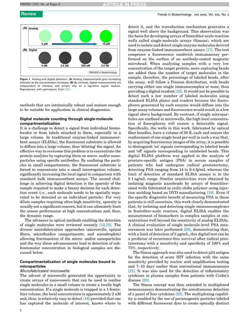
The principle of single-molecule counting can be bestillustrated with the case of fluorescence detection(Figure 1). Each fluorophore reporter emits many photonsbefore photobleaching, leading to fluorescent signals pro-portional to the fluorescent species present (Figure 1A).This type of measurement can be seen as an analog signalthat increases with the concentration of the species beingmeasured. By contrast, digital detection allows for a binarydecision for each detectable signal (ideally for each singlemolecule) enabling measurement in discrete counts ratherthan as an analog intensity (Figure 1B). In theory, anymethod that is sensitive enough to detect an event associ-ated with a single molecule would enable digital detectionby counting and offer significant advantages over ensemblemeasurements in terms of sensitivity. In fact, the ultimatelimit to analytical sensitivity is the reliable detection of asingle molecule.
Almost all methods for single-molecule detection andanalysis that we review here exploit an indirect detectionprinciple because they do not detect the biomolecule itself,but instead particles or labels that are associated with it.
Here, we also provide examples of single-molecule de-tection and their application in high-throughput biomark-er profiling, as well as discussing the challenges associatedwith these efforts. Given that nanoscale and complexsensing elements used in single-particle detection techni-ques may limit clinical applicability, we focus only on
Trends in Biotechnology xx (2015) 1–9 1
Analog
Digital(B)
(A)
0 pM 10 pM
0 aM 3.5 aM 350 aM 3.5 fM
TRENDS in Biotechnology
Figure 1. Analog and digital detection. (A) Analog measurements give increasing
intensity as the concentration increases. (B) By contrast, digital measurements are
independent of intensity and simply rely on a signal/no signal readout.
Reproduced, with permission, from [12].
Review Trends in Biotechnology xxx xxxx, Vol. xxx, No. x
TIBTEC-1253; No. of Pages 9
methods that are intrinsically robust and mature enoughto be suitable for application in clinical diagnostics.
Digital molecule counting through single-moleculecompartmentalizationIt is a challenge to detect a signal from individual biomo-lecules or from labels attached to them, especially in alarge volume. In traditional enzyme-linked immunosor-bent assays (ELISAs), the fluorescent substrate is allowedto diffuse into a large volume, thus ‘diluting’ the signal. Aneffective way to overcome this problem is to concentrate theprotein analytes by capturing them on micro- and/or nano-particles using specific antibodies. By confining the parti-cles in small compartments, the fluorescent substrate isforced to concentrate into a small interrogation volume,significantly increasing the local signal in comparison withstandard bulk immunosorbent assays. The second chal-lenge in achieving digital detection is the sparsity of thesample required to make a binary decision for each detec-tion event (i.e., each molecule needs to be spatially sepa-rated to be detected as an individual particle). For verydilute samples requiring ultra-high sensitivity, sparsity isusually not a significant concern, but it can ultimately limitthe sensor performance at high concentrations and, thus,the dynamic range.
The advances in optical methods enabling the detectionof single molecules were reviewed recently [12,15]. Thediverse microfabrication approaches (microwells, opticalfibers, microfluidics compartments, and nanodroplets)allowing fractionation of the micro- and/or nanoparticlesand the way these advancements lead to detection of sub-femtomolar concentration in biological samples are dis-cussed below.
Compartmentalization of single molecules bound tomicroparticlesMicrofabricated microwells
The advent of microwells generated the opportunity tocreate arrays of nanovessels that can be used to confinesingle molecules in a small volume to create a locally highconcentration. If a single molecule is trapped in a 1-femto-liter volume, the local concentration is approximately 2 nMand, thus, is relatively easy to detect [16] provided that onehas captured the molecule of interest, knows where to
2
detect it, and the transduction mechanism generates asignal well above the background. This observation wasthe basis for developing arrays of femtoliter-scale reactionwells called single-molecule arrays (Simoas), which areused to isolate and detect single enzyme molecules derivedfrom enzyme-linked immunosorbent assays [17]. The testcomprises a fluorescence sandwich immunoassay per-formed on the surface of an antibody-coated magneticmicrobead. When analyzing samples with a very lowconcentration of the target protein, more capturing beadsare added than the number of target molecules in thesample; therefore, the percentage of labeled beads, afterthe assay, will follow a Poisson distribution, with beadscarrying either one single immunocomplex or none, thusproviding a digital readout [18]. It would not be possible todetect such a low number of labeled molecules usingstandard ELISA plates and readers because the fluoro-phores generated by each enzyme would diffuse into thelarge assay volumes and fluorescence would result in a lowsignal above background. By contrast, if single micropar-ticles are confined in microwells, the high local concentra-tion of fluorophores will ensure a detectable signal.Specifically, the wells in this work, fabricated by opticalfiber bundles, have a volume of 50 fL each and ensure theconfinement of one single bead per well in such a way that,by acquiring fluorescence images of the array, it is possibleto distinguish ‘on’ signals corresponding to labeled beadsand ‘off’ signals corresponding to unlabeled beads. Thisdigital ELISA platform was applied to the analysis ofprostate-specific antigen (PSA) in serum samples ofpatients who had undergone radical prostatectomy,detecting PSA ranging from 14 to 9.4 fg/mL whereas thelimit of detection of standard ELISA assays is in the0.1 ng/mL range. Similar sensitivities were obtained byisolating magnetic nanobeads by arrays of femtoliter-sized wells fabricated in cyclic olefin polymer using injec-tion molding based on DVD manufacturing [19]. Even ifthe specific diagnostic benefit of measuring PSA in thosepatients is still uncertain, this work clearly demonstratedthat, by isolating and detecting single immunocomplexesin femtoliter-scale reactors, digital ELISA enables themeasurement of biomarkers in complex samples at con-centrations well beyond the sensitivity of analog ELISAs.A clinical evaluation of single molecule-level PSA mea-surement was later performed [20], demonstrating that,with a limit of detection of 3 pg/mL, this digital test can bea predictor of recurrence-free survival after radical pros-tatectomy with a sensitivity and specificity of 100% and75%, respectively.
The Simoa approach was also used to detect p24 antigenfor the detection of acute HIV infection with the samesensitivity provided by nucleic acid amplification testingand 7–10 days earlier than conventional immunoassays[21]. It was also used for the detection of inflammatorycytokines in plasma samples from patients with Crohn’sdisease [22].
The Simoa concept was then extended to multiplexedimmunoassays demonstrating the simultaneous detectionof a panel of four cytokines [23]. The multiplexing capabil-ity is enabled by the use of paramagnetic particles labeledwith different fluorescent dyes to create optically distinct

Review Trends in Biotechnology xxx xxxx, Vol. xxx, No. x
TIBTEC-1253; No. of Pages 9
subpopulations of beads decorated by antibodies tospecific targets. By using the xMAP technology developedby Luminex Corporation (http://www.luminexcorp.com/TechnologiesScience/xMAPTechnology/), such a mixtureof optically encoded microbeads is detected and countedby a flow cytometer. In this work, the beads were loadedinto the Simoa femtoliter nanovessels integrated into amicrofluidic device followed by fluorescence imaging todetermine the subpopulation identity and the presenceor absence of the single label associated with eachmicrobead; a Simoa disc for the automated microfluidicload, seal, and imaging of the arrays was used[24]. SimoaTM technology is now a proprietary platformof Quanterix (http://www.quanterix.com/technology/simoa–science).
The microsphere-based array was later integrated in afully automated platform for rapid analysis in microfluidicmicrochips containing the microwell array to accommodatesingle microparticles, wells, and channels for the automat-ed loading of the sample and all the required reagents[25]. The platform is able to analyze sample volumes assmall as 10 mL in just 70 min, providing results compara-ble with those obtained using a lab bench assay in 3.5 h.
The high potential of digital and multiplexed ELISAs indiagnostics was further demonstrated by the simultaneousdetection of six salivary biomarkers for asthma and cysticfibrosis diagnosis [26]. A large number of samples wasanalyzed and researchers found different patterns in bio-marker concentrations for healthy controls, patients withasthma, and those with cystic fibrosis. This demonstratedthe possibility of using this platform as a tool for respira-tory disease research and diagnostics.
Microfluidic compartments
An approach for the isolation of microfluidic reactions innanoliter volumes in parallel and without the use of com-plex instruments was developed called the ‘SlipChip’[27,28]. The chip does not require pumps or valves andcomprises two plates that can ‘slip’ relative to one other. Aprogram for fluid manipulation can be encoded into thechip as a pattern of ducts and wells preloaded withreagents. The wells remain isolated until they overlapwith a well or a duct onto the opposite plate; as the platesslip, wells come in or out of contact in a precise sequence,creating and breaking up transient fluidic pathways andexecuting the complete fluidic encoded program (sampleloading, washing, and detection steps). At the end of theassay, a fluorescence microscope is used to determine theconcentration of the analyte. Thus, the readout is analogand not digital, but the reduction of assay volumes to1 nanoliter enabled by SlipChip demonstrates high sensi-tivity even when handling ultrasmall volumes; withoutenhancing the sensitivity of the assay itself, the SlipChipprovides a detection limit of 13 pM for insulin [28]. Anotheradvantage of this bead-based immunoassay is that theSlipChip does not require assay-specific surface modifica-tions because the same chip can be used for differentimmunoassays by using magnetic particles decorated withdifferent antibodies. By contrast, the quality of the assayresults strongly depends on the efficiency of the separationof unbound residual antigens and detection antibodies in
the bulk solution from the sandwich complex on the surfaceof the solid phase.
Encapsulation of single molecules in nanodroplets by
emulsion
Droplet microfluidic devices have recently been developedthat enable encapsulation of single molecules or single cellswithin monodisperse nanodroplets for high-throughputand highly sensitive analysis [29]. In general, dropletsare generated by two immiscible fluids in microfluidicchannels and serve as individual reaction nanovessels,with volumes ranging from few femtoliters to few nanoli-ters. The generated droplets can be mixed, fused, split,diluted, sorted, and subjected to thermocycling accordingto the required assay protocol. Their content can be ana-lyzed by fluorescence, mass spectrometry, or electrochemi-cal methods, reaching high sensitivity due to the highconcentration of analytes in such a small environment[30]. In comparison with standard bulk immunosorbentassays, nanoconfinement in droplets reduces reagentvolumes by four orders of magnitude, while fast reagentmixing reduces the assay time from hours to minutes dueto fast mixing, which enhance binding kinetics.
Microemulsion technology enabled the development ofrapid, low-cost next-generation sequencing techniquesbased on integrated systems to isolate and amplify DNAfragments in vitro and to perform pyrophosphate-basedsequencing (pyrosequencing) in picoliter-sized wells [31].PCR has been successfully coupled to droplet microfluidics:the sample is diluted and partitioned into millions ofdroplets, each containing one or no target sequence ofinterest. At the end of the amplification reaction, thenumber of target DNA sequences can be determined bycounting the ‘on’ and ‘off’ droplets. This technique, nowcommonly referred to as droplet digital PCR (ddPCR) dueto its high sensitivity, has been extensively applied todiagnostics in cancer, where it enables researchers toaccurately quantitate mutated genes in the presence ofan excess of nonmutated genes. A detailed description ofddPCR and next-generation sequencing techniques havebeen recently provided elsewhere [32–35].
Droplets provide a closed controlled environment whenassaying cells and microorganisms, thus preventing thediffusion of the molecules secreted by the cells and provid-ing a tool for the analysis of biochemical events at thesingle cell level or for the detection of low-expressed, cellspecific surface biomarkers for cancer diagnostics. A sen-sitive and high-throughput analytical method was devel-oped to detect very small numbers of surface biomarkers onsingle cells encapsulated by microfluidic-generated dro-plets [36]. After optimization of the method, single celloccupancy of each droplet is assured because, at limitingdilutions, only a small percentage of the droplets containcells. An optical coding scheme enabling simultaneousanalysis of multiple analytes was introduced using differ-ent concentrations of a fluorescent dye. Using this plat-form, single human U937 cells were analyzed for theexpression of the low-abundance cell surface receptorsCD19 and CCR5, with increased signal discriminationand sensitivity compared with standard fluorescence-activated cell sorter (FACS) methods. In general, enhanced
3
Review Trends in Biotechnology xxx xxxx, Vol. xxx, No. x
TIBTEC-1253; No. of Pages 9
functionality could be achieved by encapsulating singlecells in nanodroplets with respect to FACS methods. Ina conventional flow cytometer, cells are interrogated byflowing them through a detector in a continuous stream ofbuffer. Given that there is no diffusional barrier betweenthe flowing cells, it is impossible to assay single cells basedon the secreted molecules, thus limiting the test to targetsthat are either confined within the cells or expressed on thecell surface.
A microfluidic cell sorting device based on nanodropletswas recently combined with rolling circle amplification(RCA) for signal amplification of low-abundance cell-sur-face biomarker and optical interrogation [37]. The RCAdetection strategy enabled the visual detection of 1–10surface protein molecules per cell with a 103-fold amplifi-cation of fluorescence intensity over direct labeling proto-cols. The single-molecule detection regime enabled by RCAcombined with the nanodroplet encapsulation allowed thedetection of a specific tumor marker, Epithelial cell adhe-sion molecule (EpCAM), directly on the surface of a tumorcell, paving the way for the application of this technique tocirculating tumor cells (CTCs).
A platform for detecting molecules secreted from singlecells was recently presented [38]. In this work, singleJurkat T cells were encapsulated in agarose droplets to-gether with functionalized capturing beads. During incu-bation, the cells produced cytokines that bound to thecapture beads confined within the same droplet. Aftergelling and breaking the emulsion, the agarose dropletswere stained with fluorescent antibodies that labeled thecytokines captured by the beads and signal intensity fromeach agarose droplet was quantified by using a flow cyt-ometer. The platform enabled the profiling of secretion at
Femtodropletsenclosing a bead
Fluorescent beads
50 µm
Brigh�ield (droplets)(A)
(B) 10
1
0.1
0.1
1
r2 = 0.995
10
Dete
rmin
edco
ncen
tra�
on (p
M)
Red fluorescenc
Prepared concentra�on (pM)
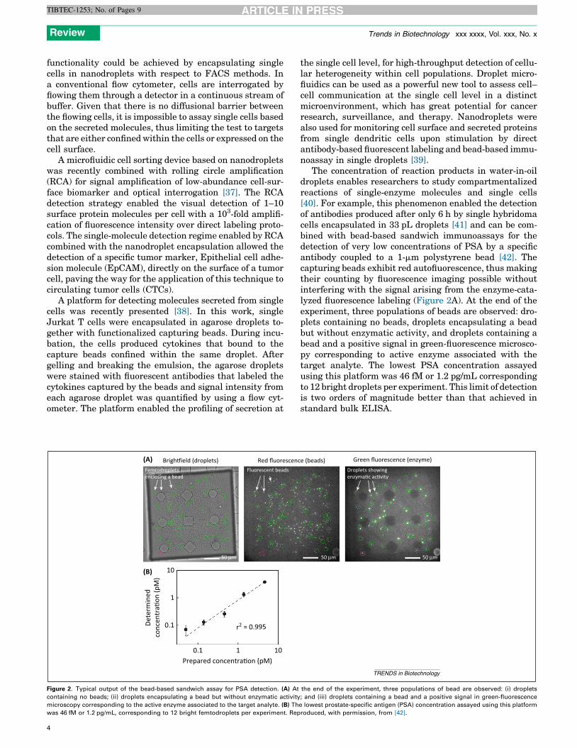
Figure 2. Typical output of the bead-based sandwich assay for PSA detection. (A) At
containing no beads; (ii) droplets encapsulating a bead but without enzymatic activity
microscopy corresponding to the active enzyme associated to the target analyte. (B) Th
was 46 fM or 1.2 pg/mL, corresponding to 12 bright femtodroplets per experiment. Rep
4
the single cell level, for high-throughput detection of cellu-lar heterogeneity within cell populations. Droplet micro-fluidics can be used as a powerful new tool to assess cell–cell communication at the single cell level in a distinctmicroenvironment, which has great potential for cancerresearch, surveillance, and therapy. Nanodroplets werealso used for monitoring cell surface and secreted proteinsfrom single dendritic cells upon stimulation by directantibody-based fluorescent labeling and bead-based immu-noassay in single droplets [39].
The concentration of reaction products in water-in-oildroplets enables researchers to study compartmentalizedreactions of single-enzyme molecules and single cells[40]. For example, this phenomenon enabled the detectionof antibodies produced after only 6 h by single hybridomacells encapsulated in 33 pL droplets [41] and can be com-bined with bead-based sandwich immunoassays for thedetection of very low concentrations of PSA by a specificantibody coupled to a 1-mm polystyrene bead [42]. Thecapturing beads exhibit red autofluorescence, thus makingtheir counting by fluorescence imaging possible withoutinterfering with the signal arising from the enzyme-cata-lyzed fluorescence labeling (Figure 2A). At the end of theexperiment, three populations of beads are observed: dro-plets containing no beads, droplets encapsulating a beadbut without enzymatic activity, and droplets containing abead and a positive signal in green-fluorescence microsco-py corresponding to active enzyme associated with thetarget analyte. The lowest PSA concentration assayedusing this platform was 46 fM or 1.2 pg/mL correspondingto 12 bright droplets per experiment. This limit of detectionis two orders of magnitude better than that achieved instandard bulk ELISA.
50 µm 50 µm
Droplets showingenzyma�c ac�vity
e (beads) Green fluorescence (enzyme)
TRENDS in Biotechnology
the end of the experiment, three populations of bead are observed: (i) droplets
; and (iii) droplets containing a bead and a positive signal in green-fluorescence
e lowest prostate-specific antigen (PSA) concentration assayed using this platform
roduced, with permission, from [42].

Review Trends in Biotechnology xxx xxxx, Vol. xxx, No. x
TIBTEC-1253; No. of Pages 9
The reduction in reagent and sample volumes requiredand faster assay times enabled by confinement in nano-droplets are also major improvements towards the devel-opment of high-sensitivity point-of-care devices [43], asrecently demonstrated in an immunosorbent platformfor the detection of antitetanus immunoglobulin (Ig)-Gin human serum [44]. This proof-of-principle platformprovided the sensitivity required to detect human antite-tanus antibodies in human serum six times faster than byusing standard ELISA.
Detection by counting nonfluorescent nanoparticlesOver the past few years, many techniques have beendeveloped for the visualization and counting of individualnanoparticles. These new tools have applications in lifesciences and medicine. For example, methods developedfor sensitive detection and characterization of biologicalnanoparticles such as viruses and exosomes will help in thediagnosis and understanding of many diseases. Motivationfor direct label-free detection of individual viruses is tosimplify assay complexity, because labels are not requiredfor signal transduction. This reduction in complexity couldlead to a rapid low-cost assay for point of care and resource-limited settings. Also, synthetic nanoparticles are beingdeveloped for vaccine and drug delivery, which could bothbenefit from nanoparticle characterization techniques.
Visualization of small nanoparticles by optical methodsis difficult because of their small scattering cross- section.To increase their visibility, researchers have utilized reso-nant techniques to enhance the interaction between theparticle and light. These techniques usually require dis-crete nanofabricated devices or complicated and low-throughput optical systems [45–48]. A widefield imagingtechnique called single particle interferometric reflectanceimaging sensor (SP-IRIS) was developed that utilizes LEDillumination and 50 � 0.8 numerical aperture (NA) objec-tive to visualize <100-nm low-index nanoparticles immo-bilized on a planar substrate. The sensor substratecomprises a silicon chip with a thin layer of thermallygrown oxide, which forms a common path interferometerto enhance the signal from bound nanoparticles [49].The two-layer substrate enhances the visualization ofnanoparticles through the interference of the reflectedlight from the substrate and the scattered light from the
Box 1. Digital detection of nanoparticles for biosensing on micro
A microarray is a solid-phase planar substrate that is functionalized
with a surface chemistry allowing adsorption or covalent binding of
a capturing agent (antibodies, DNA, RNA, aptamers, etc.). Usually,
the capturing agent is tiled or arrayed using a robotic spotter.
Different capturing agents can be arrayed on the surface with
known physical location and identity. Capturing agents are usually
arrayed in triplicates to increase confidence in detection (Figure IA).
For virus detection, the surface capture agent specifically binds the
virus to the surface, thus allowing the virus to be visualized and
counted (Figure IB). For detection of small targets, such as proteins
and nucleic acids, a secondary detection probe tagged with a
nanoparticle is used. Therefore, binding can be quantified by
digitally counting individual binding events to the probes on the
surface.
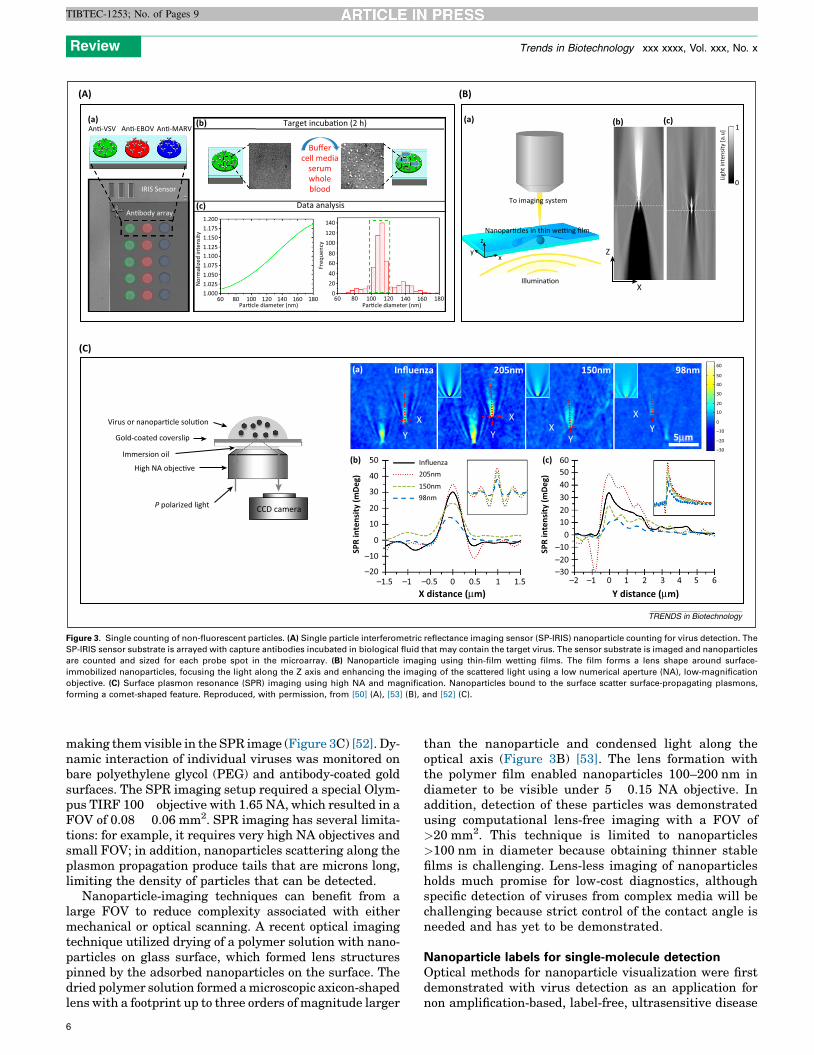
nanoparticle. Depending on the imaging sensor, SP-IRIScan visualize dielectric nanoparticles as small as 60 nm indiameter over a 0.2 mm2 field-of-view (FOV). The SP-IRISsensor can be functionalized with capture probes to form amicroarray (Box 1), which can be utilized for the specificdetection of biological nanoparticles such as viruses.Through mechanical scanning, a large number of spotscan be interrogated for multiplexed detection of a panel oftargets. Recently, virus detection was carried out usingwhole blood and serum at 5 � 103 plaque-forming units/mlfor Ebola and Marburg virus mimics [50]. The SP-IRISsensor was arrayed with antibodies against the surfaceglycoprotein of vesicular stomatitis virus, Ebola and Mar-burg viruses (Figure 3A). After incubation with sample, ifthe target (virus) was present, dots appeared captured onthe respective antibody spot. The intensity of these indi-vidual dots was then compared with a look-up table to forma size histogram of the nanoparticles captured by theantibody; this was then compared with the expected sizeof the target (virus). Digital counting of the virus particleson the SP-IRIS sensor reduced the assay to a binding andwashing step, eliminating the need for a detection probe orlabels. Therefore, direct digital detection simplifies theassay without compromising sensitivity.
Surface plasmon resonance (SPR) has been widely usedover the past decade for label-free monitoring of biomolec-ular binding on a functionalized gold surface. Recently,SPR has been coupled with high numerical aperture andmagnification imaging to enable visualization of individualnanoparticles. Typically, a prism is interfaced to the back-side of a gold-coated glass slide to excite plasmons on thegold surface at a critical angle. Binding or mass adsorptionon the gold surface changes the index of refraction at thesurface, which can be sensed by tracking a shift in thecritical coupling angle or intensity change. Huang et al.showed that, by using an immersion objective with a NAhigher than the index of refraction of the medium coveringthe gold surface (i.e., sensor), surface plasmons can beexcited and film measurements can be made by trackingthe change of the critical angle [51]. To acquire angle-resolved SPR images, the laser excitation source wasshifted with respect to the back aperture of the objective.Wang et al. recently showed that nanoparticles can scatterthe surface-propagating plasmons on the gold surface,
arrays
Single viruscoun�ng
Label-free(B)(A)
Protein/An�gen Nucleic acid
Microarray surface
Single molecule coun�ngSingle molecule microarray
TRENDS in Biotechnology
Figure I. Schematic of a typical microarray for digital detection.
5
(a) (b)
Z
XIllumina�on
Nanopar�cles in thin we�ng film
To imaging system
xy
z
(c)1
0Ligh
t int
ensit
y [a
.u]
(a)
(A) (B)
(C)
An�-VSV (b)
(c)
An�-EBOV An�-MARV
IRIS Sensor
An�body array1.2001.1751.1501.1251.1001.0751.0501.0251.000
140
120100
8060
4020
060 80 100 120 140 160 180
Par�cle diameter (nm)
Data analysis
Target incuba�on (2 h)
Buffercell media
serumwholeblood
60 80 100 120 140 160 180Par�cle diameter (nm)
Nor
mal
ized
inte
nsity
Freq
uenc
y
Virus or nanopar�cle solu�on
Gold-coated coverslip
Immersion oil
High NA objec�ve
P polarized light CCD camera
(a)
(b) (c)505060
–30
–20
–10
0
10
20
30
40
50
60
40302010
0
–20–30
–10
40
30
20
10
0
–10
–20–1.5 –1 –0.5 0.5 1 1.5 –2 –1 0
SPR
inte
nsit
y (m
Deg
)
SPR
inte
nsit
y (m
Deg
)
1 2 3 4 5 6X distance (µm) Y distance ( µm)
0
Influenza205nm150nm98nm
YX
Y
X
YX Y
X
Influenza 205nm 150nm 98nm
5µm
TRENDS in Biotechnology
Figure 3. Single counting of non-fluorescent particles. (A) Single particle interferometric reflectance imaging sensor (SP-IRIS) nanoparticle counting for virus detection. The
SP-IRIS sensor substrate is arrayed with capture antibodies incubated in biological fluid that may contain the target virus. The sensor substrate is imaged and nanoparticles
are counted and sized for each probe spot in the microarray. (B) Nanoparticle imaging using thin-film wetting films. The film forms a lens shape around surface-
immobilized nanoparticles, focusing the light along the Z axis and enhancing the imaging of the scattered light using a low numerical aperture (NA), low-magnification
objective. (C) Surface plasmon resonance (SPR) imaging using high NA and magnification. Nanoparticles bound to the surface scatter surface-propagating plasmons,
forming a comet-shaped feature. Reproduced, with permission, from [50] (A), [53] (B), and [52] (C).
Review Trends in Biotechnology xxx xxxx, Vol. xxx, No. x
TIBTEC-1253; No. of Pages 9
making them visible in the SPR image (Figure 3C) [52]. Dy-namic interaction of individual viruses was monitored onbare polyethylene glycol (PEG) and antibody-coated goldsurfaces. The SPR imaging setup required a special Olym-pus TIRF 100� objective with 1.65 NA, which resulted in aFOV of 0.08 � 0.06 mm2. SPR imaging has several limita-tions: for example, it requires very high NA objectives andsmall FOV; in addition, nanoparticles scattering along theplasmon propagation produce tails that are microns long,limiting the density of particles that can be detected.
Nanoparticle-imaging techniques can benefit from alarge FOV to reduce complexity associated with eithermechanical or optical scanning. A recent optical imagingtechnique utilized drying of a polymer solution with nano-particles on glass surface, which formed lens structurespinned by the adsorbed nanoparticles on the surface. Thedried polymer solution formed a microscopic axicon-shapedlens with a footprint up to three orders of magnitude larger
6
than the nanoparticle and condensed light along theoptical axis (Figure 3B) [53]. The lens formation withthe polymer film enabled nanoparticles 100–200 nm indiameter to be visible under 5� 0.15 NA objective. Inaddition, detection of these particles was demonstratedusing computational lens-free imaging with a FOV of>20 mm2. This technique is limited to nanoparticles>100 nm in diameter because obtaining thinner stablefilms is challenging. Lens-less imaging of nanoparticlesholds much promise for low-cost diagnostics, althoughspecific detection of viruses from complex media will bechallenging because strict control of the contact angle isneeded and has yet to be demonstrated.
Nanoparticle labels for single-molecule detectionOptical methods for nanoparticle visualization were firstdemonstrated with virus detection as an application fornon amplification-based, label-free, ultrasensitive disease
Review Trends in Biotechnology xxx xxxx, Vol. xxx, No. x
TIBTEC-1253; No. of Pages 9
diagnostics. Recently, the SP-IRIS platform was adaptedfor detection of single molecules in a sandwich assayformat (Box 1). The sensor surface can be coated or arrayedwith specific detection probes (i.e., antibodies, aptamers,and oligonucleotides) to capture one or more targets fromthe solution; then, a secondary detection probe with ananoparticle label is introduced to visualize single mole-cule-binding events on the surface. Traditionally, micro-arrays use fluorescent labels for analog-based signaltransduction, where the signal strength infers the amountof binding to the probe on the surface. However, SP-IRISutilizes nanoparticle labels that can be individuallydetected and counted, transforming microarrays readoutsinto digital detection methods. Gold nanoparticle labelsare usually used because their optical signal is higher,allowing smaller particles to be utilized; this reduces sterichindrance and diffusion effects.
Recently, digital detection of allergy-specific IgE fromserum and unprocessed human whole blood was demon-strated in an eight-plex allergy panel. Detection was donein a sandwich assay format (Box 1) using detection anti-bodies labeled with 40-nm gold nanoparticles. Standardcurves showed that the technique has sub-femtomolardetection sensitivity and a five orders of magnitude dy-namic range [54].
The use of gold nanoparticle tags for digital detection ofmolecular binding events has also been demonstratedusing 1D photonic crystals [55]. Recently, SPR imagingwas used for real-time measurement of individual DNAhybridization [56]. Using SPR imaging, the authorsshowed the detection of synthetic DNA coupled to 40-nmgold nanoparticles to the surface at concentrations of 5 pMto 100 fM in 10 mins. By increasing the illumination wave-length to 814 nm, they were able to use more commonlyavailable objectives with a NA of 1.4 instead of the Olym-pus TIRF objective with a NA of 1.65, which requires ahigh-refractive index glass substrate. However, the in-crease in wavelength results in longer plasmon propaga-tion and nanoparticle responses with 50-mm tails, whichlimit the detection density of the nanoparticles.
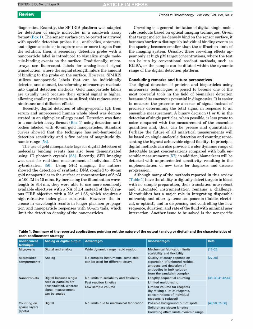
Table 1. Summary of the reported applications pointing out the naeach confinement strategy
Confinement
technique
Analog or digital output Advantages
Microwells Digital and analog Wide dynamic range, rapid re
Microfluidic
compartments
Analog No complex instruments, sam
can be used for different assa
Nanodroplets Digital because single
cells or particles are
encapsulated, whereas
signal measurement
can be analog
No limits to scalability and fle
Fast reaction kinetics
Low sample volume
Counting on
sparse layers
(spots)
Digital No limits due to mechanical
Crowding is a general limitation of digital single-mole-cule readouts based on optical imaging techniques. Giventhat target molecules densely bind on the sensor surface, itbecomes harder to distinguish individual binding events asthe spacing becomes smaller than the diffraction limit ofthe imaging system. Usually, these crowding effects ap-pear only at high pM target concentrations, where the testcan be run by conventional readout methods, such asELISA, or the sample can be diluted within the dynamicrange of the digital detection platform.
Concluding remarks and future perspectivesThe digital detection of proteins and bioparticles usingmicroarray technologies is poised to become one of themost powerful tools in the field of biomarker detectionbecause of its enormous potential in diagnostics. It is easierto measure the presence or absence of signal instead ofprecisely determining the total signal in response to anensemble measurement. A binary decision (1 or 0) in thedetection of single particles, when possible, is less prone tonoise compared with the measurement of the ensemblequantities and, thus, can be precise and quantitative.Perhaps the future of all analytical measurements willbe based on single-molecule detection and counting repre-senting the highest achievable signal fidelity. In principle,digital methods can also provide a wider dynamic range ofdetectable target concentrations compared with bulk en-semble measurements [57]; in addition, biomarkers will bedetected with unprecedented sensitivity, resulting in theimplementation of new tests for diagnosis and diseaseprogression.
Although many of the methods reported in this review(Table 1) have the ability to digitally detect targets in bloodwith no sample preparation, their translation into robustand automated instrumentation remains a challenge.Microfluidics has a major role in integrating disposablemicrochip and other systems components (fluidic, electri-cal, or optical), and in dispensing and controlling the flowsequence, duration, and rate of the fluid with minimal userinteraction. Another issue to be solved is the nonspecific
ture of the output (analog or digital) and the characteristics of
Disadvantages Refs
adout Mechanical fabrication limits
scalability and flexibility
[17–26]
e chip
ys
Quality of assay depends on
separation of unbound residual
antigens and detection of
antibodies in bulk solution
from the sandwich complex
[27,28]
xibility Lengthy sequential counting [36–39,41,42,44]
Limited multiplexing
Limited volume for reagents
(by mixing a lot of reagents,
concentrations of individual
reagents is reduced)
fabrication Possible background out of spots [49,50,52–56]
Solid-phase slower kinetics
Crowding effect limits dynamic range
7
Review Trends in Biotechnology xxx xxxx, Vol. xxx, No. x
TIBTEC-1253; No. of Pages 9
binding of nontarget proteins and enzyme labels to thebead surface, which can lead to false positive signals.Affinity binding reagents with high specificity and betterfluorogenic substrates are still major needs. Improvementsin generating large sets of antibody reagents, recombinantproteins from a variety of host cells, and other types ofcapture molecule will further accelerate advances in thisfield.
AcknowledgmentsThis work was partially supported by Regione Lombardia andFondazione Cariplo (POR-FESR, project MINER ID 46875467) and theItalian Ministry of University and Research through FIRB ‘Futuro inRicerca’ project RBFR12OO1G – NEMATIC. M.S.U. and G.G.D. acknowl-edge partial support from National Science Foundation AcceleratingInnovation Research Award 1127833 and the National Institutes ofHealth Award R01 AI1096159.
References1 Giljohann, D.A. and Mirkin, C.A. (2009) Drivers of biodiagnostic
development. Nature 462, 461–4642 Srinivas, P.R. et al. (2011) Trends in biomarker research for cancer
detection. Lancet Oncol. 2, 698–7043 Galasko, D. (2005) Biomarkers for Alzheimer’s disease: clinical needs
and application. J. Alzheimers Dis. 8, 339–3464 de Jong, D. et al. (2007) Current state and future directions of
neurochemical biomarkers for Alzheimer’s disease. Clin. Chem. Lab.Med. 45, 1421–1434
5 Barletta, J.M. et al. (2004) Lowering the detection limits of HIV-1 viralload using real-time immuno-PCR for HIV-1 p24 antigen. Am. J. Clin.Pathol. 122, 20–27
6 Adler, M. et al. (2008) Sensitivity by combination: immuno-PCR andrelated technologies. Analyst 133, 702–718
7 Nam, J.M. et al. (2003) Nanoparticle-based bio-bar codes for theultrasensitive detection of proteins. Science 301, 1884–1886
8 Li, R. et al. (2011) Label-free amperometric immunosensor for thedetection of human serum chorionic gonadotropin based onnanoporous gold and graphene. Anal. Biochem. 414, 196–201
9 Wu, S. et al. (2012) Selective electrochemical detection of cysteine incomplex serum by graphene nanoribbon. Biosens. Bioelectron. 32,293–296
10 Craighead, H. et al. (2000) Nanoelectromechanical systems. Science290, 1532–1535
11 Lee, J. et al. (2011) Sensitive and simultaneous detection of cardiacmarkers in human serum using surface acoustic wave immunosensor.Anal. Chem. 83, 8629–8635
12 Walt, D. (2013) Optical methods for single molecule detection andanalysis. Anal. Chem. 85, 1258–1263
13 Stoeva, S. et al. (2006) Multiplexed detection of protein cancermarkers with biobarcoded nanoparticle probes. J. Am. Chem. Soc.128, 8378–8379
14 Chen, J. et al. (2009) Ultrasensitive protein detection in blood serumusing gold nanoparticle probes by single molecule spectroscopy. J.Biomed. Opt. 14, 040501
15 Wang, W. et al. (2014) Detection, counting and imaging of singlenanoparticles. Anal. Chem. 86, 2–14
16 Walt, D.R. (2014) Protein measurements in microwells. Lab. Chip 14,3195–3200
17 Rissin, D.M. et al. (2010) Single-molecule enzyme-linkedimmunosorbent assay detects serum proteins at subfemtomolarconcentrations. Nat. Biotechnol. 28, 595–599
18 Rissin, D.M. et al. (2006) Digital concentration readout of singleenzyme molecules using femtoliter arrays and Poisson statistics.Nano Lett. 6, 520–523
19 Kan, C.W. et al. (2012) Isolation and detection of single molecules onparamagnetic beads using sequential fluid flows in microfabricatedpolymer array assemblies. Lab. Chip 12, 977–985
20 Lepor, H. et al. (2012) Clinical evaluation of a novel method for themeasurement of prostate-specific antigen, AccuPSATM, as a predictorof 5-year biochemical recurrence-free survival after radicalprostatectomy: results of a pilot study. BJU Int. 109, 1770–1775
8
21 Chang, L. et al. (2013) Simple diffusion-constrained immunoassay forp24 protein with the sensitivity of nucleic acid amplification fordetecting acute HIV infection. J. Virol. Methods 188, 153–160
22 Song, L.N. et al. (2011) Single molecule measurements of tumornecrosis factor alpha and interleukin-6 in the plasma of patientswith Crohn’s disease. J. Immunol. Methods 372, 177–186
23 Rissin, D.M. et al. (2013) Multiplexed single molecule immunoassays.Lab. Chip 13, 2902–2911
24 Kan, C.W. et al. (2012) Isolation and detection of single molecules onparamagnetic beads using sequential fluid flows in microfabricatedpolymer array assemblies. Lab. Chip 12, 977–995
25 Nie, S. et al. (2014) An automated integrated platform for rapid andsensitive multiplexed protein profiling using human saliva samples.Lab. Chip 14, 1087–1098
26 Nie, S. et al. (2013) Multiplexed salivary protein profiling for patientswith respiratory diseases using fiber-optic bundles and fluorescentantibody-based microarrays. Anal. Chem. 85, 9272–9280
27 Du, W. et al. (2009) SlipChip. Lab. Chip 9, 2286–229228 Liu, W. et al. (2010) SlipChip for immunoassays in nanoliter volumes.
Anal. Chem. 82, 3276–328229 Kang, D.K. et al. (2014) Droplet microfluidics for single-molecule and
single-cell analysis in cancer research and therapy. Trends Anal.Chem. 58, 145–153
30 Guo, M.T. et al. (2012) Droplet microfluidics for high-throughputbiological assays. Lab. Chip 12, 2146–2155
31 Margulies, M. et al. (2005) Genome sequencing in microfabricated high-density picolitre reactors. Nature 437, 376–380
32 Baker, M. (2012) Digital PCR hits its stride. Nat. Methods 9,541–544
33 Roedinger, S. et al. (2014) Nucleic acid detection based on the use ofmicrobeads: a review. Microchim. Acta 181, 1151–1168
34 Buemans, H.P.J. et al. (2014) Next generation sequencing technology:advances and applications. Biochim. Biophys. Acta Mol. Basis Dis. 10,1932–1941
35 Lohman, K. et al. (2014) Next generation sequencing and the future ofgenetic diagnosis. Neurotherapeutics 11, 699–707
36 Joensson, H.N. et al. (2009) Detection and analysis of low-abundancecell-surface biomarkers using enzymatic amplification in microfluidicdroplets. Angew. Chem. Int. Ed. Engl. 48, 2518–2521
37 Konry, T. et al. (2001) Ultrasensitive detection of low-abundancesurface-marker protein using isothermal rolling circle amplificationin a microfluidic nanoliter platform. Small 3, 395–400
38 Chokkalingam, V. et al. (2013) Probing cellular heterogeneity incytokine-secreting immune cells using droplet-based microfluidics.Lab. Chip 13, 4740–4744
39 Konry, T. et al. (2013) Live single cell functional phenotyping in dropletnano-liter reactors. Sci. Rep. 3, 3179–3184
40 Theberge, A.B. et al. (2010) Microdroplets in microfluidics: an evolvingplatform for discoveries in chemistry and biology. Angew. Chem. Int.Ed. Engl. 49, 5846–5868
41 Koster, S. et al. (2008) Drop-based microfluidic devices forencapsulation of single cells. Lab. Chip 8, 1110–1115
42 Shim, J. et al. (2013) Ultrarapid generation of femtoliter microfluidicdroplets for single molecule counting immunoassay. ACS Nano 7,5955–5964
43 Golberg, A. et al. (2013) Picoliter droplet microfluidic immunosorbentplatform for point-of-care diagnostics of tetanus. Microchim. Acta 180,855–860
44 Segerink, L.I. and Eijkel, J.C.T. (2014) Nanofluidics in point of careapplications. Lab. Chip 14, 3201–3205
45 Vollmer, F. et al. (2008) Single virus detection from the reactive shiftof a whispering-gallery mode. Proc. Natl. Acad. Sci. U.S.A. 105,20701–20704
46 Vollmer, F. et al. (2008) Whispering-gallery-mode biosensing:label-free detection down to single molecules. Nat. Methods 5,591–596
47 Lu, T. et al. (2011) High sensitivity nanoparticle detection using opticalmicrocavities. Proc. Natl. Acad. Sci. U.S.A. 108, 5976–5979
48 Yurt, A. et al. (2012) Single nanoparticle detectors for biologicalapplications. Nanoscale 4, 715–726
49 Daaboul, G.G. et al. (2010) High-throughput detection and sizing ofindividual low-index nanoparticles and viruses for pathogenidentification. Nano Lett. 10, 4727–4731

Review Trends in Biotechnology xxx xxxx, Vol. xxx, No. x
TIBTEC-1253; No. of Pages 9
50 Daaboul, G.G. et al. (2014) Digital sensing and sizing of Ebola andMarburg virus mimics in complex media using single nanoparticleimaging. ACS Nano 6, 6047–6055
51 Huang, B. et al. (2007) Surface plasmon resonance imaging using ahigh numerical aperture microscope objective. Anal. Chem. 79,2979–2983
52 Tao, N.J. et al. (2010) Label-free imaging, detection, and massmeasurement of single viruses by surface plasmon resonance. Proc.Natl. Acad. Sci. U.S.A. 107, 16028–16032
53 Hennequin, Y. et al. (2013) Optical detection and sizing of singlenanoparticles using continuous wetting films. ACS Nano 7, 7601–7609
54 Monroe, M.R. et al. (2013) Single nanoparticle detection formultiplexed protein diagnostics with attomolar sensitivity in serumand unprocessed whole blood. Anal. Chem. 7, 3698–3706
55 Zhuo, Y. (2014) Single nanoparticle detection using photonic crystalenhanced microscopy. Analyst 139, 1007–1015
56 Halpern, A.R. et al. (2014) Single-nanoparticle near-infrared surfaceplasmon resonance microscopy for real-time measurements of DNAhybridization adsorption. ACS Nano 8, 1022–1030
57 Kelly, S.O. et al. (2014) Advancing the speed, sensitivity and accuracyof biomolecular detection using multi-length-scale engineering. Nat.Nanotechnol. 9, 969–980
9