Limbal Stem Cell Deficiency in Chronic and Delayed-onset Mustard Gas Keratopathy
Upload
independentCategory
view
0download
0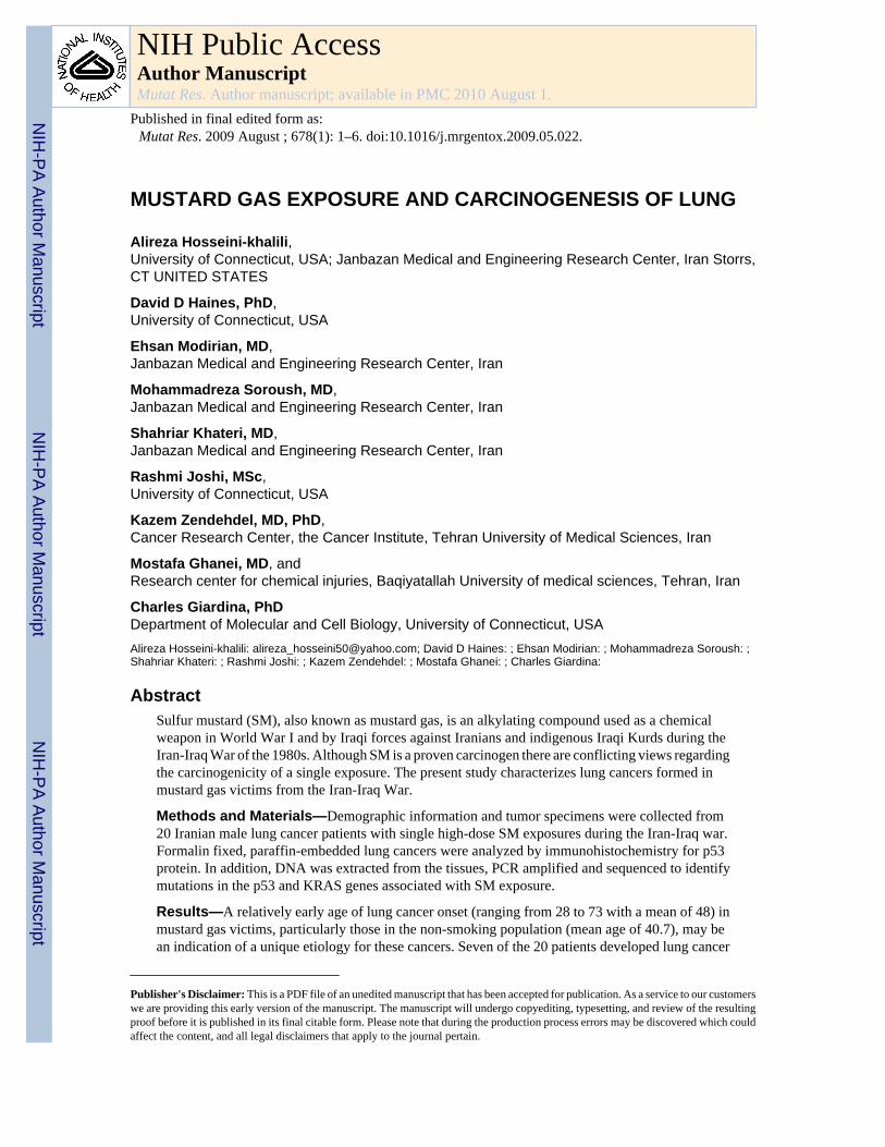
MUSTARD GAS EXPOSURE AND CARCINOGENESIS OF LUNG
Alireza Hosseini-khalili,University of Connecticut, USA; Janbazan Medical and Engineering Research Center, Iran Storrs,CT UNITED STATES
David D Haines, PhD,University of Connecticut, USA
Ehsan Modirian, MD,Janbazan Medical and Engineering Research Center, Iran
Mohammadreza Soroush, MD,Janbazan Medical and Engineering Research Center, Iran
Shahriar Khateri, MD,Janbazan Medical and Engineering Research Center, Iran
Rashmi Joshi, MSc,University of Connecticut, USA
Kazem Zendehdel, MD, PhD,Cancer Research Center, the Cancer Institute, Tehran University of Medical Sciences, Iran
Mostafa Ghanei, MD, andResearch center for chemical injuries, Baqiyatallah University of medical sciences, Tehran, Iran
Charles Giardina, PhDDepartment of Molecular and Cell Biology, University of Connecticut, USAAlireza Hosseini-khalili: [email protected]; David D Haines: ; Ehsan Modirian: ; Mohammadreza Soroush: ;Shahriar Khateri: ; Rashmi Joshi: ; Kazem Zendehdel: ; Mostafa Ghanei: ; Charles Giardina:
AbstractSulfur mustard (SM), also known as mustard gas, is an alkylating compound used as a chemicalweapon in World War I and by Iraqi forces against Iranians and indigenous Iraqi Kurds during theIran-Iraq War of the 1980s. Although SM is a proven carcinogen there are conflicting views regardingthe carcinogenicity of a single exposure. The present study characterizes lung cancers formed inmustard gas victims from the Iran-Iraq War.
Methods and Materials—Demographic information and tumor specimens were collected from20 Iranian male lung cancer patients with single high-dose SM exposures during the Iran-Iraq war.Formalin fixed, paraffin-embedded lung cancers were analyzed by immunohistochemistry for p53protein. In addition, DNA was extracted from the tissues, PCR amplified and sequenced to identifymutations in the p53 and KRAS genes associated with SM exposure.
Results—A relatively early age of lung cancer onset (ranging from 28 to 73 with a mean of 48) inmustard gas victims, particularly those in the non-smoking population (mean age of 40.7), may bean indication of a unique etiology for these cancers. Seven of the 20 patients developed lung cancer
Publisher's Disclaimer: This is a PDF file of an unedited manuscript that has been accepted for publication. As a service to our customerswe are providing this early version of the manuscript. The manuscript will undergo copyediting, typesetting, and review of the resultingproof before it is published in its final citable form. Please note that during the production process errors may be discovered which couldaffect the content, and all legal disclaimers that apply to the journal pertain.
NIH Public AccessAuthor ManuscriptMutat Res. Author manuscript; available in PMC 2010 August 1.
Published in final edited form as:Mutat Res. 2009 August ; 678(1): 1–6. doi:10.1016/j.mrgentox.2009.05.022.
NIH
-PA Author Manuscript
NIH
-PA Author Manuscript
NIH
-PA Author Manuscript
before the age of 40. Five of 16 cancers from which DNA sequence data was obtainable providedinformation on eight p53 mutations (within exons 5–8). These mutations were predominately G toA transitions; a mutation consistent with the DNA lesion caused by SM. Two of the lung cancershad multiple p53 point mutations, similar to results obtained from factory workers chronicallyexposed to mustard agent. No mutations were detected in the KRAS gene.
Discussion—The distinguishing characteristics of lung carcinogenesis in these mustard gasvictims suggest that a single exposure may increase the risk of lung cancer development in someindividuals.
KeywordsMustard Gas; Lung; Cancer; Iran; P53
INTRODUCTIONThe alkylating compound sulfur mustard (SM) has been employed as a chemical warfare agentsince its first use in World War I (1). The most recent application of this weapon was duringthe Iran-Iraq conflict by the Iraqi Baathist government. During this war, which lasted from1980–88, Iraqi dictator Saddam Hussein made frequent use of SM as a battlefield forcemultiplier and also extensively targeted unprotected civilians in Iran and within the Kurdishregions of Iraq (2). Over 50,000 survivors of SM attacks remain alive in Iran and as a groupsuffer from high rates of chronic illnesses, particularly inflammatory conditions of therespiratory system, skin and eye (3–5), which are the major target organs of SM. Latentrespiratory syndromes include asthma, chronic bronchitis, bronchiectasis, bronchiolitis,bronchial stenosis, and sinusitis (3,4). The low cost and ease with which SM may bemanufactured, transported and deployed, along with negligible odor and ability to causepermanent injuries after a few seconds exposure have made it historically the most frequentlyused chemical weapon during military operations. A particularly insidious feature of SM hasalso increased its potential as a combat force multiplier: Tissue damage due to the agenttypically does not appear for 16 hours or more after exposure. Hence civilians and Iraniantroops with often heavy SM exposure, would assume that absence of symptoms after light, orno decontamination meant that the danger had passed. Such victims typically developedsymptoms within a day, often full-body blistering, loss of most skin, deep tissue injury andorgan failure (2). These characteristics also make future battlefield and terrorist use of thischemical a real threat. Nevertheless, there are few comprehensive studies on the long-termhealth consequences of SM exposure, a gap in medical knowledge with potential forsignificantly adverse impact on public health management of future mass casualties.
The enormous pool of Iranian SM victims, with carefully documented exposure and medicalhistories, therefore offer an unparalleled resource and opportunity for developing a mechanisticunderstanding of SM-associated chronic health problems. In the present report we examineselected clinical, pathological and genetic features of Iranian SM victims suffering from lungcancer as an element of a broader investigation to assess the potential carcinogenic riskassociated with an acute SM exposure.
The long-term effects of SM exposure may develop as a result of two major processes. Onepathway leading to chronic illness in SM victims occurs due to a failure of hostimmunoregulatory mechanisms to resolve inflammation triggered by the episulfonium ion.Here, high levels of inflammatory cytokines inhibit apoptosis of polymorphonuclearleukocytes, particularly neutrophils, thus prolonging their active state and amplifying damagedone by these cells (6,7). A second major pathway leading from SM exposure to disease canoccur when the compound causes mutations in tumor suppressor and oncogenes, such as p53
Hosseini-khalili et al. Page 2
Mutat Res. Author manuscript; available in PMC 2010 August 1.
NIH
-PA Author Manuscript
NIH
-PA Author Manuscript
NIH
-PA Author Manuscript
or KRAS. SM is a known alkylating agent, and under conditions of chronic exposure, is arecognized carcinogen (8). The mechanism of SM-induced carcinogensis begins withcyclization of SM in the aqueous environment of a victim, to a highly reactive episulfoniumion which may alkylate DNA. If these are not repaired, these lesions can lead to nucleotidesubstitutions (9), most commonly the G to A transition (10). This mutation may inactivatetumor suppressor genes such as p53, and greatly increase susceptibility to lung cancer, as wasobserved in Japanese mustard gas factory workers chronically exposed to SM (11–13).Although chronic SM is a known carcinogen (14–17), there are conflicting views regardingthe carcinogenicity of a single exposure (18–21). From a public health perspective, acuteexposure is a much more relevant exposure scenario in either past and potential future militaryconflicts or terrorist attacks.
Studies of cancer incidence in survivor populations with single, high-dose SM exposures havethus far failed to demonstrate strong correlations between exposure and disease occurrence.Kang and Bulman followed the outcome of US veterans exposed to SM in World War II(21). Although analysis of these patients showed no significant increase in lung cancer risk,exposure incidences were relatively low and smoking habits were not considered. There is nostudy in the medical literature addressing the affect of a single, high-dose SM exposure on thelong-term risk of lung cancer. Now, after two decades, Iranians with well-documentedexposures to mustard agent in the 1980s can be studied to help clarify the lung cancer riskassociated with a battlefield exposure to SM.
Multiple lines of evidence will be required before a causative link between acute mustard agentexposure and lung cancer development can be established. Ongoing epidemiological studiesare being pursued in Iran to determine if mustard-exposed individuals are at greater risk forlung cancer development, but these efforts will require an extended period of time before asufficient number of lung cancer cases develop, and may take years to complete. For the presentstudy, we have collected data from Iranian SM victims with unambiguous, documentedexposure histories, who eventually developed lung cancer. Records from the Iran-Iraq war alsoprovide information on the precise exposure timeframes during the eight year conflict. Here,specific personal attributes of victims and molecular/genetic features of their cancers wereexamined to evaluate carcinogenic effect of SM exposure on lung tissue. These studies includea mutational analysis of two tumor suppressor genes: p53 and KRAS, both of which are knownindicators of gene-environment interactions impacting cancer risk. In summary, our datasupport the view that a single exposure to mustard agent may trigger cancer development insome individuals.
METHOD AND MATERIALSPatient Population
The present study was conducted using medical record data provided by Janbazan Medical andEngineering Research Center (JMERC) and archived paraffin-embedded lung tumor samplesfrom pathology departments of major Iranian medical centers treating persons exposed tochemical weapons during the Iran-Iraq war. Here, data and samples were drawn from a subjectpopulation of 20 Iranian males with single, battlefield SM exposures resulting in acute andchronic symptoms including skin and respiratory injuries. All exposures occurred in the years1982–1988 and subjects were subsequently diagnosed between the ages of 28 and 73 with threemajor forms of lung carcinoma (Table 1). Subjects were randomly selected from amongdeceased individuals for whom complete records and samples existed. Time intervals betweenexposure to the weapon and onset of disease ranged from 5–20 years. Subjects included 5current or former smokers, 9 non-smokers and 6 with indeterminate histories of tobacco use(Table 1). We were unable to assemble a significant number of matched tumor samples frompatients without a history of SM exposure and smoking that were preserved in a manner that
Hosseini-khalili et al. Page 3
Mutat Res. Author manuscript; available in PMC 2010 August 1.
NIH
-PA Author Manuscript
NIH
-PA Author Manuscript
NIH
-PA Author Manuscript

allowed DNA extraction. We therefore used International Agency for Research on Cancer(IARC) database as our baseline for p53 mutational frequencies in lung tumors (8).
Mustard exposure, inclusion and exclusion criteriaMustard exposure in this study is defined as any contact with SM in liquid or vapor form,resulting in transient or permanent disability. This definition is based on standards developedin a comprehensive national survey accomplished during the timeframe 1997–2000 thatestablished a uniform convention for designation of Iranian citizens with war-related chemicalinjuries. Under this convention, mustard exposure is defined as any contact with SM in liquidor vapor form, resulting in transient or permanent disability. Here the minimum threshold forSM-induced disability is defined according to known primary effects of the agent on its majortarget organs: eyes, skin and lungs. Threshold exposure definitions for each organ are asfollows: Eye: edema and visible inflammation of ocular membranes; Skin: rednessaccompanied by obvious blistering; and Lung: edema accompanied by inflammation and eithera productive cough, or hemoptysis in the form of bloody streaks or expectoration of clots. Thesecriteria take into account the highly variable length of time and concentration ranges of SMthat personnel are typically subjected to under battlefield conditions and make no attempt tocorrelate SM dosage with symptoms. Some estimation of SM dosage sustained by subjects ofthis study may nevertheless be estimated based on reference ranges of the agent known toproduce particular outcomes. Acute exposure guideline levels (AEGLs) for SM have beendeveloped by the U.S. National Advisory Committee (NAC). Exposure to SM at 0.60milligrams/cubic meter (mg/m3) of air for 10 minutes; or 0.013 mg/m3 for 8 hours constitutethe threshold level at which edema of the eyes, sensitivity to light, and eye irritation occur(22). These ocular symptoms also define the threshold level for SM exposure established byJanbazan organization. Therefore participants in this study were exposed to at least the levelof SM identified by the NAC as needed to produce critical ocular symptoms. Patientsparticipating in this study were selected on the basis of documented exposure to SM based onofficial certification from the Iranian Veteran’s Affairs organization (Janbazan). Thisdocumentation included records of medical treatment showing the type and extent of mustard-associated injury and/or disability. Patients with histories of serious major disease other thanlung cancer were excluded from this study. This investigation was conducted under theapproval of Janbazan organization’s ethics committee.
Tissue preparation and DNA ExtractionLung cancer tissue was obtained from biopsies or surgical sample, fixed with formalin andembedded in paraffin. Neoplastic lesions from representative areas of 4–5 unstained slidescontaining 10 μm thick tissue slices were scraped into a microcentrifuge tube. After paraffinremoval with xylene, tissues were rehydrated and the DNA was isolated using thePicoPure™ DNA extraction Kit (Arcturus Engineering Inc., Mountain View, CA), accordingto the manufacturer’s instructions.
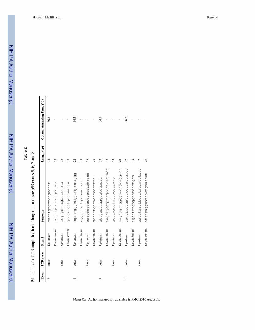
DNA Amplification and SequencingThe extracted DNA was amplified by the polymerase chain reaction (PCR), using a nestedprimer approach. The primer sets for P53 exons 5, 6, 7 and 8 are shown in Table 2. Initialamplification reactions yielded the target amplimer with a number of off-target products. Thespecific product was then selectively amplified using nested p53 primers positioned a few basesdownstream of the initial primers. The cycling conditions were as following: denaturation at95°C for 2 minutes, followed by 35 cycles with denaturation at 95°C for 30 seconds, annealingat 56.2°C (exons 5 and 8) or 64.4°C (exons 6 and 7) for 30 seconds, and then elongation at 74°C for 30 seconds. In the last cycle, the elongation step was extended to 10 minutes. Aliquotsof the PCR products were examined by electrophoresis on 1.5% agarose gel containing
Hosseini-khalili et al. Page 4
Mutat Res. Author manuscript; available in PMC 2010 August 1.
NIH
-PA Author Manuscript
NIH
-PA Author Manuscript
NIH
-PA Author Manuscript
ethidium bromide to determine whether a specific product was generated by the amplification.PCR products were prepared for sequencing by treatment of an 8 μl aliquot of the PCR reactionwith 3 μL ExoSAP-IT (Amersham Biosciences) at 37°C for 15 min, followed by inactivationat 80°C for 15 min. Primer extension sequencing was performed by GENEWIZ, Inc (SouthPlainfield, NJ) using Applied Biosystems BigDye version 3.1 (Foster City, CA). The reactionswere then run on Applied Biosystem’s 3730xl DNA Analyzer. All PCR products weresequenced in both directions. A double peak on the sequence chromatograph was consideredas a potential point mutation, which was then confirmed by comparison to the opposite strand.Mutations were confirmed by re-amplification and single strand sequencing.
The present study also analyzed the KRAS gene for presence of activating point mutations.The oligonucleotide primers used for the amplification of KRAS at codons 12 and 13 were:
Up-stream: 5′-GACTGAATATAAACTTGTGG-3′;
Down-stream: 5′-CTATTGTTGGATCATATTCG-3′.
The cycling conditions were as performed for p53 gene, except the annealing temperature was55 °C. As indicated in the Results, none of our samples harbored a KRAS mutation.
ImmunohistochemistryFour-micrometer sections of paraffin-embedded tissue were cut and immunostained using theavidin-biotin-peroxidase complex (ABC) method (23). Sections were deparafinnized bybaking at 60°C, rinsed with xylene and re-hydrated. Epitope retrieval was performed by boilingin 10 mM citrate buffer (pH 6.0) plus 0.05% Tween 20. Slides were then incubated in blockingsolution (25% goat serum, 1% BSA, 0.1% cold fish gelatin, 0.1% Triton X-100, 0.05% Tween20, 0.05% sodium azide, 10 mM PBS, ph 7.2) for 30 min and incubated in p53 DO-1 antibody(Santa Cruz Biotechnology) at 1:200 in primary antibody dilution buffer (1% BSA, 0.1% coldfish gelatin, 0.05% sodium azide, 10 mM PBS, pH 7.2) at 4°C overnight. Slides were rinsedin washing buffer (10 mM PBS, pH 7.2, 0.05% Tween 20), followed by peroxidase blockingwith 3% H2O2 for 10 min. Slides were incubated with biotinylated secondary antibody at a1:500 dilution in secondary antibody dilution buffer (10 mM PBS, pH 7.2, 0.05% sodium azide)for 30 min. Slides were rinsed in washing buffer followed by incubation with HRP-streptavidin(1:500) in HRP-Streptavidin dilution buffer (10 mM PBS, pH 7.2, 0.05% Thimerosal) for 30min. Detection was carried out using DAB solution as per the manufacturer’s instructions.Slides were counterstained with hematoxylin dehydrated and mounted.
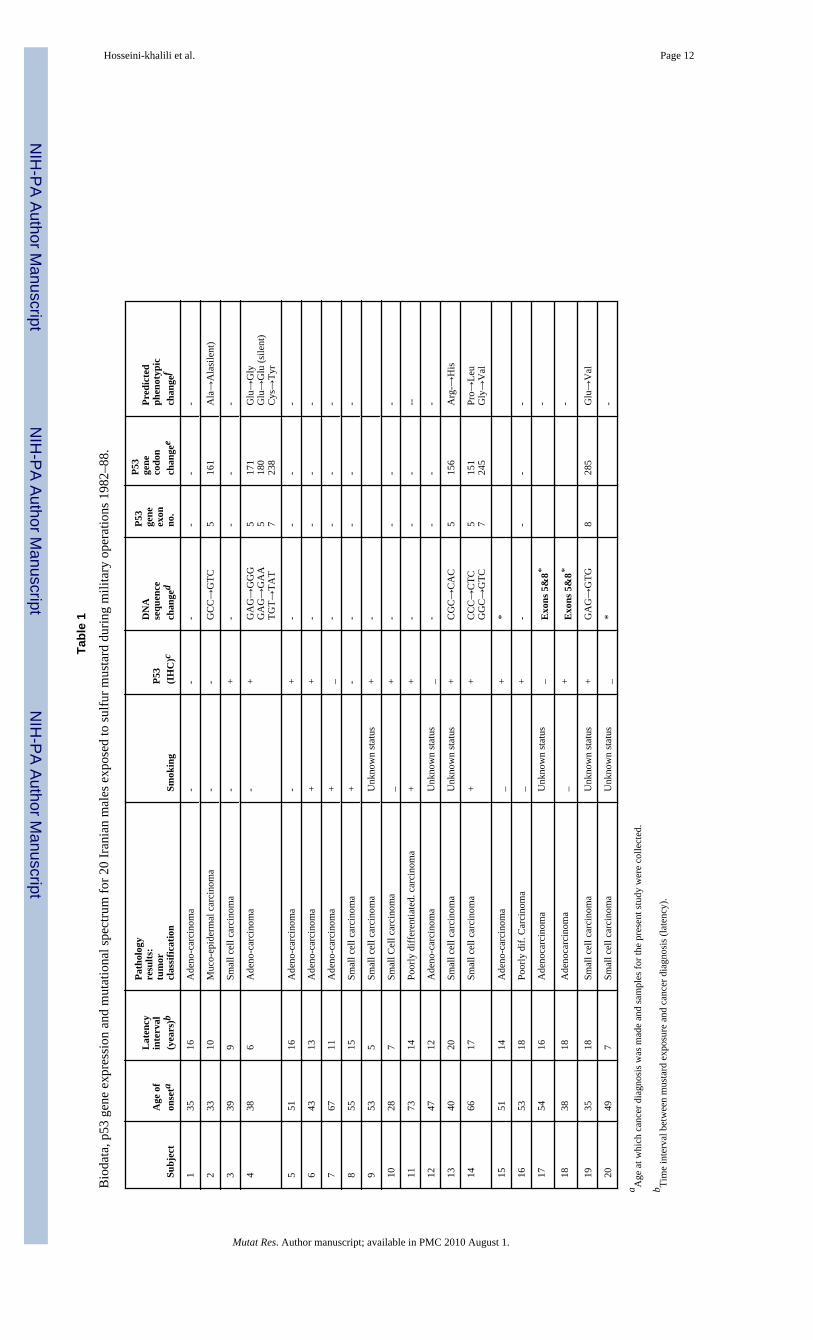
RESULTSSubjects for the present study were 20 males exposed to a single, high dose of SM duringmilitary service in the timeframe 1982–88. All subjects were observed to experience acute andchronic symptoms of mustard toxicity including skin injury and respiratory difficulties andwere included in this study based on the criteria described in the Methods and Materials. Thedistribution of subjects according to age, gender, smoking history, time lapse between exposureand lung cancer diagnosis, tumor pathology, p53 IHC and p53 sequencing data is shown inTable 1.
Of the cancers that could be accurately assessed pathologically, the most frequently occurringtype of lung cancer was Adenocarcinoma (9/20), followed by Squamous Cell Carcinoma (SCC)(4/20) and Small Cell Carcinoma (3/20). In addition, a single case of mucoepidermal lungcancer was observed in a 33 year old individual. This type of lung cancer is rare, accountingfor less than a few percent of all lung cancers.
Hosseini-khalili et al. Page 5
Mutat Res. Author manuscript; available in PMC 2010 August 1.
NIH
-PA Author Manuscript
NIH
-PA Author Manuscript
NIH
-PA Author Manuscript
Examination of the individuals’ age at lung cancer diagnosis shows ages ranging from 28 to73, with seven of the 20 patients developing lung cancer before the age of 40. This finding wasof interest, since lung cancer typically develops after the age of 50 and peaks at approximately60 years of age (24). Overall, the mean age at diagnosis was 47.4 years. On average, the timelapse between SM exposure and lung cancer development was 13.1 years.
Although DNA extraction from lung tumor samples of SM victims was difficult due to variabletissue fixation procedures used in Iran, we were able to obtain information on the p53mutational status in 18 of the subjects. Mutations identified and described in these studies weredetermined by comparison of experimental results with reference data of known normal humanp53 sequences published in GenBank
Table 1 shows the location and nature of the base change in these subjects, as well as thepredicted consequence on the p53 protein. A total of eight mutations were identified. Five ofthese were G to A transitions, with single A to G, G to T and A to T changes also observed.The frequent G to A base change in these patients matched that most frequently observedmutation in chronically exposed factory workers (11). Interestingly, two of the cases hadmultiple point mutations (Table 1). Case 3 showed 3 point mutations: one silent mutation incodon 180 and 2 missense mutations in codons 171 and 238. In this case, all silent and missensemutations were G to A transitions. In Case 14, we observed two missense point mutations: Gto A and G to T at codons 151 and 245, respectively. As discussed below, multiple p53 pointmutations are relatively rare in lung cancer, although one such case was observed in anindividual chronically exposed to SM (11). We also evaluated possible mutations in the KRASgene of these tumors, but did not find any mutations in codons 12 or 13 of this gene. Finally,IHC for the p53 protein was performed on the lung tumor samples. Just over half (13/20)showed expression of p53, which is similar to the frequency described in other lung cancerstudies. In a number of instances p53 reactivity was observed in the absence of a detectedmutation. This inconsistency may be due to false-positive IHC staining (25). Alternatively, thep53 mutation may lay outside the region analyzed by sequencing. Representative IHC resultsfrom tumors taken from subjects 1–5 are shown in Figure 1, with corresponding demographicand genetic data provided in Table 1. Figure 1 also shows an isotype-matched negative controland a colon carcinoma used as a positive control.
DISCUSSIONAll subjects of the present study had sustained single, high-dose SM exposures during the Iran-Iraq war and subsequently developed lung cancer. Analysis of tissue samples in the context ofeach subject’s biodata yielded information relevant to characterization of cancer risk andpathogenesis in mustard-exposed populations. Major significant findings included an overallyounger age of cancer onset in SM victims than that typically found for lung cancer (Table 1).In addition, p53 mutations in tumors taken from participants in this study included G to Atransitions; a mutation consistent with the DNA lesion caused by SM (Table 1). Additionallythe presence of double and triple point mutations in the p53 gene was noted in our subjects; anobservation that was also made in factory workers chronically exposed to mustard agent (11).The significance of the sequence data must nevertheless be interpreted with caution. G to Atransitions in p53 may indeed be triggered by direct SM-DNA reactivity, however there areother etiologic agents that may induce the same mutation that are also associated with lungcancer (26). For example, chronic pulmonary inflammation suffered by SM victims couldcontribute to a characteristic mutational spectrum independently of direct SM-DNA reactivity.Nonetheless, this study provides insight as to the spectrum of mutations that may be associatedwith tumors in persons with single, high-dose SM exposure; along with other features of illnessin this population, notably early age of cancer onset.
Hosseini-khalili et al. Page 6
Mutat Res. Author manuscript; available in PMC 2010 August 1.
NIH
-PA Author Manuscript
NIH
-PA Author Manuscript
NIH
-PA Author Manuscript
The relatively early age of onset of lung cancer in SM victims may be an indication of a uniqueetiology for some of these cancers. The range of lung cancer diagnosis in our subjects was from28 to 73, with seven of the 20 patients developing lung cancer before the age of 40. Lung cancertypically develops after the age of 50, peaks at approximately 60 years of age, and it is unusualamong people under 40 (24). Few studies have been published on lung cancer in Iran, butavailable data indicates that the mean age of lung cancer is higher than 60, while the mean ofage among SM victims was 48 (±12) (27). The causal inference was stronger for those whoare in their 20s or 30s at the time of diagnosis.
Many studies have shown that mutation of the p53 gene can contribute to lung cancerdevelopment, with mutations in this gene found in over half of all lung cancers (28,29). Theprotein product of the p53 gene is involved in DNA damage response. Consequently, this genemay be a preferred target for environmental carcinogens, which act as DNA damaging agents.Moreover, carcinogens leave molecular fingerprints on the p53 gene. Thus, the study of thep53 mutational spectrum has been a useful approach for implicating suspected carcinogens todifferent human cancers (30,31). However, at the time of this writing, only one report has beenpublished characterizing p53 mutations in SM-exposed individuals. This is an articledescribing p53 mutations in a small Japanese Japanese population chronically exposed to SMthrough work at a factory producing mustard agent. The p53 analysis performed on 12 tumorsisolated from Japanese factory workers showed six out of twelve lung tumors had at least onemutation in p53 (within exons 5–8); and two of the twelve tumor samples had double G to Atransitions. It was concluded that these unusual double mutations may be characteristic of lungtumors caused by interaction of SM with DNA and potentially reflects the high mutageniccapacity of mustard agent. Interestingly, we likewise observed multiple point mutations in twoof our cases (Table 1). Case 3 showed 3 point mutations: one silent mutation in codon 180 and2 missense mutations in codons 171 and 238. In this case, all silent and missense mutationswere G to A transitions. In Case 14, we observed two missense point mutations: G to A and Gto T on codons 151 and 245, respectively. Over 2000 p53 mutations have been reported inhuman lung cancers, and few double mutations have been documented thus far (11,32,33). Ina study of 15 atomic-bomb exposed patients with lung adenocarcinoma or squamous cellcarcinoma, two patients were found to have double mutations in p53. Development of lungcancer in these patients was reported in association with radiation exposure (34). Hayes et al.also reported that an excess risk of lung cancer is associated with chronic exposure to chromate(35), which were also found to carry multiple mutations in the p53 gene (36). In conclusion,multiple point mutations may be attributed to exposure to strong exogenous carcinogens, suchas SM.
It is of interest to note that only one of the observed mutations was among the most commonmutations reported for p53. Approximately 30% of p53 mutations in the world are in “hot spot”codons 175, 245, 248, 273, and 282. Among p53 mutations in lung cancers world-wide, GCbase-pairs are predominantly attacked (78–86%) (32,37,38). Such mutations have beenattributed to spontaneous deamination, or the selective carcinogen targeting, of 5-methycytosine (39,40). However, among the 8 mutations detected in SM exposed lung cancerpatients, only one transition at CpG site dinucleotides was found. The under-representation ofCpG mutations further suggests a unique etiology of lung cancers formed in SM victims.
Notwithstanding the strengths of this and a previous study of individuals chronically exposedto SM, there are a number of concerns and limitations. For example, analysis of additional p53mutations in SM victims could generate a mutational spectrum more similar to that found inother lung cancers. In addition, our present study cannot provide an estimate of the increasedlung cancer risk associated with an acute exposure to SM in the general population, nor can itsubstitute for epidemiological studies. Since genetic variation is one of the most importantfactors contributing to the risk development of cancers in humans, an individual’s sensitivity
Hosseini-khalili et al. Page 7
Mutat Res. Author manuscript; available in PMC 2010 August 1.
NIH
-PA Author Manuscript
NIH
-PA Author Manuscript
NIH
-PA Author Manuscript
to SM is likely depend in part on their innate ability to metabolize and detoxify carcinogensand detect and repair DNA lesions. The individuals studied here may represent a highlysusceptible subpopulation that might not be present at a high frequency in the generalpopulation. The later concern has been a significant motivator for an on going epidemiologicalstudy among SM-exposed Iranian populations in a newly developed program called “Study ofSurveillance Health Analysis of Mustard Chemical Exposure” (SHALAMCHE) (41). Theprogram in its present form is configured as a prospective and historical cohort study designedto track the health status of Iranian war veterans and civilians registered in the National WarfareVictims Database in Tehran. Since June 2002, 13441 participants (8500 mustard-exposed menand 4941 controls) were enrolled in SHALAMCHE, with substantially more on a weekly basis.Eventually, this program will provide enormous insight into how SM exposure affects multiplehealth parameters including epidemiology of lung cancer development SM-exposed Iranianpopulation, including the degree to which an acute SM exposure increases the risk of lungcancer development in a general population. The present report represents an initialcontribution to the dynamic database that will result from implementation of theSHALAMCHE program.
AcknowledgmentsThe authors would like to acknowledge Colleen Spurling for her assistance in analyzing the sequence data; andStephanie C. Fox, J.D., President of QueenBeeEdit Inc. for her hard work preparing the present manuscript forpublication. This work was supported by a grant from the NIEHS to CG (5R21ES013775–02).
References1. Constanc, M.; Pechura, DPR. Veterans at Risk: Health Effects of Mustard Gas and Lewisite.
Washington DC: National Academic Press; 1993. p. 218-221.2. United Nations Security Council. Report; of Specialists Appointed by the Secretary General to
investigate Allegations by the Islamic Republic of Iran Concerning the Use of Chemical Weapons.New York, NY: United Nations Security Council Publication; 1986. S/16433/1986
3. Emad A, Rezaeian GR. The diversity of the effects of sulfur mustard gas inhalation on respiratorysystem 10 years after a single exposure: analysis of 197 cases. Chest 1997;112:734–738. [PubMed:9315808]
4. Ghanei M, Adibi I, Farhat F, Aslani J. Late respiratory effects of sulfur mustard: how is the earlysymptoms severity involved? Chron Respir Dis 2008;5(2):95–100. [PubMed: 18539723]
5. Khateri S, Ghanei M, Keshavarz S, Soroush MR, Hains D. Incidence of lung, eye and skin lesions onlate complications in 34,000 Iranian with wartime exposure to mustard agent. J Occu Environ Med2003;45:1136–1143.
6. Asensi V, et al. In vivo interleukin-6 protects neutrophils from apoptosis in osteomyelitis. Infect Immun2004;72(7):3823–8. 98. [PubMed: 15213123]
7. Savill J, Haslett C. Granulocyte clearance by apoptosis in the resolution of inflammation. Semin CellBiol 1995;6:385–93. [PubMed: 8748146]
8. International Agency for Research on Cancer (IARC). Monograph: Overall Evaluations ofCarcinogenicity to Humans. 2004 [Accessed January 30, 2006.]. Available at:http://www-cie.iarc.fr/monoeval/crthall.html
9. Hemminki K, et al. Dna adducts, mutations, and cancer 2000. Regul Toxicol Pharmacol 2000;32(3):264–75. [PubMed: 11162720]
10. Shibata MA, et al. DNA methylation adduct formation and H-ras gene mutations in progression ofN-butyl-N-(4-hydroxybutyl)nitrosamine-induced bladder tumors caused by a single exposure to N-methyl-N-nitrosourea. Carcinogenesis 1994;15(12):2965–8. [PubMed: 8001265]
11. Takeshima Y, Inai K, Bennett WP, et al. P53 mutations in lung cancers from Japanese mustard gasworkers. Carcinogenesis 1994;15(10):2075–9. [PubMed: 7955036]
12. Adachi S, Takemoto K. Occupational lung cancer. A comparison between humans and experimentalanimals. Sangyo Igaku 1987;29(5):345–57. [PubMed: 2831418]
Hosseini-khalili et al. Page 8
Mutat Res. Author manuscript; available in PMC 2010 August 1.
NIH
-PA Author Manuscript
NIH
-PA Author Manuscript
NIH
-PA Author Manuscript
13. Yamakido M, et al. Former poison gas workers and cancer: incidence and inhibition of tumorformation by treatment with biological response modifier N-CWS. Environ Health Perspect 1996;104(Suppl 3):485–8. [PubMed: 8781369]
14. Wada S, Nishimoto Y, Miyanishi M, Katsuta S, Nishiki M. Malignant respiratory tract neoplasmsrelated to poison gas exposure. Hiroshima J Med Sci 1962;11:81–91.
15. Weiss A, Weiss B. Carcinogenesis due to mustard gas exposure in man, important sign for therapywith alkylating agents. Dtsch Med Wochenschr 1975;100(17):919–23. [PubMed: 1122860]
16. Yamakido M, Ishioka S, Hiyama K, Maeda A. Former poison gas workers and cancer: incidence andinhibition of tumor formation by treatment with biological response modifier N-CWS. EnvironHealth Perspect 1996 May;104:485–8. [PubMed: 8781369]
17. Easton DF, Peto J, Doll R. Cancers of the respiratory tract in mustard gas workers. Br J Ind Med1988;45(10):652–9. [PubMed: 3196660]
18. Case RAM, Lea AJ. Mustard gas poisoning, chronic bronchitis and lung cancer. An investigation intothe possibility that poisoning by mustard gas in the 1914–1918 war might be a factor in the productionof neoplasia. Br J Prev Soc Med 1955;9:62–72. [PubMed: 14378527]
19. Beebe GW. Lung cancer in world war I veterans. Possible relation to mustard gas injury and 1918influenza epidemic. J Nati Cancer Ins 1960;25:1231–1252.
20. Norman JE Jr. Lung Cancer mortality in World War I veterans with mustard gas exposure (1919–1965). J Natl Cancer Inset 1975;54(2):311–7.
21. Bullman MA, Kang PH. A Fifty-year mortality follow-up study of veterans exposed to low-levelchemical warfare agent, mustard gas. Ann Epidemiol 2000;10:333–338. [PubMed: 10942882]
22. Agency for Toxic Substances and Disease Registry (ATSDR). Toxicological Profile for SulfurMustard (Update). Atlanta, GA: U.S. Department of Health and Human Services, Public HealthService.); 2003.
23. Hsu SM, Raine L, Fanger H. A comparative study of the peroxidase–antiperoxidase method and anavidin– biotin complex method for studying polypeptide hormones with radioimmunoassayantibodies. Am J Clin Pathol 1981;75:734 –738. [PubMed: 6165237]
24. O’Rourke MA, Feussner JR, Feigl P, Laszlo J. Age trends of lung cancer stage at diagnosis.Implications for lung cancer screening in the elderly. JAMA 1987;258(7):921–6. [PubMed: 3613022]
25. Baas IO, van den Berg FM, Mulder JW, Clement MJ, et al. Potential false-positive results with antigenenhancement for immunohistochemistry of the p53 gene product in colorectal neoplasms. J Pathol1996;178(3):264–7. [PubMed: 8778330]
26. Yamada Y, Oghiso Y. Mutations in Tp53 gene sequences from lung tumors in rats that inhaledplutonium dioxide. Radiat Res 1999 Dec;152(6 Suppl):S107–9. [PubMed: 10564948]
27. Sadjadi A, Malekzadeh R, Derakhshan MH, et al. Cancer occurrence in Ardabil: results of apopulation-based cancer registry from Iran. Int J Cancer 2003;107(1):113–8. [PubMed: 12925965]
28. Hofseth LJ, Hussain SP, Harris CC. P53: 25 years after its discovery. Trends Pharmacol Sci 2004;25(4):177–81. [PubMed: 15116721]
29. Wang W, El-Deiry WS. Restoration of p53 to limit tumor growth. Curr Opin Oncol 2008;20(1):90–6. [PubMed: 18043262]
30. Bennett WP, Hussain SP, Vahakangas KH, Khan MA, Shields PG, Harris CC. Molecularepidemiology of human cancer risk: gene-environment interactions and p53 mutation spectrum inhuman lung cancer. J Pathol 1999;187(1):8–18. [PubMed: 10341702]
31. Rengul CA, Mehmet O. P53 mutations as fingerprints of environmental carcinogens. Pure Appl Chem2000;72(6):995–999.
32. Kishimoto Y, Murakami Y, Shiraishi M, Hayashi K, Sekiya T. Aberrations of the p53 tumorsuppressor gene in human non-small cell carcinomas of the lung. Cancer Res 1992;52(17):4799–804. [PubMed: 1324794]
33. Miller CW, Simon K, Aslo A, Kok K, Yokota J, Buys CH, Terada M, Koeffler HP. P53 mutationsin human lung tumors. Cancer Res 1992;52(7):1695–8. [PubMed: 1312896]
34. Nakachi K, Harris CC, Tahara E. Japan-US Cooperative Cancer Research Seminar on molecularepidemiological characteristics of lung and colon cancer development among atomic-bombsurvivors, Bethesda, USA, February 23–24, 2006. Cancer Sci 2006;97(11):1279–82. [PubMed:16965394]
Hosseini-khalili et al. Page 9
Mutat Res. Author manuscript; available in PMC 2010 August 1.
NIH
-PA Author Manuscript
NIH
-PA Author Manuscript
NIH
-PA Author Manuscript
35. Hayes RB, Sheffet A, Spirtas R. Cancer mortality among a cohort of chromium pigment workers.Am J Ind Med 1989;16:127–133. [PubMed: 2773944]
36. Kondo K, Hino N, Sasa M, Kamamura Y, Sakiyama S, Tsuyuguchi M, Hashimoto M, Uyama T,Monden Y. Mutations of the p53 gene in human lung cancer from chromate-exposed workers.Biochem Biophys Res Commun 1997 Oct 9;239(1):95–100. [PubMed: 9345276]
37. Suzuki H, Takahashi T, Kuroishi T, et al. P53 mutations in non-small cell lung cancer in Japan:association between mutations and smoking. Cancer Res 1992;52(3):734–6. [PubMed: 1310070]
38. Takahashi T, Takahashi T, Suzuki H, et al. The p53 gene is very frequently mutated in small-celllung cancer with a distinct nucleotide substitution pattern. Oncogene 1991;6(10):1775–8. [PubMed:1656362]
39. Ehrlich M, Zhang XY, Inamdar NM. Spontaneous deamination of cytosine and 5-methylcytosineresidues in DNA and replacement of 5-methylcytosine residues with cytosine residues. Mutat Res1990;238(3):277–86. [PubMed: 2188124]
40. Pfeifer GP. Mutagenesis at methylated CpG sequences. Curr Top Microbiol Immunol 2006;301:259–81. [PubMed: 16570852]
41. Zafarghandi MR, Soroush MR, Mahmoodi M, Holakoui K, Ardalan A, Dolatyari A, Abbasi A.Surveillance Health Analysis of Mustard Chemical Exposure (SHALAMCHE) Study. Design andMethods. Unpublished report.
Hosseini-khalili et al. Page 10
Mutat Res. Author manuscript; available in PMC 2010 August 1.
NIH
-PA Author Manuscript
NIH
-PA Author Manuscript
NIH
-PA Author Manuscript
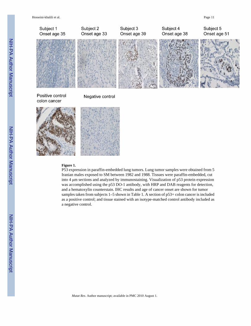
Figure 1.P53 expression in paraffin-embedded lung tumors. Lung tumor samples were obtained from 5Iranian males exposed to SM between 1982 and 1988. Tissues were paraffin-embedded, cutinto 4 μm sections and analyzed by immunostaining. Visualization of p53 protein expressionwas accomplished using the p53 DO-1 antibody, with HRP and DAB reagents for detection,and a hematoxylin counterstain. IHC results and age of cancer onset are shown for tumorsamples taken from subjects 1–5 shown in Table 1. A section of p53+ colon cancer is includedas a positive control; and tissue stained with an isotype-matched control antibody included asa negative control.
Hosseini-khalili et al. Page 11
Mutat Res. Author manuscript; available in PMC 2010 August 1.
NIH
-PA Author Manuscript
NIH
-PA Author Manuscript
NIH
-PA Author Manuscript
NIH
-PA Author Manuscript
NIH
-PA Author Manuscript
NIH
-PA Author Manuscript
Hosseini-khalili et al. Page 12
Tabl
e 1
Bio
data
, p53
gen
e ex
pres
sion
and
mut
atio
nal s
pect
rum
for 2
0 Ir
ania
n m
ales
exp
osed
to su
lfur m
usta
rd d
urin
g m
ilita
ry o
pera
tions
198
2–88
.
Subj
ect
Age
of
onse
ta
Lat
ency
inte
rval
(yea
rs)b
Path
olog
yre
sults
:tu
mor
clas
sific
atio
nSm
okin
gP5
3(I
HC
)c
DN
Ase
quen
cech
ange
d
P53
gene
exon
no.
P53
gene
codo
nch
ange
e
Pred
icte
dph
enot
ypic
chan
gef
135
16A
deno
-car
cino
ma
--
--
--
233
10M
uco-
epid
erm
al c
arci
nom
a-
-G
CC→
GTC
516
1A
la→
Ala
sile
nt)
339
9Sm
all c
ell c
arci
nom
a-
+-
--
-
438
6A
deno
-car
cino
ma
-+
GA
G→
GG
GG
AG→
GA
ATG
T→TA
T
5 5 7
171
180
238
Glu→
Gly
Glu→
Glu
(sile
nt)
Cys→
Tyr
551
16A
deno
-car
cino
ma
-+
--
--
643
13A
deno
-car
cino
ma
++
--
--
767
11A
deno
-car
cino
ma
+_
--
--
855
15Sm
all c
ell c
arci
nom
a+
--
--
-
953
5Sm
all c
ell c
arci
nom
aU
nkno
wn
stat
us+
-
1028
7Sm
all C
ell c
arci
nom
a_
+-
--
-
1173
14Po
orly
diff
eren
tiate
d. c
arci
nom
a+
+-
--
--
1247
12A
deno
-car
cino
ma
Unk
now
n st
atus
_-
--
-
1340
20Sm
all c
ell c
arci
nom
aU
nkno
wn
stat
us+
CG
C→
CA
C5
156
Arg
-→H
is
1466
17Sm
all c
ell c
arci
nom
a+
+C
CC→
CTC
GG
C→
GTC
5 715
124
5Pr
o→Le
uG
ly→
Val
1551
14A
deno
-car
cino
ma
_+
*
1653
18Po
orly
dif.
Car
cino
ma
_+
--
--
1754
16A
deno
carc
inom
aU
nkno
wn
stat
us_
Exo
ns 5
&8*
-
1838
18A
deno
carc
inom
a_
+E
xons
5&
8*-
1935
18Sm
all c
ell c
arci
nom
aU
nkno
wn
stat
us+
GA
G→
GTG
828
5G
lu→
Val
2049
7Sm
all c
ell c
arci
nom
aU
nkno
wn
stat
us_
*-
a Age
at w
hich
can
cer d
iagn
osis
was
mad
e an
d sa
mpl
es fo
r the
pre
sent
stud
y w
ere
colle
cted
.
b Tim
e in
terv
al b
etw
een
mus
tard
exp
osur
e an
d ca
ncer
dia
gnos
is (l
aten
cy).
Mutat Res. Author manuscript; available in PMC 2010 August 1.

NIH
-PA Author Manuscript
NIH
-PA Author Manuscript
NIH
-PA Author Manuscript
Hosseini-khalili et al. Page 13c D
etec
tabl
e ex
pres
sion
of p
53 p
rote
in (i
mm
unor
eact
ivity
) in
tum
or sa
mpl
es.
d Mut
atio
n de
tect
ed in
a p
artic
ular
tum
or sa
mpl
e.
e Cod
on n
umbe
r with
in c
DN
A p
53 se
quen
ce u
sed
in th
e pr
esen
t stu
dy.
f Pred
icte
d ph
enot
ypic
alte
ratio
n (a
min
o ac
id c
hang
e) a
re sh
own
for f
orm
alin
-fix
ed, p
araf
fin-e
mbe
dded
tum
or sa
mpl
es ta
ken
from
eac
h su
bjec
t.
* Sam
ples
in w
hich
DN
A e
xtra
ctio
n w
as n
ot p
ossi
ble.
Mutat Res. Author manuscript; available in PMC 2010 August 1.
NIH
-PA Author Manuscript
NIH
-PA Author Manuscript
NIH
-PA Author Manuscript
Hosseini-khalili et al. Page 14
Tabl
e 2
Prim
er se
ts fo
r PC
R a
mpl
ifica
tion
of lu
ng tu
mor
tiss
ue p
53 e
xons
5, 6
, 7 a
nd 8
.
Exo
nPC
R c
ycle
Stra
ndSe
quen
ceL
engt
h (b
p)O
ptim
al A
nnea
ling
Tem
p (°
C)
5ou
ter
Up-
stre
amcacttgtgccctgacttt
1856
.2
Dow
n-St
ream
cctggggaccctgggcaa
18“
inne
rU
p-st
ream
ttgtgccctgactttcaa
18“
Dow
n-st
ream
ggggaccctgggcaacca
18“
6ou
ter
Up-
stre
amcgacagggctggttgcccaggg
2264
.5
Dow
n-St
ream
agggccactgacaaccacc
19“
inne
rU
p-st
ream
cagggctggttgcccagggtcc
22“
Dow
n-St
ream
gccactgacaaccaccctta
20“
7ou
ter
Up-
stre
amcttgccacaggtctccccaa
2064
.5
Dow
n-St
ream
aagcagaggctggggcacagcagg
18“
inne
rU
p-st
ream
gccacaggtctccccaaggc
18“
Dow
n-St
ream
cagaggctggggcacagcaggcca
22“
8ou
ter
Up-
stre
amtaggacctgatttccttactgcct
2256
.2
Dow
n-St
ream
tgaatctgaggcataactgca
19“
inne
rU
p-st
ream
gacctgatttccttactgcctctt
22“
Dow
n-St
ream
atctgaggcataactgcaccct
20“
Mutat Res. Author manuscript; available in PMC 2010 August 1.
Copyright © 2022 FDOKUMEN