Heat stress: A risk factor for skin carcinogenesis - CORE
95
Edith Cowan University Edith Cowan University Research Online Research Online Theses: Doctorates and Masters Theses 2015 Heat stress: A risk factor for skin carcinogenesis Heat stress: A risk factor for skin carcinogenesis Leslie Calapre Edith Cowan University Follow this and additional works at: https://ro.ecu.edu.au/theses Part of the Oncology Commons Recommended Citation Recommended Citation Calapre, L. (2015). Heat stress: A risk factor for skin carcinogenesis. https://ro.ecu.edu.au/theses/1757 This Thesis is posted at Research Online. https://ro.ecu.edu.au/theses/1757 brought to you by CORE View metadata, citation and similar papers at core.ac.uk provided by Research Online @ ECU
-
Upload
khangminh22 -
Category
Documents
-
view
0 -
download
0
Transcript of Heat stress: A risk factor for skin carcinogenesis - CORE

Edith Cowan University Edith Cowan University
Research Online Research Online
Theses: Doctorates and Masters Theses
2015
Heat stress: A risk factor for skin carcinogenesis Heat stress: A risk factor for skin carcinogenesis
Leslie Calapre Edith Cowan University
Follow this and additional works at: https://ro.ecu.edu.au/theses
Part of the Oncology Commons
Recommended Citation Recommended Citation Calapre, L. (2015). Heat stress: A risk factor for skin carcinogenesis. https://ro.ecu.edu.au/theses/1757
This Thesis is posted at Research Online. https://ro.ecu.edu.au/theses/1757
brought to you by COREView metadata, citation and similar papers at core.ac.uk
provided by Research Online @ ECU

Edith Cowan UniversityResearch Online
Theses: Doctorates and Masters Theses
2015
Heat stress: A risk factor for skin carcinogenesisLeslie CalapreEdith Cowan University
This Thesis is posted at Research Online.http://ro.ecu.edu.au/theses/1757
Recommended CitationCalapre, L. (2015). Heat stress: A risk factor for skin carcinogenesis. Retrieved from http://ro.ecu.edu.au/theses/1757

USE OF THESIS
The Use of Thesis statement is not included in this version of the thesis.

Edith Cowan University
Copyright Warning
You may print or download ONE copy of this document for the purpose of your own research or study.
The University does not authorize you to copy, communicate or otherwise make available electronically to any other person any copyright material contained on this site.
You are reminded of the following:
Copyright owners are entitled to take legal action against persons who infringe their copyright.
A reproduction of material that is protected by copyright may be a copyright infringement. Where the reproduction of such material is done without attribution of authorship, with false attribution of authorship or the authorship is treated in a derogatory manner, this may be a breach of the author’s moral rights contained in Part IX of the Copyright Act 1968 (Cth).
Courts have the power to impose a wide range of civil and criminal sanctions for infringement of copyright, infringement of moral rights and other offences under the Copyright Act 1968 (Cth). Higher penalties may apply, and higher damages may be awarded, for offences and infringements involving the conversion of material into digital or electronic form.

Heat stress: A risk factor for skin carcinogenesis
This thesis is presented for the degree of
Doctor of Philosophy
Leslie Debra Lou S. Calapre
Edith Cowan University
Faculty of Computing, Health and Science
School of Medical Science
2015

The declaration page
is not included in this version of the thesis

i
ABSTRACT
BACKGROUND: The incidence of skin cancer in Australia has increased rapidly in the last
few decades. Ultraviolet radiation (UV) is a major risk factor for skin carcinogenesis. UV,
particularly the UVB spectrum, causes formation of cyclobutane pyrimidine dimers (CPD) in
cellular DNA. Persistent and incorrectly repaired CPDs lead to DNA mutations and
consequently, formation of cutaneous lesions. Interestingly, recent epidemiological studies
have shown a significant increase in skin cancer incidence in geographical locations with high
environmental temperatures. Thus, heat stress may potentiate the effects of UV exposure and
act as an additional risk factor for skin cancer. Previous studies in mice have shown that
repeated and concurrent exposure to UVB and heat stress, increases the rate and incidence of
cutaneous tumour formation relative to UVB alone. However, the effects of UVB plus heat on
human epidermal cells have yet to be determined. Furthermore, the exact mechanisms
responsible for the observed effects of heat stress need to be characterised in skin
keratinocytes to increase knowledge of its risk in skin cancer.
Heat stress induces upregulation of heat shock proteins (HSPs), particularly HSP72 and
HSP90 which are known to affect the activity of the p53 protein. Furthermore, heat stress has
been linked with increased Sirtuin1 (SIRT1) protein activity. SIRT1 is an important histone
deacetylase that helps maintain chromosomal integrity but can also induce post-translational
modifications of the p53 protein. By mediating deacetylation of the p53 protein, SIRT1 can
diminish the ability of p53 to bind to its downstream gene targets. The p53 protein is an
integral mediator of the cellular stress response in skin cells, particularly keratinocytes. Thus,
impairment of p53 transcription factor functions could compromise the ability of epidermal
cells to mount an appropriate response to DNA damage. Moreover, loss of p53 function may
induce survival of cells harbouring DNA lesions.
We hypothesise, therefore, that exposure to UVB plus heat induces survival of DNA damaged
keratinocytes and that these cells escape apoptosis surveillance as a result of heat-mediated
alteration to the p53 signalling pathway. Thus, exposure to heat stress could exacerbate the
carcinogenic effects of UV and increase the risk of skin tumour formation in humans.

ii
AIMS: In this study, we aimed to determine whether repeated exposure to UVB followed
immediately by heat stress (39°C) has a more damaging effect on human keratinocytes than
UVB alone. In particular, we assessed the effects on DNA damage, apoptosis, cell cycle and
DNA repair. Furthermore, we aimed to unravel the mechanism through which heat mediates
the survival of UVB DNA-damaged keratinocytes, focusing on the effects on the p53
signalling pathway.
MATERIALS AND METHODOLOGY: Primary adult human epidermal keratinocytes
(NHEK) and ex vivo punch biopsies of normal human skin called NativeSkin® (Genoskin,
France), were used as experimental models for this study. A UV cabinet fitted with a TL-
UVB Narrowband lamp (Philips, GERMANY), with a spectral output of 290 -315 nm, was
used to administer UVB irradiation at a dose of 1 KJ/m². Heat stress involved culture in a
normal CO2 incubator, with temperature maintained at 39°C for three hours. The temperature
used in the experiments was based on previous measurements of skin surface temperature of
open cut miners, who are prone to intense heat stress, in the Pilbara region of Western
Australia. For UVB plus heat exposures, cells and skin models were sequentially exposed to 1
KJ/m2 of UVB, (at room temperature), followed immediately by 3 hours incubation at 39°C
once per day, for four consecutive days. Unexposed skin models and NHEK, maintained at
37ºC, were used as experimental controls. Cell proliferation, apoptosis and whole genome
expression profiles were analysed at four hours post day 4 exposure, to understand earlier
events, and at 2 days post-exposure, to assess persistent outcomes of these exposures.
Treated primary NHEK cells were counted in a Vi-CellTM Viability Analyser and the level of
apoptosis for exposed primary cells was determined using Annexin V/Propidium Iodide
apoptosis assay at 4 hours and 2 days post exposure. To determine the presence of DNA
damage, total and active p53 protein, as well as total and active SIRT protein, in the skin
models and primary NHEKs, immunohistochemistry and/or immunocytochemistry was
performed. Skin FFPE and primary NHEKs were incubated with antibodies to thymine
dimers (CPD, DNA damage) and p53 (total), acetylated p53-382 (active), SIRT1 (total) or
SIRT1-p (active) antibodies.
To measure apoptosis in skin, an anti-pan-cytokeratin marker was used to label keratinocytes
and active-caspase-3 antibodies were used to identify apoptotic cells. To determine the
expression of p53-downstream target genes at 4 hours, quantitative RT-PCR was performed
using TaqMan probes for BAX, Survivin (BIRC5), ERCC1 and XPC genes, with Human 18S

iii
gene as the endogenous reference gene. Relative quantification of the expression levels of
each transcript in each sample were calculated using the Delta-Delta CT method relative to
untreated controls. A whole genome expression analysis was performed at 2 days post-
exposure using the Human HT-12 Expression v4 BeadChip (Illumina, USA). The Ingenuity
Pathway Analysis (IPA) (Qiagen, USA) software was used to annotate the effects of altered
gene expression on cell function and upstream signalling pathways. Two-way ANOVA was
used to analyse differences across treatment groups, while parametric unpaired t-tests were
used to detect differences between specific treatment groups in all experimental categories,
i.e. proliferation, apoptosis and gene expression, with p-values <0.05 considered significant.
RESULTS:
Outcome 1 –Using ex vivo skin models and NHEKs, we show for the first time that UVB plus
heat treated keratinocytes exhibit DNA damage, as observed after UVB treatment alone.
However, apoptosis was significantly reduced, possibly as a result of inactivation of the p53-
mediated stress response, in DNA damaged cells of UVB plus heat treated samples.
Furthermore, whole genome expression and IPA upstream analysis showed that heat induces
SIRT1 activation, which was confirmed via immunohistochemistry assays. Heat-induced
SIRT1 expression was linked to a decrease in acetylated p53 and consequently,
downregulation of p53-regulated pro-apoptotic and DNA damage repair genes. These results
suggest that p53-mediated cell cycle arrest and apoptosis, known to be induced by UVB, are
ablated with the addition of heat, leading to survival of DNA damaged cells after UVB plus
heat treatment.
Outcome 2 – We further confirmed that SIRT1 activation did not inhibit the transcription of
the p53 protein but mediated deacetylation of p53, resulting in significant deregulation of
expression of p53 downstream gene targets and decreased keratinocyte apoptosis in UVB plus
heat treated samples. Importantly, chemical inhibition of SIRT1 by Ex-527, a known
chemical inhibitor of SIRT1, in UVB plus heat exposed keratinocytes, resulted in re-
activation of the p53 signalling pathway and increased apoptosis of DNA damaged
keratinocytes. This clearly demonstrated the role of heat-mediated SIRT1 activation in the
survival of DNA damaged keratinocytes after exposure to UVB plus heat.

iv
CONCLUSION: In this study, we showed that the efficiency of cellular stress response to
UVB-induced DNA damage is diminished in the presence of heat and, for the first time,
provide a molecular mechanism that explains these effects. With the novel use of an ex vivo
human skin model, this study showed that heat stress prevents human keratinocytes, damaged
by UV irradiation, from undergoing apoptosis and/or necrosis. We found UV plus heat
exposure mediates SIRT1 activation which has been found to induce deacetylation of p53
and, consequently, the inactivation of the p53 signalling pathway. SIRT1 inhibition precluded
the downregulation of p53 signalling by UV plus heat exposure, restoring apoptosis levels to
those observed in UVB-only exposures. Thus, we demonstrated that SIRT1 activation is the
main molecular mechanism driving UVB plus heat-induced survival of DNA damaged
keratinocytes.
Overall, the results of this study suggest that by allowing the survival of DNA damaged
keratinocytes, via induction of SIRT1 activation, heat stress can exacerbate the carcinogenic
effects of UVB radiation. Exposure to heat stress, in addition to UV, could therefore increase
the accumulation of mutations in keratinocytes, possibly leading to the transformation of
normal cells into pre-cancerous cells. Further research is warranted to determine the role of
UVB plus heat in skin cancer pathogenesis. Such knowledge could be utilised in public health
campaigns to decrease risk, particularly for people exposed to combinations of these
environmental hazards in workplaces such as in the mining, construction and petroleum
industries.

v
ACKNOWLEDGMENTS
This thesis and its body of work would not have been made possible without the gracious help
of my supervisors, laboratory colleagues, collaborators, friends and family.
I give my sincerest thanks to my supervisors, Professor Mel Ziman and Dr Elin Gray, whom I
owe a great debt for making this thesis possible. To Elin, thank you so very much for all your
help in every aspect of my project - for the time you have invested in helping me with every
issues I encounter in the lab and most of all, for the much needed psychoanalysis you
provided me as I complete this project. I am forever grateful for your guidance, support and
most of all, for your warm friendship. To Mel, words cannot describe how grateful I am for
your tutelage and unwavering support throughout the last five years of my research journey.
Thank you so very much for having me as your student. It truly is a great privilege to be a part
of your wonderful team. Thank you also for all your help, advice and never-ending patience
as I go through the vicissitudes of my project. It is an honour to have you as a mentor.
I would also like to sincerely thank the Melanoma Research Team, the Huntington’s Disease
and the wonderful band of researchers in 17.201, for all their assistance in the lab and for their
warm friendship. Thank you most especially to Dani, Pauli, Michelle, Anna, Trav, Ash, Julie,
Jamie, Tina, Linda, Gabriela Carlos and Aaron, who have made my research journey greatly
enjoyable. Thank you all for the hugs, the laughter we have shared, and your wonderful
support. Meeting and working beside all of you are one of the greatest blessings of my life.
To my dearest friends (and greatest people on Earth) - Dani, Pauli and Aaron, thank you for
having the patience to deal with my neurosis and for loving me despite of it. Thank you all for
the trust, warmest of hugs and loving friendship we have shared. Most of all thank you for
your willingness to lend an open ear, with hands often armed with coffee, cakes and/or
chocolates, in times when I needed it the most. Your encouragement and support means the
world to me. I am forever grateful for having the most loving and supportive group of friends.
Finally, to the loves of my life - my family, words are not enough to convey my deepest
gratitude for all your love and support. You are my greatest source of strength and inspiration,
and I love you all to the ends of this world and beyond.

i
LIST OF PUBLICATIONS:
1) Heat Stress: A Risk Factor for Skin Carcinogenesis Leslie Calapre1, Elin Gray1, Mel Ziman1, 2*
1 School of Medical Science, Edith Cowan University, Perth, Western Australia 2 Department of Pathology and Laboratory Medicine, University of Western Australia, Crawley, Western Australia Published: Cancer Letters, August 2013, Volume 337, Issue 1, Pages 35-40
2) Heat-Mediated Evasion of Apoptosis in UVB-Damaged Keratinocytes Leslie Calapre1, Elin Gray1, Sandrine Kurdykowski2, Anthony David2, Prue Hart4, Pascal Descargues2, Mel Ziman1, 3*
1 School of Medical Sciences, Edith Cowan University, Perth, WA, Australia 2GENOSKIN Centre Pierre Potier, Oncopole, Toulouse, France 3Department of Pathology and Laboratory Medicine, University of Western Australia, Crawley, WA, Australia 4Telethon Kids Institute, University of Western Australia, 100 Roberts Road, Subiaco, Perth, Australia 6008 Submitted and currently under re-review: Experimental Dermatology, August 2015
3) SIRT1 Activation Mediates the Heat-Induced Survival of UVB Damaged Keratinocytes Leslie Calapre1, Elin Gray1, Sandrine Kurdykowski2, Anthony David2, Pascal Descargues2, Mel Ziman1, 3*
1 School of Medical Sciences, Edith Cowan University, Perth, WA, Australia 2GENOSKIN Centre Pierre Potier, Oncopole, Toulouse, France 3Department of Pathology and Laboratory Medicine, University of Western Australia, Crawley, WA, Australia Submitted: Journal of Investigative Dermatology, September 2015 * Corresponding Author Professor Mel Ziman, School of Medical Sciences, Edith Cowan University, Perth, WA, Australia. Address: 270 Joondalup Drive, Joondalup WA 6027, Australia. Tel: +61 8 6304 5613. Email: [email protected]

i
STATEMENT OF CONTRIBUTION OF OTHERS:
Leslie Calapre conducted the experiments, analysis of results and wrote the papers included in
this thesis. Dr Pascal Descargues, Dr Sandrine Kurdykowski and Dr Anthony David from
Genoskin provided the skin models for all experiments presented in this thesis. Dr David
Raven of Saint John of God Hospital Pathology Department provided help with processing of
tissues. Miss Christie Chee of Clinipath provided advice and assistance with tissue embedding
and sectioning. Professor Prue Hart from the Telethon Institute contributed to the study design
and critique of this research project. Dr Joe Mates and A/Prof Jacques Oosthuizen provided
the skin surface measurements. AGRF assisted with the analysis of the whole genome
expression results included in publication 2 (Chapter 3).

ii
Heat Stress: A Risk Factor for Skin Carcinogenesis
Contents
ABSTRACT ................................................................................................................................ i
ACKNOWLEDGMENTS .......................................................................................................... v
LIST OF PUBLICATIONS: ...................................................................................................... i
STATEMENT OF CONTRIBUTION OF OTHERS: .............................................................. i
LIST OF FIGURES ................................................................................................................. xi
GENERAL INTRODUCTION ................................................................................................. 1
CHAPTER 1: Environmental Stresses and their Role in Skin Carcinogenesis .............. 3 1.1 Normal Skin Physiology ............................................................................................... 3 1.2 UV Radiation and Skin Cancers ................................................................................... 6 1.3 Heat Stress and Skin Cancers ..................................................................................... 18 1.4 Conclusion .................................................................................................................. 25 1.5 Theoretical Framework ............................................................................................... 26 1.6 Hypothesis ................................................................................................................... 27 1.7 Aims ............................................................................................................................ 27
CHAPTER 2: Heat Stress: A Risk Factor for Skin Carcinogenesis .............................. 28 2.0 Abstract ....................................................................................................................... 29 2.1 Introduction ................................................................................................................. 30 2.2 Heat Stress and the Heat Shock Proteins .................................................................... 31 2.3 HSPs and Inhibition of Cell Death-inducing Pathways and Activation of Survival Pathways ........................................................................................................................... 33 2.4 Induction of Skin Cancer by Heat Stress and UV: Mediated by HSP72 and p53 Signalling Pathways .......................................................................................................... 36 2.5 Conclusion .................................................................................................................. 39
CHAPTER 3: Heat-Mediated Evasion of Apoptosis in UVB-Damaged Keratinocytes .............................................................................................................................................. 40
3.0 Abstract ....................................................................................................................... 41 3.1 Introduction ................................................................................................................. 42 3.2 Materials and Methods ................................................................................................ 43 3.3 Results ......................................................................................................................... 45 3.4 Discussion ................................................................................................................... 55

iii
CHAPTER 4: SIRT1 Activation Mediates the Heat-Induced Survival of UVB-Damaged Keratinocytes ..................................................................................................... 63
4.0 Abstract ....................................................................................................................... 64 4.1 Introduction ................................................................................................................. 65 4.2 Results ......................................................................................................................... 67 4.3 Discussion ................................................................................................................... 74 4.3 Materials and Methods ................................................................................................ 77
GENERAL DISCUSSION ...................................................................................................... 81
CHAPTER 5: Heat Stress and Skin Carcinogenesis ....................................................... 81 5.1 Effects of UVB plus Heat Exposure on Keratinocyte Biology ................................... 82 5.2 Molecular events underpinning UVB plus heat-mediated survival of DNA damaged keratinocytes ..................................................................................................................... 85 5.3 Future Directions: Induction of Skin Cancer by UVB plus Heat Stress - SIRT1-Mediated Inactivation of p53 Signalling pathways .......................................................... 90
GENERAL CONCLUSION .................................................................................................... 92
REFERENCES ....................................................................................................................... 93
APPENDICES ....................................................................................................................... 118 Ethics ............................................................................................................................... 119

xi
LIST OF FIGURES
Figure 1.1: Keratinocyte-Melanocyte Complex. .................................................................................. 4
Figure 1.2: UV-Induced Thymine Dimer Formation.. ......................................................................... 9
Figure 1.3: The Cell Cycle Checkpoints. ........................................................................................... 10
Figure 1.4: The Nucleotide Excision Repair (NER) System. ............................................................. 12
Figure 1.5: The p53 Signaling Pathway. ........................................................................................... 15
Figure 1.6: Activation of p53 by Post-Translational Modification. .................................................. 17
Figure 1.7: The Heat Shock Response ............................................................................................... 20
Figure 2.1: The Heat Shock Response. .............................................................................................. 32
Figure 2.2: The Heat Shock Proteins and JNK Pathway. ................................................................. 34
Figure 2.3: The MAPK Signalling Pathway.. .................................................................................... 35
Figure 2.4: The p53 Signalling Pathway ........................................................................................... 38
Figure 2.5: Hypothesised model for the role of heat in skin cancer induction. ................................. 39
Figure 3.1: Effects of UVB and/or heat stress on DNA damage and apoptosis in keratinocytes in vitro and in an ex vivo skin model.. .................................................................................................... 48
Figure 3.2: Exposure to UVB radiation and heat stress induced expression of genes with roles in cell survival. ....................................................................................................................................... 50
Figure 3.3: Exposure to UVB plus heat induced a significant decrease in acetylated p53 and increase in SIRT1 protein levels in NHEK and in skin models. ......................................................... 54
Figure 4.1: Exposure to repeated UVB plus heat significantly decreased acetylated p53 levels in keratinocytes. ..................................................................................................................................... 68
Figure 4.2: Effect of UV and/or heat exposure on p53 downstream gene targets and cell proliferation.. ..................................................................................................................................... 71
Figure 4.3: Inhibition of SIRT1 in UVB plus heat significantly increased acetylated p53 protein expression and apoptosis of keratinocytes in vitro.. .......................................................................... 73
Figure 5.1: UVB plus Heat-Induced Molecular Pathway for the Survival of DNA Damaged Keratinocytes. .................................................................................................................................... 88
Figure 5.2: UVB plus Heat-Mediated Skin Carcinogenesis. ............................................................. 91

xii
LIST OF TABLES Table 3.1: Upstream regulators significantly activated in UV plus heat. ......................................... 51 Table 4.1: Effects of UVB and/or heat exposure in keratinocytes of the ex vivo skin model and NHEK in vitro. ................................................................................................................................... 69
LIST OF SUPPLEMENTARY MATERIALS Spplementary Figures:
Chapter 3
Figure S3.1: Line graph showing the comparison of the average environmental temperature and surface skin temperature of miners working in the East Pilbara Region of Western Australia. ....... 58
Figure S3.2: Immunohistochemical staining of cytokeratin (CytoK) or CPD (red), p53 or active caspase-3 (green) and DAPI (blue) in untreated or UVB and/or heat treated NHEK or ex vivo skin.. ................................................................................................................................................... 59
Figure S3.3: (a) Immunohistochemical staining of SIRT1-p (green), total SIRT1 (red) and DAPI (nucleus, blue) in skin samples or primary keratinocytes that were either untreated, or exposed to heat, UVB or UVB plus heat. ......................................................................................................... 60
Figure S3.4: Exposure to UVB plus heat induced a significant decrease in acetylated p53 levels in NHEK.. ........................................................................................................................................... 61
Chapter 4
Figure S4.1: Exposure to UVB plus heat significantly decreased acetylated p53 levels in keratinocytes ...................................................................................................................................... 80
Supplementary Tables
Chapter 3
Table S3.1: Effects of UV and/or heat stress exposure on DNA damage, apoptosis and p53. .......... 62
Table S3.2: Biological activities and functions present in the heat, UVB or UVB plus heat treated samples………………………………………………………………………………………………………62

1
GENERAL INTRODUCTION
Non-melanoma skin cancers (NMSCs), particularly basal cell carcinoma (BCC) and squamous
cell carcinoma (SCC), are the most common cutaneous cancers in Caucasian populations around
the world (Xiang et al., 2014). In Australia, NMSCs account for 75% of all cancers and are
considered a great health and economic burden (Jou et al., 2010). BCC is defined as a malignant
tumour of follicular germinative cells or trichloroblasts (Barascu et al., 2012; Hsieh et al., 2014).
It accounts for approximately 80% of all diagnosed skin cancers and is considered to be the
most common malignant tumour (Epstein, 2008; Saran, 2010). BCC incidence is on the rise,
and many experts suggest that it will continue to increase in coming years (Basset-Seguin et al.,
2015; Bath-Hextall et al., 2007). BCC is known to have a very low metastatic potential and
mortality arising from this tumour type is approximately 0.1% (Lear et al., 2005; Roewert-
Huber et al., 2007). BCC can be highly invasive, however, and can often cause extensive tissue
destruction. BCC largely affects the elderly, although, the incidence of cutaneous BBCs has
been increasing in younger adults in recent years (Bath-Hextall et al., 2007; Greinert, 2009).
BCC is generally more common in males than in females, with a ratio of approximately 2:1
(Barascu et al., 2012).
The incidence of Squamous Cell Carcinoma (SCC) also appears to be on the rise (Levin et
al., 2005; Nickoloff et al., 2002). SCC accounts for 20% of all cutaneous cancers; it is the
second most common skin cancer after BCC (Kim et al., 2012; Sridhar et al., 2012). The
incidence of SCC varies in different countries, but the US and Australia have the highest
reported incidence, this being 20% of all skin cancer cases (Zhang et al., 2012). This cancer is
two to three times more common in men than in women, and mostly affects adults over sixty
years of age, though in recent years, the incidence of SCC in people below fifty years of age has
increased (Monroe et al., 2011; Sabunciyan et al., 2015). Unlike BCC, which has a low
metastatic dissemination of less than 0.1%, SCC is known to be more aggressive, and the
incidence of SCC metastasis, which normally arises after one to two years of diagnosis, is
reported to be between 5 and 20 percent (Smoller, 2006).
2
SCC, like every other skin cancer, is common in Caucasians (Gomez et al., 2015; Wei et al.,
2015). In the USA, people of Irish or Scottish ancestry have the highest prevalence (Brantsch et
al., 2008; Schwarz, 2005). In people of African and Asian descent, SCC is relatively rare but
it is the most common form of skin cancer in these groups (Monroe et al., 2011; Rao et al.,
2013). SCC in dark skin people carries a higher mortality rate, and this is attributed to delayed
diagnosis as tumours are more likely to occur in sun-protected areas such as the scalp and
sites of previous injury and scarring ( M o n r oe e t a l . , 2 0 1 1 )
Environmental stressors are considered the most common risk factors for skin cancer formation
(DeFedericis et al., 2006; Lucas et al., 2006; Rosso et al., 2008). In particular, exposure to UV
radiation is associated with formation of 80% of all skin tumours (Ananthaswamy and Pierceall,
1990; Lee et al., 2012; Xia et al., 2006). UV induces DNA damage, which often leads to
formation of mutations in key regulators of cell apoptosis, survival and proliferation signalling
pathways, which can lead to cancer initiation (Ananthaswamy and Pierceall, 1990; Brash et al.,
1996; Courdavault et al., 2005; Grujil et al., 2001; Narayanan et al., 2010). However, given the
effect of high temperature on cell viability and chromosome stability (Akerfelt et al., 2007;
Takahashi et al., 2004), heat stress could be another risk factor for skin cancer. However, the
exact molecular effects of repeated exposure of epidermal cells to high temperatures and UV
radiation, which is what is observed in the environment, needs to be clarified.

3
GENERAL INTRODUCTION
CHAPTER 1: Environmental Stresses and their Role in Skin Carcinogenesis
1.1 Normal Skin Physiology
Keratinocytes and the Epidermal-Melanin Unit
The skin is the largest organ in the body and offers a protective barrier against environmental
insults. The skin barrier is primarily maintained by the epidermis, which is further separated into
different layers comprising the basal, granular and cornified layers (Haggarty, 2015). These
epidermal layers are identified by differences in the morphology of keratinocytes, which are the
most abundant cell type in the skin (Agthong et al., 2012).
The stratum basalis, or basal layer, of the epidermis consists of stem cells which continuously
undergo mitotic division to provide new keratinocytes (Leung et al., 2015; Marshall, 2015;
Sexton-Oates et al., 2015). Daughter cells of basal keratinocytes then detach from the basal
membrane and undergo further differentiation as they move towards the epidermal surface, over
a period of approximately two weeks (Zitzmann et al., 2015). Keratinocytes then undergo
terminal differentiation, lose their nuclei and adapt a flattened morphology, to become
corneocytes of the stratum corneum of the epidermis (Santen et al., 2009; Verdin and Ott, 2015).
The terminal differentiation process of keratinocytes in the cornified layer represents the
physiological apoptosis of epidermal cells (Nowotarski et al., 2015). Imbalances in the delicate
physiological cycle of keratinocyte proliferation or differentiation can significantly affect the
integrity of the skin and result in the formation of cutaneous malignancies (Schraml et al., 2009;
Villanueva et al., 2015).
Apart from keratinocytes, one of the most prominent cell types in the skin are the melanocytes,
which are also located in the basal layer of the epidermis (Chen et al., 2015). Melanocytes are
specialised cells that are responsible for the production of melanin, an important skin
chromophore (Abdel-Malek et al., 2000; McCarty and Loeb, 2015; Ratajczak-Wrona et al.,
2009). Normal skin physiology includes an epidermal-melanin unit, comprised of keratinocytes
and melanocytes arranged at a ratio 35:1, the function of which is to produce melanin to protect
keratinocytes from the harmful effects of environmental stressors (Chen et al., 2015; Costin and
Hearing, 2007).

4
Figure 1.1: Keratinocyte-Melanocyte Complex. This figure highlights mechanisms by which keratinocyte-derived factors such as endothelins (ET) and melanocyte-stimulating hormone (MSH) (red boxes) act on human melanocyte proliferation and stimulate melanogenesis.[Adapted from: (Hirobe, 2005)]
Keratinocytes located in the epithelial layer of the stratum corneum can initiate melanin production
within melanocytes by secreting growth factors, such as endothelins and melanocyte-stimulating
hormone (MSH), which act in a paracrine manner to stimulate melanocyte function (Costin and
Hearing, 2007; Hirobe, 2005). Melanocytes and keratinocytes interact with each other through cell-
to-cell signalling to induce melanin production in melanocytes (Figure 1.1).

5
Melanin has an important role in the physiology, pathology and toxicology of the skin, eyes and
brain (Hu, 2008). This pigment acts as a filter by absorbing near-infrared, visible light and UV
photons (Gidanian et al., 2008). It also functions as a free radical scavenger, capturing oxygen and
hydroxyl radicals and preventing them from causing oxidative stress (Soumyanath et al., 2006).
Melanin is also known to have an affinity for metals and toxic substances and has a sun protection
factor (SPF) value of 2-3 (Maddodi and Selaturi, 2008). It is also known to be able to transform light
energy into heat and distribute it evenly between the capillary vessels and hair follicles (Pons and
Quintanilla, 2006).
There are two types of melanin produced by mammalian melanocytes - eumelanin and pheomelanin
(Tody et al., 1991). Eumelanin is brownish-black in colour, while pheomelanin appears as a reddish-
yellow pigment (Pons and Quintanilla, 2006). Synthesis of both involves the enzyme, tyrosinase
(Chen et al., 2009). This enzyme converts tyrosine to dopaquinone, which when further oxidised,
gives rise to eumelanin (Tody et al., 1991; Williams and Ouhtit, 2005). Pheomelanin is also formed
from further oxidation of dopaquinone, but requires the presence of sulphydryl groups from the
amino acid cysteine (Pons and Quintanilla, 2006; Tody et al., 1991). Eumelanin and pheomelanin are
found in human skin and hair in almost equal amounts, though a relatively greater concentration of
pheomelanin is found in people with red hair and those with a ‘Celtic type’ skin phenotype (Tody et
al., 1991).
Skin pigmentation varies for each individual, due to the difference in the amount of melanin
produced and distributed to neighbouring keratinocytes (Markovic et al., 2007). Lighter skin
produces less melanin (Maddodi and Selaturi, 2008), while a darker skin tone produces more
melanin (Yamaguchi et al., 2008). The distribution of melanosomes and the production of melanin is
the main reason why people with darker skin tone are more efficient at absorbing UV radiation
(Markovic et al., 2007).
While the mechanisms that regulate skin pigmentation are not completely understood, extensive data
shows that UV-induced DNA damage and/or its repair produce initiating signals that induce
melanogenesis (Lee et al., 2013; Mullard, 2015). In response to UV, melanin is transferred to
keratinocytes and repackaged as granules in keratinocytes where it accumulates above the nuclei to
protect them from the mutagenic effects of UV radiation (Costin and Hearing, 2007; Gorman et al.,
2007).

6
If melanin is produced and distributed correctly, keratinocytes are protected from the harmful effects
of UV, even as the cell is undergoing cell division (Seripa et al., 2015; Tallmadge et al., 2015;
Wilken et al., 2015). However, if the melanin production and distribution is significantly reduced, or
if alterations in the signalling pathways of the keratinocyte-melanocyte complex arise, leading to
diminished melanin production, then it is possible for UV and other environmental stresses to cause
the DNA mutations that precede skin cancers (Coelho et al., 2009).
While melanin is extremely important in protecting skin from damage caused by chemicals and
radiation, it can itself be an indirect cause of damage (Yamaguchi et al., 2008). Chronic exposure to
chemicals and radiation can result in changes to melanin properties, such that it fails to bind toxic
substances, resulting in cell damage (Hu, 2008). Furthermore, after consistent and long term
exposure to various environmental hazards and stresses, melanin accumulates free radicals, which
have the potential to disrupt the synthesis of major proteins and RNA and cause dermal cell damage
(Hu, 2008; Hube and Francastel, 2015; Tody et al., 1991).
1.2 UV Radiation and Skin Cancers
Environmental stresses and their role in skin cancer pathogenesis have been studied extensively, to
gain a better understanding of the molecular mechanisms underpinning the tumourigenic process and
to identify treatment or protection strategies. The factor identified as the main cause of most skin
cancers is UV radiation, which induces specific mutations in skin cell DNA (Ikehata and Ono, 2011;
Miller, 1985).
Solar radiation is a spectrum of electromagnetic radiation that includes ultraviolet rays, infrared and
visible light (Maddodi and Selaturi, 2008; Setlow et al., 1993; Sinha and Hader, 2002). UV radiation
is considered a potent carcinogen, and it can enter the earth’s atmosphere and cause mutations in
various mammalian cells including epidermal cells (Brenner et al., 2009). The association between
UV radiation and skin cancer was first established in the late 60’s and was confirmed by Fears et al.
(1977). Fears and colleagues showed that the incidence of non-melanoma skin cancers is far higher
in areas where people are exposed to excessive sunlight. This led them to conclude that solar
radiation is one of the main causes and suggested that formation of skin cancers is most likely related
to annual rates of UV exposure.

7
Retrospective epidemiologic data gathered by various researchers over many years supports the
claims made by Fears and colleagues (1997). One particular study conducted by Williams and Ouhtit
(2005), also showed a clear link between skin cancer risk and UV exposure. Their systematic review
showed clearly that exposure to sunlight both in childhood and adulthood contributes greatly to the
risk. This result was mirrored by a study conducted in 2004 (Solomon et al., 2004), where the
researchers also found that lifetime UV exposure leads to an increased likelihood of skin cancer.
The epidemiological link between skin cancer and UV exposure provides only half of the evidence of
the important role of this particular environmental stress in skin carcinogenesis. The discovery of UV
signature gene mutations that are present in UV-induced BCC and SCC also confirmed the role of
UV in skin carcinogenesis (Ananthaswamy and Pierceall, 1990; Andreassi, 2011; Miller, 1985).
These UV signature mutations initiate tumourigenesis when found in genes involved in signalling
cascades that control cell proliferation and apoptosis (Markovitsi et al., 2010).
UVA Radiation
Ultraviolet radiation is comprised of type A (UVA), which makes up 90% of all solar radiation and
has wavelengths between 315-400 nm, and type B, which has long wavelengths of 290-320 nm
(Weigmann et al., 2010). These relatively long wavelengths allow UV rays to penetrate the epidermis
where they inflict cellular damage, including morphological and genetic alterations to keratinocytes
and melanocytes (Courdavault et al., 2005). In particular, UVA is known to penetrate the deeper
layers of the epidermis and induce significant damage to the cells located in the stratum basale.
While UVA is capable of inducing DNA damage via thymine dimerisation (Ikehata and Ono, 2011),
the basis for UVA mutagenesis and subsequent carcinogenesis lies predominantly in production of
reactive oxygen species (ROS). ROS can covalently bind or directly oxidise proteins and can cause
reversible or irreversible modifications including protein-protein cross linking, carbonylation,
formation of adducts with lipid peroxidation products, and nitration (Perluigi et al., 2010; Vidal et
al., 2014). These modifications often result in changes in stability, as well as functional and structural
changes in proteins, which can lead to loss of protein function, protein aggregation and degradation,
in turn causing DNA missense mutations. ROS are also capable of inducing DNA strand breaks and
distorting nucleotide bases (Dai et al., 2012a).

8
UVB Radiation
Considered the most-cancer inducing spectrum, UVB induces formation of multiple thymine dimer
photoproducts, which can distort the DNA and affect its stability. UVB is implicated as the main
cause of skin cancers, particularly keratinocyte-derived carcinomas. Several studies have shown that
UVB, can induce histological and morphological alterations in epidermal cells (Delgado-Cruzata et
al., 2014; Hillman et al., 2015).
Previous studies in mice have shown that UVB induces significant changes to skin histology,
including decreased epidermal thickness, increased epidermal differentiation and significantly
reduced numbers of proliferating cells in UVB-treated skin (Luo et al., 2007; Riyahi et al., 2015;
Saksouk et al., 2014). Furthermore, irradiation with solar and/or narrowband UVB strongly alters
keratinocyte morphology, inducing cytoplasmic vacuolisation and disintegration of nuclear and
cellular membranes (Kebir et al., 2014; Reich et al., 2007). Irregular distribution and bleb-like
protrusions of the cell surface, rearrangement of the cytoskeleton and thinning or redistribution of
microfilaments to the cell periphery, are also considered some of the most prominent narrowband
UVB-induced morphological alterations in keratinocytes (Reich et al., 2007; Riyahi et al., 2015).
Formation of major carcinogenic photoproducts, such as cyclobutane pyramidine dimers (CPD) and
pyrimidine(6-4)-pyrimidone (6-4PPs), at pyrimidine sites on the DNA of epidermal cells are known
to be induced by UVB irradiation (Courdavault et al., 2005; Ikehata and Ono, 2011; Matsunaga et al.,
1991). CPDs and 6-4PPs are formed as a result of a photochemical reaction between UVB and the
DNA, leading to covalent linkage of adjacent thymine dimers (Figure 1.2), inducing significant
changes in the nucleotide structure (Besarutinia et al., 2004; Yogianti et al., 2012). CPD formation
generally occurs at sites of methylated cytosines, and thus an abundance of methylated cytosines in a
particular DNA region increases susceptibility to UV radiation-induced damage at these sites (Aigal
et al., 2015; Brenner et al., 2009). Thus, CpG islands, DNA regions containing 5-methylcytosines,
are hotspots for such DNA mutations (Ikehata et al., 2003; Ikehata and Ono, 2011).
Keratinocytes undergo rigorous processes of surveillance to ensure that cells which harbour CPDs or
6-4PPs DNA damage do not proliferate (Alonso et al., 2015; Khalkhali-Ellis et al., 2014). Conserved
processes of cell cycle arrest and photoproduct-repair, primarily via the nucleotide excision repair
pathway (NER) and activation of DNA polymerases such as translesion DNA synthesis (TLS)
polymerases, are imposed on cells with DNA lesions (Loughlin, 2015; Pathania et al., 2015; Rubin et
al., 2014; Vilahur et al., 2014). These innate cellular stress responses to DNA damage help in
guarding the genome against mutations that may induce unrestricted proliferation and/or evasion of
apoptosis, leading to the formation of tumours.

9
Figure 1.2: UV-Induced Thymine Dimer Formation. a) Diagrammatical representation of UV-induced thymine dimer formation in cells and distortion of the DNA helix (Pearson Publishing Inc, Benjamin Cummings). b) Covalent linkage of thymine bases as a result of UV radiation.
UV Damage and Cell Cycle Arrest
Cell cycle arrest is imposed on cells with DNA damage to provide time for correction of any
DNA defects (Andersen, 2015; Baud et al., 2014). The cell cycle arrest mechanism in mammalian
cells is primarily controlled by the p53 protein, a product of the TP53 gene (Geranton and
Tochiki, 2015; Kastan et al., 1991). In response to genotoxic stress, particularly UV-induced
DNA damage, the levels of p53 protein increase, which determines either a transient arrest of cell
cycle progression or triggers apoptosis (Brash, 2006; Chipuk and Green, 2004). The p53 protein
can activate agents of the cell cycle checkpoints, controlling their activity, to primarily induce a
transient arrest at a specific stage of the cell cycle (Andersen, 2015; Geranton and Tochiki, 2015;
Mourad et al., 2014). There are two well-known p53-regulated checkpoints at which DNA
damage is monitored (Figure 1.3)-, the G1/S transition checkpoint, which prevents replication of
damaged DNA, and the G2/M transition, at which stage cell cycle is inhibited in response to
persistent DNA damage and/or incompletely replicated DNA (Harris and Levine, 2005).

10
The p53 protein mediates G1/S phase arrest via the activation of p21CIP1, which inhibits the kinase
activity of cyclin/cyclin-dependent kinase complexes, which are key elements in cell cycle
progression (Jimenez et al., 1999; Pellegata et al., 1996; Smith et al., 2000). By inactivating these
kinases, p53 prevents the phosphorylation of the G1 cyclin/cyclin-dependent kinase substrate pRB
and inhibits the transition of cells from the G1 to the S phase of the cycle (Abbas and Dutta, 2009;
Brant et al., 2014). Arrest at G1 provides time for innate nucleotide excision DNA repair
mechanisms to repair critical damage, thereby avoiding the propagation of genetic lesions to
progeny cells (Yu et al., 2000). If the nucleotide excision repair is successful in removing and/or
repairing the DNA lesion, the cell cycle can resume. However, if the damage is too extensive,
cells will undergo apoptosis (Pellegata et al., 1996). Loss of the G1 checkpoint also results in
genomic instability, as seen by the increase in the frequency of gene amplifications in p53-
defective cells (Bodega and Orlando, 2014).
Figure 1.3: The Cell Cycle Checkpoints.
DNA Damage Repair
In all mammalian cells, the recognition and effective removal of DNA lesions is facilitated by an
efficient DNA repair system. The nucleotide excision repair (NER) mechanism is responsible for
the surveillance and removal of UV-induced DNA damage and other lesions that may induce
distortion of the DNA helix in various cells, most particularly in keratinocytes which are
constantly exposed to UV radiation (Du et al., 2015; Feng et al., 2014; Kratochwil and Meyer,
2015; Pacis et al., 2014).

11
Nucleotide excision repair is indispensable for the removal of UV-induced DNA lesions, as
highlighted by three clinically and genetically heterogeneous human syndromes carrying defects
in NER-associated genes; Xeroderma Pigmentosum (XP), Cockayne Syndrome (CS), and
Trichothiodystrophy (Asada et al., 2015; Kraemer et al., 1987; Pan, 2014). People with these
syndromes are all sensitive to UV exposure and, in particular, patients with Xeroderma
Pigmentosum have a >1,000-fold increased susceptibility to UVB-induced skin cancers (Anink-
Groenen et al., 2014; Sarasin, 1999). The nucleotide excision repair-mediated damage
surveillance mechanism is subdivided into two sub-pathways (Figure 1.4) – Transcription
Coupled - Nucleotide Excision Repair (TC-NER) and Global Genome - Nucleotide Excision
Repair (GG-NER). These pathways differ primarily in the way they respond to or recognise
damage (Raviram et al., 2014).
TC-NER is initiated when an elongating RNA polymerase II is stalled as a result of DNA lesions,
which disrupts transcription on the template strand of active genes (Katz et al., 2014; Kolovos et
al., 2014). This is followed by the recruitment and modifications to Cockayne Syndrome B (CSB)
and Cockayne Syndrome A (CSA) proteins, which have DNA-binding ability (Bianchi et al.,
2014; Edelstein and Smythies, 2014). The Xeroderma Pigmentosum group B and D (XPB and
XPD respectively) helicases of the 10-subunit transcription factor TFIIH then unwind the helix
surrounding the lesion, creating short stretches of single stranded DNA (ssDNA) around the
lesion (Qian et al., 2015).
The creation of ssDNA facilitates the recruitment of Xeroderma Pigmentosum group A (XPA)
and Replication Protein A (RPA), ssDNA binding proteins, which verify DNA damage and
prevent complimentary repair by aberrant NER complexes formed on undamaged DNA
(Simpson, 2014). Once DNA damage is verified, Xeroderma Pigmentosum complementation
group G (XPG) and the Excision Repair Cross-Complementation group 1 – Xeroderma
Pigmentosum group F (ERCC1-XPF) protein complex of structure-specific endonucleases, cut
the DNA strand containing CPDs at the 3′ and 5′ side of the lesion respectively (Corware et al.,
2014; Janssen et al., 2014; Yang et al., 2014). When the oligonucleotide, approximately 30nt in
length, containing the lesion has been removed, Proliferating Cell Nuclear Antigen (PCNA) is
loaded onto the DNA by Replicating Factor C (RFC) protein, as is the case in normal DNA
replication. DNA polymerases δ and ε then mediate DNA repair synthesis across the gap using
the undamaged strand as a template (Bailey et al., 2015; Murphy et al., 2015). Finally, the
remaining nick is sealed by DNA ligase I.

12
Figure 1.4: The Nucleotide Excision Repair (NER) System. The molecular events involved in nucleotide excision repair (NER) mechanism for DNA damage recognition and repair in mammalian cells. Global genome-NER (GG-NER) and Transcription coupled-NER (TC-NER) mediate excision of the DNA strand containing UVB-induced lesions (Erdmann et al., 2014).

13
By contrast, GG-NER is mediated by the protein complex XPC-hHR23B (RAD23B), which
probes the complete genome for deformation of the DNA double helix (Kobow and Blumcke,
2014; Nakajima and Kunimoto, 2014; Wang et al., 2014). As a result, GG-NER is highly
important for the recognition of the highly helix-distorting 6-4PPs, and their immediate repair
(Wang et al., 2014). However, this particular nucleotide excision repair system poorly recognizes
and removes CPDs which only mildly distort the DNA structure (Bilal et al., 2014). In humans,
this oversight in DNA damage surveillance is compensated by the activation of the p53 protein,
an important regulator of UV-induced cellular stress response (Bodega and Orlando, 2014; Saleh
et al., 2000). The p53 protein induces upregulation of the p48 subunit of the DNA damage
binding (DDB) protein to help in the recognition of CPDs and to mediate their repair (Asgari,
2014; van den Elsen et al., 2014).
From the information outlined above, it is clear that pyrimidine dimers are a main form of solar
UV-induced DNA damage and that NER is the main line of defence against the genetic
alterations that these dimers may cause in keratinocytes (Figure 4). Furthermore, it is important
to note that these repair mechanisms facilitate an error-free repair of DNA using the template
strand remaining at the CPD site. If the cellular damage persists, UVB elicits a fail-safe
mechanism evidenced by the formation of sunburn cells (SBC), representing keratinocytes
undergoing apoptosis.
Apoptosis Following UV Irradiation
As previously mentioned, when the DNA lesion is too extensive and beyond repair, cell apoptosis
occurs and sunburn cells are formed to ensure the disposal of the damaged cells. The main
function of sunburn cells, formed by a complex multifactorial process, is to reduce the risk of
malignant transformation (Brash et al., 1996; Tian and Xu, 2015). Sunburn cells exhibit
morphology characteristically observed in apoptotic cells, including pyknotic nuclei and
cytoplasmic shrinkage (Laethem et al., 2005). Sunburn cells typically accumulate in the
suprabasal and mid epidermal layers, particularly at later time points after UVB exposure (Babu
et al., 2015). The morphological features of sunburn cells result from activation of the pro-
apoptosis cysteine-protease family of caspases, which induce cleavage of cellular substrates and
rapid disassembly of keratinocytes. Caspase activation in mammalian cells occurs via two
principal pathways – extrinsic and intrinsic apoptosis pathway (Assefa et al., 2003; Kroemer et
al., 1997; Lee et al., 2013).

14
The extrinsic caspase-activation pathway is initiated when ligands bind to membrane death
receptors (DR), and an apoptosis-inducing signalling complex (DISC) is formed (Kar et al.,
2014). DISC induces the recruitment of pro-caspase initiators, procaspase-8 and/or 10, which
leads to the activation of caspase-8 and caspase-10, which in turn promote the processing and
activation of downstream effectors, procaspases-3, 6 and/or 7 (Aragane et al., 1998; Reidl et al.,
2007).
The “intrinsic” apoptosis pathway is induced via the permeabilisation of the outer mitochondrial
membrane and the release of intermembrane space proteins, such as cytochrome c, which triggers
the formation of the “apoptosome” (Clement-Lacroix et al., 1996; Wu and Cederbaum, 2008).
Apoptosomes are molecular platforms composed of apoptosis protease-activating factor-1 (Apaf-
1), dATP and cytochrome c, which recruit and activate procaspase-9. Caspase-9 then cleaves and
activates the effector procaspases-3,-6,-7 (Kroemer et al., 1997; Laethem et al., 2005). Intrinsic
apoptosis is critically regulated by pro-apoptotic genes such as BAX, BAK and BAD, and anti-
apoptotic Bcl-2 family members (Holley and St Clair, 2009; Wu and Cederbaum, 2008). Due to
the ability of UVB to induce DNA damage, induction of sunburn cell formation is achieved via
both procaspase activation pathways.
As previously mentioned, the presence of sunburn cells signifies a successful termination of
DNA-damaged keratinocytes. Thus, extensive activation of caspase-3 and/or upregulation of pro-
apoptotic genes, such as BAX, indicates that DNA damaged keratinocytes are appropriately
controlled and are in the process of being eliminated from the epidermis. Increased presence of
sunburn cells, as well as increased numbers of keratinocytes with active caspase-3 protein, are
often observed after extensive UV exposure, and are indicative of the damaging effects of this
radiation on the DNA (Assefa et al., 2003; Kroemer et al., 1997; Lee et al., 2013).
UV-Induced Keratinocyte Apoptosis and the p53 Protein
It is important to note that apoptosis following UVB exposure depends on the differentiation state
of the keratinocyte, the dose of UVB irradiation and on the balanced presence of survival and
death factors in the keratinocyte microenvironment. It is known that p53 is also an important
factor in UVB-induced cell death in murine skin and cultured human keratinocytes (Brash et al.,
1996; Henseleit et al., 1997). Differentiating populations of keratinocytes require p53 to mediate
apoptosis as a result of UVB irradiation. Paradoxically, p53 or p53-regulated proteins also
enhance DNA repair in the basal layer so as to maintain the proliferative potential of this cellular
compartment (Narine et al., 2010; Tron et al., 1998).

15
Figure 1.5: The p53 Signaling Pathway. Activation of the p53 protein (red) is initiated by checkpoint enzymes ATM/ATR. Activated p53 binds to target genes to regulate cellular processes. MDM2 induces ubiquitination (Ub) of p53, leading to the degradation of this protein. (Adapted from: (Chakraborty et al., 2011)
Upon UV-induced DNA damage, the tumour suppressor p53 proteins are stabilised, mainly
through phosphorylation-mediated disruption of the MDM2-p53 complex, and translocate to the
nucleus (Jimenez et al., 1999). In a cell- and stress-specific manner, the transcription factor p53
can trigger either cell-cycle arrest facilitating DNA repair, senescence or apoptosis, thus
preventing damaged cells from becoming cancerous. It is important to note that p53-null mice
(Attardi and Jacks, 1999) or p53-S389A mutants (Bruins et al., 2004),are more prone to develop
UV-induced skin cancers and show a dramatic reduction in the number of apoptotic cells in the
epidermis. Thus, the p53 protein appears to be a critical regulator of DNA sunburn cell induction.
Due to its role in cell cycle arrest and DNA damage repair, TP53 is one of the most commonly
mutated genes in keratinocytes, and is considered an essential factor for the malignant
transformation of keratinocytes.

16
The p53 Signalling Pathway in Keratinocytes
The TP53 gene encodes the p53 transcription factor, a cellular protein that is a key tumour-
suppressor (2008). As previously mentioned, this protein is involved in maintaining genomic
stability, and can enforce a G1 cell cycle arrest or induce apoptosis in response to cellular stresses
(Harris and Levine, 2005). As outlined above, cell cycle arrest at the G1 phase is very important
as it allows for cellular repair pathways to remove damage before DNA synthesis and cell mitosis
can begin (Terzian et al., 2010). Conversely, p53-induced apoptosis helps eliminate potential
tumour cell progenitors. Thus, p53 protein activity is important for regulating keratinocyte
biology and, in turn, maintaining skin integrity. P53 protein activity and its multiple roles are
regulated by post-translational modifications, particularly phosphorylation and acetylation.
Post-translational Modifications and p53 Activation
While the p53 protein is normally expressed at low levels in normal cells, activation and
expression of this protein increases in response to DNA damage induced by environmental
stressors, particularly UV radiation (Smith et al., 2000). In particular, UVB-induced thymine
dimer formation alters the integrity of the DNA template, resulting in the activation of UV
damage signal transducers and checkpoint kinases such as Ataxia Telangiectasia (ATM), ATM
and Rad-3 (ATR), Checkpoint Kinase 1 (Chk1) and (Chk2) enzymes (Figure 1.5). These
enzymes phosphorylate p53 at one or more serine residues at the N- or C- terminus, resulting in
the activation of the protein (Vogelstein et al., 2000). Once activated, the p53 protein translocates
to the nucleus, where it binds to specific DNA sequences comprised of 1-21 nucleotides, termed
the p53-responsive element, at the promoter of its target genes, to regulate target gene expression
(Figure 1.6).
In addition to phosphorylation, acetylation of p53 at various lysine sites within its C-terminus is
fundamental for p53 activation. Histone acetyltransferases CBP (KAT3A) and p300 (KAT3B)
mediate the acetylation of the p53 protein at six lysine sites, including K381 and K382, which can
enhance the DNA-binding ability of p53 and prevent its export from the nucleus (Li et al., 2011).
Furthermore, acetylation of p53 by these enzymes also opposes the recruitment of MDM2 and
MDM4, which are known repressors of p53, preventing p53 protein degradation.

17
The action taken by p53 is dependent on the level of DNA damage within cells (Cui et al., 2007).
If minor DNA damage is detected, p53 protein expression increases and accumulates to inhibit
cell division and promote repair before cell replication begins (Harris and Levine, 2005). If
damage is excessive, the p53 protein actively induces transcription of genes that may direct cells
to undergo apoptosis (Mullerat et al., 2003). As long as the TP53 gene is free from mutations or
otherwise active, this system is maintained. However, once the gene itself accumulates mutations
or the protein is otherwise incapacitated, by for example aberrant post-translation modifications,
the system is disrupted and damaged cells may escape surveillance (Hussein et al., 2003).
Figure 1.6: Activation of p53 by Post-Translational Modification. Checkpoint enzymes ATM/ATR and Jnk phosphorylate p53 and initiate activation of the p53 protein (red). p300 also induce activation of p53 via acetylation of lysine 382 residue at the p53 c-terminus. a=acetylation, p=phosphorylation, lys 382=lysine at position 382, Ser15 or Ser20=serine at position 15 or 20, Thr81= threonine at position 382
UV radiation, p53- inactivating mutations and Keratinocyte-derived skin cancers
The TP53 gene is commonly found to be mutated in the majority of human cancers. As
mentioned above, expression of p53 in the nucleus is elevated after UV irradiation following a
genotoxic insult, and is involved in cell cycle arrest (late G1 and G2/M), apoptosis and NER
(Athar et al., 2000; Seo and Jung, 2004). In SCC and BCC pathogenesis, the TP53 gene
accumulates UVB-induced point mutations, particularly at di-pyrimidic sites associated with C to
T transitions and 5–10% of CC to TT tandem mutations. Extensive mouse experiments
have confirmed that the TP53 gene is a target in UV-induced carcinogenesis, and it has been
found that the wavelength dependency of the induction of SCC closely parallels that of the
induction of pyrimidine dimers in the skin, especially those wavelengths within the UVB bands.

18
Experiments with hairless mice show that clusters of epidermal cells with mutant p53 occur
long before SCC becomes visible. Similarly, clusters of mutant p53 have also been found in
human skin (Dumaz et al., 1997; Grujil et al., 2001; Kanjilal et al., 1993). The presence of a
dysfunctional p53 impairs protective responses against DNA damage and oncogenic
signalling. Thus, the presence of TP53 mutations may facilitate malignant progression of
keratinocytes. Interestingly, a study has shown that BCC and SCC frequently show
chromosomal aberrations such as loss of heterozygozity (LOH), originally thought to be the
result of mutations in TP53 (Rass and Reichrath, 2008; Ren et al., 1996b). In BBC, LOH is
restricted to chromosome arm 9q. However, LOH is found to be more diverse in SCC, where
aberrations are found at 3p, 9p, 13q, 17p, 17q. Actinic keratoses (AKs), which are
considered precursors of SCCs, also carry extensive LOH and p53 mutations (Grujil et al.,
2001; Ren et al., 1996a).
Although it was previously suspected that LOH is attributable to the presence of mutant p53,
studies have shown that LOH can occur without any TP53 gene mutations. These studies
then beg the question whether LOH is caused by UVB-induced DNA damage directly, given
the significant relationship between AKs, SCC and sun exposure. However, it is important to
note that mutations in the TP53 gene are not enough to cause BCC or SCC. At the very least
some oncogenic pathway has to be activated (Brash, 2006)(de Grujil, 2008). In BCCs and
SCCs, the deregulation of the Sonic hedgehog (SHh) pathway is commonly observed. SHh
normally contributes to self-renewal of the skin and its appendages in adults, so its
oncogenic constitutive activation leads to uncontrolled cell proliferation (Reifenberger et al.,
2005).
1.3 Heat Stress and Skin Cancers
Apart from UV, heat stress is also hypothesised to be involved in skin carcinogenesis. The
possible role of heat stress in skin carcinogenesis was postulated given the observed
increased incidence of skin cancer among people constantly exposed to intense temperatures,
such as mine workers (Fortes, 2008). Furthermore, epidemiological studies have shown
significant correlation between elevated environmental temperatures and increased skin
cancer incidences in the US (Freedman et al., 2015; van der Leun et al., 2008). However, the
exact contribution of heat stress to skin carcinogenesis has yet to be determined.

19
Heat control is an important factor in homeostasis and is necessary for maintaining cellular
functions (Kregel, 2002). The core body temperature of 37°C, sustains activity and transcription of
various genes that are vital in cell differentiation and proliferation. While this temperature is
maintained by innate homeostatic mechanisms that regulate internal temperature (Sonna et al.,
2002), it is important to note that surface skin temperature, which is normally at 32ºC, tends to be
affected by environmental temperature. As a matter of fact, surface skin temperature has been found
to increase to 40ºC with direct exposure to sunlight (Lee et al., 2006). Thus, as the body is
constantly exposed to thermal fluctuations in the ambient environment, some cells, particularly
those located in the skin, may be subjected to increased temperatures (Jonak et al., 2006).
Cell exposure to elevated temperatures (heat shock), can trigger extensive denaturation, degradation
and aggregation of critical intracellular proteins which can lead to cell death (Kregel, 2002). The
deleterious consequences of intense temperatures is ameliorated by an adaptive response to ensure
cell survival (Soti et al., 2005). The cell survival pathway activated by heat shock is the heat shock
response, a cascade of events that lead to induction of heat shock proteins (HSPs) (Ciocca and
Calderwood, 2005).
The Heat Shock Response
The heat shock response is primarily controlled by heat shock factor-1 (HSF-1) at the transcription
level (Jolly et al., 2004). HSF-1 is present in the cytoplasm of normal cells as a monomer, which
has no binding capabilities (King et al., 2001). However, when cells are under duress, monomeric
HSFs combine to form trimers which translocate to the nucleus where they bind to heat shock
elements (HSEs) (Morimoto, 2002), consensus sequences located in promoters upstream of heat
shock genes (Kim et al., 2005). In the nucleus, HSF-1 is phosphorylated which allows it to bind to
DNA, leading to transcription of heat shock genes and expression of HSPs, particularly HSP72
(Figure 1.7) (Guo et al., 2007).
Expression of HSPs protect cells from apoptosis and from further damage, via interaction with
signalling pathways that control cell proliferation and apoptosis (Soti et al., 2005). HSPs are highly
conserved proteins that are expressed in all cells. There are many types of heat shock proteins, but
HSP90 and HSP72/HSp70 are the main proteins involved in the heat shock response (Volloch and
Rits, 1999; Yaglom et al., 2007). These peptides have important roles in various cellular processes
but their main function is to act as molecular chaperones, i.e. they bind to other proteins and
mediate their folding, transport and protein-protein interactions (Jonak et al., 2006). In addition,
HSPs have the ability to direct re-folding of denatured proteins, damaged as a result of heat shock,
thus suppressing further damage. As a result, cells are stabilised and protected against damage (De

20
Maio, 2011). When the stresses are removed, HSP72 binds to HSF-1 to inactivate it through a
negative feedback loop. HSF-1 then returns back to the cytoplasm as a monomer (Ciocca and
Calderwood, 2005). The role of heat shock response in cell survival and evasion of apoptosis is
further discussed in Chapter 2 of this thesis.
Figure 1.7: The Heat Shock Response
(Adapted from: (Pockley, 2003))
While HSP90 and HSP70 are vital for sustaining cellular integrity and protecting cells against UV-
induced apoptosis, they are commonly overly expressed in a variety of cancers, including those of
the skin (Nagata et al., 1999). In normal cells, activation of heat shock proteins, particularly HSP90,
maintains the native conformation of its various client proteins, including p53, during thermal stress
(Chen et al., 2002). Previous studies have shown that full-length HSP90 is able to
stabilize p53 against thermal unfolding by binding to the DNA-binding domain of p53, thereby
preserving the native conformation of this protein during thermal stress and maintaining its
activation-competent state (Park et al., 2011b; Walerych et al., 2004). However, HSP90 can also
bind to the DNA-binding domain of mutant p53, which could have implications for skin
carcinogenesis (Diaz et al., 2010; Peng et al., 2001).

21
The HSP90 and mutant p53 interaction represents an altered use of chaperone proteins to protect
mutant tumour-suppressor proteins. TP53 mutations, which are the most common genetic defect
observed in human cancers, can result in the expression of a p53 protein with an altered
conformation and impaired cell-cycle-checkpoint activity (Whitesell and Lindquist, 2005; Whitesell
et al., 1998). The aberrant conformations of mutant p53 do not prevent HSP90 from binding to its
DNA-binding domain (Whitesell et al., 1998). The extended interactions, therefore, of p53 with the
HSP90-chaperone machinery prevent the normal ubiquitination and subsequent degradation of p53
(Dai et al., 2007). This results in accumulation of dysfunctional protein within the pre-cancerous
and in tumour cells, being a hallmark of cancer (Whitesell and Lindquist, 2005; Whitesell et al.,
1998).
Previous studies have also shown that mutant p53 protein bound to HSP90 does not function as a
tumour suppressor, and that the HSP90-mutant p53 complex can interfere with the functions of
normal p53 by inducing formation of heterodimers, between normal and mutant p53, and
inappropriate transactivation of p53-regulated target genes (Lin et al., 2008). Impairment of wild-
type p53 functions, as a result of interference induced by the HSP90-mutant p53 complex, may help
facilitate the upregulation of pro-proliferation and survival genes, normally controlled by this
protein (De Maio, 2011; Whitesell et al., 2003). Thus, heat stress, via HSP90, may help contribute
to skin cancer formation by promoting the dominant-negative and positive tumour-promoting effect
of mutant p53. Thus, heat stress may not act as a primary driver of tumourigenesis but rather it may
exacerbate the carcinogenic effects of UV, a known mutagen of TP53.
SIRT1 and the Heat Shock Response
The heat shock response can also be regulated by factors that facilitate chromatin stability,
particularly SIRT1, a NAD+/NADH dependent histone deacetylase (Aarenstrup et al., 2008; Cao et
al., 2009; Glozak and Seto, 2007; Westerheide et al., 2009). SIRT1 is capable of inducing
compacted chromatin reformation through the removal of acetyl groups, and is highly important for
maintaining chromatin stability (Fritah et al., 2009; Lou et al., 2009; Raynes et al., 2013a).
However, SIRT1 is also capable of deacetylating various transcription factors, including HSF1,
which is essential for cellular response to heat stress (Westerheide et al., 2009).

22
Recent studies have shown that SIRT1 deacetylates HSF1, resulting in prolonged binding of HSF1
to the hsp70 promoter and subsequent prolonged induction of the heat shock response (Donmez et
al., 2012; Fritah et al., 2009; Lou et al., 2015; Raynes et al., 2013a; Westerheide et al., 2009).
Conversely, downregulation of SIRT1 promotes the attenuation of the heat shock response via
increased HSF1 acetylation, which inhibits its DNA binding ability. Thus, SIRT1 activation may be
an intrinsic component of the heat shock response.
Overexpression of SIRT1 was also found to mediate increased tolerance to high-temperature heat
shock in 293T and A53T cells (Westerheide et al., 2009). SIRT1 has also been shown to work
together with HSF1 to protect against α-synuclein pathology in Parkinson’s Disease (Donmez et al.,
2012). A recent study using a transgenic mouse model bearing the human α-synuclein gene with an
A53T mutation, which causes familial early-onset Parkinson disease, showed that when A53T-mice
were crossed with SIRT1 transgenic mice, SIRT1 prolonged the life span of the mice and decreased
the level of α-synuclein aggregates in the mouse brain (Donmez et al., 2012). Interestingly, the
molecular mechanism involved SIRT1 induced deacetylation of HSF1, leading to increased HSP70
levels in the brains of the A53T-mice. It is clear then that SIRT1 affects HSF1 activity, via
deacetylation, resulting in increased HSP expression under cell stress conditions.
In addition, the interaction of SIRT1 with HSF1 has also been shown to be regulated by heat stress,
as endogenous SIRT1 co-immunoprecipitates with HSF1 upon heat shock in mouse embryonic
fibroblasts (MEFs) (Donmez et al., 2012). Furthermore, previous studies have shown significant
correlation between SIRT1 protein overexpression and increased survival of cells, even at high
temperatures (Raynes et al., 2013b; Westerheide et al., 2009). Thus, SIRT1 may play a role in
mediating cell survival in the presence of heat. However, the exact mechanism as to how heat stress
induces SIRT1 activation is yet to be determined. Nevertheless, recent reports showed that SIRT1
phosphorylation is important for survival of cells under stress conditions. Phosphorylation of SIRT1
has been found to increase its enzymatic activity, and has been shown to induce activation of HSF1
(Monteiro and Cano, 2011; Sasaki et al., 2008).
It is important to note that SIRT1 is also capable of deacetylating p53, particularly at K373 and
K382, and affecting the DNA-binding ability of this protein (Aarenstrup et al., 2008; Cao et al.,
2009; Glozak and Seto, 2007; Westerheide et al., 2009). Due to its active role in regulating cell
proliferation and differentiation of keratinocytes (Boukamp, 2011), studies have shown that
inhibition or inactivation of TP53 plays a large role in skin carcinogenesis (Madan et al., 2010).
Thus, heat-mediated activation of SIRT1 may be involved in heat-mediated effects that lead to skin
cancer pathogenesis.

23
As previously mentioned, inactivation of TP53 by activating mutations prevents interaction of p53
to its downstream target genes, blocking p53-mediated transcriptional activation of these effectors
and preventing induction of cell cycle arrest or apoptosis of DNA damaged cells. Mutations are not
the only types of molecular alteration to the p53 protein that can inactivate its tumour suppressor
functions. Post-translational modifications of p53, particularly deacetylation, can also affect the
efficiency of this protein to bind to its downstream target genes and thus diminish its ability to
regulate the cellular stress response. Interestingly, while heat stress has been found to increase
SIRT1 activation, UV has been shown to inhibit it (Cao et al., 2009; Li et al., 2011; Westerheide et
al., 2009). Thus, via SIRT1, exposure to heat stress, particularly in addition to UV, may result in the
disruption of p53 function in epidermal keratinocytes, making them vulnerable to UVB-induced
mutations.
Skin Cancer Pathogenesis and Heat Stress
While the role of UV radiation in skin carcinogenesis is undisputed, the effects of heat shock on
cutaneous cancer pathogenesis remains controversial and undefined. Recent epidemiological studies
in the US have shown significant increases in skin cancer incidence, particularly BCCs and SCCs,
in geographical areas of high temperature (van der Leun et al., 2008). As a matter of fact, in their
epidemiological report, van der Leun and colleagues (2008) suggested that the carcinogenic effect
of UV appears to be potentiated by 2%, per 1°C increase in environmental temperature.
Interestingly, Freedman et al (2015) also found a significant trend towards increased risk of BCCs
in people living in areas with high UV radiation and elevated temperatures (approximately 34°C).
Thus, these epidemiological results suggest that heat stress may have a role in skin carcinogenesis,
possibly by exacerbating UV-induced risk of cutaneous tumour formation.
Despite the existence of epidemiological studies correlating heat stress exposure to increased risk of
UV-induced cutaneous cancers, there is a paucity of biological studies aiming to uncover the short
term and long-term effects of heat on epidermal cells. Nonetheless, previous studies have shown
that pre-treatment with heat stress (38-40°C) increases cell viability and decreases thymine dimer
formation in murine and human keratinocytes subsequently exposed to UV, suggesting heat-
mediated protection of UV-exposed keratinocytes against damage (Kane and Maytin, 1995; Maytin,
1995; Maytin et al., 1993; Trautinger et al., 1996). Cell protection is hypothesised to be maintained
by heat shock proteins (HSPs), particularly HSP72, which can directly interact with various survival
kinases or proteins such as c-jun N-terminal kinase (Jnk) and Akt, thereby reducing UV lethality
(Jantschitsch and Trautinger, 2003).

24
It is important to note, that previous studies only reported on the effects of a singular exposure to
heat stress and UV on keratinocyte biology. Furthermore, the cells were subjected to heat stress
prior to UV irradiation. Thus, this exposure model that may have induced the pre-activation of
protective mechanisms against UV-induced damage in keratinocytes. Environmental conditions,
however, involve continuous exposure to heat stress independently or concurrently and/or
sequentially with UV radiation in several geographical locations. Thus, it is more prudent to
determine the effects of repeated simultaneous exposure to heat and UV in human keratinocytes.
Of note, previous studies in mouse models have shown that simultaneous exposure to UV and high
temperature increases the incidence and rate of cutaneous tumour formation in mice (Bain et al.,
1943b; Freeman and Knox, 1964; van der Leun et al., 2008). Bain and colleagues have shown that
white ABC mice concurrently exposed to UV plus heat developed high numbers of cutaneous
tumours, which manifested after a short period of time, compared to mice exposed to UV radiation
at room temperature (25°) or colder (3-5°C). Freeman et al. (1964) similarly showed significant
increases in tumour development in white mice irradiated with UV at high temperatures. These
biological studies suggest that exposure to heat and UV may increase the risk of skin cancer
formation. However, these studies did not elaborate on the exact contribution or mechanism of heat
stress in UV-induced skin carcinogogenesis. Interestingly, despite the innate heat shock response in
cells, exposure to high temperatures causes DNA damage. Heat stress has been observed to
deaminate cytosine and hydrolise glycosyl bonds, leading to genome instability (Bruskov et al.,
2002; Lindahl and Nyberg, 1974; Poltev et al., 1990). In addition, exposure to heat stress can induce
formation of reactive oxygen species, which can cause G to T transversion mutations (Bruskov et
al., 2002; Ehrlich et al., 1986; Smirnova et al., 2005; Takahashi et al., 2004). Thus, exposure to heat
stress may potentially increase the risk of skin cancer formation by contributing to the accumulation
of mutations.
In order to generate a more accurate measure of the response of epidermal cells, particularly
keratinocytes, to combined exposure to UV and heat, studies that aim to determine these effects
need to use human skin models. Previous heat-related studies used primary keratinocytes in vitro
and/or mouse models to determine the effects of thermal stress in these cells. There are biological
differences to mice and humans and, thus, any heat-induced changes in mouse keratinocytes may
not accurately reflect that of human cells. Furthermore, while in vitro studies have contributed
greatly to our understanding of the consequences of environmental stress exposure on keratinocytes,
use of primary cells pose significant limitations. In particular, primary keratinocytes are not in their
natural environment and the lack of epidermal melanin complex, may affect their response to heat
stress exposures. Melanocytes and keratinocytes appear to have a symbiotic relationship, which

25
allows them to maintain a level of protection against environmental stressors, particularly UV.
Within a skin model, local immune responses are present and keratinocyte-melanocyte interaction is
preserved, and thus, effects of heat stress on keratinocyte biology may be more accurately
measured.
It is imperative that accurate heat-mediated molecular changes in epidermal cell biology are
identified, in order to gain more understanding of its possible role in UV-induced skin
tumourigenesis. Furthermore, by unravelling the mechanisms responsible for heat-mediated
alterations in keratinocyte biology, preventative strategies may be developed to abrogate heat-
induced changes in these cells, which will perhaps diminish the risk of skin cancer development.
1.4 Conclusion
In conclusion, the increasing incidence of people diagnosed with various forms of skin cancer
annually, makes this disease one of the major health concerns across the globe. The lack of cure,
especially for the deadly and metastatic forms such as melanoma and SCC, makes it imperative to
determine all possible risk factors for skin cancer in order to decrease the probability of developing
the disease. So far, studies have confirmed the role of UV in skin cancer formation. The knowledge
gained from studying UV has allowed for the development of preventative strategies, which
decrease the chances of developing skin cancer. However, the lack of information on heat, suggests
that a potential threat currently goes unabated.
Heat stress has a dramatic effect on epidermal cells and can influence the activity of signal
transduction pathways, particularly those that are important in regulating keratinocyte and
melanocyte proliferation, survival, differentiation and apoptosis, all crucial in skin cancer initiation.
Furthermore, heat has been shown to sustain and stabilise the activity of UV-transformed mutant
p53, or activation of SIRT1 leading to inhibition of p53 activity indicating that heat can exacerbate
the effects of UV. The order of heat and UV exposure maybe crucial to the downstream effects of
heat – the dichotomy of this response may be the critical point that we need to understand and
identify. For this reason, it is imperative that the role of heat in skin cancer formation be studied,
alone or in synergism with UV radiation.

26
1.5 Theoretical Framework
In recent years, epidemiological studies have shown that heat stress may increase the
incidence of UV-induced skin cancers (Freedman et al., 2015; Sun et al., 2008b; van der
Leun et al., 2008). Thus, heat stress could be a potential risk factor for skin carcinogenesis.
Studies of the effects of heat stress upon keratinocyte biology remain limited (Calapre et
al., 2013). The few studies that are in the literature have shown that heat shock prior to UV
irradiation can decrease the levels of UV-induced CPD formation and apoptosis in
keratinocytes (Kane and Maytin, 1995; Trautinger et al., 1997). The heat-mediated
protection against UV-induced DNA damage was suggested to be the result of the heat
shock response activation and induction of heat shock proteins. However, the effects of
combined exposure to UV and heat remain to be identified.
As chaperone proteins, HSPs can interact with essential DNA damage response proteins,
particularly p53, and can increase their stability and maintain their conformation, ensuring
the survival of cells (Zylicz et al., 2001). However, it is important to note that in the
environment, particularly in geographical locations with arid climates, heat stress occurs
concurrently with UV radiation. Thus, it is important to determine the effects of exposure to
UV plus heat in human epidermal cells.
Previous studies on mice have shown that concomitant exposure to UV and heat stress
increases the formation of skin lesions (Bain et al., 1943b; Freeman and Knox, 1964; van
der Leun et al., 2008). Furthermore, previous studies have shown that heat stress increases
SIRT1 protein activity (Raynes et al., 2013a; Westerheide et al., 2009). SIRT1 is known to
deacetylate p53, affecting the ability of this protein to bind to its downstream target genes
(Kim et al., 2007). Furthermore, HSP90 appears to affect the ability of wild type and
mutant p53 functions to regulate the transcription of its target genes. Thus, with subsequent
inhibition of DNA damage repair and apoptosis, exposure to heat stress after or in
combination with UV, could lead to impairment of p53-mediated cellular stress responses
in keratinocytes, leading to survival of UV-damaged cells.
27
This study was purposely designed to examine and understand the effects of heat stress, alone
or in conjunction with UV irradiation, on human keratinocyte biology, particularly the
regulation of DNA damage repair, apoptosis and proliferation in these cells. Furthermore, we
also aimed to determine whether heat stress can specifically stimulate activation or
inactivation of pathways associated with skin cancer initiation, such as the p53 signalling
pathway, or if heat-induced changes are solely mediated by the heat shock response, or heat-
induced post-translational modification to the p53 protein. This knowledge is of particular
importance given that the majority of the Australian population are constantly exposed to
extreme temperatures and UV. Understanding the potential biological consequences of UV
and heat on keratinocytes can be used to develop preventative strategies that will decrease the
risks associated with heat exposure, which may significantly impact the incidence of skin
cancer in Australia.
1.6 Hypothesis
Repeated exposure to UV plus heat will induce a significant increase in DNA damage
formation in keratinocytes. Heat enhances the processes activated by UV radiation to
induce molecular pathways in keratinocytes which are associated with skin cancer
development such as increased survival, proliferation of DNA damaged cells, and a
decrease in apoptosis.
1.7 Aims
1. To explore heat-mediated molecular changes and their impact on known regulators of
cellular stress response, proliferation and apoptosis in epidermal cells.
2. To determine if repeated exposure to heat stress and/or UV plus heat, induces changes
in keratinocyte biology, including apoptosis, proliferation and DNA damage formation
in keratinocytes.
3. To identify key molecular changes involved in or responsible for heat-mediated
changes in keratinocytes, particularly heat-mediated regulation of HSPs and SIRT1
activation, and their effects on the p53 signalling pathway.

At the author’s request,
Chapters 2, 3 and 4 are not available in this version of the thesis.

81
GENERAL DISCUSSION
CHAPTER 5: Heat Stress and Skin Carcinogenesis
The rising incidence of keratinocyte-derived skin cancers (NMSC) is a significant health and
economic burden in Australia (Makin, 2011). UV radiation, particularly UVB, is a known risk
factor for skin cancers, contributing to approximately 80% of all cutaneous tumour formation
(Boros et al., 2015; Courdavault et al., 2005). UVB induces formation of cyclobutane pyrimidine
dimers (CPDs), particularly in areas enriched for methylated cytosines in DNA (Berg et al., 1995;
Boros et al., 2015; Courdavault et al., 2005; Hochberg et al., 2006). The persistent presence of CPD
lesions can lead to morphological and genetic alterations in epidermal keratinocytes, which in turn
causes deregulation of signalling pathways associated with regulating cell apoptosis and
proliferation, resulting in the formation of cutaneous tumours (Courdavault et al., 2005).
Interestingly, epidemiological studies in the US have found that there is a significant correlation
between increased rates of skin cancer and geographical locations with high environmental
temperatures (van der Leun et al., 2008). Thus, heat stress could also be a possible risk factor for
skin carcinogenesis.
There is limited research aimed at determining the biological effects of heat stress on epidermal
cells. Nevertheless, previous studies have shown that exposure to heat stress, for 1 hour at 38-40°C,
prior to UVB irradiation, protects keratinocytes against UVB-induced DNA damage (Kane and
Maytin, 1995; Maytin et al., 1994; Trautinger et al., 1995). However, concomitant exposure to UV
and heat in mice lead to increased skin cancer incidence (Bain et al., 1943b; Freeman and Knox,
1964). Since Exposure to UVB and heat stress is often experienced either synchronously and/or
consecutively in the environment, it is more crucial to determine the effects of repeated and
concomitant exposure to UVB radiation plus heat stress on human keratinocytes biology and
whether the order of exposures present a significantly different outcome.
Altogether, this project aimed to address the effects of consecutive exposure to UVB plus heat in
keratinocytes. In order to generate a more accurate measure of the response of keratinocytes to
combined exposure to UV and heat, this study aimed to determine these effects using normal human
skin models and in vitro primary keratinocytes. Keratinocytes were evaluated in particular for DNA
damage formation, cell survival and apoptosis. Furthermore, this research also aimed to identify
molecular mechanisms that underpin the biological changes induced by sequential exposure to UVB
plus heat in keratinocytes, in order to gain a better understanding of the possible role of heat in skin
carcinogenesis.
82
5.1 Effects of UVB plus Heat Exposure on Keratinocyte Biology
UVB plus Heat and CPD damage
It has been previously shown that cumulative exposure to high doses of UVB radiation
induce significant DNA damage, in the form of CPDs, in keratinocytes (Bertrand-Vallery et
al., 2010; Rass and Reichrath, 2008). Here we examined keratinocytes two days post multiple
UVB then heat exposures, to determine the overall persistent consequences of these
exposures to these cells (Chapter 3). As expected, significantly high numbers of
keratinocytes harboured CPD damage two days post-UVB exposure. Interestingly, a high
proportion of UVB plus heat-treated keratinocytes, in the skin and in vitro, also harboured
CPD damage. These results are in direct contradiction to those observed previously, in which
pre-treatment with heat shock, and subsequent activation of the heat shock response (HSR)
mechanisms, was shown to diminish UVB-induced DNA damage formation in keratinocytes
(Jantschitsch et al., 1998; Kane and Maytin, 1995).
Heat-mediated induction of HSR is known to elevate the expression and activity of heat
shock proteins (HSP), particularly HSP70 and HSP90 (King et al., 2001; Walerych et al.,
2010; Yoshihisa et al., 2012). HSP70 and HP90 are thought to prevent CPD formation by
UVB in keratinocytes, and thus diminish the lethality of UVB in these cells (Hunt et al.,
2007; Jantschitsch et al., 1998). The persistent presence of CPDs in keratinocytes in our UVB
plus heat treated samples seem to suggest inactivity of HSP70 and HSP90. However, analysis
of HSP90 and HSP70 mRNA levels in UVB plus heat-treated keratinocytes soon after
exposures (4 hours) showed that HSR was evident, and HSPs were active at this time.
Increased HSP90 mRNA levels (data not shown) was observed in heat and UVB plus heat-
treated keratinocytes soon after exposures, indicating that the heat shock response in cells is
not compromised by combined exposure to UVB and heat stress. However, the upregulation
of HSP90 was not sustained 2 days after exposure, nor did heat exposure subsequent to UV
prevent CPD damage formation, as UVB and UVB plus heat treated sample were observed to
have similar proportions of CPD damaged keratinocytes at 4 hours post exposure. Thus,
while the heat shock response is functional, its role in cytoprotection appears to be overcome
by repeated exposure to UVB then heat stress.

83
The discrepancy between our findings and previously published results is likely a result of
the differences in the exposure models. It is important to note that previous studies adhered
to a single heat-then-UVB exposure experimental protocol, with a four-hour interval
between heat and UVB exposures (Jantschitsch et al., 1998; Kane and Maytin, 1995;
Maytin et al., 1994). Pre-treatment to heat stress prior to UVB irradiation activates the heat
shock response and HSP70 (Jantschitsch and Trautinger, 2003; Roti Roti, 2007), and thus,
these cells were provided with a pre-established protective mechanism against UVB-
induced DNA damage. By contrast, our study was based on repeated (4x) instantaneous
succession of UVB (1 KJ/m2) and then heat exposure (39°C) for three hours, interestingly
produced a significantly different experimental outcome. In a sequential UVB plus heat
exposure model, the heat shock response is not pre-established in keratinocytes.
Furthermore, repeated exposure to high doses of UVB plus heat may have overturned the
protective effects of heat against CPD formation in keratinocytes. Thus, despite activation
of heat shock response, as seen at four hours post-exposure, UVB-induced DNA damage
was allowed to accumulate in significant numbers of keratinocytes.
Our results provide evidence to show consecutive exposure to UVB plus heat affects the
ability of these cells to mount an appropriate response to UVB-mediated DNA damage in
keratinocytes. In nature, UVB and heat stress often occur in tandem and can, therefore,
synchronously affect epidermal cells. Due to limitations, this study was only able to
conduct experiments based on a consecutive UVB plus heat exposure. While we tried to
ensure that keratinocytes were treated with heat immediately after UVB irradiation, our
results may still not be an accurate representation of how the epidermal cells respond to
multiple concurrent exposures. It is likely that in a concurrent model, there will be an equal
proportion of cells protected from DNA damage and surviving cells with DNA lesions.
It is also important to note that previous heat-related studies commonly used primary
keratinocytes in vitro and/or mouse models, while this study was the first to use human skin
models in a heat and/or UV-related experiments. Due to the biological differences between
mice and humans, murine keratinocytes were thought to not necessarily provide an accurate
representation of the effects of thermal stress and UVB on these cells. Furthermore, while
in vitro studies have contributed greatly to our understanding of the consequences of
environmental stress exposure on keratinocytes, use of primary cells does pose significant
limitations. As mentioned previously, primary keratinocytes in vitro lacks interaction with
melanocytes, and the lack of an intact epidermal-melanin complex was thought to affect the

84
response of primary cells to heat and UVB exposures. Interestingly, we found no
significant differences in the way skin and in vitro keratinocytes responded to UV and heat
stress, suggesting that changes induced by combined exposure to these stressors are
independent of the epidermal-melanin complex. Our studies thus show that in vitro primary
keratinocytes are a useful biological model for some UVB plus heat-related studies in
keratinocytes.
UVB plus Heat, DNA Damage Repair and Cell Survival
This study was the first to report the UVB plus heat mediated survival and proliferation of
DNA damaged keratinocytes. As mentioned extensively in this thesis, despite the persistent
high levels of DNA damage, a high proportion of UVB plus heat treated keratinocytes
expressed ki67 protein compared to those irradiated with UVB. Thus, despite significant
DNA damage, consecutive exposure to heat stress appears to promote proliferation of
keratinocytes in UVB plus heat treated samples. Interestingly, the high number of CPD
keratinocytes that persisted in UVB plus heat-treated samples, suggests possible
impairment of the DNA damage repair mechanisms. Normally, formation of CPDs induces
p53-mediated cell cycle arrest and the activation of nucleotide excision repair (NER)
mechanisms (Cheo et al., 1996; Hall et al., 2015).
Nucleotide excision repair facilitates the recognition of DNA damage, via activation of
XPC, with repair of the DNA lesions and elimination of CPDs in the helix by the ERCC1
exonuclease (Maytin et al., 1993; Rass and Reichrath, 2008; Rocca et al., 2010; Takahata et
al., 2015; van Steeg and Kraemer, 1999). In our study, significant downregulation of XPC,
a fundamental DNA damage surveillance protein (Cheo et al., 1996; Pines et al., 2009), and
ERCC1, an exonuclease that facilitates removal of DNA lesions (Nagai et al., 1995; Yang
et al., 2007), was observed in UVB plus heat treated keratinocytes. These results appear to
confirm that exposure to both UVB and heat impairs the ability of keratinocytes to
recognise and excise CPD lesions. Impairment of DNA damage repair has not been
reported previously as a possible consequence of UVB plus heat exposures. This study is
therefore the first to show that heat exposure not only ameliorates UVB-induced apoptosis,
it also incapacitates mediators of nucleotide excision repair, and thus affects the efficiency
of DNA damage surveillance and repair in keratinocytes exposed to UVB plus heat.

85
Heat-mediated survival of UVB-damaged keratinocytes and impairment of the nucleotide
excision repair system have potential grave consequences. By allowing keratinocytes with
DNA lesions to evade apoptosis, as well as affecting the efficiency of the nucleotide excision
repair mechanism to recognise and repair CPDs, heat stress may permit the persistence of
mutations and/or contribute to the accumulation of mutations in skin cells. Indeed,
Martinconera et al. (2015) recently reported that the burden of UV-signature somatic
mutations, averaging at two to six mutations per megabase per cell, are prevalent in ‘normal’
sun exposed skin. They found that most of the key drivers of cutaneous cell carcinomas are
under strong positive selection in normal skin, with mutations found in 18-32% of normal
skin cells. Thus, high numbers of cells, carrying cancer-inducing mutations, are present in
normal epidermis. Accumulation of driver mutations is the main mechanism that underpins
tumour evolution. Thus, with subsequent exposures, heat stress, in addition to UV, may
exacerbate the formation of cutaneous malignancies and enhance the frequency of their
development.
5.2 Molecular events underpinning UVB plus heat-mediated survival of DNA damaged
keratinocytes
Inactivation of p53 signalling
The p53 protein is an important transcription factor involved in maintaining genome integrity
upon exposure to UVB, either by enforcing a G1 cell cycle arrest, which induces apoptosis or
enhancing nuclear excision repair of damaged cells (Abrahamson et al., 1995; Harris and
Levine, 2005; Hermeking et al., 1995; Leontieva et al., 2010; Livingstone et al., 1992; Ozaki
and Nakagawara, 2011; Pellegata et al., 1996; Reed and Quelle, 2014; Wan et al., 2015).
While expressed at low levels in normal cells, exposure to damaging UVB radiation leads to
a rapid increase in p53 protein levels, resulting in accurate chromosome segregation and
prevention of replication of cells harbouring DNA damage (Athar et al., 2000; de Gruijl et al.,
2001; Geyer et al., 2000; Henseleit et al., 1997; Huang et al., 2013; Nakaya et al., 2000; Prost
et al., 1998; Soehnge et al., 1997).
As expected, in our experiments, UVB irradiated keratinocytes of ex vivo human skins and in
vitro, showed increased cellular damage, and activation of cellular stress responses and pro-
apoptotic signalling pathways, including significant p53 activation and the high level of
caspase-3, a protease involved in apoptosis of damaged cells (Bratton and Salvesen, 2010;
Bushell et al., 1999; Cagnol et al., 2011; Porter and Janicke, 1999). Consequently, the

86
significant increase in cell apoptosis and necrosis in our UVB irradiated samples soon after
exposures and at longer time points, suggest that the p53 protein is functional and is inducing the
appropriate cellular stress response to UVB-induced DNA damage.
By contrast, keratinocytes treated concurrently with UVB then heat exhibited reduced numbers of
apoptotic cells. Most importantly, while the whole genome transcriptome analysis, using the
Ingenuity Pathway Analysis (IPA) program, revealed a significant persistent activation of p53-
mediated stress response in UVB irradiated cells, p53 signalling was found inactivated in UVB plus
heat treated cells (Chapter 3). Thus, UVB plus heat-mediated survival and reduction of apoptosis of
keratinocytes with CPDs appears to be a consequence of impairment of p53 signalling in these cells.
The inactivation of p53 signalling was initially thought to be a result of inhibition of the p53 protein
transcription. However, soon after exposures when the cellular stress response and transcription of
p53 is engaged, the proportion of cells expressing the p53 protein in UVB plus heat treated samples
was found comparable to those irradiated with UVB. These results indicate that heat stress does not
interfere with the induction of p53-mediated response to DNA damage. UVB plus heat samples,
however, displayed characteristics that are attributable to inactivation of p53 by post-translational
modifications at all time points. Diminished efficiency of p53-mediated cellular stress response in
UVB plus heat appears to be due to possible impairment of the levels of the protein and/or DNA-
binding capability of the p53 protein.
As discussed extensively in Chapter 1, acetylation of p53 enriches the DNA-binding ability of this
protein to the promoters of its downstream effectors, resulting in significant upregulation of its
target genes (Lu et al., 1998; Wang et al., 2004; Nakano et al., 2005). However, deacetylation
renders the p53 protein incapable of regulating transcription of downstream target genes, leading to
significant inefficient regulation of cell apoptosis and proliferation (Grossman et al., 2001; Lewis et
al., 2008; Wang et al., 2004). We observed a lack of acetylated p53 staining in a high proportion of
UVB plus heat treated keratinocytes, as well as the deregulated expression of downstream target
genes, including BAX and Survivin, in these samples at all time points. Thus, these results provide
significant evidence to support that UVB plus heat impairs p53 signalling via post-translational
modifications to the p53 protein. Increase in Survivin activity has previously been found to disrupt
cleavage of caspase-3 (Grossman et al., 2001; Lewis et al., 2008; Wang et al., 2004). Thus, heat-
mediated upregulation of Survivin, because of post-translational modifications to p53, may have
induced significant numbers of surviving keratinocytes at early time points and, as a consequence,
this may have contributed to the significant cell proliferation observed at later time points.

87
SIRT1 and SIRT1-mediated p53 deacetylation
Another important factor in p53 signalling is SIRT1, which can affect the acetylation status
of the p53 protein (Botta et al., 2012; Cao et al., 2009; Cheng et al., 2003. Significant
activation of SIRT1 was observed in UVB plus heat treated samples. SIRT1 is a key regulator
of cellular processes via its roles in the determination of chromatin structure, chromatin
remodelling and regulation of gene expression. SIRT1 functions are achieved by targeted
deacetylation of key regulatory proteins, including FOX1, HSF-1 and p53 transcription
factors (Glozak and Seto, 2007; Lee and Gu, 2013; Yi and Luo, 2010). SIRT1 facilitates
deacetylation of lysine 382 at the c-terminal end of the p53 protein, which renders this protein
unable to bind to its downstream gene targets (Botta et al., 2012; Cao et al., 2009; Cheng et
al., 2003). Thus, inactivation of p53 signalling in UVB plus heat-treated keratinocytes, and
consequent lack of apoptosis and cell cycle arrest, was hypothesised to be mediated by
SIRT1-induced deacetylation of the p53 protein.
Phosphorylation is a necessary requirement for SIRT1 activation and is important for survival
of cells under stress conditions. Phosphorylation of SIRT1 increases its enzymatic activity,
and has been shown to induce activation of HSF1 (Monteiro and Cano, 2011; Sasaki et al.,
2008). SIRT1 activation was not observed in UVB irradiated samples alone, corresponding to
previous studies showing that SIRT1 is inhibited by UVB (Chou et al., 2013a). However, an
increase in SIRT1 activation was observed in heat treated samples, which is consistent with
previous observations that SIRT1 activity is increased after heat shock (Fritah et al., 2009;
Raynes et al., 2013b; Westerheide et al., 2009). It is important to note that in UVB plus heat,
the balance between these exposures favours the activating effects of heat stress on SIRT1.
Thus, SIRT1 activation appears to be an intrinsic component of the UVB plus heat-mediated
cellular stress response.
Interestingly, in the UVB plus heat-treated samples, the significant increase in SIRT1
phosphorylation directly correlated with the increase in p53 deacetylation and deregulation of
expression of known p53 downstream gene targets. As a matter of fact, co-expression of both
phosphorylated SIRT1 and Survivin protein were observed in UVB plus heat treated
keratinocytes. Thus, SIRT1 activation appears to mediate survival of UVB plus heat-induced
survival of keratinocytes by causing deacetylation of p53 and, consequently, inactivation of
the p53 signalling pathway.

88
Inhibition of SIRT1, on the other hand, was found to induce a significant increase in p53
acetylation and activation of p53 signalling. The re-activation of the p53 signalling pathway
corresponds to our observed increase in cell apoptosis and necrosis in UVB plus heat treated
keratinocytes. Thus, these results showed that UVB plus heat-mediated survival of DNA
damaged keratinocytes was alleviated when SIRT1 was inhibited in these samples. From
these results, it is apparent that SIRT1 plays a critical role in the heat-mediated anti-apoptotic
and pro-survival effects of UVB plus heat exposures on DNA damaged keratinocytes.
UVB plus Heat-induced SIRT1 Pathway in Keratinocytes
In summary, this project provides evidence that exposure to heat stress, in addition to UVB,
can induce significant alterations in keratinocyte biology, particularly in the manner in which
these cells respond to UVB irradiation. Our results indicate that while UVB plus heat stress
did not prevent formation of DNA lesions, it promotes an alternative pathway that provides
DNA damaged keratinocytes with the capacity to survive and proliferate. In this thesis, we
propose a SIRT1 pathway that is mediated by exposure to UVB plus heat. As shown in the
diagram below (Figure 5.1), when keratinocytes are exposed to UVB plus heat exposure, the
increase in temperature activates SIRT1, which then subsequently deacetylates p53, allowing
cells to circumvent apoptosis and survive.
Figure 5.1: UVB plus Heat-Induced Molecular Pathway for the Survival of DNA Damaged Keratinocytes. UVB radiation significantly increased p53 protein activity (p53 aceytlation, red), leading to increased levels of apoptosis and DNA damage repair together with inhibition of pro-proliferation and survival mechanisms (red line). Heat-mediated SIRT1 activation (red circle) induces inactivity (deacetylation) of p53 (blue), resulting in inhibition of apoptosis and survival of damaged keratinocytes.

89
While the effects of UVB plus heat exposure on p53 signalling were expected, as outlined in
our review (Chapter 2), the role of SIRT1 in heat-mediated changes in keratinocytes is an
interesting finding. Initially, it was originally hypothesised that any heat-mediated changes in
keratinocyte biology would be induced by the heat shock response, an evolutionary
conserved mechanism for cytoprotection against high temperatures (de Maio et al., 1996;
King et al., 2001; Guo et al., 2007) (Chapter 2). Due to the effects of HSP72 and HSP90 on
the JNK, p53 and PI3K/Akt pathways (Hagn et al., 2011; Park et al., 2011a; Walerych et al.,
2010), an increase in the activity of these heat shock proteins is thought to mediate the
protective effects of heat stress on epidermal cells.
In our study, we observed overexpression or activation of the heat shock response mediators,
particularly HSP90, in our UVB plus heat-treated samples soon after exposures; however,
these changes did not persist at two days post exposure, even though significant differences
in keratinocyte apoptosis and necrosis were still prevalent between UVB and UVB plus heat
treated samples. We did, however, observe a persistent significant upregulation of HSF-1, the
mediator of the heat shock response (Chapter 3) and HSP70 (HSPA) at later time points (data
not shown), which is known to attenuate UV-induced apoptosis. HSP70 induces activation of
Jnk phosphatase, resulting in the subsequent inhibition of Jnk protein activity, and prevents
recruitment of procaspase-9, which inhibits subsequent activation of caspase-3, to ensure cell
survival (Kregel, 2002; Beeree et al., 2000). Thus, HSPs also appear to contribute to the
survival of UVB plus heat-damaged keratinocytes in response to heat. However, despite their
activity, HSPs do not appear to be the central mechanism associated with UVB plus heat-
mediated evasion of apoptosis in keratinocytes. Nevertheless, in depth query and better
interrogation times need to be defined in future studies to determine whether HSPs have a
more substantial role in UVB plus heat-mediated survival of DNA damaged keratinocytes.
Interestingly, inhibition of SIRT1 induced significant downregulation of HSP90, despite the
presence of heat stress. This finding was similar to previous reports where diminished
expression of HSPs were observed when HeLa cells were exposed to nicotinamide, another
Sirt1-inhibitor drug (Westerheide et al., 2009). SIRT1 was found to deacetylate HSF-1,
increasing its DNA binding affinity to the promoters of heat shock proteins and thus causing
significant increase in HSP expressions (Westerheide et al., 2009). Thus, our study further
reinforces the importance of SIRT1 in thermal stress response. However, the exact
mechanism as to how heat induces its activation needs to be determined.

90
5.3 Future Directions: Induction of Skin Cancer by UVB plus Heat Stress - SIRT1-Mediated
Inactivation of p53 Signalling pathways
This project both re-affirmed some of the reported effects of heat stress, and offered new perspectives
on its effects in keratinocyte biology. However, further studies are required in order to validate the
findings of this project. In particular, while this study is the first to report a possible role for SIRT1 in
UVB plus heat mediated changes in keratinocytes, it is still unclear as to how heat stress induces
significant SIRT1 activation. Previous studies have hypothesised that an increase in SIRT1 activity
after heat stress could be due to significant formation of heat-generated reactive oxygen species,
which can cause destabilisation of chromatin and induce activation of the Sirtuin 1 protein (Raynes et
al., 2009; Westerheide at al., 2009; Donmez et al., 2012). However, these scientific theories need to be
investigated. In addition, cell mediators responsible for SIRT1 activation needs to be identified.
In addition, this study was only able to conduct experiments based on a consecutive UVB plus heat
exposure model. As previously mentioned, environmental exposure to UVB and heat is not a linear
progression of events. Heat stress may be experienced prior to, concurrently or post UVB irradiation.
While previous research and this study offer perspectives on the effects of heat stress in keratinocytes
when it is provided before or after a UVB respectively, experiments using simultaneous and long-term
UVB plus heat exposures are necessary to have an accurate measure of the changes induced by
exposure to combination of these stressors in human skin physiology. Nonetheless, the dichotomous
change in outcome, dependent on the order of exposure is significant and of profound interest to skin
cancer biology.
In particular, the significant negative effect of heat stress on the regulation of apoptosis in severely
UVB- damaged keratinocytes appears to impose a greater risk of cutaneous malignant transformation,
including skin cancers. As previously mentioned, a recent study has shown that 18-32% of normal
skin cells are burdened with UV somatic mutations (Martinconera et al., 2015). Thus, heat-mediated
circumvention of apoptosis response in keratinocytes, particularly those containing DNA damage,
could lead to enhanced accumulation of mutations and transformation of normal keratinocytes to
malignant cells (Figure 5.2). However, translational experiments, which focus on SIRT1-mediated
survival pathways, found prevalent in our UVB plus heat treated keratinocytes, are necessary to
determine if heat can in fact enhance formation of UVB-induced cutaneous malignancies in vivo.
Based on our results, we hypothesise that heat will not be the primary inducer of mutations
responsible for the deregulation of signalling pathways controlling keratinocyte apoptosis and
proliferation. Rather, heat stress, which induces SIRT1 protein activation and then post-translational
modification to p53, is thought to aid and exacerbate UVB-induced skin carcinogenesis.

91
Figure 5.2: UVB plus Heat-Mediated Skin Carcinogenesis. Heat-mediated SIRT1 activation is hypothesised to induce inhibition of p53 signalling pathways, leading to survival of damaged keratinocytes, accumulation of mutations in these cells and skin tumour formation.

92
GENERAL CONCLUSION
In conclusion, the increasing incidence of people diagnosed with various forms of skin cancer
annually makes skin cancers one of the major health concerns in Australia. So far, studies
have confirmed the role of UVB radiation in skin cancer formation. Now together with
previous studies and those described here, knowledge of the role of heat has been advanced
and its dramatic effect on cells and influence on the activity of p53 signalling pathways,
which are important in coordinating cell cycle arrest and DNA damage repair in keratinocytes
while regulating proliferation, survival and apoptosis of these cells, are better understood.
In this study, we showed that the efficiency of response to cellular damage mediated by UVB
is diminished in the presence of heat and, for the first time, provide a molecular mechanism
that explains these effects of heat in the presence of UVB-induced DNA damage. With the
novel use of an ex vivo human skin model, this study showed that heat stress prevents human
keratinocytes, damaged by UVB irradiation, from undergoing apoptosis. Furthermore, UVB
plus heat exposure mediates SIRT1 activation which has been found to induce deacetylation
of p53 and, consequently, the inactivation of p53 signalling pathway. SIRT1 activation is
considered the main molecular mechanisms driving UVB plus heat-induced survival of DNA
damaged keratinocytes.
Overall, the results of this study suggest that by allowing the survival of CPD damaged
keratinocytes, via induction of SIRT1 activation, heat stress can exacerbate the carcinogenic
effects of UVB. Thus, exposure to heat stress, in addition to UVB, could increase the
accumulation of mutations in keratinocytes, possibly leading to the transformation of normal
cells into pre-cancerous cells. However, further research is warranted to determine the role of
UVB plus heat in skin cancer pathogenesis. Knowledge of the effects of UVB plus heat stress
on skin carcinogenesis can be utilised to decrease risk exposures particularly for people
exposed to combinations of these environmental hazards in workplaces such as the mining,
construction and petroleum industries.

93
REFERENCES
(2013). Climate Data Online, A.B.o. Meteorology, ed. (New South Wales: Australian Bureau of Meteorology).
Aarenstrup, L., Flindt, E.N., Otkjaer, K., Kirkegaard, M., Andersen, J.S., and Kristiansen, K. (2008). HDAC activity is required for p65/RelA-dependent repression of PPARdelta-mediated transactivation in human keratinocytes. J Invest Dermatol 128, 1095-1106.
Abbas, T., and Dutta, A. (2009). p21 in cancer: intricate networks and multiple activities. Nat Rev Cancer 9, 400-414.
Abdel-Malek, Z., Scott, M.C., Suzuki, I., Tada, A., Im, S., Lamoreux, L., Ito, S., Barsh, G., and Hearing, V.J. (2000). The melanocortin-1 receptor is a key regulator of human cutaneous pigmentation. Pigment Cell Res 13 Suppl 8, 156-162.
Abrahamson, J.L., Lee, J.M., and Bernstein, A. (1995). Regulation of p53-mediated apoptosis and cell cycle arrest by Steel factor. Mol Cell Biol 15, 6953-6960.
Agthong, S., Kaewsema, A., and Chentanez, V. (2012). Inhibition of p38 MAPK reduces loss of primary sensory neurons after nerve transection. Neurol Res 34, 714-720.
Aigal, S., Claudinon, J., and Romer, W. (2015). Plasma membrane reorganization: A glycolipid gateway for microbes. Biochim Biophys Acta 1853, 858-871.
Akerfelt, M., Trouillet, D., Mezger, V., and Sistonen, L. (2007). Heat shock factors at a crossroad between stress and development. Ann N Y Acad Sci 1113, 15-27.
Alonso, C., Perez, R., Bazaga, P., Medrano, M., and Herrera, C.M. (2015). MSAP markers and global cytosine methylation in plants: a literature survey and comparative analysis for a wild-growing species. Mol Ecol Resour.
Amin, J., Ananthan, J., and Voellmy, R. (1988). Key features of heat shock regulatory elements. Mol Cell Biol 8, 3761-3769.
Amundson, S.A., Patterson, A., Do, K.T., and Fornace, A.J., Jr. (2002). A nucleotide excision repair master-switch: p53 regulated coordinate induction of global genomic repair genes. Cancer Biol Ther 1, 145-149.
Ananthaswamy, H.N., and Pierceall, W.E. (1990). Molecular mechanisms of ultraviolet radiation carcinogenesis. Photochem Photobiol 52, 1119-1136.
Andersen, J.B. (2015). Molecular pathogenesis of intrahepatic cholangiocarcinoma. J Hepatobiliary Pancreat Sci 22, 101-113.
Andreassi, L. (2011). UV exposure as a risk factor for skin cancer. Expert Rev Dermatol 6, 445-454.
Anink-Groenen, L.C., Maarleveld, T.R., Verschure, P.J., and Bruggeman, F.J. (2014). Mechanistic stochastic model of histone modification pattern formation. Epigenetics Chromatin 7, 30.
Aragane, Y., Kulms, D., Metze, D., Wilkes, G., Poppelmann, B., Luger, T.A., and Schwarz, T. (1998). Ultraviolet light induces apoptosis via direct activation of CD95 (Fas/APO-1) independently of its ligand CD95L. J Cell Biol 140, 171-182.
Arnold, A.W., and Itin, P.H. (2010). Laptop computer-induced erythema ab igne in a child and review of the literature. Pediatrics 126, e1227-1230.

94
Asada, K., Nakajima, T., Shimazu, T., Yamamichi, N., Maekita, T., Yokoi, C., Oda, I., Ando, T., Yoshida, T., Nanjo, S., et al. (2015). Demonstration of the usefulness of epigenetic cancer risk prediction by a multicentre prospective cohort study. Gut 64, 388-396.
Asgari, S. (2014). Epigenetic modifications underlying symbiont-host interactions. Adv Genet 86, 253-276.
Assefa, Z., Garmyn, M., Vantieghem, A., Declercq, W., Vandenabeele, P., Vandenheede, J.R., and Agostinis, P. (2003). Ultraviolet B radiation-induced apoptosis in human keratinocytes: cytosolic activation of procaspase-8 and the role of Bcl-2. FEBS Lett 540, 125-132.
Athar, M., Kim, A.L., Ahmad, N., Mukhtar, H., Gautier, J., and Bickers, D.R. (2000). Mechanism of ultraviolet B-induced cell cycle arrest in G2/M phase in immortalized skin keratinocytes with defective p53. Biochem Biophys Res Commun 277, 107-111.
Attardi, L.D., and Jacks, T. (1999). The role of p53 in tumour suppression: lessons from mouse models. Cell Mol Life Sci 55, 48-63.
Babu, M., Durga Devi, T., Makinen, P., Kaikkonen, M., Lesch, H.P., Junttila, S., Laiho, A., Ghimire, B., Gyenesei, A., and Yla-Herttuala, S. (2015). Differential Promoter Methylation of Macrophage Genes Is Associated With Impaired Vascular Growth in Ischemic Muscles of Hyperlipidemic and Type 2 Diabetic Mice: Genome-Wide Promoter Methylation Study. Circ Res 117, 289-299.
Bailey, S.T., Westerling, T., and Brown, M. (2015). Loss of estrogen-regulated microRNA expression increases HER2 signaling and is prognostic of poor outcome in luminal breast cancer. Cancer Res 75, 436-445.
Bain, J.A., Rusch, H.P., and Kline, B.E. (1943a). The effect of temperature upon ultraviolet carcinogenesis with wavelengths 2,800-3,400 A. Cancer Res 3, 610-612.
Bain, J.A., Rusch, P., and Kline, B.E. (1943b). The effect of temperature upon ultraviolet carcinogenesis with wavelengths 2800-3400 A. Cancer Res 3, 610-612.
Banerji, U., Affolter, A., Judson, I., Marais, R., and Workman, P. (2008). BRAF and NRAS mutations in melanoma: potential relationships to clinical response to HSP90 inhibitors. Mol Cancer Ther 7, 737-739.
Barascu, A., Le Chalony, C., Pennarun, G., Genet, D., Imam, N., Lopez, B., and Bertrand, P. (2012). Oxidative stress induces an ATM-independent senescence pathway through p38 MAPK-mediated lamin B1 accumulation. EMBO J 31, 1080-1094.
Basset-Seguin, N., Sharpe, H.J., and de Sauvage, F.J. (2015). Efficacy of Hedgehog pathway inhibitors in basal cell carcinoma. Mol Cancer Ther.
Bath-Hextall, F., Leonardi-Bee, J., Smith, C., Meal, A., and Hubbard, R. (2007). Trends in incidence of skin basal cell carcinoma. Additional evidence from a UK primary care database study. Int J Cancer 121, 2105-2108.
Baud, M.G., Lin-Shiao, E., Cardote, T., Tallant, C., Pschibul, A., Chan, K.H., Zengerle, M., Garcia, J.R., Kwan, T.T., Ferguson, F.M., et al. (2014). Chemical biology. A bump-and-hole approach to engineer controlled selectivity of BET bromodomain chemical probes. Science 346, 638-641.
Beckerman, R., and Prives, C. (2010). Transcriptional regulation by p53. Cold Spring Harb Perspect Biol 2, a000935.
95
Beere, H.M., Wolf, B.B., Cain, K., Mosser, D.D., Mahboubi, A., Kuwana, T., Tailor, P., Morimoto, R.I., Cohen, G.M., and Green, D.R. (2000). Heat-shock protein 70 inhibits apoptosis by preventing recruitment of procaspase-9 to the Apaf-1 apoptosome. Nat Cell Biol 2, 469-475.
Benjamin, C.L., Ullrich, S.E., Kripke, M.L., and Ananthaswamy, H.N. (2008). p53 tumor suppressor gene: a critical molecular target for UV induction and prevention of skin cancer. Photochem Photobiol 84, 55-62.
Bennett, D. (2005). Familial Melanoma Genes, Melanocyte Immortalisation, and Melanoma Initiation. In From Melanocyte to Melanoma: The progression to Malignancy, V. Hearing, and S. Leong, eds. (NJ: Humana Press inc.), pp. 183-196.
Berg, R.J., de Laat, A., Roza, L., van der Leun, J.C., and de Gruijl, F.R. (1995). Substitution of equally carcinogenic UV-A for UV-B irradiations lowers epidermal thymine dimer levels during skin cancer induction in hairless mice. Carcinogenesis 16, 2455-2459.
Bernabo, P., Rebecchi, L., Jousson, O., Martinez-Guitarte, J.L., and Lencioni, V. (2011). Thermotolerance and hsp70 heat shock response in the cold-stenothermal chironomid Pseudodiamesa branickii (NE Italy). Cell Stress Chaperones 16, 403-410.
Bertrand-Vallery, V., Boilan, E., Ninane, N., Demazy, C., Friguet, B., Toussaint, O., Poumay, Y., and Debacq-Chainiaux, F. (2010). Repeated exposures to UVB induce differentiation rather than senescence of human keratinocytes lacking p16(INK-4A). Biogerontology 11, 167-181.
Besarutinia, A., Bates, S.E., Synold, T.W., and Pfeifer, G.P. (2004). Similar mutagenecity of photoactivated porphyrins and ultraviolet A radiation in mouse embryonic fibroblasts: Involvement of oxidative lesions in mutagenesis. Biochemistry 43, 15557-15566.
Bianchi, I., Carbone, M., Lleo, A., and Invernizzi, P. (2014). Genetics and epigenetics of primary biliary cirrhosis. Semin Liver Dis 34, 255-264.
Bilal, I., Chowdhury, A., Davidson, J., and Whitehead, S. (2014). Phytoestrogens and prevention of breast cancer: The contentious debate. World J Clin Oncol 5, 705-712.
Bivik, C., Rosdahl, I., and Ollinger, K. (2007). Hsp70 protects against UVB induced apoptosis by preventing release of cathepsins and cytochrome c in human melanocytes. Carcinogenesis 28, 537-544.
Bodega, B., and Orlando, V. (2014). Repetitive elements dynamics in cell identity programming, maintenance and disease. Curr Opin Cell Biol 31, 67-73.
Boros, G., Miko, E., Muramatsu, H., Weissman, D., Emri, E., van der Horst, G.T., Szegedi, A., Horkay, I., Emri, G., Kariko, K., et al. (2015). Identification of Cyclobutane Pyrimidine Dimer-Responsive Genes Using UVB-Irradiated Human Keratinocytes Transfected with In Vitro-Synthesized Photolyase mRNA. PLoS One 10, e0131141.
Botta, G., De Santis, L.P., and Saladino, R. (2012). Current advances in the synthesis and antitumoral activity of SIRT1-2 inhibitors by modulation of p53 and pro-apoptotic proteins. Curr Med Chem 19, 5871-5884.
Boukamp, P. (2011). Pathogenesis of Nonmelanoma Skin Cancer. In Skin Cancer: A World-Wide Perspective (Springer-Verlag), pp. 45-54.
Brant, J.O., Riva, A., Resnick, J.L., and Yang, T.P. (2014). Influence of the Prader-Willi syndrome imprinting center on the DNA methylation landscape in the mouse brain. Epigenetics 9, 1540-1556.

96
Brantsch, K.D., Meisner, C., Schonfisch, B., Trilling, B., Wehner-Caroli, J., Rocken, M., and Breuninger, H. (2008). Analysis of risk factors determining prognosis of cutaneous squamous-cell carcinoma: a prospective study. Lancet Oncol 9, 713-720.
Brash, D.E. (2006). Roles of the transcription factor p53 in keratinocyte carcinomas. Br J Dermatol 154 Suppl 1, 8-10.
Brash, D.E., Ziegler, A., Jonason, A.S., Simon, J.A., Kunala, S., and Leffell, D.J. (1996). Sunlight and sunburn in human skin cancer: p53, apoptosis, and tumor promotion. J Investig Dermatol Symp Proc 1, 136-142.
Bratton, S.B., and Salvesen, G.S. (2010). Regulation of the Apaf-1-caspase-9 apoptosome. J Cell Sci 123, 3209-3214.
Brenner, M., Coelho, S.G., Beer, J.Z., Miller, S.A., Wolber, R., Smuda, C., and Hearing, V.J. (2009). Long-lasting molecular changes in human skin after repetitive in situ UV irradiation. J Invest Dermatol 129, 1002-1011.
Bria, E., Furlanetto, J., Carbognin, L., Brunelli, M., Caliolo, C., Nortilli, R., Massari, F., Pedron, S., Manfrin, E., Pellini, F., et al. (2015). Human Epidermal Growth Factor Receptor 2-Positive Breast Cancer: Heat Shock Protein 90 Overexpression, Ki67 Proliferative Index, and Topoisomerase II-alpha Co-amplification as Predictors of Pathologic Complete Response to Neoadjuvant Chemotherapy With Trastuzumab and Docetaxel. Clin Breast Cancer 15, 16-23.
Bruins, W., Zwart, E., Attardi, L.D., Iwakuma, T., Hoogervorst, E.M., Beems, R.B., Miranda, B., van Oostrom, C.T., van den Berg, J., van den Aardweg, G.J., et al. (2004). Increased sensitivity to UV radiation in mice with a p53 point mutation at Ser389. Mol Cell Biol 24, 8884-8894.
Bruskov, V.I., Malakhova, L.V., Masalimov, Z.K., and Chernikov, A.V. (2002). Heat-induced formation of reactive oxygen species and 8-oxoguanine, a biomarker of damage to DNA. Nucleic Acids Res 30, 1354-1363.
Bushell, M., McKendrick, L., Janicke, R.U., Clemens, M.J., and Morley, S.J. (1999). Caspase-3 is necessary and sufficient for cleavage of protein synthesis eukaryotic initiation factor 4G during apoptosis. FEBS Lett 451, 332-336.
Cagnol, S., Mansour, A., Van Obberghen-Schilling, E., and Chambard, J.C. (2011). Raf-1 activation prevents caspase 9 processing downstream of apoptosome formation. J Signal Transduct 2011, 834948.
Calapre, L., Gray, E.S., and Ziman, M. (2013). Heat stress: a risk factor for skin carcinogenesis. Cancer Lett 337, 35-40.
Cao, C., Lu, S., Kivlin, R., Wallin, B., Card, E., Bagdasarian, A., Tamakloe, T., Wang, W.J., Song, X., Chu, W.M., et al. (2009). SIRT1 confers protection against UVB- and H2O2-induced cell death via modulation of p53 and JNK in cultured skin keratinocytes. J Cell Mol Med 13, 3632-3643.
Cavallari, V., Cicciarello, R., Torre, V., Gagliardi, M.E., Albiero, F., Palazzo, R., Siragusa, M., and Schipis, C. (2001). Chronic heat-induced skin lesions (erythema ab Igne): ultrastructural studies. Ultrastruct Pathol 25, 93-97.
Chakraborty, A., Uechi, T., and Kenmochi, N. (2011). Guarding the 'translation apparatus': defective ribosome biogenesis and the p53 signaling pathway. Wiley Interdiscip Rev RNA 2, 507-522.

97
Chalhoub, N., and Baker, S.J. (2009). PTEN and the PI3-kinase pathway in cancer. Annu Rev Pathol 4, 127-150.
Chen, H., Aksoy, I., Gonnot, F., Osteil, P., Aubry, M., Hamela, C., Rognard, C., Hochard, A., Voisin, S., Fontaine, E., et al. (2015). Reinforcement of STAT3 activity reprogrammes human embryonic stem cells to naive-like pluripotency. Nat Commun 6, 7095.
Chen, L.L., Wu, N.H., Shen, Y.F., and Guan, X.M. (2002). [The role of p53 binding site on the trans binding of p53 to Hsp90 beta gene]. Zhongguo yi xue ke xue yuan xue bao Acta Academiae Medicinae Sinicae 24, 285-288.
Chen, N., Nomura, M., She, Q.B., Ma, W.Y., Bode, A.M., Wang, L., Flavell, R.A., and Dong, Z. (2001). Suppression of skin tumorigenesis in c-Jun NH(2)-terminal kinase-2-deficient mice. Cancer Res 61, 3908-3912.
Chen, W.Y., Wang, D.H., Yen, R.C., Luo, J., Gu, W., and Baylin, S.B. (2005). Tumor suppressor HIC1 directly regulates SIRT1 to modulate p53-dependent DNA-damage responses. Cell 123, 437-448.
Chen, Y.-R., Chiou, R., Lin, T.-Y., Huang, C.-P., Tang, W.-C., Chen, S.-T., and Lin, S.-B. (2009). Identification of an Alkylhydroquinone from Rhus succedanea as an Inhibitor of Tyrosinase and Melanogenesis. Journal of Agricultural and Food Chemistry 57, 2200-2205.
Cheng, H.L., Mostoslavsky, R., Saito, S., Manis, J.P., Gu, Y., Patel, P., Bronson, R., Appella, E., Alt, F.W., and Chua, K.F. (2003). Developmental defects and p53 hyperacetylation in Sir2 homolog (SIRT1)-deficient mice. Proc Natl Acad Sci U S A 100, 10794-10799.
Cheo, D.L., Meira, L.B., Hammer, R.E., Burns, D.K., Doughty, A.T., and Friedberg, E.C. (1996). Synergistic interactions between XPC and p53 mutations in double-mutant mice: neural tube abnormalities and accelerated UV radiation-induced skin cancer. Curr Biol 6, 1691-1694.
Chinnathambi, S., Tomanek-Chalkley, A., and Bickenbach, J.R. (2008). HSP70 and EndoG modulate cell death by heat in human skin keratinocytes in vitro. Cells Tissues Organs 187, 131-140.
Chipuk, J.E., and Green, D.R. (2004). Cytoplasmic p53: bax and forward. Cell Cycle 3, 429-431.
Chou, R.H., Hsieh, S.C., Yu, Y.L., Huang, M.H., Huang, Y.C., and Hsieh, Y.H. (2013a). Fisetin inhibits migration and invasion of human cervical cancer cells by down-regulating urokinase plasminogen activator expression through suppressing the p38 MAPK-dependent NF-kappaB signaling pathway. PLoS One 8, e71983.
Chou, W.W., Chen, K.C., Wang, Y.S., Wang, J.Y., Liang, C.L., and Juo, S.H. (2013b). The role of SIRT1/AKT/ERK pathway in ultraviolet B induced damage on human retinal pigment epithelial cells. Toxicol In Vitro 27, 1728-1736.
Chung, K.W., Choi, Y.J., Park, M.H., Jang, E.J., Kim, D.H., Park, B.H., Yu, B.P., and Chung, H.Y. (2014). Molecular Insights into SIRT1 Protection Against UVB-Induced Skin Fibroblast Senescence by Suppression of Oxidative Stress and p53 Acetylation. J Gerontol A Biol Sci Med Sci.
Ciocca, D.R., and Calderwood, S.K. (2005). Heat shock proteins in cancer: diagnostic, prognostic, predictive, and treatment implications. Cell Stress Chaperones 10, 86-103.

98
Clement-Lacroix, P., Michel, L., Moysan, A., Morliere, P., and Dubertret, L. (1996). UVA-induced immune suppression in human skin: protective effect of vitamin E in human epidermal cells in vitro. Br J Dermatol 134, 77-84.
Coelho, S.G., Choi, W., Brenner, M., Miyamura, Y., Yamaguchi, Y., Wolber, R., Smuda, C., Batzer, J., Kolbe, L., Ito, S., et al. (2009). Short- and long-term effects of UV radiation on the pigmentation of human skin. J Investig Dermatol Symp Proc 14, 32-35.
Cornford, P.A., Dodson, A.R., Parsons, K.F., Desmond, A.D., Woolfenden, A., Fordham, M., Neoptolemos, J.P., Ke, Y., and Foster, C.S. (2000). Heat shock protein expression independently predicts clinical outcome in prostate cancer. Cancer Res 60, 7099-7105.
Corware, K., Yardley, V., Mack, C., Schuster, S., Al-Hassi, H., Herath, S., Bergin, P., Modolell, M., Munder, M., Muller, I., et al. (2014). Protein energy malnutrition increases arginase activity in monocytes and macrophages. Nutr Metab (Lond) 11, 51.
Costin, G.E., and Hearing, V.J. (2007). Human skin pigmentation: melanocytes modulate skin color in response to stress. FASEB J 21, 976-994.
Courdavault, S., Baudouin, C., Charveron, M., Canguilhem, B., Favier, A., Cadet, J., and Douki, T. (2005). Repair of the three main types of bipyrimidine DNA photoproducts in human keratinocytes exposed to UVB and UVA radiations. DNA Repair (Amst) 4, 836-844.
Cui, R., Widlund, H.R., Feige, E., Lin, J.Y., Wilensky, D.L., Igras, V.E., D'Orazio, J., Fung, C.Y., Schanbacher, C.F., Granter, S.R., et al. (2007). Central role of p53 in the suntan response and pathologic hyperpigmentation. Cell 128, 853-864.
D'Costa, A.M., and Denning, M.F. (2005). A caspase-resistant mutant of PKC-delta protects keratinocytes from UV-induced apoptosis. Cell Death Differ 12, 224-232.
Dai, C., Dai, S., and Cao, J. (2012a). Proteotoxic stress of cancer: implication of the heat-shock response in oncogenesis. J Cell Physiol 227, 2982-2987.
Dai, C., Santagata, S., Tang, Z., Shi, J., Cao, J., Kwon, H., Bronson, R.T., Whitesell, L., and Lindquist, S. (2012b). Loss of tumor suppressor NF1 activates HSF1 to promote carcinogenesis. J Clin Invest 122, 3742-3754.
Dai, C., Whitesell, L., Rogers, A.B., and Lindquist, S. (2007). Heat shock factor 1 is a powerful multifaceted modifier of carcinogenesis. Cell 130, 1005-1018.
Daugaard, M., Kirkegaard-Sorensen, T., Ostenfeld, M.S., Aaboe, M., Hoyer-Hansen, M., Orntoft, T.F., Rohde, M., and Jaattela, M. (2007). Lens epithelium-derived growth factor is an Hsp70-2 regulated guardian of lysosomal stability in human cancer. Cancer Res 67, 2559-2567.
de Gruijl, F.R., van Kranen, H.J., and Mullenders, L.H. (2001). UV-induced DNA damage, repair, mutations and oncogenic pathways in skin cancer. J Photochem Photobiol B 63, 19-27.
De Maio, A. (1999). Heat shock proteins: facts, thoughts, and dreams. Shock 11, 1-12.
De Maio, A. (2011). Extracellular heat shock proteins, cellular export vesicles, and the Stress Observation System: a form of communication during injury, infection, and cell damage. It is never known how far a controversial finding will go! Dedicated to Ferruccio Ritossa. Cell Stress Chaperones 16, 235-249.
DeFedericis, H., Partzeye, H., Rajecki, M., Budzinski, E., Iijma, H., Dawidzik, J., Evans, M., Greene, K., and Box, H. (2006). Singlet oxygen-induced DNA damage. Radiat Res 165, 445.
99
Delgado-Cruzata, L., Vin-Raviv, N., Tehranifar, P., Flom, J., Reynolds, D., Gonzalez, K., Santella, R.M., and Terry, M.B. (2014). Correlations in global DNA methylation measures in peripheral blood mononuclear cells and granulocytes. Epigenetics 9, 1504-1510.
Dent, P. (2014). Crosstalk between ERK, AKT, and cell survival. Cancer Biol Ther 15, 245-246.
Diaz, S., Cao, A., Villalba, A., and Carballal, M.J. (2010). Expression of mutant protein p53 and Hsp70 and Hsp90 chaperones in cockles Cerastoderma edule affected by neoplasia. Diseases of aquatic organisms 90, 215-222.
Donmez, G., Arun, A., Chung, C.Y., McLean, P.J., Lindquist, S., and Guarente, L. (2012). SIRT1 protects against alpha-synuclein aggregation by activating molecular chaperones. The Journal of neuroscience : the official journal of the Society for Neuroscience 32, 124-132.
Donoghue, A.M. (2004). Occupational health hazards in mining: an overview. Occup Med (Lond) 54, 283-289.
Du, Q., Luu, P.L., Stirzaker, C., and Clark, S.J. (2015). Methyl-CpG-binding domain proteins: readers of the epigenome. Epigenomics, 1-23.
Dumaz, N., Duthu, A., Ehrhart, J.C., Drougard, C., Appella, E., Anderson, C.W., May, P., Sarasin, A., and Daya-Grosjean, L. (1997). Prolonged p53 protein accumulation in trichothiodystrophy fibroblasts dependent on unrepaired pyrimidine dimers on the transcribed strands of cellular genes. Molecular carcinogenesis 20, 340-347.
Echchgadda, I., Roth, C.C., Cerna, C.Z., and Wilmink, G.J. (2013). Temporal gene expression kinetics for human keratinocytes exposed to hyperthermic stress. Cells 2, 224-243.
Edelstein, L., and Smythies, J. (2014). The role of epigenetic-related codes in neurocomputation: dynamic hardware in the brain. Philos Trans R Soc Lond B Biol Sci 369.
Ehrlich, M., Norris, K.F., Wang, R.Y., Kuo, K.C., and Gehrke, C.W. (1986). DNA cytosine methylation and heat-induced deamination. Biosci Rep 6, 387-393.
Epstein, E.H. (2008). Basal cell carcinomas: attack of the hedgehog. Nat Rev Cancer 8, 743-754.
Erdmann, R.M., Souza, A.L., Clish, C.B., and Gehring, M. (2014). 5-hydroxymethylcytosine is not present in appreciable quantities in Arabidopsis DNA. G3 (Bethesda) 5, 1-8.
Fears, T.R., Scotto, J., and Schneidermann, A. (1977). Mathematical models of age and ultraviolet effects on the incidnece of skin cancer among whites in the United States. American Journal of Epidemiology 105, 420-427.
Feng, L., Ma, Y., Sun, J., Shen, Q., Liu, L., Lu, H., Wang, F., Yue, Y., Li, J., Zhang, S., et al. (2014). YY1-MIR372-SQSTM1 regulatory axis in autophagy. Autophagy 10, 1442-1453.
Fitch, M.E., Cross, I.V., and Ford, J.M. (2003). p53 responsive nucleotide excision repair gene products p48 and XPC, but not p53, localize to sites of UV-irradiation-induced DNA damage, in vivo. Carcinogenesis 24, 843-850.
Ford, J.M. (2005). Regulation of DNA damage recognition and nucleotide excision repair: another role for p53. Mutat Res 577, 195-202.
Fortes, C. (2008). Nonsolar occupational risk factors for cutaneous melanoma. The International Society of Dermatology 47, 319-328.
Freedman, D.M., Kitahara, C., Linet, M., Alexander, B., Neta, G., Little, M., and Cahoon, E. (2015). Ambient temperature and risk of first primary basal cell carcinoma: A nationwide

100
United States cohort study. Journal of Photochemistry and Photobiology B: Biology 148, 284–289.
Freeman, R.G., and Knox, J.M. (1964). Influence of Temperature on Ultraviolet Injury. Arch Dermatol 89, 858-864.
Fritah, S., Col, E., Boyault, C., Govin, J., Sadoul, K., Chiocca, S., Christians, E., Khochbin, S., Jolly, C., and Vourc'h, C. (2009). Heat-shock factor 1 controls genome-wide acetylation in heat-shocked cells. Mol Biol Cell 20, 4976-4984.
Gabai, V.L., Yaglom, J.A., Waldman, T., and Sherman, M.Y. (2009). Heat shock protein Hsp72 controls oncogene-induced senescence pathways in cancer cells. Mol Cell Biol 29, 559-569.
Gaillard, J.B., Chiesa, J., Reboul, D., Arnaud, A., Brun, S., Donadio, D., Exbrayat, C., Wickenhauser, S., Bourquard, P., Jourdan, E., et al. (2012). Monosomal karyotype routinely defines a poor prognosis subgroup in acute myeloid leukemia and is frequently associated with TP53 deletion. Leuk Lymphoma 53, 336-337.
Geranton, S.M., and Tochiki, K.K. (2015). Could targeting epigenetic processes relieve chronic pain states? Curr Opin Support Palliat Care 9, 138-146.
Geyer, R.K., Nagasawa, H., Little, J.B., and Maki, C.G. (2000). Role and regulation of p53 during an ultraviolet radiation-induced G1 cell cycle arrest. Cell Growth Differ 11, 149-156.
Gidanian, S., Mentelle, M., Jr., F.M., and Farmer, P. (2008). Melanosomal Damage in Normal Human Melanocytes Induced by UVB and Metal Uptake- A basis for the pro-oxidant state of melanoma. Photochemistry and Photobiology 84, 556-564.
Glozak, M.A., and Seto, E. (2007). Histone deacetylases and cancer. Oncogene 26, 5420-5432.
Gomez, F., Perkins, J.R., Garcia-Martin, E., Canto, G., and Cornejo-Garcia, J.A. (2015). Genetic basis of hypersensitivity reactions to nonsteroidal anti-inflammatory drugs. Curr Opin Allergy Clin Immunol 15, 285-293.
Gonfloni, S., Iannizzotto, V., Maiani, E., Bellusci, G., Ciccone, S., and Diederich, M. (2014). P53 and Sirt1: routes of metabolism and genome stability. Biochem Pharmacol 92, 149-156.
Gorman, S., Tan, J.W., Yerkovich, S.T., Finlay-Jones, J.J., and Hart, P.H. (2007). CD4+ T cells in lymph nodes of UVB-irradiated mice suppress immune responses to new antigens both in vitro and in vivo. J Invest Dermatol 127, 915-924.
Green, D.R. (2006). At the gates of death. Cancer Cell 9, 328-330.
Greinert, R. (2009). Skin cancer: new markers for better prevention. Pathobiology 76, 64-81.
Grossman, D., Kim, P.J., Blanc-Brude, O.P., Brash, D.E., Tognin, S., Marchisio, P.C., and Altieri, D.C. (2001). Transgenic expression of survivin in keratinocytes counteracts UVB-induced apoptosis and cooperates with loss of p53. J Clin Invest 108, 991-999.
Grujil, F.d., Kranen, H.v., and Mullenders, L. (2001). UV-induced DNA damage, repair, mutations and oncogenic pathways in skin cancer. Journal of Photochemistry and Photobiology B: Biology 63, 19-27.
Guo, S., Wharton, W., Moseley, P., and Shi, H. (2007). Heat shock protein 70 regulates cellular redox status by modulating glutathione-related enzyme activities. Cell Stress Chaperones 12, 245-254.

101
Hacker, E., Boyce, Z., Kimlin, M.G., Wockner, L., Pollak, T., Vaartjes, S.A., Hayward, N.K., and Whiteman, D.C. (2013). The effect of MC1R variants and sunscreen on the response of human melanocytes in vivo to ultraviolet radiation and implications for melanoma. Pigment Cell Melanoma Res 26, 835-844.
Hafner, C., Landthaler, M., and Vogt, T. (2010). Activation of the PI3K/AKT signalling pathway in non-melanoma skin cancer is not mediated by oncogenic PIK3CA and AKT1 hotspot mutations. Exp Dermatol 19, e222-227.
Haggarty, P. (2015). Genetic and metabolic determinants of human epigenetic variation. Curr Opin Clin Nutr Metab Care 18, 334-338.
Hagn, F., Lagleder, S., Retzlaff, M., Rohrberg, J., Demmer, O., Richter, K., Buchner, J., and Kessler, H. (2011). Structural analysis of the interaction between Hsp90 and the tumor suppressor protein p53. Nature Structural & Molecular Biology 18, 1086-1093.
Hall, J.R., Messenger, Z.J., Tam, H.W., Phillips, S.L., Recio, L., and Smart, R.C. (2015). Long noncoding RNA lincRNA-p21 is the major mediator of UVB-induced and p53-dependent apoptosis in keratinocytes. Cell Death Dis 6, e1700.
Harris, S.L., and Levine, A.J. (2005). The p53 pathway: positive and negative feedback loops. Oncogene 24, 2899-2908.
Harrison, M.K., Adon, A.M., and Saavedra, H.I. (2011). The G1 phase Cdks regulate the centrosome cycle and mediate oncogene-dependent centrosome amplification. Cell Div 6, 2.
Hart, P.H., Grimbaldeston, M.A., Swift, G.J., Jaksic, A., Noonan, F.P., and Finlay-Jones, J.J. (1998). Dermal mast cells determine susceptibility to ultraviolet B-induced systemic suppression of contact hypersensitivity responses in mice. J Exp Med 187, 2045-2053.
Henseleit, U., Zhang, J., Wanner, R., Haase, I., Kolde, G., and Rosenbach, T. (1997). Role of p53 in UVB-induced apoptosis in human HaCaT keratinocytes. J Invest Dermatol 109, 722-727.
Herbert, K.J., Cook, A.L., and Snow, E.T. (2014). SIRT1 inhibition restores apoptotic sensitivity in p53-mutated human keratinocytes. Toxicol Appl Pharmacol 277, 288-297.
Hermeking, H., Funk, J.O., Reichert, M., Ellwart, J.W., and Eick, D. (1995). Abrogation of p53-induced cell cycle arrest by c-Myc: evidence for an inhibitor of p21WAF1/CIP1/SDI1. Oncogene 11, 1409-1415.
Hildesheim, J., Bulavin, D.V., Anver, M.R., Alvord, W.G., Hollander, M.C., Vardanian, L., and Fornace, A.J., Jr. (2002). Gadd45a protects against UV irradiation-induced skin tumors, and promotes apoptosis and stress signaling via MAPK and p53. Cancer Res 62, 7305-7315.
Hillman, S.L., Finer, S., Smart, M.C., Mathews, C., Lowe, R., Rakyan, V.K., Hitman, G.A., and Williams, D.J. (2015). Novel DNA methylation profiles associated with key gene regulation and transcription pathways in blood and placenta of growth-restricted neonates. Epigenetics 10, 50-61.
Hirobe, T. (2005). Role of keratinocyte-derived factors involved in regulating the proliferation and differentiation of mammalian epidermal melanocytes. Pigment Cell Res 18, 2-12.
Hochberg, M., Kohen, R., and Enk, C.D. (2006). Role of antioxidants in prevention of pyrimidine dimer formation in UVB irradiated human HaCaT keratinocytes. Biomed Pharmacother 60, 233-237.

102
Holley, A.K., and St Clair, D.K. (2009). Watching the watcher: regulation of p53 by mitochondria. Future Oncol 5, 117-130.
Hsieh, Y.Y., Shen, C.H., Huang, W.S., Chin, C.C., Kuo, Y.H., Hsieh, M.C., Yu, H.R., Chang, T.S., Lin, T.H., Chiu, Y.W., et al. (2014). Resistin-induced stromal cell-derived factor-1 expression through Toll-like receptor 4 and activation of p38 MAPK/ NFkappaB signaling pathway in gastric cancer cells. J Biomed Sci 21, 59.
Hu, D.-N. (2008). Methodology for Evaluation of Melanin Content and Production of Pigment Cells in Vitro. Photochemistry and Photobiology 84, 645-649.
Huang, H.C., Chang, T.M., Chang, Y.J., and Wen, H.Y. (2013). UVB irradiation regulates ERK1/2- and p53-dependent thrombomodulin expression in human keratinocytes. PLoS One 8, e67632.
Hube, F., and Francastel, C. (2015). Mammalian introns: when the junk generates molecular diversity. Int J Mol Sci 16, 4429-4452.
Hubner, A., Mulholland, D.J., Standen, C.L., Karasarides, M., Cavanagh-Kyros, J., Barrett, T., Chi, H., Greiner, D.L., Tournier, C., Sawyers, C.L., et al. (2012). JNK and PTEN cooperatively control the development of invasive adenocarcinoma of the prostate. Proc Natl Acad Sci U S A 109, 12046-12051.
Hunt, C.R., Pandita, R.K., Laszlo, A., Higashikubo, R., Agarwal, M., Kitamura, T., Gupta, A., Rief, N., Horikoshi, N., Baskaran, R., et al. (2007). Hyperthermia activates a subset of ataxia-telangiectasia mutated effectors independent of DNA strand breaks and heat shock protein 70 status. Cancer Res 67, 3010-3017.
Hussein, M.R., Haemel, A.K., and Wood, G.S. (2003). p53-related pathways and the molecular pathogenesis of melanoma. Eur J Cancer Prev 12, 93-100.
Ikehata, H., Kudo, H., Masuda, T., and Ono, T. (2003). UVA induces C-T transitions at Methyl-CpG-associated dipyramidine sites in mouse skin epidermis more frequently than UVB. Mutagenesis 18, 511-519.
Ikehata, H., and Ono, T. (2011). The mechanisms of UV mutagenesis. J Radiat Res 52, 115-125.
Jang, S.Y., Kim, S.Y., and Bae, Y.S. (2011). p53 deacetylation by SIRT1 decreases during protein kinase CKII downregulation-mediated cellular senescence. FEBS Lett 585, 3360-3366.
Janssen, B.G., Byun, H.M., Cox, B., Gyselaers, W., Izzi, B., Baccarelli, A.A., and Nawrot, T.S. (2014). Variation of DNA methylation in candidate age-related targets on the mitochondrial-telomere axis in cord blood and placenta. Placenta 35, 665-672.
Jantschitsch, C., Kindas-Mugge, I., Metze, D., Amann, G., Micksche, M., and Trautinger, F. (1998). Expression of the small heat shock protein HSP 27 in developing human skin. Br J Dermatol 139, 247-253.
Jantschitsch, C., and Trautinger, F. (2003). Heat shock and UV-B-induced DNA damage and mutagenesis in skin. Photochem Photobiol Sci 2, 899-903.
Jeffrey, K.L., Camps, M., Rommel, C., and Mackay, C.R. (2007). Targeting dual-specificity phosphatases: manipulating MAP kinase signalling and immune responses. Nat Rev Drug Discov 6, 391-403.

103
Jimenez, G.S., Khan, S.H., Stommel, J.M., and Wahl, G.M. (1999). p53 regulation by post-translational modification and nuclear retention in response to diverse stresses. Oncogene 18, 7656-7665.
Jolly, C., Metz, A., Govin, J., Vigneron, M., Turner, B.M., Khochbin, S., and Vourc'h, C. (2004). Stress-induced transcription of satellite III repeats. J Cell Biol 164, 25-33.
Joly, A.L., Wettstein, G., Mignot, G., Ghiringhelli, F., and Garrido, C. (2010). Dual role of heat shock proteins as regulators of apoptosis and innate immunity. J Innate Immun 2, 238-247.
Jonak, C., Klosner, G., and Trautinger, F. (2006). Heat shock proteins in the skin. Int J Cosmet Sci 28, 233-241.
Kane, K.S., and Maytin, E.V. (1995). Ultraviolet B-induced apoptosis of keratinocytes in murine skin is reduced by mild local hyperthermia. J Invest Dermatol 104, 62-67.
Kanitakis, J., Narvaez, D., Euvrard, S., Faure, M., and Claudy, A. (1997). Proliferation markers Ki67 and PCNA in cutaneous squamous cell carcinomas: lack of prognostic value. Br J Dermatol 136, 643-644.
Kanjilal, S., Pierceall, W.E., Cummings, K.K., Kripke, M.L., and Ananthaswamy, H.N. (1993). High frequency of p53 mutations in ultraviolet radiation-induced murine skin tumors: evidence for strand bias and tumor heterogeneity. Cancer Res 53, 2961-2964.
Kar, S., Sengupta, D., Deb, M., Shilpi, A., Parbin, S., Rath, S.K., Pradhan, N., Rakshit, M., and Patra, S.K. (2014). Expression profiling of DNA methylation-mediated epigenetic gene-silencing factors in breast cancer. Clin Epigenetics 6, 20.
Karin, M., and Gallagher, E. (2005). From JNK to pay dirt: jun kinases, their biochemistry, physiology and clinical importance. IUBMB Life 57, 283-295.
Kastan, M.B., Onyekwere, O., Sidransky, D., Vogelstein, B., and Craig, R.W. (1991). Participation of p53 protein in the cellular response to DNA damage. Cancer Res 51, 6304-6311.
Katz, T.A., Huang, Y., Davidson, N.E., and Jankowitz, R.C. (2014). Epigenetic reprogramming in breast cancer: from new targets to new therapies. Ann Med 46, 397-408.
Katzenberger, T., Petzoldt, C., Holler, S., Mader, U., Kalla, J., Adam, P., Ott, M.M., Muller-Hermelink, H.K., Rosenwald, A., and Ott, G. (2006). The Ki67 proliferation index is a quantitative indicator of clinical risk in mantle cell lymphoma. Blood 107, 3407.
Kayama, M., Nakazawa, T., Thanos, A., Morizane, Y., Murakami, Y., Theodoropoulou, S., Abe, T., Vavvas, D., and Miller, J.W. (2011). Heat shock protein 70 (HSP70) is critical for the photoreceptor stress response after retinal detachment via modulating anti-apoptotic Akt kinase. Am J Pathol 178, 1080-1091.
Kebir, O., Chaumette, B., Fatjo-Vilas, M., Ambalavanan, A., Ramoz, N., Xiong, L., Mouaffak, F., Millet, B., Jaafari, N., DeLisi, L.E., et al. (2014). Family-based association study of common variants, rare mutation study and epistatic interaction detection in HDAC genes in schizophrenia. Schizophr Res 160, 97-103.
Khalkhali-Ellis, Z., Goossens, W., Margaryan, N.V., and Hendrix, M.J. (2014). Cleavage of Histone 3 by Cathepsin D in the involuting mammary gland. PLoS One 9, e103230.
Kim, D.H., Jung, Y.J., Lee, J.E., Lee, A.S., Kang, K.P., Lee, S., Park, S.K., Han, M.K., Lee, S.Y., Ramkumar, K.M., et al. (2011). SIRT1 activation by resveratrol ameliorates cisplatin-

104
induced renal injury through deacetylation of p53. Am J Physiol Renal Physiol 301, F427-435.
Kim, E.J., Kho, J.H., Kang, M.R., and Um, S.J. (2007). Active regulator of SIRT1 cooperates with SIRT1 and facilitates suppression of p53 activity. Mol Cell 28, 277-290.
Kim, S.A., Yoon, J.H., Lee, S.H., and Ahn, S.G. (2005). Polo-like kinase 1 phosphorylates heat shock transcription factor 1 and mediates its nuclear translocation during heat stress. J Biol Chem 280, 12653-12657.
Kim, S.J., Jeong, C.W., Bae, H.B., Kwak, S.H., Son, J.K., Seo, C.S., Lee, H.J., Lee, J., and Yoo, K.Y. (2012). Protective effect of sauchinone against regional myocardial ischemia/reperfusion injury: inhibition of p38 MAPK and JNK death signaling pathways. J Korean Med Sci 27, 572-575.
King, F.W., Wawrzynow, A., Hohfeld, J., and Zylicz, M. (2001). Co-chaperones Bag-1, Hop and Hsp40 regulate Hsc70 and Hsp90 interactions with wild-type or mutant p53. EMBO J 20, 6297-6305.
Kobow, K., and Blumcke, I. (2014). Epigenetic mechanisms in epilepsy. Prog Brain Res 213, 279-316.
Kolovos, P., van de Werken, H.J., Kepper, N., Zuin, J., Brouwer, R.W., Kockx, C.E., Wendt, K.S., van, I.W.F., Grosveld, F., and Knoch, T.A. (2014). Targeted Chromatin Capture (T2C): a novel high resolution high throughput method to detect genomic interactions and regulatory elements. Epigenetics Chromatin 7, 10.
Kraemer, K.H., Lee, M.M., and Scotto, J. (1987). Xeroderma pigmentosum. Cutaneous, ocular, and neurologic abnormalities in 830 published cases. Arch Dermatol 123, 241-250.
Kratochwil, C.F., and Meyer, A. (2015). Mapping active promoters by ChIP-seq profiling of H3K4me3 in cichlid fish - a first step to uncover cis-regulatory elements in ecological model teleosts. Mol Ecol Resour 15, 761-771.
Kregel, K.C. (2002). Heat shock proteins: modifying factors in physiological stress responses and acquired thermotolerance. J Appl Physiol 92, 2177-2186.
Kroemer, G., Zamzami, N., and Susin, S.A. (1997). Mitochondrial control of apoptosis. Immunology today 18, 44-51.
Kume, S., Haneda, M., Kanasaki, K., Sugimoto, T., Araki, S., Isono, M., Isshiki, K., Uzu, T., Kashiwagi, A., and Koya, D. (2006). Silent information regulator 2 (SIRT1) attenuates oxidative stress-induced mesangial cell apoptosis via p53 deacetylation. Free Radic Biol Med 40, 2175-2182.
Laethem, A.V., Claerhout, S., Garmyn, M., and Agostinis, P. (2005). The sunburn cell: Regulation of death and survival of the keratinocyte. The International Journal of Biochemistry & Cell Biology 37, 1547-1553.
Lee, C.H., Wu, S.B., Hong, C.H., Yu, H.S., and Wei, Y.H. (2013). Molecular Mechanisms of UV-Induced Apoptosis and Its Effects on Skin Residential Cells: The Implication in UV-Based Phototherapy. Int J Mol Sci 14, 6414-6435.
Lee, E.R., Kim, J.H., Choi, H.Y., Jeon, K., and Cho, S.G. (2011). Cytoprotective effect of eriodictyol in UV-irradiated keratinocytes via phosphatase-dependent modulation of both the p38 MAPK and Akt signaling pathways. Cell Physiol Biochem 27, 513-524.
105
Lee, H.S., Kwon, S.H., Ham, J.E., Lee, J.Y., Kim, D.H., Shin, K.H., and Choi, S.H. (2012). Zaprinast activates MAPKs, NFkappaB, and Akt and induces the expressions of inflammatory genes in microglia. Int Immunopharmacol 13, 232-241.
Lee, H.S., Lee, D.H., Cho, S., and Chung, J.H. (2006). Minimal heating dose: a novel biological unit to measure infrared irradiation. Photodermatology, photoimmunology & photomedicine 22, 148-152.
Lee, J.S., Lee, J.J., and Seo, J.S. (2005). HSP70 deficiency results in activation of c-Jun N-terminal Kinase, extracellular signal-regulated kinase, and caspase-3 in hyperosmolarity-induced apoptosis. J Biol Chem 280, 6634-6641.
Lee, J.T., and Gu, W. (2013). SIRT1: Regulator of p53 Deacetylation. Genes Cancer 4, 112-117.
Leontieva, O.V., Gudkov, A.V., and Blagosklonny, M.V. (2010). Weak p53 permits senescence during cell cycle arrest. Cell Cycle 9, 4323-4327.
Leung, Y.T., Shi, L., Maurer, K., Song, L., Zhang, Z., Petri, M., and Sullivan, K.E. (2015). Interferon regulatory factor 1 and histone H4 acetylation in systemic lupus erythematosus. Epigenetics 10, 191-199.
Levin, I., Miller, M.D., Schwarzenbacher, R., McMullan, D., Abdubek, P., Ambing, E., Biorac, T., Cambell, J., Canaves, J.M., Chiu, H.J., et al. (2005). Crystal structure of an indigoidine synthase A (IndA)-like protein (TM1464) from Thermotoga maritima at 1.90 A resolution reveals a new fold. Proteins 59, 864-868.
Lewis, D.A., Yi, Q., Travers, J.B., and Spandau, D.F. (2008). UVB-induced senescence in human keratinocytes requires a functional insulin-like growth factor-1 receptor and p53. Mol Biol Cell 19, 1346-1353.
Li, Y., Matsumori, H., Nakayama, Y., Osaki, M., Kojima, H., Kurimasa, A., Ito, H., Mori, S., Katoh, M., Oshimura, M., et al. (2011). SIRT2 down-regulation in HeLa can induce p53 accumulation via p38 MAPK activation-dependent p300 decrease, eventually leading to apoptosis. Genes Cells 16, 34-45.
Lin, K., Rockliffe, N., Johnson, G.G., Sherrington, P.D., and Pettitt, A.R. (2008). Hsp90 inhibition has opposing effects on wild-type and mutant p53 and induces p21 expression and cytotoxicity irrespective of p53/ATM status in chronic lymphocytic leukaemia cells. Oncogene 27, 2445-2455.
Lindahl, T., and Nyberg, B. (1974). Heat-induced deamination of cytosine residues in deoxyribonucleic acid. Biochemistry 13, 3405-3410.
Liu, D.J., Hammer, D., Komlos, D., Chen, K.Y., Firestein, B.L., and Liu, A.Y. (2014a). SIRT1 knockdown promotes neural differentiation and attenuates the heat shock response. J Cell Physiol 229, 1224-1235.
Liu, L., Rezvani, H.R., Back, J.H., Hosseini, M., Tang, X., Zhu, Y., Mahfouf, W., Raad, H., Raji, G., Athar, M., et al. (2014b). Inhibition of p38 MAPK signaling augments skin tumorigenesis via NOX2 driven ROS generation. PLoS One 9, e97245.
Liu, M., Aneja, R., Liu, C., Sun, L., Gao, J., Wang, H., Dong, J.T., Sarli, V., Giannis, A., Joshi, H.C., et al. (2006). Inhibition of the mitotic kinesin Eg5 up-regulates Hsp70 through the phosphatidylinositol 3-kinase/Akt pathway in multiple myeloma cells. J Biol Chem 281, 18090-18097.

106
Livingstone, L.R., White, A., Sprouse, J., Livanos, E., Jacks, T., and Tlsty, T.D. (1992). Altered cell cycle arrest and gene amplification potential accompany loss of wild-type p53. Cell 70, 923-935.
Lou, G., Liu, Y., Wu, S., Xue, J., Yang, F., Fu, H., Zheng, M., and Chen, Z. (2015). The p53/miR-34a/SIRT1 Positive Feedback Loop in Quercetin-Induced Apoptosis. Cell Physiol Biochem 35, 2192-2202.
Lou, X., Zhou, Q., Yin, Y., Zhou, C., and Shen, Y. (2009). Inhibition of the met receptor tyrosine kinase signaling enhances the chemosensitivity of glioma cell lines to CDDP through activation of p38 MAPK pathway. Mol Cancer Ther 8, 1126-1136.
Loughlin, J. (2015). Genetic contribution to osteoarthritis development: current state of evidence. Curr Opin Rheumatol 27, 284-288.
Lucas, R., McMichael, T., Smith, W., and Armstrong, B. (2006). Solar ultraviolet radiation: Global burden of disease from solar ultraviolet radiation. In Environmental Burden of Disease Series (World Health Organisation).
Luo, S., Peng, Z., Zheng, Y., Zhang, L., Feng, Y., and Wang, G. (2007). Synergistic effects of acitretin and narrow-band UVB on inducing the expression of heparin-binding epidermal-growth-factor-like growth factor in normal human keratinocytes. Arch Dermatol Res 299, 409-413.
Madan, V., Lear, J.T., and Szeimies, R.M. (2010). Non-melanoma skin cancer. Lancet 375, 673-685.
Maddodi, N., and Selaturi, V. (2008). Role of UV in Cutaneous Melanoma. Photochemistry and Photobiology 84, 528-536.
Makin, J. (2011). Implications of climate change for skin cancer prevention in Australia. Health Promot J Austr 22 Spec No, S39-41.
Maloney, A., and Workman, P. (2002). HSP90 as a new therapeutic target for cancer therapy: the story unfolds. Expert Opin Biol Ther 2, 3-24.
Markovic, S., Erickson, L., Rao, R., Weenig, R., Pockaj, B., Bardia, A., Vachon, C., Schild, S., McWilliams, R., Hand, J., et al. (2007). Malignant Melanoma in the 21st Century, Part I: Epidemiology, Risk Factors, Screening, Prevention and Diagnosis. Mayo Clinic Proceedings 82, 364-380.
Markovitsi, D., Gustavsson, T., and Banyasz, A. (2010). Absorption of UV radiation by DNA: spatial and temporal features. Mutat Res 704, 21-28.
Marquette, A., Bagot, M., Bensussan, A., and Dumaz, N. (2007). Recent discoveries in the genetics of melanoma and their therapeutic implications. Arch Immunol Ther Exp 55, 363-372.
Marshall, D.J. (2015). Environmentally induced (co)variance in sperm and offspring phenotypes as a source of epigenetic effects. J Exp Biol 218, 107-113.
Martincorena, I., Roshan, A., Gerstung, M., Ellis, P., Van Loo, P., McLaren, S., Wedge, D.C., Fullam, A., Alexandrov, L.B., Tubio, J.M., et al. (2015). Tumor evolution. High burden and pervasive positive selection of somatic mutations in normal human skin. Science 348, 880-886.
Matsunaga, T., Hieda, K., and Nikaido, O. (1991). Wavelength dependent formation of thymine dimers and (6-4) photoproducts in DNA by monochromatic ultraviolet light ranging from 150 to 365 nm. Photochem Photobiol 54, 403-410.

107
Maytin, E.V. (1995). Heat shock proteins and molecular chaperones: implications for adaptive responses in the skin. The Journal of investigative dermatology 104, 448-455.
Maytin, E.V., Murphy, L.A., and Merrill, M.A. (1993). Hyperthermia induces resistance to ultraviolet light B in primary and immortalized epidermal keratinocytes. Cancer Res 53, 4952-4959.
Maytin, E.V., Wimberly, J.M., and Kane, K.S. (1994). Heat shock modulates UVB-induced cell death in human epidermal keratinocytes: evidence for a hyperthermia-inducible protective response. J Invest Dermatol 103, 547-553.
McCarty, G., and Loeb, D.M. (2015). Hypoxia-sensitive epigenetic regulation of an antisense-oriented lncRNA controls WT1 expression in myeloid leukemia cells. PLoS One 10, e0119837.
Merienne, K., Helmlinger, D., Perkin, G.R., Devys, D., and Trottier, Y. (2003). Polyglutamine expansion induces a protein-damaging stress connecting heat shock protein 70 to the JNK pathway. J Biol Chem 278, 16957-16967.
Miller, J.H. (1985). Mutagenic specificity of ultraviolet light. J Mol Biol 182, 45-65.
Ming, L., Wang, P., Bank, A., Yu, J., and Zhang, L. (2006). PUMA Dissociates Bax and Bcl-X(L) to induce apoptosis in colon cancer cells. J Biol Chem 281, 16034-16042.
Monroe, M., Alam, M., Campbell, W.J., Torre, J.I.d.l., Caputy, G., DeBacker, C., Dryden, R.M., Duffy, M.T., Freema, J., Garza, J.R., et al. (2011). Head and Neck Cutaneous Squamous Cell Carcinoma (Medscape).
Monteiro, J.P., and Cano, M.I. (2011). SIRT1 deacetylase activity and the maintenance of protein homeostasis in response to stress: an overview. Protein Pept Lett 18, 167-173.
Morimoto, R.I. (2002). Dynamic remodeling of transcription complexes by molecular chaperones. Cell 110, 281-284.
Mosser, D.D., and Morimoto, R.I. (2004). Molecular chaperones and the stress of oncogenesis. Oncogene 23, 2907-2918.
Mourad, R., Hsu, P.Y., Juan, L., Shen, C., Koneru, P., Lin, H., Liu, Y., Nephew, K., Huang, T.H., and Li, L. (2014). Estrogen induces global reorganization of chromatin structure in human breast cancer cells. PLoS One 9, e113354.
Mullard, A. (2015). The Roadmap Epigenomics Project opens new drug development avenues. Nat Rev Drug Discov 14, 223-225.
Mullerat, J., Deroide, F., Winslet, M.C., and Perrett, C.W. (2003). Proliferation and p53 expression in anal cancer precursor lesions. Anticancer Res 23, 2995-2999.
Murphy, M., Mabruk, M.J., Lenane, P., Liew, A., McCann, P., Buckley, A., C, O.F., Hevey, D., Billet, P., Robertson, W., et al. (2002). Comparison of the expression of p53, p21, Bax and the induction of apoptosis between patients with basal cell carcinoma and normal controls in response to ultraviolet irradiation. Journal of clinical pathology 55, 829-833.
Murphy, T.M., O'Donovan, A., Mullins, N., O'Farrelly, C., McCann, A., and Malone, K. (2015). Anxiety is associated with higher levels of global DNA methylation and altered expression of epigenetic and interleukin-6 genes. Psychiatr Genet 25, 71-78.
Nagai, A., Saijo, M., Kuraoka, I., Matsuda, T., Kodo, N., Nakatsu, Y., Mimaki, T., Mino, M., Biggerstaff, M., Wood, R.D., et al. (1995). Enhancement of damage-specific DNA binding of

108
XPA by interaction with the ERCC1 DNA repair protein. Biochem Biophys Res Commun 211, 960-966.
Nagata, Y., Anan, T., Yoshida, T., Mizukami, T., Taya, Y., Fujiwara, T., Kato, H., Saya, H., and Nakao, M. (1999). The stabilization mechanism of mutant-type p53 by impaired ubiquitination: the loss of wild-type p53 function and the hsp90 association. Oncogene 18, 6037-6049.
Nakajima, H., and Kunimoto, H. (2014). TET2 as an epigenetic master regulator for normal and malignant hematopoiesis. Cancer Sci 105, 1093-1099.
Nakaya, N., Lowe, S.W., Taya, Y., Chenchik, A., and Enikolopov, G. (2000). Specific pattern of p53 phosphorylation during nitric oxide-induced cell cycle arrest. Oncogene 19, 6369-6375.
Narayanan, D.L., Saladi, R.N., and Fox, J.L. (2010). Ultraviolet radiation and skin cancer. Int J Dermatol 49, 978-986.
Narine, K.A., Keuling, A.M., Gombos, R., Tron, V.A., Andrew, S.E., and Young, L.C. (2010). Defining the DNA mismatch repair-dependent apoptotic pathway in primary cells: evidence for p53-independence and involvement of centrosomal caspase 2. DNA Repair (Amst) 9, 161-168.
Nickoloff, B.J., Qin, J.Z., Chaturvedi, V., Bacon, P., Panella, J., and Denning, M.F. (2002). Life and death signaling pathways contributing to skin cancer. J Investig Dermatol Symp Proc 7, 27-35.
Nowotarski, S.L., Pachaiyappan, B., Holshouser, S.L., Kutz, C.J., Li, Y., Huang, Y., Sharma, S.K., Casero, R.A., Jr., and Woster, P.M. (2015). Structure-activity study for (bis)ureidopropyl- and (bis)thioureidopropyldiamine LSD1 inhibitors with 3-5-3 and 3-6-3 carbon backbone architectures. Bioorg Med Chem 23, 1601-1612.
Ozaki, T., and Nakagawara, A. (2011). Role of p53 in Cell Death and Human Cancers. Cancers 3, 994-1013.
Pacis, A., Nedelec, Y., and Barreiro, L.B. (2014). When genetics meets epigenetics: deciphering the mechanisms controlling inter-individual variation in immune responses to infection. Curr Opin Immunol 29, 119-126.
Pan, X. (2014). Single Cell Analysis: From Technology to Biology and Medicine. Single Cell Biol 3.
Paraiso, K.H., Xiang, Y., Rebecca, V.W., Abel, E.V., Chen, Y.A., Munko, A.C., Wood, E., Fedorenko, I.V., Sondak, V.K., Anderson, A.R., et al. (2011). PTEN loss confers BRAF inhibitor resistance to melanoma cells through the suppression of BIM expression. Cancer Res 71, 2750-2760.
Park, H.G., Han, S.I., Oh, S.Y., and Kang, H.S. (2005). Cellular responses to mild heat stress. Cell Mol Life Sci 62, 10-23.
Park, H.S., Lee, J.S., Huh, S.H., Seo, J.S., and Choi, E.J. (2001). Hsp72 functions as a natural inhibitory protein of c-Jun N-terminal kinase. EMBO J 20, 446-456.
Park, S.J., Borin, B., Martinez-Yamout, M., and Dyson, H.J. (2011a). The client protein p53 adopts a molten globule-like state in the presence of HSP90. Nature Structural & Molecular Biology 18, 537-541.
Park, S.J., Kostic, M., and Dyson, H.J. (2011b). Dynamic Interaction of Hsp90 with Its Client Protein p53. J Mol Biol 411, 158-173.

109
Pathania, R., Ramachandran, S., Elangovan, S., Padia, R., Yang, P., Cinghu, S., Veeranan-Karmegam, R., Arjunan, P., Gnana-Prakasam, J.P., Sadanand, F., et al. (2015). DNMT1 is essential for mammary and cancer stem cell maintenance and tumorigenesis. Nat Commun 6, 6910.
Pellegata, N.S., Antoniono, R.J., Redpath, J.L., and Stanbridge, E.J. (1996). DNA damage and p53-mediated cell cycle arrest: a reevaluation. Proc Natl Acad Sci U S A 93, 15209-15214.
Peng, Y., Chen, L., Li, C., Lu, W., and Chen, J. (2001). Inhibition of MDM2 by hsp90 contributes to mutant p53 stabilization. J Biol Chem 276, 40583-40590.
Perluigi, M., Di Diomenico, F., Blarzino, C., Foppoli, C., Cini, C., Giorgi, A., Grillo, C., De Marco, F., Butterfield, D., Schinina, M., et al. (2010). Effects of UVB-induced oxidative strss on protein expression and specific protein oxidation in normal epithelial keratinocytes: a proteomic approach. Proteome Science 8, 1-14.
Phillips, P.A., Dudeja, V., McCarroll, J.A., Borja-Cacho, D., Dawra, R.K., Grizzle, W.E., Vickers, S.M., and Saluja, A.K. (2007). Triptolide induces pancreatic cancer cell death via inhibition of heat shock protein 70. Cancer Res 67, 9407-9416.
Piao, M.J., Hyun, Y.J., Cho, S.J., Kang, H.K., Yoo, E.S., Koh, Y.S., Lee, N.H., Ko, M.H., and Hyun, J.W. (2012). An ethanol extract derived from Bonnemaisonia hamifera scavenges ultraviolet B (UVB) radiation-induced reactive oxygen species and attenuates UVB-induced cell damage in human keratinocytes. Mar Drugs 10, 2826-2845.
Pines, A., Backendorf, C., Alekseev, S., Jansen, J.G., de Gruijl, F.R., Vrieling, H., and Mullenders, L.H. (2009). Differential activity of UV-DDB in mouse keratinocytes and fibroblasts: impact on DNA repair and UV-induced skin cancer. DNA Repair (Amst) 8, 153-161.
Piva, R., Belardo, G., and Santoro, M.G. (2006). NF-kappaB: a stress-regulated switch for cell survival. Antioxid Redox Signal 8, 478-486.
Pockley, G. (2003). Heat shock proteins as regulators of the immune response. The Lancet.
Pognonec, P. (2010). ERK and cell death: overview. FEBS J 277, 1.
Poltev, V.I., Shulyupina, N.V., and Bruskov, V.I. (1990). The formation of mispairs by 8-oxyguanine as a pathway of mutations induced by irradiation and oxygen radicals. J Mol Recognit 3, 45-47.
Pons, M., and Quintanilla, M. (2006). Molecular biology of malignant melanoma and other cutaneous tumours. Clin Trans Oncol 8, 466-474.
Porter, A.G., and Janicke, R.U. (1999). Emerging roles of caspase-3 in apoptosis. Cell Death Differ 6, 99-104.
Prost, S., Bellamy, C.O., Clarke, A.R., Wyllie, A.H., and Harrison, D.J. (1998). p53-independent DNA repair and cell cycle arrest in embryonic stem cells. FEBS Lett 425, 499-504.
Putri, R.I., Siregar, N.C., and Siregar, B. (2014). Overexpression and amplification of Murine double minute 2 as a diagnostic tool in large lipomatous tumor and its correlation with Ki67 proliferation index: an institutional experience. Indian J Pathol Microbiol 57, 558-563.
Qian, Y., Tu, J., Tang, N.L., Kong, G.W., Chung, J.P., Chan, W.Y., and Lee, T.L. (2015). Dynamic changes of DNA epigenetic marks in mouse oocytes during natural and accelerated aging. Int J Biochem Cell Biol.

110
Quenneville, L.A., Trotter, M.J., Maeda, T., and Tron, V.A. (2002). p53-dependent regulation of heat shock protein 72. Br J Dermatol 146, 786-791.
Rao, V.G., Mandal, S., Ghosh, S., Banerjee, C., and Sarkar, N. (2013). Phase Boundaries, Structural Characteristics, and NMR Spectra of Ionic Liquid-in-Oil Microemulsions Containing Double Chain Surface Active Ionic Liquid: A Comparative Study. J Phys Chem B 117, 1480-1493.
Rass, K., and Reichrath, J. (2008). UV damage and DNA repair in malignant melanoma and nonmelanoma skin cancer. Adv Exp Med Biol 624, 162-178.
Ratajczak-Wrona, W., Jablonska, E., Marcinczyk, M., Grabowska, Z., and Piotrowski, L. (2009). Role of p38 MAPK pathway in induction of iNOS expression in neutrophils and peripheral blood mononuclear cells in patients with squamous cell carcinoma of the oral cavity. J Oral Maxillofac Surg 67, 2354-2363.
Raviram, R., Rocha, P.P., Bonneau, R., and Skok, J.A. (2014). Interpreting 4C-Seq data: how far can we go? Epigenomics 6, 455-457.
Raynes, R., Brunquell, J., and Westerheide, S.D. (2013a). Stress Inducibility of SIRT1 and Its Role in Cytoprotection and Cancer. Genes Cancer 4, 172-182.
Raynes, R., Pombier, K.M., Nguyen, K., Brunquell, J., Mendez, J.E., and Westerheide, S.D. (2013b). The SIRT1 modulators AROS and DBC1 regulate HSF1 activity and the heat shock response. PLoS One 8, e54364.
Reed, S.M., and Quelle, D.E. (2014). p53 Acetylation: Regulation and Consequences. Cancers (Basel) 7, 30-69.
Reich, A., Lehmann, B., Meurer, M., and Muller, D.J. (2007). Structural alterations provoked by narrow-band ultraviolet B in immortalized keratinocytes: assessment by atomic force microscopy. Exp Dermatol 16, 1007-1015.
Reidl, J., Starke, J., Omer, D.B., Grinvald, A., and Spors, H. (2007). Independent component analysis of high-resolution imaging data identifies distinct functional domains. NeuroImage 34, 94-108.
Reifenberger, J., Wolter, M., Knobbe, C.B., Kohler, B., Schonicke, A., Scharwachter, C., Kumar, K., Blaschke, B., Ruzicka, T., and Reifenberger, G. (2005). Somatic mutations in the PTCH, SMOH, SUFUH and TP53 genes in sporadic basal cell carcinomas. Br J Dermatol 152, 43-51.
Ren, Z.P., Hedrum, A., Ponten, F., Nister, M., Ahmadian, A., Lundeberg, J., Uhlen, M., and Ponten, J. (1996a). Human epidermal cancer and accompanying precursors have identical p53 mutations different from p53 mutations in adjacent areas of clonally expanded non-neoplastic keratinocytes. Oncogene 12, 765-773.
Ren, Z.P., Ponten, F., Nister, M., and Ponten, J. (1996b). Two distinct p53 immunohistochemical patterns in human squamous-cell skin cancer, precursors and normal epidermis. Int J Cancer 69, 174-179.
Rerole, A.L., Joly, A.L., Thuringer, D., and Garrido, C. (2010). HSP70 and HSP27: Emerging Targets in Cancer Therapy. In Apoptosome, pp. 169-202.
Rezvani, H.R., Rossignol, R., Ali, N., Benard, G., Tang, X., Yang, H.S., Jouary, T., de Verneuil, H., Taieb, A., Kim, A.L., et al. (2011). XPC silencing in normal human keratinocytes triggers metabolic alterations through NOX-1 activation-mediated reactive oxygen species. Biochim Biophys Acta 1807, 609-619.

111
Riyahi, S., Sanchez-Delgado, M., Calafell, F., Monk, D., and Senar, J.C. (2015). Combined epigenetic and intraspecific variation of the DRD4 and SERT genes influence novelty seeking behavior in great tit Parus major. Epigenetics 10, 516-525.
Rocca, C.J., Poindessous, V., Soares, D.G., Ouadrani, K.E., Sarasin, A., Guerin, E., de Gramont, A., Henriques, J.A., Escargueil, A.E., and Larsen, A.K. (2010). The NER proteins XPC and CSB, but not ERCC1, regulate the sensitivity to the novel DNA binder S23906: implications for recognition and repair of antitumor alkylators. Biochem Pharmacol 80, 335-343.
Rosso, S., Sera, F., Segnan, N., and Zanetti, R. (2008). Sun exposure prior to diagnosis is associated with improved survival in melanoma patients: results from a long-term follow-up study of Italian patients. Eur J Cancer 44, 1275-1281.
Roti Roti, J.L. (2007). Heat-induced alterations of nuclear protein associations and their effects on DNA repair and replication. Int J Hyperthermia 23, 3-15.
Roti Roti, J.L. (2008). Cellular responses to hyperthermia (40-46 degrees C): cell killing and molecular events. Int J Hyperthermia 24, 3-15.
Rubin, T.G., Gray, J.D., and McEwen, B.S. (2014). Experience and the ever-changing brain: what the transcriptome can reveal. Bioessays 36, 1072-1081.
Sabunciyan, S., Maher, B., Bahn, S., Dickerson, F., and Yolken, R.H. (2015). Association of DNA Methylation with Acute Mania and Inflammatory Markers. PLoS One 10, e0132001.
Saksouk, N., Barth, T.K., Ziegler-Birling, C., Olova, N., Nowak, A., Rey, E., Mateos-Langerak, J., Urbach, S., Reik, W., Torres-Padilla, M.E., et al. (2014). Redundant mechanisms to form silent chromatin at pericentromeric regions rely on BEND3 and DNA methylation. Mol Cell 56, 580-594.
Saleh, H.A., Jackson, H., and Banerjee, M. (2000). Immunohistochemical expression of bcl-2 and p53 oncoproteins: correlation with Ki67 proliferation index and prognostic histopathologic parameters in colorectal neoplasia. Appl Immunohistochem Mol Morphol 8, 175-182.
Santen, S., Mihaescu, A., Laschke, M.W., Menger, M.D., Wang, Y., Jeppsson, B., and Thorlacius, H. (2009). p38 MAPK regulates ischemia-reperfusion-induced recruitment of leukocytes in the colon. Surgery 145, 303-312.
Saran, A. (2010). Basal cell carcinoma and the carcinogenic role of aberrant Hedgehog signaling. Future Oncol 6, 1003-1014.
Sarasin, A. (1999). The molecular pathways of ultraviolet-induced carcinogenesis. Mutat Res 428, 5-10.
Sasaki, T., Maier, B., Koclega, K.D., Chruszcz, M., Gluba, W., Stukenberg, P.T., Minor, W., and Scrable, H. (2008). Phosphorylation regulates SIRT1 function. PLoS One 3, e4020.
Schraml, E., Fuchs, R., Kotzbeck, P., Grillari, J., and Schauenstein, K. (2009). Acute adrenergic stress inhibits proliferation of murine hematopoietic progenitor cells via p38/MAPK signaling. Stem Cells Dev 18, 215-227.
Schwarz, T. (2005). Biological effects of UV radiation on keratinocytes and Langerhans cells. Exp Dermatol 14, 788-789.
Seo, Y.R., and Jung, H.J. (2004). The potential roles of p53 tumor suppressor in nucleotide excision repair (NER) and base excision repair (BER). Experimental & molecular medicine 36, 505-509.

112
Seripa, D., Panza, F., Daragjati, J., Paroni, G., and Pilotto, A. (2015). Measuring pharmacogenetics in special groups: geriatrics. Expert Opin Drug Metab Toxicol 11, 1073-1088.
Setlow, R., Grist, E., Thompson, K., and Woodhead, A.D. (1993). Wavelengths effective in induction of malignant melanoma. Proc Natl Acad Sci USA 90.
Sexton-Oates, A., MacGregor, D., Dodgshun, A., and Saffery, R. (2015). The potential for epigenetic analysis of paediatric CNS tumours to improve diagnosis, treatment and prognosis. Ann Oncol 26, 1314-1324.
She, Q.B., Chen, N., Bode, A.M., Flavell, R.A., and Dong, Z. (2002). Deficiency of c-Jun-NH(2)-terminal kinase-1 in mice enhances skin tumor development by 12-O-tetradecanoylphorbol-13-acetate. Cancer Res 62, 1343-1348.
Shellman, Y.G., Howe, W.R., Miller, L.A., Goldstein, N.B., Pacheco, T.R., Mahajan, R.L., LaRue, S.M., and Norris, D.A. (2008). Hyperthermia induces endoplasmic reticulum-mediated apoptosis in melanoma and non-melanoma skin cancer cells. J Invest Dermatol 128, 949-956.
Simpson, J.T. (2014). Genome informatics 2014. Genome Biol 15, 543.
Sinha, R.P., and Hader, D.P. (2002). Life under solar UV radiation in aquatic organisms. Ad Space Res 30, 1547-1556.
Smirnova, V.S., Gudkov, S.V., Chernikov, A.V., and Bruskov, V.I. (2005). [The formation of 8-oxoguanine and its oxidative products in DNA in vitro at 37 degrees C]. Biofizika 50, 243-252.
Smith, M.L., Ford, J.M., Hollander, M.C., Bortnick, R.A., Amundson, S.A., Seo, Y.R., Deng, C.X., Hanawalt, P.C., and Fornace, A.J., Jr. (2000). p53-mediated DNA repair responses to UV radiation: studies of mouse cells lacking p53, p21, and/or gadd45 genes. Mol Cell Biol 20, 3705-3714.
Smoller, B.R. (2006). Squamous cell carcinoma: from precursor lesions to high-risk variants. Modern pathology : an official journal of the United States and Canadian Academy of Pathology, Inc 19 Suppl 2, S88-92.
Soehnge, H., Ouhtit, A., and Ananthaswamy, O.N. (1997). Mechanisms of induction of skin cancer by UV radiation. Front Biosci 2, d538-551.
Solomon, C., White, E., Kristal, A.R., and Vaughan, T. (2004). Melanoma and Lifetime UV Radiation. Cancer Causes Control 15, 893-902.
Solomon, J.M., Pasupuleti, R., Xu, L., McDonagh, T., Curtis, R., DiStefano, P.S., and Huber, L.J. (2006). Inhibition of SIRT1 catalytic activity increases p53 acetylation but does not alter cell survival following DNA damage. Mol Cell Biol 26, 28-38.
Solomon, S., Qin, D., Manning, M., Chen, Z., Marquis, M., Averyt, K., Tignor, M., and Miller, H. (2007). Climate Change 2007: The Physical Science Basis. In Contribution of Working Group I to the Fourth Assessment Report of the Intergovernmental Panel on Climate Change (United Kingdom and New York: Cambridge University Press).
Sonna, L.A., Fujita, J., Gaffin, S.L., and Lilly, C.M. (2002). Invited review: Effects of heat and cold stress on mammalian gene expression. J Appl Physiol 92, 1725-1742.
Soti, C., Nagy, E., Giricz, Z., Vigh, L., Csermely, P., and Ferdinandy, P. (2005). Heat shock proteins as emerging therapeutic targets. Br J Pharmacol 146, 769-780.

113
Soumyanath, A., Venkatasamy, R., Joshi, M., Faas, L., Adejuyigbe, B., Drake, A., Hider, R., and Young, A. (2006). UV Irradiation Affects Melanocytes' Strimulatory Activity and Protein Binding of Piperine. Photochemistry and Photobiology 82, 1541-1548.
Sridhar, H., Srinivasan, U., Askins, R.A., Canales-Delgadillo, J.C., Chen, C.C., Ewert, D.N., Gale, G.A., Goodale, E., Gram, W.K., Hart, P.J., et al. (2012). Positive relationships between association strength and phenotypic similarity characterize the assembly of mixed-species bird flocks worldwide. Am Nat 180, 777-790.
Sumimoto, H., Imabayashi, F., Iwata, T., and Kawakami, Y. (2006). The BRAK-MAPK signalling pathway is essential for cancer-immune evasion in human melanoma cells. The Journal of Experimental Medicine 203, 1651-1656.
Sun, B., Zhang, S., Zhang, D., Li, Y., Zhao, X., Luo, Y., and Guo, Y. (2008a). Identification of metastasis-related proteins and their clinical relevance to triple-negative human breast cancer. Clin Cancer Res 14, 7050-7059.
Sun, J., Ramnath, R.D., Zhi, L., Tamizhselvi, R., and Bhatia, M. (2008b). Substance P enhances NF-kappaB transactivation and chemokine response in murine macrophages via ERK1/2 and p38 MAPK signaling pathways. Am J Physiol Cell Physiol 294, C1586-1596.
Takahashi, A., Matsumoto, H., Nagayama, K., Kitano, M., Hirose, S., Tanaka, H., Mori, E., Yamakawa, N., Yasumoto, J., Yuki, K., et al. (2004). Evidence for the involvement of double-strand breaks in heat-induced cell killing. Cancer Res 64, 8839-8845.
Takahata, C., Masuda, Y., Takedachi, A., Tanaka, K., Iwai, S., and Kuraoka, I. (2015). Repair synthesis step involving ERCC1-XPF participates in DNA repair of the Top1-DNA damage complex. Carcinogenesis 36, 841-851.
Tallmadge, R.L., Shen, L., Tseng, C.T., Miller, S.C., Barry, J., and Felippe, M.J. (2015). Bone marrow transcriptome and epigenome profiles of equine common variable immunodeficiency patients unveil block of B lymphocyte differentiation. Clin Immunol 160, 261-276.
Tan, S., and Bertucci, V. (2000). Erythema ab igne: an old condition new again. CMAJ 162, 77-78.
Terzian, T., Torchia, E., Dai, D., Robinson, S., Murao, K., Steigman, R., Gonzalez, V., Boyle, G., Powell, M., Pollock, P., et al. (2010). p53 prevents progression of nevi to melanoma predominantly through cell cycle regulation. Pigment Cell Melanoma Res 23, 781-794.
Tian, W., and Xu, Y. (2015). Decoding liver injury: A regulatory role for histone modifications. Int J Biochem Cell Biol.
Tody, A., Higgins, E., Kazumasa, W., Ito, S., Brurchill, S., and Marks, J. (1991). Pheomelanin, as well as Eumelanin, are present in human epidermis. Journal of Investigative Dermatology 97, 340-344.
Trautinger, F., Jantschitsch, C., Strohal, R., Pourmojib, M., and Kindas-Mugge, I. (1997). Mispriming and PCR amplification of hsp27. Cell Stress Chaperones 2, 276-277.
Trautinger, F., Kindas-Mugge, I., Barlan, B., Neuner, P., and Knobler, R.M. (1995). 72-kD heat shock protein is a mediator of resistance to ultraviolet B light. The Journal of investigative dermatology 105, 160-162.

114
Trautinger, F., Knobler, R.M., Honigsmann, H., Mayr, W., and Kindas-Mugge, I. (1996). Increased expression of the 72-kDa heat shock protein and reduced sunburn cell formation in human skin after local hyperthermia. The Journal of investigative dermatology 107, 442-443.
Tron, V.A., Trotter, M.J., Tang, L., Krajewska, M., Reed, J.C., Ho, V.C., and Li, G. (1998). p53-regulated apoptosis is differentiation dependent in ultraviolet B-irradiated mouse keratinocytes. Am J Pathol 153, 579-585.
Tronov, V., Artamonov, D., and BGorbacheva, L. (2010). Genetic Markers of Melanoma. Russian Journal of Genetics 46, 146-156.
van den Elsen, P.J., van Eggermond, M.C., Puentes, F., van der Valk, P., Baker, D., and Amor, S. (2014). The epigenetics of multiple sclerosis and other related disorders. Mult Scler Relat Disord 3, 163-175.
van der Leun, J.C., Piacentini, R.D., and de Gruijl, F.R. (2008). Climate change and human skin cancer. Photochem Photobiol Sci 7, 730-733.
van Leeuwen, I.M., Higgins, M., Campbell, J., McCarthy, A.R., Sachweh, M.C., Navarro, A.M., and Lain, S. (2013). Modulation of p53 C-terminal acetylation by mdm2, p14ARF, and cytoplasmic SirT2. Mol Cancer Ther 12, 471-480.
van Steeg, H., and Kraemer, K.H. (1999). Xeroderma pigmentosum and the role of UV-induced DNA damage in skin cancer. Mol Med Today 5, 86-94.
Verdin, E., and Ott, M. (2015). 50 years of protein acetylation: from gene regulation to epigenetics, metabolism and beyond. Nat Rev Mol Cell Biol 16, 258-264.
Vidal, A.C., Benjamin Neelon, S.E., Liu, Y., Tuli, A.M., Fuemmeler, B.F., Hoyo, C., Murtha, A.P., Huang, Z., Schildkraut, J., Overcash, F., et al. (2014). Maternal stress, preterm birth, and DNA methylation at imprint regulatory sequences in humans. Genet Epigenet 6, 37-44.
Vilahur, N., Bustamante, M., Byun, H.M., Fernandez, M.F., Santa Marina, L., Basterrechea, M., Ballester, F., Murcia, M., Tardon, A., Fernandez-Somoano, A., et al. (2014). Prenatal exposure to mixtures of xenoestrogens and repetitive element DNA methylation changes in human placenta. Environ Int 71, 81-87.
Villanueva, A., Portela, A., Sayols, S., Battiston, C., Hoshida, Y., Mendez-Gonzalez, J., Imbeaud, S., Letouze, E., Hernandez-Gea, V., Cornella, H., et al. (2015). DNA methylation-based prognosis and epidrivers in hepatocellular carcinoma. Hepatology 61, 1945-1956.
Vivanco, I., Palaskas, N., Tran, C., Finn, S.P., Getz, G., Kennedy, N.J., Jiao, J., Rose, J., Xie, W., Loda, M., et al. (2007). Identification of the JNK signaling pathway as a functional target of the tumor suppressor PTEN. Cancer Cell 11, 555-569.
Vogelstein, B., Lane, D., and Levine, A.J. (2000). Surfing the p53 network. Nature 408, 307-310.
Volloch, V., and Rits, S. (1999). A natural extracellular factor that induces Hsp72, inhibits apoptosis, and restores stress resistance in aged human cells. Exp Cell Res 253, 483-492.
von Kriegsheim, A., Pitt, A., Grindlay, G.J., Kolch, W., and Dhillon, A.S. (2006). Regulation of the Raf-MEK-ERK pathway by protein phosphatase 5. Nat Cell Biol 8, 1011-1016.
Wagner, E.F., and Nebreda, A.R. (2009). Signal integration by JNK and p38 MAPK pathways in cancer development. Nat Rev Cancer 9, 537-549.

115
Walerych, D., Gutkowska, M., Klejman, M.P., Wawrzynow, B., Tracz, Z., Wiech, M., Zylicz, M., and Zylicz, A. (2010). ATP binding to Hsp90 is sufficient for effective chaperoning of p53 protein. J Biol Chem 285, 32020-32028.
Walerych, D., Kudla, G., Gutkowska, M., Wawrzynow, B., Muller, L., King, F.W., Helwak, A., Boros, J., Zylicz, A., and Zylicz, M. (2004). Hsp90 chaperones wild-type p53 tumor suppressor protein. J Biol Chem 279, 48836-48845.
Wan, D., Jiang, C., Hua, X., Wang, T., and Chai, Y. (2015). Cell cycle arrest and apoptosis induced by aspidin PB through the p53/p21 and mitochondria-dependent pathways in human osteosarcoma cells. Anticancer Drugs 26, 931-941.
Wang, C., Schroeder, F.A., Wey, H.Y., Borra, R., Wagner, F.F., Reis, S., Kim, S.W., Holson, E.B., Haggarty, S.J., and Hooker, J.M. (2014). In vivo imaging of histone deacetylases (HDACs) in the central nervous system and major peripheral organs. J Med Chem 57, 7999-8009.
Wang, S., Gao, J., Lei, Q., Rozengurt, N., Pritchard, C., Jiao, J., Thomas, G.V., Li, G., Roy-Burman, P., Nelson, P.S., et al. (2003). Prostate-specific deletion of the murine Pten tumor suppressor gene leads to metastatic prostate cancer. Cancer Cell 4, 209-221.
Wang, Z., Fukuda, S., and Pelus, L.M. (2004). Survivin regulates the p53 tumor suppressor gene family. Oncogene 23, 8146-8153.
Wei, L.K., Sutherland, H., Au, A., Camilleri, E., Haupt, L.M., Gan, S.H., and Griffiths, L.R. (2015). Methylenetetrahydrofolate Reductase CpG Islands: Epigenotyping. J Clin Lab Anal.
Weigmann, H.J., Schanzer, S., Antoniou, C., Sterry, W., and Lademann, J. (2010). Influence of the absorption behavior of sunscreens in the short-wavelength UV range (UVB) and the long-wavelength UV range (UVA) on the relation of the UVB absorption to sun protection factor. Journal of biomedical optics 15, 055008.
Wellbrock, C. (2005). Melanoma Development and Pigment Cell Transformation. In From Melanocytes to Melanoma: The Progression to Malignancy, V. Hearing, and S. Leong, eds. (NJ: Humana Press Inc.), pp. 247-261.
Welsh, M.M., Karagas, M.R., Kuriger, J.K., Houseman, A., Spencer, S.K., Perry, A.E., and Nelson, H.H. (2011). Genetic determinants of UV-susceptibility in non-melanoma skin cancer. PLoS One 6, e20019.
Westerheide, S.D., Anckar, J., Stevens, S.M., Jr., Sistonen, L., and Morimoto, R.I. (2009). Stress-inducible regulation of heat shock factor 1 by the deacetylase SIRT1. Science 323, 1063-1066.
Weston, C.R., and Davis, R.J. (2007). The JNK signal transduction pathway. Curr Opin Cell Biol 19, 142-149.
Whitesell, L., Bagatell, R., and Falsey, R. (2003). The stress response: implications for the clinical development of hsp90 inhibitors. Current cancer drug targets 3, 349-358.
Whitesell, L., and Lindquist, S.L. (2005). HSP90 and the chaperoning of cancer. Nat Rev Cancer 5, 761-772.
Whitesell, L., Sutphin, P.D., Pulcini, E.J., Martinez, J.D., and Cook, P.H. (1998). The physical association of multiple molecular chaperone proteins with mutant p53 is altered by geldanamycin, an hsp90-binding agent. Mol Cell Biol 18, 1517-1524.
116
Wiech, M., Olszewski, M.B., Tracz-Gaszewska, Z., Wawrzynow, B., Zylicz, M., and Zylicz, A. (2012). Molecular mechanism of mutant p53 stabilization: the role of HSP70 and MDM2. PLoS One 7, e51426.
Wilken, M.S., Brzezinski, J.A., La Torre, A., Siebenthall, K., Thurman, R., Sabo, P., Sandstrom, R.S., Vierstra, J., Canfield, T.K., Hansen, R.S., et al. (2015). DNase I hypersensitivity analysis of the mouse brain and retina identifies region-specific regulatory elements. Epigenetics Chromatin 8, 8.
Wilking, M.J., Singh, C., Nihal, M., Zhong, W., and Ahmad, N. (2014). SIRT1 deacetylase is overexpressed in human melanoma and its small molecule inhibition imparts anti-proliferative response via p53 activation. Arch Biochem Biophys 563, 94-100.
Williams, M., and Ouhtit, A. (2005). Towards better understanding of the molecular mechanisms involved in sunlight induced melanoma. Journal of Biomedical and Biotechnology, 57-61.
Winter, M., Moser, M.A., Meunier, D., Fischer, C., Machat, G., Mattes, K., Lichtenberger, B.M., Brunmeir, R., Weissmann, S., Murko, C., et al. (2013). Divergent roles of HDAC1 and HDAC2 in the regulation of epidermal development and tumorigenesis. EMBO J 32, 3176-3191.
Wong, R.S., Kapp, L.N., Krishnaswamy, G., and Dewey, W.C. (1993). Critical steps for induction of chromosomal aberrations in CHO cells heated in S phase. Radiat Res 133, 52-59.
Wu, D., and Cederbaum, A. (2008). Cytochrome P4502E1 sensitizes to tumor necrosis factor alpha-induced liver injury through activation of mitogen-activated protein kinases in mice. Hepatology 47, 1005-1017.
Wu, X., Bayle, J.H., Olson, D., and Levine, A.J. (1993). The p53-mdm-2 autoregulatory feedback loop. Genes Dev 7, 1126-1132.
Xia, J., Sellers, L.A., Oxley, D., Smith, T., Emson, P., and Keverne, E.B. (2006). Urinary pheromones promote ERK/Akt phosphorylation, regeneration and survival of vomeronasal (V2R) neurons. Eur J Neurosci 24, 3333-3342.
Xiang, F., Lucas, R., Hales, S., and Neale, R. (2014). Incidence of nonmelanoma skin cancer in relation to ambient UV radiation in white populations, 1978-2012: empirical relationships. JAMA dermatology 150, 1063-1071.
Xu, Y., Li, N., Xiang, R., and Sun, P. (2014). Emerging roles of the p38 MAPK and PI3K/AKT/mTOR pathways in oncogene-induced senescence. Trends Biochem Sci 39, 268-276.
Yaglom, J.A., Gabai, V.L., and Sherman, M.Y. (2007). High levels of heat shock protein Hsp72 in cancer cells suppress default senescence pathways. Cancer Res 67, 2373-2381.
Yamaguchi, Y., Beer, J., and Hearing, V. (2008). Melanin Mediated Apoptosis of Epidermal Cells Damaged by Ultraviolet Radiation: Factors infleuncing the incidence of skin cancer. Springer- Verlag Publishing 1, 43-50.
Yang, J., Liu, X., Niu, P., Zou, Y., Gong, Z., Yuan, J., and Wu, T. (2007). Dynamic changes of XPA, XPC, XPF, XPG and ERCC1 protein expression and their correlations with levels of DNA damage in human bronchial epithelia cells exposed to benzo[a]pyrene. Toxicol Lett 174, 10-17.

117
Yang, X., Yang, J., Li, L., Sun, L., Yi, X., Han, X., Si, W., Yan, R., Chen, Z., Xie, G., et al. (2014). PAAT, a novel ATPase and trans-regulator of mitochondrial ABC transporters, is critically involved in the maintenance of mitochondrial homeostasis. FASEB J 28, 4821-4834.
Yi, J., and Luo, J. (2010). SIRT1 and p53, effect on cancer, senescence and beyond. Biochim Biophys Acta 1804, 1684-1689.
Yogianti, F., Kunisada, M., Ono, R., Sakumi, K., Nakabeppu, Y., and Nishigori, C. (2012). Skin tumours induced by narrowband UVB have higher frequency of p53 mutations than tumours induced by broadband UVB independent of Ogg1 genotype. Mutagenesis 27, 637-643.
Yoshihisa, Y., Hassan, M.A., Furusawa, Y., Tabuchi, Y., Kondo, T., and Shimizu, T. (2012). Alkannin, HSP70 inducer, protects against UVB-induced apoptosis in human keratinocytes. PLoS One 7, e47903.
Yu, Z.K., Geyer, R.K., and Maki, C.G. (2000). MDM2-dependent ubiquitination of nuclear and cytoplasmic P53. Oncogene 19, 5892-5897.
Yuan, J., Luo, K., Liu, T., and Lou, Z. (2012). Regulation of SIRT1 activity by genotoxic stress. Genes Dev 26, 791-796.
Zhang, H., and Burrows, F. (2004). Targeting multiple signal transduction pathways through inhibition of Hsp90. J Mol Med (Berl) 82, 488-499.
Zhang, H.S., Chen, X.Y., Wu, T.C., Sang, W.W., and Ruan, Z. (2012). MiR-34a is involved in Tat-induced HIV-1 long terminal repeat (LTR) transactivation through the SIRT1/NFkappaB pathway. FEBS Lett 586, 4203-4207.
Zitzmann, M., Bongers, R., Werler, S., Bogdanova, N., Wistuba, J., Kliesch, S., Gromoll, J., and Tuttelmann, F. (2015). Gene expression patterns in relation to the clinical phenotype in Klinefelter syndrome. J Clin Endocrinol Metab 100, E518-523.
Zylicz, M., King, F.W., and Wawrzynow, A. (2001). Hsp70 interactions with the p53 tumour suppressor protein. EMBO J 20, 4634-4638.

118
APPENDICES

119
Ethics

120

121

122
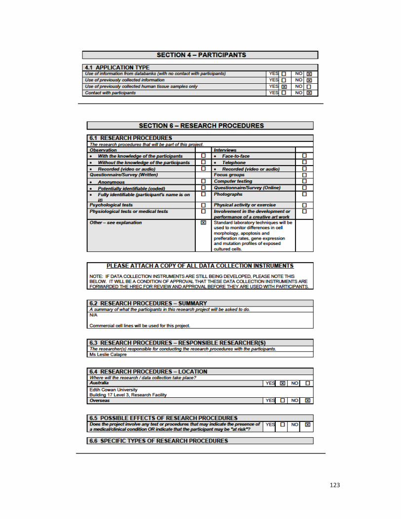
123

124

125

126

127

128

129