The effects of grape seeds polyphenols on SKH-1 mice skin irradiated with multiple doses of UV-B

Int. J. Mol. Sci. 2013, 14, 12222-12248; doi:10.3390/ijms140612222
International Journal of
Molecular Sciences ISSN 1422-0067
www.mdpi.com/journal/ijms
Review
UV Radiation and the Skin
John D’Orazio 1,*, Stuart Jarrett
2, Alexandra Amaro-Ortiz
3 and Timothy Scott
3
1 Graduate Center for Toxicology and the Departments of Pediatrics, Biomedical and Molecular
Pharmacology and Physiology, Markey Cancer Center, University of Kentucky College of Medicine,
800 Rose Street, Lexington, KY 40536, USA 2 Markey Cancer Center, University of Kentucky College of Medicine, 800 Rose Street, Lexington,
KY 40536, USA; E-Mail: [email protected] 3 Graduate Center for Toxicology, University of Kentucky College of Medicine, 800 Rose Street,
Lexington, KY 40536, USA; E-Mail: [email protected] (A.A.-O.); [email protected] (T.S.)
* Author to whom correspondence should be addressed; E-Mail: [email protected];
Tel.: +1-859-323-6238; Fax: +1-859-257-8940.
Received: 25 April 2013; in revised form: 18 May 2013 / Accepted: 24 May 2013 /
Published: 7 June 2013
Abstract: UV radiation (UV) is classified as a ―complete carcinogen‖ because it is both a
mutagen and a non-specific damaging agent and has properties of both a tumor initiator
and a tumor promoter. In environmental abundance, UV is the most important modifiable
risk factor for skin cancer and many other environmentally-influenced skin disorders.
However, UV also benefits human health by mediating natural synthesis of vitamin D and
endorphins in the skin, therefore UV has complex and mixed effects on human health.
Nonetheless, excessive exposure to UV carries profound health risks, including atrophy,
pigmentary changes, wrinkling and malignancy. UV is epidemiologically and molecularly
linked to the three most common types of skin cancer, basal cell carcinoma, squamous cell
carcinoma and malignant melanoma, which together affect more than a million Americans
annually. Genetic factors also influence risk of UV-mediated skin disease. Polymorphisms
of the melanocortin 1 receptor (MC1R) gene, in particular, correlate with fairness of skin,
UV sensitivity, and enhanced cancer risk. We are interested in developing UV-protective
approaches based on a detailed understanding of molecular events that occur after UV
exposure, focusing particularly on epidermal melanization and the role of the MC1R in
genome maintenance.
OPEN ACCESS
Int. J. Mol. Sci. 2013, 14 12223
Keywords: Ultraviolet radiation; skin; carcinogenesis; mutagenesis; pigmentation; cancer;
melanin; melanocortin 1 receptor
1. The Skin
Comprising roughly 16% of body mass, the skin is the largest organ of the body. Skin is
organized into two primary layers, epidermis and dermis, which together are made up of epithelial,
mesenchymal, glandular and neurovascular components. The epidermis, of ectodermal origin, is the
outermost layer and serves as the body’s point of contact with the environment. As such, epidermal
biological and physical characteristics play an enormous role in resistance to environmental stressors
such as infectious pathogens, chemical agents and UV [1–6]. Keratinocytes are the most abundant cells
in the epidermis and are characterized by their expression of cytokeratins and formation of desmosomes
and tight junctions with each other to form an effective physicochemical barrier. The dermis, derived
from mesoderm, underlies the epidermis and harbors cutaneous structures including hair follicles,
nerves, sebaceous glands and sweat glands. The dermis also contains abundant immune cells and
fibroblasts, which actively participate in many physiologic responses in the skin. The epidermis,
demarcated from the dermis by a basement membrane, is organized into functional layers defined
largely by keratinocyte characteristics such as size, shape, nucleation and keratin expression [7]
(Figure 1). Nascent epidermal keratinocytes formed as a result of cell division by keratinocyte stem
cells in the stratum basale undergo a programmed differentiation as they migrate outward toward the
surface of the skin to eventually form corneocytes, which are tightly-linked dead but intact cells that
form the principle barrier of the outermost epidermal layer [8,9].
Besides the creation of a highly effective physical barrier, keratinocytes also accumulate melanin
pigments as they mature, and epidermal melanin functions to potently block UV penetration into the
skin. Although melanin may be found in abundance in epidermal keratinocytes, it is not manufactured
in these cells. Rather, melanin synthesis is restricted to melanocytes, which are derived from neural
crest and are the second most abundant cell in the epidermis [10,11]. In fact, melanocytes can be found
both in the dermis and epidermis. Epidermal melanocytes are generally positioned in the basal layer
above the basement membrane. Melanocytes are also found in hair follicles to impart pigment to
nascent hair [12]. Dermal melanocytes can be found in nevi (moles). Because melanocytes are the only
source of pigment in the skin, inherited pigmentary defects such as albinism tend to be caused by
melanocytic genetic defects [10,13]. Through dendritic extensions, melanocytes may be in intimate
contact with as many as fifty neighboring keratinocytes in what is known as an ―epidermal melanin
unit‖ [11,14]. There are many contact-dependent and paracrine interactions that occur between
keratinocytes and melanocytes in the epidermal melanin unit. Pigment made by melanocytes is
transferred to adjacent keratinocytes in cellular organelles termed melanosomes by way of melanocytic
dendrites [15–17]. In fact, most of the melanin in the skin is found in keratinocytes where it
accumulates to function as a ―natural sunscreen‖ to protect the skin against incoming UV photons.
Besides blocking UV penetration into the skin, melanin may have many other important physiologic

Int. J. Mol. Sci. 2013, 14 12224
effects including regulatory influences over epidermal homeostasis, free radical scavenging to protect
against oxidative injury, and possibly even antimicrobial activity [10,18–24].
Figure 1. Epidermal structure and keratinocyte differentiation. The epidermis is a
self-renewing tissue composed mainly of keratinocytes in various stages of terminal
differentiation. Keratinocytes are produced in the stratum basale (basal layer), and move
outward through the epidermis, undergoing a programmed series of differentiation
involving enucleation and accumulation of cytokeratins and tight junctions with each other.
Keratinocytes also receive melanin from melanocytes in the form of pre-packaged
organelles termed melanosomes. The basic layers from the basement membrane outward
are the stratum basale, stratum spinosum, stratum granulosum, and the stratum corneum,
each identified by the morphology and differentiation state of the keratinocyte as indicated
by expression of cytokeratins and other proteins.
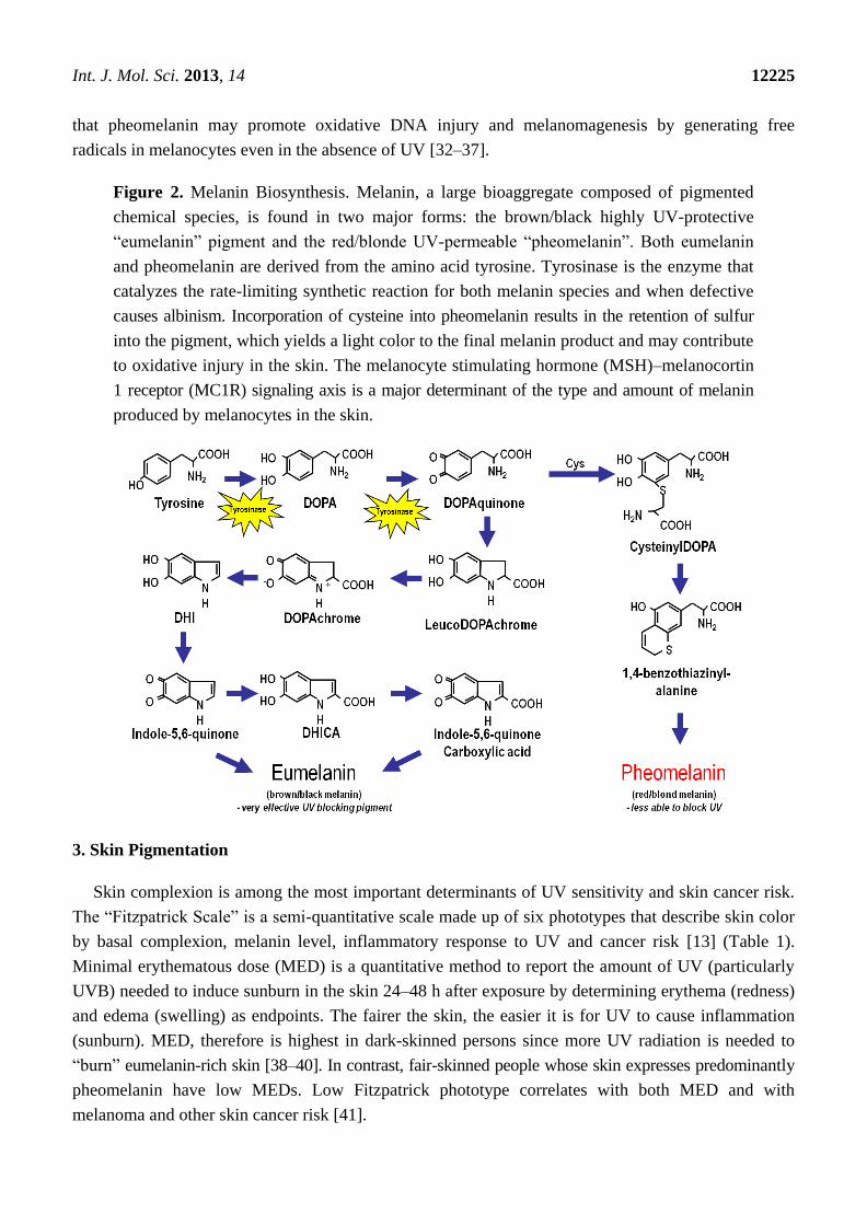
2. Melanin
The amount and type of epidermal melanin is the main factor that determines skin complexion and
UV sensitivity. Melanin is a large bio-aggregate composed of subunits of different pigment species
formed by oxidation and cyclization of the amino acid tyrosine [10,25,26] (Figure 2). Intriguingly, the
intermediates of melanogenesis may have important regulatory roles in the skin [27–29]. Melanin
exists in two main chemical forms: (1) eumelanin, a dark pigment expressed abundantly in the skin of
heavily pigmented individuals, and (2) pheomelanin, a light-colored sulfated pigment resulting from
incorporation of cysteines into melanin precursors [30]. Eumelanin is much more efficient at blocking
UV photons than pheomelanin, thus the more eumelanin in the skin, the less UV-permeable is the
epidermis [31]. Fair-skinned people who are almost always UV-sensitive and have high risk of skin
cancer have little epidermal eumelanin and therefore ―realize‖ much more UV than darker-skinned
individuals. Therefore, the fairer the skin, the more damaging UV exposure will be. In fact, pheomelanin
levels are similar between dark-skinned and light-skinned individuals, and it is the amount of
epidermal eumelanin that determines skin complexion, UV sensitivity and cancer risk. Data suggest
Int. J. Mol. Sci. 2013, 14 12225
that pheomelanin may promote oxidative DNA injury and melanomagenesis by generating free
radicals in melanocytes even in the absence of UV [32–37].
Figure 2. Melanin Biosynthesis. Melanin, a large bioaggregate composed of pigmented
chemical species, is found in two major forms: the brown/black highly UV-protective
―eumelanin‖ pigment and the red/blonde UV-permeable ―pheomelanin‖. Both eumelanin
and pheomelanin are derived from the amino acid tyrosine. Tyrosinase is the enzyme that
catalyzes the rate-limiting synthetic reaction for both melanin species and when defective
causes albinism. Incorporation of cysteine into pheomelanin results in the retention of sulfur
into the pigment, which yields a light color to the final melanin product and may contribute
to oxidative injury in the skin. The melanocyte stimulating hormone (MSH)–melanocortin
1 receptor (MC1R) signaling axis is a major determinant of the type and amount of melanin
produced by melanocytes in the skin.
3. Skin Pigmentation
Skin complexion is among the most important determinants of UV sensitivity and skin cancer risk.
The ―Fitzpatrick Scale‖ is a semi-quantitative scale made up of six phototypes that describe skin color
by basal complexion, melanin level, inflammatory response to UV and cancer risk [13] (Table 1).
Minimal erythematous dose (MED) is a quantitative method to report the amount of UV (particularly
UVB) needed to induce sunburn in the skin 24–48 h after exposure by determining erythema (redness)
and edema (swelling) as endpoints. The fairer the skin, the easier it is for UV to cause inflammation
(sunburn). MED, therefore is highest in dark-skinned persons since more UV radiation is needed to
―burn‖ eumelanin-rich skin [38–40]. In contrast, fair-skinned people whose skin expresses predominantly
pheomelanin have low MEDs. Low Fitzpatrick phototype correlates with both MED and with
melanoma and other skin cancer risk [41].

Int. J. Mol. Sci. 2013, 14 12226
Table 1. Skin pigmentation, the Fitzpatrick scale and UV risk.
Fitzpatrick
phototype Phenotype
Epidermal
eumelanin Cutaneous response to UV
MED
(mJ/cm2) *
Cancer
risk
I
Unexposed skin is bright white
Blue/green eyes typical
Freckling frequent
Northern European/British
+/−
Always burns
Peels
Never tans
15–30 ++++
II
Unexposed skin is white
Blue, hazel or brown eyes
Red, blonde or brown hair
European/Scandinavian
+
Burns easily
Peels
Tans minimally
25–40 +++/++++
III
Unexposed skin is fair
Brown eyes
Dark hair
Southern or Central European
++ Burns moderately
Average tanning ability 30–50 +++
IV
Unexposed skin is light brown
Dark eyes
Dark hair
Mediterranean, Asian or Latino
+++ Burns minimally
Tans easily 40–60 ++
V
Unexposed skin is brown
Dark eyes
Dark hair
East Indian, Native American,
Latino or African
++++ Rarely burns
Tans easily and substantially 60–90 +
VI
Unexposed skin is black
Dark eyes
Dark hair
African or Aboriginal ancestry
+++++ Almost never burns
Tans readily and profusely 90–150 +/−
Minimal erythematous dose (MED) is defined as the least amount of UVB radiation that causes reddening and
inflammation of the skin 24–48 h after exposure (i.e., the lowest UV dose that causes sunburn). The more UV sensitive an
individual is, the lower the MED of his/her skin.
4. Ultraviolet Radiation (UV)
Abundant in the environment, UV contributes to a variety of skin maladies including inflammation,
degenerative aging and cancer [1]. Historically, humans have been exposed to UV radiation mainly
through occupational exposure to sunlight. Recreational UV exposure, however, has increased
dramatically in recent years because of outdoor leisure activities and to purposely tan for cosmetic
purposes [42,43]. Being a component of the electromagnetic spectrum, UV photons fall between the
wavelengths of visible light and gamma radiation. UV energy can be subdivided into UV-A, -B and -C
components based on electro physical properties, with UV-C photons having the shortest wavelengths
(100–280 nm) and highest energy, UV-A having the longest (315–400 nm) but least energetic photons
and UV-B falling in between (Figure 3). Each component of UV can exert a variety of effects on cells,
tissues and molecules.

Int. J. Mol. Sci. 2013, 14 12227
Figure 3. Electromagnetic spectrum of visible and UV radiation and biologic effects on the
skin. Solar UV radiation can be subdivided into UVA, UVB and UVC components,
however because of atmospheric ozone that absorbs UVC, ambient sunlight is predominantly
UVA (90%–95%) and UVB (5%–10%). UV penetrates the skin in a wavelength-
dependent manner. Longer wavelength UVA penetrates deeply into the dermis reaching
well into the dermis. In contrast, UVB is almost completely absorbed by the epidermis, with
comparatively little reaching the dermis. UVA is efficient at generating reactive oxygen
species that can damage DNA via indirect photosensitizing reactions. UVB is directly
absorbed by DNA which causes molecular rearrangements forming the specific
photoproducts such as cyclobutane dimers and 6–4 photoproducts. Mutations and cancer
can result from many of these modifications to DNA.
Ambient UV exposure varies geographically according to intensity of sunlight in a particular
location on Earth. Since UV radiation can be reflected, scattered and dampened by atmospheric
particles, ambient UV dose varies according to the amount of atmosphere it must pass through, making
UV doses higher nearest the Equator (where sunlight strikes the Earth most directly), at higher
altitudes and in conditions of minimal cloud or particulate cover. Personal UV dosing depends not only
on strength of solar radiation, but also on time spent outdoors occupationally or recreationally and the
usage of UV-protective clothing, shade and sun blocks. Since equatorial locations tend to be warm and
conducive to recreational or occupational outdoor activities, people living such locales typically wear
less clothing and have more contact with ambient sunlight and usually receive much higher ambient
UV doses than persons inhabiting temperate climates. Not surprisingly, skin cancer risk generally
mirrors this geographic pattern, particularly among fair-skinned sun-sensitive persons [44–46].
Int. J. Mol. Sci. 2013, 14 12228
5. Indoor Tanning
The number and use of indoor tanning salons has skyrocketed over the last several years. In
America alone, only 1% of the population had ever used a tanning bed in the late 1980s. Now it is
estimated that over 25% of Americans have engaged in purposeful exposure to artificial UV
radiation [47]. Indoor tanning is an important industry with nearly 30 million clients, 100,000 employees
and billions of dollars of annual business. Indoor tanning machines are poorly regulated and vary widely
with respect to UV composition and strength. UV output from tanning beds can be up to ten times
more powerful than sunlight [48,49], making the tanning bed an authentic carcinogenic instrument.
Tanning can be addictive, leading to frequent and significant UV exposure over time [50–52], and
since tanning often appeals to adolescents and young adults, tanning patrons’ UV history can be
significant for many years [53].
Indoor tanning clearly increases incidence of skin cancers [54,55]. With respect to melanoma, the
deadliest of skin malignancies, lifetime risk increases by 75% if people engage in artificial tanning
before the age of 35 years [56–58]. Cancer risk increases with years of use, number of sessions, and
total number of UV h exposed [54,56,59,60]. Since the molecular pathways in the skin that activate
UV-induced tanning result from cellular and DNA damage which underlie skin damage and
carcinogenesis (Figure 4), it appears as though there is no ―safe‖ use of tanning salons [57]. The
tanning industry has engaged a powerful political lobby to further its commercial interests by downplaying
the adverse health risks of UV. Instead, the industry publicizes the health benefits of UV to its clients,
emphasizing vitamin D production which is naturally made in the skin by the chemical conversion of
7-dehydrocholesterol into vitamin D3 (cholecalciferol) after UVB exposure [61–69]. In fact, UV doses
that induce tanning far exceed what is required for adequate vitamin D production and the widespread
availability of vitamin D in supplements and fortified foods minimizes the need for UV exposure to
avoid symptoms of rickets and vitamin D deficiency [70–74]. Multiple studies report overwhelming
evidence that the risks of indoor tanning far outweigh potential health benefits, most significantly for
malignancy. Decreasing UV radiation exposure, both naturally from sunlight and artificially from tanning
bed use, may be the single best way to reduce incidence of melanoma and other skin cancers [75].
6. Cutaneous Responses to UV
UV has many effects on skin physiology, with some consequences occurring acutely and others in a
delayed manner. One of the most obvious acute effects of UV on the skin is the induction of inflammation.
UVB induces a cascade of cytokines, vasoactive and neuroactive mediators in the skin that together
result in an inflammatory response and causes ―sunburn‖ [3,4,6,76–79]. If the dose of UV exceeds
a threshold damage response, keratinocytes activate apoptotic pathways and die. Such apoptotic
keratinocytes can be identified by their pyknotic nuclei and are known as ―sunburn cells‖ [80]. UV
also leads to an increase in epidermal thickness, termed hyperkeratosis. By causing cell injury, UV
induces damage response pathways in keratinocytes. Damage signals such as p53 activation
profoundly alter keratinocyte physiology, mediating cell cycle arrest, activating DNA repair and
inducing apoptosis if the damage is sufficiently great. Several h after UV exposure, however, and
damage response signals abate, epidermal keratinocytes proliferate robustly [81], mediated by a variety

Int. J. Mol. Sci. 2013, 14 12229
of epidermal growth factors. Increased keratinocyte cell division after UV exposure leads to
accumulation of epidermal keratinocytes which increases epidermal thickness. Epidermal hyperplasia
protects the skin better against UV penetration [82].
Figure 4. Mechanisms of the physiologic tanning response. Hormonal interactions between
epidermal keratinocytes and melanocytes mediate much of the cutaneous melanization
response. DNA and cellular damage in keratinocytes up-regulates transcription of the
pro-opiomelanocortin (POMC) gene which encodes production and secretion of
melanocyte stimulating hormone (α-MSH). α-MSH binding to melanocortin 1 receptor
(MC1R) on melanocytes in the basal epidermis generates the second messenger cAMP via
interactions between MC1R and adenylyl cyclase, and leads to activation of protein kinase
A and the cAMP responsive binding element (CREB) and microphthalmia (Mitf) transcription
factors. CREB and Mitf directly enhance melanin production by raising levels of tyrosinase
and other melanin biosynthetic enzymes. Thus, MSH-MC1R signaling leads to enhanced
pigment synthesis by melanocytes and accumulation of melanin by epidermal keratinocytes.
By this mechanism, the skin is better protected against UV insults. Of note, UV-induced
pigmentation may also occur through other signaling pathways as well as direct effects of
UV on melanocytes, and there is some disagreement in the field over the role of epidermal
MSH in the adaptive pigmentary response.

Int. J. Mol. Sci. 2013, 14 12230
Coupled with epidermal hyperkeratosis is adaptive melanization of the skin, also known as
tanning [4,10,83–86]. UV up-regulates production and epidermal accumulation of melanin pigment in
the skin [87–91]. This important physiologic response protects the skin against subsequent UV damage,
and defects in this pathway are linked with cancer susceptibility. UV-mediated skin darkening is actually
biphasic, with initial skin darkening occurring from redistribution and/or molecular changes to existing
epidermal melanin pigments. Delayed increases in skin darkening, mediated by actual up-regulation in
melanin synthesis and transfer to keratinocytes, begin several h to days after UV exposure [92,93].
Adaptive melanization is likely a complex physiologic response [4,10,83,85] involving multiple skin
cell types interacting in a variety of ways (Figure 4) [86,94–102]. UV has many other effects on the
skin, including induction of an immune-tolerant or immunosuppressive state [103–110] and production
of vitamin D by direct conversion of 7-dehydrocholesterol into vitamin D3 (cholecalciferol) [61–69].
Ambient sunlight, for the most part, is a mixture of UVA and UVB, yet each UV component may exert
different and distinct effects on the skin [111,112]. UVB, for example, is a potent stimulator of
inflammation and the formation of DNA photolesions (such as mutagenic thymine dimers) [112,113],
whereas UVA is much less active in these measures but instead is a potent driver of oxidative free
radical damage to DNA and other macromolecules [114–116]. Thus, each may contribute to
carcinogenesis through different mechanisms [117–119]. The influence of UVA and UVB on skin
physiology is an active area of investigation.
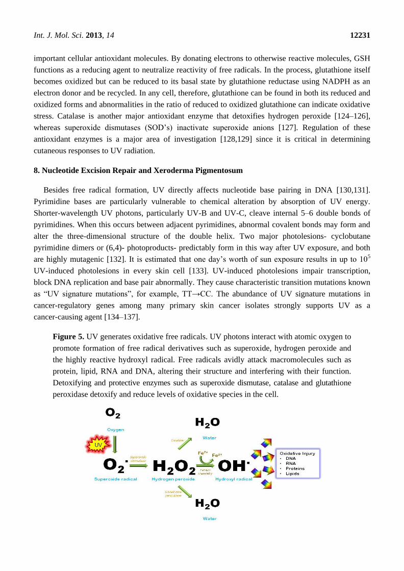
7. Oxidative Injury
Besides promoting formation of photodimers in the genome, UV causes mutations by generating
reactive oxygen species (ROS) such as superoxide anion, hydrogen peroxide and the hydroxyl
radical [21] (Figure 5). Nucleotides are highly susceptible to free radical injury. Oxidation of
nucleotide bases promotes mispairing outside of normal Watson-Crick parameters, causing
mutagenesis [120]. The transversion guanine→thymine, for example, is a well-characterized mutation
caused by ROS by oxidizing guanine at the 8th position to produce 8-hydroxy-2'-deoxyguanine
(8-OHdG) [121,122]. 8-OHdG tends to pair with an adenine instead of cytosine and therefore this
oxidative change mutates a G/C pair into an A/T pair. Such mutations can be found in tumors isolated
from the skin, suggesting that oxidative injury can be carcinogenic [123]. Cellular maintenance
pathways exist to inactivate oxidative species as well as to repair the DNA damage they cause. The
base excision repair pathway (BER) is the main molecular means by which cells reverse free radical
damage in DNA to avoid oxidative mutagenesis. This pathway is initiated by damage-specific
glycosylases that scan DNA for specific alterations including deaminated, alkylated or oxidized bases.
After altered or inappropriate bases are recognized by a lesion-specific glycosylase, the enzyme
cleaves the nucleotide base from the sugar and phosphodiesterase backbone by lysis of the
N-glycosylic bond between the base and the deoxyribose. This step forms an abasic or
apurinic/apyrimidinic (AP) site in the DNA, which is then processed and repaired using the
complementary strand as a template to ensure fidelity.
Cells also have a complex and robust network of anti-oxidant molecules that detoxify reactive
species to prevent free radical changes to DNA and other macromolecules. Glutathione (GSH) is an
oligopeptide made up of three amino acids- cysteine, glycine and glutamine and is among the most
Int. J. Mol. Sci. 2013, 14 12231
important cellular antioxidant molecules. By donating electrons to otherwise reactive molecules, GSH
functions as a reducing agent to neutralize reactivity of free radicals. In the process, glutathione itself
becomes oxidized but can be reduced to its basal state by glutathione reductase using NADPH as an
electron donor and be recycled. In any cell, therefore, glutathione can be found in both its reduced and
oxidized forms and abnormalities in the ratio of reduced to oxidized glutathione can indicate oxidative
stress. Catalase is another major antioxidant enzyme that detoxifies hydrogen peroxide [124–126],
whereas superoxide dismutases (SOD’s) inactivate superoxide anions [127]. Regulation of these
antioxidant enzymes is a major area of investigation [128,129] since it is critical in determining
cutaneous responses to UV radiation.
8. Nucleotide Excision Repair and Xeroderma Pigmentosum
Besides free radical formation, UV directly affects nucleotide base pairing in DNA [130,131].
Pyrimidine bases are particularly vulnerable to chemical alteration by absorption of UV energy.
Shorter-wavelength UV photons, particularly UV-B and UV-C, cleave internal 5–6 double bonds of
pyrimidines. When this occurs between adjacent pyrimidines, abnormal covalent bonds may form and
alter the three-dimensional structure of the double helix. Two major photolesions- cyclobutane
pyrimidine dimers or (6,4)- photoproducts- predictably form in this way after UV exposure, and both
are highly mutagenic [132]. It is estimated that one day’s worth of sun exposure results in up to 105
UV-induced photolesions in every skin cell [133]. UV-induced photolesions impair transcription,
block DNA replication and base pair abnormally. They cause characteristic transition mutations known
as ―UV signature mutations‖, for example, TT→CC. The abundance of UV signature mutations in
cancer-regulatory genes among many primary skin cancer isolates strongly supports UV as a
cancer-causing agent [134–137].
Figure 5. UV generates oxidative free radicals. UV photons interact with atomic oxygen to
promote formation of free radical derivatives such as superoxide, hydrogen peroxide and
the highly reactive hydroxyl radical. Free radicals avidly attack macromolecules such as
protein, lipid, RNA and DNA, altering their structure and interfering with their function.
Detoxifying and protective enzymes such as superoxide dismutase, catalase and glutathione
peroxidase detoxify and reduce levels of oxidative species in the cell.

Int. J. Mol. Sci. 2013, 14 12232
Figure 6. UV-induced cyclobutane dimers- structure (A) and repair by the Nucleotide
Excision DNA Repair (NER) pathway (B). The NER pathway is mediated by at least eight
enzymes that work together to identify bulky DNA lesions that distort the structure of the
double helix, excise the damaged portion and replace the excised region by DNA synthesis
directed by the complementary strand. Homozygous deficiency in any one of the NER
enzymes leads to the clinical condition known as Xeroderma Pigmentosum (XP). Although
not shown, NER can also be initiated in actively transcribed regions of the genome by
involvement of the Cockayne syndrome proteins A and B.
Nucleotide excision repair (NER) is an evolutionarily-conserved mechanism for repairing
UV-induced photoproducts and other bulky DNA lesions [138]. The importance of NER in cancer
resistance is best illustrated by considering the natural history of patients with Xeroderma
Pigmentosum (XP), a rare UV hypersensitivity syndrome caused by homozygous defects in any one of
at least eight required effector proteins of a common pathway that executes NER: XPA, ERCC1,
ERCC3 (XP-B), XPC, ERCC2 (XP-D), DDB2 (XP-E), ERCC4 (XP-F), ERCC5 (XP-G) and POLH. XP
patients demonstrate profound UV sensitivity and develop characteristic skin changes including
pigmentary abnormalities, capillary telangiectasias and atrophy on UV-exposed anatomic sites at very
early ages. Premalignant lesions and skin cancers develop in high frequency and much sooner than in
unaffected persons. Basal cell carcinomas, squamous cell carcinomas and melanomas often develop
before the second decade of life, decades before the general population [139]. Moreover, XP-associated
skin cancers frequently demonstrate ―UV signature mutations‖, clearly indicating the importance of
NER in the cancer resistance [140]. The NER pathway represents an orchestrated interaction of
enzymes that function together to repair lesions that alter the three-dimensional structure of DNA.
After recognition of damage and recruitment of a multiprotein repair complex to the damaged site, the
damage strand is nicked several nucleotides away on either side of the damaged bases. The damaged
region is excised and the resulting gap is filled in by a DNA polymerase using the non-damaged strand
as a template [141–143] (Figure 6). Though only a handful of core factors are necessary and sufficient
for the repair of UV-induced DNA lesions, there are numerous accessory factors that regulate this
genome maintenance pathway. While the importance of NER in UV and skin cancer resistance is most
Int. J. Mol. Sci. 2013, 14 12233
clearly demonstrated by the natural history of patients with XP, attention is being paid to the role of
NER polymorphisms on UV sensitivity and skin cancer incidence in sporadic populations.
9. Skin Cancer
Skin cancers are by far the most common malignancies of humans, with well over a million cases
diagnosed each year [144]. Roughly 1 in 5 Americans will develop skin cancer in their lifetime [145].
They account for nearly 15,000 deaths and more than three billion dollars each year in medical costs in
the United States alone [146,147]. Like many other cancers contributed to by environmental etiologies
(in this case UV), skin cancer incidence increases markedly with age presumably reflecting the long
latency between carcinogen exposure and cancer formation. Skin cancers are commonly grouped
into two main categories, melanoma and non-melanoma skin cancers (NMSC), based on cell of origin
and clinical behavior. Risk of skin cancer is heavily influenced by UV exposure and by skin
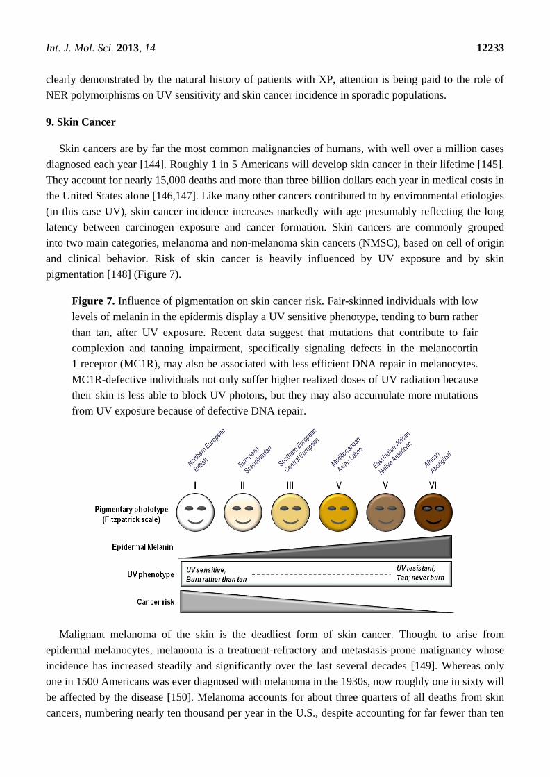
pigmentation [148] (Figure 7).
Figure 7. Influence of pigmentation on skin cancer risk. Fair-skinned individuals with low
levels of melanin in the epidermis display a UV sensitive phenotype, tending to burn rather
than tan, after UV exposure. Recent data suggest that mutations that contribute to fair
complexion and tanning impairment, specifically signaling defects in the melanocortin
1 receptor (MC1R), may also be associated with less efficient DNA repair in melanocytes.
MC1R-defective individuals not only suffer higher realized doses of UV radiation because
their skin is less able to block UV photons, but they may also accumulate more mutations
from UV exposure because of defective DNA repair.
Malignant melanoma of the skin is the deadliest form of skin cancer. Thought to arise from
epidermal melanocytes, melanoma is a treatment-refractory and metastasis-prone malignancy whose
incidence has increased steadily and significantly over the last several decades [149]. Whereas only
one in 1500 Americans was ever diagnosed with melanoma in the 1930s, now roughly one in sixty will
be affected by the disease [150]. Melanoma accounts for about three quarters of all deaths from skin
cancers, numbering nearly ten thousand per year in the U.S., despite accounting for far fewer than ten
Int. J. Mol. Sci. 2013, 14 12234
percent of all skin malignancies. Melanoma burden is predictably largest in places with large numbers
of fair-skinned individuals living in warm, sunny climates [151]. Most melanomas arise out of
pre-existing moles, therefore having many nevi is another important risk factor for the disease. If
caught early, many melanomas can be managed by surgical excision alone. However, melanomas are
quick to invade and metastasize and long-term survival is poor for advanced disease. Even with recent
progress made in targeted therapy [152–156] and immunotherapy [157,158], melanoma is notoriously
difficult to treat once it has spread beyond its original site. It is not clear why melanoma incidence has
increased so dramatically over the past several decades, but it is likely multifactorial, with contributions
from increased UV exposure, environmental and inherited cancer risk factors and better surveillance
and earlier detection [151,159–172].
Non-melanomatous skin cancers greatly outnumber melanomas in incidence, but fortunately most
are much easier to treat and have much better long-term prognosis. The two major forms, basal cell
carcinomas and squamous cell carcinomas, are both derived from epidermal keratinocytes. They are
less deadly than melanoma mainly due to their tendency to remain confined to their primary site of
disease, which makes their management much more straightforward. The overwhelming majority of
keratinocyte malignancies develop in the areas of skin most exposed to UV, such as on the face and
arms. Most are effectively treated by local control measures alone such as resection, MOHS microsurgery
or cryosurgery.
There are strong epidemiologic and molecular data linking all forms of skin cancer to UV
exposure [173], and it is estimated that UV is causative for nearly 65% of melanoma and 90% of
non-melanoma skin cancers [174,175]. UV-signature mutations in key cancer-relevant genes such as
the p53 tumor suppressor in squamous cell carcinoma for example are well-characterized, and exome
analysis of a panel of melanomas revealed strong genetic evidence for a direct mutagenic role of UV
radiation in the pathogenesis of melanoma [137,176–183]. Since UV-induced DNA mutations
represent a major causative factor for melanoma and other skin cancers, it follows that resistance to
UV-mediated mutagenesis is a critical determinant of skin cancer risk [184].
10. The Melanocortin 1 Receptor (MC1R)
The melanocortin 1 receptor (MC1R) is a critical genetic locus involved in pigmentation, the
adaptive tanning response and skin cancer susceptibility [185–192]. The MC1R is found on the surface
of melanocytes where it binds to α-melanocyte stimulating hormone (MSH) and transmits differentiation
signals into the cell through activation of adenylyl cyclase and generation of cAMP [193–195]. cAMP
signaling leads to activation of the protein kinase A (PKA) cascade which, in turn, leads to increased
levels and/or activity of many melanogenic enzymes to enhance production and export of melanin by
melanocytes [90,196,197] (Figure 4). MC1R signaling also decreases UV-mediated mutagenesis by
enhancing genome maintenance pathways in melanocytes [125,126,192,198]. Loss-of-signaling MC1R
polymorphisms are commonly found among fair-skinned, sun-sensitive and skin cancer-prone
populations (e.g., Northern Europeans). The most prevalent MC1R mutations (D84E, R151C, R160W
and D294H) are commonly referred to as ―RHC‖ (red hair color) alleles because of their association
with red hair color, freckling and tendency to burn after UV exposure [199,200]. Loss of signaling
MC1R alleles such as the RHC variants are associated with up to a four-fold increased lifetime risk of
Int. J. Mol. Sci. 2013, 14 12235
melanoma and other skin cancers [201–203]. Overall, there is much evidence placing MC1R as a
critical determinant of skin cancer risk, and regulation of eumelanin by POMC derived peptides
depends on genetic context [204].
MC1R signaling protects the skin from UV damage by at least two major mechanisms. First, by
inducing pigment synthesis in melanocytes, MC1R enhance production and accumulation of
eumelanin in the epidermis. Epidermal melanization blocks penetration of UV into the skin, reducing
realized doses of UV and decreasing mutagenesis and cancer risk. MC1R signaling also directly
influences UV resistance of melanocytes by enhancing nucleotide excision DNA repair and
oxidative resistance. Since MC1R signaling is potentially targetable by agents that influence cAMP
levels [82,84,205], pharmacologic manipulation of cutaneous cAMP may be a useful approach to
reduce UV sensitivity and cancer risk. Theoretically, raising cAMP levels in the skin can be accomplished
either by stimulating its production (e.g., adenylyl cyclase activation) or by impeding its degradation
(e.g., phosphodiesterase inhibition). Both of these approaches have been quite successful in enhancing
epidermal melanin levels in animal models [84,206] and each would be expected to be effective even
in individuals harboring loss-of-signaling functional mutations in MC1R. Alternatively, α-MSH or
agonistic MC1R peptide ligands would offer more specificity (working only on melanocytes) but might be
less effective in individuals with inherited MC1R signaling defects [192,193,207].
11. Conclusions
One of the greatest risk factors for the development of cutaneous melanoma is having a fair skin
complexion, which is characterized by low levels of a UV-blocking dark pigment called eumelanin in
the epidermis. Individuals with light skin pigmentation suffer comparatively more skin damage from
UV because it is relatively easy for UV rays to penetrate the epidermis to damage both keratinocytes
and melanocytes in the deeper layers of the epidermis. Fair-skinned individuals are exposed to higher
―realized‖ doses of UV radiation in the skin and UV-induced mutations, which directly contribute to
melanoma and other forms of skin cancer, accumulate over time. Much UV-induced pathology,
including skin cancer, can be avoided by minimizing UV exposure (Table 2).
We and others are increasingly interested in heritable factors that determine melanoma risk to be
able to intervene in the carcinogenic process. One of the most important alleles that influences skin
cancer risk is the melanocortin 1 receptor (MC1R), whose function is central to the adaptive
pigmentation (tanning) response in the skin. Besides mediating the tanning response, MC1R exerts a
powerful influence on the ability of melanocytes to repair UV-induced DNA damage by the nucleotide
excision DNA repair pathway. New insights into the ways in which MC1R and other genes function to
protect the skin against the harmful consequences of UV may allow the rational development of
pharmacologic strategies to reduce UV sensitivity and cancer risk.

Int. J. Mol. Sci. 2013, 14 12236
Table 2. UV Safety Tips.
Sun exposure Minimize time outdoors during ―peak‖ UV h (10 am to 4 pm). Seek shade as much as
possible. Be aware that sunlight bounces off reflective surfaces and can reach you even
under an umbrella or tree.
Avoid getting a sunburn. More than 5 sunburns doubles risk of skin cancer.
Use sunscreens with a sun protection factor (SPF) >15. Make sure to apply repeatedly
(especially with sweating or swimming) and as directed. Use sunblocks that offer protection
from both UV-A and UV-B rays, and be sure to cover often-missed spots- lips, ears, around
eyes, neck, scalp, hands and feet.
Wear protective clothing such as rash guards and tightly woven fabrics.
Wear a hat. Wide-brimmed hats protect head, face, ears and neck. If a baseball cap is worn,
make sure to use sunscreen on ears and neck.
Wear UV-protective sunglasses
Strength of solar UV increases at high altitude and with less cloud cover. Monitor the UV
Index (http://www.epa.gov/sunwise/uvindex.html) and plan accordingly.
Get Vitamin D safely by relying on diet and supplements rather than UV exposure.
Artificial Tanning Do not frequent tanning beds. They can be more dangerous than sunlight. Frequent use of
artificial tanning products clearly increases risk of each of the major kinds of skin cancer,
including melanoma.
Sunless self-tanning products seem safe but typically offer little sun-blocking UV protection
on their own.
Awareness Examine your skin frequently, at least once a month, head to toe. Use a full-length mirror
and a hand mirror to check your back, or involve a partner. Have a professional skin
examination annually.
Seek professional medical attention for:
Sores that do not heal
Changes in moles (growth, irregularity, asymmetry, color changes, elevation, pain, itching)
Skin cancers are much more easily treated when caught early.
Acknowledgments
The authors wish to thank current and past funding sources: the National Cancer Institute
(R01 CA131075, R01 CA131075-02S1), the Wendy Will Case Cancer Research Fund, the Markey
Cancer Foundation, the Children’s Miracle Network and the Jennifer and David Dickens Melanoma
Research Foundation.
Conflicts of Interest
The authors declare no conflicts of interest.
References
1. Elwood, J.M.; Jopson, J. Melanoma and sun exposure: An overview of published studies. Int. J.
Cancer 1997, 73, 198–203.
2. Lowe, N.J. An overview of ultraviolet radiation, sunscreens, and photo-induced dermatoses.
Dermatol. Clin. 2006, 24, 9–17.

Int. J. Mol. Sci. 2013, 14 12237
3. Slominski, A.; Wortsman, J. Neuroendocrinology of the skin. Endocr. Rev. 2000, 21, 457–487.
4. Slominski, A.; Wortsman, J.; Luger, T.; Paus, R.; Solomon, S. Corticotropin releasing hormone
and proopiomelanocortin involvement in the cutaneous response to stress. Physiol. Rev. 2000, 80,
979–1020.
5. Fuchs, E. Scratching the surface of skin development. Nature 2007, 445, 834–842.
6. Slominski, A.T.; Zmijewski, M.A.; Skobowiat, C.; Zbytek, B.; Slominski, R.M.; Steketee, J.D.
Sensing the environment: Regulation of local and global homeostasis by the skin’s
neuroendocrine system. Adv. Anat. Embryol. Cell. Biol. 2012, 212, v-115.
7. Fuchs, E.; Raghavan, S. Getting under the skin of epidermal morphogenesis. Nat. Rev. Genet.
2002, 3, 199–209.
8. Madison, K.C. Barrier function of the skin: ―la raison d'etre‖ of the epidermis. J. Invest.
Dermatol. 2003, 121, 231–241.
9. Proksch, E.; Brandner, J.M.; Jensen, J.M. The skin: An indispensable barrier. Exp. Dermatol.
2008, 17, 1063–1072.
10. Slominski, A.; Tobin, D.J.; Shibahara, S.; Wortsman, J. Melanin pigmentation in mammalian
skin and its hormonal regulation. Physiol. Rev. 2004, 84, 1155–1228.
11. Nordlund, J.J. The melanocyte and the epidermal melanin unit: An expanded concept.
Dermatol. Clin. 2007, 25, 271–281.
12. Slominski, A.; Wortsman, J.; Plonka, P.M.; Schallreuter, K.U.; Paus, R.; Tobin, D.J. Hair follicle
pigmentation. J. Invest. Dermatol. 2005, 124, 13–21.
13. Scherer, D.; Kumar, R. Genetics of pigmentation in skin cancer—A review. Mutat. Res. 2010,
705, 141–153.
14. Jimbow, K.; Salopek, T.G.; Dixon, W.T.; Searles, G.E.; Yamada, K. The epidermal melanin unit
in the pathophysiology of malignant melanoma. Am. J. Dermatopathol. 1991, 13, 179–188.
15. Joshi, P.G.; Nair, N.; Begum, G.; Joshi, N.B.; Sinkar, V.P.; Vora, S. Melanocyte-keratinocyte
interaction induces calcium signalling and melanin transfer to keratinocytes. Pigment. Cell Res.
2007, 20, 380–384.
16. Cardinali, G.; Bolasco, G.; Aspite, N.; Lucania, G.; Lotti, L.V.; Torrisi, M.R.; Picardo, M.
Melanosome transfer promoted by keratinocyte growth factor in light and dark skin-derived
keratinocytes. J. Invest. Dermatol. 2008, 128, 558–567.
17. Yamaguchi, Y.; Hearing, V.J. Physiological factors that regulate skin pigmentation. Biofactors
2009, 35, 193–199.
18. Slominski, A.; Paus, R.; Schadendorf, D. Melanocytes as ―sensory‖ and regulatory cells in the
epidermis. J. Theor. Biol. 1993, 164, 103–120.
19. Kalka, K.; Mukhtar, H.; Turowski-Wanke, A.; Merk, H. Biomelanin antioxidants in cosmetics:
Assessment based on inhibition of lipid peroxidation. Skin Pharmacol. Appl. Skin Physiol. 2000,
13, 143–149.
20. Mackintosh, J.A. The antimicrobial properties of melanocytes, melanosomes and melanin and the
evolution of black skin. J. Theor. Biol. 2001, 211, 101–113.
21. Meyskens, F.L., Jr.; Farmer, P.; Fruehauf, J.P. Redox regulation in human melanocytes and
melanoma. Pigment. Cell Res. 2001, 14, 148–154.
Int. J. Mol. Sci. 2013, 14 12238
22. Double, K.L.; Ben-Shachar, D.; Youdim, M.B.; Zecca, L.; Riederer, P.; Gerlach, M. Influence of
neuromelanin on oxidative pathways within the human substantia nigra. Neurotoxicol. Teratol.
2002, 24, 621–628.
23. Herrling, T.; Jung, K.; Fuchs, J. The role of melanin as protector against free radicals in skin and
its role as free radical indicator in hair. Spectrochim. Acta A Mol. Biomol. Spectrosc. 2008, 69,
1429–1435.
24. Wang, A.; Marino, A.R.; Gasyna, Z.; Gasyna, E.; Norris, J., Jr. Photoprotection by porcine
eumelanin against singlet oxygen production. Photochem. PhotoBiol. 2008, 84, 679–682.
25. Riley, P.A. Melanin. Int. J. Biochem. Cell. Biol. 1997, 29, 1235–1239.
26. Meredith, P.; Sarna, T. The physical and chemical properties of eumelanin. Pigment. Cell Res.
2006, 19, 572–594.
27. Slominski, A.; Paus, R. Are L-tyrosine and L-dopa hormone-like bioregulators? J. Theor. Biol.
1990, 143, 123–138.
28. Slominski, A. Neuroendocrine activity of the melanocyte. Exp. Dermatol. 2009, 18, 760–763.
29. Slominski, A.; Zmijewski, M.A.; Pawelek, J. L-tyrosine and L-dihydroxyphenylalanine as
hormone-like regulators of melanocyte functions. Pigment. Cell Melanoma Res. 2012, 25, 14–27.
30. Ito, S.; Wakamatsu, K.; Ozeki, H. Chemical analysis of melanins and its application to the study
of the regulation of melanogenesis. Pigment. Cell Res. 2000, 13, 103–109.
31. Vincensi, M.R.; d’Ischia, M.; Napolitano, A.; Procaccini, E.M.; Riccio, G.; Monfrecola, G.;
Santoianni, P.; Prota, G. Phaeomelanin versus eumelanin as a chemical indicator of ultraviolet
sensitivity in fair-skinned subjects at high risk for melanoma: A pilot study. Melanoma Res.
1998, 8, 53–58.
32. Benedetto, J.P.; Ortonne, J.P.; Voulot, C.; Khatchadourian, C.; Prota, G.; Thivolet, J. Role of
thiol compounds in mammalian melanin pigmentation: Part I. Reduced and oxidized glutathione.
J. Invest. Dermatol. 1981, 77, 402–405.
33. Benedetto, J.P.; Ortonne, J.P.; Voulot, C.; Khatchadourian, C.; Prota, G.; Thivolet, J. Role of
thiol compounds in mammalian melanin pigmentation. II. Glutathione and related enzymatic
activities. J. Invest. Dermatol. 1982, 79, 422–424.
34. Sealy, R.C.; Hyde, J.S.; Felix, C.C.; Menon, I.A.; Prota, G.; Swartz, H.M.; Persad, S.;
Haberman, H.F. Novel free radicals in synthetic and natural pheomelanins: Distinction between
dopa melanins and cysteinyldopa melanins by ESR spectroscopy. Proc. Natl. Acad. Sci. USA
1982, 79, 2885–2889.
35. Costantini, C.; d’Ischia, M.; Palumbo, A.; Prota, G. Photochemistry of 5-S-cysteinyldopa.
Photochem. PhotoBiol. 1994, 60, 33–37.
36. Prota, G. Melanins, melanogenesis and melanocytes: Looking at their functional significance
from the chemist’s viewpoint. Pigment. Cell Res. 2000, 13, 283–293.
37. Mitra, D.; Luo, X.; Morgan, A.; Wang, J.; Hoang, M.P.; Lo, J.; Guerrero, C.R.; Lennerz, J.K.;
Mihm, M.C.; Wargo, J.A.; et al. An ultraviolet-radiation-independent pathway to melanoma
carcinogenesis in the red hair/fair skin background. Nature 2012, 491, 449–453.
38. Lu, H.; Edwards, C.; Gaskell, S.; Pearse, A.; Marks, R. Melanin content and distribution in the
surface corneocyte with skin phototypes. Br. J. Dermatol. 1996, 135, 263–267.
Int. J. Mol. Sci. 2013, 14 12239
39. Andreassi, L.; Flori, M.L.; Rubegni, P. Sun and skin—Role of phototype and skin colour.
Rheumaderm 1999, 455, 469–475.
40. Kawada, A. Risk and preventive factors for skin phototype. J. Dermatol. Sci. 2000, 23, S27–S29.
41. Ravnbak, M.H. Objective determination of Fitzpatrick skin type. Dan. Med. Bull. 2010,
57, B4153.
42. Godar, D.E. UV doses worldwide. Photochem. PhotoBiol. 2005, 81, 736–749.
43. Dore, J.F.; Chignol, M.C. Tanning salons and skin cancer. Photochem. PhotoBiol. Sci. 2012, 11,
30–37.
44. Elwood, J.M.; Diffey, B.L. A consideration of ambient solar ultraviolet radiation in the
interpretation of studies of the aetiology of melanoma. Melanoma Res. 1993, 3, 113–122.
45. Tatalovich, Z.; Wilson, J.P.; Mack, T.; Yan, Y.; Cockburn, M. The objective assessment of
lifetime cumulative ultraviolet exposure for determining melanoma risk. J. Photochem.
PhotoBiol. B 2006, 85, 198–204.
46. Qureshi, A.A.; Laden, F.; Colditz, G.A.; Hunter, D.J. Geographic variation and risk of skin
cancer in US women. Differences between melanoma, squamous cell carcinoma, and basal cell
carcinoma. Arch. Intern. Med. 2008, 168, 501–507.
47. Choi, K.; Lazovich, D.; Southwell, B.; Forster, J.; Rolnick, S.J.; Jackson, J. Prevalence
and characteristics of indoor tanning use among men and women in the United States.
Arch. Dermatol. 2010, 146, 1356–1361.
48. Hornung, R.L.; Magee, K.H.; Lee, W.J.; Hansen, L.A.; Hsieh, Y.C. Tanning facility use: Are we
exceeding food and drug administration limits? J. Am. Acad. Dermatol. 2003, 49, 655–661.
49. Nilsen, L.T.; Aalerud, T.N.; Hannevik, M.; Veierod, M.B. UVB and UVA irradiances from
indoor tanning devices. Photochem. PhotoBiol. Sci. 2011, 10, 1129–1136.
50. Nolan, B.V.; Feldman, S.R. Ultraviolet tanning addiction. Dermatol. Clin. 2009, 27, 109–112.
51. Kourosh, A.S.; Harrington, C.R.; Adinoff, B. Tanning as a behavioral addiction. Am. J. Drug
Alcohol. Abuse 2010, 36, 284–290.
52. Harrington, C.R.; Beswick, T.C.; Leitenberger, J.; Minhajuddin, A.; Jacobe, H.T.; Adinoff, B.
Addictive-like behaviours to ultraviolet light among frequent indoor tanners. Clin. Exp. Dermatol.
2011, 36, 33–38.
53. Poorsattar, S.P.; Hornung, R.L. UV light abuse and high-risk tanning behavior among
undergraduate college students. J. Am. Acad. Dermatol. 2007, 56, 375–379.
54. Karagas, M.R.; Stannard, V.A.; Mott, L.A.; Slattery, M.J.; Spencer, S.K.; Weinstock, M.A. Use
of tanning devices and risk of basal cell and squamous cell skin cancers. J. Natl. Cancer Inst.
2002, 94, 224–226.
55. Levine, J.A.; Sorace, M.; Spencer, J.; Siegel, D.M. The indoor UV tanning industry: A review of
skin cancer risk, health benefit claims, and regulation. J. Am. Acad. Dermatol. 2005, 53,
1038–1044.
56. Schulman, J.M.; Fisher, D.E. Indoor ultraviolet tanning and skin cancer: Health risks and
opportunities. Curr. Opin. Oncol. 2009, 21, 144–149.
57. Fisher, D.E.; James, W.D. Indoor tanning—Science, behavior, and policy. N. Eng. J. Med. 2010,
363, 901–903.

Int. J. Mol. Sci. 2013, 14 12240
58. Weinstock, M.A.; Fisher, D.E. Indoor ultraviolet tanning: What the data do and do not show
regarding risk of melanoma and keratinocyte malignancies. J. Natl. Compr. Canc. Netw. 2010, 8,
867–873.
59. Han, J.; Colditz, G.A.; Hunter, D.J. Risk factors for skin cancers: A nested case-control study
within the Nurses’ Health Study. Int. J. Epidemiol. 2006, 35, 1514–1521.
60. Lazovich, D.; Vogel, R.I.; Berwick, M.; Weinstock, M.A.; Anderson, K.E.; Warshaw, E.M.
Indoor tanning and risk of melanoma: A case-control study in a highly exposed population.
Cancer Epidemiol. Biomark. Prev. 2010, 19, 1557–1568.
61. Holick, M.F. Sunlight ―D‖ilemma: Risk of skin cancer or bone disease and muscle weakness.
Lancet 2001, 357, 4–6.
62. Holick, M. Does sunscreen block the skin’s ability to make vitamin D? If so, how can I get
enough of this vitamin without raising my risk of skin cancer? Health News 2002, 8, 12.
63. Tangpricha, V.; Turner, A.; Spina, C.; Decastro, S.; Chen, T.C.; Holick, M.F. Tanning is
associated with optimal vitamin D status (serum 25-hydroxyvitamin D concentration) and higher
bone mineral density. Am. J. Clin. Nutr. 2004, 80, 1645–1649.
64. Holick, M.F. Sunlight and vitamin D for bone health and prevention of autoimmune diseases,
cancers, and cardiovascular disease. Am. J. Clin. Nutr. 2004, 80, 1678S–1688S.
65. Rajakumar, K.; Greenspan, S.L.; Thomas, S.B.; Holick, M.F. SOLAR ultraviolet radiation and
vitamin D: A historical perspective. Am. J. Public Health 2007, 97, 1746–1754.
66. Holick, M.F.; Chen, T.C.; Lu, Z.; Sauter, E. Vitamin D and skin physiology: A D-lightful story.
J. Bone Miner. Res. 2007, 22, V28–V33.
67. Holick, M.F. Sunlight, UV-radiation, vitamin D and skin cancer: How much sunlight do we
need? Adv. Exp. Med. Biol. 2008, 624, 1–15.
68. Holick, M.F. Vitamin D and sunlight: Strategies for cancer prevention and other health benefits.
Clin. J. Am. Soc. Nephrol. 2008, 3, 1548–1554.
69. Carbone, L.D.; Rosenberg, E.W.; Tolley, E.A.; Holick, M.F.; Hughes, T.A.; Watsky, M.A.;
Barrow, K.D.; Chen, T.C.; Wilkin, N.K.; Bhattacharya, S.K.; et al. 25-Hydroxyvitamin D,
cholesterol, and ultraviolet irradiation. Metabolism 2008, 57, 741–748.
70. Holick, M.F. Vitamin D: A millenium perspective. J. Cell. Biochem. 2003, 88, 296–307.
71. Holick, M.F. Resurrection of vitamin D deficiency and rickets. J. Clin. Investig. 2006, 116,
2062–2072.
72. Holick, M.F.; Chen, T.C. Vitamin D deficiency: A worldwide problem with health
consequences. Am. J. Clin. Nutr. 2008, 87, 1080S–1086S.
73. Reichrath, J.; Nurnberg, B. Cutaneous vitamin D synthesis versus skin cancer development: The
Janus faces of solar UV-radiation. Dermatoendocrinology 2009, 1, 253–261.
74. Gallagher, R.P.; Lee, T.K.; Bajdik, C.D.; Borugian, M. Ultraviolet radiation. Chronic. Dis. Can.
2010, 29, 51–68.
75. Lucas, R.M.; McMichael, A.J.; Armstrong, B.K.; Smith, W.T. Estimating the global disease
burden due to ultraviolet radiation exposure. Int. J. Epidemiol. 2008, 37, 654–667.
76. Clydesdale, G.J.; Dandie, G.W.; Muller, H.K. Ultraviolet light induced injury: Immunological
and inflammatory effects. Immunol. Cell. Biol. 2001, 79, 547–568.
Int. J. Mol. Sci. 2013, 14 12241
77. Matsumura, Y.; Ananthaswamy, H.N. Toxic effects of ultraviolet radiation on the skin. Toxicol.
Appl. Pharmacol. 2004, 195, 298–308.
78. Skobowiat, C.; Dowdy, J.C.; Sayre, R.M.; Tuckey, R.C.; Slominski, A. Cutaneous
hypothalamic-pituitary-adrenal axis homolog: Regulation by ultraviolet radiation. Am. J. Physiol.
Endocrinol. Metab. 2011, 301, E484–E493.
79. Skobowiat, C.; Sayre, R.M.; Dowdy, J.C.; Slominski, A.T. Ultraviolet radiation regulates cortisol
activity in a waveband-dependent manner in human skin ex vivo. Br. J. Dermatol. 2013, 168,
595–601.
80. Bayerl, C.; Taake, S.; Moll, I.; Jung, E.G. Characterization of sunburn cells after exposure to
ultraviolet light. PhotoDermatol. Photoimmunol. Photomed. 1995, 11, 149–154.
81. Coelho, S.G.; Choi, W.; Brenner, M.; Miyamura, Y.; Yamaguchi, Y.; Wolber, R.; Smuda, C.;
Batzer, J.; Kolbe, L.; Ito, S.; et al. Short- and long-term effects of UV radiation on the
pigmentation of human skin. J. Investig. Dermatol. Symp. Proc. 2009, 14, 32–35.
82. Scott, T.L.; Christian, P.A.; Kesler, M.V.; Donohue, K.M.; Shelton, B.; Wakamatsu, K.; Ito, S.;
D’Orazio, J. Pigment-independent cAMP-mediated epidermal thickening protects against
cutaneous UV injury by keratinocyte proliferation. Exp. Dermatol. 2012, 21, 771–777.
83. Slominski, A.; Wortsman, J.; Pisarchik, A.; Zbytek, B.; Linton, E.A.; Mazurkiewicz, J.E.;
Wei, E.T. Cutaneous expression of corticotropin-releasing hormone (CRH), urocortin, and CRH
receptors. FASEB J. 2001, 15, 1678–1693.
84. D’Orazio, J.A.; Nobuhisa, T.; Cui, R.; Arya, M.; Spry, M.; Wakamatsu, K.; Igras, V.; Kunisada, T.;
Granter, S.R.; Nishimura, E.K.; et al. Topical drug rescue strategy and skin protection based on
the role of Mc1r in UV-induced tanning. Nature 2006, 443, 340–344.
85. Slominski, A.; Wortsman, J.; Tuckey, R.C.; Paus, R. Differential expression of HPA axis
homolog in the skin. Mol. Cell. Endocrinol. 2007, 265–266, 143–149.
86. Cui, R.; Widlund, H.R.; Feige, E.; Lin, J.Y.; Wilensky, D.L.; Igras, V.E.; D’Orazio, J.;
Fung, C.Y.; Schanbacher, C.F.; Granter, S.R.; et al. Central role of p53 in the Suntan response
and pathologic hyperpigmentation. Cell 2007, 128, 853–864.
87. McGill, G.G.; Horstmann, M.; Widlund, H.R.; Du, J.; Motyckova, G.; Nishimura, E.K.; Lin, Y.L.;
Ramaswamy, S.; Avery, W.; Ding, H.F.; et al. Bcl2 regulation by the melanocyte master
regulator Mitf modulates lineage survival and melanoma cell viability. Cell 2002, 109, 707–718.
88. Widlund, H.R.; Fisher, D.E. Microphthalamia-associated transcription factor: A critical regulator
of pigment cell development and survival. Oncogene 2003, 22, 3035–3041.
89. Rouzaud, F.; Costin, G.E.; Yamaguchi, Y.; Valencia, J.C.; Berens, W.F.; Chen, K.G.; Hoashi, T.;
Bohm, M.; Abdel-Malek, Z.A.; Hearing, V.J. Regulation of constitutive and UVR-induced skin
pigmentation by melanocortin 1 receptor isoforms. FASEB J. 2006, 20, 1927–1929.
90. Levy, C.; Khaled, M.; Fisher, D.E. MITF: Master regulator of melanocyte development and
melanoma oncogene. Trends Mol. Med. 2006, 12, 406–414.
91. Mitra, D.; Fisher, D.E. Transcriptional regulation in melanoma. Hematol. Oncol. Clin. N. Am.
2009, 23, 447–465.
92. Park, S.B.; Huh, C.H.; Choe, Y.B.; Youn, J.I. Time course of ultraviolet-induced skin reactions
evaluated by two different reflectance spectrophotometers: DermaSpectrophotometer and
Minolta spectrophotometer CM-2002. PhotoDermatol. Photoimmunol. Photomed. 2002, 18, 23–28.

Int. J. Mol. Sci. 2013, 14 12242
93. Beattie, P.E.; Dawe, R.S.; Ferguson, J.; Ibbotson, S.H. Dose-response and time-course
characteristics of UV-A1 erythema. Arch. Dermatol. 2005, 141, 1549–1555.
94. Chedekel, M.R.; Zeise, L. Sunlight, melanogenesis and radicals in the skin. Lipids 1988, 23,
587–591.
95. Pawelek, J.M.; Chakraborty, A.K.; Osber, M.P.; Orlow, S.J.; Min, K.K.; Rosenzweig, K.E.;
Bolognia, J.L. Molecular cascades in UV-induced melanogenesis: A central role for
melanotropins? Pigment. Cell Res. 1992, 5, 348–356.
96. Eller, M.S.; Ostrom, K.; Gilchrest, B.A. DNA damage enhances melanogenesis. Proc. Natl.
Acad. Sci. USA 1996, 93, 1087–1092.
97. Gilchrest, B.A.; Park, H.Y.; Eller, M.S.; Yaar, M. Mechanisms of ultraviolet light-induced
pigmentation. Photochem. PhotoBiol. 1996, 63, 1–10.
98. Funasaka, Y.; Chakraborty, A.K.; Hayashi, Y.; Komoto, M.; Ohashi, A.; Nagahama, M.;
Inoue, Y.; Pawelek, J.; Ichihashi, M. Modulation of melanocyte-stimulating hormone receptor
expression on normal human melanocytes: Evidence for a regulatory role of ultraviolet B,
interleukin-1alpha, interleukin-1beta, endothelin-1 and tumour necrosis factor-alpha. Br. J.
Dermatol. 1998, 139, 216–224.
99. Duval, C.; Regnier, M.; Schmidt, R. Distinct melanogenic response of human melanocytes in
mono-culture, in co-culture with keratinocytes and in reconstructed epidermis, to UV exposure.
Pigment. Cell Res. 2001, 14, 348–355.
100. Raffin-Sanson, M.L.; de Keyzer, Y.; Bertagna, X. Proopiomelanocortin, a polypeptide precursor
with multiple functions: From physiology to pathological conditions. Eur. J. Endocrinol. 2003,
149, 79–90.
101. Corre, S.; Primot, A.; Sviderskaya, E.; Bennett, D.C.; Vaulont, S.; Goding, C.R.; Galibert, M.D.
UV-induced expression of key component of the tanning process, the POMC and MC1R genes, is
dependent on the p-38-activated upstream stimulating factor-1 (USF-1). J. Biol. Chem. 2004,
279, 51226–51233.
102. Slominski, A.; Tobin, D.J.; Paus, R. Does p53 regulate skin pigmentation by controlling
proopiomelanocortin gene transcription? Pigment. Cell Res. 2007, 20, 307–308.
103. Kripke, M.L.; Fisher, M.S. Immunologic parameters of ultraviolet carcinogenesis. J. Natl.
Cancer Inst. 1976, 57, 211–215.
104. Kripke, M.L.; Lofgreen, J.S.; Beard, J.; Jessup, J.M.; Fisher, M.S. In vivo immune responses of
mice during carcinogenesis by ultraviolet irradiation. J. Natl. Cancer Inst. 1977, 59, 1227–1230.
105. Kripke, M.L. Effects of UV radiation on the immune system: Consequences for UV
carcinogenesis. Adv. Exp. Med. Biol. 1979, 121, 589–598.
106. Kripke, M.L. Immunological unresponsiveness induced by ultraviolet radiation. Immunol. Rev.
1984, 80, 87–102.
107. Schade, N.; Esser, C.; Krutmann, J. Ultraviolet B radiation-induced immunosuppression:
Molecular mechanisms and cellular alterations. Photochem. PhotoBiol. Sci. 2005, 4, 699–708.
108. Norval, M. The mechanisms and consequences of ultraviolet-induced immunosuppression.
Prog. Biophys. Mol. Biol. 2006, 92, 108–118.
109. De Gruijl, F.R. UV-induced immunosuppression in the balance. Photochem. PhotoBiol. 2008,
84, 2–9.
Int. J. Mol. Sci. 2013, 14 12243
110. Kripke, M.L. Reflections on the field of photoimmunology. J. Invest. Dermatol. 2013, 133,
27–30.
111. Polefka, T.G.; Meyer, T.A.; Agin, P.P.; Bianchini, R.J. Effects of solar radiation on the skin.
J. Cosmet. Dermatol. 2012, 11, 134–143.
112. Sklar, L.R.; Almutawa, F.; Lim, H.W.; Hamzavi, I. Effects of ultraviolet radiation, visible light,
and infrared radiation on erythema and pigmentation: A review. Photochem. PhotoBiol. Sci.
2013, 12, 54–64.
113. Pfeifer, G.P.; You, Y.H.; Besaratinia, A. Mutations induced by ultraviolet light. Mutat. Res.
2005, 571, 19–31.
114. Tyrrell, R.M. Ultraviolet radiation and free radical damage to skin. Biochem. Soc. Symp. 1995,
61, 47–53.
115. Sander, C.S.; Chang, H.; Hamm, F.; Elsner, P.; Thiele, J.J. Role of oxidative stress and the
antioxidant network in cutaneous carcinogenesis. Int. J. Dermatol. 2004, 43, 326–335.
116. Burke, K.E. Photoaging: The role of oxidative stress. G Ital. Dermatol. Venereol. 2010, 145,
445–459.
117. De Gruijl, F.R. Photocarcinogenesis: UVA vs. UVB. Methods Enzymol. 2000, 319, 359–366.
118. Ikehata, H.; Ono, T. The mechanisms of UV mutagenesis. J. Radiat. Res. 2011, 52, 115–125.
119. Sage, E.; Girard, P.M.; Francesconi, S. Unravelling UVA-induced mutagenesis. Photochem.
PhotoBiol. Sci. 2012, 11, 74–80.
120. Schulz, I.; Mahler, H.C.; Boiteux, S.; Epe, B. Oxidative DNA base damage induced by singlet
oxygen and photosensitization: Recognition by repair endonucleases and mutagenicity.
Mutat. Res. 2000, 461, 145–156.
121. Nishimura, S. Involvement of mammalian OGG1(MMH) in excision of the 8-hydroxyguanine
residue in DNA. Free Radic. Biol. Med. 2002, 32, 813–821.
122. Kunisada, M.; Sakumi, K.; Tominaga, Y.; Budiyanto, A.; Ueda, M.; Ichihashi, M.; Nakabeppu, Y.;
Nishigori, C. 8-Oxoguanine formation induced by chronic UVB exposure makes Ogg1 knockout
mice susceptible to skin carcinogenesis. Cancer Res. 2005, 65, 6006–6010.
123. Agar, N.S.; Halliday, G.M.; Barnetson, R.S.; Ananthaswamy, H.N.; Wheeler, M.; Jones, A.M.
The basal layer in human squamous tumors harbors more UVA than UVB fingerprInt. mutations:
A role for UVA in human skin carcinogenesis. Proc. Natl. Acad. Sci. USA 2004, 101, 4954–4959.
124. Schallreuter, K.U.; Moore, J.; Wood, J.M.; Beazley, W.D.; Gaze, D.C.; Tobin, D.J.;
Marshall, H.S.; Panske, A.; Panzig, E.; Hibberts, N.A. In vivo and in vitro evidence for hydrogen
peroxide (H2O2) accumulation in the epidermis of patients with vitiligo and its successful
removal by a UVB-activated pseudocatalase. J. Investig. Dermatol. Symp. Proc. 1999, 4, 91–96.
125. Song, X.; Mosby, N.; Yang, J.; Xu, A.; Abdel-Malek, Z.; Kadekaro, A.L. Alpha-MSH activates
immediate defense responses to UV-induced oxidative stress in human melanocytes. Pigment.
Cell Melanoma Res. 2009, 22, 809–818.
126. Kadekaro, A.L.; Chen, J.; Yang, J.; Chen, S.; Jameson, J.; Swope, V.B.; Cheng, T.; Kadakia, M.;
Abdel-Malek, Z. Alpha-melanocyte-stimulating hormone suppresses oxidative stress through a
p53-mediated signaling pathway in human melanocytes. Mol. Cancer Res. 2012, 10, 778–786.
127. Krol, E.S.; Kramer-Stickland, K.A.; Liebler, D.C. Photoprotective actions of topically applied
vitamin E. Drug Metab. Rev. 2000, 32, 413–420.

Int. J. Mol. Sci. 2013, 14 12244
128. Bickers, D.R.; Athar, M. Oxidative stress in the pathogenesis of skin disease. J. Invest. Dermatol.
2006, 126, 2565–2575.
129. Kokot, A.; Metze, D.; Mouchet, N.; Galibert, M.D.; Schiller, M.; Luger, T.A.; Bohm, M.
Alpha-melanocyte-stimulating hormone counteracts the suppressive effect of UVB on Nrf2 and
Nrf-dependent gene expression in human skin. Endocrinology 2009, 150, 3197–206.
130. Cleaver, J.E.; Crowley, E. UV damage, DNA repair and skin carcinogenesis. Front. BioSci.
2002, 7, d1024–d1043.
131. Wei, Q.; Lee, J.E.; Gershenwald, J.E.; Ross, M.I.; Mansfield, P.F.; Strom, S.S.; Wang, L.E.;
Guo, Z.; Qiao, Y.; Amos, C.I.; et al. Repair of UV light-induced DNA damage and risk of
cutaneous malignant melanoma. J. Natl. Cancer Inst. 2003, 95, 308–315.
132. Sarasin, A. The molecular pathways of ultraviolet-induced carcinogenesis. Mutat. Res. 1999,
428, 5–10.
133. Hoeijmakers, J.H. DNA damage, aging, and cancer. N. Eng. J. Med. 2009, 361, 1475–1485.
134. Kanjilal, S.; Pierceall, W.E.; Cummings, K.K.; Kripke, M.L.; Ananthaswamy, H.N. High
frequency of p53 mutations in ultraviolet radiation-induced murine skin tumors: Evidence for
strand bias and tumor heterogeneity. Cancer Res. 1993, 53, 2961–2964.
135. Sato, M.; Nishigori, C.; Zghal, M.; Yagi, T.; Takebe, H. Ultraviolet-specific mutations in p53
gene in skin tumors in xeroderma pigmentosum patients. Cancer Res. 1993, 53, 2944–2946.
136. Daya-Grosjean, L.; Dumaz, N.; Sarasin, A. The specificity of p53 mutation spectra in sunlight
induced human cancers. J. Photochem. PhotoBiol. B 1995, 28, 115–124.
137. Hodis, E.; Watson, I.R.; Kryukov, G.V.; Arold, S.T.; Imielinski, M.; Theurillat, J.P.; Nickerson, E.;
Auclair, D.; Li, L.; Place, C.; et al. A landscape of driver mutations in melanoma. Cell 2012, 150,
251–263.
138. Nouspikel, T. DNA repair in mammalian cells: Nucleotide excision repair: Variations on
versatility. Cell. Mol. Life Sci. 2009, 66, 994–1009.
139. DiGiovanna, J.J.; Kraemer, K.H. Shining a light on xeroderma pigmentosum. J. Invest.
Dermatol. 2012, 132, 785–796.
140. Daya-Grosjean, L. Xeroderma pigmentosum and skin cancer. Adv. Exp. Med. Biol. 2008, 637,
19–27.
141. Sancar, A.; Lindsey-Boltz, L.A.; Unsal-Kacmaz, K.; Linn, S. Molecular mechanisms of mammalian
DNA repair and the DNA damage checkpoints. Ann. Rev. Biochem. 2004, 73, 39–85.
142. Reed, S.H. Nucleotide excision repair in chromatin: The shape of things to come. DNA Repair
2005, 4, 909–918.
143. Leibeling, D.; Laspe, P.; Emmert, S. Nucleotide excision repair and cancer. J. Mol. Histol. 2006,
37, 225–238.
144. Rogers, H.W.; Weinstock, M.A.; Harris, A.R.; Hinckley, M.R.; Feldman, S.R.; Fleischer, A.B.;
Coldiron, B.M. Incidence estimate of nonmelanoma skin cancer in the United States, 2006.
Arch. Dermatol. 2010, 146, 283–287.
145. Donaldson, M.R.; Coldiron, B.M. No end in sight: The skin cancer epidemic continues. Semin.
Cutan. Med. Surg. 2011, 30, 3–5.
146. De Gruijl, F.R. Skin cancer and solar UV radiation. Eur. J. Cancer 1999, 35, 2003–2009.
Int. J. Mol. Sci. 2013, 14 12245
147. Chen, J.G.; Fleischer, A.B., Jr.; Smith, E.D.; Kancler, C.; Goldman, N.D.; Williford, P.M.;
Feldman, S.R. Cost of nonmelanoma skin cancer treatment in the United States. Dermatol. Surg.
2001, 27, 1035–1038.
148. Narayanan, D.L.; Saladi, R.N.; Fox, J.L. Ultraviolet radiation and skin cancer. Int. J. Dermatol.
2010, 49, 978–986.
149. Berwick, M.; Wiggins, C. The current epidemiology of cutaneous malignant melanoma.
Front. BioSci. 2006, 11, 1244–1254.
150. Croyle, R.T. SEER Stat Fact Sheets: Melanoma of the Skin. Available online:
http://www.seer.cancer.gov/statfacts/html/melan.html#incidence-mortality (accessed on 4
March 2013).
151. Marks, R. Epidemiology of melanoma. Clin. Exp. Dermatol. 2000, 25, 459–463.
152. Flaherty, K.T.; Puzanov, I.; Kim, K.B.; Ribas, A.; McArthur, G.A.; Sosman, J.A.; O’Dwyer, P.J.;
Lee, R.J.; Grippo, J.F.; Nolop, K.; et al. Inhibition of mutated, activated BRAF in metastatic
melanoma. N. Eng. J. Med. 2010, 363, 809–819.
153. Sosman, J.A.; Kim, K.B.; Schuchter, L.; Gonzalez, R.; Pavlick, A.C.; Weber, J.S.; McArthur, G.A.;
Hutson, T.E.; Moschos, S.J.; Flaherty, K.T.; et al. Survival in BRAF V600-mutant advanced
melanoma treated with vemurafenib. N. Eng. J. Med. 2012, 366, 707–714.
154. Nikolaou, V.A.; Stratigos, A.J.; Flaherty, K.T.; Tsao, H. Melanoma: New insights and new
therapies. J. Invest. Dermatol. 2012, 132, 854–863.
155. Ji, Z.; Flaherty, K.T.; Tsao, H. Targeting the RAS pathway in melanoma. Trends Mol. Med.
2012, 18, 27–35.
156. Flaherty, K.T. Targeting metastatic melanoma. Annu. Rev. Med. 2012, 63, 171–183.
157. Hodi, F.S.; Oble, D.A.; Drappatz, J.; Velazquez, E.F.; Ramaiya, N.; Ramakrishna, N.; Day, A.L.;
Kruse, A.; Mac Rae, S.; Hoos, A.; et al. CTLA-4 blockade with ipilimumab induces significant
clinical benefit in a female with melanoma metastases to the CNS. Nat. Clin. Pract. Oncol. 2008,
5, 557–561.
158. Hodi, F.S.; O’Day, S.J.; McDermott, D.F.; Weber, R.W.; Sosman, J.A.; Haanen, J.B.; Gonzalez, R.;
Robert, C.; Schadendorf, D.; Hassel, J.C.; et al. Improved survival with ipilimumab in patients
with metastatic melanoma. N. Eng. J. Med. 2010, 363, 711–723.
159. Franceschi, S.; Cristofolini, M. Cutaneous malignant melanoma: Epidemiological considerations.
Semin. Surg. Oncol. 1992, 8, 345–352.
160. Rivers, J.K. The detection and management of dysplastic nevi and early melanoma. World J.
Surg. 1992, 16, 166–172.
161. Garland, C.F.; Garland, F.C.; Gorham, E.D. Rising trends in melanoma. An hypothesis
concerning sunscreen effectiveness. Ann. Epidemiol. 1993, 3, 103–110.
162. Boyle, P.; Maisonneuve, P.; Dore, J.F. Epidemiology of malignant melanoma. Br. Med. Bull.
1995, 51, 523–547.
163. Liu, T.; Soong, S.J. Epidemiology of malignant melanoma. Surg. Clin. N. Am. 1996, 76,
1205–1222.
164. Weinstock, M.A. Issues in the epidemiology of melanoma. Hematol. Oncol. Clin. N. Am. 1998,
12, 681–698.
Int. J. Mol. Sci. 2013, 14 12246
165. Brochez, L.; Naeyaert, J.M. Understanding the trends in melanoma incidence and mortality:
Where do we stand? Eur. J. Dermatol. 2000, 10, 71–75.
166. Diepgen, T.L.; Mahler, V. The epidemiology of skin cancer. Br. J. Dermatol. 2002, 146, 1–6.
167. Rigel, D.S. The effect of sunscreen on melanoma risk. Dermatol. Clin. 2002, 20, 601–606.
168. Weinstock, M.A. Cutaneous melanoma: Public health approach to early detection. Dermatol.
Ther. 2006, 19, 26–31.
169. Norval, M.; Cullen, A.P.; de Gruijl, F.R.; Longstreth, J.; Takizawa, Y.; Lucas, R.M.;
Noonan, F.P.; van der Leun, J.C. The effects on human health from stratospheric ozone depletion
and its interactions with climate change. Photochem. PhotoBiol. Sci. 2007, 6, 232–251.
170. Leiter, U.; Garbe, C. Epidemiology of melanoma and nonmelanoma skin cancer—The role of
sunlight. Adv. Exp. Med. Biol. 2008, 624, 89–103.
171. Tucker, M.A. Melanoma epidemiology. Hematol. Oncol. Clin. N. Am. 2009, 23, 383–395.
172. Norval, M.; Lucas, R.M.; Cullen, A.P.; de Gruijl, F.R.; Longstreth, J.; Takizawa, Y.;
van der Leun, J.C. The human health effects of ozone depletion and interactions with climate
change. Photochem. PhotoBiol. Sci. 2011, 10, 199–225.
173. Linos, E.; Swetter, S.M.; Cockburn, M.G.; Colditz, G.A.; Clarke, C.A. Increasing burden of
melanoma in the United States. J. Invest. Dermatol. 2009, 129, 1666–1674.
174. Armstrong, B.K.; Kricker, A. How much melanoma is caused by sun exposure? Melanoma Res.
1993, 3, 395–401.
175. Pleasance, E.D.; Cheetham, R.K.; Stephens, P.J.; McBride, D.J.; Humphray, S.J.; Greenman, C.D.;
Varela, I.; Lin, M.L.; Ordonez, G.R.; Bignell, G.R.; et al. A comprehensive catalogue of somatic
mutations from a human cancer genome. Nature 2010, 463, 191–196.
176. Ling, G.; Persson, A.; Berne, B.; Uhlen, M.; Lundeberg, J.; Ponten, F. Persistent p53 mutations
in single cells from normal human skin. Am. J. Pathol. 2001, 159, 1247–1253.
177. Lacour, J.P. Carcinogenesis of basal cell carcinomas: Genetics and molecular mechanisms.
Br. J. Dermatol. 2002, 146, 17–19.
178. Bolshakov, S.; Walker, C.M.; Strom, S.S.; Selvan, M.S.; Clayman, G.L.; El-Naggar, A.;
Lippman, S.M.; Kripke, M.L.; Ananthaswamy, H.N. p53 mutations in human aggressive and
nonaggressive basal and squamous cell carcinomas. Clin. Cancer Res. 2003, 9, 228–234.
179. Reifenberger, J.; Wolter, M.; Knobbe, C.B.; Kohler, B.; Schonicke, A.; Scharwachter, C.;
Kumar, K.; Blaschke, B.; Ruzicka, T.; Reifenberger, G. Somatic mutations in the PTCH, SMOH,
SUFUH and TP53 genes in sporadic basal cell carcinomas. Br. J. Dermatol. 2005, 152, 43–51.
180. Daya-Grosjean, L.; Sarasin, A. The role of UV induced lesions in skin carcinogenesis: An
overview of oncogene and tumor suppressor gene modifications in xeroderma pigmentosum skin
tumors. Mutat. Res. 2005, 571, 43–56.
181. Queille, S.; Luron, L.; Spatz, A.; Avril, M.F.; Ribrag, V.; Duvillard, P.; Hiesse, C.; Sarasin, A.;
Armand, J.P.; Daya-Grosjean, L. Analysis of skin cancer risk factors in immunosuppressed renal
transplant patients shows high levels of UV-specific tandem CC to TT mutations of the p53 gene.
Carcinogenesis 2007, 28, 724–731.
182. Benjamin, C.L.; Melnikova, V.O.; Ananthaswamy, H.N. P53 protein and pathogenesis of
melanoma and nonmelanoma skin cancer. Adv. Exp. Med. Biol. 2008, 624, 265–282.

Int. J. Mol. Sci. 2013, 14 12247
183. Beissert, S.; Loser, K. Molecular and cellular mechanisms of photocarcinogenesis.
Photochem. PhotoBiol. 2008, 84, 29–34.
184. Rees, J.L. The genetics of sun sensitivity in humans. Am. J. Hum. Genet. 2004, 75, 739–751.
185. Valverde, P.; Healy, E.; Jackson, I.; Rees, J.L.; Thody, A.J. Variants of the melanocyte-stimulating
hormone receptor gene are associated with red hair and fair skin in humans. Nat. Genet. 1995,
11, 328–330.
186. Valverde, P.; Healy, E.; Sikkink, S.; Haldane, F.; Thody, A.J.; Carothers, A.; Jackson, I.J.;
Rees, J.L. The Asp84Glu variant of the melanocortin 1 receptor (MC1R) is associated with
melanoma. Hum. Mol. Genet. 1996, 5, 1663–1666.
187. Koppula, S.V.; Robbins, L.S.; Lu, D.; Baack, E.; White, C.R., Jr.; Swanson, N.A.; Cone, R.D.
Identification of common polymorphisms in the coding sequence of the human MSH receptor
(MCIR) with possible biological effects. Hum. Mutat. 1997, 9, 30–36.
188. Rees, J.L.; Healy, E. Melanocortin receptors, red hair, and skin cancer. J. Investig. Dermatol.
Symp. Proc. 1997, 2, 94–98.
189. Abdel-Malek, Z.A.; Kadekaro, A.L.; Kavanagh, R.J.; Todorovic, A.; Koikov, L.N.; McNulty, J.C.;
Jackson, P.J.; Millhauser, G.L.; Schwemberger, S.; Babcock, G.; et al. Melanoma prevention
strategy based on using tetrapeptide alpha-MSH analogs that protect human melanocytes from
UV-induced DNA damage and cytotoxicity. FASEB J. 2006, 20, 1561–1563.
190. Landi, M.T.; Bauer, J.; Pfeiffer, R.M.; Elder, D.E.; Hulley, B.; Minghetti, P.; Calista, D.;
Kanetsky, P.A.; Pinkel, D.; Bastian, B.C. MC1R germline variants confer risk for BRAF-mutant
melanoma. Science Signal. 2006, 313, 521.
191. Roberts, D.W.; Newton, R.A.; Leonard, J.H.; Sturm, R.A. Melanocytes expressing MC1R
polymorphisms associated with red hair color have altered MSH-ligand activated pigmentary
responses in coculture with keratinocytes. J. Cell. Physiol. 2008, 215, 344–355.
192. Abdel-Malek, Z.A.; Ruwe, A.; Kavanagh-Starner, R.; Kadekaro, A.L.; Swope, V.;
Haskell-Luevano, C.; Koikov, L.; Knittel, J.J. Alpha-MSH tripeptide analogs activate the
melanocortin 1 receptor and reduce UV-induced DNA damage in human melanocytes. Pigment.
Cell Melanoma Res. 2009, 22, 635–644.
193. Suzuki, I.; Cone, R.D.; Im, S.; Nordlund, J.; Abdel-Malek, Z.A. Binding of melanotropic
hormones to the melanocortin receptor MC1R on human melanocytes stimulates proliferation
and melanogenesis. Endocrinology 1996, 137, 1627–1633.
194. Sturm, R.A.; Duffy, D.L.; Box, N.F.; Newton, R.A.; Shepherd, A.G.; Chen, W.; Marks, L.H.;
Leonard, J.H.; Martin, N.G. Genetic association and cellular function of MC1R variant alleles in
human pigmentation. Ann. N. Y. Acad. Sci. 2003, 994, 348–358.
195. Kadekaro, A.L.; Kanto, H.; Kavanagh, R.; Abdel-Malek, Z.A. Significance of the melanocortin 1
receptor in regulating human melanocyte pigmentation, proliferation, and survival. Ann. N. Y.
Acad. Sci. 2003, 994, 359–365.
196. Bertolotto, C.; Abbe, P.; Hemesath, T.J.; Bille, K.; Fisher, D.E.; Ortonne, J.P.; Ballotti, R.
Microphthalmia gene product as a signal transducer in cAMP-induced differentiation of
melanocytes. J. Cell. Biol. 1998, 142, 827–835.

Int. J. Mol. Sci. 2013, 14 12248
197. Newton, R.A.; Roberts, D.W.; Leonard, J.H.; Sturm, R.A. Human melanocytes expressing
MC1R variant alleles show impaired activation of multiple signaling pathways. Peptides 2007,
28, 2387–2396.
198. Hauser, J.E.; Kadekaro, A.L.; Kavanagh, R.J.; Wakamatsu, K.; Terzieva, S.; Schwemberger, S.;
Babcock, G.; Rao, M.B.; Ito, S.; Abdel-Malek, Z.A. Melanin content and MC1R function
independently affect UVR-induced DNA damage in cultured human melanocytes. Pigment.
Cell Res. 2006, 19, 303–314.
199. Sturm, R.A. Skin colour and skin cancer—MC1R, the genetic link. Melanoma Res. 2002, 12,
405–416.
200. Beaumont, K.A.; Shekar, S.N.; Cook, A.L.; Duffy, D.L.; Sturm, R.A. Red hair is the null
phenotype of MC1R. Hum. Mutat. 2008, 29, E88–E94.
201. Landi, M.T.; Kanetsky, P.A.; Tsang, S.; Gold, B.; Munroe, D.; Rebbeck, T.; Swoyer, J.;
Ter-Minassian, M.; Hedayati, M.; Grossman, L.; et al. MC1R, ASIP, and DNA repair in sporadic
and familial melanoma in a Mediterranean population. J. Natl. Cancer Inst. 2005, 97, 998–1007.
202. Fargnoli, M.C.; Spica, T.; Sera, F.; Pellacani, G.; Chiarugi, A.; Seidenari, S.; Carli, P.;
Chimenti, S.; Peris, K. Re: MC1R, ASIP, and DNA repair in sporadic and familial melanoma in
a Mediterranean population. J. Natl. Cancer Inst. 2006, 98, 144–145.
203. Wong, S.S.; Ainger, S.A.; Leonard, J.H.; Sturm, R.A. MC1R variant allele effects on
UVR-induced phosphorylation of p38, p53, and DDB2 repair protein responses in melanocytic
cells in culture. J. Invest. Dermatol. 2012, 132, 1452–1461.
204. Slominski, A.; Plonka, P.M.; Pisarchik, A.; Smart, J.L.; Tolle, V.; Wortsman, J.; Low, M.J.
Preservation of eumelanin hair pigmentation in proopiomelanocortin-deficient mice on a
nonagouti (a/a) genetic background. Endocrinology 2005, 146, 1245–1253.
205. Spry, M.L.; Vanover, J.C.; Scott, T.; Abona-Ama, O.; Wakamatsu, K.; Ito, S.; D’Orazio, J.A.
Prolonged treatment of fair-skinned mice with topical forskolin causes persistent tanning and UV
protection. Pigment. Cell Melanoma Res. 2009, 22, 219–229.
206. Khaled, M.; Levy, C.; Fisher, D.E. Control of melanocyte differentiation by a MITF-PDE4D3
homeostatic circuit. Genes Dev. 2010, 24, 2276–2281.
207. Ruwe, A.R.; Koikov, L.; Abdel-Malek, Z.; Haskell-Luevano, C.; Dirain, M.L.; Portillo, F.;
Xiang, Z.; Wortman, M.; Knittel, J.J. Semi-rigid tripeptide agonists of melanocortin receptors.
Bioorg. Med. Chem. Lett. 2009, 19, 5176–5181.
© 2013 by the authors; licensee MDPI, Basel, Switzerland. This article is an open access article
distributed under the terms and conditions of the Creative Commons Attribution license
(http://creativecommons.org/licenses/by/3.0/).
Copyright © 2022 FDOKUMEN


![Menschenhaut [Human skin]](https://static.fdokumen.com/doc/165x107/6326d24f24adacd7250b1364/menschenhaut-human-skin.jpg)