Effects of UV radiation on transcript and metabolite ...
13
http://www.diva-portal.org This is the published version of a paper published in Physiologia Plantarum: An International Journal for Plant Biology. Citation for the original published paper (version of record): Palma, C F., Castro Alves, V., Rosenqvist, E., Ottosen, C-O., Strid, Å. et al. (2021) Effects of UV radiation on transcript and metabolite accumulation are dependent on monochromatic light background in cucumber Physiologia Plantarum: An International Journal for Plant Biology, 173(3): 750-761 https://doi.org/10.1111/ppl.13551 Access to the published version may require subscription. N.B. When citing this work, cite the original published paper. Permanent link to this version: http://urn.kb.se/resolve?urn=urn:nbn:se:oru:diva-94050
-
Upload
khangminh22 -
Category
Documents
-
view
1 -
download
0
Transcript of Effects of UV radiation on transcript and metabolite ...

http://www.diva-portal.org
This is the published version of a paper published in Physiologia Plantarum: AnInternational Journal for Plant Biology.
Citation for the original published paper (version of record):
Palma, C F., Castro Alves, V., Rosenqvist, E., Ottosen, C-O., Strid, Å. et al. (2021)Effects of UV radiation on transcript and metabolite accumulation are dependent onmonochromatic light background in cucumberPhysiologia Plantarum: An International Journal for Plant Biology, 173(3): 750-761https://doi.org/10.1111/ppl.13551
Access to the published version may require subscription.
N.B. When citing this work, cite the original published paper.
Permanent link to this version:http://urn.kb.se/resolve?urn=urn:nbn:se:oru:diva-94050

S P E C I A L I S S U E A R T I C L E
Effects of UV radiation on transcript and metaboliteaccumulation are dependent on monochromatic lightbackground in cucumber
Carolina Falcato Fialho Palma1 | Victor Castro-Alves2 | Eva Rosenqvist3 |
Carl-Otto Ottosen1 | Åke Strid2 | Luis Orlando Morales2
1Department of Food Science, Aarhus
University, Aarhus, Denmark
2School of Science and Technology, Life
Science Centre, Örebro University, Örebro,
Sweden
3Section of Crop Sciences, Institute of Plant
and Environmental Sciences, University of
Copenhagen, Tåstrup, Denmark
Correspondence
Luis Orlando Morales, School of Science and
Technology, Life Science Center, Örebro
University, SE-70182 Örebro, Sweden.
Email: [email protected]
Funding information
The project was funded by GUDP (Danish
Ministry of Food, Agriculture and Fisheries) for
the project Dynamic light, Interreg North Sea
project SMARTGREEN and research center
iFood. This project was also funded by
research grants from the Knowledge
Foundation (http://kks.se; grant #20130164),
and the Swedish Research Council Formas
(http://formas.se/en; grant #942-2015-516).
In addition, the project was supported by the
Faculty for Business, Science and Technology
at Örebro University and by Örebro University
Vice Chancellor's strategic research
programme on “Food and Health.”Furthermore, part of the Ph.D. project was
covered by the Research School for Science
and Technology and iFood (Aarhus University
Center for Innovative Food Research).
Edited by: F. Pescheck
Abstract
During recent years, we have advanced our understanding of plant molecular
responses to ultraviolet radiation (UV, 280–400 nm); however, how plants respond
to UV radiation under different spectral light qualities is poorly understood. In this
study, cucumber plants (Cucumis sativus “Lausanna RZ F1”) were grown under mono-
chromatic blue, green, red, and broadband white light in combination with UV radia-
tion. The effects of light quality and UV radiation on acclimatory responses were
assessed by measuring transcript accumulation of ELONGATED HYPOCOTYL 5 (HY5),
CHALCONE SYNTHASE 2 (CHS2), and LIGHT HARVESTING COMPLEX II (LHCII), and
the accumulation of flavonoids and hydroxycinnamic acids in the leaves. The growth
light backgrounds differentially regulated gene expression and metabolite accumula-
tion. While HY5 and CHS2 transcripts were induced by blue and white light, LHCII
was induced by white and red light. Furthermore, UV radiation antagonized the
effects of blue, red, green, and white light on transcript accumulation in a gene-
dependent manner. Plants grown under blue light with supplementary UV radiation
increased phenylalanine, flavonol disaccharide I and caffeic acid contents compared
to those exposed only to blue light. UV radiation also induced the accumulation of
flavonol disaccharide I and II, ferulic acid hexose and coumaric acid hexose in plants
grown under green light. Our findings provide a further understanding of plant
responses to UV radiation in combination with different light spectra and contribute
to the design of light recipes for horticultural practices that aim to modify plant
metabolism and ultimately improve crop quality.
1 | INTRODUCTION
Light is a key environmental cue that allows plants to regulate their
growth and development according to the surrounding environment
(Smith 1982; Huché-Thélier et al. 2016). Plants perceive changes in
the spectral distribution and intensity of light through multiple photo-
receptors, which initiate a wide range of signaling cascades leading to
light acclimation (Fankhauser & Chory 1997). Plant photoreceptors
Received: 30 June 2021 Revised: 25 August 2021 Accepted: 1 September 2021
DOI: 10.1111/ppl.13551
Physiologia Plantarum
This is an open access article under the terms of the Creative Commons Attribution-NonCommercial License, which permits use, distribution and reproduction in any
medium, provided the original work is properly cited and is not used for commercial purposes.
© 2021 The Authors. Physiologia Plantarum published by John Wiley & Sons Ltd on behalf of Scandinavian Plant Physiology Society.
750 Physiologia Plantarum. 2021;173:750–761.wileyonlinelibrary.com/journal/ppl

generally have broad peaks of absorption, which allows them to
detect and respond to multiple regions of the spectrum (Heijde &
Ulm 2012; Rai et al. 2021). Blue light (400–500 nm) and ultraviolet
(UV) radiation A (UV-A, 315–400 nm) are perceived by cryptochromes
and phototropins, whereas red (600–700 nm) and far-red (700–
800 nm) light are perceived by phytochromes. Green light (500–
600 nm), which triggers weaker plant responses than other regions of
the spectrum, is absorbed by both phytochromes and cryptochromes
(Folta & Maruhnich 2007). UV-B radiation (280–315 nm) is perceived
by UV RESISTANCE LOCUS 8 (UVR8) (Rizzini et al. 2011), which has
a more narrow absorption peak than the other photoreceptors.
Different wavelengths of the solar spectrum play specific roles in reg-
ulating photomorphogenesis and plant responses. Blue light perception is
often involved in stomata opening (Briggs & Huala 1999; Boccalandro
et al. 2012), chlorophyll formation, chloroplast movement, and phototro-
pism (Ahmad et al. 1998; Jarillo et al. 2001; Sakai et al. 2001; de Carbonnel
et al. 2010; Boccaccini et al. 2020). Green light can promote early stem
elongation (Folta 2004) and inhibit stomatal opening stimulated by blue
light (Frechilla et al. 2000; Smith et al. 2017). It is also known that red light-
mediated responses affect plant phototropism and shade-avoidance syn-
drome (SAS) (Fankhauser & Chory 1997; Demotes-Mainard et al. 2016),
regulating plant vegetative development and architecture. Outside of the
visible spectra, UV perception affects leaf thickness, leaf curling and auxil-
iary branching whilst reducing stem extension and leaf expansion
(Jansen 2002; Wargent et al. 2009; Klem et al. 2012; Qian et al. 2020).
Despite multiple effects of light on plant physiology andmetabolism, plant
responses to different types of radiation are often species- or even
variety-specific (Ouzounis et al. 2015; Yan et al. 2019).
In response to UV and other types of radiation, plants accumulate
an array of metabolites that help them acclimate to UV-B and other
environmental factors (Day et al. 1993; Demkura & Ballaré 2012;
Zhao et al. 2020). Light-induced accumulation of antioxidants, includ-
ing ascorbic acid, glutathione and phenolic compounds, is commonly
reported in plants exposed to different wavebands of the electromag-
netic spectrum (Sullivan et al. 2003; Tegelberg et al. 2004; Ohashi-
Kaneko et al. 2007; Li & Kubota 2009; Siipola et al. 2015; Zhao
et al. 2020). Phenolics play important roles as sunscreens and antioxi-
dants, protecting plants from potential UV damage (Day et al. 1993;
Lattanzio et al. 2006; Agati & Tattini 2010). Low doses of UV radiation
promote the accumulation of flavonoid glycosides and hydrox-
ycinnamic acids, two groups of phenolic compounds with strong UV-
absorbing properties, both under artificial and natural conditions
(Krizek et al. 1997; Tegelberg et al. 2004; Morales et al. 2013; Rai
et al. 2019). However, how UV regulates the accumulation of individ-
ual phenolic compounds is dependent on their chemical structure
(Olsson et al. 1998; Neugart et al. 2014), UV spectral composition
(Kotilainen et al. 2008; Morales et al. 2010) and plant species and
genetic variation between cultivars (Robson et al. 2019; Yan
et al. 2019). The accumulation of flavonoids in plant cells is preceded
by changes in the expression of CHALCONE SYNTHASE (CHS), the
gene encoding the first enzyme in the flavonoid biosynthesis pathway
(Jenkins 2009). Another key signaling gene that is transcriptionally
regulated by UV and visible radiation is ELONGATED HYPOCOTYL5
(HY5). This transcription factor mediates the expression of hundreds
of genes responding to UV radiation, including those involved in the
flavonoid biosynthesis pathway (Fuglevand et al. 1996; Osterlund
et al. 2000; Ulm et al. 2004; Brown et al., 2005, 2009; Brown &
Jenkins 2008; Stracke et al. 2010; Binkert et al. 2014). During recent
years, we have gained a substantial understanding of transcriptional
responses regulated by UV in plants grown under different levels of
photosynthetically active radiation (PAR, 400–700 nm) (Ulm
et al. 2004; Morales et al., 2013, 2015; Rai et al. 2019; Tissot &
Ulm 2020). However, how different monochromatic light backgrounds
mediate acclimation in plants through changes in gene expression
remains poorly understood. It is also unclear to which degree the dif-
ferent light signals interact with UV radiation to regulate transcription
and accumulation of phenolic compounds.
Plant responses to UV radiation are highly dependent on UV dose,
general light environment (natural sunlight vs. artificial lighting), previous
acclimation to UV and other abiotic factors (Huché-Thélier et al. 2016;
Jenkins 2017; Rai et al. 2021). Most plant responses to UV radiation
have been assessed using different UV:PAR ratios (Krizek 2004; Lidon
et al. 2012; Jenkins 2017). The effects of different spectra within the
PAR region on plant metabolism have been studied by exposing plants
to different PAR intensities provided by fluorescent lamps, broadband
white light-emitting diode (LED) lamps or natural sunlight. However, how
individual wavebands (“colors”) within the PAR spectrum affect UV
responses at the molecular level remains poorly understood.
Most of the mechanistic information related to light perception
and subsequent regulation of gene expression and metabolite accu-
mulation in plants has been obtained in the model plant Arabidopsis
thaliana (Osterlund et al. 2000; Favory et al. 2009; Rizzini et al. 2011;
Zhang et al. 2011; Rai et al., 2019, 2020). However, compared to Ara-
bidopsis, we know much less on how other plant species use light
cues to acclimate under different environmental conditions. This lack
of knowledge hinders the possibility of using light spectra that can
potentially improve crop production and quality in an agricultural or
horticultural setting. Cucumber (Cucumis sativus L.), is an important
food crop with a fast growth rate and a sensitivity to the spectral
composition of light (Qian et al. 2019, 2020, 2021; Palma et al. 2021).
The aim of this study was to investigate the effects of different mono-
chromatic lights and supplementary UV radiation on the acclimatory
responses in cucumber, including the expression of key signaling
genes and the accumulation of phenolic compounds. We hypothe-
sized that (1) different monochromatic wavelengths of light trigger
differential accumulation of gene transcripts and phenolic compounds
in cucumber leaves, and (2) the response of cucumber to UV radiation
is highly dependent on the monochromatic light backgrounds.
2 | MATERIALS AND METHODS
2.1 | Plant material and growing conditions
Cucumber seeds (cv. “Lausanna RZ F1”, Semenco, Asmundtorp, Sweden)
were individually germinated in 0.24 L plastic pots filled with peat
PALMA ET AL. 751Physiologia Plantarum

substrate (Grön Torvmull 50-L, SW Horto, Hasselfors Garden). Seedlings
with newly expanded cotyledons were randomly transferred to four
custom-made trolleys containing four different colored LED assemblies
(white, red, blue, and green) as described in Palma et al. (2021). The trol-
leys were placed in a room without natural light, where temperature and
relative humidity were maintained at 22 ± 1/18 ± 1�C day/night and RH
of 60 ± 5%. After 9 days of acclimation to the light environment,
72 plants in each trolley were randomly divided into two treatments
groups: (1) PAR alone and (2) PAR plus UV-treated plants. Fresh leaf tis-
sue samples were harvested on day 0 (18 h prior to the UV treatment),
day 1, day 3, day 5, and day 14 of UV radiation treatment. Four cucum-
ber seedlings per light treatment and sampling day were randomly
selected and the first fully expanded leaves were harvested, frozen in liq-
uid nitrogen and stored at �80�C until further use. Samples were
harvested right after the UV exposure ended (15:00 h).
2.2 | Light treatments
Four different light treatments were applied in each of the trolleys:
(1) broadband white (33% blue [400–500 nm], 40% green [500–
600 nm], and 27% red [600–700 nm]) created with FL300 Sunlight
LED luminaires (Senmatic); (2) blue (wavelength peak at 448 nm);
(3) green (528 nm); and (4) red (660 nm). All light treatments gave
200–212 μmol m�2 s�1 PAR (Figure S1).
UV radiation was provided by fluorescent tubes (Philips TL20/12
UV). In each trolley, two open top, front and backside Perspex boxes
(OTFB boxes; c.f. Qian et al. 2019) were placed to filter the UV radia-
tion. For the exposure of control plants, all UV radiation was blocked
by covering the open top, front and backsides of the OTFB boxes with
sheets of Perspex, while for the UV-treated plants, 0.13 mm cellulose
diacetate (CA) sheets (Nordbergs Tekniska AB) were used to block the
UV-C radiation (<292 nm) from the UV lamps to reach the plants. The
spectral composition and photon irradiance were determined inside
the OTFB boxes with an OL756 double monochromator
spectroradiometer (Optronic Laboratories; Figure S1). The plant-
weighted UV-normalized to 300 nm (Thimijan et al. 1978; Yu &
Björn 1997; Kalbina et al. 2008) was quantified to 42.4
± 3.4 mW m�2, corresponding to 0.912 ± 0.074 kJ m�2 day�1
obtained by a 6 h daily UV exposure (Palma et al. 2021).
2.3 | Gene expression analysis by quantitativereal-time PCR (qPCR)
Frozen leaf tissue samples were pooled and manually ground using a
mortar and pestle using liquid nitrogen. Total RNA was extracted with
a GeneJet Plant RNA Purification Kit (Thermo Fisher Scientific Inc.)
following the manufacturer's instructions. RNA concentration and
quality were determined with a NanoDrop spectrophotometer ND-
100 (Thermo Fisher Scientific). 1 μg of total RNA from each sample
was treated with DNase I (Thermo Fisher Scientific) in 20 μl reactions
for 30 min at 37�C. DNase I was inactivated by adding 2 μl of 50 mM
EDTA to the reactions and incubated for 10 min at 65�C. Dnase-
treated RNA was reverse transcribed using RevertAid Reverse Tran-
scriptase (Thermo Fisher Scientific), random hexamer primers (Thermo
Fisher Scientific) and dNTPs in 30 μl reaction volume. The cDNA reac-
tions were incubated for 10 min at 25�C, immediately followed by 2 h
at 50�C using a gradient thermal cycler (Eppendorf). The cDNA sam-
ples were stored at �20�C until further analysis.
qPCR was performed using a Step One Plus Real-Time PCR sys-
tem (Applied Biosystems). The cDNA reactions were diluted with
water to a final volume of 70 μl, and 4 μl were used as template for
PCR using 5� HOT FIREPol EvaGreen solution (Solis BioDyne). The
reactions were performed in 20 μl in duplicates. PCR was initiated at
95�C for 5 min followed by 40 cycles at 95�C for 3 s, 60�C for 30 s,
and 72�C for 30 s. The cycle thresholds (Ct) values determined by
Step One Plus were imported in qbase+ (version 3.2, Biogazelle).
The target genes included known UV markers such as HY5 and
CHS2 (Chappell & Hahlbrock 1984; Ulm et al. 2004), genes involved in
photosynthesis and regulated by UV-B (LIGHT-HARVESTING COM-
PLEX II, LHCII and RIBULOSE BIPHOSPHATE CARBOXYLASE SMALL
CHAIN, RBCS) (Jordan et al. 1991; Brosché et al. 1999), and a UV-B-
induced transcription factor involved in drought responses (DEHY-
DRATION-RESPONSIVE ELEMENTBINDING PROTEIN 2A, DREB2A)
(Favory et al. 2009; Rai et al. 2020). Protein sequences from
A. thaliana were retrieved from The Arabidopsis Information Resource
database and blasted against the cucumber genome database
(https://www.ncbi.nlm.nih.gov) to find cucumber homolog gene
sequences. Primers were designed from mRNA sequences using
primer blast (https://www.ncbi.nlm.nih.gov). The primer sequences of
the reference and target genes are shown in Table S1.
To identify reference genes suitable for normalization of the
qPCR data, seven candidates were tested: ELONGATION FACTOR
1-ALPHA (EF1), CYCLOPHILIN (CYP), ALPHA-TUBULIN (TUA),
UBIQUITIN-LIKE PROTEIN (UB1), UBIQUITIN EXTENSION PROTEIN
(UBIEP), ACTIN (ACT) and18S rRNA. Because of the high abundance of
ribosomal RNAs, the cDNAs used with 18S rRNA were diluted to
1:1000. Primer sequences for the reference genes were reported in
previous studies (Migocka & Papierniak 2011; Qian et al. 2019; Wang
et al., 2014). geNorm analysis was performed in qbase+ to select the
three most stably expressed reference genes across samples and
experiments, which then were used to normalize data. All samples
belonging to the same experiment were run on the same plate. In each
run, the normalized expression values were scaled to the average
expression of all targets in the run, log10-transformed, and exported
from qbase+ as Excel files for statistical analyses in R (R Core Devel-
opment Team, 2017) version (3.3.1).
2.4 | Ultra-high-performance liquid-chromatography quadrupole time-of-flight massspectrometry (UHPLC-qToF-MS) analysis
Frozen samples (35 mg) were vortex-mixed with 1 ml of cold metha-
nol containing 0.1% formic acid and 1 μg/ml of ferulic acid-d3 (internal
752 PALMA ET AL.Physiologia Plantarum

standard, IS). After incubation on an ultrasonic bath for 10 min, sam-
ples were centrifuged at 11,000�g for 10 min and supernatant was
collected for analysis. Samples were stored at �80�C until analysis.
Samples were analyzed on a UHPLC 1290 Infinity system (Agilent
Technologies) interfaced to a 6545 qToF-MS system (Agilent Technol-
ogies) in negative ion mode. A mixed-mode (reversed-phase/anion-
exchange) Atlantis Premier BEH C18 AX column (2.1 � 100 mm,
1.7 μm; Waters) was used for analysis. Mobile phases were composed
of 0.2% (v/v) formic acid and 10 mM ammonium formate either in
(MPA) water or (MPB) acetonitrile (95% v/v in water). The flow was
set at 0.4 ml/min starting with 0% MPB (0–2 min), 0–100% MPB (2–
4 min), 100% MPB (4–10 min), followed by re-equilibration with 0%
MPB for 6 min (16 min/sample). The column temperature was
maintained at 50�C, while the autosampler was maintained at 10�C
during analysis. The injection volume was 10 μl. Accurate mass spec-
tra were acquired (2 spectra/s) with m/z range 50–1200. The
electrospray ionization source was programmed as follows: collision
energy 0 V, capillary voltage 3.6 kV, nozzle voltage 1500 V, and nitro-
gen flow rate and temperature as sheath gas set at 10 L/min and
379�C, respectively. Samples were also analyzed using collision
energy of 10 V for confirmation of fragmentation patterns. Retention
time, accurate m/z of the precursor ion and fragmentation pattern
(10 V) of quercetin, kaempferol, ferulic acid, caffeic acid, coumaric acid
and phenylalanine were confirmed with external standards (Table 1).
Ferulic acid hexose and hydroxy flavones (kaempferol-3-O-
gentiobioside and kaempferol-3-O-sophoroside) were putatively iden-
tified based on their accurate m/z and comparison with predicted LC–
MS/MS spectra (10 V) available in FooDB (FDB016664 and
FDB097304).
2.5 | Statistical analysis
The data were collected from three independent experiments and
analysis of variance (ANOVA) was performed in R (version 3.3.1., R
Core Development Team, 2017). Linear mixed-effects models with
experimental replicate as random-grouping factors were fitted using
the nlme package (Pinheiro & Bates 2000). For each measured vari-
able, the effects of the different PAR backgrounds on transcript and
metabolite accumulation were tested for significance (p < 0.05). Con-
trasts between the four PAR backgrounds were fitted using the func-
tion fit.contrasts from the gmodels package (Warnes et al. 2018) where
ANOVA identified significant main effects of the treatment. All
p values from individual contrasts were adjusted using the function p.
adjust (Holm 1979). The effect of UV radiation was tested solely
within the same PAR background, using linear mixed-effects models
with experimental replicate as a random factor. Differences between
control and UV-treated plants were assessed using ANOVA. The out-
put of the statistical analysis is described in Tables S2–S5.
3 | RESULTS
3.1 | Transcript accumulation in cucumber leavesis dependent on monochromatic light background
Transcript accumulation of selected genes under different monochro-
matic light backgrounds was measured with qPCR after 1 and 3 days
of UV treatment. geNorm analysis showed that CYP, EF1a, and UB1
were the most stable reference genes in our experiment (Figure S2).
They had a geNorm value M = 0.33, and coefficient of variation
CV = 0.13, and were used to correct for technical variation in the
gene expression data.
On day 1, the monochromatic light background had a signifi-
cant effect on the expression of CHS2, HY5, and LHCII (Figure 1).
Plants grown under white and blue light had higher transcript levels
of CHS2 and HY5 than those grown under green and red light
(Figure 1). Transcripts of LHCII were accumulated at higher levels in
plants grown under red and white light compared to those receiving
only blue light (Figure 1). No significant effects of the light back-
ground on the expression of RBCS and DREB2A were detected at
day 1 (Figure S3).
TABLE 1 Metabolites identified byUHPLC-qToF-MS analysis in cucumberleaves Compound RT
m/z
0 V 10 V
Kaempferol 4.81 285.0397 285.0395 (100)
Quercetin 5.10 301.0361 301.0358 (100)
Ferulic acid 4.44 193.0535 134.0362 (100), 178.0252 (60)
Caffeic acid 4.27 179.0339 179.0333 (100), 135.0437 (25)
Coumaric acid 4.42 163.0411 119.0512 (100), 163.0406 (25)
Ferulic acid hexose 4.85 369.0835 207.0486 (100), 369.0833 (40)
Coumaric acid hexose 4.69 325.0962 325.0962 (100), 163.0399 (70)
Phenylalanine 3.97 164.0715 147.0445 (100), 164.0711 (75)
Flavonol disaccharide I 6.08 609.1446 609.1448 (100), 285.0399 (80)
Flavonol disaccharide II 6.12 609.1454 609.1454 (100), 285.0396 (90)
Note: Retention time (RT), mass (m/z). Samples were individually analyzed without inducing
fragmentation (0 V) or pooled and fragmented (10 V).
PALMA ET AL. 753Physiologia Plantarum
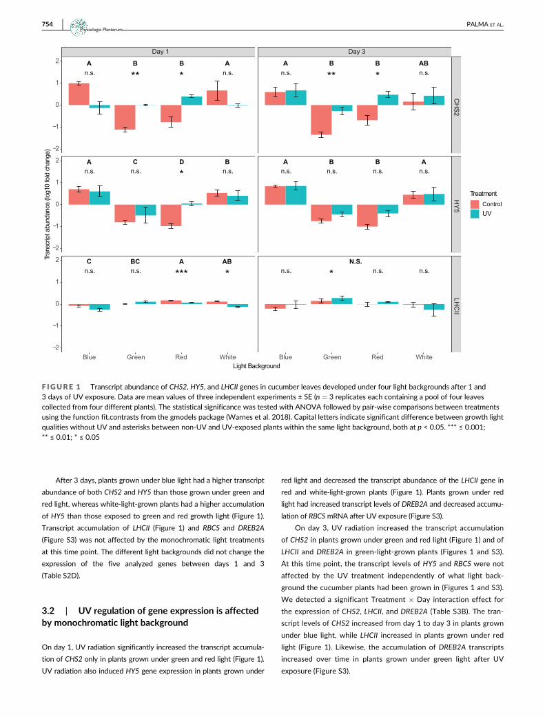
After 3 days, plants grown under blue light had a higher transcript
abundance of both CHS2 and HY5 than those grown under green and
red light, whereas white-light-grown plants had a higher accumulation
of HY5 than those exposed to green and red growth light (Figure 1).
Transcript accumulation of LHCII (Figure 1) and RBCS and DREB2A
(Figure S3) was not affected by the monochromatic light treatments
at this time point. The different light backgrounds did not change the
expression of the five analyzed genes between days 1 and 3
(Table S2D).
3.2 | UV regulation of gene expression is affectedby monochromatic light background
On day 1, UV radiation significantly increased the transcript accumula-
tion of CHS2 only in plants grown under green and red light (Figure 1).
UV radiation also induced HY5 gene expression in plants grown under
red light and decreased the transcript abundance of the LHCII gene in
red and white-light-grown plants (Figure 1). Plants grown under red
light had increased transcript levels of DREB2A and decreased accumu-
lation of RBCSmRNA after UV exposure (Figure S3).
On day 3, UV radiation increased the transcript accumulation
of CHS2 in plants grown under green and red light (Figure 1) and of
LHCII and DREB2A in green-light-grown plants (Figures 1 and S3).
At this time point, the transcript levels of HY5 and RBCS were not
affected by the UV treatment independently of what light back-
ground the cucumber plants had been grown in (Figures 1 and S3).
We detected a significant Treatment � Day interaction effect for
the expression of CHS2, LHCII, and DREB2A (Table S3B). The tran-
script levels of CHS2 increased from day 1 to day 3 in plants grown
under blue light, while LHCII increased in plants grown under red
light (Figure 1). Likewise, the accumulation of DREB2A transcripts
increased over time in plants grown under green light after UV
exposure (Figure S3).
Day 1 Day 3
CH
S2H
Y5LH
CII
Blue Green Red White Blue Green Red White
−2
−1
0
1
2
−2
−1
0
1
2
−2
−1
0
1
2
Light Background
Tran
scrip
t abu
ndan
ce (l
og10
fold
cha
nge)
TreatmentControlUV
A AB Bn.s. ** * n.s.
A B B ABn.s. n.s.** *
A C D Bn.s. n.s. n.s.*
A B B An.s. n.s. n.s. n.s.
C BC A ABn.s. n.s. *** *
N.S.n.s. n.s. n.s.*
F IGURE 1 Transcript abundance of CHS2, HY5, and LHCII genes in cucumber leaves developed under four light backgrounds after 1 and3 days of UV exposure. Data are mean values of three independent experiments ± SE (n = 3 replicates each containing a pool of four leavescollected from four different plants). The statistical significance was tested with ANOVA followed by pair-wise comparisons between treatmentsusing the function fit.contrasts from the gmodels package (Warnes et al. 2018). Capital letters indicate significant difference between growth lightqualities without UV and asterisks between non-UV and UV-exposed plants within the same light background, both at p < 0.05. *** ≤ 0.001;** ≤ 0.01; * ≤ 0.05
754 PALMA ET AL.Physiologia Plantarum

3.3 | Differential accumulation of metabolites incucumber leaves under monochromatic lightbackgrounds
The effects of monochromatic light backgrounds on the accumulation
of metabolites were assessed on non-UV-treated plants at days 0, 5,
and 14 after initiation of the experiment. The metabolites identified
included phenylalanine, four different flavonoids (kaempferol, querce-
tin, flavonol disaccharide I, and flavonol disaccharide II) and five
hydroxycinnamic acid compounds (ferulic acid, ferulic acid hexose,
caffeic acid, coumaric acid and coumaric acid hexose) (Table 1).
On day 0, the day of the onset of UV exposure, the spectral qual-
ity had a significant effect on the accumulation of flavonol disaccha-
ride II, kaempferol, ferulic acid, caffeic acid and coumaric acid hexose
(Figures S4 and S5). Plants grown under blue and red light had a
higher accumulation of flavonol disaccharide II than those grown
under white and green light (Figure S4). In contrast, plants grown in
red light had a significantly higher accumulation of kaempferol than
those grown under white light (Figure S4). Ferulic acid accumulated at
higher levels in plants grown under blue and white light compared to
those grown under green and red light (Figure S5). Blue-light-grown
plants had a higher accumulation of caffeic acid than plants grown
under the other light backgrounds and a higher content of coumaric
acid hexose than plants grown under green light (Figure S5). At this
time point, the light background had no effect on phenylalanine accu-
mulation (Figure S6).
On day 5, the PAR background significantly affected the accumu-
lation of flavonol disaccharide I and II, phenylalanine, ferulic acid,
caffeic acid and coumaric acid hexose (Figures 2 and 3). Plants grown
under blue light accumulated flavonol disaccharide I, ferulic acid,
caffeic acid and coumaric acid hexose at higher levels than those
grown under green, red, and white light (Figures 2 and 3); they also
had a higher concentration of flavonol disaccharide II than those
exposed to green light (Figure 2). White light-grown plants had a
higher concentration of the hydroxycinnamic acid and flavonoid pre-
cursor phenylalanine and caffeic acid compared to those grown under
Day 5 Day 14
Fla
vo
no
l dis
acch
arid
e I
Fla
vo
no
l dis
acch
arid
e II
Ph
en
yla
lan
ine
Blue Green Red White Blue Green Red White
0
100
200
300
400
500
0
100
200
300
400
500
0
1000
2000
3000
4000
Light Background
Peak
are
a (
arb
itrary
units
)
Treatment
Control
UV
A C C B AB A B A
AB ABBA A B AB A
AB B AB A A B B AB
n.s. * * n.s. ** *** n.s. n.s.
n.s. n.s. n.s. n.s. n.s. n.s. n.s. n.s.
n.s.* n.s. n.s. n.s. n.s. n.s. n.s.
F IGURE 2 Accumulation of phenylalanine and flavonoids in leaves of cucumber plants grown under four light backgrounds after 5 and14 days of UV exposure. Boxplots (center line, median; box limits, upper and lower quartiles; whiskers, 1.5� interquartile range; points, outliers)represent all measurements from three independent experiments (n = 3 replicates each containing four individual leaves collected from fourdifferent plants). The statistical significance was tested with ANOVA followed by pair-wise comparissons between treatments using the functionfit.contrasts from the gmodels package (Warnes et al. 2018). Capital letters indicate significant differences between growth light qualities withoutUV and asterisks between non-UV and UV-exposed plants within the same light background, both at p < 0.05. *** ≤ 0.001; ** ≤ 0.01; * ≤ 0.05
PALMA ET AL. 755Physiologia Plantarum

green light (Figures 2 and 3). They also had an enhanced level of flavo-
nol disaccharide I, ferulic acid and coumaric acid hexose compared to
plants grown under green and red light (Figure 3). Despite the signifi-
cant effects of the light background detected with ANOVA for
kaempferol and ferulic acid hexose, we could not identify differences
among treatments after fitting the contrasts (Tables S4A and S4B).
On day 14, the accumulation of flavonol disaccharide I and II,
phenylalanine, ferulic acid, ferulic acid hexose, caffeic acid and
coumaric acid hexose in cucumber leaves was dependent on the spec-
tral quality of the growth light (Figures 2 and 3). Plants grown under
white and green light increased their flavonol disaccharide I content
compared to those grown under red light (Figure 2). White and blue
light-grown plants had a higher accumulation of flavonol disaccharide
II than those exposed to green light and had higher levels of ferulic
acid than plants grown under green and red light (Figures 2 and 3).
Blue light induced the highest levels of caffeic acid (Figure 3). The
accumulation of phenylalanine was higher in plants grown under blue
light than those grown under green and red light (Figure 2). The
hydroxycinnamic acid glycosides ferulic acid hexose and coumaric acid
hexose accumulated at higher levels in plants grown under red light
than in plants exposed to blue, green, and white light (Figure 3). These
two compounds were also differentially accumulated in blue light-
grown plants compared to those grown under green and white light
(Figure 3). The different light backgrounds had no effect on the accu-
mulation of quercetin, kaempferol (Figure S7), and coumaric acid
(Figure 3). The ANOVA also revealed that ferulic acid hexose and
coumaric acid hexose concentrations increased in time in plants
grown under red light (Figure 3 and Table S4C).
3.4 | UV-induced effects on metaboliteaccumulation are dependent on monochromatic lightbackground
On day 5, UV radiation effects on the accumulation of phenolic com-
pounds and precursors were dependent on the light background. The
Day 5 Day 14
Fe
rulic
acid
Fe
rulic
acid
he
xo
se
Caffe
ic a
cid
Co
um
aric
acid
Co
um
aric
acid
he
xo
se
Blue Green Red White Blue Green Red White
0
100
200
300
0
100
200
0
200
400
0
50
100
150
200
0
200
400
600
Light Background
Peak
are
a (
arb
itrary
units
)
Treatment
Control
UV
A C D B B BA A
N.S. B C A C
A C BC B A C BC C
N.S. N.S.
A C C B B D A C
n.s. n.s. n.s. n.s. n.s.
n.s. n.s. n.s. n.s. n.s.
n.s. n.s. n.s. n.s. n.s.
n.s. n.s. n.s. n.s. n.s.
n.s. n.s. n.s. n.s. n.s.
* ** **
** * ***
* * *
*
******
n.s. n.s.
F IGURE 3 Accumulation of phenolic acids in leaves of cucumber plants grown under four light backgrounds after 5 and 14 days of UVexposure. Boxplots (center line, median; box limits, upper and lower quartiles; whiskers, 1.5� interquartile range; points, outliers) represent allmeasurements from three independent experiments (n = 3 replicates each containing four individual leaves collected from four different plants).The statistical significance was tested with ANOVA followed by pair-wise comparissons between treatments using the function fit.contrasts fromthe gmodels package (Warnes et al. 2018). Capital letters indicate significant differences between growth light qualities without UV and asterisksbetween non-UV and UV -exposed plants within the same light background, both at p < 0.05. *** ≤ 0.001; ** ≤ 0.01; * ≤ 0.05
756 PALMA ET AL.Physiologia Plantarum

UV treatment increased the accumulation of phenylalanine and caffeic
acid in plants grown under blue light (Figures 2 and 3) and decreased
ferulic acid and quercetin content in plants grown under white light
(Figures 2 and S7). UV radiation enhanced the accumulation of flavo-
nol disaccharide I, ferulic acid hexose and coumaric acid hexose under
green light background (Figures 2 and 3) and flavonol disaccharide I
and ferulic acid contents in plants grown under red light (Figure 2). In
contrast, the levels of flavonol disaccharide II (Figure 2), kaempferol
and quercetin (Figure S7) and coumaric acid (Figure 3) were not
affected by UV radiation in any light background (Table S5A).
On day 14, UV radiation increased the accumulation of flavonol
disaccharide I in plants grown under blue light (Figure 2) and flavonol
disaccharide I, ferulic acid, ferulic acid hexose, and coumaric acid hex-
ose under green light (Figures 2 and 3). In contrast, UV radiation
decreased ferulic acid hexose, coumaric acid hexose and coumaric
acid contents in plants grown under red light (Figure 3). Caffeic acid
accumulation was increased by UV radiation in plants grown both
under white and red light (Figure 3). At this time point, the accumula-
tion of flavonol disaccharide II and phenylalanine (Figure 2) as well as
kaempferol and quercetin (Figure S7) were not affected by UV radia-
tion in any light background (Table S5A). ANOVA detected a signifi-
cant Treatment � Day interaction for ferulic acid, ferulic acid hexose,
caffeic acid and coumaric acid hexose. However, we could not detect
differences among treatments after fitting the contrasts (Tables S5B
and S5C).
4 | DISCUSSION
Light-mediated acclimation of plants to different types of radiation
and environmental conditions involves the activation of molecular
mechanisms enabling plants to alter their metabolism, physiology, and
performance. Thus, understanding the molecular events mediating
light responses is key to implement horticultural practices that aim to
use light to improve crop yield and quality. Here, we show that differ-
ent monochromatic light backgrounds alone and in combination with
UV radiation can differentially affect the accumulation of gene tran-
scripts and metabolites in cucumber leaves. These acclimatory
responses were induced by low levels of UV radiation and are compa-
rable to those previously reported in cucumber and other species at
higher UV doses under full PAR (Siipola et al. 2015; Wargent
et al. 2015; Qian et al., 2019, 2020; Rai et al. 2019). Thus, our data
demonstrate that cucumber is a crop species highly responsive to mul-
tiple levels of UV radiation and different monochromatic light
backgrounds.
Plants respond to UV and other types of radiation by altering the
transcription of responsive genes (Jenkins 2009). To accurately assess
changes in gene expression with qPCR, stably expressed reference
genes must be used in the normalization step (Hellemans et al. 2007).
We show that the reference genes (ACT and 18S) that have been tra-
ditionally used to assess UV and stress responses in cucumber and
other plant species (Wan et al. 2010; Migocka & Papierniak 2011;
Joseph et al. 2018) in an ambient or white light background may not
be effective in experiments with monochromatic light sources such as
LEDs emitting red, blue or green light (Figure S2). These findings high-
light the need to accurately test and evaluate the suitability of differ-
ent reference genes in gene expression studies that involve plants
grown under different light spectra.
In agreement with previous studies reporting early light-induced
changes in gene expression, most changes in transcript accumulation
mediated by monochromatic light and UV were detected during the
first sampling day (Morales et al. 2013; Qian et al. 2019; Rai
et al. 2019). Our data also show specific wavelength-mediated effects
on transcript accumulation dependent on gene function (Figure 1).
While blue light induced the expression of genes involved in the
phenylpropanoid pathway (CHS2) and UV signaling (HY5), these genes
were repressed under blue-light-depleted environments (green and
red PAR backgrounds). Conversely, the transcript accumulation of the
photosynthesis-related gene LHCII was induced under red and white
PAR backgrounds and repressed in plants grown under blue and green
PAR backgrounds. Furthermore, UV radiation antagonized the effects
of blue, red, green, and white light on transcript accumulation in a
gene-dependent manner. For genes involved in UV signaling and
response (CHS2 and HY5), UV had mainly antagonistic effects in green
and red light and for LHCII with red and white light. This indicates that
the transcriptional regulation mediated by signaling interactions
downstream of PAR and UV perception is dependent on the physio-
logical function of the gene and the spectral quality within PAR. Since
the UV treatment used in the experiment is mainly enriched in wave-
lengths below 350 nm (Figure S1), a region that primarily activates
UVR8 (Rai et al. 2019, 2020), it could thus be expected that the UV-
induced changes in gene expression detected in cucumber leaves are
largely mediated by UVR8.
Previous studies reported the induction of CHS2 in cucumber by
supplementary UV radiation in a broadband PAR background provided
by high-pressure sodium lamps (Qian et al. 2019). In contrast, our
white light treatment in combination with UV radiation at a similar
dose to that of Qian et al. (2019) (0.6 kJ m�2 day�1 at 4 h daily expo-
sure) did not affect the expression of CHS2 after 24 or 72 h of UV
treatment (Figure 1). Thus, our data suggest that the spectral composi-
tion of the PAR background plays an important role on the regulation
of gene expression by UV radiation in cucumber. Commercial lamps
for plant growth from different providers can have different ratios of
blue, red, and green light, which possibly could explain the difference
in responses.
Compared to blue and red light-grown plants, the transcript accu-
mulation in plants grown under green light has not been extensively
studied. Green light is perceived by both phytochromes and
cryptochromes (Folta & Maruhnich 2007). However, our green light
treatment falls precisely outside the range of the phytochrome
absorption spectra (Figure S1), indicating that the responses observed
under green light are likely mediated by cryptochromes or by another
not yet identified green light photoreceptor. Our results show that
green light represses the transcript accumulation of CHS2 and HY5,
opposite to what was observed under blue and white light. Antagonis-
tic effects between green and blue light have been reported for
PALMA ET AL. 757Physiologia Plantarum

several biochemical and physiological traits measured in both shade-
tolerant and shade-intolerant plant species, including the accumula-
tion of anthocyanins (Wang et al. 2020). Blue light triggers a different
redox state of flavin (the active chromophore in cryptochromes) than
that induced by green light (Smith et al. 2017), which might explain
the antagonistic effects observed between these two light qualities.
Furthermore, changes in transcript accumulation induced by supple-
mental UV differed between blue and green light-grown plants
(Figure 1). UV exposure for 1 and 3 days increased the transcript level
of CHS2 in green light-grown plants but not in plants grown under
blue light. In Arabidopsis, the transcript accumulation of CHS, induced
by both monochromatic and polychromatic UV treatments, is depen-
dent on UVR8 (Brown et al. 2005; Favory et al. 2009; Morales
et al. 2013; Rai et al. 2019). Previous research also demonstrated that
UVR8 signaling is repressed in the presence of blue light by REPRES-
SOR OF UV-B PHOTOMORPHOGENESIS (RUP) proteins, a response
that is mediated by cryptochrome signaling (Tissot & Ulm 2020). Our
data suggest that UVR8 signaling is not repressed under the green
and red light treatments as it could be expected for blue light-grown
plants (Tissot & Ulm 2020). However, further research using photore-
ceptor mutants is required to reveal the mechanisms mediating tran-
scriptional regulation in plants exposed to UV radiation under green
and red light backgrounds.
UV radiation can potentially damage the plant's photosynthetic
system and down-regulate the expression of genes encoding photo-
synthetic proteins (LHCII and RBCS) in pea (Jordan et al. 1994, 1998;
Mackerness et al. 1997) and rice (Takeuchi et al. 2002). The UV treat-
ment used in this study repressed LHCII transcript accumulation in
plants grown under red and white light only after 1 day of exposure
(Figure 1). However, plants grown under red light quickly acclimated
to UV, and the low levels of LHCII transcripts detected at day 1 were
unlikely to affect the high photosynthetic performance and morpho-
logical parameters measured after 14 days of UV exposure (Palma
et al. 2021).
We also studied the accumulation of selected compounds of the
hydroxycinnamic acid (ferulic acid, and caffeic acid) and flavonoid
(kaempferol and quercetin) biosynthesis pathways, including their
common precursors (phenylalanine and coumaric acid) in addition to
their downstream glycosylated compounds. Phenylalanine and
coumaric acid thus feed into both competing pathways, in which CHS
is the first committed enzymatic step of the flavonoid biosynthetic
route. Our data show that the spectral composition of growth light
differentially affected the accumulation of the identified compounds
in cucumber leaves. A higher accumulation of flavonol disaccharide I,
ferulic acid, caffeic acid and coumaric acid hexose was observed at
day 5 in plants grown under blue or white light compared to those
grown under green or red light. These findings confirm the role of blue
light in regulating the accumulation of total phenolic and total flavo-
noid content in tomato (Kim et al. 2014), rice (Jung et al. 2013), pea
(Siipola et al. 2015) and in callus cultures of purple basil (Nazir
et al. 2020). Thus, our gene expression and metabolite data in cucum-
ber, together with previous reports in other species, indicate that blue
light could be the main PAR component regulating the accumulation
of flavonoids and hydroxycinnamic acids in plants. Interestingly, the
hexose forms (unknown sugar moiety conjugated to coumaric and fer-
ulic acids) responded very differently to red light compared to their
non-conjugated counterparts at day 14 (Figure 3). Both ferulic acid
hexose and coumaric acid hexose accumulated at higher levels in
plants grown under red light than those grown in any of the other
light qualities at day 14. Furthermore, red light increased their accu-
mulation from day 5 to day 14, indicating that continuous exposure to
monochromatic red light could trigger glycosylation of selected
hydroxycinnamic acids. However, the mechanisms of red-light-
induced glycosylation of ferulic and coumaric acid, and the roles of
the conjugated compounds in cucumber leaves remains to be
determined.
Our metabolite data also demonstrate that UV responses in
cucumber are highly dependent on both the growth light background
and the duration of UV exposure (Figures 2 and 3). The observed
increased phenylalanine content by supplementary UV radiation in
plants grown under blue light at day 5 is most likely related to the ele-
vated expression of cucumber PAL genes (Qian et al. 2019). Further-
more, UV radiation up-regulates downstream reactions in both the
flavonoid and hydroxycinnamic acid pathways and this regulation pat-
tern is compound-specific. Interestingly, after prolonged UV exposure
(14 days), plants grown under green light showed an increased con-
centration of flavonol disaccharide I, and ferulic acid, ferulic acid hex-
ose and coumaric acid hexose (Figures 2 and 3). This indicates a
positive interaction between UV and green light perception, which
results in an increment in metabolite accumulation, primarily of glyco-
sylated forms of both flavonoids and hydroxycinnamic acids. In con-
trast, red-light-grown cucumber plants exposed to supplementary UV
radiation for 14 days exhibited increased caffeic acid accumulation
but decreased levels of ferulic acid hexose, coumaric acid and
coumaric acid hexose. This downregulation of specific compounds
induced by UV radiation is mainly observed under red light, suggesting
negative crosstalk between UV and red light absorption that results in
a reduced metabolite accumulation.
5 | CONCLUSIONS
In agreement with our first hypothesis, our study shows that different
monochromatic lights induce differential accumulation of transcripts
and phenylpropanoid compounds in cucumber leaves. Furthermore,
these monochromatic light-driven responses are gene- and
metabolite-dependent. The accumulation of flavonoids and hydrox-
ycinnamic acids was generally increased by blue and white light. The
response of cucumber to low levels of UV radiation is highly depen-
dent on the growth light background, which agrees with our second
hypothesis. UV radiation antagonized the effects of blue, red, green,
and white light on transcript accumulation in a gene-dependent man-
ner. Our data indicate that light-dependent acclimatory responses in
cucumber can be induced by both using monochromatic light treat-
ments alone and in combination with low levels of UV radiation. In
addition to the mechanistic implications of light perception and
758 PALMA ET AL.Physiologia Plantarum

crosstalk between different photoreceptors, this gives the possibility
to implement multiple light recipes in horticultural practices that aim
to modify plant metabolism for improving crop quality. It also high-
lights the importance of the spectral composition within PAR when
assessing the effects of UV radiation on plant transcriptomics and
metabolomics in controlled environments.
ACKNOWLEDGMENTS
The authors would like to thank Dr. Irina Kalbina for technical assis-
tance and guidance.
AUTHOR CONTRIBUTIONS
Carolina Falcato Fialho Palma and Luis Orlando Morales designed the
experiment with contributions by Eva Rosenqvist, Carl-Otto Ottosen,
and Åke Strid. Carolina Falcato Fialho Palma performed the experi-
ment and analyzed the data with contributions by Luis Orlando
Morales. Åke Strid assisted with data analysis. Victor Castro-Alves
performed metabolite analysis. Luis Orlando Morales prepared the fig-
ures. Carolina Falcato Fialho Palma and Luis Orlando Morales wrote
the manuscript with contributions by Victor Castro-Alves, Eva
Rosenqvist, Carl-Otto Ottosen, and Åke Strid. All authors contributed
to the article and approved the submitted version.
DATA AVAILABILITY STATEMENT
The data that support the findings of this study are available from the
corresponding authors upon reasonable request.
ORCID
Carolina Falcato Fialho Palma https://orcid.org/0000-0002-2034-
1641
Åke Strid https://orcid.org/0000-0003-3315-8835
Luis Orlando Morales https://orcid.org/0000-0002-9233-7254
REFERENCES
Agati, G. & Tattini, M. (2010) Multiple functional roles of flavonoids in
photoprotection. The New Phytologist, 186, 786–793.Ahmad, M., Jarillo, J.A., Smirnova, O. & Cashmore, A.R. (1998)
Cryptochrome blue-light photoreceptors of Arabidopsis implicated in
phototropism. Nature, 392, 720–723.Binkert, M., Kozma-Bognár, L., Terecskei, K., de Veylder, L., Nagy, F. &
Ulm, R. (2014) UV-B-responsive association of the Arabidopsis bZIP
transcription factor ELONGATED HYPOCOTYL5 with target genes,
including its own promoter. Plant Cell, 26, 4200–4213.Boccaccini, A., Legris, M., Krahmer, J., Allenbach-Petrolati, L., Goyal, A.,
Galvan-Ampudia, C. et al. (2020) Low blue light enhances phototro-
pism by releasing cryptochrome1-mediated inhibition of PIF4 expres-
sion. Plant Physiology, 183, 1780–1793.Boccalandro, H.E., Giordano, C.V., Ploschuk, E.L., Piccoli, P.N., Bottini, R. &
Casal, J.J. (2012) Phototropins but not cryptochromes mediate the
blue light-specific promotion of stomatal conductance, while both
enhance photosynthesis and transpiration under full sunlight. Plant
Physiology, 158, 1475–1484.Briggs, W.R. & Huala, E. (1999) Blue-light photoreceptors in higher plants.
Annual Review of Cell and Developmental Biology, 15, 33–62.Brosché, M., Fant, C., Bergkvist, S.W., Strid, H., Svensk, A., Olsson, O. et al.
(1999) Molecular markers for UV-B stress in plants: alteration of the
expression of four classes of genes in Pisum sativum and the formation
of high molecular mass RNA adducts. Biochimica et Biophysica Acta -
Gene Structure and Expression, 1447, 185–198.Brown, B.A., Cloix, C., Jiang, G.H., Kaiserli, E., Herzyk, P., Kliebenstein, D.J.
et al. (2005) A UV-B-specific signaling component orchestrates plant
UV protection. Proceedings of the National Academy of Sciences of the
United States of America, 102, 18225–18230.Brown, B.A., Headland, L.R. & Jenkins, G.I. (2009) UV-B action spectrum
for UVR8-mediated HY5 transcript accumulation in Arabidopsis. Photo-
chemistry and Photobiology, 85, 1147–1155.Brown, B.A. & Jenkins, G.I. (2008) UV-B signaling pathways with different
fluence-rate response profiles are distinguished in mature Arabidopsis
leaf tissue by requirement for UVR8, HY5, and HYH. Plant Physiology,
146, 576–588.Chappell, J. & Hahlbrock, K. (1984) Transcription of plant defence genes in
response to UV light or fungal elicitor. Nature, 311, 76–78.Day, T.A., Martin, G. & Vogelmann, T.C. (1993) Penetration of UV-B radia-
tion in foliage: evidence that the epidermis behaves as a non-uniform
filter. Plant, Cell & Environment, 16, 735–741.de Carbonnel, M., Davis, P., Roelfsema, M.R.G., Inoue, S.-I., Schepens, I.,
Lariguet, P. et al. (2010) The Arabidopsis PHYTOCHROME KINASE
SUBSTRATE2 protein is a phototropin signaling element that regu-
lates leaf flattening and leaf positioning. Plant Physiology, 152, 1391–1405.
Demkura, P.V. & Ballaré, C.L. (2012) UVR8 mediates UV-B-induced Ara-
bidopsis defense responses against Botrytis cinerea by controlling sin-
apate accumulation. Molecular Plant, 5, 642–652.Demotes-Mainard, S., Péron, T., Corot, A., Bertheloot, J., Le Gourrierec, J.,
Pelleschi-Travier, S. et al. (2016) Plant responses to red and far-red
lights, applications in horticulture. Environmental and Experimental Bot-
any, 121, 4–21.Fankhauser, C. & Chory, J. (1997) Light control of plant development.
Annual Review of Cell and Developmental Biology, 13, 203–229.Favory, J.J., Stec, A., Gruber, H., Rizzini, L., Oravecz, A., Funk, M. et al.
(2009) Interaction of COP1 and UVR8 regulates UV-B-induced photo-
morphogenesis and stress acclimation in Arabidopsis. The EMBO Jour-
nal, 28, 591–601.Folta, K.M. (2004) Green light stimulates early stem elongation. Plant Phys-
iology, 135, 1407–1416.Folta, K.M. & Maruhnich, S.A. (2007) Green light: a signal to slow down or
stop. Journal of Experimental Botany, 58, 3099–3111.Frechilla, S., Talbott, L.D., Bogomolni, R.A. & Zeiger, E. (2000) Reversal of
blue light-stimulated stomatal opening by green light. Plant & Cell Phys-
iology, 41, 171–176.Fuglevand, G., Jackson, J.A. & Jenkins, G.I. (1996) UV-B, UV-A, and blue light
signal transduction pathways interact synergistically to regulate chalcone
synthase gene expression in Arabidopsis. Plant Cell, 8, 2347–2357.Heijde, M. & Ulm, R. (2012) UV-B photoreceptor-mediated signalling in
plants. Trends in Plant Science, 17, 230–237.Hellemans, J., Mortier, G., De Paepe, A., Speleman, F. & Vandesompele, J.
(2007) qBase relative quantification framework and software for man-
agement and automated analysis of real-time quantitative PCR data.
Genome Biology, 8.
Holm, S. (1979) A simple sequentially rejective multiple test procedure.
Scandinavian Journal of Statistics, 6, 65–70.Huché-Thélier, L., Crespel, L., Le Gourrierec, J., Morel, P., Sakr, S. &
Leduc, N. (2016) Light signaling and plant responses to blue and UV
radiations: perspectives for applications in horticulture. Environmental
and Experimental Botany, 121, 22–38.Jansen, M.A.K. (2002) Ultraviolet-B radiation effects on plants: induction
of morphogenic responses. Physiologia Plantarum, 116, 423–429.Jarillo, J.A., Gabrys, H., Capel, J., Alonso, J.M., Ecker, J.R. & Cashmore, A.R.
(2001) Phototropin-related NPL1 controls chloroplast relocation
induced by blue light. Nature, 410, 952–954.Jenkins, G.I. (2009) Signal transduction in responses to UV-B radiation.
Annual Review of Plant Biology, 60, 407–431.
PALMA ET AL. 759Physiologia Plantarum
Jenkins, G.I. (2017) Photomorphogenic responses to ultraviolet-B light.
Plant, Cell & Environment, 40, 2544–2557.Jordan, B.R., Chow, W.S., Strid, Å. & Anderson, J.M. (1991) Reduction in
cab and psb A RNA transcripts in response to Supplementary
ultraviolet-B radiation. FEBS Letters, 284, 5–8.Jordan, B.R., James, P.E. & A-H-Mackerness, S. (1998) Factors affecting
UV-B-induced changes in Arabidopsis thaliana L. gene expression: the
role of development, protective pigments and the chloroplast signal.
Plant & Cell Physiology, 39, 769–778.Jordan, B.R., James, P.E., Strid, Å. & Anthony, R.G. (1994) The effect of
ultraviolet-B radiation on gene expression and pigment composition in
etiolated and green pea leaf tissue: UV-B-induced changes are gene-
specific and dependent upon the developmental stage. Plant, Cell &
Environment, 17, 45–54.Joseph, J.T., Poolakkalody, N.J. & Shah, J.M. (2018) Plant reference genes
for development and stress response studies. Journal of Biosciences,
43, 173–187.Jung, E.S., Lee, S., Lim, S.H., Ha, S.H., Liu, K.H. & Lee, C.H. (2013) Metabo-
lite profiling of the short-term responses of rice leaves (Oryza sativa
cv. Ilmi) cultivated under different LED lights and its correlations with
antioxidant activities. Plant Science, 210, 61–69.Kalbina, I., Li, S., Kalbin, G., Björn, L.O. & Strid, Å. (2008) Two separate
UV-B radiation wavelength regions control expression of different
molecular markers in Arabidopsis thaliana. Functional Plant Biology, 35,
222–227.Kim, E.Y., Park, S.A., Park, B.J., Lee, Y. & Oh, M.M. (2014) Growth and anti-
oxidant phenolic compounds in cherry tomato seedlings grown under
monochromatic light-emitting diodes. Horticulture, Environment and
Biotechnology, 55, 506–513.Klem, K., Ac, A., Holub, P., Kovác, D., Špunda, V., Robson, T.M. et al.
(2012) Interactive effects of PAR and UV radiation on the physiology,
morphology and leaf optical properties of two barley varieties. Environ-
mental and Experimental Botany, 75, 52–64.Kotilainen, T., Tegelberg, R., Julkunen-Tiitto, R., Lindfors, A. & Aphalo, P.J.
(2008) Metabolite specific effects of solar UV-A and UV-B on alder
and birch leaf phenolics. Global Change Biology, 14, 1294–1304.Krizek, D.T. (2004) Influence of PAR and UV-A in determining plant sensi-
tivity and photomorphogenic responses to UV-B radiation. Photochem-
istry and Photobiology, 79, 307–315.Krizek, D.T., Mirecki, R.M. & Britz, S.J. (1997) Inhibitory effects of ambient
levels of solar UV-A and UV-B radiation on growth of cucumber.
Physiologia Plantarum, 100, 886–893.Lattanzio, V., Lattanzio, V.M.T., Cardinali, A. & Amendola, V. (2006) Role of
phenolics in the resistance mechanisms of plants against fungal patho-
gens and insects. In: Imperato, F. (Ed.) Phytochemistry: advances in
research. Trivandrum, India: Research Signpost, pp. 23–67.Li, Q. & Kubota, C. (2009) Effects of supplemental light quality on growth
and phytochemicals of baby leaf lettuce. Environmental and Experimen-
tal Botany, 67, 59–64.Lidon, F.J.C., Reboredo, F.H., Leit~ao, A.E., Silva, M.M.A., Duarte, M.P. &
Ramalho, J.C. (2012) Impact of UV-B radiation on photosynthesis: an
overview. Emirates Journal of Food and Agriculture, 24, 546–556.Mackerness, S.A.H., Thomas, B. & Jordan, B.R. (1997) The effect of Sup-
plementary ultraviolet-B radiation on mRNA transcripts, translation
and stability of chloroplast proteins and pigment formation in Pisum
sativum L. Journal of Experimental Botany, 48, 729–738.Migocka, M. & Papierniak, A. (2011) Identification of suitable reference
genes for studying gene expression in cucumber plants subjected to
abiotic stress and growth regulators. Molecular Breeding, 28, 343–357.Morales, L.O., Brosché, M., Vainonen, J., Jenkins, G.I., Wargent, J.J.,
Sipari, N. et al. (2013) Multiple roles for UV RESISTANCE LOCUS8 in
regulating gene expression and metabolite accumulation in Arabidopsis
under solar ultraviolet radiation. Plant Physiology, 161, 744–759.Morales, L.O., Brosché, M., Vainonen, J.P., Sipari, N., Lindfors, A.V.,
Strid, Å. et al. (2015) Are solar UV-B- and UV-A-dependent gene
expression and metabolite accumulation in Arabidopsis mediated by
the stress response regulator RADICAL-INDUCED CELL DEATH1?
Plant, Cell & Environment, 38, 878–891.Morales, L.O., Tegelberg, R., Brosché, M., Keinänen, M., Lindfors, A. &
Aphalo, P.J. (2010) Effects of solar UV-A and UV-B radiation on gene
expression and phenolic accumulation in Betula pendula leaves. Tree
Physiology, 30, 923–934.Nazir, M., Ullah, M.A., Younas, M., Siddiquah, A., Shah, M., Giglioli-
Guivarc'h, N. et al. (2020) Light-mediated biosynthesis of
phenylpropanoid metabolites and antioxidant potential in callus cul-
tures of purple basil (Ocimum basilicum L. var purpurascens). Plant Cell,
Tissue and Organ Culture, 142, 107–120.Neugart, S., Fiol, M., Schreiner, M., Rohn, S., Zrenner, R., Kroh, L.W. et al.
(2014) Interaction of moderate UV-B exposure and temperature on
the formation of structurally different flavonol glycosides and hydrox-
ycinnamic acid derivatives in kale (Brassica oleracea var. sabellica). Jour-
nal of Agricultural and Food Chemistry, 62, 4054–4062.Ohashi-Kaneko, K., Takase, M., Kon, N., Fujiwara, K. & Kurata, K. (2007)
Effect of light quality on growth and vegetable quality in leaf lettuce,
spinach and komatsuna. Environmental Control in Biology, 45, 189–198.Olsson, L.C., Veit, M., Weissenböck, G. & Bornman, J.F. (1998) Differential
flavonoid response to enhanced UV-B radiation in Brassica napus. Phy-
tochemistry, 49, 1021–1028.Osterlund, M.T., Hardtke, C.S., Wei, N. & Deng, X.W. (2000) Targeted
destabilization of HY5 during light-regulated development of Ara-
bidopsis. Nature, 405, 462–466.Ouzounis, T., Razi Parjikolaei, B., Fretté, X., Rosenqvist, E. & Ottosen, C.-
O. (2015) Predawn and high intensity application of supplemental blue
light decreases the quantum yield of PSII and enhances the amount of
phenolic acids, flavonoids, and pigments in Lactuca sativa. Frontiers in
Plant Science, 6, 1–14.Palma, C.F.F., Castro-Alves, V., Morales, L.O., Rosenqvist, E., Ottosen, C.-
O. & Strid, Å. (2021) Spectral composition of light affects sensitivity to
UV-B and photoinhibition in cucumber. Frontiers in Plant Science, 11,
610011.
Pinheiro, J. & Bates, D.M. (2000) Linear mixed-effects models: basic con-
cepts and examples. In: Mixed-effects models in S and S-PLUS. New
York, NY: Springer US, pp. 3–56.Qian, M., Kalbina, I., Rosenqvist, E., Jansen, M.A.K., Teng, Y. & Strid, Å.
(2019) UV regulates the expression of phenylpropanoid biosynthesis
genes in cucumber (Cucumis sativus L.) in an organ and spectrum
dependent manner. Photochemical & Photobiological Sciences, 18,
424–433.Qian, M., Rosenqvist, E., Flygare, A.M., Kalbina, I., Teng, Y., Jansen, M.A.K.
et al. (2020) UV-A light induces a robust and dwarfed phenotype in
cucumber plants (Cucumis sativus L.) without affecting fruit yield.
Scientia Horticulturae (Amsterdam), 263, 109110.
Qian, M., Rosenqvist, E., Prinsen, E., Pescheck, F., Flygare, A.-M., Kalbina, I.
et al. (2021) Downsizing in plants: UV light induces pronounced mor-
phological changes in the absence of stress. Plant Physiology, 187,
378–395. https://doi.org/10.1093/plphys/kiab262Rai, N., Morales, L.O. & Aphalo, P.J. (2021) Perception of solar UV radia-
tion by plants: photoreceptors and mechanisms. Plant Physiology, 186,
1382–1396.Rai, N., Neugart, S., Yan, Y., Wang, F., Siipola, S.M., Lindfors, A.V. et al.
(2019) How do cryptochromes and UVR8 interact in natural and simu-
lated sunlight? Journal of Experimental Botany, 70, 4975–4990.Rai, N., O'Hara, A., Farkas, D., Safronov, O., Ratanasopa, K., Wang, F. et al.
(2020) The photoreceptor UVR8 mediates the perception of both
UV-B and UV-A wavelengths up to 350 nm of sunlight with res-
ponsivity moderated by cryptochromes. Plant, Cell & Environment, 43,
1513–1527.Rizzini, L., Favory, J.J., Cloix, C., Faggionato, D., O'Hara, A., Kaiserli, E. et al.
(2011) Perception of UV-B by the Arabidopsis UVR8 protein. Science
(80-.), 332, 103–106.
760 PALMA ET AL.Physiologia Plantarum

Robson, T.M., Aphalo, P.J., Bana�s, A.K., Barnes, P.W., Brelsford, C.C.,
Jenkins, G.I. et al. (2019) A perspective on ecologically relevant plant-
UV research and its practical application. Photochemical & Photobiologi-
cal Sciences, 18, 970–988.Sakai, T., Kagawa, T., Kasahara, M., Swartz, T.E., Christie, J.M., Briggs, W.R.
et al. (2001) Arabidopsis nph1 and npl1: blue light receptors that medi-
ate both phototropism and chloroplast relocation. Proceedings of the
National Academy of Sciences, 98, 6969–6974.Siipola, S.M., Kotilainen, T., Sipari, N., Morales, L.O., Lindfors, A.V.,
Robson, T.M. et al. (2015) Epidermal UV-A absorbance and whole-leaf
flavonoid composition in pea respond more to solar blue light than to
solar UV radiation. Plant, Cell and Environment, 38, 941–952.Smith, H. (1982) Light quality, photoperception, and plant strategy. Annual
Review of Plant Physiology, 33, 481–518.Smith, H.L., Mcausland, L. & Murchie, E.H. (2017) Don't ignore the green
light: exploring diverse roles in plant processes. Journal of Experimental
Botany, 68, 2099–2110.Stracke, R., Favory, J.J., Gruber, H., Bartelniewoehner, L., Bartels, S.,
Binkert, M. et al. (2010) The Arabidopsis bZIP transcription factor HY5
regulates expression of the PFG1/MYB12 gene in response to light
and ultraviolet-B radiation. Plant, Cell and Environment, 33, 88–103.Sullivan, J.H., Gitz, D.C., Peek, M.S. & McElrone, A.J. (2003) Response of three
eastern tree species to supplemental UV-B radiation: leaf chemistry and
gas exchange. Agricultural and Forest Meteorology, 120, 219–228.Takeuchi, A., Yamaguchi, T., Hidema, J., Strid, A. & Kumagai, T. (2002)
Changes in synthesis and degradation of Rubisco and LHCII with leaf
age in rice (Oryza sativa L.) growing under Supplementary UV-B radia-
tion. Plant, Cell and Environment, 25, 695–706.Tegelberg, R., Julkunen-Tiitto, R. & Aphalo, P.J. (2004) Red:far-red light
ratio and UV-B radiation: their effects on leaf phenolics and growth of
silver birch seedlings. Plant, Cell and Environment, 27, 1005–1013.Thimijan, R.W., Carns, H.R. & Campbell, L.E. (1978) Final report (EPA-IAG-
D6-0168): radiation sources and relative environmental control for biolog-
ical and climatic effects of UV research (BACER). Washington, DC; Envi-
ronmental Protection Agency.
Tissot, N. & Ulm, R. (2020) Cryptochrome-mediated blue-light signalling
modulates UVR8 photoreceptor activity and contributes to UV-B tol-
erance in Arabidopsis. Nature Communications, 11, 2–11.Ulm, R., Baumann, A., Oravecz, A., Máté, Z., �Adám, �E., Oakeley, E.J. et al.
(2004) Genome-wide analysis of gene expression reveals function of
the bZIP transcription factor HY5 in the UV-B response of Arabidopsis.
Proceedings of the National Academy of Sciences of the United States of
America, 101, 1397–1402.Wan, H., Zhao, Z., Qian, C., Sui, Y., Malik, A.A. & Chen, J. (2010) Selection
of appropriate reference genes for gene expression studies by quanti-
tative real-time polymerase chain reaction in cucumber. Analytical Bio-
chemistry, 399, 257–261.
Wang, L., Li, P., Wang, Z., Liu, J., Hu, J. & Li, J. (2014) Identification and val-
idation of suitable internal reference genes for SYBR-GREEN qRT-
PCR studies during cucumber development. Journal of Horticultural
Science and Biotechnology, 89, 312–320.Wang, Q.W., Robson, T.M., Pieristè, M., Oguro, M., Oguchi, R., Murai, Y.
et al. (2020) Testing trait plasticity over the range of spectral composi-
tion of sunlight in forb species differing in shade tolerance. Journal of
Ecology, 108, 1923–1940.Wargent, J.J., Moore, J.P., Roland Ennos, A. & Paul, N.D. (2009) Ultraviolet
radiation as a limiting factor in leaf expansion and development. Photo-
chemistry and Photobiology, 85, 279–286.Wargent, J.J., Nelson, B.C.W., Mcghie, T.K. & Barnes, P.W. (2015) Acclimation
to UV-B radiation and visible light in Lactuca sativa involves up-regulation
of photosynthetic performance and orchestration of metabolome-wide
responses. Plant, Cell and Environment, 38, 929–940.Warnes, A. G. R., Bolker, B., Lumley, T., and Randall, C. (2018). Package
‘gmodels.’Yan, Y., Stoddard, F.L., Neugart, S., Sadras, V.O., Lindfors, A., Morales, L.O.
et al. (2019) Responses of flavonoid profile and associated gene
expression to solar blue and UV radiation in two accessions of Vicia
faba L. from contrasting UV environments. Photochemical & Photobio-
logical Sciences, 18, 434–447.Yu, S.G. & Björn, L.O. (1997) Effects of UVB radiation on light-dependent
and light-independent protein phosphorylation in thylakoid proteins.
Journal of Photochemistry and Photobiology B: Biology, 37, 212–218.Zhang, T., Maruhnich, S.A. & Folta, K.M. (2011) Green light induces shade
avoidance symptoms. Plant Physiology, 157, 1528–1536.Zhao, B., Wang, L., Pang, S., Jia, Z., Wang, L., Li, W. et al. (2020) UV-B pro-
motes flavonoid synthesis in Ginkgo biloba leaves. Industrial Crops and
Products, 151, 112483.
SUPPORTING INFORMATION
Additional supporting information may be found in the online version
of the article at the publisher's website.
How to cite this article: Palma, C.F.F., Castro-Alves, V.,
Rosenqvist, E., Ottosen, C.-O., Strid, Å. & Morales, L.O. (2021)
Effects of UV radiation on transcript and metabolite
accumulation are dependent on monochromatic light
background in cucumber. Physiologia Plantarum, 173(3),
750–761. Available from: https://doi.org/10.1111/ppl.13551
PALMA ET AL. 761Physiologia Plantarum