Clean technological change in developing-country industrial clusters: Mexican leather tanning
Upload
independentCategory
view
0download
0
Mechanisms of Skin Tanning in Different Racial/Ethnic Groupsin Response to Ultraviolet Radiation
Taketsugu Tadokoro,� Yuji Yamaguchi,� Jan Batzer,w Sergio G. Coelho,z Barbara Z. Zmudzka,zSharon A. Miller,z Rainer Wolber,w Janusz Z. Beer,z and Vincent J. Hearing��Laboratory of Cell Biology, National Cancer Institute, National Institutes of Health, Bethesda, Maryland, USA; wR&D, Skin Research, Beiersdorf AG, Hamburg,Germany; zCenter for Devices and Radiological Health, Food and Drug Administration, Rockville, Maryland, USA
Ultraviolet radiation stimulates pigmentation in human skin, but the mechanism(s) whereby this increase in melanin
production (commonly known as tanning) occurs is not well understood. Few studies have examined the molecular
consequences of UV on human skin of various racial backgrounds in situ. We investigated the effects of UV on human
skin of various races before and at different times after a single 1 minimal erythemal dose UV exposure. We measured
the distribution of DNA damage that results, as well as the melanin content/distribution and the expression of various
melanocyte-specific genes. The density of melanocytes at the epidermal:dermal junction in different types of human
skin are remarkably similar and do not change significantly within 1 wk after UV exposure. The expression of me-
lanocyte-specific proteins (including TYR (tyrosinase), TYRP1 (tyrosinase-related protein 1), DCT (tyrosinase-related
protein 2), MART1 (melanoma antigens recognized by T-cells) gp100 (Pmel17/silver), and MITF (micropthalmia tran-
scription factor)) increased from 0 to 7 d after UV exposure, but the melanin content of the skin increased only slightly.
The most significant change, however, was a change in the distribution of melanin from the lower layer upwards to the
middle layer of the skin, which was more dramatic in the darker skin. These results provide a basis for understanding
the origin of different skin colors and responses to UV within different races.
Key words: melanocyte/melanosome/photoprotection/pigmentation/ultravioletJ Invest Dermatol 124:1326 –1332, 2005
Ultraviolet radiation (UV) stimulates pigmentation in humanskin, but the mechanism(s) whereby this occurs is not wellunderstood. The effect observed, commonly called the tan-ning reaction, is readily visible but few studies have exam-ined differences in responses to UV of human skin in variousracial groups and phototypes. To explore these differences,we used the Office of Management and Budget classifica-tion of races. This classification is subjective and based tosome extent on geopolitical descriptions. Although it is notperfect, it is commonly accepted. There has been a recentcall for more studies to address genetic and phenotypicdifferences among racial ethnic groups (Nature Genet Sup-pl. 11, 2004). Two types of tanning response are known,immediate pigment darkening which can occur within min-utes after UV exposure and delayed tanning which takesseveral days or longer to become apparent. Human skinpigmentation is considered an important protective factoragainst skin cancer development following UV, and theconstitutive color of the skin dramatically affects the inci-dence of various types of skin cancers (Preston and Stern,1992; Kricker et al, 1994; Halder and Bridgeman-Shah,
1995). Previously, we reported that UV-induced DNA dam-age depends on racial/ethnic (R/E) origin (Tadokoro et al,2003). In this report, we describe UV-induced DNA damage,melanin formation and redistribution, and the expression ofvarious melanocyte-specific genes.
Skin pigmentation depends directly on the function ofmelanocytes, specialized dendritic cells localized in the skinat the epidermal:dermal junction (Levine, 1993; Nordlundet al, 1998). Melanocytes express specific proteins involvedin the synthesis and deposition of the biopolymer melaninin melanosomes. Some of those proteins, e.g., tyrosinase(TYR), tyrosinase-related protein 1 (TYRP1), and tyrosinase-related protein 2 (DCT), are enzymes whereas others, e.g.,Pmel17/silver (gp100) are structural components of melano-somes (Kushimoto et al, 2001; Kushimoto et al, 2003). Spe-cific antibodies to those melanocyte markers are nowavailable as are chemical methods to analyze the type andamount of melanin in the skin. Microphthalmia transcrip-tion factor (MITF) is considered the master regulator ofmelanocyte function, and modulates the transcription ofa number of melanocyte-specific genes, including thoseencoding many of the melanosomal proteins noted above(Shibahara et al, 2000; Tachibana, 2000).
Previously, we studied human subjects of various R/Eorigin exposed to a single 1 minimal erythema dose (MED)of UVA/UVB radiation, after which biopsies were taken atdifferent times (Tadokoro et al, 2003). We used immuno-histochemical approaches to analyze DNA damage and itsT. Tadokoro and Y. Yamaguchi contributed equally to this study.
Abbreviations: AA, apparent absorbance; DCT, tyrosinase-relatedprotein 2; DR, diffuse reflectance; gp100, Pmel17/silver; MED,minimal erythema dose; MITF, microphthalmia transcription factor;R/E, racial/ethnic; TYR, tyrosinase; TYRP1, tyrosinase-related pro-tein 1; UV, ultraviolet
Copyright r 2005 by The Society for Investigative Dermatology, Inc.
1326
subsequent removal from the skin. In this study, we usedthe same skin specimens to examine the expression ofmelanocyte-specific proteins prior to UV exposure and at7 min, 24 h, and 7 d after UV exposure. We have character-ized the density of melanocytes at the epidermal:dermaljunction in different types of human skin and analyzed theexpression of melanocyte specific proteins as well as thedistribution of melanin before and after UV exposure.
Results and Discussion
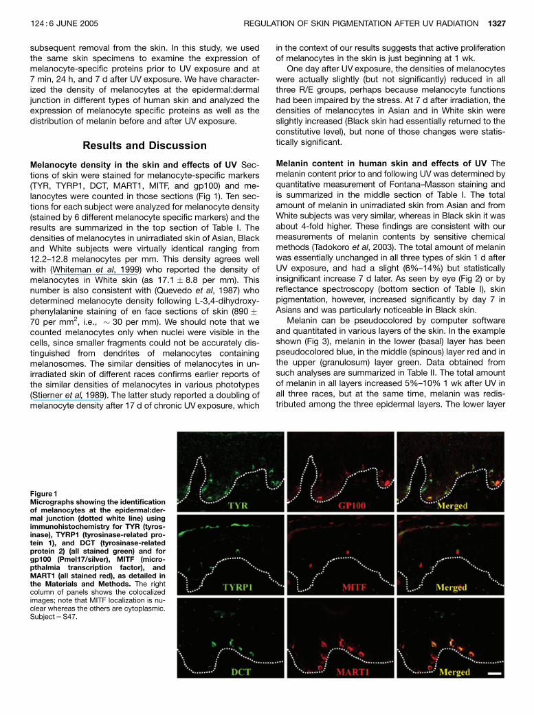
Melanocyte density in the skin and effects of UV Sec-tions of skin were stained for melanocyte-specific markers(TYR, TYRP1, DCT, MART1, MITF, and gp100) and me-lanocytes were counted in those sections (Fig 1). Ten sec-tions for each subject were analyzed for melanocyte density(stained by 6 different melanocyte specific markers) and theresults are summarized in the top section of Table I. Thedensities of melanocytes in unirradiated skin of Asian, Blackand White subjects were virtually identical ranging from12.2–12.8 melanocytes per mm. This density agrees wellwith (Whiteman et al, 1999) who reported the density ofmelanocytes in White skin (as 17.1 � 8.8 per mm). Thisnumber is also consistent with (Quevedo et al, 1987) whodetermined melanocyte density following L-3,4-dihydroxy-phenylalanine staining of en face sections of skin (890 �70 per mm2, i.e., � 30 per mm). We should note that wecounted melanocytes only when nuclei were visible in thecells, since smaller fragments could not be accurately dis-tinguished from dendrites of melanocytes containingmelanosomes. The similar densities of melanocytes in un-irradiated skin of different races confirms earlier reports ofthe similar densities of melanocytes in various phototypes(Stierner et al, 1989). The latter study reported a doubling ofmelanocyte density after 17 d of chronic UV exposure, which
in the context of our results suggests that active proliferationof melanocytes in the skin is just beginning at 1 wk.
One day after UV exposure, the densities of melanocyteswere actually slightly (but not significantly) reduced in allthree R/E groups, perhaps because melanocyte functionshad been impaired by the stress. At 7 d after irradiation, thedensities of melanocytes in Asian and in White skin wereslightly increased (Black skin had essentially returned to theconstitutive level), but none of those changes were statis-tically significant.
Melanin content in human skin and effects of UV Themelanin content prior to and following UV was determined byquantitative measurement of Fontana–Masson staining andis summarized in the middle section of Table I. The totalamount of melanin in unirradiated skin from Asian and fromWhite subjects was very similar, whereas in Black skin it wasabout 4-fold higher. These findings are consistent with ourmeasurements of melanin contents by sensitive chemicalmethods (Tadokoro et al, 2003). The total amount of melaninwas essentially unchanged in all three types of skin 1 d afterUV exposure, and had a slight (6%–14%) but statisticallyinsignificant increase 7 d later. As seen by eye (Fig 2) or byreflectance spectroscopy (bottom section of Table I), skinpigmentation, however, increased significantly by day 7 inAsians and was particularly noticeable in Black skin.
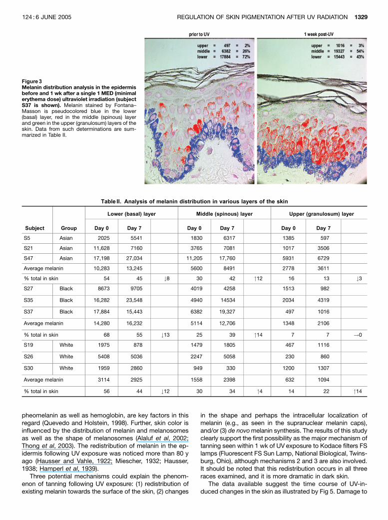
Melanin can be pseudocolored by computer softwareand quantitated in various layers of the skin. In the exampleshown (Fig 3), melanin in the lower (basal) layer has beenpseudocolored blue, in the middle (spinous) layer red and inthe upper (granulosum) layer green. Data obtained fromsuch analyses are summarized in Table II. The total amountof melanin in all layers increased 5%–10% 1 wk after UV inall three races, but at the same time, melanin was redis-tributed among the three epidermal layers. The lower layer
Figure 1Micrographs showing the identificationof melanocytes at the epidermal:der-mal junction (dotted white line) usingimmunohistochemistry for TYR (tyros-inase), TYRP1 (tyrosinase-related pro-tein 1), and DCT (tyrosinase-relatedprotein 2) (all stained green) and forgp100 (Pmel17/silver), MITF (micro-pthalmia transcription factor), andMART1 (all stained red), as detailed inthe Materials and Methods. The rightcolumn of panels shows the colocalizedimages; note that MITF localization is nu-clear whereas the others are cytoplasmic.Subject¼S47.
REGULATION OF SKIN PIGMENTATION AFTER UV RADIATION 1327124 : 6 JUNE 2005

of the epidermis contained 54%–68% of the melanin, themiddle layer 25%–30%, whereas the upper layer containedonly 7%–16% of the pigment. One week after the exposureto 1 MED UV, however, the percentage of melanin wasdecreased from 8%–13% in the basal layer in all threeraces, whereas it increased 4%–14% in the middle layerof the epidermis. Changes in the distribution of melanin inthe upper layer was variable and ranged from a 3%decrease to a 14% increase. It is possible that more timeis needed for preexisting melanin to reach the upper layersof the epidermis.
Expression of melanogenic proteins following UV MITFis considered the master regulator of melanocyte functionsince it regulates the expression, at least in part, of genesencoding the melanosome proteins TYR, TYRP1, DCT,gp100, and MART1 (Tachibana, 2000; Shibahara et al, 2001;Yasumoto et al, 2002; Du et al, 2003). Expression of thosegenes is regulated by melanocyte stimulating hormone(MSH), which functions through MC1R, and by ASP, an an-tagonist of that receptor (Sakai et al, 1997; Abdel-Maleket al, 2001), but their response to UV has not been previ-ously reported. Immunohistochemical analysis showed thatconstitutive expression of MITF in unirradiated skin washighest in Black skin, but in Asian and in White skin wasdetectable in amounts 450% of that found in Black skin(Fig 4). All three types of skin responded to UV with similarincreases in the expression of MITF within 1 d after UV. Theincreases in MITF were still present 7 d after this singlemoderate UV dose.
Constitutive levels of the melanosomal proteins (TYR,TYRP1, DCT, gp100, and MART1) were typically higher inBlack skin, although not always dramatically so. Interest-ingly, expression of all five melanosomal proteins in skinfrom all three races was increased within 1 day after UV, andthese responses were strongest in Black skin. TYR is themost critical enzyme for synthesis of pigment, and its levelsshowed a marked response to UV, even at day 1 after UVR,in all three races. DCT, an enzyme that modulates the typeof pigment synthesized was the least responsive. Interest-ingly, gp100, the basic building block for melanosomes,was highly responsive to UV, even more so than TYR, in all 3races. It is interesting to note that TYR levels are quite sim-ilar (within 2-fold) in all three races. Such observations sup-port an earlier hypothesis that pigmentation of human skinreflects in large part the post-translational processing andactivation of tyrosinase (Fuller et al, 2001). It has also beenshown that many sequence variations/polymorphisms existin the TYR gene (Oetting, 2000). Many of them do not affecttranscription of the gene but do affect enzyme function(Shriver et al, 2003) and may thus also play important rolesin regulating constitutive pigmentation.
Distribution of melanin following UV Several importantparameters determine the appearance of skin color, and theamount and composition of chromophores are of primaryimportance. The two types of melanin, eumelanin and
Figure 2Photographs of a representative subjectfrom each racial group; photos were taken7 d after the single 1 MED (minimal erythemadose) ultraviolet (UV) exposure. Color scalesare included in each photograph to ensure ac-curate color reproduction. (Top) The square ar-eas on the right side of each back receivedvarying doses of UV to establish the MED onday 1; the square areas on the left side of eachback received 1 MED UV on day 0; marks frombiopsies taken can be seen in some areas.(bottom). The areas outlined with the dashedboxes at the top show areas receiving 1 MEDbut without biopsy, and are magnified � 5 (ar-rows point to the UV exposed areas comparedto surrounding skin). Asian¼S21, Black¼S35,White¼S26. Gray ruler at waist is 8 incheswide.
Table I. Melanocyte density and melanin content after UV
exposure
Group Unirradiated 24 h 7 d
Melanocyte density
Asian 12.3 � 0.7 11.3 � 0.8NS 13.6 � 0.7NS
Black 12.2 � 0.7 11.1 � 0.9NS 12.0 � 0.7NS
White 12.8 � 1.2 11.8 � 1.1NS 14.3 � 1.4NS
Melanin content (by Fontana–Masson stain)
Asian 3.5 � 0.9 3.5 � 1.1NS 3.7 � 1.0NS
Black 16.1 � 3.9 17.3 � 5.1NS 18.3 � 6.9NS
White 4.7 � 3.1 4.5 � 2.9NS 5.2 � 3.7NS
Change in melanin content (by reflectance spectrometry)
Asian 0 0.09 � 0.04NS 0.10 � 0.04�
Black 0 0.09 � 0.06NS 0.33 � 0.06��
White 0 0.02 � 0.02NS 0.04 � 0.02NS
Melanocyte density is calculated based upon measurements of tensections each from specimens stained at each time point for expressionof MITF, TYR, TYRP1, DCT, MART1, and gp100; data are reported asmeans � SEM (melanocytes per mm). Melanin content is based uponmeasurements of ten sections each from specimens stained withFontana–Masson; data are reported as means � SEM (in arbitrary units).Changes in melanin content were measured by diffuse reflectance di-rectly on human skin; data are reported as means � SEM relative tounirradiated skin. Subjects analyzed were: Asian¼S5, S16, S21, andS47; Black¼S27, S35, and S37; White¼S19, S26, and S30.�po0.05.��po0.001.UV, ultraviolet; NS, not significant; MITF, micropthalmia transcription
factor; TYR, tyrosinase; TYRP1, tyrosinase-related protein 1; DCT, tyros-inase-related protein 2; gp100, Pmel17/silver.
1328 TADOKORO ET AL THE JOURNAL OF INVESTIGATIVE DERMATOLOGY
pheomelanin as well as hemoglobin, are key factors in thisregard (Quevedo and Holstein, 1998). Further, skin color isinfluenced by the distribution of melanin and melanosomesas well as the shape of melanosomes (Alaluf et al, 2002;Thong et al, 2003). The redistribution of melanin in the ep-idermis following UV exposure was noticed more than 80 yago (Hausser and Vahle, 1922; Miescher, 1932; Hausser,1938; Hamperl et al, 1939).
Three potential mechanisms could explain the phenom-enon of tanning following UV exposure: (1) redistribution ofexisting melanin towards the surface of the skin, (2) changes
in the shape and perhaps the intracellular localization ofmelanin (e.g., as seen in the supranuclear melanin caps),and/or (3) de novo melanin synthesis. The results of this studyclearly support the first possibility as the major mechanism oftanning seen within 1 wk of UV exposure to Kodace filters FSlamps (Fluorescent FS Sun Lamp, National Biological, Twins-burg, Ohio), although mechanisms 2 and 3 are also involved.It should be noted that this redistribution occurs in all threeraces examined, and it is more dramatic in dark skin.
The data available suggest the time course of UV-in-duced changes in the skin as illustrated by Fig 5. Damage to
Table II. Analysis of melanin distribution in various layers of the skin
Subject Group
Lower (basal) layer Middle (spinous) layer Upper (granulosum) layer
Day 0 Day 7 Day 0 Day 7 Day 0 Day 7
S5 Asian 2025 5541 1830 6317 1385 597
S21 Asian 11,628 7160 3765 7081 1017 3506
S47 Asian 17,198 27,034 11,205 17,760 5931 6729
Average melanin 10,283 13,245 5600 8491 2778 3611
% total in skin 54 45 #8 30 42 "12 16 13 #3
S27 Black 8673 9705 4019 4258 1513 982
S35 Black 16,282 23,548 4940 14534 2034 4319
S37 Black 17,884 15,443 6382 19,327 497 1016
Average melanin 14,280 16,232 5114 12,706 1348 2106
% total in skin 68 55 #13 25 39 "14 7 7 !0
S19 White 1975 878 1479 1805 467 1116
S26 White 5408 5036 2247 5058 230 860
S30 White 1959 2860 949 330 1200 1307
Average melanin 3114 2925 1558 2398 632 1094
% total in skin 56 44 #12 30 34 "4 14 22 "14
Figure 3Melanin distribution analysis in the epidermisbefore and 1 wk after a single 1 MED (minimalerythema dose) ultraviolet irradiation (subjectS37 is shown). Melanin stained by Fontana–Masson is pseudocolored blue in the lower(basal) layer, red in the middle (spinous) layerand green in the upper (granulosum) layers of theskin. Data from such determinations are sum-marized in Table II.
REGULATION OF SKIN PIGMENTATION AFTER UV RADIATION 1329124 : 6 JUNE 2005
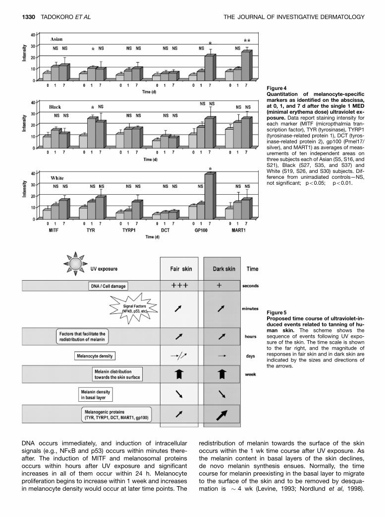
DNA occurs immediately, and induction of intracellularsignals (e.g., NFkB and p53) occurs within minutes there-after. The induction of MITF and melanosomal proteinsoccurs within hours after UV exposure and significantincreases in all of them occur within 24 h. Melanocyteproliferation begins to increase within 1 week and increasesin melanocyte density would occur at later time points. The
redistribution of melanin towards the surface of the skinoccurs within the 1 wk time course after UV exposure. Asthe melanin content in basal layers of the skin declines,de novo melanin synthesis ensues. Normally, the timecourse for melanin preexisting in the basal layer to migrateto the surface of the skin and to be removed by desqua-mation is � 4 wk (Levine, 1993; Nordlund et al, 1998).
Figure 4Quantitation of melanocyte-specificmarkers as identified on the abscissa,at 0, 1, and 7 d after the single 1 MED(minimal erythema dose) ultraviolet ex-posure. Data report staining intensity foreach marker (MITF (micropthalmia tran-scription factor), TYR (tyrosinase), TYRP1(tyrosinase-related protein 1), DCT (tyros-inase-related protein 2), gp100 (Pmel17/silver), and MART1) as averages of meas-urements of ten independent areas onthree subjects each of Asian (S5, S16, andS21), Black (S27, S35, and S37) andWhite (S19, S26, and S30) subjects. Dif-ference from unirradiated controls—NS,not significant; �po0.05; ��po0.01.
Figure 5Proposed time course of ultraviolet-in-duced events related to tanning of hu-man skin. The scheme shows thesequence of events following UV expo-sure of the skin. The time scale is shownto the far right, and the magnitude ofresponses in fair skin and in dark skin areindicated by the sizes and directions ofthe arrows.
1330 TADOKORO ET AL THE JOURNAL OF INVESTIGATIVE DERMATOLOGY

These responses are no doubt influenced by the UV spec-trum and dose(s) used. Increases in pigmentation to a givendose of UV are quicker and more dramatic in dark skin thanin light skin.
So what factors are critical for the increase in skin pig-mentation that can be observed within several days after asingle UV exposure? Obviously, the distribution of melaninin keratinocytes, particularly in the upper layers of the skinis quite important. We have previously reported that UVexposure stimulates the secretion of melanosomes bymelanocytes, and concurrently increases the ingestion ofmelanosomes by keratinocytes (Virador et al, 2002). Themechanisms that regulate pigment transfer and the subcel-lular machinery involved in that process within melanocytesand keratinocytes are gradually being unraveled. Microarrayor proteomic analysis of human skin after UV exposuremight help further define genes and/or proteins that arecritical to that process. The redistribution of melanin to up-per layers of the skin following a single, relatively mild doseof UV is critical to the increased photoprotection of the skin,and is another example of the plasticity and adaptability ofbiological systems.
Materials and Methods
Study subjects, UV exposure, and biopsy methods This studyinvolved human subjects representing six R/E groups (according toUnited States Office of Management and Budget Classification0990-0208; American Indian or Alaska Native; Asian; Black or Af-rican American; Hispanic or Latino; Native Hawaiian or Other Pa-cific Islander; White). Details of the protocols, subjects of the studyand UV doses have been published (Tadokoro et al, 2003). Thisstudy was approved by the FDA Research Involving Human Sub-jects Committee, and adhered to the Helsinki Guidelines; informedconsent was obtained from each of the study subjects. In thisstudy, we restricted our analysis to the three major R/E groups inthe US—Asian (for this study we selected East Asians only), Black,or African American (hereafter referred to as Black) and White.There was a minimum of ten subjects in each R/E group anddifferent numbers of specimens from each group were analyzed fordifferent end points; the numbers of subjects examined by eachprocedure are noted in the table and figure legends.
An FS lamp (National Biological) was used as the source of UVA/UVB. The distance between the lamps and skin was 22–27 cmand was adjusted to deliver approximately 100 J per m2-eff(CIE erythema effective, i.e., weighted with the CIE action spec-trum for erythema (Diffey et al, 1997) per min (fluence rate of1.67 W per m2-eff, 250–400 nm). Kodacel filtration (EastmanChemical Products, Kingsport, Tennessee) was used to remove theUVC component. The MED of each individual was determined toassess the sensitivity to UV by irradiating a series of 2 � 2 cm2 onone side of the back of each subject. The following day, the MEDdose was established and several 2 � 2 cm2 on the other side ofthe back were irradiated with 1 MED ranging from 250 to 480 J perm2 for Whites, 300–600 J per m2 for Asians and 600–850 J per m2
for Blacks. Shave biopsies, � 4 mm in diameter, were taken priorto the UV exposure, immediately after ( � 7 min), 1 d later and 7 dlater. Each biopsy was placed dermis side down on a Millipore(Millipore, Bedford, Massachusetts) filter (pore size, 0.45 mm; di-ameter 13 mm), fixed in 4% formaldehyde, embedded in paraffinand sectioned using standard techniques.
Immunohistochemical staining Specimens fixed with formalde-hyde and embedded in paraffin were sectioned at 3 mm thickness,then mounted on silane-coated glass slides. The expression ofmelanosomal proteins was measured in the sections by indirect
immunofluorescence using polyclonal antibodies, aPEP7h specificfor tyrosinase (Virador et al, 2001), aPEP1 specific for TRP-1(Jimenez et al, 1989) and aPEP8h specific for TRP-2 (Virador et al,2001), and monoclonal antibodies, HMB45 (DAKO, Carpinteria,California) specific for gp100, D5 (NeoMarkers, Fremont, California)specific for MITF, and MART-1 Ab-3 (NeoMarkers) specific forMART1. Samples were deparaffinized with xylene and dehydratedwith ethanol, then treated with cold methanol and boiled in 10 mMcitrate buffer (pH 6.0) for antigen retrieval. They were subsequentlyincubated with 10% goat serum (Vector Laboratories, Burlingame,California) containing 2% bovine serum albumin (AmershamPharmacia Biotech, Piscataway, New Jersey) and 0.05% Tween20for 30 min at 371C, and then with aPEP7h at 1:4000, aPEP1 at1:2000, aPEP8h at 1:15,000, HMB45 at 1:100, D5 at 1:100, orMART-1 Ab-3 at 1:400 dilution with 2% goat serum at 41C over-night. The slides were incubated with Alexa Fluor(R)594 goat anti-rabbit IgG (Hþ L) and Alexa Fluor(R)488 goat anti-mouse IgG(Hþ L) (Molecular Probes, Eugene, Oregon) at 371C for 30 min at1:200 dilution with 2% goat serum, and covered by a drop of Pro-long Antifade solution (Molecular Probes). The Alexa594 fluores-cence (Eugene, Oregan) was superimposed over the Alexa 488fluorescence to show co-localization. Alexa fluorescence was ob-served and analyzed using a Leica DMRB/DMLD laser microscope(Leica Microsystems, Bannockburn, Illinois) and ScionImage soft-ware (Scion, Frederick, Maryland). This system allows one toeliminate background level fluorescence and to quantitatefluorescence intensity from original images. Sections from subjectS24 at 7 d after UV were stained each time as an internal control forantibody staining.
Melanocyte density, melanin content and distribution Thenumbers of melanocytes along the basement membrane were di-rectly counted using the Leica DMRB/DMLD microscope afterstaining of melanosomal proteins. Melanocytes were only countedif the nucleus was visible.
Melanin content was analyzed following staining of sections bythe Fontana–Masson method (Bancroft and Stevens, 1982) underthe same controlled conditions. The Leica DMRB/DMLD laser mi-croscope measures transmitted light intensity. Melanin quantitywas analyzed from integrated density in the sections.
Melanin distribution was analyzed in digital images of Fontana–Masson stained sections using the image analysis software ‘‘KS400’’ from Carl Zeiss (Thornwood, New York). First, the pictureswere segmented according to the gray level, and dark pixels with agray level o128 (threshold level for melanin-positive structures)were measured. Subsequently, the area of these detected spotswas measured using the image analysis software for the differentepidermal layers (basal, granulosum and spinous) in each image.The size of the detected area represents the melanin content. Thethreshold level (in this case 128, on a gray-value scale from255¼white to 0¼black) was determined for each series of images;this was under exactly the same illumination conditions to allowcomparisons between experiments.
Diffuse reflectance (DR) spectra in the 400–700 nm range weremeasured at 10 nm increments using a Minolta CM-2002 spec-trophotometer (Minolta, Ramsey, New Jersey). Such spectra wereobtained for two UV-exposed 2 � 2 cm skin areas and one adja-cent unexposed control area. In each area, three measurementswere performed in different locations and the averages of the threeresults were then used to generate reflectance spectra used in theanalyses. These spectra were then transformed into apparent ab-sorbance (AA) spectra, where AA represents the logarithm of theratio of the DR of the control skin and the DR from UV-exposedskin, AA¼ log (DRC/DRUV) (Kollias et al, 1994). The linear part of the630-700 nm spectrum was used to calculate melanin content ac-cording to (Kollias et al, 1994) with modification of Jacques, 2001(http://omlc.ogi.edu/spectra/melanin/opticaldepth.html).
Photography The study areas were photographed before expo-sure and at all post-exposure stages of the investigation. Photo-
REGULATION OF SKIN PIGMENTATION AFTER UV RADIATION 1331124 : 6 JUNE 2005
graphs were taken under standardized conditions using a CanonRebel 2000 35 mm camera (Canon, Lake success, New York) witha 28–80 mm lens set at 80 mm and f¼ 32. The shutter (1/90 s) wassynchronized with a Speedotron Light (Chicago, Illinois) systemconsisting of two M11/CC light units, each with a 7 inch umbrellareflector and its own white umbrella diffuser. The lamps were5500K color-corrected flash bulbs. Both lamps were directed at a451 angle at the study area, i.e. the subject’s back. A color scale(Kodak Color Separation Guide Q-13 Rochester, New York) wasincluded in each image. The 35 mm film was processed by acommercial laboratory and was scanned at high resolution with nocolor or contrast correction. The digitized images were standard-ized to the Kodak color scale.
Statistical analyses The statistical software, JMP 5.1, was usedto determine t values and correlation coefficients. A p value ofo0.05 is defined as significant using paired or unpaired t tests.
DOI: 10.1111/j.0022-202X.2005.23760.x
Manuscript received November 23, 2004; revised January 21, 2005;accepted for publication February 23, 2005
Address correspondence to: Dr Vincent J. Hearing, Laboratory of CellBiology, Building 37, Room 2132, National Cancer Institute, NationalInstitutes of Health, Bethesda, MD 20892, USA. Email: [email protected]
References
Abdel-Malek ZA, Scott MC, Furumura M, Lamoreux ML, Ollmann M, Barsh GS,
Hearing VJ: The melanocortin 1 receptor is the principal mediator of the
effects of agouti signaling protein on mammalian melanocytes. J Cell Sci
114:1019–1024, 2001
Alaluf S, Atkins D, Barrett K, Blount M, Carter N, Heath A: The impact of ep-
idermal melanin on objective measurements of human skin colour. Pig-
ment Cell Res 15:119–126, 2002
Bancroft JD, Stevens A: Theory and Practice of Histological Techniques. New
York: Churchill Livingstone, 1982
Diffey BL, Jansen CT, Urbach F, Wulf HC: The standard erythema dose: A new
photobiological concept. Photodermatol Photoimmunol Photomed 13:
64–66, 1997
Du J, Miller AJ, Widlund HR, Horstmann MA, Ramaswamy S, Fisher DE: MLANA/
MART1 and SILV/PMEL17/GP100 are transcriptionally regulated by MITF
in melanocytes and melanoma. Am J Path 163:333–343, 2003
Fuller BB, Spaulding DT, Smith DR: Regulation of the catalytic activity of pre-
existing tyrosinase in Black and Caucasian human melanocyte cell cul-
tures. Exp Cell Res 262:197–208, 2001
Halder RM, Bridgeman-Shah S: Skin cancer in African Americans. Cancer
75:667–673, 1995
Hamperl H, Henschke U, Schulze R: Vergleich de Hautreaktion beim Best-
rahlungserythem und bei der direkten Pigmentierung. Virchows Arch
[Pathol Anat] 304:19–33, 1939
Hausser KW: Ueber die spezifische Wirkung des langwelligen ultravioletten Lichts
auf die menschliche Haut. Strahlentherapie 62:315–322, 1938
Hausser KW, Vahle W: Die Abhaengigkeit des Lichterythems und der Pigment-
bildung von der Schwingungszahl (Wellenlaenge) der erregenden Strah-
lung. Strahlentherapie 13:41–71, 1922
Jimenez M, Maloy WL, Hearing VJ: Specific identification of an authentic clone
for mammalian tyrosinase. J Biol Chem 264:3397–3403, 1989
Kollias N, Baqer AH, Sadiq I: Minimum erythema dose determination in individ-
uals of skin type V and VI with diffuse reflectance spectroscopy.
Phoodermatol Photoimmunol Photomed 10:249–254, 1994
Kricker A, Armstrong BK, English DR: Sun exposure and non-melanocytic skin
cancer. Cancer Causes Control 5:367–392, 1994
Kushimoto T, Basrur V, Matsunaga J, Vieira WD, Muller J, Appella E, Hearing VJ:
A new model for melanosome biogenesis based on the purification
and mapping of early melanosomes. Proc Natl Acad Sci USA 98:
10698–10703, 2001
Kushimoto T, Valencia JC, Costin GE, et al: The Seiji Memorial Lecture—The
melanosome: An ideal model to study cellular differentiation. Pigment Cell
Res 16:237–244, 2003
Levine N: Pigmentation and Pigmentary Disorders. Boca Raton: CRC Press,
1993; p 1–553
Miescher G: Untersuchungen ueber die Bedeutung des Pigments fuer den
UV.Lichtschutz der Haut. Strahlentherapie 435:201–216, 1932
Nordlund JJ, Boissy RE, Hearing VJ, King RA, Ortonne JP: The Pigmentary Sys-
tem: Physiology and Pathophysiology. New York: Oxford University
Press, 1998; p 1–1025
Oetting WS: The tyrosinase gene and oculocutaneous albinism type 1 (OCA1): A
model for understanding the molecular biology of melanin formation.
Pigment Cell Res 13:320–325, 2000
Preston DS, Stern RS: Nonmelanoma cancers of the skin. N Eng J Med
327:1649–1662, 1992
Quevedo WC Jr, Fitzpatrick TB, Szabo G, Jimbow K: Biology of melanocytes.
In: Fitzpatrick TB, Eisen AZ, Wolff K, Freedberg IM, Austen KF
(eds). Dermatology in General Medicine. New York: McGraw-Hill, 1987;
p 224–251
Quevedo WC Jr., Holstein TJ: General biology of mammalian pigmentation. In:
Nordlund JJ, Boissy RE, Hearing VJ, King RA, Oetting WS (eds). The
Pigmentary System: Physiology and Pathophysiology. New York: Oxford
University Press, 1998; p 43–58
Sakai C, Ollmann M, Kobayashi T, et al: Modulation of murine melanocyte func-
tion in vitro by agouti signal protein. EMBO J 16:3544–3552, 1997
Shibahara S, Takeda K, Yasumoto K, Udono T, Watanabe K, Saito H, Takahashi
K: Microphthalmia-associated transcription factor (MITF): Multiplicity in
structure, function and regulation. J Invest Dermatol (Suppl. 6):99–104,
2001
Shibahara S, Yasumoto K, Amae S, Udono T, Watanabe K, Saito H, Takeda K:
Regulation of pigment cell specific gene expression by MITF. Pigment
Cell Res 13:98–102, 2000
Shriver MD, Parra EJ, Dios S, et al: Skin pigmentation, biogeographical ancestry
and admixture mapping. Hum Genet 112:387–399, 2003
Stierner U, Rosdahl IK, Augustsson A, Kagedal B: UVB irradiation induces me-
lanocyte increase in both exposed and shielded human skin. J Invest
Dermatol 92:561–564, 1989
Tachibana M: MITF: A stream flowing for pigment cells. Pigment Cell Res 13:
230–240, 2000
Tadokoro T, Kobayashi N, Zmudzka BZ, et al: UV-induced DNA damage and
melanin content in human skin differing in racial/ethnic origin and pho-
tosensitivity. FASEB J 17:1177–1179, 2003
Thong H-Y, Jee S-H, Sun C-C, Boissy RE: The patterns of melanosome distri-
bution in keratinocytes of human skin as one determining factor of skin
colour. Br J Dermatol 149:498–505, 2003
Virador V, Matsunaga N, Matsunaga J, et al: Production of melanocyte-specific
antibodies to human melanosomal proteins: Expression patterns in
normal human skin and in cutaneous pigmented lesions. Pigment Cell
Res 14:289–297, 2001
Virador V, Muller J, Wu X, et al: Influence of a-melanocyte stimulating hormone
and ultraviolet radiation on the transfer of melanosomes to keratinocytes.
FASEB J 16:105–107, 2002
Whiteman DC, Parsons PG, Green AC: Determinants of melanocyte density in
adult human skin. Arch Dermatol Res 291:511–516, 1999
Yasumoto K, Takeda K, Saito H, Watanabe K, Takahashi K, Shibahara S: Micro-
phthalmia-associated transcription factor interacts with LEF-1, a medi-
ator of Wnt signaling. EMBO J 21:2703–2714, 2002
1332 TADOKORO ET AL THE JOURNAL OF INVESTIGATIVE DERMATOLOGY
Copyright © 2022 FDOKUMEN