MicroRNA-208a Increases Myocardial Fibrosis via Endoglin in ...
8
MicroRNA-208a Increases Myocardial Fibrosis via Endoglin in Volume Overloading Heart Bao-Wei Wang 1,2 , Gong-Jhe Wu 3,4 , Wen-Ping Cheng 2 , Kou-Gi Shyu 2,5 * 1 School of Medicine, Fu-Jen Catholic University, New Taipei City, Taiwan, 2 Division of Cardiology, Shin Kong Wu Ho-Su Memorial Hospital, Taipei, Taiwa, 3 School of Medicine, College of Medicine, Taipei Medical University, Taipei, Taiwan, 4 Department of Anesthesiology, Shin Kong Wu Ho-Su Memorial Hospital, Taipei, Taiwan, 5 Graduate Institute of Clinical Medicine, Taipei Medical University, Taipei, Taiwan Abstract MicroRNA-208a (mir-208a) is essential for cardiac hypertrophy and fibrosis. Endoglin, a co-receptor of transforming growth factor-b is also essential for cardiac fibrosis. Endoglin has been shown to be a target of mir-208a in the in vitro mechanical stress model. Volume overload can lead to heart failure and cardiac fibrosis. The role of mir-208a and endoglin in volume overload heart failure is well known. We sought to investigate the mechanism of regulation of mir-208a and endoglin in volume overload-induced heart failure. Aorta-caval (AV) shunt was performed in adult Sprague-Dawley rats to induce volume overload. Heart weight and heart weight/body weight ratio significantly increased in AV shunt animals. AV shunt significantly increased left ventricular end-diastolic dimension as compared to sham group. Mir-208a was significantly induced by AV shunt from 3 to 14 days. Endoglin, myosin heavy chain-b and brain natriuretic peptide were significantly induced by AV shunt from 3 to 14 days. Overexpression of mir-208a in the sham group without AV shunt significantly increased endoglin expression similar to the AV shunt group. Antagomir-208a attenuated the endoglin expression induced by AV shunt. Pretreatment with atorvastatin also attenuated the endoglin expression induced by AV shunt. AV shunt significantly increased myocardial fibrosis as compared to sham group. Overexpression of mir-208a in the sham group significantly increased myocardial fibrosis. Antagomir-208a and atorvastatin significantly attenuated the myocardial fibrosis induced by AV shunt. In conclusion, mir-208a increased endoglin expression to induce myocardial fibrosis in volume overloaded heart failure. Treatment with atorvastatin can attenuate the myocardial fibrosis induced by volume overload through inhibition of endoglin expression. Citation: Wang B-W, Wu G-J, Cheng W-P, Shyu K-G (2014) MicroRNA-208a Increases Myocardial Fibrosis via Endoglin in Volume Overloading Heart. PLoS ONE 9(1): e84188. doi:10.1371/journal.pone.0084188 Editor: Dinender K Singla, University of Central Florida, United States of America Received August 26, 2013; Accepted November 13, 2013; Published January 2, 2014 Copyright: ß 2014 Wang et al. This is an open-access article distributed under the terms of the Creative Commons Attribution License, which permits unrestricted use, distribution, and reproduction in any medium, provided the original author and source are credited. Funding: This study was sponsored in part from National Science Council, Executive Yuan, Taiwan and Shin Kong Wu Ho-Su Memorial Hospital, Taipei, Taiwan. The funders had no role in study design, data collection and analysis, decision to publish, or preparation of the manuscript. Competing Interests: The authors have declared that no competing interests exist. * E-mail: [email protected] Introduction Cardiac fibrosis is closely associated with heart failure because cardiac fibrosis may cause the loss of normal cardiac function [1]. Endoglin is a homeodimeric membrane glycoprotein that is a co- receptor of transforming growth factor-b1 (TGF-b1) and b3 [2]. Endoglin is a potent mediator of profibrotic effects of angiotensin II on cardiac fibroblasts [3] and can modulate the effect of TGF- b1 on extracellular matrix synthesis [4]. These data indicate that endoglin plays an important role in fibrogenesis in cardiac remodeling. A microRNA (mir) is small, 22-nucleotide non-protein-coding RNA that inhibits transcription or translation by interacting with the 39untranslated regions of target mRNA and promoting target mRNA degradation (gene silencing) [5]. Recently, Place et al. have demonstrated mir functioning to induce gene expression [6]. Because of their capability to monitor the expression levels of the genes that control both adaptive and maladaptive cardiac remodeling processes, mirs may be vitally involved in the pathogenesis of heart failure [7,8]. Mir-208a seems to be fundamental for the expression of genes involved in cardiac fibrosis and hypertrophic growth [9,10]. Mir-208a is upregulated in pressure overloading with thoracic aortic banding [9] and is activated by mechanical stress [11]. The role of mir-208a in volume overload is not known. Mir-208a can increase endoglin expression in cardiac myoblast [11]. Endoglin expression is increased in patients with heart failure [12]. Since volume overload can lead to heart failure and myocardial fibrosis, we sought to investigate the regulation of mir-208a in volume overloading heart. Materials and Methods The aorta-caval shunt (AV shunt) rat model AV shunt was performed on rats to induce volume overload. On the day of surgery, the Sprague-Dawley rats weighing 280 to 330 g were anesthetized with 2% isoflurane and the vena cava and aorta were exposed via abdominal midline incision after confirming a fully anaesthetized state (.e.g. no response to toe pinching). The aorta-caval shunt was produced as previously described [13]. Sham-operated control animals were prepared similar manner, except that the aorta was not punctured. Atorvastatin at 30 mg/kg was given by oral gavage for 2 weeks after induction of AV shunt. After 2 weeks of AV shunt induction, rats were euthanized with an overdose of isoflurane. Left ventricular tissue was obtained for Western blot analysis and immunohistochemical staining. Mas- PLOS ONE | www.plosone.org 1 January 2014 | Volume 9 | Issue 1 | e84188
-
Upload
khangminh22 -
Category
Documents
-
view
1 -
download
0
Transcript of MicroRNA-208a Increases Myocardial Fibrosis via Endoglin in ...

MicroRNA-208a Increases Myocardial Fibrosis viaEndoglin in Volume Overloading HeartBao-Wei Wang1,2, Gong-Jhe Wu3,4, Wen-Ping Cheng2, Kou-Gi Shyu2,5*
1 School of Medicine, Fu-Jen Catholic University, New Taipei City, Taiwan, 2 Division of Cardiology, Shin Kong Wu Ho-Su Memorial Hospital, Taipei, Taiwa, 3 School of
Medicine, College of Medicine, Taipei Medical University, Taipei, Taiwan, 4 Department of Anesthesiology, Shin Kong Wu Ho-Su Memorial Hospital, Taipei, Taiwan,
5 Graduate Institute of Clinical Medicine, Taipei Medical University, Taipei, Taiwan
Abstract
MicroRNA-208a (mir-208a) is essential for cardiac hypertrophy and fibrosis. Endoglin, a co-receptor of transforming growthfactor-b is also essential for cardiac fibrosis. Endoglin has been shown to be a target of mir-208a in the in vitro mechanicalstress model. Volume overload can lead to heart failure and cardiac fibrosis. The role of mir-208a and endoglin in volumeoverload heart failure is well known. We sought to investigate the mechanism of regulation of mir-208a and endoglin involume overload-induced heart failure. Aorta-caval (AV) shunt was performed in adult Sprague-Dawley rats to inducevolume overload. Heart weight and heart weight/body weight ratio significantly increased in AV shunt animals. AV shuntsignificantly increased left ventricular end-diastolic dimension as compared to sham group. Mir-208a was significantlyinduced by AV shunt from 3 to 14 days. Endoglin, myosin heavy chain-b and brain natriuretic peptide were significantlyinduced by AV shunt from 3 to 14 days. Overexpression of mir-208a in the sham group without AV shunt significantlyincreased endoglin expression similar to the AV shunt group. Antagomir-208a attenuated the endoglin expression inducedby AV shunt. Pretreatment with atorvastatin also attenuated the endoglin expression induced by AV shunt. AV shuntsignificantly increased myocardial fibrosis as compared to sham group. Overexpression of mir-208a in the sham groupsignificantly increased myocardial fibrosis. Antagomir-208a and atorvastatin significantly attenuated the myocardial fibrosisinduced by AV shunt. In conclusion, mir-208a increased endoglin expression to induce myocardial fibrosis in volumeoverloaded heart failure. Treatment with atorvastatin can attenuate the myocardial fibrosis induced by volume overloadthrough inhibition of endoglin expression.
Citation: Wang B-W, Wu G-J, Cheng W-P, Shyu K-G (2014) MicroRNA-208a Increases Myocardial Fibrosis via Endoglin in Volume Overloading Heart. PLoS ONE 9(1):e84188. doi:10.1371/journal.pone.0084188
Editor: Dinender K Singla, University of Central Florida, United States of America
Received August 26, 2013; Accepted November 13, 2013; Published January 2, 2014
Copyright: � 2014 Wang et al. This is an open-access article distributed under the terms of the Creative Commons Attribution License, which permitsunrestricted use, distribution, and reproduction in any medium, provided the original author and source are credited.
Funding: This study was sponsored in part from National Science Council, Executive Yuan, Taiwan and Shin Kong Wu Ho-Su Memorial Hospital, Taipei, Taiwan.The funders had no role in study design, data collection and analysis, decision to publish, or preparation of the manuscript.
Competing Interests: The authors have declared that no competing interests exist.
* E-mail: [email protected]
Introduction
Cardiac fibrosis is closely associated with heart failure because
cardiac fibrosis may cause the loss of normal cardiac function [1].
Endoglin is a homeodimeric membrane glycoprotein that is a co-
receptor of transforming growth factor-b1 (TGF-b1) and b3 [2].
Endoglin is a potent mediator of profibrotic effects of angiotensin
II on cardiac fibroblasts [3] and can modulate the effect of TGF-
b1 on extracellular matrix synthesis [4]. These data indicate that
endoglin plays an important role in fibrogenesis in cardiac
remodeling.
A microRNA (mir) is small, 22-nucleotide non-protein-coding
RNA that inhibits transcription or translation by interacting with
the 39untranslated regions of target mRNA and promoting target
mRNA degradation (gene silencing) [5]. Recently, Place et al. have
demonstrated mir functioning to induce gene expression [6].
Because of their capability to monitor the expression levels of the
genes that control both adaptive and maladaptive cardiac
remodeling processes, mirs may be vitally involved in the
pathogenesis of heart failure [7,8]. Mir-208a seems to be
fundamental for the expression of genes involved in cardiac
fibrosis and hypertrophic growth [9,10]. Mir-208a is upregulated
in pressure overloading with thoracic aortic banding [9] and is
activated by mechanical stress [11]. The role of mir-208a in
volume overload is not known. Mir-208a can increase endoglin
expression in cardiac myoblast [11]. Endoglin expression is
increased in patients with heart failure [12]. Since volume
overload can lead to heart failure and myocardial fibrosis, we
sought to investigate the regulation of mir-208a in volume
overloading heart.
Materials and Methods
The aorta-caval shunt (AV shunt) rat modelAV shunt was performed on rats to induce volume overload. On
the day of surgery, the Sprague-Dawley rats weighing 280 to 330 g
were anesthetized with 2% isoflurane and the vena cava and aorta
were exposed via abdominal midline incision after confirming a
fully anaesthetized state (.e.g. no response to toe pinching). The
aorta-caval shunt was produced as previously described [13].
Sham-operated control animals were prepared similar manner,
except that the aorta was not punctured. Atorvastatin at 30 mg/kg
was given by oral gavage for 2 weeks after induction of AV shunt.
After 2 weeks of AV shunt induction, rats were euthanized with an
overdose of isoflurane. Left ventricular tissue was obtained for
Western blot analysis and immunohistochemical staining. Mas-
PLOS ONE | www.plosone.org 1 January 2014 | Volume 9 | Issue 1 | e84188

son’s trichrome staining was performed to delineate fibrosis tissue
from viable myocardium. All study protocols were approved by
our Committee of Animal Care and Use of Shin Kong Wu Ho-Su
Memorial Hospital (permit number:091217021) and were carried
out in accordance with the Guide for the Care and Use of
Laboratory Animals (NIH publication No. 86-23, revised 2011).
Hemodynamic monitorHemodynamic monitor of rats was performed with polyethylene
catheters to measure through a Grass model tachogragh pream-
plifier as previously described [13].
Assessment of cardiac hypertrophy and functionCardiac function of rats was evaluated noninvasively by
echocardiography performed with an Acuson Sequoia 512
machine using a 15-MHz probe at the day of sacrifice, 7 and 14
days (AV shunt) after the surgery as previously described [13]. The
sonographer was blinded to the randomization of rats.
Construction and delivery of mir-208a expression vectorA 71 bp rat-mir-208a precursor construct was generated as
follows. Genomic DNA was amplified with forward primer,
CAACAGAAGTGCTTGGAAG and reverse primer,
GGCTGATCGACGGTAGCT. The 165 bp amplified product
was digested with EcoRI and BamHI restriction enzymes and
ligated into pmR-ZsGreen1 plasmid vector (coexpression mir-208
and green fluorescent protein, Clontech Laboratories, Mountain
View, CA, USA) digested with the same enzymes. The constructed
plasmid (co-expression mir-208 and green fluorescent protein) was
transfected into left ventricular myocardium using a low pressure-
accelerated gene gun (Bioware Technologies, Taipei, Taiwan)
essentially following the protocol from the manufacturer. In brief,
2 mg of plasmid DNA was suspended in 5 ml of PBS and then
100 ml was added to the loading hole near the nozzle. Pushing the
trigger of the low pressure gene gun released the DNA-containing
solution, which was directly propelled by helium at a pressure of
15 psi into left ventricular myocardium of the rat. The distribution
of fluorescent image in treated rat was visualized by a dissecting
fluorescence microscope with high resolution CCD (HAMA-
MATSU PHOTONICS, Japan). After 3 days, the rat chest was
re-open and the fluorescent image on left ventricular myocardium
was detected. If the fluorescent image was able to visualize, it was
regarded as a successful transfection. The efficiency of using this
method is around 30%.
Western blot analysisWestern blot was performed as previously described ‘[14].
Monoclonal rat anti-mouse endoglin antibody, polyclonal myosin
heavy chain and polyclonal brain natriuretic peptide antibodies
(Santa Cruz Biotechnology, Inc., CA, USA) were used. Equal
protein loading of the samples was verified by staining monoclonal
antibody a-tubulin (Sigma, St. Louis, MI, USA). Signals were
visualized by chemiluminenescent detection. All Western blots
were quantified using densitometry.
Quantitative analysis of microRNAsTaqManH MicroRNA real-time quantitative assays were used
to quantitate mir as previously described [11]. All fold changes
between samples were determined using the DDCT method [15].
In brief, each 15 ml RT reaction contained purified 10 ng of total
RNA, 3 ml miR-208 RT primer (Applied BiosystemsH, Life
Technologies, Grand Island, NY, USA), 16RT buffer (Applied
Biosystems), 0.25 mM each of dNTPs, 3.33 U/ml MultiScribeTM Ta
ble
1.
He
mo
dyn
amic
and
ech
oca
rdio
gra
ph
icp
aram
ete
rs.
Sh
am
Sh
un
t7
DS
hu
nt
14
DS
ha
m/m
iR-2
08
aS
ha
m/M
ut-
20
8a
Sh
un
t7D
/An
tag
om
ir-
20
8a
Sh
un
t7
D/M
ut-
20
8a
Sh
un
t7D
/Ato
rva
sta
tin
N6
66
66
66
6
Bo
dy
we
igh
t,g
30
96
16
30
56
20
31
46
26
27
96
11
28
76
19
30
46
12
30
76
16
29
76
21
He
art
we
igh
t,m
g8
016
42
89
56
57
*1
10
46
61
+8
216
40
81
16
22
84
46
41
88
26
29
83
76
38
He
art
we
igh
t/b
od
yw
eig
ht,
mg
/g2
.56
0.4
2.8
60
.4*
3.4
96
0.5
*2
.96
0.3
2.7
60
.62
.86
0.6
2.9
60
.52
.76
0.5
He
art
rate
,m
in3
156
27
34
66
11
30
46
22
33
26
31
31
96
29
33
26
22
33
86
21
32
46
32
MA
P,
mm
Hg
826
97
56
87
16
48
06
78
66
97
76
68
46
98
16
11
IVST
d,
mm
1.3
60
.21
.26
0.4
1.1
60
.61
.16
0.5
1.2
60
.41
.26
0.2
1.2
60
.41
.26
0.2
LVP
WT
,m
m1
.26
0.2
1.2
60
.41
.06
0.4
1.3
60
.21
.36
0.3
1.3
60
.41
.16
0.4
1.3
60
.3
LVED
D,
mm
6.5
60
.36
.96
0.4
7.1
60
.5*
6.2
60
.46
.46
0.5
6.2
60
.56
.46
0.4
6.3
60
.4
LVES
D,
mm
3.2
60
.33
.66
0.3
4.1
60
.4*
3.5
60
.33
.36
0.6
3.2
60
.63
.46
0.5
3.2
60
.5
FS,
%4
76
74
46
64
26
74
56
54
76
54
76
94
56
84
66
8
MA
P=
me
anar
teri
alp
ress
ure
.IV
STd
=in
ter-
ven
tric
ula
rse
ptu
me
nd
-dia
sto
licth
ickn
ess
.LV
PW
T=
left
ven
tric
ula
rp
ost
eri
or
wal
lth
ickn
ess
.LV
EDD
=le
ftve
ntr
icu
lar
en
d-d
iast
olic
dim
en
sio
n.
LVES
D=
left
ven
tric
ula
re
nd
-sys
tolic
dim
en
sio
n.
FS=
frac
tio
nsh
ort
en
ing
.*P
,0
.01
vs.
sham
.d
oi:1
0.1
37
1/j
ou
rnal
.po
ne
.00
84
18
8.t
00
1
MicroRNA-208a Increases Myocardial Fibrosis
PLOS ONE | www.plosone.org 2 January 2014 | Volume 9 | Issue 1 | e84188
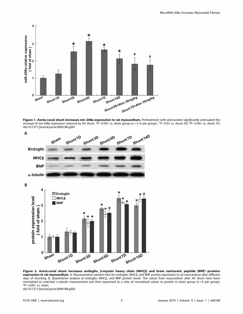
Figure 1. Aorta-caval shunt increases mir-208a expression in rat myocardium. Pretreatment with atorvastatin significantly attenuated theincrease of mir-208a expression induced by AV shunt. *P,0.001 vs. sham group (n = 5–6 per group). #P,0.01 vs. shunt 5D. `P,0.001 vs. shunt 7D.doi:10.1371/journal.pone.0084188.g001
Figure 2. Aorta-caval shunt increases endoglin, b-myosin heavy chain (MHCb) and brain natriuretic peptide (BNP) proteinsexpression in rat myocardium. A. Representative western blot for endoglin, MHCb, and BNP protein expression in rat myocardium after differentdays of shunting. B, Quantitative analysis of endoglin, MHCb, and BNP protein levels. The values from myocardium after AV shunt have beennormalized to matched a-tubulin measurement and then expressed as a ratio of normalized values to protein in sham group (n = 6 per group).*P,0.001 vs. sham.doi:10.1371/journal.pone.0084188.g002
MicroRNA-208a Increases Myocardial Fibrosis
PLOS ONE | www.plosone.org 3 January 2014 | Volume 9 | Issue 1 | e84188

reverse transcriptase (Applied Biosystems) and 0.25 U/ml RNase
inhibitor (Applied Biosystems). The reactions were incubated in an
Applied Biosystems 9700 Thermocycler in a 96-well plate for
30 min at 16uC, 30 min at 42uC, followed by 5 min at 85uC, and
then held at 4uC. Each real-time PCR for each microRNA assay
(20 ml volume) was carried out in triplicate, and each 20 ml
reaction mixture included 1.33 ml of RT product, 10 ml of
26TaqManH Universal PCR Master Mix, 1 mM 206TaqMan
MicroRNA assay. The reaction was incubated in an Applied
Biosystems 7300 Real-Time PCR System in 96-well plate at 95uCfor 10 min, followed by 40 cycles of 95uC for 15 sec and 60uC for
1 min. The expression levels of target mirs were normalized to U6.
Immunohistochemical analysisThe left ventricle was harvested and fixed in 10% formaldehyde
and sliced into 5 mm paraffin sections. For immunohistochemical
stain, the slides were postfixed in 4% paraformaldehyde for
20 min, treated in 3% hydrogen peroxide/PBS for 25 min,
blocked in 5% normal rabbit serum for 20 min, blocked with
biotin/avidin for 15 min each, and incubated with fluorescent
isothiocyanate (FITC)-conjugated rat monoclonal anti-endoglin
antibody, polyclonal myosin heavy chain antibody (Santa Cruz
Biotechnology). For 2 hours at room temperature, biotinylated
rabbit-anti mouse IgG at 1:400 for 30 min, and Vector Elite ABC
biotin-avidin-peroxidase complex for 30 min. Sections were then
developed with diaminobenzidine and diaminobenzidine enhanc-
er (Vector), counterstained with hematoxylin. Images were
examined with a fluorescent microscope.
In situ hybridization assayFive-micrometer-thick tissue sections of left ventricular myocar-
dium were mounted on positively charged barrier frame slides,
dewaxed in xylenes, and rehydrated through an ethanol dilution
series (100% to 25%). Tissue sections were digested with 5 mg/mL
of proteinase K for 20 minutes at 37uC to facilitate probe
penetration and exposure of miRNA species. To minimize
nonspecific binding based on charge interactions, tissues were
subjected to a brief acetylation reaction [66 mmol/L HCl, 0.66%
acetic anhydride (v/v) and 1.5% triethanolamine (v/v) in RNase-
free water]. Then, tissue sections were prehybridized at the
hybridization temperature for 30 minutes in prehybridization
solution which consisted of 50% deionized formamide, 56sodium
chloride/sodium citrate buffer, 16 Denhardt’s solution, 500 mg/
mL of yeast tRNA, and 0.01% Tween. The prehybridization
solution was replaced with 200 ml of hybridization solution
containing 10 pmol of the FAM-labeled LNA miR-208a probe
(product sequence 59-39, CTTTTTGCTCGTCTTAT, Exiqon,
Vedbaek, Denmark) and tissues were incubated for 90 minutes at
the hybridization temperature and washed twice for 10 minutes in
sodium chloride/sodium citrate buffer.
Figure 3. Mir-208a mediates the myocardial endoglin expression in AV shunt rat. A, Representative western blot for endoglin and MHCbprotein expression in the rat myocardium after 7 days of shunting. Mir-208 expression vector was transfected into left ventricular myocardium by lowpressure-accelerated gene gun. Overexpression of mir-208a in the sham group significantly increased endoglin and MHCb proteinexpression.*P,0.001 vs. sham. #P,0.001 vs. shunt 7D. (n = 6 per group). Pretreatment with atorvastatin significantly attenuated the increase ofendoglin and MHCb protein expression induced by AV shunt.doi:10.1371/journal.pone.0084188.g003
MicroRNA-208a Increases Myocardial Fibrosis
PLOS ONE | www.plosone.org 4 January 2014 | Volume 9 | Issue 1 | e84188

Statistical analysisThe data were expressed as mean+SD. Statistical significance
was performed with analysis of variance (GraphPad Software Inc.,
San Diego, CA, USA). The Dunnett’s test was used to compare
multiple groups to a single control group. Tukey-Kramer
comparison test was used for pairwise comparisons between
multiple groups after the ANOVA. A value of P,0.05 was
considered to denote statistical significance.
Results
AV shunt increases heart sizeThe heart weight and heart weight/body weight ratio
significantly increased after AV shunt for 7 and 14 days
(Table 1). The heart rate and mean arterial blood pressure did
not change significantly. LV end-diastolic and end-systolic
dimension significantly increased after AV shunt for 14 days and
inter-ventricular septum thickness and left ventricular posterior
wall thickness did not significantly change, indicating the volume-
overload induced by AV shunt.
AV shunt increases myocardial mir-208a expressionAs shown in Figure 1, AV shunt significantly increased
myocardial mir-208a expression at 3 days after shunting, reached
a maximal of 3.160.2-fold at 5 days and remained elevated for up
to 14 days after shunting. Pretreatment with atorvastatin
significantly attenuated the increase of mir-208a induced by AV
shunt. However, the mir-208a level was still higher in the
atorvastatin-treated group than in the sham group, indicating
that atorvastatin partially but not completely inhibited the increase
of mir-208a expression induced by AV shunt.
AV shunt increases myocardial endoglin expressionAs shown in Figure 2, AV shunt significantly increased
myocardial endoglin protein expression from 3 days up to 14 days.
The cardiac hypertrophic markers such as bMHC and BNP were
also significantly induced by AV shunt from 3 to 14 days as the
endoglin protein.
Mir-208a mediates the myocardial endoglin expressionTo investigate the effect of mir-208a on myocardial endoglin
expression, over-expression of antogomir208a and mutant type
mir-208a (mut-208a) in the left ventricle was performed. AV shunt
at 7 days significantly increased myocardial endoglin and bMHC
protein expression and over-expression of antagomir208a signif-
icantly inhibited the increase of myocardial endoglin and bMHC
protein expression induced by AV shunt. Over-expression of
mutant mir-208a (mut-208a) did not have the effect on myocardial
endoglin and bMHC expression induced by AV shunt. Over-
expression of mir-208a in the sham group without AV shunt
significantly increased myocardial endoglin and bMHC protein
expression while over-expression of mut-208a in the sham group
did not induce myocardial endoglin and bMHC protein expres-
sion (Figure 3). Pretreatment of atorvastatin significantly atten-
uated the increase of myocardial endoglin and bMHC protein
expression induced by AV shunt (Figure S1). The transfection of
mir-208a into myocardium was monitored by a dissecting
fluorescence microscope as shown in Figure S2. The presence of
mir-208a in the cytoplasm of cardiac myocyte was confirmed by in
situ hybridization assay (Figure 4). Immunohistochemical staining
showed that increased myocardial endoglin and bMHC expres-
sion after AV shunt and over-expression of mir-208a in the sham
group (Figure 5). Mutant mir-208a did not change myocardial
endoglin and bMHC expression after AV shunt. Myocardial
endoglin and bMHC were not stained in the control sham group.
Figure 4. In situ hybridization assay detects the presence of mir-208a in the cardiac myocytes. Representative microscopic imagesshowing the presence of mir-208a (green color) in the cytoplasm of cardiac myocytes from left ventricular myocardium in AV shunt rats. The shamgroup or scrambled probe did not detect the presence of mir-208a.doi:10.1371/journal.pone.0084188.g004
MicroRNA-208a Increases Myocardial Fibrosis
PLOS ONE | www.plosone.org 5 January 2014 | Volume 9 | Issue 1 | e84188

Mir-208a increases myocardial fibrosisAV shunt and over-expression of mir-208a in the sham group
significantly increased myocardial fibrosis area as compared to
sham group (Figure 6). Over-expression of mut-208a in the sham
group did not change the fibrosis area as compared to the sham
group. Overexpression of antagomir208a and pretreatment with
atorvastatin in the AV shunt group significantly decreased
myocardial fibrosis area induced by AV shunt. Over-expression
of mut-208a in the AV shunt did not decrease the fibrosis area
induced by AV shunt. This finding indicates that mir-208a plays a
crucial role in the myocardial fibrosis after AV shunt.
Discussion
Endoglin expression is increased in human hearts with severe
left ventricular failure and in heart failure induced by transaortic
constriction, a pressure overload in mice [12]. Kapur et al. have
demonstrated that reduced endoglin activity by soluble endoglin
can limit cardiac fibrosis and improve survival in heart failure [12].
Endoglin also regulates angiotensin-mediated fibrosis via angio-
tensin receptor [16]. In the present study, endoglin expression is
elevated in acute volume-overload heart induced by AV shunt,
indicating that endoglin plays a crucial role in pressure- and
volume-overload heart failure. Endoglin has been used as a
noninvasive measure of left ventricular filling pressure in heart
failure [17] and has been regarded as a new biomarker for acute
Figure 5. Immunohistochemnical staining of left ventricular myocardium after induction of aorta-caval shunt with or withoutantagomir-208a treatment. There are significantly increased immunoreactive signals for endoglin and MHCb after overexpression of mir-208a andAV shunt for 7 days. Antagomir-208a significantly decreased the immunoreactive signal induced by AV shunt. Rare endoglin signals were seen in thesham group.doi:10.1371/journal.pone.0084188.g005
MicroRNA-208a Increases Myocardial Fibrosis
PLOS ONE | www.plosone.org 6 January 2014 | Volume 9 | Issue 1 | e84188

heart failure [18]. Therefore, targeting endoglin to prevent fibrosis
and heart failure may improve clinical outcome in patients with
heart failure in addition to the current clinical benefits of b-
adrenergic receptor antagonists, angiotensin-converting enzyme or
receptor blockers and aldosterone antagonists [19]. AV shunt
model in our study increased heart weight, heart weight/body
weight ratio, and increased left ventricular size without increased
septal and posterior wall thickness, indicating an eccentric
hypertrophy which is consistent with volume overload heart
failure status.
Mirs are reported to be aberrantly expressed in hypertrophic
heart [20]. Mir-208 is expressed specifically in the heart with trace
expression in the lung [9]. Mir-208a is also essential for the
expression of the genes involved in cardiac hypertrophic growth.
Although mir-208a was upregulated and cardiac hypertrophy was
induced in thoracic aortic banding, a pressure overload model [8],
it is not known whether mir-208a is also induced in volume
overload. In the present study, we demonstrated for the first time
that mir-208a was induced in acute volume overload by AV shunt
in rat. Over-expression of mir-208a in the sham group without AV
shunt significantly increased myocardial endoglin expression.
Over-expression of antagomir208a in the AV shunt group
significantly decreased myocardial fibrosis area induced by AV
shunt, indicating that mir-208a plays a crucial role in the
myocardial fibrosis after AV shunt. The increased endoglin to
induce myocardial fibrosis induced by AV shunt was mediated by
mir-208a. Bedsides endoglin, bMHC is also a target of mir-208a.
Overexpression of mir-208a in cardiac myocytes increases bMHC
protein expression and addition of antagomir-208a significantly
attenuates the increase of bMHC induced by overexpression of
mir-208a [21]. Overexpression of mir-208a did not increase
protein expression of thyroid hormone receptor associated protein
1, brain natriuretic peptide and aMHC [21].
Patients with congenital heart disease may have myocardial
fibrosis similar to patients with acquired heart failure [22]. In
animal model of pulmonary artery banding and trans-valvular
patch to induce right ventricular failure and mimic repaired
tetralogy of Fallot, myocardial fibrosis was observed in infant
piglets [23]. These data indicate that cardiac fibrosis may begin at
the embryonic and early infant stage with heart defect causing
volume overload. Mir208a may also play a pivotal role in the
formation of cardiac fibrosis in congenital heart disease, not just in
acquired heart disease. Therapeutic innovation target miR208a to
improve cardiac fibrosis may warrant further research.
Statin, a 3-hydroxy 3-methyl glutaryl-CoA reductase (HMG-
CoA reductase) inhibitor, improves survival in patients with
ischemic and non-ischemic heart failure [24]. Recently, high dose
atorvastatin significantly reduces hospitalization for heart failure in
patients with stable coronary heart disease [25]. Atorvastatin can
reduce endoglin expression in endothelium in apo-E deficient mice
and C57BL/6J mice [26,27]. Statin provides antifibrotic effect via
blocking the angiotensin II-mediated oxidative stress and procol-
lagen-1 expression in cardiac fibroblast [28]. Recently, we have
demonstrated that atorvastatin can inhibit endoglin expression
induced by TGF-b1 in cultured cardiac fibroblast [12]. The
antifibrotic effect of statin has also been demonstrated in cardiac
myocytes through RhoA-extracellular signal kinase-serum re-
sponse factor signaling pathway [29]. In this study, we further
confirm that atorvastatin can reduce myocardial fibrosis through
reducing endoglin expression in volume overloading heart. We
Figure 6. Antagomir-208a and atorvastatin decreases myocardial fibrosis induced by aorta-caval shunt. A. Representative Masson’strichrome stain for cross-section of myocardium. Masson’s trichrome staining was performed to delineate fibrosis tissue from viable myocardium. B.Quantitative analysis of fibrosis area. N = 6 per group. *P,0.001 vs. sham group. #P,0.001 vs. shunt 7D.doi:10.1371/journal.pone.0084188.g006
MicroRNA-208a Increases Myocardial Fibrosis
PLOS ONE | www.plosone.org 7 January 2014 | Volume 9 | Issue 1 | e84188

have previously demonstrated that TGF-b1 can activate mir-208a
expression in cardiac myocytes [21] and atorvastatin can inhibit
the TGF-b1 expression. Therefore, the reason that pretreatment
of atorvastatin in volume overload model can reduce mir-208a
possibly is through the anti-inflammatory or pleiotropic effect of
atorvastatin, partially by the anti-TGF-b1 effect. The dose of
atorvastatin used in the animal study ranged from 10 mg/kg/day
to 50 mg/kg/day [16,30,31]. In the present study, we chose
30 mg/kg/day of atorvastatin as the therapeutic dose because
several previous studies used this dose [30,32,33]. Statin therapy
may become another therapeutic strategy for controlling endoglin-
associated pathologic cardiovascular disease in humans.
In conclusion, we demonstrate for the first time that mir-208a
increases endoglin expression to induce myocardial fibrosis in
volume overloaded heart failure. Treatment with atorvastatin can
attenuate myocardial fibrosis induced by volume overload through
inhibition of endoglin expression.
Supporting Information
Figure S1 Immunohistochemnical staining of left ven-tricular myocardium after induction of aorta-cavalshunt with or without atorvastatin treatment. There are
significantly increased immunoreactive signals for endoglin and
MHCb after AV shunt for 7 days. Pretreatment with atorvastatin
significantly decreased the immunoreactive signal induced by AV
shunt. Rare endoglin signals were seen in the sham group.
(TIF)
Figure S2 Transfection of mir-208a into myocardiumwas monitored by a dissecting fluorescence microscopeas shown in green color.(TIF)
Author Contributions
Conceived and designed the experiments: BWW GJW WPC KGS.
Analyzed the data: BWW GJW WPC. Wrote the paper: KGS.
References
1. Sutton MGSJ, Sharpe N (2000) Left ventricular remodeling after myocardial
infarction. Pathophysiology and therapy. Circulation 101:2981–2988.
2. Fonsatti E, Altomonte M, Arslan P, Maio M (2003) Endoglin (CD105): a targetfor anti-angiogenic cancer therapy. Curr Drug Targets 4:291–296.
3. Chen K, Mehta JL, Li D, Joseph L, Joseph J (2004) Transforming growth factor-b receptor endoglin is expressed in cardiac fibroblasts and modulates
profibrogenic actions of angiotensin II. Circ Res 95:1167–1173.
4. Rodrıguez-Barbero A, Obreo J, Alvarez-Munoz P, Pandiella A, Bernabeu C, etal. (2006) Endoglin modulation of TGF-b1-induced collagen synthesis is
dependent on ERK1/2 MAPK activation. Cell Physiol Biochem 18:135–142.5. Bartel DP (2004) MicroRNAs: genomics, biogenesis, mechanism, and function.
Cell 116:281–297.6. Place RF, Li LC, Pookot D, Noonan EJ, Dahiya R (2008) MicroRNA-373
induces expression of genes with complementary promoter sequences. Proc Natl
Acad Sci USA 105:1608–1613.7. Ikeda S, Kong SW, Lu J, Bisping E, Zhang H, et al. (2007) Altered microRNA
expression in human heart disease. Physiol. Genomics 31:367–3738. Sayed D, Hong C, Chen IY, Lypowy J, Abdellatif M (2007) MicroRNAs play an
essential role in the development of cardiac hypertrophy. Circ Res 100: 416–
424.9. van Rooij E, Sutherland LB, Qi X, Richardson JA, Hill J, Olson EN (2007)
Control of stress dependent cardiac growth and gene expression by amicroRNA. Science 316:575–579.
10. Callis TE, Pandya K, Seok HY, Tang RH, Tatsuguchi M, et al. (2009)
MicroRNA-208a is a regulator of cardiac hypertrophy and conduction in mice.J Clin Invest 119:2772–2786.
11. Shyu KG, Wang BW, Wu GJ, Lin CM, Chang H (2013) Mechanical stretch viatransforming growth factor-b1 activates microRNA208a to regulate endoglin
expression in cultured rat cardiac myoblasts. Eur J Heart Fail 15:36–45.12. Kapur NK, Wilson S, Yunis AA, Qiao X, Mackey E, et al. (2012) Reduced
endoglin activity limits cardiac fibrosis and improves survival in heart failure.
Circulation 125:2728–2738.13. Shyu KG, Lu MJ, Chang H, Sun HY, Wang BW, et al. (2005) Carvedilol
modulates the expression of hypoxia-inducible factor-1a and vascular endothe-lial growth factor in a rat model of volume-overload heart failure. J Card Fail
11:156–165.
14. Shyu KG, Ko WS, Yang WS, Wang BW, Kuan P (2005) Insulin-like growthfactor-1 mediates stretch-induced upregulation of myostatin expression in
neonatal rat cardiomyocytes. Cardiovasc Res 68:405–14.15. Schmittgen TD, Livak KJ (2008) Analyzing real-time PCR data by the
comparative C(T) method. Nat Protoc 3:1101–1108.16. Shyu KG, Wang BW, Chen WJ, Kuan P, Hung CR (2010) Mechanism of the
inhibitory effect of atorvastatin on endoglin expression induced by transforming
growth factor-beta1 in cultured cardiac fibroblasts. Eur J Heart Fail 12:219–226.
17. Kapur NK, Heffernan KS, Yunis AA, Parpos P, Kiernan MS, et al. (2010)Usefulness of soluble endoglin as a noninvasive measure of left ventricular filling
pressure in heart failure. Am J Cardiol 106:1770–1776.
18. Yanavitski M, Givertz MM (2011) Novel biomarkers in acute heart failure. CurrHeart Fail Rep 8:206–211.
19. Benjamin IJ (2012) Targeting endoglin, an auxiliary transforming growth factor-
b coreceptor, to prevent fibrosis and heart failure. Circulation 125:2689–2691.
20. Cheng Y, Ji R, Yue J, Yang J, Liu X, et al. (2007) MicroRNAs are aberrantly
expressed in hypertrophic heart. Do they play a role in cardiac hypertrophy?
Am J Pathol 170:1831–1839.
21. Wang BW, Wu GJ, Cheng WP, Shyu KG (2013) Mechanical stretch via
transforming growth factor-b1 activates microRNA-208a to regulate hypertro-
phy in cultured rat cardiac myocytes. J Formo Med Assoc 112;635–643.
22. Broberg CS, Chung SS, Conklin C, Jerosch-Herold M (2010) Quantitation of
diffuse myocardial fibrosis and its association with myocardial dysfunction in
congenital heart disease. Circ Cardiovasc Imaging 3:727–734.
23. Lambert V, Capderou A, Le Bret E, Rucker-Martin C, Deroubaix E, et al.
(2010) Right ventricular failure secondary to chronic overload in congenital
heart disease: an experimental model for therapeutic innovation. J Thorac
Cardiovasc Surg 139:1197–1204.
24. Horwich TB, MacLellan R, Fonarow GC (2004) Statin therapy is associated
with improved survival in ischemic and non-ischemic heart failure. J Am Coll
Cardiol 43:642–648.
25. Khush KK, Waters DD, Bittner V, Deedwania PC, Kastelein JJ, et al. (2007)
Effect of high-dose atorvastatin on hospitalization for heart failure: subgroup
analysis of the treating to new targets (TNT) study. Circulation 115:576–583.
26. Pospisilova N, Semeeky V, Jamborova G, Pospechova K, Solichova D, et al.
(2006) Endoglin expression in hypercholesterolemia and after atorvastatin
treatment in apo-E deficient mice. J Pharm Pharmaceut Sci 9:388–397.
27. Nachtigal P, Pospisilova N, Jamborova G, Pospechova K, Solichova D, et al.
(2007) Endothelial expression of endoglin in normocholesterolemic and
hypercholesterolemic C57BL/6J mice before and after atorvastatin treatment.
Can J Physiol Pharmacol 85:767–773.
28. Chen J, Mehta J (2006) Angiotensin II-mediated oxidative stress and
procollagen-I expression in cardiac fibroblasts: blockade by pravastatin and
pioglitazone. Am J Physiol Heart Circ Physiol 291: H1738–H1745.
29. Brown JH, Del Re DP, Sussman MA (2006) The Rac and Rho hall of fame: a
decade of hypertrophic signaling hits. Circ Res 98:730–742.
30. Kurata T, Miyazaki K, Kozuki M, Panin V, Morimoto N, et al. (2011)
Atorvastatin and pitavastatin improve cognitive function and reduce senile
plaque and phosphorylated tau in aged APP mice. Brain Res 1371:161–170.
31. Landmesser U, Engberding N, Bahilman FH, Schaefer A, Wiencke A, et al.
(2004) Statin-induced improvement of endothelial progenitor cell mobilization,
myocardial neovascularization, left ventricular function, and survival after
experimental myocardial infarction requires endothelial nitric oxide synthase.
Circulation 110:1933–1939.
32. Chiu CZ, Wang BW, Shyu KG (2012) Atorvastatin, valsartan, and N-
acetylcysteine prevent cardiac hypertrophy and overexpression of myocardin
in pressure-overloaded rat heart. Acta Cardiol Sin 28:286–296.
33. Dubey G, Sharma PL, Sharma S (2013) Possible involvement of ‘‘Phosphati-
dylinositol-3-kinase and endothelial nitric oxide synthase’’ in experimental
obesity induced vascular endothelial dysfunction. J Applied Pharmaceutical Sci
3:52–60.
MicroRNA-208a Increases Myocardial Fibrosis
PLOS ONE | www.plosone.org 8 January 2014 | Volume 9 | Issue 1 | e84188