Pirfenidone effectively reverses experimental liver fibrosis
9
Pirfenidone effectively reverses experimental liver fibrosis q Leonel Garcı ´a 1 , Ivan Herna ´ndez 1 , Ana Sandoval 1 , Adriana Salazar 1 , Jesus Garcia 1 , Jose Vera 1 , G. Grijalva 1 , Pablo Muriel 2 , Solomon Margolin 3 , Juan Armendariz-Borunda 1, * 1 Institute for Molecular Biology in Medicine and Gene Therapy, CUCS, University of Guadalajara, Apdo. Postal 2-123, Guadalajara, Jal. 44281, Mexico 2 Departamento de Farmacologı ´a, CINVESTAV, IPN, Mexico City, Mexico 3 Marnac, Inc., Dallas, TX 75225, USA Background/Aims: Our group has been involved in searching for different strategies to ameliorate hepatic cirrhosis. The aim of this study was to evaluate the effect of Pirfenidone in the reversion or prevention of cirrhosis experimentally induced in rats by chronic administration of CCl 4 and bile-duct ligation (BDL). Methods: Male cirrhotic Wistar rats (8 weeks of intoxication and then hepatotoxin was discontinued) received either oral saline or Pirfenidone at 500 mg/kg per day. Results: High levels of alanine aminotransferase, aspartate aminotransferase, and alkaline phosphatase decreased significantly (P , 0.001) in animals treated with Pirfenidone (n ¼ 11) with regard to saline-administrated animals (n ¼ 9). Prothrombin activity and bilirubins were also reduced. Computerized fibrosis index demonstrated a 70% decrease (P , 0.001) along with less hydroxyproline content, reduction in activated HSC and higher active cell regen- eration. A rearrangement of the parenchyma was also noted and gene expression of collagens I, III and IV, transform- ing growth factor b-1, Smad-7, TIMP-1 and PAI-1 decreased considerably in treated animals. Cirrhotic rats in which CCl 4 was not discontinued displayed 40% liver fibrosis reduction. In a different cirrhosis model, 4-week BDL rats treated with the drug showed a significant 50% reduction in hepatic fibrosis (P , 0.01). Conclusions: This new drug might be useful in healing human disease. q 2002 European Association for the Study of the Liver. Published by Elsevier Science B.V. All rights reserved. Keywords: Hepatic regeneration; Antifibrotic therapy; Gene regulation; Metalloprotease 1. Introduction Hepatic fibrosis, regardless of its cause is characterized by an increased accumulation of extracellular matrix proteins (ECM), mainly collagens. This excessive collagen deposition has been attributed chiefly to an excess in the synthesis (fibrogenesis), though evidence also suggests a role for down-regulation of collagenolytic mechanisms [1]. In Mexico, similarly as in many countries, heavy consumption of alcohol strongly correlates with occurrence of cirrhosis [2]. However, in recent years viral infections, mainly by Hepatitis C virus, are becoming major etiologic agents. Most of the treatments have been oriented at suppressing or inactivating the harmful agent. Nonetheless, in many clinical settings this can not be achieved and the disease progresses to cirrhosis and its complications. In order to find out the usefulness of a putative remedy for cirrhosis, adequate experimental models are desirable to run pre-clinical studies. Liver cirrhosis induced by bile- duct ligation and by chronic CCl 4 administration to rats, represent adequate experimental models of cirrhosis amen- able to test curative therapies [3]. A continuously growing list of drugs have been tested as antifibrogenic agents [4]. Among them are glucocorticoids, colchicine, silymarin, sho-koto-shu, and interferon-a [4,5], which are effective to some degree in animals as well as in humans. Neverthe- less, conclusive evidence concerning efficacy has proven elusive. Recently, sophisticated biotechnological approaches, i.e. Gene Therapy protocols [6–8] have been instrumented and have shown exciting data on efficacy on fibrosis reversion and liver cell regeneration, although criti- cal aspects of safety on human use have been raised. Journal of Hepatology 37 (2002) 797–805 0168-8278/02/$20.00 q 2002 European Association for the Study of the Liver. Published by Elsevier Science B.V. All rights reserved. PII: S0168-8278(02)00272-6 www.elsevier.com/locate/jhep Received 14 November 2001; received in revised form 12 July 2002; accepted 1 August 2002 q The authors state that they did not receive funding from the manufac- turers to carry out their research. * Corresponding author. Tel./fax: 152-33-3617-4159. E-mail address: [email protected] (J. Armendariz-Borunda).
-
Upload
independent -
Category
Documents
-
view
4 -
download
0
Transcript of Pirfenidone effectively reverses experimental liver fibrosis

Pirfenidone effectively reverses experimental liver fibrosisq
Leonel Garcıa1, Ivan Hernandez1, Ana Sandoval1, Adriana Salazar1, Jesus Garcia1, Jose Vera1,G. Grijalva1, Pablo Muriel2, Solomon Margolin3, Juan Armendariz-Borunda1,*
1Institute for Molecular Biology in Medicine and Gene Therapy, CUCS, University of Guadalajara, Apdo. Postal 2-123, Guadalajara, Jal. 44281, Mexico2Departamento de Farmacologıa, CINVESTAV, IPN, Mexico City, Mexico
3Marnac, Inc., Dallas, TX 75225, USA
Background/Aims: Our group has been involved in searching for different strategies to ameliorate hepatic cirrhosis.The aim of this study was to evaluate the effect of Pirfenidone in the reversion or prevention of cirrhosis experimentallyinduced in rats by chronic administration of CCl4 and bile-duct ligation (BDL).
Methods: Male cirrhotic Wistar rats (8 weeks of intoxication and then hepatotoxin was discontinued) received eitheroral saline or Pirfenidone at 500 mg/kg per day.
Results: High levels of alanine aminotransferase, aspartate aminotransferase, and alkaline phosphatase decreasedsignificantly (P , 0.001) in animals treated with Pirfenidone (n ¼ 11) with regard to saline-administrated animals(n ¼ 9). Prothrombin activity and bilirubins were also reduced. Computerized fibrosis index demonstrated a 70%decrease (P , 0.001) along with less hydroxyproline content, reduction in activated HSC and higher active cell regen-eration. A rearrangement of the parenchyma was also noted and gene expression of collagens I, III and IV, transform-ing growth factor b-1, Smad-7, TIMP-1 and PAI-1 decreased considerably in treated animals. Cirrhotic rats in whichCCl4 was not discontinued displayed 40% liver fibrosis reduction. In a different cirrhosis model, 4-week BDL ratstreated with the drug showed a significant 50% reduction in hepatic fibrosis (P , 0.01).
Conclusions: This new drug might be useful in healing human disease.q 2002 European Association for the Study of the Liver. Published by Elsevier Science B.V. All rights reserved.
Keywords: Hepatic regeneration; Antifibrotic therapy; Gene regulation; Metalloprotease
1. Introduction
Hepatic fibrosis, regardless of its cause is characterized
by an increased accumulation of extracellular matrix
proteins (ECM), mainly collagens. This excessive collagen
deposition has been attributed chiefly to an excess in the
synthesis (fibrogenesis), though evidence also suggests a
role for down-regulation of collagenolytic mechanisms
[1]. In Mexico, similarly as in many countries, heavy
consumption of alcohol strongly correlates with occurrence
of cirrhosis [2]. However, in recent years viral infections,
mainly by Hepatitis C virus, are becoming major etiologic
agents. Most of the treatments have been oriented at
suppressing or inactivating the harmful agent. Nonetheless,
in many clinical settings this can not be achieved and the
disease progresses to cirrhosis and its complications. In
order to find out the usefulness of a putative remedy for
cirrhosis, adequate experimental models are desirable to
run pre-clinical studies. Liver cirrhosis induced by bile-
duct ligation and by chronic CCl4 administration to rats,
represent adequate experimental models of cirrhosis amen-
able to test curative therapies [3]. A continuously growing
list of drugs have been tested as antifibrogenic agents [4].
Among them are glucocorticoids, colchicine, silymarin,
sho-koto-shu, and interferon-a [4,5], which are effective
to some degree in animals as well as in humans. Neverthe-
less, conclusive evidence concerning efficacy has proven
elusive. Recently, sophisticated biotechnological
approaches, i.e. Gene Therapy protocols [6–8] have been
instrumented and have shown exciting data on efficacy on
fibrosis reversion and liver cell regeneration, although criti-
cal aspects of safety on human use have been raised.
Journal of Hepatology 37 (2002) 797–805
0168-8278/02/$20.00 q 2002 European Association for the Study of the Liver. Published by Elsevier Science B.V. All rights reserved.
PII: S0168-8278(02)00272-6
www.elsevier.com/locate/jhep
Received 14 November 2001; received in revised form 12 July 2002;
accepted 1 August 2002q The authors state that they did not receive funding from the manufac-
turers to carry out their research.
* Corresponding author. Tel./fax: 152-33-3617-4159.
E-mail address: [email protected] (J. Armendariz-Borunda).

Pirfenidone (5 methyl-1-phenyl-2-(1H)-pyridone), a
newly developed anti-fibrotic agent has proven effective in
vitro and in vivo for preventing and resolving the accumula-
tion of fibrous tissue in experimental models of lung fibrosis
[9], peritoneal adhesions [10], uterine fibromyomas [11],
kidney fibrosis [12], obliterative bronchiolitis [13], and
keloid scars [14]. Several studies have been performed in
order to clarify the drug’s mechanisms of action, although
excepting this study, no other communication is available
concerning the use of Pirfenidone in hepatic cirrhosis.
Worth mentioning, and while this paper was in review,
Angulo and coworkers showed that Pirfenidone did not
correct fibrosis in humans affected by primary sclerosing
cholangitis [15]. On the other hand, this drug has already
been approved by the United States Food and Drug Admin-
istration to be used in Phase II protocols in pulmonary fibro-
sis [16] and renal fibrosis. All these facts, along with the
epidemiological importance of cirrhosis, prompted us to test
the effects of Pirfenidone on two different models of experi-
mental liver cirrhosis.
2. Material and methods
2.1. Animals and schedule of Pirfenidone dosing
Fig. 1 describes the experimental model which consisted of animals
undergoing chronic intoxication with CCl4. Briefly, in Fig. 1A animals
weighing 80 g received three doses a week i.p. of a mixture 1:6 of CCl4–
mineral oil for the first week; then the second week the ratio was 1:5, the
third week 1:4, and the 4th–8th weeks the ratio was 1:3 [17,18]. CCl4
intoxication was stopped and cirrhotic animals were then fed by gavage
either with the drug (n ¼ 11) or saline (n ¼ 9) for 3 more weeks as indi-
cated. As shown in Fig. 1B, two more groups were included to elucidate the
effectiveness of the drug at lower dose and in the continuous presence of the
hepatotoxin. Thus, a series of rats intoxicated for 11 weeks with CCl4 were
given for the last 21 days 200 mg/kg Pirfenidone orally by gavage (n ¼ 7)
and another series of rats were administered Pirfenidone mixed with food
(0.5% w/w) ad libitum as indicated for the last 21 days (n ¼ 7). Control
animals received only saline (n ¼ 8).
Fibrosis induced by biliary obstruction was achieved by ligation and
sectioning of the common bile duct. Three groups of rats were used. The
first group consisted of bile duct-ligated animals (n ¼ 10) which were given
saline. The second group of rats (n ¼ 10) were also ligated, but treated conco-
mitantly with daily doses of 500 mg/kg Pirfenidone. Rats in the third group
(n ¼ 10) were ligated and administered daily doses of 200 mg/kg Pirfenidone.
Surviving animals in all groups were killed 4 weeks after bile-duct ligation
(BDL) surgery. All animal studies were performed on male Wistar rats in
accordance with University of Guadalajara’s animal guidelines.
2.2. Statistical analysis
Results relative to the number of experiments indicated, are expressed as
mean ^ SD. Statistical analyses were perfomed using Student’s t-test.
2.3. Preparation of liver homogenates
Rats were killed at indicated times and liver homogenates were prepared
from 150 mg of tissue as described [19] and kept at 270 8C. At the same
time, serum samples were obtained and kept at 220 8C until used. Total
protein levels in serum were determined using Bradford assay of protein
quantification [20]. For MMP-2 assay, samples were homogenized using a
high speed mixer homogenizer (Politron PT 3000, Kinematica AG, Brink-
mann, Switzerland) for 5 min at 8000 £ g in 4 ml of 0.15 M NaCl at 4 8C.
After three freeze–thaw cycles, the homogenate of each sample was soni-
cated twice at 21 kilocycles per second for 1 min at 4 8C and centrifuged at
8000 £ g for 10 min at 4 8C, aliquoted and kept at 270 8C until further use.
The Biotrak MMP-2 activity assay system from Amersham Pharmacia
Biotech provides a simple, specific and precise quantitative determination
of active or pro MMP-2 in tissue homogenates and others. Active MMP-2
may be measured in the range 0.75–12 ng/ml and the sensitivity of the assay
is 0.5 ng/ml.
2.4. Biochemical assays
Blood was drawn from control and experimental cirrhotic animals at the
moment of sacrifice and serum transaminases alanine aminotransferase
(ALT), aspartate aminotransferase (AST), alkaline phosphatase and bilir-
L. Garcıa et al. / Journal of Hepatology 37 (2002) 797–805798
Fig. 1. Schedule of administration of chronic intoxication with CCl4–
mineral oil and treatment with Pirfenidone for 3 weeks. (A) Intoxica-
tion with CCl4–mineral oil was stopped after 8 weeks, and then 500 mg/
kg of Pirfenidone were administered by gavage daily for 3 weeks
(n ¼ 11) while cirrhotic control animals received only saline (n ¼ 9).
(B) Animals were CCl4-intoxicated for 11 weeks and Pirfenidone was
concomitantly administered orally, on a daily basis, for the last 3 weeks
by feeding tube (200 mg/kg, n ¼ 7) or mixed with rat food (0.5% w/w,
n ¼ 7). Control cirrhotic animals were given saline (n ¼ 8) and CCl4–
mineral oil was administered up to 11 weeks.

ubins were determined in an automated Sincron-7 machine at Hospital Civil
de Guadalajara.
2.5. Fibrosis index, histological examination and
immunohistochemistry of liver sections
Liver sections (approx. 0.5 cm2) were randomly taken from the right,
median and left lobes of each rat liver and immediately fixed by immersion
in 10% para-formaldehyde diluted in phosphate saline buffer (PBS), dehy-
drated in graded ethylic alcohol and embedded in paraffin. Sections (5 mm
thick) were stained with hematoxylin–eosin and Masson’s trichrome. In these
latter slides the percentage of liver tissue affected by fibrosis was determined
using a computer-assisted automated image analyzer (Qwin Leica) by analyz-
ing 15 random fields per slide and calculating the ratio of connective tissue to
L. Garcıa et al. / Journal of Hepatology 37 (2002) 797–805 799
Fig. 2. (A) A representative macroscopic view of cirrhotic rat livers treated (right panel, n ¼ 11) and non-treated (left panel, n ¼ 9) with 500 mg/kg
Pirfenidone showing remarkable differences in the entire granular texture of the liver, characteristic of micronodular cirrhosis. (B,C) Liver sections
were obtained from the above livers and Masson’s and Picro Sirius red staining were performed, respectively. Representative photographs ( £ 100)
are shown where the increase in extracellular matrix and distortion of hepatic architecture in non-treated animals (left panel) are paramount. (D) A
reduced number of activated a-smooth muscle actin positive cells in treated animals (right panel). (E) Hepatocyte regeneration was measured with an
anti-PCNA monoclonal antibody rendering brown-colored nuclei cells in greater number in Pirfenidone-treated animals (right panel).

the whole area of the liver [6]. For immunohistochemistry, liver sections were
mounted in silane-covered slides, deparaffinized, and the endogenous activity
of peroxidase was quenched with 3% H2O2 in absolute methanol. Liver
sections were incubated overnight at room temperature with mouse monoclo-
nal antibodies against proliferating cell nuclear antigen (PCNA) anda-smooth
muscle actin (Boehringer Manheim, Germany) diluted 1/20 and 1/50, respec-
tively, in PBS. Bound antibodies were detected with peroxidase-labeled rabbit
polyclonal antibodies against mouse immunoglobulins, and diaminobenzi-
dine, and counterstained with hematoxylin. For quantification, ten random
fields of intralobular and periportal areas were evaluated at £400 magnifica-
tion. Immunohistochemical positive and negative cells were counted by an
automated image analyzer (Qwin, Leica) and the data expressed as percentage
of positive cells. Histopathology was interpreted by two independent board-
certified pathologists who were blinded to the study.
L. Garcıa et al. / Journal of Hepatology 37 (2002) 797–805800
Fig. 3. (A,B) Computerized image morphometric analysis of multiple liver sections obtained from each treated (gray bars, n ¼ 11) and non-treated
(black bars, n ¼ 9) animals (A). The mean and SD of the results grouped are shown in (B). A substantial reduction in fibrosis score is clearly detected
(treated vs. not treated; (**P , 0.001). (C) Relative collagen content (per g of liver), as measured by hydroxyproline biochemical determinations;
P , 0.05. (D) Single functional hepatic tests (FHT) are greatly improved in treated animals. (E) When the total animals are grouped, the tendency of
FHT to restoration to normal values is evident (**P , 0.001). (F) Quantitative data a-smooth muscle actin positive cells as determined by computer-
assisted image analyses (*P , 0.05). (G) Quantitative data of MMP-2 levels in liver tissue extracts of treated and non-treated rats. (H) Quantitative
determination of PCNA staining in treated (gray bar) and non-treated (black bar) rats.

2.6. Hydroxyproline content biochemical determinations
Liver samples were obtained at the moment of sacrifice and 150 mg were
subjected to acid hydrolysis to determine the amount of hydroxyproline
according to Rojkind and coworkers [21].
2.7. Semiquantitative RT–PCR of cDNA and determination
of gene expression
Total RNA was prepared immediately after liver was extracted at the
time of sacrifice and reverse transcription–polymerase chain reaction (RT–
PCR) was carried out essentially as described [22] with some modifications
by Delgado-Rizo et al. [23]. Thirty cycles of PCR were performed, each one
consisting of denaturation for 5 min at 94 8C, annealing for 1 min at 60 8C,
and elongation for 1.5 min at 72 8C. In order to avoid experimental caveats,
final RT–PCR determinations were carried out between the linear range
after performing amplifications at different number of cycles. Then, stan-
dardization of constitutive gene expression was accomplished with HPRT
gene. Target genes were Smad-7, collagen I, collagen III, collagen IV,
TIMP-I and PAI-1 using specific oligonucleotide primers. Intensity of
multiple photographed bands were recorded with a digital camera and
analyzed by Kodak digital science software. The results were expressed
as arbitrary absorbance units.
3. Results and discussion
In the first experimental model, rats were injured for 8
weeks with CCl4, at which time hepatotoxin was discontin-
ued, and then treated for 3 weeks with either daily admin-
istrations of 500 mg of Pirfenidone per kilogram, or saline.
A dramatic effect of oral Pirfenidone was seen through an
improvement in gross nodular appearance, texture of liver
(Fig. 2A), fibrosis index (Fig. 2B,C, and Fig. 3A,B) and
other histological and functional parameters. Pirfenidone
treatment rendered more than 70% reduction (P , 0:001)
in pericentral and midzonal fibrosis as measured by compu-
ter-assisted morphometric analysis of liver sections
(n ¼ 11) as compared with saline-treated animals (n ¼ 9)
(Fig. 3B). It is also important to notice the remarkable
difference in fibrotic index when data from single animals
are plotted (gray bars vs. black bars in Fig. 3A). Biochem-
ical determinations of hydroxyproline demonstrated to be
significantly lower in Pirfenidone-treated rat livers corro-
borating and extending our findings (Fig. 3C). Besides, an
important rearrangement of the hepatic architecture was
evident along with diminished inflammatory cells. Fig. 3D
illustrates functional hepatic tests for each rat in the study,
which grouped in Fig. 3E show a clear and strong improve-
ment. Notably, AST decreased 3-fold in Pirfenidone-treated
animals (P , 0:001). ALT decreased 2-fold (P , 0:001).
Liver samples from this group were used to carry out experi-
ments to gain insight on cellular and molecular mechanisms
involved in the regulation of fibrosis reversion induced by
Pirfenidone. Thus, we found hepatic stellate cells (HSC)
displaying activated phenotype (expression of a-smooth
muscle actin) were scarce in treated animals and diminished
around 70% (P , 0:05) (Figs. 2D and 3F) compared with
L. Garcıa et al. / Journal of Hepatology 37 (2002) 797–805 801
Fig. 4. Decrease in fibrosis index in Pirfenidone-treated animals even in the presence of continuous exposure to CCl4. Rats were intoxicated with CCl4
for 11 weeks. Computerized image analyses show fibrosis ratio in control cirrhotic rats concomitantly administered for the last 21 days with either
saline only (black bars, n ¼ 8) or 200 mg/kg Pirfenidone daily by gavage (gray bars, n ¼ 7) or Pirfenidone mixed with food (0.5% w/w) (crossed bars,
n ¼ 7). (A) indicates plotted data from single animals while (B) shows grouped data which was statistically significant (*P , 0.05).

non-treated animals, suggesting that the drug might induce
its anti-fibrogenic activity via clearance of activated HSC or
a direct inhibitory effect on stellate cell activation [4].
We also looked for Pirfenidone-induced liver cell regen-
eration. Figs. 2E and 3H depict immunohistochemistry with
an anti-PCNA antibody and quantitative data gathered from
multiple stained liver sections. A small, yet consistent
difference in percentage of PCNA-positive cells was
detected between saline-treated and drug-treated animals.
Although that difference in cell regeneration does not suffice
to explain the recovery of functional hepatic tests and the
improvement in histologic stage, we believe that most of the
liver cell DNA synthesis is taking place at earlier stages
after initiation of drug treatment [6].
MMP-2 activity was measured in livers of treated animals
by means of commercial enzyme-linked immunosorbent
assay (Biotrack, Amersham Pharmacia Biotech) and no
significant difference in activity was found as compared
with non-treated rats (Fig. 3G). Noteworthy, the levels of
MMP-2 detected were similar with previous studies
performed in our laboratory [6]. This piece of data strongly
suggests scar breakdown as achieved by other MMPs and is
in line with previous findings, where Pirfenidone did not
increase the levels of metalloproteases already raised in
the lung or kidney fibrosis models [12].
We then designed experiments (Figs. 1B and 4) to guar-
antee that liver fibrosis reversion was not a mere experi-
mental caveat because of hepatotoxin’s discontinuation.
Also, we examined the effect of the drug’s different regi-
mens of administration. Therefore, two more groups were
included to elucidate the effectiveness of the drug at lower
dosage and in the continuous presence of CCl4. Thus, a
series of rats intoxicated for 11 continuous weeks with
CCl4 were given for the last 3 weeks 200 mg/kg Pirfenidone
every day orally by gavage (n ¼ 7) (Fig. 4, gray bars).
Another series of cirrhotic rats were administered Pirfeni-
L. Garcıa et al. / Journal of Hepatology 37 (2002) 797–805802
Fig. 5. Right panel shows representative experiments of semi-quantitative RT–PCR of representative treated (lanes 3 and 4) and non-treated (lanes 1
and 2) animals to determine gene expression. Histograms are shown in left panel where intensity of multiple photographed bands were recorded with a
digital camera and quantified with a computer program to average levels of mRNA transcripts for the specified genes of all animals. In order to rule
out experimental caveats, final RT–PCR determinations were carried out between the linear range and then standardization of each cDNA was
accomplished with HPRT gene as a constitutively expressed gene. Black bars correspond to non-treated rats, while gray bars represent treated
animals.
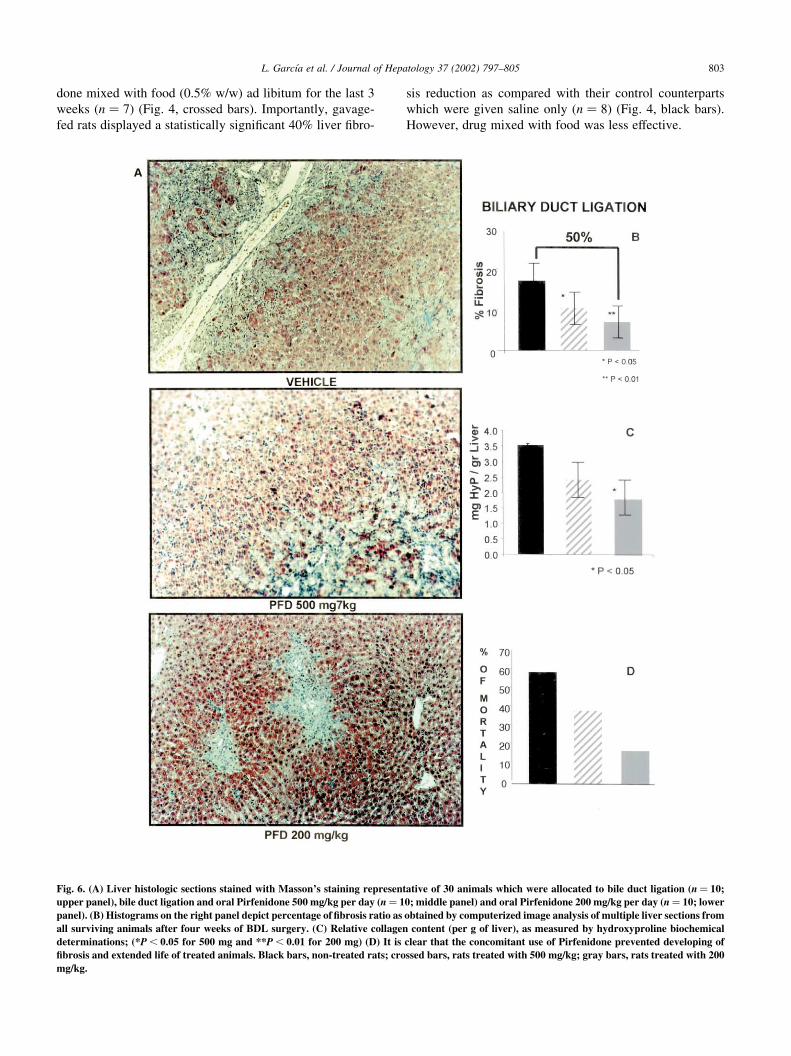
done mixed with food (0.5% w/w) ad libitum for the last 3
weeks (n ¼ 7) (Fig. 4, crossed bars). Importantly, gavage-
fed rats displayed a statistically significant 40% liver fibro-
sis reduction as compared with their control counterparts
which were given saline only (n ¼ 8) (Fig. 4, black bars).
However, drug mixed with food was less effective.
L. Garcıa et al. / Journal of Hepatology 37 (2002) 797–805 803
Fig. 6. (A) Liver histologic sections stained with Masson’s staining representative of 30 animals which were allocated to bile duct ligation (n ¼ 10;
upper panel), bile duct ligation and oral Pirfenidone 500 mg/kg per day (n ¼ 10; middle panel) and oral Pirfenidone 200 mg/kg per day (n ¼ 10; lower
panel). (B) Histograms on the right panel depict percentage of fibrosis ratio as obtained by computerized image analysis of multiple liver sections from
all surviving animals after four weeks of BDL surgery. (C) Relative collagen content (per g of liver), as measured by hydroxyproline biochemical
determinations; (*P , 0.05 for 500 mg and **P , 0.01 for 200 mg) (D) It is clear that the concomitant use of Pirfenidone prevented developing of
fibrosis and extended life of treated animals. Black bars, non-treated rats; crossed bars, rats treated with 500 mg/kg; gray bars, rats treated with 200
mg/kg.
In trying to elucidate the hitherto unknown anti-fibro-
genic mechanism of Pirfenidone in liver cirrhosis, we
demonstrate in Fig. 5 the results of the present study show-
ing that the drug significantly suppresses steady-state levels
of interstitial and basement membrane collagen mRNAs.
This important decrease in mRNA transcripts, as deter-
mined by semi-quantitative RT–PCR, for collagens I, III
and IV (Fig. 5) may reflect an inhibitory effect on gene
transcription or a diminished mRNA life span. Other studies
of experimental pulmonary fibrosis had shed light on the
finding that Pirfenidone inhibits proline hydroxylase levels
[9] and, consequently, might reduce the availability of suffi-
cient hydroxyproline required for collagen synthesis. Thus,
Pirfenidone could inhibit collagen synthesis at the transla-
tional level as well. Down-regulation of PAI-1 and TIMP-1
genes helped us to understand the picture in our system. As
Pirfenidone is metabolized, unavailability of TIMP-1 and
PAI-1 in the local milieu of fibrosis-reversed livers help
tilt the balance to a major concentration of active MMPs
which in turn would promote the breakdown of excessive
extracellular matrix deposited in the liver. These results
correlate well with our previous studies were interferon-
a2a administration to cirrhotic bile-duct ligated rats resulted
in partial scar remodeling which was characterized by a
drop in PAI-1 activity [5]. The notable transforming growth
factor (TGF) b-1 gene down-regulation found in livers from
Pirfenidone-treated animals deserves further consideration
(Fig. 5). This piece of data correlates well with the signifi-
cant decreased Smad-7 mRNA steady-state levels. Others
have shown in different systems that one of the most impor-
tant roles of Pirfenidone is to down-regulate TGFb-1
mRNA transcripts. Here we spotlighted the deleterious
key role of this pleiotropic cytokine in liver cirrhosis and
the redundancy of molecular events under way in different
fibrotic organs. Smad-7 is a regulatory Smad protein that is
able to antagonize signal transduction by TGFb-1 and it has
been recently shown that regulation of the Smad-7 gene is
positively mediated at the transcriptional level by TGFb,
through a direct activation of Smad-3 and Smad-4 and
further binding to the Smad-7 promoter region [24]. All
this rationale suggests that inhibitory Smad-7 protein may
play a role in a negative feedback loop that modulates
TGFb-1 signaling. Thus, there a chance in our system that
Pirfenidone shuts down TGFb-1 expression resulting in
Smad-7 gene down-regulation, as is shown in Fig. 5. Impor-
tantly, Pirfenidone induced a remarkable reduction in
elevated hepatic TFGb-1 mRNA (central point in the initia-
tion of pro-fibrogenic events); and since TGFb-1 up-regu-
lates collagens, TIMP-1 and PAI-1 mRNAs, down-
regulation of these latter molecules by Pirfenidone is most
likely mediated through suppression of TGFb-1.
Concerning our findings in the BDL model and while this
paper was in review, a study was published concerning
tolerability and efficacy of Pirfenidone in the treatment of
patients with well-defined primary sclerosing cholangitis
(PSC) [15]. Twenty-four patients were given 800 mg
tablets, three times a day for 1 year, but the results of this
pilot study showed no improvement in liver histology (fibro-
sis, degree of inflammation or histologic stage) and
suggested that oral Pirfenidone does not provide benefit to
patients with PSC.
However, our data shown in Fig. 6 using the model of
secondary biliary fibrosis after complete BD occlusion
which results in progressive and aggressive fibrosis in the
virtual absence of inflammation and necrosis, clearly shows
that daily 200 mg/kg Pirfenidone prevented to a great extent
(50%; P , 0:01) the development of liver fibrosis 4 weeks
after BDL. The biochemical determinations of hydroxypro-
line strongly correlated with the diminished fibrosis as
measured by image morphometric analysis (see Fig.
6B,C). At this dose, it is clear that Pirfenidone also
prolonged life in the treated animals. We believe that in
this particular model of aggressive hepatic fibrosis, Pirfeni-
done, at high doses, is not efficiently metabolized by the
diseased organ and no real effect can be seen. Along with
this line of reasoning, researchers at the Mayo Clinic [15]
mentioned above used 2400 mg Pirfenidone per day for
their PSC clinical trial, which is the same dose used for
treating pulmonary fibrosis patients with an otherwise
normal liver, capable of metabolizing relatively elevated
doses of Pirfenidone. Thus, the therapeutic window for
this drug might be different when used for treating fibrosis
affecting dissimilar organs.
Finally, regardless of the ample variety of pharmacological
agents to prevent or even to cure liver fibrosis, not all agents
showing promise in experimental animals prove effective in
patients. However, our data presented here warrant further
studies as a potential treatment in a clinical setting.
Acknowledgements
This work was supported in part by a CONACyT grant to
J.A.B. and by Marnac, Inc., Dallas, TX. The authors are
indebted to Dr. Pedro DIaz-Ezquivel, Director of Animal
Facilities.
References
[1] Friedman SL. The cellular basis of hepatic fibrosis. N Engl J Med
1993;328(25):1828–1835.
[2] Maher JJ. Alcoholic liver diseases. In: Sleisenger MH, Fordtrans JS,
editors. 6th ed. Gastrointestinal and liver diseases, vol. 2. Philadel-
phia, PA: Saunders, 1998. pp. 1199–1214.
[3] Recknagel RO, Glende Jr EA, Dolak JA, Waller RL. Carbon tetra-
chloride hepatotoxicity. Status quo and future prospects. Trends Phar-
macol Sci 1983;4:129–131.
[4] Friedman SL, Maher JJ, Bisell DM. Mechanism and therapy of hepa-
tic fibrosis: report of the AASLD single topic basic research confer-
ence. Hepatology 2000;32(6):1403–1438.
[5] Bueno RM, Daneri A, Armendariz-Borunda J. Cholestasis-induced
fibrosis is reduced by interferon a-2a and associates with elevated
liver metalloprotease activity. J Hepatol 2000;33(6):915–925.
[6] Salgado S, Garcia J, Vera J, Siller F, Bueno M, Miranda A, et al. Liver
L. Garcıa et al. / Journal of Hepatology 37 (2002) 797–805804

cirrosis is reverted by urokinase-type plasminogen activator gene
therapy. Mol Ther 2000;2(6):545–551.
[7] Ueki T, Kaneda Y, Tsutsui H, Nakanishi K, Sawa Y, Morishita R, et
al. Hepatocyte growth factor gene therapy of liver cirrhosis in rats.
Nat Med 1999;5(2):226–230.
[8] Rudolph KL, Chang S, Millard M, Schreiber-Agus N, DePinho R.
Inhibition of experimental liver cirrhosis in mice by telomerase gene
delivery. Science 2000;287:1253–1258.
[9] Iyer SN, Wild JS, Schiedt MJ, Hyde DM, Margolin SB, Giri SN.
Dietary intake of Pirfenidone ameliorates bleomycin-induced lung
fibrosis in hamsters. J Lab Clin Med 1995;125(6):779–785.
[10] Al-Took S, Murray C, Tulandi T. Effects of Pirfenidone and dermoid
cyst fluid on adhesion formation. Fertil Steril 1998;69(2):341–342.
[11] Lee B-S, Margolin SB, Nowak AR. Pirfenidone: a novel pharmaco-
logical agent that inhibits leiomyoma cell proliferation and collagen
production. J Clin Endocrinol Metab 1998;83(1):219–223.
[12] Shimizu T, Kuroda T, Hata S, Fukagawa M, Margolin SB, Kurokawa
K. Pirfenidone improves renal function and fibrosis in the post-
obstructed kidney. Kidney Int 1998;54:99–109.
[13] Dosanjh AK, Wan B, Throndset W, Sherwood S, Morris RE. Pirfe-
nidone: a novel antifibrogenic agent with implications for the treat-
ment of obliterative bronchiolitis. Transplant Proc 1998;30:1910–
1911.
[14] Shetlar MR, Shetlar DJ, Bloom RF, Shetlar CL, Margolin SB.
Involution of keloid implants in athymic mice treated with Pirfeni-
done or with triamcinolone. J Lab Clin Med 1998;132(6):491–
496.
[15] Angulo P, MacCarty RL, Sylvestre PB, Jorgensen RA, Wiesner RH,
LaRusso NA, Lindor KD. Pirfenidone in the treatment of primary
sclerosing cholangitis. Dig Dis Sci 2002;47(1):157–161.
[16] Raghu G, Johnson CW, Lockhart D, Mageto Y. Treatment of idio-
pathic pulmonary fibrosis with a new antifibrotic agent, Pirfenidone.
Am J Respir Crit Care Med 1999;159:1061–1069.
[17] Armendariz-Borunda J, LeGross Jr L, Campollo O, Panduro A,
Rincon AR. Antisense S-oligodeoxynucleotides down-regulate
TGF-b production by Kupffer cell from CCl4-injured rat livers.
Biochim Biophys Acta 1997;1353:241–252.
[18] Armendariz-Borunda J, Seyer JM, Postlethwaite AE, Kang AH.
Kupffer cells from carbon tetrachloride injured rat livers produce
chemotactic factors for fibroblasts and monocytes: the role of tumor
necrosis factor-a. Hepatology 1991;14:895–900.
[19] Gao C, Jokerst R, Gondipalli P, Cai SH, Kennedy S, Ponder KP.
Intramuscular of an hepatic transduction with a retroviral vector in
mice. Hum Gene Ther 1999;10:911–922.
[20] Bradford MM. A rapid and sensitive method for the quantitation of
microgram quantities of protein utilizing the principle of protein–dye
binding. Anal Biochem 1976;72:248–254.
[21] Rojkind M, Gonzalez E. An improved method for determining special
radioactivities of proline and hydroxyproline. Anal Biochem
1974;57:1–7.
[22] Chomczynski P, Sacchi N. Single-step method of RNA isolation by
acid guanidinium thiocyanate-phenol chloroform extraction. Anal
Biochem 1987;162:156–159.
[23] Delgado-Rizo V, Salazar A, Panduro A, Armendariz-Borunda J.
Treatment with anti-transforming growth factor b antibodies influ-
ences an altered pattern of cytokines gene expression in injured rat
liver. Biochim Biophys Acta 1998;1442:20–27.
[24] Nagarajan RP, Zhang J, Li W, Chen Y. Regulation of Smad-7 promo-
ter by direct association with Smad-3 and Smad-4. J Biol Chem
1999;274:33412–33418.
L. Garcıa et al. / Journal of Hepatology 37 (2002) 797–805 805