Identification of Two Candidate Tumor Suppressor Genes on Chromosome I7pl3.3l
7
1996;56:1997-2002. Cancer Res David C. Schultz, Lisa Vanderveer, David B. Berman, et al. Chromosome 17p13.3 Identification of Two Candidate Tumor Suppressor Genes on Updated version http://cancerres.aacrjournals.org/content/56/9/1997 Access the most recent version of this article at: E-mail alerts related to this article or journal. Sign up to receive free email-alerts Subscriptions Reprints and . [email protected] Department at To order reprints of this article or to subscribe to the journal, contact the AACR Publications Permissions . [email protected] Department at To request permission to re-use all or part of this article, contact the AACR Publications Research. on January 3, 2014. © 1996 American Association for Cancer cancerres.aacrjournals.org Downloaded from Research. on January 3, 2014. © 1996 American Association for Cancer cancerres.aacrjournals.org Downloaded from
-
Upload
independent -
Category
Documents
-
view
3 -
download
0
Transcript of Identification of Two Candidate Tumor Suppressor Genes on Chromosome I7pl3.3l
1996;56:1997-2002. Cancer Res David C. Schultz, Lisa Vanderveer, David B. Berman, et al. Chromosome 17p13.3Identification of Two Candidate Tumor Suppressor Genes on
Updated version
http://cancerres.aacrjournals.org/content/56/9/1997
Access the most recent version of this article at:
E-mail alerts related to this article or journal.Sign up to receive free email-alerts
Subscriptions
Reprints and
To order reprints of this article or to subscribe to the journal, contact the AACR Publications
Permissions
To request permission to re-use all or part of this article, contact the AACR Publications
Research. on January 3, 2014. © 1996 American Association for Cancercancerres.aacrjournals.org Downloaded from
Research. on January 3, 2014. © 1996 American Association for Cancercancerres.aacrjournals.org Downloaded from
[CANCER RESEARCH 56. 1997-2002. May 1. 1996]
Advances in Brief
Identification of Two Candidate Tumor Suppressor Genes on Chromosome I7pl3.3l
David C. Schultz,2 Lisa Vanderveer,2 David B. Berman, Thomas C. Hamilton, Albert J. Wong,and Andrew K. Godwin3
Department of Medical Oncology. Fox Chase Cancer Center. Philadelphia. Pennsylvania 19111 {D. C. S.. L V., D. B. B., T. C. H.. A. K. G.], and Department of Microbiology andImmunology. Jefferson Cancer Institute, Thomas Jefferson University; Philadelphia, Pennsylvania 19107 ¡A.J. W.j
Abstract
A second tumor suppressor locus on 17p that is distinct from 77*5.1has
been identified in brain, breast, lung, and ovarian tumors. Using allelicloss mapping and positional cloning methods, we have recently identifiedtwo novel genes, which we refer to as OVCAI and OVCA2, that map to17pl3.3. The two genes are ubiquitously expressed and encode proteins of443 and 227 amino acids, respectively, with no known functional motifs.Sequence comparison of OVCAI and OVCA2 revealed extensive sequenceidentity and similarity to hypothetical proteins from Saccharomyces cer-
evisiae, Caenorhabditis elegans, and Rattus species. Northern blot analysisreveals that OVCAI and OVCA2 mRNA were expressed in normal surfaceepithelial cells of the ovary, but the level of this transcript is significantlyreduced or is undetectable in 92% (11/12) of the ovarian tumors andtumor cell lines analyzed. The location, high degree of amino acid conservation, and reduced expression in ovarian tumors and tumor cell linessuggest that decreased expression of these two genes contributes to ovarian tumorigenesis and should be considered candidate tumor suppressorgenes.
Introduction
The molecular basis of ovarian cancer is rapidly being defined (1).The inactivation of multiple tumor suppressor genes seems to beimportant in the etiology of ovarian cancers. One strategy of locatingputative tumor suppressor genes is to survey tumors for high rates ofLOH.4 The combined data from four separate allelotyping studies of
ovarian cancers demonstrate the observation of allelic loss for polymorphic DNA markers on nearly every chromosome arm. Furthermore, these studies revealed that >30% of the tumors studied showedLOH on chromosomes 6, 9, 13q, 17, 18q, 19p, 22q, and Xp (2-5).
Additional studies indicate that chromosome 17 is clearly the mostfrequently altered chromosome in ovarian carcinomas (6-9).
The ovarian carcinomas surveyed for LOH have been largely highstage, and allelic loss in at least some of the arms listed above may notbe causally related to carcinogenesis or metastasis. Furthermore, high-
stage ovarian carcinomas tend to have more numerical chromosomeabnormalities, which can confound attempts to distinguish betweencandidate tumor suppressor genes on the same deleted chromosome.Chromosome 17 has a number of potential cancer causing genes,including TP53 at 17pl3.1, the BRCA1 gene at 17q21, prohibitin andNM23 at 17q23-24, and the proto-oncogene ERBB2 at 17q21. Re
cently, it has been shown that alterations at 17pl3.3 may be an
Received 2/8/96; accepted 3/18/96.The costs of publication of this article were defrayed in part by the payment of page
charges. This article must therefore be hereby marked advertisement in accordance with18 U.S.C. Section 1734 solely to indicate this fact.
1This work was supported in part by NIH Grant ROÕCA60643 (A. K. G.), the Hoxie
Harrison Smith Foundation, the Mary Smith Charitable Lead Trust, Butler Family Fund,Emile Zola Chapter of Brith Shalom Women, and the Ladies Auxiliary to the Veterans ofForeign Wars of the United States of America.
2 These authors contributed equally to this work.3 To whom requests for reprints should be addressed, at Department of Medical
Oncology, Fox Chase Cancer Center. 7701 Burholme Avenue, Philadelphia, PA 19111.Phone: (215) 728-2557; Fax; (215) 728-2741.
4 The abbreviations used are: LOH, loss of heterozygosity; kbp, kilobase pair; STRP,
simple tandem repeat polymorphism; EF-2, elongation factor 2.
important early event in stage I ovarian carcinomas and tumors of lowmalignant potential (7). Interestingly, in tumors of low malignantpotential, allelic losses at 17pl3.3 were not accompanied by LOH atTP53, suggesting there is a more distal tumor suppressor gene whoseloss of function is required for early tumorigenesis. This same regionhas shown frequent LOH in breast cancers (10, 11), primitive neuro-ectodermal tumors (12), carcinoma of the cervix uteri (13), medullo-
blastoma (14, 15), osteosarcoma (16), and astrocytoma (17). suggesting that this tumor suppressor gene(s) residing on chromosome17pl3.3 is involved in the development of many types of cancers. Wehave found a high rate of allelic loss on 17pl3.3 in our cohort ofovarian tumors and have identified a minimum region of allelic loss.Using positional cloning methods, we report the identification of twogenes located within this critical region on 17pl3.3 that show loss ofexpression and are candidates for being the tumor suppressor gene.
Materials and Methods
Tumor Samples and Cell lines. Ovarian tumor specimens were collectedfrom consenting patients undergoing surgery for ovarian cancer at the American Oncological Hospital and (he Lankenau Hospital in Philadelphia, or wereobtained from the Gynecological Oncology Group/Cooperative Human TissueNetwork Ovarian Tissue Bank (Columbus, OH). Ovarian cancer cell lines andhuman ovarian surface epithelial cells were maintained as described previously(18).
Isolation of DNA and RNA from Tumors and Matched Blood Samples.Preparation of RNA for Northern blot analysis, DNA isolation for Southernblot analysis, and LOH studies have been previously described by us (8, 18).
PCR Analysis of STRPs. STRPs were typed in a PCR-based assay containing 15-30 ng genomic DNA, 10 mM Tris-HCl (pH 8.3). 50 mut KC1. 1.5
HIMMgCl2, 0.001% gelatin, 0.4 yM each primer. dCTP, dGTP, and TTP eachat 16 JAM,dATP at 2 JHM,0.65 fiCi [a-"S]dATP (DuPom/New England
Nuclear), 5% DMSO, and 0.25 units Amplitaq DNA polymerase (PerkinElmer/Cetus) in a final volume of 5 fil. Following an initial denaturation stepat 94°Cfor 4 min. DNA was amplified through 20 cycles consisting of 5 sdenaturing at 94°C,1 min annealing at 68°C-0.5°C/cycle.and 1 min extensionat 72°C.The samples were then subjected to an additional 25 cycles consistingof 5 s denaturation at 94°C,1 min at 58°C,and 1 min extension at 72°Canda final extension at 72°Cfor 5 min. PCR products were diluted 1:1 in 90%
formamide, 20 mM EDTA, 0.3% bromophenol blue, 0.3% xylene cyanol,denatured at 94°Cfor 5 min and 4 fj.\ loaded onto a 6% denaturing polyacryl-
amide gel, and electrophoresed at 90 W in 1X TBE ( 1x = 0.09 M Tris, 0.09
M boric acid, and 0.002 M EDTA). After electrophoresis, gels were dried at70°C under vacuum and exposed to Kodak XAR-5 film for 24 to 28 h.
Polymorphisms used to map a minimum region of allelic loss are as follows:DÌ7S34,DÌ7S926.D17SS49. D17S28, AKG2-1. DI7S5, DÌ7S786,D17S796,D17S5Ì3, and TP53 (19-22). The simple tandem repeat polymorphism
AKG2-1 was identified in our laboratory during the construction of a physicalmap of 17pl3.3. The primers used were 5'-TCC TGC TCT GCA ACA GTGAC-3' (sense) and 5'-CAG CCC CTG CTA TCT GAT TC-3' (antisense).
Genomic Library Screening. Cosmid clones were isolated from agenomic human placenta! cosmid library (Stratagene). Duplicate lifts fromeach plate were made to GeneScreen NEF-978x hybridization membranes(Dupont). Membranes were hybridized with [<*-'2P]dATP random primed
DÌ7S5(YNZ22.2) and DÌ7S28(YNH37.3) as probes. Hybridization andwashing conditions have been described previously (8).
1997
Research. on January 3, 2014. © 1996 American Association for Cancercancerres.aacrjournals.org Downloaded from

CANDIDATE TUMOR SUPPRESSOR GENES ON 17p]3.3
Fig. 1. Allelic loss patterns of ovarian tumorsfor the short arm of chromosome 17. DN A samplesfrom normal blood and ovarian tumor tissue weretyped with STRPs from 17p. For each tumor, allinformative loci are shown. •¿�constitutional het-erozygosity with LOH; D. constilulional heterozy-gosity with no LOH; blank spaces, uninformative.With the assumption that alÃelesin all regions between loci showing allelic loss are lost, solid linesindicate retained regions of chromosome 17p andopen areas represent regions of allelic loss. ,regions that are uncertain in tumors with LOH forsome loci.
13.3
13.2
13.1
I12
11.2
11.1
11.2
Locus17S34
D17S926D17S28AKG2-1D17S5D17S786D17S796D17S513TP53D17S31
Ovarian Tumors
54 5557 58 61 66 71 74 79 81 85 111125132143144
9 9 pÕ]!•t]]]'t¡n
piii
ilcc111]
r[?h-,i1
MinimumRegion
ofLoss!
Exon Trapping. Exon sequences were isolated from BamttllBgtll- andP.cfl-digested cosmid 7-2 DNA shotgun subcloned into the pSPL3 vector andtransfected into Cos-1 cells. Total RNA was isolated 48 h posttransfection,reverse transcribed (18), and amplified using sequence-specific primers according to the manufacturer's instructions (Life Technologies, Inc.). Potential
exon sequences were subcloned into pGEM-T (Promega), and the inserts weresequenced using vector primers by the single-stranded dideoxy method (United
States Biochemical). Reaction products were analyzed on a 6% acrylamide/7M urea gel. Sequencing results were analyzed by the GCG program. Exonswere mapped by hybridizing trapped sequences to EcoRI-digested cosmid 7-2DNA. Exons were analyzed by hybridization to Southern "zoo" blots and total
RNA Northern blots.cDNA Cloning and Northern Blot Analysis. A 1.6-kbp EcoRI fragment
from cosmid 7-2 was used to screen to 1 X IO6 plaques from an oligo(dT)-
primed fetal brain cDNA library (Stratagene) or a library made from the A2780ovarian cancer cell line. Sequencing of the cDNA clones was performed asdescribed above with vector primers or primers derived from the obtainedsequence. The complete nucleotide sequence of OVCAI and OVCA2 will besubmitted to the Genbank nucleotide sequence data base. Intron/exon boundaries were determined by sequencing cosmid clone 7-2 using a Model 373Aautomated fluorescence-based cycle sequencer (Applied Biosystems) and Taq
dye terminator chemistry. Expression patterns and transcript sizes of cDNAclones were determined by hybridization of plasmid inserts to commerciallyavailable multiple-tissue Northern blots (Clontech).
Results
Definition of a Minimum Region of Allelic Loss on 17pl3.3.Using DNA isolated from ovarian tumors, including Caucasian, Hispanic, and African-American patients of varying ages, we typedovarian carcinomas for LOH on chromosome 17p using 10 highlypolymorphic markers (Fig. 1). Forty-one (60%) of 68 informativeovarian tumors exhibited LOH, a frequency consistent with previousmeasurements. No correlations could be made between LOH andhistopathological parameters such as subtype, stage, or grade. Acommon region of allelic loss was defined to be distal to YNZ22(D/755) and proximal to YNH37.3 (D17S28; Fig. 1). The predictedgenetic distance between these two markers has been estimated to beapproximately 3.5 cM (23). We have found that this region spans lessthan 30 kbp, which is in agreement with the results of Ledbetter et al.(19).
Isolation of Candidate Cancer Gene Sequences. Cosmid clonessurrounding and including these two loci were isolated from a humanplacental DNA cosmid library, and several strategies were employedto evaluate these clones for potential expressed sequences. Cosmidclones containing genomic inserts spanning the limited region ofallelic loss were introduced into the ovarian cancer cell line A2780
along with the bacterial TnSneo gene (an aminoglycoside 3' phospho-
transferase) expressed from the SV40 early promoter (pKOneo). Evaluation of clonal outgrowth in the presence of geneticin (G418) revealed that cosmid clones 7-2 and 2-1 were the most effective insuppressing clonal outgrowth as compared to control cosmids (datanot shown). This data suggested that a growth suppressor gene(s) islikely to be contained within these cosmids, and. moreover, thisgrowth suppressor gene maps to the minimum region of allelic loss.Evaluation of clones 2-1 and 7-2 for expressed sequences by exonamplification identified a 65-bp exon which mapped to a 1.6-kbpEcoRI DNA fragment. Hybridization of the 65-bp fragment at lowstringency to a zoo blot revealed conservation among other mammals(data not shown). Sequencing of the 1.6-kbp EcoRI fragment revealeda second potential open reading frame of 152bp that was 122 bp awayfrom the putative 65-bp exon.
Screening of a human fetal brain cDNA library using the 1.6-kbpgenomic EcoRI fragment as the probe yielded six positive clones.Only two of the six clones (fb67-l and 77-1) hybridized to any of theclones of the 17pl3.3 cosmid "contig," indicating the presence of apotential family of genes.5 Sequence analysis of clones fb67-l andfb77-l revealed a 1350-bp open reading frame and an 847-bp of the3' untranslated region, including a polyadenylation signal 19-bp 5' to
the poly(A) tail. To identify the initiation codon, we employed an"anchored" PCR. Thirty-five additional nucleotides were identified,including 18 bases of the 5' untranslated region and two potential
initiation codons. We provisionally refer to this gene as OVCAI, forovarian cancer gene 1.The reading frame using the first AUG encodesa 443-amino acid protein with a predicted Mr —¿�50,000which has been
verified by in vitro translation, bacterial expression, and Western blotanalysis (data not shown).
Characterization of OVCAI and OVCA2. Hybridization ofNorthern blots containing poly(A)+ RNA from different human tis
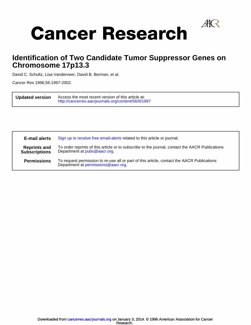
sues with the full-length OVCAI cDNA revealed two distinct transcripts of approximately 2.3 kb and 1.1 kb that were ubiquitouslyexpressed (Fig. 2A). Isolation of a cDNA clone for the smallertranscript from an ovarian tumor cell line library revealed that thistranscript was composed of the last 828 bp of OVCAI and 184 bp ofthe unique sequence. We refer to this transcript as OVCA2 (ovariancancer gene 2). Furthermore, Northern blot analysis of ovarian tumorsand tumor cell lines revealed that the 2.3-kb OVCAI mRNA wasexpressed in normal surface epithelial cells of the ovary, but the levelof the 2.3-kb transcript was significantly reduced or was undetectable
1A. K. Godwin, unpublished data.
1998
Research. on January 3, 2014. © 1996 American Association for Cancercancerres.aacrjournals.org Downloaded from

CANDIDATE TUMOR SUPPRESSOR GENES ON I7pl3.3
Fig. 2. A, tissue expression pattern of OVCAI and OVC4ZBlots containing 5 ¿igpoly( A) *-selected mRNA from the
indicated human tissues were hybridized with a full-lengthOVCAI cDNA fragment. Note that the tissues are heterogeneous and the percentage of relevant epithelial cells in theovary can be variable. Size standards are in kb. Lower panel.blots were reprobed with ß-actin.The heart and skeletalmuscle express two forms of /3-actin, a l.8-kb and a 2.0-kbtranscript. B, Northern blot analysis of OVCVW-specific RNAin ovarian tumor cell lines (OVCAK). C, fresh ovarian tumors(t//W) as compared to normal ovarian surface epithelial cells(HIO or HO). Lower panels, ethidium bromide-stained gel
prior to blotting; the position of the 28S and 18S rRNA isindicated.
7.5-
4.4-
2.4-
1.4-
OVCAI
B
actin
in the majority (11/12) of the ovarian tumors and tumor cell linesevaluated (P < 0.01; Fig. 2, B and Q. Similarly, RNA levels forOVCA2 were also reduced in these same samples (data not shown).
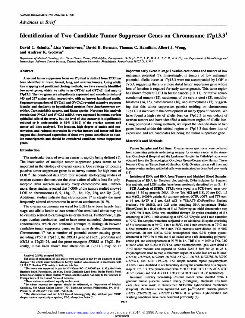
Restriction mapping of genomic clones using cDNA probes andsequence comparison between cDNA and genomic clones indicatedthat OVCAI has 13 exons, 12 which are coding, and spans approximately 20-kbp of genomic DNA. The entire OVCAI and OVCA2cDNA sequences are present in the insert of cosmids 7-2 and 2-1, thesame cosmids demonstrating the most significant clonal outgrowth inthe in vitro suppression assay. The position of the 13exons of OVCAÃŒand the 2 exons of OVCA2 relative to the common region of allelicloss defined by DNA markers D17S5 and D17S28 is shown in Figure3. Furthermore, the transcriptional orientation of OVCAI is fromtelomere to centromere with the transcriptional start site of OVCAItelomeric to DI7S28. The unique sequence of OVCA2 has beenpositioned in the intron between exons 12 and 13 of OVCAI (Fig. 3).
Hybridization of genomic DNA samples from several differentspecies with a full-length OVCAI cDNA fragment revealed stronglyhybridizing fragments in tissue from humans, bovine, cat, dog, equine,monkey, mouse, porcine, rat, and yeast. These results suggested thatOVCAI was highly conserved in mammals (data not shown). Basiclocal alignment search tool (BLAST) searches of the Genbank and
SwissProt data bases revealed extensive sequence identity at thenucleotide and the amino acid level to two recently identified sequences from Caenorhabditis elegans and Saccharomyces cerevisiae(Fig. 4A). The predicted gene product of OVCAI showed 60% and53% sequence identity over 328 and 367 of the 443-amino acidresidues when compared to the nematode and yeast proteins, respectively. If conserved amino acids substitutions are considered, thesequence similarity is increased to 77 and 89%, respectively. Becausethey were identified as the result of nematode and yeast genomesequencing projects, the function of these two predicted proteins is atthe present time unknown. The fact that the OVCAI protein is sohighly conserved at the amino acid level with organisms lower on thephylogenetic tree argues that it possesses an important cellular function.
The predicted gene product of OVCA2is a 227-amino acid proteinwith an approximate Mr 24,000, as determined by expression inbacteria (data not shown). Blast searches of the OVCA2gene identified extensive sequence identity at the 5' end of this gene with a
recently identified expressed sequence tag from the Rattus species(24). OVCA2 was observed to be 76% identical over 104 amino acidsto the predicted rat gene product and 80% similar when conservedamino acids are considered (Fig. 40).
1999
Research. on January 3, 2014. © 1996 American Association for Cancercancerres.aacrjournals.org Downloaded from

^:
CANDIDATE TUMOR SUPPRESSOR GENES ON 17pl3.3
Minimum Region of Loss Cen -
H ATG AATAAA
^: 2-11-2 C
BN N N N N N
//\/sar(CAH-Q)AKG2-1m
19-1OVCA2] 7-2
^yl
Fig. 3. Schematic of chromosome 17pl3.3 containing the OVCAl and OVCA2 genes. Stippled ¿at«,open reading frame of OVC4J; black box, unique exon of OVCA2. Cosmidclones, used to identify OVCAl and OVCA2. which span the minimum region of allelic loss in ovarian cancer are indicated. The position of D/755 and D/752S and one of the fournewly identified (CA-GT)n repeat polymorphisms are indicated relative to OVCAl. Lower hatched boxes, regions of genomic DNA that have been sequenced. B, ßssHII;E, £coRI;H, Hindin, N. Noll; S. Sfl\; *, site in the vector.
ovcalyik3
cecl4blMSGSTESKKQ
1QVMA ALVVSIRFIGRKS GNSNNDKLTT
IAEQGGRDGENGNEIIH
P G R G|K Q K S|
IAPRlI A L|
V AlS VI
10 IIHvl
IPEE D
ovcal KNPQHQA^HR vyik3 i..••¿�'•¿�•:H::-0Q;1. L
cecl4bl M I T F Q
ovcal
cecl4bl
ovcalyik3
cecl4bl
ovcalyik3
cecl4bl
42600
385408
QWTÕIRMIPW 331
ovcalyik3
IS S Li IP W|R .
cecl4bl V LfflR I I MK RI
I*HIiRNl IRNGNLI LLSKPKIHSR ELSYFNEI
IVQEGSARPP
ILAKKVHKSI
423425391
ovcal S A V A C E|HC S C RDEKVAPLAP 443; 425
cecl4bl EQLKRHHPDV QISTEPTKYL LVSNSSILCG VSLEELEEIF LPLDELAEFI VYPNKRSYSF 451
BMAAQRPLRovca2
ratovca2
ovca2ratovca2
Fig. 4. A, comparison of the predicted amino acid sequence of the OVCAl protein with the hypothetical S. cerevisiae protein YIK3, chromosome IX cosmid 9150. and C. eleganscosmid C14B1 predicted proteins. B, comparison of the predicted amino acid sequence of the OVCA2 protein with the predicted protein of EST 110891 Remus species. Identical aminoacids are in black', conservative amino acids are shaded in gray. Gaps introduced for maximum alignment are marked with dois. Numbers on the righi, amino acid position for each
of the predicted proteins. Arrowheads, approximate position of exon/intron boundaries of OVCAl (A) and OVCA2 (B).
2000
Research. on January 3, 2014. © 1996 American Association for Cancercancerres.aacrjournals.org Downloaded from

CANDIDATE TUMOR SUPPRESSOR GENES ON 17pl.1.3
Discussion
Molecular analyses of ovarian carcinomas has implicated one ormore tumor suppressor genes on chromosome 17 (3, 6-9). To date,
only a limited number of sporadic ovarian cancers have been found topossess mutations in the BRCA1 gene (25, 26). Furthermore, evidenceof allelic loss on chromosome 17p often coincides with mutations inTP53 at 17pl3.1. In ovarian carcinomas, mutations in TP53 occur inadvanced stage tumors when compared to either benign lesions,borderline tumors, or low grade carcinomas (27). We report in thisstudy that 60% of sporadic ovarian tumors show LOH for markersdistal to TP53, and that many retain TP53 at 17pl3.1 and the BRCA1locus at 17q21. This frequency is in agreement with a similar study inwhich tumors of low malignant potential and low-stage carcinomasdemonstrated allelic loss for markers at 17pl3.3 (7-9). These data
suggest that in addition to the TP53 gene at 17pl3.3, at least oneadditional tumor suppressor gene resides on chromosome 17p.
Mapping of minimum regions of allelic loss have been helpful indefining chromosomal regions in which a tumor suppressor gene mayreside (e.g., DCC, CDKN2, and DPC4) and allow positional cloningstrategies to be initiated. These methods have allowed us to identifyOVCA1 and OVCA2. Sequence analysis of OVCA1 and OVCA2 identified no known functional domains; however, OVCAl showed significant sequence identity and similarity to a yeast and nematodesequence (Fig. 4).
The predicted protein for the C. elegans clone CEC14B1, whichdisplays significant similarity to OVCAl, has sequence similaritiesto diphtheria toxin resistance like protein (28). In yeast, diphtheriaresistance mutants form at least five complementation groups, andthe enzymes corresponding to two of the complementation groupshave been cloned (29). To a lesser extent, OVCAl is 20% identicaland 50% similar in amino acid sequence to the yeast dipthamidebiosynthesis protein DPH2. Dipthamide, is the result of posttrans-lationally modified histidine found almost exclusively in the eu-karyotic translation EF-2. ADP ribosylation of dipthamide by
bacterial exotoxins or by endogenous ADP ribosyltransferasesinhibits EF-2 from translocating the growing peptide and ulti
mately arrests translation (30). However, OVCAl is more similarto the yeast and nematode proteins of unknown function (YIK3 andCEC14B1) than to the yeast DPH2. Therefore, OVCAl is likelynot to be the homologue of DPH2, but could represent a humanhomologue for the other complementation groups in the dipthamide biosynthesis pathway or possess a similar role in a differentbiosynthetic pathway.
Northern blot analysis performed on fresh ovarian tumors andtumor cell lines indicates that the expression of OVCAl and OVCA2are decreased as compared to normal human ovarian surface epithelialcells (Fig. 2, B and C; data not shown). It is conceivable that mutationsin upstream regulatory sequences may be the predominate mode ofinactivation of these candidate tumor suppressor genes. For example,a nucleotide transversion near the exon/intron boundary of exon 12was detected in the germ line of an individual diagnosed with ovariancancer at age 45 years, and this change was homozygous in the tumor(data not shown). Studies are under way to determine whether thisalteration affects the expression of OVCA2 in the tumor, since the Cto A transversion destroys a SP1 site in the promoter of OVCA2 (datanot shown). This alteration has not been observed in >50 constitutional DNA samples collected from unaffected individuals. In additionto genetic changes, epigenetic changes such as de novo methylation ofpromoter CpG islands may also result in the inactivation of theretained alÃele.Silencing of gene transcription in association withhypermethylation of normally unmethylated 5'-CpG islands has been
implicated as the major mechanism for the inactivation of the tumor
suppressor genes CDKN2 and VHL in sporadic disease (31, 32). It haspreviously been shown that the region of 17pl3.3 that containsOVCAl and OVCA2 is aberrantly hypermethylated in colon, lung,neural, renal, and ovarian tumors (33, 34). These changes in methylation patterns could contribute to the decreased level of expressionobserved in ovarian tumor specimens and tumor cell lines. Preliminary evidence suggests that expression of OVCAl and OVCA2 may beregulated by methylation of a 5'-CpG island.6 Furthermore, it was
recently reported that a novel zinc finger protein, HIC-1 (hypermethy
lated in cancer gene 1), is localized to 17pl3.3 and its expressionfound to be dependent on the methylation status of its promoter (35).Although this putative tumor suppressor gene maps outside our minimum region of allelic loss, its inactivation may further contribute totumorigenesis alone or in combination with OVCAl and OVCA2.
Mutational analysis of the 13 exons of OVCAl and the unique exonto OVCA2, including adjacent intron boundaries, is currently beinginvestigated in a large panel of breast, brain, and ovarian tumors.Mutation or deregulation of protein expression of either gene mayprove to be causative in tumorigenesis and support the hypothesis thatone or both of these genes are tumor suppressor genes. Overall, thechromosomal location, high degree of amino acid conservation, andderegulation of mRNA expression suggest that one or both genes arelikely to be involved in the pathogenesis of ovarian cancer as well asother neoplasms such as breast and brain.
Acknowledgments
We thank Drs. W. Michael Hogan. Matthew Boente, Nelly Auersperg, andalso Josephine Costalas, Brian Kane, and Alena Schnarr for their invaluablehelp and contributions to this work.
References
1. Godwin, A. K., Schulte, D. C., Hamilton. T. C., and Knudson, A. G. Oncogenes andtumor suppressor genes. In: W. J. Hoskins. C. A. Perez, and R. C. Young (eds.),Gynecological Oncology: Principles and Practice. Philadelphia: J. B. LippincottCompany, in press. 1996.
2. Sato, T.. Saito. H., Monta. R., Koi. S., Lee, J. H., and Nakamura. Y. Allelotype ofhuman ovarian cancer. Cancer Res.. 51: 5118-5122, 1991.
3. Yang-Feng, T. L., Han, H., Chen, K-C, Li, S-b., Claus, E. B.. Carcangiu, M. L.,Chambers, S. K.. Chambers. J. T.. and Schwartz, P. E. Allelic loss in ovarian cancer.Ini. J. Cancer, 54: 546-541, 1993.
4. Cliby, W., Ritland. S., Hartman. L.. Dodson. M.. Hailing. K. C.. Keeney. G.. Podratz.K. C., and Jenkins, R. B. Human epithelial ovarian cancer allelotype. Cancer Res., 53:2393-2398, 1993.
5. Osborne, R. J., and Leech, V. PCR allelotyping of human ovarian cancers. Br. J.Cancer, 69: 429-438, 1994.
6. Foulkes, W. D., Black, D. M.. Stamp. G. W. H., Solomon. E. and Trowsdale. J. Veryfrequent loss of heterozygosity throughout chromosome 17 in sporadic ovariancarcinoma. Int. J. Cancer. 54: 220-225, 1993.
7. Phillips. N.. Ziegler, M., SaHa, B., and Xynos, F. Allelic loss on chromosome 17 inhuman ovarian cancer. Int. J. Cancer, 54: 85-91, 1993.
8. Godwin, A. K., Vanderveer, L., Schultz, D. C., Lynch, H. T., Altomare, D. A.,Buetow, K. H., Daly, M., Getts. L. A., Masny. A., Rosenblum. N.. Hogan. M., Ozols.R. F., and Hamilton. T. C. A common region of deletion on chromosome 17q in bothsporadic and familial epithelial ovarian tumors distal to BRCA1. Am. J. Hum. Genet..55: 666-677. 1994.
9. Phillips. N. J.. Ziegler. M. R.. Radford. D. M.. Fair. K. L.. Steinbrueck. T., Xynos.F. P., and Donis-Keller. H. Allelic deletion on chromosome 17p 13.3 in early ovariancancer. Cancer Res., 56: 606-611. 1996.
10. Coles. C., Thompson. A. M.. Elder, P. A., Cohen, B. B.. Mackenzie. 1. M.. Cranston.G., Chetty, U., Mackay, J., MacDonald. M.. Nakamura, Y., Hoyheim. B., and Steel.C. M. Evidence implicating at least two genes on chromosome 17p in breastcarcinogenesis. Lancet, 336: 761-763. 1990.
11. Cornells, R. S.. van Vliet. M., Vos, C. B. J., Cleton-Jansen. A-M.. van de Vijver.M. J., Peterse, J. L.. Khan, P. M.. Borresen. A-L.. Comelisse, C. J.. and Devilee. P.Evidence for a gene on 17pl3.3. distal to TP53 as a target for alÃeleloss in breasttumors without p53 mutations. Cancer Res.. 54: 4200-4206, 1994.
12. Biegel, J. A., Burk, C. D., Barr. F. G.. and Emanuel, B. S. Evidence for a I7p tumorlocus distinct from p53 in pediatrie primitive neuroectodermal tumors. Cancer Res..52: 3391-3395, 1992.
13. Atkin, N. B.. and Baker, M. C. Chromosome 17p loss in carcinoma of the cervix uteri.Cancer Genet. Cytogenet., 37: 229-233, 1989.
s H. Ling and A. K. Godwin, manuscript in preparation.
2001
Research. on January 3, 2014. © 1996 American Association for Cancercancerres.aacrjournals.org Downloaded from

CANDIDATE TUMOR SUPPRESSOR GENES ON 17pl3.3
14. Cogen, P. H., Daneshvar, L.. Metzger, A. K., Duyk, G., Edwards, M. S. B.. andSheffield, V. C. Involvement of multiple chromosome 17p loci in medulloblastomatumorigenesis. Am. J. Hum. Genet., 50: 584-589, 1992.
15. Saylors. R., Sidransky, D., Friedman, H. S.. Bigner, S. H., Bigner, D. D.. Vogelstein.B., and Brodeur. G. M. Infrequent p53 gene mutations in medulloblastomas. CancerRes., SI: 4721-4723, 1991.
16. Toguchida, J., Ishizaki, K., Nakamura, Y., Sasaki, M. S., Ikenaga, M.. Kalo, M.,Sugimoto, M., Kotoura, Y., and Yamamuro, T. Assignment of a common alÃeleloss in osteosarcoma to the subregion 17pl3. Cancer Res.. 49: 6247-6251.
1989.17. Saxena, A., Clark, W. C.. Robertson, J. T., Ikejri, B., Oldfield, E. H., and Ali, I.
Evidence for the involvement of a potential second tumor suppressor gene onchromosome 17 distinct from p53 in malignant atrocytomas. Cancer Res., 52:6716-6721, 1992.
18. Schultz, D. C., Vanderveer, L., Buetow. K. H., Boeme, M. P., Ozols, R. F., Hamilton,T. C., and Godwin, A. K. Characterization of chromosome 9 in human ovarianneoplasia identifies frequent genetic imbalance on 9q and rare alterations involving9p. including CDKN2. Cancer Res., 55: 2150-2157. 1995.
19. Ledbetter, D. H., Ledbetter, S. A., vanTuinen. P.. Summers, K. M.. Robinson, T. J.,Nakamura, Y., Wolff, R., White, R.. Barker. D. F.. Wallace, M. R.. Collins, F. S., andDobyns. W. B. Molecular dissection of a contiguous gene syndrome: frequentsubmicroscopic deletions, evolutionary conserved sequences, and a hypomethylated"island" in the Miller-Dicker chromosome region. Proc. Nati. Acad. Sci. USA. 86:
5136-5140, 1989.20. Gypay, G., Morissette, J.. Vignai. A., Dib, C., Fizames, C.. Millasseau, P.. Marc, S.,
Bernardi, G-, Lathrop, M., and Weissenbach, J. The 1993-94 Généthonhumangenetic linkage map. Nat. Genet., 7: 246-339, 1994.
21. Jones, M., H. and Nakamura. Y. Detection of loss of helerozygosity at the humanTP53 locus using a dinucleotide repeat polymorphism. Genes Chromosomes &Cancer, 5: 89-90, 1992.
22. Oliphant, A. R., Wright. E. C., Swensen, J.. GruÃs,N. A.. Goldgar, D., and Skolnick,M. H. Dinucleotide repeat polymorphism at the DI7S513 locus. Nucleic Acids Res.,19: 4794, 1993.
23. Murray, J. C., Buetow, K. H., Weber, J. L., Ludwigsen, S., Scherpbier-Heddema, T.,
Manion, F., Quillen, J., Sheffield, V. C., Sunden, S., Duyk, G. M.. Weissenbach, J.,Gyapay, G., Dib, C., Morissette, J., Lathrop, G. M., Vignai, A., White, R..Matsunami, N., Gerken, S., Melis, R., Albertsen, H.. Plaelke. R., Odelberg. S.,Ward. D., Dausset, J., Cohen. D.. and Cunn, H. A comprehensive human linkage mapwith centiMorgan density. Cooperative Human Linkage Center (CHLC). Science(Washington DC). 265: 2049-2054, 1994.
24. Lee, N. H., Weinstock, K. G., Kirkness, E. F., Earle-Hughes, J. A., Fuldner. R. A..
Marmaras, S., Glodek, A., Gocayne. J. D.. Adams, M. D., Kerlavage, A. R., Fraser,C. M., and Venter, J. C. Comparative expressed sequence tag analysis of differential
gene expression profiles in PC-12 cells before and after nerve growth factor treatment. Proc. Nati. Acad. Sci., 92: 8303-8307, 1995.
25. Hosking, L.. Trowsdale, J.. Nicolai, H., Solomon, E., Foulkes, W., Stamp, G., Signer.E., and Jeffreys, A. A somatic BRCA1 mutation in an ovarian tumor. Nat. Genet., 9:343-344, 1995.
26. Merajver, SD, Pham, T. M.. Caduft. R. F.. Chen. M.. Poy, E. L.. Cooney, K. A.,Weber, B. L., Collins, F. S.. Johnston, C., and Frank. T. S. Somatic mutations in theBRCA1 gene in sporadic ovarian tumors. Nat. Genet., 9: 439-443, 1995.
27. Zheng, J.. Benedict, W. F., Xu, H-J.. Hu, S-X., Kim, T. M., Velicescu, M., Wan, M.,
Cofer, K. F., and Dubeau, L. Genetic disparity between morphologically benign cystscontiguous to ovarian adenocarcinomas and solitary cystadenomas. J. Nati. Cancerhist., 87: 1146-1153, 1995.
28. Wilson, R., Ainscough, R., Andersen, K., Baynes, C., Berks, M., Bonfield. J.. Burton.J.. Connell, M., Copsey, T., Cooper. J., Coulson, A., Craxton, M., Dear, S.. Du. Z.,Durbin, R., Favello, A., Fulton, L.. Gardner, A.. Green. P.. Hawkins. T., Hillier, L..Jier, M., Johnston, L., Jones. M., Kershaw. J.. Kirsten. J., Laister, N., Latrielle. P..Lightning. J., Lloyd, C., McMurray, A.. Mortimore, B.. O'Callaghan. M.. Parsons. J.,
Percy, C., Rifken, L.. Roopra, A., Saunders. D., Shownkeen, R.. Smaldon, N., Smith,A., Sonnhammer, E.. Staden. R.. Sulston. J., Thierry-Mieg. J., Thomas, K., Vaudin.M., Vaughan, K., Waterston, R.. Watson. A.. Weinstock. L.. Wilkinson-Sproat, J..and Wohldman, P. 2.2Mb of contiguous nucleotide sequence from chromosome III ofC. elegans. Nature (Lond.l, 368: 32-38, 1994.
29. Mattheakis, L. C., Sor, F., and Collier, R. J. Diphthamide synthesis in Saccharomycescerevisiae: structure of the DPH2 gene. Gene, 132: 149-154, 1993.
30. Iglewski. W. J. Cellular ADP-ribosylation of elongation factor 2. Mol. Cell. Bio-chem., 138: 131-133, 1994.
31. Herman, J. G.. Latif, F., Weng, Y.. Lerman, M., Zbar. B.. Liu, S., Samid, D.. Duan.D-S. R.. Gnarra. J. R., Linehan, W. M., and Baylin, S. B. Silencing of the VHLtumor-suppressor gene by DNA methylalion in renal carcinoma. Proc. Nail. Acad.Sci. USA, 91: 9700-9704. 1994.
32. Merlo, A., Herman, J. G., Mao, L., Lee, D. J., Gabrielson. E., Burger, P. C.. Baylin.S. B., and Sidransky, D. 5' CpG island methylation is associated with transcriptional
silencing of the tumor suppressor ¡>16/CDKN2iMTSl in human cancers. Nat. Med., /:686-692, 1995.
33. Makos, M.. Nelkin, B. D., Chazin. V. R., Cavenee, W. K., Brodeur, G., and Baylin,S. B. DNA hypermethylation is associated with 17p allelic loss in neural tumors.Cancer Res., 53: 2715-2718, 1993.
34. Pieretti, M., Powell. D. E.. Gallion, H. H.. Conway, P. S.. Case. E. A., and Turker.M. S. Hypermethylation at a chromosome 17 "hot spot" is a common event in ovarian
cancer. Hum. Pathol., 26: 398-401, 1995.35. Wales, M. M., Biel, M. A.. Deiry. W. E.. Nelkin. B. D.. Issa, J-P., Cavenne, W. K.,
Kuerbit/, S. J., and Baylin, S. B. p53 activates expression of HIC-1. a new candidatetumor suppressor gene on I7pl3.3. Nat. Med., /: 570-577, 1995.
2002
Research. on January 3, 2014. © 1996 American Association for Cancercancerres.aacrjournals.org Downloaded from