DOC2/hDab2, a candidate tumor suppressor gene involved in the development of gestational...
6
DOC-2/hDab2, a candidate tumor suppressor gene involved in the development of gestational trophoblastic diseases Vilmos Fulop 2 , Cristiano V Colitti 1 , David Genest 1 , Ross S Berkowitz 1 , Gary K Yiu 1 , Shu-wing Ng 1 , Janos Szepesi 2 and Samuel C Mok 1 1 Laboratory of Gynecologic Oncology, Division of Gynecologic Oncology, Department of Obstetrics, Gynecology and Reproductive Biology, Brigham and Women’s Hospital, Harvard Medical School, Boston, Massachusetts 02115, USA; 2 Department of Obstetrics and Gynecology, Haynal Imre University for Health Sciences, Budapest, Hungary Gestational trophoblastic diseases comprise a spectrum of interrelated diseases including partial mole, complete mole and gestational choriocarcinoma. Using reverse transcriptase PCR (RT – PCR) analysis, we identified higher levels of DOC-2/hDab2 expression in the normal trophoblast cells in culture than in choriocarcinoma cell lines. Subsequent study using immunohistochemistry showed high levels of DOC-2/hDab2 protein expression in normal trophoblast tissues but significantly lower levels of expression in gestational trophoblastic disease tissues, particularly in complete mole and choriocarcino- ma. When DOC-2/hDab2 was transfected into the choriocarcinoma cell lines, Jar, JEG and BeWo, the stable transfectants showed significantly reduced growth rate in culture. These data suggest that down regulation of DOC-2/hDab2 may play an important role in the development of gestational trophoblastic diseases. Keywords: trophoblastic diseases; DOC-2; Dab2; tumor suppressor; cancer Introduction Gestational trophoblastic diseases comprise a spectrum of interrelated disease including partial mole, complete mole and gestational choriocarcinoma which have varying propensity for local uterine invasion and metastasis. Because at least part of their genetic background is of paternal origin, gestational tropho- blastic diseases are at least partial allografts. Gesta- tional trophoblastic diseases are one of the few human tumors that are highly curable with chemotherapy even in the presence of widespread metastases (Hancock et al., 1997). To date early molecular biologic predictors for the development of persistent gestational trophoblastic disease after evacuation of molar pregnancies have not been determined. Cellular proliferation is an intricately regulated process mediated by the coordi- nated interactions of critical proto-oncogenes and tumor suppressor genes. The dierences between physiological oncogene expression in early placenta and altered oncogene expression in gestational tropho- blastic tumors are incompletely understood (Cheung et al., 1993). Activation of some oncogenes and altered expression of tumor suppressor genes have been demonstrated in gestational trophoblastic tumors (Fulop et al., 1998a,b). Further analysis of gene expression in placenta and gestational trophoblastic diseases can provide insight into the role played by specific proto-oncogenes and tumor suppressor genes in the pathogenesis of these diseases. Genes that show dierential expression between normal and tumor tissues are likely to function either directly in growth regulation or cellular dierentiation or indirectly as a response to changes in the cellular environment. Several of the tumor suppressor genes characterized to date control growth in epithelial cells (Lee et al., 1987; Miki et al., 1994). Accumulated evidence suggests that additional growth suppressors will be identified in various cell types. One candidate tumor suppressor gene is the DOC-2/hDab2 gene which has been shown to be expressed in all normal ovarian epithelial cells but significantly down-regulated or absent in all of a series of ovarian carcinoma cell lines tested. Furthermore, it has been shown to suppress ovarian tumor growth in vitro and in vivo (Mok et al., 1994, 1998). The predicted DOC-2/hDab2 protein sequence shows that it contains a phosphotyr- osine interacting domain and multiple SH3 binding motifs suggesting that it is a signal transduction molecule (Mok et al., 1998). Since DOC-2/hDab2 has also been demonstrated to be highly expressed in normal trophoblastic cells, it is of interest to evaluate whether down-regulation of DOC-2/hDab2 is also involved in the development of trophoblastic diseases. The current study was under- taken to examine the dierential expression pattern of DOC-2/hDab2 transcript and protein in placenta, partial mole, complete mole and choriocarcinoma by Western blot analysis, reverse transcriptase PCR and immunohistochemistry. Furthermore, transfection ex- periments have also been performed to evaluate the putative tumor suppressor role of DOC-2/hDab2 gene in choriocarcinoma. We suggest that DOC-2/hDab2 may play an important role in the development of trophoblastic diseases. Results Immunohistochemistry and Western blot analysis To evaluate the expression pattern of the DOC-2/ hDab2 protein in normal placenta and gestational trophoblastic disease tissues in vivo, we performed immunohistochemistry in 18 placentas, 17 partial mole, Correspondence: SC Mok Received 25 November 1997; revised 2 March 1998; accepted 4 March 1998 Oncogene (1998) 17, 419 – 424 1998 Stockton Press All rights reserved 0950 – 9232/98 $12.00 http://www.stockton-press.co.uk/onc
-
Upload
hms-harvard -
Category
Documents
-
view
1 -
download
0
Transcript of DOC2/hDab2, a candidate tumor suppressor gene involved in the development of gestational...

DOC-2/hDab2, a candidate tumor suppressor gene involved in thedevelopment of gestational trophoblastic diseases
Vilmos Fulop2, Cristiano V Colitti1, David Genest1, Ross S Berkowitz1, Gary K Yiu1,Shu-wing Ng1, Janos Szepesi2 and Samuel C Mok1
1Laboratory of Gynecologic Oncology, Division of Gynecologic Oncology, Department of Obstetrics, Gynecology and ReproductiveBiology, Brigham and Women's Hospital, Harvard Medical School, Boston, Massachusetts 02115, USA; 2Department of Obstetricsand Gynecology, Haynal Imre University for Health Sciences, Budapest, Hungary
Gestational trophoblastic diseases comprise a spectrumof interrelated diseases including partial mole, completemole and gestational choriocarcinoma. Using reversetranscriptase PCR (RT ±PCR) analysis, we identi®edhigher levels of DOC-2/hDab2 expression in the normaltrophoblast cells in culture than in choriocarcinoma celllines. Subsequent study using immunohistochemistryshowed high levels of DOC-2/hDab2 protein expressionin normal trophoblast tissues but signi®cantly lowerlevels of expression in gestational trophoblastic diseasetissues, particularly in complete mole and choriocarcino-ma. When DOC-2/hDab2 was transfected into thechoriocarcinoma cell lines, Jar, JEG and BeWo, thestable transfectants showed signi®cantly reduced growthrate in culture. These data suggest that down regulationof DOC-2/hDab2 may play an important role in thedevelopment of gestational trophoblastic diseases.
Keywords: trophoblastic diseases; DOC-2; Dab2; tumorsuppressor; cancer
Introduction
Gestational trophoblastic diseases comprise a spectrumof interrelated disease including partial mole, completemole and gestational choriocarcinoma which havevarying propensity for local uterine invasion andmetastasis. Because at least part of their geneticbackground is of paternal origin, gestational tropho-blastic diseases are at least partial allografts. Gesta-tional trophoblastic diseases are one of the few humantumors that are highly curable with chemotherapy evenin the presence of widespread metastases (Hancock etal., 1997).To date early molecular biologic predictors for the
development of persistent gestational trophoblasticdisease after evacuation of molar pregnancies havenot been determined. Cellular proliferation is anintricately regulated process mediated by the coordi-nated interactions of critical proto-oncogenes andtumor suppressor genes. The di�erences betweenphysiological oncogene expression in early placentaand altered oncogene expression in gestational tropho-blastic tumors are incompletely understood (Cheung etal., 1993). Activation of some oncogenes and altered
expression of tumor suppressor genes have beendemonstrated in gestational trophoblastic tumors(Fulop et al., 1998a,b). Further analysis of geneexpression in placenta and gestational trophoblasticdiseases can provide insight into the role played byspeci®c proto-oncogenes and tumor suppressor genes inthe pathogenesis of these diseases.Genes that show di�erential expression between
normal and tumor tissues are likely to function eitherdirectly in growth regulation or cellular di�erentiationor indirectly as a response to changes in the cellularenvironment. Several of the tumor suppressor genescharacterized to date control growth in epithelial cells(Lee et al., 1987; Miki et al., 1994). Accumulatedevidence suggests that additional growth suppressorswill be identi®ed in various cell types. One candidatetumor suppressor gene is the DOC-2/hDab2 genewhich has been shown to be expressed in all normalovarian epithelial cells but signi®cantly down-regulatedor absent in all of a series of ovarian carcinoma celllines tested. Furthermore, it has been shown tosuppress ovarian tumor growth in vitro and in vivo(Mok et al., 1994, 1998). The predicted DOC-2/hDab2protein sequence shows that it contains a phosphotyr-osine interacting domain and multiple SH3 bindingmotifs suggesting that it is a signal transductionmolecule (Mok et al., 1998).Since DOC-2/hDab2 has also been demonstrated to
be highly expressed in normal trophoblastic cells, it isof interest to evaluate whether down-regulation ofDOC-2/hDab2 is also involved in the development oftrophoblastic diseases. The current study was under-taken to examine the di�erential expression pattern ofDOC-2/hDab2 transcript and protein in placenta,partial mole, complete mole and choriocarcinoma byWestern blot analysis, reverse transcriptase PCR andimmunohistochemistry. Furthermore, transfection ex-periments have also been performed to evaluate theputative tumor suppressor role of DOC-2/hDab2 genein choriocarcinoma. We suggest that DOC-2/hDab2may play an important role in the development oftrophoblastic diseases.
Results
Immunohistochemistry and Western blot analysis
To evaluate the expression pattern of the DOC-2/hDab2 protein in normal placenta and gestationaltrophoblastic disease tissues in vivo, we performedimmunohistochemistry in 18 placentas, 17 partial mole,
Correspondence: SC MokReceived 25 November 1997; revised 2 March 1998; accepted 4March 1998
Oncogene (1998) 17, 419 ± 424 1998 Stockton Press All rights reserved 0950 ± 9232/98 $12.00
http://www.stockton-press.co.uk/onc

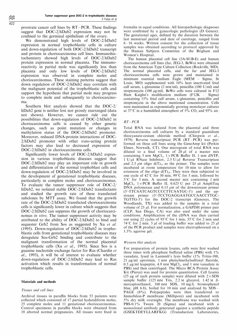
25 complete moles and 11 choriocarcinomas using theaM2 antibody.Fourteen (77.7%) of 18 placentas, ®ve (29.4%) of 17
partial moles, three (12%) of 25 complete moles andnone of 11 choriocarcinomas showed strong stainingfor DOC-2/hDab2 protein. Conversely, none of theplacenta samples, two (11.7%) partial mole, 17 (68%)complete moles and seven (63.6%) choriocarcinomaswere scored as weak or negative for DOC-2/hDab2antibody immunostaining (Table 1). The staining forDOC-2/hDab2 in placenta was signi®cantly strongerthan in partial mole, complete mole and choriocarci-noma (P50.014; P50.0001; P50.0001, respectively).Partial mole also showed signi®cantly stronger DOC-2/hDab2 protein expression than complete mole andchoriocarcinoma (P50.001; P50.015, respectively).The staining appeared in the cytoplasm of bothcytotrophoblast and syncytiotrophoblast but in thelatter it was somewhat stronger (Figure 1). Theintravillous stroma did not display any DOC-2/hDab2 immunoreactivity.Western blot analysis using an anti-DOC-2/hDab2
monoclonal antibody generated from amino acidresidue 31 ± 44 of the DOC-2/hDab2 protein identi®edone major 105 kDa protein band in the normalplacental cell lines with intensity approximately 10-fold higher than that of the choriocarcinoma cell lines(Figure 2).
RT±PCR
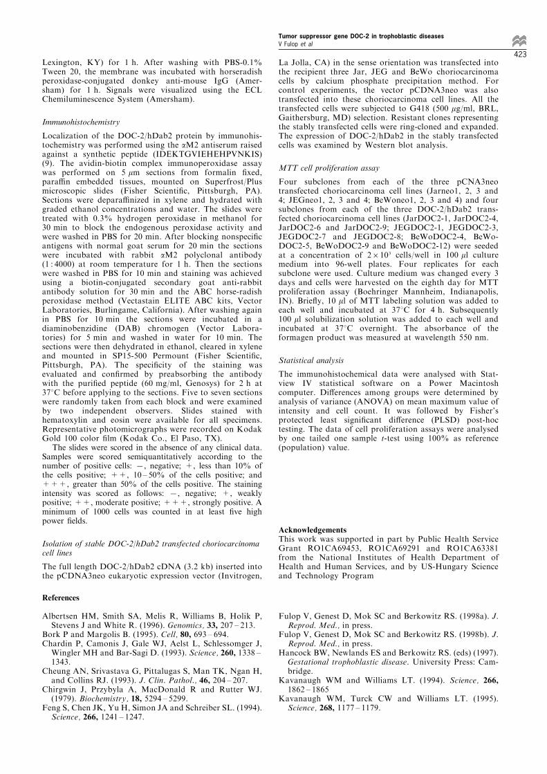
To verify the Western blot results, reverse transcriptasePCR analysis on RNA isolated from the placental cellline and the choriocarcinoma cell lines Jar, JEG-3 and
BeWo was also performed. The results demonstrated astronger 524 bp band (3 ± 4-fold) for DOC-2/hDab2cDNA in normal placental cells than in all chorio-carcinoma cell lines (Figure 3) suggesting that DOC-2/hDab2 message expressed at higher levels in normalplacental cells.
MTT cell proliferation study on DOC-2/hDab2transfected choriocarcinoma cell lines
To evaluate the e�ect of DOC-2/hDab2 on the growthof choriocarcinoma cells, we transfected full-lengthDOC-2/hDab2 cDNA in pcDNA3neo expressionvector into choriocarcinoma cell lines Jar, JEG-3 andBeWo. Five stably transfected subclones were obtainedfrom Jar (JarDOC2-1, JarDOC2-3, JarDOC2-4,JarDOC2-6 and JarDOC2-9), eight from JEG-3(JEGDOC2-1, JEGDOC2-2, JEGDOC2-3, JEG-DOC2-5, JEGDOC2-7, JEGDOC2-8, JEGDOC2-9
Table 1 Expression of DOC-2 protein in human placenta and gestational trophoblastic diseases
Immunoreactivity MeanSample Strong Weak or maximum Standard
Specimen number positive (3+) negative (1+,7) value error
PlacentaPartial moleComplete moleChoriocarcinoma
18172511
14 (77.7%)5 (29.4%)3 (12%)
±
±2 (11.7%)17 (68%)7 (63.6%)
2.782.121.281.36
0.100.190.180.28
Number in parenthesis represents percentages of specimens which showed strong or weak/negative immunoreactivity
Figure 1 Immunohistochemical localization of DOC-2/hDab2 in normal placenta (a) and choriocarcinoma tissue (b) using aM2antiserum on para�n sections. The syncytiotrophoblast displayed strong immunoreactivity (arrow heads) while the intravillousstroma did not show any immunoreactivity (S). Choriocarcinoma showed much lower levels of DOC-2/hDab2 expression (b). Bar,50 mm
Figure 2 Western blot analysis on one normal placental cell line(3A-SUB-E) and three choriocarcinoma cell lines (Jar, JEG-3 andBeWo) using a monoclonal anti-p96 monoclonal antibody
Tumor suppressor gene DOC-2 in trophoblastic diseasesV Fulop et al
420

and JEGDOC2-10) and eight from BeWo (BeWo-DOC2-1, BeWoDOC2-2, BeWoDOC2-3, BeWoDOC2-4, BeWoDOC2-5, BeWoDOC2-8, BeWoDOC2-9,BeWoDOC2-12) cell lines. The presence and theexpression of the transfected cDNA in these cell lineswere con®rmed by Western blot analysis. The resultsshowed that all the stable DOC-2/hDab2 transfectedlines for Jar, JEG-3 and BeWo expressed higher levelsof the unspliced 105 kDa DOC-2/hDab2 protein thantheir stable neo transfected counterparts (Jarneo1, 2, 3and 4; JEGneo1, 2, 3 and 4; BeWoneo1, 2, 3 and 4).However, the DOC-2/hDab2 protein band of thecontrol placenta cell line remained the strongest(Figure 4). On the eighth day, MTT cell proliferationassay showed that the average growth rate for theBeWo DOC-2/hDab2 transfectants (BeWoDOC2-4,BeWoDOC2-5, BeWoDOC2-9 and BeWoDOC2-12)was only 14.5% (P50.0001) of that of the BeWoneotransfectants. Similar study was also performed on theJar and JEG-3 choriocarcinoma cell lines. The growthrates for Jar and JEG-3 DOC-2/hDab2 transfectants(JarDOC2-1, JarDOC2-4, JarDOC2-6 and JarDOC2-9;JEGDOC2-1, JEGDOC2-3, JEGDOC2-7 and JEG-DOC2-8) were 64.4% (P50.0001) and 85.7%(P50.0002) respectively in comparing to their neotransfected counterparts. These results suggest thatDOC-2/hDab2 can suppress choriocarcinoma cellgrowth in vitro (Figure 5).
Discussion
Using RNA ®ngerprinting, we identi®ed DOC-2/hDab2 which is preferentially expressed in normalHOSE cells comparing to the ovarian carcinoma cellsin vitro and in vivo (Mok et al., 1998). Homologysearch shows that it is a human homologue of themouse p96/mDab2, which has been shown to be anovel phosphoprotein rapidly phosphorylated inmacrophages in response to mitogen stimulation ofCSF-1 (Xu et al., 1995). DOC-2 and the p96/mDab2have several characteristics in common. Both DOC-2/hDab2 and p96/mDab2 contain a highly conservedPID at the amino-terminus suggesting that they mayinteract with other proteins phosphorylated on tyrosine(Bork and Margolis, 1995; Kavanaugh and Williams,1994; Kavanaugh et al., 1995). The homology betweenDOC-2/hDab2 and p96/mDab2 is weakened towardsthe carboxy-terminus of the protein. They both haveproline-rich domains in this region which containsmultiple SH3 binding motifs (Yu et al., 1994; Feng etal., 1994; Sparks et al., 1994; Viguera et al., 1994). Xuet al. (1995) has recently demonstrated that the proline-rich domain of mouse p96/mDab2 can interact with theSH3 domains of Grb2 and reduce the binding betweenGrb2 and Sos suggesting DOC-2/hDab2 may alsoshare the same property and involve in Grb2-Sossignaling pathway.Using Northern blot analysis, we identi®ed high
levels of DOC-2/hDab2 expression in the ovariansurface epithelial cells comprising the germinalepithelium of the ovary (Mok et al., 1998). However,other organs including brain, stem, heart, and testisalso express signi®cant levels of DOC-2/hDab2transcripts (data not shown). Albertsen et al. (1996)has recently identi®ed DOC-2/hDab2 from a brainstem library and a fetal retina library, and shownDOC-2/hDab2 expression in three normal tissues and
Figure 3 RT±PCR analysis on RNA isolated from one normalplacental cell line (3A-SUB-E) and three choriocarcinoma celllines (Jar, JEG-3 and BeWo) using a primer set speci®c for DOC-2/hDab2. Another primer set speci®c for GAPDH was also usedfor normalization
Figure 5 Histogram showing the percentage of reduction ingrowth rate in the DOC-2/hDab2 transfected choriocarcinomacell lines (BeWo, Jar and JEG-3) comparing to their mock neotransfected counterparts. Five stably DOC-2/hDab2 transfectedsubclones from Jar, eight from JEG-3 and eight from BeWo wereused. All cells were seeded at a concentration of 26103 cells/wellin 100 ml culture medium into 96-well plates. Four replicates foreach subclone were used
Figure 4 Western blot analysis on four pCDNA3neo transfectedcell lines (Jar/neo1, 2, 3 and 4) and four DOC-2/hDab2transfected lines (Jar/DOC2-1, 2, 3 and 4) using an anti-DOC-2monoclonal antibody
Tumor suppressor gene DOC-2 in trophoblastic diseasesV Fulop et al
421
prostrate cancer cell lines by RT ±PCR. These ®ndingssuggest that DOC-2/hDab2 expression may not becon®ned to the germinal epithelium of the ovary.We demonstrated high levels of DOC-2/hDab2
expression in normal trophoblastic cells in cultureand down-regulation of both DOC-2/hDab2 transcriptand protein in choriocarcinoma cell lines. Immunohis-tochemistry showed high levels of DOC-2/hDab2protein expression in normal placenta. The immuno-reactivity in partial mole was signi®cantly less thanplacenta and only low levels of DOC-2/hDab2expression was observed in complete moles andchoriocarcinomas. These staining patterns suggest thatdown regulation of DOC-2/hDab2 may correlate withthe malignant potential of the trophoblastic cells andsupport the hypothesis that partial mole may progressto complete mole and subsequently to choriocarcino-ma.Southern blot analysis showed that the DOC-2/
hDab2 gene is neither lost nor grossly rearranged (datanot shown). However, we cannot rule out thepossibilities that down-regulation of DOC-2/hDab2 inchoriocarcinoma cells is caused by other geneticchanges, such as point mutation or changes inmethylation status of the DOC-2/hDab2 promoter.Moreover, reduced DNA-protein interactions of DOC-2/hDab2 promoter with other trans-acting proteinfactors may also lead to decreased expression ofDOC-2/hDab2 in choriocarcinoma cells.Signi®cantly lower levels of DOC-2/hDab2 expres-
sion in various trophoblastic diseases suggest thatDOC-2/hDab2 may play an important role in growthand di�erentiation of the normal trophoblast cells anddown-regulation of DOC-2/hDab2 may be involved inthe development of gestational trophoblastic diseases,particularly in complete moles and choriocarcinomas.To evaluate the tumor suppressor role of DOC-2/hDab2, we isolated stable DOC-2/hDab2 transfectantsand studied the growth rate of these transfectedsubclones by MTT assay. We found that the growthrate of the DOC-2/hDab2 transfected choriocarcinomacells is signi®cantly lower in culture which suggests thatDOC-2/hDab2 can suppress the growth of choriocarci-nomas in vitro. The tumor suppressor activity may beattributed to the ability of DOC-2/hDab2 to bind andsequester Grb2 from Sos as suggested by Xu et al.(1995). Down-regulation of DOC-2/hDab2 in tropho-blastic cells from gestational trophoblastic diseases mayderegulate Sos-Grb2 binding and contribute to themalignant transformation of the normal placentaltrophoblastic cells (Xu et al., 1995). Since Sos is aguanine nucleotide exchange factor for Ras (Chardin etal., 1993), it will be of interest to evaluate whetherdown-regulation of DOC-2/hDab2 may lead to Rasactivation which results in transformation of normaltrophoblastic cells.
Materials and methods
Tissues and cell lines
Archival tissues in para�n blocks from 53 patients werecollected which consisted of 17 partial hydatidiform moles,25 complete moles and 11 gestational choriocarcinomas.Control specimens in para�n blocks were obtained from18 aborted normal pregnancies. All tissues were ®xed in
formalin in equal conditions. All histopathologic diagnoseswere con®rmed by a gynecologic pathologist (D Genest).The gestational ages, de®ned by the duration between thelast menstrual period and date of curettage, ranged from6 ± 16 weeks. Written consent for the collection of tissuesamples was obtained according to protocol approved bythe Human Subjects Committee of the Brigham andWomen's Hospital.
The human placental cell line (3A-SUB-E) and humanchoriocarcinoma cell lines (Jar, JEG-3, BeWo) were obtainedfrom the American Type Culture Collection (Rockville, MD).The normal placental cells, and the Jar and JEG-3choriocarcinoma cells were grown and maintained inminimum essential medium Eagle (MEM ± Sigma, StLouis, MO) supplemented with 10% heat inactivated fetalcalf serum, L-glutamine (2 mM/ml), penicillin (100 U/ml) andstreptomycin (100 mg/ml). BeWo cells were cultured in F12Ham Kaighn's modi®cation medium (F12K ± Sigma)containing 15% fetal calf serum, L-glutamine, penicillin andstreptomycin in the above mentioned concentration. Cellswere maintained as exponentially growing monolayer culturesat 378C in a humidi®ed atmosphere of 5% CO2 and 95% air.
RT±PCR
Total RNA was isolated from the placental and threechoriocarcinoma cell cultures by a standard guanidiumthiocyanate-cesium chloride method (Chirgwin et al.,1979). Reverse transcriptase PCR (RT ± PCR) was per-formed on these cell lines using the GeneAmp kit (PerkinElmer, Norwalk, CT). One microgram of total RNA wasdiluted to a ®nal volume of 20 ml of a master mixcontaining 5 mM MgCl2, 16PCR Bu�er II, 1 mM dNTP,1 U/ml RNase Inhibitor, 2.5 U/ml Reverse Transcriptaseand 2.5 mM oligo d(T)16 as the primer. The samples wereincubated at room temperature for 10 min to allow forextension of the oligo d(T)16. They were then subjected toone cycle of 428C for 30 min, 998C for 5 min, followed by58C for 5 min. A second master mix containing 1 mM
MgCl2, 16PCR Bu�er II, 0.625 U per 25 ml AmpliTaqDNA polymerase and 0.15 mM of the downstream primer(5'-TTCAATCAGTCCCCTTCAATGC-3') and the up-stream primer (5'-TCCTGAGGAATGCCAACCTTGC-TGTTG-3') for the DOC-2 transcript (Genosys, TheWoodlands, TX) was added to the samples in a totalvolume of 25 ml. For normalization, a GAPDH primer pair(CLP, San Diego, CA) was also used under the sameconditions. Ampli®cation of the cDNA was then carriedout using 22 cycles of 958C for 1 min, 558C for 2 min and748C for 2 min. 5 ml of loading bu�er was added to 25 mlof the PCR product and samples were electrophoresed on a1.5% agarose gel.
Western blot analysis
For preparation of protein lysates, cells were ®rst washedthree times with phosphate bu�ered saline (PBS) with 1%vanadate, lysed in Laemmli's lysis bu�er (1% Tritin-100,21 mg/ml aprotinin, 1 mM phenylmethylsulfonyl ¯uoride,0.5 mg/ml leupeptin, 4.9 mM MgCl2, and 1 mM vanadate inPBS) and then centrifuged. The Micro BCA Protein AssayKit (Pierce) was used for protein quantitation. Cell lysates(25 mg of each protein sample) were diluted with 26SDSsample bu�er (125 mM Tris, 2.2 M glycerol, 1.42 M b-mercaptoethanol, 160 mM SDS, 10 mg/L bromophenolblue, pH 6.8), boiled for 10 min and analysed by SDS ±PAGE (8%). Polypeptides were then transferred toImmobilon-P membrane (Millipore) and incubated with5% dry milk overnight. The membrane was washed withPBS-0.1% Tween 20 (Sigma) and incubated with amonoclonal antibody generated against a synthetic peptide(GSEKTDEYLLARFKG) (Transduction Laboratories,
Tumor suppressor gene DOC-2 in trophoblastic diseasesV Fulop et al
422
Lexington, KY) for 1 h. After washing with PBS-0.1%Tween 20, the membrane was incubated with horseradishperoxidase-conjugated donkey anti-mouse IgG (Amer-sham) for 1 h. Signals were visualized using the ECLChemiluminescence System (Amersham).
Immunohistochemistry
Localization of the DOC-2/hDab2 protein by immunohis-tochemistry was performed using the aM2 antiserum raisedagainst a synthetic peptide (IDEKTGVIEHEHPVNKIS)(9). The avidin-biotin complex immunoperoxidase assaywas performed on 5 mm sections from formalin ®xed,para�n embedded tissues, mounted on Superfrost/Plusmicroscopic slides (Fisher Scienti®c, Pittsburgh, PA).Sections were depara�nized in xylene and hydrated withgraded ethanol concentrations and water. The slides weretreated with 0.3% hydrogen peroxidase in methanol for30 min to block the endogenous peroxidase activity andwere washed in PBS for 20 min. After blocking nonspeci®cantigens with normal goat serum for 20 min the sectionswere incubated with rabbit aM2 polyclonal antibody(1 : 4000) at room temperature for 1 h. Then the sectionswere washed in PBS for 10 min and staining was achievedusing a biotin-conjugated secondary goat anti-rabbitantibody solution for 30 min and the ABC horse-radishperoxidase method (Vectastain ELITE ABC kits, VectorLaboratories, Burlingame, California). After washing againin PBS for 10 min the sections were incubated in adiaminobenzidine (DAB) chromogen (Vector Labora-tories) for 5 min and washed in water for 10 min. Thesections were then dehydrated in ethanol, cleared in xyleneand mounted in SP15-500 Permount (Fisher Scienti®c,Pittsburgh, PA). The speci®city of the staining wasevaluated and con®rmed by preabsorbing the antibodywith the puri®ed peptide (60 mg/ml, Genosys) for 2 h at378C before applying to the sections. Five to seven sectionswere randomly taken from each block and were examinedby two independent observers. Slides stained withhematoxylin and eosin were available for all specimens.Representative photomicrographs were recorded on KodakGold 100 color ®lm (Kodak Co., El Paso, TX).
The slides were scored in the absence of any clinical data.Samples were scored semiquantitatively according to thenumber of positive cells: 7, negative; +, less than 10% ofthe cells positive; ++, 10 ± 50% of the cells positive; and+++, greater than 50% of the cells positive. The stainingintensity was scored as follows: 7, negative; +, weaklypositive; ++, moderate positive; +++, strongly positive. Aminimum of 1000 cells was counted in at least ®ve highpower ®elds.
Isolation of stable DOC-2/hDab2 transfected choriocarcinomacell lines
The full length DOC-2/hDab2 cDNA (3.2 kb) inserted intothe pCDNA3neo eukaryotic expression vector (Invitrogen,
La Jolla, CA) in the sense orientation was transfected intothe recipient three Jar, JEG and BeWo choriocarcinomacells by calcium phosphate precipitation method. Forcontrol experiments, the vector pCDNA3neo was alsotransfected into these choriocarcinoma cell lines. All thetransfected cells were subjected to G418 (500 mg/ml, BRL,Gaithersburg, MD) selection. Resistant clones representingthe stably transfected cells were ring-cloned and expanded.The expression of DOC-2/hDab2 in the stably transfectedcells was examined by Western blot analysis.
MTT cell proliferation assay
Four subclones from each of the three pCNA3neotransfected choriocarcinoma cell lines (Jarneo1, 2, 3 and4; JEGneo1, 2, 3 and 4; BeWoneo1, 2, 3 and 4) and foursubclones from each of the three DOC-2/hDab2 trans-fected choriocarcinoma cell lines (JarDOC2-1, JarDOC2-4,JarDOC2-6 and JarDOC2-9; JEGDOC2-1, JEGDOC2-3,JEGDOC2-7 and JEGDOC2-8; BeWoDOC2-4, BeWo-DOC2-5, BeWoDOC2-9 and BeWoDOC2-12) were seededat a concentration of 26103 cells/well in 100 ml culturemedium into 96-well plates. Four replicates for eachsubclone were used. Culture medium was changed every 3days and cells were harvested on the eighth day for MTTproliferation assay (Boehringer Mannheim, Indianapolis,IN). Brie¯y, 10 ml of MTT labeling solution was added toeach well and incubated at 378C for 4 h. Subsequently100 ml solubilization solution was added to each well andincubated at 378C overnight. The absorbance of theformagen product was measured at wavelength 550 nm.
Statistical analysis
The immunohistochemical data were analysed with Stat-view IV statistical software on a Power Macintoshcomputer. Di�erences among groups were determined byanalysis of variance (ANOVA) on mean maximum value ofintensity and cell count. It was followed by Fisher'sprotected least signi®cant di�erence (PLSD) post-hoctesting. The data of cell proliferation assays were analysedby one tailed one sample t-test using 100% as reference(population) value.
AcknowledgementsThis work was supported in part by Public Health ServiceGrant RO1CA69453, RO1CA69291 and RO1CA63381from the National Institutes of Health Department ofHealth and Human Services, and by US-Hungary Scienceand Technology Program
References
Albertsen HM, Smith SA, Melis R, Williams B, Holik P,Stevens J and White R. (1996). Genomics, 33, 207 ± 213.
Bork P and Margolis B. (1995). Cell, 80, 693 ± 694.Chardin P, Camonis J, Gale WJ, Aelst L, Schlessomger J,Wingler MH and Bar-Sagi D. (1993). Science, 260, 1338 ±1343.
Cheung AN, Srivastava G, Pittalugas S, Man TK, Ngan H,and Collins RJ. (1993). J. Clin. Pathol., 46, 204 ± 207.
Chirgwin J, Przybyla A, MacDonald R and Rutter WJ.(1979). Biochemistry, 18, 5294 ± 5299.
Feng S, Chen JK, Yu H, Simon JA and Schreiber SL. (1994).Science, 266, 1241 ± 1247.
Fulop V, Genest D, Mok SC and Berkowitz RS. (1998a). J.Reprod. Med., in press.
Fulop V, Genest D, Mok SC and Berkowitz RS. (1998b). J.Reprod. Med., in press.
Hancock BW, Newlands ES and Berkowitz RS. (eds) (1997).Gestational trophoblastic disease. University Press: Cam-bridge.
Kavanaugh WM and Williams LT. (1994). Science, 266,1862 ± 1865
Kavanaugh WM, Turck CW and Williams LT. (1995).Science, 268, 1177 ± 1179.
Tumor suppressor gene DOC-2 in trophoblastic diseasesV Fulop et al
423

Lee WH, Bookstein R, Hong F, Young LJ, Shew JY and LeeEY. (1987). Science, 235, 1394 ± 1399.
Miki Y, Swensen J, Shattuck-Eidens D, Futreal PA,Harsham K, Tavtigian S, Liu Q, Cochran C, BennettLM, Ding W, Bell R, Rosenthal J, Hussey C, Tran T,McClure M, Frye C, Hattier T, Phelps R, Haugen-StranoA, Katcher H, Yakumo K, Gholami Z, Sha�er D, Stone S,Bayer S, Wray C, Bogden R, Dayanath P, Ward J, ToninP, Norad S, Bristow PK, Norris FH, Helvering L,Morrison P, Rosteck P, Lai M, Barrett C, Lewis C,Neuhausen S, Cannon-Albright L, Goldgar D, WeisemanR, Kamb A and Skolnick MH. (1994). Science, 266, 66 ±71.
Mok SC, Wong KK, Chan RKW, Ching CC, Tsao SW,Knapp RC and Berkowitz RS. (1994). Gyn. Oncology, 52,247 ± 252
Mok SC, Chan WY, Wong KK, Cheung KK, Lau CC, NgSW, Baldini A, Colitti CV, Rock CO and Berkowitz RS.(1998). Oncogene, 15, in press.
Sparks AB, Quilliam LA, Thorn JM, Der CJ and Kay BK.(1994). J. Biol. Chem., 269, 23853 ± 23856.
Viguera AR, Arrondo JLR, Musacchio A, Saraste M andSerrano L. (1994). Biochemistry, 33, 10925 ± 10933.
Xu XX, Yang W, Jackowski S and Rock CO. (1995). J. Biol.Chem., 270, 14184 ± 14191.
Xu X, Mittlestaedt S, Yi T and Lambeth JD. (1997). Am.Assoc. Cancer Res. Proc., 38, 157.
Yu H, Chen JK, Feng S, Dalgarno DC, Brauer AW andSchreiber SL. (1994). Cell, 76, 933 ± 945.
Tumor suppressor gene DOC-2 in trophoblastic diseasesV Fulop et al
424