Animal models for clinical and gestational diabetes: maternal and fetal outcomes
7
BioMed Central Page 1 of 7 (page number not for citation purposes) Diabetology & Metabolic Syndrome Open Access Research Animal models for clinical and gestational diabetes: maternal and fetal outcomes Ana CI Kiss, Paula HO Lima, Yuri K Sinzato, Mariana Takaku, Marisa A Takeno, Marilza VC Rudge and Débora C Damasceno* Address: Laboratory of Experimental Research of Gynecology and Obstetrics, Department of Gynecology and Obstetrics, Botucatu Medical School - São Paulo State University (Unesp), Botucatu, São Paulo, Brazil Email: Ana CI Kiss - [email protected]; Paula HO Lima - [email protected]; Yuri K Sinzato - [email protected]; Mariana Takaku - [email protected]; Marisa A Takeno - [email protected]; Marilza VC Rudge - [email protected]; Débora C Damasceno* - [email protected] * Corresponding author Abstract Background: Diabetes in pregnant women is associated with an increased risk of maternal and neonatal morbidity and remains a significant medical challenge. Diabetes during pregnancy may be divided into clinical diabetes and gestational diabetes. Experimental models are developed with the purpose of enhancing understanding of the pathophysiological mechanisms of diseases that affect humans. With regard to diabetes in pregnancy, experimental findings from models will lead to the development of treatment strategies to maintain a normal metabolic intrauterine milieu, improving perinatal development by preventing fetal growth restriction or macrosomia. Based on animal models of diabetes during pregnancy previously reported in the medical literature, the present study aimed to compare the impact of streptozotocin-induced severe (glycemia >300 mg/dl) and mild diabetes (glycemia between 120 and 300 mg/dl) on glycemia and maternal reproductive and fetal outcomes of Wistar rats to evaluate whether the animal model reproduces the maternal and perinatal results of clinical and gestational diabetes in humans. Methods: On day 5 of life, 96 female Wistar rats were assigned to three experimental groups: control (n = 16), severe (n = 50) and mild diabetes (n = 30). At day 90 of life, rats were mated. On day 21 of pregnancy, rats were killed and their uterine horns were exposed to count implantation and fetus numbers to determine pre- and post-implantation loss rates. The fetuses were classified according to their birth weight. Results: Severe and mild diabetic dams showed different glycemic responses during pregnancy, impairing fetal glycemia and weight, confirming that maternal glycemia is directly associated with fetal development. Newborns from severe diabetic mothers presented growth restriction, but mild diabetic mothers were not associated with an increased rate of macrosomic fetuses. Conclusion: Experimental models of severe diabetes during pregnancy reproduced maternal and fetal outcomes of pregnant women presenting uncontrolled clinical diabetes. On the other hand, the mild diabetes model caused mild hyperglycemia during pregnancy, although it was not enough to reproduce the increased rate of macrosomic fetuses seen in women with gestational diabetes. Published: 19 October 2009 Diabetology & Metabolic Syndrome 2009, 1:21 doi:10.1186/1758-5996-1-21 Received: 4 March 2009 Accepted: 19 October 2009 This article is available from: http://www.dmsjournal.com/content/1/1/21 © 2009 Kiss et al; licensee BioMed Central Ltd. This is an Open Access article distributed under the terms of the Creative Commons Attribution License (http://creativecommons.org/licenses/by/2.0 ), which permits unrestricted use, distribution, and reproduction in any medium, provided the original work is properly cited.
-
Upload
independent -
Category
Documents
-
view
4 -
download
0
Transcript of Animal models for clinical and gestational diabetes: maternal and fetal outcomes

BioMed Central
Diabetology & Metabolic Syndrome
ss
Open AcceResearchAnimal models for clinical and gestational diabetes: maternal and fetal outcomesAna CI Kiss, Paula HO Lima, Yuri K Sinzato, Mariana Takaku, Marisa A Takeno, Marilza VC Rudge and Débora C Damasceno*Address: Laboratory of Experimental Research of Gynecology and Obstetrics, Department of Gynecology and Obstetrics, Botucatu Medical School - São Paulo State University (Unesp), Botucatu, São Paulo, Brazil
Email: Ana CI Kiss - [email protected]; Paula HO Lima - [email protected]; Yuri K Sinzato - [email protected]; Mariana Takaku - [email protected]; Marisa A Takeno - [email protected]; Marilza VC Rudge - [email protected]; Débora C Damasceno* - [email protected]
* Corresponding author
AbstractBackground: Diabetes in pregnant women is associated with an increased risk of maternal andneonatal morbidity and remains a significant medical challenge. Diabetes during pregnancy may bedivided into clinical diabetes and gestational diabetes. Experimental models are developed with thepurpose of enhancing understanding of the pathophysiological mechanisms of diseases that affecthumans. With regard to diabetes in pregnancy, experimental findings from models will lead to thedevelopment of treatment strategies to maintain a normal metabolic intrauterine milieu, improvingperinatal development by preventing fetal growth restriction or macrosomia. Based on animalmodels of diabetes during pregnancy previously reported in the medical literature, the presentstudy aimed to compare the impact of streptozotocin-induced severe (glycemia >300 mg/dl) andmild diabetes (glycemia between 120 and 300 mg/dl) on glycemia and maternal reproductive andfetal outcomes of Wistar rats to evaluate whether the animal model reproduces the maternal andperinatal results of clinical and gestational diabetes in humans.
Methods: On day 5 of life, 96 female Wistar rats were assigned to three experimental groups:control (n = 16), severe (n = 50) and mild diabetes (n = 30). At day 90 of life, rats were mated. Onday 21 of pregnancy, rats were killed and their uterine horns were exposed to count implantationand fetus numbers to determine pre- and post-implantation loss rates. The fetuses were classifiedaccording to their birth weight.
Results: Severe and mild diabetic dams showed different glycemic responses during pregnancy,impairing fetal glycemia and weight, confirming that maternal glycemia is directly associated withfetal development. Newborns from severe diabetic mothers presented growth restriction, but milddiabetic mothers were not associated with an increased rate of macrosomic fetuses.
Conclusion: Experimental models of severe diabetes during pregnancy reproduced maternal andfetal outcomes of pregnant women presenting uncontrolled clinical diabetes. On the other hand,the mild diabetes model caused mild hyperglycemia during pregnancy, although it was not enoughto reproduce the increased rate of macrosomic fetuses seen in women with gestational diabetes.
Published: 19 October 2009
Diabetology & Metabolic Syndrome 2009, 1:21 doi:10.1186/1758-5996-1-21
Received: 4 March 2009Accepted: 19 October 2009
This article is available from: http://www.dmsjournal.com/content/1/1/21
© 2009 Kiss et al; licensee BioMed Central Ltd. This is an Open Access article distributed under the terms of the Creative Commons Attribution License (http://creativecommons.org/licenses/by/2.0), which permits unrestricted use, distribution, and reproduction in any medium, provided the original work is properly cited.
Page 1 of 7(page number not for citation purposes)

Diabetology & Metabolic Syndrome 2009, 1:21 http://www.dmsjournal.com/content/1/1/21
BackgroundDiabetes mellitus (DM) is a disease characterized by dis-arrangements in carbohydrate, protein and lipid metabo-lism caused by the complete or relative insufficiency ofinsulin secretion and/or insulin action [1]. Diabetes inpregnant women is associated with an increased risk ofmaternal and neonatal morbidity and remains a signifi-cant medical challenge. Diabetes during pregnancy maybe divided into clinical diabetes (women previously diag-nosed with type 1 or type 2 diabetes) and gestational dia-betes, defined as any glucose intolerance detected duringpregnancy that has evolved from a diagnosis associatedwith the metabolic risk of type 2 diabetes to a clinical con-dition associated with higher risks for maternal and peri-natal morbidity [2]. Fortunately, the prognosis haschanged dramatically due to an increased clinical aware-ness of the potential risks for the mother and the infant.
Experimental models are developed with the purpose ofenhancing understanding of the pathophysiologicalmechanisms of diseases that affect humans. With regardto diabetes in pregnancy, experimental findings frommodels will lead to the development of treatment strate-gies to maintain the closest to normal metabolic intrauter-ine milieu, improving perinatal development bypreventing fetal growth restriction or macrosomia. The rat(and the rabbit) is often used in reproductive toxicitystudies [3]. In general, the uncontrolled human type 1DM clinical status during pregnancy is reproduced bystreptozotocin (STZ) administration (40 mg/kg) to ratsduring adult life using the venous route [4-6]. In thisexperimental model, rats present with severe diabetes,with glycemia above 300 mg/dl, and the fetuses of damsare classified as small fetuses for gestational age, character-izing intrauterine growth restriction. Human type 2 DMand gestational DM conditions are reproduced in animalsby administration of different doses of STZ in the neona-tal period [7-16], before mating [17-20] or during preg-nancy [21-30]. Adult animals present with glycemiabetween 120 and 300 mg/dl, characterizing moderate ormild diabetes [31-33]. Merzouk and colleagues [23-25]and Soulimane-Mokhtari and colleagues [30] verified thatmildly hyperglycemic dams have fetuses that are large forgestational age, classified as macrosomic.
Evidence in the literature indicates that neonatal ratstreated with STZ at birth exhibit altered insulin and glu-cose tolerance tests [8,9,13] and plasmatic insulin[11,15]. Based on the insulin action response and glucosetolerance test, Triadou and colleagues [15] established anexperimental design that reproduces the development ofgestational diabetes in women. Several reports in the liter-ature describe the effects of severe and mild diabetes onpregnancy, fetal glycemia and development, but thesestudies did not investigate correlations between maternal
and fetal repercussions in these two different glycemicranges. Therefore, the present study aimed to compare theimpact of STZ-induced severe and mild diabetes on glyc-emia and maternal reproductive and fetal outcomes ofWistar rats to evaluate whether the animal model repro-duces the maternal and perinatal results of clinical andgestational diabetes in humans.
MethodsSubjectsWistar rats were obtained from São Paulo State University(Unesp) Botucatu, São Paulo State, Brazil. They weremaintained in an experimental room under controlledconditions of temperature (22 ± 2°C), humidity (50 ±10%), and a 12-hour light/dark cycle. All experimentalprocedures presented in this study were approved by thelocal Committee of Ethics in Animal Experimentation,which assures adherence to the standards established bythe Guide for the Care and Use of Laboratory Animals.
Experimental proceduresOn day 5 of life, 64 female Wistar rats were randomlyassigned to three experimental groups: control (n = 16) -rats that received citrate buffer solution (0.1 M, pH 6.5)intraperitoneally on day 5 of life; severe diabetes (n = 50)- rats that received STZ (SIGMA Chemical Company, StLouis, MO, USA; 40 mg/kg intravenously) on day 75 oflife; and mild diabetes (n = 30) - rats that received STZ (70mg/kg intraperitoneally) dissolved in citrate buffer solu-tion on day 5 of life. At day 90 of life, the female rats weremated and the morning on which sperm were found inthe vaginal smear was designated pregnancy day 0. Bloodsamples were obtained from cut tail tips for glycemicdetermination (glucose oxidase) using a typical glucome-ter; values are expressed in milligrams per deciliter (mg/dl). Inclusion criteria required glycemia levels on day 0 ofpregnancy of <120 mg/dl for the control group (n = 16),>300 mg/dl for the severe diabetic group (n = 18), andbetween 120 and 300 mg/dl for the mild diabetic group(n = 6). Body weight, food intake and glycemia were eval-uated on days 0, 7, 14 and 21 of pregnancy. On day 21 ofpregnancy, the dams were anesthetized with sodiumpentobarbital (Hypnol® 3%) and their uterine hornsexposed to count implantation and fetus numbers fordetermination of pre- and post-implantation loss rates.The fetuses were removed, weighed and classified accord-ing to their birth weight as follows: large for pregnancy age(LPA) if their birth weight was greater than 1.0 standarddeviation of the mean birth weight of the control dampups; small for pregnancy age (SPA) if their birth weightwas lower than 1.0 standard deviation of the mean birthweight of the control dam pups; and appropriate for preg-nancy age (APA) if their birth weight was included in ± 1.0standard deviation of the mean birth weight of the control
Page 2 of 7(page number not for citation purposes)

Diabetology & Metabolic Syndrome 2009, 1:21 http://www.dmsjournal.com/content/1/1/21
dam pups [34]. Blood pool glycemia levels were deter-mined from three newborns from each litter.
Statistical analysisResults are presented as mean ± standard error of mean.The proportion test (Chi-square) was used for fetal weightclassification. Two-way analysis of variance (ANOVA) fol-lowed by the Student-Newman-Keuls test was employedto compare the data for maternal glycemia, food intake,body weight during pregnancy, number of implantationsites and number of live fetuses. Pre- and post-implanta-tion loss rates were analyzed by Mann Whitney non-para-metric test. Maternal and fetal glycemia correlation wasdetermined using Pearson correlation. The statistical sig-nificance interval is considered as P < 0.05 for all data. Allstatistical analyses were performed with Statistica software(Statsoft, Tulsa, OK, USA).
ResultsAll 16 rats assigned to the control group were mated, hada positive pregnancy diagnosis and were included in thisstudy. Only 18 of 50 rats administered STZ as adults(severe diabetic rats) had a positive pregnancy diagnosisand were included in this study following the inclusioncriteria for their experimental group. All 30 rats adminis-tered streptozotocin as neonates were also mated, butonly 16 presented with a positive pregnancy diagnosisand only 6 achieved the inclusion criteria. The rats thatdid not reach inclusion criteria were used in anotherstudy. There were no significant differences in the numberof implantation sites in the severe and mild diabeticgroups compared to the control group nor between thesevere and mild diabetic groups. A lower mean number oflive fetuses and a higher post-implantation loss rate wereobserved in severe diabetic rats compared to the controland mild diabetes groups (Table 1).
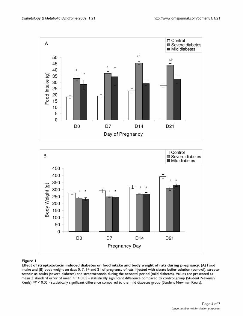
Rats with severe diabetes had a higher food intake com-pared to mild diabetic rats on days 14 to 21 of pregnancy,
and compared to control rats on all days of pregnancy.Mild diabetic rats had a higher food intake compared tothe control group only on day 0 of pregnancy (Figure 1A).Both severe and mild diabetic rats had lower body weightcompared to the control group (Figure 1B).
During their entire pregnancy, control rats had normalglycemic values (around 80 mg/dl). Glycemia remainedabove 300 mg/dl in the severe diabetic rats and between120 and 300 mg/dl in the mild diabetic rats. Both severeand mild diabetic rats had higher glycemia levels through-out pregnancy compared to the control group. Whencompared to the mild diabetes group, severe diabeticdams had higher glycemia levels prior to mating and dur-ing pregnancy. Newborns from severe diabetic dams hadhigher glycemia levels compared to newborns from boththe control and mild diabetic groups (Table 2). There wasa positive correlation (P < 0.05) between maternal andfetal glycemia in all experimental groups.
In both the severe and mild diabetes groups, there was ahigher proportion of SPA fetuses and a reduced percent-age of APA and LPA fetuses compared to the controlgroup. Severe diabetic rats also had higher SPA and lowerAPA rates compared to mild diabetic rats. The proportionsof LPA fetuses from the severe and mild diabetes groupswere similar (Table 3).
DiscussionSTZ is often used to induce DM in experimental animalsdue to its toxic effects on pancreatic beta-cells [35,36]. It isa potent alkylating agent able to methylate DNA [37-39]and although it is generally accepted that the cytotoxicityproduced by STZ depends on DNA alkylation [37,39],several lines of evidence indicate that free radicals play anessential role in its mechanism of DNA damage and cyto-toxicity. The nitrosurea moiety of STZ is responsible for itscellular toxicity, which is probably mediated through adecrease in NAD levels and the production of intracellular
Table 1: Maternal reproductive outcomes of control, severe diabetic and mild diabetic rats
Variables Control Severe diabetes Mild diabetes
Number of rats used 16 50 30Number of rats that achieved inclusion criteria 16 (100%) 18 (36%) 6 (20%)Implantation number 159 199 70
Mean ± SEM 11.67 ± 0.33 11.71 ± 0.39 11.67 ± 0.56Live fetus number 153 115 66
Mean ± SEM 11.50 ± 0.22 6.76 ± 1.15a, b 11.00 ± 0.37Pre-implantation loss (%) 4.85% 8,44% 1,52%Post-implantation loss (%) 1.28% 42,27%c, d 5,24%
Rats were injected with citrate buffer solution (control), streptozotocin as adults (severe diabetes) and streptozotocin during the neonatal period (mild diabetes). Values are presented as mean ± standard deviation and proportions (%). aP < 0.05 - statistically significant difference compared to control group (Student Newman Keuls); bP < 0.05 - statistically significant difference compared to mild diabetes group (Student Newman Keuls); cP < 0.05 - statistically significant difference compared to control group (Mann Whitney); dP < 0.05 - statistically significant difference compared to mild diabetes group (Mann Whitney). SEM, standard error of the mean.
Page 3 of 7(page number not for citation purposes)
Diabetology & Metabolic Syndrome 2009, 1:21 http://www.dmsjournal.com/content/1/1/21
Page 4 of 7(page number not for citation purposes)
Effect of streptozotocin induced diabetes on food intake and body weight of rats during pregnancyFigure 1Effect of streptozotocin induced diabetes on food intake and body weight of rats during pregnancy. (A) Food intake and (B) body weight on days 0, 7, 14 and 21 of pregnancy of rats injected with citrate buffer solution (control), strepto-zotocin as adults (severe diabetes) and streptozotocin during the neonatal period (mild diabetes). Values are presented as mean ± standard error of mean. aP < 0.05 - statistically significant difference compared to control group (Student Newman Keuls); bP < 0.05 - statistically significant difference compared to the mild diabetes group (Student Newman Keuls).
A
05
101520253035404550
D0 D7 D14 D21
Day of Pregnancy
Fo
od
Inta
ke (g
)ControlSevere diabetesMild diabetes
a,b a,b
a a
a
B
0
50
100
150
200
250
300
350
400
450
D0 D7 D14 D21
Pregnancy Day
Bo
dy
Wei
gh
t (g
)
ControlSevere diabetesMild diabetes
a a
a a a a a a

Diabetology & Metabolic Syndrome 2009, 1:21 http://www.dmsjournal.com/content/1/1/21
free radicals. The deoxyglucose moiety of STZ facilitates itstransport across the cell membrane, in which the GLUT-2glucose-transporter appears to play an essential role. Theinsulin-producing beta-cells of the islets of Langerhansnot only express high levels of GLUT-2 transporters butalso have a relatively low NAD content, making them par-ticularly vulnerable to STZ toxicity [40].
In the mild diabetes group, STZ treatment created a rangeof damage to beta cells, leading to a variable range of insu-lin insufficiency. Only 6 (20%) of the initial 30 rats had apositive pregnancy diagnosis and presented with milddiabetes on pregnancy day 0 according to the inclusioncriteria previously established (glycemia between 120 and300 mg/dl). Although the success rate of this model mayappear low, models in which high doses of STZ areadministered in the neonatal period to achieve mild dia-betes are well established [7-16]. However, these studiesdo not mention how many animals achieved hyperglyc-emia in adult life. STZ has a beta-cell specific toxicity thatproduces severe and permanent diabetes when given toadult rats. When given during the neonatal period, thereis a spontaneous recovery from the damage caused to thebeta-cells in the first 2 weeks of life. However, beta-cellregeneration is incomplete and this reduced beta-cellmass results in the appearance of a form of diabetes inadult life that resembles DM type 2 in humans [9]. Indi-vidual differences in STZ metabolism [41] and beta-cell
regeneration capacity [9] may explain why so many ratsthat receive STZ do not present mild diabetes in adult life.
In the present study, rats with glycemia above 300 mg/dl(severe diabetes) had higher food intake but reducedbody weight during pregnancy, both common features ofthe severe diabetic state. The reduced body weight is aconsequence of metabolic alterations caused by hypergly-cemia/hypoinsulinemia, such as asthenia, as described byDamasceno and colleagues [4]. Rats injected neonatallywith STZ had mild diabetes (glycemia from 120 to 300mg/dl) without a significant increase in food intake, butreduced body weight, which can also be explained by met-abolic alterations despite the lower glycemia compared tothe severely diabetic rats. Although maternal hypoin-sulinemia/hyperglycemia has a major impact on fetalweight, the reduced maternal body weight of mild dia-betic rats, resulting from low weight gain during preg-nancy, could be a cause of the low number of LPA fetusesin this group.
Severe diabetic rats had glycemia levels above 300 mg/dlthroughout pregnancy. This result was expected and is inagreement with other studies previously performed in ourlaboratory [4-6], reproducing the hyperglycemia thatsome women with uncontrolled clinical diabetes presentduring pregnancy. The mild diabetic rats maintained theirglycemia between 120 and 300 mg/dl during pregnancy.STZ administration in the neonatal period caused mild
Table 2: Glycemia of control, severe diabetic and mild diabetic rats throughout pregnancy and of newborns
Control (n = 16) Severe diabetes (n = 18) Mild diabetes (n = 6)
Prior mating 84.33 ± 0.76 343.56 ± 14,36a, b 177.12 ± 45.53Day 0 78.17 ± 3.89 351.78 ± 12.79a, b 186.67 ± 26.04a
Day 7 77.67 ± 3.68 294.11 ± 11.01a, b 177.67 ± 32.84a
Day 14 77.17 ± 5.85 327.44 ± 12.70a, b 179.69 ± 39.85a
Day 21 79.83 ± 6.65 322.61 ± 17.95a, b 170.67 ± 30.21a
Newborns 68.25 ± 7.94 464.33 ± 28.95a, b 115.9 ± 37.57
Glycemia (mean ± standard error of mean) were taken prior to mating and on days 0, 7, 14 and 21 of pregnancy from rats injected with citrate buffer solution (control), streptozotocin as adults (severe diabetes) and streptozotocin during the neonatal period (mild diabetes). Blood pool glycemia was determined from three newborns from each litter. aP < 0.05 - statistically significant difference compared to control group (Student Newman Keuls);bP < 0.05 - statistically significant difference compared to mild diabetes group (Student Newman Keuls).
Table 3: Fetal weight classification of offspring born to control, severe diabetic and mild diabetic rats
Variable/groups Control Severe diabetes Mild diabetes
SPA 46/192 (24%) 112/129 (87%)a, b 39/65 (60%)a
APA 99/192 (52%) 14/129 (11%)a, b 22/65 (34%)a
LPA 47/192 (24%) 3/129 (2%)a 4/65 (6%)a
Fetal weight classification is defined as small for pregnancy age (SPA), appropriate for pregnancy age (APA) or large for pregnancy age (LPA). The offspring were born to rats injected with citrate buffer solution (control), streptozotocin as adults (severe diabetes) or streptozotocin during the neonatal period (mild diabetes). Values are number/total (percent). aP < 0.05 - statistically significant difference compared to control group (Chi-square test); bP < 0.05 - statistically significant difference compared to mild diabetes group (Chi-square test).
Page 5 of 7(page number not for citation purposes)

Diabetology & Metabolic Syndrome 2009, 1:21 http://www.dmsjournal.com/content/1/1/21
hyperglycemia during pregnancy, which has also beenreported by Triadou and colleagues [15], Capobianco andcolleagues [10] and Kiss [42], reproducing the hyperglyc-emia that some women with gestational diabetes presentduring pregnancy.
In our study, the lower number of live fetuses and the highpost-implantation loss rate in the severe diabetes groupare characteristic of a hyperglycemic (glycemia above 300mg/dl) intrauterine milieu, and are in agreement withother studies [6,43]. In the present study, the high glyc-emic levels did not prevent embryo implantation but didimpair development, leading to fetal death, as confirmedby the low number of live fetuses. Our results also showthat rats with severe diabetes had newborns with intrau-terine growth restriction. This can be explained by fetalbeta-cell collapse, which eventually leads to fetal hypoin-sulinemia that causes the growth restriction [19,44,45].
There is evidence that the hyperglycemic intrauterinemilieu of a mildly diabetic mother stimulates the fetalendocrine pancreas to hyperinsulinemia and acceleratedanabolism, resulting in fetal and neonatal macrosomia.Many reports in the literature indicate that animal modelsin which STZ is injected during the neonatal period arecompatible with human gestational diabetes conditions,with the presence of macrosomic fetuses [23-25,30] thatare intolerant to glucose [44,46]. In contrast, our resultsshow that the mild diabetic dams did not have anincreased percentage of newborns classified as LPA. Simi-larly, Kervran and colleagues [19] also did not obtainmacrosomic fetuses when studying the offspring of ratswith mild hyperglycemia during pregnancy, and suggestthat the differences between the clinical findings inhumans and the experimental results using rats are due tothe short pregnancy time in the rat and differences in thepercentages of adipose tissue in rat fetuses (1%) andhuman offspring (16%) and the greater weight gain in thehuman species.
The offspring of the mild diabetic dams did not haveimpaired glycemia compared to the control group. How-ever, the offspring of the severe diabetic dams showedhigher glycemia levels compared to both the control andmild diabetes groups. Many clinical and experimentalstudies have shown that offspring that developed in anintrauterine milieu that has been modified by hyperglyc-emia show intolerance to glucose [44,46]. In the presentstudy, offspring were not submitted to the glucose toler-ance test, so there is no evidence that they are intolerantto glucose, but their glycemia levels correlate positivelywith those of their mothers. Kervran and colleagues [19]also observed a positive correlation between maternal andfetal glycemia levels in both severe and mild diabeticdams.
ConclusionSTZ-induced severe and mild diabetic dams showed dif-ferent glycemic responses during pregnancy, althoughboth adversely affected fetal glycemia and weight, con-firming that maternal glycemia is directly associated withfetal development. Newborn from severe diabetic moth-ers presented intrauterine growth restriction, but mild dia-betic mothers did not have an increased percentage of LPAfetuses. The experimental model of severe diabetes duringpregnancy reproduced maternal and fetal outcomes ofwomen with uncontrolled clinical diabetes. On the otherhand, the mild diabetes model caused mild hyperglyc-emia during pregnancy, although it was not enough toreproduce the increased rate of macrosomic fetuses seenin women with gestational diabetes.
AbbreviationsAPA: appropriate for pregnancy age; DM: diabetes melli-tus; LPA: large for pregnancy age; SPA: small for pregnancyage; STZ: streptozotocin.
Competing interestsThe authors declare that they have no competing interests.
Authors' contributionsACIK participated in the acquisition, analysis and inter-pretation of data and helped to draft the manuscript.PHOL participated in the acquisition of data and helpedto draft the manuscript. YKS participated in the acquisi-tion of data and helped to draft the manuscript. MT par-ticipated in the acquisition of data and helped to draft themanuscript. MAT participated in the acquisition of dataand helped to draft the manuscript. MVCR helped to draftthe manuscript. DCD conceived the study, participated inits design, coordination, analysis and interpretation ofdata and helped to draft the manuscript. All authors readand approved the final manuscript.
AcknowledgementsThe authors are grateful to CAPES (Brazil) for financial support and to the Research Support Center (RSC) of the Botucatu Medical School, São Paulo State University (Unesp), for their invaluable contribution to the study design and statistical analysis.
References1. American Diabetes Association: Diagnosis and classification of
diabetes mellitus. Diabetes Care 2009, 32(Suppl 1):S62-67.2. Forsbach-Sanchez G, Tamez-Perez HE, Vazquez-Lara J: Diabetes
and pregnancy. Arch Med Res 2005, 36:291-299.3. de Rijk EP, van Esch E, Flik G: Pregnancy dating in the rat: pla-
cental morphology and maternal blood parameters. ToxicolPathol 2002, 30:271-282.
4. Damasceno DC, Volpato GT, Calderon Ide M, Aguilar R, Rudge MV:Effect of Bauhinia forficata extract in diabetic pregnant rats:maternal repercussions. Phytomedicine 2004, 11:196-201.
5. Rudge MV, Damasceno DC, Volpato GT, Almeida FC, Calderon IM,Lemonica IP: Effect of Ginkgo biloba on the reproductive out-come and oxidative stress biomarkers of streptozotocin-induced diabetic rats. Braz J Med Biol Res 2007, 40:1095-1099.
Page 6 of 7(page number not for citation purposes)

Diabetology & Metabolic Syndrome 2009, 1:21 http://www.dmsjournal.com/content/1/1/21
6. Volpato G, Damasceno D, Campos K, Rocha R, Rudge M, Calderon I:Avaliação do efeito do exercício físico no metabolismo deratas diabéticas prenhes. Revista Brasileira de Medicina do Esporte2006, 12:229-233.
7. Blondel O, Bailbe D, Portha B: Relation of insulin deficiency toimpaired insulin action in NIDDM adult rats given streptozo-cin as neonates. Diabetes 1989, 38:610-617.
8. Blondel O, Bailbe D, Portha B: Insulin resistance in rats with non-insulin-dependent diabetes induced by neonatal (5 days)streptozotocin: evidence for reversal following phlorizintreatment. Metabolism 1990, 39:787-793.
9. Bonner-Weir S, Trent DF, Honey RN, Weir GC: Responses of neo-natal rat islets to streptozotocin: limited B-cell regenerationand hyperglycemia. Diabetes 1981, 30:64-69.
10. Capobianco E, Jawerbaum A, White V, Pustovrh C, Sinner D,Gonzalez ET: Elevated levels of endothelin-1 and prostaglan-din E2 and their effect on nitric oxide generation in placentaltissue from neonatal streptozotocin-induced diabetic rats.Prostaglandins Leukot Essent Fatty Acids 2003, 68:225-231.
11. Movassat J, Saulnier C, Portha B: Insulin administration enhancesgrowth of the beta-cell mass in streptozotocin-treated new-born rats. Diabetes 1997, 46:1445-1452.
12. Murali B, Goyal RK: Improvement in insulin sensitivity by losa-rtan in non-insulin-dependent diabetic (NIDDM) rats. Phar-macol Res 2001, 44:385-389.
13. Portha B, Kergoat M: Dynamics of glucose-induced insulinrelease during the spontaneous remission of streptozocindiabetes induced in the newborn rat. Diabetes 1985,34:574-579.
14. Portha B, Levacher C, Picon L, Rosselin G: Diabetogenic effect ofstreptozotocin in the rat during the perinatal period. Diabetes1974, 23:889-895.
15. Triadou N, Portha B, Picon L, Rosselin G: Experimental chemicaldiabetes and pregnancy in the rat. Evolution of glucose tol-erance and insulin response. Diabetes 1982, 31:75-79.
16. Tsuji K, Taminato T, Usami M, Ishida H, Kitano N, Fukumoto H, KohG, Kurose T, Yamada Y, Yano H, et al.: Characteristic features ofinsulin secretion in the streptozotocin-induced NIDDM ratmodel. Metabolism 1988, 37:1040-1044.
17. Caluwaerts S, Holemans K, van Bree R, Verhaeghe J, Van Assche FA:Is low-dose streptozotocin in rats an adequate model forgestational diabetes mellitus? J Soc Gynecol Investig 2003,10:216-221.
18. Eriksson U, Dahlstrom E, Larsson KS, Hellerstrom C: Increasedincidence of congenital malformations in the offspring of dia-betic rats and their prevention by maternal insulin therapy.Diabetes 1982, 31:1-6.
19. Kervran A, Guillaume M, Jost A: The endocrine pancreas of thefetus from diabetic pregnant rat. Diabetologia 1978, 15:387-393.
20. Kinney BA, Rabe MB, Jensen RA, Steger RW: Maternal hyperglyc-emia leads to gender-dependent deficits in learning andmemory in offspring. Exp Biol Med (Maywood) 2003, 228:152-159.
21. Heinze E, Vetter U: Skeletal growth of fetuses from streptozo-tocin diabetic rat mothers: in vivo and in vitro studies. Diabe-tologia 1987, 30:100-103.
22. Lopez-Soldado I, Herrera E: Different diabetogenic response tomoderate doses of streptozotocin in pregnant rats, and itslong-term consequences in the offspring. Exp Diabesity Res2003, 4:107-118.
23. Merzouk H, Madani S, Boualga A, Prost J, Bouchenak M, Belleville J:Age-related changes in cholesterol metabolism in macro-somic offspring of rats with streptozotocin-induced diabetes.J Lipid Res 2001, 42:1152-1159.
24. Merzouk H, Madani S, Chabane Sari D, Prost J, Bouchenak M, Bel-leville J: Time course of changes in serum glucose, insulin, lip-ids and tissue lipase activities in macrosomic offspring of ratswith streptozotocin-induced diabetes. Clin Sci (Lond) 2000,98:21-30.
25. Merzouk H, Madani S, Hichami A, Prost J, Belleville J, Khan NA: Age-related changes in fatty acids in obese offspring of streptozo-tocin-induced diabetic rats. Obes Res 2002, 10:703-714.
26. Mulay S, Philip A, Solomon S: Influence of maternal diabetes onfetal rat development: alteration of insulin receptors in fetalliver and lung. J Endocrinol 1983, 98:401-410.
27. Oh W, Gelardi NL, Cha CJ: Maternal hyperglycemia in pregnantrats: its effect on growth and carbohydrate metabolism inthe offspring. Metabolism 1988, 37:1146-1151.
28. Oh W, Gelardi NL, Cha CJ: The cross-generation effect of neo-natal macrosomia in rat pups of streptozotocin-induced dia-betes. Pediatr Res 1991, 29:606-610.
29. Plagemann A, Harder T, Janert U, Rake A, Rittel F, Rohde W, DornerG: Malformations of hypothalamic nuclei in hyperinsulinemicoffspring of rats with gestational diabetes. Dev Neurosci 1999,21:58-67.
30. Soulimane-Mokhtari NA, Guermouche B, Yessoufou A, Saker M,Moutairou K, Hichami A, Merzouk H, Khan NA: Modulation oflipid metabolism by n-3 polyunsaturated fatty acids in gesta-tional diabetic rats and their macrosomic offspring. Clin Sci(Lond) 2005, 109:287-295.
31. Gelardi NL, Cha CJ, Oh W: Glucose metabolism in adipocytesof obese offspring of mild hyperglycemic rats. Pediatr Res 1990,28:641-645.
32. Guermouche B, Yessoufou A, Soulimane N, Merzouk H, MoutairouK, Hichami A, Khan NA: n-3 fatty acids modulate T-cell calciumsignaling in obese macrosomic rats. Obes Res 2004,12:1744-1753.
33. Vercheval M, De Hertogh R, Pampfer S, Vanderheyden I, Michiels B,De Bernardi P, De Meyer R: Experimental diabetes impairs ratembryo development during the preimplantation period.Diabetologia 1990, 33:187-191.
34. Calderon I, Rudge M, Ramos M, Peraçoli J: Estudo longitudinal,bioquímico e histoquímico de placentas de ratas diabéticas:relação com a macrossomia e o retardo de crescimentointra-uterino. Revista Brasileira de Ginecologia e Obstetrícia 1999,21:91-99.
35. Junod A, Lambert AE, Orci L, Pictet R, Gonet AE, Renold AE: Studiesof the diabetogenic action of streptozotocin. Proc Soc Exp BiolMed 1967, 126:201-205.
36. Rakieten N, Rakieten ML, Nadkarni MV: Studies on the diabe-togenic action of streptozotocin (NSC-37917). Cancer Chem-other Rep 1963, 29:91-98.
37. Bennett RA, Pegg AE: Alkylation of DNA in rat tissues followingadministration of streptozotocin. Cancer Res 1981,41:2786-2790.
38. Randerath K, Reddy MV, Gupta RC: 32P-labeling test for DNAdamage. Proc Natl Acad Sci USA 1981, 78:6126-6129.
39. Tjälve H: Streptozotocin: distribution, metabolism and mech-anisms of action. Uppsala J Med Sci 1983:145-147.
40. Schnedl WJ, Ferber S, Johnson JH, Newgard CB: STZ transportand cytotoxicity. Specific enhancement in GLUT2-express-ing cells. Diabetes 1994, 43:1326-1333.
41. Kim E, Sohn S, Lee M, Jung J, Kineman RD, Park S: Differentialresponses of the growth hormone axis in two rat models ofstreptozotocin-induced insulinopenic diabetes. J Endocrinol2006, 188:263-270.
42. Kiss A: Análise do desenvolvimento, atividade geral, compor-tamento sexual e prenhez de ratas com diabete induzido porstreptozotocin no período neonatal. UNESP, Faculdade deMedicina de Botucatu; 2008.
43. Volpato GT, Damasceno DC, Rudge MV, Padovani CR, Calderon IM:Effect of Bauhinia forficata aqueous extract on the maternal-fetal outcome and oxidative stress biomarkers of streptozo-tocin-induced diabetic rats. J Ethnopharmacol 2008, 116:131-137.
44. Aerts L, Van Assche FA: Animal evidence for the transgenera-tional development of diabetes mellitus. Int J Biochem Cell Biol2006, 38:894-903.
45. Holemans K, Aerts L, Van Assche FA: Fetal growth restrictionand consequences for the offspring in animal models. J SocGynecol Investig 2003, 10:392-399.
46. Fernandez-Twinn DS, Ozanne SE: Mechanisms by which poorearly growth programs type-2 diabetes, obesity and the met-abolic syndrome. Physiol Behav 2006, 88:234-243.
Page 7 of 7(page number not for citation purposes)
http://www.ncbi.nlm.nih.gov/entrez/query.fcgi?cmd=Retrieve&db=PubMed&dopt=Abstract&list_uids=2523831
http://www.ncbi.nlm.nih.gov/entrez/query.fcgi?cmd=Retrieve&db=PubMed&dopt=Abstract&list_uids=2523831
http://www.ncbi.nlm.nih.gov/entrez/query.fcgi?cmd=Retrieve&db=PubMed&dopt=Abstract&list_uids=2523831
http://www.ncbi.nlm.nih.gov/entrez/query.fcgi?cmd=Retrieve&db=PubMed&dopt=Abstract&list_uids=2198430
http://www.ncbi.nlm.nih.gov/entrez/query.fcgi?cmd=Retrieve&db=PubMed&dopt=Abstract&list_uids=2198430
http://www.ncbi.nlm.nih.gov/entrez/query.fcgi?cmd=Retrieve&db=PubMed&dopt=Abstract&list_uids=2198430
http://www.ncbi.nlm.nih.gov/entrez/query.fcgi?cmd=Retrieve&db=PubMed&dopt=Abstract&list_uids=6112177
http://www.ncbi.nlm.nih.gov/entrez/query.fcgi?cmd=Retrieve&db=PubMed&dopt=Abstract&list_uids=6112177
http://www.ncbi.nlm.nih.gov/entrez/query.fcgi?cmd=Retrieve&db=PubMed&dopt=Abstract&list_uids=6112177
http://www.ncbi.nlm.nih.gov/entrez/query.fcgi?cmd=Retrieve&db=PubMed&dopt=Abstract&list_uids=9287045
http://www.ncbi.nlm.nih.gov/entrez/query.fcgi?cmd=Retrieve&db=PubMed&dopt=Abstract&list_uids=9287045
http://www.ncbi.nlm.nih.gov/entrez/query.fcgi?cmd=Retrieve&db=PubMed&dopt=Abstract&list_uids=9287045
http://www.ncbi.nlm.nih.gov/entrez/query.fcgi?cmd=Retrieve&db=PubMed&dopt=Abstract&list_uids=3891470
http://www.ncbi.nlm.nih.gov/entrez/query.fcgi?cmd=Retrieve&db=PubMed&dopt=Abstract&list_uids=3891470
http://www.ncbi.nlm.nih.gov/entrez/query.fcgi?cmd=Retrieve&db=PubMed&dopt=Abstract&list_uids=3891470
http://www.ncbi.nlm.nih.gov/entrez/query.fcgi?cmd=Retrieve&db=PubMed&dopt=Abstract&list_uids=4279194
http://www.ncbi.nlm.nih.gov/entrez/query.fcgi?cmd=Retrieve&db=PubMed&dopt=Abstract&list_uids=4279194
http://www.ncbi.nlm.nih.gov/entrez/query.fcgi?cmd=Retrieve&db=PubMed&dopt=Abstract&list_uids=6759216
http://www.ncbi.nlm.nih.gov/entrez/query.fcgi?cmd=Retrieve&db=PubMed&dopt=Abstract&list_uids=6759216
http://www.ncbi.nlm.nih.gov/entrez/query.fcgi?cmd=Retrieve&db=PubMed&dopt=Abstract&list_uids=6759216
http://www.ncbi.nlm.nih.gov/entrez/query.fcgi?cmd=Retrieve&db=PubMed&dopt=Abstract&list_uids=3054430
http://www.ncbi.nlm.nih.gov/entrez/query.fcgi?cmd=Retrieve&db=PubMed&dopt=Abstract&list_uids=3054430
http://www.ncbi.nlm.nih.gov/entrez/query.fcgi?cmd=Retrieve&db=PubMed&dopt=Abstract&list_uids=3054430
http://www.ncbi.nlm.nih.gov/entrez/query.fcgi?cmd=Retrieve&db=PubMed&dopt=Abstract&list_uids=6759206
http://www.ncbi.nlm.nih.gov/entrez/query.fcgi?cmd=Retrieve&db=PubMed&dopt=Abstract&list_uids=6759206
http://www.ncbi.nlm.nih.gov/entrez/query.fcgi?cmd=Retrieve&db=PubMed&dopt=Abstract&list_uids=3552823
http://www.ncbi.nlm.nih.gov/entrez/query.fcgi?cmd=Retrieve&db=PubMed&dopt=Abstract&list_uids=6225818
http://www.ncbi.nlm.nih.gov/entrez/query.fcgi?cmd=Retrieve&db=PubMed&dopt=Abstract&list_uids=6225818
http://www.ncbi.nlm.nih.gov/entrez/query.fcgi?cmd=Retrieve&db=PubMed&dopt=Abstract&list_uids=6225818
http://www.ncbi.nlm.nih.gov/entrez/query.fcgi?cmd=Retrieve&db=PubMed&dopt=Abstract&list_uids=2973549
http://www.ncbi.nlm.nih.gov/entrez/query.fcgi?cmd=Retrieve&db=PubMed&dopt=Abstract&list_uids=2973549
http://www.ncbi.nlm.nih.gov/entrez/query.fcgi?cmd=Retrieve&db=PubMed&dopt=Abstract&list_uids=2973549
http://www.ncbi.nlm.nih.gov/entrez/query.fcgi?cmd=Retrieve&db=PubMed&dopt=Abstract&list_uids=1866217
http://www.ncbi.nlm.nih.gov/entrez/query.fcgi?cmd=Retrieve&db=PubMed&dopt=Abstract&list_uids=1866217
http://www.ncbi.nlm.nih.gov/entrez/query.fcgi?cmd=Retrieve&db=PubMed&dopt=Abstract&list_uids=1866217
http://www.ncbi.nlm.nih.gov/entrez/query.fcgi?cmd=Retrieve&db=PubMed&dopt=Abstract&list_uids=2284163
http://www.ncbi.nlm.nih.gov/entrez/query.fcgi?cmd=Retrieve&db=PubMed&dopt=Abstract&list_uids=2284163
http://www.ncbi.nlm.nih.gov/entrez/query.fcgi?cmd=Retrieve&db=PubMed&dopt=Abstract&list_uids=2347432
http://www.ncbi.nlm.nih.gov/entrez/query.fcgi?cmd=Retrieve&db=PubMed&dopt=Abstract&list_uids=2347432
http://www.ncbi.nlm.nih.gov/entrez/query.fcgi?cmd=Retrieve&db=PubMed&dopt=Abstract&list_uids=4864021
http://www.ncbi.nlm.nih.gov/entrez/query.fcgi?cmd=Retrieve&db=PubMed&dopt=Abstract&list_uids=4864021
http://www.ncbi.nlm.nih.gov/entrez/query.fcgi?cmd=Retrieve&db=PubMed&dopt=Abstract&list_uids=6454479
http://www.ncbi.nlm.nih.gov/entrez/query.fcgi?cmd=Retrieve&db=PubMed&dopt=Abstract&list_uids=6454479
http://www.ncbi.nlm.nih.gov/entrez/query.fcgi?cmd=Retrieve&db=PubMed&dopt=Abstract&list_uids=7031643
http://www.ncbi.nlm.nih.gov/entrez/query.fcgi?cmd=Retrieve&db=PubMed&dopt=Abstract&list_uids=7031643
http://www.ncbi.nlm.nih.gov/entrez/query.fcgi?cmd=Retrieve&db=PubMed&dopt=Abstract&list_uids=7926307
http://www.ncbi.nlm.nih.gov/entrez/query.fcgi?cmd=Retrieve&db=PubMed&dopt=Abstract&list_uids=7926307