The intrauterine metabolic environment modulates the gene expression pattern in fetal rat islets:...
10
ARTICLE The intrauterine metabolic environment modulates the gene expression pattern in fetal rat islets: prevention by maternal taurine supplementation B. Reusens & T. Sparre & L. Kalbe & T. Bouckenooghe & N. Theys & M. Kruhøffer & T. F. Ørntoft & J. Nerup & C. Remacle Received: 6 November 2007 / Accepted: 11 January 2008 / Published online: 3 March 2008 # Springer-Verlag 2008 Abstract Aims/hypothesis Events during fetal life may in critical time windows programme tissue development leading to organ dysfunction with potentially harmful consequences in adulthood such as diabetes. In rats, the beta cell mass of progeny from dams fed with a low-protein (LP) diet during gestation is decreased at birth and metabolic perturbation lasts through adulthood even though a normal diet is given after birth or after weaning. Maternal and fetal plasma taurine levels are suboptimal. Maternal taurine supplementation prevents these induced abnormalities. In this study, we aimed to reveal changes in gene expression in fetal islets affected by the LP diet and how taurine may prevent these changes. Methods Pregnant Wistar rats were fed an LP diet (8% [wt/wt] protein) supplemented or not with taurine in the drinking water or a control diet (20% [wt/wt] protein). At 21.5 days of gesta- tion, fetal pancreases were removed, digested and cultured for 7 days. Neoformed islets were collected and transcriptome analysis was performed. Results Maternal LP diet significantly changed the expres- sion of more than 10% of the genes. Tricarboxylic acid cycle and ATP production were highly targeted, but so too were cell proliferation and defence. Maternal taurine supplemen- tation normalised the expression of all altered genes. Conclusions/interpretation Development of the beta cells and particularly their respiration is modulated by the intrauterine environment, which may epigenetically modify expression of the genome and programme the beta cell towards a pre-diabetic phenotype. This mis-programming by maternal LP diet was prevented by early taurine intervention. Keywords Diabetes . Early programming . Fetal islets . Maternal low-protein diet . Rats . Taurine . Transcriptome Abbreviations EST expressed sequence tag LP low protein LPT low-protein diet supplemented with taurine SAM significance analysis of microarrays TCA tricarboxylic acid Diabetologia (2008) 51:836–845 DOI 10.1007/s00125-008-0956-5 B. Reusens and T. Sparre contributed equally to this study. Electronic supplementary material The online version of this article (doi:10.1007/s00125-008-0956-5) contains supplementary material, which is available to authorised users. B. Reusens (*) : L. Kalbe : T. Bouckenooghe : N. Theys : C. Remacle Laboratoire de Biologie Cellulaire, Université catholique de Louvain, 5, Place Croix du Sud, 1348 Louvain-la-Neuve, Belgium e-mail: [email protected] T. Sparre : J. Nerup Steno Diabetes Center, Gentofte, Denmark M. Kruhøffer : T. F. Ørntoft Molecular Diagnostic Laboratory, Department of Clinical Biochemistry, Aarhus University Hospital Skejby, Aarhus, Denmark J. Nerup Department of Clinical Science, University of Lund, Lund, Sweden
Transcript of The intrauterine metabolic environment modulates the gene expression pattern in fetal rat islets:...

ARTICLE
The intrauterine metabolic environment modulates the geneexpression pattern in fetal rat islets: preventionby maternal taurine supplementation
B. Reusens & T. Sparre & L. Kalbe & T. Bouckenooghe &
N. Theys & M. Kruhøffer & T. F. Ørntoft & J. Nerup &
C. Remacle
Received: 6 November 2007 /Accepted: 11 January 2008 /Published online: 3 March 2008# Springer-Verlag 2008
AbstractAims/hypothesis Events during fetal life may in criticaltime windows programme tissue development leading toorgan dysfunction with potentially harmful consequences inadulthood such as diabetes. In rats, the beta cell mass ofprogeny from dams fed with a low-protein (LP) diet duringgestation is decreased at birth and metabolic perturbationlasts through adulthood even though a normal diet isgiven after birth or after weaning. Maternal and fetal
plasma taurine levels are suboptimal. Maternal taurinesupplementation prevents these induced abnormalities. Inthis study, we aimed to reveal changes in gene expressionin fetal islets affected by the LP diet and how taurine mayprevent these changes.Methods PregnantWistar rats were fed an LP diet (8% [wt/wt]protein) supplemented or not with taurine in the drinking wateror a control diet (20% [wt/wt] protein). At 21.5 days of gesta-tion, fetal pancreases were removed, digested and cultured for7 days. Neoformed islets were collected and transcriptomeanalysis was performed.Results Maternal LP diet significantly changed the expres-sion of more than 10% of the genes. Tricarboxylic acid cycleand ATP production were highly targeted, but so too werecell proliferation and defence. Maternal taurine supplemen-tation normalised the expression of all altered genes.Conclusions/interpretation Development of the beta cellsand particularly their respiration is modulated by theintrauterine environment, which may epigenetically modifyexpression of the genome and programme the beta celltowards a pre-diabetic phenotype. This mis-programmingby maternal LP diet was prevented by early taurineintervention.
Keywords Diabetes . Early programming . Fetal islets .
Maternal low-protein diet . Rats . Taurine . Transcriptome
AbbreviationsEST expressed sequence tagLP low proteinLPT low-protein diet supplemented with taurineSAM significance analysis of microarraysTCA tricarboxylic acid
Diabetologia (2008) 51:836–845DOI 10.1007/s00125-008-0956-5
B. Reusens and T. Sparre contributed equally to this study.
Electronic supplementary material The online version of this article(doi:10.1007/s00125-008-0956-5) contains supplementary material,which is available to authorised users.
B. Reusens (*) : L. Kalbe : T. Bouckenooghe :N. Theys :C. RemacleLaboratoire de Biologie Cellulaire,Université catholique de Louvain,5, Place Croix du Sud,1348 Louvain-la-Neuve, Belgiume-mail: [email protected]
T. Sparre : J. NerupSteno Diabetes Center,Gentofte, Denmark
M. Kruhøffer : T. F. ØrntoftMolecular Diagnostic Laboratory,Department of Clinical Biochemistry,Aarhus University Hospital Skejby,Aarhus, Denmark
J. NerupDepartment of Clinical Science, University of Lund,Lund, Sweden
Introduction
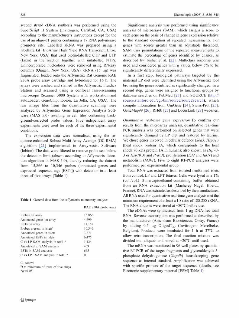
The growth and development of the fetus is a highlycontrolled process encoded in its genetic potential. Thisprocess is highly susceptible to changes in the intrauterineenvironment. Epidemiological studies have suggested anassociation between poor fetal or infant growth andincreased risk of developing glucose intolerance andmetabolic syndrome later in life [1, 2]. A recent reviewstudy suggested that inadequate maternal nutrition mightdisturb the development of the fetus, which must adoptstrategies to ensure survival that will programme its futurehealth [3]. In experimental animal models, alteration of theintrauterine environment induced, for example, by gesta-tional diabetes [4], placental insufficiency [5, 6] or poormaternal nutrition [7–11] compromise the development ofendocrine pancreas in the progeny and impact on its futurehealth even in the subsequent generation [4, 6, 12].Reduction of food intake by 50% in rats during gestationreduced the beta cell mass in offspring at birth by 30% [9].When maternal diet was reduced in protein content duringgestation, a number of findings were observed: reduction inbeta cell mass due to diminished proliferation [8, 13],enhanced apoptosis [11, 13], reduced insulin secretion invitro from isolated islets [14], impaired islet vascularisation[8, 15] and enhanced sensitivity to cytotoxic effects ofcytokines in vitro [11]. Increased susceptibility to cytokinecytotoxicity and lower insulin secretion persisted inadulthood despite limiting the low-protein (LP) diet to theperiod of gestation and lactation [7, 12]. Maternal and fetalplasma glucose levels were not altered by maternalmalnutrition, but the plasma amino acid profile wasperturbed both in the mother and their fetuses [16]. Theconcentration of taurine, a sulphur amino acid that does notparticipate in protein synthesis, was decreased in maternaland fetal plasma. Taurine is important during development[17]. Supplementation of the maternal LP diet with taurineto normalise the fetal plasma level corrected pancreasdevelopment, the insulin secretory deficiency and suscep-tibility to cytokines and toxins [11, 12, 15, 18, 19].
Previously, we have used proteomics to study fetal ratislet protein expression in animals fed a control or a LPdiet, demonstrating that expression of proteins involved in anumber of different biological pathways was modified by amaternal LP diet [20]. The present study aimed: (1) todetermine the changes in gene expression that are inducedby the maternal LP diet and may favour a ‘prediabeticphenotype’ and (2) to investigate to what extent and bywhich pathways maternal taurine supplementation restoresthe normal phenotype.
Methods
Animals Out-bred virgin female Wistar rats (Janvier, LeGenest Saint Isle, France) aged 3 months were cagedovernight with male rats for fertilisation. Copulation wasverified the next morning by detection of a vaginal plug.The animals were housed at 25°C with 10 h dark–14 h lightcycle. One group of pregnant rats was fed a normal controldiet (20% protein content), a second group an isoenergeticLP diet (8% [wt/wt] protein content) and a third was fed theLP diet supplemented with taurine (LPT) (2.5% [wt/wt];Sigma Chemical, Brussels, Belgium) in the drinking water.The composition of both diets has been described previ-ously [8]. Diets were purchased from Hope Farms(Woerden, The Netherlands). They were similar in fatcontent and rendered isoenergetic by addition of carbohy-drate to the LP diet. The rats were given free access to theirrespective diets. Pregnant rats in all three groups ate thesame quantity of food and drank the same volume of water.At day 21.5 of gestation, dams and their fetuses were killedby decapitation and the fetal pancreases were removed forislet isolation.
All animal experiments were carried out according tonational and international law and ethical standards. Theexperiments were approved by the Animal Ethics Commit-tees of the Université Catholique de Louvain, Belgium.
Isolation, culture and labelling of islets for microarrayanalysis Briefly, the pancreases were removed from 21.5-day-old control, LP and LPT fetuses, digested withcollagenase (specific activity 381 U/ml; Sigma, St Louis,MO, USA) and cultured for 7 days in RPMI 1640+10%(vol./vol.) fetal bovine serum and antibiotics (one pancreas/2 ml medium). From day 2 of culture, the medium wasreplaced every 24 h. After 7 days of culture, neoformedislets were handpicked as described elsewhere [14]. Thesecultures provided islets (90–95% of beta cells) thataggregated progressively on the layer of non-endocrinecells.
For microarray analyses as well as for RT-PCR and ATPanalyses, 15–19 pregnant rats, 200 fetuses, 12 cultures and10,000 neoformed islets were used per experimental group.Batches of 500 neoformed fetal islets were handpicked andtotal RNA was extracted by Trizol method and stored untiluse.
Gene expression analysis The following procedures wereall performed according to Affymetrix (Santa Clare, CA,USA) standard procedures. Briefly, 5 μg total RNA wasused as starting material for the target preparation. First and
Diabetologia (2008) 51:836–845 837
second strand cDNA synthesis was performed using theSuperScript II System (Invitrogen, Carlsbad, CA, USA)according to the manufacturer’s instructions except for theuse of an oligo-dT primer containing a T7 RNA polymerasepromoter site. Labelled aRNA was prepared using alabelling kit (BioArray High Yield RNA Transcript; Enzo,New York, USA) that used biotin-labelled CTP and UTP(Enzo) in the reaction together with unlabelled NTPs.Unincorporated nucleotides were removed using RNeasycolumns (Qiagen, New York, USA). cRNA (15 μg) wasfragmented, loaded onto the Affymetrix Rat Genome RAE230A probe array cartridge and hybridised for 16 h. Thearrays were washed and stained in the Affymetrix FluidicsStation and scanned using a confocal laser-scanningmicroscope (Scanner 3000 System with workstation andautoLoader; GeneChip; Iobion, La Jolla, CA, USA). Theraw image files from the quantitative scanning wereanalysed by Affymetrix Gene Expression Analysis Soft-ware (MAS 5.0) resulting in cell files containing back-ground-corrected probe values. Five independent arrayexperiments were used for each of the three experimentalconditions.
The expression data were normalised using the se-quence-enhanced Robust Multi-Array Average (GC-RMA)algorithm [21] implemented in ArrayAssist Software(Iobion). The data were filtered to remove probe sets belowthe detection limit (absent according to Affymetrix detec-tion algorithm in MAS 5.0), thereby reducing the datasetfrom 15,866 to 10,346 probes (annotated genes andexpressed sequence tags [ESTs]) with detection in at leastthree of five arrays (Table 1).
Significance analysis was performed using significanceanalysis of microarrays (SAM), which assigns a score toeach gene on the basis of change in gene expression relativeto the standard deviation of repeated measurements. Forgenes with scores greater than an adjustable threshold,SAM uses permutations of the repeated measurements toestimate the percentage of genes identified by chance, asdescribed by Tusher et al. [22]. Multiclass response wasused and considered genes with q values below 5% to besignificantly differentially expressed.
In a first step, biological pathways targeted by thematernal LP diet were identified using the Affymetrix toolbrowsing the genes identified as significantly changed. In asecond step, genes were assigned to functional groups bydatabase searches on PubMed [23] and SOURCE (http://source.stanford.edu/cgi-bin/source/sourceSearch), whichcompile information from UniGene [24], Swiss-Prot [25],GeneMap99 [26], RHdb [27] and LocusLink [28] websites.
Quantitative real-time gene expression To confirm ourresults from the microarray analysis, quantitative real-timePCR analysis was performed on selected genes that weresignificantly changed by LP diet and restored by taurine.We chose genes involved in cellular defence (Sod3, Hspa1a[heat shock protein 1A, which corresponds to the heatshock 70 kDa protein 1A in humans; also known as Hsp70-3 or Hsp70.3] and Prdx3), proliferation (Igf2 and Igf1r) andmetabolism (Mdh1). Five to eight RT-PCR analyses wereperformed per experimental group.
Total RNA was extracted from isolated neoformed isletsfrom control, LP and LPT fetuses. Cells were lysed in a 1%(vol./vol.) β-mercaptoethanol-containing buffer obtainedfrom an RNA extraction kit (Macherey Nagel, Hoerdt,France); RNAwas extracted as described by the manufacturer.All RNA used for quantitative real-time gene analysis met theminimum requirement of at least a 1.8 ratio of 18S:28S rRNA.The RNA aliquots were stored at −80°C before use.
The cDNAs were synthesised from 1 μg DNA-free totalRNA. Reverse transcription was performed as described bythe manufacturer (Amersham Biosciences, Orsay, France)by adding 0.5 μg OligodT20 (Invitrogen, Merelbeke,Belgium). Products were incubated for 1 h at 37°C toallow retro-transcription. The final reaction mixture wasdivided into aliquots and stored at −20°C until used.
The mRNA was monitored in 96-well plates by quantita-tive RT-PCR of the target fragments and glyceraldehyde-3-phosphate dehydrogenase (Gapdh) housekeeping genesequence as internal standard. Amplification was achievedwith specific primers of the target sequence (details, seeElectronic supplementary material [ESM] Table 1).
Table 1 General data from the Affymetrix microarray analyses
RAE 230A probe array
Probes on array 15,866Annotated genes on array 4,699ESTs on array 11,167Probes present in isletsa 10,346Annotated genes in islets 3,871Annotated ESTs in islets 6,475C vs LP SAM analysis in total * 1,124Annotated in SAM analysis 459ESTs in SAM analysis 665C vs LPT SAM analysis in total * 0
C, controla On minimum of three of five chips*p<0.05
838 Diabetologia (2008) 51:836–845
The template concentration per reaction represented one-tenth of the cDNA reaction performed on 1 μg total RNA.Amplification was achieved in 20 μl reaction mixturecontaining 2 μl cDNA, 25 pmol/l of each oligonucleotideprimer and 2× Sybr Green Master Mix (Westburg, Leusden,The Netherlands).
After activation of the hot-start DNA polymerase for15 min at 95°C, solutions underwent 40 cycles of ampli-fication in a sequence detection system (ABI PRISM 7000;Applied Biosystems, Lennik, Belgium). Amplificationparameters included 15 s denaturation at 94°C and a1 min annealing and extension step at 60°C. Directdetection of PCR products was monitored by measuringthe increase in fluorescence caused by the binding of SYBRGreen to double-stranded DNA. RT blank control PCRsshowed no product amplification for all genes examined inthis study (data not shown).
All quantifications were achieved with the comparativeCt method and normalised to the endogenous controlGapdh mRNAs as described by the manufacturer. Eachsample was analysed in duplicate.
Primers were designed using the software PrimerExpress (Applied Biosystems). For each gene of interest,at least three couples of primers were tested for efficacy.Primers were selected only when their efficacy, calculatedas described by the manufacturer, exceeded 95%.
For each gene of interest, samples of islet preparations ineach condition were amplified in 96-well plates concomi-tantly with specific primers for the gene of interest and forGapdh as internal standard. As described by the manufac-turer, quantification requires the calculation of: (1) themean Ct value of the replicate wells for each sample; (2)the difference between the mean Ct values of the samples inthe wells containing the gene of interest and those of theinternal standards (ΔCt); and (3) the difference between themean ΔCt values of the samples and the mean ΔCt valueof the control sample (ΔΔCt). The relative quantity valuewas expressed as 2�ΔΔCt.
ATP measurement Islets collected after 7 days of culturewere washed in KRB and incubated in KRB withoutglucose at 37°C in 5% CO2/O2 air for 60 min. Islets werethen divided into batches of 100 islets and transferred intodishes containing KRB with 3.3 or 16.7 mmol/l glucose.After 2 h incubation, islets were transferred in cold PBSand lysis buffer for ATP extraction was added. Islets weresonicated for 30 s. Samples were immediately placed onice. Islet lysate ATP content was measured using ATPluminescent assay kit (Promega, Madison, WI, USA) using50 μl in duplicate. ATP was calculated per μg protein in theislet homogenate. Five to seven independent experimentswere performed for each experimental group.
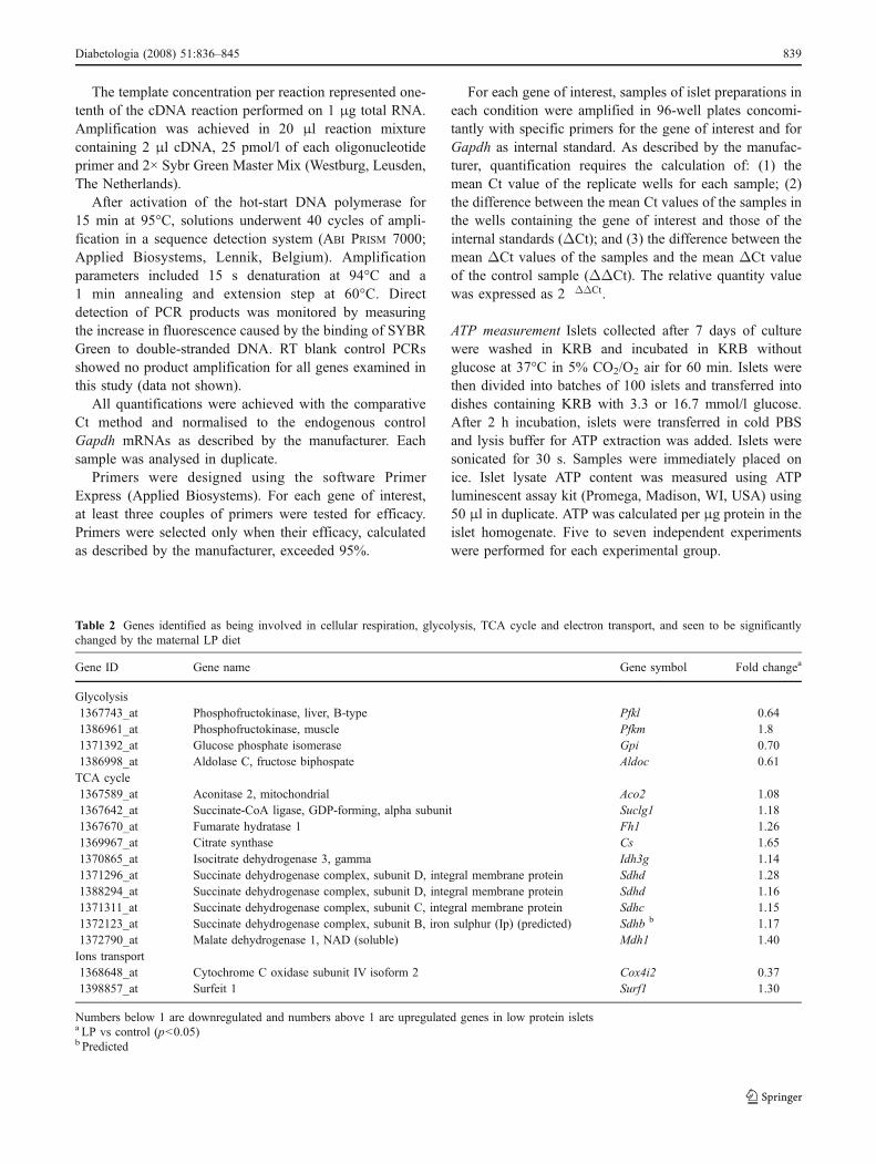
Table 2 Genes identified as being involved in cellular respiration, glycolysis, TCA cycle and electron transport, and seen to be significantlychanged by the maternal LP diet
Gene ID Gene name Gene symbol Fold changea
Glycolysis1367743_at Phosphofructokinase, liver, B-type Pfkl 0.641386961_at Phosphofructokinase, muscle Pfkm 1.81371392_at Glucose phosphate isomerase Gpi 0.701386998_at Aldolase C, fructose biphospate Aldoc 0.61TCA cycle1367589_at Aconitase 2, mitochondrial Aco2 1.081367642_at Succinate-CoA ligase, GDP-forming, alpha subunit Suclg1 1.181367670_at Fumarate hydratase 1 Fh1 1.261369967_at Citrate synthase Cs 1.651370865_at Isocitrate dehydrogenase 3, gamma Idh3g 1.141371296_at Succinate dehydrogenase complex, subunit D, integral membrane protein Sdhd 1.281388294_at Succinate dehydrogenase complex, subunit D, integral membrane protein Sdhd 1.161371311_at Succinate dehydrogenase complex, subunit C, integral membrane protein Sdhc 1.151372123_at Succinate dehydrogenase complex, subunit B, iron sulphur (Ip) (predicted) Sdhb b 1.171372790_at Malate dehydrogenase 1, NAD (soluble) Mdh1 1.40Ions transport1368648_at Cytochrome C oxidase subunit IV isoform 2 Cox4i2 0.371398857_at Surfeit 1 Surf1 1.30
Numbers below 1 are downregulated and numbers above 1 are upregulated genes in low protein isletsa LP vs control (p<0.05)b Predicted
Diabetologia (2008) 51:836–845 839
Results
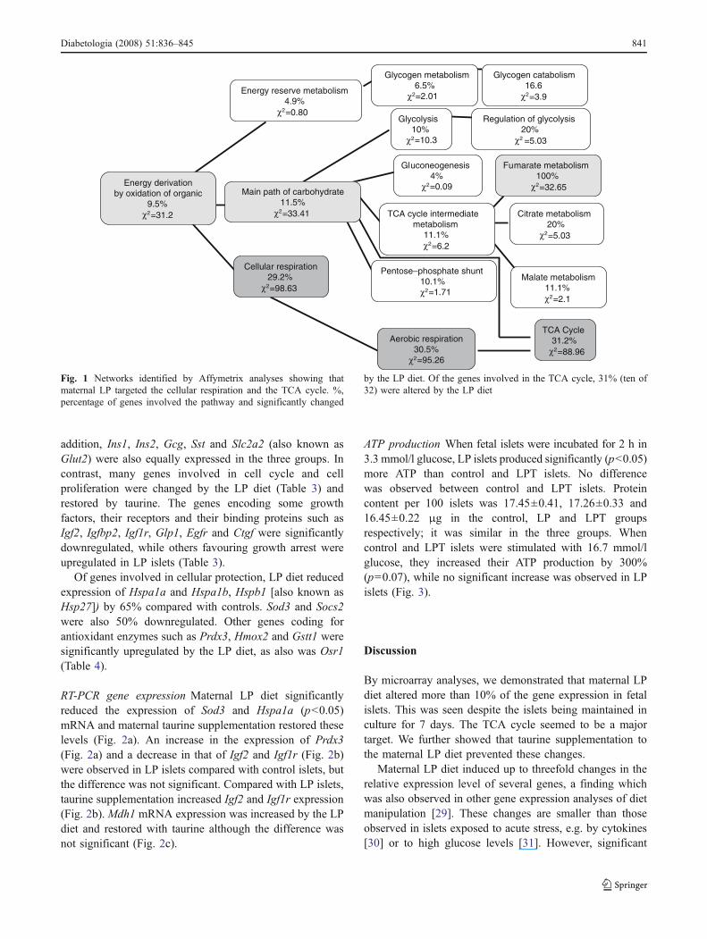
Microarray analysis Analysing the expressed mRNA in thefetal islets collected after 7 days of culture, we found that of15,866 RNAs probes, 10,346 were detectable in the islets(Table 1). Of the 10,346 probes present, 3,871 probesrepresented annotated genes and 6,475 ESTs. Maternal LPdiet significantly changed the expression (p<0.05) of 1,124genes and ESTs, of which 74.4% (836) were upregulatedand 25.6% (288) downregulated. The maximally increasedor decreased expression level was threefold (Tables 2, 3 and4). Every gene modified by the LP diet was restored bytaurine, since no significantly different gene expressionlevels were identified when comparing control and LPTislets.
First, we identified biological pathways targeted by thematernal LP diet using the Affymetrix tool, NetAffx AnalysisCenter, browsing the genes annotated that were significantly
(p<0.05) changed (n=459 genes; Table 1). This analysisstrongly suggested that the metabolism of the fetal beta cellwas dramatically altered (Fig. 1). Maternal LP diet affected5.1% of genes known to be involved in cellular metabolismpathways (256/5,019 genes). One of the most affectedpathways was cellular respiration (Table 2), in which 12genes were significantly (χ2 test=98.6) modulated by the LPdiet (Fig. 1). Eleven were upregulated and one, cytochromeC oxidase subunit IV isoform 2, was downregulated. Amongthese 12 genes, ten contribute to the tricarboxylic acid (TCA)cycle and were all significantly (χ2 test=88.96) upregulated(Fig. 1, Table 2). In addition, more than 10% of the 459genes identified as significantly changed in the LP diet isletsencoded for mitochondrial proteins.
Control as well as LP and LPT fetal islets expressedtranscriptional factors involved in beta cell differentiation.Pdx1, Hnf6, Hnf1, Ngn3, Nkx6, Pax4 and Pax6 genes wereall present and similarly expressed in the three groups. In
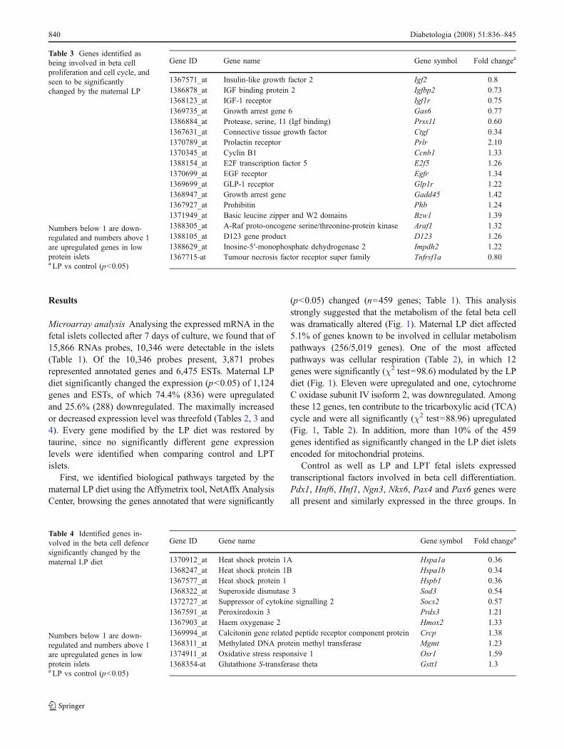
Table 3 Genes identified asbeing involved in beta cellproliferation and cell cycle, andseen to be significantlychanged by the maternal LP
Numbers below 1 are down-regulated and numbers above 1are upregulated genes in lowprotein isletsa LP vs control (p<0.05)
Gene ID Gene name Gene symbol Fold changea
1367571_at Insulin-like growth factor 2 Igf2 0.81386878_at IGF binding protein 2 Igfbp2 0.731368123_at IGF-1 receptor Igf1r 0.751369735_at Growth arrest gene 6 Gas6 0.771386884_at Protease, serine, 11 (Igf binding) Prss11 0.601367631_at Connective tissue growth factor Ctgf 0.341370789_at Prolactin receptor Prlr 2.101370345_at Cyclin B1 Ccnb1 1.331388154_at E2F transcription factor 5 E2f5 1.261370699_at EGF receptor Egfr 1.341369699_at GLP-1 receptor Glp1r 1.221368947_at Growth arrest gene Gadd45 1.421367927_at Prohibitin Phb 1.241371949_at Basic leucine zipper and W2 domains Bzw1 1.391388305_at A-Raf proto-oncogene serine/threonine-protein kinase Araf1 1.321388105_at D123 gene product D123 1.261388629_at Inosine-5′-monophosphate dehydrogenase 2 Impdh2 1.221367715-at Tumour necrosis factor receptor super family Tnfrsf1a 0.80
Table 4 Identified genes in-volved in the beta cell defencesignificantly changed by thematernal LP diet
Numbers below 1 are down-regulated and numbers above 1are upregulated genes in lowprotein isletsa LP vs control (p<0.05)
Gene ID Gene name Gene symbol Fold changea
1370912_at Heat shock protein 1A Hspa1a 0.361368247_at Heat shock protein 1B Hspa1b 0.341367577_at Heat shock protein 1 Hspb1 0.361368322_at Superoxide dismutase 3 Sod3 0.541372727_at Suppressor of cytokine signalling 2 Socs2 0.571367591_at Peroxiredoxin 3 Prdx3 1.211367903_at Haem oxygenase 2 Hmox2 1.331369994_at Calcitonin gene related peptide receptor component protein Crcp 1.381368311_at Methylated DNA protein methyl transferase Mgmt 1.231374911_at Oxidative stress responsive 1 Osr1 1.591368354-at Glutathione S-transferase theta Gstt1 1.3
840 Diabetologia (2008) 51:836–845
addition, Ins1, Ins2, Gcg, Sst and Slc2a2 (also known asGlut2) were also equally expressed in the three groups. Incontrast, many genes involved in cell cycle and cellproliferation were changed by the LP diet (Table 3) andrestored by taurine. The genes encoding some growthfactors, their receptors and their binding proteins such asIgf2, Igfbp2, Igf1r, Glp1, Egfr and Ctgf were significantlydownregulated, while others favouring growth arrest wereupregulated in LP islets (Table 3).
Of genes involved in cellular protection, LP diet reducedexpression of Hspa1a and Hspa1b, Hspb1 [also known asHsp27]) by 65% compared with controls. Sod3 and Socs2were also 50% downregulated. Other genes coding forantioxidant enzymes such as Prdx3, Hmox2 and Gstt1 weresignificantly upregulated by the LP diet, as also was Osr1(Table 4).
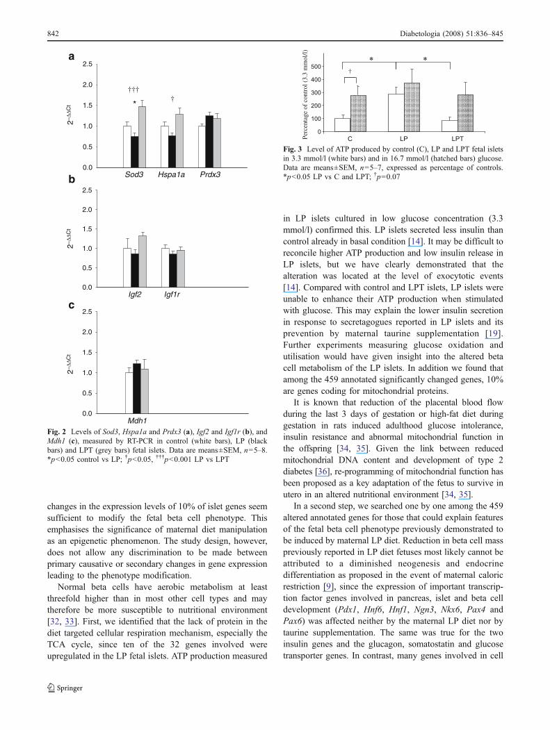
RT-PCR gene expression Maternal LP diet significantlyreduced the expression of Sod3 and Hspa1a (p<0.05)mRNA and maternal taurine supplementation restored theselevels (Fig. 2a). An increase in the expression of Prdx3(Fig. 2a) and a decrease in that of Igf2 and Igf1r (Fig. 2b)were observed in LP islets compared with control islets, butthe difference was not significant. Compared with LP islets,taurine supplementation increased Igf2 and Igf1r expression(Fig. 2b). Mdh1 mRNA expression was increased by the LPdiet and restored with taurine although the difference wasnot significant (Fig. 2c).
ATP production When fetal islets were incubated for 2 h in3.3 mmol/l glucose, LP islets produced significantly (p<0.05)more ATP than control and LPT islets. No differencewas observed between control and LPT islets. Proteincontent per 100 islets was 17.45±0.41, 17.26±0.33 and16.45±0.22 μg in the control, LP and LPT groupsrespectively; it was similar in the three groups. Whencontrol and LPT islets were stimulated with 16.7 mmol/lglucose, they increased their ATP production by 300%(p=0.07), while no significant increase was observed in LPislets (Fig. 3).
Discussion
By microarray analyses, we demonstrated that maternal LPdiet altered more than 10% of the gene expression in fetalislets. This was seen despite the islets being maintained inculture for 7 days. The TCA cycle seemed to be a majortarget. We further showed that taurine supplementation tothe maternal LP diet prevented these changes.
Maternal LP diet induced up to threefold changes in therelative expression level of several genes, a finding whichwas also observed in other gene expression analyses of dietmanipulation [29]. These changes are smaller than thoseobserved in islets exposed to acute stress, e.g. by cytokines[30] or to high glucose levels [31]. However, significant
Energy derivationby oxidation of organic
9.5% χ2 =31.2
Energy reserve metabolism 4.9%
χ2=0.80
Main path of carbohydrate11.5%
χ2=33.41
Cellular respiration29.2%
χ2=98.63
Glycogen metabolism6.5%
χ2=2.01
Glycolysis 10%
χ2=10.3
Gluconeogenesis4%
χ2 =0.09
TCA cycle intermediate metabolism
11.1% χ2 =6.2
Pentose–phosphate shunt 10.1% χ2=1.71
Aerobic respiration 30.5%
χ2=95.26
Glycogen catabolism16.6
χ2 =3.9
Regulation of glycolysis 20%
χ2 =5.03
Fumarate metabolism100%
χ2=32.65
Citrate metabolism20%
χ2 =5.03
Malate metabolism 11.1% χ2 =2.1
TCA Cycle31.2%
χ2=88.96
Fig. 1 Networks identified by Affymetrix analyses showing thatmaternal LP targeted the cellular respiration and the TCA cycle. %,percentage of genes involved the pathway and significantly changed
by the LP diet. Of the genes involved in the TCA cycle, 31% (ten of32) were altered by the LP diet
Diabetologia (2008) 51:836–845 841
changes in the expression levels of 10% of islet genes seemsufficient to modify the fetal beta cell phenotype. Thisemphasises the significance of maternal diet manipulationas an epigenetic phenomenon. The study design, however,does not allow any discrimination to be made betweenprimary causative or secondary changes in gene expressionleading to the phenotype modification.
Normal beta cells have aerobic metabolism at leastthreefold higher than in most other cell types and maytherefore be more susceptible to nutritional environment[32, 33]. First, we identified that the lack of protein in thediet targeted cellular respiration mechanism, especially theTCA cycle, since ten of the 32 genes involved wereupregulated in the LP fetal islets. ATP production measured
in LP islets cultured in low glucose concentration (3.3mmol/l) confirmed this. LP islets secreted less insulin thancontrol already in basal condition [14]. It may be difficult toreconcile higher ATP production and low insulin release inLP islets, but we have clearly demonstrated that thealteration was located at the level of exocytotic events[14]. Compared with control and LPT islets, LP islets wereunable to enhance their ATP production when stimulatedwith glucose. This may explain the lower insulin secretionin response to secretagogues reported in LP islets and itsprevention by maternal taurine supplementation [19].Further experiments measuring glucose oxidation andutilisation would have given insight into the altered betacell metabolism of the LP islets. In addition we found thatamong the 459 annotated significantly changed genes, 10%are genes coding for mitochondrial proteins.
It is known that reduction of the placental blood flowduring the last 3 days of gestation or high-fat diet duringgestation in rats induced adulthood glucose intolerance,insulin resistance and abnormal mitochondrial function inthe offspring [34, 35]. Given the link between reducedmitochondrial DNA content and development of type 2diabetes [36], re-programming of mitochondrial function hasbeen proposed as a key adaptation of the fetus to survive inutero in an altered nutritional environment [34, 35].
In a second step, we searched one by one among the 459altered annotated genes for those that could explain featuresof the fetal beta cell phenotype previously demonstrated tobe induced by maternal LP diet. Reduction in beta cell masspreviously reported in LP diet fetuses most likely cannot beattributed to a diminished neogenesis and endocrinedifferentiation as proposed in the event of maternal caloricrestriction [9], since the expression of important transcrip-tion factor genes involved in pancreas, islet and beta celldevelopment (Pdx1, Hnf6, Hnf1, Ngn3, Nkx6, Pax4 andPax6) was affected neither by the maternal LP diet nor bytaurine supplementation. The same was true for the twoinsulin genes and the glucagon, somatostatin and glucosetransporter genes. In contrast, many genes involved in cell
0
100
200
300
400
500
C
†
LP LPTPerc
enta
ge o
f co
ntro
l (3.
3 m
mol
/l)
* *
Fig. 3 Level of ATP produced by control (C), LP and LPT fetal isletsin 3.3 mmol/l (white bars) and in 16.7 mmol/l (hatched bars) glucose.Data are means±SEM, n=5–7, expressed as percentage of controls.*p<0.05 LP vs C and LPT; †p=0.07Sod3 Hspa1a Prdx3
0.0
0.5
1.0
1.5
2.0
2.5
Igf2 Igf1r
2–∆∆
Ct
0.0
0.5
1.0
1.5
2.0
2.5
Mdh1
2–∆∆
Ct
0.0
0.5
1.0
1.5
2.0
2.52–
∆∆C
t *
††††
a
b
c
Fig. 2 Levels of Sod3, Hspa1a and Prdx3 (a), Igf2 and Igf1r (b), andMdh1 (c), measured by RT-PCR in control (white bars), LP (blackbars) and LPT (grey bars) fetal islets. Data are means±SEM, n=5–8.*p<0.05 control vs LP; †p<0.05, †††p<0.001 LP vs LPT
842 Diabetologia (2008) 51:836–845
cycle and cell proliferation were changed by the LP dietand their expression restored by taurine. This may fit inwith the lengthening of the beta cell cycle and the lowerproliferation rate that we previously described in fetusesfrom dams fed an LP diet [8, 13, 37].
Evidence for the involvement of the IGF gene/proteinfamily in beta cell development is abundant. IGFs aremitogenic for beta cell and act as cell survival factors byinhibiting apoptosis [38]. IGFs are perturbed in plasma andhepatocytes of fetus from LP-fed mothers [39]. Thepancreatic expression of Igf2 mRNA and protein was lowerin LP fetus and neonates [13]. By microarray analyses, wefound that Igf2, Igf1r and Igfbp2 were downregulated in LPislets and restored by taurine. IGF-2 exerts its mitogenicand antiapoptotic action via its binding to the IGF-1receptor. IGF-1 receptor knockout mice have 50% reduc-tion in their beta cell mass [40] and deletion of the IGF-1and insulin receptors in the beta cell induced reduced betacell mass and increased apoptosis and default in insulinsecretion, three alterations that were also observed in the LPfetal islets [8, 11, 14].
Normal beta cells are equipped with lower antioxidantdefences than others cell types [41] and are therefore morevulnerable to reactive oxygen, nitrogen species and cyto-kines. Beta cells from LP fetuses feature an increased rateof apoptosis after exposure to interleukin 1β and nitricoxide [11]. This might be caused by reduction in expressionof Hspa1a, Hspa1b, Hspb1 and Sod3 as observed in themicroarray, Hspa1a being confirmed by RT-PCR. Theincreased expression of Osr1 could contribute to the highervulnerability. Ten proteins involved in protein folding andchaperoning were found to be altered by the LP diet inproteomic analyses [20]. The microarray analysis alsorevealed that increased expression of Hmox2, Prdx3 andMgmt involved cellular defence and DNA repair. Crcp,which is known to be upregulated in response to inflam-mation, was upregulated in the LP islets. This may be takenas proxy of increased oxidative stress in these islets.Interestingly, islets from adult offspring of mothers fed an8% protein diet secrete more nitric oxide then control islets,even in absence of cytokines [42]. These findings suggestthat the maternal LP diet promotes a fetal beta cellphenotype with increased susceptibility to cytokines, betacell toxins and oxidative stress.
The simple supplementation of maternal diet withtaurine abolished the effects of the LP diet on geneexpression. We have previously shown that maternaltaurine supplementation of the LP diet preserved the normalfetal beta cell phenotype [11, 12, 15, 18, 19]. No singlegene or specific pathways responsible for the taurine effectcould be identified, since every gene had an expressionlevel similar to that in control islets. Furthermore, thepreventive effect of taurine supplementation cannot be
accounted for by a change in the expression of Slc6a6, thegene encoding for the taurine transporter, since it waspresent but not changed either by the LP diet or by taurine.
Hence, we have no explanation for the preventive actionof taurine. Taurine prevented the changes in expression ofgenes involved in metabolism and the TCA cycle, and ATPproduction was completely normalised. Taurine might beincorporated into mitochondria through a putative mito-chondrial taurine transporter [43]. In beta cells overexpress-ing uncoupling protein 2, taurine increased mitochondrialCa2+ influx through the Ca2+ uniporter, thereby enhancingmitochondrial metabolic function and increasing the ATP/ADP ratio [44]. It is important to note that addition oftaurine to the LP diet normalised islet ATP production inthe presence of low and high glucose concentration. It hasbeen shown by others that increased reactive oxygenspecies production in hyperglycaemic adult rats infusedwith glucose may be prevented by taurine co-infusion [45].Therefore, the taurine effects we reported might bemediated by normalisation of mitochondrial metabolism.Interestingly, a recent report suggested that taurine couldcritically affect mitochondrial function. Yasukawa et al.[46] found two novel taurine-containing modified uridinesin human mitochondrial DNA. When the uridine modifica-tions were not present, defective mitochondrial functionoccurred and diseases such as mitochondrial myopathy,encephalopathy, lactic acidosis and stroke-like episodes(MERRF) syndrome and myoclonic epilepsy with raggedred fibres (MELAS) syndrome may ensue.
In conclusion, this study demonstrates that reduction ofprotein intake during pregnancy has considerable impact atthe level of the fetal insulin-secreting cell and affectsexpression of more than 10% of its genes. Cellular respirationseems to be the major target. Our finding that expressionchanges persist in culture after exposure to abnormal maternalmetabolic environment supports the notion that the intrauter-ine programming of gene expression is involved. Importantly,LP diet-induced changes in gene expression can be fullyprevented by adding taurine to the maternal LP diet. Thisobservation emphasises that taurine plays an important role inthe development of the endocrine pancreas and needs to befurther investigated. One question that remains is: is thealtered phenotype that we observed the consequence of fewmodifications of gene expression leading to many beta cellsecondary effects, or does it result from induction of a widenetwork of relatively minor imbalances?
Acknowledgements This work was supported by the EuropeanCommission (NUTRIX QLK1–2000–00083, 5th Frame Programme),the Parthenon Trust (London, UK), Fonds de la Recherche Scientifi-que (FNRS), The Danish Diabetes Association, Novo Nordisk A/S,Denmark, the Michaelsen Foundation and University of Copenhagen.
Diabetologia (2008) 51:836–845 843

Duality of interest The authors declare that there is no duality ofinterest associated with this manuscript.
References
1. Hales CN, Barker DJ, Clark PM et al (1991) Fetal and infantgrowth and impaired glucose tolerance at age 64. BMJ 303:1019–1022
2. Hales CN, Barker DJP (2001) The thrifty phenotype hypothesis.Br Med Bull 60:5–20
3. McMillen C, Robinson JS (2005) Developmental origins of themetabolic syndrome: prediction, plasticity and programming.Physiol Rev 85:571–633
4. Aerts L, Van Assche FA (2006) Animal evidence for thetransgenerational development of diabetes mellitus. Int J BiochemCell Biol 38:894–903
5. Simmons RA, Templeton LJ, Gertz SJ (2001) Intrauterine growthretardation leads to the development of type 2 diabetes in the rat.Diabetes 50:2279–2286
6. Boloker J, Shira JG, Simmons R (2002) Gestational diabetes leadsto the development of diabetes in adulthood in the rat. Diabetes51:1499–1506
7. Dahri S, Snoeck A, Reusens B, Remacle C, Hoet JJ (1991) Isletfunction in offspring of mothers on low protein diet duringgestation. Diabetes 40:115–120
8. Snoeck A, Remacle C, Reusens B, Hoet JJ (1990) Effect of lowprotein diet during pregnancy on the fetal rat endocrine pancreas.Biol Neonate 57:107–118
9. Garofano A, Czernichow P, Bréant B (1997) In uteroundernutrition impairs rat b-cell development. Diabetologia40:1231–1234
10. Garofano A, Czernichow P, Bréant B (1998) Beta-cell mass andproliferation following late fetal and early postnatal malnutritionin the rat. Diabetologia 34:373–384
11. Merezak S, Hardikar A, Yajnick CS, Remacle C, Reusens B (2001)Intrauterine low protein diet increases fetal b cell sensitivity to NOand IL-1-b: the protective role of taurine. J Endocrinol 171:299–308
12. Merezak S, Reusens B, Ahn MT, Remacle C (2004) Effect ofmaternal low protein diet and taurine on the vulnerability of adultWistar rat islets to cytokines. Diabetologia 7:669–675
13. Petrik J, Reusens B, Arany E et al (1999) A low protein dietalters the balance of islet cell replication and apoptosis in thefetal and neonatal rat and is associated with a reduced pancreaticexpression of insulin-like growth factor II. Endocrinology14:4861–4873
14. Cherif H, Reusens B, Dahri S, Remacle C (2001) A proteinrestricted diet during pregnancy alters in vitro insulin secretionfrom islets of fetal Wistar rats. J Nutr 131:1555–1559
15. Boujendar S, Arany E, Hill D, Remacle C, Reusens B (2003)Taurine supplementation of a low protein diet fed to rat damsnormalized the vascularization of the fetal endocrine pancreas.J Nutr 133:2820–2825
16. Reusens B, Dahri S, Snoeck A, Bennis-Taleb N, Remacle C, HoetJJ (1995) Long-term consequences of diabetes and its complica-tions may have a fetal origin: experimental evidence. In: CowettRM (ed) Diabetes. Nestlé Nutrition Workshop Series, Vol 25.Vevey/Raven, New York, pp 187–198
17. Sturman GA (1993) Taurine in development. Physiol Rev 73:119–147
18. Boujendar S, Reusens B, Ahn MT, Arany E, Hill DJ, Remacle C(2002) Taurine supplementation to a low protein diet during fetaland early postnatal life restores a normal proliferation andapoptosis of rat pancreatic islets. Diabetologia 45:856–866
19. Cherif H, Reusens B, Ahn MT, Hoet JJ, Remacle C (1998) Effectsof taurine on the insulin secretion of rat fetal islets from dams feda low protein diet. J Endocrinol 159:341–348
20. Sparre T, Reusens B, Cherif H et al (2003) Intrauterineprogramming of fetal islet gene expression in rats―effects ofmaternal protein restriction during gestation revealed by proteomeanalysis. Diabetologia 46:1497–1511
21. Wu Z, Irizarry RA (2004) Preprocessing of oligonucleotide arraydata. Nat Biotechnol 22:656–658
22. Tusher VG, Tibshirani R, Chu G (2001) Significance analysis ofmicroarrays applied to the ionizing radiation response. Proc NatlAcad Sci USA 98:5116–5121
23. PubMed at the National Center for Biotechnology Information(NCBI). http://www.ncbi.nlm.nih.gov/entrez/query.fcgi?CMD=search&DB=pubmed. Accessed 9 January 2006
24. UniGene at the National Center for Biotechnology Information(NCBI). http://www.ncbi.nlm.nih.gov/entrez/query.fcgi?db=unigene. Accessed 9 January 2006
25. Swiss-Prot ExPASy Molecular Biology Server. http://www.expasy.org. Accessed 9 January 2006
26. GeneMap99 at the National Center for Biotechnology Information(NCBI). http://www.ncbi.nlm.nih.gov/projects/genome/genemap99/. Accessed 9 January 2006
27. RHdb at the European Bioinformatics Institute (EMBL-EBI).http://srs.ebi.ac.uk/srsbin/cgi-bin/wgetz?-page+srsq2+-noSession.Accessed 9 January 2006
28. LocusLink at the National Center for Biotechnology Information.Previously available from http://www.ncbi.nlm.nih.gov/entrez/query.fcgi?db=unigene, discontinued 1 March 2005
29. Sparks LM, Xie H, Koza RA et al (2005) A high-fat dietcoordinately downregulates genes required for mitochondrialoxidative phosphorylation in skeletal muscle. Diabetes 54:1926–1933
30. Nielsen K, Kruhoffer F, Orntorft T et al (2004) Gene expressionprofiles during beta cell maturation and after IL-1beta exposurereveal important roles of Pdx1 and Nkx6.1 for IL-1betasensitivity. Diabetologia 47:2185–2199
31. Shalev A, Pise-Masison CA, Radonovich M et al (2002)Oligonucleotide microarray analysis of intact human pancreaticislets: identification of glucose responsive genes and highlyregulated TGFb signaling pathway. Endocrinology 143:3695–3698
32. Sekine N, Cirulli V, Regazzi R et al (1994) Low lactatedehydrogenase and high mitochondrial glycerol phosphate dehy-drogenase in pancreatic b-cells. Potential role in nutrient sensing.J Biol Chem 269:4895–4902
33. Schuit F, De Vos A, Farfari S et al (1997) Metabolic fate ofglucose in purified islet cells. Glucose-regulated anaplerosis in b-cell. J Biol Chem 272:18572–18579
34. Taylor PD, McConnell J, Khan IY et al (2005) Impaired glucosehomeostasis and mitochondrial abnormalities in offspring of ratsfed a fat-rich diet in pregnancy. Am J Physiol Regul Integr CompPhysiol 288:134–139
35. Peterside IE, Selak MA, Simmons RA (2003) Impaired oxidativephosphorylation in hepatic mitochondria in growth-retarded rats.Am J Physiol Endocrinol Metab 285:E1258–E1256
36. Lee YY, Park KS, Pak YK, Lee HK (2005) The role ofmitochondrial DNA in the development of type 2 diabetes causedby fetal malnutrition. J Nutr Biochem 16:195–204
37. Dumortier O, Blondeau B, Duvillié B, Reusens B, Bréant B,Remacle C (2007) Different mechanisms operating during differentcritical time-windows reduce rat fetal beta cell mass due to amaternal low-protein or low-energy diet. Diabetologia 50:2495–2503
38. Hill DJ, Petrik J, Arany E (1998) Growth factors in the regulationin fetal growth. Diabetes Care (Suppl 2):B60–B69
844 Diabetologia (2008) 51:836–845
39. El-Khattabi I, Grégoire F, Remacle C, Reusens B (2003)Isocaloric maternal low protein diet alters IGF-I, IGFBPs, andhepatocyte proliferation in the fetal rat. Am J Physiol EndocrinolMetab 285:E991–E1000
40. Withers DJ, Burks DJ, Towery HH, Altamuro SL, Flint CL, WhiteMF (1999) Irs-2 coordinates Igf-1 receptor-mediated beta-celldevelopment and peripheral insulin signalling. Nat Genet 23:32–40
41. Tiedge M, Lortz S, Drinkgern J, Lenzen S (1997) Relationbetween antioxidant enzyme gene expression and antioxidativedefence status of insulin-producing cells. Diabetes 46:1733–1742
42. Goosse K, Balteau M, Reusens B, Remacle C (2006) Search formolecules involved in the increased vulnerability of adult b-cellsafter a maternal low protein diet. Prevention by taurine.Diabetologia 49(Suppl 1):266–267
43. Suzuki T, Wada T, Saigo K, Watanabe K (2002) Taurine as aconstituent of mitochondria tRNAs: new insight into the functionsof taurine and human mitochondrial diseases. EMBO J 21:6581–6589
44. Han J, Bae JH, Kim SY et al (2004) Taurine increases glucosesensitivity of UCP2-overexpressing b-cells by amelioratingmitochondrial metabolism. Am J Physiol Endocrinol Metab 287:E1008–E1018
45. Tang C, Han P, Oprescu A et al (2007) Evidence for a role ofsuperoxide generation in glucose induced b-cell dysfunction invivo. Diabetes 56:2722–2731
46. Yasukawa T, Kirino Y, Ishii N et al (2005) Wobble modificationdeficiency in mutant tRNA in patients with mitochondrialdiseases. FEBS Lett 23:2948–2952
Diabetologia (2008) 51:836–845 845