Mitochondrial regulation of insulin production in rat pancreatic islets
11
Diabetologia (2005) 48: 1549–1559 DOI 10.1007/s00125-005-1811-6 ARTICLE G. Leibowitz . M. Z. Khaldi . A. Shauer . M. Parnes . A. I. Oprescu . E. Cerasi . J.-C. Jonas . N. Kaiser Mitochondrial regulation of insulin production in rat pancreatic islets Received: 13 December 2004 / Accepted: 25 March 2005 / Published online: 29 June 2005 # Springer-Verlag 2005 Abstract Aims/hypothesis: The study was designed to identify the key metabolic signals of glucose-stimulated proinsulin gene transcription and translation, focusing on the mechanism of succinate stimulation of insulin production. Methods: Wistar rat islets were incubated in 3.3 mmol/l glucose with and without esters of different mitochondrial metabolites or with 16.7 mmol/l glucose. Proinsulin biosynthesis was analysed by tritiated leucine incorporation into newly synthesised proinsulin. Preproin- sulin gene transcription was evaluated following transduc- tion with adenoviral vectors expressing the luciferase reporter gene under the control of the rat I preproinsulin promoter. Steady-state preproinsulin mRNA was deter- mined using relative quantitative PCR. The mitochondrial membrane potential was measured by microspectrofluori- metry using rhodamine-123. Results: Succinic acid mono- methyl ester, but not other mitochondrial metabolites, stimulated preproinsulin gene transcription and translation. Similarly to glucose, succinate increased specific prepro- insulin gene transcription and biosynthesis. The inhibitor of succinate dehydrogenase (SDH), 3-nitropropionate, abolished glucose- and succinate-stimulated mitochondrial membrane hyperpolarisation and proinsulin biosynthesis, indicating that stimulation of proinsulin translation de- pends on SDH activity. Partial inhibition of SDH activity by exposure to fumaric acid monomethyl ester abolished the stimulation of preproinsulin gene transcription, but only partially inhibited the stimulation of proinsulin bio- synthesis by glucose and succinate, suggesting that SDH activity is particularly important for the transcriptional re- sponse to glucose. Conclusions/interpretation: Succinate is a key metabolic mediator of glucose-stimulated prepro- insulin gene transcription and translation. Moreover, suc- cinate stimulation of insulin production depends on its metabolism via SDH. The differential effect of fumarate on preproinsulin gene transcription and translation sug- gests that these processes have different sensitivities to metabolic signals. Keywords Insulin secretion . Islets . Mitochondria . Proinsulin biosynthesis . Preproinsulin gene transcription . Rat . Succinate . Succinate dehydrogenase Abbreviations BCH: 2(+/−)-2-aminobicyclo-(2,2,1) heptane-2-carboxylic acid . CMV: cytomegalovirus . DCIP: 2,6-dichloroindophenol . DMM: malic acid dimethyl ester . FAM: fumaric acid monoethyl ester . FBS: fetal bovine serum . FCCP: carbonyl cyanide p-(trifluoromethoxy) phenylhydrazone . KRBH: Krebs– Ringer–bicarbonate–HEPES . NPA: 3-nitropropionate . PDX-1: pancreatic-duodenal homeobox 1 . SAM: succinic acid monomethyl ester . SDH: succinate dehydrogenase . TCA: tricarboxylic acid Introduction Glucose is the major physiological stimulus for insulin secretion and production in pancreatic beta cells. Tight coupling between secretion and production of insulin in response to nutrient stimuli is essential to maintain a pancreatic insulin reserve for normal glucose homeostasis. It is well established that glucose must be metabolised in beta cells to provide signals for both secretion and pro- duction of insulin [1–3]. Glucose is converted via gly- colysis to pyruvate, which enters the mitochondria and is metabolised in the tricarboxylic acid (TCA) cycle. Pyru- vate carbon metabolism leads to generation of ATP and hence to an increased cytosolic ATP : ADP ratio, which is G. Leibowitz (*) . A. Shauer . M. Parnes . A. I. Oprescu . E. Cerasi . N. Kaiser Endocrinology and Metabolism Service, Department of Medicine, Hadassah-Hebrew University Medical Center, P.O. Box 12000, Jerusalem, 91120, Israel e-mail: [email protected] Tel.: +972-2-6777951 Fax: +972-2-6437940 M. Z. Khaldi . J.-C. Jonas Unit of Endocrinology and Metabolism, University of Louvain Faculty of Medicine, Brussels, Belgium
-
Upload
independent -
Category
Documents
-
view
1 -
download
0
Transcript of Mitochondrial regulation of insulin production in rat pancreatic islets
Diabetologia (2005) 48: 1549–1559DOI 10.1007/s00125-005-1811-6
ARTICLE
G. Leibowitz . M. Z. Khaldi . A. Shauer . M. Parnes .A. I. Oprescu . E. Cerasi . J.-C. Jonas . N. Kaiser
Mitochondrial regulation of insulin production in ratpancreatic islets
Received: 13 December 2004 / Accepted: 25 March 2005 / Published online: 29 June 2005# Springer-Verlag 2005
Abstract Aims/hypothesis: The study was designed toidentify the key metabolic signals of glucose-stimulatedproinsulin gene transcription and translation, focusingon the mechanism of succinate stimulation of insulinproduction. Methods: Wistar rat islets were incubated in3.3 mmol/l glucose with and without esters of differentmitochondrial metabolites or with 16.7 mmol/l glucose.Proinsulin biosynthesis was analysed by tritiated leucineincorporation into newly synthesised proinsulin. Preproin-sulin gene transcription was evaluated following transduc-tion with adenoviral vectors expressing the luciferasereporter gene under the control of the rat I preproinsulinpromoter. Steady-state preproinsulin mRNA was deter-mined using relative quantitative PCR. The mitochondrialmembrane potential was measured by microspectrofluori-metry using rhodamine-123. Results: Succinic acid mono-methyl ester, but not other mitochondrial metabolites,stimulated preproinsulin gene transcription and translation.Similarly to glucose, succinate increased specific prepro-insulin gene transcription and biosynthesis. The inhibitorof succinate dehydrogenase (SDH), 3-nitropropionate,abolished glucose- and succinate-stimulated mitochondrialmembrane hyperpolarisation and proinsulin biosynthesis,indicating that stimulation of proinsulin translation de-pends on SDH activity. Partial inhibition of SDH activityby exposure to fumaric acid monomethyl ester abolishedthe stimulation of preproinsulin gene transcription, butonly partially inhibited the stimulation of proinsulin bio-
synthesis by glucose and succinate, suggesting that SDHactivity is particularly important for the transcriptional re-sponse to glucose. Conclusions/interpretation: Succinateis a key metabolic mediator of glucose-stimulated prepro-insulin gene transcription and translation. Moreover, suc-cinate stimulation of insulin production depends on itsmetabolism via SDH. The differential effect of fumarateon preproinsulin gene transcription and translation sug-gests that these processes have different sensitivities tometabolic signals.
Keywords Insulin secretion . Islets . Mitochondria .Proinsulin biosynthesis . Preproinsulin gene transcription .Rat . Succinate . Succinate dehydrogenase
Abbreviations BCH: 2(+/−)-2-aminobicyclo-(2,2,1)heptane-2-carboxylic acid . CMV: cytomegalovirus .DCIP: 2,6-dichloroindophenol . DMM: malic aciddimethyl ester . FAM: fumaric acid monoethyl ester .FBS: fetal bovine serum . FCCP: carbonyl cyanidep-(trifluoromethoxy) phenylhydrazone . KRBH: Krebs–Ringer–bicarbonate–HEPES . NPA: 3-nitropropionate .PDX-1: pancreatic-duodenal homeobox 1 . SAM: succinicacid monomethyl ester . SDH: succinate dehydrogenase .TCA: tricarboxylic acid
Introduction
Glucose is the major physiological stimulus for insulinsecretion and production in pancreatic beta cells. Tightcoupling between secretion and production of insulin inresponse to nutrient stimuli is essential to maintain apancreatic insulin reserve for normal glucose homeostasis.It is well established that glucose must be metabolised inbeta cells to provide signals for both secretion and pro-duction of insulin [1–3]. Glucose is converted via gly-colysis to pyruvate, which enters the mitochondria and ismetabolised in the tricarboxylic acid (TCA) cycle. Pyru-vate carbon metabolism leads to generation of ATP andhence to an increased cytosolic ATP : ADP ratio, which is
G. Leibowitz (*) . A. Shauer . M. Parnes . A. I. Oprescu .E. Cerasi . N. KaiserEndocrinology and Metabolism Service,Department of Medicine,Hadassah-Hebrew University Medical Center,P.O. Box 12000, Jerusalem, 91120, Israele-mail: [email protected].: +972-2-6777951Fax: +972-2-6437940
M. Z. Khaldi . J.-C. JonasUnit of Endocrinology and Metabolism,University of Louvain Faculty of Medicine,Brussels, Belgium
crucial for insulin secretion [4, 5] and biosynthesis [3].Thus, the energetic state of the beta cell is an importantcomponent of the stimulus–secretion coupling mechanismof glucose-induced insulin production and secretion. Fur-thermore, efflux and refilling of TCA cycle intermediates(cataplerosis and anaplerosis) may generate additional im-portant signals [2, 6, 7].
In normal beta cells, nutrient-induced insulin secretion isalways associated with a marked increase in insulin pro-duction. However, the kinetics of insulin release and ofproinsulin biosynthesis, and the metabolic signals mediat-ing these processes, are not identical [3, 8]. In fact, the roleof various glycolytic and TCA cycle intermediates inglucose-stimulated insulin secretion is controversial [7, 9–16], and even less is known about the metabolic signalsfor glucose-stimulated insulin production. A previous re-port by Alarcon et al. suggested that succinate is the keysignal for glucose stimulation of preproinsulin mRNAtranslation [3]. In that study, inhibition of the mitochon-drial conversion of succinate to fumarate via succinatedehydrogenase (SDH) stimulated proinsulin biosynthesis.Moreover, it was shown that cytosolic succinate could beconverted to succinyl CoA, which led to the speculationthat succinyl CoA is responsible for the rapid stimulation ofproinsulin biosynthesis in response to glucose [3].
We and others have shown previously that glucose has adual stimulatory effect on insulin production. On the onehand, it rapidly stimulates preproinsulin mRNA transla-tion, leading to a marked increase in proinsulin biosynthe-sis within less than 1 h of glucose stimulation. This rapidincrease in proinsulin biosynthesis is not dependent onpreproinsulin gene transcription and is not mediated bythe secreted insulin [17, 18]. On the other hand, glucosestimulates preproinsulin gene transcription, leading to atime-dependent increase in preproinsulin mRNA levels,which seems to be essential for the maintenance of pro-insulin biosynthesis and islet insulin stores during pro-longed stimulation [19]. The identity of the metabolicsignals that mediate the glucose effect on preproinsulingene transcription is not known.
In the present study we investigated the mechanisminvolved in succinate stimulation of insulin production. Wefound that, in addition to its previously reported stimula-tory effect on preproinsulin mRNA translation [3], succi-nate is a key mediator of glucose-stimulated preproinsulingene transcription. In contrast to previously published data,we demonstrate that succinate stimulation of insulin pro-duction is dependent on succinate metabolism via SDH,rather than being the consequence of a direct effect ofsuccinate itself.
Materials and methods
Islet isolation and culture Male Wistar rats were obtainedfrom Harlan (Jerusalem, Israel). Islets were prepared by
Fig. 1 Dose–response curves for the stimulation by succinate ofa insulin secretion, b proinsulin biosynthesis, c total protein bio-synthesis, and d specific proinsulin biosynthesis (corrected for totalprotein biosynthesis). Rat islets were incubated for 1 h at 37°C with3.3 mmol/l glucose (G3.3) alone or supplemented with differentconcentrations of succinic acid monomethyl ester (SAM) (0.5–10 mmol/l), 10 mmol/l malic acid dimethyl ester (DMM) or with16.7 mmol/l glucose (G16.7). Insulin secretion and tritiated leucineincorporation into proinsulin and total protein were determined asdescribed under Materials and methods. Results are means±SEM forfour or five individual experiments, each performed on islets pooledfrom three animals. Results are normalised to 3.3 mmol/l glucose.**p<0.01 and ***p<0.001 relative to islets at 3.3 mmol/l glucose
1550
collagenase digestion (Collagenase P; Roche Diagnostics,Mannheim, Germany) as described [20]. When largequantities were required, islets were hand-picked onceunder the stereomicroscope, followed by purification onHistopaque 1083 density gradient (Sigma, St Louis, MO,USA). The islets were used after repeated washes withHanks’ balanced salt solution, unless otherwise specified.Batches of 200–300 islets of similar size were collectedand maintained at 37°C in a 5% CO2 atmosphere in sus-pension in 5 ml RPMI 1640 medium (Biological In-dustries, Beit-Haemek, Israel), containing 3.3 mmol/l or16.7 mmol/l glucose, 10% fetal bovine serum (FBS; Bi-ological Industries) and different agents according to theexperimental protocols. The use of animals was approvedby the Institutional Animal Care and Use Committee ofthe Hebrew University and Hadassah Medical Organiza-tion, and principles of laboratory animal care (NIH Pub-lication no. 85–23, revised 1985) were followed.
Experimental protocols Isolated islets were cultured in thepresence of 3.3 or 16.7 mmol/l glucose with and with-out different concentrations of esters of the mitochondrialmetabolites: succinate (succinic acid monomethyl ester,SAM), fumarate (fumaric acid monoethyl ester, FAM),glutamate (glutamic acid methyl ester), oxaloacetate (ox-aloacetic acid diethyl ester) and malate (malic acid di-methyl ester, DMM). In other experiments, islets weretreated with 10 mmol/l glutamine together with 10 mmol/l2(+/−)-2-aminobicyclo-(2, 2, 1)heptane-2-carboxylic acid(BCH), which allosterically activates glutamate dehydrog-enase [21], thereby stimulating mitochondrial metabolism.To study the role of SDH we used the SDH inhibitor 3-nitropropionate (NPA). Islets were treated with the dif-ferent mitochondrial metabolites and/or NPA for 1 and 24 hto study their short- and long-term effects on insulinsecretion and on preproinsulin gene transcription andtranslation. All esters of mitochondrial metabolites, gluta-mine, BCH and NPA were purchased from Sigma.
Proinsulin biosynthesis For the short-term experiments,islets were cultured in RPMI 1640 medium containing3.3 mmol/l glucose for 3 h to allow recovery from thecollagenase isolation, whereas for long-term experimentsthey were cultured for 23 h with RPMI 1640 containing3.3 or 16.7 mmol/l glucose and test compounds, as in-dicated. Groups of 25 islets were collected followingculture and incubated for 1 h at 37°C in modified Krebs–Ringer bicarbonate buffer containing 20 mmol/l HEPESand 0.25% BSA (KRBH-BSA), supplemented with glu-cose and test compounds [19]. After incubation, the isletswere centrifuged and labelled in 50 μl fresh KRBH-BSAbuffer containing glucose and test compounds, as above,as well as 25 μCi L-[4,5-3H]leucine (150 Ci per mmol/l;Amersham, Aylesbury, UK). Leucine incorporation wasterminated after 15 min of labelling at 37°C by the ad-dition of 1 ml ice-cold glucose-free KRBH-BSA bufferand rapid centrifugation. The islet pellet was suspended in450 μl of 0.2 mol/l glycine buffer containing 0.1% RIA-grade BSA and 0.5% NP-40, pH 8.8 (GB/NP40 buffer)and subjected to four freeze–thaw cycles in liquid ni-trogen. Each sample (50 μl) was pretreated with protein ASepharose (Sigma) prior to immunoprecipitation with anti-insulin serum (Sigma), to correct for non-specific binding.This procedure was validated by HPLC. Aliquots wereused for determination of total insulin content by RIA andfor measurement of total protein biosynthesis by trichlo-roacetic acid precipitation [22].
Insulin release and content during culture Batches of isletswere incubated for 1 h in KRBH-BSA containing variousglucose concentrations and test compounds. At the endof the incubation the medium was collected, centrifugedand frozen at −20°C pending insulin analysis. Islets werecollected and counted for reference. Islet insulin contentwas determined by RIA in extracts of batches of isletssubjected to repeated freeze–thaw cycles in 1.5 ml micro-fuge tubes containing 450 μl of 0.1% BSA in 0.1 N HCl,
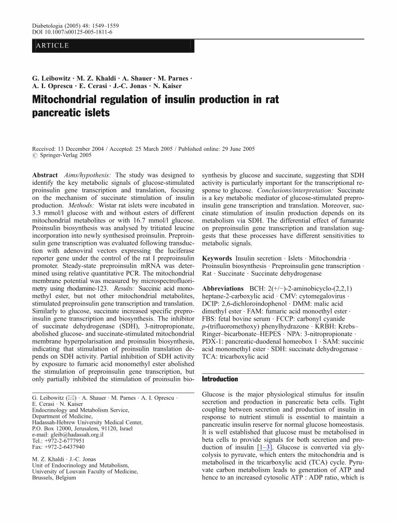
Fig. 2 Effects of glutamine plus 2(+/−)-2-aminobicyclo-(2,2,1)heptane-2-carboxylic acid (BCH) on a insulin secretion and b pro-insulin, total protein and specific proinsulin biosynthesis (correctedfor total protein biosynthesis). Rat islets were incubated for 1 h at37°C with 3.3 mmol/l glucose alone (white columns) or supple-mented with 10 mmol/l glutamine and 10 mmol/l BCH (shaded
columns) or with 16.7 mmol/l glucose (black columns). Resultsare means±SEM for three individual experiments, each performedon islets pooled from three animals. Results are normalised to3.3 mmol/l glucose (G3.3). *p<0.05, **p<0.01 and *** p<0.001relative to islets at 3.3 mmol/l glucose
1551
followed by centrifugation. Insulin RIA was performedusing anti-insulin-coated tubes (ICN Pharmaceuticals,Costa Mesa, CA, USA) and 125I-insulin from Linco Re-search (St Charles, MO, USA). Rat insulin was used as astandard. The routine intra-assay CV was 4–6% and theinter-assay CV 6–10%.
Quantification of preproinsulin mRNA by RT-PCR Totalislet RNA was extracted from 200–300 islets usingRNAzol B (Tel-Test, Friendswood, TX, USA). For PCRanalysis, total RNA was reverse-transcribed using AMVreverse transcriptase (Promega, Madison, WI, USA). Theresulting cDNA was amplified by PCR using oligonucle-otides complementary to sequences in the rat prepro-insulin I gene: 5′-CCTGCCCAGGCTTTTGTCA-3′ and5′-GGTGCAGCACTGATCCACAATG-3′. Primers weredesigned to cross an intron and amplified fragments of208 bp of the coding sequence of the rat preproinsulingene. 18S rRNA (QuantumRNA kit; Ambion, Austin, TX,USA) was used as an internal control. The polymerisationreaction was performed in a 25 μl reaction volume con-taining 2.5 μl cDNA (25 ng RNA equivalents), 80 μmol/lcold dNTPs, 2.5 μCi (alpha-32P)dCTP, 100 pmol/l of ap-propriate oligonucleotide primers and 1.5 U of Taq po-lymerase (MBI Fermentas, Amherst, NY, USA). PCRamplification conditions were as follows: 5 min at 94°Cfollowed by 14 cycles of 94°C, 60°C and 72°C, 30 s eachstep. Using these amplification conditions, the PCR re-action was linear over a wide range of RNA concentra-tions (10–50 ng) for both preproinsulin and 18S rRNA.The amplimers were separated on a 6% polyacrylamidegel in Tris-borate EDTA (TBE) buffer, the gel was driedand the incorporated (alpha 32P)dCTP measured by Phos-phorImager (FUJIX, BAS 1000; Fuji Photo Film, Tokyo,Japan). For quantification of islet preproinsulin mRNA,the ratio of preproinsulin : 18S rRNA band intensity wasdetermined for each reaction.
Preproinsulin gene transcription A recombinant adenovi-rus encoding firefly luciferase under the control of the ratpreproinsulin I promoter (corresponding to bases −310 to−12 relative to the transcription start site) (Ad-RIP-Luc)was kindly provided by Christopher J. Rhodes (NorthwestResearch Institute, Seattle, WA, USA) [23]. An adeno-virus encoding firefly luciferase under the control of thecytomegalovirus (CMV) promoter (Ad-CMV-Luc) wasused as a control. Batches of 200 islets were transducedovernight with 107 plaque-forming units/islet of Ad-RIP-Luc or Ad-CMV-Luc in RPMI 1640 containing 10% FBSand 11.1 mmol/l glucose and then in 3.3 mmol/l glucose for6 h. Islets were then transferred to fresh RPMI 1640containing 3.3 mmol/l or 16.7 mmol/l glucose and differenttreatments according to the experimental protocols, andincubated for 24 h. Luciferase activity was measured inislet extracts using the Luciferase Assay Kit (Promega)according to the manufacturer’s instructions. Preproinsulinpromoter activity was normalised to the correspondingCMV promoter activity.
Mitochondrial membrane potential measurements Wistarrat islets were precultured for 5–7 days in serum-freeRPMI medium containing 10 mmol/l glucose and 5 g/lbovine serum albumin. These preculture conditions pre-serve glucose stimulus-secretion coupling events and isletcell morphology for at least 2 weeks [24]. After culture,the islets were incubated for 1 h in Krebs buffer containing3.3 or 16.7 mmol/l glucose and various test substances.Rhodamine-123 (10 μg/ml; Molecular Probes, Eugene,OR, USA) was added to the medium for the last 20 minof that incubation. After loading, the islets were rinsedand perifused for 30 min in the same medium withoutrhodamine-123, and the fluorescence of rhodamine-123
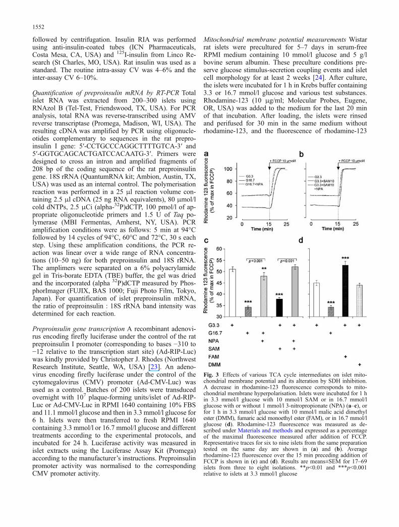
Fig. 3 Effects of various TCA cycle intermediates on islet mito-chondrial membrane potential and its alteration by SDH inhibition.A decrease in rhodamine-123 fluorescence corresponds to mito-chondrial membrane hyperpolarisation. Islets were incubated for 1 hin 3.3 mmol/l glucose with 10 mmol/l SAM or in 16.7 mmol/lglucose with or without 1 mmol/l 3-nitropropionate (NPA) (a–c), orfor 1 h in 3.3 mmol/l glucose with 10 mmol/l malic acid dimethylester (DMM), fumaric acid monoethyl ester (FAM), or in 16.7 mmol/lglucose (d). Rhodamine-123 fluorescence was measured as de-scribed under Materials and methods and expressed as a percentageof the maximal fluorescence measured after addition of FCCP.Representative traces for six to nine islets from the same preparationtested on the same day are shown in (a) and (b). Averagerhodamine-123 fluorescence over the 15 min preceding addition ofFCCP is shown in (c) and (d). Results are means±SEM for 17–69islets from three to eight isolations. **p<0.01 and ***p<0.001relative to islets at 3.3 mmol/l glucose
1552
wasmeasured bymicrospectrofluorimetry (excitation/emis-sion 490/530 nm, 505 nm long-pass dichroic mirror) 15 minbefore and after addition of 10 μmol/l carbonyl cyanidep-(trifluoromethoxy) phenylhydrazone (FCCP) [25]. Afterbackground subtraction, the rhodamine-123 fluorescencein each islet was normalised to the maximal fluorescencemeasured in the presence of FCCP [26], and the averagerelative fluorescence over the 15 min preceding FCCP ad-dition was calculated.
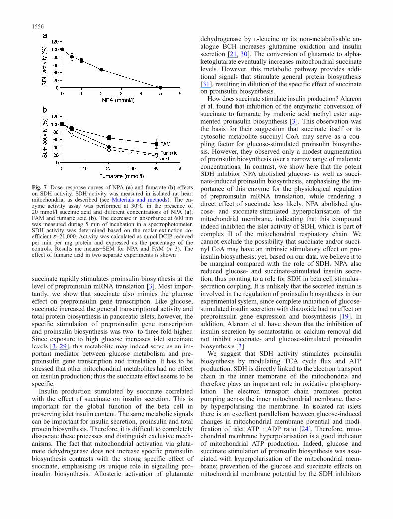
Measurement of SDH activity SDH enzymatic activity wasmeasured in isolated mitochondria according to Rosenet al. [27]. Mitochondria were isolated from rat heartand isolated islets, as described [28], and suspended in50 mmol/l potassium phosphate buffer, pH 7.4. The mi-tochondrial extract was adjusted to give a protein con-centration of 100 μg in 50 μl solution, which was added toa 1 ml cuvette containing 20 mmol/l sodium succinate,144 μmol/l 2,6-dichloroindophenol (DCIP), 2.6 mmol/lphenazine metasulphate and 100 μmol/l sodium cyanide inpotassium phosphate buffer and incubated for 1 min at30°C. The decrease in absorbance at 600 nm was measuredduring the following 5 min in a spectrophotometer. SDHactivity was determined based on the molar extinction co-efficient ɛ=21,000. Activity was calculated as mmol DCIPreduced per min per mg protein.
Data presentation and statistical analysis Data shown aremeans±SEM. The statistical significance of differencesbetween groups was determined by one-way ANOVA fol-lowed by the Newman–Keuls test using the InStat sta-tistical program (GraphPad Software, San Diego, CA, USA).
A paired-sample t-test was used when the differencebetween a reference (taken as 100%) and test was an-alysed. A p value of <0.05 was considered significant.
Results
Succinate stimulates proinsulin biosynthesis Succinic acidmonomethyl ester (SAM) stimulated insulin secretion andproinsulin biosynthesis in a dose-dependent manner inislets incubated for 1 h (Fig. 1a and b). Maximal proinsulinbiosynthesis occurred at 10 mmol/l SAM and was similarto that induced by 16.7 mmol/l glucose. SAM also aug-mented total protein biosynthesis (Fig. 1c), but to a lesserextent than the stimulation of proinsulin biosynthesis(when calculated as a percentage of the value for 3.3 mmol/lglucose), indicating that succinate has a specific effect onproinsulin biosynthesis (Fig. 1d). In contrast, 10 mmol/ldimethyl ester of the TCA cycle intermediate malate(DMM) had no effect on proinsulin, total protein andspecific proinsulin biosynthesis, or on insulin secretion(Fig. 1), indicating that the effect of succinate was specificand did not result from the supply of TCA cycleintermediates alone.
Since the effect of succinate on proinsulin biosynthesiscorrelated with its effect on insulin secretion, we per-formed additional studies to test the specificity of thesuccinate effect on proinsulin biosynthesis. In contrast tosuccinate, activation of mitochondrial metabolism withglutamine plus BCH had no specific effect on proinsulinbiosynthesis despite stimulating insulin secretion (Fig. 2).
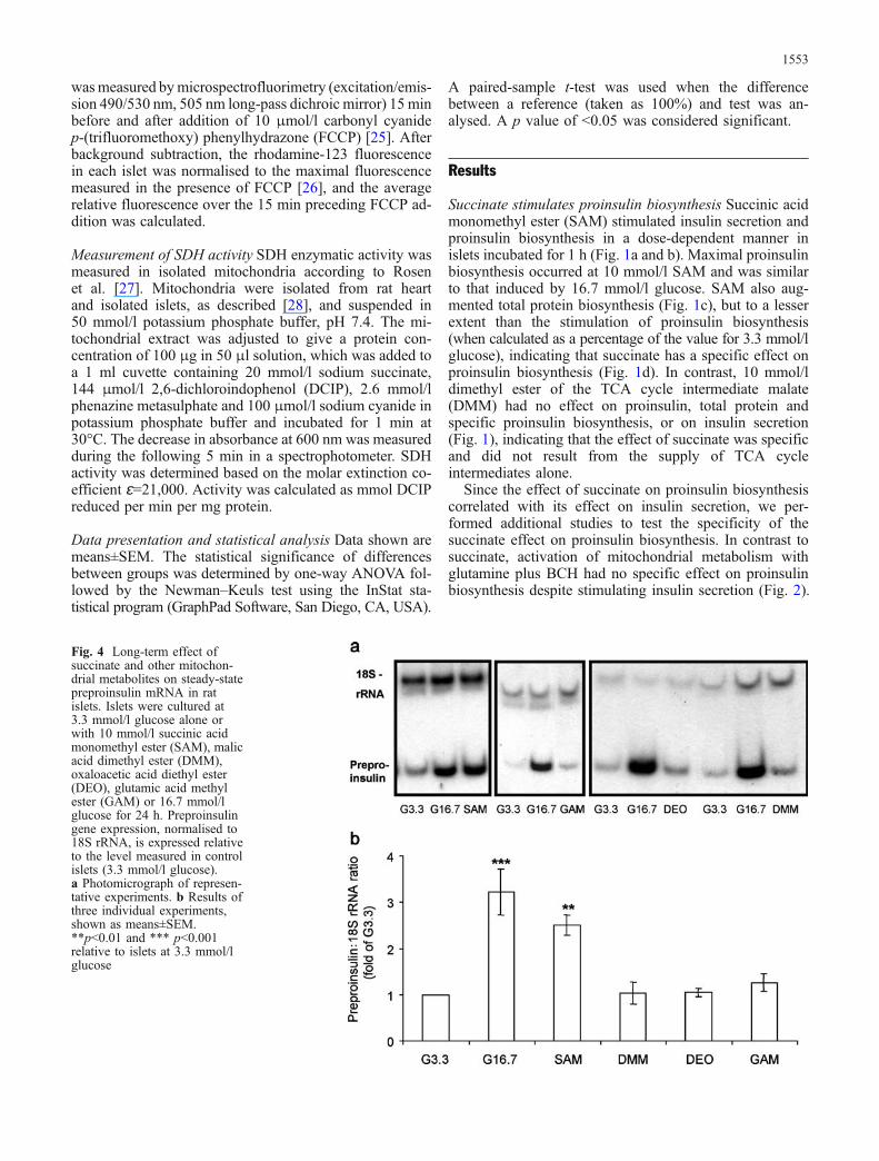
Fig. 4 Long-term effect ofsuccinate and other mitochon-drial metabolites on steady-statepreproinsulin mRNA in ratislets. Islets were cultured at3.3 mmol/l glucose alone orwith 10 mmol/l succinic acidmonomethyl ester (SAM), malicacid dimethyl ester (DMM),oxaloacetic acid diethyl ester(DEO), glutamic acid methylester (GAM) or 16.7 mmol/lglucose for 24 h. Preproinsulingene expression, normalised to18S rRNA, is expressed relativeto the level measured in controlislets (3.3 mmol/l glucose).a Photomicrograph of represen-tative experiments. b Results ofthree individual experiments,shown as means±SEM.**p<0.01 and *** p<0.001relative to islets at 3.3 mmol/lglucose
1553
Comparison of the effects of glucose and differentmitochondrial intermediates on mitochondrial membranepotential A decrease in rhodamine-123 fluorescence re-flects mitochondrial membrane hyperpolarisation [24, 25].Compared with control islets maintained in 3.3 mmol/lglucose, the mitochondrial membrane was hyperpolarisedby a 1-h exposure to 16.7 glucose or 3.3 mmol/l glucosewith 10 mmol/l SAM, but the effect of the latter wasslightly lower than that of 16.7 mmol/l glucose (Fig. 3a, b).The specificity of the glucose and succinate effect isdemonstrated by the observation that the SDH inhibitorNPA completely abolished the induction of mitochon-drial membrane hyperpolarisation (Fig. 3a–c). In contrast,addition of 10 mmol/l DMM to 3.3 mmol/l glucose didnot affect the mitochondrial membrane potential, whereas10 mmol/l FAM depolarised the mitochondrial membrane(Fig. 3d).
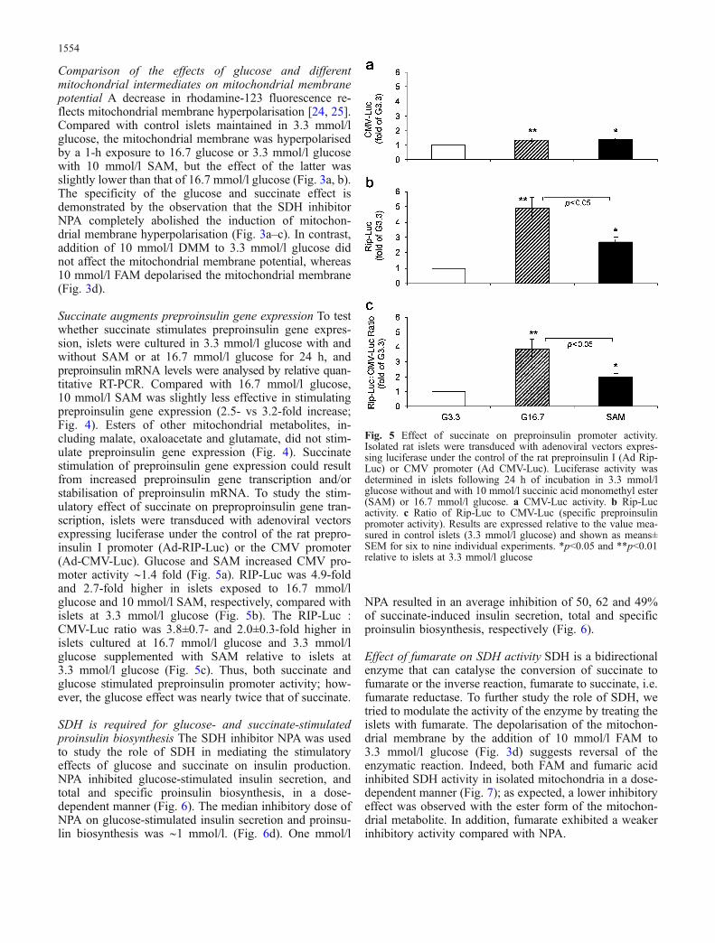
Succinate augments preproinsulin gene expression To testwhether succinate stimulates preproinsulin gene expres-sion, islets were cultured in 3.3 mmol/l glucose with andwithout SAM or at 16.7 mmol/l glucose for 24 h, andpreproinsulin mRNA levels were analysed by relative quan-titative RT-PCR. Compared with 16.7 mmol/l glucose,10 mmol/l SAM was slightly less effective in stimulatingpreproinsulin gene expression (2.5- vs 3.2-fold increase;Fig. 4). Esters of other mitochondrial metabolites, in-cluding malate, oxaloacetate and glutamate, did not stim-ulate preproinsulin gene expression (Fig. 4). Succinatestimulation of preproinsulin gene expression could resultfrom increased preproinsulin gene transcription and/orstabilisation of preproinsulin mRNA. To study the stim-ulatory effect of succinate on preproproinsulin gene tran-scription, islets were transduced with adenoviral vectorsexpressing luciferase under the control of the rat prepro-insulin I promoter (Ad-RIP-Luc) or the CMV promoter(Ad-CMV-Luc). Glucose and SAM increased CMV pro-moter activity ∼1.4 fold (Fig. 5a). RIP-Luc was 4.9-foldand 2.7-fold higher in islets exposed to 16.7 mmol/lglucose and 10 mmol/l SAM, respectively, compared withislets at 3.3 mmol/l glucose (Fig. 5b). The RIP-Luc :CMV-Luc ratio was 3.8±0.7- and 2.0±0.3-fold higher inislets cultured at 16.7 mmol/l glucose and 3.3 mmol/lglucose supplemented with SAM relative to islets at3.3 mmol/l glucose (Fig. 5c). Thus, both succinate andglucose stimulated preproinsulin promoter activity; how-ever, the glucose effect was nearly twice that of succinate.
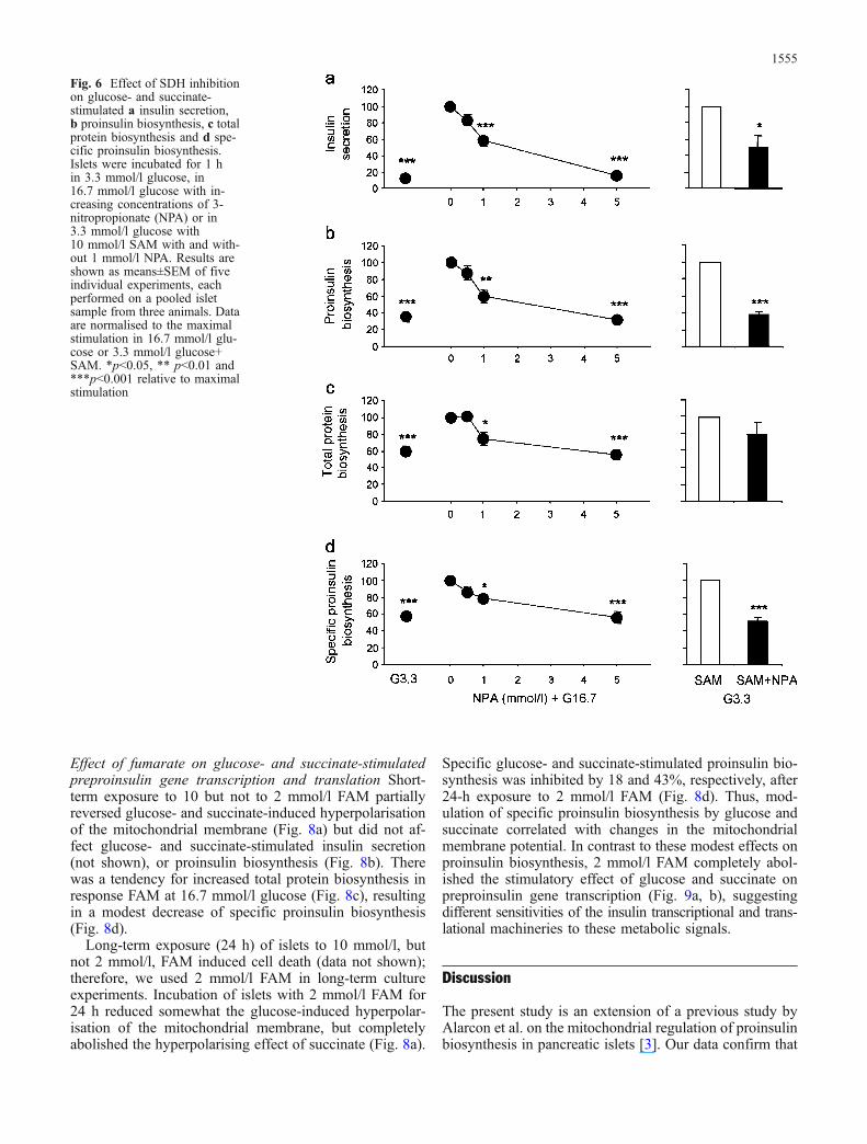
SDH is required for glucose- and succinate-stimulatedproinsulin biosynthesis The SDH inhibitor NPA was usedto study the role of SDH in mediating the stimulatoryeffects of glucose and succinate on insulin production.NPA inhibited glucose-stimulated insulin secretion, andtotal and specific proinsulin biosynthesis, in a dose-dependent manner (Fig. 6). The median inhibitory dose ofNPA on glucose-stimulated insulin secretion and proinsu-lin biosynthesis was ∼1 mmol/l. (Fig. 6d). One mmol/l
NPA resulted in an average inhibition of 50, 62 and 49%of succinate-induced insulin secretion, total and specificproinsulin biosynthesis, respectively (Fig. 6).
Effect of fumarate on SDH activity SDH is a bidirectionalenzyme that can catalyse the conversion of succinate tofumarate or the inverse reaction, fumarate to succinate, i.e.fumarate reductase. To further study the role of SDH, wetried to modulate the activity of the enzyme by treating theislets with fumarate. The depolarisation of the mitochon-drial membrane by the addition of 10 mmol/l FAM to3.3 mmol/l glucose (Fig. 3d) suggests reversal of theenzymatic reaction. Indeed, both FAM and fumaric acidinhibited SDH activity in isolated mitochondria in a dose-dependent manner (Fig. 7); as expected, a lower inhibitoryeffect was observed with the ester form of the mitochon-drial metabolite. In addition, fumarate exhibited a weakerinhibitory activity compared with NPA.
Fig. 5 Effect of succinate on preproinsulin promoter activity.Isolated rat islets were transduced with adenoviral vectors expres-sing luciferase under the control of the rat preproinsulin I (Ad Rip-Luc) or CMV promoter (Ad CMV-Luc). Luciferase activity wasdetermined in islets following 24 h of incubation in 3.3 mmol/lglucose without and with 10 mmol/l succinic acid monomethyl ester(SAM) or 16.7 mmol/l glucose. a CMV-Luc activity. b Rip-Lucactivity. c Ratio of Rip-Luc to CMV-Luc (specific preproinsulinpromoter activity). Results are expressed relative to the value mea-sured in control islets (3.3 mmol/l glucose) and shown as means±SEM for six to nine individual experiments. *p<0.05 and **p<0.01relative to islets at 3.3 mmol/l glucose
1554
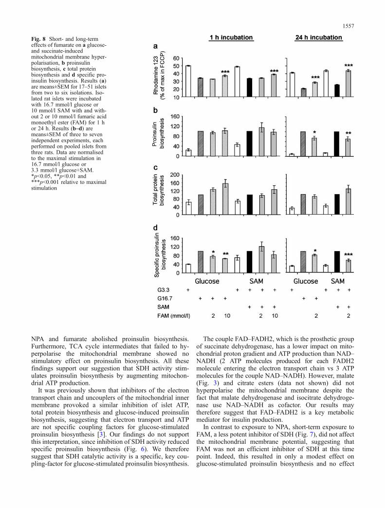
Effect of fumarate on glucose- and succinate-stimulatedpreproinsulin gene transcription and translation Short-term exposure to 10 but not to 2 mmol/l FAM partiallyreversed glucose- and succinate-induced hyperpolarisationof the mitochondrial membrane (Fig. 8a) but did not af-fect glucose- and succinate-stimulated insulin secretion(not shown), or proinsulin biosynthesis (Fig. 8b). Therewas a tendency for increased total protein biosynthesis inresponse FAM at 16.7 mmol/l glucose (Fig. 8c), resultingin a modest decrease of specific proinsulin biosynthesis(Fig. 8d).
Long-term exposure (24 h) of islets to 10 mmol/l, butnot 2 mmol/l, FAM induced cell death (data not shown);therefore, we used 2 mmol/l FAM in long-term cultureexperiments. Incubation of islets with 2 mmol/l FAM for24 h reduced somewhat the glucose-induced hyperpolar-isation of the mitochondrial membrane, but completelyabolished the hyperpolarising effect of succinate (Fig. 8a).
Specific glucose- and succinate-stimulated proinsulin bio-synthesis was inhibited by 18 and 43%, respectively, after24-h exposure to 2 mmol/l FAM (Fig. 8d). Thus, mod-ulation of specific proinsulin biosynthesis by glucose andsuccinate correlated with changes in the mitochondrialmembrane potential. In contrast to these modest effects onproinsulin biosynthesis, 2 mmol/l FAM completely abol-ished the stimulatory effect of glucose and succinate onpreproinsulin gene transcription (Fig. 9a, b), suggestingdifferent sensitivities of the insulin transcriptional and trans-lational machineries to these metabolic signals.
Discussion
The present study is an extension of a previous study byAlarcon et al. on the mitochondrial regulation of proinsulinbiosynthesis in pancreatic islets [3]. Our data confirm that
Fig. 6 Effect of SDH inhibitionon glucose- and succinate-stimulated a insulin secretion,b proinsulin biosynthesis, c totalprotein biosynthesis and d spe-cific proinsulin biosynthesis.Islets were incubated for 1 hin 3.3 mmol/l glucose, in16.7 mmol/l glucose with in-creasing concentrations of 3-nitropropionate (NPA) or in3.3 mmol/l glucose with10 mmol/l SAM with and with-out 1 mmol/l NPA. Results areshown as means±SEM of fiveindividual experiments, eachperformed on a pooled isletsample from three animals. Dataare normalised to the maximalstimulation in 16.7 mmol/l glu-cose or 3.3 mmol/l glucose+SAM. *p<0.05, ** p<0.01 and***p<0.001 relative to maximalstimulation
1555
succinate rapidly stimulates proinsulin biosynthesis at thelevel of preproinsulin mRNA translation [3]. Most impor-tantly, we show that succinate also mimics the glucoseeffect on preproinsulin gene transcription. Like glucose,succinate increased the general transcriptional activity andtotal protein biosynthesis in pancreatic islets; however, thespecific stimulation of preproinsulin gene transcriptionand proinsulin biosynthesis was two- to three-fold higher.Since exposure to high glucose increases islet succinatelevels [3, 29], this metabolite may indeed serve as an im-portant mediator between glucose metabolism and pre-proinsulin gene transcription and translation. It has to bestressed that other mitochondrial metabolites had no effecton insulin production; thus the succinate effect seems to bespecific.
Insulin production stimulated by succinate correlatedwith the effect of succinate on insulin secretion. This isimportant for the global function of the beta cell inpreserving islet insulin content. The same metabolic signalscan be important for insulin secretion, proinsulin and totalprotein biosynthesis. Therefore, it is difficult to completelydissociate these processes and distinguish exclusive mech-anisms. The fact that mitochondrial activation via gluta-mate dehydrogenase does not increase specific proinsulinbiosynthesis contrasts with the strong specific effect ofsuccinate, emphasising its unique role in signalling pro-insulin biosynthesis. Allosteric activation of glutamate
dehydrogenase by L-leucine or its non-metabolisable an-alogue BCH increases glutamine oxidation and insulinsecretion [21, 30]. The conversion of glutamate to alpha-ketoglutarate eventually increases mitochondrial succinatelevels. However, this metabolic pathway provides addi-tional signals that stimulate general protein biosynthesis[31], resulting in dilution of the specific effect of succinateon proinsulin biosynthesis.
How does succinate stimulate insulin production? Alarconet al. found that inhibition of the enzymatic conversion ofsuccinate to fumarate by malonic acid methyl ester aug-mented proinsulin biosynthesis [3]. This observation wasthe basis for their suggestion that succinate itself or itscytosolic metabolite succinyl CoA may serve as a cou-pling factor for glucose-stimulated proinsulin biosynthe-sis. However, they observed only a modest augmentationof proinsulin biosynthesis over a narrow range of malonateconcentrations. In contrast, we show here that the potentSDH inhibitor NPA abolished glucose- as well as succi-nate-induced proinsulin biosynthesis, emphasising the im-portance of this enzyme for the physiological regulationof preproinsulin mRNA translation, while rendering adirect effect of succinate less likely. NPA abolished glu-cose- and succinate-stimulated hyperpolarisation of themitochondrial membrane, indicating that this compoundindeed inhibited the islet activity of SDH, which is part ofcomplex II of the mitochondrial respiratory chain. Wecannot exclude the possibility that succinate and/or succi-nyl CoA may have an intrinsic stimulatory effect on pro-insulin biosynthesis; yet, based on our data, we believe it tobe marginal compared with the role of SDH. NPA alsoreduced glucose- and succinate-stimulated insulin secre-tion, thus pointing to a role for SDH in beta cell stimulus–secretion coupling. It is unlikely that the secreted insulin isinvolved in the regulation of proinsulin biosynthesis in ourexperimental system, since complete inhibition of glucose-stimulated insulin secretion with diazoxide had no effect onpreproinsulin gene expression and biosynthesis [19]. Inaddition, Alarcon et al. have shown that the inhibition ofinsulin secretion by somatostatin or calcium removal didnot inhibit succinate- and glucose-stimulated proinsulinbiosynthesis [3].
We suggest that SDH activity stimulates proinsulinbiosynthesis by modulating TCA cycle flux and ATPproduction. SDH is directly linked to the electron transportchain in the inner membrane of the mitochondria andtherefore plays an important role in oxidative phosphory-lation. The electron transport chain promotes protonpumping across the inner mitochondrial membrane, there-by hyperpolarising the membrane. In isolated rat isletsthere is an excellent parallelism between glucose-inducedchanges in mitochondrial membrane potential and modi-fication of islet ATP : ADP ratio [24]. Therefore, mito-chondrial membrane hyperpolarisation is a good indicatorof mitochondrial ATP production. Indeed, glucose andsuccinate stimulation of proinsulin biosynthesis was asso-ciated with hyperpolarisation of the mitochondrial mem-brane; prevention of the glucose and succinate effects onmitochondrial membrane potential by the SDH inhibitors
Fig. 7 Dose–response curves of NPA (a) and fumarate (b) effectson SDH activity. SDH activity was measured in isolated rat heartmitochondria, as described (see Materials and methods). The en-zyme activity assay was performed at 30°C in the presence of20 mmol/l succinic acid and different concentrations of NPA (a),FAM and fumaric acid (b). The decrease in absorbance at 600 nmwas measured during 5 min of incubation in a spectrophotometer.SDH activity was determined based on the molar extinction co-efficient ɛ=21,000. Activity was calculated as mmol DCIP reducedper min per mg protein and expressed as the percentage of thecontrols. Results are means±SEM for NPA and FAM (n=3). Theeffect of fumaric acid in two separate experiments is shown
1556
NPA and fumarate abolished proinsulin biosynthesis.Furthermore, TCA cycle intermediates that failed to hy-perpolarise the mitochondrial membrane showed nostimulatory effect on proinsulin biosynthesis. All thesefindings support our suggestion that SDH activity stim-ulates proinsulin biosynthesis by augmenting mitochon-drial ATP production.
It was previously shown that inhibitors of the electrontransport chain and uncouplers of the mitochondrial innermembrane provoked a similar inhibition of islet ATP,total protein biosynthesis and glucose-induced proinsulinbiosynthesis, suggesting that electron transport and ATPare not specific coupling factors for glucose-stimulatedproinsulin biosynthesis [3]. Our findings do not supportthis interpretation, since inhibition of SDH activity reducedspecific proinsulin biosynthesis (Fig. 6). We thereforesuggest that SDH catalytic activity is a specific, key cou-pling-factor for glucose-stimulated proinsulin biosynthesis.
The couple FAD–FADH2, which is the prosthetic groupof succinate dehydrogenase, has a lower impact on mito-chondrial proton gradient and ATP production than NAD–NADH (2 ATP molecules produced for each FADH2molecule entering the electron transport chain vs 3 ATPmolecules for the couple NAD–NADH). However, malate(Fig. 3) and citrate esters (data not shown) did nothyperpolarise the mitochondrial membrane despite thefact that malate dehydrogenase and isocitrate dehydroge-nase use NAD–NADH as cofactor. Our results maytherefore suggest that FAD–FADH2 is a key metabolicmediator for insulin production.
In contrast to exposure to NPA, short-term exposure toFAM, a less potent inhibitor of SDH (Fig. 7), did not affectthe mitochondrial membrane potential, suggesting thatFAM was not an efficient inhibitor of SDH at this timepoint. Indeed, this resulted in only a modest effect onglucose-stimulated proinsulin biosynthesis and no effect
Fig. 8 Short- and long-termeffects of fumarate on a glucose-and succinate-inducedmitochondrial membrane hyper-polarisation, b proinsulinbiosynthesis, c total proteinbiosynthesis and d specific pro-insulin biosynthesis. Results (a)are means±SEM for 17–51 isletsfrom two to six isolations. Iso-lated rat islets were incubatedwith 16.7 mmol/l glucose or10 mmol/l SAM with and with-out 2 or 10 mmol/l fumaric acidmonoethyl ester (FAM) for 1 hor 24 h. Results (b–d) aremeans±SEM of three to sevenindependent experiments, eachperformed on pooled islets fromthree rats. Data are normalisedto the maximal stimulation in16.7 mmol/l glucose or3.3 mmol/l glucose+SAM.*p<0.05, **p<0.01 and***p<0.001 relative to maximalstimulation
1557
on succinate-stimulated proinsulin biosynthesis at thelevel of translation. In contrast, long-term (24 h) exposureto 2 mmol/l FAM completely reversed succinate-inducedmitochondrial hyperpolarisation. It is likely that the con-centration of fumarate in the islet increases time-depen-dently as FAM is hydrolysed to the active product fumaricacid. This could explain the lag in the effect of FAM onthe mitochondrial membrane potential. The possibility thatprolonged exposure to 2 mmol/l FAM induced a smallamount of cell death cannot be completely excluded;however, propidium iodide staining showed no increase inthe mortality of cells treated with low-dose FAM relative tocontrol islets in 16.7 mmol/l glucose (not shown). More-over, this treatment did not impair insulin secretion andcellular protein biosynthesis. FAM inhibition of succinate-induced hyperpolarisation was higher than that of glucose,resulting in greater inhibition of specific succinate-inducedproinsulin biosynthesis. Whereas fumarate had only amodest effect on proinsulin biosynthesis, it completelyabolished glucose- and succinate-stimulated preproinsulingene transcription, indicating greater sensitivity of thetranscriptional machinery to reduction in SDH activity.Glucose-stimulated preproinsulin gene transcription isrequired for maintaining proinsulin biosynthesis duringprolonged exposure to a high concentration of glucose[19]. As an example, complete inhibition of preproinsu-lin gene transcription with actinomycin D resulted in a
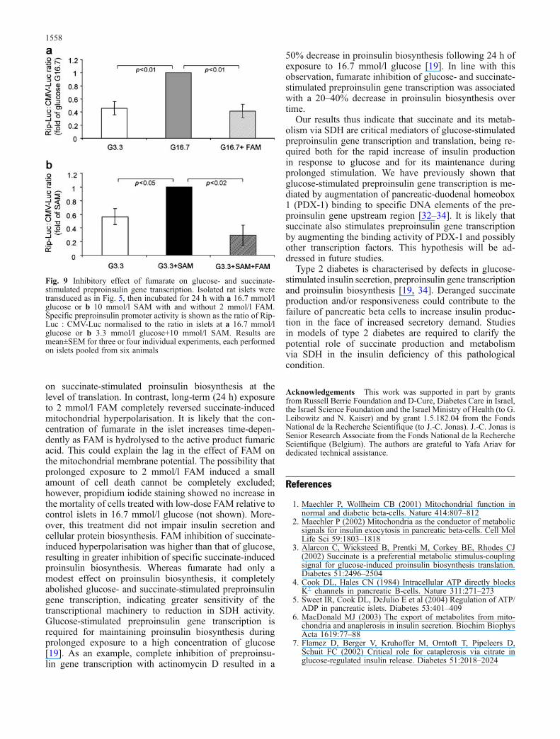
50% decrease in proinsulin biosynthesis following 24 h ofexposure to 16.7 mmol/l glucose [19]. In line with thisobservation, fumarate inhibition of glucose- and succinate-stimulated preproinsulin gene transcription was associatedwith a 20–40% decrease in proinsulin biosynthesis overtime.
Our results thus indicate that succinate and its metab-olism via SDH are critical mediators of glucose-stimulatedpreproinsulin gene transcription and translation, being re-quired both for the rapid increase of insulin productionin response to glucose and for its maintenance duringprolonged stimulation. We have previously shown thatglucose-stimulated preproinsulin gene transcription is me-diated by augmentation of pancreatic-duodenal homeobox1 (PDX-1) binding to specific DNA elements of the pre-proinsulin gene upstream region [32–34]. It is likely thatsuccinate also stimulates preproinsulin gene transcriptionby augmenting the binding activity of PDX-1 and possiblyother transcription factors. This hypothesis will be ad-dressed in future studies.
Type 2 diabetes is characterised by defects in glucose-stimulated insulin secretion, preproinsulin gene transcriptionand proinsulin biosynthesis [19, 34]. Deranged succinateproduction and/or responsiveness could contribute to thefailure of pancreatic beta cells to increase insulin produc-tion in the face of increased secretory demand. Studiesin models of type 2 diabetes are required to clarify thepotential role of succinate production and metabolismvia SDH in the insulin deficiency of this pathologicalcondition.
Acknowledgements This work was supported in part by grantsfrom Russell Berrie Foundation and D-Cure, Diabetes Care in Israel,the Israel Science Foundation and the Israel Ministry of Health (to G.Leibowitz and N. Kaiser) and by grant 1.5.182.04 from the FondsNational de la Recherche Scientifique (to J.-C. Jonas). J.-C. Jonas isSenior Research Associate from the Fonds National de la RechercheScientifique (Belgium). The authors are grateful to Yafa Ariav fordedicated technical assistance.
References
1. Maechler P, Wollheim CB (2001) Mitochondrial function innormal and diabetic beta-cells. Nature 414:807–812
2. Maechler P (2002) Mitochondria as the conductor of metabolicsignals for insulin exocytosis in pancreatic beta-cells. Cell MolLife Sci 59:1803–1818
3. Alarcon C, Wicksteed B, Prentki M, Corkey BE, Rhodes CJ(2002) Succinate is a preferential metabolic stimulus-couplingsignal for glucose-induced proinsulin biosynthesis translation.Diabetes 51:2496–2504
4. Cook DL, Hales CN (1984) Intracellular ATP directly blocksK+ channels in pancreatic B-cells. Nature 311:271–273
5. Sweet IR, Cook DL, DeJulio E et al (2004) Regulation of ATP/ADP in pancreatic islets. Diabetes 53:401–409
6. MacDonald MJ (2003) The export of metabolites from mito-chondria and anaplerosis in insulin secretion. Biochim BiophysActa 1619:77–88
7. Flamez D, Berger V, Kruhoffer M, Orntoft T, Pipeleers D,Schuit FC (2002) Critical role for cataplerosis via citrate inglucose-regulated insulin release. Diabetes 51:2018–2024
Fig. 9 Inhibitory effect of fumarate on glucose- and succinate-stimulated preproinsulin gene transcription. Isolated rat islets weretransduced as in Fig. 5, then incubated for 24 h with a 16.7 mmol/lglucose or b 10 mmol/l SAM with and without 2 mmol/l FAM.Specific preproinsulin promoter activity is shown as the ratio of Rip-Luc : CMV-Luc normalised to the ratio in islets at a 16.7 mmol/lglucose or b 3.3 mmol/l glucose+10 mmol/l SAM. Results aremean±SEM for three or four individual experiments, each performedon islets pooled from six animals
1558
8. Skelly RH, Bollheimer LC, Wicksteed BL, Corkey BE, RhodesCJ (1998) A distinct difference in the metabolic stimulus-response coupling pathways for regulating proinsulin biosyn-thesis and insulin secretion that lies at the level of a requirementfor fatty acyl moieties. Biochem J 331:553–561
9. Maechler P, Wollheim CB (1999) Mitochondrial glutamate actsas a messenger in glucose-induced insulin exocytosis. Nature402:685–689
10. MacDonald MJ, Fahien LA (2000) Glutamate is not amessenger in insulin secretion. J Biol Chem 275:34025–34027
11. MacDonald MJ, Fahien LA, Buss JD, Hasan NM, Fallon MJ,Kendrick MA (2003) Citrate oscillates in liver and pancreaticbeta cell mitochondria and in INS-1 insulinoma cells. J BiolChem 278:51894–51900
12. Farfari S, Schulz V, Corkey B, Prentki M (2000) Glucose-regulated anaplerosis and cataplerosis in pancreatic beta-cells:possible implication of a pyruvate/citrate shuttle in insulinsecretion. Diabetes 49:718–726
13. Newgard CB, Lu D, Jensen MVet al (2002) Stimulus/secretioncoupling factors in glucose-stimulated insulin secretion: in-sights gained from a multidisciplinary approach. Diabetes 51[Suppl 3]:S389–S393
14. Mulder H, Lu D, Finley J et al (2001) Overexpression of amodified human malonyl-CoA decarboxylase blocks the glu-cose-induced increase in malonyl-CoA level but has no impacton insulin secretion in INS-1-derived (832/13) beta-cells. J BiolChem 276:6479–6484
15. Bertrand G, Ishiyama N, Nenquin M, Ravier MA, Henquin JC(2002) The elevation of glutamate content and the amplificationof insulin secretion in glucose-stimulated pancreatic islets arenot causally related. J Biol Chem 277:32883–32891
16. Roduit R, Nolan C, Alarcon C et al (2004) A role for themalonyl-CoA/long-chain acyl-CoA pathway of lipid signalingin the regulation of insulin secretion in response to both fueland nonfuel stimuli. Diabetes 53:1007–1019
17. Leibowitz G, Oprescu AI, Uckaya G, Gross DJ, Cerasi E,Kaiser N (2003) Insulin does not mediate glucose stimulationof proinsulin biosynthesis. Diabetes 52:998–1003
18. Wicksteed B, Alarcon C, Briaud I, Lingohr MK, Rhodes CJ(2003) Glucose-induced translational control of proinsulin bio-synthesis is proportional to preproinsulin mRNA levels in isletbeta-cells but not regulated via a positive feedback of secretedinsulin. J Biol Chem 278:42080–42090
19. Leibowitz G, Uckaya G, Oprescu AI, Cerasi E, Gross DJ,Kaiser N (2002) Glucose-regulated proinsulin gene expressionis required for adequate insulin production during chronicglucose exposure. Endocrinology 143:3214–3220
20. Gadot M, Leibowitz G, Shafrir E, Cerasi E, Gross D, Kaiser N(1994) Hyperproinsulinemia and insulin deficiency in thediabetic Psammomys obesus. Endocrinology 135:610–616
21. Sener A, Malaisse-Lagae F, Malaisse WJ (1981) Stimulation ofpancreatic islet metabolism and insulin release by a nonmetab-olizable amino acid. Proc Natl Acad Sci U S A 78:5460–5464
22. Alarcon C, Lincoln B, Rhodes CJ (1993) The biosynthesis ofthe subtilisin-related proprotein convertase PC3, but not that ofthe PC2 convertase, is regulated by glucose in parallel toproinsulin biosynthesis in rat pancreatic islets. J Biol Chem268:4276–4280
23. Kelpe CL, Moore PC, Parazzoli SD, Wicksteed B, Rhodes CJ,Poitout V (2003) Palmitate inhibition of insulin gene expres-sion is mediated at the transcriptional level via ceramide syn-thesis. J Biol Chem 278:30015–30021
24. Khaldi MZ, Guiot Y, Gilon P, Henquin JC, Jonas JC (2004)Increased glucose sensitivity of both triggering and amplifyingpathways of insulin secretion in rat islets cultured for 1 week inhigh glucose. Am J Physiol Endocrinol Metab 287:E207–E217
25. Scaduto RC Jr, Grotyohann LW (1999) Measurement ofmitochondrial membrane potential using fluorescent rhodaminederivatives. Biophys J 76:469–477
26. Antinozzi PA, Ishihara H, Newgard CB, Wollheim CB (2002)Mitochondrial metabolism sets the maximal limit of fuel-stimulated insulin secretion in a model pancreatic beta cell: asurvey of four fuel secretagogues. J Biol Chem 277:11746–11755
27. Rosen H, Rakita RM, Waltersdorph AM, Klebanoff SJ (1987)Myeloperoxidase-mediated damage to the succinate oxidasesystem of Escherichia coli. Evidence for selective inactivationof the dehydrogenase component. J Biol Chem 262:15004–15010
28. Holmuhamedov EL, Ozcan C, Jahangir A, Terzic A (2001)Restoration of Ca2+-inhibited oxidative phosphorylation incardiac mitochondria by mitochondrial Ca2+ unloading. MolCell Biochem 220:135–140
29. MacDonald MJ (2002) Differences between mouse and ratpancreatic islets: succinate responsiveness, malic enzyme, andanaplerosis. Am J Physiol Endocrinol Metab 283:E302–E310
30. Li C, Najafi H, Daikhin Y et al (2003) Regulation of leucine-stimulated insulin secretion and glutamine metabolism in iso-lated rat islets. J Biol Chem 278:2853–2858
31. McDaniel ML, Marshall CA, Pappan KL, Kwon G (2002)Metabolic and autocrine regulation of the mammalian target ofrapamycin by pancreatic beta-cells. Diabetes 51:2877–2885
32. Melloul D, Ben-Neriah Y, Cerasi E (1993) Glucose modulatesthe binding of an islet-specific factor to a conserved sequencewithin the rat I and the human insulin promoters. Proc NatlAcad Sci U S A 90:3865–3869
33. Marshak S, Totary H, Cerasi E, Melloul D (1996) Purificationof the beta-cell glucose-sensitive factor that transactivates theinsulin gene differentially in normal and transformed islet cells.Proc Natl Acad Sci U S A 93:15057–15062
34. Leibowitz G, Ferber S, Apelqvist A et al (2001) IPF1/PDX1deficiency and beta-cell dysfunction in Psammomys obesus, ananimal with type 2 diabetes. Diabetes 50:1799–1806
1559