Copper induced upregulation of apoptosis related genes in zebrafish (Danio rerio) gill
Upload
independentCategory
view
0download
0
Upregulation of the expression of tight and adherens junction-associated proteins during
maturation of neonatal pancreatic islets in vitro
Carla B. Collares-Buzato1,*, Carolina P. F. Carvalho1, Archimedes G. Furtado1 & Antonio C. Boschero2
1Department of Histology and Embryology, 2Department of Physiology and Biophysics, State University ofCampinas (UNICAMP), 13083-970, Campinas, SP, Brazil
*Author for correspondence (e-mail: [email protected])
Summary
Cell–cell contacts mediated by intercellular junctions are crucial for proper insulin secretion in the endocrine pan-creas. The biochemical composition of the intercellular junctions in this organ and the role of junctional proteinsin endocrine pancreatic dysfunctions are still unclear. In this study, we investigated the expression and cellularlocation of junctional and cytoskeletal proteins in cultured neonatal rat pancreatic islets. Neonatal B-cells had animpaired insulin secretion compared to adult cells. Cultured neonatal islets showed a time-dependent increase inthe glucose-induced secretory response. The maturation of B-cells in vitro was accompanied by upregulation of theexpression of some junctional proteins in islet cells. Neonatal islets cultured for only 24 h showed a low expressionand a diffuse cytoplasmic location of the tight junctional proteins occludin and ZO-1 and of the adherens junc-tional proteins a- and b-catenins, as demonstrated by immunoblotting and immunocytochemistry. Culturing isletsfor up to 8 days significantly increased the cell expression of these junctional proteins but not of the cytoskeletalproteins vinculin and a-actinin. A translocation of ZO-1 and catenins to the cell–cell contact region, as well as ahigher association of F-actin with the intercellular junction, were also observed in neonatal islets following pro-longed culturing. ZO-1 and b-catenin were immunolocated in the endocrine pancreas of adult rats indicating thatthese junctional proteins are also expressed in this organ in situ. In conclusion, endocrine pancreatic cells expressseveral junctional proteins that are upregulated following differentiation of the endocrine pancreas in vitro.
Introduction
The cells of tissues and organs are connected to eachother by specialized, membrane-associated structuresknown as intercellular junctions (IJs). There are fourtypes of junctions that are structurally and function-ally distinct, namely tight junction (TJ), adherensjunction, desmosome and gap junction (GJ) (Faquhar& Palade 1963, Gilula 1978). The first three of thesejunctions are essentially intercellular adhesion devices,whereas GJs consist of connexin-containing channelsinvolved in intercellular communication. Extensiveinvestigation of the molecular biology of IJs has ledto the identification of several protein componentsand to an understanding of the mechanisms regulat-ing their organization and function. Occludin andclaudins are specific proteins present in the tight junc-tional strands that associate with a cytoplasmic pro-tein complex formed by ZO-1, ZO-2, ZO-3, cingulinand other proteins (Zahraoui et al. 2000). Adhesionmolecules belonging to the cadherin superfamily arethe main components of adherens junctions and des-mosomes (Geiger & Ayalon 1992). These adhesion
glycoproteins are linked to the cytoskeleton througha distinct group of proteins that includes catenins, inthe case of the adherens junction, and plakoglobin(also known as c-catenin) and desmoplakin, in thecase of desmosomes (Geiger et al. 1995, Burdett 1998,Harington & Syrigos 2000). All IJs and their proteinsare dynamic, regulated membrane structures that playan important role in various cellular events, such asmigration, differentiation, intercellular communica-tion, cell–cell recognition, cell growth and death(Collares-Buzato et al. 1994a, b, 1998a, b, 2001, Allenet al. 1996, Gottardi et al. 2001, Li et al. 2001, Linget al. 2001).
Cell–cell contacts are crucial for a proper secretoryresponse by the endocrine pancreas. The synthesisand release of insulin are markedly altered after dis-persion of B-cells in vitro, but rapidly recover aftercell reaggregation, indicating that the secretory mech-anism depends on intercellular interactions within thepancreatic islet (Halban et al. 1982, Bosco et al.1989, Pipelleers et al. 1994). Ultrastructural analysishas shown that endocrine pancreatic cells interactwithin the islet through IJs such as TJs, desmo-
Journal of Molecular Histology 35: 811–822, 2004.� 2004 Kluwer Academic Publishers. Printed in the Netherlands.

somes, adherens-type junctions and GJs (Faquhar &Palade 1963). Among the IJs, GJs have been themost studied in the pancreas. The importance ofintercellular contact for normal pancreas functioninghas been attributed mainly to the GJs interconnect-ing the islet cells. Several studies in vivo and in vitrohave demonstrated that insulin secretion stimulatedby glucose and other secretagogues is associated withincreased GJ-mediated coupling between B-cells,whereas the pharmacological blockage of GJ chan-nels impairs the secretory function of these islet cells(Meda & Orci 1979, Meda et al. 1983, 1990, 1991,Collares-Buzato et al. 2001). The biochemical compo-sition of the other IJ components in this organ andthe role of junctional proteins in endocrine pancre-atic dysfunctions are still unclear.
In this work, we investigated the expression and cel-lular location of junctional and cytoskeletal proteins incultured pancreatic islets from neonatal rats. NeonatalB-cells have an impaired insulin secretion compared tothat of islets from adult rats (Boschero et al. 1988,1993). The maintenance of neonatal islets in culturefor prolonged periods results in a time-dependentimprovement in the glucose-induced secretory responsethrough a mechanism that is not completely under-stood (Boschero et al. 1993, Crepaldi-Alves et al. 1997,Collares-Buzato et al. 2001). Thus, cultured neonatalislets provide a useful model for studying the cellularprocesses underlying the maturation of the pancreaticsecretory machine. In addition, such studies may yieldresults that could form the basis for optimizing the useof cultured neonatal islets or precursor cells for trans-plantation in the treatment of diabetes. As shown here,endocrine pancreatic cells express several junctionalproteins that are upregulated following differentiationof the endocrine pancreas in vitro. These findings sug-gest a possible role for IJs in determining an adequateendocrine function of the pancreas.
Materials and methods
Materials
Cell culture media were supplied by Sigma (St. Louis,MO, USA) and culture medium supplements were pur-chased from Cultilab or Nutricell (Campinas, SP,Brazil). Sterile plastic materials were purchased fromCorning (Corning, NY, USA). Radiolabeled insulinand protein A were obtained from Amersham Bio-sciences (Cleveland, OH). Primary antibodies, FITC-or TRITC-conjugated secondary antibodies andTRITC-phalloidin were purchased from Sigma, Zymed(San Francisco, CA), Dako (Carpinteria, CA, USA)and Upstate Biotechnology Inc. (New York, NY,USA). All other chemicals and reagents were suppliedby Sigma and Merck (Darmstadt, Germany).
Animals
Neonatal male and female Wistar rats (2–48 h old)and adult male Wistar rats (3–4 months old) wereobtained from the breeding colony at the State Univer-sity of Campinas (UNICAMP, Brazil). The animalswere housed and subsequently killed to obtain thepancreas in accordance with the guidelines of the insti-tutional Committee for Ethics in Animal Experimenta-tion (UNICAMP).
Islet culture and isolation
Islets from neonatal Wistar rats were cultured as previ-ously described (Collares-Buzato et al. 2001). Eachneonatal rat pancreas yielded 100–200 islets. Briefly,the islets were cultured for varying periods of time(maximum of 8 days) in sterile Petri dishes at 37 �C ina humidified 5% CO2/air atmosphere. The culturemedium consisted of RPMI-1640 containing 10 mMglucose and supplemented with 5% fetal calf serum(heat inactivated) and 100 IU of penicillin/ml and100 lg of streptomycin/ml. The culture medium waschanged every second day. At the end of the cultureperiod, the islets were collected individually under adissecting microscope using a micropipette. Some ofthese islets were used to determine the levels of insulinsecretion and the remaining ones were processed forimmunocytochemistry or for immunoblotting.
Morphological studies
The cellular location of some junctional and cytoskele-tal proteins was determined by a standard indirectimmunofluorescence technique in cryosections of iso-lated neonatal islets and of whole pancreas of adultrats (Collares-Buzato et al. 2001). Briefly, a pool ofisolated islets (�1000–2000 islets) was fixed in 2%paraformaldehyde plus 10% saccharose (in 0.01 Mphosphate-buffered saline, PBS, pH 7.4) for 30 minand included in gelatin solutions of increasing concen-tration (5, 10 and 25%) at 37 �C. The gelatin blocksor unfixed pancreas were frozen in n-hexane withliquid nitrogen. Cryostat sections of pancreas werepicked up on poly-L-lysine-coated glass slides and fixedfor 10 min with 2% paraformaldehyde. Islet and pan-creas sections were then treated with 0.1% Triton X-100 (in PBS) and incubated with primary antibody.The following primary antibodies were used at theindicated dilutions (in PBS, pH 7.4): rabbit anti-occlu-din (dilution 1 : 50) (Zymed), rabbit anti-ZO-1 (dilu-tion 1 : 50) (Zymed), mouse anti-b-catenin (dilution1 : 50) (Zymed), rabbit anti-a-catenin (dilution 1 : 50)(Sigma), mouse anti-a-actinin (dilution 1 : 50) (Upstate)and mouse anti-vinculin (dilution 1 : 50) (Sigma).Following incubation with the specific secondary anti-body conjugated to fluorescein (dilution 1 : 75), the
812 Collares-Buzato et al.

sections were mounted in a commercial antifadingagent (Vectashield, Vector Laboratories, Burlingame,CA, USA). Double immunolabeling of b-catenin orZO-1 and insulin was also done described above butwith two additional incubation steps with guinea piganti-insulin (dilution 1 : 500) (Dako) for 1 h followedby another 1 h incubation with the specific secondaryantibody conjugated to rodhamine (dilution 1 : 100).Cell labeling was visualized by confocal laser scanningmicroscopy (CLSM; BioRad MRC 1024UV; Bio-RadLaboratories, Hercules, CA, USA) using an invertedfluorescence microscope (Nikon). Stainings wereabsent from all the negative control tissue sections(pancreas) in which the primary antibody tested (i.e.all of one listed above) was replaced by 3% horseserum in PBS.
The cytochemistry for F-actin was done in cryosec-tions of paraformaldehyde-fixed and Triton-permeabi-lized pancreas and isolated islets by incubating themwith TRITC-labeled phalloidin (diluted 1 : 50, in PBS,pH 7.4) at 37 �C, in the dark. All cell labelings werevisualized by CLSM.
To allow comparison of the degree of fluorescenceamong the experimental groups, the immunostainingand microscopic examination of islets cultured for dif-ferent time periods were done simultaneously, with thesame antibody aliquot, in the same experimental ses-sion using the same CLSM sensitivity. The observa-tions were confirmed in at least three independent setsof experiments.
Western blotting
Immunoblotting to detect the expression of junc-tional proteins in islet cells was done as previouslydescribed (Collares-Buzato et al. 2001). Briefly, apool of at least 1000 islets from each experimentalgroup was homogenized by sonication in an anti-protease cocktail (10 mM imidazole, pH 8.0, 4 mMEDTA, 1 mM EGTA, 0.5 lg of pepstatin A/ml,200 KIU of aprotinin/ml, 2.5 lg of leupeptin/ml,30 lg of trypsin inhibitor/ml, 200 lM DL-dithiothrei-tol, DTT, and 200 lM phenyl methyl sulfonyl fluo-ride, PMSF). The sonicate was centrifuged at3000 � g for 10 min (4 �C), and the supernatant wascollected and its total protein content determinedusing a DC protein assay kit (Bio-Rad). Samples ofeach experimental group containing 70 lg of totalprotein were fractionated by electrophoresis in 6.5%(for ZO-1 and occludin) or 8% (for b-, a-cateninsand vinculin) polyacrylamide gels and, the proteinsthen electrotransferred to nitrocellulose membranes(Bio-Rad). The membranes were stained with Pon-ceau S solution (Sigma) to check efficiency of trans-fer and only those membranes in which all of thelanes had an identical color intensity, indicatingidentical protein loading, were used for immunoblot-
ting. The membranes were initially incubated withthe primary antibody (the same as used in immuno-cytochemistry) (dilution 1 : 500) followed by incuba-tion with I125-labeled protein A (dilution 1 : 1000)(Amersham). In case of b-catenin, an additionalincubation with a rabbit anti-mouse IgG (dilution1 : 1500) was done between the primary antibodyand the protein A incubation steps. Radiolabeledprotein bound to the antibodies was detected byautoradiography. Band intensities were quantified byoptical densitometry of the developed autoradiogramusing the Scion image analysis software, Beta 4.02for Windows.
Insulin secretion
After culture, the islets were gently detached from theplates under a dissecting microscope. Groups of 8–10clean islets from the different experimental groups wereplaced in the wells of 24-well plates and incubated for2 h at 37 �C with 1 ml of bicarbonate-buffered Krebssolution (pH 7.4) (composition in mM: NaCl 115, KCl5, CaCl2 1, MgCl2 1, NaHCO3 24 and glucose 10, sup-plemented with 3 mg of bovine serum albumin/ml andequilibrated with a mixture of 95% O2–5% CO2).After a 2 h incubation, aliquots of the supernatantwere taken and stored at )20 �C. The insulin contentof these samples was determined by radioimmunoassayand was expressed as nanograms per islet/h (Collares-Buzato et al. 2001).
Statistical analysis
All numerical results were expressed as the means ±standard error (SE). For multiple comparisons, thestatistical significance was assessed by ANOVA fol-lowed by the Bonferroni test. The significance levelwas set at P < 0.05.
Results
In agreement with previous reports (Boschero et al.1988, 1993, Crepaldi-Alves et al. 1997), culture of neo-natal rat islets induces significant increase in insulinrelease into the culture medium. Islets cultured for3 days (0.46 ± 0.06 (n ¼ 17 wells; 8–10 islets per well)ng insulin/islet/h; P < 0.05) or 7 days (1.030 ± 0.17(n ¼ 16 wells; 8–10 islets per well) ng insulin/islet/h;P < 0.001) showed an approximately 2-fold and 3-fold increase in insulin release compared to 24 h-cul-tured islets (0.26 ± 0.02 (n ¼ 17 wells; 8–10 islets perwell) ng insulin/islet/h).
As shown in Figures 1 and 2, neonatal rat islets cul-tured for only 24 h expressed a relatively low amountof tight junctional (occludin and ZO-1) and adherensjunctional (a- and b-catenins) proteins, as seen in
813Regulation of tight and adherens junctional proteins in endocrine pancreas

Western blots. However, after 3 or 8 days in culture,there was a significantly greater expression of all ofthese junctional proteins; a maximum level of expres-sion was obtained after 3 days in culture. In islets cul-tured for 3 days there was an approximately 3-foldand 10-fold increase in the expression of the TJ-associ-ated occludin and ZO-1, respectively, (Figure 1) and a6-fold and 3-fold increase in a- and b-catenin expres-sion, respectively (Figure 2). However, there were nochanges in the expression of the cytoskeletal protein,vinculin, in islet homogenates after culturing (Fig-ure 2c, c¢).
These findings obtained by immunoblotting wereconfirmed by immunocytochemical analysis. Asshown in Figures 3 and 4, pancreatic islets culturedfor only 24 h contained endocrine cells that stainedfaintly for ZO-1 (Figure 3b) and b-catenin (Figure 4c)distributed throughout the cytoplasm. A similar pat-tern of staining for a-catenin was observed in isletsections, but was greater than that observed for theother junctional proteins (Figure 4a). In islets cul-tured for 3 days, there was a dramatic increase in thecell content of the junctional proteins ZO-1 and b-catenin, and a moderate increase associated with atranslocation of a-catenin to the junctional region asobserved by indirect immunofluorescent labeling. Inaddition, the stainings for all the junctional proteins
were confined mainly to the regions of cell–cell con-tact (Figures 3 and 4). There were no changes in theexpression or cell distribution of the cytoskeletal pro-teins vinculin and a-actinin in neonatal islets afterculturing (Figure 5). Immunolabeling for these pro-teins revealed a relatively faint cytoplasmic distribu-tion in islet cells, regardless of the duration ofculture. Figure 6 shows that neonatal islets culturedfor 3 days had a higher content of F-actin associatedwith the junctional region of the membrane comparedwith islets cultured for only 24 h.
To confirm that these changes in the expression anddistribution of IJ-associated proteins occurred mainlyin B-cells and not in undifferentiated islet cells or non-endocrine cells, dual immunolabeling for b-catenin andinsulin was done in neonatal islets cultured for 7 days.As shown in Figure 7, there was a significant increasein the junctional content of b-catenin in insulin-secret-ing B-cells after culturing. Non-B-cells were alsoimmunoreactive for this junctional protein (Figure 7).
b-Catenin and ZO-1 were also expressed by cells inislets of Langerhans of adult rats in situ (Figure 8).Both B- and non-B-cells showed a similar junctionallocalization of both proteins. Cytochemistry with phal-loidin revealed that the endocrine cells of the adultpancreas had actin microfilaments that were concen-trated mainly around the IJs.
Figure 1. Increase in the expression of the tight junctional proteins ZO-1 and occludin in neonatal pancreatic islets following tissue culture.
The levels of both junctional proteins were assessed by Western blotting of islet sonicates. Note that N3d (lane 2) and N8d (lane 3) showed a
significantly higher expression of these junctional proteins than N1d (lane 1) (in a and b). The antibody against occludin recognized a broad
band between 41.8 and 71 kDa corresponding to the occludin protein (�65 kDa) whereas the antibody against ZO-1 recognized two bands just
above 202 kDa corresponding to the two isoforms a+ and a) isoforms of this protein. The densitometric values for each experimental group
shown in (a¢) and (b¢) are the mean + SE of five and three independent experiments, respectively. In (b¢), the densitometric data represent both
ZO-1 isoforms. Legends: N1d, neonatal islets cultured for 24 h; N3d, neonatal islets cultured for 3 days; N8d, neonatal islets cultured for
8 days. *P < 0.05 in comparison with the N1d.
814 Collares-Buzato et al.

Discussion
Insulin secretion is a complex, multistep process reg-ulated by metabolic and neurohormonal signals, aswell as by direct cell–cell interactions among isletcells (Halban et al. 1982, Bosco et al. 1989, Pipelleerset al. 1994). Intercellular contacts are establishedthrough membrane specializations such as adherensjunctions, desmosomes, gap and tight junctions. Eachof these forms of cell contact has been ultrastructur-ally identified in islet cells, but their functional sig-nificance and biochemical composition are not yetfully understood (Orci et al. 1975, Bendayan 1981,Yamamoto & Kataoka 1984, 1988). To our knowl-edge, the present study is the first study to assessthe biochemical features of these IJs in the endocrine
pancreas. Our results indicate that the expression ofthese junctional proteins can be regulated in a spe-cific manner (since cytoskeleton-associated proteinswere not affected) in neonatal islets followingextended culture. These findings indicate that IJs aredynamically regulated structures in islet cells and, byanalogy to their role in other tissues, they may influ-ence the differentiation, growth and secretory pro-cesses in the endocrine pancreas. In support of thisidea, the culture-induced increase in the expressionof junctional proteins occurred in parallel with animprovement in the insulin secretory response of cul-tured neonatal islets. It is well documented that fetaland neonatal rat islets exhibit a reduced secretoryresponse to glucose when compared with adult islets(Masaki et al. 1987, Boschero et al. 1988, 1993). In
Figure 2. Increase in the expression of the adherens junction-associated proteins a- and b-catenins, but not of vinculin, in neonatal pancreatic
islets following tissue culture. The levels of these junctional proteins were assessed by Western blotting of islet sonicates. Note that N3d (lane
2) and N8d (lane 3) showed a significantly higher expression of both catenins than N1d (lane 1) (in a and b), recognized by the primary anti-
bodies as single band between 71 and 133 kDa corresponding to (a) a- (102 kDa) and (b) b-catenin (92 kDa). There were no differences after
islet culturing in the expression of the cytoskeletal protein vinculin, that appeared as a single band below 133 kDa in all of the immunoblots
(c, c¢). The densitometric values for each experimental group shown in (a¢–c¢) are expressed as the mean + SE of at least three independent
experiments. Legends: N1d, neonatal islets cultured for 24 h; N3d, neonatal islets cultured for 3 days; N8d, neonatal islets cultured for 8 days.
*P < 0.05 in comparison with the N1d.
815Regulation of tight and adherens junctional proteins in endocrine pancreas

accordance with our work, numerous studies haveshown that fetal and neonatal islets, when culturedfor a certain period of time, display an increasedinsulin response to suprastimulatory concentrationsof glucose as well as to other secretagogues (Shimizuet al. 1983, Dudek et al. 1984, Freinkel et al. 1984,Masaki et al. 1987, Boschero et al. 1993, Yderstraede& Flindt-Egebak 1995, Collares-Buzato et al. 2001).Such process of in vitro maturation is even enhancedby exposure to prolactin (Boschero et al. 1993, Crep-aldi et al. 1997, Crepaldi-Alves et al. 1997, Collares-Buzato et al. 2001). It has been suggested that thegradual increase in insulin secretory capacity of isletsand fetal pancreata in vitro can not only beexplained by islet hyperplasia but involves the matu-ration of critical steps of stimulus-secretion couplingprocess in the B-cell (Dudek et al. 1984, Freinkel
et al. 1984, Boschero et al. 1993, Yderstraede &Flindt-Egebak 1995, Crepaldi et al. 1997, Crepaldi-Alves et al. 1997). The culture of fetal or neonatalislets, therefore, can constitute a valuable tool forinvestigating the role of the IJ-mediated islet cellaggregation upon the secretory function of the endo-crine pancreas.
Tight junctions (TJs) have been well studied intransporting epithelia, where they function as a regu-lated, paracellular route-limiting permeability barrier(Collares-Buzato et al. 1994a, b, Stevenson & Keon1998, Zahraoui et al. 2000). In transmission electronmicrographs of thin sections, TJs appear as a singleor a series of discrete sites of apparent fusion(‘kisses’), involving the outer leaflet of the plasmamembrane of adjacent cells (Faquhar & Palade 1963,Stevenson & Keon 1998). Freeze–fracture images of
Figure 3. Increased junctional content of ZO-1 in islet cells following tissue culture: (b–d) ‘En face’ (X–Z) confocal images showing indirect im-
munoflorescence staining of ZO-1 in neonatal islets cultured for 24 h (b) or 3 days (c, d). The images were obtained using the same sensitivity
of the confocal laser scanning microscope. ZO-1 was seen as bright, discontinuous labeling in regions of cell–cell contact in 3-day cultures of
neonatal islets (c, arrow) whereas islets cultured for only 24 h showed a faint, diffuse labeling for this protein (b). The bright, linear labeling in
(d) may correspond to an islet capillary. In (a), the optical micrograph shows the appearance of an islet after 3 days in culture. For this analy-
sis, a pellet of cultured islets was fixed in 2% paraformaldehyde, embedded in historesin and sections (5 lm thick) were stained with blue tolui-
dine. Scale bar, lm.
816 Collares-Buzato et al.
TJs in epithelia show a complex network of anasto-mosing strands and complementary grooves that rep-resent the ‘kiss’ sites identified in thin sections(Stevenson & Keon 1998). Transmembrane integralproteins (occludin and claudins) form the epithelialTJ strands and bind to a complex of cytoplasmicsubmembranous proteins (ZO-1, ZO-2, ZO-3, cingu-lin, 7H6 antigen and symplekin) (Stevenson and Keon1998, Zahraoui 2000). These cytoplasmic proteinsappear to organize the occludin and claudins withinthe TJ site and couple them to actin microfilaments.Interactions between TJ-associated proteins and thecytoskeleton play a pivotal role in the regulation ofepithelial paracellular permeability (Collares-Buzatoet al. 1994a, b, Zahraoui 2000).
In contrast to epithelia, the functional and biochem-ical features of the TJs in non-transporting epitheliahave been largely ignored. In the endocrine pancreas,
TJs between endocrine cells have been identified bytransmission electron microscopy and freeze-fracturingstudies. (Orci et al. 1975, Bendayan 1981, Yamamoto& Kataoka 1984, 1988). Unlike the belt-like feature ofTJs in transporting epithelia, these junctions appear asshort anastomosing strands frequently associated withsmall clusters of gap junctional particles in islet cells(Yamamoto & Kataoka 1984, 1988). The number offocal TJs (or macula occludens) increases in isolatedislets under experimental conditions that enhance theinsulin secretory activity, i.e. after exposure to pronaseand glucose (Orci et al. 1973, Semino et al. 1987). Inagreement with these data, we have shown here thatneonatal isolated islets express the TJ-associated pro-teins occludin and ZO-1 and that both junctional pro-teins were upregulated in vitro. Immunoblots for ZO-1revealed two immunoreactive bands above 200 kDathat may represent the a+ and a)-isoforms of this pro-
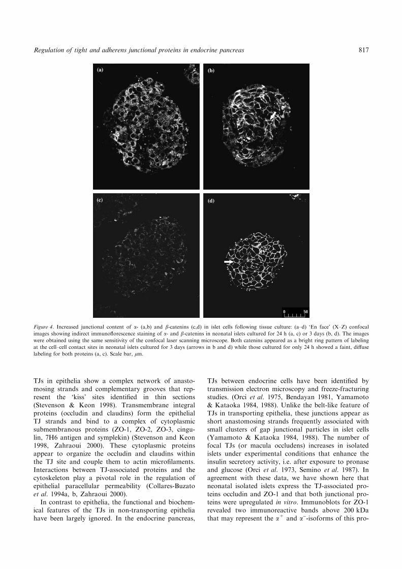
Figure 4. Increased junctional content of a- (a,b) and b-catenins (c,d) in islet cells following tissue culture: (a–d) ‘En face’ (X–Z) confocal
images showing indirect immunoflorescence staining of a- and b-catenins in neonatal islets cultured for 24 h (a, c) or 3 days (b, d). The images
were obtained using the same sensitivity of the confocal laser scanning microscope. Both catenins appeared as a bright ring pattern of labeling
at the cell–cell contact sites in neonatal islets cultured for 3 days (arrows in b and d) while those cultured for only 24 h showed a faint, diffuse
labeling for both proteins (a, c). Scale bar, lm.
817Regulation of tight and adherens junctional proteins in endocrine pancreas
tein since the antibody used recognized both ZO-1 iso-forms (manufacturer’s data). Interestingly, the presenceof the a)-isoform of ZO-1 has been associated withcells displaying structurally dynamic TJs, such aspodocytes, Sertoli cells and mammary gland cells(Balda & Anderson 1993, Stelwagen et al. 1999). Inthese cells, TJs can rapidly open and reseal.
ZO-1 appeared as discontinuous lines in the cell–cellcontact region and resembled the macula occludens seenultrastructurally between endocrine pancreatic cells.The finding that ZO-1 was also present in the endocrinepancreas of adult rats implies a possible role for this TJ-associated protein in this organ in situ. However, thisresult differs from other studies that found no evidencefor TJs in the normal endocrine pancreas of certain ani-mal species, including rats and humans (In’t Veld et al.1984, Yamamoto & Kataoka 1984). According to thesestudies, rather than being involved in normal islet cellfunction, TJs may provide an adaptative structure
intended to protect the islet microdomain against sud-den perturbations such as occuring during islet isola-tion. We have no explanation for the discrepancy ofthese studies and our data. However, it should be men-tioned that, although ZO-1 is an ubiquitous protein inTJ, in the absence of this junction this protein may asso-ciate with adheren-type junctions in other cells (Ho-warth et al. 1992, Itoh et al. 1993, 1997). The functionalimportance of TJs in the endocrine pancreas in siturequires further investigation.
Cultured neonatal islets also express the adherens-associated proteins a- and b- catenins. Catenins are cyto-plasmic proteins that connect cadherin adhesion mole-cules to actin filaments (Geiger & Ayalon 1992, Geigeret al. 1995, Harington & Syrigos 2000). Perturbation ofthe cadherin–catenin interaction or decreased expressionof either of the components of this complex invariablyleads to reduced cell–cell adhesion in several cell types(Collares-Buzato et al., 1998a, b, Harington & Syrigos
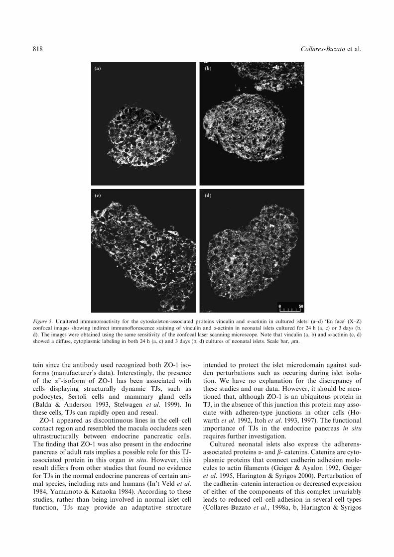
Figure 5. Unaltered immunoreactivity for the cytoskeleton-associated proteins vinculin and a-actinin in cultured islets: (a–d) ‘En face’ (X–Z)
confocal images showing indirect immunoflorescence staining of vinculin and a-actinin in neonatal islets cultured for 24 h (a, c) or 3 days (b,
d). The images were obtained using the same sensitivity of the confocal laser scanning microscope. Note that vinculin (a, b) and a-actinin (c, d)
showed a diffuse, cytoplasmic labeling in both 24 h (a, c) and 3 days (b, d) cultures of neonatal islets. Scale bar, lm.
818 Collares-Buzato et al.

2000, Ling et al. 2001, Liliem et al. 2002). Cadherins arecell surface glycoproteins responsible for Ca2+-depen-dent, homophilic intercellular adhesion. The cadherin–catenin also plays a pivotal role in cell migration,
differentiation, cell–cell recognition, cell fate and death(Geiger & Ayalon 1992, Geiger et al. 1995, Harington &Syrigos 2000). Islet cells express the E-cadherin subtype,as well as N-CAM, a Ca2+-independent cell adhesion
Figure 6. F-actin distribution in islets cultured for 24 h or 3 days: ‘En face’ (X–Z) confocal images showing cytochemistry for F-actin using
TRITC-phalloidin in neonatal islets cultured for 24 h (a) or 3 days (b). The images were obtained using the same sensitivity of the confocal
laser scanning microscope. Note the higher junctional labeling for F-actin in neonatal islets cultured for 3 days compared to those for only
24 h. Scale bar, lm.
Figure 7. Dual immunolabeling for b-catenin and insulin in islets cultured for 24 h or 3 days: (a–f) ‘En face’ (X–Z) confocal images showing
dual immunoflorescence staining of b-catenin (a, d) and insulin (b, e) in neonatal islets cultured for 24 h (a–c) or 3 days (d–f). All images were
obtained using the same sensitivity of the confocal laser scanning microscope. Note that insulin secreting B-cells in 24 h cultures showed no im-
munoreaction for b-catenin at this level of CLSM sensitivity (a–c), while in islets cultured for 7 days, B-cells displayed a bright ring pattern of
labeling at cell–cell contact sites (d–f). In addition, non-B-cells, in 3 day-cultured islet, also show junctional immunolabeling for b-catenin (f).
Scale bar, lm.
819Regulation of tight and adherens junctional proteins in endocrine pancreas
molecule (CAM) belonging to the immunoglobulinsuperfamily (Moller et al. 1992, Bernard-Kargar et al.2001). Non-B-cells express significantly higher levels ofN-CAM compared to B-cells, whereas E-cadherin isidentically expressed by both cell types. The differentialexpression of N-CAM has been accounted for the typi-cal topographical arrangement of cells within the isletcharacterized by a core of insulin-secreting B-cells sur-rounded by non-B-cells secreting glucagon, somato-statin and polypeptide P (Moller et al. 1992, Bernard-Kargar et al. 2001).
Exposure to tumor necrosis factor or antibodiesagainst E-cadherin results in an abnormal islet archi-
tecture and a decreased cell adhesion, respectively,which are associated with a decrease in insulin release(Cirulli et al. 1993, Yamagata et al. 2002). These find-ings reinforce the idea that CAM-mediated cell–cellcontact is crucial for a proper secretory response bythe endocrine pancreas. In agreement with this idea,we found an increase in the expression of catenins andin the junctional content of F-actin in neonatal isletscultured for more than 3 days. In addition, the immu-nolocalization of catenins at the intercellular region inthe adult endocrine pancreas and cultured neonatalislets agreed with the islet expression of E-cadherinshown by others. Catenins appear to be expressed sim-ilarly in both cell types, B- and non-B-cells, as revealedby dual immunolabeling for b-catenin and insulin.
In conclusion, endocrine pancreatic cells express sev-eral junctional proteins (occludin, ZO-1, and a- and b-catenins) that are upregulated following differentiationof the endocrine pancreas in vitro. Culturing neonatalislets also increased the junctional content of F-actin,but did not alter the expression of cytoskeleton-associ-ated proteins, such as vinculin and a-actinin. ZO-1 andb-catenin were observed in the endocrine pancreas ofadult rats, indicating that these junctional proteinswere also expressed in situ. Further studies in vitro andin situ on the IJs and their constitutive proteins willlead to a better understanding of the functional role ofthese important structures in B-cells.
Acknowledgements
We thank Baltazar Pereira de Paula and Marta BeatrizLeonardo for excellent technical assistance, and Dr.Renato Mortara (Department of Morphology; UNI-FESP, Brazil) for help in using the CLSM. AGF andCPFC were supported by PIBIC/CNPq and FAPESP(Brazil), respectively. The CLSM was acquired throughgrants from FAPESP (94/3737-1 and 96/0101-2). Addi-tional support for materials and equipments was pro-vided by FINEP/PRONEX, FAPESP (Grant 99/06846-0) and FAEP/UNICAMP. We thank Dr. Ste-phen Hyslop for the English review.
References
Allen E, Yu Q-C, Fuchs E (1996) Mice expressing a mutant desmo-
somal cadherin exhibit abnormalities in desmosomes, proliferation,
and epidermal differentiation. J Cell Biol 133: 1367–1382.
Balda MS, Anderson JM (1993) Two classes of tight junctions are
revealed by ZO-1 isoforms. Am J Physiol 264: C918–C924.
Bendayan M (1981) Contacts between endocrine and exocrine cells
in the pancreas. Cell Tissue Res 222: 227–230.
Bernard-Kargar C, Kassis N, Berthault M-F, Pralong W, Ktorza A
(2001) Sialylated form of the neural cell adhesion molecule
(NCAM): A new tool for the identification and sorting of b-cellsubpopulations with different functional activity. Diabetes 50:
S125–S130.
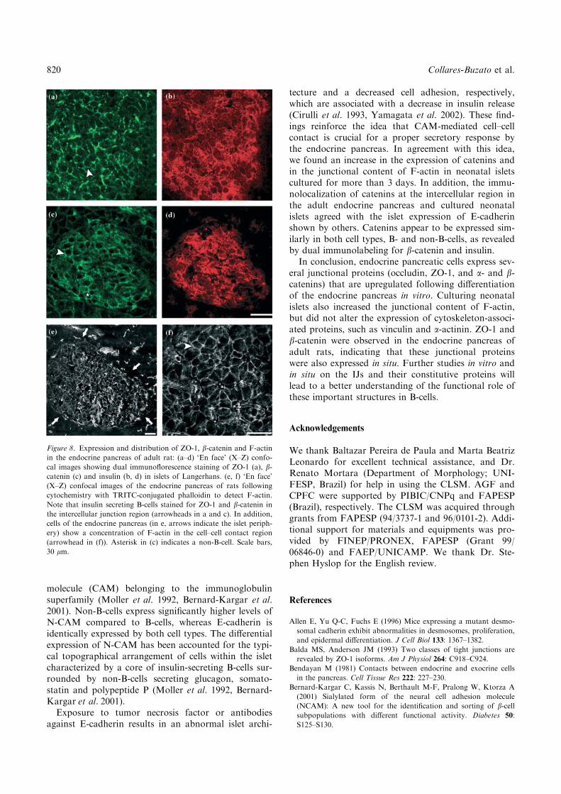
Figure 8. Expression and distribution of ZO-1, b-catenin and F-actin
in the endocrine pancreas of adult rat: (a–d) ‘En face’ (X–Z) confo-
cal images showing dual immunoflorescence staining of ZO-1 (a), b-catenin (c) and insulin (b, d) in islets of Langerhans. (e, f) ‘En face’
(X–Z) confocal images of the endocrine pancreas of rats following
cytochemistry with TRITC-conjugated phalloidin to detect F-actin.
Note that insulin secreting B-cells stained for ZO-1 and b-catenin in
the intercellular junction region (arrowheads in a and c). In addition,
cells of the endocrine pancreas (in e, arrows indicate the islet periph-
ery) show a concentration of F-actin in the cell–cell contact region
(arrowhead in (f)). Asterisk in (c) indicates a non-B-cell. Scale bars,
30 lm.
820 Collares-Buzato et al.

Boschero AC, Crepaldi SC, Carneiro EM, Delattre E, Atwater I
(1993) Prolactin induces maturation of glucose sensing mecha-
nisms in cultured neonatal rat islets. Endocrinology 133: 515–520.
Boschero AC, Tombaccini D, Atwater I (1988) Effects of glucose on
insulin release and 86Rb permeability in cultured neonatal and
adult rat islets. FEBS Lett 236: 375–379.
Bosco D, Orci L, Meda P (1989) Homologous but not heterologous
contact increases the insulin secretion of individual pancreatic B-
cells. Exp Cell Biol 184: 72–80.
Burdett IDJ (1998) Aspects of the structure and assembly of desmo-
somes. Micron 29: 309–328.
Cirulli V, Halban PA, Rouiller DG (1993) Tumor necrosis factor amodifies adhesion properties of rat islet B cells. J Clin Invest 91:
1868–1876.
Collares-Buzato CB, Jepson MA, McEwan GTA, Hirst BH, Sim-
mons NL (1998a) Co-culture of two MDCK strains with dis-
tinct junctional protein expression: A model for intercellular
jucntion rearrangement and cell sorting. Cell Tissue Res 291:
267–276.
Collares-Buzato CB, Jepson MA, Simmons NL, Hirst BH (1998b)
Increased tyrosine phosphorylation causes redistribution of adhe-
rens junction and tight junction proteins and perturbs paracellu-
lar barrier function in MDCK epithelia. Eur J Cell Biol 76: 85–
92.
Collares-Buzato CB, Jepson MA, McEwan GTA, Simmons NL,
Hirst BH (1994a) Junctional uvomorulin/E-cadherin and phosp-
hotyrosine-modified protein content are correlated with paracellu-
lar permeability in Madin–Darby canine kidney (MDCK)
epithelia. Histochemistry 101: 185–194.
Collares-Buzato CB, McEwan GTA, Jepson MA, Simmons NL,
Hirst BH (1994b) Paracellular barrier and junctional protein distri-
bution depend on basolateral extracellular Ca2+ iuvomorulin/E-
cadherin and phosphotyrosine-modified protein content are corre-
lated with paracellular permeability in Madin–Darby canine kid-
ney (MDCK) epithelia. Biochim Biophys Acta 1222: 147–158.
Collares-Buzato CB, Leite AR, Boschero AC (2001) Modulation of
gap and adherens junctional proteins in cultured neonatal pancre-
atic islets. Pancreas 23: 177–185.
Crepaldi SC, Carneiro EM, Boschero AC (1997) Long-term of pro-
lactin treatment on glucose-induced insulin secretion in cultured
neonatal rat islets. Horm Metab Res 29: 220–224.
Crepaldi-Alves SC, Carneiro EM, Boschero AC (1997) Synergistic
effect of glucose and prolactin on GLUT2 expression in cultured
neonatal rat islets. Braz J Med Biol Res 30: 359–361.
Dudek RW, Kawabe T, Brinn JE, O’Brien K, Poole MC, Morgan
CR (1984) Glucose affects in vitro maturation of fetal rat islets.
Endocrinology 114: 582–587.
Faquhar MG, Palade GE (1963) Junctional complexes in various
epithelia. J Cell Biol 17: 375–412.
Freinkel N, Lewis NJ, Johson R, Swenne I, Bone A, Hellerstrom C
(1984) Differential effects of age versus glycemic stimulation on
the maturation of insulin stimulus-secretion coupling during cul-
ture of fetal rat islets. Diabetes 33: 1028–1038.
Geiger B, Ayalon O (1992) Cadherins. Ann Rev Cell Biol 8: 307–332.
Geiger B, Yehuda-Levenberg S, Bershadsky AD (1995) Molecular
interactions in the submembrane plaque of cell–cell and cell–
matrix adhesions. Acta Anat 154: 46–62.
Gilula NB (1978) Structure of intercellular junctions. In: Feldman J,
Gilula NB, Pitts JD, eds. Intercellular Junctions and Synapses.
London: Chapman and Hall, pp. 3–22.
Gottardi CJ, Wong E, Gumbiner BM. (2001) E-cadherin supresses
cellular transformation by inhibiting beta-catenin signaling in an
adhesion-independent manner. J Cell Biol 153: 1049–1060.
Halban PA, Wollheim CB, Blondel B, Meda P, Niesor EN, Mintz DH
(1982) The possible importance of contact between pancreatic islet
cells for the control of insulin release. Endocrinology 111: 86–94.
Harington KJ, Syrigos KN (2000) The role of E-cadherin–catenin com-
plex: More than an intercellular glue? Ann Surg Oncol 7: 783–788.
Howarth AG, Hughes MR, Stevenson BR (1992) Detection of the
tight junction associated protein 20-1 in astrocytes and other none-
pithelial cell types. Am. J. Physiol. 262: 461–469.
In’t Veld PA, Pipeleers DG, Gepts W (1984) Evidence against the
presence of tight junctions in normal endocrine pancreas. Diabetes
33: 101–104.
Itoh M, Nagafuchi A, Moroi S, Tsukita S (1997) Involvement of
ZO-1 in cadherin-based cell adhesion through its direct binding to
a catenin and actin filaments. J. Cell. Biol. 138: 181–192.
Itoh M, Nagafuchi A, Yonemura S, Kitani-Yasuda T, Tsukita S,
Tsukita S (1993) The 220-kD protein colocalizing with cadherins
in non-epithelial cells is identical to ZO-1, a tight junction-
associated protein in epithelial cells: cDNA cloning and immunoe-
lectron microscopy. J. Cell Biol. 121: 491–502.
Li G, Satyamoorthy K, Herlyn M (2001) N-cadherin-mediated inter-
cellular interactions promote survival and migration of melanoma
cells. Cancer Res 61: 3819–3825.
Liliem J, Balsamo J, Arregui C, Xu G (2002) Turn-off, drop out,
functional state switching of cadherins. Dev Dyn 224: 18–29.
Ling Y-L, Zhong Y, Perez-Soler R (2001) Disruption of cell adhe-
sion and caspase-mediated proteolysis of b- and c-catenins and
APC protein in paclitaxel-induced apoptosis. Mol Pharmacol 59:
593–603.
Masaki Y, Tanigawa K, Ohguni S, Note S (1987) Rat fetal islets as
a useful model for the study of insulin release failure. Pancreas 2:
632–637.
Meda P, Bosco D, Chanson M, Giordano E, Vallar L, Wollheim C,
Orci L (1990) Rapid and reversible secretion changes during uncou-
pling of rat insulin-producing cells. J Clin Invest 86: 759–768.
Meda P, Chanson M, Pepper M, Giordano E, Bosco MD, Traub O,
Willecke K, Aoumari AE, Gros D, Beyer EC, Orci L, Spray DC
(1991) In vivo modulation of connexin 43 gene expression and
junctional coupling of pancreatic B-cell. Exp Cell Res 192: 469–
480.
Meda P, Michaelis RL, Halban PA, Orci L, Sheridan JD (1983) In
vivo modulation of gap junctions and dye coupling between B-cells
of the intact pancreatic islet. Diabetes 32: 858–868.
Meda P, Orci L (1979) Increase of gap junctions between pancreatic
B-cells during stimulation of insulin secretion. J Cell Biol 82: 441–
448.
Moller CJ, Christgau S, Williamson MR, Madsen OD, Zhan-Po N,
Bock E, Baekkeskov S (1992) Differential expression of neural cell
adhesion molecule and cadherins in pancreatic islets, glucagono-
mas, and insulinomas. Endocrinology 6: 1332–1342.
Orci L, Amherdt M, Henquin JC, Lambert AE, Unger RH, Renold
AE (1973) Pronase effect on pancreatic beta cell secretion and
morphology. Science 180: 647–649.
Orci L, Malaisse-Lagae F, Amherdt M, Ravazzola M, Weisswange
A, Dobbs R, Perrelet A, Unger R (1975) Cell contacts in human
islets of Langerhans. J Clin Endocrinol Metab 41: 841–844.
Pipeleers D, Veld PI, Maes E, Van de Winkel M (1994) Glucose-
induced insulin release depends on functional cooperation between
islet cells. Proc Natl Acad Sci USA 107: 1429–1436.
Semino MC, Gagliardino EEP, Gagliardino JJ (1987) Islet cells-tight
junctions: Changes in its number induced by glucose. Acta Physiol
Pharmacol Latinoam 37: 533–539.
Shimizu S, Jha B, Kagawa S, Nakao K, Yoshida K, Wakabayashi S,
Matsuoka A (1983) The effect of 2-deoxy-D-glucose on
insulin release by neonatal rat B cells in culture. Endocrinol Jpn 30:
255–60.
Stelwagen K, McFadden HA, Demmer J (1999) Prolactin, alone or
in combination with glucocorticoids, enhances tight junction for-
mation and expression of the tight junction protein occludin in
mammary cells. Mol Cell Endocrinol 156: 55–61.
Stevenson BR, Keon BH (1998) The tight junction: Morphology to
molecules. Annu Rev Cell Dev Biol 14: 89–109.
Yamagata K, Nammo T, Moriwaki M, Ihara A, Iizuka K, Yang Q,
Satoh T, Li M, Uenaka R, Okita K, Iwahashi H, Zhu Q, Cao Y,
821Regulation of tight and adherens junctional proteins in endocrine pancreas

Imagawa A, Tochino Y, Hanafusa T, Miyagawa J, Matsuzawa Y
(2002) Overexpression of dominant-negative mutant hepatocyte
nuclear factor-1a in pancreatic b-cells causes abnormal islet archi-
tecture with decreased expression of E-cadherin, reduced b-cellproliferation, and diabetes. Diabetes 51: 114–123.
Yamamoto M, Kataoka K (1984) A comparative study of the
intercellular canalicular system and intercellular junctions in
the pancreatic islets of some rodents. Arch Histol Jap 47:
485–493.
Yamamoto M, Kataoka K (1988) An electron microscope study of the
development of the exocrine and endocrine pancreas with special
reference to intercellular junctions. Arch Histol Cytol 51: 315–325.
Yderstraede KB, Flindt-Egebak P (1995) Neonatal rat islets of Lan-
gerhans and fetal rat pancreas. Isolation, immunohistochemical,
functional, and autoradiographic evaluation. Acta Diabetol. 32:
95–101.
Zahraoui A, Louvard D, Galli T (2000) Tight junction, a platform for
traffing and signalling protein complexes. J Cell Biol 151: F31–F36.
822 Collares-Buzato et al.
Copyright © 2022 FDOKUMEN