Developmental Upregulation of an Alternative Form of pcp2 with Reduced GDI Activity
8
ORIGINAL PAPER Developmental Upregulation of an Alternative Form of pcp2 with Reduced GDI Activity Jaroslaw J. Barski & Brad M. Denker & Jiazhen Guan & Matthias Lauth & Fabio Spreafico & Andrzej Fertala & Michael Meyer Published online: 5 October 2013 # The Author(s) 2013. This article is published with open access at Springerlink.com Abstract The pcp2/L7 gene is characterized by its very cell type-specific expression restricted to cerebellar Purkinje cells and retinal bipolar neurons. Although remarkable progress as to the biochemical properties of the encoded protein has been made, knowledge on its physiological functions remains sparse. While characterizing a pcp2-driven transgenic strain, we ob- served the presence of a longer, so far unknown, pcp2 transcript. Different from another recently discovered splice variant, ret-pcp2, expression of this novel transcript is observed in bipolar as well as cerebellar Purkinje cells of mid-postnatal mice. The protein encoded by our novel variant appears to be less efficient in binding to Gα subunits compared to the original L7/pcp2 protein and it is also less inhibitory with respect to GTPγ binding. Its expression in the eye appears to be independent from eye opening in postnatal mice. Keywords pcp2 . L7 . Splice variant . Cerebellum Introduction A remarkable feature of the pcp2/L7 gene is its cell type- specific expression. Since its discovery in 1988 [20, 21], its protein and mRNA products have been repeatedly localized exclusively to cerebellar Purkinje cells (PC) and retinal bipo- lar cells in various species, including mouse and human [4, 5, 15, 16, 22, 27, 28, 35]. High conservation of protein and DNA sequence, restricted expression, and the relatively late appearance of the gene products after neurogenesis [20, 21, 26, 32, 35] suggested important roles for neuronal maturation. Besides a partial re- semblance to platelet-derived growth factor [21], initial se- quence comparison failed to provide functional insight. Surprisingly, two different targeted null mutant mouse strains lacked functional or morphological alterations [17, 27]. More detailed studies however found, that mice null mutant for pcp2 show enhanced motor learning abilities [11]. Studies into the cell biology of pcp2 revealed an interesting dissociation of mRNA and protein localization in PC, with the former accu- mulating in dendrites and the latter being more equally distrib- uted within all cellular compartments [5, 28, 34, 35]. Moreover, the potential for activity-dependent regulation of pcp2 was demonstrated [29]. Insight into function was boosted with the discovery of interaction of pcp2 protein with Gα subunits [12–14, 18, 19, 22, 31], and the recognition of other Gα- interacting proteins sharing a structural feature, the GoLOCO motif, with pcp2 [23]. While characterizing a pcp2-driven Jaroslaw J. Barski and Brad M. Denker contributed equally to this work. J. J. Barski (*) Center for Experimental Medicine, Medical University of Silesia, ul. Medykόw 4, 40-752 Katowice, Poland e-mail: [email protected] J. J. Barski Department of Physiology, Medical University of Silesia, 40-752 Katowice, Poland B. M. Denker : J. Guan Department of Medicine, Renal Division, Brigham and Women’ s Hospital and Harvard Medical School, Boston, MA 02115, USA M. Lauth Institute of Molecular Biology and Tumor Research, Philipps Universität, 35032 Marburg, Germany F. Spreafico Formerly Max-Planck-Institute of Neurobiology, 82152 Martinsried, Germany A. Fertala Department of Orthopaedic Surgery, Thomas Jefferson University, Philadelphia, PA 19107, USA M. Meyer Institute of Physiology, Ludwig-Maximilians University, 80336 Munich, Germany Cerebellum (2014) 13:207–214 DOI 10.1007/s12311-013-0529-0
-
Upload
independent -
Category
Documents
-
view
0 -
download
0
Transcript of Developmental Upregulation of an Alternative Form of pcp2 with Reduced GDI Activity
ORIGINAL PAPER
Developmental Upregulation of an Alternative Form of pcp2with Reduced GDI Activity
Jaroslaw J. Barski & Brad M. Denker & Jiazhen Guan &
Matthias Lauth & Fabio Spreafico & Andrzej Fertala &
Michael Meyer
Published online: 5 October 2013# The Author(s) 2013. This article is published with open access at Springerlink.com
Abstract The pcp2/L7 gene is characterized by its very celltype-specific expression restricted to cerebellar Purkinje cellsand retinal bipolar neurons. Although remarkable progress asto the biochemical properties of the encoded protein has beenmade, knowledge on its physiological functions remains sparse.While characterizing a pcp2-driven transgenic strain, we ob-served the presence of a longer, so far unknown, pcp2 transcript.Different from another recently discovered splice variant,ret-pcp2, expression of this novel transcript is observed in bipolaras well as cerebellar Purkinje cells of mid-postnatal mice. Theprotein encoded by our novel variant appears to be less efficient
in binding to Gα subunits compared to the original L7/pcp2protein and it is also less inhibitory with respect to GTPγbinding. Its expression in the eye appears to be independentfrom eye opening in postnatal mice.
Keywords pcp2 . L7 . Splice variant . Cerebellum
Introduction
A remarkable feature of the pcp2/L7 gene is its cell type-specific expression. Since its discovery in 1988 [20, 21], itsprotein and mRNA products have been repeatedly localizedexclusively to cerebellar Purkinje cells (PC) and retinal bipo-lar cells in various species, including mouse and human[4, 5, 15, 16, 22, 27, 28, 35].
High conservation of protein and DNA sequence, restrictedexpression, and the relatively late appearance of the geneproducts after neurogenesis [20, 21, 26, 32, 35] suggestedimportant roles for neuronal maturation. Besides a partial re-semblance to platelet-derived growth factor [21], initial se-quence comparison failed to provide functional insight.Surprisingly, two different targeted null mutant mouse strainslacked functional or morphological alterations [17, 27]. Moredetailed studies however found, that mice null mutant for pcp2show enhanced motor learning abilities [11]. Studies into thecell biology of pcp2 revealed an interesting dissociation ofmRNA and protein localization in PC, with the former accu-mulating in dendrites and the latter being more equally distrib-uted within all cellular compartments [5, 28, 34, 35]. Moreover,the potential for activity-dependent regulation of pcp2 wasdemonstrated [29]. Insight into function was boosted with thediscovery of interaction of pcp2 protein with Gα subunits[12–14, 18, 19, 22, 31], and the recognition of other Gα-interacting proteins sharing a structural feature, the GoLOCOmotif, with pcp2 [23]. While characterizing a pcp2-driven
Jaroslaw J. Barski and Brad M. Denker contributed equally to this work.
J. J. Barski (*)Center for Experimental Medicine, Medical University of Silesia, ul.Medykόw 4, 40-752 Katowice, Polande-mail: [email protected]
J. J. BarskiDepartment of Physiology, Medical University of Silesia,40-752 Katowice, Poland
B. M. Denker : J. GuanDepartment of Medicine, Renal Division, Brigham and Women’sHospital and Harvard Medical School, Boston, MA 02115, USA
M. LauthInstitute of Molecular Biology and Tumor Research, PhilippsUniversität, 35032 Marburg, Germany
F. SpreaficoFormerly Max-Planck-Institute of Neurobiology,82152 Martinsried, Germany
A. FertalaDepartment of Orthopaedic Surgery, Thomas Jefferson University,Philadelphia, PA 19107, USA
M. MeyerInstitute of Physiology, Ludwig-Maximilians University,80336 Munich, Germany
Cerebellum (2014) 13:207–214DOI 10.1007/s12311-013-0529-0

transgenic strain [2, 3], we realized the presence of a longerpcp2 transcript. Subsequently, we determined its structure,expression pattern, activity-dependent regulation, and subcel-lular localization as well as key biochemical properties of theencoded protein. The alternative transcript characterized here isdifferent from another recently published product [33, 35].
Material and methods
C57Bl/6 mice were used for all experiments. To determine theinfluence of light on pcp2 expression, adult mice or motherswith their litters were kept in darkness, and then sacrificed inthe dark. Controls were housed under standard light conditions(12 h light/12 h dark). All animal holding and experimentswere done in accordance with the national animal welfareregulations and institutional rules.
RNA isolation was performed as described elsewhere [8]. Forreverse transcription, 1 μg of total RNA and 0.5 μg oligo-dTprimer (GIBCO BRL) were mixed in 12 μl of Tris-EDTA (TE)buffer pH 7.4 and then incubated for 5 min at 90 °C, 10 min at37 °C and 15 min at room temperature. Each sample was thenincubated at 37 °C for 1 h with 4 μl 5× first-strand buffer(GIBCO BRL), 2 μl DTT (0.1 M), 0.5 μl of 0.25 mM dNTPmix, 0.5 μl RNAse inhibitor (40 U/μl, Boehringer), and 0.8 μlreverse transcriptase (200 U/μl, SuperScript, GIBCO BRL).Afterwards, 10 μl of TE buffer were added to obtain a totalvolume of 30 μl.
PCR (36 cycles) was carried out on 1 μl of cDNA in areaction mixture containing 1× PCR buffer, 1.5 mM MgCl2,0.4 μMof each primer, and 0.625 U Taq polymerase (GIBCOBRL) according to the scheme: 5′95 °C [30″95 °C, 30″72 °C,1′72 °C] 5′72 °C.
To control for sample loading, reverse transcription poly-merase chain reaction (RT-PCR) for GAPDH (21 cycles) wascarried out for all samples. The whole reaction mixture (25 μl)was loaded on 2 % agarose gels containing ethidium bromide.
Both splice variants were detected with primers: L7sense 5′-AAGGCTTCTTCAACCTGCTGA-3′ and L7anti 5′-GCTGTTCCTGCGGAAGCTGAG-3′. The new splice variant wasspecifically detected with primers: L7 sense (as above) andL73Aanti 5′-TCCCAGTACTCAAGAAACAGG-3′ (Fig. 1b).GAPDH expression was detected with primers: sense 5′-ACCACAGTCCATGCCATCAC-3′ and antisense 5′-TCCACCACCCTGTTGCTGTA-3′.
To verify the PCR products, bands of interest were extractedfrom the gel, subcloned into the pCRII plasmid (TA CloningKit, Invitrogen) and sequenced on an ABI PRISM 377 DNASequencer (Perkin-Elmer). Sequence comparisons wereperformed using BLAST (NCBI, Bethesda, MD, USA).
Molecular modeling was performed using the SYBYL soft-ware, version 7.2 (Tripos Inc.). The model was generated, inpart, by homology modeling with filamentous hemagglutinin
secretion domain (Protein Data Bank, ID code 1rwr) as atemplate. The model was energy-minimized using a conjugategradient method and subjected to repeating cycles of moleculardynamics using Kollman force fields and united atoms [30].
In situ hybridization for detection of the novel transcriptwas carried out at 38 °C according to a protocol describedelsewhere [24]. The oligonucleotide DNA probe was comple-mentary to base pairs 2,461–2,507 of the pcp2 genomic se-quence (Fig. 1a; accession number S40022) [25]. The hybrid-ization specificity was checked by competing with 100× ex-cess of the cold probe.
As described later (see “Results” section), the novel longertranscript encodes a shorter protein variant than the knownshorter transcript. We refer to the native proteins as long pcp2and short pcp2 proteins, respectively.
For the generation of recombinant glutathione S-transferase(GST)–pcp2 fusion proteins, cDNAs corresponding to shortand longer transcript were cloned into the EcoRI and XhoI sitesof the pGEX-4T-1 vector (GE Healthcare) in frame with theglutathione-S-transferase/thrombin cleavage site. A single di-rect primer (L7Eco.d: AGG GCG AAT TCATGG AGG AGCAGC GCT GTT C), containing an EcoRI site, was used toamplify both forms of pcp2 from mouse cerebellar cDNA. Asreverse primers, both containing an XhoI restriction site,L7lgXho.r ( TAG GAT CTC GAG AAC TCT CAA GGAGCTTGTG) provided specificity for the short transcript, whileL7shXho.r (AAC AGG CTCGAGGAT TAT TTT GAG TTCAAA GC) amplified the long transcript only.
The resulting plasmids (GLL7 for the long form and GSL7for the short form) were transformed into the JM109Escherichia coli to yield recombinant proteins (we refer tothe GST-short pcp2 protein as SL7 and to the GST-long pcp2protein as L7) as was the empty pGEX vector for the produc-tion of GST alone as a control. Proteins were obtained after1 mM IPTG induction and purified by sepharose–glutathioneaffinity chromatography. Purity and integrity of the recombi-nant fusion proteins were verified by sodium dodecyl sulfate-polyacrylamide gel electrophoresis (SDS-PAGE; not shown).
Recombinant baculovirus expressed purified Gαo and Gαi1[10] were kindly provided by Steve Graber (Morgantown West,VA, USA) and were preincubated at 1:1 M ratio (2.5 pmol) withpurified noncleaved GST fusion proteins for AGS3 (cDNAprovided by Steve Lanier, New Orleans, LA, USA), long pcp2,short pcp2, and GSTalone in buffer A (20 mMHEPES, pH 8.0,130mMNaCl, 3 mMMgCl2, 1 mMDTT, 1mMEDTA, 10 μg/μl, bovine serum albumin, and 0.1%TritonX-100) for 20min at25 °C in the presence of 1 μM GDP. At t=0, GTPγS35 (1 μM;specific activity, 1×10⁵ cpm/pmol, New England Nuclear) wasadded for 60min. Reactions were terminated by dilution into ice-cold phosphate-buffered saline (PBS) and rapidly filteredthrough 25 mm round nitrocellulose membranes (Millipore,Billerica, MA, USA; 0.45 μM HA), washed twice with 20 mlof cold PBS. Filters were placed in scintillation fluid and counted
208 Cerebellum (2014) 13:207–214
in a Packard Tri-Carb A2200 liquid scintillation counter. Blankfilters (no protein) were used to determine background and non-specific binding was determined with parallel filters incubatedwith excess (0.1 mM) cold GTPγS (Sigma).
In vitro translated Gα subunits were [S35]-methionine labeledand prepared as previously described using the TNT coupledin vitro translation system (Promega) [9]. Similar amounts oftranslate (5–10 μl) were incubated with 0.3 μg of GST, AGS3,long pcp2, or short pcp2 for 2 h at room temperature in bufferA +0.1 % Triton X-100, 150 mM NaCl, and 20 μM GDP(binding buffer). Glutathione agarose beads were added for
30min at room temperature, centrifuged, andwashed three timeswith 1 ml ice-cold binding buffer. Samples were eluted in 2×SDS-PAGE sample buffer by heating at 100 °C for 5 min andanalyzed by SDS-PAGE and autoradiography.
Results
RT-PCR on eye and cerebellar RNA using primers placed inexon 2 (L7sense) and 4 (L7anti) (Fig. 1b), consistently detected anovel 350 bp product together with the 300 bp band expected
2333 TGTTTGTTTTAG GTATACACTAAC 2416exon 3B
TERTATTTAGCTTTGAACTCAAAA TCCTCCTGCCTGTTTCTTGAGTACTGGGATTACAGTAATATTTAGCTTTGAACTCAAAA TCCTCCTGCCTGTTTCTTGAGTACTGGGATTACAGTAA
b
c
d
short pcp2
long pcp2
MEEQRCSLQAGPGQNPESQGGPAPEMDNLMDMLVNTQGRRMDDQRVTVDSLPGFQPIGPKYLALNSK
ATG
4kb 300
70 115 125 58 182
150 754 134 2kbI IIII IIIIII IVIV
a
1 3 2
short pcp2
long pcp2
MEEQRCSLQAGPGQNPESQGGPAPEMDNLMDMLVNTQGRRMDDQRVTVDSLPGFQPIGPKDGMQKRPGTLSPQPLLTPQDPAALSFRRNSSPQPQTQAP
short pcp2long pcp2
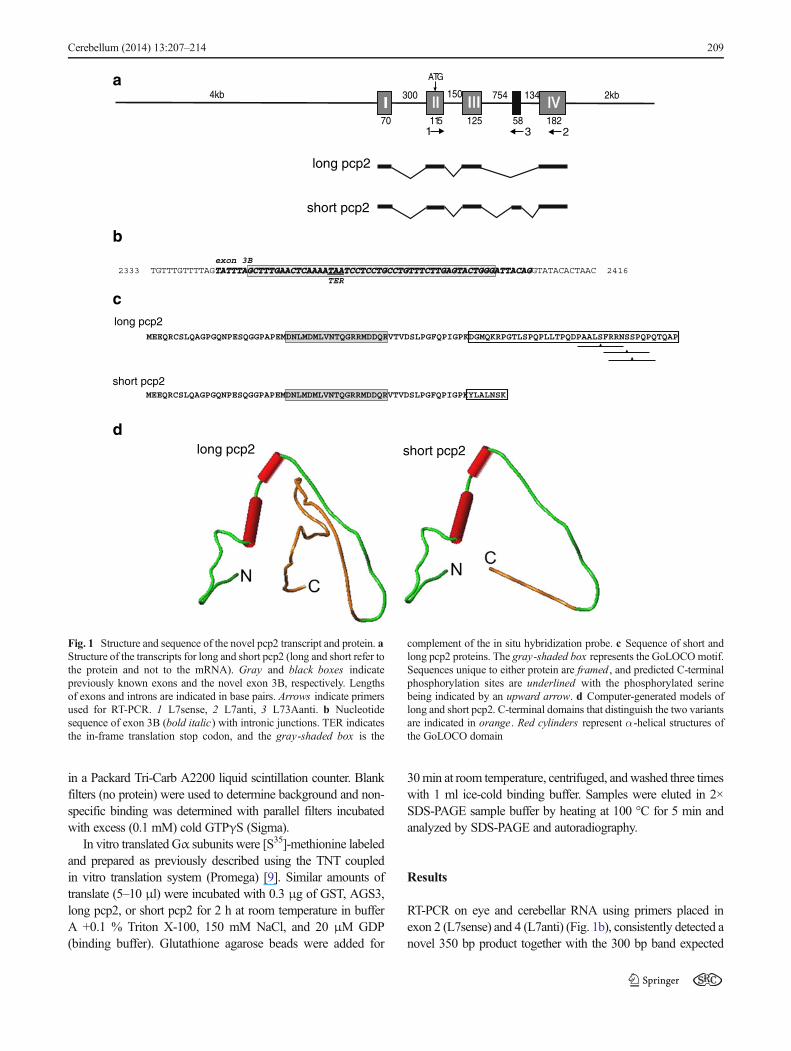
Fig. 1 Structure and sequence of the novel pcp2 transcript and protein. aStructure of the transcripts for long and short pcp2 (long and short refer tothe protein and not to the mRNA). Gray and black boxes indicatepreviously known exons and the novel exon 3B, respectively. Lengthsof exons and introns are indicated in base pairs. Arrows indicate primersused for RT-PCR. 1 L7sense, 2 L7anti, 3 L73Aanti. b Nucleotidesequence of exon 3B (bold italic) with intronic junctions. TER indicatesthe in-frame translation stop codon, and the gray-shaded box is the
complement of the in situ hybridization probe. c Sequence of short andlong pcp2 proteins. The gray-shaded box represents the GoLOCOmotif.Sequences unique to either protein are framed , and predicted C-terminalphosphorylation sites are underlined with the phosphorylated serinebeing indicated by an upward arrow. d Computer-generated models oflong and short pcp2. C-terminal domains that distinguish the two variantsare indicated in orange. Red cylinders represent α-helical structures ofthe GoLOCO domain
Cerebellum (2014) 13:207–214 209

from the known pcp2 gene structure (Fig. 2). Sequencing andanalysis of the RT-PCR product revealed a novel stretch of 58 bpbetween 2,455 and 2,513 bp within intron 3 (Fig. 1a). Thisanalysis also revealed splice donor and acceptor sites flankingthis exon. The novel exon, dubbed 3B, can be included orexcluded resulting in two different pcp2 transcripts (Fig. 1b). Inthe following, we will refer to the originally published exon 3 asexon 3A. Interestingly, when included, exon 3B introduces an inframe stop codon. Thus, exon 4 does not contribute to the codingof this alternative product. As the translated product is 32 aminoacids shorter than the originally published protein (here referredto as long pcp2 protein), we call it short pcp2 protein. TheGoLOCO motif of long pcp2 is found between amino acids 27and 45, and is entirely encoded by exon 3A. It is, therefore, notaltered in short pcp2. The C-terminal, exon 4-encoded part ofpcp2 contains a cluster of predicted phosphorylation sites (scoreshigher than 0.95; NetPhos 2.0, CBS Technical University ofDenmark) [6]. These are a putative PKC site at serine 85(PAALSFRNN), a CaM kinase II/PKA site around serine 90
(FRRNSSPQP), and a Cam kinase II/PKA/PKG site aroundserine 91 (Fig. 1c). Two of them are conserved between mouse,rat, and human [35]. With the exception of another predicted siteat serine 18 (QNPSESQGGP), none of the putative phosphory-lation sites of long pcp2 is present in the short pcp2.
Computer analysis of long and short pcp2 protein variantsdid not reveal many characteristic structural motives. Exceptfor α-helical domains corresponding to the GoLOCO motif,the proteins are essentially characterized by an extendedstructure (Fig. 1d).
To study expression of the novel transcript, we designed aprimer placed in the novel exon 3B (L73Aanti; Fig. 1b). RT-PCRusing primers L7sense and L73Aanti confirmed the presence ofthe novel transcript in eye and cerebellum of adult mice (Fig. 2).During development, expression of the novel form is first detect-ed between P12 and P14 in eye and cerebellum. The presence ofthe long form was observed already at P7 (data not shown).
To localize the novel transcript at the cellular level, in situhybridization was performed on cerebellar and retinal sections
P1 P7 P12 P14 P19 6M
GAPDH
Short pcp2
cere
bellu
mce
rebe
llum
cere
bellu
mce
rebe
llum
cere
bellu
mce
rebe
llum
cere
bellu
mce
rebe
llum
cere
bellu
mce
rebe
llum
cere
bellu
m
eye
eye
eye
eye
eye
eye
eye
eye
eye
eye
eye
Fig. 2 Developmentalexpression of the novel pcp2transcript. Transcripts encodingshort pcp2 protein were detectedby transcript-specific RT-PCR atthe indicated postnatal time points(upper panel)
fPE
R C&
ONL
OPL
INL
IPLGCL
ed
gpm
a
c
bFig. 3 Localization of transcriptscoding for the novel short pcp2protein by in situ hybridizationin adult C57BL/6 mice. a Darkfield imaging of the cerebellar.g Granular cell layer, p Purkinjecell layer, m molecular layer.Bar 100 μm. b Control section.Bar 100 μm. c Bright fieldpicture of Purkinje cells showingsilver grains over cell bodies ofPurkinje cells (arrow heads). Bar100 μm. d Phase contrast imageof mouse retinal layers. PEpigment epithelium, R&C rodand cone photoreceptors, ONLouter nuclear layer, OPL outerplexiform layer, INL innernuclear layer, IPL inner plexiformlayer, GCL ganglion cell layer.Bar 100 μm. e Dark field imageof a hybridized retina section.Bar 100 μm. f Control section.Bar 100 μm
210 Cerebellum (2014) 13:207–214

using an oligonucleotide probe corresponding to 46 bp ofexon 3B. Microscopic analysis revealed specific signals with-in the cerebellar cortex at the level of the PC layer (Fig. 3a, b).Higher magnification microscopy revealed the presence ofsilver grains over the cell bodies of PC (Fig. 3c). In sectionsof the retina, labeling was detected in the outer part of innernuclear layer, where bipolar neurons are localized (Fig. 3d, e).Specific signal was also found at the outer plexiform layer.
Because expression of transcripts encoding the novel shortpcp2 protein was first detected around the time of eye openingin mice (P12-14) [25], we wondered whether it would be mod-ulated by maintenance of mice in the dark (Fig. 4). Pcp2 expres-sion was analyzed in adult animals housed in the dark for 60 h or6 days, respectively, and compared to animals kept under standardlight–dark conditions (Fig. 4a). After the indicated time, animalswere sacrificed in the dark, and tissue samples were immediatelydissected for analysis. RT-PCR using the exon 2–exon 4 primerpair detected the presence of both transcripts in the eye and thecerebellum. It failed, however, to reveal any differences betweengroups. To investigate a possible induction of expression by lightat the time of eye opening, P1 litters were maintained in the darktogether with their mothers (Fig 4b). Pups were sacrificed in thedark at P15 and P19, tissues dissected and RT-PCR performed asabove. Controls were pups kept under standard light–dark condi-tions. Again, both splice variants were detected in both tissues atlevels apparently not influenced by dark rearing.
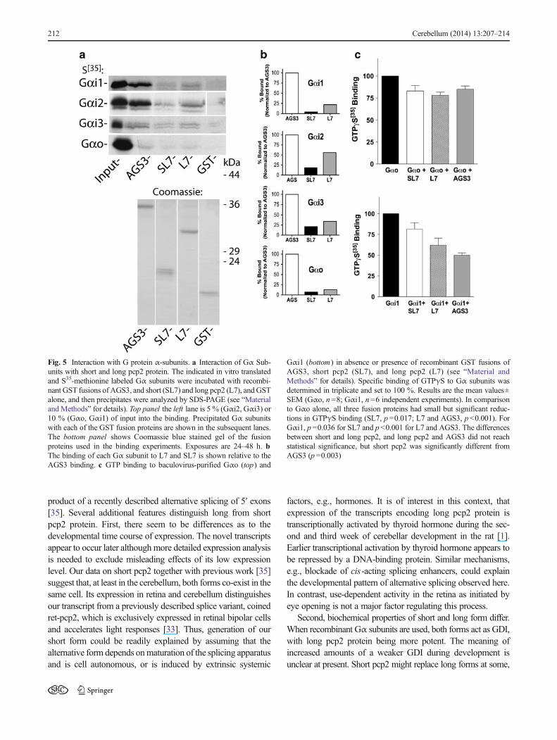
To investigate whether short pcp2 protein functions as aGDI for Gα subunits, we first examined whether short pcp2interacts with Gα subunits. Figure 5a shows in vitro translatedand [S35]-methionine labeled Gαo, Gαi1, 2, and 3 used inGST pull-down experiments with GST (negative control),AGS (positive control), long pcp2 (L7), and short pcp2(SL7). The GST proteins are shown in the Coomassie-stainedgel below and the amount of labeled Gα subunit precipitatingunder each condition is shown in the autoradiogram. The relativeinteraction of SL7 and L7 with Gαi1, Gαi-2, Gαi-3, and Gαo issummarized in the bar graphs (Fig. 5b) relative to the AGS3binding for this experiment. The experiment was repeated twicewith similar results. It appears that for all four tested Gα sub-units, the preferential binding was AGS>>long pcp2>shortpcp2. Albeit, binding of SL7 was weak when compared toAGS3 it was higher than binding of GST alone. Importantly,binding of SL7 was clearly less than that of LL7. As expectedfrom other reports [18], binding to Gαo was weak for all threeGST fusion proteins in comparison with the Gαi family. Theredid not appear to be any preference for short pcp2 in comparisonwith long pcp2 for interacting with Gαo or any of the Gαisubunits.
We next examined the GDI potency of short pcp2 proteinin comparison with long pcp2 and AGS3 (Fig. 5c).Baculovirus-purified Gαo and Gαi1 were used for theseexperiments and GTPγS[35] binding to each Gα subunit wascompared in the presence and absence of GST (negativecontrol and binding set to 100 %), GST-pcp2 long (L7),GST-pcp2 short (SL7), and GST-AGS3 under conditions verysimilar to those previously used to characterize differencesbetween pcp2 and AGS proteins (e.g., [7]). The degree ofGTPγS[35] inhibition in these conditions mirrored the bindingresults shown in Fig. 5a, b. There was a small but consistentinhibition of GTPγS[35] binding to purified Gαo (Fig. 5c) andthere was no significant difference in inhibition for AGS3,short or long pcp2. However, the inhibition of GTPγS[35]
binding to Gαi1 revealed relative potencies indicated alreadyby the binding experiments; AGS3>long pcp2>short pcp2.There was no significant difference in inhibition of GTPγS[35]
binding to Gαi1 for short or long pcp2, but short pcp2 proteinwas significantly different from control and AGS3. Takentogether, the GST pulldowns and GTPγS[35] binding resultsare consistent with short pcp2 binding less avidly to Gαi1 andtherefore being a slightly less effective GDI than long pcp2.
Discussion
Our study describes alternative splicing of the pcp2/L7 gene.The protein product encoded by the novel long transcript(short pcp2 protein) lacks most of the carboxy-terminal half ofthe originally described protein (long pcp2 protein), but stillcontains the N-terminal GPR motif. It is different from the
60hours
6days control
GAPDH
cerebellu
m
cerebellu
m
cerebellu
meye
eyeeye
- 300bp- 350bp
control
- 300bp- 350bp
P 19
GAPDH
cere
bellu
m
cere
bellu
m
cere
bellu
m
eye
eye
eye
a
bP 15 P 19
Fig. 4 Influence of dark breeding on expression of pcp2. a Adult C57Bl/6mice were kept in darkness for 60 h or 6 days after which tissues wereprocessed for RT-PCR common for both transcripts. b P1 pups were keptin darkness with their mothers until tissue dissection at P15 and P19.Controls were kept under standard light conditions. The length of theamplimers is given in base pairs
Cerebellum (2014) 13:207–214 211
product of a recently described alternative splicing of 5′ exons[35]. Several additional features distinguish long from shortpcp2 protein. First, there seem to be differences as to thedevelopmental time course of expression. The novel transcriptsappear to occur later although more detailed expression analysisis needed to exclude misleading effects of its low expressionlevel. Our data on short pcp2 together with previous work [35]suggest that, at least in the cerebellum, both forms co-exist in thesame cell. Its expression in retina and cerebellum distinguishesour transcript from a previously described splice variant, coinedret-pcp2, which is exclusively expressed in retinal bipolar cellsand accelerates light responses [33]. Thus, generation of ourshort form could be readily explained by assuming that thealternative form depends onmaturation of the splicing apparatusand is cell autonomous, or is induced by extrinsic systemic
factors, e.g., hormones. It is of interest in this context, thatexpression of the transcripts encoding long pcp2 protein istranscriptionally activated by thyroid hormone during the sec-ond and third week of cerebellar development in the rat [1].Earlier transcriptional activation by thyroid hormone appears tobe repressed by a DNA-binding protein. Similar mechanisms,e.g., blockade of cis-acting splicing enhancers, could explainthe developmental pattern of alternative splicing observed here.In contrast, use-dependent activity in the retina as initiated byeye opening is not a major factor regulating this process.
Second, biochemical properties of short and long form differ.When recombinant Gα subunits are used, both forms act as GDI,with long pcp2 protein being more potent. The meaning ofincreased amounts of a weaker GDI during development isunclear at present. Short pcp2 might replace long forms at some,
Fig. 5 Interaction with G protein α-subunits. a Interaction of Gα Sub-units with short and long pcp2 protein. The indicated in vitro translatedand S35-methionine labeled Gα subunits were incubated with recombi-nant GST fusions of AGS3, and short (SL7) and long pcp2 (L7), and GSTalone, and then precipitates were analyzed by SDS-PAGE (see “Materialand Methods” for details). Top panel the left lane is 5 % (Gαi2, Gαi3) or10 % (Gαo, Gαi1) of input into the binding. Precipitated Gα subunitswith each of the GST fusion proteins are shown in the subsequent lanes.The bottom panel shows Coomassie blue stained gel of the fusionproteins used in the binding experiments. Exposures are 24–48 h. bThe binding of each Gα subunit to L7 and SL7 is shown relative to theAGS3 binding. c GTP binding to baculovirus-purified Gαo (top) and
Gαi1 (bottom) in absence or presence of recombinant GST fusions ofAGS3, short pcp2 (SL7), and long pcp2 (L7) (see “Material andMethods” for details). Specific binding of GTPγS to Gα subunits wasdetermined in triplicate and set to 100 %. Results are the mean values±SEM (Gαo, n =8; Gαi1, n=6 independent experiments). In comparisonto Gαo alone, all three fusion proteins had small but significant reduc-tions in GTPγS binding (SL7, p=0.017; L7 and AGS3, p <0.001). ForGαi1, p =0.036 for SL7 and p <0.001 for L7 and AGS3. The differencesbetween short and long pcp2, and long pcp2 and AGS3 did not reachstatistical significance, but short pcp2 was significantly different fromAGS3 (p=0.003)
212 Cerebellum (2014) 13:207–214

but not all, specific intracellular sites. Transfection ex-periments so far failed to reveal such specific intracellu-lar targeting (data not shown). This might be due tomasking caused by overexpression of the protein.Alternatively, the upregulation of the short form mighttune the interaction of the long form in homo- orheteromeric molecular complexes. Our data indicate thatrelative expression of short to long variant is stronger inretina than cerebellum. This might suggest that retinalbipolar cells, which are known to express an additionalspecific pcp2 isoform [33], might be particularly depen-dent on fine tuning of pcp2 and G-protein function. It isinteresting to speculate on the differing potencies of GDIactivity of long and short pcp2. Both proteins contain asingle GoLOCO motif, so the differences are likely toresult from important other conformational or structuraldifferences between the isoforms. Further differentiationin the biochemical properties of the short variant mightdepend on the use of the alternative exon 1B, whichwould introduce a second GoLOCO motif [35]. We can-not exclude that functional differences between bothforms are stronger when additional Gα subunits aretested. In addition, there are intriguing differences inputative post-translational modifications of short andlong pcp2.
A cluster of phosphorylation sites predicted in the C-terminus of long pcp2 is lost in the short variant. This includesa site for phosphorylation by protein kinase G, which isabundantly expressed in PC, and possibly also in retinalbipolar cells. Further experimentation has to be awaited tosee whether pcp2 is indeed a substrate for G kinase, andsubsequently, how hyperphosphorylation would interfere withthe developmental function of pcp2.
As indicated by the work on ret-pcp2 [33], pcp2 splicevariants subserve delicate and important biological roles.These might depend on the proportion of the differentsplice forms and their products. Future progress in under-standing the biological function of pcp2 will require acareful comprehensive analysis of the expression of all itsdifferent splice variants.
Acknowledgments We are grateful to Steve Graber and Steve Lanierfor provision of materials and B. Kunkel and A. Steinberg for excellenttechnical assistance. Many thanks to Hans Thoenen, Y-A Barde, and toour colleagues at the Institute of Ophthalmology, University CollegeLondon.
Conflict of Interest The authors declare that the research wasconducted in the absence of any commercial or financial relationshipsthat could be construed as a potential conflict of interest.
Open Access This article is distributed under the terms of the CreativeCommons Attribution License which permits any use, distribution, andreproduction in any medium, provided the original author(s) and thesource are credited.
References
1. Anderson GW, Larson RJ, Oas DR, Sandhofer CR, Schwartz HL,Mariash CN, et al. Chicken ovalbumin upstream promoter-transcriptionfactor (COUP-TF) modulates expression of the Purkinje cell protein-2gene. A potential role for COUP-TF in repressing premature thyroidhormone action in the developing brain. J Biol Chem. 1998;273:16391–9.
2. Barski JJ, Dethleffsen K, Meyer M. Cre recombinase expression incerebellar Purkinje cells. Genesis. 2000;28:93–8.
3. Barski JJ, Lauth M, Meyer M. Genetic targeting of cerebellarPurkinje cells: history, current status and novel strategies. Cerebel-lum. 2002;1:111–8.
4. Berrebi AS, Oberdick J, Sangameswaran L, Christakos S, Morgan JI,Mugnaini E. Cerebellar Purkinje cell markers are expressed in retinalbipolar neurons. J Comp Neurol. 1991;308:630–49.
5. Bian F, Chu T, Schilling K, Oberdick J. Differential mRNA transportand the regulation of protein synthesis: selective sensitivity ofPurkinje cell dendritic mRNAs to translational inhibition. Mol CellNeurosci. 1996;7:116–33.
6. Blom N, Gammeltoft S, Brunak S. Sequence and structure-basedprediction of eukaryotic protein phosphorylation sites. J Mol Biol.1999;294:1351–62.
7. Cao X, Cismowski MJ, Sato M, Blumer JB, Lanier SM. Identifica-tion and characterization of AGS4: a protein containing threeG-protein-regulatory motifs that regulate the activation state ofGialpha. J Biol Chem. 2004;279:27567–74.
8. Chomczyński P, Sacchi N. Single-step method of RNA isolation byacid guanidinium thiocyanate-phenol-chloroform extraction. AnalBiochem. 1987;162:156–9.
9. Denker BM, Boutin PM, Neer EJ. Interactions between the amino-and carboxyl-terminal regions of G.alpha subunits: analysis of mutat-ed G.alpha.o/G.alpha.i2 chimeras. Biochemistry. 1995;34:5544–53.
10. Graber SG, Figler RA, Garrison JC. Expression and purification offunctional G protein alpha subunits using a baculovirus expressionsystem. J Biol Chem. 1992;267:1271–8.
11. Iscru E, Serinagaoglu Y, Schilling K, Tian J, Bowers-Kidder SL,Zhang R, et al. Sensorimotor enhancement in mouse mutants lackingthe Purkinje cell-specific Gi/o modulator, Pcp2(L7). Mol CellNeurosci. 2009;40:62–75.
12. Kimple R, Kimple M, Betts L, Sondek J, Siderovski D. Structuraldeterminants for GoLoco-induced inhibition of nucleotide release byGalpha subunits. Nature. 2002;416:878–81.
13. Kinoshita-Kawada M, Oberdick J, Xi ZM. A Purkinje cell specificGoLoco domain protein, L7/Pcp-2, modulates receptor-mediatedinhibition of Cav2.1 Ca2+ channels in a dose-dependent manner.Brain Res Mol. 2004;132:73–86.
14. Luo Y, Denker BM. Interaction of heterotrimeric G protein galphaowith purkinje cell protein-2. Evidence for a novel nucleotide ex-change factor. J Biol Chem. 1999;274:10685–8.
15. Martin PR, Grunert U. Spatial density and immunoreactivity ofbipolar cells in the macaque monkey retina. J Comp Neurol.1992;23:269–87.
16. Mochizuki N, Cho G, Wen B, Insel PA. Identification and cDNAcloning of a novel human mosaic protein, LGN, based on interactionwith G[alpha]i2. Gene. 1996;181:39–43.
17. Mohn AR, Feddersen RM, Nguyen MS, Koller BH. Phenotypicanalysis of mice lacking the highly abundant Purkinje cell- and bipolarneuron-specific PCP2 protein. Mol Cell Neurosci. 1997;9:63–76.
18. Natochin M, Gasimov KG, Artemyev NO. Inhibition of GDP/GTPexchange on G alpha subunits by proteins containing G-proteinregulatory motifs. Biochemistry. 2001;40:5322–8.
19. Natochin M, Gasimov KG, Artemyev NO. A GPR–protein interac-tion surface of Gi(alpha): implications for the mechanism of GDP-release inhibition. Biochemistry. 2002;41:258–65.
Cerebellum (2014) 13:207–214 213
20. Nordquist DT, Kozak CA, Orr HT. cDNA cloning and characteriza-tion of three genes uniquely expressed in cerebellum by Purkinjeneurons. J Neurosci. 1988;8:4780–9.
21. Oberdick J, Levinthal F, Levinthal C. A Purkinje cell differentiationmarker shows a partial DNA sequence homology to the cellular sis/PDGF2 gene. Neuron. 1988;1:367–76.
22. Redd KJ, Oberdick J, McCoy J, Denker BM, Luo Y. Association andcolocalization of G protein alpha subunits and Purkinje cell protein 2(Pcp2) in mammalian cerebellum. J Neurosci Res. 2002;70:631–7.
23. Siderovski DP, Diverse-Pierluissi M, De Vries L. The GoLoco motif:a Galphai/o bindingmotif and potential guanine-nucleotide exchangefactor. Trends Biochem Sci. 1999;24:340–1.
24. Spreafico F, Barski JJ, Farina C, Meyer M. Mouse DREAM/Calsenilin/KChIP3: gene structure, coding potential, and expression.Mol Cell Neurosci. 2001;17:1–16.
25. Theiler K. The house mouse. Development and normal stages fromfertilization to 4 weeks of age. Berlin: Springer; 1972.
26. Vandaele S, Nordquist DT, Feddersen RM, Tretjakoff I, Peterson AC,Orr HT. Purkinje cell protein-2 regulatory regions and transgeneexpression in cerebellar compartments. Genes Dev. 1991;5:1136–48.
27. Vassileva G, Smeyne RJ, Morgan JI. Absence of neuroanatomicaland behavioral deficits in L7/pcp-2-null mice. Brain Res Mol BrainRes. 1997;46:333–7.
28. Wanner I, Baader SL, Brich M, Oberdick J, Schilling K. Subcellularlocalization of specific mRNAs and their protein products in Purkinjecells by combined fluorescence in situ hybridization and immunocy-tochemistry. Histochem Cell Biol. 1997;108:345–57.
29. Wanner I, Baader SL, Oberdick J, Schilling K. Changing subcellulardistribution and activity-dependent utilization of a dendritically lo-calized mRNA in developing Purkinje cells. Mol Cell Neurosci.2000;15:275–87.
30. Weiner SJ, Kollman PA, Case DA, Singh UC, Ghio C, AlagonaG, et al. A new force field for molecular mechanical simulationof nucleic acids and proteins. J Am Chem Soc. 1984;106:765–84.
31. Willard FS, McCudden CR, Siderovski DP. G-protein alpha subunitinteraction and guanine nucleotide dissociation inhibitor activity ofthe dual GoLoco motif protein PCP-2 (Purkinje cell protein-2). CellSignal. 2006;18:1226–34.
32. Wu Y, Cutting GR. Developmentally regulated expression of GABAreceptor rho1 and rho2 subunits, L7 and cone-rod homeobox (CRX)genes in mouse retina. Brain Res. 2001;912:1–8.
33. Xu Y, Sulaiman P, Feddersen RM, Liu J, Smith RG, Vardi N. RetinalON bipolar cells express a new PCP2 splice variant that acceleratesthe light response. J Neurosci. 2000;28:8873–84.
34. Zhang R, Zhang X, Bian F, Pu XA, Schilling K, Oberdick J. 3′UTR-dependent localization of a Purkinje cell messenger RNA in den-drites. Cerebellum. 2008;7:482–93.
35. Zhang X, Zhang H, Oberdick J. Conservation of the developmentallyregulated dendritic localization of a Purkinje cell-specific mRNA thatencodes a G-protein modulator: comparison of rodent and humanPcp2(L7) gene structure and expression. Brain Res Mol Brain Res.2002;105:1–10.
214 Cerebellum (2014) 13:207–214