Reduced Prefrontal Connectivity in Psychopathy
20
Reduced Prefrontal Connectivity in Psychopathy Julian C. Motzkin 1 , Joseph P. Newman 2 , Kent A. Kiehl 3,4 , and Michael Koenigs 1 1 Department of Psychiatry, University of Wisconsin-Madison, Madison, Wisconsin 53719 2 Department of Psychology, University of Wisconsin-Madison, Madison, Wisconsin 53706 3 The MIND Research Network, Albuquerque, New Mexico 87131 4 Departments of Psychology and Neuroscience, University of New Mexico, Albuquerque, New Mexico 87131 Abstract Linking psychopathy to a specific brain abnormality could have significant clinical, legal, and scientific implications. Theories on the neurobiological basis of the disorder typically propose dysfunction in a circuit involving ventromedial prefrontal cortex (vmPFC). However, to date there is limited brain imaging data to directly test whether psychopathy may indeed be associated with any structural or functional abnormality within this brain area. In this study, we employ two complementary imaging techniques to assess the structural and functional connectivity of vmPFC in psychopathic and non-psychopathic criminals. Using diffusion tensor imaging, we show that psychopathy is associated with reduced structural integrity in the right uncinate fasciculus, the primary white matter connection between vmPFC and anterior temporal lobe. Using functional magnetic resonance imaging, we show that psychopathy is associated with reduced functional connectivity between vmPFC and amygdala as well as between vmPFC and medial parietal cortex. Together, these data converge to implicate diminished vmPFC connectivity as a characteristic neurobiological feature of psychopathy. Introduction Psychopathy is a neuropsychiatric disorder that imposes a substantial burden on society. Typified by callous and impulsive antisocial behavior, psychopathy is present in approximately a quarter of adult prison inmates, and it is associated with a disproportionately high incidence of violent crime and recidivism (Hare, 2003). The identification of neural correlates of the disorder could thus have profound implications for the clinical and legal management of psychopathic criminals, as well as for the basic understanding of the biological substrates underlying human social behavior. However, consistent and replicable neural correlates of psychopathy have remained elusive, due in large part to the methodological and logistical challenges associated with using neuroimaging to study this population (Koenigs et al., 2011a). Systems-level neurobiological models of psychopathy have focused primarily on limbic and paralimbic structures (Kiehl, 2006) involved in the regulation of emotion and social behavior, particularly the amygdala and ventromedial prefrontal cortex (vmPFC) (Blair, 2007, 2008). Perhaps the most compelling support for a central role of vmPFC dysfunction Copyright © 2011 the authors Correspondence should be addressed to Michael Koenigs, Department of Psychiatry, University of Wisconsin-Madison, 6001 Research Park Boulevard, Madison, WI 53719. [email protected]. Author contributions: J.P.N., K.A.K., and M.K. designed research; J.C.M. and M.K. analyzed data; J.C.M. and M.K. wrote the paper. NIH Public Access Author Manuscript J Neurosci. Author manuscript; available in PMC 2012 March 24. Published in final edited form as: J Neurosci. 2011 November 30; 31(48): 17348–17357. doi:10.1523/JNEUROSCI.4215-11.2011. NIH-PA Author Manuscript NIH-PA Author Manuscript NIH-PA Author Manuscript
Transcript of Reduced Prefrontal Connectivity in Psychopathy

Reduced Prefrontal Connectivity in Psychopathy
Julian C. Motzkin1, Joseph P. Newman2, Kent A. Kiehl3,4, and Michael Koenigs1
1Department of Psychiatry, University of Wisconsin-Madison, Madison, Wisconsin 537192Department of Psychology, University of Wisconsin-Madison, Madison, Wisconsin 537063The MIND Research Network, Albuquerque, New Mexico 871314Departments of Psychology and Neuroscience, University of New Mexico, Albuquerque, NewMexico 87131
AbstractLinking psychopathy to a specific brain abnormality could have significant clinical, legal, andscientific implications. Theories on the neurobiological basis of the disorder typically proposedysfunction in a circuit involving ventromedial prefrontal cortex (vmPFC). However, to date thereis limited brain imaging data to directly test whether psychopathy may indeed be associated withany structural or functional abnormality within this brain area. In this study, we employ twocomplementary imaging techniques to assess the structural and functional connectivity of vmPFCin psychopathic and non-psychopathic criminals. Using diffusion tensor imaging, we show thatpsychopathy is associated with reduced structural integrity in the right uncinate fasciculus, theprimary white matter connection between vmPFC and anterior temporal lobe. Using functionalmagnetic resonance imaging, we show that psychopathy is associated with reduced functionalconnectivity between vmPFC and amygdala as well as between vmPFC and medial parietalcortex. Together, these data converge to implicate diminished vmPFC connectivity as acharacteristic neurobiological feature of psychopathy.
IntroductionPsychopathy is a neuropsychiatric disorder that imposes a substantial burden on society.Typified by callous and impulsive antisocial behavior, psychopathy is present inapproximately a quarter of adult prison inmates, and it is associated with adisproportionately high incidence of violent crime and recidivism (Hare, 2003). Theidentification of neural correlates of the disorder could thus have profound implications forthe clinical and legal management of psychopathic criminals, as well as for the basicunderstanding of the biological substrates underlying human social behavior. However,consistent and replicable neural correlates of psychopathy have remained elusive, due inlarge part to the methodological and logistical challenges associated with usingneuroimaging to study this population (Koenigs et al., 2011a).
Systems-level neurobiological models of psychopathy have focused primarily on limbic andparalimbic structures (Kiehl, 2006) involved in the regulation of emotion and socialbehavior, particularly the amygdala and ventromedial prefrontal cortex (vmPFC) (Blair,2007, 2008). Perhaps the most compelling support for a central role of vmPFC dysfunction
Copyright © 2011 the authorsCorrespondence should be addressed to Michael Koenigs, Department of Psychiatry, University of Wisconsin-Madison, 6001Research Park Boulevard, Madison, WI 53719. [email protected] contributions: J.P.N., K.A.K., and M.K. designed research; J.C.M. and M.K. analyzed data; J.C.M. and M.K. wrote the paper.
NIH Public AccessAuthor ManuscriptJ Neurosci. Author manuscript; available in PMC 2012 March 24.
Published in final edited form as:J Neurosci. 2011 November 30; 31(48): 17348–17357. doi:10.1523/JNEUROSCI.4215-11.2011.
NIH
-PA Author Manuscript
NIH
-PA Author Manuscript
NIH
-PA Author Manuscript

in psychopathy has emerged from the study of neurological patients with focal vmPFClesions. For decades, neurologists have noted that the personality changes accompanyingvmPFC damage (e.g., lack of empathy, irresponsibility, and poor decision making) bearstriking resemblance to hallmark psychopathic personality traits. Indeed, the personalitychanges associated with vmPFC damage have been dubbed “pseudopsychopathy” (Blumerand Benson, 1975) and “acquired sociopathy” (Eslinger and Damasio, 1985). In recentstudies, we have addressed this similarity experimentally. Using laboratory decision-makingtests, we found that psychopaths—particularly the low-anxious or “primary” subtype—perform remarkably similar to vmPFC lesion patients on measures of social economicchoice (Koenigs et al., 2010) and moral judgment (Koenigs et al., 2011b). Together, thesedata suggest that vmPFC dysfunction may be a critical neurobiological substrate underlyingpsychopathy.
Of course, the hypothesized role of vmPFC in psychopathy depends on the reciprocalinteractions between vmPFC and other cortical and subcortical brain regions involved inaffective processing, social cognition, and decision making. Two areas of particular interestin this regard are the amygdala and a medial parietal area including the precuneus andposterior cingulate cortex (PCC). Both the amygdala and precuneus/PCC are densely andreciprocally connected with vmPFC (Price, 1999; Barbas, 2000; Raichle et al., 2001;Greicius et al., 2003), and both areas have been associated with reduced activity inpsychopathy (Kiehl et al., 2001; Glenn et al., 2009). Moreover, the interactions betweenvmPFC and these areas are thought to subserve key functions related to psychopathy.vmPFC–amygdala interactions are thought to underlie aspects of emotion regulation,aggression, and stimulus-reinforcement associations (Davidson et al., 2000; Milad et al.,2006; Blair, 2008; Delgado et al., 2008), while vmPFC–precuneus/PCC interactions arethought to underlie aspects of self-reflective processing (Buckner et al., 2008; Qin andNorthoff, 2011). Therefore, investigating the structural and functional connectivity betweenvmPFC and these brain regions will be a key step toward understanding the disorderedneural interactions that contribute to psychopathic behavior.
In the present study, we employ two complementary neuroimaging methods to quantify thestructural and functional connectivity of vmPFC in psychopathic and non-psychopathicprison inmates. Using diffusion tensor imaging (DTI), we test the hypothesis thatpsychopathy is associated with reduced structural integrity of the uncinate fasciculus (UF),the primary white matter pathway connecting vmPFC with anterior temporal lobe structures,including amygdala. Using functional magnetic resonance imaging (fMRI), we test thehypothesis that psychopathy is associated with lower levels of correlated activity betweenvmPFC and interconnected brain structures (amygdala and medial parietal cortex) at rest.For both imaging measures, we conduct follow-up analyses to determine whether any of theobserved neurobiological differences between psychopaths and non-psychopaths are drivenby a particular subtype of psychopathy (low-anxious or “primary” vs high-anxious or“secondary”).
Materials and MethodsParticipants
Participants were adult male inmates recruited from a medium-security Wisconsincorrectional institution. Inmates were eligible if they met the following criteria: <45 years ofage, IQ > 70, no history of psychosis or bipolar disorder, and not currently takingpsychotropic medications. Informed consent was obtained both orally and in writing.
The Psychopathy Checklist-Revised (PCL-R) (Hare, 2003) was used to assess psychopathy.The PCL-R assessment involves a 60–90 min interview and file review to obtain
Motzkin et al. Page 2
J Neurosci. Author manuscript; available in PMC 2012 March 24.
NIH
-PA Author Manuscript
NIH
-PA Author Manuscript
NIH
-PA Author Manuscript

information used to rate 20 psychopathy-related items as 0, 1, or 2, depending on the degreeto which each trait characterizes the individual. A substantial literature supports thereliability and validity of PCL-R assessments with incarcerated offenders (Hare, 2003). Toevaluate interrater reliability, a second rater who was present during interviews providedindependent PCL-R ratings for eight inmates. The intraclass correlation coefficient was0.85. PCL-R Factor 1 and 2 scores were computed following procedures outlined in thePCL-R manual (Hare, 2003).
Participant groupsParticipants were classified as psychopathic if their PCL-R scores were 30 or greater andnon-psychopathic if their PCL-R scores were 20 or less (Hare, 2003). (The use of these cutscores affords clear distinction between high and low levels of psychopathy, but precludesthe correlation of imaging data with PCL-R factor or facet scores, which would require amore continuous range of PCL-R scores.) Following the convention of previous studiesidentifying psychopathic subtypes (Arnett et al., 1997; Lorenz and Newman, 2002; Brinkleyet al., 2004; Hiatt et al., 2004; Koenigs et al., 2010), psychopaths were subdivided based ona median split of Welsh Anxiety Scale (WAS) scores (Welsh, 1956). Thus, in our samplelow-anxious psychopathy was defined as having a PCL-R score of 30 or greater and a WASscore of 13 or less, while high-anxious psychopathy was defined as having a PCL-R score of30 or greater and a WAS score of 14 or greater.
MRI data collectionAll MR imaging data were acquired using the Mind Research Network’s mobile Siemens1.5 T Avanto Mobile MRI System with advanced SQ gradients (max slew rate 200 T/m/s,346 T/m/s vector summation, rise time 200 µs) equipped with a 12-element head coil. Headmotion was limited using padding and restraint. All prisoners were scanned on correctionalfacility grounds.
DTI studyDTI participants—A total of 27 inmates (n = 14 psychopaths and n = 13 non-psychopaths) meeting the inclusion criteria participated in the DTI study (Table 1).
DTI data collection: Diffusion-weighted echo-planar magnetic resonance images wereacquired by applying diffusion sensitizing gradients along 30 non-collinear directions (bvalue = 800 s/mm2). Five interleaved non-diffusion-weighted (b value = 0 s/mm2) volumeswere collected during each run to enable corrections for motion and eddy current distortions.Images were collected with the following parameters: repetition time (TR) = 9200 ms, echotime (TE) = 84 ms, field of view (FOV) = 256 mm × 256 mm, matrix size = 128 × 128, slicethickness = 2 mm, no gap, voxel size = 2 mm × 2 mm × 2 mm, 70 slices. The sequence wasrepeated twice and the data combined to improve SNR.
Data processing was conducted using the FSL (http://www.fmrib.ox.ac.uk/fsl/) andCAMINO (Cook et al., 2006) software packages. Eddy current distortions and headmovements were corrected by affine registration of all images to the first non-diffusion-weighted volume (FSL). Nonbrain tissue was removed using the brain extraction toolbox(BET) in FSL (Smith, 2002), and the resulting brain masks were carefully inspected beforeperforming nonlinear diffusion tensor estimation using CAMINO (Jones and Basser, 2004;Alexander and Barker, 2005). Diffusion tensor images for each subject were screened forextreme outlier voxels and resampled to 1.75 mm × 1.75 mm × 2.5 mm resolution.Fractional anisotropy (FA), a scalar measure of fiber coherence and microstructural whitematter integrity, was calculated from the diffusion tensors at each brain voxel.
Motzkin et al. Page 3
J Neurosci. Author manuscript; available in PMC 2012 March 24.
NIH
-PA Author Manuscript
NIH
-PA Author Manuscript
NIH
-PA Author Manuscript

FA maps for each subject were nonlinearly registered to the standard MNI152 white mattertemplate and resampled to 1 mm3 resolution (Andersson et al., 2007). Aligned images wereaveraged into a group mean FA image, which was thinned to create a study-specific whitematter skeleton mask. This mask was projected onto each subject’s native space FA imageand whole-brain estimates were acquired by extracting the mean FA value across the whitematter skeleton. Group differences were examined in the UF, the primary structuralconnection between the amygdala and vmPFC, and in comparison tracts with documentedfrontal and temporal connectivity, in which no group differences were expected (inferiorlongitudinal fasciculus/inferior fronto-occipital fasciculus, ILF/IFOF; superior longitudinalfasciculus, SLF; superior fronto-occipital fasciculus, SFOF). Individual FA estimates foreach region of interest (ROI) were acquired by projecting the Johns Hopkins University(JHU) white matter atlas onto each subject’s native space FA image and computing themean value across the tract labels of interest (Mori et al., 2005). FA values for each ROIwere scaled to whole-brain FA and entered into two-sample t tests to assess groupdifferences between psychopaths and non-psychopaths. To determine the effects ofpsychopathy subtype (primary, “low-anxious” and secondary, “high-anxious”) on structuralconnectivity, mean scaled FA estimates from the right UF were entered into nonparametricMann–Whitney U tests. All DTI analyses were considered significant at p < 0.05.
To confirm the anatomical validity of UF ROIs used in the between-groups analysis, UFtracts were verified in every subject using tractography. Briefly, fiber tracking wasperformed from every voxel in the brain (“brute force” method) using the tensor deflection(TEND) algorithm in CAMINO (Lazar et al., 2003). Individual UF tracts were isolated byfiltering tractography results to include only those tracts that passed through the UF ROIdefined by the JHU atlas (Fig. 1a). Tracts acquired in this way were compared to tractsderived using the manual two-ROI approach, a technique shown to reliably identify tractswith the characteristic “C” shape of UF fibers (Wakana et al., 2007; Hua et al., 2008). Fibertrajectories for each subject were confirmed using known neuroanatomical landmarks andwhite matter atlases to verify that the ROIs sampled from white matter regions containingpredominantly UF fibers. All tractography data were processed and analyzed using TrackVissoftware (Wang et al., 2007).
Rest-fMRI studyRest-fMRI participants—A total of 40 inmates (n = 20 psychopaths and n = 20 non-psychopaths) meeting the inclusion criteria participated in the rest-fMRI study (Table 1).Twenty-six inmates had both DTI and rest-fMRI data.
Rest-fMRI data collection: Resting state functional images were collected while subjectslay still and awake, passively viewing a fixation cross. T2*-weighted gradient-echoechoplanar functional images (EPIs) were acquired with the following parameters: TR =2000 ms, TE = 39 ms, flip angle = 75 °, FOV = 24 × 24 cm, matrix = 64 × 64, slicethickness = 4 mm, gap = 1 mm, voxel size = 3.75 mm × 3.75 mm × 5 mm, 27 sequentialaxial oblique slices. Resting-state scans lasted 5.5 min (158 volumes). A high-resolution T1-weighted structural image was acquired using a four-echo MPRAGE sequence (TR = 2530;TE = 1.64, 3.5, 5.36, 7.22 ms; flip angle = 7°, FOV = 256 × 256 mm, matrix = 128 × 128,slice thickness = 1.33 mm, no gap, voxel size = 1 mm × 1 mm × 1.33 mm, 128 interleavedsagittal slices). All four echoes were averaged into a single high-resolution image, whichwas used to aid in the spatial normalization of EPI volumes and visualization of groupstatistics.
Preprocessing: All fMRI data analysis was performed using AFNI (Cox, 1996) and FSL(http://www.fmrib.ox.ac.uk/fsl/). EPI volumes were slice time corrected using the first slice
Motzkin et al. Page 4
J Neurosci. Author manuscript; available in PMC 2012 March 24.
NIH
-PA Author Manuscript
NIH
-PA Author Manuscript
NIH
-PA Author Manuscript

as a reference (sequential acquisition, Fourier interpolation) and motion corrected by rigidbody alignment to the first EPI acquisition. Any subject with motion >4 mm in any directionwas excluded from further analysis. The first three volumes were omitted from the EPI timeseries and the data were despiked to remove extreme time series outliers. Time series datawere bandpass filtered (0.009 < f < 0.08) before spatially smoothing with a 4 mm full-widthat half-maximum Gaussian kernel. EPI time series data and high-resolution T1 images werenormalized to the Talairach coordinate system (Talairach and Tournoux, 1988) using a 12-parameter linear warp and the EPI data were resampled to 3 mm cubic voxels for subsequentfunctional connectivity analyses. Normalized T1 anatomical images were segmented intogray matter, white matter, and CSF segments using FAST in FSL (Zhang et al., 2001).White matter and CSF segments were used as masks to extract a representative time seriesfrom each tissue type.
ROI selection and correlation analysis: To investigate differences in vmPFC connectivitybetween psychopaths and non-psychopaths, we examined two functional networks withdocumented vmPFC involvement. To test the hypothesis that psychopathy is associated withreduced vmPFC–amygdala connectivity, we seeded the right and left amygdala in eachsubject. Amygdala ROIs were manually traced on the Talairach-aligned group average T1anatomical image (Nacewicz et al., 2006) and subsequently edited by cycling through eachsubject’s T1 image, excluding voxels in which the group mask overlapped with adjacent,nonamygdalar, tissue (Fig. 2a). The final exclusionary mask contained only voxelsoverlying the amygdala in all subjects. To test the hypothesis that psychopathy is associatedwith reduced vmPFC–medial parietal connectivity, we seeded the precuneus/PCC region(Fig. 3b) using the coordinates reported by Fox et al. (2005) (−5, −49, 40; 6-mm-radiussphere).
Functional connectivity was assessed by computing whole-brain correlations with the meantime series derived from each of the seed ROIs. The mean time series was included in aGLM with eight regressors of no interest, including six motion parameters (threetranslations, three rotations) obtained from the rigid body alignment of EPI volumes, theventricular time series acquired by averaging across the CSF mask, the white matter timeseries acquired by averaging across the white matter mask, and a second-order polynomialto model baseline signal and slow drift. Voxelwise correlation coefficients for each ROIwere converted to z scores via Fisher’s r to z transform and the resulting z-score maps wereentered into second-level statistical analyses.
Statistical analyses of correlation maps: To investigate differences in functionalconnectivity between psychopathic and non-psychopathic prisoners, we performed unpairedtwo-sample t tests on the z-score maps derived from each seed region of interest. Based onour a priori interest in connectivity with the vmPFC, we restricted group comparisons to aregion encompassing the medial surface of the PFC bilaterally, ventral to the genu of thecorpus callosum (z = −6.5; Figs. 2a, 3b). The vmPFC ROI was created using the WFUPickAtlas (Maldjian et al., 2003) and subsequently edited in AFNI to exclude regions dorsalto the genu. Group difference maps were corrected for multiple comparisons using cluster-extent thresholding at an uncorrected p < 0.005, α = 0.05 (cluster size = 14 voxels, 378mm3). Cluster extents were computed using Monte Carlo simulations implemented in the3dClustSim program (AFNI). Group correlation maps were overlaid on the normalizedmean anatomical image. All coordinates are reported in Talairach space.
To assess whether group differences were specific to vmPFC, we examined connectivity intwo additional regions per seed in which we expected no group differences in functionalconnectivity. For the right amygdala seed, we investigated connectivity with the anteriorsuperior temporal gyrus (aSTG) and contralateral amygdala, regions with documented right
Motzkin et al. Page 5
J Neurosci. Author manuscript; available in PMC 2012 March 24.
NIH
-PA Author Manuscript
NIH
-PA Author Manuscript
NIH
-PA Author Manuscript

amygdala connectivity (Roy et al., 2009). Connectivity with the aSTG was assessed byplacing a sphere of 6 mm radius at coordinates reported by Roy et al. (2009) (aSTG: 34,−10, −26). Connectivity with the left amygdala was assessed using the hand-drawnamygdala ROI from the previous connectivity analysis. For the precuneus/PCC seed,connectivity was assessed in bilateral parahippocampal gyrus and inferior parietal lobule(IPL) masks created in using the WFU PickAtlas (Maldjian et al., 2003). Group differenceswere considered significant at a corrected p < 0.05 (uncorrected p < 0.005, cluster size = 14voxels, 378 mm3).
To determine the effects of psychopathy subtype on resting connectivity, masks weregenerated from significant clusters and parameter estimates were extracted for each subject.These estimates were entered into a 2 × 2 ANOVA with anxiety and psychopathy asbetween-subjects factors.
ResultsStructural connectivity: DTI results
FA is a scalar measure that is sensitive to the microstructural integrity and fiber coherence ofwhite matter tracts (Pierpaoli and Basser, 1996). Here, we used FA to probe differences inwhite matter structural connectivity between psychopathic and nonpsychopathic prisoners.Before examining FA values in the UF, we first computed a measure of whole-brain FA foreach subject, equal to the average FA within all major white matter tracts. Overall,psychopaths had significantly lower whole-brain FA than non-psychopaths (t = −3.5, p =0.002). Therefore, in subsequent analyses of specific tracts, we computed a scaled FA value,equal to the FA value in the specific tract of interest divided by the subject’s whole-brain FAvalue. Using the scaled FA values, we addressed our first hypothesis—that psychopathswould have reduced structural integrity (lower FA values) in the UF. Indeed, relative to non-psychopaths, psychopaths had significantly lower scaled FA values in the right UF (t = −2.1,p = 0.048) (Fig. 1a,b,f). To assess the anatomical specificity of this finding, we computedscaled FA values for three additional major fiber tracts involving the frontal lobe: thesuperior fronto-occipital fasciculus, the superior longitudinal fasciculus, and the inferiorlongitudinal fasciculus/inferior fronto-occipital fasciculus. In none of these comparisontracts were FA values significantly different between psychopaths and non-psychopaths (allp values >0.14) (Fig. 1c–e). These DTI data associate psychopathy with a reduction of whitematter integrity in the right UF.
Functional connectivity: rest-fMRI resultsTo assess the resting functional connectivity of vmPFC, we defined two “seed” regionsknown to be functionally interconnected with vmPFC at rest: amygdala (Roy et al., 2009;Kim et al., 2011) and precuneus/PCC (Greicius et al., 2003; Fox et al., 2005).
In light of our DTI findings in the right UF, we first examined resting functionalconnectivity between the right amygdala and vmPFC (Fig. 2a). For each group (psychopathsand non-psychopaths), we computed the spatial map of voxels in vmPFC exhibiting asignificant correlation with the BOLD signal derived from the right amygdala seed region(Fig. 2b). To test the hypothesis that psychopaths would have reduced vmPFC–amygdalafunctional connectivity at rest, we then computed the spatial map of vmPFC voxels withsignificant differences in correlated activity with right amygdala (Fig. 2b). In support of ourhypothesis, we found a cluster of voxels in the right anterior vmPFC exhibiting significantlylower correlation with right amygdala in psychopaths than in non-psychopaths.
To address the anatomical specificity of this finding, we also assessed resting connectivitybetween the amygdala seed region and two other areas of the brain that have previously been
Motzkin et al. Page 6
J Neurosci. Author manuscript; available in PMC 2012 March 24.
NIH
-PA Author Manuscript
NIH
-PA Author Manuscript
NIH
-PA Author Manuscript

shown to be functionally connected with the right amygdala: the contralateral (left)amygdala and a region of right aSTG (Roy et al., 2009). Although there were significantBOLD correlations between right and left amygdala as well as between right amygdala andright aSTG in both psychopaths and non-psychopaths, there were no significant between-group differences in amygdala connectivity with either area (both p values >0.17). In otherwords, psychopaths exhibited normal functional connectivity between right amygdala andleft amygdala, as well as between right amygdala and right aSTG. These fMRI resultsassociate psychopathy with a reduction in vmPFC–amygdala functional connectivity.
To examine whether psychopaths also exhibit reduced functional connectivity betweenvmPFC and cortical brain regions, we computed resting BOLD correlations with a seedregion located in right precuneus/PCC (Fig. 3b). The precuneus/PCC and vmPFC are knownto exhibit significant correlations in BOLD signal at rest; together, they comprise the majormidline structures of the “default mode network” (Shulman et al., 1997; Raichle et al., 2001;Greicius et al., 2003; Fox et al., 2005). Indeed, in our sample, both subject groups(psychopaths and non-psychopaths) demonstrated significant correlations betweenprecuneus/PCC and areas within vmPFC (Fig. 3a). In the between-group comparison, weagain found a cluster of voxels in the right anterior vmPFC exhibiting significantly lowercorrelation with right precuneus/PCC in psychopaths than in non-psychopaths, as well as acluster in more posterior vmPFC overlapping the rostral anterior cingulate cortex (rACC)(Fig. 3a).
To address the anatomical specificity of this finding, we assessed resting connectivitybetween the precuneus/PCC seed region and two additional areas within the default modenetwork that have been shown to be functionally connected with precuneus/PCC at rest: theIPL and the parahippocampal gyrus (Greicius et al., 2003; Fox et al., 2005). Although bothpsychopaths and non-psychopaths had significant positive connectivity between each areaand the precuneus/PCC, in neither ipsilateral area were there significant differences inconnectivity between groups. In other words, psychopaths exhibited normal functionalconnectivity between right precuneus/PCC and right IPL, as well as between rightprecuneus/PCC and right parahippocampal gyrus. By contrast, psychopaths exhibitedabnormally low levels of functional connectivity between right precuneus/PCC and vmPFC.
In sum, these fMRI data associate psychopathy with a reduction of resting functionalconnectivity in circuits involving vmPFC.
Follow-up analyses: psychopathy subtypesFor each of the three main findings associated with psychopathy in this study (reduced FA inright UF, reduced resting vmPFC BOLD signal correlation with amygdala, and reducedresting vmPFC BOLD signal correlation with precuneus/PCC), we next considered whethereach finding was driven by a particular subtype of psychopathy (low-anxious/“primary” vshigh-anxious/“secondary”).
First we examined the DTI finding of reduced FA in the right UF among psychopaths.Because of the relatively small numbers of inmates with primary and secondarypsychopathy in the DTI sample (n = 7 and n = 7, respectively), we used non-parametricMann–WhitneyUtests to assess differences between subtypes. As can be seen in Figure 4b,the high-anxious non-psychopaths exhibited significantly lower right UF FA values than thelow-anxious non-psychopaths (p = 0.046), whereas the high-anxious psychopaths and low-anxious psychopaths did not significantly differ (p = 0.75).
Next we considered the rest-fMRI finding of lower vmPFC–amygdala BOLD signalcorrelation among psychopaths. Given the slightly larger number of inmates with primary
Motzkin et al. Page 7
J Neurosci. Author manuscript; available in PMC 2012 March 24.
NIH
-PA Author Manuscript
NIH
-PA Author Manuscript
NIH
-PA Author Manuscript

and secondary psychopathy in the rest-fMRI sample (n = 12 and n = 8, respectively), weconducted a 2 × 2 (psychopathy: high/low × anxiety high/low) ANOVA. Here we found asignificant interaction between psychopathy and anxiety (F = 9.1, p = 0.005), such thathigher anxiety was associated with greater connectivity among non-psychopaths, but lowerconnectivity among psychopaths (Fig. 4c). Post hoc t tests revealed no significant differencein connectivity between high- and low-anxious non-psychopaths (t = 1.7, p = 0.12), butsignificantly greater connectivity in low-anxious (“primary”) psychopaths than high-anxious(“secondary”) psychopaths (t = 2.6, p = 0.02). Thus, in addition to discriminating betweenpsychopaths and non-psychopaths, amygdala–vmPFC connectivity distinguishes betweenpsychopathy subtypes.
Finally we considered the rest-fMRI finding of lower vmPFC–precuneus/PCC BOLD signalcorrelation among psychopaths. Here we found no significant interaction betweenpsychopathy and anxiety for either the anterior vmPFC area (F = 0.002, p = 0.97) (Fig. 4d)or the rACC area (F = 0.73, p = 0.40) (Fig. 4e).
In sum, consideration of psychopathy subtype (low-anxious vs high-anxious) yieldedsignificant effects for UF structural integrity and vmPFC–amygdala functional connectivity,but not for vmPFC–medial parietal functional connectivity.
DiscussionIn this study of psychopathic and non-psychopathic prison inmates, we found evidence ofsignificantly reduced vmPFC connectivity among psychopaths: lower FA in right UF andlower BOLD signal correlations with amygdala and precuneus/PCC at rest. In addition, wefound preliminary evidence that the structural and functional differences in vmPFC–amygdala connectivity between psychopaths and non-psychopaths may vary as a function ofpsychopathic subtype. In the following sections, we discuss each of these findings in turn.
Structural connectivity: reduced FA in right UFOurs is not the first study to use DTI to assess white matter structural integrity inpsychopathy. The only previous study to do so also found that individuals with higherpsychopathy scores had significantly lower FA values in right UF than those with lowerpsychopathy scores (Craig et al., 2009). Although the data presented here support theconclusion of the previous study (reduced structural connectivity between vmPFC andamygdala in psychopathy), our study has several notable methodological differences.
One advantage is a more rigorous subject classification scheme. The previous study includedn = 9 adult males with relatively high levels of psychopathy, defined as PCL-R score ≥25(mean = 28.4). We used a more stringent PCL-R cutoff of ≥30 to define our psychopathicgroup, in accordance with the PCL-R recommendations (Hare, 2003) and in recognition ofthe fact that individuals with intermediate PCL-R scores (21–29) are in some experimentalparadigms more similar to non-psychopaths (PCL-R ≤20) than actual psychopaths (PCL-R≥30) [e.g., in tests of emotion-modulated startle (Patrick et al., 1993) and moral judgment(Koenigs et al., 2011b)]. In addition, our psychopathic and non-psychopathic groups werewell matched on demographic variables, intelligence, and substance abuse histories (Table1).
A second distinguishing feature of the DTI component of our study is the single ROI-basedapproach to calculate UF FA values. The ROI we used was within the dense insularsegment, or “isthmus,” of the UF. This ROI excludes terminal regions of the UF thatintermingle with adjacent tracts (Ebeling and von Cramon, 1992). Combining this ROI
Motzkin et al. Page 8
J Neurosci. Author manuscript; available in PMC 2012 March 24.
NIH
-PA Author Manuscript
NIH
-PA Author Manuscript
NIH
-PA Author Manuscript

approach with the correction for whole-brain FA differences allowed us to isolate regions ofparticular interest, while accounting for non-specific global differences throughout the brain.
Finally, due to a larger sample of psychopaths (n = 14), we were able to address the role ofpsychopathic subtypes in mediating the observed effect (see “Psychopathic subtypes” belowfor detailed discussion of this point).
Despite the methodological differences between studies, the fact that both DTI studies revealthe same specific finding supports a robust association between psychopathy and reducedFA in right UF.
Functional connectivity: reduced vmPFC–amygdala correlation at restAlthough several previous fMRI studies have associated psychopathy with abnormalactivation of the amygdala and vmPFC (see Koenigs et al., 2011a for review), our study isthe first to demonstrate aberrant functional connectivity between vmPFC and amygdala inpsychopathy using rest-fMRI. Moreover, the collection of neuroimaging data from n = 20psychopaths with PCL-R scores ≥30 is the largest such sample ever reported in an MRIstudy.
The finding of reduced vmPFC–amygdala correlation at rest coincides with our structuralDTI data; both findings indicate diminished connectivity between vmPFC and amygdala inpsychopathy. These convergent results support the central hypothesis of this study, and joina substantial corpus of research highlighting the importance of vmPFC–amygdalainteractions in the regulation of emotion and social behavior (Barbas, 2000; Davidson, 2002;Milad et al., 2006; Delgado et al., 2008). Additionally, these neuroimaging findings areconsistent with the longstanding psychological perspective that impaired decision making inpsychopathy is due to deficient integration of affective information (Cleckley, 1976; Hiattand Newman, 2006; Blair and Mitchell, 2009). The amygdala is understood to signal theaffective salience of environmental stimuli (Davis and Whalen, 2001; Adolphs, 2010), andthe anterior vmPFC is purported to discriminate the relative value of options and outcomesduring decision making (O’Doherty, 2004; Grabenhorst and Rolls, 2011). Accordingly, ourfinding of reduced functional connectivity between these regions suggests a plausibleneurobiological mechanism underlying abnormal decision making in psychopathy. Moregenerally, the convergent DTI and rest-fMRI results suggest that the socio-affective deficitscharacterizing psychopathy may reflect impaired communication between vmPFC andamygdala.
This conclusion is supported by a previous study of adolescents with callous andunemotional traits, which revealed reduced functional connectivity between the amygdalaand vmPFC during facial affect processing (Marsh et al., 2008). The convergence offindings across distinct demographic samples and experimental contexts suggests thatreducedamygdala–vmPFC connectivity may be a consistent neurobiological feature ofpopulations in which callous unemotionality and impaired empathy are majorcharacteristics.
A goal for future research will be to use task-related fMRI to examine whether reducedvmPFC–amygdala connectivity is an invariant feature of the psychopathic brain, or if thisabnormality is diminished or exaggerated during certain cognitive or affective tasks.Additionally, it will be important to investigate with more anatomically precise imagingtechniques whether the connectivity differences we observe in psychopaths are driven byparticular subnuclei of the amygdala.
Motzkin et al. Page 9
J Neurosci. Author manuscript; available in PMC 2012 March 24.
NIH
-PA Author Manuscript
NIH
-PA Author Manuscript
NIH
-PA Author Manuscript
Functional connectivity: reduced vmPFC–precuneus/PCC correlation at restThe second major rest-fMRI finding of this study is reduced connectivity between vmPFCand precuneus/PCC. Again, ours is the first study to demonstrate this effect. The vmPFC andprecuneus/PCC comprise two nodes of the default mode network (Raichle et al., 2001;Greicius et al., 2003). It is important to note that the reduced correlation between vmPFCand precuneus/PCC activity among psychopaths does not simply reflect diminishedfunctional connectivity throughout the default mode network. Functional connectivity wasnormal between right PCC/precuneus and two other nodes of the network: right inferiorparietal lobule and right parahippocampal gyrus.
Although the functional significance of the vmPFC–precuneus/PCC circuit remains to befully elucidated, preliminary accounts suggest involvement in self-reflective cognition(Buckner et al., 2008; Qin and Northoff, 2011). Previous empirical and theoretical work onpsychopathy has proposed an underlying defect in self-reflection, particularly as it pertainsto observed decision-making impairments (Newman and Lorenz, 2003; Koenigs et al.,2010). However, at this point it may be premature to speculate on the precise functionalsignificance of reduced vmPFC–medial parietal connectivity in psychopathy. For theprimary aim of this study (identifying neural correlates of psychopathy), the default networkprovides a well validated means of testing the functional connectivity of vmPFC. As withthe amygdala seed, the precuneus/PCC seed reveals diminished functional connectivity ofvmPFC in the psychopathic brain.
Psychopathic subtypesThis is the first neuroimaging study to directly test whether the neural correlates ofpsychopathy vary as a function of psychopathic subtype. In light of the decades oftheoretical and empirical work emphasizing a distinction between low-anxious (“primary”)and high-anxious (“secondary”) psychopathic subtypes (e.g., Karpman, 1946; Lykken, 1957;Blackburn, 1975; Cleckley, 1976; Newman et al., 1990, 1992; Hiatt et al., 2004; Koenigs etal., 2010), we evaluated whether trait anxiety/negative affect among psychopaths has anysignificant bearing on the observed neurobiological differences between psychopaths andnon-psychopaths.
With respect to the DTI finding of reduced FA in right UF, the effect appears to be drivenprimarily by a difference between low-anxious psychopaths and low-anxious non-psychopaths. Among non-psychopaths, we observed the expected relationship between UFand anxiety; namely, lower levels of anxiety were associated with greater structural integrityof the UF (Kim and Whalen, 2009), presumably due to more efficient regulation of emotionthrough greater PFC–amygdala connectivity (Davidson, 2002; Milad et al., 2006).Psychopaths, on the other hand, did not exhibit this relationship between UF integrity andanxiety. We speculate that the low levels of anxiety in primary psychopathy may bemediated by a different neurocognitive mechanism than efficient “top-down” regulation ofemotion through a PFC–amygdala circuit. Perhaps the low anxiety (and overall emotionalunresponsiveness) of primary psychopaths is driven instead by deficient “bottom-up”integration of affective information (Hiatt and Newman, 2006; Blair and Mitchell, 2009;Baskin-Sommers et al., 2011).
With respect to the rest-fMRI findings of less correlated activity in vmPFC circuits amongpsychopaths, we saw a significant interaction between psychopathy and anxiety for vmPFC–amygdala connectivity, but no significant interaction between psychopathy and anxiety forthe vmPFC–precuneus/PCC connectivity. Thus, the significant effect of psychopathicsubtype is restricted to structural and functional connectivity between vmPFC andamygdala. In particular, we note that vmPFC–amygdala functional connectivity not only
Motzkin et al. Page 10
J Neurosci. Author manuscript; available in PMC 2012 March 24.
NIH
-PA Author Manuscript
NIH
-PA Author Manuscript
NIH
-PA Author Manuscript

distinguishes psychopaths from non-psychopaths, but also primary (low-anxious)psychopaths from secondary (high-anxious) psychopaths. Although this finding meritsattention as the first evidence for dissociable neural substrates of psychopathic subtypes, thefunctional significance of these subtype data is not immediately clear. Future work withlarger samples and tasks specifically designed to elicit vmPFC–amygdala interactions (e.g.,reversal learning or emotion regulation) will be necessary to better understand the effect ofpsychopathy subtype on amygdala–vmPFC connectivity.
We believe that the present data, while preliminary, indicate that consideration ofpsychopathic subtypes may be of critical importance for clarifying the neural correlates ofpsychopathy. Further research will be necessary to elucidate whether, within theneurobiological characteristics that broadly distinguish psychopaths from non-psychopaths,there are also features that reliably distinguish between psychopathic subtypes.
LimitationsA limitation of the extreme groups design used in this study is that we are unable todetermine whether our findings are driven by a particular factor of psychopathy(interpersonal-affective or impulsive-antisocial) or by particular behavioral characteristics(e.g., aggression). Future work in populations with more continuous distributions ofpsychopathy scores will be necessary to assess whether a particular dimensional factordrives the effects we report here.
ConclusionCollectively, the structural and functional neuroimaging data presented here converge tospecify diminished vmPFC connectivity as a robust neural correlate of psychopathy. Humanlesion studies have long implicated vmPFC dysfunction in the pathogenesis of“psychopathic” behavioral and affective traits (Eslinger and Damasio, 1985; Damasio et al.,1990; Koenigs et al., 2010). The imaging findings presented here provide direct evidence ofvmPFC dysfunction in criminal psychopaths, and putatively identify a specificneurobiological abnormality underlying psychopathy.
AcknowledgmentsThis work was supported by a University of Wisconsin (UW)-Madison/UW-Milwaukee Intercampus ResearchIncentive Grant and grants from the National Institutes of Health (MH070539, DA026505, MH086787,T32MH018931-21, and T32GM007507). We thank Keith Harenski for his assistance with MRI data collection andC. P. Frost for his assistance with amygdala tracing. We thank many at the Wisconsin Department of Correctionsfor making this research possible.
ReferencesAdolphs R. What does the amygdala contribute to social cognition? Ann N Y Acad Sci. 2010;
1191:42–61. [PubMed: 20392275]Alexander DC, Barker GJ. Optimal imaging parameters for fiber-orientation estimation in diffusion
MRI. Neuroimage. 2005; 27:357–367. [PubMed: 15921931]Andersson, JLR.; Jenkinson, M.; Smith, S. Non-linear registration, aka spatial normalisation. FMRIB
technical report TR07JA2. 2007.Arnett PA, Smith SS, Newman JP. Approach and avoidance motivation in psychopathic criminal
offenders during passive avoidance. J Pers Soc Psychol. 1997; 72:1413–1428. [PubMed: 9177023]Barbas H. Connections underlying the synthesis of cognition, memory, and emotion in primate
prefrontal cortices. Brain Res Bull. 2000; 52:319–330. [PubMed: 10922509]Baskin-Sommers AR, Curtin JJ, Newman JP. Specifying the attentional selection that moderates the
fearlessness of psychopathic offenders. Psychol Sci. 2011; 22:226–234. [PubMed: 21245494]
Motzkin et al. Page 11
J Neurosci. Author manuscript; available in PMC 2012 March 24.
NIH
-PA Author Manuscript
NIH
-PA Author Manuscript
NIH
-PA Author Manuscript
Blackburn R. An empirical classification of psychopathic personality. Br J Psychiatry. 1975; 127:456–460. [PubMed: 1203635]
Blair RJ. The amygdala and ventromedial prefrontal cortex in morality and psychopathy. Trends CognSci. 2007; 11:387–392. [PubMed: 17707682]
Blair RJ. The amygdala and ventromedial prefrontal cortex: functional contributions and dysfunctionin psychopathy. Philos Trans R Soc Lond B Biol Sci. 2008; 363:2557–2565. [PubMed: 18434283]
Blair RJ, Mitchell DG. Psychopathy, attention and emotion. Psychol Med. 2009; 39:543–555.[PubMed: 18700991]
Blumer, D.; Benson, DF. Personality changes with frontal and temporal lesions. In: Benson, DF.;Blumer, D., editors. Psychiatric aspects of neurological disease. New York: Stratton; 1975.
Brinkley CA, Newman JP, Widiger TA, Lyman DR. Two approaches to parsing the heterogeneity ofpsychopathy. Clin Psychol Sci Practice. 2004; 11:69–94.
Buckner RL, Andrews-Hanna JR, Schacter DL. The brain’s default network: anatomy, function, andrelevance to disease. Ann N Y Acad Sci. 2008; 1124:1–38. [PubMed: 18400922]
Cleckley, H. The mask of sanity, Ed 5. St. Louis: Mosby; 1976.Cook, PA.; Bai, Y.; Nedjati-Gilani, S.; Seunarine, KK.; Hall, MG.; Parker, GJ. CAMINO: open-source
diffusion-MRI reconstruction and processing. 14th Scientific Meeting of the International Societyfor Magnetic Resonance in Medicine; Seattle. 2006.
Cox RW. AFNI: software for analysis and visualization of functional magnetic resonanceneuroimages. Comput Biomed Res. 1996; 29:162–173. [PubMed: 8812068]
Craig MC, Catani M, Deeley Q, Latham R, Daly E, Kanaan R, Picchioni M, McGuire PK, Fahy T,Murphy DG. Altered connections on the road to psychopathy. Mol Psychiatry. 2009; 14:946–953.907. [PubMed: 19506560]
Damasio AR, Tranel D, Damasio H. Individuals with sociopathic behavior caused by frontal damagefail to respond autonomically to social stimuli. Behav Brain Res. 1990; 41:81–94. [PubMed:2288668]
Davidson RJ. Anxiety and affective style: role of prefrontal cortex and amygdala. Biol Psychiatry.2002; 51:68–80. [PubMed: 11801232]
Davidson RJ, Putnam KM, Larson CL. Dysfunction in the neural circuitry of emotion regulation—apossible prelude to violence. Science. 2000; 289:591–594. [PubMed: 10915615]
Davis M, Whalen PJ. The amygdala: vigilance and emotion. Mol Psychiatry. 2001; 6:13–34. [PubMed:11244481]
Delgado MR, Nearing KI, Ledoux JE, Phelps EA. Neural circuitry underlying the regulation ofconditioned fear and its relation to extinction. Neuron. 2008; 59:829–838. [PubMed: 18786365]
Ebeling U, von Cramon D. Topography of the uncinate fascicle and adjacent temporal fiber tracts.Acta Neurochir (Wien). 1992; 115:143–148. [PubMed: 1605083]
Eslinger PJ, Damasio AR. Severe disturbance of higher cognition after bilateral frontal lobe ablation:patient EVR. Neurology. 1985; 35:1731–1741. [PubMed: 4069365]
First, MB., editor. Structured Clinical Interview for DSM-IV-TR Axis I Disorders, Research Version,Non-patient Edition. (SCID-I/NP). New York: Biometrics Research, New York State PsychiatricInstitute; 2002.
Fox MD, Snyder AZ, Vincent JL, Corbetta M, Van Essen DC, Raichle ME. The human brain isintrinsically organized into dynamic, anticor-related functional networks. Proc Natl Acad Sci U SA. 2005; 102:9673–9678. [PubMed: 15976020]
Glenn AL, Raine A, Schug RA. The neural correlates of moral decision-making in psychopathy. MolPsychiatry. 2009; 14:5–6. [PubMed: 19096450]
Grabenhorst F, Rolls ET. Value, pleasure and choice in the ventral prefrontal cortex. Trends Cogn Sci.2011; 15:56–67. [PubMed: 21216655]
Greicius MD, Krasnow B, Reiss AL, Menon V. Functional connectivity in the resting brain: a networkanalysis of the default mode hypothesis. Proc Natl Acad Sci U S A. 2003; 100:253–258. [PubMed:12506194]
Hare, RD. The Hare psychopathy checklist-revised, Ed 2. Toronto: Multi-Health Systems; 2003.
Motzkin et al. Page 12
J Neurosci. Author manuscript; available in PMC 2012 March 24.
NIH
-PA Author Manuscript
NIH
-PA Author Manuscript
NIH
-PA Author Manuscript

Hiatt, KD.; Newman, JP. Understanding psychopathy: the cognitive side. In: Patrick, CJ., editor.Handbook of psychopathy. New York: Guilford; 2006. p. 334-352.
Hiatt KD, Schmitt WA, Newman JP. Stroop tasks reveal abnormal selective attention amongpsychopathic offenders. Neuropsychology. 2004; 18:50–59. [PubMed: 14744187]
Hua K, Zhang J, Wakana S, Jiang H, Li X, Reich DS, Calabresi PA, Pekar JJ, van Zijl PC, Mori S.Tract probability maps in stereotaxic spaces: analyses of white matter anatomy and tract-specificquantification. Neuroimage. 2008; 39:336–347. [PubMed: 17931890]
Jones DK, Basser PJ. “Squashing peanuts and smashing pumpkins”: how noise distorts diffusion-weighted MR data. Magn Reson Med. 2004; 52:979–993. [PubMed: 15508154]
Karpman B. Psychopathy in the scheme of human typology. J Nerv Ment Dis. 1946; 103:276–288.[PubMed: 21018003]
Kiehl KA. A cognitive neuroscience perspective on psychopathy: evidence for paralimbic systemdysfunction. Psychiatry Res. 2006; 142:107–128. [PubMed: 16712954]
Kiehl KA, Smith AM, Hare RD, Mendrek A, Forster BB, Brink J, Liddle PF. Limbic abnormalities inaffective processing by criminal psychopaths as revealed by functional magnetic resonanceimaging. Biol Psychiatry. 2001; 50:677–684. [PubMed: 11704074]
Kim MJ, Whalen PJ. The structural integrity of an amygdala-prefrontal pathway predicts trait anxiety.J Neurosci. 2009; 29:11614–11618. [PubMed: 19759308]
Kim MJ, Gee DG, Loucks RA, Davis FC, Whalen PJ. Anxiety dissociates dorsal and ventral medialprefrontal cortex functional connectivity with the amygdala at rest. Cereb Cortex. 2011; 21:1667–1673. [PubMed: 21127016]
Koenigs M, Kruepke M, Newman JP. Economic decision-making in psychopathy: a comparison withventromedial prefrontal lesion patients. Neuropsychologia. 2010; 48:2198–2204. [PubMed:20403367]
Koenigs M, Baskin-Sommers A, Zeier J, Newman JP. Investigating the neural correlates ofpsychopathy: a critical review. Mol Psychiatry. 2011a; 16:792–799. [PubMed: 21135855]
Koenigs M, Kruepke M, Zeier J, Newman JP. Utilitarian moral judgment in psychopathy. Soc CognAffect Neurosci. 2011b
Lazar M, Weinstein DM, Tsuruda JS, Hasan KM, Arfanakis K, Meyerand ME, Badie B, Rowley HA,Haughton V, Field A, Alexander AL. White matter tractography using diffusion tensor deflection.Hum Brain Mapp. 2003; 18:306–321. [PubMed: 12632468]
Lorenz AR, Newman JP. Deficient response modulation and emotion processing in low-anxiousCaucasian psychopathic offenders: results from a lexical decision task. Emotion. 2002; 2:91–104.[PubMed: 12899184]
Lykken DT. A study of anxiety in the sociopathic personality. J Abnorm Psychol. 1957; 55:6–10.[PubMed: 13462652]
Maldjian JA, Laurienti PJ, Kraft RA, Burdette JH. An automated method for neuroanatomic andcytoarchitectonic atlas-based interrogation of fMRI data sets. Neuroimage. 2003; 19:1233–1239.[PubMed: 12880848]
Marsh AA, Finger EC, Mitchell DG, Reid ME, Sims C, Kosson DS, Towbin KE, Leibenluft E, PineDS, Blair RJ. Reduced amygdala response to fearful expressions in children and adolescents withcallous-unemotional traits and disruptive behavior disorders. Am J Psychiatry. 2008; 165:712–720. [PubMed: 18281412]
Milad MR, Rauch SL, Pitman RK, Quirk GJ. Fear extinction in rats: implications for human brainimaging and anxiety disorders. Biol Psychol. 2006; 73:61–71. [PubMed: 16476517]
Mori, S.; Wakana, S.; Nagae-Poetscher, L.; van Zijl, PCM. MRI atlas of human white matter.Amsterdam: Elsevier; 2005.
Nacewicz BM, Dalton KM, Johnstone T, Long MT, McAuliff EM, Oakes TR, Alexander AL,Davidson RJ. Amygdala volume and nonverbal social impairment in adolescent and adult maleswith autism. Arch Gen Psychiatry. 2006; 63:1417–1428. [PubMed: 17146016]
Newman, JP.; Lorenz, AR. Response modulation and emotion processing: implications forpsychopathy and other dysregulatory psychopathology. In: Davidson, RJ.; Scherer, K.; Goldsmith,HH., editors. Handbook of affective sciences. Oxford: Oxford UP; 2003. p. 904-929.
Motzkin et al. Page 13
J Neurosci. Author manuscript; available in PMC 2012 March 24.
NIH
-PA Author Manuscript
NIH
-PA Author Manuscript
NIH
-PA Author Manuscript

Newman JP, Patterson CM, Howland EW, Nichols SL. Passive avoidance in psychopaths: the effectsof reward. Pers Individ Dif. 1990; 11:1101–1114.
Newman JP, Kosson DS, Patterson CM. Delay of gratification in psychopathic and nonpsychopathicoffenders. J Abnorm Psychol. 1992; 101:630–636. [PubMed: 1430601]
O’Doherty JP. Reward representations and reward-related learning in the human brain: insights fromneuroimaging. Curr Opin Neurobiol. 2004; 14:769–776. [PubMed: 15582382]
Patrick CJ, Bradley MM, Lang PJ. Emotion in the criminal psychopath: startle reflex modulation. JAbnorm Psychol. 1993; 102:82–92. [PubMed: 8436703]
Pierpaoli C, Basser PJ. Toward a quantitative assessment of diffusion anisotropy. Magn Reson Med.1996; 36:893–906. [PubMed: 8946355]
Price JL. Prefrontal cortical networks related to visceral function and mood. Ann N Y Acad Sci. 1999;877:383–396. [PubMed: 10415660]
Qin P, Northoff G. How is our self related to midline regions and the default-mode network?Neuroimage. 2011; 57:1221–1233. [PubMed: 21609772]
Raichle ME, MacLeod AM, Snyder AZ, Powers WJ, Gusnard DA, Shulman GL. A default mode ofbrain function. Proc Natl Acad Sci U S A. 2001; 98:676–682. [PubMed: 11209064]
Roy AK, Shehzad Z, Margulies DS, Kelly AM, Uddin LQ, Gotimer K, Biswal BB, Castellanos FX,Milham MP. Functional connectivity of the human amygdala using resting state fMRI.Neuroimage. 2009; 45:614–626. [PubMed: 19110061]
Shulman GL, Fiez JA, Corbetta M, Buckner RL, Miezin FM, Raichle ME, et al. Common blood flowchanges across visual tasks: II. Decreases in cerebral cortex. J Cogn Neurosci. 1997; 9:648–663.
Smith SM. Fast robust automated brain extraction. Hum Brain Mapp. 2002; 17:143–155. [PubMed:12391568]
Talairach, J.; Tournoux, P. Co-planar stereotaxic atlas of the human brain. New York: ThiemeMedical; 1988.
Wakana S, Caprihan A, Panzenboeck MM, Fallon JH, Perry M, Gollub RL, Hua K, Zhang J, Jiang H,Dubey P, Blitz A, van Zijl P, Mori S. Reproducibility of quantitative tractography methods appliedto cerebral white matter. Neuroimage. 2007; 36:630–644. [PubMed: 17481925]
Wang R, Benner T, Sorensen AG, Wedeen VJ. Diffusion toolkit: a software package for diffusionimaging data processing and tractography. Proc Int Soc Mag Reson Med. 2007; 15:3720.
Welsh, G. Factor dimensions A and R. In: Welsh, GS.; Dahlstrom, WG., editors. Basic readings on theMMPI in psychology and medicine. Minneapolis: University of Minnesota Press; 1956. p.264-281.
Zachary, RA. Shipley Institute of Living Scale: Revised Manual. Los Angeles: Western PsychologicalServices; 1986.
Zhang Y, Brady M, Smith S. Segmentation of brain MR images through a hidden Markov randomfield model and the expectation-maximization algorithm. IEEE Trans Med Imaging. 2001; 20:45–57. [PubMed: 11293691]
Motzkin et al. Page 14
J Neurosci. Author manuscript; available in PMC 2012 March 24.
NIH
-PA Author Manuscript
NIH
-PA Author Manuscript
NIH
-PA Author Manuscript

Figure 1.DTI results: reduced white matter integrity is specific to the right UF in psychopaths. a, TheUF ROI (circled in red) in serial axial slices and a single coronal slice. b, The UF ROI (red)superimposed on an entire UF tract, as computed with tractography (see Materials andMethods for additional details). c– e, Bar plots of mean scaled FA values in threecomparison tracts, in each hemisphere: ILF/IFOF (c), SLF (d), and SFOF (e). f, Bar plots ofmean scaled FA values in the UF. Psychopaths exhibited significantly lower scaled FAvalues only in right UF. Error bars indicate SEM. *p < 0.05.
Motzkin et al. Page 15
J Neurosci. Author manuscript; available in PMC 2012 March 24.
NIH
-PA Author Manuscript
NIH
-PA Author Manuscript
NIH
-PA Author Manuscript

Figure 2.Functional connectivity between the right amygdala and anterior vmPFC is reduced inpsychopaths. a, Group differences in connectivity were assessed in the vmPFC mask (red)for correlation coefficients computed using the mean time series extracted from the handdrawn right amygdala seed (blue). b, Mean right amygdala–vmPFC connectivity maps fornon-psychopaths and psychopaths are shown separately on the group mean anatomicalimage, thresholded at a cluster corrected p < 0.05. Scale bars depict the uncorrected tstatistic. Both groups exhibit significant resting connectivity between right amygdala andregions of vmPFC. The group difference map indicates an area in anterior vmPFC wherenon-psychopaths have significantly greater connectivity than psychopaths (x = −3, y = −66,z = −10, cluster size = 14 voxels). c, A three-dimensional rendering of the group meananatomical image shows the location of the amygdala seed (blue) and significant vmPFCcluster (red) in relation to the UF (green). d, The bar plot depicts the significant groupdifference in connectivity estimates (Fisher z-transformed correlation coefficients) withinthe vmPFC cluster. Error bars indicate SEM. Filled circles represent values from individualsubjects.
Motzkin et al. Page 16
J Neurosci. Author manuscript; available in PMC 2012 March 24.
NIH
-PA Author Manuscript
NIH
-PA Author Manuscript
NIH
-PA Author Manuscript
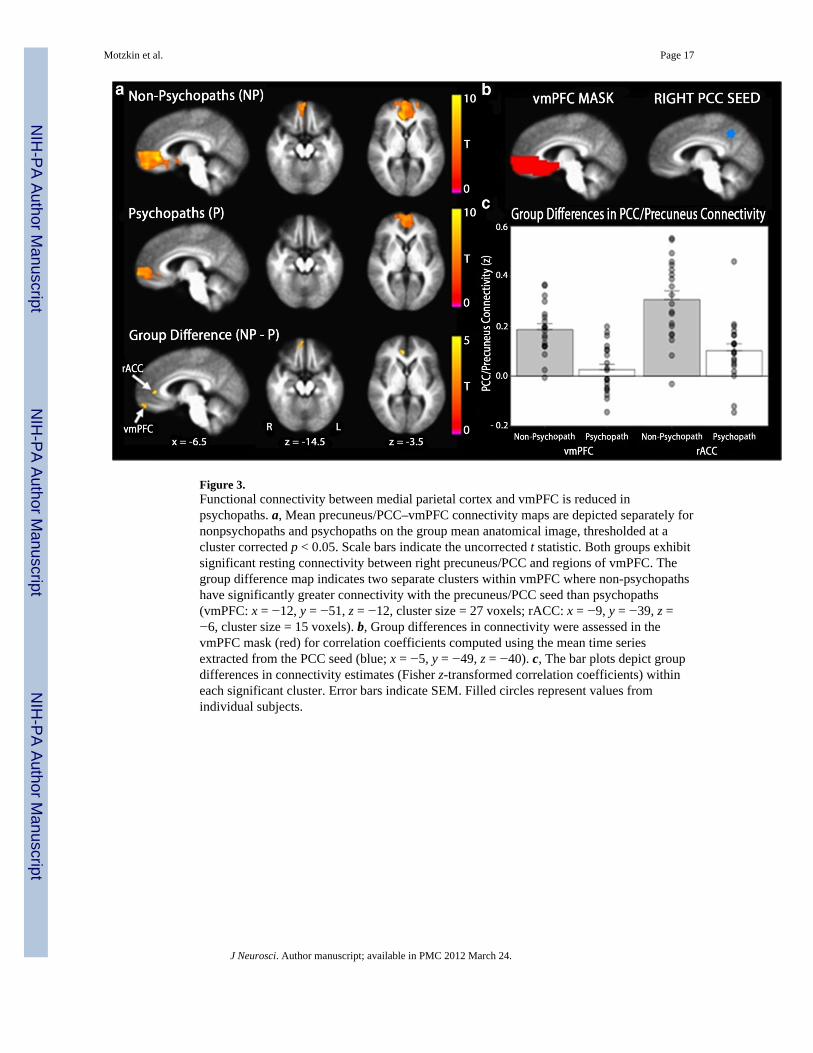
Figure 3.Functional connectivity between medial parietal cortex and vmPFC is reduced inpsychopaths. a, Mean precuneus/PCC–vmPFC connectivity maps are depicted separately fornonpsychopaths and psychopaths on the group mean anatomical image, thresholded at acluster corrected p < 0.05. Scale bars indicate the uncorrected t statistic. Both groups exhibitsignificant resting connectivity between right precuneus/PCC and regions of vmPFC. Thegroup difference map indicates two separate clusters within vmPFC where non-psychopathshave significantly greater connectivity with the precuneus/PCC seed than psychopaths(vmPFC: x = −12, y = −51, z = −12, cluster size = 27 voxels; rACC: x = −9, y = −39, z =−6, cluster size = 15 voxels). b, Group differences in connectivity were assessed in thevmPFC mask (red) for correlation coefficients computed using the mean time seriesextracted from the PCC seed (blue; x = −5, y = −49, z = −40). c, The bar plots depict groupdifferences in connectivity estimates (Fisher z-transformed correlation coefficients) withineach significant cluster. Error bars indicate SEM. Filled circles represent values fromindividual subjects.
Motzkin et al. Page 17
J Neurosci. Author manuscript; available in PMC 2012 March 24.
NIH
-PA Author Manuscript
NIH
-PA Author Manuscript
NIH
-PA Author Manuscript

Figure 4.Analysis of DTI and rest-fMRI findings with respect to psychopathy subtype. a, Depictionof each significant finding in the main between-group analyses (psychopaths vsnonpsychopaths). b, For right UF FA values, among non-psychopaths, the low- and high-anxious subgroups significantly differ (p = 0.046); there is no such difference amongpsychopaths. c, For the cluster identified in the right amygdala–vmPFC connectivityanalysis, there is a significant interaction between psychopathy and anxiety (p = 0.005),indicating that connectivity is differentially modulated by anxiety subgroup in psychopathsand non-psychopaths. d, e, There are no significant interactions between psychopathy andanxiety for medial parietal–vmPFC connectivity. Error bars indicate SEM. Asterisksindicate significant differences between anxiety subtypes.
Motzkin et al. Page 18
J Neurosci. Author manuscript; available in PMC 2012 March 24.
NIH
-PA Author Manuscript
NIH
-PA Author Manuscript
NIH
-PA Author Manuscript

NIH
-PA Author Manuscript
NIH
-PA Author Manuscript
NIH
-PA Author Manuscript
Motzkin et al. Page 19
Tabl
e 1
Parti
cipa
nt g
roup
cha
ract
eris
tics
DT
I stu
dyR
est-f
MR
I stu
dy
Var
iabl
eN
on-p
sych
opat
hs (n
= 1
3)Ps
ycho
path
s (n
= 14
)p
Non
-psy
chop
aths
(n =
20)
Psyc
hopa
ths (
n =
20)
p
Dem
ogra
phic
A
ge 3
1.7
(7.9
)32
.9 (6
.9)
0.6
9 3
1.1
(7.5
)32
.6 (6
.8)
0.5
3
R
ace
(Cau
c/A
fr A
m)
12/1
10/4
0.3
318
/215
/5 0
.41
Neu
rops
ycho
logi
cal
IQ
a10
2.3
(12.
5)97
.9 (1
1.1)
0.3
610
1.4
(12.
6)99
.6 (1
0.2)
0.6
3
D
igit
span
bac
k
7.1
(2.6
) 6
.4 (3
.3)
0.5
7
6.9
(2.2
) 6
.7 (3
.4)
0.8
4
A
nxie
ty/N
eg a
ffec
tb 1
3.5
(10.
7)13
.2 (7
.2)
0.9
3 1
1.2
(9.5
)13
.4 (8
.3)
0.4
4
Psyc
hopa
thy
P
CL-
R to
tal
14.
6 (2
.7)
32.2
(1.8
)<0
.001
14.
2 (3
.4)
31.9
(1.7
)<0
.001
F
acto
r 1
5.3
(1.9
)12
.1 (1
.7)
<0.0
01
4.7
(2.1
)11
.8 (1
.8)
<0.0
01
F
acto
r 2
7.5
(3.0
)17
.4 (1
.4)
<0.0
01
7.9
(3.1
)17
.2 (1
.5)
<0.0
01
Subs
tanc
e ab
usec
A
lcoh
ol
Prev
alen
ce 5
/10
7/1
1 0
.67
9/1
611
/16
0.7
2
Age
of o
nset
21.
0 (5
.2)
17.7
(2.2
) 2
2.4
(6.5
)19
.0 (3
.4)
C
anna
bis
Prev
alen
ce 4
/10
7/1
1 0
.39
7/1
611
/16
0.2
9
Age
of o
nset
19.
8 (4
.7)
19.7
(8.6
) 1
7.9
(4.7
)19
.3 (6
.8)
C
ocai
ne
Prev
alen
ce 2
/10
3/1
1 0
.99
3/1
6 6
/16
0.4
3
Age
of o
nset
17, 1
816
, 16,
26
19.
7 (2
.9)
20.2
(5.4
)
S
timul
ants
Prev
alen
ce 1
/10
2/1
1 0
.99
2/1
6 4
/16
0.6
5
Age
of o
nset
3020
, 23
16, 1
815
, 20,
23,
30
O
pioi
ds
Prev
alen
ce 2
/10
2/1
1 0
.99
3/1
6 6
/16
0.4
3
Age
of o
nset
15, 3
516
, 20
20.
7 (5
.1)
21.7
(7.3
)
J Neurosci. Author manuscript; available in PMC 2012 March 24.

NIH
-PA Author Manuscript
NIH
-PA Author Manuscript
NIH
-PA Author Manuscript
Motzkin et al. Page 20
DT
I stu
dyR
est-f
MR
I stu
dy
Var
iabl
eN
on-p
sych
opat
hs (n
= 1
3)Ps
ycho
path
s (n
= 14
)p
Non
-psy
chop
aths
(n =
20)
Psyc
hopa
ths (
n =
20)
p
S
edat
ives
Prev
alen
ce 0
/10
1/1
1 0
.99
1/1
6 2
/16
0.9
9
Age
of o
nset
n/a
2027
20/2
2
H
allu
cino
gens
Prev
alen
ce 0
/10
3/1
1 0
.21
1/1
6 4
/16
0.3
3
Age
of o
nset
n/a
15, 2
0, 2
120
15, 1
7, 2
0, 2
1
a Bas
ed o
n Sh
iple
y In
stitu
te o
f Liv
ing
Scal
e (Z
acha
ry, 1
986)
,
b base
d on
Wel
sh A
nxie
ty S
cale
,
c base
d on
dia
gnos
is o
f abu
se o
r dep
ende
nce
in th
e St
ruct
ured
Clin
ical
Inte
rvie
w fo
r DSM
-IV
Dis
orde
rs (S
CID
) (Fi
rst,
2002
), w
hich
was
adm
inis
tere
d to
10/
13 n
on-p
sych
opat
hs a
nd 1
1/14
psy
chop
aths
inth
e D
TI st
udy
and
16/2
0 no
n-ps
ycho
path
s and
16/
20 p
sych
opat
hs in
the
rest
-fM
RI s
tudy
. p v
alue
s for
race
dis
tribu
tion
and
subs
tanc
e ab
use
prev
alen
ce w
ere
com
pute
d w
ith F
ishe
r’s e
xact
test
. All
othe
r pva
lues
are
bas
ed o
n t t
est (
mea
ns p
rese
nted
follo
wed
by
SDs i
n pa
rent
hese
s). p
val
ues w
ere
not c
alcu
late
d fo
r sub
stan
ce a
buse
age
of o
nset
due
to re
lativ
ely
smal
l sam
ple
size
s of a
buse
rs fo
r mos
tsu
bsta
nces
.
J Neurosci. Author manuscript; available in PMC 2012 March 24.