Centrosomal Chk2 in DNA damage responses and cell cycle progession
A Novel Role for the Centrosomal Protein, Pericentrin, inRegulation of Insulin Secretory Vesicle Docking in MousePancreatic b-cellsAgata Jurczyk1, Steven C. Pino1, Bryan O’Sullivan-Murphy1, Martha Addorio1, Erich A. Lidstone1, Philip
diIorio1, Kathryn L. Lipson2, Clive Standley3, Kevin Fogarty3, Lawrence Lifshitz3, Fumihiko Urano4,5,
John P. Mordes1, Dale L. Greiner1,5, Aldo A. Rossini1,5, Rita Bortell1*
1 Department of Medicine, University of Massachusetts Medical School, Worcester, Massachusetts, United States of America, 2 Department of Physical and Biological
Sciences, Western New England College, Springfield, Massachusetts, United States of America, 3 Department of Physiology, University of Massachusetts Medical School,
Worcester, Massachusetts, United States of America, 4 Program in Gene Function and Expression, University of Massachusetts Medical School, Worcester, Massachusetts,
United States of America, 5 Program in Molecular Medicine, University of Massachusetts Medical School, Worcester, Massachusetts, United States of America
Abstract
The centrosome is important for microtubule organization and cell cycle progression in animal cells. Recently, mutations inthe centrosomal protein, pericentrin, have been linked to human microcephalic osteodysplastic primordial dwarfism (MOPDII), a rare genetic disease characterized by severe growth retardation and early onset of type 2 diabetes among other clinicalmanifestations. While the link between centrosomal and cell cycle defects may account for growth deficiencies, themechanism linking pericentrin mutations with dysregulated glucose homeostasis and pre-pubertal onset of diabetes isunknown. In this report we observed abundant expression of pericentrin in quiescent pancreatic b-cells of normal animalswhich led us to hypothesize that pericentrin may have a critical function in b-cells distinct from its known role in regulatingcell cycle progression. In addition to the typical centrosome localization, pericentrin was also enriched with secretoryvesicles in the cytoplasm. Pericentrin overexpression in b-cells resulted in aggregation of insulin-containing secretoryvesicles with cytoplasmic, but not centrosomal, pericentriolar material and an increase in total levels of intracellular insulin.RNAi- mediated silencing of pericentrin in secretory b-cells caused dysregulated secretory vesicle hypersecretion of insulininto the media. Together, these data suggest that pericentrin may regulate the intracellular distribution and secretion ofinsulin. Mice transplanted with pericentrin-depleted islets exhibited abnormal fasting hypoglycemia and inability to regulateblood glucose normally during a glucose challenge, which is consistent with our in vitro data. This previously unrecognizedfunction for a centrosomal protein to mediate vesicle docking in secretory endocrine cells emphasizes the adaptability ofthese scaffolding proteins to regulate diverse cellular processes and identifies a novel target for modulating regulatedprotein secretion in disorders such as diabetes.
Citation: Jurczyk A, Pino SC, O’Sullivan-Murphy B, Addorio M, Lidstone EA, et al. (2010) A Novel Role for the Centrosomal Protein, Pericentrin, in Regulation ofInsulin Secretory Vesicle Docking in Mouse Pancreatic b-cells. PLoS ONE 5(7): e11812. doi:10.1371/journal.pone.0011812
Editor: Kathrin Maedler, University of Bremen, Germany
Received February 8, 2010; Accepted June 24, 2010; Published July 27, 2010
Copyright: � 2010 Jurczyk et al. This is an open-access article distributed under the terms of the Creative Commons Attribution License, which permitsunrestricted use, distribution, and reproduction in any medium, provided the original author and source are credited.
Funding: This work was supported by National Institutes of Health Grants AI46629, DK53006, DK49106 and DK25306, the Beta Cell Biology Consortium, andgrants from the Juvenile Diabetes Research Foundation, the Iacocca Foundation, and the Helmsley Foundation. Core resources supported by the DiabetesEndocrinology Research Center grant DK32520 from the National Institute of Diabetes and Digestive and Kidney Diseases were used; RB is a member of the UMassDERC (DK32520). The funders had no role in study design, data collection and analysis, decision to publish, or preparation of the manuscript.
Competing Interests: The authors have declared that no competing interests exist.
* E-mail: [email protected]
Introduction
Control of blood glucose levels is largely regulated by the release
of insulin from the pancreatic islets of Langerhans. Pancreatic b-
cells are professional secretory cells where insulin is packaged,
stored in mature secretory vesicles and secreted upon appropriate
stimulus. Glucose induced stimulation of insulin secretion is
typically divided into two phases. A rapid first phase release of
insulin was thought to be mediated by granules docked at the cell
surface and primed for release [1]. During the second phase of
insulin secretion the reserve pool of granules, further away from
the plasma membrane, was mobilized to the membrane for release
[2]. More recent reports, however, demonstrate that the early
phase of insulin secretion is mediated by both predocked granules
and those further away from the plasma membrane [3,4].
Although insulin granule docking is not essential for secretion
[5], it seems to provide a temporal constraint for fusion of
secretory granules which are further away from the plasma
membrane [3].
As with most types of regulated secretory cells, vesicle
trafficking and tethering to the plasma membrane in b-cells are
regulated by numerous proteins including those contained in the
soluble N-ethylmaleimide-sensitive factor attachment protein
receptor (SNARE) and exocyst complexes [6]. SNARE complex
assembly is necessary but not sufficient for membrane fusion in
vivo, and other proteins are required for regulating vesicle
exocytosis [7,8]. Relevant to our work, the centrosomal protein,
centriolin, was also shown to ‘scaffold’ vesicle-docking exocyst
and vesicle-fusion SNARE complexes during vesicle-mediated
cytokinesis [9].
PLoS ONE | www.plosone.org 1 July 2010 | Volume 5 | Issue 7 | e11812
Recently, loss-of-function mutations in another centrosomal
gene, human pericentrin, have been causally linked to a severe
form of dwarfism and microcephaly [10,11]. Other suggestive
features of MOPD II included diabetes, dyslipidemia and
hyperinsulinism [11,12]. Although the cell cycle defects resulting
from pericentrin mutations provide a plausible mechanism for the
dwarfism associated with MOPD II, the mechanism linking
pericentrin with dysregulated glucose homeostasis and diabetes is
unknown.
Pericentrin is a component of pericentriolar material (PCM)
that surrounds the two centrioles of a centrosome [13]. The
centrosome is an organelle in animal cells that regulates cell cycle
progression and also serves as the major microtubule organizing
center (MTOC). Pericentrin is expressed as B (360 kDa), A
(255 kDa), and S (250 kDa) isoforms [13,14,15]. These large
molecules interact with numerous proteins and protein complexes,
including the c-tubulin ring complex [16,17], cytoplasmic dynein
[18], protein kinase (PK)A, PKCbII [19,20], DISC1 [21], and
PCM-1 [22]. These interactions provide a molecular ‘scaffold’ for
many signaling pathways [23]. The scaffolding PCM proteins are
very dynamic in trafficking between the centrosome-bound pool
and the cytoplasmic pool [24], yet the functional importance of
this cytoplasmic localization is not understood.
Here we identify granular cytoplasmic immunostaining of
pericentrin associating with insulin secretory vesicles of mitotically
quiescent pancreatic islets. In b-cells, RNAi-mediated depletion of
pericentrin caused insulin granule hypersecretion, dysregulation of
granule docking at the plasma membrane and abnormalities in
glucose tolerance tests in vivo. This novel function for centrosomal
protein in regulation of insulin secretion illustrates the versatility of
these scaffolding proteins to modulate multiple cellular processes
and identifies a new level of complexity in the governance of
regulated protein secretion.
Results
Pericentrin Localizes to the Centrosome and InsulinCytoplasmic Granules in Pancreatic Islets and InsulinomaCells
Although pericentrin is known to be expressed in a wide range
of mammalian tissues and transformed cell lines, its expression in
the pancreas has not been reported. To gain insight into
pericentrin function in differentiated secretory cells, we first
analyzed its expression in mouse primary pancreatic islets, as well
as transformed mouse insulinoma (MIN6), and NIH 3T3
fibroblast cells. We found all of the pericentrin isoforms
(pericentrin B and A/S) to be expressed in fibroblast cells (as
previously reported [13]), as well as in professional secretory cells
that express high levels of insulin (Figure 1A). Since we did not
have specific antibodies to each of the pericentrin isoforms, we
could not address their preferential expression. However, based on
western analysis (Figure 1A) pericentrin B appears more enriched
in the secretory cells than in fibroblasts. Next we examined
pericentrin localization within freshly isolated mouse pancreatic
islets. We were surprised to find abundant pericentrin staining at
both the centrosome and also scattered through the cytoplasm as
punctate ‘granules’ (Figure 1B). The filamentous and amorphous
appearance of centrosomes was unusual, and prompted us to
confirm this morphology using islets isolated from GFP-centrin-2
mice [25]. In these islets we found that GFP-centrin-2 signal
localized to typical well-defined centriole doublets, but not to
cytoplasmic granules (Figure 1C). In contrast, pericentrin
immunostaining of the same islets revealed much larger areas of
staining covering both centrioles (consistent with PCM) and
prominent granular cytoplasmic staining. We further investigated
this atypical centrosome by co-staining with pericentrin and ctubulin antibodies (Figure 1D); c tubulin staining was typical of
PCM staining, whereas pericentrin staining was more abundant
and amorphous. This abundance of pericentrin staining in
differentiated islet cells that divide very slowly (,20 days; [26])
was unexpected given that pericentrin’s most recognized function
is in regulating cell cycle progression [23]. We further character-
ized the sites of subcellular localization of pericentrin using
immuno-electron microscopy (EM). As expected EM of isolated
mouse pancreatic islets showed pericentrin staining at the
centrosome (Figure 1E, red box, cross section through a centriole).
Suprisingly, cytoplasmic pericentrin was associated with insulin
granules, which were identified by their characteristic dense core
and surrounding lighter halo (Figure 1E, black box). Control
rabbit IgG for the pericentrin affinity-purified antibodies showed
no specific staining (Figure 1F).
In mouse insulinoma cells pericentrin co-localized with insulin
staining granules along the plasma membrane as shown by single
z-sections taken at the ‘top’ (Figure 1G) and through the middle
(Figure 1H) of the insulinoma cells. Association of pericentrin with
insulin granules from mouse insulinoma cells was also observed by
immunofluorescence of purified granules (Figure S1A) and by
Western analyses of post-nuclear supernatant fractions (excluding
DNA and centrosomes) derived from iodixinol density gradients.
Pericentrin co-migrated with proinsulin, insulin, Sec6 (an exocyst
component expressed on insulin granules and shown to be
important for insulin secretion [27]), SNAP 25 (a SNARE
component), and chromogranin B (which is located within
secretory granules) (Figure S1B). These results strongly support
an association of pericentrin with insulin and other secretory
vesicle proteins that constitute the insulin granule.
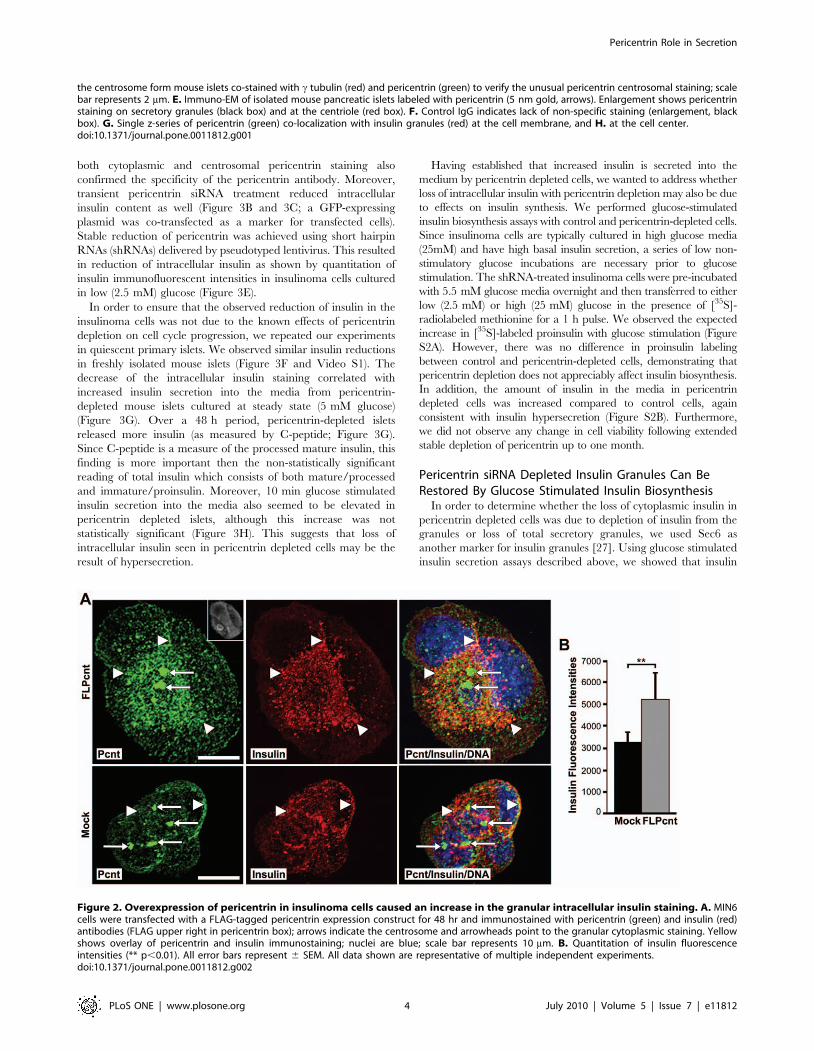
Overexpression of Pericentrin Co-Sequesters InsulinGranules with Granular Cytoplasmic Pericentrin
It has been reported previously that pericentrin overexpression
in S phase-arrested CHO cells results in formation of a PCM
‘‘cloud’’ consisting of pericentrin and c tubulin around the
multiple centrioles [28]. Our data show that in MIN6 insulinoma
cells, overexpression of pericentrin causes an increase in
centrosome size (Figure 2A), which could be similar to the PCM
‘‘cloud’’ observed in CHO cells. Increased granular pericentrin
staining was also observed in the cytoplasm, and intracellular
insulin levels were elevated in pericentrin overexpressing cells as
compared to control cells (Figure 2A and 2B). The co-
sequestration of pericentrin and insulin with cytoplasmic granules,
but not with the centrosomes, further supports close association
between insulin granules and cytoplasmic pericentrin staining.
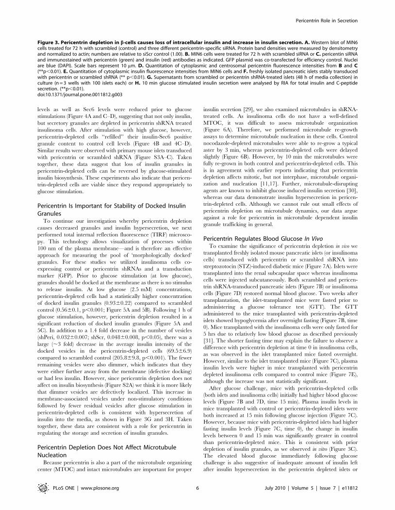
Pericentrin Depletion Causes a Loss of IntracellularInsulin and Hypersecretion of Mature Insulin withoutAffecting Proinsulin Biosynthesis
To study the function of pericentrin in insulin secreting cells, we
depleted pericentrin in pancreatic islets and MIN6 insulinoma
cells using RNAi. Western analyses demonstrated pericentrin
depletion in insulinoma cells using siRNAs targeting three distinct
regions of pericentrin (Figure 3A). Immunofluorescence micros-
copy revealed centrosomal and granulate staining of pericentrin in
scrambled siRNA-transfected cells (Figure 3B). In contrast,
pericentrin-specific siRNA-transfected cells showed marked de-
pletion of granular cytoplasmic pericentrin staining (Figure 3C and
3D), and centrosomal pericentrin staining was also diminished as
compared to control (siScr) cells (Figure 3C and 3D). Depletion of
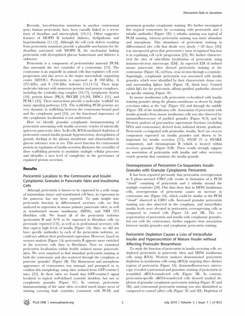
Pericentrin Role in Secretion
PLoS ONE | www.plosone.org 2 July 2010 | Volume 5 | Issue 7 | e11812
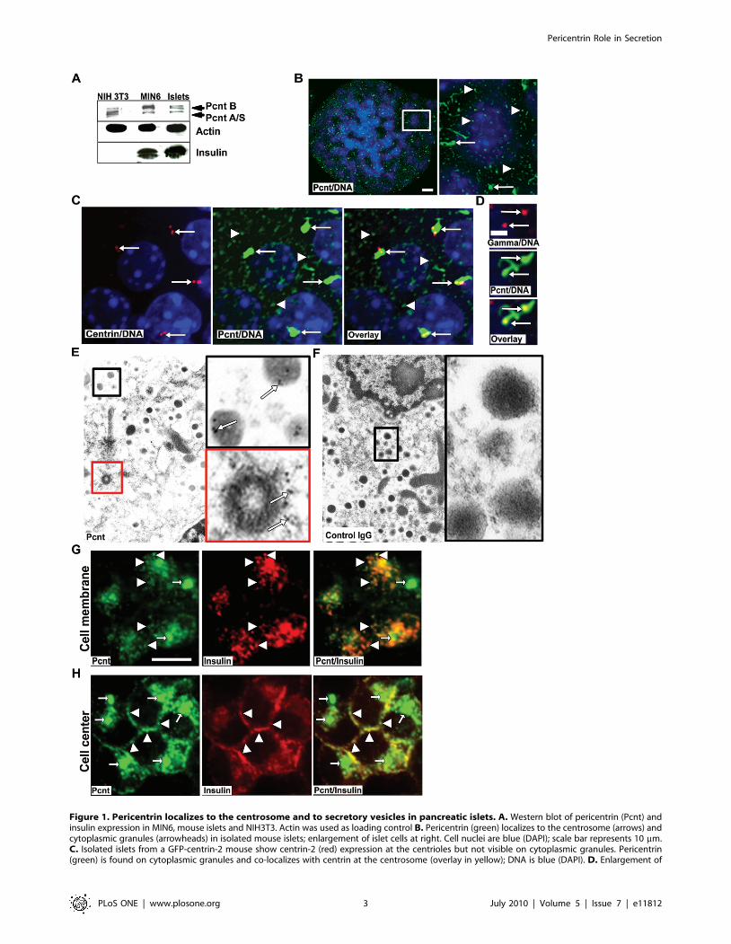
Figure 1. Pericentrin localizes to the centrosome and to secretory vesicles in pancreatic islets. A. Western blot of pericentrin (Pcnt) andinsulin expression in MIN6, mouse islets and NIH3T3. Actin was used as loading control B. Pericentrin (green) localizes to the centrosome (arrows) andcytoplasmic granules (arrowheads) in isolated mouse islets; enlargement of islet cells at right. Cell nuclei are blue (DAPI); scale bar represents 10 mm.C. Isolated islets from a GFP-centrin-2 mouse show centrin-2 (red) expression at the centrioles but not visible on cytoplasmic granules. Pericentrin(green) is found on cytoplasmic granules and co-localizes with centrin at the centrosome (overlay in yellow); DNA is blue (DAPI). D. Enlargement of
Pericentrin Role in Secretion
PLoS ONE | www.plosone.org 3 July 2010 | Volume 5 | Issue 7 | e11812
both cytoplasmic and centrosomal pericentrin staining also
confirmed the specificity of the pericentrin antibody. Moreover,
transient pericentrin siRNA treatment reduced intracellular
insulin content as well (Figure 3B and 3C; a GFP-expressing
plasmid was co-transfected as a marker for transfected cells).
Stable reduction of pericentrin was achieved using short hairpin
RNAs (shRNAs) delivered by pseudotyped lentivirus. This resulted
in reduction of intracellular insulin as shown by quantitation of
insulin immunofluorescent intensities in insulinoma cells cultured
in low (2.5 mM) glucose (Figure 3E).
In order to ensure that the observed reduction of insulin in the
insulinoma cells was not due to the known effects of pericentrin
depletion on cell cycle progression, we repeated our experiments
in quiescent primary islets. We observed similar insulin reductions
in freshly isolated mouse islets (Figure 3F and Video S1). The
decrease of the intracellular insulin staining correlated with
increased insulin secretion into the media from pericentrin-
depleted mouse islets cultured at steady state (5 mM glucose)
(Figure 3G). Over a 48 h period, pericentrin-depleted islets
released more insulin (as measured by C-peptide; Figure 3G).
Since C-peptide is a measure of the processed mature insulin, this
finding is more important then the non-statistically significant
reading of total insulin which consists of both mature/processed
and immature/proinsulin. Moreover, 10 min glucose stimulated
insulin secretion into the media also seemed to be elevated in
pericentrin depleted islets, although this increase was not
statistically significant (Figure 3H). This suggests that loss of
intracellular insulin seen in pericentrin depleted cells may be the
result of hypersecretion.
Having established that increased insulin is secreted into the
medium by pericentrin depleted cells, we wanted to address whether
loss of intracellular insulin with pericentrin depletion may also be due
to effects on insulin synthesis. We performed glucose-stimulated
insulin biosynthesis assays with control and pericentrin-depleted cells.
Since insulinoma cells are typically cultured in high glucose media
(25mM) and have high basal insulin secretion, a series of low non-
stimulatory glucose incubations are necessary prior to glucose
stimulation. The shRNA-treated insulinoma cells were pre-incubated
with 5.5 mM glucose media overnight and then transferred to either
low (2.5 mM) or high (25 mM) glucose in the presence of [35S]-
radiolabeled methionine for a 1 h pulse. We observed the expected
increase in [35S]-labeled proinsulin with glucose stimulation (Figure
S2A). However, there was no difference in proinsulin labeling
between control and pericentrin-depleted cells, demonstrating that
pericentrin depletion does not appreciably affect insulin biosynthesis.
In addition, the amount of insulin in the media in pericentrin
depleted cells was increased compared to control cells, again
consistent with insulin hypersecretion (Figure S2B). Furthermore,
we did not observe any change in cell viability following extended
stable depletion of pericentrin up to one month.
Pericentrin siRNA Depleted Insulin Granules Can BeRestored By Glucose Stimulated Insulin Biosynthesis
In order to determine whether the loss of cytoplasmic insulin in
pericentrin depleted cells was due to depletion of insulin from the
granules or loss of total secretory granules, we used Sec6 as
another marker for insulin granules [27]. Using glucose stimulated
insulin secretion assays described above, we showed that insulin
Figure 2. Overexpression of pericentrin in insulinoma cells caused an increase in the granular intracellular insulin staining. A. MIN6cells were transfected with a FLAG-tagged pericentrin expression construct for 48 hr and immunostained with pericentrin (green) and insulin (red)antibodies (FLAG upper right in pericentrin box); arrows indicate the centrosome and arrowheads point to the granular cytoplasmic staining. Yellowshows overlay of pericentrin and insulin immunostaining; nuclei are blue; scale bar represents 10 mm. B. Quantitation of insulin fluorescenceintensities (** p,0.01). All error bars represent 6 SEM. All data shown are representative of multiple independent experiments.doi:10.1371/journal.pone.0011812.g002
the centrosome form mouse islets co-stained with c tubulin (red) and pericentrin (green) to verify the unusual pericentrin centrosomal staining; scalebar represents 2 mm. E. Immuno-EM of isolated mouse pancreatic islets labeled with pericentrin (5 nm gold, arrows). Enlargement shows pericentrinstaining on secretory granules (black box) and at the centriole (red box). F. Control IgG indicates lack of non-specific staining (enlargement, blackbox). G. Single z-series of pericentrin (green) co-localization with insulin granules (red) at the cell membrane, and H. at the cell center.doi:10.1371/journal.pone.0011812.g001
Pericentrin Role in Secretion
PLoS ONE | www.plosone.org 4 July 2010 | Volume 5 | Issue 7 | e11812

Pericentrin Role in Secretion
PLoS ONE | www.plosone.org 5 July 2010 | Volume 5 | Issue 7 | e11812
levels as well as Sec6 levels were reduced prior to glucose
stimulations (Figure 4A and C–D), suggesting that not only insulin,
but secretory granules are depleted in pericentrin shRNA treated
insulinoma cells. After stimulation with high glucose, however,
pericentrin-depleted cells ‘‘refilled’’ their insulin-Sec6 positive
granule content to control cell levels (Figure 4B and 4C–D).
Similar results were observed with primary mouse islets transduced
with pericentrin or scrambled shRNA (Figure S3A–C). Taken
together, these data suggest that loss of insulin granules in
pericentrin-depleted cells can be reversed by glucose-stimulated
insulin biosynthesis. These experiments also indicate that pericen-
trin-depleted cells are viable since they respond appropriately to
glucose stimulation.
Pericentrin Is Important for Stability of Docked InsulinGranules
To continue our investigation whereby pericentrin depletion
causes decreased granules and insulin hypersecretion, we next
performed total internal reflection fluorescence (TIRF) microsco-
py. This technology allows visualization of processes within
100 nm of the plasma membrane—and is therefore an effective
approach for measuring the pool of ‘morphologically docked’
granules. For these studies we utilized insulinoma cells co-
expressing control or pericentrin shRNAs and a transduction
marker (GFP). Prior to glucose stimulation (at low glucose),
granules should be docked at the membrane as there is no stimulus
to release insulin. At low glucose (2.5 mM) concentrations,
pericentrin-depleted cells had a statistically higher concentration
of docked insulin granules (0.9360.22) compared to scrambled
control (0.5660.1, p,0.001; Figure 5A and 5B). Following 1 h of
glucose stimulation, however, pericentrin depletion resulted in a
significant reduction of docked insulin granules (Figure 5A and
5C). In addition to a 1.4 fold decrease in the number of vesicles
(shPeri, 0.03260.007; shScr, 0.04860.008, p,0.05), there was a
large (,3 fold) decrease in the average insulin intensity of the
docked vesicles in the pericentrin-depleted cells (69.566.9)
compared to scrambled control (205.869.8, p,0.001). The fewer
remaining vesicles were also dimmer, which indicates that they
were either further away from the membrane (defective docking)
or had less insulin. However, since pericentrin depletion does not
affect on insulin biosynthesis (Figure S2A) we think it is more likely
that dimmer vesicles are defectively localized. This increase in
membrane-associated vesicles under non-stimulatory conditions
followed by fewer residual vesicles after glucose stimulation in
pericentrin-depleted cells is consistent with hypersecretion of
insulin into the media, as shown in Figure 3G and 3H. Taken
together, these data are consistent with a role for pericentrin in
regulating the storage and secretion of insulin granules.
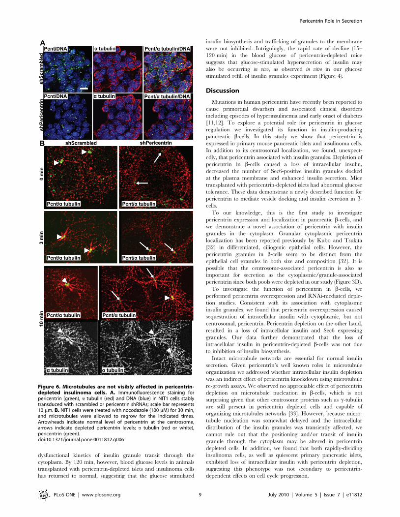
Pericentrin Depletion Does Not Affect MicrotubuleNucleation
Because pericentrin is also a part of the microtubule organizing
center (MTOC) and intact microtubules are important for proper
insulin secretion [29], we also examined microtubules in shRNA-
treated cells. As insulinoma cells do not have a well-defined
MTOC, it was difficult to assess microtubule organization
(Figure 6A). Therefore, we performed microtubule re-growth
assays to determine microtubule nucleation in these cells. Control
nocodazole-depleted microtubules were able to re-grow a typical
aster by 3 min, whereas pericentrin-depleted cells were delayed
slightly (Figure 6B). However, by 10 min the microtubules were
fully re-grown in both control and pericentrin-depleted cells. This
is in agreement with earlier reports indicating that pericentrin
depletion affects mitotic, but not interphase, microtubule organi-
zation and nucleation [11,17]. Further, microtubule-disrupting
agents are known to inhibit glucose induced insulin secretion [30],
whereas our data demonstrate insulin hypersecretion in pericen-
trin-depleted cells. Although we cannot rule out small effects of
pericentrin depletion on microtubule dynamics, our data argue
against a role for pericentrin in microtubule dependent insulin
granule trafficking in general.
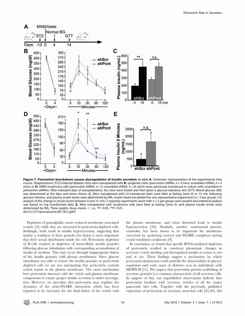
Pericentrin Regulates Blood Glucose In VivoTo examine the significance of pericentrin depletion in vivo we
transplanted freshly isolated mouse pancreatic islets (or insulinoma
cells) transduced with pericentrin or scrambled shRNA into
streptozotocin (STZ)-induced diabetic mice (Figure 7A). Islets were
transplanted into the renal subcapsular space whereas insulinoma
cells were injected subcutaneously. Both scrambled and pericen-
trin shRNA-transduced pancreatic islets (Figure 7B) or insulinoma
cells (Figure 7D) restored normal blood glucose. Two weeks after
transplantation, the islet-transplanted mice were fasted prior to
administering a glucose tolerance test (GTT). The GTT
administered to the mice transplanted with pericentrin-depleted
islets showed hypoglycemia after overnight fasting (Figure 7B, time
0). Mice transplanted with the insulinoma cells were only fasted for
5 hrs due to relatively low blood glucose as described previously
[31]. The shorter fasting time may explain the failure to observe a
difference with pericentrin depletion at time 0 in insulinoma cells,
as was observed in the islet transplanted mice fasted overnight.
However, similar to the islet transplanted mice (Figure 7C), plasma
insulin levels were higher in mice transplanted with pericentrin
depleted insulinoma cells compared to control mice (Figure 7E),
although the increase was not statistically significant.
After glucose challenge, mice with pericentrin-depleted cells
(both islets and insulinoma cells) initially had higher blood glucose
levels (Figure 7B and 7D, time 15 min). Plasma insulin levels in
mice transplanted with control or pericentrin-depleted islets were
both increased at 15 min following glucose injection (Figure 7C).
However, because mice with pericentrin-depleted islets had higher
fasting insulin levels (Figure 7C, time 0), the change in insulin
levels between 0 and 15 min was significantly greater in control
than pericentrin-depleted mice. This is consistent with prior
depletion of insulin granules, as we observed in vitro (Figure 3C).
The elevated blood glucose immediately following glucose
challenge is also suggestive of inadequate amount of insulin left
after insulin hypersecretion in the pericentrin depleted islets or
Figure 3. Pericentrin depletion in b-cells causes loss of intracellular insulin and increase in insulin secretion. A. Western blot of MIN6cells treated for 72 h with scrambled (control) and three different pericentrin-specific siRNA. Protein band densities were measured by densitometryand normalized to actin; numbers are relative to siScr control (1.00). B. MIN6 cells were treated for 72 h with scrambled siRNA or C. pericentin siRNAand immunostained with pericentrin (green) and insulin (red) antibodies as indicated. GFP plasmid was co-transfected for efficiency control. Nucleiare blue (DAPI). Scale bars represent 10 mm. D. Quantitation of cytoplasmic and centrosomal pericentrin fluorescence intensities from B and C(**p,0.01). E. Quantitation of cytoplasmic insulin fluorescence intensities from MIN6 cells and F. freshly isolated pancreatic islets stably transducedwith pericentrin or scrambled shRNA (** p,0.01). G. Supernatants from scrambled or pericentrin shRNA-treated islets (48 h of media collection) inculture (n = 3 wells with 100 islets each) or H. 10 min glucose stimulated insulin secretion were analysed by RIA for total insulin and C-peptidesecretion. (**p,0.01).doi:10.1371/journal.pone.0011812.g003
Pericentrin Role in Secretion
PLoS ONE | www.plosone.org 6 July 2010 | Volume 5 | Issue 7 | e11812
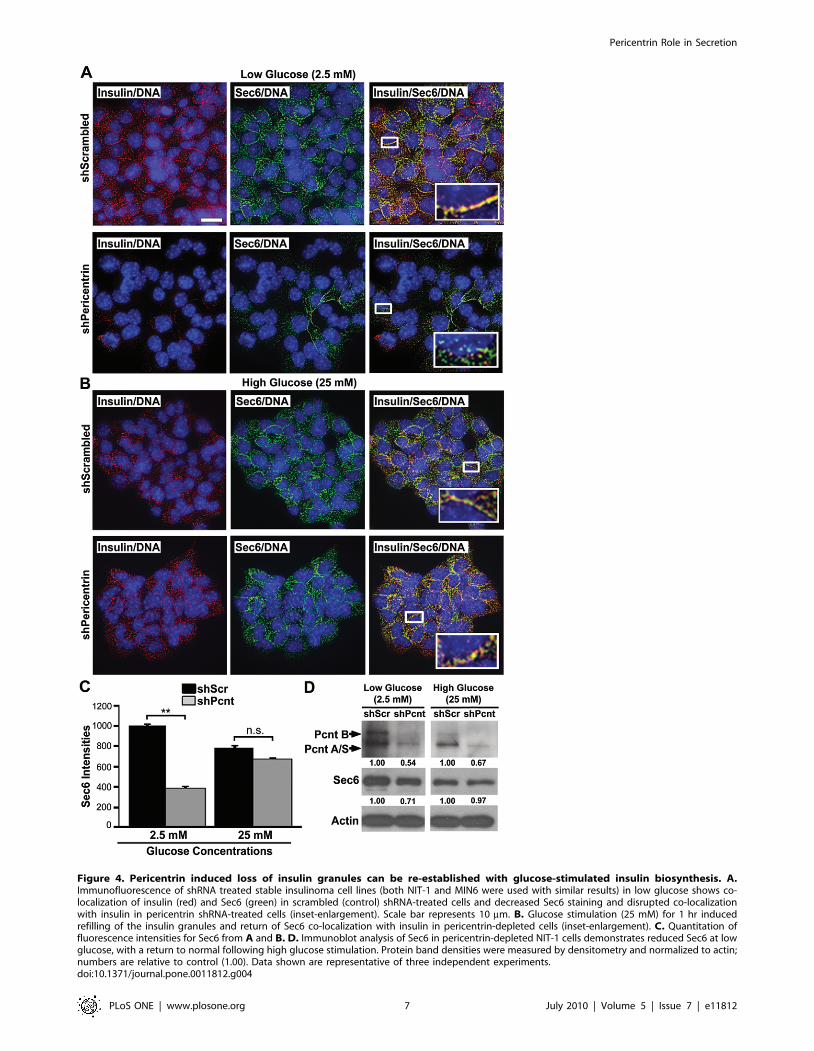
Figure 4. Pericentrin induced loss of insulin granules can be re-established with glucose-stimulated insulin biosynthesis. A.Immunofluorescence of shRNA treated stable insulinoma cell lines (both NIT-1 and MIN6 were used with similar results) in low glucose shows co-localization of insulin (red) and Sec6 (green) in scrambled (control) shRNA-treated cells and decreased Sec6 staining and disrupted co-localizationwith insulin in pericentrin shRNA-treated cells (inset-enlargement). Scale bar represents 10 mm. B. Glucose stimulation (25 mM) for 1 hr inducedrefilling of the insulin granules and return of Sec6 co-localization with insulin in pericentrin-depleted cells (inset-enlargement). C. Quantitation offluorescence intensities for Sec6 from A and B. D. Immunoblot analysis of Sec6 in pericentrin-depleted NIT-1 cells demonstrates reduced Sec6 at lowglucose, with a return to normal following high glucose stimulation. Protein band densities were measured by densitometry and normalized to actin;numbers are relative to control (1.00). Data shown are representative of three independent experiments.doi:10.1371/journal.pone.0011812.g004
Pericentrin Role in Secretion
PLoS ONE | www.plosone.org 7 July 2010 | Volume 5 | Issue 7 | e11812
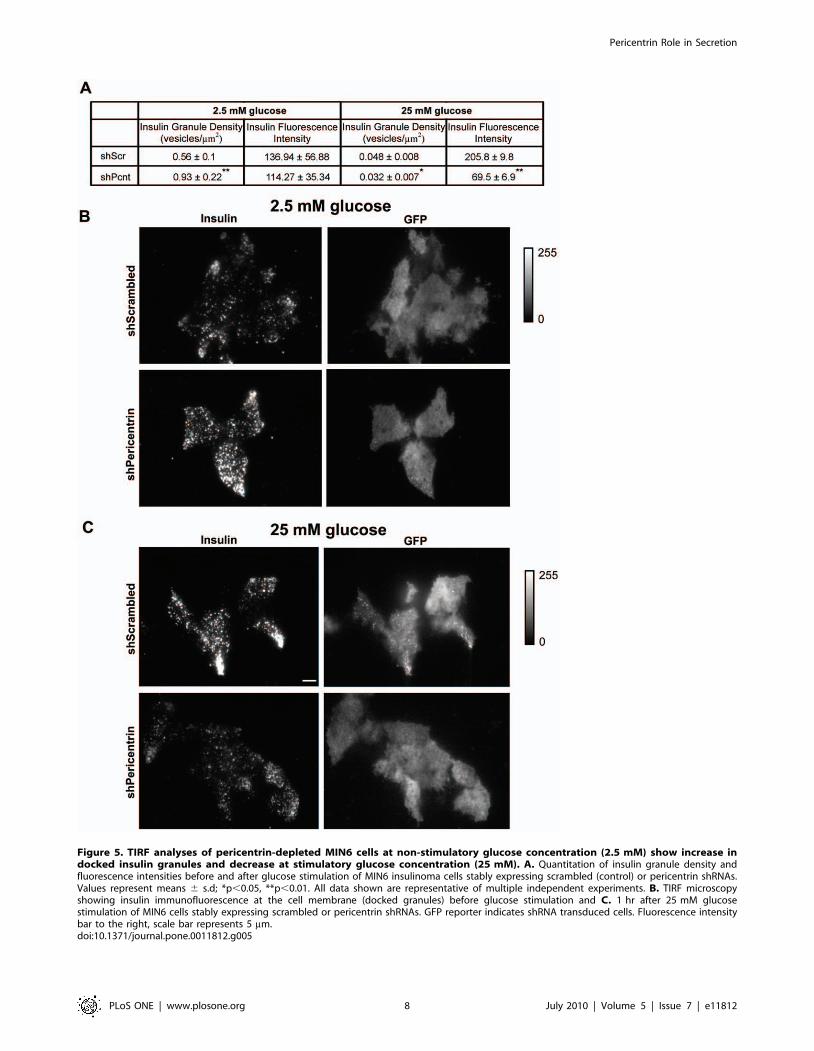
Figure 5. TIRF analyses of pericentrin-depleted MIN6 cells at non-stimulatory glucose concentration (2.5 mM) show increase indocked insulin granules and decrease at stimulatory glucose concentration (25 mM). A. Quantitation of insulin granule density andfluorescence intensities before and after glucose stimulation of MIN6 insulinoma cells stably expressing scrambled (control) or pericentrin shRNAs.Values represent means 6 s.d; *p,0.05, **p,0.01. All data shown are representative of multiple independent experiments. B. TIRF microscopyshowing insulin immunofluorescence at the cell membrane (docked granules) before glucose stimulation and C. 1 hr after 25 mM glucosestimulation of MIN6 cells stably expressing scrambled or pericentrin shRNAs. GFP reporter indicates shRNA transduced cells. Fluorescence intensitybar to the right, scale bar represents 5 mm.doi:10.1371/journal.pone.0011812.g005
Pericentrin Role in Secretion
PLoS ONE | www.plosone.org 8 July 2010 | Volume 5 | Issue 7 | e11812
dysfunctional kinetics of insulin granule transit through the
cytoplasm. By 120 min, however, blood glucose levels in animals
transplanted with pericentrin-depleted islets and insulinoma cells
has returned to normal, suggesting that the glucose stimulated
insulin biosynthesis and trafficking of granules to the membrane
were not inhibited. Intriguingly, the rapid rate of decline (15–
120 min) in the blood glucose of pericentrin-depleted mice
suggests that glucose-stimulated hypersecretion of insulin may
also be occurring in vivo, as observed in vitro in our glucose
stimulated refill of insulin granules experiment (Figure 4).
Discussion
Mutations in human pericentrin have recently been reported to
cause primordial dwarfism and associated clinical disorders
including episodes of hyperinsulinemia and early onset of diabetes
[11,12]. To explore a potential role for pericentrin in glucose
regulation we investigated its function in insulin-producing
pancreatic b-cells. In this study we show that pericentrin is
expressed in primary mouse pancreatic islets and insulinoma cells.
In addition to its centrosomal localization, we found, unexpect-
edly, that pericentrin associated with insulin granules. Depletion of
pericentrin in b-cells caused a loss of intracellular insulin,
decreased the number of Sec6-positive insulin granules docked
at the plasma membrane and enhanced insulin secretion. Mice
transplanted with pericentrin-depleted islets had abnormal glucose
tolerance. These data demonstrate a newly described function for
pericentrin to mediate vesicle docking and insulin secretion in b-
cells.
To our knowledge, this is the first study to investigate
pericentrin expression and localization in pancreatic b-cells, and
we demonstrate a novel association of pericentrin with insulin
granules in the cytoplasm. Granular cytoplasmic pericentrin
localization has been reported previously by Kubo and Tsukita
[32] in differentiated, ciliogenic epithelial cells. However, the
pericentrin granules in b-cells seem to be distinct from the
epithelial cell granules in both size and composition [32]. It is
possible that the centrosome-associated pericentrin is also as
important for secretion as the cytoplasmic/granule-associated
pericentrin since both pools were depleted in our study (Figure 3D).
To investigate the function of pericentrin in b-cells, we
performed pericentrin overexpression and RNAi-mediated deple-
tion studies. Consistent with its association with cytoplasmic
insulin granules, we found that pericentrin overexpression caused
sequestration of intracellular insulin with cytoplasmic, but not
centrosomal, pericentrin. Pericentrin depletion on the other hand,
resulted in a loss of intracellular insulin and Sec6 expressing
granules. Our data further demonstrated that the loss of
intracellular insulin in pericentrin-depleted b-cells was not due
to inhibition of insulin biosynthesis.
Intact microtubule networks are essential for normal insulin
secretion. Given pericentrin’s well known roles in microtubule
organization we addressed whether intracellular insulin depletion
was an indirect effect of pericentrin knockdown using microtubule
re-growth assays. We observed no appreciable effect of pericentrin
depletion on microtubule nucleation in b-cells, which is not
surprising given that other centrosome proteins such as c-tubulin
are still present in pericentrin depleted cells and capable of
organizing microtubules networks [33]. However, because micro-
tubule nucleation was somewhat delayed and the intracellular
distribution of the insulin granules was transiently affected, we
cannot rule out that the positioning and/or transit of insulin
granule through the cytoplasm may be altered in pericentrin
depleted cells. In addition, we found that both rapidly-dividing
insulinoma cells, as well as quiescent primary pancreatic islets,
exhibited loss of intracellular insulin with pericentrin depletion,
suggesting this phenotype was not secondary to pericentrin-
dependent effects on cell cycle progression.
Figure 6. Microtubules are not visibly affected in pericentrin-depleted insulinoma cells. A. Immunofluorescence staining forpericentrin (green), a tubulin (red) and DNA (blue) in NIT1 cells stablytransduced with scrambled or pericentrin shRNAs; scale bar represents10 mm. B. NIT1 cells were treated with nocodazole (100 mM) for 30 min,and microtubules were allowed to regrow for the indicated times.Arrowheads indicate normal level of pericentrin at the centrosome,arrows indicate depleted pericentrin levels; a tubulin (red or white),pericentrin (green).doi:10.1371/journal.pone.0011812.g006
Pericentrin Role in Secretion
PLoS ONE | www.plosone.org 9 July 2010 | Volume 5 | Issue 7 | e11812
Depletion of granulphilin causes reduced membrane associated
vesicles [5], while they are increased in pericentrin-depleted cells.
Strikingly, both result in insulin hypersecretion, suggesting that
maybe a readiness of these granules for fusion is more important
that their actual distribution inside the cell. Pericentrin depletion
of b-cells resulted in depletion of intracellular insulin granules
following glucose stimulation with corresponding accumulation of
insulin in medium. This may occur through inappropriate fusion
of the insulin granules with plasma membranes. Since glucose
stimulation was able to restore the insulin granules in pericentrin
depleted cells we are not anticipating that pericentrin controls
vesicle transit to the plasma membrane. The exact mechanism
how pericentrin interacts with the vesicle and plasma membrane
components to ensure proper insulin secretion is under investiga-
tion. However, we speculate that pericentrin may regulate the
dynamics of the actin-SNARE interaction which has been
reported to be necessary for the final fusion of the vesicle with
the plasma membrane, and when disturbed leads to insulin
hypersecretion [34]. Similarly, another centrosomal protein,
centriolin, has been shown to be important for membrane
exocytosis by anchoring exocyst and SNARE complexes during
vesicle-mediated cytokinesis [9].
In conclusion, we found that specific RNAi-mediated depletion
of pericentrin resulted in consistent phenotypic changes in
secretory vesicle docking and dysregulated insulin secretion in vitro
and in vivo. These findings suggest a mechanism by which
pericentrin dysfunction could underlie the abnormalities in glucose
regulation and early onset of diabetes seen in individuals with
MOPD II [11]. We suspect that pericentrin protein scaffolding of
secretory granules is a common characteristic of all secretory cells.
In support of this, our unpublished observations indicate that
pericentrin localizes with secretory vesicles of all the major
pancreatic islet cells. Together with the previously published
expression of pericentrin in secretory neuronal cells [35,36], this
Figure 7. Pericentrin knockdown causes dysregulation of insulin secretion in vivo. A. Schematic representation of the experimental timecourse. Streptozotocin (STZ)-induced diabetic mice were transplanted with B. syngeneic islets (pericentrin shRNA, n = 3 mice; scrambled shRNA, n = 3mice) or D. MIN6 insulinoma cells (pericentrin shRNA, n = 3; scrambled shRNA, n = 4) which were previously transduced in culture with scrambled orpericentrin shRNAs. After indicated days of transplantation, the mice were fasted and then given a glucose tolerance test (GTT). Blood glucose (BG)was determined at the days and times shown. C. Mice transplanted with LV-transduced islets were bled at fasting (time 0) or 15 min followingglucose infusion, and plasma insulin levels were determined by RIA. Insulin levels are plotted for one representative experiment (n = 3 per group). Foranalysis of the change in insulin levels between 0 and 15 min, 2 separate experiments (each with n = 3 per group) were pooled and statistical analysiswas based on log transformed data. E. Mice transplanted with insulinoma cells were bled at fasting (time 0), and plasma insulin levels weredetermined by RIA. These graphs show means 6 s.e., *P,0.05, **P,0.01.doi:10.1371/journal.pone.0011812.g007
Pericentrin Role in Secretion
PLoS ONE | www.plosone.org 10 July 2010 | Volume 5 | Issue 7 | e11812
suggests a functional role for this centrosomal protein in secretion
and identifies pericentrin as a novel regulator of exocytosis.
Materials and Methods
Ethics StatementAnimals were housed in a viral-antibody-free facility and
maintained in accordance with the Guide for the Care and Use of
Laboratory Animals (Institute of Laboratory Animal Resources, 1996)
and guidelines of the University of Massachusetts Institutional
Animal Care and Use Committee (IACUC). All research involving
animals in these studies was approved by the University of
Massachusetts IACUC.
Antibodies and immunoblottingActin, Sec6, chromogranin B, SNAP25 and a tubulin antibodies
were from Chemicon International (Temecula, CA); insulin and
glucagon were from Dako, Inc. (Carpinteria, CA). Pericentrin
mAb was from BD Bioscience and rabbit affinity-purified
pericentrin Ab was from S. Doxsey [13]. Horseradish peroxidase
(HRP) IgG secondary Abs were from Santa Cruz Biotechnology,
and Alexa-Fluor- and Cy5-labeled probes from Molecular Probes
and Jackson Immunoresearch, respectively. Actin was used as a
loading control for immunoblotting.
Cell culture and RNAiMouse insulinoma (MIN6, NIT1, and TC6) cells were obtained
from ATCC (Bethesda, MD). Insulinoma cells were cultured in D-
MEM medium (Invitrogen) and isolated islets were cultured on
Matrigel-coated plates in CMRL Medium 1066 (Invitrogen). All
media were supplemented with 10% FBS, 1 mM sodium
pyruvate, 100 U/ml penicillin, and 100 mg/ml streptomycin at
37uC in an atmosphere of 5% CO2.
Transient knockdown with siRNA. The siRNAs were
designed and synthesized at Dharmacon Research (antisense):
siPcnt1 AAUCUCUAAAUCUCUCUGCUU, siPcnt2 UUCUC-
CAUGAUCUCUUUCCUU, siPcnt3 UCUCGCUCCUUCU-
CUCUCCUU, scrambled (control) 59 CAGUCGCGUUUGC-
GACUGG. The Cell Line Nucleofector Kit R and Nucleofector
Device (Amaxa Biosystems, Gaithersburg, MD) were used for
transfection of cells. Experiments were performed at 72–96 h
when knockdown of pericentrin was determined to be maximal.
Stable knockdown with shRNA. Stable shRNA constructs
were made by insertion of the appropriate sequence into the pLL3.7
lentiviral (LV) vector [37]. Two different shRNAs for pericentrin
were used: 1) 59 GCAGCTGAGCTGAAGGAGA 2) 59 CGAA-
GACTTTATCGTAACA. Scrambled shRNA was used as control:
59 CAGUCGCGUUUGCGACUGG. Transfection of 293FT cells
with the LV constructs and packaging plasmids, DR8.9 and VSVg,
was performed using Lipofectamine 2000 as per manufacturer’s
instructions (Invitrogen, Carlsbad, CA). LV was harvested at 48 and
72 h post-transfection. The LV supernatant was centrifuged at
120,0006g for 90 min at 4uC, and the concentrated virus was stored
at 280uC until use. Concentrated virus was used to infect islets or
insulinoma cells. Experiments with primary islets were performed on
freshly isolated islets harvested by collagenase digestion as described
[38] after 72–96 h LV infection (when pericentrin knockdown was
determined to be maximal). Insulinoma cells were FACS-sorted (for
the GFP reporter) one week after LV infection, and GFP-positive cells
were used for experiments.
Glucose stimulationsRNAi-treated insulinoma islets or islets were pre-incubated
overnight (16–18 h) in 5.5 mM glucose medium and then
incubated for 1 h in KRB buffer consisting of 135 mM NaCl,
3.6 mM KCl, 10 mM Hepes [pH 7.4], 5 mM NaHCO3, 0.5 mM
NaH3PO4, 0.5 mM MgCl2, 1.5 mM CaCl2, and 2.5 mM (low)
glucose. Stimulation was performed with 25 mM (high) glucose for
the indicated periods of time.
Insulin biosynthesi. Proinsulin biosynthesis was analyzed by
proinsulin immunoprecipitation of [35S] methionine-labeled
insulinoma lysates as described [39].
Radioimmunoassay (RIA). Isolated mouse pancreatic
islets were transduced with scrambled or pericentrin shRNAs
for 48 h and then transferred to fresh media containing basal
glucose (5.5 mM) for an additional 48 h for analysis of insulin
secretion. RIA for secreted insulin and C-peptide in the culture
supernatants was performed by Linco Diagnostics (St. Charles,
MO).
Mice Pancreatic islet isolation and transplantationBalb/c mice were obtained from Charles River (Wilmington,
MA). Eight- to 15-week-old mice of either sex were used. Mice
were rendered hyperglycemic with a single intraperitoneal
injection of 170 mg/kg of streptozotocin (Sigma, St. Louis,
MO). Diabetes was defined as a plasma glucose concentration
.250 mg/dl (Accu-Chek Active meter, Roche Diagnostics,
Indianapolis, IN) on two successive days. Mouse pancreatic islets
were harvested by collagenase digestion as described [38]. Islets
were stably transduced with lentivirus shRNAs and transplanted at
a dose of 20/g body weight into the renal sub-capsular space of
recipient mice. In some experiments 26106 MIN6 cells in a
volume of 100 ml of Matrigel (BD Pharmingen) solution were
transplanted subcutaneously instead of islets.
ImmunofluorescenceIslets were fixed in 3.7% paraformaldahyde for 30 min at
room temp. Immunofluorescence was performed as described
[9]. Secondary Alexa-Fluor antibodies were used at 1:1000, Cy-
5 at 1:100, and DAPI at 1:10,000 (Sigma-Aldrich). Coverslips
were mounted with Prolong Antifade medium (Invitrogen).
Images were captured using spinning-disk confocal microscopy
on a Nikon Eclipse TE2000-E microscope, deconvolved and
analyzed using MetaMorph software. Unless otherwise indicat-
ed, all immunofluorescence images were two-dimensional
projections of three-dimensional reconstructions to ensure that
all stained material was visible in two-dimensional images. All
cell tracing and recording of average fluorescence intensities
were performed on multiple random fields of cells. To
determine centrosomal fluorescence intensity, a square was
centered on the centrosome and the average intensity per pixel
was recorded. For the cytoplasmic pericentrin intensity, the
centrosome intensity was subtracted from the total cytoplasmic
intensity. For the insulin cytoplasmic intensities, the total
cytoplasmic intensity was used (Metamorph software, Molecular
Devices, Downington, PA).
TIRF microscopyTIRF imaging was performed as described [40]. To observe
GFP we used 488 nm laser for excitation and a 510–540 nm filter
for emission. Insulin was visualized with a 568 nm laser and 600–
640 nm filter for emission. For quantitative analysis, images of
insulin immunofluorescence and GFP fluorescence (as proxy for
either scrambled or pericentrin shRNA) were analyzed for the
number and brightness of insulin granules. Images were first
corrected by subtracting the average background fluorescence as
determined from a non-cellular region in the images. The GFP
image was used to construct a binary-intensity image identifying
Pericentrin Role in Secretion
PLoS ONE | www.plosone.org 11 July 2010 | Volume 5 | Issue 7 | e11812

just the shRNA expressing cells, which was used to mask the
insulin image. Individual granules were then identified. The
masked insulin image was convolved with a difference-of-
Gaussians filter (smaller Gaussian of 0.42 mm diameter at FWHM
intensity; larger Gaussian of 0.82 mm diameter at FWHM
intensity) designed to enhance spots of fluorescence about the size
of insulin granules against their local intensity background. Those
pixels that were local 2-dimensional intensity maxima (compared
to all 8 neighboring pixels) were identified and their positions and
peak intensities (taken from the corresponding position in
unfiltered insulin image) were saved. The binary mask from the
GFP image was used to estimate the area of the shRNA expressing
cells visible in TIRF and their density and the average brightness
of the granules was calculated. We observe a little bleed through
from the insulin channel into the GFP channel; however, since
GFP was only used to identified the transduced cells, this had no
effect on our final results.
Insulin granule isolationSubcellular fractionation was used to obtain microsomal
fraction-containing insulin granules [41]. MIN6 cells were rinsed
three times with PBS containing 0.5 mM sodium orthovanadate
and scraped into 500 ml of ice-cold hypotonic lysis buffer (10 mM
Tris-HCl pH 7.4, 10 mM NaCl, 30 mM MgCl2, 50 mM sucrose,
1 mM Na2VO4, 10 mM Na4P2O7, 10 mM NaF and protease
inhibitors) per 10 cm culture plate. After 4–5 min on ice, the
sucrose was adjusted to 250 mM and lysates homogenized by
passing 10 times through a 25 gauge needle. Centrifugation steps
were as follows: 2006 g for 10 min, the supernatant was
centrifuged 3,0006g for 10 min, the supernatant was centrifuged
at 16,0006g for 10 min, finally the supernatant was centrifuged at
100,0006 g for 1 h and the pellet containing the microsomal
fraction and granules was resuspended in 200 ml of homogeniza-
tion buffer. The microsomal/granule fraction was spun onto
coverslips, fixed in 3.7% paraformaldahyde and immunofluores-
cence was performed.
Iodixinol density gradientTC6 cells were treated for 30 min on ice with nocodazole
(5 mg/ml) and cytochalasin B (5 mg/ml) and homogenized (0.25 M
sucrose, 20 mM Hepes-KOH, pH 7.2, 90 mM KOAc, 2 mM
Mg(OAc)2 with protease inhibitors). Homogenization was per-
formed by passing the cell lysate through a 22 gauge needle (66),
26 gauge needle (66) and 106with a Dounce homogenizer. The
homogenate was centrifuged at 3,0006 g for 10 min and the
supernatant was collected (post nuclear supernatant – PNS). The
PNS was resuspended in 200 ml of homogenization buffer and
layered on top of 21% iodixanol (0.5 ml). A continuous 8–19%
iodixinol gradient was layered on top of the PNS. The gradient
was spun using a SW41Ti rotor at 160,0006 g for 16 h as
described in the Opti-Prep Application Sheet and as previously
described [42]. The size markers included bovine serum albumin
(4.3S) and thyroglobulin (19S).
EMFor post-embedding immuno-EM, islets were fixed in 4%
formaldehyde and 0.05% glutaraldehyde in PBS for 30 min at
4uC and processed by standard methods [43]. Primary antibodies
were those used for immunofluorescence. Controls included PBS
alone and the appropriate IgG corresponding to the primary
antibody used; 5 or 10 nm gold-conjugated secondary antibodies
(SPI Supplies, West Chester, PA) were used at a concentration of
1:50. Sections were examined in a Philips CM10 transmission
electron microscope.
StatisticsStatistical analyses were performed with GraphPad Prism
software (Graphpad Software, San Diego, CA). Differences were
compared by two-tailed unpaired t-tests. Values of p,0.05 were
considered statistically significant. In one experiment (Figure 7C),
a log transformation was used for analysis; however, for the sake of
interpretation, the non-log transformed values are presented.
Supporting Information
Figure S1 Association of pericentrin with insulin granules was
observed by immunofluorescence of purified granules and
iodixinol density gradients. A. Subcellular fractionation of MIN6
cells. The fraction predicted to contain insulin secretory granules
was immunostained for insulin and pericentrin, with overlay in
yellow (3rd panel). Enlargement of inset (4th panel) shows the
granules are less than 1 mm in size, consistent with the size of
insulin granules (,300–350 nm; [44,45]; scale bar represents
1 mm. B. Iodixinol density gradient of TC6 cells. Aliquots of post-
nuclear supernatants (PNS) and gradient fractions were analyzed
by immunoblotting with pericentrin (Pcnt), Sec6, SNAP25,
chomogranin B, and insulin antibodies recognizing both pro-
and mature insulin. Fraction numbers are shown at the top;
arrows indicate calculated gradient density in Svedberg units. The
experiment was repeated four times with similar results.
Found at: doi:10.1371/journal.pone.0011812.s001 (7.37 MB TIF)
Figure S2 Pericentrin depletion caused insulin hypersecretion
without affecting glucose-stimulated insulin biosynthesis. A. Blot of
insulin immunoprecipitation from insulinoma cells incubated with
low (2.5 mM) or high (25 mM) glucose for 1 h in the presence of
[S35]-methionine. B. Blot of media from insulinoma cells grown in
25 mM glucose. [S35]-labeled proinsulin was visualized by
phosphoimager; pericentrin was visualized by Western blot of cell
lysates from parallel experiment.
Found at: doi:10.1371/journal.pone.0011812.s002 (2.02 MB TIF)
Figure S3 Glucose stimulation of isolated mouse islets in vitro.
A. Islets were stably transduced with pericentrin or control
(scrambled) shRNAs. Immunofluorescence staining before glucose
stimulations in a low glucose media (2.5 mM) showed the expected
depletion of pericentrin (green) and reduction of insulin (red).
Scale bar represents 10 mm. B. 1 hr stimulations with high glucose
media (25 mM) showed that pericentrin-depleted islets were able
to refill their insulin granule content. C. Fluorescence quantitation
for intracellular insulin from A and B.
Found at: doi:10.1371/journal.pone.0011812.s003 (8.15 MB TIF)
Video S1 Spinning-disk confocal z-series through a mouse islet
stably expressing pericentrin shRNA showed GFP expression
throughout the mouse islet. The video shows GFP reporter
expressed by 54% of the islet cells. DNA (blue) was visualized with
DAPI. The z-series show the whole islet at 0.25 mm steps (xy
pixels = 0.183 mm).
Found at: doi:10.1371/journal.pone.0011812.s004 (1.06 MB
MOV)
Acknowledgments
We thank Darcy Langevin, Linda Leehy, Chris Powers and Linda Paquin
for their technical assistance.
Author Contributions
Conceived and designed the experiments: AJ SCP Pd FU JPM DLG AAR
RB. Performed the experiments: AJ BOM MA EAL KLL CS. Analyzed
the data: AJ KF LL AAR RB. Wrote the paper: AJ Pd JPM AAR RB.
Pericentrin Role in Secretion
PLoS ONE | www.plosone.org 12 July 2010 | Volume 5 | Issue 7 | e11812
References
1. Daniel S, Noda M, Straub SG, Sharp GW (1999) Identification of the docked
granule pool responsible for the first phase of glucose-stimulated insulinsecretion. Diabetes 48: 1686–1690.
2. MacDonald PE, Joseph JW, Rorsman P (2005) Glucose-sensing mechanisms inpancreatic beta-cells. Philos Trans R Soc Lond B Biol Sci 360: 2211–2225.
3. Kasai K, Fujita T, Gomi H, Izumi T (2008) Docking is not a prerequisite but atemporal constraint for fusion of secretory granules. Traffic 9: 1191–1203.
4. Shibasaki T, Takahashi H, Miki T, Sunaga Y, Matsumura K, et al. (2007)
Essential role of Epac2/Rap1 signaling in regulation of insulin granule dynamicsby cAMP. Proc Natl Acad Sci U S A 104: 19333–19338.
5. Gomi H, Mizutani S, Kasai K, Itohara S, Izumi T (2005) Granuphilinmolecularly docks insulin granules to the fusion machinery. J Cell Biol 171:
99–109.
6. Jahn R, Sudhof TC (1999) Membrane fusion and exocytosis. Annu RevBiochem 68: 863–911.
7. Cai H, Reinisch K, Ferro-Novick S (2007) Coats, tethers, Rabs, and SNAREswork together to mediate the intracellular destination of a transport vesicle. Dev
Cell 12: 671–682.
8. Kummel D, Heinemann U (2008) Diversity in structure and function oftethering complexes: evidence for different mechanisms in vesicular transport
regulation. Curr Protein Pept Sci 9: 197–209.9. Gromley A, Yeaman C, Rosa J, Redick S, Chen CT, et al. (2005) Centriolin
anchoring of exocyst and SNARE complexes at the midbody is required forsecretory-vesicle-mediated abscission. Cell 123: 75–87.
10. Griffith E, Walker S, Martin CA, Vagnarelli P, Stiff T, et al. (2008) Mutations in
pericentrin cause Seckel syndrome with defective ATR-dependent DNA damagesignaling. Nat Genet 40: 232–236.
11. Rauch A, Thiel CT, Schindler D, Wick U, Crow YJ, et al. (2008) Mutations inthe pericentrin (PCNT) gene cause primordial dwarfism. Science 319: 816–819.
12. Willems M, Genevieve D, Borck G, Baumann C, Baujat G, et al. (2009)
Molecular analysis of Pericentrin gene (PCNT) in a series of 24 Seckel/MOPDII families. J Med Genet.
13. Doxsey SJ, Stein P, Evans L, Calarco PD, Kirschner M (1994) Pericentrin, ahighly conserved centrosome protein involved in microtubule organization. Cell
76: 639–650.14. Flory MR, Davis TN (2003) The centrosomal proteins pericentrin and kendrin
are encoded by alternatively spliced products of one gene. Genomics 82:
401–405.15. Miyoshi K, Asanuma M, Miyazaki I, Matsuzaki S, Tohyama M, et al. (2006)
Characterization of pericentrin isoforms in vivo. Biochem Biophys Res Commun351: 745–749.
16. Dictenberg JB, Zimmerman W, Sparks CA, Young A, Vidair C, et al. (1998)
Pericentrin and gamma-tubulin form a protein complex and are organized into anovel lattice at the centrosome. J Cell Biol 141: 163–174.
17. Zimmerman WC, Sillibourne J, Rosa J, Doxsey SJ (2004) Mitosis-specificanchoring of gamma tubulin complexes by pericentrin controls spindle
organization and mitotic entry. Mol Biol Cell 15: 3642–3657.18. Purohit A, Tynan SH, Vallee R, Doxsey SJ (1999) Direct interaction of
pericentrin with cytoplasmic dynein light intermediate chain contributes to
mitotic spindle organization. J Cell Biol 147: 481–492.19. Diviani D, Langeberg LK, Doxsey SJ, Scott JD (2000) Pericentrin anchors
protein kinase A at the centrosome through a newly identified RII-bindingdomain. Curr Biol 10: 417–420.
20. Chen D, Purohit A, Halilovic E, Doxsey SJ, Newton AC (2004) Centrosomal
anchoring of protein kinase C betaII by pericentrin controls microtubuleorganization, spindle function, and cytokinesis. J Biol Chem 279: 4829–4839.
21. Miyoshi K, Asanuma M, Miyazaki I, Diaz-Corrales FJ, Katayama T, et al.(2004) DISC1 localizes to the centrosome by binding to kendrin. Biochem
Biophys Res Commun 317: 1195–1199.
22. Li Q, Hansen D, Killilea A, Joshi HC, Palazzo RE, et al. (2001) Kendrin/pericentrin-B, a centrosome protein with homology to pericentrin that
complexes with PCM-1. J Cell Sci 114: 797–809.
23. Doxsey S, Zimmerman W, Mikule K (2005) Centrosome control of the cell
cycle. Trends Cell Biol 15: 303–311.24. Young A, Dictenberg JB, Purohit A, Tuft R, Doxsey SJ (2000) Cytoplasmic
dynein-mediated assembly of pericentrin and gamma tubulin onto centrosomes.Mol Biol Cell 11: 2047–2056.
25. Higginbotham H, Bielas S, Tanaka T, Gleeson JG (2004) Transgenic mouse linewith green-fluorescent protein-labeled Centrin 2 allows visualization of the
centrosome in living cells. Transgenic Res 13: 155–164.
26. Brennand K, Huangfu D, Melton D (2007) All beta cells contribute equally toislet growth and maintenance. PLoS Biol 5: e163.
27. Tsuboi T, Ravier MA, Xie H, Ewart MA, Gould GW, et al. (2005) Mammalianexocyst complex is required for the docking step of insulin vesicle exocytosis.
J Biol Chem 280: 25565–25570.
28. Loncarek J, Hergert P, Magidson V, Khodjakov A (2008) Control of daughtercentriole formation by the pericentriolar material. Nat Cell Biol 10: 322–328.
29. Varadi A, Tsuboi T, Johnson-Cadwell LI, Allan VJ, Rutter GA (2003) Kinesin Iand cytoplasmic dynein orchestrate glucose-stimulated insulin-containing vesicle
movements in clonal MIN6 beta-cells. Biochem Biophys Res Commun 311:
272–282.30. Farshori PQ, Goode D (1994) Effects of the microtubule depolymerizing and
stabilizing agents Nocodazole and taxol on glucose-induced insulin secretionfrom hamster islet tumor (HIT) cells. J Submicrosc Cytol Pathol 26: 137–146.
31. Asanuma N, Kitamura T, Xu X, Sumitani S, Saito H, et al. (2002) Newanalytical method for pancreas and liver regeneration: normalization of
streptozotocin-induced hyperglycemia by retrograde injection of insulin
producing cells. Endocr J 49: 449–457.32. Kubo A, Tsukita S (2003) Non-membranous granular organelle consisting of
PCM-1: subcellular distribution and cell-cycle-dependent assembly/disassembly.J Cell Sci 116: 919–928.
33. Jurczyk A, Gromley A, Redick S, San Agustin J, Witman G, et al. (2004)
Pericentrin forms a complex with intraflagellar transport proteins andpolycystin-2 and is required for primary cilia assembly. J Cell Biol 166: 637–643.
34. Jewell JL, Luo W, Oh E, Wang Z, Thurmond DC (2008) Filamentous actinregulates insulin exocytosis through direct interaction with Syntaxin 4. J Biol
Chem 283: 10716–10726.35. Miyoshi K, Onishi K, Asanuma M, Miyazaki I, Diaz-Corrales FJ, et al. (2006)
Embryonic expression of pericentrin suggests universal roles in ciliogenesis. Dev
Genes Evol 216: 537–542.36. Endoh-Yamagami S, Karkar KM, May SR, Cobos I, Thwin MT, et al. A
mutation in the pericentrin gene causes abnormal interneuron migration to theolfactory bulb in mice. Dev Biol.
37. Rubinson DA, Dillon CP, Kwiatkowski AV, Sievers C, Yang L, et al. (2003) A
lentivirus-based system to functionally silence genes in primary mammalian cells,stem cells and transgenic mice by RNA interference. Nat Genet 33: 401–406.
38. Parker DC, Greiner DL, Phillips NE, Appel MC, Steele AW, et al. (1995)Survival of mouse pancreatic islet allografts in recipients treated with allogeneic
small lymphocytes and antibody to CD40 ligand. Proc Natl Acad Sci U S A 92:9560–9564.
39. Lipson KL, Fonseca SG, Ishigaki S, Nguyen LX, Foss E, et al. (2006) Regulation
of insulin biosynthesis in pancreatic beta cells by an endoplasmic reticulum-resident protein kinase IRE1. Cell Metab 4: 245–254.
40. Leonard D, Hayakawa A, Lawe D, Lambright D, Bellve KD, et al. (2008)Sorting of EGF and transferrin at the plasma membrane and by cargo-specific
signaling to EEA1-enriched endosomes. J Cell Sci 121: 3445–3458.
41. Coda L, Salcini AE, Confalonieri S, Pelicci G, Sorkina T, et al. (1998) Eps15R isa tyrosine kinase substrate with characteristics of a docking protein possibly
involved in coated pits-mediated internalization. J Biol Chem 273: 3003–3012.42. Buchanan TA (2001) Pancreatic B-cell defects in gestational diabetes:
implications for the pathogenesis and prevention of type 2 diabetes. J Clin
Endocrinol Metab 86: 989–993.43. Hayat MA (1986) Basic Techniques for Transmission Electron Microscopy;
Academic Press I, editor. Orlando.
Pericentrin Role in Secretion
PLoS ONE | www.plosone.org 13 July 2010 | Volume 5 | Issue 7 | e11812
Copyright © 2022 FDOKUMEN