Regulated secretion: SNARE density, vesicle fusion and calcium dependence
11
Introduction Research on Ca 2+ -triggered exocytosis, the fundamental cellular process underlying most secretion, has focused on three important proteins that form a SNARE complex (Burgoyne and Morgan, 1998; Jahn and Sudhof, 1999; Wickner and Haas, 2000; Chen and Scheller, 2001; Pelham, 2001) – proteins that are absolutely required for membrane trafficking in a variety of biological systems. The appealing ‘SNARE hypothesis’ – that SNAREs force membranes together to promote fusion (Jahn and Hanson, 1998; Sutton et al., 1998; Weber et al., 1998; Chen et al., 1999; Hua and Charlton, 1999; McNew et al., 2000) – is now a major paradigm in cell biology. However, the SNARE complex has been shown to markedly affect the Ca 2+ regulation of exocytosis but not fusion per se (Nonet et al., 1993; Bittner et al., 1996; Broadi, 1996; Nagamatsu et al., 1996; Coorssen et al., 1998; Deitcher et al., 1998; Reist et al., 1998; Tahara et al., 1998; Yoshihara et al., 1999; Peters et al., 2001; Schoch et al., 2001; Washbourne et al., 2002; Zimmerberg et al., 2000), and there are indications that other proteins function downstream of SNAREs in both exocytosis and in yeast vacuolar homotypic fusion (Coorssen et al., 1998; Tahara et al., 1998; Ungermann et al., 1998; Peters et al., 2001). To explain these contrary results, modified hypotheses that retain SNARE centrality to fusion include catalysis of fusion reactions by SNARE- stabilized transition states (Schoch et al., 2001) and multistep, Ca 2+ -dependent assembly of SNARE complexes (Xu et al., 1999; Chen et al., 2001; Scales et al., 2001). However, the exact function of SNAREs is difficult to establish because the regulated exocytotic pathway is interconnected and cyclic: interfering with any step eventually blocks secretion. By studying the massively synchronous cortical vesicle (CV) exocytosis of fertilization, which occurs but once, the cycle is bypassed and the final Ca 2+ -dependent fusion steps are isolated. The exocytosis of CV in vitro occurs with physiological [Ca 2+ ] free (Baker and Whitaker, 1978). To differentiate between the density of CV SNAREs, changes in the Ca 2+ sensitivity of CV fusion, and the rate and extent of CV fusion, advantage was taken of the size, purity homogeneity and high preparative yields that make CV useful for coupled functional-biochemical analyses (Coorssen et al., 1998; Tahara et al., 1998; Zimmerberg et al., 2000). Endogenously docked CVs from eggs of Strongylocentrotus purpuratus are fully Ca 2+ sensitive and release-ready, and they fuse with the plasma membrane (PM) within milliseconds of exposure to optimal [Ca 2+ ] free (exocytosis in vitro) (Shafi et al., 1994). Ca 2+ -triggered homotypic CV fusion retains the essential features of regulated exocytosis (Coorssen et al., 1998; Zimmerberg et al., 2000); only by removing CVs from the PM is there full access to all vesicle surface proteins. Our approach uses proteases with differing spectra of endogenous substrates on intact CVs, to quantitatively change the density 2087 SNAREs such as VAMP, SNAP-25 and syntaxin are essential for intracellular trafficking, but what are their exact molecular roles and how are their interactions with other proteins manifest? Capitalizing on the differential sensitivity of SNAREs to exogenous proteases, we quantified the selective removal of identified SNAREs from native secretory vesicles without loss of fusion competence. Using previously established fusion assays and a high sensitivity immunoblotting protocol, we analyzed the relationship between these SNARE proteins and Ca 2+ - triggered membrane fusion. Neither the extent of fusion nor the number of intermembrane fusion complexes per vesicle were correlated with the measured density of identified egg cortical vesicle (CV) SNAREs. Without syntaxin, CVs remained fusion competent. Surprisingly, for one (but not another) protease the Ca 2+ dependence of fusion was correlated with CV SNARE density, suggesting a native protein complex that associates with SNAREs, the architecture of which ensures high Ca 2+ sensitivity. As SNAREs may function during CV docking in vivo, and as further proteolysis after SNARE removal eventually ablates fusion, we hypothesize that the triggered steps of regulated fusion (Ca 2+ sensitivity and the catalysis and execution of fusion) require additional proteins that function downstream of SNAREs. Key words: Secretory vesicles, Exocytosis, Membrane fusion, Sea urchins, Quantitative immunoblotting Summary Regulated secretion: SNARE density, vesicle fusion and calcium dependence Jens R. Coorssen 1,3, *, Paul S. Blank 1 , Fernando Albertorio 1 , Ludmila Bezrukov 1 , Irina Kolosova 1 , Xiongfong Chen 2 , Peter S. Backlund Jr 1 and Joshua Zimmerberg 1, * 1 Laboratory of Cellular and Molecular Biophysics, and 2 Unit on Biologic Computation, National Institute of Child Health and Human Development, National Institutes of Health, Bethesda, MD 20892-1855, USA 3 Department of Physiology and Biophysics, Neuroscience Research Group, Faculty of Medicine, University of Calgary, Calgary, Alberta T2N 4N1, Canada *Authors for correspondence (e-mail: [email protected] or [email protected]) Accepted 20 January 2003 Journal of Cell Science 116, 2087-2097 Published by The Company of Biologists Ltd doi:10.1242/jcs.00374 Research Article
-
Upload
westernsydney -
Category
Documents
-
view
0 -
download
0
Transcript of Regulated secretion: SNARE density, vesicle fusion and calcium dependence
IntroductionResearch on Ca2+-triggered exocytosis, the fundamentalcellular process underlying most secretion, has focused onthree important proteins that form a SNARE complex(Burgoyne and Morgan, 1998; Jahn and Sudhof, 1999;Wickner and Haas, 2000; Chen and Scheller, 2001; Pelham,2001) – proteins that are absolutely required for membranetrafficking in a variety of biological systems. The appealing‘SNARE hypothesis’ – that SNAREs force membranestogether to promote fusion (Jahn and Hanson, 1998; Sutton etal., 1998; Weber et al., 1998; Chen et al., 1999; Hua andCharlton, 1999; McNew et al., 2000) – is now a majorparadigm in cell biology. However, the SNARE complex hasbeen shown to markedly affect the Ca2+ regulation ofexocytosis but not fusion per se (Nonet et al., 1993; Bittner etal., 1996; Broadi, 1996; Nagamatsu et al., 1996; Coorssen etal., 1998; Deitcher et al., 1998; Reist et al., 1998; Tahara et al.,1998; Yoshihara et al., 1999; Peters et al., 2001; Schoch et al.,2001; Washbourne et al., 2002; Zimmerberg et al., 2000), andthere are indications that other proteins function downstreamof SNAREs in both exocytosis and in yeast vacuolar homotypicfusion (Coorssen et al., 1998; Tahara et al., 1998; Ungermannet al., 1998; Peters et al., 2001). To explain these contraryresults, modified hypotheses that retain SNARE centrality tofusion include catalysis of fusion reactions by SNARE-stabilized transition states (Schoch et al., 2001) and multistep,
Ca2+-dependent assembly of SNARE complexes (Xu et al.,1999; Chen et al., 2001; Scales et al., 2001). However, the exactfunction of SNAREs is difficult to establish because theregulated exocytotic pathway is interconnected and cyclic:interfering with any step eventually blocks secretion. Bystudying the massively synchronous cortical vesicle (CV)exocytosis of fertilization, which occurs but once, the cycleis bypassed and the final Ca2+-dependent fusion steps areisolated. The exocytosis of CV in vitro occurs withphysiological [Ca2+]free (Baker and Whitaker, 1978).
To differentiate between the density of CV SNAREs,changes in the Ca2+ sensitivity of CV fusion, and the rate andextent of CV fusion, advantage was taken of the size, purityhomogeneity and high preparative yields that make CV usefulfor coupled functional-biochemical analyses (Coorssen etal., 1998; Tahara et al., 1998; Zimmerberg et al., 2000).Endogenously docked CVs from eggs ofStrongylocentrotuspurpuratus are fully Ca2+ sensitive and release-ready, and theyfuse with the plasma membrane (PM) within milliseconds ofexposure to optimal [Ca2+]free(exocytosis in vitro) (Shafi et al.,1994). Ca2+-triggered homotypic CV fusion retains theessential features of regulated exocytosis (Coorssen et al.,1998; Zimmerberg et al., 2000); only by removing CVs fromthe PM is there full access to all vesicle surface proteins. Ourapproach uses proteases with differing spectra of endogenoussubstrates on intact CVs, to quantitatively change the density
2087
SNAREs such as VAMP, SNAP-25 and syntaxin areessential for intracellular trafficking, but what are theirexact molecular roles and how are their interactions withother proteins manifest? Capitalizing on the differentialsensitivity of SNAREs to exogenous proteases, wequantified the selective removal of identified SNAREs fromnative secretory vesicles without loss of fusion competence.Using previously established fusion assays and a highsensitivity immunoblotting protocol, we analyzed therelationship between these SNARE proteins and Ca2+-triggered membrane fusion. Neither the extent of fusionnor the number of intermembrane fusion complexes pervesicle were correlated with the measured density ofidentified egg cortical vesicle (CV) SNAREs. Without
syntaxin, CVs remained fusion competent. Surprisingly, forone (but not another) protease the Ca2+ dependence offusion was correlated with CV SNARE density, suggestinga native protein complex that associates with SNAREs, thearchitecture of which ensures high Ca2+ sensitivity. AsSNAREs may function during CV docking in vivo, and asfurther proteolysis after SNARE removal eventuallyablates fusion, we hypothesize that the triggered steps ofregulated fusion (Ca2+ sensitivity and the catalysis andexecution of fusion) require additional proteins thatfunction downstream of SNAREs.
Key words: Secretory vesicles, Exocytosis, Membrane fusion, Seaurchins, Quantitative immunoblotting
Summary
Regulated secretion: SNARE density, vesicle fusionand calcium dependenceJens R. Coorssen 1,3,*, Paul S. Blank 1, Fernando Albertorio 1, Ludmila Bezrukov 1, Irina Kolosova 1,Xiongfong Chen 2, Peter S. Backlund Jr 1 and Joshua Zimmerberg 1,*1Laboratory of Cellular and Molecular Biophysics, and 2Unit on Biologic Computation, National Institute of Child Health and Human Development,National Institutes of Health, Bethesda, MD 20892-1855, USA 3Department of Physiology and Biophysics, Neuroscience Research Group, Faculty of Medicine, University of Calgary, Calgary, Alberta T2N 4N1,Canada *Authors for correspondence (e-mail: [email protected] or [email protected])
Accepted 20 January 2003Journal of Cell Science 116, 2087-2097 Published by The Company of Biologists Ltddoi:10.1242/jcs.00374
Research Article
2088
of different sets of proteins; the [Ca2+]free-dependent rate andextent of CV fusion is then tested, and the density of SNAREproteins measured. This approach (1) makes no assumptions asto which proteins are essential, (2) allows for the involvementof low-abundance proteins, (3) uses native vesicles ofendogenous size and composition, and (4) is not subject toissues of compensatory proteins as are genetic knockoutstudies. This approach does require quantitative assays ofabsolute protein amount that are antibody dependent (Coorssenet al., 2002).
On average, isolated CV have ~5500, ~700 and ~330 copiesof VAMP, SNAP-25 and syntaxin, respectively (Coorssenet al., 2002). We combined (1) a sensitive, quantitativeimmunoblotting assay and polyclonal antibodies (to minimizeunderestimation due to epitope loss) to measure SNAREproteins (Coorssen et al., 2002); (2) fusion assays to assess theeffects of SNARE removal; and (3) a method for determiningthe average number of active fusion complexes per vesicle, n–,that combines the relationship between the extent of fusion andn–, with the exponential decrease in n– after protease treatment(Vogel et al., 1996; Coorssen et al., 1998). This analysis wasimportant because there is redundancy in the number of fusioncomplexes on CV; for any one vesicle, fusion will not beinhibited until all functional fusion complexes on that vesicleare inactivated, although the rate of fusion will be progressivelyinhibited by reductions in the density of critical components(Vogel et al., 1996; Blank et al., 2001).
Materials and MethodsQuantitative immunodetection of SNAREs was as describedpreviously (Coorssen et al., 2002). Sea urchin (S. purpuratus)maintenance and most methods were as previously described (Vogelet al., 1996; Coorssen et al., 1998; Tahara et al., 1998). For incubationswith exogenous proteases, CV suspensions (A405 ~1.0-1.2) weremixed with equal volumes of baseline-intracellular medium (B-IM)buffer (Coorssen et al., 2002) containing trypsin (Fluka, St Louis,MO), papain (Calbiochem, La Jolla, CA) or clostripain (Sigma) andincubated for 1-4 hours at 25°C; CV integrity and clumping wereassessed throughout. To optimize protease exposures, suspensionswere gently triturated when minor clumping was evident. Incubationswere stopped with an equal volume of ice cold B-IM containing astandard mix of protease inhibitors (8 mM benzamidine HCl, 8 mMdithiothreitol (DTT), 2.5 µM aprotinin, 23.4 µM pepstatin, 33.7 µMleupeptin and 168 µM 4-(2-aminoethyl)benzenesulfonylfluoride(AEBSF)) supplemented with 10 µM soybean trypsin inhibitor(SBTI) and 3.4 mM Nα-tosyl-Lys chloromethyl ketone (TLCK) (fortrypsin) or 2.1 mM AEBSF and 2.3 mM Nα-tosyl-Phe chloromethylketone (TPCK) (for papain) or 2 mM benzamidine, 4 mM AEBSFand 0.5 mM TLCK (for clostripain). Stopped samples wereimmediately centrifuged (2000 g for 15 minutes at 4°C) and recoveredCVs were suspended in B-IM (pH 6.7) containing the proteaseinhibitors described above; aliquots of each were taken for counting,protein extraction and fusion assays.
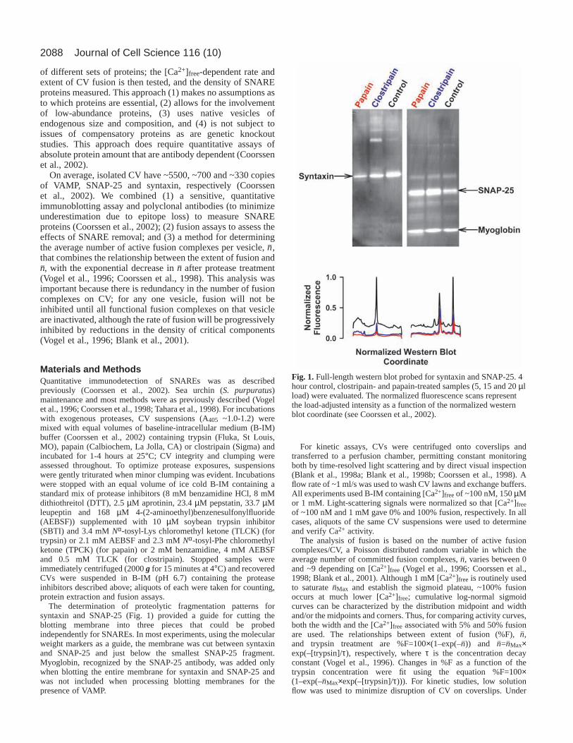
The determination of proteolytic fragmentation patterns forsyntaxin and SNAP-25 (Fig. 1) provided a guide for cutting theblotting membrane into three pieces that could be probedindependently for SNAREs. In most experiments, using the molecularweight markers as a guide, the membrane was cut between syntaxinand SNAP-25 and just below the smallest SNAP-25 fragment.Myoglobin, recognized by the SNAP-25 antibody, was added onlywhen blotting the entire membrane for syntaxin and SNAP-25 andwas not included when processing blotting membranes for thepresence of VAMP.
For kinetic assays, CVs were centrifuged onto coverslips andtransferred to a perfusion chamber, permitting constant monitoringboth by time-resolved light scattering and by direct visual inspection(Blank et al., 1998a; Blank et al., 1998b; Coorssen et al., 1998). Aflow rate of ~1 ml/s was used to wash CV lawns and exchange buffers.All experiments used B-IM containing [Ca2+]freeof ~100 nM, 150 µMor 1 mM. Light-scattering signals were normalized so that [Ca2+]freeof ~100 nM and 1 mM gave 0% and 100% fusion, respectively. In allcases, aliquots of the same CV suspensions were used to determineand verify Ca2+ activity.
The analysis of fusion is based on the number of active fusioncomplexes/CV, a Poisson distributed random variable in which theaverage number of committed fusion complexes, n–, varies between 0and ~9 depending on [Ca2+]free (Vogel et al., 1996; Coorssen et al.,1998; Blank et al., 2001). Although 1 mM [Ca2+]free is routinely usedto saturate n–Max and establish the sigmoid plateau, ~100% fusionoccurs at much lower [Ca2+]free; cumulative log-normal sigmoidcurves can be characterized by the distribution midpoint and widthand/or the midpoints and corners. Thus, for comparing activity curves,both the width and the [Ca2+]free associated with 5% and 50% fusionare used. The relationships between extent of fusion (%F), n–,and trypsin treatment are %F=100×(1–exp(–n–)) and n–=n–Max×exp(–[trypsin]/τ), respectively, where τ is the concentration decayconstant (Vogel et al., 1996). Changes in %F as a function of thetrypsin concentration were fit using the equation %F=100×(1–exp(–n–Max×exp(–[trypsin]/τ))). For kinetic studies, low solutionflow was used to minimize disruption of CV on coverslips. Under
Journal of Cell Science 116 (10)
Fig. 1.Full-length western blot probed for syntaxin and SNAP-25. 4hour control, clostripain- and papain-treated samples (5, 15 and 20 µlload) were evaluated. The normalized fluorescence scans representthe load-adjusted intensity as a function of the normalized westernblot coordinate (see Coorssen et al., 2002).

2089Differential proteolysis of cortical vesicle proteins
these conditions, the transition from active to committed state is notresolved; the kinetic data were fit using the original two-state modeland a temporal offset (Vogel et al., 1996; Blank et al., 2001). Theinitial rate of fusion can be approximated by n–×p (Vogel et al., 1996;Blank et al., 2001). With p=κ×n– (Blank et al., 2001), where κrepresents a characteristic efficiency constant, the initial rate can beexpressed as κ×n–2. This function suggests that κ is proportional to arate constant, whereas n– is analogous to a concentration term. With κproportional to a fusion rate constant, the change in energy resultingfrom papain treatment can be calculated using the relationshipκcontrol/κpapain=exp(–(Econtrol–Epapain)/kT).
The exponential correlation between SNARE density andthe midpoint of the calcium activity curve, Midpoint=A×exp(–Density/B)+C, was evaluated with weighting using 1/(s.e.)2 ofthe midpoint estimates. The error in SNARE density was ignored inthe fitting after determining that the error range in the parameterestimates (95% confidence) was greater than changes in the estimatesarising from fitting the midpoint vs. the density values shifted by ±the error in the density. Overall, the error in the density divided bythe density was 19%, 16% and 5% for VAMP, SNAP-25 and syntaxin,respectively. We could determine syntaxin density to better than 1molecule/CV†.
ResultsThe Ca2+ activity and kinetics of CV fusion are described byan analysis developed for CV-PM fusion (Vogel et al., 1996;Coorssen et al., 1998; Blank et al., 2001). CV-CV fusion ischaracterized by sigmoidal Ca2+ activity curves; midpoints andwidths of the Ca2+ threshold distributions were determined(Fig. 2A; Table 1) (Coorssen et al., 1998; Blank et al., 1998a).The Ca2+ activity curves for CV-CV fusion are minimallyshifted from those for CV-PM fusion (Coorssen et al., 1998),which predict [Ca2+]free that are comparable to those measuredduring fertilization (Vogel et al., 1996); 5%/50% fusion occursat a [Ca2+]free of 3/6 µM. One hour in buffer has little effecton CV fusion, with 5%/50% fusion occurring at [Ca2+]free of3/7 µM (Fig. 2A; Table 1); after 4 hours in buffer, there is asignificant shift in the midpoint of the Ca2+ activity curve and5%/50% fusion occurs at a [Ca2+]free of 36/76 µM. Thus,results of protease treatments were compared to parallel,untreated controls.
Trypsin had a systematic and progressive affect on Ca2+
activity (sensitivity and extent) (Fig. 2A; Table 1). 700 units/mltrypsin (1 hour) shifted Ca2+ activity rightward, but with noloss in maximal extent of fusion elicited by a saturating
[Ca2+]free; the 5%/50% fusion level occurs at 3/17 µM. 3500units/ml trypsin (1 hour) caused a further rightward shift in Ca2+
activity and ~30% loss of fusion. Prolonged incubations orhigher trypsin doses further digested certain CV proteins,correlating with losses in Ca2+ activity and progressively greaterinhibition of fusion; 700 units/ml (3 hours) resulted in ~70%inhibition (data not shown). In comparison, 35,000 units/mltrypsin (1 hour) rendered CV fusion incompetent (99±1%inhibition at saturating [Ca2+]free). Like N-ethylmaleimide(NEM), trypsin inhibited fusion in a manner consistent with asingle, crucial site of action and the number of these sites,n–Max=8.4±1.0, agrees with previous determinations (Vogel etal., 1996; Coorssen et al., 1998). By contrast, papain (2000units/ml, 1 and 4 hours) shifted and broadened Ca2+ activitywithout altering the maximal extent of fusion (Fig. 2A,B; Table1); 5%/50% fusion occurs at 3/45 and 68/197 µM [Ca2+]free,respectively, following 1 and 4 hour treatments. Clostripain(100 units/ml, 1 and 4 hours) had minimal effect on Ca2+
activity (Fig. 2A,B; Table 1, 5%/50% fusion occurs at 2/11 and40/69 µM [Ca2+]free, respectively, following 1 and 4 hourtreatments), but distinct effects on the CV protein profile (Fig.2C). When added hourly, fresh papain (but not clostripain)gradually inhibited fusion (~20-30% fusion at saturating[Ca2+]free after 3 hours treatment, data not shown). In all cases,comparable results were obtained after simply settling protease-treated CV into contact, confirming that the measured fusion isdependent on proteins rather than on centrifugation (Coorssenet al., 1998). Similar results were also obtained for exocytosisin vitro after 1 hour treatment of CV-PM preparations (Fig. 2D;Table 2). Trypsin does not appear to affect docking per se, asCV undocking was not observed. The effects of papain (3000units/ml) were less dramatic, suggesting limited access to theCV-PM contact site. Thus, each of these proteases had adifferent effect on CV fusion, enabling us to distinguishbetween changes in the Ca2+ sensitivity or extent of fusion.
To determine whether the differential protease effects on Ca2+
activity and fusion extent were due to differential proteolysis of
†Confidence intervals for syntaxin copies per CV (Copy #/CV).Copy #/CV=(fmoles syntaxin)×6.02×10⁸/[(CV count)×(Dilution factor)×(Extractionefficiency)].
The error in determining Copy #/CV is:
σCopy #/CV=Copy #/CV×[(σfmoles/fmoles)2+(σCv count/CV count)2+(σDilution factor/dilutionfactor)2+(σExtraction efficiency/Extraction efficiency)2]0.5.
Assuming that the relative error for each of these terms is 5% (0.05), an extremelyconservative value for all terms except the extraction efficiency, then the total error canbe approximated byσCopy #/CV=Copy #/CV×0.1.
Using a weighted average of the results obtained at the two different protein loads (30and 15 µl of sample), the average copy numbers are 0.107±0.008 and 0.404±0.032. If thecalculated error is representative of the underlying error then the Copy #/CV issignificantly less than one copy at the 99% confidence level (3×σCopy #/CV). If thecalculated error represents an estimate of the unknown underlying distribution then theappropriate t value with one degree of freedom (two samples were averaged, n–1 degreesof freedom) must be used. In this case, the t value for one degree of freedom at the 95%confidence interval is 12.71 and the error would be 0.107±0.102 and 0.404±0.407; thesevalues also indicate significantly less than one copy at the 95% confidence level.Therefore, within >95% probability, these two experiments show that our determinationof less than one copy of syntaxin per CV is statistically significant.
Table 2. CV-PM Ca2+ activity curve parametersExtent of Midpoint Width
Protease (units/ml) fusion (%) (µM) (pCa units)
1 hour control (0) 100 21.4±0.6 0.28±0.02Clostripain (100) 100 23.8±0.7 0.38±0.02Papain (3000) 96 35.9±0.5 0.22±0.01Trypsin (700) 100 31.3±0.7 0.30±0.01Trypsin (14,000) 51 35.3±0.5 0.20±0.01
Table 1. CV-CV Ca2+ activity curve parameters Extent of Midpoint Width
Protease (units/ml) fusion (µM) (pCa units)
0 hours control (0) 100% 5.8±0.1 0.21±0.011 hour control (0) 100% 7.1±0.2 0.23±0.024 hours control (0) 100% 76.3±2.2 0.20±0.021 hour clostripain (100) 100% 11.2±0.7 0.44±0.034 hours clostripain (100) 100% 68.5±1.4 0.14±0.011 hour papain (2000) 100% 45.3±3.9 0.72±0.054 hours papain (2000) 100% 196.9±7.2 0.28±0.021 hour trypsin (700) 100% 16.9±0.7 0.44±0.021 hour trypsin (3500) 71% 41.7±3.0 0.51±0.04
2090
SNARE proteins, we used the fact that known SNAREs havesubstantial numbers of potential trypsin, papain and clostripaincleavage sites throughout their cytoplasmic domains (Table 3).The initial number of accessible proteolytic cleavage sites is
expected to be lower due to structural constraints (Hubbard etal., 1998; Hubbard, 1998). Using the program Nickpred(Hubbard et al., 1998), examination of SNAREs in theBrookhaven Protein Data Base predicted multiple limitedproteolytic sites. Subsequent loss of structure is expected toexpose additional sites to further proteolysis. Indeed, ladders ofmultiple proteolytic fragments migrating below the main VAMPand SNAP-25 protein bands were detected with lower proteaseconcentrations or shorter treatment time (1 hour). Following 4hour protease treatments, syntaxin appears to be either intact orfully proteolyzed, as no fragments were detected (Fig. 1). Thisobservation is consistent with the complete loss of syntaxincomplex immunoreactivity observed following trypsinproteolysis (Lawrence and Dolly, 2002). SNAP-25
Journal of Cell Science 116 (10)
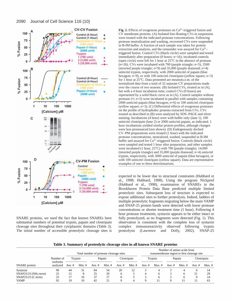
Fig. 2. Effects of exogenous proteases on Ca2+-triggered fusion andCV membrane proteins. (A) Isolated free-floating CVs in suspensionwere treated with the indicated protease concentrations. Followingprotease neutralization and washing, recovered CVs were suspendedin B-IM buffer. A fraction of each sample was taken for proteinextraction and analysis, and the remainder was assayed for Ca2+-triggered fusion. Control CVs (black circle) were sampled and testedimmediately after preparation (0 hours; n=16); incubated controls(open circle) were left for 1 hour at 25°C in the absence of protease(n=26). CVs were incubated with 700 (purple triangle; n=5), 3500(inverted purple triangle; n=9) and 35,000 (purple diamond; n=4)units/ml trypsin, respectively, with 2000 units/ml of papain (bluehexagon; n=9), or with 100 units/ml clostripain (yellow square; n=3)for 1 hour at 25°C. Data presented are means±s.e.m. of thenormalized data from a total of 32 separate CV preparations madeover the course of two seasons. (B) Isolated CVs, treated as in (A),but with a 4 hour incubation time; control CVs (0 hours) arerepresented by a solid black curve as in (A). Control samples withoutprotease (×; n=5) were incubated in parallel with samples containing2000 units/ml papain (blue hexagon; n=6) or 100 units/ml clostripain(yellow square; n=3). (C) Differential effects of exogenous proteaseson the profile of hydrophobic proteins extracted from CVs. CVstreated as described in (B) were analyzed by SDS–PAGE and silverstaining. Incubations (4 hour) were with buffer only (lane 1), 100units/ml clostripain (lane 2) or 2000 units/ml papain, as indicated. 1hour incubations yielded similar protein profiles, although changeswere less pronounced (not shown). (D) Endogenously dockedCV–PM preparations were treated (1 hour) with the indicatedprotease concentrations, neutralized, washed, suspended in B-IMbuffer and assayed for Ca2+-triggered fusion. Controls (black circle)were sampled and tested 1 hour after preparation, and other sampleswere incubated (1 hour, 25°C) with 700 (purple triangle), 14,000(inverted purple triangle) and 35,000 (purple diamond; n=4) units/mltrypsin, respectively, with 3000 units/ml of papain (blue hexagon), orwith 100 units/ml clostripain (yellow square). Data are representativeexamples of one to three determinations.
Table 3. Summary of proteolytic cleavage sites in all known SNARE proteinsNumber of amino acids from
Total number of protease cleavage sites transmembrane region to first cleavage site
Number of Trypsin Papain Clostripain Trypsin Papain Clostripainisoforms
SNARE protein analyzed Ave. # Min. # Ave. # Min. # Ave. # Min. # Ave. # Max. # Ave. # Max. # Ave. # Max. #
Syntaxin 90 44 31 84 54 20 12 3 4 3 4 6 14SNAP25/23 (NH2-term) 25 12 8 25 20 6 5 4 6 3 6 21 29SNAP25/23 (C-term) 25 17 10 37 17 8 5 3 6 3 6 9 14VAMP 92 19 10 42 21 9 4 9 11 9 11 11 63
2091Differential proteolysis of cortical vesicle proteins
immunoreactivity, which may correspond to fragments, wasdetected in both control and treated samples (Fig. 1); VAMPimmunoreactivity often appeared as a doublet (data not shown).The amounts of these SNARE fragments decreased withincreased protease concentration or treatment time and wereincluded in the quantitative determinations of SNARE density.Thus, unlike Clostridial toxins, these proteases could destroy allknown SNAREs (including toxin-insensitive homologs).
Trypsin (700 units/ml, 1 hour) digested substantial amountsof VAMP, SNAP-25 and syntaxin but had no effect on theextent of fusion (Fig. 3). Increased trypsin (3500 units/ml)removed >90% of VAMP and syntaxin; ~60% SNAP-25remained (Fig. 4). Higher trypsin doses (Fig. 4) or longerincubation times (700 units/ml, 3 hours) did not reduceSNAREs to levels lower than detected after 1 hour with 3500units/ml, but did block fusion. Papain (2000 units/ml) andclostripain (100 units/ml) removed most SNAREs after 1 hour;there was little added affect of 4 hour incubations. Papainreduced VAMP, SNAP-25 and syntaxin by 94%, 91% and>99%, respectively, and clostripain by 92%, 87% and 94%; CVremained fully Ca2+ sensitive and 100% fusion competent (Fig.3). A post-protease wash with chaotropic buffer resulted insome further loss of SNARE fragments nonspecifically boundto CV, but there was no change in fusion at saturating[Ca2+]free, indicating that free SNARE fragments were notpromoting fusion; although ineffective, these fragments werestill included in our density assessments to ensure exhaustiveSNARE quantification. Intact SNAREs are not removed by thechaotropic buffer wash (data not shown). Post-protease fusionwas not due to nonphysiological Ca2+-induced membranedestabilization, or fusion would remain high after extensive
trypsinization; this was not the case (Figs 2, 3 and Fig 4A;35,000 units/ml trypsin). Thus, despite SNARE removal,fusion activity remained. Furthermore, when added hourly,fresh papain, but not clostripain, gradually inhibited fusiondespite comparable reductions in SNARE densities. Fusionappears to require proteins other than the SNAREs.
Reducing syntaxin to <1 copy/CV (‘biochemical knockout’;0.1 or 0.4 copy/CV at the 95% confidence level for each oftwo samples with a range from 0-0.8†) by the most potentlySNARE-destructive treatment (papain; 2000 units/ml replacedevery hour for 3 hours) (Fig. 5) decreased but did not abolishthe extent of fusion elicited by saturating [Ca2+]free (two trials,final extent 20-30%). Eight more trials with 2000 units/mlpapain gave 100±1% fusion with only 1-<3 syntaxincopies/CV in 4 trials and >3 copies/CV in 4 trials; 50% fusionoccurred at 45 and ~200 µM [Ca2+]free after 1 and 4 hourtreatments, respectively. Overall, the reduced syntaxin densityas a function of papain treatment ranged from <1-80 copies/CVdepending upon the papain concentration (300–5000 units/ml)and treatment time (1-4 hours). For comparison, the reducedsyntaxin density as a function of clostripain treatment ranged
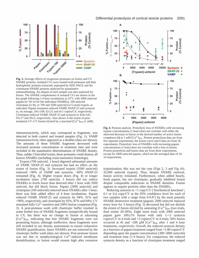
Fig. 3. Average effects of exogenous proteases on fusion and CVSNARE proteins. Isolated CVs were treated with proteases and theirhydrophobic proteins extracted, separated by SDS–PAGE and theconstituent SNARE proteins analyzed by quantitativeimmunoblotting. An aliquot of each sample was also analyzed forfusion. The SNARE complements of isolated CVs are shown in thebar graph following 1-4 hour incubations at 25°C with 2000 units/mlpapain (n=10-14 for the individual SNAREs), 100 units/mlclostripain (n=8), or 700 and 3500 units/ml (n=5 each) trypsin, asindicated. Papain treatment reduced VAMP, SNAP-25 and syntaxinto, on average, 341±108, 62±21 and 4±1 copies/CV, respectively.Clostripain reduced VAMP, SNAP-25 and syntaxin to 424±143,93±17 and 20±5, respectively. Also shown is the extent of post-treatment CV–CV fusion elicited by a maximal [Ca2+]free (1 mM).
Fig. 4. Poisson analysis. Proteolytic loss of SNAREs with increasingtrypsin concentrations (1 hour) does not correlate with either theobserved decrease in fusion or the derived number of active fusioncomplexes (n–) at 1 mM [Ca2+]free. Protein proteolysis data are fromfive separate experiments; the fusion curve and n– data are from 26experiments. Proteolytic loss of SNAREs with increasing papainconcentrations (1 hour) does not correlate with a loss in fusion.Protein proteolysis and fusion data are from three experiments,except for 2000 units/ml papain, which are the averaged data of 10-14 experiments.

2092
from 5 to 50 copies/CV. If an individual SNARE complexcatalyzes fusion, and proteolysis is random, then Poissonanalysis (Vogel et al., 1996; Coorssen et al., 1998; Blank et al.,2001) can be used to predict the extent of fusion at saturating[Ca2+]free. The extents of fusion predicted by the measuredsyntaxin densities following papain treatment are notconsistent with the observed fusion (χ2, P<0.05); neithersyntaxin nor syntaxin-containing complexes have theproperties of a Poisson-distributed fusogen. Three SNAREcomplexes per vesicle fusion event have been suggested (Huaand Scheller, 2001), and it is thus significant that the maximumextent of fusion (100%) elicited by saturating [Ca2+]free isindependent of syntaxin density from 1 to 330 copies/CV(papain treated relative to control).
Shifts in Ca2+ activity preserving fusion competence areobserved in both control (4 hours) and papain-treated samples,
suggesting disruption of a Ca2+ regulatory system mediatingfusion. As clostripain-treated samples did not have large shiftsin Ca2+ activity relative to control and the Ca2+ activitymidpoint was not correlated with SNARE density (slope notsignificantly different from zero), it is somewhat surprisingthat, for papain-treated samples, the midpoints of the Ca2+
activity curve were correlated with SNARE density (Fig. 6).For all three SNAREs, the correlation between [Ca2+]free(EC50) and SNARE density was described by the exponentialrelationship, midpoint=A×exp(–density/B)+C, where A+C isthe midpoint (EC50, µM) of the Ca2+ activity curve in theabsence of SNAREs and B is the density decay constant. Theextrapolated midpoint at high SNARE density (parameter C)varied from 13 to 20 µM, depending on the SNARE, and isconsistent with control midpoints (6–20 µM for 0, 1 and 2 hourincubations). When low-density values are normalized to the
Journal of Cell Science 116 (10)
Fig. 5.Proteolytic ablation of syntaxin: average density <1 copy/CV.Using a calibration curve derived from serial dilution of purifiedglutathione S-transferase (GST)–syntaxin (Coorssen et al., 2001)0.73 fmoles, corresponding to 113 molecules/CV, were detected in 0hour controls. No syntaxin was detected in papain-treated (2000units/ml, replaced every hour for 3 hours) samples. The limit ofdetection for this calibration was <0.039 fmoles or 0.3 TreatedEquivalent (T.E.) molecules/CV for the same protein load (CVnumber) used to visualize the papain-treated sample. The maximalsyntaxin density in this sample is 0.3 molecules/CV. The plot ofnormalized fluorescence vs. Western Blot Coordinate shows how thesignal associated with 0.039 fmoles is easily quantified. The curvesare drawn with an offset for visual comparison.
Fig. 6.Ca2+ activity and SNARE density. The midpoint of the Ca2+
activity curve following papain treatment at either 1 hour (blue) or 4hours (red) is correlated with SNARE density (Copy #/CV). The datawere fit using the relationship A×exp(–density/B)+C, where A+C(µM) is the midpoint of the Ca2+ activity curve in the absence ofSNAREs (248±10, 433±66 and 305±22) and B is the density decayconstant (212±63, 42±10 and 11±3) for VAMP, SNAP-25 andsyntaxin, respectively. When SNARE density was normalized by thedensity decay constant, the pooled transformed data were describedby the relationship A×exp(–normalized density/B)+7.1; the midpointof the Ca2+ activity curve in the absence of SNAREs (A+7.1) is260±22 µM.

2093Differential proteolysis of cortical vesicle proteins
density at 1 hour and the high-density midpoint constrained tothe value of the 1 hour control (7.1 µM), all three SNAREswere described by a common exponential decay with a low-density midpoint of ~250 µM [Ca2+]free. Thus, papaintreatment reveals an intrinsic Ca2+-dependent process with anactivity midpoint that can be modulated by proteolysis between~6 and ~250 µM [Ca2+]free. If this reflects the inherent Ca2+
sensitivity of the native regulated fusion complex, then theEC50 of this endogenous fusion complex is ~250 µM [Ca2+]free.
Decreases in the density of components crucial to triggeredfusion are predicted to inhibit the rate of fusion (Vogel et al.,1996; Blank et al., 2001). As clostripain (100 units/ml, 4 hours)failed to affect Ca2+ activity (Fig. 2B) and had no effect onfusion kinetics (Fig. 7A), but did digest SNARES, it is unlikelythat the identified SNARES in this system are crucial to fusion.Papain (2000 units/ml, 4 hours) reduced the kinetics and extenttriggered by 150 µM Ca2+ but not the final extent of fusiontriggered by saturating Ca2+, indicating that both the
probability per unit time (p) and n– decreased (Fig. 7A).However, a limited papain treatment (2000 units/ml, 3 hours)altered the kinetics triggered by 150 µM Ca2+ with minimalchanges in the extent (Fig. 7B). Analysis (see Materials andMethods) ruled out a direct reduction in n–, indicating thatpapain reduced P, the probability that any given vesicle willfuse (Vogel et al., 1996; Blank et al., 2001). After 3 hours, p/n–
(a measure of efficiency) in papain-treated CV is only ~17%of control, but is unaffected by clostripain (4 hours) (Fig. 7A).The changes in fusion rate after 3 hours papain treatment areconsistent with an increased energy barrier for fusion. Treatingp/n– as a rate constant (see Materials and Methods), the energycontributed by papain-sensitive proteins is estimated to be ~2kT (0.6 kcal/mol) per fusion complex. This is about half thatrequired to overcome the hydration layer at membrane surfaces(Leiken et al., 1993) or to mediate molecular rearrangementsthought necessary for bilayer merger (Kuzmin et al., 2001).Thus, papain (but not clostripain or low doses of trypsin)removes or modifies crucial modulatory proteins that havepronounced effects on the efficiency of triggered fusion.
DiscussionThe ability to determine with precision the number of copiesof syntaxin/CV allowed a quantitative evaluation of SNAREhypotheses, as the SNARE core complex requires SNAREmotif helices from syntaxin that cannot be substituted byother SNAREs (Scales et al., 2000a). Ca2+- and protein-dependent fusion of native secretory vesicles proceededafter substantial removal of identified SNAREs, even aftersyntaxin was stoichiometrically eliminated (biochemical‘knockout’). It should be noted that the effective CV SNAREdensities remaining after proteolysis may be even lower thanthe values we report, as we included all detectable SNAREfragments in the overall quantification of our high sensitivityimmunoblots. If ineffective SNARE fragments are present (asindicated by the use of chaotropic washes), they have, forthe sake of thoroughness, nevertheless been included in thequantitative evaluations of density. Thus, although theidentified SNAREs do not effect fusion, the Ca2+ sensitivitydata suggest that they may interact with modulators of Ca2+
efficiency that directly influence the capacity for fusion in thisCa2+-triggered system.
Lack of a direct role for SNAREs in membrane fusion:alternate interpretationsOur conclusion, that SNAREs are neither driving CVmembranes together nor inextricably linked to Ca2+-regulatedfusion via an absolute requirement for the presence of theircytosolic domains, is dependent on a correct interpretation ofSNARE proteolysis, antibody interactions and the fusionprocess. Two classes of alternative hypothesis that preserve adirect role for SNARE proteins can be considered. The firstclass is that the ‘wrong’ SNAREs were evaluated in this study:the necessary SNAREs were still present on protease-treatedCV at a sufficient density to support fusion. This class can besubdivided into SNARE homologs and SNARE fragments thatdo not cross-react with the antibodies used. The second classis that multiple fusion pathways exist, and include SNARE-dependent and -independent mechanisms; the latter can be
Fig. 7.Kinetics of Ca2+-triggered CV–CV fusion. (A) Differentialeffects on Ca2+-triggered CV–CV fusion kinetics following 4 hourprotease treatments. Papain, but not clostripain, inhibits response to150 µM [Ca2+]free. However, at 1 mM [Ca2+]free, 100% fusion isobserved in all cases. The decrease in the rate and extent of fusion at150 µM [Ca2+]free is consistent with a reduction in n–, as determinedfrom a rightward shift in the Ca2+ activity curve. (B) Limiting papaintreatment to 3 h alters the rate of fusion elicited with 150 µM[Ca2+]free, with minimal change in the extent of fusion. Thus, the rateof fusion decreased without a substantial change in n–; limited papaintreatment decreased the fusion probability, p.
2094
subdivided into protein-mediated and protein-independentmechanisms.
Do we have the right SNAREs?The interpretation of genetic studies of SNARE activity can beinfluenced by the presence of gene homologs (e.g. Vilinsky etal., 2002). By contrast, the biochemical knockout approachrelies on the structural conservation of putative homologs. Theconservation of the different potential protease cleavage sitesin all known SNARE homologs is very high (Table 3),particularly for the trypsin, papain and clostripain cleavagesites closest to the PM (i.e. cleavage at this site would result inthe complete release of the cytoplasmic domain) and in thepotential coiled-coil domains postulated to be essential toSNARE-mediated fusion. Thus, although we do not know allthe epitopes recognized by the antibodies employed, completedestruction of other SNARE homologs should also occur underthe conditions of our experiments. If this interpretation isincorrect, then a putative protease resistant ‘SNARE’ wouldeither have a sequence that is significantly different from allknown SNAREs, such that not even one functional proteolyticsite exists (a feature that would question the nature of SNAREhomology), or exist in a stable conformation such that itscleavage sites were never accessible to protease action duringthe extended incubation times used in our experiments.
Antibodies raised specifically against nonconserved regionsof the protein can certainly identify different homologs ofSNAREs. In this study, polyclonal antibodies raised against thewhole cytosolic region (e.g. including the extended highlyconserved domains) were used. Because SNAREs maintainhighly conserved regions it is more likely that our antibodieswould cross-react with any protein containing these conservedregions than would monoclonal antibodies. We have usedantibodies against SNAREs from other species in the past(Coorssen et al., 1998; Coorssen et al., 2002; Tahara et al., 1998),and have always identified only these SNAREs on the CVmembrane. To accept the alternate hypothesis, that SNAREsdrive the fusion step, occult SNARE homologs to (or proteolyticfragments of) syntaxin, VAMP and SNAP-25 need to exist inprotease-resistant conformations that are not recognized by ourpolyclonal antibodies (i.e. lack all the epitopes recognized by theantibodies used here) at all protein concentrations and dilutionsassessed with an ultrasensitive immunodetection protocol(Coorssen et al., 2002), and the putative proteolytic fragmentswould have to remain stably bound in the presence of chaotropicbuffers. Although this is an alternative explanation, we think itsimpler and thus more likely that CV–CV fusion can occur ineither the absence of syntaxin or with 91-99% overall loss ofSNARE proteins. This interpretation is also most consistent withthe substantial differences in SNARE densities measured onnative secretory vesicles compared with those required for fusionin reconstituted preparations (Weber et al., 1998; Coorssen et al.,2002). In the absence of SNARE and other modulatoryinfluences, the fusion process has an endogenous Ca2+-activitywhose midpoint is ~250 µM [Ca2+]free.
Are there multiple protein-dependent or protein-independent fusion mechanisms?
Our analysis of the Ca2+ activity and kinetics of fusion is based
on the hypothesis that an increase in [Ca2+]free increases thenumber of participating fusion complexes and that thesecomplexes are randomly distributed among CVs (Vogel et al.,1996; Blank et al., 2001). This analysis has consistentlyexplained the experimental data observed in this system (Vogelet al., 1996; Blank et al., 1998a; Blank et al., 1998b; Coorssenet al., 1998; Tahara et al., 1998; Blank et al., 2001; Ikebuchi etal., 2001). However, if changes in the Ca2+ activity and thekinetics of fusion do indicate a shift from one fusion pathwayto another, then perhaps SNARE proteins normally mediatefusion, but in their absence alternative mechanisms becomeevident; redundant pathways may exist. This is an intriguingand provocative alternative hypothesis. However, no changesoccurred in kinetics following clostripain treatment, despitesubstantial decreases in SNARE density, whereas comparabledecreases in SNARE density following papain treatment didalter the kinetics of fusion. This is perhaps the most directevidence against an essential role for the SNAREs in fusion,as any change in the density of an essential component of thefusion complex would be predicted to inhibit the rate of fusion(Vogel et al., 1996; Blank et al., 2001). The differential effectsof protease treatments are difficult to reconcile with a simpleswitch from a SNARE-mediated to a SNARE-independent,protein-mediated fusion pathway without postulating theexistence of multiple classes of SNAREs such that clostripainpreserves the SNARE-mediated pathway by acting only onnonfunctional SNAREs while papain and trypsin target onlythe functional SNAREs. Rather, the demonstrated relationshipbetween SNARE density and Ca2+ activity, seen in the papaindata (Fig. 6), clearly indicates the existence of a single fusionpathway (with an inherent Ca2+ activity). The Ca2+ sensitivityof this fundamental fusion pathway is modulated in the‘physiological’ range of [Ca2+]free either by SNAREs coupledwith other modulatory components such as ‘Ca2+ sensors,’ orby other components alone that track the papain-dependentchanges in SNARE density via similar proteolytic sensitivity.Redundancy in modulatory factors may be a conservedhallmark of triggered exocytosis; the data indicate that trypsinand papain more broadly affect these modulatory factors thandoes clostripain.
A switch from a SNARE-mediated to a protein-independentmechanism (e.g. Ca2+-mediated lipid fusion) can not explainthe data because extensive trypsinization ablates the fusionresponse, even to several millimolar [Ca2+]free; if any of thefusion observed was nonphysiological or purely lipid-mediated, then it should also have been observed under theseconditions. As the fusion response is also lost after treatmentswith other broad-spectrum proteases (e.g. chymotrypsin; datanot shown), it is unlikely that purely lipid-mediated fusionoccurs under our experimental conditions. In fact, even whenusing pure lipid vesicles (LV) as the target membrane (CV–LVfusion; Ca2+-dependent fusion at <60 µM [Ca2+]free),proteinaceous machinery is still essential to the triggeredfusion steps, as determined by thiol sensitivity (Vogel et al.,1992). The shift in the midpoint of the Ca2+ activity curve forCV–CV fusion never enters the range of [Ca2+]free required toinduce purely lipid fusion; fusion of lipid membranescontaining mixtures of neutral and negatively charged lipidsrequires tens of millimolar [Ca2+]free (Cohen et al., 1980;Cohen et al., 1984; Duzgunes et al., 1981; Coorssen and Rand,1995; Zimmerberg and Chernomordik, 1999). The Ca2+
Journal of Cell Science 116 (10)

2095Differential proteolysis of cortical vesicle proteins
activity curves for all conditions, except for papain where thedistribution of Ca2+ sensitivity is believed to be significantlyaltered, indicate that >95% fusion occurs between ~10 and 160µM [Ca2+]free, a range that is consistent with the physiology ofCa2+-triggered exocytosis. Because it takes time for theproteases to work, we must also consider a shift in mechanismdue to incubation time. However, the invariance of p/n– withincubation time (0-4 hours), indicating no change in theestimated energy of fusion, makes a time-dependent shift inmechanism unlikely.
Although extensive trypsinization (≥3500 units/ml, 1 hour)does not simply create an environment for ‘lipid-mediated’fusion, this proteolytic treatment did reduce VAMP andsyntaxin to <10% of control, or ~30 syntaxin/CV; a maximumof ~30 syntaxin-limited complexes (1:1:1 VAMP, SNAP-25,syntaxin complexes or syntaxin with other binding partners)are possible per trypsin-treated CV. If syntaxin and fusioncomplexes are stoichiometrically proportional, then reducingn–Max by 90% (n–Max, trypsin=0.84) would still support >50%fusion at saturating [Ca2+]free. If an individual syntaxincomplex (trimeric SNARE complex or other) (Lawrence andDolly, 2002) causes fusion (i.e. is both fusogen and Ca2+
sensor) (Sutton et al., 1998; Chen et al., 2001; Peters et al.,2001) then, assuming a uniform surface distribution, anycontacting CV domains >130 nm in diameter would ensure100% fusion at saturating [Ca2+]free (for CVs having 30syntaxin/CV). But extensively trypsinized CVs do not fuse(Fig. 3, Fig. 4A). As trypsin effects on Ca2+ activity did notcorrelate with SNARE removal (Fig. 2A, Fig. 3, Fig. 4A), othertrypsin-sensitive proteins must function in defining the activitycurve for Ca2+-sensitive fusion; notably, the trypsin sensitivityof synaptotagmin is well established (Tugal et al., 1991).
SNAREs and the process of exocytosisThe simplest hypothesis is that there is only one endogenousmechanism of protein-mediated fusion in this system, and ourdissection of this mechanism results in altered responses.According to this hypothesis, although SNARES have key rolesin the pathway of exocytosis, they are not members of anyminimal protein set required for Ca2+-triggered fusion in thisregulated exocytotic system – certainly not in any capacitythat requires their intact cytosolic domains; their rapidintermembrane binding does not appear to be essential to theminimal mechanism of native membrane merger (Coorssen etal., 1998). Of course, SNAREs are enormously important to theprocess of exocytosis; there is good evidence that SNAREsaffect an early stage of target membrane recognition, vesicledocking and/or priming (Pelham, 2001; McNew et al., 2000;Scales et al., 2000a; Scales et al., 2000b). Indeed, our newestimates indicate that SNAREs, together with other modulatoryproteins, can contribute energy to the fusion mechanism,although not enough to directly trigger fusion, except perhaps atdensities in excess of those in native vesicle membranes (Weberet al., 1998; Coorssen et al., 2002). SNAREs probably play arole in trafficking and the localization of CV to PM dockingsites, which occurs during the oocyte-to-egg transition (Berg andWessel, 1997), because fusion can be disrupted by clostridialtoxins injected into eggs whose CVs have been de-docked (Biet al., 1995); these PM docking sites may be associated withfusion complexes (Ikebuchi et al., 2001).
In other systems, genetic knockouts of VAMP and SNAP-25 affect the regulation of exocytosis but not the ultimatecapacity to release vesicular contents (Nonet et al., 1998;Yoshihara et al., 1999; Schoch et al., 2001; Rao et al., 2001;Washbourne et al., 2002), leading to two hypotheses – thatSNAREs catalyze fusion reactions by stabilizing transitionstates (Schoch et al., 2001) and that multistep, Ca2+-dependentassembly of SNAREs forces membranes together to promotefusion (Xu et al., 1999; Chen et al., 2001; Scales et al., 2001).Considering the findings presented here, neither of thesehypotheses is probable because the rate of CV fusion (ratherthan of multiple late steps in the exocytotic pathway) inclostripain-treated samples was independent of SNAREdensity, and the removal of SNAREs and other modulatoryproteins suggests an inherent Ca2+ activity in the triggeredsteps of fusion.
In regulated fusion, SNAREs may function as structuralproteins that prepare or optimize local release sites (Coorssenet al., 1998), perhaps by coordinating the right vesicles(Rothman, 1994; McNew et al., 2000) to the right Ca2+
channels (Rettig et al., 1997; Jarvis et al., 2000), for which theythen act as regulatory components. This represents anotherinterpretation of SNARE knockout data (Schoch et al., 2001;Washbourne et al., 2002). If VAMP loss alters presynapticarchitecture such that vesicles dock less effectively (outsidedomains of increased intracellular Ca2+), leading to diminishedfunctional docking by ~10%, then spontaneous release andmeasures of docked, primed vesicles (i.e. sucrose pools) wouldbe diminished by ~10% (Parsons et al., 1995) and the fastevoked response to Ca2+ would be lost (Xu et al., 1998).
A calcium regulatory complex that is papain sensitiveAlthough we do not yet know whether the fusion complex itselfis inherently Ca2+ sensitive, or whether this property is residentto an associated sensor, this sensitivity can be correlated toSNARE density following proteolysis by papain. This suggestsa native papain-sensitive complex that includes SNAREs andassociated proteins. However, this complex is not essentialper se in the triggered fusion steps of exocytosis because in ourwork higher [Ca2+]free overcame the block to fusion (seen at‘physiological’ [Ca2+]free) caused by proteolysis. Suchrecovery has also been documented in earlier experiments withClostridial toxins (Dreyer and Schmitt, 1983; Ahnert-Hilgerand Weller, 1993; Bittner and Holz, 1993; Lawrence et al.,1994; Glenn and Burgoyne, 1996; Lawrence et al., 1996;Capogna et al., 1997; Land et al., 1997; Fassio et al., 1999),suggesting that the concept of a regulatory complex mayextend beyond the present system. In general, the testing andrescue of exocytosis at only one [Ca2+]free after complexdisruption tells us a great deal about the modulatory role ofthe complex. Incomplete Ca2+ activity curves provide only apartial analysis of the pathway in question, leading to modelsthat explain only a very circumscribed stage of exocytosis,upstream of the fusion steps. Complexes of SNAREs andaccessory proteins may promote fusion efficiency in vivo. Ourfinding that proteolysis by papain reduced the probability offusion supports this interpretation. Although similar influencesof SNAREs on the probability of fusion have been noted inother systems (Finley et al., 2002; Stewart et al., 2002), themechanism of this action was not discernible; here, we
2096
hypothesize that a complex of SNAREs and associated proteinsmay modulate the inherent Ca2+ sensitivity of the native fusioncomplex, and that these modulatory proteins are more sensitiveto papain than to clostripain.
In summary, the isolated CV preparation is unique in itsstage specificity, allowing the dissection of post-SNARE, Ca2+-dependent steps of exocytosis. Proteolysis has revealed twodifferent functional activities involving the Ca2+ sensor(s) andthe fusogen, in accordance with other studies indicating theinvolvement of additional proteins acting after SNAREs(Coorssen et al., 1998; Tahara et al., 1998; Ungermann et al.,1998; Peters et al., 2001). In the current study we achieved abiochemical ‘knockout’ of syntaxin in stage-specific nativesecretory vesicles, without the loss of triggered fusion. Thesestudies have also provided intriguing estimates of the inherentCa2+ sensitivity of the endogenous fusogen-fusion complexseparate from a regulatory complex that appears to includeSNAREs.
The authors thank K. Timmers for discussions and J.O. Dolly forgenerously providing the HV-62 antibody to VAMP. J.R.C.acknowledges support of the Alberta Heritage Foundation for MedicalResearch, the Canadian Institutes of Health Research, and the Heartand Stroke Foundation of Canada.
ReferencesAhnert-Hilger, G. and Weller, U. (1993). Comparison of the intracellular
effects of clostridial neurotoxins on exocytosis from streptolysin O-permeabilized rat pheochromocytoma (PC12) and bovine adrenalchromaffin cells. Neuroscience53, 547-552.
Baker, P. F. and Whitaker, M. J. (1978). Influence of ATP and calcium onthe cortical reaction in sea urchin eggs. Nature 276, 513-515.
Berg, L. K. and Wessel, G. M.(1997). Cortical granules of the sea urchintranslocate early in oocyte maturation. Development124, 1845-1850.
Bi, G. Q., Alderton, J. M. and Steinhardt, R. A.(1995). Calcium-regulatedexocytosis is required for cell membrane resealing. J. Cell Biol. 131, 1747-1758.
Bittner, M. A. and Holz, R. W. (1993). Protein kinase C and Clostridialneurotoxins affect discrete and related steps in the secretory pathway. Cell.Mol. Neurobiol. 13, 649-664.
Bittner, M. A., Bennett, M. K. and Holz, R. W. (1996). Evidence thatsyntaxin1A is involved in storage in the secretory pathway. J. Biol. Chem.271, 11214-11221.
Blank, P. S., Cho, M.-S., Vogel, S. S., Kaplan, D., Kang, A., Malley, J. andZimmerberg, J. (1998a). J. Sub-maximal responses in calcium-triggeredexocytosis are explained by differences in the calcium sensitivity ofindividual secretory vesicles. J. Gen. Physiol.112, 559-567.
Blank, P. S., Vogel, S. S., Cho, M.-S., Kaplan, D., Bhuva, D., Malley, J. andZimmerberg, J. (1998b). The calcium sensitivity of individual secretoryvesicles is invariant with the rate of calcium delivery. J. Gen. Physiol. 112,569-176.
Blank, P. S., Vogel, S. S., Malley, J. and Zimmerberg, J.(2001). A kineticanalysis of calcium-triggered exocytosis.J. Gen. Physiol.118, 145-156.
Broadie, K. S. (1996). Molecular mechanisms of neurotransmitter release.Biochem. Soc. Trans. 24, 639-645.
Burgoyne, R. D. and Morgan, A.(1998). Analysis of regulated exocytosis inadrenal chromaffin cells:insights into NSF/SNAP/SNARE function.BioEssays20, 328-335.
Capogna, M., McKinney, R. A., O’Connor, V., Gähwiler, B. H. andThompson, S. M. (1997). Ca2+ or Sr2+ partially rescues synaptictransmission in hippocampal cultures treated with botulinum toxin A and C,but not tetanus toxin. J. Neurosci. 17, 7190–7202.
Chen, Y. A. and Scheller, R. H. (2001). Snare-mediated membrane fusion.Nature Rev.2, 93-106.
Chen, Y. A., Scales, S. J. and Scheller, R. H.(2001). Sequential SNAREassembly underlies priming and triggering of exocytosis. Neuron30, 161-170.
Chen, Y. A., Scales, S. J., Patel, S. M., Doung, Y.-C. and Scheller, R. H.
(1999). SNARE complex formation is triggered by Ca2+ and drivesmembrane fusion. Cell 97, 165-174.
Cohen, F. S., Zimmerberg, J. and Finkelstein, A.(1980). Fusion ofphospholipids vesicles with planar phospholipids bilayer membranes. J.Gen. Physiol. 75, 251-270.
Cohen, F. S., Akabas, M. H., Zimmerberg, J. and Finkelstein, A.(1984).Parameters affecting the fusion of unilamellar phospholipids vesicles withplanar bilayer membranes. J. Cell Biol. 98, 1054-1062.
Coorssen, J. R. and Rand, R. P.(1995). Structural effects of neutral lipidson divalent cation-induced interactions of phosphatidylserine-containingbilayers. Biophys. J.68, 1009-1018.
Coorssen, J. R., Blank, P. S., Tahara, M. and Zimmerberg, J.(1998).Biochemical and functional studies of cortical vesicle fusion: the SNAREcomplex and Ca2+ sensitivity. J. Cell Biol. 143, 1845-1857.
Coorssen, J. R., Blank, P. S., Albertorio, F., Bezrukov, L., Kolosova, I.,Backlund, P. S., Jr and Zimmerberg, J.(2002). Quantitative femto- toatto-mole sensitive quantitative immuno-detection of regulated secretoryvesicle proteins critical to exocytosis. Anal. Biochem.307, 54-62.
Deitcher, D. L., Ueda, A., Stewart, B. A., Burgess, R. W., Kidokoro, Y. andSchwarz, T. L. (1998). Distinct requirements for evoked and spontaneousrelease of neurotransmitter are revealed by mutations in the Drosophila geneneuronal-synaptobrevin. J. Neurosci. 18, 2028-2039.
Dreyer, F. and Schmitt, A. (1983). Transmitter release in tetanus andbotulinum A toxin-poisoned mammalian motor endplates and itsdependence on nerve stimulation and temperature. Pflügers Arch.399,228-234.
Duzgunes, N., Wilshut, J., Fraley, R. and Papahadjopoulos, D.(1981).Studies on the mechanism of membrane fusion: role of head groupcomposition in calcium- and magnesium-induced fusion of mixedphospholipids vesicles. Biochim. Biophys. Acta. 642, 182-195.
Fassio, A., Sala, R., Bonanno, G., Marchi, M. and Raiteri, M.(1999).Evidence for calcium-dependent vesicular transmitter release insensitiveto neurotoxin tetanus and botulinum toxin type F. Neuroscience90, 893-902.
Finley, M. F., Patel, S. M., Madison, D. V. and Scheller R. H.(2002). Thecore membrane fusion complex governs the probability of synaptic vesiclefusion but not transmitter release kinetics. J. Neurosci. 22, 1266-1272.
Glenn, D. E. and Burgoyne, R. D.(1996). Botulinum neurotoxin light chainsinhibit both Ca2+-induced and GTP analogue-induced catecholamine releasefrom permeabilised adrenal chromaffin cells. FEBS Lett. 386, 137-140.
Hua, S. Y. and Charlton, M. P.(1999). Activity-dependent changes in partialVAMP complexes during neurotransmitter release. Nat. Neurosci. 2, 1078-1083.
Hua, Y. and Scheller, R. H. (2001). Three SNARE complexes cooperate tomediate membrane fusion. Proc. Natl. Acad. Sci. USA 98, 8065-8070.
Hubbard, S. J., Beynon, R. J. and Thornton, J. M. (1998). Assessment ofconformational parameters as predictors of limited proteolytic sites in nativeprotein structures. Protein Eng. 11, 349-359.
Hubbard, S. J. (1998). The structural aspects of limited proteolysis of nativeproteins. Biochim. Biophys. Acta. 1382, 191-206.
Ikebuchi, Y., Baibakov, B., Smith, R. M. and Vogel, S. S.(2001). Plasmamembrane ‘resident fusion’ complexes mediate reconstituted exocytosis.Traffic 2, 654-657.
Jahn, R. and Hanson, P. I.(1998). Membrane fusion. SNAREs line up in newenvironment. Nature 393, 14-15.
Jahn, R. and Sudhof, T. C.(1999). Membrane fusion and exocytosis. Annu.Rev. Biochem. 68, 863-911.
Jarvis, S. E., Magga, J. M., Beedle, A. M., Braun, J. E. and Zamponi, G.W. (2000). G protein modulation of N-type calcium channels is facilitatedby physical interactions between syntaxin 1A and G beta-gamma. J. Biol.Chem. 275, 6388-6394.
Kuzmin, P. I., Zimmerberg, J., Chizmadzhev, Y. A. and Cohen, F. S.(2001). A quantitative model for membrane fusion based on low-energyintermediates. Proc. Natl. Acad. Sci. USA98, 7235-7240.
Land, J., Zhang, H., Vaidyanathan, V. V., Sadoul, K., Niemann, H. andWollheim, C. B. (1997). Transient expression of botulinum neurotoxin C1light chain differentially inhibits calcium and glucose induced insulinsecretion in clonal beta-cells, FEBS Lett. 419, 13-17.
Lawrence, G. W., Weller, U. and Dolly, J. O.(1994). Botulinum A and thelight chain of tetanus toxin inhibit distinct stages of Mg.ATP dependentcatecholamine exocytosis from permeabilised chromaffin cells. Eur. J.Biochem. 222, 325-333.
Lawrence, G. W., Foran, P. and Dolly, J. O.(1996). Distinct exocytoticresponses of intact and permeabilized chromaffin cells after cleavage of the
Journal of Cell Science 116 (10)

2097Differential proteolysis of cortical vesicle proteins
25-kDa synaptosomal-associated protein (SNAP-25) or synaptobrevin bybotulinum toxin A or B. Eur. J. Biochem. 236, 877-896.
Lawrence, G. W. and Dolly, J. O. (2002). Multiple forms of SNAREcomplexes in exocytosis from chromaffin cells: effects of Ca2+, MgATP andbotulinum toxin type A. J. Cell Sci. 115, 667-673.
Leikin, S., Parsegian, V. A., Rau, D. C. and Rand, R. P.(1993). Hydrationforces. Annu. Rev. Phys. Chem. 44, 369-395.
McNew, J. A., Parlati, F., Fukuda, R., Johnston, R. J., Paz, K., Paumet,F., Söllner, T. H. and Rothman, J. E.(2000). Compartment specificity ofcellular membrane fusion encoded in SNARE proteins. Nature 407, 153-159.
Nagamatsu, S. T., Fujiwara, Y., Nakamichi, T., Watanabe, H., Katahira,H., Sawa, H. and Akagawa, K.(1996). Expression and functional role ofsyntaxin 1/HPC-1 in pancreatic beta cells. Syntaxin 1A, but not 1B, playsa negative role in regulatory insulin release pathway. J. Biol. Chem.271,1160-1165.
Nonet, M. L., Grundahl, K., Meyer, B. J. and Rand, J. B.(1993). Synapticfunction is impaired but not eliminated in C. elegans mutants lackingsynaptotagmin. Cell 73, 1291-1305.
Nonet, M. L., Saifee, O., Zhao, H., Rand, J. B. and Wei, L.(1998). Synaptictransmission deficits in Caenorhabditis eleganssynaptobrevin mutants. J.Neurosci. 18, 70-80.
Parsons, T. D., Coorssen, J. R., Horstmann, H. and Almers, W.(1995).Docked granules, the exocytic burst, and the need for ATP hydrolysis inendocrine cells. Neuron15, 1085-1096.
Pelham, H. R. B.(2001). SNAREs and the specificity of membrane fusion.Trends Cell Biol.11, 99-101.
Peters, C., Bayer, M. J., Buhler, S., Anderson, J. S., Mann, M. and Mayer,A. (2001). Trans-complex formation by proteolipid channels in the terminalphase of membrane fusion. Nature409, 581-588.
Rao, S. S., Stewart, B. A., Rivlin, P. K., Vilinsky, I., Watson, B. O., Lang,C., Boulianne, G., Salpeter, M. M. and Deitcher, D. L. (2001). Twodistinct effects on neurotransmission in a temperature-sensitive SNAP-25mutant. EMBO J. 20, 6761-6771.
Reist, N. E., Buchanan, J., Li, J., DiAntonio, A., Buxton, E. M. andSchwarz, T. L. (1998). Morphologically docked synaptic vesicles arereduced in synaptotagmin mutants of Drosophila. J. Neurosci. 18, 7662-7673.
Rettig, J., Heinemann, C., Ashery, U., Sheng, Z., Yokoyama, C. T.,Catterall, W. A. and Neher, E.(1997). Alteration of Ca2+ dependence ofneurotransmitter release by disruption of Ca2+ channel/syntaxin interaction.J. Neurosci. 17, 6647-6658.
Rothman, J. E.(1994). Mechanisms of intracellular protein transport. Nature372, 55-63.
Scales, S. J., Bock, J. B. and Scheller, R. H.(2000a). The specifics ofmembrane fusion. Nature407, 144-146.
Scales, S. J., Chen, Y. A., Yoo, B. Y., Patel, S. M., Doung, Y. C. and Scheller,R. H. (2000b). SNAREs contribute to the specificity of membrane fusion.Neuron 26, 457-464.
Scales, S. J., Finley, M. F. A. and Scheller, R. H.(2001). Fusion withoutSNAREs? Science294, 1015-1016.
Schoch, S., Deak, F., Konigstorfer, A., Mozhayeva, M., Sara, Y., Sudhof,T. C. and Kavalali, E. T. (2001). SNARE function analysed insynaptobrevin/VAMP knockout mice. Science294, 1117-1122.
Schulze, K. L., Broadie, K., Perin, M. S. and Bellen, H. J.(1995). Genetic
and electrophysiological studies of Drosophila syntaxin-1A demonstrate itsrole in nonneuronal secretion and neurotransmission. Cell 80, 311-320.
Shafi, N. I., Vogel, S. S. and Zimmerberg, J.(1994). Using caged calcium tostudy sea urchin egg cortical granule exocytosis in vitro. Methods6, 82-92.
Stewart, B. A., Mohtashami, M., Rivlin, P., Deitcher, D. L., Trimble, W. S.and Boulianne, G. L. (2002). Dominant-negative NSF2 disrupts thestructure and function of Drosophila neuromuscular synapses. J. Neurobiol.51, 261-271.
Sutton, R. B., Fasshauer, D., Jahn, R. and Brunger, A. T.(1998). Crystalstructure of a SNARE complex involved in synaptic exocytosis at 2.4 Åresolution. Nature395, 347-353.
Tahara, M., Coorssen, J. R., Timmers, K., Blank, P. S., Whalley, T.,Scheller, R. and Zimmerberg, J.(1998). Calcium can disrupt the SNAREprotein complex on sea urchin egg secretory vesicles without irreversiblyblocking fusion. J. Biol. Chem. 273, 33667-33673.
Tugal, H. B., van Leeuwen, F., Apps, D. K., Haywood, J. and Phillips, J.H. (1991). Glycosylation and transmembrane topography of bovinechromaffin granule p65. Biochem. J. 279, 699-703.
Ungermann, C., Sato, K. and Wickner, W.(1998). Defining the functions oftrans-SNARE pairs. Nature396, 543-548.
Vilinsky, I., Stewart, B. A., Drummond, J., Robinson, I. and Deitcher, D.(2002). A Drosophila SNAP-25 null mutant reveals context-dependentredundancy with SNAP-24 in neurotransmission. Genetics162, 259-271.
Vogel, S. S., Chernomordik, L. V. and Zimmerberg, J.(1992). Calcium-triggered fusion of exocytotic granules requires proteins in only onemembrane. J. Biol. Chem. 267, 25640-25643.
Vogel, S. S., Blank, P. S. and Zimmerberg, J.(1996). Poisson-distributedactive fusion complexes underlie the control of the rate and extent ofexocytosis by calcium. J. Cell Biol. 134, 329-338.
Washbourne, P., Thompson, P. M., Carta, M., Costa, E. T., Mathews, J.R., Lopez-Bendito, G., Molnar, Z., Becher, M. W., Valenzuela, C. F.,Partridge, L. D. and Wilson, M. C. (2002). Genetic ablation of the t-SNARE SNAP-25 distinguishes mechanisms of neuroexocytosis. Nat.Neurosci.5, 19-26.
Weber, T., Zemelman, B. V., McNew, J. A., Westernmann, B., Gmachl, M.,Parlati, F., Söllner, T. H. and Rothman, J. E. (1998). SNAREpins:minimal machinery for membrane fusion. Cell 92, 759-772.
Wickner, W. and Haas, A.(2000). Yeast homotypic vacuole fusion: a windowon organelle trafficking mechanisms. Annu. Rev. Biochem.69, 347-375.
Xu, T., Binz, T., Niemann, H. and Neher, E. (1998). Multiple kineticcomponents of exocytosis distinguished by neurotoxin sensitivity. Nat.Neurosci.1, 192-200.
Xu, T., Rammner, B., Margittai, M., Artalejo, A. R., Neher, E. and Jahn,R. (1999). Inhibition of SNARE complex assembly differentially affectskinetic components of exocytosis. Cell 99, 713-722.
Yoshihara, M., Ueda, A., Zhang, D., Deitcher, D. L., Schwarz, T. L. andKidokoro, Y. (1999). Selective effects of neuronal-synaptobrevinmutationson transmitter release evoked by sustained versus transient Ca2+ increasesand by cAMP. J. Neurosci. 19, 2432-2441.
Zimmerberg, J. and Chernomordik, L. V. (1999). Membrane fusion. Adv.Drug Del. Rev.38, 197-205.
Zimmerberg, J., Blank, P. S., Kolosova, I., Cho, M.-S., Tahara, M. andCoorssen, J. R.(2000). A stage-specific preparation to study the Ca2+-triggered fusion steps of exocytosis: Rational and perspectives. Biochimie82, 303-314.