Genetic ablation of the t-SNARE SNAP-25 distinguishes mechanisms of neuroexocytosis
8
articles nature neuroscience • volume 5 no 1 • january 2002 19 In neurons, vesicular transport and fusion are not only required for the concerted release of neurotransmitter at the active zones, but also for the delivery and insertion of material for building neuronal processes essential for establishing the multitude of connections within neural circuits. Whereas synaptic transmis- sion is accomplished through action potential (AP)-driven neu- roexocytosis, neurotransmitter is also released spontaneously, independent of action potentials both by mature neurons 1 , at growth cones 2 and throughout axons 3 of developing neurons. Studies of vesicle trafficking have led to the identification of pro- teins required for the fusion of synaptic vesicles with the plas- ma membrane. In particular, a well-characterized core complex is comprised of SNAP-25, syntaxin and VAMP/synaptobrevin, termed t- and v-SNAREs to signify their location on target plas- ma membrane and vesicle membranes 4 . While this core com- plex is likely to provide a framework for a variety of protein interactions involved in assembly, fusion and recycling of synap- tic vesicles (for review, see ref. 5), in vitro evidence indicates that the complex can promote fusion between lipid vesicles 6 . Evi- dence that clostridium neurotoxins abolish synaptic transmis- sion by selectively cleaving these proteins has clearly established the critical importance of these neural SNARE proteins in stim- ulus-driven neurotransmitter release 7 . It remains unclear whether the same core protein machin- ery is used for different neuron-specific vesicular fusion events, Genetic ablation of the t-SNARE SNAP-25 distinguishes mechanisms of neuroexocytosis Philip Washbourne 1,4 , Peter M. Thompson 1,5 , Mario Carta 1 , Edmar T. Costa 1,6 , James R. Mathews 1 , Guillermina Lopez-Benditó 3 , Zoltán Molnár 3 , Mark W. Becher 2 , C. Fernando Valenzuela 1 , L. Donald Partridge 1 and Michael C. Wilson 1 1 Department of Neurosciences, University of New Mexico Health Science Center, Albuquerque, New Mexico 87131, USA 2 Department of Pathology, University of New Mexico Health Science Center, Albuquerque, New Mexico 87131, USA 3 Department of Human Anatomy and Genetics, University of Oxford, South Parks Road OX1 3QX, UK 4 Present address: Center for Neuroscience, University of California, Davis, California 95616, USA 5 Present address: Department of Psychiatry, University of Texas Health Sciences Center, San Antonio, Texas 78284, USA 6 Present address: Department of Physiology, Federal University of Pará, 66075-900 Belém, Pará, Brazil The first two authors contributed equally to this work Correspondence should be addressed to M.C.W. ([email protected]) Published online: 19 December 2001, DOI: 10.1038/nn783 Axon outgrowth during development and neurotransmitter release depends on exocytotic mechanisms, although what protein machinery is common to or differentiates these processes remains unclear. Here we show that the neural t-SNARE (target-membrane-associated–soluble N- ethylmaleimide fusion protein attachment protein (SNAP) receptor) SNAP-25 is not required for nerve growth or stimulus-independent neurotransmitter release, but is essential for evoked synaptic transmission at neuromuscular junctions and central synapses. These results demonstrate that the development of neurotransmission requires the recruitment of a specialized SNARE core complex to meet the demands of regulated exocytosis. or if their distinct functions rely on the recruitment of selective accessory proteins. The large number of SNARE family mem- bers 8 suggests that related but distinct SNAREs could contribute to the specificity of distinct membrane fusion events that occur in developing and mature synapses. However, support for such diversified roles for SNARE proteins in neuroexocytosis has been controversial. In invertebrates, mutations or neurotoxins that ablate neural SNARE expression and evoked synaptic responses do not affect synapse formation, but can lead to impairment of spontaneous synaptic activity 9–12 . In contrast, clostridium neu- rotoxins, which target these SNAREs, have been reported to effectively abolish both evoked and AP-independent synaptic transmission in vertebrate neurons 13 . Furthermore, constitutive exocytosis in developing axons is insensitive to tetanus toxin (TeNT), which cleaves VAMP-2, but is inhibited by botulinum neurotoxins (BoNT) A and E that target SNAP-25 (ref. 14). Other studies have reported that antisense and BoNT blockade of SNAP-25 expression inhibit neurite outgrowth 15–17 . These findings advocate the involvement of an alternative v-SNARE, such as Ti-VAMP 18 , in membrane fusion for axon elongation, but raise the possibility that SNAP-25 could have a common role in all forms of neuroexocytosis. Here we examined the involvement of SNAP-25 in neuronal exocytosis by generating Snap25 null mutant mice. Our results show that this mutant’s nervous system develops normally © 2001 Nature Publishing Group http://neurosci.nature.com © 2001 Nature Publishing Group http://neurosci.nature.com
Transcript of Genetic ablation of the t-SNARE SNAP-25 distinguishes mechanisms of neuroexocytosis

articles
nature neuroscience • volume 5 no 1 • january 2002 19
In neurons, vesicular transport and fusion are not only requiredfor the concerted release of neurotransmitter at the active zones,but also for the delivery and insertion of material for buildingneuronal processes essential for establishing the multitude ofconnections within neural circuits. Whereas synaptic transmis-sion is accomplished through action potential (AP)-driven neu-roexocytosis, neurotransmitter is also released spontaneously,independent of action potentials both by mature neurons1, atgrowth cones2 and throughout axons3 of developing neurons.Studies of vesicle trafficking have led to the identification of pro-teins required for the fusion of synaptic vesicles with the plas-ma membrane. In particular, a well-characterized core complexis comprised of SNAP-25, syntaxin and VAMP/synaptobrevin,termed t- and v-SNAREs to signify their location on target plas-ma membrane and vesicle membranes4. While this core com-plex is likely to provide a framework for a variety of proteininteractions involved in assembly, fusion and recycling of synap-tic vesicles (for review, see ref. 5), in vitro evidence indicates thatthe complex can promote fusion between lipid vesicles6. Evi-dence that clostridium neurotoxins abolish synaptic transmis-sion by selectively cleaving these proteins has clearly establishedthe critical importance of these neural SNARE proteins in stim-ulus-driven neurotransmitter release7.
It remains unclear whether the same core protein machin-ery is used for different neuron-specific vesicular fusion events,
Genetic ablation of the t-SNARESNAP-25 distinguishes mechanismsof neuroexocytosis
Philip Washbourne1,4, Peter M. Thompson1,5, Mario Carta1, Edmar T. Costa1,6, James R. Mathews1, Guillermina Lopez-Benditó3, Zoltán Molnár3, Mark W. Becher2, C. Fernando Valenzuela1, L. Donald Partridge1 and Michael C. Wilson1
1 Department of Neurosciences, University of New Mexico Health Science Center, Albuquerque, New Mexico 87131, USA2 Department of Pathology, University of New Mexico Health Science Center, Albuquerque, New Mexico 87131, USA3 Department of Human Anatomy and Genetics, University of Oxford, South Parks Road OX1 3QX, UK4 Present address: Center for Neuroscience, University of California, Davis, California 95616, USA5 Present address: Department of Psychiatry, University of Texas Health Sciences Center, San Antonio, Texas 78284, USA6 Present address: Department of Physiology, Federal University of Pará, 66075-900 Belém, Pará, Brazil
The first two authors contributed equally to this work
Correspondence should be addressed to M.C.W. ([email protected])
Published online: 19 December 2001, DOI: 10.1038/nn783
Axon outgrowth during development and neurotransmitter release depends on exocytoticmechanisms, although what protein machinery is common to or differentiates these processesremains unclear. Here we show that the neural t-SNARE (target-membrane-associated–soluble N-ethylmaleimide fusion protein attachment protein (SNAP) receptor) SNAP-25 is not required fornerve growth or stimulus-independent neurotransmitter release, but is essential for evoked synaptictransmission at neuromuscular junctions and central synapses. These results demonstrate that thedevelopment of neurotransmission requires the recruitment of a specialized SNARE core complex tomeet the demands of regulated exocytosis.
or if their distinct functions rely on the recruitment of selectiveaccessory proteins. The large number of SNARE family mem-bers8 suggests that related but distinct SNAREs could contributeto the specificity of distinct membrane fusion events that occurin developing and mature synapses. However, support for suchdiversified roles for SNARE proteins in neuroexocytosis has beencontroversial. In invertebrates, mutations or neurotoxins thatablate neural SNARE expression and evoked synaptic responsesdo not affect synapse formation, but can lead to impairment ofspontaneous synaptic activity9–12. In contrast, clostridium neu-rotoxins, which target these SNAREs, have been reported toeffectively abolish both evoked and AP-independent synaptictransmission in vertebrate neurons13. Furthermore, constitutiveexocytosis in developing axons is insensitive to tetanus toxin(TeNT), which cleaves VAMP-2, but is inhibited by botulinumneurotoxins (BoNT) A and E that target SNAP-25 (ref. 14).Other studies have reported that antisense and BoNT blockadeof SNAP-25 expression inhibit neurite outgrowth15–17. Thesefindings advocate the involvement of an alternative v-SNARE,such as Ti-VAMP18, in membrane fusion for axon elongation,but raise the possibility that SNAP-25 could have a common rolein all forms of neuroexocytosis.
Here we examined the involvement of SNAP-25 in neuronalexocytosis by generating Snap25 null mutant mice. Our resultsshow that this mutant’s nervous system develops normally
©20
01 N
atu
re P
ub
lish
ing
Gro
up
h
ttp
://n
euro
sci.n
atu
re.c
om
© 2001 Nature Publishing Group http://neurosci.nature.com

20 nature neuroscience • volume 5 no 1 • january 2002
articles
Fig. 1. The Snap25– mutation abolishes expression of SNAP-25 protein. (a) Targeting construct was generated from agenomic clone containing exons 5 and 6 in which the regioncontaining both exon 5a and 5b defined by NsiI and AvrII siteswas replaced by PGKneo. The 3´ flanking XhoI/Pst1 fragmentwas used in Southern blots to distinguish the 10-kb PstI frag-ment of the disrupted Snap25 gene, compared to the endoge-nous 12.5-kb fragment. (b) Southern blot analysis and genomicPCR distinguish the phenotypes of fetuses within a representa-tive litter by restriction fragment length and SNAP-25 PCRproduct comparison to IL-β1, another chromosome 2 gene(top). Western blot analysis demonstrates the lack of SNAP-25 protein using N-terminal and C-terminal antibodies toSNAP-25 (bottom). (c) The characteristic gross morphologySnap25–/– fetuses at E17.5–18.5 distinguishes the mutantsfrom normal littermates. In addition to their immobile stature,the mutant fetuses are smaller than normal littermates(Snap25+/+, 1.270 g; Snap25+/–, 1.264 g; Snap25–/–, 1.151 g; n = 3, 3, 2, respectively, from one E18.5 litter). (d) RNA analy-sis by RT-PCR demonstrates a lack of SNAP-25 expression,confirming that the exon 5a/5b deletion results in a null muta-tion. The low level (1–2% wild type) of truncated transcript(broken arrow) corresponds to an exon 5-less transcript.
in utero, despite the lack of this component of the neuronalSNARE complex. Moreover, we find that axonal outgrowth, tar-geting of synaptic contacts, and AP-independent, spontaneoustransmitter release can occur, although AP-dependent release iscompletely abolished. Our results suggest that the vesicularfusions necessary for membrane addition, neurite outgrowth andfor stimulus-independent synaptic activity proceed by mecha-nisms distinct from those used in Ca2+-triggered synaptic trans-mission.
RESULTSFetal development of SNAP-25-deficient miceSNAP-25-deficient mice were generated by gene targeting using avector that replaced the alternatively spliced exons 5a and 5b (ref. 19), and the 5´ portion of the downstream intron with a PGK-neor gene cassette (Fig. 1a, and Supplementary methods, availableon the Nature Neuroscience web site). Exons 5a/5b encode residuesGlu56–Arg94 that are required for direct interaction with syntaxin1 (ref. 20) and contribute to the four barrel helix formed in theternary SNARE core complex21. These exons also encode alternativemotifs of four conserved cysteine residues that are sites of palmi-toylation22 that can provide an intrinsic membrane anchor23,24 andhave a direct role in recycling the core complex components aftermembrane fusion and neurotransmitter secretion25.
Mice heterozygous for the Snap25 mutation were robust, fer-tile and phenotypically indistinguishable from wild-type litter-mates. In contrast, no homozygous mutants were obtained fromheterozygote crosses, consistent with an embryonic lethal phe-notype. At early stages of fetal development (embryonic day (E)10.5 and 13.5), the homozygous mutants were indistinguishable
at a gross morphological level, and at E17.5–18.5 themutants were recovered at the expected Mendelian ratio(0.248, 0.502, 0.248 homozygote mutant, heterozygoteand wild-type, respectively; n = 205). At this stage,homozygous mutant fetuses were readily distinguishedby their characteristic tucked position, smaller size (Fig. 1c), and failed to exhibit either spontaneous move-ment or sensorimotor reflexes in response to mechani-cal stimuli. Despite the clear loss of neuromuscularfunction, E18.5 mutants had a beating heart and all
internal organs appeared intact and appropriately located. How-ever, the mutants often had an external blotchy appearance, sug-gesting an underlying vascular abnormality of the skin. Thesehyperemic blotches likely correlate with numerous dilated vas-cular channels found in the subcutaneous soft tissue of theSnap25–/– fetuses (Fig. 3d, arrowhead). Examination of mutantdiaphragms (see below) showed that while muscle fibers areinnervated by the respiratory phrenic nerve, they failed to exhib-it stimulus-evoked contraction, suggesting that the lethality atbirth is a consequence of respiratory failure.
Western blot analysis of brain proteins showed a dose-depen-dent loss of SNAP-25 protein in heterozygote and homozygotemutants (Figs. 1b and 2b). Comparable results were obtainedboth with a polyclonal antibody to the C-terminal peptidesequence and a monoclonal antibody SMI-81 that detects an epi-tope within the N-terminal 31 residues (P.W., data not shown).Based on the genomic sequence, abnormal splicing of exon 4 to6 in the absence of exon 5 should lead to a shift in open readingframe with termination after one codon into exon 6, and trans-lation of a 55 residue, 6.326-kD polypeptide. SNAP-25 mRNA,however, was not detectable by northern blots of mutant totalbrain RNA (data not shown) and reverse transcriptase-poly-merase chain reaction (RT–PCR) analysis, within the linear rangeof amplification, revealed only low amounts of slightly shorterSNAP-25 transcripts corresponding to 1–2% of that found inwild type (Fig. 1d). After excessive amplification (35 cycles), thesetranscripts were mapped with restriction enzymes and found tobe consistent with an exon 4/6 spliced RNA (data not shown).Western blot analysis of low molecular weight proteins using theN-terminal MAb, however, failed to detect a SNAP-25 polypep-
a
bc
d
©20
01 N
atu
re P
ub
lish
ing
Gro
up
h
ttp
://n
euro
sci.n
atu
re.c
om
© 2001 Nature Publishing Group http://neurosci.nature.com

articles
nature neuroscience • volume 5 no 1 • january 2002 21
SNAP-25 deficient embryos (see below) that is entirely abolishedin the munc18-1 mutant.
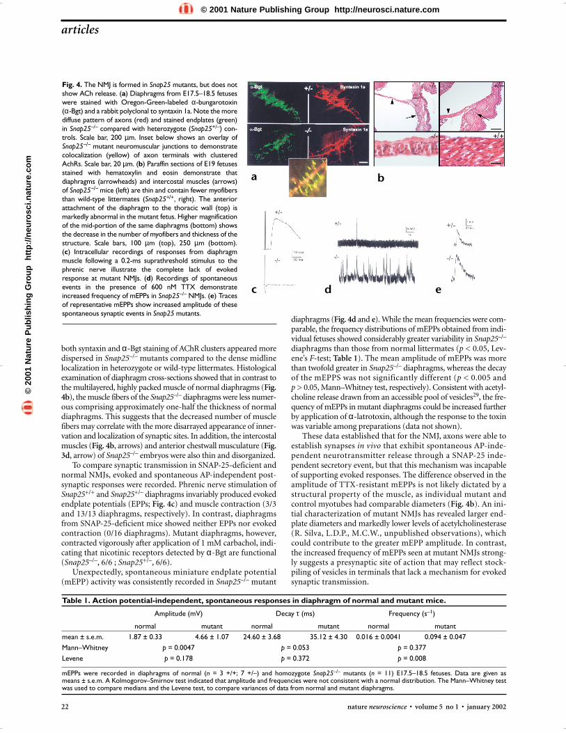
Spontaneous but not stimulus-driven release at NMJsThe normal appearance of fetal brain development in SNAP-25mutants suggested that processes that accompany neuronal differ-entiation, especially initial neurite formation and outgrowth, andpossibly initial synapse formation, may occur without SNAP-25-mediated neuroexocytosis. To examine this question, we first eval-uated the neuromuscular junction synapse in situ in the fetaldiaphragm. Fluorescent immunohistochemistry of whole-mountpreparations stained with syntaxin 1a antibodies (Fig. 4a) demon-strated phrenic nerve innervation of the diaphragm in Snap25–/–
late-stage (E17.5–18.5) fetuses. Fluorescently labeled α-bungaro-toxin (α-Bgt) also readily distinguished acetylcholine receptor(AChR) clusters in the muscle that closely matched the pattern ofsyntaxin-positive fibers and colocalized with axon terminals (Fig.4a, inset). However, the overall pattern of synaptic sites seen with
Fig. 3. Histopathological examination of SNAP-25-deficient fetuses.The neocortex (a, b), hippocampus, thalamus, and midbrain (c) are nor-mally developed in E19 null mutant mice as compared to Snap25+/– lit-termate controls. The midbrain of the Snap25–/– mutant fetus is wellpopulated by tyrosine hydroxylase immunoreactive neurons (c, inset).(d, e) The dermis of knockout mice contains large ectatic vascular chan-nels (arrowhead, anterior chest wall) not found in Snap25+/– or wild-type (data not shown) fetuses. In addition, the subcutaneousmusculature (arrows) is sparse and disorganized in mutant mice. (a–e) Hematoxylin and eosin; (c, inset) anti-tyrosine hydroxylase DABstain. Scale bars, 50 µm (a, b, d, e, inset c), 250 µm (c).
tide of ∼ 6 kD, confirming that neither the abnormally splicedmRNA nor the truncated polypeptide accumulate to a signifi-cant level in mutant fetal brain. Based on these observations, weconclude that the Snap25 exon5a/5b deletion results in a nullmutation, which we designate Snap25–.
Brain development proceeds normallyMajor brain structures including the hippocampus and neocor-tex were morphologically normal in Snap25–/– embryos com-pared with wild-type and Snap25+/– embryos (Fig. 2a). A surveyof presynaptic proteins revealed no detectable difference in theexpression among wild-type, heterozygote and homozygousmutants (Fig. 2b). There was also no apparent change in the levelof SNAP-23, the non-neuronal homolog of SNAP-25 (ref. 26).Because SNAP-23 is expressed at much lower levels than SNAP-25 in neurons27, we cannot entirely exclude the possibility of asmall increase of SNAP-23 among neuronal cell populations thatcould be obscured by the contribution of non-neuronal cells ofthe developing brain. These data indicate that the Snap25 loss-of-function mutation does not result in major compensatorychanges in expression of other proteins that are prominent medi-ators of presynaptic function.
Histological examination of hematoxylin- and eosin- or cre-syl-violet-stained paraffin sections taken from E17.5 and E18.5Snap25–/– brains showed no evidence for appreciable cellulardeficits. Comparable cell densities were observed in neocortex ofmutant and wild-type fetuses (Fig. 3a and b) and the dien-cephalon and brainstem of the mutants (Fig. 3c) showed noapparent cellular or architectural defects. Moreover, tyrosinehydroxylase immunocytochemistry (Fig. 3c, inset), a marker formature catecholaminergic neurons, demonstrated a normal pat-tern of immunoreactivity in the Snap25–/– brainstem. This dif-fers from the marked cell loss reported for munc18-1/nsec-1mutants28 and suggests that the lack of SNAP-25 results in a moresubtle phenotype, possibly acting downstream of munc18-1 medi-ated function(s) in neurotransmitter release. This is likely due tothe persistence of spontaneous release of neurotransmitter in
Fig. 2. Brain structure and synaptic protein levels appear normal inSnap25–/– mice. (a) Parasagittal sections of cortex and hippocampusstained with hematoxylin and eosin shows normal morphology ofSnap25–/– mutant brain. (b) Western blot analysis of E17.5 forebrainshows no dramatic differences in levels of expression of synaptic pro-teins. Only 2 samples of Snap25–/– proteins were probed with antibod-ies to synaptophysin, GAP-43 and β-tubulin. Importantly, SNAP-23, thenon-neuronal homolog of SNAP-25, is not significantly induced in theSnap25 null mutant brain.
a
b
a b c
d e
©20
01 N
atu
re P
ub
lish
ing
Gro
up
h
ttp
://n
euro
sci.n
atu
re.c
om
© 2001 Nature Publishing Group http://neurosci.nature.com
22 nature neuroscience • volume 5 no 1 • january 2002
articles
diaphragms (Fig. 4d and e). While the mean frequencies were com-parable, the frequency distributions of mEPPs obtained from indi-vidual fetuses showed considerably greater variability in Snap25–/–
diaphragms than those from normal littermates (p < 0.05, Lev-ene’s F-test; Table 1). The mean amplitude of mEPPs was morethan twofold greater in Snap25–/– diaphragms, whereas the decayof the mEPPS was not significantly different (p < 0.005 and p > 0.05, Mann–Whitney test, respectively). Consistent with acetyl-choline release drawn from an accessible pool of vesicles29, the fre-quency of mEPPs in mutant diaphragms could be increased furtherby application of α-latrotoxin, although the response to the toxinwas variable among preparations (data not shown).
These data established that for the NMJ, axons were able toestablish synapses in vivo that exhibit spontaneous AP-inde-pendent neurotransmitter release through a SNAP-25 inde-pendent secretory event, but that this mechanism was incapableof supporting evoked responses. The difference observed in theamplitude of TTX-resistant mEPPs is not likely dictated by astructural property of the muscle, as individual mutant andcontrol myotubes had comparable diameters (Fig. 4b). An ini-tial characterization of mutant NMJs has revealed larger end-plate diameters and markedly lower levels of acetylcholinesterase(R. Silva, L.D.P., M.C.W., unpublished observations), whichcould contribute to the greater mEPP amplitude. In contrast,the increased frequency of mEPPs seen at mutant NMJs strong-ly suggests a presynaptic site of action that may reflect stock-piling of vesicles in terminals that lack a mechanism for evokedsynaptic transmission.
both syntaxin and α-Bgt staining of AChR clusters appeared moredispersed in Snap25–/– mutants compared to the dense midlinelocalization in heterozygote or wild-type littermates. Histologicalexamination of diaphragm cross-sections showed that in contrast tothe multilayered, highly packed muscle of normal diaphragms (Fig.4b), the muscle fibers of the Snap25–/– diaphragms were less numer-ous comprising approximately one-half the thickness of normaldiaphragms. This suggests that the decreased number of musclefibers may correlate with the more disarrayed appearance of inner-vation and localization of synaptic sites. In addition, the intercostalmuscles (Fig. 4b, arrows) and anterior chestwall musculature (Fig.3d, arrow) of Snap25–/– embryos were also thin and disorganized.
To compare synaptic transmission in SNAP-25-deficient andnormal NMJs, evoked and spontaneous AP-independent post-synaptic responses were recorded. Phrenic nerve stimulation ofSnap25+/+ and Snap25+/– diaphragms invariably produced evokedendplate potentials (EPPs; Fig. 4c) and muscle contraction (3/3and 13/13 diaphragms, respectively). In contrast, diaphragmsfrom SNAP-25-deficient mice showed neither EPPs nor evokedcontraction (0/16 diaphragms). Mutant diaphragms, however,contracted vigorously after application of 1 mM carbachol, indi-cating that nicotinic receptors detected by α-Bgt are functional(Snap25–/–, 6/6 ; Snap25+/–, 6/6).
Unexpectedly, spontaneous miniature endplate potential(mEPP) activity was consistently recorded in Snap25–/– mutant
Fig. 4. The NMJ is formed in Snap25 mutants, but does notshow ACh release. (a) Diaphragms from E17.5–18.5 fetuseswere stained with Oregon-Green-labeled α-bungarotoxin(α-Bgt) and a rabbit polyclonal to syntaxin 1a. Note the morediffuse pattern of axons (red) and stained endplates (green)in Snap25–/– compared with heterozygote (Snap25+/–) con-trols. Scale bar, 200 µm. Inset below shows an overlay ofSnap25−/− mutant neuromuscular junctions to demonstratecolocalization (yellow) of axon terminals with clusteredAchRs. Scale bar, 20 µm. (b) Paraffin sections of E19 fetusesstained with hematoxylin and eosin demonstrate thatdiaphragms (arrowheads) and intercostal muscles (arrows)of Snap25−/− mice (left) are thin and contain fewer myofibersthan wild-type littermates (Snap25+/+, right). The anteriorattachment of the diaphragm to the thoracic wall (top) ismarkedly abnormal in the mutant fetus. Higher magnificationof the mid-portion of the same diaphragms (bottom) showsthe decrease in the number of myofibers and thickness of thestructure. Scale bars, 100 µm (top), 250 µm (bottom).(c) Intracellular recordings of responses from diaphragmmuscle following a 0.2-ms suprathreshold stimulus to thephrenic nerve illustrate the complete lack of evokedresponse at mutant NMJs. (d) Recordings of spontaneousevents in the presence of 600 nM TTX demonstrateincreased frequency of mEPPs in Snap25–/– NMJs. (e) Tracesof representative mEPPs show increased amplitude of thesespontaneous synaptic events in Snap25 mutants.
Table 1. Action potential-independent, spontaneous responses in diaphragm of normal and mutant mice.
Amplitude (mV) Decay τ (ms) Frequency (s–1)
normal mutant normal mutant normal mutantmean ± s.e.m. 1.87 ± 0.33 4.66 ± 1.07 24.60 ± 3.68 35.12 ± 4.30 0.016 ± 0.0041 0.094 ± 0.047Mann–Whitney p = 0.0047 p = 0.053 p = 0.377Levene p = 0.178 p = 0.372 p = 0.008
mEPPs were recorded in diaphragms of normal (n = 3 +/+; 7 +/–) and homozygote Snap25–/– mutants (n = 11) E17.5–18.5 fetuses. Data are given as means ± s.e.m. A Kolmogorov–Smirnov test indicated that amplitude and frequencies were not consistent with a normal distribution. The Mann–Whitney testwas used to compare medians and the Levene test, to compare variances of data from normal and mutant diaphragms.
a b
c d e
©20
01 N
atu
re P
ub
lish
ing
Gro
up
h
ttp
://n
euro
sci.n
atu
re.c
om
© 2001 Nature Publishing Group http://neurosci.nature.com
articles
nature neuroscience • volume 5 no 1 • january 2002 23
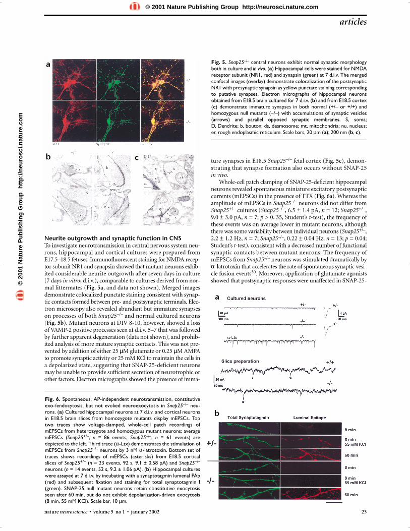
Neurite outgrowth and synaptic function in CNSTo investigate neurotransmission in central nervous system neu-rons, hippocampal and cortical cultures were prepared fromE17.5–18.5 fetuses. Immunofluorescent staining for NMDA recep-tor subunit NR1 and synapsin showed that mutant neurons exhib-ited considerable neurite outgrowth after seven days in culture (7 days in vitro; d.i.v.), comparable to cultures derived from nor-mal littermates (Fig. 5a, and data not shown). Merged imagesdemonstrate colocalized punctate staining consistent with synap-tic contacts formed between pre- and postsynaptic terminals. Elec-tron microscopy also revealed abundant but immature synapseson processes of both Snap25–/– and normal cultured neurons (Fig. 5b). Mutant neurons at DIV 8-10, however, showed a lossof VAMP-2 positive processes seen at d.i.v. 5–7 that was followedby further apparent degeneration (data not shown), and prohib-ited analysis of more mature synaptic contacts. This was not pre-vented by addition of either 25 µM glutamate or 0.25 µM AMPAto promote synaptic activity or 25 mM KCl to maintain the cells ina depolarized state, suggesting that SNAP-25-deficient neuronsmay be unable to provide sufficient secretion of neurotrophic orother factors. Electron micrographs showed the presence of imma-
ture synapses in E18.5 Snap25–/– fetal cortex (Fig. 5c), demon-strating that synapse formation also occurs without SNAP-25 in vivo.
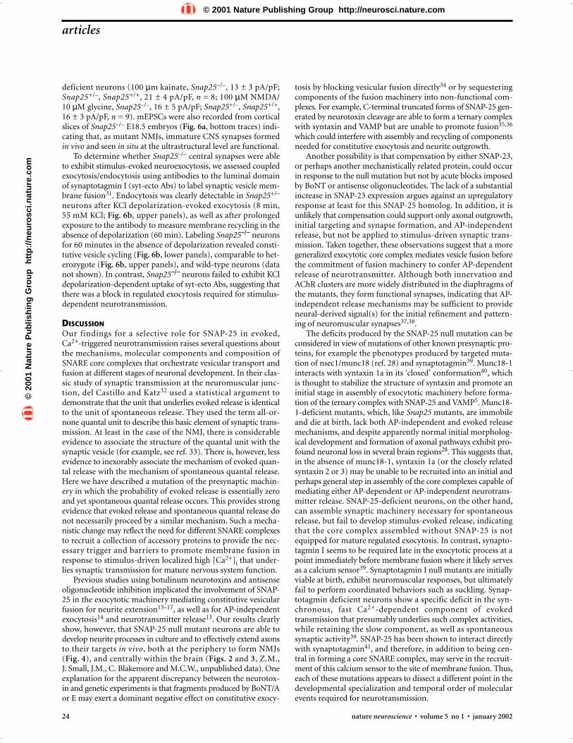
Whole-cell patch clamping of SNAP-25-deficient hippocampalneurons revealed spontaneous miniature excitatory postsynapticcurrents (mEPSCs) in the presence of TTX (Fig. 6a). Whereas theamplitude of mEPSCs in Snap25–/– neurons did not differ fromSnap25+/– cultures (Snap25–/–, 6.5 ± 1.4 pA, n = 12; Snap25+/–,9.0 ± 3.0 pA, n = 7; p > 0. 35, Student’s t-test), the frequency ofthese events was on average lower in mutant neurons, althoughthere was some variability between individual neurons (Snap25+/–,2.2 ± 1.2 Hz, n = 7; Snap25–/–, 0.22 ± 0.04 Hz, n = 13; p = 0.04;Student’s t-test), consistent with a decreased number of functionalsynaptic contacts between mutant neurons. The frequency ofmEPSCs from Snap25–/– neurons was stimulated dramatically byα-latrotoxin that accelerates the rate of spontaneous synaptic vesi-cle fusion events30. Moreover, application of glutamate agonistsshowed that postsynaptic responses were unaffected in SNAP-25-
Fig. 5. Snap25–/– central neurons exhibit normal synaptic morphologyboth in culture and in vivo. (a) Hippocampal cells were stained for NMDAreceptor subunit (NR1, red) and synapsin (green) at 7 d.i.v. The mergedconfocal images (overlay) demonstrate colocalization of the postsynapticNR1 with presynaptic synapsin as yellow punctate staining correspondingto putative synapses. Electron micrographs of hippocampal neuronsobtained from E18.5 brain cultured for 7 d.i.v. (b) and from E18.5 cortex(c) demonstrate immature synapses in both normal (+/– or +/+) andhomozygous null mutants (–/–) with accumulations of synaptic vesicles(arrows) and parallel opposed synaptic membranes. S, soma; D, Dendrite; b, bouton; ds, desmosome; mt, mitochondria; nu, nucleus;er, rough endoplasmic reticulum. Scale bars, 20 µm (a); 200 nm (b, c).
Fig. 6. Spontaneous, AP-independent neurotransmission, constitutiveexo-/endocytosis, but not evoked neuroexocytosis in Snap25–/– neu-rons. (a) Cultured hippocampal neurons at 7 d.i.v. and cortical neuronsin E18.5 brain slices from homozygote mutants display mEPSCs. Toptwo traces show voltage-clamped, whole-cell patch recordings ofmEPSCs from heterozygote and homozygous mutant neurons; averagemEPSCs (Snap25+/–, n = 86 events; Snap25–/–, n = 61 events) aredepicted to the left. Third trace (α-Ltx) demonstrates the stimulation ofmEPSCs from Snap25–/– neurons by 3 nM α-latrotoxin. Bottom set oftraces shows recordings of mEPSCs (asterisks) from E18.5 corticalslices of Snap25+/+ (n = 23 events, 92 s, 9.1 ± 0.58 pA) and Snap25–/–
neurons (n = 14 events, 52 s, 9.2 ± 1.06 pA). (b) Hippocampal cultureswere assayed at 7 d.i.v. by incubating with a synaptotagmin lumenal PAb(red) and subsequent fixation and staining for total synaptotagmin I(green). SNAP-25 null mutant neurons retain constitutive exocytosisseen after 60 min, but do not exhibit depolarization-driven exocytosis(8 min, 55 mM KCl). Scale bar, 10 µm.
a
b
a
b
c
©20
01 N
atu
re P
ub
lish
ing
Gro
up
h
ttp
://n
euro
sci.n
atu
re.c
om
© 2001 Nature Publishing Group http://neurosci.nature.com
24 nature neuroscience • volume 5 no 1 • january 2002
articles
deficient neurons (100 µm kainate, Snap25–/–, 13 ± 3 pA/pF;Snap25+/–, Snap25+/+, 21 ± 4 pA/pF, n = 8; 100 µM NMDA/10 µM glycine, Snap25–/–, 16 ± 5 pA/pF; Snap25+/–, Snap25+/+,16 ± 3 pA/pF, n = 9). mEPSCs were also recorded from corticalslices of Snap25–/– E18.5 embryos (Fig. 6a, bottom traces) indi-cating that, as mutant NMJs, immature CNS synapses formed in vivo and seen in situ at the ultrastructural level are functional.
To determine whether Snap25–/– central synapses were ableto exhibit stimulus-evoked neuroexocytosis, we assessed coupledexocytosis/endocytosis using antibodies to the luminal domainof synaptotagmin I (syt-ecto Abs) to label synaptic vesicle mem-brane fusion31. Endocytosis was clearly detectable in Snap25+/–
neurons after KCl depolarization-evoked exocytosis (8 min, 55 mM KCl; Fig. 6b, upper panels), as well as after prolongedexposure to the antibody to measure membrane recycling in theabsence of depolarization (60 min). Labeling Snap25−/− neuronsfor 60 minutes in the absence of depolarization revealed consti-tutive vesicle cycling (Fig. 6b, lower panels), comparable to het-erozygote (Fig. 6b, upper panels), and wild-type neurons (datanot shown). In contrast, Snap25−/− neurons failed to exhibit KCldepolarization-dependent uptake of syt-ecto Abs, suggesting thatthere was a block in regulated exocytosis required for stimulus-dependent neurotransmission.
DISCUSSIONOur findings for a selective role for SNAP-25 in evoked, Ca2+-triggered neurotransmission raises several questions aboutthe mechanisms, molecular components and composition ofSNARE core complexes that orchestrate vesicular transport andfusion at different stages of neuronal development. In their clas-sic study of synaptic transmission at the neuromuscular junc-tion, del Castillo and Katz32 used a statistical argument todemonstrate that the unit that underlies evoked release is identicalto the unit of spontaneous release. They used the term all-or-none quantal unit to describe this basic element of synaptic trans-mission. At least in the case of the NMJ, there is considerableevidence to associate the structure of the quantal unit with thesynaptic vesicle (for example, see ref. 33). There is, however, lessevidence to inexorably associate the mechanism of evoked quan-tal release with the mechanism of spontaneous quantal release.Here we have described a mutation of the presynaptic machin-ery in which the probability of evoked release is essentially zeroand yet spontaneous quantal release occurs. This provides strongevidence that evoked release and spontaneous quantal release donot necessarily proceed by a similar mechanism. Such a mecha-nistic change may reflect the need for different SNARE complexesto recruit a collection of accessory proteins to provide the nec-essary trigger and barriers to promote membrane fusion inresponse to stimulus-driven localized high [Ca2+]i that under-lies synaptic transmission for mature nervous system function.
Previous studies using botulinum neurotoxins and antisenseoligonucleotide inhibition implicated the involvement of SNAP-25 in the exocytotic machinery mediating constitutive vesicularfusion for neurite extension15–17, as well as for AP-independentexocytosis14 and neurotransmitter release13. Our results clearlyshow, however, that SNAP-25 null mutant neurons are able todevelop neurite processes in culture and to effectively extend axonsto their targets in vivo, both at the periphery to form NMJs (Fig. 4), and centrally within the brain (Figs. 2 and 3, Z.M., J. Small, J.M., C. Blakemore and M.C.W., unpublished data). Oneexplanation for the apparent discrepancy between the neurotox-in and genetic experiments is that fragments produced by BoNT/Aor E may exert a dominant negative effect on constitutive exocy-
tosis by blocking vesicular fusion directly34 or by sequesteringcomponents of the fusion machinery into non-functional com-plexes. For example, C-terminal truncated forms of SNAP-25 gen-erated by neurotoxin cleavage are able to form a ternary complexwith syntaxin and VAMP but are unable to promote fusion35,36
which could interfere with assembly and recycling of componentsneeded for constitutive exocytosis and neurite outgrowth.
Another possibility is that compensation by either SNAP-23,or perhaps another mechanistically related protein, could occurin response to the null mutation but not by acute blocks imposedby BoNT or antisense oligonucleotides. The lack of a substantialincrease in SNAP-23 expression argues against an upregulatoryresponse at least for this SNAP-25 homolog. In addition, it isunlikely that compensation could support only axonal outgrowth,initial targeting and synapse formation, and AP-independentrelease, but not be applied to stimulus-driven synaptic trans-mission. Taken together, these observations suggest that a moregeneralized exocytotic core complex mediates vesicle fusion beforethe commitment of fusion machinery to confer AP-dependentrelease of neurotransmitter. Although both innervation andAChR clusters are more widely distributed in the diaphragms ofthe mutants, they form functional synapses, indicating that AP-independent release mechanisms may be sufficient to provideneural-derived signal(s) for the initial refinement and pattern-ing of neuromuscular synapses37,38.
The deficits produced by the SNAP-25 null mutation can beconsidered in view of mutations of other known presynaptic pro-teins, for example the phenotypes produced by targeted muta-tion of nsec1/munc18 (ref. 28) and synaptotagmin39. Munc18-1interacts with syntaxin 1a in its ‘closed’ conformation40, whichis thought to stabilize the structure of syntaxin and promote aninitial stage in assembly of exocytotic machinery before forma-tion of the ternary complex with SNAP-25 and VAMP5. Munc18-1-deficient mutants, which, like Snap25 mutants, are immobileand die at birth, lack both AP-independent and evoked releasemechanisms, and despite apparently normal initial morpholog-ical development and formation of axonal pathways exhibit pro-found neuronal loss in several brain regions28. This suggests that,in the absence of munc18-1, syntaxin 1a (or the closely relatedsyntaxin 2 or 3) may be unable to be recruited into an initial andperhaps general step in assembly of the core complexes capable ofmediating either AP-dependent or AP-independent neurotrans-mitter release. SNAP-25-deficient neurons, on the other hand,can assemble synaptic machinery necessary for spontaneousrelease, but fail to develop stimulus-evoked release, indicatingthat the core complex assembled without SNAP-25 is notequipped for mature regulated exocytosis. In contrast, synapto-tagmin I seems to be required late in the exocytotic process at apoint immediately before membrane fusion where it likely servesas a calcium sensor39. Synaptotagmin I null mutants are initiallyviable at birth, exhibit neuromuscular responses, but ultimatelyfail to perform coordinated behaviors such as suckling. Synap-totagmin deficient neurons show a specific deficit in the syn-chronous, fast Ca2+-dependent component of evokedtransmission that presumably underlies such complex activities,while retaining the slow component, as well as spontaneoussynaptic activity39. SNAP-25 has been shown to interact directlywith synaptotagmin41, and therefore, in addition to being cen-tral in forming a core SNARE complex, may serve in the recruit-ment of this calcium sensor to the site of membrane fusion. Thus,each of these mutations appears to dissect a different point in thedevelopmental specialization and temporal order of molecularevents required for neurotransmission.
©20
01 N
atu
re P
ub
lish
ing
Gro
up
h
ttp
://n
euro
sci.n
atu
re.c
om
© 2001 Nature Publishing Group http://neurosci.nature.com
Because axonal outgrowth proceeds in the absence of SNAP-25 or munc18-1 (ref. 28) and is resistant to TeNT14, pre-cluding the involvement of VAMP-2, we suggest that SNAREcomplexes responsible for constitutive membrane fusion requiredfor the construction of axon processes involve non-neuronal andmore generalized members of the SNAP-25, syntaxin and VAMPgene families. Similarly, the utilization of more generalizedSNARE components may also underlie AP-independent sponta-neous secretion in which the position of SNAP-25 may be heldby a homologous SNARE, possibly SNAP-23. The diversity of thecomposition of the SNARE complex thus may reflect the need todistinguish ongoing vesicle fusion for axonal outgrowth andspontaneous neurotransmitter release from the fusion machinerythat is equipped and likely to trigger synaptic exocytosis. Forexample, spontaneous neurotransmitter release could providefor specific signaling in the morphological development of post-synaptic dendrite spines and in synaptic plasticity42.
Our results provide genetic evidence that the neuronal SNAREprotein SNAP-25, a constituent of the exocytosis complex pre-sent in mature presynaptic nerve terminals, is specifically respon-sible for evoked synaptic transmission, but is not essential forAP-independent, spontaneous release of neurotransmitter orprocess outgrowth and axonal elongation during neural devel-opment. Recently, it was reported that cultured neurons fromsynaptobrevin/VAMP-2 null mutant mice also exhibit sponta-neous synaptic transmission (although at much reduced fre-quency) in the absence of evoked responses43. It was suggestedthat such Ca2+-independent spontaneous activity from neurons,which must establish and maintain synapses in vivo, could reflectexocytosis in the absence of SNARE complexes. Whereas ourresults indicate AP-independent activity may not be comparablyreduced at synapses formed in situ, these studies underscore theneed to define the molecular mechanism that drives membranefusion for spontaneous neurosecretion, and to establish what rolethis may have in synapse formation and, ultimately, for activity-dependent neurotransmission. Together with other mutants tar-geting presynaptic proteins, SNAP-25-deficient mice will providean important means to resolve the complex protein assembliesrequired for these different acts of neuroexocytosis in develop-ment and in mature nervous system function.
METHODSMutant mice. The Snap25 null mutation was generated by targeted genereplacement in embryo stem cells using standard positive-negative selec-tion procedures44. SNAP-25 coding sequence was disrupted in the genetargeting construct (p4.2) by replacing both exons 5a and 5b with PGK-neo (for details and genotyping, see Fig. 1a and Supplementary meth-ods). For the studies reported, littermates at E17.5–18.5, judged by themorning of the plug, were obtained from mice backcrossed onto aC57BL/6J background for 4–5 generations. Studies were done in accor-dance with guidelines of the University of New Mexico HSC LaboratoryAnimal Care and Use Committee and the NIH.
Western blotting and RT-PCR. Fetal brain extracts were prepared byhomogenization in 0.32 M sucrose, 20 mM Tris pH 7.4, 1 mM DTT, 1%Nonidet P-40 and protease inhibitors (Complete Minitablet, Boehringer-Mannheim, Indianapolis, Indiana). The detergent extracts were cen-trifuged, and protein concentration of the solubilized protein wasdetermined with Micro BCA assay kit (Pierce, Rockford, Illinois). Equalamounts of protein were loaded on polyacrylamide gels, transferred tonitrocellulose and probed with the antibodies to the following proteins:SNAP-25 C-terminus and N-terminus (Sternberger Monoclonals,Lutherville, Maryland), VAMP-2 (gift from C. Montecucco, Univ. ofPadova, Italy), syntaxin 1 (Sigma, St. Louis, Missouri), synaptotagmin 1(Wako, Richmond, Virginia), synapsin 1 (Synaptic Systems, Göttingen,Germany), Munc18-1, Doc-2 (BD Transduction Laboratories, Lexing-
ton, Kentucky), synaptophysin, GAP-43 (Boehringer-Mannheim), SNAP-23 (gift from A. Klip, The Hospital for Sick Children, Toronto, Ontario)and β-tubulin (Chemicon, Temecula, California). Primary antibodieswere detected with either anti-mouse 125I-labeled IgG, or anti-rabbitHRP conjugated IgG followed by chemiluminescence assay (ECL-Plus,Amersham Pharmacia, Piscataway, New Jersey) and quantitated on aSTORM PhosphoImager system (Molecular Dynamics, Sunnyvale, Cal-ifornia). RT-PCR was done on RNA extracted with guanidinium thio-cyanate and phenol:chloroform using the Titan RT-PCR 1 tube system(Boehringer-Mannheim) with 32P-dCTP added as a tracer, and primersto the start and stop codons of SNAP-25 coding sequence. Amplificationwas limited to 22 cycles to be in the linear range of the reaction and theproducts were electrophoresed in 8% polyacrylamide gels and visualizedby phosphoimaging.
Histology, immunohistochemistry and electron microscopy. Fetal brainswere dissected and fixed by immersion in 4% paraformaldehyde/0.1 Mphosphate buffer, pH 7.4 for 24 h. Whole embryos were fixed in 10%formalin. Brains or whole fetuses were paraffin embedded, sectioned at8 µm and stained with hematoxylin and eosin, cresyl violet, or tyrosinehydroxylase antibodies (Boehringer-Mannheim). Diaphragms were fixedby immersing in 2% paraformaldehyde/0.1 M phosphate buffer for 1 h,and permeablized with 1% Triton X-100 (ref. 45). AChRs were visual-ized by staining with Oregon green labeled α-bungarotoxin (MolecularProbes, Eugene, Oregon) and axons with rabbit anti-syntaxin 1 (gift C.Montecucco, Univ. of Padova, Italy). Vesicle cycling was assayed essen-tially as described previously14. Briefly, cortical neuronal cultures wereeither incubated with anti-synaptotagmin antibody, syt-ecto Ab (giftfrom P. De Camilli, Yale University) as indicated before fixation or fixeddirectly with 4% paraformaldehyde and 4% sucrose for 20 min at 37°C.Cells were treated with 0.2% Triton-X-100, 4% normal donkey and goatserums, washed with PBS, and incubated with secondary antibodies con-jugated to FITC or Cy3 (Jackson Immunochemicals, West Grove, Penn-sylvania) for fluorescence microscopy. For dual-fluorescent confocalmicroscopy, neuron cultures were fixed with methanol and stained withmonoclonal antibodies to NMDA receptor subunit 1 (PharMingen, SanDiego, California) and polyclonal antibodies to synapsin (Chemicon),visualized with Cy3 and Cy2-labeled secondary antibodies, respectively,and viewed using an Olympus Fluroview IX70 confocal microscope. Con-focal images were displayed using Adobe Photoshop.
For electron microscopy of synapses, brains obtained from E18.5 fetus-es were fixed in 2% paraformaldehyde/2% glutaraldehyde, and corticalcells were fixed with 3% glutaraldehyde. Further fixation with OsO4, anduranyl acetate staining were performed by standard techniques. (See Sup-plementary methods for details.)
Electrophysiology. Postsynaptic responses from dissected diaphragmswere recorded at room temperature as described46. For evoked respons-es, a 0.2-ms suprathreshold stimulus was applied to the phrenic nerveusing a suction electrode, while mEPPs were recorded in 600 nM TTXwithout phrenic nerve stimulation. mEPP amplitudes were linearlyadjusted to Em = –75 mV before comparing data from different cells.Decay time constants were only calculated where the mEPP decay couldbe clearly fit by a single exponential curve. No differences were seenbetween homozygous wild-type and heterozygous genotypes, and thedata were pooled as normal NMJ responses.
Vibratome coronal sections from E18.5 embryos were pre-incu-bated for 45 min at 37°C in Krebs bicarbonate buffer containing 0.5mM Ca2+ and 10 mM Mg2+ equilibrated with O2/CO2 (95:5) and thenin standard Krebs bicarbonate buffer for 30–60 min as described47.Cortical neurons were visualized with infrared differential interfer-ence contrast using an Olympus BX50WI microscope. In all cases,neuronal currents were measured in the whole-cell patch-clamp con-figuration as described48. (See Supplementary methods for details.)The internal solution contained 4 mM NaCl, 0.4 mM CaCl2, 5 mMEGTA,10 mM HEPES, 140 mM K-gluconate, pH 7.25 280 mOsm.Membrane potential was clamped at –60 or –70 mV. mEPSCs wererecorded in the presence of 300–600 nM TTX and analyzed with theMinis Analysis program (Synaptosoft, Decatur, Georgia). Culturedneurons were prepared from E17.5–18.5 fetuses as previouslydescribed48 (see Supplementary methods).
articles
nature neuroscience • volume 5 no 1 • january 2002 25
©20
01 N
atu
re P
ub
lish
ing
Gro
up
h
ttp
://n
euro
sci.n
atu
re.c
om
© 2001 Nature Publishing Group http://neurosci.nature.com

26 nature neuroscience • volume 5 no 1 • january 2002
20. Chapman, E. R., An, S., Barton, N. & Jahn, R. SNAP-25, a t-SNARE whichbinds to both syntaxin and synaptobrevin via domains that may form coiledcoils. J. Biol. Chem. 269, 27427–27432 (1994).
21. Sutton, R. B., Fasshauer, D., Jahn, R. & Brunger, A. T. Crystal structure of aSNARE complex involved in synaptic exocytosis at 2.4 A resolution. Nature395, 347–353 (1998).
22. Hess, D. T., Slater, T. M., Wilson, M. C. & Skene, J. H. P. The 25 kDasynaptosomal-associated protein SNAP-25 is the major methionine-richpolypeptide in rapid axonal transport and a major substrate forpalmitoylation in adult CNS. J. Neurosci. 12, 4634–4641 (1992).
23. Veit, M., Sollner, T. H. & Rothman, J. E. Multiple palmitoylation ofsynaptotagmin and the t-SNARE SNAP-25. FEBS Lett. 385, 119–123(1996).
24. Lane, S. R. & Liu, Y. Characterization of the palmitoylation domain of SNAP-25. J. Neurochem. 69, 1864–1869 (1997).
25. Washbourne, P. et al. The cysteines of synaptosome-associated protein of 25kDa (SNAP-25) are required for SNARE disassembly and exocytosis, not formembrane targeting. Biochem. J. 357, 625–634 (2001).
26. Ravichandran, V., Chawla, A. & Roche, P. Identification of a novel syntaxin-and synaptobrevin/VAMP-binding protein, SNAP-23, expressed in non-neuronal tissues. J. Biol. Chem. 271, 13300–13303 (1996).
27. Chen, D., Minger, S., Honer, W. & Whiteheart, S. Organization of thesecretory machinery in the rodent brain: distribution of t-SNAREs SNAP-25and SNAP-23. Brain Res. 831, 11–24 (1999).
28. Verhage, M. et al. Synaptic assembly of the brain in the absence ofneurotransmitter secretion. Science 287, 864–869 (2000).
29. Ceccarelli, B. & Hurlbut, W. Ca2+-dependent recycling of synaptic vesicles atthe frog neuromuscular junction. J. Cell Biol. 87, 297–303 (1980).
30. Capogna, M., Gahwiler, B. H. & Thompson, S. M. Calcium-independentactions of α-latrotoxin on spontaneous and evoked synaptic transmission inthe hippocampus. J. Neurophysiol. 76, 149–158 (1996).
31. Matteoli, M., Takei, K., Perin, M. S., Südhof, T. C. & De Camilli, P.Exoendocytotic recycling of synaptic vesicles in developing processes ofcultured hippocampal neurons. J. Cell. Biol. 117, 849–869 (1992).
32. del Castillo, J. & Katz, B. Quantal components of the end-plate potential. J. Physiol. (Lond.) 124, 560–573 (1954).
33. Heuser, J. & Reese, T. Structural changes after transmitter release at the frogneuromuscular junction. J. Cell Biol. 88, 564–580 (1981).
34. Mehta, P. P., Battenburg, E. & Wilson, M. C. SNAP-25 and synaptotagmininvolvement in the final Ca+2-dependent triggering of neurotransmitterexocytosis. Proc. Natl. Acad. Sci. USA 93, 10471–10476 (1996).
35. Hayashi, T. et al. Synaptic vesicle membrane fusion complex: action ofclostriridal neurotoxins on assembly. EMBO J. 13, 5051–5061 (1994).
36. Pellegrini, L. L., O’Connor, V., Lottspeich, F. & Betz, H. Clostridal neurotoxinscompromise the stability of a low energy SNARE complex mediating NSFactivation of synaptic vesicle fusion. EMBO J. 14, 4705–4713 (1995).
37. Yang, X. et al. Patterning of muscle acetylcholine receptor gene expression inthe absence of motor innervation. Neuron 30, 399–410 (2001).
38. Lin, W. et al. Distinctive roles of nerve and muscle in postsynapticdifferentiation of the neuromuscular synapse. Nature 410, 1057–1064(2001).
39. Geppert, M. et al. Synaptotagmin I: a major Ca2+ sensor for transmitterrelease at a central synapse. Cell 79, 717–727 (1994).
40. Dulubova, I. et al. A conformational switch in syntaxin during exocytosis:role of munc18. EMBO J. 18, 4372–4382 (1999).
41. Schiavo, G., Stenbeck, G., Rothman, J. E. & Sollner, T. H. Binding of thesynaptic vesicle v-SNARE, synaptotagmin, to the plasma membrane t-SNARE, SNAP-25, can explain docked vesicles at neurotoxin-treatedsynapses. Proc. Natl. Acad. Sci. USA 94, 997–1001 (1997).
42. McKinney, R. A., Capogna, M., Durr, R., Gahwiler, B. H. & Thompson, S. M.Miniature synaptic events maintain dendritic spines via AMPA receptoractivation. Nat. Neurosci. 2, 44–49 (1999).
43. Schoch, S. et al. SNARE function analyzed in synaptobrevin/VAMP knockoutmice. Science 294, 1117–1122 (2001).
44. Mansour, S. L., Thomas, K. R. & Capecchi, M. R. Disruption of the proto-oncogene int-2 in mouse embryo-derived stem cells: a general strategy fortargeting mutations to non-selectable genes. Nature 336, 348–352 (1988).
45. Gautam, M. et al. Defective neuromuscular synaptogenesis in agrin-deficientmutant mice. Cell 85, 525–535 (1996).
46. Plomp, J., van Kempen, G. & Molenaar, P. Adaptation of quantal content todecreased postsynaptic sensitivity at single endplates in α-bungarotoxin-treated rats. J. Physiol. (Lond.) 458, 487–499 (1992).
47. Aitken, P. G. et al. Preparative methods for brain slices: a discussion. J. Neurosci. Methods. 59, 139–149 (1995).
48. Costa, E., Soto, E., Cardoso, R., Olivera, D. & Valenzuela, C. Acute effects ofethanol on kainate receptors in cultured hippocampal neurons. Alcohol Clin.Exp. Res. 24, 220–225 (2000).
articles
Note: Supplementary methods are available on the Nature Neuroscience web site
(http://neuroscience.nature.com/web_specials).
AcknowledgementsWe thank J. Plomp for advice and help in embryonic neuromuscular
electrophysiology, S. Nixon for technical assistance, E. Padilla at the UNM-HSC
Animal Resource Facility for maintaining the mouse colony and G. Adamson at
the UC Davis EM Pathology lab for help with electron microscopy. We also
thank P. De Camilli and A. Klip for antisera, and B. Shuttleworth and L. Anna
Cunningham for discussions and for reading the manuscript. The work was
supported by NIH MH 4-8989 (M.C.W.).
Competing interests statementThe authors declare that they have no competing financial interests.
RECEIVED 9 OCTOBER; ACCEPTED 26 NOVEMBER 2001
1. Katz, B. The Release of Neural Transmitter Substances (Liverpool UniversityPress, Liverpool, 1969).
2. Young, S. & Poo, M. Spontaneous release of transmitter from growth cones ofembryonic neurones. Nature 305, 634–637 (1983).
3. Zakharenko, S., Chang, S., O’Donoghue, M. & Popov, S. V. Neurotransmittersecretion along growing nerve processes: comparison with synaptic vesicleexocytosis. J. Cell Biol. 144, 507–518 (1999).
4. Söllner, T. et al. SNAP receptors implicated in vesicle targeting and fusion.Nature 362, 318–324 (1993).
5. Brunger, A. Structural insights into the molecular mechanism of Ca2+-dependent exocytosis. Curr. Opin. Neurobiol. 10, 293–302 (2000).
6. Weber, T. et al. SNAREpins: minimal machinery for membrane fusion. Cell92, 759–772 (1998).
7. Schiavo, G., Matteoli, M. & Montecucco, C. Neurotoxins affectingneuroexocytosis. Physiol. Rev. 80, 717–766 (2000).
8. Weimbs, T. et al. A conserved domain is present in different families ofvesicular fusion proteins: a new superfamily. Proc. Natl. Acad. Sci. USA 94,3046–3051 (1997).
9. Deitcher, D. et al. Distinct requirements for evoked and spontaneous releaseof neurotransmitter are revealed by mutations in the Drosophila geneneuronal-synaptobrevin. J. Neurosci. 18, 2028–2939 (1998).
10. Broadie, K. et al. Syntaxin and synaptobrevin function downstream of vesicledocking in Drosophila. Neuron 15, 663–673 (1995).
11. Sweeney, S., Broadie, K., Keane, J., Niemann, H. & O’Kane, C. Targetedexpression of tetanus toxin light chain in Drosophila specifically eliminatessynaptic transmission and causes behavioral defects. Neuron 14, 341–351(1995).
12. Hua, S., Raciborska, D., Trimble, W. & Charlton, M. DifferentVAMP/synaptobrevin complexes for spontaneous and evoked transmitterrelease at the crayfish neuromuscular junction. J. Neurophysiol. 80,3233–3246 (1998).
13. Capogna, M., McKinney, R. A., O’Connor, V., Gahwiler, B. H. & Thompson,S. M. Ca2+ or Sr2+ partially rescues synaptic transmission in hippocampalcultures treated with botulinum toxin A and C, but not tetanus toxin. J. Neurosci. 17, 7190–7202 (1997).
14. Verderio, C. et al. Tetanus toxin blocks the exocytosis of synaptic vesiclesclustered at synapses but not of synaptic vesicles in isolated axons. J. Neurosci.19, 6723–6732 (1999).
15. Osen-Sand, A. et al. Inhibition of axon growth by SNAP-25 antisenseoligonucleotides in vitro and in vivo. Nature 364, 445–448 (1993).
16. Osen-Sand, A. et al. Common and distinct fusion proteins in axonal growthand transmitter release. J. Comp. Neurol. 222–234 (1996).
17. Morihara, T. et al. Distribution of synaptosomal-associated protein 25 innerve growth cones and reduction of neurite outgrowth by botulinumneurotoxin A without altering growth cone morphology in dorsal rootganglion neurons and PC-12 cells. Neuroscience 91, 695–706 (1999).
18. Coco, S. et al. Subcellular localization of tetanus neurotoxin-insensitive vesicle-associated membrane protein (VAMP)/VAMP7 in neuronal cells: evidence fora novel membrane compartment. J. Neurosci. 19, 9803–9812 (1999).
19. Bark, I. C. Structure of the chicken gene for SNAP-25 reveals duplicated exonsencoding distinct isoforms of the protein. J. Mol. Biol. 233, 67–76 (1993).
©20
01 N
atu
re P
ub
lish
ing
Gro
up
h
ttp
://n
euro
sci.n
atu
re.c
om
© 2001 Nature Publishing Group http://neurosci.nature.com