
Maternal Nonthyroidal Illness and Fetal Thyroid Hormone Status, as Studied in the...
11
Maternal Nonthyroidal Illness and Fetal Thyroid Hormone Status, as Studied in the Streptozotocin- Induced Diabetes Mellitus Rat Model* ROSA CALVO, GABRIELLA MORREALE DE ESCOBAR, FRANCISCO ESCOBAR DEL REY, AND MARIA-JESU ´ S OBREGO ´ N Unidad de Endocrinologı ´a Molecular, Instituto de Investigaciones Biome ´dicas, Consejo Superior de Investigaciones Cientı ´ficas, and Facultad de Medicina, University Auto ´noma de Madrid, Madrid, Spain ABSTRACT We have used the streptozotocin-induced diabetes mellitus preg- nant rat as a model of maternal nonthyroidal illness. We measured the effects of different degrees of diabetes mellitus on maternal body weight, the outcome of pregnancy, circulating glucose, insulin, T 4 ,T 3 , rT 3 , and TSH in mother and fetus, T 4 and T 3 in maternal and fetal tissues, and iodothyronine deiodinases in liver, lung, and brain. All of the changes in thyroid hormone status typical of nonthyroidal illnesses were observed in the mothers and were related to the degree of the metabolic imbalances. Most were controlled with a daily insulin dose of 0.5 U/100 g BW. Normalization of maternal placental T 4 , however, required higher insulin doses than in other maternal tissues. The number and body weight of the fetuses, their pituitary GH contents, and their thyroid hormone status were severely affected. The total extrathyroidal T 4 and T 3 pools decreased to one third of normal fetal values. T 4 and T 3 concentrations in the fetal brain were lower than normal, and the expected increase in type II 59deiodinase activity was not observed. The low cerebral T 3 only improved with adequate insulin treatment of the dams. It is concluded that maternal diabetes mellitus, and possibly other nonthyroidal illnesses that impair the availability of intracellular energy stores, may affect fetal brain T 3 when thyroid hormones are essential for normal development. (Endocrinology 138: 1159 –1169, 1997) D IABETES mellitus leads to alterations of thyroid hor- mone status typical of other so called nonthyroidal illnesses (1–3). The major alterations in thyroid hormone economy are a reduction in the TSH stimulation of the thy- roid gland, probably caused by central hypothyroidism, and in the peripheral generation of T 3 from T 4 (3). The injection of streptozotocin (STZ) in rats is frequently used to obtain an experimental model for the study of diabetes mellitus, often as a model of nonthyroidal illness. In the STZ-diabetic adult rat, the alterations in the hypo- thalamo-pituitary-thyroid axis are numerous; hypothalamic and plasma TRH (4, 5), pituitary and plasma TSH, as well as TSH secretion rate are reduced (4, 6, 7), and the TSH response to TRH is decreased despite normal peripheral TSH metab- olism (6). T 3 and T 4 production (8) and iodide uptake by the thyroid are diminished. There are also important structural changes in the thyroid gland and pituitary that are accom- panied by marked alterations in their secretory activity. In addition, T 4 deiodination to T 3 in peripheral tissues is de- creased (8 –10). As a consequence of all of these changes, circulating levels of T 4 and T 3 are markedly reduced as are the concentrations of both iodothyronines in most tissues (8 –10). These alterations have been shown in the adult diabetic rat, but to our knowledge very little is known about the possible influence of maternal diabetes on the thyroid hormone status of the fetus. In a preliminary study (11) we have shown that STZ-induced maternal diabetes mellitus also affects fetal thy- roid hormone economy, as studied at 20 days gestation, causing a decrease in T 4 and T 3 in plasma and most fetal tissues, including brain. These preliminary results also sug- gested that the normal response of 59-deiodinase type II (59D-II) to low T 4 concentrations was impaired in the fetal brain. The present study has been undertaken to further define the effects of different degrees of maternal nonthyroidal ill- ness, as induced by diabetes mellitus, on fetal thyroid hor- mone status and their prevention with adequate control of the maternal metabolic imbalances. Materials and Methods Experimental design Female Wistar rats were used for this study. The guidelines for humane treatment of animals were followed, and the study was ap- proved by the committee of our institute. They were maintained at 22 C with 12-h periods of light and darkness. They were mated with normal males, and the morning of appearance of the vaginal plug was consid- ered day 0 of gestation. Thirty pregnant rats were divided into six groups. One group served as normal pregnant controls (C). At 7 days gestation (dg), the other five groups of rats were injected into the femoral vein with 4.5 mg/100 g BW STZ dissolved in 50 mm citrate buffer, pH 4.5 (12). One of the groups of STZ-treated dams was not injected with Received July 11, 1997. Address all correspondence and requests for reprints to: Dr. M. J. Obrego ´ n, Instituto Investigaciones Biome ´dicas, Arturo Duperier 4, 28029 Madrid, Spain. E-mail: [email protected]. * This study has been performed in memorium of Prof. Norbert Freinkel, who initially contributed so much to thyroid research and later to our understanding of diabetes and pregnancy. This work was sup- ported by Grant 92– 0888 from the Fondo de Investigaciones Sanitarias, Spain. 0013-7227/97/$03.00/0 Vol. 138, No. 3 Endocrinology Printed in U.S.A. Copyright © 1997 by The Endocrine Society 1159
-
Upload
independent -
Category
Documents
-
view
4 -
download
0
Transcript of Maternal Nonthyroidal Illness and Fetal Thyroid Hormone Status, as Studied in the...

Maternal Nonthyroidal Illness and Fetal ThyroidHormone Status, as Studied in the Streptozotocin-Induced Diabetes Mellitus Rat Model*
ROSA CALVO, GABRIELLA MORREALE DE ESCOBAR,FRANCISCO ESCOBAR DEL REY, AND MARIA-JESUS OBREGON
Unidad de Endocrinologıa Molecular, Instituto de Investigaciones Biomedicas, Consejo Superior deInvestigaciones Cientıficas, and Facultad de Medicina, University Autonoma de Madrid, Madrid,Spain
ABSTRACTWe have used the streptozotocin-induced diabetes mellitus preg-
nant rat as a model of maternal nonthyroidal illness. We measuredthe effects of different degrees of diabetes mellitus on maternal bodyweight, the outcome of pregnancy, circulating glucose, insulin, T4, T3,rT3, and TSH in mother and fetus, T4 and T3 in maternal and fetaltissues, and iodothyronine deiodinases in liver, lung, and brain.
All of the changes in thyroid hormone status typical of nonthyroidalillnesses were observed in the mothers and were related to the degreeof themetabolic imbalances.Most were controlled with a daily insulindose of 0.5 U/100 g BW. Normalization of maternal placental T4,however, required higher insulin doses than in other maternaltissues.
The number and body weight of the fetuses, their pituitary GHcontents, and their thyroid hormone status were severely affected.The total extrathyroidal T4 and T3 pools decreased to one third ofnormal fetal values. T4 and T3 concentrations in the fetal brain werelower than normal, and the expected increase in type II 59deiodinaseactivity was not observed. The low cerebral T3 only improved withadequate insulin treatment of the dams.
It is concluded that maternal diabetes mellitus, and possibly othernonthyroidal illnesses that impair the availability of intracellularenergy stores, may affect fetal brain T3 when thyroid hormones areessential for normal development. (Endocrinology 138: 1159–1169,1997)
DIABETES mellitus leads to alterations of thyroid hor-mone status typical of other so called nonthyroidal
illnesses (1–3). The major alterations in thyroid hormoneeconomy are a reduction in the TSH stimulation of the thy-roid gland, probably caused by central hypothyroidism, andin the peripheral generation of T3 from T4 (3). The injectionof streptozotocin (STZ) in rats is frequently used to obtain anexperimental model for the study of diabetes mellitus, oftenas a model of nonthyroidal illness.In the STZ-diabetic adult rat, the alterations in the hypo-
thalamo-pituitary-thyroid axis are numerous; hypothalamicand plasma TRH (4, 5), pituitary and plasma TSH, as well asTSH secretion rate are reduced (4, 6, 7), and the TSH responseto TRH is decreased despite normal peripheral TSH metab-olism (6). T3 and T4 production (8) and iodide uptake by thethyroid are diminished. There are also important structuralchanges in the thyroid gland and pituitary that are accom-panied by marked alterations in their secretory activity. Inaddition, T4 deiodination to T3 in peripheral tissues is de-creased (8–10). As a consequence of all of these changes,circulating levels of T4 and T3 are markedly reduced as are
the concentrations of both iodothyronines in most tissues(8–10).These alterations have been shown in the adult diabetic rat,
but to our knowledge very little is known about the possibleinfluence ofmaternal diabetes on the thyroid hormone statusof the fetus. In a preliminary study (11) we have shown thatSTZ-inducedmaternal diabetesmellitus also affects fetal thy-roid hormone economy, as studied at 20 days gestation,causing a decrease in T4 and T3 in plasma and most fetaltissues, including brain. These preliminary results also sug-gested that the normal response of 59-deiodinase type II(59D-II) to low T4 concentrations was impaired in the fetalbrain.The present study has been undertaken to further define
the effects of different degrees of maternal nonthyroidal ill-ness, as induced by diabetes mellitus, on fetal thyroid hor-mone status and their prevention with adequate control ofthe maternal metabolic imbalances.
Materials and MethodsExperimental design
Female Wistar rats were used for this study. The guidelines forhumane treatment of animals were followed, and the study was ap-proved by the committee of our institute. They were maintained at 22Cwith 12-h periods of light and darkness. Theywerematedwith normalmales, and the morning of appearance of the vaginal plug was consid-ered day 0 of gestation. Thirty pregnant rats were divided into sixgroups. One group served as normal pregnant controls (C). At 7 daysgestation (dg), the other five groups of ratswere injected into the femoralvein with 4.5 mg/100 g BW STZ dissolved in 50 mm citrate buffer, pH4.5 (12). One of the groups of STZ-treated dams was not injected with
Received July 11, 1997.Address all correspondence and requests for reprints to: Dr. M. J.
Obregon, Instituto Investigaciones Biomedicas, Arturo Duperier 4,28029 Madrid, Spain. E-mail: [email protected].
* This study has been performed in memorium of Prof. NorbertFreinkel, who initially contributed somuch to thyroid research and laterto our understanding of diabetes and pregnancy. This work was sup-ported by Grant 92–0888 from the Fondo de Investigaciones Sanitarias,Spain.
0013-7227/97/$03.00/0 Vol. 138, No. 3Endocrinology Printed in U.S.A.Copyright © 1997 by The Endocrine Society
1159
insulin and served as the long duration diabetes mellitus (D) group, as14 days had elapsed since injection of the dams with STZ. A secondgroup of STZ-injected pregnant rats received 1.5 U bovine insulin(Ultralente, Novo Nordisk, Bagsvaerd, Denmark)/100 g BWzday, sc,once daily from 9–15 days gestation and vehicle (Diluting Medium forLenteMC, Novo Nordisk) from 15–20 dg; this group is referred to as sD,for short duration diabetesmellitus, as the damswere left untreated onlyfor the last 5 days. Three groups of rats were injected sc once daily with0.5, 1.0, or 1.5 U bovine insulin/100 g BWzday from 9–20 dg (D10.5 Ins,D11.0 Ins, and D11.5 Ins groups, respectively). Most results obtainedin D and sD groups were very similar, so only results from D dams willbe presented; differences between D and sD groups, if any, are indicatedin the text, tables, or figure legends.
On themorning of 21 dg and 24 h after the last of the insulin injections,all dams were anesthetized with ether, bled, and perfused with 40–50ml 0.05 m phosphosaline buffer, pH 7.4, as previously described (13).
Maternal plasma, liver, brain, lung, heart, and samples of mammarytissue were obtained and frozen. The uterus was dissected out andcarefully rinsed and blotted free of maternal blood. The fetuses werethen dissected out, bled, separated from the placenta, weighed, andimmediately placed on ice. The fetal brain, liver, and lungwere dissectedout and quickly frozen on dry ice; the thyroid, adhering to the trachea,was withdrawn and frozen. Two or three fetal thyroids and pancreasfrom each litter were fixed in toto by immersion in PBS containing 4%formaldehyde for morphological study. The rest of the fetus, referred tohere as the carcass (whole embryo minus the blood, trachea, thyroid,liver, lung, brain, and heart) was stored frozen. The placentas wereseparated, weighed, and divided into the basal (maternal) and labyrinth(fetal) sides with blunt forceps and frozen rapidly, as previouslydescribed (14).
Determination of thyroid hormone concentrations
Thyroid hormone levels were determined by RIAs after extractionand purification of plasma and tissues (15). In brief, methanol is addedto the still frozen tissue sample and homogenized, with tracer amountsof [131I]T4 and [125I]T3 added to each homogenate. This is followed bythe addition of chloroform in a volume double that of methanol, cen-trifugation, and a further extraction of the pellet with chloroform-meth-anol (2:1). This extracts more than 90% of the endogenous and addediodothyronines. The iodothyronines are then back-extracted into anaqueous phase and purified by passing this aqueous phase throughBio-Rad AG 1 3 2 resin columns (Bio-Rad, Hercules, CA). After a pHgradient, the iodothyronines are eluted with 70% acetic acid, which isthen evaporated to dryness and dissolved in RIA buffer. Each extract isextensively counted to determine the recovery of the [131I]T4 and [
125I]T3added to each sample during the initial homogenization process. Thesamples are submitted to highly sensitive RIAs for the determination ofT4 and T3; the limits of sensitivity are 2.5 pg T4 and 1.5 pg T3/tube. Thecross-reactivities of the different iodothyronines and metabolites wereas reported previously (15, 16). Each sample is processed in duplicate ortriplicate at two or more dilutions. Concentrations were then calculatedusing the amounts of T4 and T3 found in the respective RIAs, the indi-vidual recovery of the [131I]T4 and [
125I]T3 added to each sample duringthe initial homogenization process, and the weight of the tissue samplesubmitted to extraction.
Maternal samples were processed individually. Plasma from differ-ent fetuses were pooled to obtain 300- to 400-ml aliquots. Fetal tissueswere pooled (two or three organs per pool) for the determination of T4and T3. Pools were obtained from fetuses of the same litter.
The fetal thyroids were pooled in groups of two or three, homoge-nized, and submitted to proteolytic digestion, followed by methanolextraction. The methanol extracts were submitted to evaporation todryness in a microwave oven at maximum heat for 5–10 min; thisprevents artifacts in the RIAs, presumably due to residual proteolyticactivity transferred into the RIA tube. RIA buffer was added, and T4 andT3 were determined by RIA, as described above.
Percentage of circulating free T4 and T3These percentages were determined by ultrafiltration of undiluted
plasma samples, as described by Mendel et al. (17) with modifications.High specific activity [125I]T4 or [
125I]T3 (;300,000 cpm) were added in
a 5-ml volume to 300 ml plasma and incubated at room temperature for1 h. A 280-ml aliquot of each was submitted to ultrafiltration usingMicrocon 10 microconcentrators (Amicon Division, W. R. Grace Co.,Beverly, MA) and a 20-min centrifugation at 14,000 rpm. A measuredvolume of each ultrafiltrate was added to 0.5 ml bovine serum, sub-mitted to precipitation with 10% trichloroacetic acid (TCA), and cen-trifuged; the pelletwaswashed twicewith the same solution of TCA. Thewashed pellet was counted, and its radioactivity was calculated as apercentage of the initial added tracer, submitted to the same TCA pre-cipitation and washing procedure. This percentage of free T4 (% FT4) orfree T3 (% FT3) and the T4 and T3 concentrations determined byRIAwereused to calculate the concentrations of FT4 and free FT3, respectively.
Iodothyronine 59- and 5-D activities
Before each assay, [125I]rT3 or [125I]T4 was purified by paper electro-
phoresis to separate the iodide. Iodothyronine 59D-I activitywas assayedas previously described (16), using 2 mm dithiothreitol (DTT) and 400or 200 nm rT3 for maternal and fetal liver, respectively, and 2 nm rT3 and20 mm DTT for maternal and fetal lung (59D-I). Maternal and fetal brain59 D-II activities were assayed (18) using 2 nm T4, 1 mm T3, and 20 mmDTT in the presence of 1 mm 2-N-propyl-6-thiouracil (PTU). The 125I2
released was separated by ion exchange chromatography on Dowex-50W-X2 columns equilibrated in 10% acetic acid. The production ofequal amounts of iodide and 39,3-diiodothyronine (39,3-T2) was checkedin some assays. The protein content was determined by the method ofLowry, after precipitation of the homogenates with 10% TCA to avoidinterference from DTT in the colorimetric reaction (16).
5D activity was measured in maternal and fetal brain homogenates(19), incubating 20–50mg protein in 100mm potassiumphosphate buffer(pH 7.4), 1 mm EDTAwith approximately 50,000 cpm inner ring labeled5-[125I]T3, 50 nm T3, 20 mm DTT, and 1 mm PTU for 60 min at 37 C.Radioiodide release wasmeasured as described above.When necessary,inner ring labeled ([5-125I]T3 was repurified before use with disposableSep-Pak C18 cartridges (Waters Associates, Milford, MA) and methanol.
Other determinations
rT3 concentrations in maternal and fetal plasma and in placentalextracts were determined by RIA, as previously described (14).
Maternal and fetal plasma glucose levels were determined by theglucose oxidase method (20), using 10–25 ml plasma.
Insulin levels in maternal and fetal plasma were measured using thespecific RIA adapted for rat insulin with reagents supplied by NovoNordisk (Bagsvaerd, Denmark). We used rat insulin as standard, anti-porcine antiserum, and human 125I-labeled insulin as antigen (18). Abovine insulin standard was used for the standard curve when theplasma was obtained from mothers injected with bovine insulin.
TSH was determined in 200-ml aliquots of maternal plasma, and GHwas determined in fetal pituitaries, using the immunoreactants for RIAkindly supplied by theNIH (Bethesda,MD) andmade available throughthe Rat Pituitary Agency of the NIDDK. Concentrations are expressedin weight equivalents of the rat TSH RP-3 and rat GH RP-2 referencepreparations (21).
Drugs and reagents
T4, T3, 3,5-T2, PTU, and DTTwere obtained from Sigma Chemical Co.(St. Louis, MO). rT3 and 39,3-T2 were obtained from Henning Berlin(Berlin, Germany). High specific activity [131I]T4, [
125I]T3, [125I]T4, and
[125I]rT3 (3000mCi/mg)were synthesized in our laboratory (15) and usedfor highly sensitive T4, T3, and rT3 RIAs, as recovery tracers for extrac-tions, and as substrates for 59-D.
Inner ring labeled 5-[125I]T3 (80 mCi/mg) was used as substrate for5-D. It was provided by Drs. R. Thoma and H. Rokos from HenningBerlin.
Statistical analysis
After testing for homogeneity of variance using Bartlett’s procedurefor groups of unequal size, data were submitted to one-way ANOVA.Square root or logarithmic transformations usually ensured homoge-neity of variance when this was not achieved with the raw data. Sig-
1160 MATERNAL AND FETAL THYROID STATUS IN DIABETES MELLITUS Endo • 1997Vol 138 • No 3

nificant differences among groups were assessed using the protectedleast significant difference test. All statistical calculations were per-formed as described by Snedecor and Cochran (22). The se appearing inthe tables and figures is the mean se calculated by ANOVA and usedfor the identification of statistically significant differences betweengroups by the least significant difference test. For the sake of clarity, the6se is shown in figures only on the C value bar.
Possible interrelations among variables were tested by curve fittingof the individual data using Cricket Graph III for Macintosh (ComputerAssociates International, Inc., Islandia, NY), and the degree of fit wasassessed from the corresponding value of r and the degrees of freedom.
ResultsExperimental design
The aim of the present experimental design was to inducematernal diabetes mellitus of different duration and degreeand to assess the effects of these different degrees ofmaternalnonthyroidal illness on fetal thyroid hormone status nearterm.
Degree of nonthyroidal illness: diabetic state and weight loss of thedams. Figure 1 shows the insulin and glucose concentrationsin thematernal plasma (M-plasma) from the different groupsat 21 dg. Insulin decreased significantly in all D dams. Asexpected, circulating glucose levels were very high in allSTZ-injected dams that did not receive insulin. The injectionof insulin affected both insulin and glucose levels in thematernal circulation. Normal levels of insulin, as measured24 h after the last injection, were found in the D10.5 Ins orD11.0 Ins groups, with higher than normal values in theD11.5 Ins dams. Circulating glucose was somewhat higherthan C values in the D10.5 Ins dams, although markedly
decreased compared to those in D animals, and comparableto those in C dams in the D11.0 Ins and D11.5 Ins groups.The circulating glucose was inversely related to plasma in-sulin by a power function (n 5 30; r 5 0.88; P , 0.001).Figure 1 also shows the calculated change, between 7 and
21 dg, in the body weight of the pregnant rats, free of theconceptus (M-BW), namely the total measured weight of thedamminus theweight of the conceptus. The change inM-BWwas calculated from data reported in Table 1, such as thechange in total weight, the number of fetuses per litter, thebody weights of the fetuses (F-BW), and the weights of theplacentas. The weight of the conceptus was calculated foreach animal from the sum of the weights of all fetuses andplacentas in each dam. Although extraembryonic fluids andmembranes had not been collected, the sum of the fetal andplacental weights appears to be a reasonable approximationof the totalweight of the conceptus. The change inM-BWwascalculated by subtracting this calculated weight of the con-ceptus from the measured change in total weight of theanimal. As shown in Fig. 1, the mean net increase in M-BWof C dams was 27.8 6 4.3 g. In contrast, in all D dams, therewas a net loss of M-BW, which was prevented by insulintreatment.Both the increment in total weight and the change in
M-BW were significantly related to decreasing glucose andincreasing insulin concentrations in the maternal circulationand appear to be good indexes of the degree of maternalillness. The closest fit was found for the change in M-BW vs.the logarithm of the M-plasma insulin levels (Fig. 2).
Effects of maternal illness on the outcome of pregnancy. No re-productive abnormalities were observed in the normal (C)dams (Table 1). There were originally eight dams in the Dgroup, four dams in the sD group, and four each in the threeD1insulin groups.Whenever only reabsorbed fetuses or lessthan two apparently viable fetuses were found in the uterinecavity of these dams, they were excluded from the study.This reduced the number of dams in the D group. The pro-portion of dams inwhich such abnormalitieswere foundwasmore frequent in the D groups. Treatment with insulin onlyappeared to effectively prevent these abnormalities in theD10.5 Ins dams, which in these reproductive aspects werecomparable to C animals.The weight of the conceptus (not shown) was lower than
normal in D groups. This was due to a smaller number ofapparently viable fetuses, a decreased F-BW, or both. Treat-ment with insulin had variable effects; F-BW improved withrespect to theDdams in theD10.5 Ins andD11.5 Ins groups,especially in the former, althoughC valueswere not reached,whereas both number and F-BW in theD11.0 Ins groupwereas poor as in D dams.The various treatments did not similarly affect the fetal
and maternal sides of the placenta. Although mean F-pla-cental weights tended to change in parallel with the totalplacental weight, the weight of the M-placenta decreased inD groups compared to that in C dams. In the insulin-treatedanimals, the weight of the M-placenta only increased to nor-mal values in the D10.5 Ins group and was actually smallerthan normal in the D1 1.5 Ins group.In summary, the outcome of pregnancy was affected by
FIG. 1. The mean (6SE) circulating insulin and glucose concentra-tions in the plasma of dams from the different groups are shown. Theright panel shows the difference between the change in total weightand theweight that can be attributed to the conceptus. This differencerepresents the change in the body weight of the mother herself (M-BW) between 7 and 21 dg. In this and the following figures, the SEshown only on the C value bar is the mean SE calculated by ANOVA.The shaded area corresponds to themeanC value6 SE. *, Statisticallysignificant differences vs. C dams; #, statistically significant differ-ences vs. the D group. Other statistically significant differences arenot identified for the sake of clarity. The circulating insulin levels ofsD dams (not shown) were very low, but somewhat higher than thoseof D dams, and the net loss inM-BW in sD dams was also less markedthan that in D dams. For insulin or glucose values there was nostatistically significant difference between the D10.5 Ins and D11.0Ins dams, whereas insulin and glucose were different in the plasmaof D11.0 Ins vs. D11.5 Ins animals. There were no statisticallysignificant differences in the change in total body weight, weight ofthe conceptus, or maternal body weight among the three insulin-injected groups, with the exception of the weight of the conceptus inthe D10.5 Ins dams, which was greater than those in the D11.0 Insand D11.5 Ins dams.
MATERNAL AND FETAL THYROID STATUS IN DIABETES MELLITUS 1161

the maternal diabetic state and illness. The negative effectswere best prevented with the administration of 0.5 Uinsulin/100 g BWzday.
Effects on thyroid hormone status of the mothers
The circulating concentrations of T4, T3, rT3, and TSH aswell as the % FT4, % FT4, % FT3, and % FT3 are shown in Fig.3. Figure 4 shows the concentrations of T4 and T3 in the liver,lung, brain, heart, andmammary tissue. Iodothyronine deio-dinase activities in somematernal tissues are shown in Fig. 5.
Effects of the diabetic state.Mean values of T4, T3, and rT3 in thematernal circulation and of T4 and T3 in most tissues studiedwere lower inDdams than in Cmothers, with the differencesbetween C and D dams being statistically significant withfew exceptions (brain T4 and mammary gland T3concentrations).Circulating % FT4 was increased in the diabetic dams to
twice the values normal for the pregnant dam. This increasewas comparable to the decrease in total circulating T4, as a
result of which the mean circulating FT4, although lower inD compared to C dams, was not statistically different fromthat in the C mothers. The % FT3 also increased in D com-pared to C dams, but not to the extent that it could com-pensate for the decrease in circulating total T3, and the FT3concentration was lower than that in C dams.59D-I activity in the liver and lung were decreased in the
diabetic dams (Fig. 5), a finding consistentwith the decreasedhepatic andpulmonary thyroid hormone concentrations. De-spite the decreased plasma T4 concentrations, no significantchange was observed in the 59D-II activity of the cortex, afinding consistent with the lack of decrease in the cerebral T4concentration. The 5D-III activity of the cortex was not dif-ferent in D and C dams.
Effects of insulin treatment of the D dams on their thyroid hormonestatus. The injection of insulin either normalized circulatingT4, T3, and rT3 concentrations or resulted in supraphysiologi-
TABLE 1. Mean (6SEM) values of the increments in total body weight (BW) between 7–21 days gestation, number of fetuses per dam,body weights of fetuses (F-BW), and weights of the placenta (total, maternal, and fetal sides) at 21 days gestation, of normal (C) andstreptozotocin-injected dams (sD and D), and of D dams treated with different daily doses of insulin
Group No. ofdamsa
Increment in totalwt (g)
No. of fetuses/dam F-BW (mg) Placental wt (mg) M-placental
wt (mg)F-placentalwt (mg)
C 7 (7;0) 100.0 6 2.5 12.4 6 1.0 4872 6 32 510.6 6 7.9 154 6 9 339 6 11D 7 (8;5) 31.6 6 8.8b 10.0 6 1.3 3196 6 66b 568.1 6 12.2b 116 6 4b 361 6 11D 1 0.5 Ins 4 (4;0) 71.3 6 10.6b,c 10.8 6 1.8 4097 6 65b,c 531.2 6 14.6c 136 6 5 326 6 11D 1 1.0 Ins 4 (4;2) 58.3 6 7.6b,c 7.5 6 1.0b,d 3141 6 85b,d 482.6 6 10.5c,d 126 6 6b 317 6 9D 1 1.5 Ins 4 (4;3) 65.3 6 8.4b,c 8.8 6 1.1e 3465 6 150b,c,e,f 472.4 6 10.4b,c,e 107 6 8b,e,f 299 6 15b,c
a The figures given are those of the number of dams included in the final evaluation of the results of the present study. When only reabsorbedembryos (or fewer than two apparently viable fetuses) were found in the uterine cavity, the dams were excluded from the study; their numberminus that initially allotted to each treatment group is shown. This initial figure is given inside parentheses followed, in italics, by the numberof dams in which reabsorbed fetuses were found.
b Statistically significant differences (P , 0.05) vs. C group. Although not shown, the F-BW of sD animals was significantly different.c Statistically significant differences (P , 0.05) vs. D group. Although not shown, the F-BW of sD animals was significantly different.d Statistically significant differences (P , 0.05) between D 1 0.5 Ins and D 1 1.0 Ins.e Statistically significant differences (P , 0.05) between D 1 0.5 Ins and D 1 1.5 Ins.f Statistically significant differences (P , 0.05) between D 1 1.0 Ins and D 1 1.5 Ins.
FIG. 2. The mean change in total weight (DW(T)) or in maternal bodyweight (DM-BW) are plotted against the mean maternal plasma in-sulin concentrations (left panel) or themeanmaternal plasma glucoselevels (right panel) on logarithmic scales. The r values shown corre-spond to the curve fit obtained using all individual paired values (n 530 for each correlation) and were all statistically significant (P ,0.001). The closest fit of the data was found for the positive linealcorrelation between the calculated change in maternal body weightand the logarithm of circulating insulin.
FIG. 3. The mean concentrations of T4, % T4, FT4, T3, % T3, FT3, rT3,and TSH in thematernal circulation are shown for the various groupsof dams. See Fig. 1 for the meaning of the shaded area and thesymbols. Data for sD dams (not shown) were comparable to those forD dams, except for circulating TSH, which was still comparable tothose for C dams. There were no statistically significant differencesin the rT3 and TSH concentrations among the three groups of insulin-injected dams. The T4 and T3 levels were the same for the D10.5 Insand D11.0 dams, but were higher in the D11.5 animals.
1162 MATERNAL AND FETAL THYROID STATUS IN DIABETES MELLITUS Endo • 1997Vol 138 • No 3

cal levels (Fig. 3). Thus, the maternal hypothyroxinemiacaused by the diabetic state was avoided in all of the insulin-treated D dams. TSH concentrations improved with the in-jection of insulin, except in the D11.0 Ins dams.The effects of treatmentwith insulin depended on the dose
used. In the D10.5 Ins dams, all parameters of thyroid func-tion that were affected by the diabetic condition were main-tained within the normal range, with the exception of heartT4 concentrations, which remained lower than normal. Theeffects in the two groups on the higher doses of insulin weremore variable; higher than normal concentrations of the io-dothyronines were often found in plasma and tissues. Thisoccurred at different insulin doses, depending on the tissueand whether T4 or T3 concentrations were being considered.The injection of insulin reversed to normal the decreased
liver and lung 59D-I activities or increased this activity tosupraphysiological levels in the lung of the D11.0 dams. Onthe contrary, the mean 59D-II activity in the maternal cortexdecreased with increasing insulin doses; the difference withrespect to both D and C dams was significant in the D11.5Ins group. The 5D-III activity of the cortex increased in theD10.5 Ins group above the values found in C and D dams,then decreased in an apparently insulin dose-dependentfashion.
Relationships between the degree of maternal illness and severalindexes of maternal thyroid hormone status. To exclude differ-ences related to the duration of the diabetic state, data fromsD dams were not used for this evaluation. The changes inall circulating parameters of thyroid status (T4, % FT4, T3, %FT3, rT3, and TSH) were closely correlated to the degree ofmaternal diabetes and illness, whether measured by the cir-culating glucose or insulin levels or the change in M-BW.When the parameters of thyroid hormone statuswere plottedagainst the above indexes of maternal metabolic imbalances,data fit linear functions, with values of r ranging from 0.77–0.85 (P , 0.001 in all cases).The changes occurring in the activities of liver and lung
59D-I were also closely related to the circulating glucose andinsulin levels and the change in M-BW, with r values for thefit to linear functions ranging from 0.75–0.84 (P , 0.001). Incontrast, no such relationships were found between the ce-rebral cortex 59D-II and 5D-III activities and these indexes ofthe diabetic state.
Relationships between several indexes of maternal thyroid hormonestatus. Circulating TSH changed in parallel to plasma T3 andnot inversely, as expected if the negative feedback betweenthe pituitary and thyroid was operative.Although the patterns of changes in T4 and T3 concentra-
tions in tissues appeared similar to those described for theirrespective circulating levels, we observed that the changesoccurring in the different tissues could not be predicted fromthose in the corresponding plasma levels (total or free). Thus,for instance, the concentrations of T3 in brain, heart, andmammary tissue of the D dams were at least 2-fold higherthan expected from plasma T3 or FT3 levels.Liver 59D-I activity was fitted to a linear function of liver
T3 concentration, with an r 5 0.826 (P , 0.001). The lung59D-I, cortex 59D-II, and cortex 5D-III levels were morepoorly fitted to linear functions when plotted against theirtissue or plasma concentration of T4 and T3, with r values 0.60or less.
Effects on the fetal compartment
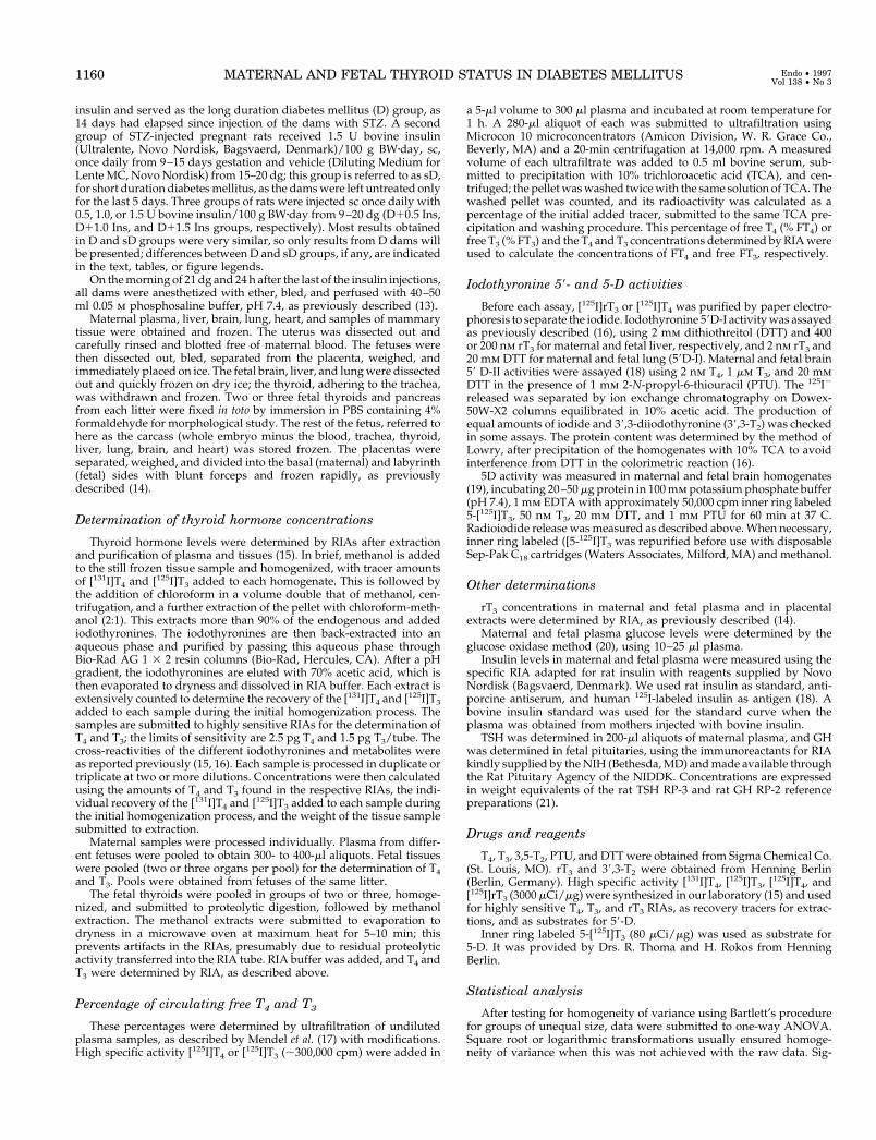
Thyroid hormone status of the placenta. Figure 6 shows theconcentrations of T4, T3, and rT3 in the M- and F-placenta,which decreased in the D dams and returned to normal or
FIG. 4. The mean (6SE) concentrations(nanograms per g wet wt) of T4 and T3are shown for liver, lung, brain, heart,andmammary tissue. The shaded areasand symbols are explained in Fig. 1.There were no statistically significantdifferences in the T4 concentrationsfound in liver, lung, and brain of damsreceiving the various insulin doses. Theconcentration of T3 in these tissues waslower in theD10.5 Ins dams than in theD11.0 Ins and/or D11.5 Ins mothers.
FIG. 5. Themean (6SE) activities of the outer ring 59D, type I for liver(picomoles of I2 per min/mg protein) and lung (femtomoles of I2 perh/mg protein), type II for cortex (Cx; femtomoles of I2 per h/mg pro-tein), and Cx inner ring 5D-III (picomoles of I2 per h/mg protein) areshown. The shaded areas and symbols are explained in Fig. 1. Al-though decreased in sD dams (not shown) compared to those in Cdams, liver and lung 59D-I activities were somewhat higher thanthose in D animals. There were no insulin dose-related differences inliver or brain 59D activities. The activity in lung in D11.0 Ins damswas higher than those in D10.5 and D11.5 Ins rats.
MATERNAL AND FETAL THYROID STATUS IN DIABETES MELLITUS 1163
higher than normal values in D1Ins dams. There were quan-titative differences between the M- and F-placenta with re-gard to the effects of diabetes and the various doses ofinsulin.In the F-placenta, the changes in T4 and rT3 concentrations
are comparable to those described in maternal circulatinglevels. On the contrary, in the M-placenta, T4 and rT3 con-centrations were lower than expected from the circulatinglevels; the concentrations in the M-placenta of D dams de-creased more in C dams than the observed change in therespective circulating levels and increased to only half theexpected levels with insulin treatment. M-placenta T4 wasonly normal in the D11.5 Ins group, in which the circulatingconcentrations were clearly above C values. In contrast, T3concentrations in theM-placenta of D dams, whether treatedwith insulin or not, were higher than expected from T3 cir-culating levels.
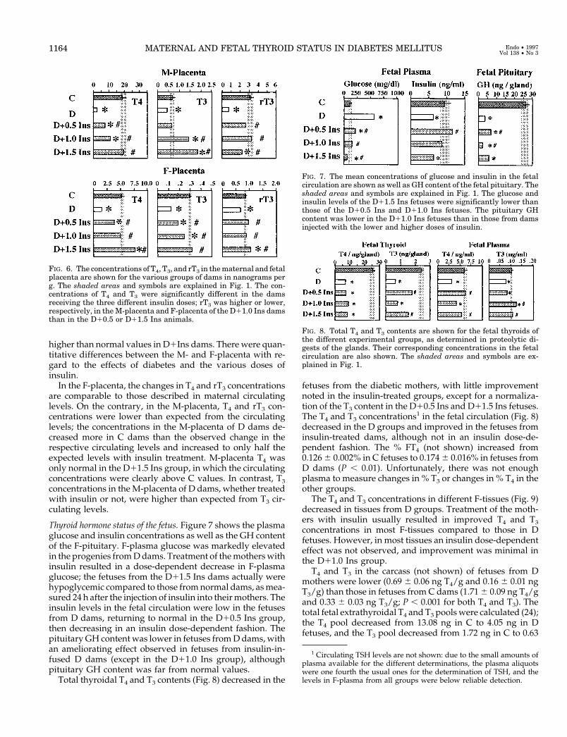
Thyroid hormone status of the fetus. Figure 7 shows the plasmaglucose and insulin concentrations as well as the GH contentof the F-pituitary. F-plasma glucose was markedly elevatedin the progenies fromDdams. Treatment of themotherswithinsulin resulted in a dose-dependent decrease in F-plasmaglucose; the fetuses from the D11.5 Ins dams actually werehypoglycemic compared to those fromnormal dams, asmea-sured 24 h after the injection of insulin into theirmothers. Theinsulin levels in the fetal circulation were low in the fetusesfrom D dams, returning to normal in the D10.5 Ins group,then decreasing in an insulin dose-dependent fashion. Thepituitary GH content was lower in fetuses fromDdams, withan ameliorating effect observed in fetuses from insulin-in-fused D dams (except in the D11.0 Ins group), althoughpituitary GH content was far from normal values.Total thyroidal T4 and T3 contents (Fig. 8) decreased in the
fetuses from the diabetic mothers, with little improvementnoted in the insulin-treated groups, except for a normaliza-tion of the T3 content in the D10.5 Ins and D11.5 Ins fetuses.The T4 and T3 concentrations
1 in the fetal circulation (Fig. 8)decreased in the D groups and improved in the fetuses frominsulin-treated dams, although not in an insulin dose-de-pendent fashion. The % FT4 (not shown) increased from0.1266 0.002% in C fetuses to 0.1746 0.016% in fetuses fromD dams (P , 0.01). Unfortunately, there was not enoughplasma to measure changes in % T3 or changes in % T4 in theother groups.The T4 and T3 concentrations in different F-tissues (Fig. 9)
decreased in tissues from D groups. Treatment of the moth-ers with insulin usually resulted in improved T4 and T3concentrations in most F-tissues compared to those in Dfetuses. However, in most tissues an insulin dose-dependenteffect was not observed, and improvement was minimal inthe D11.0 Ins group.T4 and T3 in the carcass (not shown) of fetuses from D
mothers were lower (0.69 6 0.06 ng T4/g and 0.16 6 0.01 ngT3/g) than those in fetuses from C dams (1.716 0.09 ng T4/gand 0.33 6 0.03 ng T3/g; P , 0.001 for both T4 and T3). Thetotal fetal extrathyroidal T4 and T3 poolswere calculated (24);the T4 pool decreased from 13.08 ng in C to 4.05 ng in Dfetuses, and the T3 pool decreased from 1.72 ng in C to 0.63
1 Circulating TSH levels are not shown: due to the small amounts ofplasma available for the different determinations, the plasma aliquotswere one fourth the usual ones for the determination of TSH, and thelevels in F-plasma from all groups were below reliable detection.
FIG. 7. The mean concentrations of glucose and insulin in the fetalcirculation are shown as well as GH content of the fetal pituitary. Theshaded areas and symbols are explained in Fig. 1. The glucose andinsulin levels of the D11.5 Ins fetuses were significantly lower thanthose of the D10.5 Ins and D11.0 Ins fetuses. The pituitary GHcontent was lower in the D11.0 Ins fetuses than in those from damsinjected with the lower and higher doses of insulin.
FIG. 8. Total T4 and T3 contents are shown for the fetal thyroids ofthe different experimental groups, as determined in proteolytic di-gests of the glands. Their corresponding concentrations in the fetalcirculation are also shown. The shaded areas and symbols are ex-plained in Fig. 1.
FIG. 6. The concentrations of T4, T3, and rT3 in thematernal and fetalplacenta are shown for the various groups of dams in nanograms perg. The shaded areas and symbols are explained in Fig. 1. The con-centrations of T4 and T3 were significantly different in the damsreceiving the three different insulin doses; rT3 was higher or lower,respectively, in theM-placenta and F-placenta of the D11.0 Ins damsthan in the D10.5 or D11.5 Ins animals.
1164 MATERNAL AND FETAL THYROID STATUS IN DIABETES MELLITUS Endo • 1997Vol 138 • No 3

ng in D animals. The extrathyroidal pools of T4 and T3 in Dfetuses were markedly reduced, to 31% and 36%, respec-tively, of the normal values.In D fetuses, 59D-activity was lower than normal in F-liver
and F-lung (Fig. 9) and were restored to normal values infetuses from insulin-treated dams, with the exception of F-liver in the D11.0 Ins group, where it was actually reducedbelow D levels. 59D-II activity in the F-brain was unchanged,except for a decrease in D11.0 Ins fetuses. Fetal brain 5D-IIIactivity (not shown) of normal fetuses was 6.09 pmol/hzmgprotein and did not decrease significantly in fetuses from Ddams, but increased slightly with insulin treatment of themothers to 6.83 pmol/hzmg protein (similar results wereobserved regardless of the insulin dose).
Relationships between fetal and maternal thyroid hormonelevels and between parameters of fetal thyroid hormonestatus
There was a very good correlation between the glucoseconcentrations in the fetal and maternal circulations (n 5 33;r5 0.96; P, 0.001), but not between insulin levels. The latterwas due to the finding that in the insulin-treated groups,insulin levels in M-plasma increased proportionally to theadministered dose, whereas they decreased in F-plasma.The F-BW (Table 1) was affected by the maternal diabetic
state. This decrease was not totally corrected with any of the
doses of insulin administered to the dams, but the closest tonormal F-BW were observed for fetuses from the D10.5 Insmothers, and the lowest was found in fetuses fromD11.0 Insdams. The F-BWwas related to other indexes of the outcomeof pregnancy, such as the number of viable fetuses per litter(n 5 29; r 5 0.63; P , 0.001). F-BW was clearly related to theGH content of the fetal pituitary (Fig. 8), with a good fit toa linear function of F-BW vs. the logarithm of the GH content(n 5 33; r 5 0.87, P , 0.001). With the exception of theF-plasma T3 level, the T4 and T3 concentrations in F-tissueswere more closely correlated to the F-BW and pituitary GHcontent than to the changes in maternal thyroid hormonestatus.The total T4 and T3 contents in the thyroid gland of fetuses
from the different treatment groups did not correlate withtheir changes in F-plasma or F-tissues. The changes observedin the concentrations of T4 andT3 in the F-tissueswere similarto those in the corresponding hormone in F-plasma, as theF-tissue to F-plasma ratios (not shown) were usually thesame as those in fetuses fromC dams, except for T3 in F-liver,F-lung, and F-brain in the D11.0 Ins group, whichwas lowerthan expected from the F-plasma levels.Changes in the thyroid hormone status of the fetuses from
D groups were similar to those in their mothers. On thecontrary, the effects of insulin treatment on T4 and T3 con-centrations were different. Treatment of the D dams withinsulin usually resulted in insulin dose-dependent changes,whereas there was no clear relationship between the insulindose and fetal T4 and T3 concentrations. There was also nocorrelation between the changes in thyroid status of the fe-tuses and F-plasma insulin. As indicated above, there was abetter correlation with F-BW and pituitary GH content.The activities of 59D-I in F-liver and F-lung and of 59D-II
and 5D-III in F-brain, either did not correlate with the T4 andT3 in F-plasma or in F-liver and F-lung, or did so poorly, withr values of 0.5 or less. No clearly significant correlations werefound between these enzyme activities and the F-plasmaglucose levels. Changes in the activities of 59D-I in F-liver andF-lung and 59D-II in F-brain were less marked than those inthe corresponding maternal tissues.
Discussion
Present results confirm and extend our preliminary ob-servations on the effects of maternal STZ-induced diabetesmellitus on fetal thyroid hormone status, as studied on 20 dg(11), which are likely to result from the altered carbohydratemetabolism of their mothers, and not from a direct destruc-tive action of STZ on the fetal pancreas. The half-life ofdisappearance of STZ from the circulation is 5 h in rodents(23). The drug, injected at 7 dg, would no longer be presentby the time the b-cells of the fetal pancreas develop in the rat(12.5–13.5 dg), long before themorphological structure of theb-cells is achieved on 18 dg (24). Moreover, when maternalhyperglycemia was mitigated with insulin, F-plasma insulinwas normal, indicating that fetal b-cells were functional. Asthe placenta is impermeable to maternal insulin (25), thiswould not occur if the fetal pancreas had been directly af-fected by the drug. The pancreas of the hyperglycemic fe-tuses of the present D dams showed hyperplasia, hypertro-
FIG. 9. The three upper panels show the concentrations (in nano-grams per g) of T4 in the liver, lung, and brain of the fetuses from thevarious groups of dams, with the middle panels showing the corre-sponding T3 concentrations. The three lower panels show the activityof the outer ring 59D, type I for liver (picomoles of I2 per min/mgprotein) and lung (femtomoles of I2 per h/mg protein) and type II forcortex (Cx; femtomoles of I2 per h/mg protein). The shaded areas andsymbols are explained in Fig. 1.
MATERNAL AND FETAL THYROID STATUS IN DIABETES MELLITUS 1165

phy, and degranulation of immunochemically identifiedinsulin-positive b-cells (our unpublished data), confirmingthe findings of others (26) and indicating b-cell overstimu-lation and exhaustion.A direct toxic effect of STZ on the placenta is also unlikely.
Ultrastructural changes in the placenta have been described,but were less frequent when insulin was supplied (27),whereas a direct toxic effect would be independent fromtreatment with insulin. In a previous study (unpublished) inwhich the rats were treated with STZ before conception andmaintained with insulin until midgestation, we found thesame changes in the concentrations of both T4 and T3 in theM-placenta as those described here for dams treated withSTZ during pregnancy; T4 decreased to 16% of C values, andT3 to 63%. The F-BW of the dams given STZ pregestationallywas similarly affected as that of the present D fetuses, de-creasing to 67% of C values.
The pregnant rat with diabetes mellitus as a model ofmaternal nonthyroidal illness
The changes in thyroid hormone status occurring in non-thyroidal illness are at present considered adaptive re-sponses to a limited availability of intracellular energy, asituation in which a decrease in T3-dependent catabolic ef-fects would be beneficial (1, 2). Several mechanisms are in-volved in these responses, of which two are best known,namely 1) a decreased thyroidal secretion of both T4 and T3,whichwould lower the pool of T4 available for extrathyroidalgeneration of T3, and 2) a decrease in the activity of enzymesinvolved in the extrathyroidal generation of T3 from T4.1) The sequence of events involves decreased release of
hypothalamic TRH (4, 5), secretion of TSH (4, 6, 7), andsensitivity of the thyroid to TSH (6), which supersede thenormal feedback mechanism; TSH decreases despite thelower levels of circulating total and/or free T4 andT3. Indeed,a return to a normal secretion of TSH and thyroidal releaseof hormones is considered an indication that the illness isremitting or that the metabolic alterations are under control.The present changes in circulating total and free T4 and T3together with decreased TSH and the low T4 and T3 levelsfound in all of the tissues studied are in agreement with thechanges described in the nonpregnant diabetic rats (8) and inpatients dying from nonthyroidal illnesses (28).2) Direct measurement of 59D-I activities in the liver and
lung has confirmed that generation of T3 fromT4 is decreasedin diabetic rats (11, 29, 30) and is consistent with a decreasedexpression of 59D-Imessenger RNA (29). 59D-II activity in thecerebral cortex of D damswas not changed, possibly becausecerebral T4 was not decreased, and confirming the lack ofchange described in nonpregnant diabetic rats (10, 31).
Treatment with insulin: varying severity of maternal illness
The diabetic state of the dams improved. The best resultswere observed in the D10.5 Ins group both with respect toparameters of diabetes mellitus and reproductive compe-tence and with respect to thyroid hormone status, includingcirculating TSH and liver and lung 59D-I activities.The two higher doses of insulin used in the present study
might well have been excessive; the glucose and insulin
levels were normal 24 h after the last injection, suggestingdaily periods of hypoglycemia. Most, but not all, changes inparameters of maternal thyroid hormone economy appearedto be insulin dose dependent.
Effects on the placenta
In diabetic women and rats, hyperglycemia increases pla-cental weight and placental glycogen content (32). The pla-centas from the present D dams were clearly affected. It isvery difficult to properly regulate placental function andfetal metabolism in diabetic mothers with insulin (33) ad-ministered once daily,2 possibly because of the daily cyclesof hyper- and hypoglycemia, or constant hypoglycemia.These were more likely to occur in the D11.0 Ins and D11.5Ins dams. Indeed, the best resultswere observed in theD10.5Ins dams, which were not likely to have undergone pro-longed periods of hypoglycemia.The placenta plays a very important role in the supply of
nutrients and indetermining themetabolic status of the fetus.Posner et al. (34) suggested that insulin might promote sub-strate transport across the placenta, which has insulin re-ceptors. The rat placenta is permeable to maternal T4 and T3(13, 14) and is active in the local metabolism of both iodo-thyronines, as it contains iodothyronine deiodinase isoen-zymes, with 59D-II activity highest in the M-placenta (35),and 5-III highest in the F-placenta (36).Although T4 and T3 decreased in the placenta of D dams
and reversed to normal or supraphysiological levels withinsulin treatment, the changes occurring in the M-placentawere quantitatively different from those found in M-plasma.T4 concentrations decreased more than expected from thechanges in circulating total T4 or FT4, in agreement with ourprevious report (11). The opposite was observed for the con-centrations of T3 in the M-placenta, which increased morewith insulin treatment than expected from the circulatingchanges in total T3 and FT3. The differences in thyroid hor-mone status of the M-placenta compared to other M-tissuesmay well be related to the decreased uteroplacental bloodflow caused by a significant reduction in the arterial bloodvelocity in the uterine artery, placenta, umbilical artery, andfetal aorta (37). The mechanisms, if any, regulating placentalpermeability to the iodothyronines and their metabolism inthe M- and F-placenta are unknown at present, so that thepossibility that their transfer is altered by the hyperglycemicstate or the lack of insulin has not yet been studied. Changesin the activities of the different iodothyronine-deiodinatingisoenzymes may also contribute to the observed concentra-tions, but were not defined in the present experiment.
Effects on fetal development and thyroid hormone status
The reproductive competence of the present STZ-induceddiabetes mellitus pregnant rats was impaired, not only withregard to the decreased number of fetuses per litter and theincreased frequency of resorptions, but also with respect to
2 Administration of insulin by infusionwithminipumpswas not usedfor the present study, as despite repeated attempts with different insulinpreparations, the number of rats in which the pump did not clot waslimited, thus complicating the experimental approach considerably.
1166 MATERNAL AND FETAL THYROID STATUS IN DIABETES MELLITUS Endo • 1997Vol 138 • No 3
the development of their fetuses, as assessed by BW. In ourstudy, the F-BWwas significantly reduced in fetuses from allgroups of STZ-injected dams, in agreement with the severityof the diabetes and with reports by others using modelssimilar to the present one (12, 26) or with pregestationaldiabetes (38) and data of babies born from poorly controlleddiabetic women (39–41). The growth impairment that existsin the adult rat with diabetes mellitus is attributed at least inpart to decreased pituitary GH content and secretion (7)resulting fromdecreasedGHmessenger RNA and decreasedGH transcription rate (42). Although the control of fetalgrowth is not usually attributed to fetal GH, but to insulin-like growth factors, the pituitary content of this hormonewaslower in the fetuses from theDdams of the present study andwas closely correlated to their F-BW.The changes in F-plasma glucose levels were clearly re-
lated to those in the maternal circulation, as previouslyshown by others in rats (12) and sheep (43), as they have alimited capacity to handle the glucose coming from theirhyperglycemic mothers. It is quite likely that the hypergly-cemia of the D fetuses was responsible for exhaustion of anoverstimulated fetal pancreas (26), resulting in decreasedF-plasma insulin levels (11, 12). The thyroid hormone statusof the fetus was clearly affected by the maternal diabetesmellitus, with regard to both intrathyroidal T4 and T3 con-tents and extrathyroidal pools; bothweremarkedly reduced.Morphological parameters, such as the decreased area ofepithelial cells, were consistent with a dormant thyroid infetuses from D dams (our unpublished observations), asdescribed for adult rats. This is in conceptual agreementwiththe finding that insulin is essential for the transcription oftwo thyroid-specific genes, namely the thyroglobulin andthyroid peroxidase genes (44, 45).In fetuses from D dams, the concentrations of T4 and T3
were reduced in the circulation and all tissues studied, in-cluding the carcass, with the total extrathyroidal stores re-duced to one third of C values. This might not only be dueto decreased secretion by the fetal thyroid, but also to thedecrease in maternal T4 and T3 pools available for transport(46) into the fetal compartment. A decreased availability ofmaternal hormones could be aggravated by effects of thematernal diabetes mellitus on M-placental function.Whatever the relative roles played by the different mech-
anisms, the hyperglycemic fetuses presented all of thechanges found in theirmothers, including an increased%FT4and decreased liver and lung 59D-I activities. 59D-II activityin the fetal brain did not show the increase expected from thelow F-plasma and F-brain T4 (13–16), as a result of whichcerebral T3 concentrations were lower than those in normalfetuses.In contrast, the effects of insulin treatment of the dams on
fetal weight, circulating insulin levels, and thyroid hormoneeconomy did not parallel those observed in their mothers.The different doses of insulin resulted in a normal increasein the M-BW in all three groups of insulin-treated D damsand dose-related increases in circulating insulin. The con-centrations of T4 and T3 in M-plasma and most tissues alsoappeared to increase with increasing insulin doses. In con-trast, a clear improvement in F-BWwas only observed in theD10.5 Ins group, the only group in which the number of
fetuses per litter was normal and resorptions absent and inwhich F-plasma insulin levels were restored to normal val-ues. Despite this improvement, a normal F-BW was not at-tained, in agreementwith previous reports (11, 12). A normalfetal pituitary GH content was not attained with any of theinsulin doses used in the present study. Although the fetalpituitary GH content was not measured, Erikson et al. (38)did not find normal F-BW when rat dams, injected with STZweeks before the onset of pregnancy, were treated with in-sulin. We cannot at present explain these results and cannotexclude a direct effect of STZ, injected on day 7 of gestationinto themothers, on the fetal pituitary. This appears unlikely,considering that the decrease in pituitary GH in adult STZ-treated rats is corrected with insulin (7). Maternal hyperin-sulinemia (47) and intermittent hypoglycemia (48) also affectfetal growth, and it is likely that such events accounted forthe poor results obtained by uswith the higher insulin doses,considering that Ultralente insulin was used in the presentstudy.3 F-BW in these groups was closely related to theirpituitary GH content, not to F-plasma glucose or insulinlevels. F-plasma insulin levels decreased with the increasingdose of insulin injected into their mothers and were as lowas those in fetuses from D dams in the hypoglycemic fetusesof the D11.5 Ins dams, possibly because of continuous over-stimulation leading to exhaustion of their b-cells.Most parameters of thyroid status improved or actually
reverted to normal values in the fetuses from the D10.5 Insdams, including attainment of normal T3 concentrations inthe F-brain. The changes in T4 and T3 in the F-thyroid, F-plasma, and F-tissues did not appear to be dose dependentwith respect to the insulin administered to their mothers,being often more closely related to the F-BW and pituitaryGH content. If the latter parameters are taken as indexes offetal metabolic abnormalities and intracellular energy avail-ability, it would appear that if adequate control of the ma-ternal diabetic condition were achieved, the alterations infetal thyroid hormone economy, including brain T3 levels,would be avoided.
Possible clinical relevance of the present findings
Fetal damage, abnormalities, and resorptions are wellknown hazards of diabetes in pregnancy, whether in humanor experimental models (38, 49), especially when inade-quately controlled, as evidenced by increasedmaternal b-hy-droxybutyrate and triglyceride levels. Rizzo et al. (50) re-ported that diabetes mellitus during pregnancy affects theintellectual development and behavior of the progeny. Theyfound a correlation between the alterations in lipid metab-olism of pregnant woman and the intelligence quotient oftheir 2- to 3-yr-old children; the higher the levels of b-hy-droxybutyrate and FFA, the lower the intelligence quotientof the offspring. Considering that if the present results arepertinent to man, brain T3 might have been low during a
3 We have no explanation regarding the very poor results obtained inthe D11.0 Ins fetuses, except that their mothers might have undergonea wider range of daily fluctuations between a hyperglycemic and ahypoglycemic state than the D11.5 Ins dams, which might have beenhypoglycemic throughout. Such fluctuations appear to be especiallyharmful for the placenta (47).
MATERNAL AND FETAL THYROID STATUS IN DIABETES MELLITUS 1167
phase of development when thyroid hormones are of greatimportance for brain development, it is possible that alter-ations in fetal thyroid hormone status are contributing to thisintellectual impairment. Unfortunately, although the presentresults would indicate that adequate control of the diabetesmellitus appears to be of prime importance to prevent thedecrease in cerebral T3, such control is not easily achieved.
Relevance of present findings for nonthyroidal illness otherthan diabetes mellitus
The diabetic dam displayed all of the changes in thyroidhormone status considered typical of nonthyroidal illness,changes that could be modulated and/or totally correctedwith insulin. We cannot at present exclude that some of theeffects observed in both the dams and the conceptus werespecifically related to the shortage of insulin and would notbe found in other models of nonthyroidal illness. However,the changes in the concentrations of T4 in the circulation andtissues of nonpregnant STZ-induced diabetes mellitus andfood-restricted rats have been shown to be related to energyavailability, as measured by the changes in BW, and notspecifically to the insulin levels (8). The same was found forthe concentration of T3 in brain, cerebellum, liver, and pi-tuitary, whereas the T3 level in brown adipose tissue, heart,muscle, and kidney were more specifically related to theavailability of insulin. Although the present experimentaldesign was not adequate to distinguish between the effectsof energy shortage and the lack of insulin, the findings wereport here may be relevant for other nonthyroidal illnessesin which intracellular energy availability is impaired.
Conclusions
The present results show that maternal diabetes mellitusand possibly maternal nonthyroidal illnesses compromisingintracellular energy availability result in severe impairmentof the thyroid hormone status of the fetus. This includes lowcerebral concentrations of T3 during a critical period of braindevelopment. Correction of the illness is necessary to protectthe brain, as compensatory mechanisms usually involved inmaintaining cerebral T3 homeostasis do not appear to beoperative.
Acknowledgments
We thank Drs. R. Thoma and H. Rokos (Henning, Berlin) for thegenerous gift of the inner ring labeled [5-125I]T3. We are also indebtedto S. Duran, A. Hernandez, and M. J. Presas for invaluable technicalassistance. We are grateful to Dr. J. Schroder-van der Elst and Prof. D.van der Heide (The Netherlands) for their helpful comments anddiscussions.
References
1. Wartofski L, Burman KD 1982 Alterations in thyroid function in patients withsystemic illness: the “euthyroid sick syndrome.” Endocr Rev 3:164–217
2. Tibaldi JM, Surks MI 1985 Animal models of non-thyroidal disease. EndocrRev 6:87–101
3. Pittman CA, Suda AK, Chambers JB, McDaniel HG, Ray GY 1979 Abnor-malities of thyroid turnover in patients with diabetes mellitus before and afterinsulin therapy. J Clin Endocrinol Metab 48:854–860
4. Gonzalez C, Montoya E, Jolın T 1980 Effect of streptozotocin diabetes on thehypothalamic-pituitary-thyroid axis in the rat. Endocrinology 107:2099–2103
5. Rondeel JMM, de Greef WJ, Heide R, Visser TJ 1992 Hypothalamo-hypoph-
ysial-thyroid axis in streptozotocin-induced diabetes. Endocrinology130:216–220
6. Pastor R, Jolın T 1983 Peripheral metabolism and secretion rate of thyrotropinin STZ-diabetic rats. Endocrinology 112:1454–1459
7. Ortız-Caro J, Gonzalez C, Jolın T 1984 Diurnal variations of plasma growthhormone, thyrotropin, thyroxine and triiodothyronine in streptozotocin-dia-betic and food-restricted rats. Endocrinology 115:2227–2232
8. Schroeder van der Elst JP, van der Heide D 1992 Effects of streptozotocin-induced diabetes and food restriction on quantities and source of T4 and T3 inrat tissues. Diabetes 41:147–152
9. Bjorn-Hansen-Gotzsche LS, Gotzche OJ, Flyvbjerg A, Boye N 1990 Earlychanges in thyroid hormonemetabolism in the heart, liver, and brown adiposetissue during the induction of low T3 syndrome in streptozotocin-diabetic rats.Acta Endocrinol (Copenh) 123:67–72
10. Ortız-Caro J, Obregon MJ, Pascual A, Jolın T 1984 Decreased T4 to T3 con-version in tissues of STZ-diabetic rats. Acta Endocrinol (Copenh) 106:86–91
11. Calvo R, Obregon MJ, Escobar del Rey F, Morreale de Escobar G 1991 Theeffects of maternal diabetes mellitus on thyroid hormone economy of ratfetuses. In: Gordon A, Gross J, Hennemann G (eds) Progress in ThyroidResearch. Balkema, Rotterdam, pp 813–816
12. Herrera E, Palacın M, Martın M, Lasuncion MA 1985 Relationship betweenmaternal and fetal fuels and placental glucose transfer in rats with maternaldiabetes of varying severity. Diabetes [2 ] 34:42–46
13. Calvo RM, Obregon MJ, Escobar del Rey F, Morreale de Escobar G 1990Congenital hypothyroidism, as studied in rats. Crucial role of thyroxine but notof triiodothyronine in the protection of the fetal brain. J Clin Invest 86:889–899
14. Calvo R, Obregon MJ, Escobar del Rey F, Morreale de Escobar G 1992 Therat placenta and the transfer of thyroid hormones from the mother to the fetus.Effects of maternal thyroid status. Endocrinology 131:357–365
15. Morreale de Escobar G, Calvo R, Escobar F, Obregon MJ 1994 Thyroidhormones in tissues from fetal and adult rats. Endocrinology 134:2410–2415
16. Ruiz de Ona C,Morreale de Escobar G, Calvo R, Escobar del Rey F, ObregonMJ 1991 Thyroid hormones and 59-deiodinase in the rat fetus late in gestation:effects of maternal hypothyroidism. Endocrinology 128:422–432
17. Mendel CM, Laughton CW, McMahon FA, Cavalieri RR 1991 Inability todetect an inhibitor of thyroxine-serum protein binding in sera from patientswith non-thyroidal illness. Metabolism 40:491–502
18. Obregon MJ, Ruiz de Ona C, Hernandez A, Calvo R, Escobar del Rey F,Morreale de Escobar G 1989 Thyroid hormones and 59-dediodinase in ratbrown adipose tissue during fetal life. Am J Physiol 257:E625–E631
19. Campos-Barros A, Kohler R,Muller F, EravciM,MeinholdH,WesemannW,Baumgartner A 1993 The influence of sleep deprivation on thyroid hormonemetabolism in rat frontal cortex. Neurosci Lett 162:145–148
20. Hugget ASG, Nixon DA 1957 Use of glucose oxidase, peroxidase and o-dianisidine in determination of blood and urinary glucose. Lancet 1:368–70
21. Morreale de Escobar G, Calvo R, Escobar del Rey F, Obregon MJ 1993Differential effects of thyroid hormones on growth and thyrotropic hormonein rat fetuses near term. Endocrinology 132:2056–2064
22. Snedecor GW, Cochran WG 1980 Statistical Methods, ed 7. Iowa State Uni-versity Press, Ames
23. Schein PS, Loftus S 1968 Streptozotocin: depression of mouse liver pyridinenucleotides. Cancer Res 28:1501–1506
24. Portha B 1990 Development of the pancreatic b cell: growth patterns andfunctional maturation. In: Cuezva JM, Pascual-Leone AM, Patel MS, SaffordG (eds) Endocrine and Biochemical Development of the Fetus and Neonate.Plenum Press, New York, pp 33–43
25. Knobil E, Yosimovich JB 1958 Placental transfer of thyrotropic hormone,thyroxine, triiodothyronine and insulin. Ann NY Acad Sci 75:895–904
26. Aerts L, van Assche FA 1977 Rat fetal endocrine pancreas in experimentaldiabetes J Endocrinol 73:339–346
27. Padmanabhan R, Al-Zuhair AG 1990 Ultrastructural studies on the placentaeof streptozotocin induced maternal diabetes in the rat. Z Mikrosk Anat Forsch104:213–220
28. AremR,WienerGJ,KaplanSG,KimHS,Reichlin S,KaplanM 1993Reducedtissue thyroid hormone levels in fatal illness. Metabolism 42:1102–1108
29. O’Mara BA, Dittrich W, Lauterio TJ, StGermain DL 1993 Pretranslationalregulation of type I 59-deiodinase by thyroid hormones and in fasted anddiabetic rats. Endocrinology 133:1715–1723
30. Gavin L A, McMahon FA, Mueller M 1981 The mechanism of impaired T3production from T4 in diabetes. Diabetes 30:694–699
31. Gavin LA, Cavalieri RR 1986 Iodothyronine deiodination in the brain ofdiabetic rats: influence of thyroid status. J Endocrinol Invest 9:127–133
32. Shafrir E, BarashV 1991 Placental glycogenmetabolism in diabetic pregnancy.Isr J Med Sci 27:449–461
33. Copeland AD, Hendrich CE, Porterfield SP 1990 Distribution of free amino-acids in streptozotocin-induced diabetic pregnant rats, their placentae andfetuses. Horm Metab Res 22:65–70
34. Posner BE 1974 Insulin receptors in human and animal placental tissue. Di-abetes 23:209–217
35. Kaplan MM, Shaw E 1984 Type II iodothyronine 59-deiodination by humanand rat placenta in vitro. J Clin Endocrinol Metab 59:253–257
36. EmersonCH 1989 Role of the placenta in fetal thyroid homeostasis. In: Delange
1168 MATERNAL AND FETAL THYROID STATUS IN DIABETES MELLITUS Endo • 1997Vol 138 • No 3

F, Fisher DA, Glinoer D (eds) Research in Congenital Hypothyroidism, seriesA. Plenum Press, New York, vol 161:31–43
37. Chartrel NC, Clabaut MT, Boismare FA, Schrub JC 1990 Uteroplacentalhemodynamic disturbances in establishment of fetal growth retardation instreptozotocin-induced diabetic rats. Diabetes 39:743–746
38. Eriksson RSM, Thunberg L, Eriksson UJ 1989 Effect of interrupted insulintreatment on fetal outcome of pregnant diabetic rats. Diabetes 38:764–772
39. Pedersen J F, Molsted-Pedersen L 1981 Early growth delay detected by ul-trasound marks increased risk of congenital malformation in diabetic preg-nancy. Br Med J 283:269–271
40. Hill DJ 1982 Fetal effects of insulin. Obstet Gynaecol Ann 11:133–14941. Rudolf MCJ, Sherwin RS, Markowitz R 1982 Effect of intensive insulin
treatment on linear growth in the young diabetic patient. J Pediatr 101:333–33942. Bedo G, Santisteban P, Jolin T, Aranda A 1991 Expression of the growth
hormone gene and the pituitary-specific transcription factor GHF-1 in diabeticrats. Mol Endocrinol 5:1730–1739
43. Hay Jr WW, Sparks JW, Wilkening RB, Battaglia FC, Meschia G 1984 Fetalglucose uptake and utilization as functions of maternal glucose concentration.Am J Physiol 246:E237–E242
44. Santisteban P, Acebron A, Polycarpou-Schwarz M, DiLauro R 1992 Insulin
and insulin-like growth factor I regulate a thyroid-specific nuclear protein thatbinds to the thyroglobulin promoter. Mol Endocrinol 6:1310–1317
45. Aza-Blanc P, Di Lauro R, Santisteban P 1993 Identification of a cis-regu-latory element and a thyroid-specific nuclear factor mediating the hormonalregulation of rat thyroid peroxidase promoter activity. Mol Endocrinol7:1297–1306
46. Morreale de Escobar G, Calvo R, Obregon MJ, Escobar del Rey F 1990Contribution of maternal thyroxine to fetal thyroxine in normal rats near term.Endocrinology 126:2765–2767
47. Ogata ES, Paul RI, Finley SL 1987 Limited maternal fuel availability due tohyperinsulinemia retards fetal growth and development in the rat. Pediatr Res22:432–437
48. Sodoyez-Goffaux F, Sodoyez JC 1976 Effects of intermittent hypoglycemia inpregnant rats on the functional development of the pancreatic B-cells of theiroffspring. Diabetologia 12:73–76
49. Mills JL, Baker L, Goldman AS 1979 Malformations in infants of diabeticmothers occur before the seventh gestational week. Diabetes 28:292–293
50. Rizzo T, Metzger BE, Burns WJ, Burns K 1991 Correlations between ante-partum maternal metabolism and intelligence of offspring. N Engl J Med325:911–916
MATERNAL AND FETAL THYROID STATUS IN DIABETES MELLITUS 1169