Neural and hormonal mechanisms of reproductive-related arousal in fishes
Upload
independentCategory
view
0download
0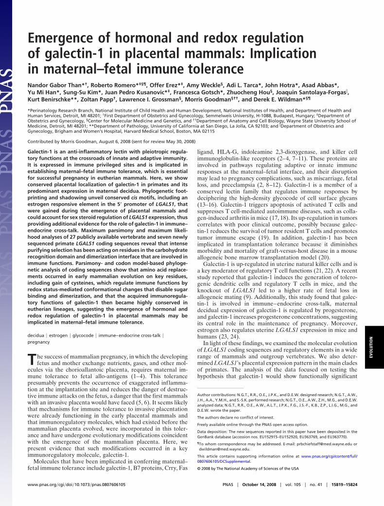
Emergence of hormonal and redox regulationof galectin-1 in placental mammals: Implicationin maternal–fetal immune toleranceNandor Gabor Than*†, Roberto Romero*‡§¶, Offer Erez*‡, Amy Weckle§, Adi L. Tarca*, John Hotra*, Asad Abbas*,Yu Mi Han*, Sung-Su Kim*, Juan Pedro Kusanovic*‡, Francesca Gotsch*, Zhuocheng Hou§, Joaquin Santolaya-Forgas�,Kurt Benirschke**, Zoltan Papp†, Lawrence I. Grossman§, Morris Goodman§††, and Derek E. Wildman*§¶
*Perinatology Research Branch, National Institute of Child Health and Human Development, National Institutes of Health, and Department of Health andHuman Services, Detroit, MI 48201; †First Department of Obstetrics and Gynecology, Semmelweis University, H-1088, Budapest, Hungary; ‡Department ofObstetrics and Gynecology, §Center for Molecular Medicine and Genetics, and ††Department of Anatomy and Cell Biology, Wayne State University School ofMedicine, Detroit, MI 48201; **Department of Pathology, University of California at San Diego, La Jolla, CA 92103; and �Department of Obstetrics andGynecology, Brigham and Women’s Hospital, Harvard Medical School, Boston, MA 02115
Contributed by Morris Goodman, August 6, 2008 (sent for review May 30, 2008)
Galectin-1 is an anti-inflammatory lectin with pleiotropic regula-tory functions at the crossroads of innate and adaptive immunity.It is expressed in immune privileged sites and is implicated inestablishing maternal–fetal immune tolerance, which is essentialfor successful pregnancy in eutherian mammals. Here, we showconserved placental localization of galectin-1 in primates and itspredominant expression in maternal decidua. Phylogenetic foot-printing and shadowing unveil conserved cis motifs, including anestrogen responsive element in the 5� promoter of LGALS1, thatwere gained during the emergence of placental mammals andcould account for sex steroid regulation of LGALS1 expression, thusproviding additional evidence for the role of galectin-1 in immune–endocrine cross-talk. Maximum parsimony and maximum likeli-hood analyses of 27 publicly available vertebrate and seven newlysequenced primate LGALS1 coding sequences reveal that intensepurifying selection has been acting on residues in the carbohydraterecognition domain and dimerization interface that are involved inimmune functions. Parsimony- and codon model-based phyloge-netic analysis of coding sequences show that amino acid replace-ments occurred in early mammalian evolution on key residues,including gain of cysteines, which regulate immune functions byredox status-mediated conformational changes that disable sugarbinding and dimerization, and that the acquired immunoregula-tory functions of galectin-1 then became highly conserved ineutherian lineages, suggesting the emergence of hormonal andredox regulation of galectin-1 in placental mammals may beimplicated in maternal–fetal immune tolerance.
decidua � estrogen � glycocode � immune–endocrine cross-talk �pregnancy
The success of mammalian pregnancy, in which the developingfetus and mother exchange nutrients, gases, and other mol-
ecules via the chorioallantoic placenta, requires maternal im-mune tolerance to fetal allo-antigens (1–4). This tolerancepresumably prevents the occurrence of exaggerated inflamma-tion at the implantation site and reduces the danger of destruc-tive immune attacks on the fetus, a danger that the first mammalswith an invasive placenta would have faced (5, 6). It seems likelythat mechanisms for immune tolerance to invasive placentationwere already functioning in the early placental mammals andthat immunoregulatory molecules, which had existed before themammalian placenta evolved, were incorporated in this toler-ance and have undergone evolutionary modifications coincidentwith the emergence of the mammalian placenta. Here, wepresent evidence that such modifications occurred in a keyimmunoregulatory molecule, galectin-1.
Molecules that have been implicated in conferring maternal–fetal immune tolerance include galectin-1, B7 proteins, Crry, Fas
ligand, HLA-G, indoleamine 2,3-dioxygenase, and killer cellimmunoglobulin-like receptors (2–4, 7–11). These proteins areinvolved in pathways regulating adaptive or innate immuneresponses at the maternal–fetal interface, and their disruptionmay lead to pregnancy complications, such as miscarriage, fetalloss, and preeclampsia (2, 8–12). Galectin-1 is a member of aconserved lectin family that regulates immune responses bydeciphering the high-density glycocode of cell surface glycans(13–16). Galectin-1 triggers apoptosis of activated T cells andsuppresses T cell-mediated autoimmune diseases, such as colla-gen-induced arthritis in mice (17, 18). Its up-regulation in tumorscorrelates with poor clinical outcome, possibly because galec-tin-1 reduces the survival of tumor resident T cells and promotestumor immune escape (19). In addition, galectin-1 has beenimplicated in transplantation tolerance because it diminishesmorbidity and mortality of graft-versus-host disease in a mouseallogeneic bone marrow transplantation model (20).
Galectin-1 is up-regulated in uterine natural killer cells and isa key moderator of regulatory T cell functions (21, 22). A recentstudy reported that galectin-1 induces the generation of tolero-genic dendritic cells and regulatory T cells in mice, and theknockout of LGALS1 led to a higher rate of fetal loss inallogeneic mating (9). Additionally, this study found that galec-tin-1 is involved in immune–endocrine cross-talk, maternaldecidual expression of galectin-1 is regulated by progesterone,and galectin-1 increases progesterone concentrations, suggestingits central role in the maintenance of pregnancy. Moreover,estrogen also regulates uterine LGALS1 expression in mice andhumans (23, 24).
In light of these findings, we examined the molecular evolutionof LGALS1 coding sequences and regulatory elements in a widerange of mammals and outgroup vertebrates. We also deter-mined LGALS1’s placental expression pattern in the main cladesof primates. The analysis of the data focused on testing thehypothesis that galectin-1 would show functionally significant
Author contributions: N.G.T., R.R., O.E., J.P.K., and D.E.W. designed research; N.G.T., A.W.,J.H., A.A., Y.M.H., and S.-S.K. performed research; N.G.T., O.E., A.W., Z.H., M.G., and D.E.W.analyzed data; N.G.T., R.R., O.E., A.W., A.L.T., J.P.K., F.G., J.S.-F., K.B., Z.P., L.I.G., M.G., andD.E.W. wrote the paper.
The authors declare no conflict of interest.
Freely available online through the PNAS open access option.
Data deposition: The new sequences reported in this paper have been deposited in theGenBank database (accession nos. EU152915–EU152920, EU363769, and EU363770).
¶To whom correspondence may be addressed. E-mail: [email protected] [email protected].
This article contains supporting information online at www.pnas.org/cgi/content/full/0807606105/DCSupplemental.
© 2008 by The National Academy of Sciences of the USA
www.pnas.org�cgi�doi�10.1073�pnas.0807606105 PNAS � October 14, 2008 � vol. 105 � no. 41 � 15819–15824
EVO
LUTI
ON
evolutionary modifications early in placental mammalian phy-logeny and then would be highly conserved in the descendentmammalian species. The experimental and inferential evidencepresented herein indicate that indeed functionally importantchanges likely involved in the emergence of maternal–fetalimmune tolerance were gained and then conserved in placentalmammals.
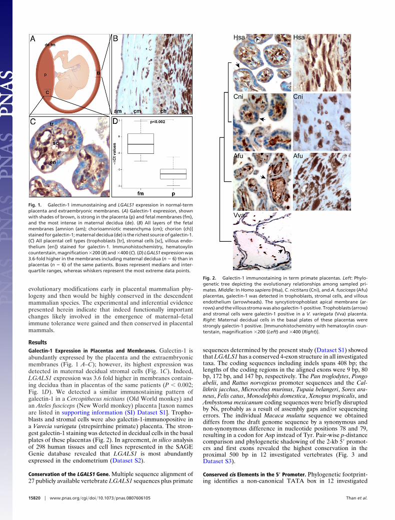
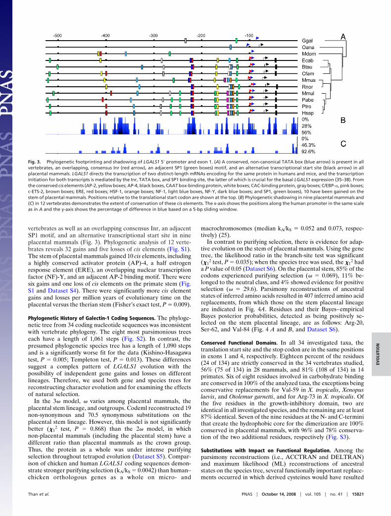
ResultsGalectin-1 Expression in Placentas and Membranes. Galectin-1 isabundantly expressed by the placenta and the extraembryonicmembranes (Fig. 1 A–C); however, its highest expression wasdetected in maternal decidual stromal cells (Fig. 1C). Indeed,LGALS1 expression was 3.6 fold higher in membranes contain-ing decidua than in placentas of the same patients (P � 0.002;Fig. 1D). We detected a similar immunostaining pattern ofgalectin-1 in a Cercopithecus nictitans (Old World monkey) andan Ateles fusciceps (New World monkey) placenta [taxon namesare listed in supporting information (SI) Dataset S1]. Tropho-blasts and stromal cells were also galectin-1-immunopositive ina Varecia variegata (strepsirrhine primate) placenta. The stron-gest galectin-1 staining was detected in decidual cells in the basalplates of these placentas (Fig. 2). In agreement, in silico analysisof 298 human tissues and cell lines represented in the SAGEGenie database revealed that LGALS1 is most abundantlyexpressed in the endometrium (Dataset S2).
Conservation of the LGALS1 Gene. Multiple sequence alignment of27 publicly available vertebrate LGALS1 sequences plus primate
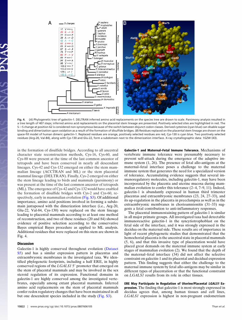
sequences determined by the present study (Dataset S1) showedthat LGALS1 has a conserved 4-exon structure in all investigatedtaxa. The coding sequences including indels spans 408 bp; thelengths of the coding regions in the aligned exons were 9 bp, 80bp, 172 bp, and 147 bp, respectively. The Pan troglodytes, Pongoabelii, and Rattus norvegicus promoter sequences and the Cal-lithrix jacchus, Microcebus murinus, Tupaia belangeri, Sorex ara-neus, Felis catus, Monodelphis domestica, Xenopus tropicalis, andAmbystoma mexicanum coding sequences were briefly disruptedby Ns, probably as a result of assembly gaps and/or sequencingerrors. The individual Macaca mulatta sequence we obtaineddiffers from the draft genome sequence by a synonymous andnon-synonymous difference in nucleotide positions 78 and 79,resulting in a codon for Asp instead of Tyr. Pair-wise p-distancecomparison and phylogenetic shadowing of the 2-kb 5� promot-ers and first exons revealed the highest conservation in theproximal 500 bp in 12 investigated vertebrates (Fig. 3 andDataset S3).
Conserved cis Elements in the 5� Promoter. Phylogenetic footprint-ing identifies a non-canonical TATA box in 12 investigated
Fig. 1. Galectin-1 immunostaining and LGALS1 expression in normal-termplacenta and extraembryonic membranes. (A) Galectin-1 expression, shownwith shades of brown, is strong in the placenta (p) and fetal membranes (fm),and the most intense in maternal decidua (de). (B) All layers of the fetalmembranes [amnion (am); chorioamniotic mesenchyma (cm); chorion (ch)]stained for galectin-1; maternal decidua (de) is the richest source of galectin-1.(C) All placental cell types (trophoblasts [tr], stromal cells [sc], villous endo-thelium [en]) stained for galectin-1. Immunohistochemistry, hematoxylincounterstain, magnification �200 (B) and �400 (C). (D) LGALS1 expression was3.6-fold higher in the membranes including maternal decidua (n � 6) than inplacentas (n � 6) of the same patients. Boxes represent medians and inter-quartile ranges, whereas whiskers represent the most extreme data points.
Fig. 2. Galectin-1 immunostaining in term primate placentas. Left: Phylo-genetic tree depicting the evolutionary relationships among sampled pri-mates. Middle: In Homo sapiens (Hsa), C. nictitans (Cni), and A. fusciceps (Afu)placentas, galectin-1 was detected in trophoblasts, stromal cells, and villousendothelium (arrowheads). The syncytiotrophoblast apical membrane (ar-rows) and the villous stroma was also galectin-1-positive. Trophoblasts (arrow)and stromal cells were galectin-1 positive in a V. variegata (Vva) placenta.Right: Maternal decidual cells in the basal plates of these placentas werestrongly galectin-1 positive. [Immunohistochemistry with hematoxylin coun-terstain, magnification �200 (Left) and �400 (Right)].
15820 � www.pnas.org�cgi�doi�10.1073�pnas.0807606105 Than et al.
vertebrates as well as an overlapping consensus Inr, an adjacentSP1 motif, and an alternative transcriptional start site in nineplacental mammals (Fig. 3). Phylogenetic analysis of 12 verte-brates reveals 32 gains and five losses of cis elements (Fig. S1).The stem of placental mammals gained 10 cis elements, includinga highly conserved activator protein (AP)-4, a half estrogenresponse element (ERE), an overlapping nuclear transcriptionfactor (NF)-Y, and an adjacent AP-2 binding motif. There weresix gains and one loss of cis elements on the primate stem (Fig.S1 and Dataset S4). There were significantly more cis elementgains and losses per million years of evolutionary time on theplacental versus the therian stem (Fisher’s exact test, P � 0.009).
Phylogenetic History of Galectin-1 Coding Sequences. The phyloge-netic tree from 34 coding nucleotide sequences was inconsistentwith vertebrate phylogeny. The eight most parsimonious treeseach have a length of 1,061 steps (Fig. S2). In contrast, thepresumed phylogenetic species tree has a length of 1,090 stepsand is a significantly worse fit for the data (Kishino-Hasagawatest, P � 0.005; Templeton test, P � 0.013). These differencessuggest a complex pattern of LGALS1 evolution with thepossibility of independent gene gains and losses on differentlineages. Therefore, we used both gene and species trees forreconstructing character evolution and for examining the effectsof natural selection.
In the 3� model, � varies among placental mammals, theplacental stem lineage, and outgroups. Codeml reconstructed 19non-synonymous and 70.5 synonymous substitutions on theplacental stem lineage. However, this model is not significantlybetter (�1
2 test, P � 0.868) than the 2� model, in whichnon-placental mammals (including the placental stem) have adifferent ratio than placental mammals as the crown group.Thus, the protein as a whole was under intense purifyingselection throughout tetrapod evolution (Dataset S5). Compar-ison of chicken and human LGALS1 coding sequences demon-strate stronger purifying selection (kA/kS � 0.0042) than human–chicken orthologous genes as a whole on micro- and
macrochromosomes (median kA/kS � 0.052 and 0.073, respec-tively) (25).
In contrast to purifying selection, there is evidence for adap-tive evolution on the stem of placental mammals. Using the genetree, the likelihood ratio in the branch-site test was significant(�1
2 test, P � 0.035); when the species tree was used, the �12 had
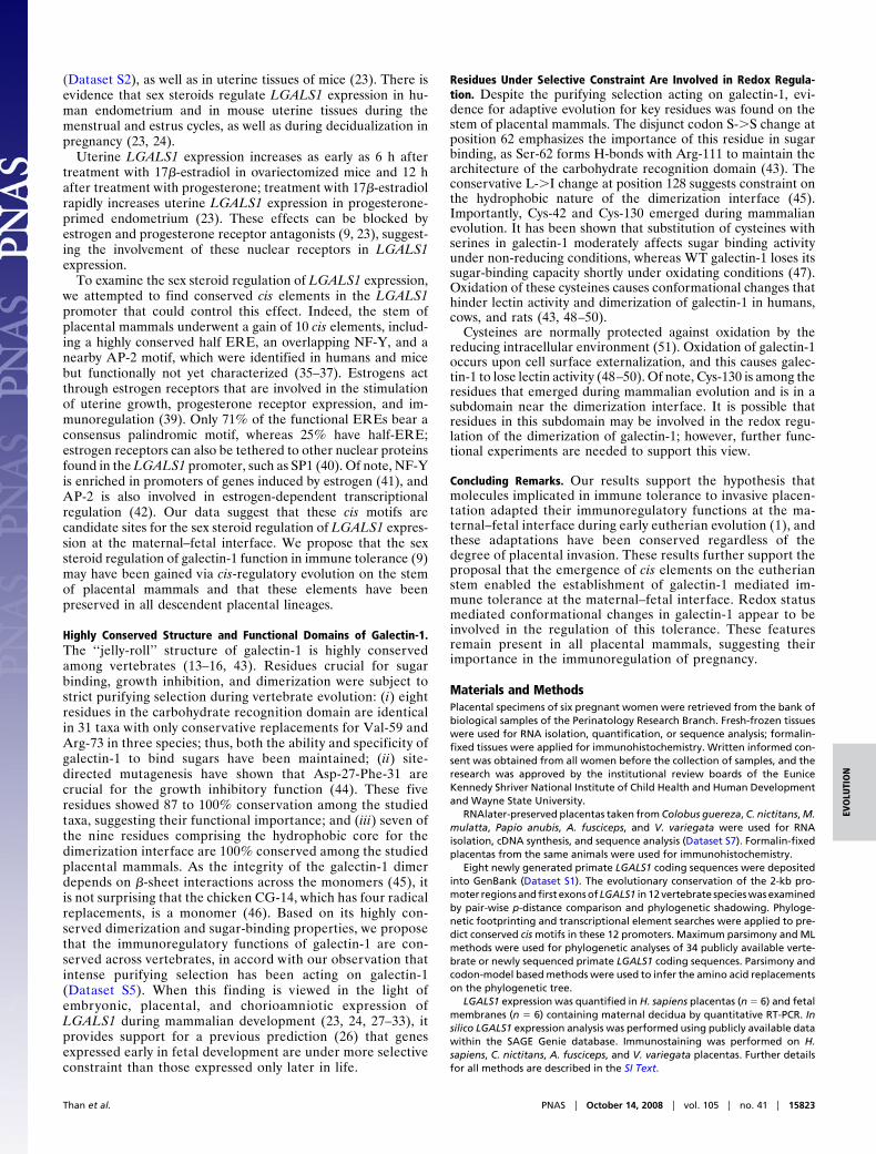
a P value of 0.05 (Dataset S6). On the placental stem, 85% of thecodons experienced purifying selection (� � 0.069), 11% be-longed to the neutral class, and 4% showed evidence for positiveselection (� � 29.6). Parsimony reconstructions of ancestralstates of inferred amino acids resulted in 407 inferred amino acidreplacements, from which those on the stem placental lineageare indicated in Fig. 4A. Residues and their Bayes–empiricalBayes posterior probabilities, detected as being positively se-lected on the stem placental lineage, are as follows: Arg-20,Ser-62, and Val-84 (Fig. 4 A and B, and Dataset S6).
Conserved Functional Domains. In all 34 investigated taxa, thetranslation start site and the stop codon are in the same positionsin exons 1 and 4, respectively. Eighteen percent of the residues(24 of 134) are strictly conserved in the 34 vertebrates studied,56% (75 of 134) in 28 mammals, and 81% (108 of 134) in 14primates. Six of eight residues involved in carbohydrate bindingare conserved in 100% of the analyzed taxa, the exceptions beingconservative replacements for Val-59 in X. tropicalis, Xenopuslaevis, and Otolemur garnetti, and for Arg-73 in X. tropicalis. Ofthe five residues in the growth-inhibitory domain, two areidentical in all investigated species, and the remaining are at least87% identical. Seven of the nine residues at the N- and C-terminithat create the hydrophobic core for the dimerization are 100%conserved in placental mammals, with 96% and 78% conserva-tion of the two additional residues, respectively (Fig. S3).
Substitutions with Impact on Functional Regulation. Among theparsimony reconstructions (i.e., ACCTRAN and DELTRAN)and maximum likelihood (ML) reconstructions of ancestralstates on the species tree, several functionally important replace-ments occurred in which derived cysteines would have resulted
Fig. 3. Phylogenetic footprinting and shadowing of LGALS1 5� promoter and exon 1. (A) A conserved, non-canonical TATA box (blue arrow) is present in allvertebrates, an overlapping, consensus Inr (red arrow), an adjacent SP1 (green boxes) motif, and an alternative transcriptional start site (black arrow) in allplacental mammals. LGALS1 directs the transcription of two distinct-length mRNAs encoding for the same protein in humans and mice, and the transcriptioninitiation for both transcripts is mediated by the Inr, TATA box, and SP1 binding site, the latter of which is crucial for the basal LGALS1 expression (35–38). Fromthe conserved cis elements (AP-2, yellow boxes; AP-4, black boxes, CAAT box-binding protein, white boxes; CAC-binding protein, gray boxes; C/EBP-�, pink boxes;c-ETS-2, brown boxes; ERE, red boxes; HSF-1, orange boxes; NF-1, light blue boxes, NF-Y, dark blue boxes; and SP1, green boxes), 10 have been gained on thestem of placental mammals. Positions relative to the translational start codon are shown at the top. (B) Phylogenetic shadowing in nine placental mammals and(C) in 12 vertebrates demonstrates the extent of conservation of these cis elements. The x-axis shows the positions along the human promoter in the same scaleas in A and the y-axis shows the percentage of difference in blue based on a 5-bp sliding window.
Than et al. PNAS � October 14, 2008 � vol. 105 � no. 41 � 15821
EVO
LUTI
ON
in the formation of disulfide bridges. According to all ancestralcharacter state reconstruction methods, Cys-16, Cys-60, andCys-88 were present at the time of the last common ancestor oftetrapods and have been conserved in nearly all descendantlineages. Cys-42 and Cys-132 emerged on either the stem mam-malian lineage (ACCTRAN and ML) or the stem placentalmammal lineage (DELTRAN). Finally, Cys-2 emerged on eitherthe stem lineage leading to birds and mammals (parsimony) orwas present at the time of the last common ancestor of tetrapods(ML). The emergence of Cys-42 and Cys-132 would have enabledthe formation of disulfide bridges with Cys-2 and Cys-60, re-spectively, early in mammalian evolution (Fig. S3). Of additionalimportance, amino acid positions involved in forming a subdo-main juxtaposed with the dimerization interface (i.e., Arg-20,Glu-22, Val-84, Cys-130) were replaced on the stem lineageleading to placental mammals according to at least one methodof reconstruction, and two of these residues (20 and 84) showedevidence of positive selection according to the conservativeBayes empirical Bayes procedure as applied to ML analysis.Additional residues that were replaced on this stem are shown inFig. 4.
DiscussionGalectin-1 is highly conserved throughout evolution (DatasetS5) and has a similar expression pattern in placentas andextraembryonic membranes in the investigated taxa. We iden-tified phylogenetic footprints, including a half ERE, in highlyconserved regions of the LGALS1 5� promoter that emerged onthe stem of placental mammals and may be involved in the sexsteroid regulation of its expression. Functional domains ingalectin-1 are highly conserved among the investigated verte-brates, especially among extant placental mammals. Inferredamino acid replacements on the stem of placental mammalsconfer redox regulatory changes that have been maintained in allbut one descendent species included in the study (Fig. S3).
Galectin-1 and Maternal–Fetal Immune Tolerance. Mechanisms ofvertebrate immune tolerance were presumably necessary toprevent self-attack during the emergence of the adaptive im-mune system (1, 26). The presence of fetal allo-antigens at thematernal–fetal interface poses a challenge to the maternalimmune system that generates the need for a specialized versionof tolerance. Accumulating evidence suggests that several im-munoregulatory molecules, including galectin-1, may have beenincorporated by the placenta and uterine mucosa during mam-malian evolution to confer this tolerance (2–4, 7–9, 11). Indeed,galectin-1 is abundantly expressed in human third trimesterplacentas and extraembryonic membranes (23, 24, 27–33), andits up-regulation in the placenta in preeclampsia as well as in theextraembryonic membranes in chorioamnionitis (31–33) sug-gests a fetal contribution to anti-inflammatory responses.
The placental immunostaining pattern of galectin-1 is similarin all major primate groups. All investigated taxa had detectableimmunoreactive galectin-1 in the syncytiotrophoblast on thefetal side of the interface, and it was strongly expressed in thedecidua on the maternal side. These results are of importance inlight of recent phylogenetic studies that demonstrated that thehemochorial placenta is the ancestral state in placental mammals(5, 6), and that this invasive type of placentation would haveplaced great demands on the maternal immune system at earlystages of mammalian evolution (2). We found that the depth ofthe maternal–fetal interface (34) did not affect the selectiveconstraint on galectin-1 and its placental and decidual expressionpattern. This finding suggests that either the challenge to thematernal immune system by fetal allo-antigens may be similar indifferent types of placentation or that the functional constrainton LGALS1 results from its role in other tissues.
ERE May Participate in Regulation of Uterine/Placental LGALS1 Ex-pression. The finding that galectin-1 is most strongly expressed indecidua agrees that, among human tissues and cell lines,LGALS1 expression is highest in non-pregnant endometrium
Fig. 4. (A) Phylogenetic tree of galectin-1. DELTRAN inferred amino acid replacements on the species tree are drawn to scale. Parsimony analysis resulted ina tree length of 407 steps; inferred amino acid replacements on the placental stem lineage are presented. Positively selected sites are highlighted in red. TheS-�S change at position 62 is considered non-synonymous because of the switch between disjunct codon classes. Derived cysteines (cyan blue) can disable sugarbinding and dimerization upon oxidation as a result of the formation of disulfide bridges. (B) Residues replaced on the placental stem lineage are shown on thespace-fill model of human dimeric galectin-1. Replaced residues are orange, positively selected residues are red, Cys-130 is cyan blue. Two positively selectedresidues (Arg-20, Val-84), along with Cys-130 and Glu-22, form a subdomain next to the dimerization interface. X-ray crystallographic data: 1GZW (43).
15822 � www.pnas.org�cgi�doi�10.1073�pnas.0807606105 Than et al.
(Dataset S2), as well as in uterine tissues of mice (23). There isevidence that sex steroids regulate LGALS1 expression in hu-man endometrium and in mouse uterine tissues during themenstrual and estrus cycles, as well as during decidualization inpregnancy (23, 24).
Uterine LGALS1 expression increases as early as 6 h aftertreatment with 17�-estradiol in ovariectomized mice and 12 hafter treatment with progesterone; treatment with 17�-estradiolrapidly increases uterine LGALS1 expression in progesterone-primed endometrium (23). These effects can be blocked byestrogen and progesterone receptor antagonists (9, 23), suggest-ing the involvement of these nuclear receptors in LGALS1expression.
To examine the sex steroid regulation of LGALS1 expression,we attempted to find conserved cis elements in the LGALS1promoter that could control this effect. Indeed, the stem ofplacental mammals underwent a gain of 10 cis elements, includ-ing a highly conserved half ERE, an overlapping NF-Y, and anearby AP-2 motif, which were identified in humans and micebut functionally not yet characterized (35–37). Estrogens actthrough estrogen receptors that are involved in the stimulationof uterine growth, progesterone receptor expression, and im-munoregulation (39). Only 71% of the functional EREs bear aconsensus palindromic motif, whereas 25% have half-ERE;estrogen receptors can also be tethered to other nuclear proteinsfound in the LGALS1 promoter, such as SP1 (40). Of note, NF-Yis enriched in promoters of genes induced by estrogen (41), andAP-2 is also involved in estrogen-dependent transcriptionalregulation (42). Our data suggest that these cis motifs arecandidate sites for the sex steroid regulation of LGALS1 expres-sion at the maternal–fetal interface. We propose that the sexsteroid regulation of galectin-1 function in immune tolerance (9)may have been gained via cis-regulatory evolution on the stemof placental mammals and that these elements have beenpreserved in all descendent placental lineages.
Highly Conserved Structure and Functional Domains of Galectin-1.The ‘‘jelly-roll’’ structure of galectin-1 is highly conservedamong vertebrates (13–16, 43). Residues crucial for sugarbinding, growth inhibition, and dimerization were subject tostrict purifying selection during vertebrate evolution: (i) eightresidues in the carbohydrate recognition domain are identicalin 31 taxa with only conservative replacements for Val-59 andArg-73 in three species; thus, both the ability and specificity ofgalectin-1 to bind sugars have been maintained; (ii) site-directed mutagenesis have shown that Asp-27-Phe-31 arecrucial for the growth inhibitory function (44). These fiveresidues showed 87 to 100% conservation among the studiedtaxa, suggesting their functional importance; and (iii) seven ofthe nine residues comprising the hydrophobic core for thedimerization interface are 100% conserved among the studiedplacental mammals. As the integrity of the galectin-1 dimerdepends on �-sheet interactions across the monomers (45), itis not surprising that the chicken CG-14, which has four radicalreplacements, is a monomer (46). Based on its highly con-served dimerization and sugar-binding properties, we proposethat the immunoregulatory functions of galectin-1 are con-served across vertebrates, in accord with our observation thatintense purifying selection has been acting on galectin-1(Dataset S5). When this finding is viewed in the light ofembryonic, placental, and chorioamniotic expression ofLGALS1 during mammalian development (23, 24, 27–33), itprovides support for a previous prediction (26) that genesexpressed early in fetal development are under more selectiveconstraint than those expressed only later in life.
Residues Under Selective Constraint Are Involved in Redox Regula-tion. Despite the purifying selection acting on galectin-1, evi-dence for adaptive evolution for key residues was found on thestem of placental mammals. The disjunct codon S-�S change atposition 62 emphasizes the importance of this residue in sugarbinding, as Ser-62 forms H-bonds with Arg-111 to maintain thearchitecture of the carbohydrate recognition domain (43). Theconservative L-�I change at position 128 suggests constraint onthe hydrophobic nature of the dimerization interface (45).Importantly, Cys-42 and Cys-130 emerged during mammalianevolution. It has been shown that substitution of cysteines withserines in galectin-1 moderately affects sugar binding activityunder non-reducing conditions, whereas WT galectin-1 loses itssugar-binding capacity shortly under oxidating conditions (47).Oxidation of these cysteines causes conformational changes thathinder lectin activity and dimerization of galectin-1 in humans,cows, and rats (43, 48–50).
Cysteines are normally protected against oxidation by thereducing intracellular environment (51). Oxidation of galectin-1occurs upon cell surface externalization, and this causes galec-tin-1 to lose lectin activity (48–50). Of note, Cys-130 is among theresidues that emerged during mammalian evolution and is in asubdomain near the dimerization interface. It is possible thatresidues in this subdomain may be involved in the redox regu-lation of the dimerization of galectin-1; however, further func-tional experiments are needed to support this view.
Concluding Remarks. Our results support the hypothesis thatmolecules implicated in immune tolerance to invasive placen-tation adapted their immunoregulatory functions at the ma-ternal–fetal interface during early eutherian evolution (1), andthese adaptations have been conserved regardless of thedegree of placental invasion. These results further support theproposal that the emergence of cis elements on the eutherianstem enabled the establishment of galectin-1 mediated im-mune tolerance at the maternal–fetal interface. Redox statusmediated conformational changes in galectin-1 appear to beinvolved in the regulation of this tolerance. These featuresremain present in all placental mammals, suggesting theirimportance in the immunoregulation of pregnancy.
Materials and MethodsPlacental specimens of six pregnant women were retrieved from the bank ofbiological samples of the Perinatology Research Branch. Fresh-frozen tissueswere used for RNA isolation, quantification, or sequence analysis; formalin-fixed tissues were applied for immunohistochemistry. Written informed con-sent was obtained from all women before the collection of samples, and theresearch was approved by the institutional review boards of the EuniceKennedy Shriver National Institute of Child Health and Human Developmentand Wayne State University.
RNAlater-preserved placentas taken from Colobus guereza, C. nictitans, M.mulatta, Papio anubis, A. fusciceps, and V. variegata were used for RNAisolation, cDNA synthesis, and sequence analysis (Dataset S7). Formalin-fixedplacentas from the same animals were used for immunohistochemistry.
Eight newly generated primate LGALS1 coding sequences were depositedinto GenBank (Dataset S1). The evolutionary conservation of the 2-kb pro-moter regions and first exons of LGALS1 in 12 vertebrate species was examinedby pair-wise p-distance comparison and phylogenetic shadowing. Phyloge-netic footprinting and transcriptional element searches were applied to pre-dict conserved cis motifs in these 12 promoters. Maximum parsimony and MLmethods were used for phylogenetic analyses of 34 publicly available verte-brate or newly sequenced primate LGALS1 coding sequences. Parsimony andcodon-model based methods were used to infer the amino acid replacementson the phylogenetic tree.
LGALS1 expression was quantified in H. sapiens placentas (n � 6) and fetalmembranes (n � 6) containing maternal decidua by quantitative RT-PCR. Insilico LGALS1 expression analysis was performed using publicly available datawithin the SAGE Genie database. Immunostaining was performed on H.sapiens, C. nictitans, A. fusciceps, and V. variegata placentas. Further detailsfor all methods are described in the SI Text.
Than et al. PNAS � October 14, 2008 � vol. 105 � no. 41 � 15823
EVO
LUTI
ON

ACKNOWLEDGMENTS. We acknowledge the valuable contributions of Dr.Chong Jai Kim, Sandy Field, Nancy Hauff, Gerardo Rodriguez, and thenursing staff of the Perinatology Research Branch. The authors thank Dr.Susan Land and Daniel Lott at the Applied Genomics Technology Center ofWayne State University for performing the qRT-PCR reactions, and SaraTipton for critical reading of the manuscript. The following kindly providedDNA and/or tissue samples: Dr. Caro-Beth Stewart (SUNY, Albany, NY), Dr.
Kathy Neiswanger (University of Pittsburgh, Pittsburgh, PA), the NewEngland Regional Primate Center (Southborough, MA), the Duke Univer-sity Primate Center (Durham, NC), and the San Diego Zoo (San Diego,CA). This research was supported by the Intramural Program of theEunice Kennedy Shriver National Institute of Child Health and HumanDevelopment/National Institutes of Health/Department of Health and Hu-man Services.
1. Medawar PB (1953) Some immunological and endocrinological problems raised by theevolution of viviparity in vertebrates. Symp Soc Exp Biol 44:320–338.
2. Moffett A, Loke C (2006) Immunology of placentation in eutherian mammals. Nat RevImmunol 6:584–594.
3. Niederkorn JY (2006) See no evil, hear no evil, do no evil: the lessons of immuneprivilege. Nat Immunol 7:354–359.
4. Trowsdale J, Betz AG (2006) Mother’s little helpers: mechanisms of maternal-fetaltolerance. Nat Immunol 7:241–246.
5. Wildman DE, et al. (2006) Evolution of the mammalian placenta revealed by phylo-genetic analysis. Proc Natl Acad Sci USA 103:3203–3208.
6. Elliot MG, Crespi BJ (2006) Placental invasiveness mediates the evolution of hybridinviability in mammals. Am Nat 168:114–120.
7. Petroff MG, Chen L, Phillips TA, Hunt JS (2002) B7 family molecules: novel immuno-modulators at the maternal-fetal interface. Placenta 23(Suppl A):S95–101.
8. Terness P, et al. (2007) Tolerance signaling molecules and pregnancy: IDO, galectins,and the renaissance of regulatory T cells. Am J Reprod Immunol 58:238–254.
9. Blois SM, et al. (2007) A pivotal role for galectin-1 in fetomaternal tolerance. Nat Med13:1450–1457.
10. Saito S, et al. (2007). The role of the immune system in preeclampsia. Mol Aspects Med28:192–209.
11. Hunt JS, Petroff MG, McIntire RH, Ober C (2005) HLA-G and immune tolerance inpregnancy. FASEB J 19:681–693.
12. Aluvihare VR, Kallikourdis M, Betz AG (2004) Regulatory T cells mediate maternaltolerance to the fetus. Nat Immunol 5:266–271.
13. Barondes SH, et al. (1994) Galectins: a family of animal beta-galactoside-bindinglectins. Cell 76:597–598.
14. Cooper DN (2002) Galectinomics: finding themes in complexity. Biochim Biophys Acta1572:209–231.
15. Gabius HJ, Andre S, Kaltner H, Siebert HC (2002) The sugar code: functional lectinomics.Biochim Biophys Acta 1572:165–177.
16. Houzelstein D, et al. (2004) Phylogenetic analysis of the vertebrate galectin family. MolBiol Evol 21:1177–1187.
17. He J, Baum LG (2004) Presentation of galectin-1 by extracellular matrix triggers T celldeath. J Biol Chem 279:4705–4712.
18. Rabinovich GA, et al. (1999) Recombinant galectin-1 and its genetic delivery suppresscollagen-induced arthritis via T cell apoptosis. J Exp Med 190:385–398.
19. Rubinstein N, et al. (2004) Targeted inhibition of galectin-1 gene expression in tumorcells results in heightened T cell-mediated rejection: a potential mechanism of tumor-immune privilege. Cancer Cell 5:241–251.
20. Baum LG, et al. (2003). Amelioration of graft versus host disease by galectin-1. ClinImmunol 109:295–307.
21. Koopman LA, et al. (2003) Human decidual natural killer cells are a unique NK cellsubset with immunomodulatory potential. J Exp Med 198:1201–1212.
22. Garin MI, et al. (2007) Galectin-1: a key effector of regulation mediated byCD4�CD25� T cells. Blood 109:2058–2065.
23. Choe YS, et al. (1997) Expression of galectin-1 mRNA in the mouse uterus is under thecontrol of ovarian steroids during blastocyst implantation. Mol Reprod Dev 48:261–266.
24. von Wolff M, Wang X, Gabius HJ, Strowitzki T (2005) Galectin fingerprinting in humanendometrium and decidua during the menstrual cycle and in early gestation. Mol HumReprod 11:189–194.
25. International Chicken Genome Sequencing Consortium (2004) Sequence and compar-ative analysis of the chicken genome provide unique perspectives on vertebrateevolution. Nature 432:695–716.
26. Goodman M (1963) in Viking Fund publications in anthropology. Number thirty-seven.Classification and human evolution, ed Washburn SL (Wenner-Gren Foundation forAnthropological Research Inc., New York), pp 204–234.
27. Bevan BH, et al. (1994) Immunohistochemical localization of a beta-D-galactoside-binding lectin at the human maternofetal interface. Histochem J 26:582–586.
28. Walzel H, et al. (1995) Immunohistochemical and glycohistochemical localization ofthe beta-galactoside-binding S-type lectin in human placenta. Acta Histochem 97:33–42.
29. Maquoi E, van den Brule FA, Castronovo V, Foidart JM (1997) Changes in the distribu-tion pattern of galectin-1 and galectin-3 in human placenta correlates with thedifferentiation pathways of trophoblasts. Placenta 18:433–439.
30. Vicovac L, Jankovic M, Cuperlovic M (1998) Galectin-1 and �3 in cells of the firsttrimester placental bed. Hum Reprod 13:730–735.
31. Jeschke U, et al. (2007) Expression of galectin-1, -3 (gal-1, gal-3) and the Thomsen-Friedenreich (TF) antigen in normal, IUGR, preeclamptic and HELLP placentas. Placenta28:1165–1173.
32. Than NG, et al. (2008) Severe preeclampsia is characterized by increased placentalexpression of galectin-1. J Matern Fetal Neonatal Med 21:429–442.
33. Than NG, et al. (2008) Chorioamnionitis and increased galectin-1 expression inPPROM–an anti-inflammatory response in the fetal membranes? Am J Reprod Immu-nol, doi:10.1111/j.1600-0897.2008.00624.x.
34. MossmanHW(1987)Vertebratefetalmembranes (RutgersUnivPress,NewBrunswick,NJ).35. Gitt MA, Barondes SH (1991) Genomic sequence and organization of two members of
a human lectin gene family. Biochemistry 30:82–89.36. Salvatore P, et al. (1995) Characterization and functional dissection of the galectin-1
gene promoter. FEBS Lett 373:159–163.37. Lu Y, Lotan R (1999) Transcriptional regulation by butyrate of mouse galectin-1 gene
in embryonal carcinoma cells. Biochim Biophys Acta 1444:85–91.38. De Gregorio E, Chiariotti L, Di Nocera PP (2001) The overlap of Inr and TATA elements
sets the use of alternative transcriptional start sites in the mouse galectin-1 genepromoter. Gene 268:215–223.
39. Dahlman-Wright K, et al. (2006) International Union of Pharmacology LXIV Estrogenreceptors. Pharmacol Rev 58:773–781.
40. Lin CY, et al (2007). Whole-genome cartography of estrogen receptor alpha bindingsites. PLoS Genet 3:e87.
41. Scafoglio C, et al. (2006) Comparative gene expression profiling reveals partiallyoverlapping but distinct genomic actions of different antiestrogens in human breastcancer cells. J Cell Biochem 98:1163–1184.
42. Orso F, et al. (2004) Activator protein-2gamma (AP-2gamma) expression is specificallyinduced by oestrogens through binding of the oestrogen receptor to a canonicalelement within the 5�-untranslated region. Biochem J 377:429–438.
43. Lopez-Lucendo MF, et al. (2004) Growth-regulatory human galectin-1: crystallographiccharacterisation of the structural changes induced by single-site mutations and theirimpact on the thermodynamics of ligand binding. J Mol Biol 343:957–970.
44. Scott K, Zhang J (2002) Partial identification by site-directed mutagenesis of a cellgrowth inhibitory site on the human galectin-1 molecule. BMC Cell Biol 3:3.
45. Cho M, Cummings RD (1996) Characterization of monomeric forms of galectin-1generated by site-directed mutagenesis. Biochemistry 35:13081–13088.
46. Varela PF, et al. (1999) The 2.15 Å crystal structure of CG-16, the developmentallyregulated homodimeric chicken galectin. J Mol Biol 294:537–549.
47. Hirabayashi J, Kasai K (1991) Effect of amino acid substitution by sited-directedmutagenesis on the carbohydrate recognition and stability of human 14-kDa beta-galactoside-binding lectin. J Biol Chem 266:23648–23653.
48. Tracey BM, et al. (1992) Subunit molecular mass assignment of 14,654 Da to the solublebeta-galactoside-binding lectin from bovine heart muscle and demonstration of in-tramolecular disulfide bonding associated with oxidative inactivation. J Biol Chem267:10342–10347.
49. Yamaoka K, et al. (1996) Structural and functional characterization of a novel tumor-derived rat galectin-1 having transforming growth factor (TGF) activity: the relationshipbetween intramolecular disulfide bridges and TGF activity. J Biochem 119:878–886.
50. Inagaki Y, et al. (2000) Oxidized galectin-1 promotes axonal regeneration in peripheralnerves but does not possess lectin properties. Eur J Biochem 267:2955–2964.
51. Sen CK (1998) Redox signaling and the emerging therapeutic potential of thiolantioxidants. Biochem Pharmacol 55:1747–1758.
15824 � www.pnas.org�cgi�doi�10.1073�pnas.0807606105 Than et al.
Copyright © 2022 FDOKUMEN