Hormonal and Genetic Influences on Processing Reward and Social Information

Dc
PLa
b
c
d
e
a
ARRA
KCCHMMPT
1
i2tpmeCsdViacv
0d
Environmental and Experimental Botany 67 (2009) 387–394
Contents lists available at ScienceDirect
Environmental and Experimental Botany
journa l homepage: www.e lsev ier .com/ locate /envexpbot
ifferential ultrastructural changes in tomato hormonal mutants exposed toadmium
riscila L. Gratão a , Carolina C. Monteiro a , Mônica L. Rossi b , Adriana P. Martinelli b , Lázaro E.P. Peres c ,eonardo O. Medici d, Peter J. Lea e, Ricardo A. Azevedo a,∗
Departamento de Genética, Escola Superior de Agricultura Luiz de Queiroz, Universidade de São Paulo, Piracicaba 13418-900, SP, BrazilCentro de Energia Nuclear na Agricultura, Universidade de São Paulo, Piracicaba 13400-970, SP, BrazilDepartamento de Ciências Biológicas, Escola Superior de Agricultura Luiz de Queiroz, Universidade de São Paulo, Piracicaba 13418-900, SP, BrazilDepartamento de Ciências Fisiológicas, Universidade Federal Rural do Rio de Janeiro, Seropédica 23890-000, RJ, BrazilLancaster Environment Centre, University of Lancaster, Lancaster LA1 4YQ, UK
r t i c l e i n f o
rticle history:eceived 16 March 2009eceived in revised form 26 June 2009ccepted 27 June 2009
eywords:admium
a b s t r a c t
Cadmium (Cd) is a toxic heavy metal, which can cause severe damage to plant development. The aimof this work was to characterize ultrastructural changes induced by Cd in miniature tomato cultivarMicro-Tom (MT) mutants and their wild-type counterpart. Leaves of diageotropica (dgt) and Never ripe(Nr) tomato hormonal mutants and wild-type MT were analysed by light, scanning and transmissionelectron microscopy in order to characterize the structural changes caused by the exposure to 1 mMCdCl2. The effect of Cd on leaf ultrastructure was observed most noticeably in the chloroplasts, which
hloroplasteavy metalsicro-Tomutants
lant anatomyomato
exhibited changes in organelle shape and internal organization, of the thylakoid membranes and stroma.Cd caused an increase in the intercellular spaces in Nr leaves, but a decrease in the intercellular spaces indgt leaves, as well as a decrease in the size of mesophyll cells in the mutants. Roots of the tomato hormonalmutants, when analysed by light microscopy, exhibited alterations in root diameter and disintegration ofthe epidermis and the external layers of the cortex. A comparative analysis has allowed the identificationof specific Cd-induced ultrastructural changes in wild-type tomato, the pattern of which was not always
.
exhibited by the mutants. Introduction
Contamination of the soil by toxic elements such as heavy metalss a major environmental concern (Gratão et al., 2005; Teklic et al.,008; Paiva et al., 2009). Cadmium (Cd) is probably one of the mostoxic heavy metals, particularly at high concentrations, inhibitinglant growth and development, whereas at low concentrations Cday also stimulate growth depending on the plant species (Gratão
t al., 2005, 2008a; Mobin and Khan, 2007; Wahid and Ghani, 2008).d can also negatively interfere with important plant processesuch as water transport, oxidative phosphorylation in mitochon-ria, photosynthesis and chlorophyll content (Djebali et al., 2005;itória et al., 2006; Tukaj et al., 2007; Lage-Pinto et al., 2008), which
s dependent upon metal concentration, plant species, organ/tissuend duration of exposure (Benavides et al., 2005). Moreover, Cd isapable of inducing oxidative, stress which in turn can result in aariety of antioxidant responses (Ferreira et al., 2002; Fornazier et
∗ Corresponding author. Tel.: +55 19 34294475; fax: +55 19 34336706.E-mail address: [email protected] (R.A. Azevedo).
098-8472/$ – see front matter © 2009 Elsevier B.V. All rights reserved.oi:10.1016/j.envexpbot.2009.06.017
© 2009 Elsevier B.V. All rights reserved.
al., 2002; Gomes-Junior et al., 2006; Lindberg et al., 2007; Gratãoet al., 2008b,c; Smeets et al., 2008; Tamas et al., 2008).
The biochemical responses of plants to Cd (see Benavides et al.,2005 for a review) and the effect of Cd on the ultrastructure ofplant organs (Vitória et al., 2003, 2006) have been investigated insome detail. However, as far as we are aware, there is no specificinformation available as to how hormonal mutations causing a lossof sensitivity to auxin or ethylene can modify anatomical structureand the response of plants to Cd.
Some of the damage reported is related to leaf structure dis-organization, reduced intercellular air spaces, drastic structuralthylakoid alterations in the chloroplast (Aravind and Prasad, 2005;Djebali et al., 2005), stomatal closure, softening of cell wall thick-ening (Vitória et al., 2003) and decrease in chlorophyll content andefficiency of Rubisco activity (Vassilev et al., 2003). In addition, Cdhas also been reported to cause disruption of the nuclear envelope,plasmalemma and mitochondrial membranes, severe plasmolysis
and high chromatin condensation (Stoyanova and Tchakalova, 1997;Liu and Kottke, 2004).The use of several model systems has been a successful approachto study the complex network of factors influencing plant responsesto the environment and their modulation by hormones. The minia-

3 Experimental Botany 67 (2009) 387–394
tgps1r((Bwpaic
2
2
ai(atppAthaowbgwppfa
2
gfict(wd2(ioet
2
mpSaw
Table 1Effect of Cd on closure of stomatal pores in abaxial epidermis of Micro-Tom (MT),diageotropica (dgt) and Never ripe (Nr) leaves.
Cultivar Treatment %Closed stomata
Micro-Tom (MT) 0 mM of CdCl2 31.4 ± 1.14Micro-Tom (MT) 1 mM of CdCl2 77.0 ± 2.6diageotropica (dgt) 0 mM of CdCl2 31.2 ± 1.58diageotropica (dgt) 1 mM of CdCl2 27.7 ± 0.83Never ripe (Nr) 0 mM of CdCl2 36.4 ± 1.64
88 P.L. Gratão et al. / Environmental and
ure tomato cultivar Micro-Tom (MT) has been proposed as aenetic model based on its small size, growth at high density androduction of viable fruits and seeds in pots with 50–100 mL ofubstrate, all within a reduced life cycle ranging between 70 and00 d (Meissner et al., 1997). Moreover, MT plants harbouring natu-al genetic variations and hormonal mutations have been producedLima et al., 2004; Zsögön et al., 2008), such as the diageotropicadgt) mutation that causes a loss of sensitivity to auxin (Kelly andradford, 1986; Oh et al., 2006), and the Never ripe (Nr) mutation,hich blocks ethylene perception in tomato leading to incom-
lete fruit ripening (Wilkinson et al., 1995; Lin et al., 2008). Theim of this work was to characterize the ultrastructural changesnduced by Cd in these two mutants and their wild-type Micro-Tomounterpart.
. Materials and methods
.1. Plant material
Seeds of Lycopersicon esculentum cv. MT and the mutants dgtnd Nr near isogenic to MT (Zsögön et al., 2008) were germinatedn boxes containing a mixture of 1:1 (v/v) commercial pot mixPlantmax HT Eucatex, Brazil) and vermiculite, supplemented withmixture of 1 g L−1 NPK 10:10:10 and 4 g L−1 lime. After the first
rue leaves appeared, seedlings were transplanted to 1 L Leonardots (Vincent, 1975) (2 seedlings per pot) filled with sand andolystyrene (4:3) and Hoagland’s nutrient solution (Hoagland andrnon, 1950). After 30 d, the growth of the plants was continued in
he same Leonard pots filled with a mixture of sand and polystyrene,alf of the pots were maintained on Hoagland’s solution (control)nd half were treated with Hoagland’s solution in the presencef 1 mM CdCl2 (Gratão et al., 2008a). The solutions were changedeekly and the total volume was maintained at a constant level
y using distilled water. The experiments were carried out in alasshouse under natural daylight (May–August 2006 and 2007)ith temperatures in the range of 20–30 ◦C. After a period of 85 d
ost germination, corresponding to 40 d of exposure to CdCl2, sam-les were collected during the morning period (9:00–10:00 a.m.)
rom the median third of the 3rd or 4th leaf from the apex, rinsednd used for analyses.
.2. Scanning electron microscopy (SEM)
Leaf samples were collected and immediately fixed in 2%lutaraldehyde in 0.05 M sodium cacodylate buffer, at pH 7.2. Post-xation was carried out in 1.0% osmium tetroxide in 0.1 M sodiumacodylate buffer, pH 7.2, for 1 h. The samples were then rinsed 3imes in distilled water and dehydrated through an ethanol series30, 50, 70, 80%), followed by 3 washes in 100% ethanol. The samplesere finally critical point dried through liquid carbon dioxide. Theried samples were mounted in metal stubs, sputter coated with0 nm gold and examined under a scanning electron microscopeLEO 435 VP, Cambridge, England) at 20 kV, and the images digital-zed. The number of totally closed stomata in the abaxial epidermisf the leaves of the MT wild-type and dgt and Nr mutants werexpressed as a percentage derived from an average of 12 observa-ions of leaf micrographs (about 1 cm2 each).
.3. Light (LM) and transmission electron microscopy (TEM)
Leaf and root fragments of tomato plants were fixed in a
odified Karnovsky solution with 2% glutaraldehyde and 2.5%araformaldehyde in 0.05 M cacodylate buffer (pH 7.2) for 48 h.ubsequently, the samples were rinsed in cacodylate buffer (0.1 M)nd fixed for 1 h at room temperature with 1.0% osmium tetroxideith the same buffer. The post-fixed samples were dehydrated in a
Never ripe (Nr) 1 mM of CdCl2 28.3 ± 1.8
Results are expressed as mean ± s.e. Each value is the average of 12 observations ofleaves.
graded acetone series and embedded in Epon resin for 48 h. Semi-thin sections (120–200 nm) were collected in glass slides, stainedwith toluidine blue (2% in water), for 5 min, rinsed in distilled waterand air-dried. The sections were permanently mounted in Entellanresin, observed and documented using an upright light microscope(AxioPlan, Zeiss, Jena, Germany). Ultra-thin sections (60–90 nm)of leaves were collected on copper grids (300 mesh) and stainedwith 2.5% uranyl acetate followed by 0.1% lead citrate (Reynolds,1963). Sections were observed at 50 kV using a transmission elec-tron microscope (Em 900, Zeiss, West Germany) and the imagesdigitalized.
3. Results
The majority of the stomata on the abaxial epidermis of MT, Nrand dgt tomato leaves were open in the control plants not sub-jected to Cd treatment (Table 1, Fig. 1A, C and E). However, in theCd-treated wild-type MT plants, the majority of the stomata wereclosed (Table 1, Fig. 1B), whilst in the Nr and dgt mutants, most ofthe stomata appeared to remain open in the presence of Cd (Table 1,Fig. 1D and F).
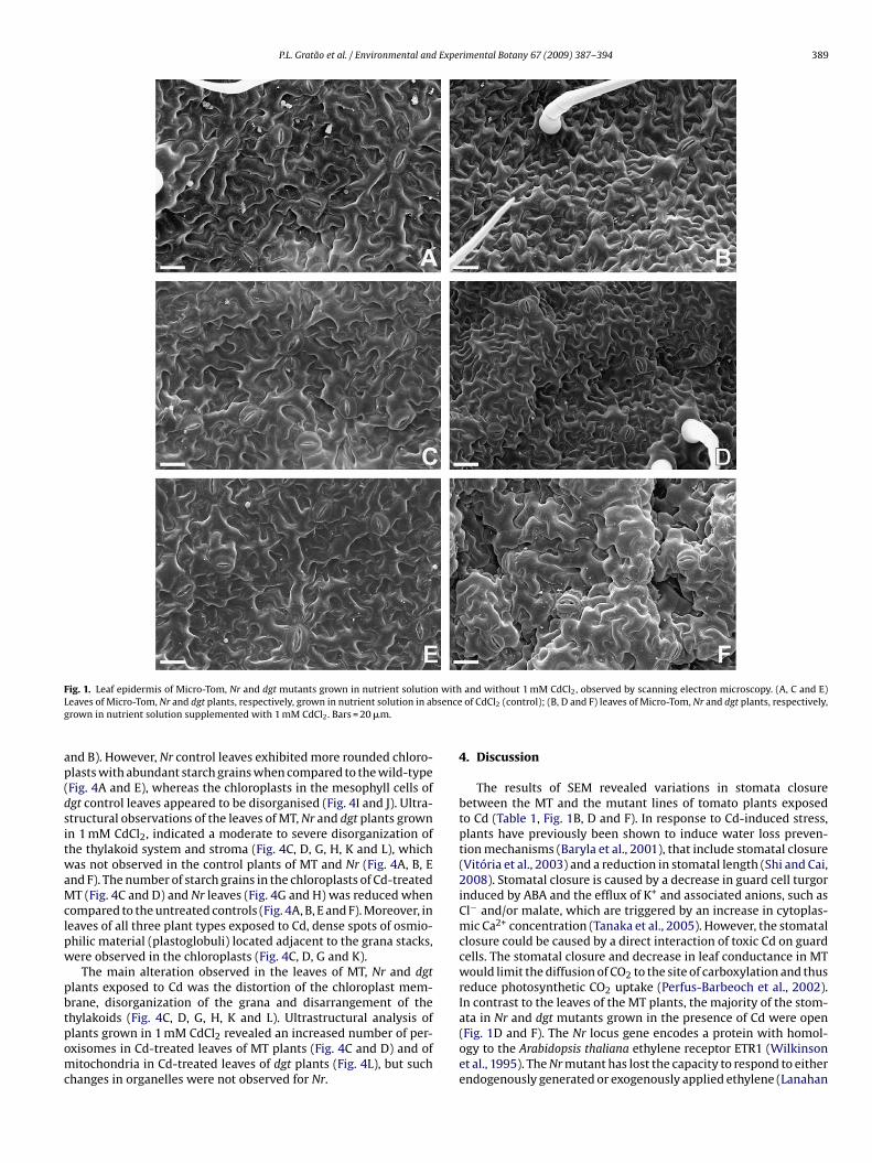
Histological analyses under light microscopy of representativeleaves of Nr and dgt exposed to 1 mM CdCl2 for 40 d, revealedimportant anatomical alterations. These could be seen as a thin-ner leaf blade resulting from smaller palisade parenchyma cells(especially in the second layer) and a decrease in the number andsize of the spongy parenchyma cells (Fig. 2E and H). Cd treat-ment induced an increase in the area of intercellular spaces in Nrleaves (Fig. 2E), but a decrease in the area in dgt leaves (Fig. 2H).No differences were observed in the MT leaves in relation to theintercellular spaces or size of the palisade parenchyma cells, whenthe control plants (Fig. 2A) were compared with Cd-exposed plants(Fig. 2B).
A disorganization of the chloroplasts in the mesophyll cells ofboth untreated mutant controls, Nr (Fig. 2F) and dgt (Fig. 2I) wasnoticeable, when compared to the MT control (Fig. 2C).
Cross-sections of the roots of MT, Nr and dgt controls (zero CdCl2)(Fig. 3A, C and E) exhibited a well-defined epidermis, a cortex com-posed of several layers of turgid highly vacuolated cells and thevascular cylinder. Roots of MT (Fig. 3B) and the Nr mutant (Fig. 3D)exposed to Cd, exhibited considerable anatomical differences whencompared to the controls. MT plants exposed to Cd exhibited adecrease in root diameter, disintegration of the epidermis and themore external cortical cells layers and more conspicuous inter-cellular air spaces (Fig. 3B), when compared to the control plants(Fig. 3A). In roots of Nr (Fig. 3D) exposed to Cd, a greatly reduceddiameter was the most important alteration observed. These effectsfollowing Cd exposure in MT and Nr, were not observed with the
same intensity in dgt roots (Fig. 3F).Distinct differences in chloroplast ultrastructure were observedamong MT, Nr and dgt control plants grown in the absence of Cd.In MT leaves, chloroplasts were elongated with an ellipsoidal mor-phology and a typical arrangement of the grana and stroma (Fig. 4A
P.L. Gratão et al. / Environmental and Experimental Botany 67 (2009) 387–394 389
F withL senceg
ap(dsitwaMclpw
pbtpomc
ig. 1. Leaf epidermis of Micro-Tom, Nr and dgt mutants grown in nutrient solutioneaves of Micro-Tom, Nr and dgt plants, respectively, grown in nutrient solution in abrown in nutrient solution supplemented with 1 mM CdCl2. Bars = 20 �m.
nd B). However, Nr control leaves exhibited more rounded chloro-lasts with abundant starch grains when compared to the wild-typeFig. 4A and E), whereas the chloroplasts in the mesophyll cells ofgt control leaves appeared to be disorganised (Fig. 4I and J). Ultra-tructural observations of the leaves of MT, Nr and dgt plants grownn 1 mM CdCl2, indicated a moderate to severe disorganization ofhe thylakoid system and stroma (Fig. 4C, D, G, H, K and L), whichas not observed in the control plants of MT and Nr (Fig. 4A, B, E
nd F). The number of starch grains in the chloroplasts of Cd-treatedT (Fig. 4C and D) and Nr leaves (Fig. 4G and H) was reduced when
ompared to the untreated controls (Fig. 4A, B, E and F). Moreover, ineaves of all three plant types exposed to Cd, dense spots of osmio-hilic material (plastoglobuli) located adjacent to the grana stacks,ere observed in the chloroplasts (Fig. 4C, D, G and K).
The main alteration observed in the leaves of MT, Nr and dgtlants exposed to Cd was the distortion of the chloroplast mem-rane, disorganization of the grana and disarrangement of the
hylakoids (Fig. 4C, D, G, H, K and L). Ultrastructural analysis oflants grown in 1 mM CdCl2 revealed an increased number of per-xisomes in Cd-treated leaves of MT plants (Fig. 4C and D) and ofitochondria in Cd-treated leaves of dgt plants (Fig. 4L), but suchhanges in organelles were not observed for Nr.
and without 1 mM CdCl2, observed by scanning electron microscopy. (A, C and E)of CdCl2 (control); (B, D and F) leaves of Micro-Tom, Nr and dgt plants, respectively,
4. Discussion
The results of SEM revealed variations in stomata closurebetween the MT and the mutant lines of tomato plants exposedto Cd (Table 1, Fig. 1B, D and F). In response to Cd-induced stress,plants have previously been shown to induce water loss preven-tion mechanisms (Baryla et al., 2001), that include stomatal closure(Vitória et al., 2003) and a reduction in stomatal length (Shi and Cai,2008). Stomatal closure is caused by a decrease in guard cell turgorinduced by ABA and the efflux of K+ and associated anions, such asCl− and/or malate, which are triggered by an increase in cytoplas-mic Ca2+ concentration (Tanaka et al., 2005). However, the stomatalclosure could be caused by a direct interaction of toxic Cd on guardcells. The stomatal closure and decrease in leaf conductance in MTwould limit the diffusion of CO2 to the site of carboxylation and thusreduce photosynthetic CO2 uptake (Perfus-Barbeoch et al., 2002).In contrast to the leaves of the MT plants, the majority of the stom-
ata in Nr and dgt mutants grown in the presence of Cd were open(Fig. 1D and F). The Nr locus gene encodes a protein with homol-ogy to the Arabidopsis thaliana ethylene receptor ETR1 (Wilkinsonet al., 1995). The Nr mutant has lost the capacity to respond to eitherendogenously generated or exogenously applied ethylene (Lanahan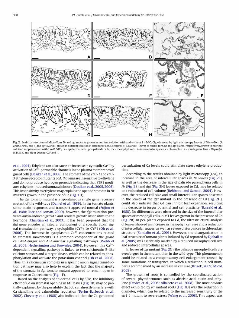
390 P.L. Gratão et al. / Environmental and Experimental Botany 67 (2009) 387–394
Fig. 2. Leaf cross-sections of Micro-Tom, Nr and dgt mutants grown in nutrient solution with and without 1 mM CdCl2, observed by light microscopy. Leaves of Micro-Tom (Aa ontros = mesB
eag3aaTm
msavhdn2tcadcpTtor
etC2
nd C), Nr (D and F) and dgt (G and I) grown in nutrient solution in absence of CdCl2 (colution supplemented with 1 mM CdCl2; e = epidermal cells; pc = palisade cells; mc, D, E, G and H) or 20 �m (C, F and I).
t al., 1994). Ethylene can also cause an increase in cytosolic Ca2+ byctivation of Ca2+-permeable channels in the plasma membranes ofuard cells (Desikan et al., 2006). The stomata of the etr1-1 and etr1-ethylene receptor mutants of A. thaliana are insensitive to ethylenend do not produce hydrogen peroxide indicating that ETR1 medi-tes ethylene-induced stomatal closure (Desikan et al., 2005, 2006).his insensitivity to ethylene may explain the opened stomata in Nrutants grown in the presence of Cd (Fig. 1D).
The dgt tomato mutant is a spontaneous single gene recessiveutant of the wild-type (Daniel et al., 1989). In dgt tomato plants,
ome auxin responses and transport appeared normal (Fujino etl., 1988; Rice and Lomax, 2000), however, the dgt mutation pre-ents auxin-induced growth and renders growth insensitive to theormone (Christian et al., 2003). It has been proposed that thegt gene encodes an integral component of a specific auxin sig-al transduction pathway, a cyclophilin (CYP), Le-CYP1 (Oh et al.,006). The increase in cytoplasmic Ca2+ concentrations relatedo stomatal movements is a common component of the guardell ABA-turgor and ABA-nuclear signalling pathways (Webb etl., 2001; Hetherington and Brownlee, 2004). However, this Ca2+-ependent signalling pathway is linked to two calcineurin B-likealcium sensors and a target kinase, which can be related to phos-horylation and activate the potassium channel (Oh et al., 2008).hus, this calcineurin complex in a specific auxin signal transduc-ion pathway may also help to explain the fact that the majorityf the stomata in dgt tomato mutant appeared to remain open inesponse to Cd treatment (Fig. 1F).
Based on the analysis of epidermal cells by SEM, the inhibitoryffect of Cd on stomatal opening in MT leaves (Fig. 1B) may be par-ially explained by the possibility that Cd can directly interfere witha signalling and calmodulin regulation (Perfus-Barbeoch et al.,002). Cheverry et al. (1988) also indicated that the Cd-generated
l); (B, E and H) leaves of Micro-Tom, Nr and dgt plants, respectively, grown in nutrientophyll cells; i = intercellular spaces; c = chloroplast; s = starch grain. Bars = 50 �m (A,
perturbation of Ca levels could stimulate stress ethylene produc-tion.
According to the results obtained by light microscopy (LM), anincrease in the area of intercellular spaces in Nr leaves (Fig. 2E),as well as the decrease in the size of palisade parenchyma cells inNr (Fig. 2E) and dgt (Fig. 2H) leaves exposed to Cd, may be relatedto a reduction of cell volume (Behboodi and Samadi, 2004). How-ever, the reduced cell size and small intercellular spaces observedin the leaves of the dgt mutant in the presence of Cd (Fig. 2H),could also indicate that Cd can inhibit leaf expansion, resultingin a decrease in turgor potential and cell plasticity (Barceló et al.,1988). No differences were observed in the size of the intercellularspaces or mesophyll cells in MT leaves grown in the presence of Cd(Fig. 2B). In pea plants exposed to Cd, the ultrastructural analysisof leaves showed an increase in mesophyll cell size and a reductionof intercellular spaces, as well as severe disturbances in chloroplaststructure (Sandalio et al., 2001). However, the disorganization inleaf structure of tomato plants induced by Cd reported by Djebali etal. (2005) was essentially marked by a reduced mesophyll cell sizeand reduced intercellular spaces.
In leaves of dgt mutant (Fig. 2G), the palisade mesophyll cells areeven bigger in the mutant than in the wild-type. This phenomenoncould be related to a compensatory cell enlargement caused bysome mutations or transgenes, in which a reduction in cell num-ber is accompanied by an increase in cell size (Krizek, 2009; Micol,2009).
The growth of roots is controlled by the coordinated action
of several phytohormones such as abscisic acid, auxin and ethy-lene (Davies et al., 2005; Albacete et al., 2008). The most obviouseffect exhibited by Nr mutant roots (Fig. 3D) was the reduction indiameter, which can be related to the increased sensitivity of theetr1-1 mutant to severe stress (Wang et al., 2008). This aspect was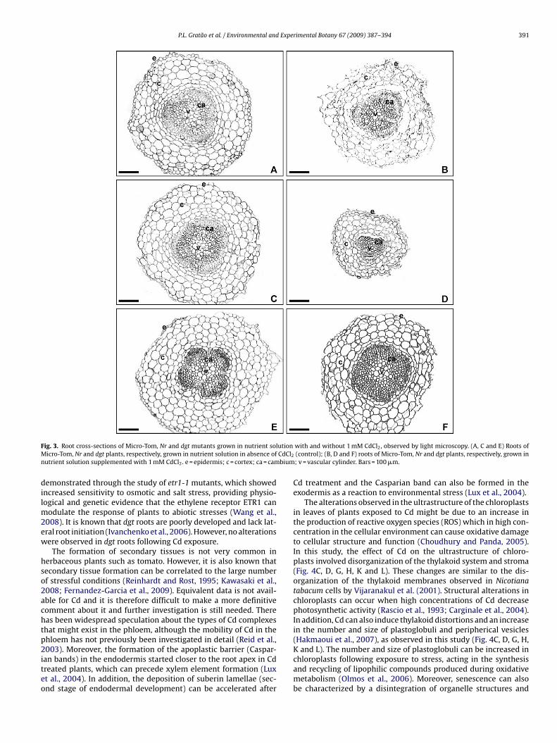
P.L. Gratão et al. / Environmental and Experimental Botany 67 (2009) 387–394 391
F utionM CdCl2
n bium
dilm2ew
hso2achtp2iteo
ig. 3. Root cross-sections of Micro-Tom, Nr and dgt mutants grown in nutrient solicro-Tom, Nr and dgt plants, respectively, grown in nutrient solution in absence of
utrient solution supplemented with 1 mM CdCl2. e = epidermis; c = cortex; ca = cam
emonstrated through the study of etr1-1 mutants, which showedncreased sensitivity to osmotic and salt stress, providing physio-ogical and genetic evidence that the ethylene receptor ETR1 can
odulate the response of plants to abiotic stresses (Wang et al.,008). It is known that dgt roots are poorly developed and lack lat-ral root initiation (Ivanchenko et al., 2006). However, no alterationsere observed in dgt roots following Cd exposure.
The formation of secondary tissues is not very common inerbaceous plants such as tomato. However, it is also known thatecondary tissue formation can be correlated to the large numberf stressful conditions (Reinhardt and Rost, 1995; Kawasaki et al.,008; Fernandez-Garcia et al., 2009). Equivalent data is not avail-ble for Cd and it is therefore difficult to make a more definitiveomment about it and further investigation is still needed. Thereas been widespread speculation about the types of Cd complexeshat might exist in the phloem, although the mobility of Cd in thehloem has not previously been investigated in detail (Reid et al.,
003). Moreover, the formation of the apoplastic barrier (Caspar-an bands) in the endodermis started closer to the root apex in Cdreated plants, which can precede xylem element formation (Luxt al., 2004). In addition, the deposition of suberin lamellae (sec-nd stage of endodermal development) can be accelerated after
with and without 1 mM CdCl2, observed by light microscopy. (A, C and E) Roots of(control); (B, D and F) roots of Micro-Tom, Nr and dgt plants, respectively, grown in; v = vascular cylinder. Bars = 100 �m.
Cd treatment and the Casparian band can also be formed in theexodermis as a reaction to environmental stress (Lux et al., 2004).
The alterations observed in the ultrastructure of the chloroplastsin leaves of plants exposed to Cd might be due to an increase inthe production of reactive oxygen species (ROS) which in high con-centration in the cellular environment can cause oxidative damageto cellular structure and function (Choudhury and Panda, 2005).In this study, the effect of Cd on the ultrastructure of chloro-plasts involved disorganization of the thylakoid system and stroma(Fig. 4C, D, G, H, K and L). These changes are similar to the dis-organization of the thylakoid membranes observed in Nicotianatabacum cells by Vijaranakul et al. (2001). Structural alterations inchloroplasts can occur when high concentrations of Cd decreasephotosynthetic activity (Rascio et al., 1993; Carginale et al., 2004).In addition, Cd can also induce thylakoid distortions and an increasein the number and size of plastoglobuli and peripherical vesicles(Hakmaoui et al., 2007), as observed in this study (Fig. 4C, D, G, H,
K and L). The number and size of plastoglobuli can be increased inchloroplasts following exposure to stress, acting in the synthesisand recycling of lipophilic compounds produced during oxidativemetabolism (Olmos et al., 2006). Moreover, senescence can alsobe characterized by a disintegration of organelle structures and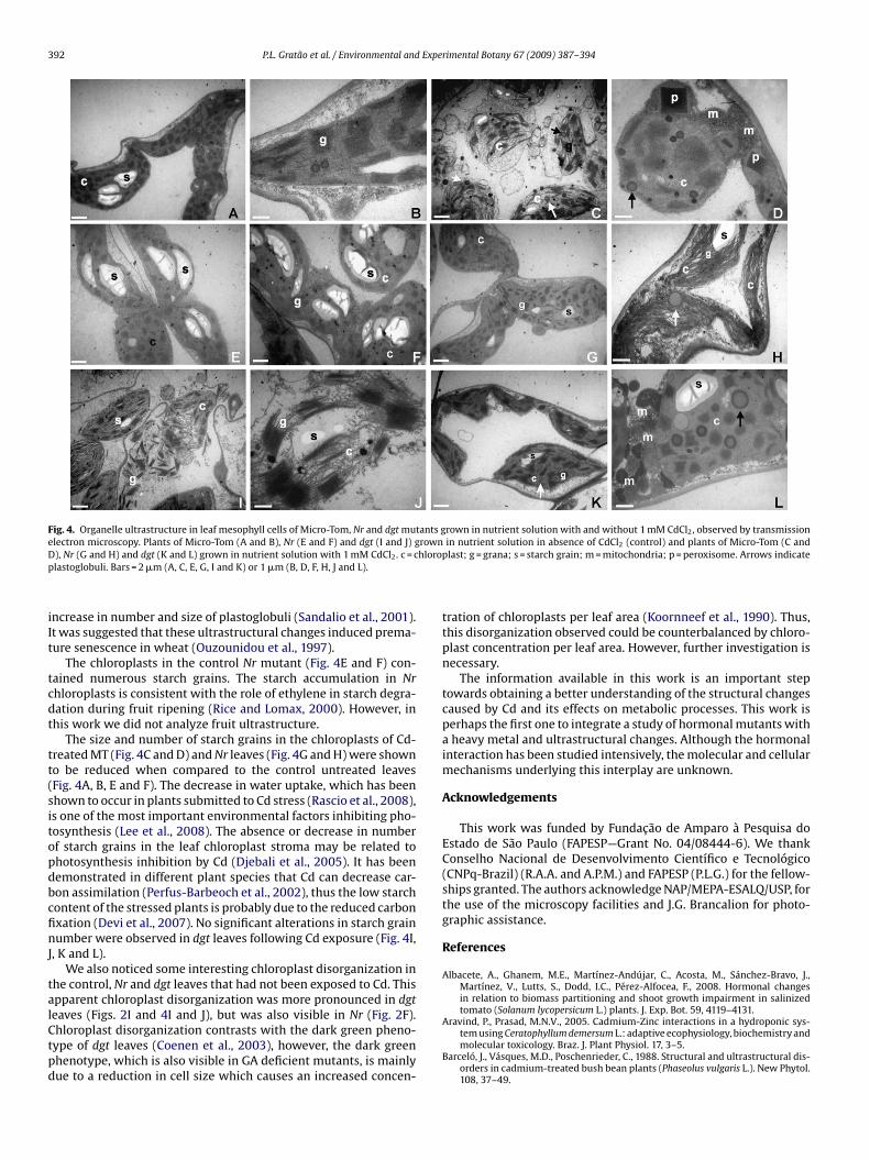
392 P.L. Gratão et al. / Environmental and Experimental Botany 67 (2009) 387–394
Fig. 4. Organelle ultrastructure in leaf mesophyll cells of Micro-Tom, Nr and dgt mutants grown in nutrient solution with and without 1 mM CdCl2, observed by transmissione rownD hlorop
iIt
tcdt
tt(sitopdbcfinJ
talCtpd
lectron microscopy. Plants of Micro-Tom (A and B), Nr (E and F) and dgt (I and J) g), Nr (G and H) and dgt (K and L) grown in nutrient solution with 1 mM CdCl2. c = clastoglobuli. Bars = 2 �m (A, C, E, G, I and K) or 1 �m (B, D, F, H, J and L).
ncrease in number and size of plastoglobuli (Sandalio et al., 2001).t was suggested that these ultrastructural changes induced prema-ure senescence in wheat (Ouzounidou et al., 1997).
The chloroplasts in the control Nr mutant (Fig. 4E and F) con-ained numerous starch grains. The starch accumulation in Nrhloroplasts is consistent with the role of ethylene in starch degra-ation during fruit ripening (Rice and Lomax, 2000). However, inhis work we did not analyze fruit ultrastructure.
The size and number of starch grains in the chloroplasts of Cd-reated MT (Fig. 4C and D) and Nr leaves (Fig. 4G and H) were showno be reduced when compared to the control untreated leavesFig. 4A, B, E and F). The decrease in water uptake, which has beenhown to occur in plants submitted to Cd stress (Rascio et al., 2008),s one of the most important environmental factors inhibiting pho-osynthesis (Lee et al., 2008). The absence or decrease in numberf starch grains in the leaf chloroplast stroma may be related tohotosynthesis inhibition by Cd (Djebali et al., 2005). It has beenemonstrated in different plant species that Cd can decrease car-on assimilation (Perfus-Barbeoch et al., 2002), thus the low starchontent of the stressed plants is probably due to the reduced carbonxation (Devi et al., 2007). No significant alterations in starch grainumber were observed in dgt leaves following Cd exposure (Fig. 4I,
, K and L).We also noticed some interesting chloroplast disorganization in
he control, Nr and dgt leaves that had not been exposed to Cd. Thispparent chloroplast disorganization was more pronounced in dgt
eaves (Figs. 2I and 4I and J), but was also visible in Nr (Fig. 2F).hloroplast disorganization contrasts with the dark green pheno-ype of dgt leaves (Coenen et al., 2003), however, the dark greenhenotype, which is also visible in GA deficient mutants, is mainlyue to a reduction in cell size which causes an increased concen-in nutrient solution in absence of CdCl2 (control) and plants of Micro-Tom (C andplast; g = grana; s = starch grain; m = mitochondria; p = peroxisome. Arrows indicate
tration of chloroplasts per leaf area (Koornneef et al., 1990). Thus,this disorganization observed could be counterbalanced by chloro-plast concentration per leaf area. However, further investigation isnecessary.
The information available in this work is an important steptowards obtaining a better understanding of the structural changescaused by Cd and its effects on metabolic processes. This work isperhaps the first one to integrate a study of hormonal mutants witha heavy metal and ultrastructural changes. Although the hormonalinteraction has been studied intensively, the molecular and cellularmechanisms underlying this interplay are unknown.
Acknowledgements
This work was funded by Fundacão de Amparo à Pesquisa doEstado de São Paulo (FAPESP—Grant No. 04/08444-6). We thankConselho Nacional de Desenvolvimento Científico e Tecnológico(CNPq-Brazil) (R.A.A. and A.P.M.) and FAPESP (P.L.G.) for the fellow-ships granted. The authors acknowledge NAP/MEPA-ESALQ/USP, forthe use of the microscopy facilities and J.G. Brancalion for photo-graphic assistance.
References
Albacete, A., Ghanem, M.E., Martínez-Andújar, C., Acosta, M., Sánchez-Bravo, J.,Martínez, V., Lutts, S., Dodd, I.C., Pérez-Alfocea, F., 2008. Hormonal changesin relation to biomass partitioning and shoot growth impairment in salinizedtomato (Solanum lycopersicum L.) plants. J. Exp. Bot. 59, 4119–4131.
Aravind, P., Prasad, M.N.V., 2005. Cadmium-Zinc interactions in a hydroponic sys-tem using Ceratophyllum demersum L.: adaptive ecophysiology, biochemistry andmolecular toxicology. Braz. J. Plant Physiol. 17, 3–5.
Barceló, J., Vásques, M.D., Poschenrieder, C., 1988. Structural and ultrastructural dis-orders in cadmium-treated bush bean plants (Phaseolus vulgaris L.). New Phytol.108, 37–49.
Expe
B
B
B
C
C
C
C
C
D
D
D
D
D
D
F
F
F
F
G
G
G
G
G
H
H
H
I
K
K
K
P.L. Gratão et al. / Environmental and
aryla, A., Carrier, P., Franck, F., Coulomb, C., Sahut, C., Havaux, M., 2001. Leafchlorosis in oilseed rape plants (Brassica napus) grown on cadmium-pollutedsoil: causes and consequences for photosynthesis and growth. Planta 212,696–709.
ehboodi, B.S.H., Samadi, L., 2004. Detection of apoptotic bodies and oligonucleoso-mal DNA fragments in cadmium-treated root apical cells of Allium cepa Linnaeus.Plant Sci. 167, 411–416.
enavides, M.P., Gallego, S.M., Tomaro, M.L., 2005. Cadmium toxicity in plants. Braz.J. Plant Physiol. 17, 21–34.
arginale, V., Sorbo, S., Capasso, C., Trinchella, F., Cafiero, G., Basile, A., 2004. Accu-mulation, localization, and toxic effects of cadmium in the liverwort Lunulariacruciata. Protoplasma 223, 53–61.
heverry, J.L., Pouliquean, J., Le Guyader, H., Marcellin, P., 1988. Calcium regulationof exogenous and endogenous 1-aminocyclopropane-1-carboxylic acid biocon-version to ethylene. Physiol. Plant. 74, 53–57.
houdhury, S., Panda, S.K., 2005. Toxic effects, oxidative stress and ultrastructuralchanges in moss Taxithelium nepalense (Schwaegr.) Broth. under chromium andlead phytotoxicity. Water, Air, Soil Pollut. 167, 73–90.
hristian, M., Steffens, B., Schenck, D., Lüthen, H., 2003. The diageotropica mutationof tomato disrupts a signalling chain using extracellular auxin binding protein 1as a receptor. Planta 218, 309–314.
oenen, C., Christian, M., Lüthen, H., Lomax, T., 2003. Cytokinin inhibits a subset ofdiageotropica-dependent primary auxin responses in tomato. Plant Physiol. 131,1692–1704.
aniel, S.G., Rayle, D.L., Cleland, R.E., 1989. Auxin physiology of the tomato mutantdiageotropica. Plant Physiol. 91, 804–807.
avies, W.J., Kudoyarova, G., Hartung, W., 2005. Long-distance ABA signaling andits relation to other signaling pathways in the detection of soil drying and themediation of the plants response to drought. J. Plant Growth Regul. 24, 285–295.
esikan, R., Hancock, J.T., Bright, J., Harrison, J., Weir, I., Hooley, R., Neil, S.J., 2005.A role for ETR1 in hydrogen peroxide signaling in stomatal guard cells. PlantPhysiol. 137, 831–834.
esikan, R., Last, K., Harrett-Williams, R., Tagliavia, C., Harter, K., Hooley, R., Hancock,J.T., Neil, S.J., 2006. Ethylene-induced stomatal closure in Arabidopsis occurs viaAtrbohF- mediated hydrogen peroxide synthesis. Plant J. 47, 907–916.
evi, R., Munjral, N., Gupta, A.K., Kaur, N., 2007. Cadmium induced changes in carbo-hydrate status and enzymes of carbohydrate metabolism, glycolysis and pentosephosphate pathway in pea. Environ. Exp. Bot. 61, 167–174.
jebali, W., Zarrouk, M., Brouquisse, R., El Kahoui, S., Limam, F., Ghorbel, H., Chaibi,W., 2005. Ultrastructure and lipid alterations induced by cadmium in tomato(Lycopersicon sculentum) chloroplast membranes. Plant Biol. 7, 358–368.
ernandez-Garcia, N., Lopez-Perez, L., Hernandez, M., Olmos, E., 2009. Role of phicells and the endodermis under salt stress in Brassica oleracea more options.New Phytol. 181, 347–360.
erreira, R.R., Fornazier, R.F., Vitoria, A.P., Lea, P.J., Azevedo, R.A., 2002. Changes inantioxidant enzyme activities in soybean under cadmium stress. J. Plant Nutr.25, 327–342.
ornazier, R.F., Ferreira, R.R., Vitoria, A.P., Molina, S.M.G., Lea, P.J., Azevedo, R.A., 2002.Effects of cadmium on antioxidant enzyme activities in sugar-cane. Biol. Plant.42, 91–97.
ujino, D.W., Nissen, S.J., Jones, A.D., Burger, D.W., Bradford, K.J., 1988. Quantificationof indole-3-acetic acid in dark-grown seedlings of diageotropica and epinasticmutants of tomato. Plant Physiol. 88, 780–784.
omes-Junior, R.A., Moldes, C.A., Delite, F.S., Pompeu, G.B., Gratão, P.L., Mazzafera, P.,Lea, P.J., Azevedo, R.A., 2006. Antioxidant metabolism of coffee cell suspensioncultures in response to cadmium. Chemosphere 65, 1330–1337.
ratão, P.L., Polle, A., Lea, P.J., Azevedo, R.A., 2005. Making the life of heavy metal-stressed plants a little easier. Funct. Plant Biol. 32, 481–494.
ratão, P.L., Monteiro, C.C., Antunes, A.M., Peres, L.E.P., Azevedo, R.A., 2008a. Acquiredtolerance of tomato (Lycopersicon esculentum cv. Micro-Tom) plants to cadmium-induced stress. Ann. Appl. Biol. 153, 321–333.
ratão, P.L., Monteiro, C.C., Peres, L.E.P., Azevedo, R.A., 2008b. The isolation of antiox-idant enzymes from mature tomato (cv. Micro-Tom) plants. Hortscience 43,1608–1610.
ratão, P.L., Pompeu, G.B., Capaldi, F.R., Vitorello, V.A., Lea, P.J., Azevedo, R.A., 2008c.Antioxidant response of Nicotiana tabacum cv. Bright Yellow 2 cells to cadmiumand nickel stress. Plant Cell Tissue Org. 94, 73–83.
akmaoui, A., Ater, M., Boka, K., Baron, M., 2007. Copper and cadmium tolerance,uptake and effect on chloroplast ultrastructure. Studies on Salix purpurea andPhragmites australis. Zeitschrift fur Naturforschung C-A. J. Biosci. 62, 417–426.
etherington, A.M., Brownlee, C., 2004. The generation of Ca2+ signals in plants.Annu. Rev. Plant Biol. 55, 401–427.
oagland, D., Arnon, D.I., 1950. In: Hoagland, D., Arnon, D.I. (Eds.), The Water Cul-ture Method for Growing Plants Without Soil. California Agricultural ExperimentStation, Berkeley.
vanchenko, M.G., Coffeen, W.C., Lomax, T.L., Dubrovsky, J.G., 2006. Mutations inthe diageotropica (Dgt) gene uncouple patterned cell division during lateral rootinitiation from proliferative cell division in the pericycle. Plant J. 46, 436–447.
awasaki, M., Takatsuji, A., Taniguchi, M., Miyake, H., 2008. Localization of casparianbands and crystall cells in relation to aluminum distribution in the primary root
of Eddo under aluminum treatment. Plant Prod. Sci. 11, 238–242.elly, M.O., Bradford, K.J., 1986. Insensitivity of the diageotropica tomato mutant toauxin. Plant Physiol. 82, 713–717.
oornneef, M., Bosma, T.D.G., Hanhart, C.J., Van der Veen, J.H., Zeevart, J.A.D., 1990.The isolation and characterization of gibberellin-deficient mutants in tomato.Theor. Appl. Genet. 80, 852–857.
rimental Botany 67 (2009) 387–394 393
Krizek, B.A., 2009. Making bigger plants: key regulators of final organ size. Curr. Opin.Plant Biol. 12, 17–22.
Lage-Pinto, F., Oliveira, J.G., Da Cunha, M., Souza, C.M.M., Rezende, C.E., Azevedo,R.A., Vitória, A.P., 2008. Chlorophyll a fluorescence and ultrastructural changesin chloroplast of water hyacinth as indicators of environmental stress. Environ.Exp. Bot. 64, 307–313.
Lanahan, M.B., Yen, H.C., Giovannoni, J.J., Klee, H., 1994. The Never ripe mutationblocks ethylene perception in tomato. Plant Cell 6, 521–530.
Lee, B., Jina, Y., Jung, W., Avice, J., Morvan-Bertrand, A., Ourry, A., Park, C., Kim,T., 2008. Water-deficit accumulates sugars by starch degradation – not by denovo synthesis – in white clover leaves (Trifolium repens). Physiol. Plant. 134,403–411.
Lima, J.E., Carvalho, R.F., Neto, A.T., Figueira, A., Peres, L.E.P., 2004. Micro-MsK: atomato genotype with miniature size, short life cycle, and improved in vitroshoot regeneration. Plant Sci. 167, 753–757.
Lin, Z.F., Arciga-Reyes, L., Zhong, S.L., Alexander, L., Hackett, R., Wilson, I., Grierson,D., 2008. SlTPR1, a tomato tetratricopeptide repeat protein, interacts with theethylene receptors NR and LeETR1, modulating ethylene and auxin responsesand development. J. Exp. Bot. 59, 4271–4287.
Lindberg, S., Landberg, T., Greger, M., 2007. Cadmium uptake and interaction withphytochelatins in wheat protoplasts. Plant Physiol. Biochem. 45, 47–53.
Liu, D., Kottke, I., 2004. Subcellular localization of cadmium in the root cells ofAllium cepa by electron energy loss spectroscopy and cytochemistry. J. Biosci.29, 329–335.
Lux, A., Sttníková, A., Opatrná, J., Greger, M., 2004. Differences in structure ofadventitious roots in Salix clones with contrasting characteristics of cadmiumaccumulation and sensitivity. Physiol. Plant. 120, 537–545.
Meissner, R., Jacobson, Y., Melamed, S., Levyatuv, S., Shalev, G., Ashri, A., Elkind, Y.,Levy, A., 1997. A new model system for tomato genetics. Plant J. 12, 1465–1472.
Micol, J.L., 2009. Leaf development: time to turn over a new leaf? Curr. Opin. PlantBiol. 12, 9–16.
Mobin, M., Khan, N.A., 2007. Photosynthetic activity, pigment composition andantioxidative response of two mustard (Brassica juncea) cultivars differing inphotosynthetic capacity subjected to cadmium stress. J. Plant Physiol. 164,601–610.
Oh, K., Ivanchenko, M.G., White, T.J., Lomax, T.L., 2006. The diageotropica gene oftomato encodes a cyclophilin: a novel player in auxin signalling. Planta 224,133–144.
Oh, S.I., Park, J., Yoon, S., Kim, Y., Park, S., Ryu, M., Nam, M.J., Ok, S.H., Kim, J.K., Shin,J.S., Kim, K.N., 2008. The Arabidopsis calcium sensor calcineurin B-like 3 inhibitsthe 5′-methylthioadenosine nucleosidase in a calcium-dependent manner. PlantPhysiol. 148, 1883–1896.
Olmos, E., Kiddle, G., Pellny, T.K., Kumar, S., Foyer, C.H., 2006. Modulation of plantmorphology, root architecture, and cell structure by low vitamin C in Arabidopsisthaliana. J. Exp. Bot. 57, 1645–1655.
Ouzounidou, G., Moustakas, M., Eleftheriou, E.P., 1997. Physiological and ultrastruc-tural effects of cadmium on wheat (Triticum aestivum L.) leaves. Arch. Environ.Contam. Toxicol. 32, 154–160.
Paiva, L.B., Oliveira, J.G., Azevedo, R.A., Ribeiro, D.R., Silva, M.G., Vitória, A.P., 2009.Ecophysiological responses of water hyacinth exposed to Cr3+ and Cr6+. Environ.Exp. Bot. 65, 403–409.
Perfus-Barbeoch, L., Leonhardt, N., Vavasseur, A., Forestier, C., 2002. Heavy metaltoxicity: cadmium permeates through calcium channels and disturbs the plantwater status. Plant J. 32, 539–548.
Rascio, N., Dallavechia, F., Ferretti, M., Merlo, L., Chisi, R., 1993. Some effects of cad-mium on maize plants. Arch. Environ. Contam. Toxicol. 25, 244–249.
Rascio, N., Vecchia, F.D., La Rocca, N., Barbato, R., Pagliano, C., Raviolo, M., Gonnelli,C., Gabbrielli, R., 2008. Metal accumulation and damage in rice (cv. Vialone nano)seedlings exposed to cadmium. Environ. Exp. Bot. 62, 267–278.
Reid, R.J., Dunbar, K.R., Mclaughlin, M.J., 2003. Cadmium loading into potato tubers:the roles of the periderm, xylem and phloem. Plant Cell Environ. 26, 201–206.
Reinhardt, D.H., Rost, T.L., 1995. Salinity accelerates endodermal development andinduces an exodermis in cotton seedling roots. Environ. Exp. Bot. 35, 563–574.
Reynolds, E.S., 1963. The use of lead citrate at high pH as an electron-opaque stainin electron microscopy. J. Cell Biol. 17, 208–212.
Rice, M.S., Lomax, T.L., 2000. The auxin-resistant diageotropica mutant of tomatoresponds to gravity via an auxin-mediated pathway. Planta 210, 906–913.
Sandalio, L.M., Dalurzo, H.C., Gomez, M., Romero-Puertas, M.C., del Rio, L.A., 2001.Cadmium-induced changes in the growth and oxidative metabolism of peaplants. J. Exp. Bot. 52, 2115–2126.
Shi, G.R., Cai, Q.S., 2008. Photosynthetic and anatomic responses of peanut leaves tocadmium stress. Photosynthetica 46, 627–630.
Smeets, K., Ruytinx, J., Semane, B., Van Belleghem, F., Remans, T., Van Sanden, S.,Vangronsveld, J., Cuypers, A., 2008. Cadmium-induced transcriptional and enzy-matic alterations related to oxidative stress. Environ. Exp. Bot. 63, 1–8.
Stoyanova, D.P., Tchakalova, E.S., 1997. Cadmium-induced ultrastructural changes inchloroplasts of the leaves and stems parenchyma in Myriophyllum spicatum L.Photosynthetica 34, 241–248.
Tamas, L., Dudikova, J., Durcekova, K., Huttova, J., 2008. The impact of heavy metalson the activity of some enzymes along the barley root. Environ. Exp. Bot. 62,
86–91.Tanaka, Y., Sano, T., Tamaoki, M., Nakajima, N., Kondo, N., Hasezawa, S., 2005. Ethyleneinhibits abscisic acid-induced stomatal closure in Arabidopsis. Plant Physiol. 138,2337–2343.
Teklic, T., Engler, M., Cesar, V., Lepedus, H., Paraikovic, N., Loncaric, Z., Stolfa, I.,Marotti, T., Mikac, N., Zarkovic, N., 2008. Influence of excess copper on lettuce

3 Expe
T
V
V
V
V
94 P.L. Gratão et al. / Environmental and
(Lactuca sativa L.) grown in soil and nutrient solution. J. Food Agric. Environ. 6,439–444.
ukaj, Z., Bascik-Remisiewicz, A., Skowronski, T., Tukaj, C., 2007. Cadmium effect onthe growth, photosynthesis, ultrastructure and phytochelatin content of greenmicroalga Scenedesmus armatus: a study at low and elevated CO2 concentration.Environ. Exp. Bot. 60, 291–299.
assilev, A., Lidon, F., Campos, P.S., Ramalho, J.C., Barreiro, M.G., Yordanov, I., 2003.Cu-induced changes in chloroplast lipids and photosystem 2 activity in barleyplants. Bulg. J. Plant Physiol. 29, 33–43.
ijaranakul, U., Jayaswal, R.K., Nadakavukaren, M.J., 2001. Alteration in chloroplast
ultrastructure of suspension cultured Nicotiana tabacum cells by cadmium. Sci.Asia 27, 227–231.incent, J.M., 1975. Manual practico de rizobiologia, 1st ed. Hemisfério Sur, BuenosAires, p. 200.
itória, A.P., Da Cunha, M., Azevedo, R.A., 2006. Ultrastructural changes of radish leafexposed to cadmium. Environ. Exp. Bot. 58, 47–52.
rimental Botany 67 (2009) 387–394
Vitória, A.P., Rodriguez, A.P.M., Cunha, M., Lea, P.J., Azevedo, R.A., 2003. Structuralchanges in radish seedlings exposed to cadmium. Biol. Plant. 47, 561–568.
Wahid, A., Ghani, A., 2008. Varietal differences in mung bean (Vigna radiata) forgrowth, yield, toxicity symptoms and cadmium accumulation. Ann. Appl. Biol152, 59–69.
Wang, Y., Wang, T., Li, K., Li, X., 2008. Genetic analysis of involvement of ETR1 in plantresponse to salt and osmotic stress. Plant Growth Regul. 54, 261–269.
Webb, A.A.R., Larman, M.G., Montgomery, L.T., Taylor, J.E., Hetherington, A.M., 2001.The role of calcium in ABA-induced gene expression and stomatal movements.Plant J. 26, 351–362.
Wilkinson, J.Q., Lanahan, M.B., Yen, H.C., Giovannoni, J.J., Klee, H.J., 1995. An ethyleneinducible component of signal transduction encoded by Never ripe. Science 270,1807–1809.
Zsögön, A., Lambais, M.R., Benedito, V.A., Figueira, A.V.O., Peres, L.E.P., 2008. Reducedarbuscular mycorrhizal colonization in tomato ethylene mutants. Sci. Agr. 65,259–267.
Copyright © 2022 FDOKUMEN