Ultrastructural characterisation of melanogenesis in adult ...
116
Ultrastructural characterisation of melanogenesis in adult human retinal pigment epithelial cells after adenoviral transduction with the tyrosinase gene der Fakultät für Biologie der EBERHARD KARLS UNIVERSITÄT TÜBINGEN zur Erlangung des Grades eines Doktors der Naturwissenschaften von Antje Kristina Biesemeier aus Gifhorn vorgelegte DISSERTATION 2010
-
Upload
khangminh22 -
Category
Documents
-
view
1 -
download
0
Transcript of Ultrastructural characterisation of melanogenesis in adult ...
Ultrastructural characterisation of melanogenesis in adult human retinal pigment epithelial cells after adenoviral
transduction with the tyrosinase gene
der Fakultät für Biologie
der EBERHARD KARLS UNIVERSITÄT TÜBINGEN
zur Erlangung des Grades eines Doktors
der Naturwissenschaften
von
Antje Kristina Biesemeier
aus Gifhorn
vorgelegte
DISSERTATION
2010


Tag der mündlichen Prüfung: 11.11.2009
Dekan der Fakultät für Biologie: Prof. Dr. Hanspeter Mallot
1. Berichterstatter Prof. Dr. Ulrich Schraermeyer
2. Berichterstatter Prof. Dr. Hanspeter Mallot

Danksagung
Im Folgenden möchte ich allen nachstehenden Personen mein ganz persönliches Dankeschön entbieten:
Prof. Dr. Ulrich Schraermeyer danke ich für die Überlassung des interessanten Themas meiner Arbeit und für die freundliche Betreuung und wertvollen wissenschaftlichen Hilfestellungen, Anregungen und Diskussionen in dieser sehr intensiven Zeit.
Prof. Dr. Hanspeter Mallot, Prof. Dr. Konrad Kohler, Prof. Dr. Hans-Georg Rammensee und dem verstorbenen Prof. Dr. Werner Schmidt danke ich für die Betreuung meiner Arbeit von Seiten der biologischen Fakultät.
Dr. Petra Blitgen-Heinecke danke ich für die HPLC-Analysen. Der AG Berneburg (Hautklinik) danke ich für die Hilfe bei den UV-Versuchen und der AG Rodemann (Radioonkologie) danke ich für die Benutzung des Röntgengenerators.
Dr. Peter Heiduschka und Dr. Sylvie Julien danke für die vielen Male, in denen sie mir aus wissenschaftlichen Klemmen geholfen haben, für das Korrekturlesen diverser Manuskripte und nicht zuletzt dieser Arbeit.
Sigrid Schultheiß verdanke ich mein Wissen über die Geheimnisse der Elektronenmikroskopie.
Judith Birch, my particular gratitude to you for your corrections of my “German English” and for you always being helpful und friendly whenever I entered your office.
Moni Rittgarn, Du warst mir immer eine liebe und geduldige Begleiterin, ob unter der Sterilbank, beim Augenpulen oder beim Kiesern. Danke.
Sabine Hofmeister, Dir danke ich für die eine oder andere Färbung, die Du mir abgenommen hast und für die netten Plaudereien in den Inkubationspausen.
Dr. Barbara Wallenfels-Thilo, Dr. Sigrid Henke-Fahle und den Histodamen Christl Fischer-Lamprecht, Claudia Riedinger und Monika Wild danke ich für die stete Hilfe bei allen Fragen rund ums Labor.
Sven Schnichels, Dir danke ich für die nette Zeit, die wir zusammen in unserem kleinen Büro brütend, lesend, schreibend und redent verbracht haben. Toitoitoi für den Endspurt!
Martin Spitzer, Focke Ziemsen, Charly Frank, Kai Januschowski, Qi Zhu, und all den anderen Medizinern im Breuninger Bau danke ich für die Kollegialität und Freundschaft, die sie mir entgegen gebracht haben. Weiterhin danke ich allen ehemaligen Mitarbeitern, den übrigen Ärzten, Pförtnern, „Werkstättlern“ und sämtlichen wissenschaftlichen und nicht-wissenschaftlichen Kollegen für die schöne Zeit im „Breuninger Bau“.
Außerdem danke ich Euch allen für die amüsanten privaten Gespräche bei einer Tasse Kaffee oder beim Kegeln, Langlaufen und diversen Feiern.
Mein herzlichstes Dankeschön ergeht an alle meine Freunde in Tübingen, Erlangen und Ingolstadt und an meine Eltern, ohne eure seelische und moralische Unterstützung wäre diese Arbeit nicht möglich gewesen.

Summary
1
Summary
Until recently, it was widely accepted that melanogenesis does not occur in the adult
retinal pigment epithelium (RPE), since the typical hallmarks of melanogenesis, the
premelanosome and the expression of melanogenic proteins like tyrosinase and
melanocyte-associated protein 17 (PMEL17), were absent post-natal. In the
meantime, active tyrosinase has been observed in the adult RPE of different animal
species, e.g. after phagocytosis of retinal photoreceptor outer segments (ROS). The
aim of this thesis was to investigate whether melanogenesis can be induced in adult
human RPE cells in response to ROS phagocytosis or after transduction with a
tyrosinase vector. The role of the melanogenic proteins PMEL17 and TRP1 and the
classical melanosomal stages, known from pre-natal melanin synthesis, were also to
determine.
As a model system tyrosinase transduced amelanotic RPE cells were used to study
tyrosinase function and melanogenesis and the influence of phagocytosis in adult
RPE. The presences of the melanogenic proteins tyrosinase, tyrosinase-related
protein 1 (TRP1) and PMEL17 were investigated using immunocytochemistry.
Tyrosinase activity and loclisation was further studied with electron microscopical
DOPA histochemistry. The ultrastructural morphology of melanogenic stages was
compared to that of pigmented melanoma cells (MNT-1), which were used as a
positive control for typical melanogenesis. Melanin synthesis was detected with
HPLC analysis. MTT tests confirmed that viability was not affected after tyrosinase
transduction and melanin synthesis. Post-natal RPE melanogenesis was also studied
in animal experiments after subretinal injection of the tyrosinase vector in rats and
rabbits.
Compared to controls, tyrosinase active cells had a redifferentiated cobblestone
morphology, were pigmented and had an improved phagocytosis rate. Tyrosinase
trafficking was different to the classical model found in MNT-1 cells, since a DOPA
reaction was not observed in Golgi-derived vesicles, but as membrane-less, small
DOPA granules free-floating in the cytoplasm. Melanogenesis occurred without the
involvement of TRP1, PMEL17 and typical striated premelanosomes and
melanogenic stages. In contrast, melanin was synthesised in lysosome-like
organelles. Thus, a new pathway of melanogenesis is described for this model
system. Transduced RPE cells of living rats and rabbits and cultured cells of human
Summary
2
donors showed also a similar morphology of melanogenesis as the ARPE-19 cells.
Interestingly, control MNT-1 cells contained similar melanosomes in addition to the
classical stages of melanogenesis.
Although a transport of ingested material to newly-formed ARPE-19 melanosomes
was not observed, phagocytosis led to an improved tyrosinase activity and to an
accelerated melanogenesis, compared to non-fed transduced cells.
In conclusion, tyrosinase transduction in combination with phagocytosis led to a
morphological reorganisation and functional improvement of cultured ARPE-19 cells.
Additionally, melanogenesis has been induced, which is independent of
premelanosome formation. It can be transferred to the in vivo situation by gene
therapy.

Table of contents
3
Table of contents
SUMMARY ............................................................................................................................................ 1
TABLE OF CONTENTS ...................................................................................................................... 3
I. GLOSSARY AND ABBREVIATIONS ............................................................................................ 5
II. INTRODUCTION ............................................................................................................................ 9
1. The retinal pigment epithelium .................................................................................................................... 9
2. Melanogenesis ........................................................................................................................................... 112.1 Melanosomal proteins ................................................................................................................................ 122.2 The Raper-Mason Scheme of melanin formation ....................................................................................... 142.3 Cellular actions– the 4-stage model of eumelanogenesis .......................................................................... 162.4 The premelanosome ................................................................................................................................... 16
3. Age-related changes in the RPE - Lipofuscin ............................................................................................... 17
4. Previous findings ........................................................................................................................................ 19
5. Aim of the thesis ........................................................................................................................................ 22
III. SUMMARISED RESULTS OF THE MANUSCRIPTS ENCLOSED ..................................... 24
Manuscript 1: ................................................................................................................................................. 24
Manuscript 2: ................................................................................................................................................. 26
Manuscript 3: ................................................................................................................................................. 28
Manuscript 4: ................................................................................................................................................. 32
IV. DISCUSSION ............................................................................................................................... 35
Age pigments impair the antioxidative capacity of melanin and zinc in aged RPE cells .................................. 35
Involvement of ROS phagocytosis in tyrosinase expression of adult human RPE cells .................................... 35
Phagocytosis of ROS enhances melanogenesis in tyrosinase transduced ARPE-19 cells ................................. 36
Tyrosinase protects against oxidative stress after phagocytosis ..................................................................... 37
Improvement of RPE characteristics after tyrosinase transduction ................................................................ 38
The premelanosome and the classical pathway of melanogenesis are not involved in ARPE-19 cell melanogenesis ............................................................................................................................................... 39
Table of contents
4
Findings from animal experiments ................................................................................................................. 42
Tyrosinase as a possible application in AMD therapy ..................................................................................... 43
V. REFERENCES ............................................................................................................................... 46
VI. TABLE OF FIGURES .................................................................................................................. 53
VII. APPENDIX ................................................................................................................................. 54
1. Erklärung zum Eigenanteil an den Manuskripten ....................................................................................... 54
2. Publikationen ............................................................................................................................................. 56
3. Manuskripte ............................................................................................................................................... 58
I. Glossary and Abbreviations
5
I. Glossary and Abbreviations
AdTyr: an adenoviral vector, carrying the human tyrosinase cDNA under a hCMV-
promoter in the E1 (envelope 1)-deleted region
AMD: Age-related macular degeneration is the main cause of blindness in the elderly
population (60+) of the western world: In early stages, lipofuscin and drusen
accumulate between the retinal pigment epithelium (RPE) and Bruch’s membrane.
Later stages can be divided in to two different types, the dry and the wet form: 1)
“dry”, non-exudative AMD: Atrophic changes in the macula region lead to visual
impairment, but the lesions are not as severe as the lesions in “wet” AMD. Therapies
for dry AMD are still lacking. 2) The “wet”, exudative form affects about 15% of AMD
patients. Neovascularisation of leaking vessels from the choriocappilaris into the
retina leads to enhanced RPE and retinal detachment. These vessels have a greater
tendency to leakage and bleeding into the macula, ultimately leading to irreversible
damage to the photoreceptors. Laser coagulation and medication that minimises
oxidative stress and blood vessel outgrowth build the main strategies of treatment,
but they can only attenuate vision loss.
AP: the adaptor-protein complexes transport coated vesicles from the Golgi
apparatus (AP-1, 3, 4), or from the cell membrane (AP-2) to their destination
organelles.
ARPE-19: is an immortalised, highly differentiated amelanotic (pigment-less) RPE
cell line, spontaneously arisen from primary pigmented RPE cells of a 19-year-old
male human donor. ARPE-19 cells express typical RPE marker proteins like
CRALBP (cellular retinaldehyde-binding protein), MERTK (mer-tyrosine kinase) and
RPE65 (retinal pigment epithelium-specific protein with 65kDa), are highly polarised
and keep their epithelial and phagocytic functions even with high passages.
Bruch’s membrane: is the border between the retina and the choroid; it is formed by
the basal membranes of the RPE and the vessels of the choriocapillaris and contains
several layers of collagen and elastic fibres.
Choriocapillaris: highly fenestrated blood vessels at the apical border of the choroid
DHI: 5,6-dihydroxyindol is a melanin precursor in eumelanogenesis.
I. Glossary and Abbreviations
6
DHICA: 5,6-dihydroxyindol-2-carboxylic acid is a melanin precursor in eumelano-
genesis.
DOPA: L-dihydroxyphenylalanine is a melanin precursor and the second substrate in
eu- and phaeomelanogenesis.
DOPA reaction: an electron microscopical method, in which fixed tissue/cells can be
checked for tyrosinase activity. Incubation of the fixed material with the substrate L-
DOPA leads to melanin formation at sites where active tyrosinase is present. Thus,
the localisation of the enzyme can be documented ultrastructurally.
Endosome: a class of clathrin-coated vesicles; e.g. phagosomes and Golgi-derived vesicles
ER: endoplasmic reticulum, rER rough ER; sER smooth ER
Eumelanin: black or brown melanin composed of variable amounts of DHI and
DHICA monomers
hRPE: primary human RPE cells
LAMP-1: lysosome-associated membrane-glycoprotein 1; a lysosomal marker
enzyme, also found in melanosomes
Lipofuscin: age pigment, which accumulates in lysosomes of postmitotic cells with
highly oxidised undegradable material
Lysosome: a degradative organelle of the cell, which fuses with phagosomes to
digest ingested material
Lysosome-related organelle (LRO): are a family of cell type-specific organelles that
include melanosomes, platelet-dense bodies, and cytotoxic T cell granules. All of
these organelles contain subsets of lysosomal proteins (e.g. LAMP-1) in addition to
cell type-specific proteins (e.g. TRPs for melanosomes).
Macula (macula lutea): the centre of the visual field, containing the highest density
of photoreceptors in the whole retina; the macula is important for the recognition of
faces and for reading.
MART1: also known as melan A, a melanogenic protein with unknown function
Melanocyte: neural crest-derived pigment cell
Melanogenesis: synthesis of the pigment melanin catalysed by the enzyme
tyrosinase inside specialised pigment cells
I. Glossary and Abbreviations
7
Melanolipofuscin: product of the “fusion” of melanin and lipofuscin granules
MERTK: mer-tyrosine kinase: a receptor at the apical surface of RPE cells, which is
involved in photoreceptor outer segment phagocytosis
MNT-1: a pigmented melanoma cell line, continuously showing typical stages of
melanogenesis
MVBs, MLBs: multi-vesicular/ -lamellar bodies of mostly unknown origin, which occur
in many cells often in association with digested material
Neural crest: stage in the development of the neuroepithelium; after closure of the
neural tube, neural crest-derived cells migrate to their destinations, differentiating into
multiple cell types (e.g. melanocytes, smooth muscles, sympathetic neurons).
Neural tube: stage in the development of the neuroepithelium, which differentiates
into the central nervous system (CNS)
Neuromelanin: kind of melanin, which resides in dopaminergic neurons; it is not
synthesised by tyrosinase but by tyrosine-hydroxylase or by auto-oxidation of
dopamine or epinephrine.
Phaeomelanin: kind of melanin, composed of tyrosine and thiol-compounds (e.g.
cysteine) with catalytic action of tyrosinase
Phagocytosis: ingestion of extracellular material by a cell; here: ingestion of retinal
photoreceptor outer segments by the RPE; the endocytic vesicle is called
phagosome
PMEL17: melanocyte-associated protein 17 is a structural protein, essential for the
synthesis of premelanosomes
Premelanosome: initial stage of melanin synthesis in pigment cells
Pre-natal/ post-natal: occurrences before and after birth, respectively
ROS: retinal photoreceptor outer segments: they form the apical parts of rods and
cones, which receive light from the lens and convert it into electric signals. In rods,
the visual pigment is called rhodopsin. It can be used for the immuno-localisation of
ROS after phagocytosis by RPE cells.
I. Glossary and Abbreviations
8
RPE: retinal pigment epithelium: epithelial monolayer of neural tube origin; together
with the endothelial cells of the choriocapillaris it builds the blood-retinal barrier
between choroid and retina
Tyrosinase: the main catalytic enzyme of melanogenesis
Tyrosinase gene family: family of proteins, which are close to tyrosinase and also
function in melanin synthesis; members are called tyrosinase-related proteins (TRP).
Tyrosine: the amino acid L-tyrosine, which is the main substrate of melanogenesis
Uvea: the pigmented, middle layer of the eye; it contains the choroid, iris, ciliary body
and pars plana.

II. Introduction
9
II. Introduction
The pigment melanin is synthesised in specialised neurons of the brain, in
melanocytes of the skin, hair, ear and the connective tissue of inner organs. Uveal
melanocytes and pigment epithelial cells of the iris, ciliary body and retinal pigment
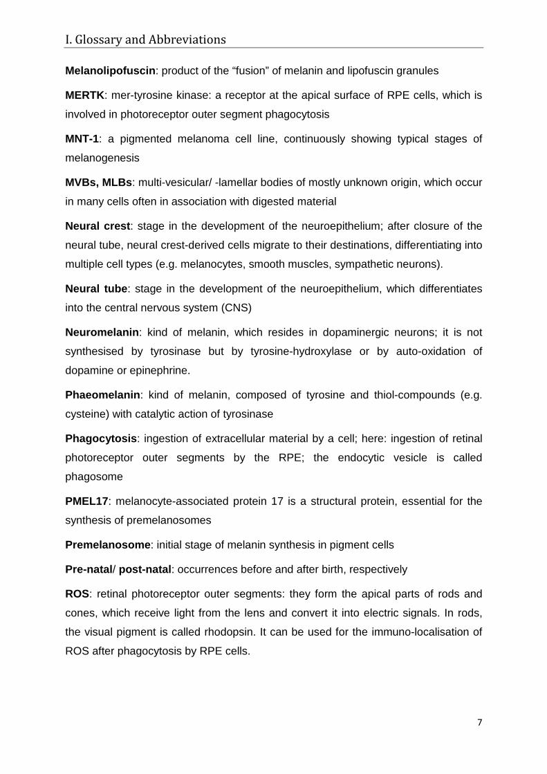
epithelium (RPE) are the pigment cells of the eye (Marks & Seabra, 2001). Figure 1
illustrates the structure of a vertebrate eye and highlights the position and
morphology of the RPE and the choroidal melanocytes. Among other functions, the
pigment cells of the eye are responsible for iris colour and the absorption of scattered
light in the background of the eye (Dayhaw-Barker, 2002; Roberts, 2002).
This chapter gives a short overview of the pigmentary system of the retinal pigment
epithelium (RPE), with regard to cell morphology, melanogenesis, and age-related
changes in the eyes of elderly individuals. Finally, a review of the findings leading to
the objectives of this thesis is presented.
1. The retinal pigment epithelium
The RPE is a monolayer of pigmented cells of neural tube origin in the background of
the eye (Figure 1 A, B). Together with the endothelial cells of the choriocapillaris it
forms the blood-retinal barrier (Konari et al, 1995). RPE cells are cubical and highly
polarised. Basal infoldings and apical microvilli serve as enlargement of the surface.
The typical oval shaped melanosomes of the RPE are located in the microvilli, and
round shaped melanosomes can be found in the cytoplasm near the nucleus (Figure
1 C). Besides its barrier function, which is facilitated by tight junctions, the RPE has
several properties essential for vision (Marmorstein et al, 1998), including the
balanced nutritional supply of the retina (Bialek & Miller, 1994). One of the most
important functions of the RPE is the phagocytosis and degradation of retinal
photoreceptor outer segment discs (ROS), which are shed during the light perception
process (Bosch et al, 1993; Young & Bok, 1969). After ingestion, the RPE degrades
the material and recycles certain lipids and 11-cis-retinal, the reactive component of
the visual pigment of the rods (Flannery et al, 1990; Rando, 1992). Loss of
phagocytic activity leads to retinal damage (Nandrot & Finnemann, 2008), due to the
accumulation of undegraded ROS in the sub-retinal space. In RCS (Royal College of
Surgeons) rats for example, a mutation in the surface receptor Mertk (mer-tyrosine
II. Introduction
10
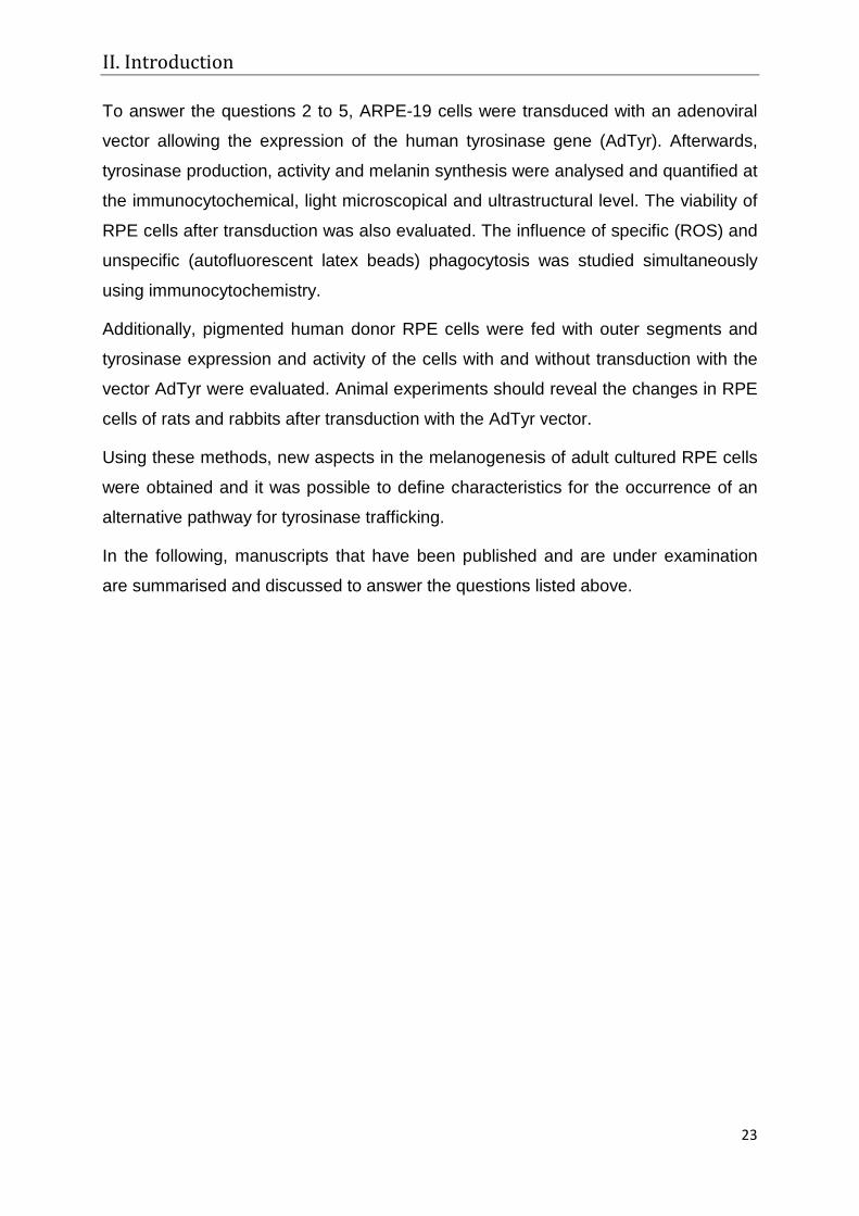
Figure 1: Histology of the retinal pigment epithelium (RPE) A) A scheme of the vertebrate eye shows the RPE (R) lying between the retina (r) and the choroid (c); sclera (s), c cornea, cb ciliary body, i iris, l lens, on optic nerve, v vitreous body; the highlighted square is magnified in B. B) Cross section of a monkey eye: the apical part of the RPE is closely related to the photoreceptors of the retina. Bm Bruch’s membrane; C) Schematic diagram of the RPE, as highlighted in B: basal infoldings (bi) and apical microvilli (mv) enlarge the surface; tight junctions (tj) of the RPE together with the blood vessels (bv) of the choriocapillaris build the blood-retinal barrier; choroidal melanocytes (cm) are densely packed with round melanosomes (black spots), while RPE melanosomes are more oval shaped, e erythrocyte;
kinase) leads to loss of specific phagocytosis of ROS and to loss of the visual
function of the retina (D'Cruz et al, 2000).
Other RPE functions are assumed to be related to their melanin content (reviewed by
(Sarna, 1992). The melanosomes of the RPE absorb scattered light, which would
otherwise disturb visual acuity (Peters et al, 2006) and, together with antioxidative
enzymes and non-enzymatic antioxidants (Newsome, 1994), melanosomes protect
against oxidative stress in the background of the eye (Schraermeyer & Heimann,
1999). They accumulate heavy metals, free radicals and toxic substances, like
gentamycin (Zemel et al, 1995), which would otherwise be harmful to the cell.
Additionally, the melanosomes are responsible for short-term storage of metal ions
like Ca2+, K+ and Zn2+ (Kokkinou et al, 2005; Samuelson et al, 1993) that are needed
for cell metabolism. However, aged melanosomes may also have prooxidative
functions (reviewed by (Boulton et al, 2001)).
II. Introduction
11
2. Melanogenesis
Melanocytes of the skin and hair are able to produce melanin for their whole lifetime
inside specialised organelles, the melanosomes (Jimbow et al, 1976). Frequently,
they transfer the pigment granules to neighbouring keratinocytes, where the
melanosomes function as antioxidative scavengers, and absorber of light and toxic
substances (reviewed by (Sarna, 1992)). In contrast, melanosomes of the RPE and
other melanocytes are not recycled under normal conditions and are not transferred
to neighbouring cells. It is surmised that RPE melanosomes are synthesised only
before birth (Carr & Siegel, 1979; Miyamoto & Fitzpatrick, 1957; Sarna, 1992),
although post-natal melanogenesis has been described by several groups in the
meantime (Dorey et al, 1990; Herman & Steinberg, 1982; Peters et al, 2000;
Schraermeyer & Heimann, 1999; Schraermeyer & Stieve, 1994; Thumann et al,
1999).
Melanocytes and RPE cells have a different embryonic origin and development
(Smith-Thomas et al, 1996): While RPE cells emerge from the neural tube,
melanocytes are neural crest-derived cells. Consequently some differences exist
between melanocyte and RPE melanogenesis (Bharti et al, 2008; Bharti et al, 2006;
Murisier et al, 2007), which affect, for example the transcription of melanogenic
genes in the embryo by using different transcription and growth factors. Also the
shape of mature RPE melanosomes differs from that of melanocytes in being oval
(Schraermeyer & Heimann, 1999). Despite this, melanogenesis in RPE cells has
been described as sharing a common pathway with melanocytes. They use the same
melanogenic proteins and the ultrastructural characterisation of morphologically
distinct melanosomal stages I to IV has been confirmed for both melanocytes and
pre-natal RPE cells (Lopes et al, 2007). RPE pigmentation is still not fully
understood, thus findings from melanocyte pigmentation are summarised below to
give a small introduction regarding the melanising machinery of pigment cells.
Melanogenesis is the synthesis of melanin from tyrosine residues by the enzyme
tyrosinase in pigment cells. Different types of melanin can be distinguished,
eumelanin, phaeomelanin and neuromelanin. Eu- and phaeomelanin can be found in
melanocytes and pigment epithelial cells, while neuromelanin is located only in
specialised brain neurons (reviewed by (Breathnach, 1988)). For the formation of
eumelanin, which is black to brown, the proteins tyrosinase, tyrosinase-related

II. Introduction
12
protein (TRP) 1 and 2 and the structural protein melanocyte-associated protein 17
(PMEL17) are necessary. The main precursors of eumelanin are 5,6-dihydroxyindol
(DHI) and 5,6-dihydroxyindol-2-carboxyilc acid (DHICA). Details are described in 2.2.
The yellow to red phaeomelanin is composed of tyrosine together with cysteine or
other thiol-compounds. Originally, it was believed that melanin granules contained
only polymers of one monomer (Mason, 1967). It is now accepted that most melanin
granules are mixed melanosomes, composed of eumelanin (Oetting and King 1994;
(Prota, 1992) and phaeomelanin (reviewed by (Ito & Wakamatsu, 2003; Ito &
Wakamatsu, 2008)). Nevertheless, RPE and choroidal pigmentation contain mostly
eumelanin.
2.1 Melanosomal proteins
2.1.1 Tyrosinase and Tyrosinase-related proteins (TRPs)
The tyrosinase gene family members tyrosinase, TRP1 and TRP2, are the main
enzymes and regulatory proteins of melanogenesis (Tsukamoto et al, 1992). They
are type 1 membrane-glycoproteins and share a high nucleotide sequence homology
and a similar protein structure throughout the animal kingdom (Camacho-Hubner et
al, 2002). The transcriptional regulation of the tyrosinase gene family is rather
complex and is reviewed in several publications (Murisier & Beermann, 2006; Sturm,
2009; Tachibana, 2000). Tyrosinase and TRPs are metallo-proteins and have two
putative histidine binding sites for metal ions (Huber & Lerch, 1988; Jackman et al,
1991). Tyrosinase binds copper, while the other two family members have other
cofactors, zinc for TRP2 (Solano et al, 1994) and possibly iron or copper for TRP1
(Martinez-Esparza et al, 1997).
Tyrosinase, initially translated as a 60 kDa polypeptide and heavily glycosilated in
the rough ER (rER) to a 70-74 kDa form, has seven glycosilation sequons (Ujvari et
al, 2001). It contains a bicubric active site (2 regions CuA and CuB) each with three
highly conserved histidines, responsible for metal binding and redox-reactions.
Glycosilation, folding and copper acquisition are regulated by glycosilation enzymes
and ER chaperones (Branza-Nichita et al, 1999). In addition, the substrates L-DOPA
and L-tyrosine (Halaban et al, 2001) and TRP1 (Francis et al, 2003; Kobayashi et al,
1998) apparently facilitate the maturation and dimerisation of tyrosinase in the rER.
II. Introduction
13
Tyrosinase undergoes sequential modifications of the sugar residues first in the ER
and then in the Golgi apparatus and is shuttled through the latter via
glycosphingolipids (Sprong et al, 2001). Before this, copper binding at the CuB site
facilitates initial processing of tyrosinase into a form capable of being transported out
of the rER (Olivares et al, 2003). Upon reaching the trans-cisterns of the Golgi
apparatus, the mature, copper-ladden tyrosinase is a catalytically active enzyme
(Maul, 1969).
Tyrosinase (Huizing et al, 2001) and TRP1 (Jimbow et al, 1997; Orlow et al, 1993)
contain a dileucine-based motif with flanking regulatory amino acids at their
cytoplasmic tail that interact with adaptor-protein complexes (AP-3 and AP-1,
respectively), which facilitate the shipment from the Golgi apparatus to late
endosomal and lysosomal compartments (Bonifacino, 2004) and finally to the
melanosome (Calvo et al, 1999; Simmen et al, 1999). In AP-3 mutants, tyrosinase
does not reach the melanosome, while TRP1 is transported properly (Huizing et al,
2001).
Integrated into the melanosomal membrane, tyrosinase facilitates the first two steps
of melanogenesis. In humans, it also catalyses the last steps from DHICA monomers
to eumelanin polymers. Besides its function as a rate limiting enzyme in
melanogenesis, tyrosinase also has a low catalase function (Wood & Schallreuter,
1991) and seems to be involved in the antioxidative defence of pigment cells
(Valverde et al, 1996a; Valverde et al, 1996b). Aberrations in the composition and
structure of the enzyme result in degradation of the molecule by endoplasmic
reticulum and proteasome-associated mechanisms (Berson et al, 2000; Halaban et
al, 1997; Mosse et al, 1998). This may lead to hypopigmentation and oculo-
cutaneous albinism (OCA) type 1 (Halaban et al, 2000; Spritz et al, 1990).
TRP1 has a low “pseudo-tyrosinase” activity (Jimenez-Cervantes et al, 1993) and it
has also been found to assist as a chaperone-like protein in tyrosinase dimerisation
and in heat denaturation defence (Francis et al, 2003). Absence of TRP1 leads to
OCA type 3 (Boissy et al, 1996). TRP2 (also called dopachromtautomerase)
catalyses the non-decarboxylative tautomerisation of dopachrome to DHICA in
eumelanogenesis (Kroumpouzos et al, 1994).

II. Introduction
14
2.1.2 Other melanogenic proteins
PMEL17 localises to early stages of melanogenesis (Kobayashi et al, 1994; Raposo
et al, 2001) and there it builds the typical structures needed for melanin accumulation
(Berson et al, 2003). It also binds the eumelanin-intermediates DHI and DHICA
(Chakraborty et al, 1996; Fowler et al, 2006; Lee et al, 1996) and it has been
suggested that it serves as a sink within melanosomes for the detoxification of
cytotoxic intermediates (Fowler et al, 2006; Theos et al, 2006). The transport
pathways of the PMEL17 protein are still not fully understood, but trafficking by AP
transporters AP-1, AP-2 and AP-3 are discussed (Valencia et al, 2006). Further
proteins, involved in the formation of functional melanosomes are the OA1 protein
(ocular albinism protein 1), MART1 (also known as melan-A), the P-protein (pink
eyed dilution homolog protein; OCA 2) and MATP (membrane-associated transporter
protein; OCA 4) which either facilitate trafficking of proteins and other molecules to
the melanosomes or have other yet unknown functions (Hearing, 2005). A high
molecular weight complex in the melanosomal membrane, consisting of tyrosinase,
TRP1, TRP2 and the lysosomal protein LAMP-1 (lysosomal-associated membrane
protein 1), seems to be responsible for the conversion of tyrosine to eumelanin
(Hearing et al, 1982; Jimenez-Cervantes et al, 1998). Other proteins like PMEL17
(Lee et al, 1996), P-protein, MART1 and melanogenic inhibitors may be connected
(Hearing et al, 1992). The melanogenic complex is still not understood, but it seems
to stabilise tyrosinase conformation and action in the melanosome.
2.2 The Raper-Mason Scheme of melanin formation
The Raper-Mason-Scheme describes the reaction cascade for tyrosinase-catalysed
eu- and phaeomelanin synthesis (Figure 2 for details). Eumelanin synthesis involves
a complex series of oxidations and rearrangements of tyrosine, which result in the
formation of an indole-quinone ring-structure that readily polymerises to high-
molecular weight biopolymers (reviewed by (Riley, 1993)). The melanin polymers
formed differ in length, absorption and physical and chemical properties dependent
on the ratio of the DHI and DHICA monomers (Aroca et al, 1992; Pawelek, 1991).
II. Introduction
15
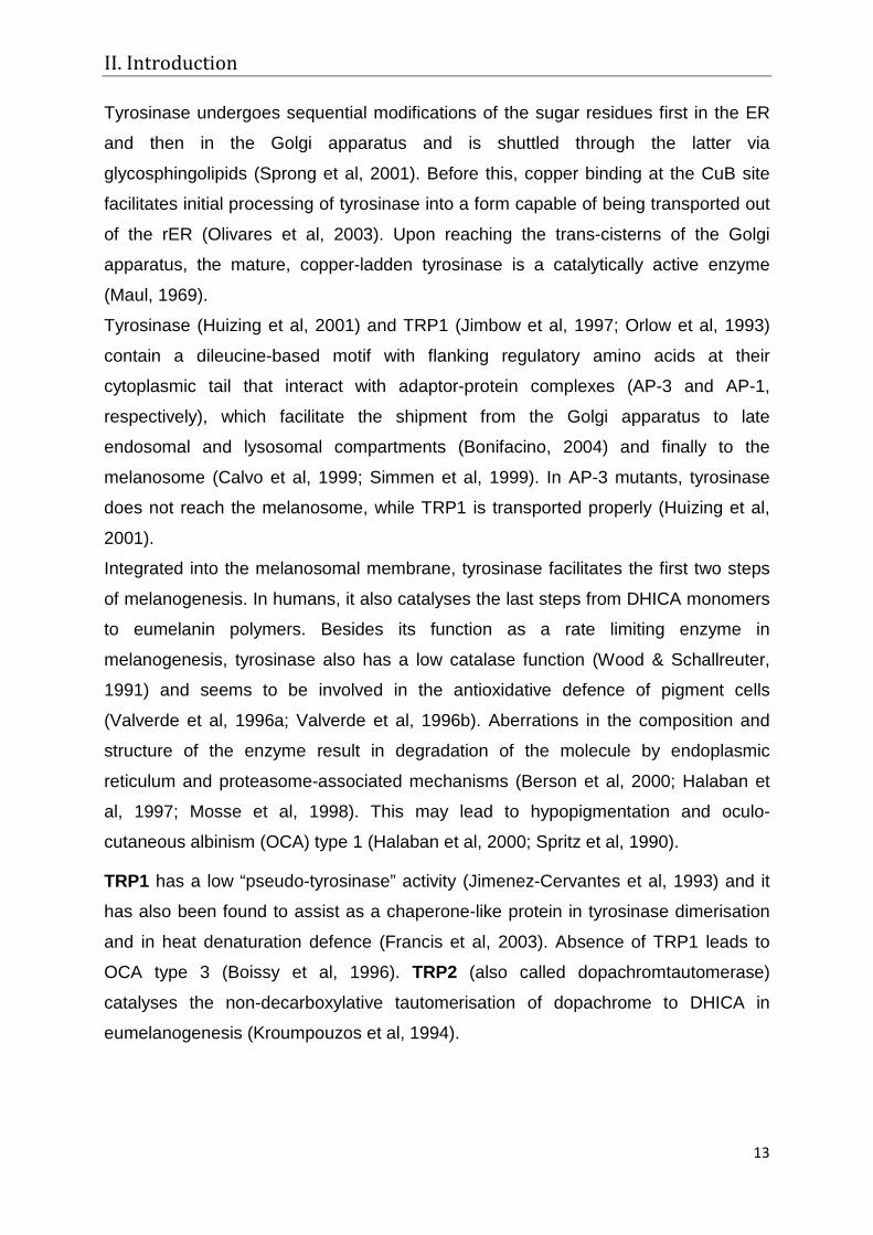
Figure 2: The Raper-Mason Scheme (adapted from http://omlc.ogi.edu/spectra/melanin/melaninsynth.gif (Prota, 1988)). 1+2) The first two steps, hydroxylation of tyrosine to DOPA and the oxidation from DOPA to Dopaquinone are catalysed by tyrosinase. 3) From Dopaquinone two different reactions are possible (Formation of eu- or pheomelanin): a) In presence of compounds with free thiol groups (glutathione, cysteine) and low pH rapid spontaneous reaction to sulfour-containing compounds (cysteinyldopas, benzothiazinylalanines) leads to phaeomelanin production. This reaction is favoured. b) In absence of thiol-compounds, Dopaquinone is transformed into eumelanin (continue with 4). 4) At neutral pH, the slow reaction from Dopaquinone to Leucodopachrome which is highly unstable, can be achieved, but spontaneously collapses into DOPA and Dopachrome. 5) From Dopachrome two different reactions can occur, leading to different eumelanin species. This reaction is regulated by TRP2: a) spontaneous decarboxylation to DHI b) with action of TRP2, the production of non-decarboxylated DHICA is favoured. 6) DHI and DHICA are transformed into 5,6-indolchinones (with and without carboxyl-group). DHI oxidizes spontaneously (or faster with tyrosinase action); DHICA transformation is slower but needs TRP1 catalysis in mice or tyrosinase in humans (Boissy et al., 1989). 7) Indolchinones react with dihydroxyindoles forming semichinones that polymerize spontaneously to eumelanin.
II. Introduction
16
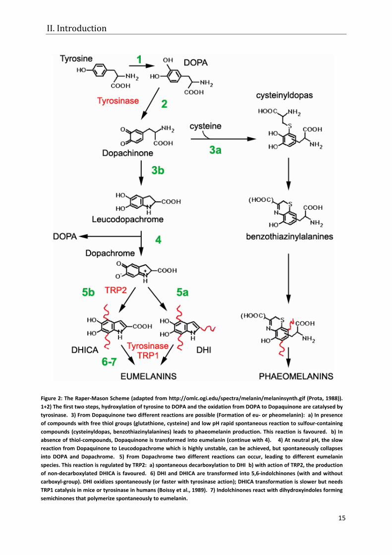
2.3 Cellular actions– the 4-stage model of eumelanogenesis
The cellular procedure of pigment
synthesis (=melanogenesis) has
been described first by Seiji (Seiji
et al, 1963). This model still
serves as the standard scheme
for melanocytes and pre-natal
RPE cells. Meanwhile, it has been
extended by other authors and is
actually described as follows
(Hearing, 2005) (Figure 3): The
melanogenesis starts with the
occurrence of an amorphous
vesicle, the premelanosome,
which is positive for the protein
PMEL17. PMEL17 is responsible
for the building of internal, fibrillar
striations that give the vesicle a
typical structure (stage I pre-
melanosome). After the intra
luminal arrangement is completed
(stage II), Golgi-derived vesicles,
filled with melanogenic proteins, can enter the premelanosome. After integration in
the melanosomal membrane, tyrosinase synthesises melanin, which is deposited
along the internal striations of the organelle (stage III). When the whole organelle is
filled with melanin, it is termed stage IV or mature melanosome (=melanin granule).
2.4 The premelanosome
The premelanosome is the essential starting point of melanogenesis. Absence of the
protein PMEL17 leads to the loss of premelanosome formation, to mistargeting and
degradation of melanogenic enzymes and to defective pigmentation in affected
individuals (Theos et al, 2006). The origin of the amorphous vesicle, forming the
premelanosome is still not understood. Earlier hypotheses proposed that the
Figure 3: Classical stages of melanogenesis: The electron micrograph shows different stages of melanogenesis in MNT-1 melanoma cells. A scheme, describing the four possible stages is given on the right.
II. Introduction
17
premelanosome of melanocytes (reviewed by (Jimbow et al, 1976)) and RPE cells
(Eppig, 1970; Eppig & Dumont, 1972; Mishima et al, 1978; Stanka, 1971; Stanka et
al, 1981) is formed out of cisterns or vesicles of the endoplasmic reticulum or the
Golgi-Endoplasmic Reticulum-Lysosome zone (GERL), which is part of the smooth
ER (Novikoff et al, 1968). Observations, resembling the ultrastructural similarity of
melanocyte melanosomes to lysosomes (Schraermeyer, 1995) and the occurrence of
lysosomal enzymes in the melanosome (Diment et al, 1995; Orlow et al, 1993),
indicated that the melanocytes melanosome might well be a specialised lysosome
(reviewed by (Orlow, 1995)). Additionally, RPE melanosomes have been associated
with phagosomes and lysosome-like organelles (Schraermeyer & Stieve, 1994),
further indicating that the RPE melanosome is also a lysosome-like organelle
(reviewed by (Schraermeyer & Heimann, 1999)). Recent ultrastructural studies, using
immuno-gold labelled antibodies, have clarified that the premelanosome of
melanocytes and melanoma cells is an endosome (Raposo & Marks, 2002). Thus, in
melanocytes, PMEL 17 travels to endosomes, where it builds internal striations and
matures to stage II melanosomes. For RPE cells, corresponding studies are still
lacking.
3. Age-related changes in the RPE - Lipofuscin
In young adult people, another pigment, lipofuscin, accumulates in the cells of the
retinal pigment epithelium (Roberts, 2002). It is also found in the liver and the brain of
aged persons. The composition of lipofuscin in the RPE is still not fully understood
(Ng et al, 2008), but it has been found to contain undegradable remnants of
phagocytosed photoreceptor outer segments (Feeney-Burns & Eldred, 1983;
Feeney-Burns et al, 1988; Warburton et al, 2005). Heterogeneous groups of lipids
and protein aggregates that are highly oxidised and thus auto-fluorescent, build the
main components of this age pigment (Eldred, 1989; Eldred et al, 1982). From the
fourth decade onwards, melanolipofuscin granules become visible (Feeney-Burns et
al, 1984). They have a melanin core and a lipofuscin envelope, but it is unknown how
these granules develop. Since undegradable phagocytosed material can be
integrated into the melanosomal membrane (Schraermeyer & Stieve, 1994), and
tyrosinase has been found in association with phagosomes (Julien et al, 2007;
Schraermeyer et al, 2006), the following is speculated: tyrosinase scavenges radicals
II. Introduction
18
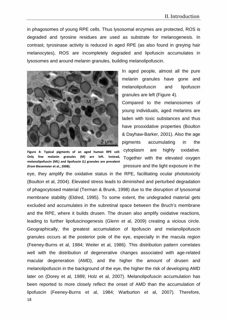
Figure 4: Typical pigments of an aged human RPE cell: Only few melanin granules (M) are left. Instead, melanolipofuscin (ML) and lipofuscin (L) granules are prevalent (from Biesemeier et al., 2008).
in phagosomes of young RPE cells. Thus lysosomal enzymes are protected, ROS is
degraded and tyrosine residues are used as substrate for melanogenesis. In
contrast, tyrosinase activity is reduced in aged RPE (as also found in greying hair
melanocytes), ROS are incompletely degraded and lipofuscin accumulates in
lysosomes and around melanin granules, building melanolipofuscin.
In aged people, almost all the pure
melanin granules have gone and
melanolipofuscin and lipofuscin
granules are left (Figure 4).
Compared to the melanosomes of
young individuals, aged melanins are
laden with toxic substances and thus
have prooxidative properties (Boulton
& Dayhaw-Barker, 2001). Also the age
pigments accumulating in the
cytoplasm are highly oxidative.
Together with the elevated oxygen
pressure and the light exposure in the
eye, they amplify the oxidative status in the RPE, facilitating ocular phototoxicity
(Boulton et al, 2004). Elevated stress leads to diminished and perturbed degradation
of phagocytosed material (Terman & Brunk, 1998) due to the disruption of lysosomal
membrane stability (Eldred, 1995). To some extent, the undegraded material gets
excluded and accumulates in the subretinal space between the Bruch’s membrane
and the RPE, where it builds drusen. The drusen also amplify oxidative reactions,
leading to further lipofuscinogenesis (Glenn et al, 2009) creating a vicious circle.
Geographically, the greatest accumulation of lipofuscin and melanolipofuscin
granules occurs at the posterior pole of the eye, especially in the macula region
(Feeney-Burns et al, 1984; Weiter et al, 1986). This distribution pattern correlates
well with the distribution of degenerative changes associated with age-related
macular degeneration (AMD), and the higher the amount of drusen and
melanolipofuscin in the background of the eye, the higher the risk of developing AMD
later on (Dorey et al, 1989; Holz et al, 2007). Melanolipofuscin accumulation has
been reported to more closely reflect the onset of AMD than the accumulation of
lipofuscin (Feeney-Burns et al, 1984; Warburton et al, 2007). Therefore,
II. Introduction
19
melanolipofuscin deposition is widely accepted as being one major cause of retinal
degeneration and blindness in the elderly population of the Western world (Bird et al,
1995; von Ruckmann et al, 1997). Thus a better understanding of the mechanisms
causing (melano-) lipofuscin formation is desirable.
4. Previous findings
Many pigment cell functions are related to their melanin content, including light
absorption and scavenging of oxidative reactions (Sarna, 1992). Age-related
changes to eye pigmentation in elderly people create a prooxidative environment and
can cause diseases that may result in blindness. Consequently, it is highly
questionable that melanin is only synthesised pre-natally and not renewed over a
lifetime. Meanwhile, some authors have demonstrated that there are intermediate
stages of melanogenesis, occurring spontaneously in the adult RPE of opossums
(Herman & Steinberg, 1982) and cattle (Schraermeyer, 1992) and also in cell culture
(Dorey et al, 1990). They could be upregulated under certain conditions: e.g. after
stimulation with melanogenesis activating agents such as lactic acid (Mishima, 1994)
and Ca2+(Rak et al, 2006). In vivo, the induction of photic stress in hamsters, led to
the occurrence of striated melanosomal stages in the RPE (Schraermeyer, 1992).
Furthermore, tyrosinase expression has been found to occur in adult RPE tissue
(Dryja et al, 1978), or has been induced in cell culture by stimulation with growth
factors, like the melanocyte-stimulating hormone (Abul-Hassan et al, 2000). After
phagocytosis of retinal outer segments, both tyrosinase (Schraermeyer et al, 2006)
and melanogenic stages were observed in cell culture of bovine RPE cells
(Schraermeyer, 1995; Schraermeyer et al, 1999b; Schraermeyer & Stieve, 1994) and
in rabbit (Thumann et al, 1999) and rat eyes (Peters et al, 2000). Residues of the
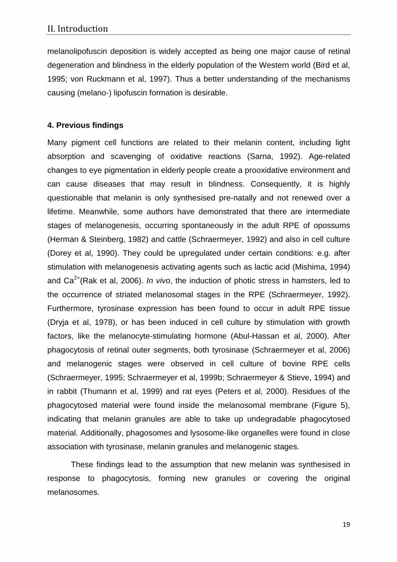
phagocytosed material were found inside the melanosomal membrane (Figure 5),
indicating that melanin granules are able to take up undegradable phagocytosed
material. Additionally, phagosomes and lysosome-like organelles were found in close
association with tyrosinase, melanin granules and melanogenic stages.
These findings lead to the assumption that new melanin was synthesised in
response to phagocytosis, forming new granules or covering the original
melanosomes.
II. Introduction
20
On the other hand, a defective degradation of the ingested material can lead
to lipofuscinogenesis inside the melanosome and to the production of
melanolipofuscin granules. Accordingly, the renewal of RPE melanin or the
prevention of melanolipofuscinogenesis could prevent age-related diseases in the
background of the eye.
New findings concerning the origin of melanosomes in melanocytes (Raposo &
Marks, 2002) and that melanogenic proteins share transport mechanisms with
endosomes, lysosomes and other lysosome-related organelles (Bonifacino, 2004),
correlate well with the findings from the RPE cells mentioned above. These also
showed a relation between the degradative pathway and melanisation. However, in
the former experiments, no quantitative evidence has been obtained that new
melanin was synthesised in RPE cells. The association of melanosomes and
lysosome-like organelles might as well be a sign of melanin degradation (Sarna,
1992). Although tyrosinase has been observed in adult tissue, the presence of
PMEL17 in adult RPE has not yet been reported (Lopes et al, 2007).
Figure 5: Phagocytosed material is integrated into the melanosomal membrane: The inset (former figure 10) shows two typical phagosomes of an RPE cell, which contain ROS material that is labelled with gold granules. The main picture (former figure 11) shows an RPE melanosome, five days following after phagocytosis of gold-labelled ROS: the space between the limiting membrane and the melanin matrix is enlarged and contains gold granules (arrowheads) and homogeneous electron opaque material (asterisk) (adapted from Schraermeyer et al., 1999b).
II. Introduction
21
In fibroblasts, melanogenesis has been found to take place in lysosomes after
tyrosinase transfection (Borovansky et al, 1997; Winder et al, 1993). Melanogenesis
occurred without the involvement of other melanosomal proteins and led to a
melanosomal structure, which was different to the classical model (Borovansky et al,
1997), suggesting an alternative pathway of melanin formation inside lysosomes, as
has been proposed for post-natal RPE melanogenesis.
Probably, post-natal melanin synthesis occurs without the involvement of
typical PMEL17-positive striated premelanosomes inside lysosome-like organelles.
On the other hand, it is possible that tyrosinase expression in adults is not correlated
with melanogenesis but serves a yet unknown function, maybe in the defence
against oxidative stress, as assumed for melanocytes (Kim & Han, 2003; Perluigi et
al, 2003; Wood & Schallreuter, 1991). The background of the eye is a highly
oxidative environment due to the high oxygen pressure from the choroid, the diurnal
turnover of retinal material and light exposure. As in melanocytes, melanosomes of
the RPE have been found to scavenge oxidative reactions (Rozanowski et al, 2008a;
Rozanowski et al, 2008b; Zadlo et al, 2007). However, aged and photosensitised
melanin has a prooxidative function (Rozanowska et al, 1997) and tyrosinase and
melanogenic intermediates (Heiduschka et al, 2007) are discussed as having anti- or
prooxidative capacity depending on their concentration and the surrounding
circumstances (Seagle et al, 2005a; Seagle et al, 2005b; Urabe et al, 1994).
Nevertheless, Schraermeyer found melanogenic stages after induction of light stress
in hamster RPE (Schraermeyer, 1992), suggesting an upregulation of tyrosinase
activity.
Possibly, tyrosinase machinery is activated to scavenge free radical building
due to oxidative reactions after light induction or phagocytosis of outer segments.
Thus the function of tyrosinase and melanin in the adult retinal pigment epithelium is
still unknown and many theories have been raised, which require further
investigation.

II. Introduction
22
5. Aim of the thesis
Regarding previous studies, mostly done on animal cells and tissues, the aim of this
thesis was to investigate the relationship of tyrosinase, pigmentation and
phagocytosis in adult human RPE, as set out in the following questions:
1) Are tyrosinase and melanisation involved in the defence against oxidative
stress in aged human RPE cells?
2) Can melanogenesis be induced in non-pigmented adult human RPE cells?
a. Is the classical scheme of melanogenesis true for adult human RPE
melanogenesis or can an alternative pathway be described?
b. Is the typical striated premelanosome essential for melanogenesis?
c. Is the trafficking of adenovirally transduced tyrosinase altered compared
to the pathway described in the literature? Can it occur at free
ribosomes in the cytoplasm?
3) How does ROS phagocytosis influence tyrosinase activity and melanogenesis
in this system?
4) Are remnants of phagosomal degradation transported to or do they fuse with
melanosomes, generating melanolipofuscin?
5) Is the expression of tyrosinase useful or toxic for adult RPE cells?
To answer the first question, a method was established to induce UV-A light-
mediated oxidative stress in human retinal pigment epithelial cell culture (primary
pigmented human donor RPE cells (hRPE) versus the amelanotic retinal pigment
epithelium 19 cell line (ARPE-19)). Then the antioxidative capacity, apoptosis and
viability of the cells were tested. It was found that the antioxidative capacity of
melanised donor RPE cells was highly affected by the amount of prooxidative age
pigments in the cells.
Before studying the effects of tyrosinase and melanisation any further, a system had
to be established where pigmented/ tyrosinase-active cells were generated without
the disturbing presence of aged donor pigments. Since ARPE-19 cells lack an overall
pigmentation on the one hand, but are highly differentiated RPE cells with typical
phagocytic function on the other hand, they presented an ideal system to study the
new synthesis and function of melanin without the interference of old melanin
granules.
II. Introduction
23
To answer the questions 2 to 5, ARPE-19 cells were transduced with an adenoviral
vector allowing the expression of the human tyrosinase gene (AdTyr). Afterwards,
tyrosinase production, activity and melanin synthesis were analysed and quantified at
the immunocytochemical, light microscopical and ultrastructural level. The viability of
RPE cells after transduction was also evaluated. The influence of specific (ROS) and
unspecific (autofluorescent latex beads) phagocytosis was studied simultaneously
using immunocytochemistry.
Additionally, pigmented human donor RPE cells were fed with outer segments and
tyrosinase expression and activity of the cells with and without transduction with the
vector AdTyr were evaluated. Animal experiments should reveal the changes in RPE
cells of rats and rabbits after transduction with the AdTyr vector.
Using these methods, new aspects in the melanogenesis of adult cultured RPE cells
were obtained and it was possible to define characteristics for the occurrence of an
alternative pathway for tyrosinase trafficking.
In the following, manuscripts that have been published and are under examination
are summarised and discussed to answer the questions listed above.
III. Results
24
III. Summarised results of the manuscripts enclosed
For a better understanding, the manuscripts are not listed in chronological order, but
according to their logical relevance to this thesis.
Manuscript 1: Biesemeier A., Kokkinou D., Julien S., Heiduschka P., Berneburg M.,
Bartz-Schmidt K.U., Schraermeyer U. UV-A induced oxidative stress is more prominent in naturally pigmented aged human RPE cells compared to non-pigmented human RPE cells independent of zinc treatment. J Photochem
Photobiol B. 2008 Feb 27;90(2):113-20. Epub 2007 Dec 4
Age-related macular degeneration is the leading cause of blindness in the elderly
population of the western world (Bird et al, 1995). Changes in RPE pigmentation play
a key role in age-related oxidative stress and disease (Boulton et al, 2004). While
melanin in young RPE is assumed to have an antioxidative function, the age-
pigments lipofuscin and melanolipofuscin are prooxidative and lead to an increase in
oxidative stress reactions due to radical building. Dark skinned people have fewer
age-related eye diseases than Caucasians (Ambati et al, 2003), and it is discussed if
this is due to their higher melanin/lipofuscin ratio in the RPE (Gregor & Joffe, 1978).
Zinc treatment together with a vitamin diet is one treatment strategy for AMD (Age-
related Eye Disease Study Research Group, 2001). Zinc, which can be stored by
melanins (Kokkinou et al, 2005; Potts & Au, 1976), is used as a cofactor in many
antioxidative enzymes and is thus involved in cell protection (Bray & Bettger, 1990;
Brewer et al, 1983; Deng et al, 2000). In this study, it was investigated whether
melanised donor RPE cells have more antioxidative capacity against UV-light
induced oxidative stress compared to non-pigmented ARPE-19 cells. In addition,
cells were supplemented with 100 µM zinc chloride to analyse whether the
antioxidative action of zinc and its storage in melanosomes were able to support the
antioxidative action in melanised cells.
Cells of the non-pigmented ARPE-19 cell line and human donor RPE cells (hRPE;
age of donors: 60-70 years) were supplemented with 100 µM zinc chloride and they
were then illuminated with UV-A light (330-440 nm) for 15 to 60 minutes. Afterwards,
oxidative stress (DCFDA assay of H2O2 production), apoptotic events (TUNEL assay)
and viability (MTT test) were investigated.
III. Results
25
Without irradiation, hRPE cells showed less oxidative stress when supplemented with
zinc compared to their control. Despite its significance, this effect was very small.
After irradiation, all samples had significantly increased their H2O2 production
compared to controls. No zinc-related effect was observed in either ARPE-19 or
hRPE cells. Apoptosis and viability were also affected by irradiation independent of
zinc treatment. In contrast to the theory, pigmented hRPE cells showed a lower
viability after irradiation compared to the non-pigmented ARPE-19 cells. Analogous
effects were found also in the TUNEL assay. The non-pigmented cells showed less
apoptosis than the pigmented cells.
The results obtained were unexpected since it was supposed that pigmented cells
would have a higher antioxidative capacity due to their melanin content than non-
pigmented cells. The human donor RPE cells used in the study contained not only
melanin but also high amounts of melanolipofuscin and lipofuscin. Hence, the
protective effects of melanin and the supplemented zinc were nullified by the
prooxidative effects of the age pigments. As expected, zinc had no effect on ARPE-
19 cells, since these cells lack melanin, which is responsible for storage of trace
metal ions.
However, the results of this study are only valid for aged human RPE, possibly
resembling the oxidative state of pre-AMD persons. This study cannot describe the
effects of UV-light on RPE cells from young adults or children, which do not contain
the prooxidative melanolipofuscins. In future research, also cells from younger
donors (<20 years), containing no melanolipofuscin and less lipofuscin granules, will
also be used to investigate the different oxidative properties of RPE pigments,
according to their age and composition.
III. Results
26
Manuscript 2: Julien S., Kociok N., Kreppel F., Kopitz F., Kochanek S., Biesemeier A., Blitgen-Heinecke P., Heiduschka P., Schraermeyer U. Tyrosinase biosynthesis and trafficking in adult human retinal pigment epithelial cells. Graefes Arch Clin
Exp Ophthalmol. 2007 Oct;245(10):1495-505. Epub 2007 Feb 21.
Tyrosinase, the catalysing enzyme of melanogenesis, is found predominantly in pre-
natal RPE (Carr & Siegel, 1979; Miyamoto & Fitzpatrick, 1957) and it is still not clear
if it also appears in adult RPE (Schraermeyer, 1993; Schraermeyer et al, 2006). Here
it is demonstrated that tyrosinase can be upregulated by phagocytosis of retinal outer
segments (ROS) in human donor RPE (hRPE) cultures.
Primary cultures of hRPE cells were fed with bovine outer segments and latex beads
for five and twenty hours respectively, and then tyrosinase expression and activity
were analysed with different methods: tyrosinase mRNA expression was evaluated
with quantitative real time RT-PCR. Fluorescence immunocytochemistry with anti-
human tyrosinase antibody and light and electron DOPA histochemistry showed the
location of tyrosinase in the cell. Tyrosinase activity was analysed by measuring the
[3H]-tyrosine hydroxylase activity of the enzyme after phagocytic events. As controls,
cells were cultured with assay medium only. Additionally, tyrosinase staining of cells
with the tyrosinase vector AdTyr is presented.
Fluorescence immunocytochemistry showed immunoreactivity of tyrosinase in hRPE
cells only after phagocytosis of ROS and not under control conditions. Transduction
with AdTyr, an adenoviral vector allowing the expression of the human tyrosinase
gene, led to an intense tyrosinase staining also in non-fed hRPE cells. Tyrosinase
presence and activity in fed cells were also tested using the DOPA-oxidase assay in
light and electron microscopical samples. It was found that a positive reaction took
place in small granular vesicles (DOPA granules) throughout the cytoplasm five and
twenty hours after feeding. In addition, the tyrosinase was located inside
phagosomes (20 h), which contained either lamellar material, resembling ROS
residues or ingested latex beads (Figure 6). Without feeding, no DOPA reaction was
observed with light and electron microscopy. The intracellular location of tyrosinase
was also tested with antibodies and a co-localisation of rhodopsin (from ROS) and
tyrosinase was observed in some organelles, twenty hours after feeding.

III. Results
27
The mRNA levels of tyrosinase and GAPDH (glyceraldehyde-3-phosphate
dehydrogenase) in control and fed cells were compared using real time RT-PCR. The
level of tyrosinase mRNA was calibrated to the amount of GAPDH in control cells.
Five and twenty hours after feeding, the mRNA level of tyrosinase was reduced
compared to the controls. However, it had re-increased above the control-level forty
hours after feeding. Additionally, tyrosine-hydroxylase activity of tyrosinase was
measured five, twenty and forty hours after phagocytosis of ROS in monkey RPE
cells. Tritiated L-tyrosine was used as a substrate and the release of tritium from
tyrosine was measured in homogenised samples. The absence of the enzyme
tyrosine-hydroxylase, which would give unspecific results in the activity-assay, was
also confirmed by immunocytochemistry. It was found that ROS fed cells had a
higher hydroxylase activity than controls (x 180). Human melanoma cells were used
Figure 6: DOPA histochemistry of a fed donor RPE cell: DOPA granules are spread over the cytoplasm and fuse with multi-lamellar bodies, resembling phagosomes (arrows). The inset shows a huge phagosome with ingested latex beads, which has also incorporated DOPA granules.

III. Results
28
as positive control and cells from the amelanotic region of monkey eyes were used
as negative control. Pure ROS were almost free of tyrosinase contamination as
shown by a very low hydroxylase activity.
This study showed that generation of the tyrosinase protein and its activity can be
stimulated by feeding cultured hRPE cells with outer segments. Active tyrosinase has
been found to reside in electron-dark granules all over the cytoplasm and in
association with phagosomes. The tyrosine-hydroxylase activity of the enzyme was
also confirmed to be higher when cells were fed with ROS. However, the mRNA
levels were reduced at both five and twenty hours after feeding and re-increased
after forty hours. It is surmised that tyrosinase mRNA is always present in adult
human RPE cells, but effective translation and protein synthesis is inhibited under
normal conditions. When cells have phagocytosed ROS, the inhibition is lifted and
tyrosinase protein can be synthesised without post-translational degradation.
Manuscript 3: Biesemeier A., Kreppel F., Kochanek S., Schraermeyer U. The classical premelanosome, known from pre-natal melanogenesis, is not essential for melanogenesis in adult RPE cells (Cell & Tissue Research, in print).
The pathway of organelle formation in melanogenesis was characterised by (Seiji et
al, 1963). The first structure described is the premelanosome. It has a body of
typically striated fibrils, which are formed by the protein PMEL17. After the internal
structure is completed, tyrosinase enters the premelanosome and catalyses the
formation of melanin polymers, which gather at the internal striations, forming
electron-dark stripes, which grow bigger and finally cover the whole organelle. The
organelle is now called a mature melanosome and it shows neither an internal
structure nor further tyrosinase activity. It is proposed that melanogenesis does not
occur when PMEL17 is absent and premelanosomes cannot therefore be formed
(Theos et al, 2006). However, overexpression of tyrosinase in non-pigment cells
showed melanin synthesis in lysosomes without the occurrence of typical
premelanosomes (Borovansky et al, 1997; Winder, 1991; Winder et al, 1995; Winder
et al, 1993).
To obtain new melanogenesis without the interference of old melanin granules in the
cell, non-pigmented ARPE-19 cells were transduced with the adenoviral vector
III. Results
29
AdTyr. The expression of different melanogenic proteins (tyrosinase, TRP1,
PMEL17) was investigated using fluorescence immunocytochemistry, and the
presence of active tyrosinase was analysed using ultrastructural DOPA
histochemistry. Occurring melanogenesis was investigated with light and electron
microscopy. An MTT assay confirmed the viability of the cells after transduction. Non-
transduced ARPE-19 cells served as negative controls. Additionally, cells from a
pigmented melanoma cell line (MNT-1) were used as positive controls for
melanogenesis occurring in the classical way (Raposo et al, 2001).
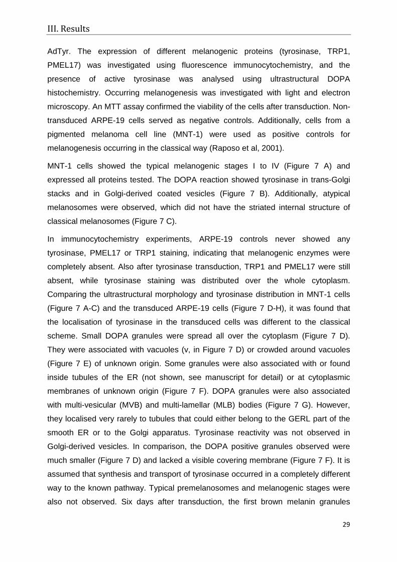
MNT-1 cells showed the typical melanogenic stages I to IV (Figure 7 A) and
expressed all proteins tested. The DOPA reaction showed tyrosinase in trans-Golgi
stacks and in Golgi-derived coated vesicles (Figure 7 B). Additionally, atypical
melanosomes were observed, which did not have the striated internal structure of
classical melanosomes (Figure 7 C).
In immunocytochemistry experiments, ARPE-19 controls never showed any
tyrosinase, PMEL17 or TRP1 staining, indicating that melanogenic enzymes were
completely absent. Also after tyrosinase transduction, TRP1 and PMEL17 were still
absent, while tyrosinase staining was distributed over the whole cytoplasm.
Comparing the ultrastructural morphology and tyrosinase distribution in MNT-1 cells
(Figure 7 A-C) and the transduced ARPE-19 cells (Figure 7 D-H), it was found that
the localisation of tyrosinase in the transduced cells was different to the classical
scheme. Small DOPA granules were spread all over the cytoplasm (Figure 7 D).
They were associated with vacuoles (v, in Figure 7 D) or crowded around vacuoles
(Figure 7 E) of unknown origin. Some granules were also associated with or found
inside tubules of the ER (not shown, see manuscript for detail) or at cytoplasmic
membranes of unknown origin (Figure 7 F). DOPA granules were also associated
with multi-vesicular (MVB) and multi-lamellar (MLB) bodies (Figure 7 G). However,
they localised very rarely to tubules that could either belong to the GERL part of the
smooth ER or to the Golgi apparatus. Tyrosinase reactivity was not observed in
Golgi-derived vesicles. In comparison, the DOPA positive granules observed were
much smaller (Figure 7 D) and lacked a visible covering membrane (Figure 7 F). It is
assumed that synthesis and transport of tyrosinase occurred in a completely different
way to the known pathway. Typical premelanosomes and melanogenic stages were
also not observed. Six days after transduction, the first brown melanin granules
III. Results
30
accumulated in the transduced cultures. Ultrastructurally, these melanin granules (as
in Figure 7 H) resembled electron-dark, cloudy material inside multi-vesicular and
multi-lamellar bodies. While the MVBs and MLBs were unmelanised in negative
controls and in cells up to 48 h after transduction, these bodies were almost
completely filled with electron-dark, melanin-like material two weeks after
transduction. Additionally, multi-shaped organelles (mixed organelles) filled with
lamellar, vesicular and electron-dark material were observed, which seemed to build
a linking stage between MVBs and MLBs and the first stage of melanogenesis. The
multi-vesicular, multi-lamellar and mixed bodies resembled organelles, which have
been recognised as (auto-) phagosomes (Novikoff, 1973) and degradative organelles
(Peters et al, 2000; Schraermeyer & Stieve, 1994) before). Hence, it is assumed that
melanogenesis can take place inside lysosome-like organelles without the
occurrence of typical premelanosomes inside these bodies. Interestingly, MNT-1
cells also showed similar electron-dark bodies to some degree (Figure 7 C).
Since premelanosomes are thought to be necessary for a safe melanogenesis,
preventing oxidation of cytoplasmic proteins by the highly oxidative melanogenic
reactions, the viability of the transduced cells was investigated with an MTT assay. It
showed no differences between control and transduced cells during the first two
weeks after transduction.
In summary, it was shown that melanogenesis can take place without the formation
of typical premelanosomes and without support from the proteins PMEL17 and TRP1
inside multi-vesicular and multi-lamellar organelles. Additionally, an alteration in
tyrosinase transport seems possible, due to the lack of typical DOPA staining in
Golgi-derived vesicles. Instead, DOPA granules, which lack a covering membrane,
supported tyrosinase transport. The results indicate the existence of an alternative
pathway of melanin formation, which might also occur to some degree in MNT-1
cells.
III. Results
31

III. Results
32
Figure 7: Ultrastructural observation of MNT-1 (A-C) and transduced ARPE-19 cells (D-H): A) The classical stages of melanogenesis and B) typical DOPA reaction of trans-Golgi cisterns (G) and Golgi-derived vesicles (arrow) are frequently seen in MNT-1 cells. C) Electron-dark organelles, which resemble the atypical melanogenic stages of transduced ARPE-19 cells, are also observed in MNT-1 cells. D) Twenty four hours after transduction, DOPA positive granules (Dg) are spread over the cytoplasm of a transduced ARPE-19 cell. Frequently, they fuse with vesicles (white arrow) or vacuoles (v) of unknown origin and they can clearly be distinguished from Golgi-derived vesicles (Gv). E) Hundreds of DOPA granules are clustered together and accumulate close to a huge membrane-bound vacuole of unknown origin. F) Often, DOPA granules accumulate around intracellular membranes. Here, it can be seen that the DOPA granules lack a covering membrane, while the double membrane of the mitochondrion at the upper right can clearly be seen. G) Multi-vesicular and multi-lamellar bodies are also associated with DOPA granules. H) Six days after transduction, DOPA granules are associated with phagocytosed ROS material (Ph) and electron-dark organelles (arrows). Some ROS material is still not ingested (bold arrow). A mature melanin granule is also present (M). (Figures 7 A, D, E, and F are adapted from manuscript 3).
Manuscript 4: Biesemeier A., Blitgen-Heinecke P., Kreppel F., Kochanek S.,
Schraermeyer U. Tyrosinase in conjunction with phagocytosis of retinal outer segments influences the morphology and melanogenesis in cultured human ARPE-19 cells (submitted to Exp Eye Res).
Findings from Manuscript 2 and from earlier studies (Peters et al, 2000;
Schraermeyer et al, 2006) indicate the following: 1) Tyrosinase expression is
upregulated after phagocytosis of retinal outer segments in donor RPE cells and 2) in
adult RPE, melanin granules can be colocalised with remnants of phagocytosis,
suggesting incorporation of the ingested material in the melanosome and eventually
synthesis of new melanogenic stages. The purpose of this study was to investigate
further interrelations between phagocytosis and tyrosinase expression.
Changes in the morphology and functions of tyrosinase transduced ARPE-19 cells
were analysed after phagocytosis of retinal outer segments (specific phagocytosis)
and latex beads (unspecific phagocytosis). Therefore, fluorescence
immunocytochemistry, ultrastructural DOPA histochemistry, and light and electron
microscopy were used. Melanin synthesis was confirmed with HPLC analysis. Cells
without phagocytosis and transduction were used as controls. Labelling of ROS with
rhodopsin antibodies visualised phagocytic events.
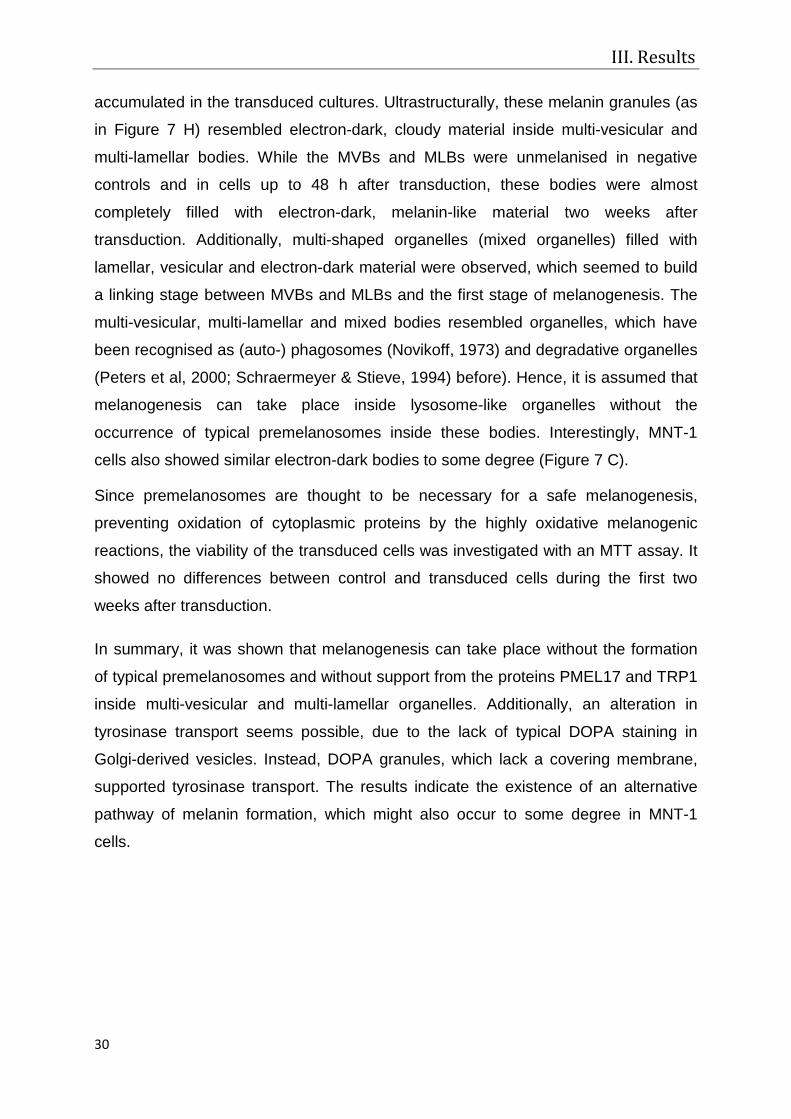
In contrast to control cells (Figure 8 A), transduced cells appeared “redifferentiated”
showing a typical cobblestone morphology (Figure 8 B) and melanin pigmentation
(Figure 8 C). Additionally, transduced cells appeared to be higher than neighbouring
non- transduced cells (Figure 8 D). This might be due to a rearrangement of the cell
structure into the cubic form they have in vivo.
III. Results
33
Comparing the influence of tyrosinase on phagocytosis and vice-versa it was
observed that while the transduced cells were able to phagocytose four times more
ROS than non-transduced cells, the fed transduced samples had significantly more
tyrosinase positive cells than non-fed transduced samples. It was also observed that
fed transduced samples produced visible amounts of melanin more rapidly than non-
fed cells. Possibly this was due to an increase in substrate availability by
phagocytosis of outer segments, since the unspecific phagocytosis of latex beads
had no additional effect on tyrosinase expression. Hence only the specific
phagocytosis of outer segments had an effect on melanising machinery. Besides,
non-transduced control cells showed a base level of DOPA positive organelles, which
was not observable with fluorescence immunocytochemistry.
Figure 8: Morphology of ARPE-19 cells: A-C light microscopy, D electron micrograph: A) Control ARPE-19 cells had a mostly spindle shaped, non-pigmented appearance (here: day 7). B) In the first days after transduction, cells developed a hexagonal morphology, grew bigger and appeared more prominent than control cells (here: day 6). C) After one week of transduction, first melanin granules were observed. The light micrograph shows a transduced and fed sample, 11 days after transduction. D) Electron micrograph of DOPA reaction at low magnification: the transduced cell in the middle, DOPA positive vesicles can clearly be recognised, is to 1/3 higher than neighbouring cells, which lack DOPA staining and thus might not be transduced (Figure from manuscript 4).

III. Results
34
In conclusion, tyrosinase expression caused a more differentiated morphology
resembling the in vivo state, including pigmentation, epithelial morphology, and an
improved function (increased phagocytic activity) compared to the control. In
addition, the specific phagocytosis of outer segments was able to increase both
tyrosinase expression and function leading to an accelerated melanogenesis in
transduced and fed ARPE-19 cells.

IV. Discussion
35
IV. Discussion
Age pigments impair the antioxidative capacity of melanin and zinc in aged RPE cells
The aim of this work was to evaluate the function of tyrosinase and melanin in adult
human RPE cells. For this purpose, it was first investigated whether melanin has an
antioxidative function in human RPE (Manuscript 1) by analysing the response of
human donor RPE cells to UV-A induced oxidative stress in culture. Typical donors
had an age above 50 years, implicating that the RPE cells contained not only
granules with antioxidative melanin but also aged melanin, melanolipofuscin and
lipofuscin, which lack antioxidative capacity and even nullified the effects of the
supplemented antioxidant zinc chloride. Aged melanin (Rozanowski et al, 2008b;
Zareba et al, 2006) and melanolipofuscin (Warburton et al, 2007) have been found to
act prooxidatively and can increase the stress in the eye when illuminated with UV
and blue light. Consequently, the non-pigmented ARPE-19 cells showed a better
survival and less apoptosis than the pigmented donor RPE cells. Since it was difficult
to obtain younger donor-RPE cells, which lack age pigments, this project was halted
and a system had to be generated, in which melanin pigmentation could be studied
without disturbing age pigments. As a consequence, the main part of the work
concentrated on the question of how melanogenesis can be induced in human RPE
cells.
Involvement of ROS phagocytosis in tyrosinase expression of adult human RPE cells
First, it was investigated whether tyrosinase can be induced in cultured human RPE
cells after phagocytosis of retinal outer segments, as published by Schraermeyers
group for bovine RPE cells (Schraermeyer et al, 2006). It was found that pigmented
donor RPE cells were able to express the tyrosinase protein in response to
phagocytic events, and that the enzyme was closely associated with remnants of the
phagocytosed material, as observed ultrastructurally (colocalisation of DOPA
reaction with latex beads) and in immunocytochemistry experiments (colocalisation of
rhodopsin and tyrosinase antibodies) (Manuscript 2). It was not surprising that
tyrosinase expression can be affected by ROS phagocytosis, since ROS
phagocytosis has been found to affect the expression of multiple genes of many
IV. Discussion
36
different functions in the RPE (Chowers et al, 2004). Hence, pathways yet unknown
may have been activated or silenced to make melanogenesis possible in the present
experiments.
Since it was difficult to observe tyrosinase activity and the renewal of melanosomes
in pigmented tissues, the experiments were repeated with non-pigmented ARPE-19
cells as a next step. In cell culture, RPE cells lose their original pigmentation, due to
ongoing mitosis without restoration of the pigment granules. Immortalised ARPE-19
cells are thus more or less unpigmented but retain the ability to produce small
amounts of melanin (Dunn et al, 1996) as can be observed ultrastructurally and with
DOPA histochemistry (Manuscript 4), but this pigmentation has only minor
significance.
ARPE-19 cells acted completely differently compared to donor RPE cells, since they
always showed a small number of DOPA granules, but in an amount which was not
observable with fluorescent microscopical methods, and did not express more
tyrosinase in response to feeding (Manuscript 4). This might be due to changes in
differentiation and protein expression of the immortalised ARPE-19 cells compared to
primary donor RPE cells. Differences in the gene expression of primary RPE cells
and ARPE-19 cells have been shown before by (Cai & Del Priore, 2006). Thus
tyrosinase expression had to be increased by transduction with the adenoviral vector
AdTyr. Subsequently, a phagocytosis-induced upregulation of tyrosinase activity was
also observed, as anticipated (Manuscript 4) and likewise the formation of
melanosomes was accelerated.
Phagocytosis of ROS enhances melanogenesis in tyrosinase transduced ARPE-19 cells
In transduced ARPE-19 cells, purely the ingestion of ROS, not the ingestion of latex
beads, enhanced tyrosinase expression and activity. Moreover, the observed
melanin synthesis was further enhanced only by phagocytosis of retinal outer
segments. This suggests that merely the specific phagocytosis of ROS via MERTK
receptors (which is reviewed by (Strauss, 2005)) and downstream effectors is able to
interact with tyrosinase activity in ARPE-19 melanogenesis. Possibly, tyrosine
residues from the ingested material were used for melanin synthesis, since
biochemical experiments have shown that peptides and proteins can be converted
IV. Discussion
37
into melano-protein by tyrosinase (Ito et al, 1984; Marumo & Waite, 1986;
Schraermeyer, 1994).
However, the transport of phagocytosed material to newly formed melanosomes was
not confirmable, since the labelling of ROS with gold granules (data not shown) was
not successful. It was also difficult to recognise the ROS inside phagosomes of
routine EM experiments, because the ingested material often did not have the typical
membranous layers as has been previously observed (Schraermeyer et al, 1999b).
Due to the threat of bovine spongiform encephalitis (BSE), it is no longer permitted to
use bovine eyes and therefore porcine material was used. Since porcine eyes are
smaller than bovine eyes, much more material was needed to gather enough ROS
for the experiments. Thus they had to be frozen after preparation and thawed later for
the phagocytosis tests. Possibly the freezing process affected the ultrastructural
morphology of the ROS and their binding ability to immuno-gold labelling.
Nevertheless, melanin was synthesised in vesicles, resembling degradative
organelles by ultrastructure, as discussed below. This melanogenesis was enhanced
after phagocytosis of ROS.
Interestingly, phagocytosis-related auto-fluorescence or lipofuscinogenesis have not
been observed either. In contrast, earlier cell culture studies showed an increase in
phagocytosis-related auto-fluorescence in RPE and to a higher extent in IPE (iris
pigment epithelium) cells after ROS feeding, which was contributed to the formation
of undegradable material. The effect was greater in IPE cells, since IPE cells do not
phagocytose ROS under natural conditions (Dintelmann et al, 1999). It is known that
the accumulation of undegradable material in the RPE leads not only to
lipofuscinogenesis but also to further impairment of degradation and to a prooxidative
atmosphere in the background of the eye. The improved phagocytic capacity of
tyrosinase transduced ARPE-19 cells, which was also observed in the present work
(four times more phagosomes compared to control cells), leads to the assumption
that a better degradation is possible in the presence of tyrosinase.
Tyrosinase protects against oxidative stress after phagocytosis
The upregulation of tyrosinase after phagocytosis on the one hand and the
upregulation of phagocytosis after tyrosinase expression on the other hand, can be
explained as follows. Possibly, tyrosinase acts in defence against phagocytosis-
IV. Discussion
38
related oxidative stress, either by production of antioxidative melanin or by
scavenging the stress itself. Thereupon, phagocytosis was upregulated without
increasing oxidative reactions. In melanocytes, tyrosinase has been found to interact
with oxidants like H2O2 (Wood & Schallreuter, 1991), but it is still unclear whether it
has pro- or antioxidative function here. RPE melanosomes have also been found to
have peroxidase activity, when they fused with phago-lysosomes (Schraermeyer &
Stieve, 1994). Thus, they might be able to decrease peroxide production of ingested
material. Furthermore, dark skinned people suffer less oxidative stress in the
background of the eye than Caucasians, resulting in a reduced prevalence for AMD
(Ambati et al, 2003; Gregor & Joffe, 1978; Pauleikhoff & Holz, 1996; Weiter et al,
1986). Whether this is due to the antioxidative action of tyrosinase, melanin itself or
its precursors remains unknown. Additional data (not shown in the manuscripts; n=1,
6 wells) showed that transduced, pigmented ARPE-19 cells had the ability to survive
more than 38 days in cell culture without passaging, while control cells died earlier.
The results gathered do not prove, but strongly indicate, a function of tyrosinase in
reducing oxidative stress in the adult RPE. Future experiments, using tyrosinase
transduced ARPE-19 cells in the UV-A light set up, as introduced in Manuscript 1,
might clarify the hypothesis completely.
Improvement of RPE characteristics after tyrosinase transduction
Like other cell types, RPE cells also undergo senescence in culture. They lose typical
RPE characteristics like their epithelial morphology (Grisanti & Guidry, 1995), their
pigmentation and phagocytic function (Carr et al, 2009). Likewise, immortalised
ARPE-19 cells dedifferentiate with ongoing passages and gain a spindle-shaped,
fibroblastic appearance, although they keep many functions for a long time (Dunn et
al, 1996). However, it was found that ARPE-19 cells, which were transduced with
AdTyr, appeared “redifferentiated” compared to control cells, and regained their
hexagonal, epithelial morphology and pigmentation, and they also showed an
enhanced phagocytic activity. It is assumed that tyrosinase activated downstream
effectors of differentiation, leading to changes in morphology and improved
functioning of RPE cells. This is also indicated by findings from Gargiulo et al., who
observed that retinal pigmentation and vision were regained in a mouse model of
oculo-cutaneous albinism after tyrosinase transduction (Gargiulo et al, 2009).

IV. Discussion
39
The premelanosome and the classical pathway of melanogenesis are not involved in ARPE-19 cell melanogenesis
In the experiments for Manuscript 3, it was observed that melanogenesis can occur
without the formation of premelanosomes. The lack of essential melanogenic
proteins (PMEL17 and TRP1) together with the complete absence of typical
melanogenic stages in ARPE-19 cells revealed that an alternative pathway of
melanin formation does exist in this artificial
system. Tyrosinase did not show the typical
distribution inside trans-Golgi cisterns and
Golgi-derived vesicles (approx. 59 nm in
diameter), as described for melanocytes
(Hearing, 2005; Raposo et al, 2007) and
pre-natal RPE in the literature (Stanka,
1971). Instead, it was transported from the
ER to smaller DOPA granules (approx. 34
nm in diameter), which had no membrane
(Figure 9) and further to multi-shaped
organelles of unknown origin, where melanin
was synthesised later on. Hence, an
alternative pathway of tyrosinase trafficking is proposed. It is assumed that
tyrosinase was synthesised at the ER and transported to the melanogenic stages
without processing in the Golgi apparatus, as indicated by the frequent DOPA
staining of cytoplasmic membranes of unknown origin and the staining of typical ER
tubules. On the other hand, the high occurrence of free tyrosinase in the cytoplasm
indicates a production of the enzyme at the free ribosomes, as proposed by (Moyer,
1963). Normally, aberrations in processing and transportation of tyrosinase lead to
degradation of the protein inside proteasomes (Berson et al, 2000; Halaban et al,
1997) and lysosomes (Chen et al, 2009; Wasmeier et al, 2006). Here, proper
glycosilation and processing of the enzymes was not evaluated, but the presence of
eumelanin inside former multi-lamellar and multi-vesicular bodies in the present work
indicates a suitable processing.
Figure 9: DOPA granules of a transduced APRE 19 cell: DOPA granules (Dg) have the half diameter of Golgi-derived vesicles (Gv) and lack a covering membrane. In contrast, the double membrane of the mitochondria (M) can clearly be observed as two independent lines. Inset: as in 9, higher magnification

IV. Discussion
40
For comparison to the classical stages I to IV, the melanogenic stages in ARPE-19
cells were termed stages 1 and 2 for early stages and stage 3 and 4 for later stages.
Table 1 summarises the occurrence of MVBs, MLBs and mixed bodies as precursors
of the melanogenic stages 1-4 at different time points after transduction. The
resulting mature melanin granule (stage 4) was morphologically similar to the round
melanosomes of melanocytes, but not to typical oval RPE melanosomes.
Table 1: Occurrence of unusual melanogenic stages 1 to 4 in controls (c) and at different time points after transduction: - rarely seen (less than 3% of counted organelles), + present (5-7%), ++ frequently seen (-15%), +++ often seen (-25%), ++++ mainly seen (>26%). In addition, multi-vesicular bodies (MVB), multi-lamellar bodies (MLB) and mixed bodies are included, which are suggested to be the precursors of melanogenesis in ARPE-19 cells (Figure from manuscript 3).
Since DOPA vesicles were found inside phagosomes (Figure 7 H) and the
ultrastructure of the melanised multi-shaped organelles was similar to the phagocytic
vesicles of Manuscript 2, and earlier studies (Novikoff, 1973; Schraermeyer &
Stieve, 1994) it is speculated whether the MVBs and MLBs documented in this work
belong to the phago-lysosomal pathway. Although it is still not confirmed that
phagocytosed material was used for melanin synthesis, it is assumed that
melanogenesis occurred in lysosome-like organelles without the additional
involvement of premelanosomes, Golgi-derived vesicles and the typical stages of
melanin formation.
Interestingly, similar electron-dark and cloudy organelles were also found in the
MNT-1 cells, together with the typical melanogenic stages. It is assumed that the
pathway of melanin synthesis which has been described for the transduced ARPE-19
cells, does also occur in MNT-1 cells. A comparison of the classical pathway of
melanogenesis and the alternative pathway, described here is given in Figure 10.
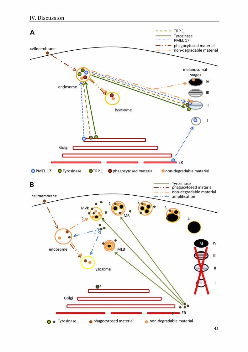
IV. Discussion
41

IV. Discussion
42
Figure 10: Comparison of the typical pathway of melanogenesis in pigment cells (A) and a newly discovered pathway for adult ARPE-19 cells found in this work (B): A) The scheme describes the typical transport pathways of melanogenic proteins to the classical stages of melanogenesis (scheme adapted from Hearing 2005): Tyrosinase, TRP1 and PMEL17 travel inside coated Golgi-derived vesicles first to endocytic organelles (early and late endosomes) and later to stage II melanosomes. PMEL17 is also able to travel directly to stage I melanosomes. Additionally, the transport of phagocytosed material to endocytic organelles and finally to lysosomes and to melanosomes (non-degradable material) in RPE cells is described. B) Alternative pathway of melanin synthesis in tyrosinase-transduced ARPE-19 cells. Only tyrosinase, without support from Pmel17 and TRP1, is needed for melanogenesis. Premelanosomes and typical stages of melanogenesis (I-IV) have not been found; instead, melanin formation takes place in multi-lamellar (MLB), multi-vesicular (MVB) and mixed (MB) lysosome-like organelles. The assumed sequence of melanogenic phases in this system is indicated by the numbers 1-4. Tyrosinase trafficking is also different to the classical scheme, since small DOPA granules, which lack a covering membrane, are observed in the cytoplasm instead of typical Golgi-derived vesicles. Actually, it is not evident if the Golgi apparatus and related vesicles are involved in this pathway (black ?) and it is still not confirmed whether phagocytosed material is integrated in the melanosomal matrix (gray ?). Phagocytosis and tyrosinase presence amplify each other’s occurrence and
activity (blue +).
Findings from animal experiments
For the animal experiments (which are not included in the manuscripts), the vector
AdTyr was injected sub-retinally into the eyes of albino rats (n=4 Wistar, n=1
Sprague Dawley) and one albino rabbit (New Zealand White). The eyes were
investigated with DOPA and routine electron microscopy at different time points after
transduction. DOPA staining was observed in ER tubules, but was lacking in Golgi
cisterns and related vesicles. Membrane-bound electron-dark organelles, resembling
melanin granules, were frequently found (Figure 11). Whether these electron-dark
organelles are composed of melanin polymers has not been studied yet.
Nevertheless, the experiments implicate that the findings from the ARPE-19 cells can
also be transferred to the in vivo system. Future experiments, using novel electron
microscopical methods like EDX (Energy Dispersive X-ray spectroscopy) and EELS
(Electron Energy Loss Spectroscopy) on the one hand and cryo-electron microscopy
and labelling of retinal outer segments with gold-labelled antibodies or 3H-labeleld
protein on the other hand will determine respectively the chemical composition of
RPE pigments and whether degraded material can be incorporated in newly formed
melanin granules.
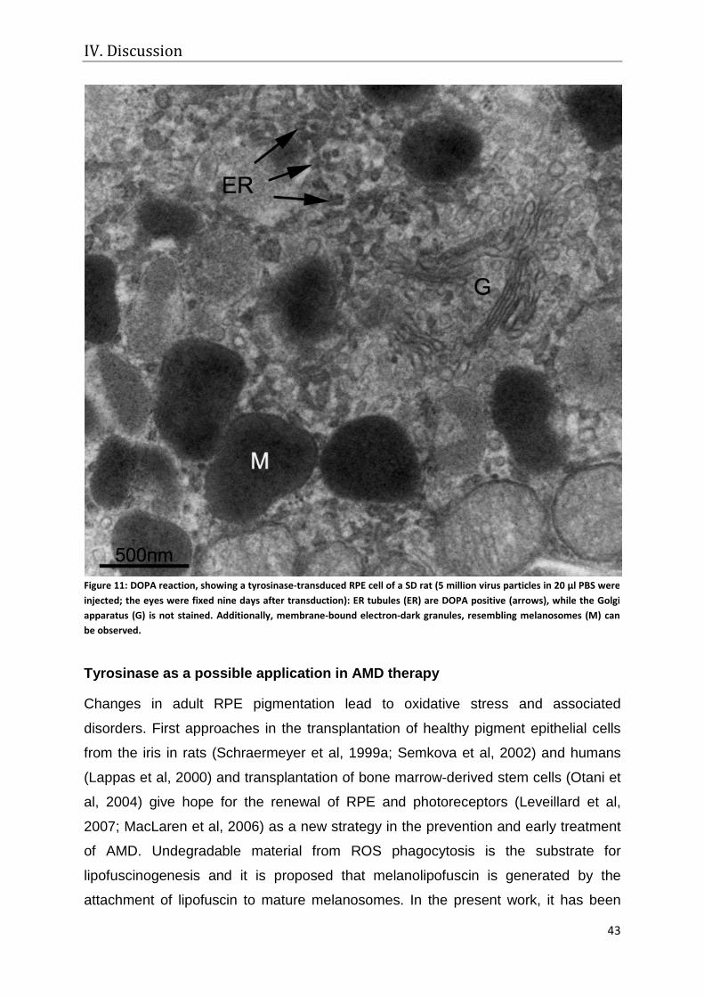
IV. Discussion
43
Tyrosinase as a possible application in AMD therapy
Changes in adult RPE pigmentation lead to oxidative stress and associated
disorders. First approaches in the transplantation of healthy pigment epithelial cells
from the iris in rats (Schraermeyer et al, 1999a; Semkova et al, 2002) and humans
(Lappas et al, 2000) and transplantation of bone marrow-derived stem cells (Otani et
al, 2004) give hope for the renewal of RPE and photoreceptors (Leveillard et al,
2007; MacLaren et al, 2006) as a new strategy in the prevention and early treatment
of AMD. Undegradable material from ROS phagocytosis is the substrate for
lipofuscinogenesis and it is proposed that melanolipofuscin is generated by the
attachment of lipofuscin to mature melanosomes. In the present work, it has been
Figure 11: DOPA reaction, showing a tyrosinase-transduced RPE cell of a SD rat (5 million virus particles in 20 µl PBS were injected; the eyes were fixed nine days after transduction): ER tubules (ER) are DOPA positive (arrows), while the Golgi apparatus (G) is not stained. Additionally, membrane-bound electron-dark granules, resembling melanosomes (M) can be observed.
IV. Discussion
44
shown that tyrosinase expression enhances phagocytosis of RPE cells, leads to a
morphological redifferentiation of cultured cells and to repigmentation. It has also
been observed that phagocytosis of ROS enhances melanogenesis in this system
without the generation of prooxidative lipofuscin. The properties of the tyrosinase
vector bear the possibility of rescuing RPE cells of elderly people. Future
experiments, with more improved tyrosinase vectors might help in creating a gene
therapy for the prevention of age-related maculopathies.
In conclusion, the question whether tyrosinase and melanisation are involved in
defence against stress has not been fully answered. Unfortunately, the influence of
melanin in primary RPE cells could not be investigated, because lipofuscin
accumulation in aged cells distorted the results. Nevertheless, some findings indicate
a protective role for tyrosinase, since transduced cells appeared morphologically
reorganised and had improved functions. In addition, the viability of ARPE-19 cells
after transduction was not altered compared to non-transduced controls,
demonstrating that tyrosinase and newly formed melanin were not toxic to the cells.
The hypothesis that melanogenesis can be induced in non-pigmented adult human
RPE cells has been verified, since melanin synthesis was measured quantitatively
with light and electron microscopy and qualitatively with HPLC analysis after
transduction with the vector AdTyr. Furthermore, a pathway of melanogenesis is
presented, which is different to the classical scheme described in the literature. This
pathway lacks both typical premelanosomes and tyrosinase trafficking using Golgi-
derived vesicles. Instead, a formation of tyrosinase at free ribosomes and trafficking
of the soluble protein to melanosomal stages are indicated.
Evaluating the influence of ROS phagocytosis on tyrosinase activity and
melanogenesis, diverging results have been obtained for primary donor RPE cells
and ARPE-19 cells, respectively. While primary cells were able to activate tyrosinase
in response to phagocytosis, non-transduced ARPE-19 cells did not produce
tyrosinase in response to feeding alone. Nevertheless, after induction of tyrosinase
with the adenoviral vector AdTyr, they synthesised more tyrosinase in response to
ROS feeding than merely transduced cells. Additionally, they were also able to build
more melanin. The transfer of ingested material to melanosomes could not be
confirmed for ARPE-19 cells. Nevertheless, in the present experiments, no lipofuscin
IV. Discussion
45
was generated, demonstrating that the degradation machinery was not inhibited by
the experiments. Moreover, the absence of lipofuscin indicates a beneficial role for
tyrosinase in ROS degradation.
V. References
46
V. References
Abul-Hassan K, Walmsley R, Tombran-Tink J, Boulton M (2000) Regulation of tyrosinase expression and activity in cultured human retinal pigment epithelial cells. Pigment Cell Res 13(6): 436-441
Age-related Eye Disease Study Research Group A (2001) A randomized, placebo-controlled, clinical trial of high-dose supplementation with vitamins C and E, beta carotene, and zinc for age-related macular degeneration and vision loss: AREDS report no. 8. Arch Ophthalmol 119(10): 1417-1436
Ambati J, Ambati BK, Yoo SH, Ianchulev S, Adamis AP (2003) Age-related macular degeneration: etiology, pathogenesis, and therapeutic strategies. Surv Ophthalmol 48(3): 257-293
Aroca P, Solano F, Salinas C, Garcia-Borron JC, Lozano JA (1992) Regulation of the final phase of mammalian melanogenesis. The role of dopachrome tautomerase and the ratio between 5,6-dihydroxyindole-2-carboxylic acid and 5,6-dihydroxyindole. Eur J Biochem 208(1): 155-163
Berson JF, Frank DW, Calvo PA, Bieler BM, Marks MS (2000) A common temperature-sensitive allelic form of human tyrosinase is retained in the endoplasmic reticulum at the nonpermissive temperature. J Biol Chem 275(16): 12281-12289
Berson JF, Theos AC, Harper DC, Tenza D, Raposo G, Marks MS (2003) Proprotein convertase cleavage liberates a fibrillogenic fragment of a resident glycoprotein to initiate melanosome biogenesis. J Cell Biol 161(3): 521-533
Bharti K, Liu W, Csermely T, Bertuzzi S, Arnheiter H (2008) Alternative promoter use in eye development: the complex role and regulation of the transcription factor MITF. Development 135(6): 1169-1178
Bharti K, Nguyen MT, Skuntz S, Bertuzzi S, Arnheiter H (2006) The other pigment cell: specification and development of the pigmented epithelium of the vertebrate eye. Pigment Cell Res 19(5): 380-394
Bialek S, Miller SS (1994) K+ and Cl- transport mechanisms in bovine pigment epithelium that could modulate subretinal space volume and composition. J Physiol 475(3): 401-417
Bird AC, Bressler NM, Bressler SB, Chisholm IH, Coscas G, Davis MD, de Jong PT, Klaver CC, Klein BE, Klein R, et al. (1995) An international classification and grading system for age-related maculopathy and age-related macular degeneration. The International ARM Epidemiological Study Group. Surv Ophthalmol 39(5): 367-374
Boissy RE, Zhao H, Oetting WS, Austin LM, Wildenberg SC, Boissy YL, Zhao Y, Sturm RA, Hearing VJ, King RA, Nordlund JJ (1996) Mutation in and lack of expression of tyrosinase-related protein-1 (TRP-1) in melanocytes from an individual with brown oculocutaneous albinism: a new subtype of albinism classified as "OCA3". Am J Hum Genet 58(6): 1145-1156
Bonifacino JS (2004) Insights into the biogenesis of lysosome-related organelles from the study of the Hermansky-Pudlak syndrome. Ann N Y Acad Sci 1038: 103-114
Borovansky J, Mommaas AM, Smit NP, Eygendaal D, Winder AJ, Vermeer BJ, Pavel S (1997) Melanogenesis in transfected fibroblasts induces lysosomal activation. Arch Dermatol Res 289(3): 145-150
Bosch E, Horwitz J, Bok D (1993) Phagocytosis of outer segments by retinal pigment epithelium: phagosome-lysosome interaction. J Histochem Cytochem 41(2): 253-263
Boulton M, Dayhaw-Barker P (2001) The role of the retinal pigment epithelium: topographical variation and ageing changes. Eye 15(Pt 3): 384-389
Boulton M, Rozanowska M, Rozanowski B (2001) Retinal photodamage. J Photochem Photobiol B 64(2-3): 144-161
Boulton M, Rozanowska M, Rozanowski B, Wess T (2004) The photoreactivity of ocular lipofuscin. Photochem Photobiol Sci 3(8): 759-764
Branza-Nichita N, Petrescu AJ, Dwek RA, Wormald MR, Platt FM, Petrescu SM (1999) Tyrosinase folding and copper loading in vivo: a crucial role for calnexin and alpha-glucosidase II. Biochem Biophys Res Commun 261(3): 720-725
Bray TM, Bettger WJ (1990) The physiological role of zinc as an antioxidant. Free Radic Biol Med 8(3): 281-291
Breathnach AS (1988) Extra-cutaneous melanin. Pigment Cell Res 1(4): 234-237
Brewer GJ, Hill GM, Prasad AS, Cossack ZT (1983) Biological roles of ionic zinc. Prog Clin Biol Res 129: 35-51
Cai H, Del Priore LV (2006) Gene expression profile of cultured adult compared to immortalized human RPE. Mol Vis 12: 1-14
Calvo PA, Frank DW, Bieler BM, Berson JF, Marks MS (1999) A cytoplasmic sequence in human tyrosinase defines a second class of di-leucine-based sorting signals for late endosomal and lysosomal delivery. J Biol Chem 274(18): 12780-12789
Camacho-Hubner A, Richard C, Beermann F (2002) Genomic structure and evolutionary conservation of the tyrosinase gene family from Fugu. Gene 285(1-2): 59-68
V. References
47
Carr AJ, Vugler A, Lawrence J, Chen LL, Ahmado A, Chen FK, Semo M, Gias C, da Cruz L, Moore HD, Walsh J, Coffey PJ (2009) Molecular characterization and functional analysis of phagocytosis by human embryonic stem cell-derived RPE cells using a novel human retinal assay. Mol Vis 15: 283-295
Carr RE, Siegel IM (1979) The retinal pigment epitehlium in ocular albinism;. In The retinal pigment epithelium, Zinn KM, Mamor MF (eds), pp 413-423. Cambridge: Harvard University Press
Chakraborty AK, Platt JT, Kim KK, Kwon BS, Bennett DC, Pawelek JM (1996) Polymerization of 5,6-dihydroxyindole-2-carboxylic acid to melanin by the pmel 17/silver locus protein. Eur J Biochem 236(1): 180-188
Chen H, Lukas TJ, Du N, Suyeoka G, Neufeld AH (2009) Dysfunction of the retinal pigment epithelium with age: increased iron decreases phagocytosis and lysosomal activity. Invest Ophthalmol Vis Sci 50(4): 1895-1902
Chowers I, Kim Y, Farkas RH, Gunatilaka TL, Hackam AS, Campochiaro PA, Finnemann SC, Zack DJ (2004) Changes in retinal pigment epithelial gene expression induced by rod outer segment uptake. Invest Ophthalmol Vis Sci 45(7): 2098-2106
D'Cruz PM, Yasumura D, Weir J, Matthes MT, Abderrahim H, LaVail MM, Vollrath D (2000) Mutation of the receptor tyrosine kinase gene Mertk in the retinal dystrophic RCS rat. Hum Mol Genet 9(4): 645-651
Dayhaw-Barker P (2002) Retinal pigment epithelium melanin and ocular toxicity. Int J Toxicol 21(6): 451-454
Deng Z, Liu X, Chen J, Yang R (2000) [Zinc protective effects on pig retinal pigment epithelial cell damage of lipid peroxide induced by 2450 MHz microwave]. Wei Sheng Yan Jiu 29(3): 129-131
Diment S, Eidelman M, Rodriguez GM, Orlow SJ (1995) Lysosomal hydrolases are present in melanosomes and are elevated in melanizing cells. J Biol Chem 270(9): 4213-4215
Dintelmann TS, Heimann K, Kayatz P, Schraermeyer U (1999) Comparative study of ROS degradation by IPE and RPE cells in vitro. Graefes Arch Clin Exp Ophthalmol 237(10): 830-839
Dorey CK, Torres X, Swart T (1990) Evidence of melanogenesis in porcine retinal pigment epithelial cells in vitro. Exp Eye Res 50(1): 1-10
Dorey CK, Wu G, Ebenstein D, Garsd A, Weiter JJ (1989) Cell loss in the aging retina. Relationship to lipofuscin accumulation and macular degeneration. Invest Ophthalmol Vis Sci 30(8): 1691-1699
Dryja TP, O'Neil-Dryja M, Pawelek JM, Albert DM (1978) Demonstration of tyrosinase in the adult bovine uveal tract and retinal pigment epithelium. Invest Ophthalmol Vis Sci 17(6): 511-514
Dunn KC, Aotaki-Keen AE, Putkey FR, Hjelmeland LM (1996) ARPE-19, a human retinal pigment epithelial cell line with differentiated properties. Exp Eye Res 62(2): 155-169
Eldred GE (1989) Vitamins A and E in RPE lipofuscin formation and implications for age-related macular degeneration. Prog Clin Biol Res 314: 113-129
Eldred GE (1995) Lipofuscin fluorophore inhibits lysosomal protein degradation and may cause early stages of macular degeneration. Gerontology 41 Suppl 2: 15-28
Eldred GE, Miller GV, Stark WS, Feeney-Burns L (1982) Lipofuscin: resolution of discrepant fluorescence data. Science 216(4547): 757-759
Eppig JJ, Jr. (1970) Melanogenesis in amphibians. I. A study of the fine structure of the normal and phenylthiourea-treated pigmented epithelium in Rana pipiens tadpole eyes. Z Zellforsch Mikrosk Anat 103(2): 238-246
Eppig JJ, Jr., Dumont JN (1972) Cytochemical localization of tyrosinase activity in pigmented epithelial cells of Rana pipiens and Xenopus laevis larvae. J Ultrastruct Res 39(3): 397-410
Feeney-Burns L, Eldred GE (1983) The fate of the phagosome: conversion to 'age pigment' and impact in human retinal pigment epithelium. Trans Ophthalmol Soc U K 103 ( Pt 4): 416-421
Feeney-Burns L, Gao CL, Berman ER (1988) The fate of immunoreactive opsin following phagocytosis by pigment epithelium in human and monkey retinas. Invest Ophthalmol Vis Sci 29(5): 708-719
Feeney-Burns L, Hilderbrand ES, Eldridge S (1984) Aging human RPE: morphometric analysis of macular, equatorial, and peripheral cells. Invest Ophthalmol Vis Sci 25(2): 195-200
Flannery JG, O'Day W, Pfeffer BA, Horwitz J, Bok D (1990) Uptake, processing and release of retinoids by cultured human retinal pigment epithelium. Exp Eye Res 51(6): 717-728
Fowler DM, Koulov AV, Alory-Jost C, Marks MS, Balch WE, Kelly JW (2006) Functional amyloid formation within mammalian tissue. PLoS Biol 4(1): e6
Francis E, Wang N, Parag H, Halaban R, Hebert DN (2003) Tyrosinase maturation and oligomerization in the endoplasmic reticulum require a melanocyte-specific factor. J Biol Chem 278(28): 25607-25617

V. References
48
Gargiulo A, Bonetti C, Montefusco S, Neglia S, Di Vicino U, Marrocco E, Corte MD, Domenici L, Auricchio A, Surace EM (2009) AAV-mediated Tyrosinase Gene Transfer Restores Melanogenesis and Retinal Function in a Model of Oculo-cutaneous Albinism Type I (OCA1). Mol Ther
Glenn JV, Mahaffy H, Wu K, Smith G, Nagai R, Simpson DA, Boulton ME, Stitt AW (2009) Advanced glycation end product (AGE) accumulation on Bruch's membrane: links to age-related RPE dysfunction. Invest Ophthalmol Vis Sci 50(1): 441-451
Gregor Z, Joffe L (1978) Senile macular changes in the black African. Br J Ophthalmol 62(8): 547-550
Grisanti S, Guidry C (1995) Transdifferentiation of retinal pigment epithelial cells from epithelial to mesenchymal phenotype. Invest Ophthalmol Vis Sci 36(2): 391-405
Halaban R, Cheng E, Svedine S, Aron R, Hebert DN (2001) Proper folding and endoplasmic reticulum to golgi transport of tyrosinase are induced by its substrates, DOPA and tyrosine. J Biol Chem 276(15): 11933-11938
Halaban R, Cheng E, Zhang Y, Moellmann G, Hanlon D, Michalak M, Setaluri V, Hebert DN (1997) Aberrant retention of tyrosinase in the endoplasmic reticulum mediates accelerated degradation of the enzyme and contributes to the dedifferentiated phenotype of amelanotic melanoma cells. Proc Natl Acad Sci U S A 94(12): 6210-6215
Halaban R, Svedine S, Cheng E, Smicun Y, Aron R, Hebert DN (2000) Endoplasmic reticulum retention is a common defect associated with tyrosinase-negative albinism. Proc Natl Acad Sci U S A 97(11): 5889-5894
Hearing VJ (2005) Biogenesis of pigment granules: a sensitive way to regulate melanocyte function. J Dermatol Sci 37(1): 3-14
Hearing VJ, Korner AM, Pawelek JM (1982) New regulators of melanogenesis are associated with purified tyrosinase isozymes. J Invest Dermatol 79(1): 16-18
Hearing VJ, Tsukamoto K, Urabe K, Kameyama K, Montague PM, Jackson IJ (1992) Functional properties of cloned melanogenic proteins. Pigment Cell Res 5(5 Pt 2): 264-270
Heiduschka P, Blitgen-Heinecke P, Tura A, Kokkinou D, Julien S, Hofmeister S, Bartz-Schmidt KU, Schraermeyer U (2007) Melanin precursor 5,6-dihydroxyindol: protective effects and cytotoxicity on retinal cells in vitro and in vivo. Toxicol Pathol 35(7): 1030-1038
Herman KG, Steinberg RH (1982) Melanosome metabolism in the retinal pigmented epithelium of the opossum. Cell Tissue Res 227(3): 485-507
Holz FG, Bindewald-Wittich A, Fleckenstein M, Dreyhaupt J, Scholl HP, Schmitz-Valckenberg S (2007) Progression of geographic atrophy and impact of fundus autofluorescence patterns in age-related macular degeneration. Am J Ophthalmol 143(3): 463-472
Huber M, Lerch K (1988) Identification of two histidines as copper ligands in Streptomyces glaucescens tyrosinase. Biochemistry 27(15): 5610-5615
Huizing M, Sarangarajan R, Strovel E, Zhao Y, Gahl WA, Boissy RE (2001) AP-3 mediates tyrosinase but not TRP-1 trafficking in human melanocytes. Mol Biol Cell 12(7): 2075-2085
Ito S, Kato T, Shinpo K, Fujita K (1984) Oxidation of tyrosine residues in proteins by tyrosinase. Formation of protein-bonded 3,4-dihydroxyphenylalanine and 5-S-cysteinyl-3,4-dihydroxyphenylalanine. Biochem J 222(2): 407-411
Ito S, Wakamatsu K (2003) Quantitative analysis of eumelanin and pheomelanin in humans, mice, and other animals: a comparative review. Pigment Cell Res 16(5): 523-531
Ito S, Wakamatsu K (2008) Chemistry of mixed melanogenesis--pivotal roles of dopaquinone. Photochem Photobiol 84(3): 582-592
Jackman MP, Hajnal A, Lerch K (1991) Albino mutants of Streptomyces glaucescens tyrosinase. Biochem J 274 ( Pt 3): 707-713
Jimbow K, Gomez PF, Toyofuku K, Chang D, Miura S, Tsujiya H, Park JS (1997) Biological role of tyrosinase related protein and its biosynthesis and transport from TGN to stage I melanosome, late endosome, through gene transfection study. Pigment Cell Res 10(4): 206-213
Jimbow K, Quevedo WC, Jr., Fitzpatrick TB, Szabo G (1976) Some aspects of melanin biology: 1950-1975. J Invest Dermatol 67(1): 72-89
Jimenez-Cervantes C, Garcia-Borron JC, Valverde P, Solano F, Lozano JA (1993) Tyrosinase isoenzymes in mammalian melanocytes. 1. Biochemical characterization of two melanosomal tyrosinases from B16 mouse melanoma. Eur J Biochem 217(2): 549-556
Jimenez-Cervantes C, Martinez-Esparza M, Solano F, Lozano JA, Garcia-Borron JC (1998) Molecular interactions within the melanogenic complex: formation of heterodimers of tyrosinase and TRP1 from B16 mouse melanoma. Biochem Biophys Res Commun 253(3): 761-767
V. References
49
Julien S, Kociok N, Kreppel F, Kopitz J, Kochanek S, Biesemeier A, Blitgen-Heinecke P, Heiduschka P, Schraermeyer U (2007) Tyrosinase biosynthesis and trafficking in adult human retinal pigment epithelial cells. Graefes Arch Clin Exp Ophthalmol 245(10): 1495-1505
Kim SM, Han S (2003) Tyrosinase scavenges tyrosyl radical. Biochem Biophys Res Commun 312(3): 642-649
Kobayashi T, Imokawa G, Bennett DC, Hearing VJ (1998) Tyrosinase stabilization by Tyrp1 (the brown locus protein). J Biol Chem 273(48): 31801-31805
Kobayashi T, Urabe K, Orlow SJ, Higashi K, Imokawa G, Kwon BS, Potterf B, Hearing VJ (1994) The Pmel 17/silver locus protein. Characterization and investigation of its melanogenic function. J Biol Chem 269(46): 29198-29205
Kokkinou D, Kasper HU, Schwarz T, Bartz-Schmidt KU, Schraermeyer U (2005) Zinc uptake and storage: the role of fundus pigmentation. Graefes Arch Clin Exp Ophthalmol 243(10): 1050-1055
Konari K, Sawada N, Zhong Y, Isomura H, Nakagawa T, Mori M (1995) Development of the blood-retinal barrier in vitro: formation of tight junctions as revealed by occludin and ZO-1 correlates with the barrier function of chick retinal pigment epithelial cells. Exp Eye Res 61(1): 99-108
Kroumpouzos G, Urabe K, Kobayashi T, Sakai C, Hearing VJ (1994) Functional analysis of the slaty gene product (TRP2) as dopachrome tautomerase and the effect of a point mutation on its catalytic function. Biochem Biophys Res Commun 202(2): 1060-1068
Lappas A, Weinberger AW, Foerster AM, Kube T, Rezai KA, Kirchhof B (2000) Iris pigment epithelial cell translocation in exudative age-related macular degeneration. A pilot study in patients. Graefes Arch Clin Exp Ophthalmol 238(8): 631-641
Lee ZH, Hou L, Moellmann G, Kuklinska E, Antol K, Fraser M, Halaban R, Kwon BS (1996) Characterization and subcellular localization of human Pmel 17/silver, a 110-kDa (pre)melanosomal membrane protein associated with 5,6,-dihydroxyindole-2-carboxylic acid (DHICA) converting activity. J Invest Dermatol 106(4): 605-610
Leveillard T, Mohand-Said S, Sahel JA (2007) [Retinal repair by transplantation of photoreceptor precursors]. Med Sci (Paris) 23(3): 240-242
Lopes VS, Wasmeier C, Seabra MC, Futter CE (2007) Melanosome maturation defect in Rab38-deficient retinal pigment epithelium results in instability of immature melanosomes during transient melanogenesis. Mol Biol Cell 18(10): 3914-3927
MacLaren RE, Pearson RA, MacNeil A, Douglas RH, Salt TE, Akimoto M, Swaroop A, Sowden JC, Ali RR (2006) Retinal repair by transplantation of photoreceptor precursors. Nature 444(7116): 203-207
Marks MS, Seabra MC (2001) The melanosome: membrane dynamics in black and white. Nat Rev Mol Cell Biol 2(10): 738-748
Marmorstein AD, Finnemann SC, Bonilha VL, Rodriguez-Boulan E (1998) Morphogenesis of the retinal pigment epithelium: toward understanding retinal degenerative diseases. Ann N Y Acad Sci 857: 1-12
Martinez-Esparza M, Jimenez-Cervantes C, Garcia-Borron JC, Lozano JA, del Marmol V, Ghanem G, Solano F (1997) Comparison of TRPs from murine and human malignant melanocytes. Pigment Cell Res 10(4): 229-235
Marumo K, Waite JH (1986) Optimization of hydroxylation of tyrosine and tyrosine-containing peptides by mushroom tyrosinase. Biochim Biophys Acta 872(1-2): 98-103
Maul GG (1969) Golgi-melanosome relationship in human melanoma in vitro. J Ultrastruct Res 26(1): 163-176
Mishima H, Hasebe H, Fujita H (1978) Melanogenesis in the retinal pigment epithelial cell of the chick embryo. Dopa-reaction and electron microscopic autoradiography of 3H-dopa. Invest Ophthalmol Vis Sci 17(5): 403-411
Mishima Y (1994) Molecular and biological control of melanogenesis through tyrosinase genes and intrinsic and extrinsic regulatory factors. Pigment Cell Res 7(6): 376-387
Miyamoto M, Fitzpatrick TB (1957) On the nature of the pigment in retinal pigment epithelium. Science 126(3271): 449-450
Mosse CA, Meadows L, Luckey CJ, Kittlesen DJ, Huczko EL, Slingluff CL, Shabanowitz J, Hunt DF, Engelhard VH (1998) The class I antigen-processing pathway for the membrane protein tyrosinase involves translation in the endoplasmic reticulum and processing in the cytosol. J Exp Med 187(1): 37-48
Moyer FH (1963) Genetic effects on melanosome fine structure and ontogeny in normal and malignant cells. Ann N Y Acad Sci 100: 584-606
Murisier F, Beermann F (2006) Genetics of pigment cells: lessons from the tyrosinase gene family. Histol Histopathol 21(5): 567-578
Murisier F, Guichard S, Beermann F (2007) Distinct distal regulatory elements control tyrosinase expression in melanocytes and the retinal pigment epithelium. Dev Biol 303(2): 838-847
V. References
50
Nandrot EF, Finnemann SC (2008) Lack of alphavbeta5 integrin receptor or its ligand MFG-E8: distinct effects on retinal function. Ophthalmic Res 40(3-4): 120-123
Newsome DA (1994) Are antioxidants or supplements protective for age-related macular degeneration? Arch Ophthalmol 112(6): 729-730
Ng KP, Gugiu B, Renganathan K, Davies MW, Gu X, Crabb JS, Kim SR, Rozanowska MB, Bonilha VL, Rayborn ME, Salomon RG, Sparrow JR, Boulton ME, Hollyfield JG, Crabb JW (2008) Retinal pigment epithelium lipofuscin proteomics. Mol Cell Proteomics 7(7): 1397-1405
Novikoff AB (1973) Lysosomes, a personal account. In Lysosomes abd Storage diseases, Hers G. VHF (ed), p 1. New York
Novikoff AB, Albala A, Biempica L (1968) Ultrastructural and cytochemical observations on B-16 and Harding-Passey mouse melanomas. The origin of premelanosomes and compound melanosomes. J Histochem Cytochem 16(5): 299-319
Olivares C, Solano F, Garcia-Borron JC (2003) Conformation-dependent post-translational glycosylation of tyrosinase. Requirement of a specific interaction involving the CuB metal binding site. J Biol Chem 278(18): 15735-15743
Orlow SJ (1995) Melanosomes are specialized members of the lysosomal lineage of organelles. J Invest Dermatol 105(1): 3-7
Orlow SJ, Boissy RE, Moran DJ, Pifko-Hirst S (1993) Subcellular distribution of tyrosinase and tyrosinase-related protein-1: implications for melanosomal biogenesis. J Invest Dermatol 100(1): 55-64
Otani A, Dorrell MI, Kinder K, Moreno SK, Nusinowitz S, Banin E, Heckenlively J, Friedlander M (2004) Rescue of retinal degeneration by intravitreally injected adult bone marrow-derived lineage-negative hematopoietic stem cells. J Clin Invest 114(6): 765-774
Pauleikhoff D, Holz FG (1996) [Age-related macular degeneration. 1. Epidemiology, pathogenesis and differential diagnosis]. Ophthalmologe 93(3): 299-315
Pawelek JM (1991) After dopachrome? Pigment Cell Res 4(2): 53-62
Perluigi M, De Marco F, Foppoli C, Coccia R, Blarzino C, Marcante ML, Cini C (2003) Tyrosinase protects human melanocytes from ROS-generating compounds. Biochem Biophys Res Commun 305(2): 250-256
Peters S, Kayatz P, Heimann K, Schraermeyer U (2000) Subretinal injection of rod outer segments leads to an increase in the number of early-stage melanosomes in retinal pigment epithelial cells. Ophthalmic Res 32(2-3): 52-56
Peters S, Lamah T, Kokkinou D, Bartz-Schmidt KU, Schraermeyer U (2006) Melanin protects choroidal blood vessels against light toxicity. Z Naturforsch C 61(5-6): 427-433
Potts AM, Au PC (1976) The affinity of melanin for inorganic ions. Exp Eye Res 22(5): 487-491
Prota G (1992) The role of peroxidase in melanogenesis revisited. Pigment Cell Res Suppl 2: 25-31
Prota G, M. D'Ischia, M, Napolitano, A (1988) The chemistry of melanins and related metabolites. In The Pigmentary System, al. JNe (ed). Oxford: University Press
Rak DJ, Hardy KM, Jaffe GJ, McKay BS (2006) Ca++-switch induction of RPE differentiation. Exp Eye Res 82(4): 648-656
Rando RR (1992) Molecular mechanisms in visual pigment regeneration. Photochem Photobiol 56(6): 1145-1156
Raposo G, Marks MS (2002) The dark side of lysosome-related organelles: specialization of the endocytic pathway for melanosome biogenesis. Traffic 3(4): 237-248
Raposo G, Marks MS, Cutler DF (2007) Lysosome-related organelles: driving post-Golgi compartments into specialisation. Curr Opin Cell Biol 19(4): 394-401
Raposo G, Tenza D, Murphy DM, Berson JF, Marks MS (2001) Distinct protein sorting and localization to premelanosomes, melanosomes, and lysosomes in pigmented melanocytic cells. J Cell Biol 152(4): 809-824
Riley PA (1993) Mechanistic aspects of the control of tyrosinase activity. Pigment Cell Res 6(4 Pt 1): 182-185
Roberts JE (2002) Screening for ocular phototoxicity. Int J Toxicol 21(6): 491-500
Rozanowska M, Bober A, Burke JM, Sarna T (1997) The role of retinal pigment epithelium melanin in photoinduced oxidation of ascorbate. Photochem Photobiol 65(3): 472-479
Rozanowski B, Burke JM, Boulton ME, Sarna T, Rozanowska M (2008a) Human RPE melanosomes protect from photosensitized and iron-mediated oxidation but become pro-oxidant in the presence of iron upon photodegradation. Invest Ophthalmol Vis Sci 49(7): 2838-2847
Rozanowski B, Cuenco J, Davies S, Shamsi FA, Zadlo A, Dayhaw-Barker P, Rozanowska M, Sarna T, Boulton ME (2008b) The phototoxicity of aged human retinal melanosomes. Photochem Photobiol 84(3): 650-657
V. References
51
Samuelson DA, Smith P, Ulshafer RJ, Hendricks DG, Whitley RD, Hendricks H, Leone NC (1993) X-ray microanalysis of ocular melanin in pigs maintained on normal and low zinc diets. Exp Eye Res 56(1): 63-70
Sarna T (1992) Properties and function of the ocular melanin--a photobiophysical view. J Photochem Photobiol B 12(3): 215-258
Schraermeyer U (1992) Evidence for melanogenesis in the retinal pigment epithelium of adult cattle and golden hamster. Comp Biochem Physiol B 103(2): 435-442
Schraermeyer U (1993) Does melanin turnover occur in the eyes of adult vertebrates? Pigment Cell Res 6(4 Pt 1): 193-204
Schraermeyer U (1994) Transformation of albumin into melanin by hydroxyl radicals. Comp Biochem Physiol C Pharmacol Toxicol Endocrinol 108(3): 281-288
Schraermeyer U (1995) Transport of endocytosed material into melanin granules in cultured choroidal melanocytes of cattle--new insights into the relationship of melanosomes with lysosomes. Pigment Cell Res 8(4): 209-214
Schraermeyer U, Heimann K (1999) Current understanding on the role of retinal pigment epithelium and its pigmentation. Pigment Cell Res 12(4): 219-236
Schraermeyer U, Kociok N, Heimann K (1999a) Rescue effects of IPE transplants in RCS rats: short-term results. Invest Ophthalmol Vis Sci 40(7): 1545-1556
Schraermeyer U, Kopitz J, Peters S, Henke-Fahle S, Blitgen-Heinecke P, Kokkinou D, Schwarz T, Bartz-Schmidt KU (2006) Tyrosinase biosynthesis in adult mammalian retinal pigment epithelial cells. Exp Eye Res 83(2): 315-321
Schraermeyer U, Peters S, Thumann G, Kociok N, Heimann K (1999b) Melanin granules of retinal pigment epithelium are connected with the lysosomal degradation pathway. Exp Eye Res 68(2): 237-245
Schraermeyer U, Stieve H (1994) A newly discovered pathway of melanin formation in cultured retinal pigment epithelium of cattle. Cell Tissue Res 276(2): 273-279Seagle BL, Rezai KA, Gasyna EM, Kobori Y, Rezaei KA, Norris JR, Jr. (2005a) Time-resolved detection of melanin free radicals quenching reactive oxygen species. J Am Chem Soc 127(32): 11220-11221
Seagle BL, Rezai KA, Kobori Y, Gasyna EM, Rezaei KA, Norris JR, Jr. (2005b) Melanin photoprotection in the human retinal pigment epithelium and its correlation with light-induced cell apoptosis. Proc Natl Acad Sci U S A 102(25): 8978-8983
Seiji M, Shimao K, Birbeck MS, Fitzpatrick TB (1963) Subcellular localization of melanin biosynthesis. Ann N Y Acad Sci 100: 497-533
Semkova I, Kreppel F, Welsandt G, Luther T, Kozlowski J, Janicki H, Kochanek S, Schraermeyer U (2002) Autologous transplantation of genetically modified iris pigment epithelial cells: a promising concept for the treatment of age-related macular degeneration and other disorders of the eye. Proc Natl Acad Sci U S A 99(20): 13090-13095
Simmen T, Schmidt A, Hunziker W, Beermann F (1999) The tyrosinase tail mediates sorting to the lysosomal compartment in MDCK cells via a di-leucine and a tyrosine-based signal. J Cell Sci 112 ( Pt 1): 45-53
Smith-Thomas L, Richardson P, Thody AJ, Graham A, Palmer I, Flemming L, Parsons MA, Rennie IG, MacNeil S (1996) Human ocular melanocytes and retinal pigment epithelial cells differ in their melanogenic properties in vivo and in vitro. Curr Eye Res 15(11): 1079-1091
Solano F, Martinez-Liarte JH, Jimenez-Cervantes C, Garcia-Borron JC, Lozano JA (1994) Dopachrome tautomerase is a zinc-containing enzyme. Biochem Biophys Res Commun 204(3): 1243-1250
Spritz RA, Strunk KM, Giebel LB, King RA (1990) Detection of mutations in the tyrosinase gene in a patient with type IA oculocutaneous albinism. N Engl J Med 322(24): 1724-1728
Sprong H, Degroote S, Claessens T, van Drunen J, Oorschot V, Westerink BH, Hirabayashi Y, Klumperman J, van der Sluijs P, van Meer G (2001) Glycosphingolipids are required for sorting melanosomal proteins in the Golgi complex. J Cell Biol 155(3): 369-380
Stanka P (1971) [Electron microscopic study of the origin of premelanosomes of the retinal pigment epithelium in chick embryos]. Z Zellforsch Mikrosk Anat 112(1): 120-128
Stanka P, Rathjen P, Sahlmann B (1981) Evidence of membrane transformation during melanogenesis. Electron microscopic study on the retinal pigment epithelium of chick embryos. Cell Tissue Res 214(2): 343-353
Strauss O (2005) The retinal pigment epithelium in visual function. Physiol Rev 85(3): 845-881
Sturm RA (2009) Molecular genetics of human pigmentation diversity. Hum Mol Genet 18(R1): R9-17
Tachibana M (2000) MITF: a stream flowing for pigment cells. Pigment Cell Res 13(4): 230-240
Terman A, Brunk UT (1998) Lipofuscin: mechanisms of formation and increase with age. APMIS 106(2): 265-276

V. References
52
Theos AC, Berson JF, Theos SC, Herman KE, Harper DC, Tenza D, Sviderskaya EV, Lamoreux ML, Bennett DC, Raposo G, Marks MS (2006) Dual loss of ER export and endocytic signals with altered melanosome morphology in the silver mutation of Pmel17. Mol Biol Cell 17(8): 3598-3612
Thumann G, Bartz-Schmidt KU, Kociok N, Kayatz P, Heimann K, Schraermeyer U (1999) Retinal damage by light in the golden hamster: an ultrastructural study in the retinal pigment epithelium and Bruch's membrane. J Photochem Photobiol B 49(2-3): 104-111
Tsukamoto K, Jimenez M, Hearing VJ (1992) The nature of tyrosinase isozymes. Pigment Cell Res Suppl 2: 84-89
Ujvari A, Aron R, Eisenhaure T, Cheng E, Parag HA, Smicun Y, Halaban R, Hebert DN (2001) Translation rate of human tyrosinase determines its N-linked glycosylation level. J Biol Chem 276(8): 5924-5931
Urabe K, Aroca P, Tsukamoto K, Mascagna D, Palumbo A, Prota G, Hearing VJ (1994) The inherent cytotoxicity of melanin precursors: a revision. Biochim Biophys Acta 1221(3): 272-278
Valencia JC, Watabe H, Chi A, Rouzaud F, Chen KG, Vieira WD, Takahashi K, Yamaguchi Y, Berens W, Nagashima K, Shabanowitz J, Hunt DF, Appella E, Hearing VJ (2006) Sorting of Pmel17 to melanosomes through the plasma membrane by AP1 and AP2: evidence for the polarized nature of melanocytes. J Cell Sci 119(Pt 6): 1080-1091
Valverde P, Manning P, McNeil CJ, Thody AJ (1996a) Activation of tyrosinase reduces the cytotoxic effects of the superoxide anion in B16 mouse melanoma cells. Pigment Cell Res 9(): 77-84
Valverde P, Manning P, Todd C, McNeil CJ, Thody AJ (1996b) Tyrosinase may protect human melanocytes from the cytotoxic effects of the superoxide anion. Exp Dermatol 5(5): 247-253
von Ruckmann A, Fitzke FW, Bird AC (1997) In vivo fundus autofluorescence in macular dystrophies. Arch Ophthalmol 115(5): 609-615
Warburton S, Davis WE, Southwick K, Xin H, Woolley AT, Burton GF, Thulin CD (2007) Proteomic and phototoxic characterization of melanolipofuscin: correlation to disease and model for its origin. Mol Vis 13: 318-329
Warburton S, Southwick K, Hardman RM, Secrest AM, Grow RK, Xin H, Woolley AT, Burton GF, Thulin CD (2005) Examining the proteins of functional retinal lipofuscin using proteomic analysis as a guide for understanding its origin. Mol Vis 11: 1122-1134
Wasmeier C, Romao M, Plowright L, Bennett DC, Raposo G, Seabra MC (2006) Rab38 and Rab32 control post-Golgi trafficking of melanogenic enzymes. J Cell Biol 175(2): 271-281
Weiter JJ, Delori FC, Wing GL, Fitch KA (1986) Retinal pigment epithelial lipofuscin and melanin and choroidal melanin in human eyes. Invest Ophthalmol Vis Sci 27(2): 145-152
Winder AJ (1991) Expression of a mouse tyrosinase cDNA in 3T3 Swiss mouse fibroblasts. Biochem Biophys Res Commun 178(2): 739-745
Winder AJ, Odh G, Rosengren E, Rorsman H (1995) Fibroblasts co-expressing tyrosinase and the b-protein synthesize both eumelanin and phaeomelanin. Biochim Biophys Acta 1268(3): 300-310
Winder AJ, Wittbjer A, Rosengren E, Rorsman H (1993) The mouse brown (b) locus protein has dopachrome tautomerase activity and is located in lysosomes in transfected fibroblasts. J Cell Sci 106 ( Pt 1): 153-166
Wood JM, Schallreuter KU (1991) Studies on the reactions between human tyrosinase, superoxide anion, hydrogen peroxide and thiols. Biochim Biophys Acta 1074(3): 378-385
Young RW, Bok D (1969) Participation of the retinal pigment epithelium in the rod outer segment renewal process. J Cell Biol 42(2): 392-403
Zadlo A, Rozanowska MB, Burke JM, Sarna TJ (2007) Photobleaching of retinal pigment epithelium melanosomes reduces their ability to inhibit iron-induced peroxidation of lipids. Pigment Cell Res 20(1): 52-60
Zareba M, Raciti MW, Henry MM, Sarna T, Burke JM (2006) Oxidative stress in ARPE-19 cultures: do melanosomes confer cytoprotection? Free Radic Biol Med 40(1): 87-100
Zemel E, Loewenstein A, Lei B, Lazar M, Perlman I (1995) Ocular pigmentation protects the rabbit retina from gentamicin-induced toxicity. Invest Ophthalmol Vis Sci 36(9): 1875-1884
VI. Table of figures
53
VI. Table of figures
Figure 1: Histology of the retinal pigment epithelium (RPE) ........................................................................... 10
Figure 2: The Raper-Mason Scheme ............................................................................................................... 15
Figure 3: Classical stages of melanogenesis .................................................................................................... 16
Figure 4: Typical pigments of an aged human RPE cell ................................................................................... 18
Figure 5: Phagocytosed material is integrated into the melanosomal membrane .......................................... 20
Figure 6: DOPA histochemistry of a fed donor RPE cell ................................................................................... 27
Figure 7: Ultrastructural observation of MNT-1 and transduced ARPE-19 cells .............................................. 32
Figure 8: Morphology of ARPE 19 cells ........................................................................................................... 33
Figure 9: DOPA granules of a transduced APRE 19 cell ................................................................................... 39
Table 1: Occurrence of unusual melanogenic stages 1 to 4 in controls and at different time points after
transduction ................................................................................................................................................... 40
Figure 10: Comparison of the typical pathway of melanogenesis in pigment cells and a newly discovered
pathway for adult ARPE-19 cells found in this work ....................................................................................... 41
Figure 11: DOPA reaction, showing a tyrosinase transduced RPE cell of a SD rat ….……………………………….43

VII. Appendix
54
VII. Appendix
1. Erklärung zum Eigenanteil an den Manuskripten
Biesemeier A., Kokkinou D., Julien S., Heiduschka P., Berneburg M., Bartz-Schmidt
K.U., Schraermeyer U. UV-A induced oxidative stress is more prominent in naturally pigmented aged human RPE cells compared to non-pigmented human RPE cells independent of zinc treatment. J Photochem Photobiol B. 2008
Feb 27;90(2):113-20. Epub 2007 Dec 4.
20% der konzeptionellen Planung und Anfertigung des Manuskriptes und 20% der
Versuchsvorbereitung wurden von meiner Vorgängerin Frau Dr. med. Despina
Kokkinou durchgeführt. Nach ihrem Weggang wurden sie von mir weiterbearbeitet,
angepasst und zur Publikation gebracht. 80% der konzeptionellen Planung und
Anfertigung des Manuskriptes, 80% der Versuchsvorbereitung und 100% der
Versuchsdurchführung und Auswertung wurden von mir geleistet. Herr Prof. Dr.
Berneburg hat uns die UV-A-Bestrahlungs-Einheit zur Verfügung gestellt und Herr
Dr. Heiduschka und Frau Dr. Julien haben Korrektur gelesen.
Julien S., Kociok N., Kreppel F., Kopitz F., Kochanek S., Biesemeier A., Blitgen-
Heinecke P., Heiduschka P., Schraermeyer U. Tyrosinase biosynthesis and trafficking in adult human retinal pigment epithelial cells. Graefes Arch Clin Exp
Ophthalmol. 2007 Oct;245(10):1495-505. Epub 2007 Feb 21.
Originalveröffentlichung erschienen auf www.springerlink.com.
Das Manuskript wurde von Frau Dr. Julien geplant und geschrieben. Ich habe die
Isolation der RPE-Zellen und deren Kultur durchgeführt und weitere Techniken
(Phagozytosetest, Transfektionsstudien) unserer Arbeitsgruppe dabei erlernt. Die
adenoviralen Vektoren wurden von Herrn Prof. Dr. Kochanek und Herrn Dr. Kreppel
(Institut für Gentherapie, Universität Ulm) hergestellt. Die RT-PCR wurde von Herrn
Prof. Dr. Kociok (Netzhautlabor, Köln), die Tyrosin-Hydroxylase-Aktivitäts-Studien
wurden von Herrn Prof. Kopitz (Institut für Pathologie und Neurochemie, Universität
Heidelberg) durchgeführt.

VII. Appendix
55
Biesemeier A., Kreppel F., Kochanek S., Schraermeyer U. The classical premelanosome, known from pre-natal melanogenesis, is not essential for melanisation in adult RPE cells. (Cell & Tissue Research, in print).
Die Originalveröffentlichung wird erscheinen auf www.springerlink.com.
Die vollständige konzeptionelle Planung und Anfertigung des Manuskriptes und
100% der Versuchsvorbereitung, Durchführung und Auswertung der Experimente
wurden von mir geleistet. Die Standard-Einbettung der Zellen für die
Elektronenmikroskopie wurde von mir für die Verwendung in Zellkulturplatten leicht
abgewandelt. Die adenoviralen Vektoren wurden von Herrn Prof. Dr. Kochanek und
Herrn Dr. Kreppel (Institut für Gentherapie, Universität Ulm) hergestellt und die zu
verwendende Menge an infektiösen Partikeln für die Verwendung an humanen RPE
Zellen von mir etabliert.
Biesemeier A., Blitgen-Heinecke P., Kreppel F., Kochanek S., Schraermeyer U.
Tyrosinase in conjunction with phagocytosis of retinal outer segments influences the morphology and melanogenesis in cultured human ARPE-19 cells. (submitted to Exp Eye Res).
Ich habe die Versuche vollständig selbst geplant, durchgeführt und ausgewertet und
das Manuskript selbst geschrieben. Nur die HPLC Analysen wurden von Frau Dr.
Petra Blitgen-Heinecke durchgeführt. Die adenoviralen Vektoren wurden von Herrn
Prof. Dr. Kochanek und Herrn Dr. Kreppel (Institut für Gentherapie, Universität Ulm)
hergestellt.
Bei keinem der angeführten Manuskripte ging der Anteil von Prof. Dr. Schraermeyer
sowie Prof. Dr. Mallot über das im Rahmen eines Betreuungsverhältnisses übliche
Maß hinaus.
VII. Appendix
56
2. Publikationen
Taylor BN, Staib P, Binder A, Biesemeier A, Sehnal M, Röllinghoff M, Morschhauser J, Schröppel K. Role of Candida albicans-secreted aspartic proteinase elicited during vaginal infection. Infect Immun. 2005 Mar; 73(3): 1828-35. Taylor BN, Hannemann H, Sehnal M, Biesemeier A, Schweizer A, Rollinghoff M, Schroppel K. Induction of SAP7 correlates with virulence in an intravenous infection model of candidiasis but not in a vaginal infection model in mice. Infect Immun. 2005 Oct; 73(10):7061-3. Julien S, Kociok N, Kreppel F, Kopitz J, Kochanek S, Biesemeier A, Blitgen-Heinecke P, Heiduschka P, Schraermeyer U. Tyrosinase biosynthesis and trafficking in adult human retinal pigment epithelial cells. Graefes Arch Clin Exp Ophthalmol. 2007 Oct;245(10):1495-505 Klaffschenkel RA, Biesemeier A, Waidmann M, Northoff H, Steurer W, Königsrainer A, Lembert N. A closed system for islet isolation and purification using the COBE2991 cell processor may reduce the need of clean room facilities. Cell Transplant. 2007;16(6):587-94. Biesemeier A, Kokkinou D, Julien S, Heiduschka P, Berneburg M, Bartz-Schmidt KU, Schraermeyer U. UV-A induced oxidative stress is more prominent in naturally pigmented aged human RPE cells compared to non-pigmented human RPE cells independent of zinc treatment. J Photochem Photobiol B. 2008 Feb 27;90(2):113-20. Biesemeier A, Blitgen-Heinecke P, Kreppel F., Kochanek S., Schraermeyer U. Tyrosinase in conjunction with phagocytosis of retinal outer segments influences the morphology and melanogenesis in cultured human ARPE-19 cells. Submitted to Exp Eye Res. Biesemeier A, Kreppel F, Kochanek S, Schraermeyer U. The classical premelanosome, known from prenatal melanogenesis, is not essential for melanisation in adult RPE. Cell & Tissue Research, in print. Kokkinou D, Julien S, Biesemeier A, Schraermeyer U. Zinc deficiency leads to lipofuscin accumulation in pigmented but not in albino rat eye. Submitted.
Vorträge Biesemeier A, Schraermeyer U. Das klassische Prämelanosom - Organell der Pigmentsynthese im Melanozyten - ist für die Melanogenese des RPE nicht essentiell notwendig. Symposium des Netzwerks Elektronenmikroskopie Tübingen (NET), Tuebingen, 26.05.2008
Biesemeier A, Schraermeyer U, Eibl O. Lokalisierung von Tyrosinase mit Hilfe analytischer Elektronenmikroskopie. Symposium des Netzwerks Elektronenmikroskopie Tübingen (NET), Tuebingen, 09. 11. 09
Poster Schraermeyer U, Biesemeier A, Kreppel F, Kochanek S. Phagocytosis of rod outer segments induces melanogenesis in ARPE-19 cells. ISOCB Cambridge, UK, 09.09.2006

VII. Appendix
57
Biesemeier A, Kokkinou D, Schraeremyer U. Protection of human retinal pigment epithelial cells from UV-A irradiation by zinc is melanin dependent. ESPCR, Barcelona, Spain, 25.09.2006, Pigment Cell Res 19:5, 522, Abstract No. PP021
Biesemeier A, Kreppel F, Kochanek S, Schraermeyer U (2008) The classical premelanosome, known from melanocyte pigmentation, is not essential for RPE melanogenesis (IPCC 2008 and IMCR 2008, Sapporo, Japan, May 2008) (IPCC 2008 and IMCR 2008, Sapporo, Japan, May 2008) Pigment Cell Res. 21:2, 302, Abstract No. PP4-6
Schraermeyer U, Biesemeier A, Eibl O (2009) Combined analytical TEM for chemical analysis of ocular melanosomes (ESPCR, Münster, Germany, September 2009), Pigment Cell Res. 22:5, 686, Abstract No. PP20
Biesemeier A, Blitgen-Heinecke P, Schraermeyer U (2009) Tyrosinase and phagocytosis of retinal outer segments together influence the morphology and melanogenesis in cultured human ARPE-19 cells. (ESPCR, Münster, Germany, September 2009), Pigment Cell Res. 22:5, 686, Abstract No. PP23

VII. Appendix
58
3. Manuskripte
Author's personal copy
UV-A induced oxidative stress is more prominentin naturally pigmented aged human RPE cells compared
to non-pigmented human RPE cells independent of zinc treatment
Antje Biesemeier a,*, Despina Kokkinou a, Sylvie Julien a, Peter Heiduschka a,Mark Berneburg c, Karl Ulrich Bartz-Schmidt b, Ulrich Schraermeyer a
a Section of Experimental Vitreoretinal Surgery, University Eye Hospital Tuebingen, Schleichstr. 12/1, 72076 Tuebingen, Germanyb Department of Ophthalmology I, University Eye Hospital Tuebingen, Schleichstr. 12/1, 72076 Tuebingen, Germany
c Molecular Oncology and Ageing, Department of Dermatology, University of Tuebingen, Liebermeisterstr. 25, 72076 Tuebingen, Germany
Received 18 June 2007; received in revised form 23 November 2007; accepted 23 November 2007Available online 4 December 2007
Abstract
To investigate the effects of zinc supplementation on human amelanotic (ARPE-19) and native pigmented retinal pigment epithelialcells (hRPE) under normal light conditions and after ultraviolet A light exposure.
hRPE cells, containing both melanin and lipofuscin granules, were prepared from human donor eyes of 60–70 year old patients. Cellsof the amelanotic ARPE-19 cell line and pigmented hRPE cells were treated with zinc chloride and subjected to oxidative stress by UV-Airradiation. Intracellular H2O2 formation was measured using a fluorescence oxidation assay. Additionally, apoptosis and viability assayswere performed. Control cells were treated identically except for irradiation and zinc supplementation.
Under normal light conditions, zinc treated hRPE cells produced less H2O2 than unsupplemented hRPE cells. Viability and apoptosisevents did not change.
After UV-A irradiation, ARPE and hRPE cells were greatly impaired in all tests performed compared to the non-irradiated controls.No differences were found after zinc supplementation. hRPE cells showed a higher apoptosis and mortality rate than non-pigmented cellswhen stressed by UV-A light.
ARPE cells never showed any zinc related effects. In contrast, without irradiation, zinc supplementation reduced H2O2 production inpigmented hRPE cells slightly. We did not find any zinc effect in irradiated hRPE cells. After UV light exposure, pigmented cells showeda higher apoptosis and mortality than cells lacking any pigmentation. We conclude that cells with pigmentation consisting of melaninand lipofuscin granules have more prooxidative than antioxidative capacity when stressed by UV light exposure compared to cells lack-ing any pigmentation.� 2007 Elsevier B.V. All rights reserved.
Keywords: Human retinal pigment epithelial cells; Melanin; Melanolipofuscin; Lipofuscin; Oxidative stress; UV-A irradiation
1. Introduction
Age-related macular degeneration (AMD) is the leadingcause of blindness in the elderly population of the Western
world [1]. While some population-based studies postulatethat there is no association between skin/ eye colour andAMD development [2–4], others showed that AMD is morepronounced in white than in black people [5–8]. Further-more, accumulation of age pigments (lipofuscin) was foundto occur more in Caucasians than in Afro-Americans [7].
The functional integrity of the retina depends on themaintenance activity of the pigmented retinal epithelium[9]. High pigmentation, mainly melanin, is believed to play
1011-1344/$ - see front matter � 2007 Elsevier B.V. All rights reserved.
doi:10.1016/j.jphotobiol.2007.11.005
* Corresponding author. Tel.: +49 0 7071 29 84015; fax: +49 0 7071 294554.
E-mail address: [email protected] (A. Biese-meier).
www.elsevier.com/locate/jphotobiol
Available online at www.sciencedirect.com
Journal of Photochemistry and Photobiology B: Biology 90 (2008) 113–120
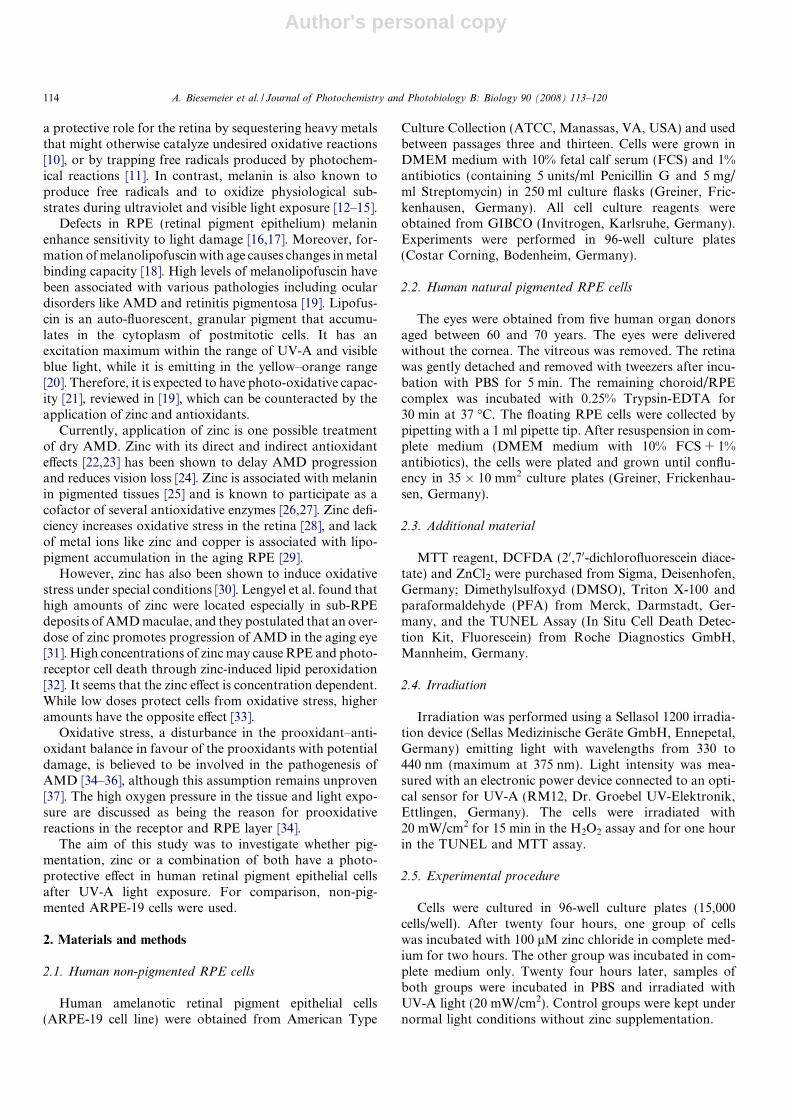
Author's personal copy
a protective role for the retina by sequestering heavy metalsthat might otherwise catalyze undesired oxidative reactions[10], or by trapping free radicals produced by photochem-ical reactions [11]. In contrast, melanin is also known toproduce free radicals and to oxidize physiological sub-strates during ultraviolet and visible light exposure [12–15].
Defects in RPE (retinal pigment epithelium) melaninenhance sensitivity to light damage [16,17]. Moreover, for-mation of melanolipofuscin with age causes changes in metalbinding capacity [18]. High levels of melanolipofuscin havebeen associated with various pathologies including oculardisorders like AMD and retinitis pigmentosa [19]. Lipofus-cin is an auto-fluorescent, granular pigment that accumu-lates in the cytoplasm of postmitotic cells. It has anexcitation maximum within the range of UV-A and visibleblue light, while it is emitting in the yellow–orange range[20]. Therefore, it is expected to have photo-oxidative capac-ity [21], reviewed in [19], which can be counteracted by theapplication of zinc and antioxidants.
Currently, application of zinc is one possible treatmentof dry AMD. Zinc with its direct and indirect antioxidanteffects [22,23] has been shown to delay AMD progressionand reduces vision loss [24]. Zinc is associated with melaninin pigmented tissues [25] and is known to participate as acofactor of several antioxidative enzymes [26,27]. Zinc defi-ciency increases oxidative stress in the retina [28], and lackof metal ions like zinc and copper is associated with lipo-pigment accumulation in the aging RPE [29].
However, zinc has also been shown to induce oxidativestress under special conditions [30]. Lengyel et al. found thathigh amounts of zinc were located especially in sub-RPEdeposits of AMD maculae, and they postulated that an over-dose of zinc promotes progression of AMD in the aging eye[31]. High concentrations of zinc may cause RPE and photo-receptor cell death through zinc-induced lipid peroxidation[32]. It seems that the zinc effect is concentration dependent.While low doses protect cells from oxidative stress, higheramounts have the opposite effect [33].
Oxidative stress, a disturbance in the prooxidant–anti-oxidant balance in favour of the prooxidants with potentialdamage, is believed to be involved in the pathogenesis ofAMD [34–36], although this assumption remains unproven[37]. The high oxygen pressure in the tissue and light expo-sure are discussed as being the reason for prooxidativereactions in the receptor and RPE layer [34].
The aim of this study was to investigate whether pig-mentation, zinc or a combination of both have a photo-protective effect in human retinal pigment epithelial cellsafter UV-A light exposure. For comparison, non-pig-mented ARPE-19 cells were used.
2. Materials and methods
2.1. Human non-pigmented RPE cells
Human amelanotic retinal pigment epithelial cells(ARPE-19 cell line) were obtained from American Type
Culture Collection (ATCC, Manassas, VA, USA) and usedbetween passages three and thirteen. Cells were grown inDMEM medium with 10% fetal calf serum (FCS) and 1%antibiotics (containing 5 units/ml Penicillin G and 5 mg/ml Streptomycin) in 250 ml culture flasks (Greiner, Fric-kenhausen, Germany). All cell culture reagents wereobtained from GIBCO (Invitrogen, Karlsruhe, Germany).Experiments were performed in 96-well culture plates(Costar Corning, Bodenheim, Germany).
2.2. Human natural pigmented RPE cells
The eyes were obtained from five human organ donorsaged between 60 and 70 years. The eyes were deliveredwithout the cornea. The vitreous was removed. The retinawas gently detached and removed with tweezers after incu-bation with PBS for 5 min. The remaining choroid/RPEcomplex was incubated with 0.25% Trypsin-EDTA for30 min at 37 �C. The floating RPE cells were collected bypipetting with a 1 ml pipette tip. After resuspension in com-plete medium (DMEM medium with 10% FCS + 1%antibiotics), the cells were plated and grown until conflu-ency in 35 � 10 mm2 culture plates (Greiner, Frickenhau-sen, Germany).
2.3. Additional material
MTT reagent, DCFDA (20,70-dichlorofluorescein diace-tate) and ZnCl2 were purchased from Sigma, Deisenhofen,Germany; Dimethylsulfoxyd (DMSO), Triton X-100 andparaformaldehyde (PFA) from Merck, Darmstadt, Ger-many, and the TUNEL Assay (In Situ Cell Death Detec-tion Kit, Fluorescein) from Roche Diagnostics GmbH,Mannheim, Germany.
2.4. Irradiation
Irradiation was performed using a Sellasol 1200 irradia-tion device (Sellas Medizinische Gerate GmbH, Ennepetal,Germany) emitting light with wavelengths from 330 to440 nm (maximum at 375 nm). Light intensity was mea-sured with an electronic power device connected to an opti-cal sensor for UV-A (RM12, Dr. Groebel UV-Elektronik,Ettlingen, Germany). The cells were irradiated with20 mW/cm2 for 15 min in the H2O2 assay and for one hourin the TUNEL and MTT assay.
2.5. Experimental procedure
Cells were cultured in 96-well culture plates (15,000cells/well). After twenty four hours, one group of cellswas incubated with 100 lM zinc chloride in complete med-ium for two hours. The other group was incubated in com-plete medium only. Twenty four hours later, samples ofboth groups were incubated in PBS and irradiated withUV-A light (20 mW/cm2). Control groups were kept undernormal light conditions without zinc supplementation.
114 A. Biesemeier et al. / Journal of Photochemistry and Photobiology B: Biology 90 (2008) 113–120
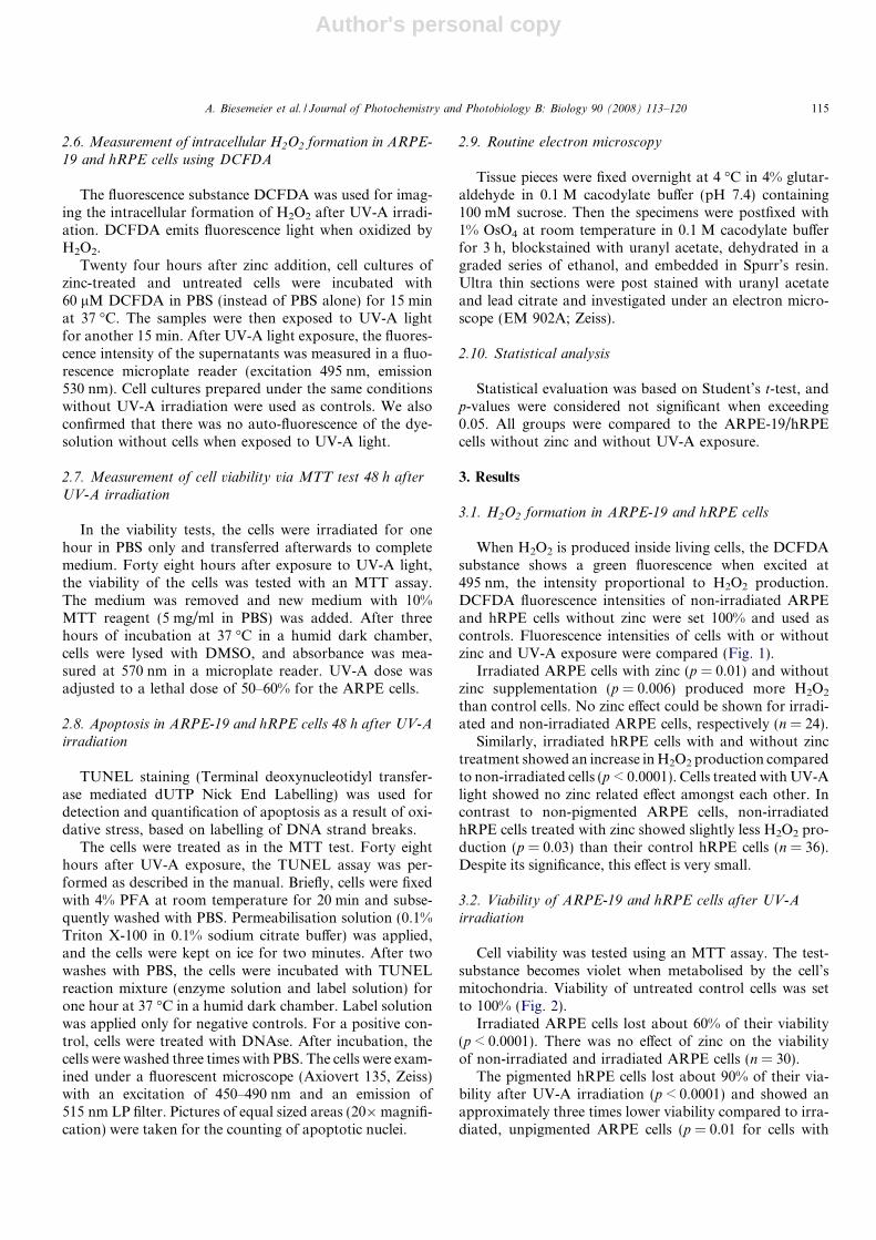
Author's personal copy
2.6. Measurement of intracellular H2O2 formation in ARPE-
19 and hRPE cells using DCFDA
The fluorescence substance DCFDA was used for imag-ing the intracellular formation of H2O2 after UV-A irradi-ation. DCFDA emits fluorescence light when oxidized byH2O2.
Twenty four hours after zinc addition, cell cultures ofzinc-treated and untreated cells were incubated with60 lM DCFDA in PBS (instead of PBS alone) for 15 minat 37 �C. The samples were then exposed to UV-A lightfor another 15 min. After UV-A light exposure, the fluores-cence intensity of the supernatants was measured in a fluo-rescence microplate reader (excitation 495 nm, emission530 nm). Cell cultures prepared under the same conditionswithout UV-A irradiation were used as controls. We alsoconfirmed that there was no auto-fluorescence of the dye-solution without cells when exposed to UV-A light.
2.7. Measurement of cell viability via MTT test 48 h after
UV-A irradiation
In the viability tests, the cells were irradiated for onehour in PBS only and transferred afterwards to completemedium. Forty eight hours after exposure to UV-A light,the viability of the cells was tested with an MTT assay.The medium was removed and new medium with 10%MTT reagent (5 mg/ml in PBS) was added. After threehours of incubation at 37 �C in a humid dark chamber,cells were lysed with DMSO, and absorbance was mea-sured at 570 nm in a microplate reader. UV-A dose wasadjusted to a lethal dose of 50–60% for the ARPE cells.
2.8. Apoptosis in ARPE-19 and hRPE cells 48 h after UV-A
irradiation
TUNEL staining (Terminal deoxynucleotidyl transfer-ase mediated dUTP Nick End Labelling) was used fordetection and quantification of apoptosis as a result of oxi-dative stress, based on labelling of DNA strand breaks.
The cells were treated as in the MTT test. Forty eighthours after UV-A exposure, the TUNEL assay was per-formed as described in the manual. Briefly, cells were fixedwith 4% PFA at room temperature for 20 min and subse-quently washed with PBS. Permeabilisation solution (0.1%Triton X-100 in 0.1% sodium citrate buffer) was applied,and the cells were kept on ice for two minutes. After twowashes with PBS, the cells were incubated with TUNELreaction mixture (enzyme solution and label solution) forone hour at 37 �C in a humid dark chamber. Label solutionwas applied only for negative controls. For a positive con-trol, cells were treated with DNAse. After incubation, thecells were washed three times with PBS. The cells were exam-ined under a fluorescent microscope (Axiovert 135, Zeiss)with an excitation of 450–490 nm and an emission of515 nm LP filter. Pictures of equal sized areas (20�magnifi-cation) were taken for the counting of apoptotic nuclei.
2.9. Routine electron microscopy
Tissue pieces were fixed overnight at 4 �C in 4% glutar-aldehyde in 0.1 M cacodylate buffer (pH 7.4) containing100 mM sucrose. Then the specimens were postfixed with1% OsO4 at room temperature in 0.1 M cacodylate bufferfor 3 h, blockstained with uranyl acetate, dehydrated in agraded series of ethanol, and embedded in Spurr’s resin.Ultra thin sections were post stained with uranyl acetateand lead citrate and investigated under an electron micro-scope (EM 902A; Zeiss).
2.10. Statistical analysis
Statistical evaluation was based on Student’s t-test, andp-values were considered not significant when exceeding0.05. All groups were compared to the ARPE-19/hRPEcells without zinc and without UV-A exposure.
3. Results
3.1. H2O2 formation in ARPE-19 and hRPE cells
When H2O2 is produced inside living cells, the DCFDAsubstance shows a green fluorescence when excited at495 nm, the intensity proportional to H2O2 production.DCFDA fluorescence intensities of non-irradiated ARPEand hRPE cells without zinc were set 100% and used ascontrols. Fluorescence intensities of cells with or withoutzinc and UV-A exposure were compared (Fig. 1).
Irradiated ARPE cells with zinc (p = 0.01) and withoutzinc supplementation (p = 0.006) produced more H2O2
than control cells. No zinc effect could be shown for irradi-ated and non-irradiated ARPE cells, respectively (n = 24).
Similarly, irradiated hRPE cells with and without zinctreatment showed an increase in H2O2 production comparedto non-irradiated cells (p < 0.0001). Cells treated with UV-Alight showed no zinc related effect amongst each other. Incontrast to non-pigmented ARPE cells, non-irradiatedhRPE cells treated with zinc showed slightly less H2O2 pro-duction (p = 0.03) than their control hRPE cells (n = 36).Despite its significance, this effect is very small.
3.2. Viability of ARPE-19 and hRPE cells after UV-A
irradiation
Cell viability was tested using an MTT assay. The test-substance becomes violet when metabolised by the cell’smitochondria. Viability of untreated control cells was setto 100% (Fig. 2).
Irradiated ARPE cells lost about 60% of their viability(p < 0.0001). There was no effect of zinc on the viabilityof non-irradiated and irradiated ARPE cells (n = 30).
The pigmented hRPE cells lost about 90% of their via-bility after UV-A irradiation (p < 0.0001) and showed anapproximately three times lower viability compared to irra-diated, unpigmented ARPE cells (p = 0.01 for cells with
A. Biesemeier et al. / Journal of Photochemistry and Photobiology B: Biology 90 (2008) 113–120 115
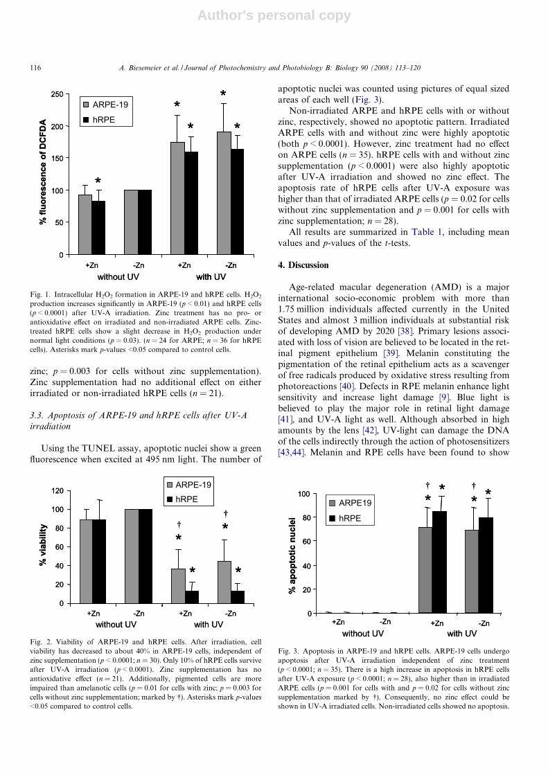
Author's personal copy
zinc; p = 0.003 for cells without zinc supplementation).Zinc supplementation had no additional effect on eitherirradiated or non-irradiated hRPE cells (n = 21).
3.3. Apoptosis of ARPE-19 and hRPE cells after UV-Airradiation
Using the TUNEL assay, apoptotic nuclei show a greenfluorescence when excited at 495 nm light. The number of
apoptotic nuclei was counted using pictures of equal sizedareas of each well (Fig. 3).
Non-irradiated ARPE and hRPE cells with or withoutzinc, respectively, showed no apoptotic pattern. IrradiatedARPE cells with and without zinc were highly apoptotic(both p < 0.0001). However, zinc treatment had no effecton ARPE cells (n = 35). hRPE cells with and without zincsupplementation (p < 0.0001) were also highly apoptoticafter UV-A irradiation and showed no zinc effect. Theapoptosis rate of hRPE cells after UV-A exposure washigher than that of irradiated ARPE cells (p = 0.02 for cellswithout zinc supplementation and p = 0.001 for cells withzinc supplementation; n = 28).
All results are summarized in Table 1, including meanvalues and p-values of the t-tests.
4. Discussion
Age-related macular degeneration (AMD) is a majorinternational socio-economic problem with more than1.75 million individuals affected currently in the UnitedStates and almost 3 million individuals at substantial riskof developing AMD by 2020 [38]. Primary lesions associ-ated with loss of vision are believed to be located in the ret-inal pigment epithelium [39]. Melanin constituting thepigmentation of the retinal epithelium acts as a scavengerof free radicals produced by oxidative stress resulting fromphotoreactions [40]. Defects in RPE melanin enhance lightsensitivity and increase light damage [9]. Blue light isbelieved to play the major role in retinal light damage[41], and UV-A light as well. Although absorbed in highamounts by the lens [42], UV-light can damage the DNAof the cells indirectly through the action of photosensitizers[43,44]. Melanin and RPE cells have been found to show
0
50
100
150
200
250
+Zn -Zn without UV
-Zn with UV
% fl
uore
scen
ceof
DC
FDA
ARPE-19hRPE
*
**
*
*
0
50
100
150
200
250
+Zn +Zn +Zn -Zn without UV
-Zn with UV
-Zn with UV
% fl
uore
scen
ceof
DC
FDA
ARPE-19
hRPE
*
**
*
*
Fig. 1. Intracellular H2O2 formation in ARPE-19 and hRPE cells. H2O2
production increases significantly in ARPE-19 (p < 0.01) and hRPE cells(p < 0.0001) after UV-A irradiation. Zinc treatment has no pro- orantioxidative effect on irradiated and non-irradiated ARPE cells. Zinc-treated hRPE cells show a slight decrease in H2O2 production undernormal light conditions (p = 0.03). (n = 24 for ARPE; n = 36 for hRPEcells). Asterisks mark p-values <0.05 compared to control cells.
0
20
40
60
80
100
120
+Zn -Zn +Zn -Zn without UV with UV
% v
iabi
lity
ARPE-19hRPE
*
*
*
*
††
0
20
40
60
80
100
120
+Zn -Zn +Zn -Zn without UV with UV
% v
iabi
lity
ARPE-19hRPE
*
*
*
*
††
Fig. 2. Viability of ARPE-19 and hRPE cells. After irradiation, cellviability has decreased to about 40% in ARPE-19 cells, independent ofzinc supplementation (p < 0.0001; n = 30). Only 10% of hRPE cells surviveafter UV-A irradiation (p < 0.0001). Zinc supplementation has noantioxidative effect (n = 21). Additionally, pigmented cells are moreimpaired than amelanotic cells (p = 0.01 for cells with zinc; p = 0.003 forcells without zinc supplementation; marked by �). Asterisks mark p-values<0.05 compared to control cells.
100
0
20
40
60
80
+Zn -Zn +Zn -Zn without UV with UV
% a
popt
otic
nucl
ei
ARPE19hRPE
* ** *
† †100
0
20
40
60
80
+Zn -Zn +Zn -Zn without UV with UV
% a
popt
otic
nucl
ei
ARPE19
hRPE* *
* *† †
Fig. 3. Apoptosis in ARPE-19 and hRPE cells. ARPE-19 cells undergoapoptosis after UV-A irradiation independent of zinc treatment(p < 0.0001; n = 35). There is a high increase in apoptosis in hRPE cellsafter UV-A exposure (p < 0.0001; n = 28), also higher than in irradiatedARPE cells (p = 0.001 for cells with and p = 0.02 for cells without zincsupplementation marked by �). Consequently, no zinc effect could beshown in UV-A irradiated cells. Non-irradiated cells showed no apoptosis.
116 A. Biesemeier et al. / Journal of Photochemistry and Photobiology B: Biology 90 (2008) 113–120

Author's personal copy
oxidative reactions, including hydrogen peroxide produc-tion when exposed to light [45,46].
The UV-light device used in this study emitted light witha maximum in the UV-A range, approaching the near bluelight. Although we presumed a protective effect of pigmen-tation in hRPE cells after irradiation, naturally pigmentedhRPE cells showed a lower vitality and a higher apoptosisthan non-pigmented cells. The donors of the hRPE cells,aged between 60 and 70 years, showed an RPE pigmenta-tion consisting of melanin and high amounts of lipofuscin(Fig. 4). We assume that lipofuscin pigmentation in combi-nation with light exposure had a toxic effect on the hRPEcells that could not be counteracted by the remainingmelanin. Other groups have published that exposure of lip-ofuscin-fed cells to blue light (400–550 nm) leads to a time-dependent loss of cells from the monolayer with theremaining cells exhibiting a change in cell morphology,increased vacuolation and blistering of the cell membrane[47], also seen in our experiments (data not shown). Thus,cumulative toxic damage over a lifetime may contribute toa variety of changes in the outer retina, which may be cau-sal factors in the development of AMD [40,41,48–51]. Ourfindings are consistent with the findings that lipofuscin hasprooxidative capacity. Hence, we can postulate that cellswithout pigmentation are less stressed by UV-A irradiationthan cells with lipofuscin pigmentation.
Zinc has antioxidative capacity, and zinc supplementa-tion in RPE cells has been shown to enhance catalase activ-ity [27]. The ability of melanin to bind metal ions including
zinc was widely examined in the past [25]. Borovansky et al.reported a high zinc concentration in isolated melanosomes[52,53]. It has also been reported that its concentration inpigmented ocular tissues is high compared to others[54,55]. The number of functional melanosomes in the fun-dus [56] and iris [57] is most likely important for the stor-age, transport or release of zinc. However, high amountsof zinc have been shown to induce oxidative reactions inamelanotic RPE cells, because melanin is absent [58].Cytotoxicity of zinc has also been reported for normal pig-mented eyes [30,59] and depends mainly on its concentra-tion [60]. Zinc is also found to induce cell death andapoptosis after retinal ischemia [61].
This is the first study focused on a direct comparison ofamelanotic and naturally pigmented human cells in combi-nation with zinc uptake experiments and UV-A inducedoxidative stress. Non-irradiated and irradiated ARPE cellsshowed no zinc related effects at all, whereas non-irradiatedhRPE cells produced slightly less H2O2 when treated withzinc, compared to their controls. A protective effect of zinccould not be proved for the irradiated hRPE cells. Pigmentgranules (lipofuscin, melanin) in human diseased RPE losethe ability to bind metal ions, including zinc [62]. Melaninundergoes age-related changes, to which photooxidationcan contribute, playing a central role in pathogenesis[63,64]. Aerobic photoreactivity increases with age anddecreasing wavelength and so it is expected to lead to thedevelopment of aging disorders like AMD. Melanin andRPE cells have been shown to produce hydrogen peroxide
Table 1Mean values of the different tests and their p-values compared to control samples
Sample name H2O2 production(I) Viability(II) Apoptosis(III)
1 ARPE cells (control 1) 100% 100% 0%2 ARPE cells with zinc 91.7% ± 15.7% 89.2% ± 10.9% 0%3 ARPE cells with UV 190.4% ± 44.3% p = 0.006 to
control 144.7% ± 23.2% p < 0.0001 tocontrol 1
69.0% ± 19.2% p > 0.0001 to control1
4 ARPE cells with zinc andUV
174.2% ± 42.0% p = 0.01 to control1
36.4% ± 20.6% p < 0.0001 tocontrol 1
71.15% ± 16.6% p > 0.0001 tocontrol 1
5 hRPE cells (control 2) 100% 100% 0%6 hRPE cells with zinc 82.6% ± 17.3% p = 0.03 to control 2 89.3% ± 20.5% 0%7 hRPE cells with UV 163.1% ± 21.6% p < 0.0001 to
control 212.9% ± 8.0% p < 0.0001 tocontrol 2
79.4% ±16.4% p > 0.0001 to control2
8 hRPE cells with zinc andUV
158.9% ± 24.1% p < 0.0001 tocontrol 2
12.9% ± 9.6% p < 0.0001 tocontrol 2
84.4% ± 13.3% p > 0.0001 to control2
Additional parameters:
(I) H2O2-production (t-test):Sample 4 compared to 2: p = 0.01;Sample 8 compared to 6: p < 0.0001
(II) Viability: t-test:Sample 4 compared to 2: p < 0.0001Sample 8 compared to 6: p < 0.0001Sample 7 compared to 3: p = 0.003(�)Sample 8 compared to 4: p = 0.01(�)
(III) Apoptosis: t-test:Sample 4 compared to 2: p < 0.0001Sample 8 compared to 6: p < 0.0001Sample 7 compared to 3: p = 0.02(�)Sample 8 compared to 4: p = 0.001(�).
A. Biesemeier et al. / Journal of Photochemistry and Photobiology B: Biology 90 (2008) 113–120 117

Author's personal copy
when exposed to light. Melanin can photogenerate super-oxide anions and hydrogen peroxide [46] and has the abilityto bind several chemicals and drugs leading to the develop-ment of ocular or inner ear lesions [65]. Nevertheless,phototoxicity of melanin is lower than that of lipofuscinand photoprotective effects are of greater importance. Itwas also shown previously, that the formation of melano-lipofuscin rather than the loss of melanin and the increasein lipofuscin has toxic effects on RPE cells and is closelyrelated to the onset of AMD [66].
Our results are consistent with this observation. Pig-mented cells showed a higher oxidative damage than unpig-mented cells. The high melanolipofuscin amount in ourdonor eyes was probably the reason we obtained a proox-idative effect after UV-A exposure, rather than an antioxi-dative effect as a result of the melanin.
The hRPE cells used in this study were isolated from fivedifferent donors. Cells of one eye were grown to confluenceand then split in three parts for the three different tests toavoid too many passages, and to obtain heavily pigmentedcell cultures (Fig. 4A). Individual differences in the pigmen-tation were present, therefore results differed strongly.Even small differences in pigmentation and cell number,despite confluence, can result in wide differences in theeffect of drugs and stress [67]. Additionally, the appropriate
concentration of zinc was hard to select in this study,because various groups have found different effects of zincon RPE cells dependent on conditions and methods. Tateet al. found that 100 lM ZnCl2 protect zinc deficientRPE cells from oxidative stress [27,68]. Ha et al. found thatconcentrations of zinc chloride and zinc sulphate up to150 lM were tolerated by normally fed ARPE-19 cellsand could induce antioxidative pathways, while concentra-tions over 200 lM were toxic and resulted in cell death,whereas others stated that zinc concentrations exceeding10 lM cause apoptosis in RPE cells [33]. In our experi-ments, zinc supplementation to cells with and without pig-mentation led neither to an anti nor to a prooxidativeeffect. Although we used similar methods, there were somedifferences compared to the formerly named studies. In ourexperiments, the cells did not suffer from zinc deficiency,and we used zinc chloride instead of zinc sulphate or othersalts. In addition, our donor eyes, obtained from 60–70year old Caucasians, contained high amounts of lipofuscin.
5. Conclusion
In the present study, we investigated the toxic effect ofUV-A light on human amelanotic and naturally pigmentedRPE cells. Additionally, the effect of zinc was investigated.
Fig. 4. Pigmentation of hRPE cells. (A) Light image of melanin pigmentation in hRPE cells. (B) Auto-fluorescence of lipofuscin pigmentation of the samearea as in (A). Magnification 40�, excitation 450–490 nm LP filter, emission 530 nm. (C) Electron micrograph of an aged human RPE, showing manymelanolipofuscin (ML) and pure lipofuscin (L) granules. Hardly any melanin (M) granules are left. This picture demonstrates how the pigmentation in theeye has changed with age. The donor had no obvious eye disease and was the same age as the donors of the irradiated cells.
118 A. Biesemeier et al. / Journal of Photochemistry and Photobiology B: Biology 90 (2008) 113–120

Author's personal copy
UV-A irradiation led to an increase in H2O2 productionand apoptosis and a significant decrease in viability in bothhRPE and ARPE-19 cells. The antioxidant capacity of thecells could not be enhanced by addition of zinc in eitheramelanotic or naturally pigmented RPE cells. However,the results of this study are only valid for aged RPE cellsand it is unknown whether they are also true for RPE cellsof young people. Here, we demonstrate that pigmentationdoes not always guarantee a better survival of cells. Wheneyes of donors aged 60–70 years are used, the age pigmentlipofuscin is present in high amounts, and the antioxidativecapacity of the cells cannot be improved by zinc supple-mentation when stressed by UV-A light.
Acknowledgements
This work was supported by grants to U. Schraermeyerfrom the Deutsche Forschungsgemeinschaft (DFG SCHR436/12-2). Thanks to Sigrid Schultheiß and Monika Ritt-garn for technical assistance. Special thanks to WinfriedSchuller and York Kamenisch from the Department ofDermatology, Tuebingen for assistance with the UV-A de-vice and helping with the methods.
References
[1] A.C. Bird, N.M. Bressler, S.B. Bressler, I.H. Chisholm, G. Coscas,M.D. Davis, P.T. de Jong, C.C. Klaver, B.E. Klein, R. Klein, Aninternational classification and grading system for age-related mac-ulopathy and age-related macular degeneration. The internationalARM epidemiological study group, Surv. Ophthalmol. 39 (1995) 367–374.
[2] W. Smith, J. Assink, R. Klein, P. Mitchell, C.C.W. Klaver,B.E.K. Klein, A. Hofman, S. Jensen, J.J. Wang, P.T.V.M. DeJong, Risk factors for age related macular degeneration –pooled findings from three continents, Ophthalmology 108(2001) 697–704.
[3] T.T.J.M. Berendschot, J.J.M. Willemse-Assink, M. Bastiaanse,P.T.V.M. De Jong, D. van Norren, Macular pigment and melaninin age-related maculopathy in a general population, Invest. Ophth.Vis. Sci. 43 (2002) 1928–1932.
[4] R. Klein, B.E. Klein, K.J. Cruickshanks, The prevalence of age-related maculopathy by geographic region and ethnicity, Prog. Retin.Eye Res. 18 (1999) 371–389.
[5] J.J. Weiter, F.C. Delori, G.L. Wing, K.A. Fitch, Relationship ofsenile macular degeneration to ocular pigmentation, Am. J. Ophthal-mol. 99 (1985) 185–187.
[6] D. Pauleikhoff, F.G. Holz, Age-related macular degeneration. 1.Epidemiology, pathogenesis and differential diagnosis, Ophthalmo-loge 93 (1996) 299–315.
[7] Z. Gregor, L. Joffe, Senile macular changes in the black African, Br. J.Ophthalmol. 62 (1978) 547–550.
[8] J. Ambati, B.K. Ambati, S.H. Yoo, S. Ianchulev, A.P. Adamis, Age-related macular degeneration: etiology, pathogenesis, and therapeuticstrategies, Surv. Ophthalmol. 48 (2003) 257–293.
[9] U. Schraermeyer, K. Heimann, Current understanding on the role ofretinal pigment epithelium and its pigmentation, Pigment Cell Res. 12(4) (1999) 219–236.
[10] T. Sarna, Properties and function of the ocular melanin – aphotobiophysical view, J. Photochem. Photobiol. B 12 (1992) 215–258.
[11] N.L. Sakina, A.E. Dontsov, G.G. Afanas’ev, M.A. Ostrovski, I.I.Pelevina, The accumulation of lipid peroxidation products in the eye
structures of mice under whole-body X-ray irradiation, Radiobiologia30 (1990) 28–31.
[12] C.C. Felix, J.S. Hyde, T. Sarna, R.C. Sealy, Melanin photoreactionsin aerated media: electron spin resonance evidence for production ofsuperoxide and hydrogen peroxide, Biochem. Biophys. Res. Com-mun. 84 (1978) 335–341.
[13] W. Korytowski, B. Pilas, T. Sarna, B. Kalyanaraman, Photoinducedgeneration of hydrogen peroxide and hydroxyl radicals in melanins,Photochem. Photobiol. 45 (1987) 185–190.
[14] R.D. Glickman, R. Sowell, K.W. Lam, Kinetic properties of light-dependent ascorbic acid oxidation by melanin, Free Radic. Biol. Med.15 (1993) 453–457.
[15] M. Rozanowska, A. Bober, J.M. Burke, T. Sarna, The role of retinalpigment epithelium melanin in photoinduced oxidation of ascorbate,Photochem. Photobiol. 65 (1997) 472–479.
[16] W.G. Robison Jr., T. Kuwabara, D.G. Cogan, Lysosomes andmelanin granules of the retinal pigment epithelium in a mouse modelof the Chediak-Higashi syndrome, Invest. Ophthalmol. 14 (1975)312–317.
[17] D. van Norren, L.F. Tiemeijer, Spectral reflectance of the human eye,Vis. Res. 26 (1986) 313–320.
[18] D.A. Samuelson, S. Konkal, P. Lewis, M. Chisholm, Changes ofmetal binding in human RPE melanin and lipofuscin associated withaging and drusen (ARVO Abstract), Invest. Ophthalmol. Vis. Sci.37(3) 15-2-1996 Ref Type: Abstract.
[19] M. Boulton, M. Rozanowska, B. Rozanowski, T. Wess, Thephotoreactivity of ocular lipofuscin, Photochem. Photobiol. Sci. 3(2004) 759–764.
[20] U.T. Brunk, C.B. Jones, R.S. Sohal, A novel hypothesis of lipofu-scinogenesis and cellular aging based on interactions betweenoxidative stress and autophagocytosis, Mutat. Res. 275 (1992) 395–403.
[21] M. Boulton, A. Dontsov, J. Jarvis-Evans, M. Ostrovsky, D.Svistunenko, Lipofuscin is a photoinducible free radical generator,J. Photochem Photobiol. B 19 (1993) 201–204.
[22] T.M. Bray, W.J. Bettger, The physiological role of zinc as anantioxidant, Free Radic. Biol. Med. 8 (1990) 281–291.
[23] Z. Deng, X. Liu, J. Chen, R. Yang, Zinc protective effects onpig retinal pigment epithelial cell damage of lipid peroxideinduced by 2450 MHz microwave, Wei Sheng Yan Jiu 29 (2000)129–131.
[24] Age-Related Eye Disease Study Research Group, A randomized,placebo-controlled, clinical trial of high-dose supplementation withvitamins C and E, beta carotene, and zinc for age-related maculardegeneration and vision loss: AREDS report no. 8, Arch. Ophthal-mol. 119 (2001) 1417–1436.
[25] A.M. Potts, P.C. Au, The affinity of melanin for inorganic ions, Exp.Eye Res. 22 (1976) 487–491.
[26] G.J. Brewer, G.M. Hill, A.S. Prasad, Z.T. Cossack, Biological roles ofionic zinc, Prog. Clin. Biol. Res. 129 (1983) 35–51.
[27] D.J. Tate, M.V. Miceli, D.A. Newsome, Zinc induces catalaseexpression in cultured fetal human retinal pigment epithelial cells,Curr. Eye Res. 16 (1997) 1017–1023.
[28] M.V. Miceli, D.J. Tate, N.W. Alcock, D.A. Newsome, Zinc deficiencyand oxidative stress in the retina of pigmented rats, Invest. Ophthal.Vis. Sci. 40 (1999) 1238–1244.
[29] D.A. Samuelson, D. Armstrong, R. Jolly, X-ray microprobe analysisof the retina and RPE in sheep with ovine ceroid-lipofuscinosis,Neurobiol. Aging 11 (1990) 663–667.
[30] J. Borovansky, P.A. Riley, Cytotoxicity of zinc in vitro, Chem. Biol.Interact. 69 (1989) 279–291.
[31] I. Lengyel, J.M. Flinn, T. Peto, D.H. Linkous, K. Cano, A.C. Bird,A. Lanzirotti, C.J. Frederickson, F.J. van Kuijk, High concentrationof zinc in sub-retinal pigment epithelial deposits, Exp. Eye Res. 84(2007) 772–780.
[32] N.N. Osborne, J.P.M. Wood, The beta-adrenergic receptor antago-nist metipranolol blunts zinc-induced photoreceptor and RPE apop-tosis, Invest. Ophthal. Vis. Sci. 47 (2006) 3178–3186.
A. Biesemeier et al. / Journal of Photochemistry and Photobiology B: Biology 90 (2008) 113–120 119
Author's personal copy
[33] J.P.M. Wood, N.N. Osborne, The influence of zinc on caspase-3 andDNA breakdown in cultured human retinal pigment epithelial cells,Arch. Ophthal. 119 (2001) 81–88.
[34] B.S. Winkler, M.E. Boulton, J.D. Gottsch, P. Sternberg, Oxidativedamage and age-related macular degeneration, Mol. Vis. 5 (1999).
[35] J.Y. Cai, K.C. Nelson, M. Wu, P. Sternberg, D.P. Jones, Oxidativedamage and protection of the RPE, Prog. Retin. Eye Res. 19 (2000)205–221.
[36] R. Hogg, U. Chakravarthy, AMD and micronutrient antioxidants,Curr. Eye Res. 29 (2004) 387–401.
[37] S. Beatty, H. Koh, M. Phil, D. Henson, M.E. Boulton, The role ofoxidative stress in the pathogenesis of age-related macular degener-ation, Surv. Ophthalmol. 45 (2000) 115–134.
[38] D.S. Friedman, B.J. O’Colmain, B. Munoz, S.C. Tomany, C.McCarty, P.T. de Jong, B. Nemesure, P. Mitchell, J. Kempen,Prevalence of age-related macular degeneration in the United States,Arch. Ophthalmol. 122 (2004) 564–572.
[39] W.R. Green, P.J. McDonnell, J.H. Yeo, Pathologic features of senilemacular degeneration, Ophthalmology 92 (1985) 615–627.
[40] R.W. Young, Solar radiation and age-related macular degeneration,Surv. Ophthalmol. 32 (1988) 252–269.
[41] M. Rozanowska, J. Jarvis-Evans, W. Korytowski, M.E. Boulton,J.M. Burke, T. Sarna, Blue light-induced reactivity of retinal agepigment. In vitro generation of oxygen-reactive species, J. Biol. Chem.270 (1995) 18825–18830.
[42] S.K. West, F.S. Rosenthal, N.M. Bressler, S.B. Bressler, B. Munoz,S.L. Fine, H.R. Taylor, Exposure to sunlight and other risk factorsfor age-related macular degeneration, Arch. Ophthalmol. 107 (1989)875–879.
[43] C. Reme, J. Reinboth, M. Clausen, F. Hafezi, Light damage revisited:converging evidence, diverging views? Graefes Arch. Clin. Exp.Ophthalmol. 234 (1996) 2–11.
[44] J.A. Zuclich, Ultraviolet-induced photochemical damage in oculartissues, Health Phys. 56 (1989) 671–682.
[45] R.D. Glickman, K.W. Lam, Oxidation of ascorbic acid as anindicator of photooxidative stress in the eye, Photochem. Photobiol.55 (1992) 191–196.
[46] G. Scott, Atmospheric oxidation and antioxidants, in: S.H. Sarna T(Ed.), Interactions of Melanin with Oxygen (and related) Species, vol.III, Elsevier, Amsterdam, 1993, pp. 129–169.
[47] S. Davies, M.H. Elliott, E. Floor, T.G. Truscott, M. Zareba, T.Sarna, F.A. Shamsi, M.E. Boulton, Photocytotoxicity of lipofuscin inhuman retinal pigment epithelial cells, Free Radic. Biol. Med. 31(2001) 256–265.
[48] K. Reszka, G.E. Eldred, R.H. Wang, C. Chignell, J. Dillon, Thephotochemistry of human retinal lipofuscin as studied by EPR,Photochem. Photobiol. 62 (1995) 1005–1008.
[49] M.L. Katz, K.R. Parker, G.J. Handelman, T.L. Bramel, E.A. Dratz,Effects of antioxidant nutrient deficiency on the retina and retinalpigment epithelium of albino rats: a light and electron microscopicstudy, Exp. Eye Res. 34 (1982) 339–369.
[50] J.J. Weiter, F.C. Delori, G.L. Wing, K.A. Fitch, Retinal pigmentepithelial lipofuscin and melanin and choroidal melanin in humaneyes, Invest. Ophthalmol. Vis. Sci. 27 (1986) 145–152.
[51] K.V. Fite, L. Bengston, B. Donaghey, Experimental light damageincreases lipofuscin in the retinal pigment epithelium of Japanesequail (Coturnix coturnix japonica), Exp. Eye Res. 57 (1993) 449–460.
[52] J. Borovansky, J. Horcicko, J. Duchon, The hair melanosome:another tissue reservoir of zinc, Physiol. Bohemoslov. 25 (1976) 87–91.
[53] J. Borovansky, E. Vedralova, P. Hach, An estimate of melanosomeconcentration in pigment tissues, Pigment Cell Res. 4 (1991) 222–224.
[54] M.A. Galin, H.D. Nano, T. Hall, Ocular zinc concentration, Invest.Ophthalmol. 1 (1962) 142–148.
[55] J.P. Sarks, S.H. Sarks, M.C. Killingsworth, Evolution of soft drusenin age-related macular degeneration, Eye 8 (Pt 3) (1994) 269–283.
[56] D. Kokkinou, H.U. Kasper, T. Schwarz, K.U. Bartz-Schmidt, U.Schraermeyer, Zinc uptake and storage: the role of fundus pigmen-tation, Graefes Arch. Clin. Exp. Ophthalmol. 243 (2005) 1050–1055.
[57] D. Kokkinou, H.U. Kasper, K.U. Bartz-Schmidt, U. Schraermeyer,The pigmentation of human iris influences the uptake and storing ofzinc, Pigment Cell Res. 17 (2004) 515–518.
[58] K.N. Ha, Y. Chen, J.Y. Cai, P. Sternberg, Increased glutathionesynthesis through an ARE-Nrf2-dependent pathway by zinc in theRPE: Implication for protection against oxidative stress, Invest.Ophthal.Vis. Sci. 47 (2006) 2709–2715.
[59] D. Kleiner, The effect of Zn2+ ions on mitochondrial electrontransport, Arch. Biochem. Biophys. 165 (1974) 121–125.
[60] M. Ugarte, N.N. Osborne, The localization of endogenous zinc andthe in vitro effect of exogenous zinc on the GABA immunoreactivityand formation of reactive oxygen species in the retina, Gen.Pharmacol. 30 (1998) 297–303.
[61] M.H. Yoo, J.Y. Lee, S.E. Lee, J.Y. Koh, Y.H. Yoon, Protection bypyruvate of rat retinal cells against zinc toxicity in vitro, and pressure-induced ischemia in vivo, Invest. Ophthalmol. Vis. Sci. 45 (2004)1523–1530.
[62] D.A. Samuelson, P. Smith, R.J. Ulshafer, D.G. Hendricks, R.D.Whitley, H. Hendricks, N.C. Leone, X-ray microanalysis of ocularmelanin in pigs maintained on normal and low zinc diets, Exp. EyeRes. 56 (1993) 63–70.
[63] M. Boulton, P. Dayhaw-Barker, The role of the retinal pigmentepithelium: topographical variation and ageing changes, Eye 15(2001) 384–389.
[64] T. Sarna, J.M. Burke, W. Korytowski, M. Rozanowska, C.M.Skumatz, A. Zareba, M. Zareba, Loss of melanin from human RPEwith aging: possible role of melanin photooxidation, Exp. Eye Res. 76(2003) 89–98.
[65] B.S. Larsson, Interaction between chemicals and melanin, PigmentCell Res. 6 (1993) 127–133.
[66] L. Feeney-Burns, E.S. Hilderbrand, S. Eldridge, Aging human RPE:morphometric analysis of macular, equatorial, and peripheral cells,Invest. Ophthalmol. Vis. Sci. 25 (1984) 195–200.
[67] M. Zareba, M.W. Raciti, M.M. Henry, T. Sarna, J.M. Burke,Oxidative stress in ARPE-19 cultures: do melanosomes confercytoprotection? Free Radic. Biol. Med. 40 (2006) 87–100.
[68] D.J. Tate, M.V. Miceli, D.A. Newsome, N.W. Alcock, P.D. Oliver,Influence of zinc on selected cellular functions of cultured humanretinal pigment epithelium, Curr. Eye Res. 14 (1995) 897–903.
120 A. Biesemeier et al. / Journal of Photochemistry and Photobiology B: Biology 90 (2008) 113–120
LABORATORY INVESTIGATION
Tyrosinase biosynthesis and trafficking in adulthuman retinal pigment epithelial cells
Sylvie Julien & Norbert Kociok & Florian Kreppel &Jürgen Kopitz & Stefan Kochanek & Antje Biesemeier &
Petra Blitgen-Heinecke & Peter Heiduschka &
Ulrich Schraermeyer
Received: 31 August 2006 /Revised: 15 January 2007 /Accepted: 21 January 2007 /Published online: 21 February 2007# Springer-Verlag 2007
AbstractBackground Tyrosinase (EC 1.14.18.1) is the key enzymeof melanin pigment formation and it is unclear whether it issynthesized in human postnatal retinal pigment epithelium(RPE). In this study, we investigated if phagocytosis of rodouter segments (ROS) can increase tyrosinase expression invitro.Methods Primary cultures of human RPE cells were fedwith isolated ROS from cattle and with latex particles. Afterphagocytosis, RPE cells were tested for tyrosinase presenceand activity with several independent methods: (1) immu-nocytochemistry with anti-tyrosinase antibodies and (2)ultrastructural as well as light microscopic DOPA histo-chemistry; (3) mRNAwas isolated from human RPE beforeincubation with ROS and 5, 20 and 40 h after feeding withROS. The amount of tyrosinase mRNA was determined
quantitatively by real-time reverse transcription polymerasechain reaction (RT-PCR), and the tyrosinase activity wasinvestigated by measuring tyrosine hydroxylase activityusing [3H]tyrosine.Results Tyrosinase was found in fed RPE cells using thesemethods, but was absent without feeding. Furthermore, weshowed co-localization of rhodopsin and tyrosinase in thefed RPE cells. Contrary to tyrosinase activity, the mRNAfor tyrosinase was clearly present in the cultured RPE cellswhich had not been exposed to ROS, decreased signifi-cantly from 5 h after exposure to ROS and returned to itsoriginal non-fed level 40 h after ROS feeding.Conclusion Our study does not present new evidence thatde novo melanogenesis takes place in the adult differenti-ated RPE. However, in contrast to the classic hypothesis,which states that tyrosinase is only detected in embryos, weprovide evidence with several independent methods that theexpression of tyrosinase and its enzymatic activity areinduced in cultured human adult RPE by phagocytosis ofROS.
Keywords Tyrosinase . Biosynthesis .
Retinal pigment epithelium . Human . Phagosome
Introduction
In humans, melanin pigment is biosynthesized in theneuroectodermic retinal pigment epithelium (RPE) andneural crest-derived melanocytes [27]. Melanin-producingcells contain specific organelles, the premelanosomes inwhich the binuclear copper enzyme tyrosinase catalysesmelanin biosynthesis [28, 48].
The RPE is located between the retina and the choroid ofthe eye and plays an essential role in ocular metabolism.
Graefe’s Arch Clin Exp Ophthalmol (2007) 245:1495–1505DOI 10.1007/s00417-007-0543-3
S. Julien (*) :A. Biesemeier : P. Blitgen-Heinecke :P. Heiduschka :U. SchraermeyerSection for Experimental Vitreoretinal Surgery,University Eye Hospital Tuebingen,Tuebingen, Germanye-mail: [email protected]
N. KociokNetzhautlabor, Cell Center Cologne,Josef-Stelzmann-Str. 50,50931 Cologne, Germany
F. Kreppel : S. KochanekDivision of Gene Therapy, University of Ulm,Helmholtzstr. 8/1,89081 Ulm, Germany
J. KopitzInstitute for Patho- and General Neurochemistry,University of Heidelberg,Im Neuenheimer Feld 220,69120 Heidelberg, Germany

Basic adult RPE functions are the formation of the outerblood-retina barrier, transepithelial transport, regulatedretinoid storage, scavenging of light-induced reactiveoxygen species and digestion of the shed rod outer segmentdisk membranes [45]. The disk shedding and succeedingphagocytosis processes are regulated by circadian rhythm inRPE cells [23]. Differentiated RPE cells do not divide andremain functional throughout the life of an individual.Tyrosinase is the rate-limiting enzyme of melanin biosyn-thesis and catalyses three reactions of melanin synthesis:the hydroxylation of L-tyrosine to 3,4-dihydroxyphenyl-L-alanine (L-dopa), the oxidation of L-dopa to dopaquinone[24] and the oxidation of 5,6-dihydroxyindole to 5,6-dihydroxyquinone [21]. It is assumed that RPE melano-genesis is only found prenatally, since tyrosinase, the keyenzyme in melanin biosynthesis, was detected in humanearly stage embryos only and was not detectable aftergestation stopped [5, 29, 37, 49].
However, premelanosomes and early-stage melano-somes have been found in adult RPE [10, 41, 45, 55],which led to the hypothesis questioned here. Tyrosinaseactivity has been found in adult cultured bovine [3, 42],porcine [10], mouse [33], rabbit [52], rat [53] and humanRPE cells, too [2]. Additionally, tyrosinase activity was alsodemonstrated in adult bovine RPE obtained from an eyecup [11].
Tyrosinase promoter activity was significantly upregu-lated in cultured human RPE cells treated with pigmentepithelium-derived factor (PEDF), basic fibroblast growthfactor (bFGF), α-melanocyte-stimulating hormone (α-MSH)or L-tyrosine compared with control cells [1]. In conclusion,the tyrosinase gene is not only expressed but can beregulated in response to different chemicals in culturedhuman RPE cells. However, tyrosinase enzymatic activitywas not found in this study [1]. In addition, a recent studyshows that phagocytosis of rod outer segments (ROS)induces expression of various genes in RPE cells [6].
The aim of our study was to examine the presence oftyrosinase protein as well as its enzymatic activity in adulthuman RPE. One of the most important functions of theRPE is phagocytosis of shed rod photoreceptor outersegment disks. Therefore, we investigated whether phago-cytosis of ROS can increase tyrosinase expression in vitro.
Materials and methods
Culture of human RPE cells
Human RPE cells were harvested from the eyes of twomale keratoplasty donors aged 50 and 61 years within 8 hpost mortem. The cells were maintained in minimal
essential medium (MEM) containing 5% (v/v) fetal calfserum (FCS). Each third-passage RPE cell preparation waschecked by cytokeratin staining. Cell counts were obtainedafter trypsination. Viability was assessed by crystal violetstaining of adherent cells or trypan blue dye exclusion oftrypsinized cells.
Isolation of rod outer segments
The procedure was carried out as specified by Schraermeyerand Stieve [42]. In brief, isolated bovine retinae wereagitated for 2 min in KCl buffer [0.3 M KCl, 10 mMhydroxyethylpiperazine ethanesulfonic acid (HEPES),0.5 mM CaCl2, 1 mM MgCl2 and 48% (w/v) sucrose] atpH 7.0 and then centrifuged at 2,000 rpm in a tabletopcentrifuge (type UJ 1, Heraeus-Christ, Osterode, Germany)for 5 min. The supernatant was filtered through a tubegauze finger stall, diluted with KCl buffer (1:1) and thencentrifuged for 10 min at 2,500 rpm. The pellet,containing ROS fragments, was washed and centrifugedin complete culture medium without serum before feedingto the RPE.
Phagocytosis of rod outer segments/latex particlesby human RPE cells
Human RPE cells were exposed to rod outer segments thathad been isolated as described above in full culture mediumcontaining serum for 5 h on a microscope slide. Ascontrols, cells were cultured with assay medium only andwith latex beads (diameter 0.1 μm, 5×10⁷ particles/ml,Sigma, Deisenhofen, Germany) for 5 and 20 h.
Electron-microscopic localization of tyrosinase
The enzyme tyrosinase was localized by electron-micro-scopic histochemistry [39, 40]. In brief, third-passage RPEcells that had been exposed to fragmented rod outersegments as described above were fixed in 0.1 M sodiumcacodylate buffer (pH 6.8) with 2.5% glutaraldehyde for1 h. RPE cells without feeding were treated in the samemanner. Specimens were washed twice in sodium cacody-late buffer and kept at 2-8°C overnight in this buffer,containing 5 mM L-dihydroxyphenylalanine (L-dopa,obtained from Sigma-Aldrich, Taufkirchen, Germany).Thereafter, the solutions were renewed, and the RPE cellswere incubated for a further 5 h at 37°C. The RPE cellswere washed in sodium cacodylate buffer and immersed for1 h at room temperature in the same buffer containing amixture of osmium tetroxide (1%) and potassium ferrocy-anide (1.5%). Finally, the RPE cells were dehydrated andembedded in Spurr’s resin for routine electron microscopy.Ultrathin sections were stained with uranyl acetate and lead
1496 Graefe’s Arch Clin Exp Ophthalmol (2007) 245:1495–1505

citrate and examined with a Zeiss EM 902A electronmicroscope.
Generation of Ad-Tyr
To generate the Ad-Tyr vector, the plasmid 123.B2 (kindgift from T. Woelfel, Mainz) was digested with EcoRI torelease a 1,906-bp fragment containing the human tyrosi-nase cDNA [4]. The fragment was blunt ended withKlenow and cloned into the EcoRV site of pCMVPac(+)under generation of pVI01. pCMVPac(+) is based onpCMVβ (Invitrogen, Carlsbad, CA, USA) with twomodifications: (1) the β-galactosidase coding sequence(NotI fragment) is replaced with a polylinker containing aunique EcoRV site and (2) upstream of the hCMV promoterand downstream of the SV40 polyA PacI sites are insertedwhich allow for release of the expression cassette. Thisplasmid serves as a tool to construct PacI-flanked expres-sion cassettes driven by the hCMV promoter and contain-ing an SV40 late 19s intron for strong ubiquitousexpression of cDNAs (further details about pCMVPac(+)can be obtained from the authors upon request). The PacIfragment from pVI01 containing the hCMV promoter,SV40 intron, human tyrosinase cDNA and SV40 polyAwas isolated from pVI01 and cloned into PacI of pGS70[38] under generation of pVI02. Finally, the PacI fragmentfrom pVI02 was cloned into PacI of pGS66 [38] undergeneration of pVI03, an infectious adenovirus plasmidcoding for the E1-deleted Ad vector Ad-Tyr.
The virus backbone was released from pVI03 by digestionwith SwaI and transfected into N52E6 cells [38]. Afterappearance of a cytopathic effect, the virus vector wasamplified on N52E6 cells and purified by double CsClbanding and desalting with PD-10 columns (AmershamBiosciences, Freiburg, Germany). The infectious and totalparticle titres were determined on A549 cells as described byKreppel et al. [22]. Vector genome integrity was confirmedby restriction analysis of DNA prepared from purifiedvirions. Expression of tyrosinase was confirmed in Westerntransfer experiments with cell lysates from A549 cellstransduced with different amounts of Ad-Tyr.
Transfection of human RPE cells with Ad-Tyr
Optimal gene transfer conditions were determined for eachmethod using 100,000 cells per well in 24-well plates.Tyrosinase positivity was evaluated 5 days after gene transferby immunodetection using the mouse anti-tyrosinase mono-clonal antibody AB-1 as primary antibody. Ad-Tyr with 108
infectious units/μl were added to the human RPE cell cultureand the amount of the vector DNA varied from 1 μl, 2 μland 5 μl to 1 μl diluted 10 times. Optimal results wereobtained using 1 μl of Ad-Tyr with 108 infectious units/μl.
Antibodies
The mouse anti-tyrosinase monoclonal antibody AB-1 waspurchased from NeoMarkers (Fremont, CA, USA), the mouseanti-tyrosinase monoclonal antibody 2G10 from ChemiconInt. Inc. (Temecula, CA, USA) and is described by themanufacturer as an antibody against human tyrosinase [7].The secondary purified goat Cy3-conjugated anti-mouse IgGantiserum was obtained from Rockland ImmunochemicalsInc. (Gilbertsville, PA, USA). This antiserum had been raisedagainst mouse IgG. The monoclonal anti-tyrosine hydroxy-lase antibody, clone TH-2 and IgG1 isotype, was fromSigma-Aldrich Chemie GmbH (Taufkirchen, Germany) andwas used as a control. This antibody was raised against anepitope of rat tyrosine hydroxylase, which is also present inhuman tyrosine hydroxylase.
Immunodetection of tyrosinase and tyrosine hydroxylase
Antibodies (3–8 μg/ml) were used on paraformaldehyde-fixed RPE monolayers as described elsewhere [44].Primary antibodies were diluted at 1:200 and incubated inbuffer containing 0.01 M sodium phosphate and 0.25 MNaCl for 30 min at room temperature. The secondaryantibodies were diluted 1:1000 and incubated in the samebuffer for 1 h. Cells were photographed using an Axioplan2 microscope (Zeiss, Oberkochen, Germany). Image pro-cessing was performed using an Orca camera (HamamatsuPhotonics, Herrsching,Germany) and Openlab 4 software(Improvision, Tübingen, Germany).
Histochemical evaluation of tyrosinase-active RPE cells
Bioactive tyrosinase in confluent ROS-fed human RPE cellsplated on microscope slides was determined by light micro-scopic (LM) histochemistry [10]. The cells were washed twicewith phosphate-buffered saline (PBS) and fixed in 0.1 Mcacodylate buffer (pH 7.2) with glutaraldehyde (1%). After15 min, the cells were washed three times with PBS andincubated for 60 h at room temperature in 0.05 M sodiumphosphate buffer (pH 7.0) with 5 mM L-dopa, 5 mM D-dopa,or without dopa. Subsequently, the cells were rinsed withsodium phosphate buffer and photographed using an Axio-Cam color camera (Zeiss, Oberkochen, Germany).
Localization of rhodopsin and tyrosinase in human RPEcells
Human RPE cells (P0) were isolated from donor eyes forkeratoplasty (aged 51–61 years). RPE phenotype wasexamined by pan-cytokeratin immunostaining. The cellswere fed with rod outer segments for 5 h on a microscopeslide. Immunostaining of the RPE monolayer was done
Graefe’s Arch Clin Exp Ophthalmol (2007) 245:1495–1505 1497

24 h after feeding with an anti-rhodopsin antibody (cloneRET-P1) as primary and a Cy3-conjugated as secondaryantibody. Tyrosinase was detected with a mouse mono-clonal antibody (2G10) and a Cy2-conjugated secondaryantibody. Briefly, we first performed the indirect immuno-labelling of rhodopsin and before the second indirectimmunolabelling of tyrosinase, the cells were incubatedwith bovine serum albumin (BSA) 5% for 1 h. To verify thespecificity of the double indirect labelling, we performed asequential double labelling again, but this time we detectedfirst tyrosinase and secondly rhodopsin. We obtained thesame results.
A merged image was made for verification of possibleco-localization of rhodopsin and tyrosinase expression(orange) in ROS phagosomes. The nuclear staining wasperformed with Hoechst dye (blue).
Real-time reverse transcription polymerase chain reaction(RT-PCR)
The mRNA levels for tyrosinase and glyceraldehyde-3-phosphate dehydrogenase (GAPDH) in unfed cells werecompared to ROS-fed cells by real-time RT-PCR usingSYBR Green I (Molecular Probes, Eugene, OR, USA) onan iCycler (Bio-Rad Laboratories, Hercules, CA, USA).Using the primer analysis software OLIGO 4.1 (NationalBiosciences, Plymouth, MN), we selected gene-specificprimers suitable for real-time RT-PCR (tyrosinase: ACTTAC TGG GAT AGC GGA TGC CTC T and TGC ATTGGC TTC TGG ATA AAC TTC TT, GAPDH: TCA ACGACC ACT TTG TCA AGC TCA and GCT GGT GGTCCA GGG GTC TTA CT). The Tm of the primers waschosen between 58.5 and 59.5°C. The expected fragmentlength ranges between 119 and 199 bp. With these primers,the mRNA expression of tyrosinase together with GAPDHas calibrator were analyzed simultaneously in doublereactions in three separate experiments. The analysis wasrepeated once. Aliquots of the diluted cDNA correspondingto 15 ng initially used total RNA were mixed with 10×reaction buffer containing Tris-HCl and KCl, (NH4)2SO4,3.5 mM MgCl2 (Qiagen, Hilden, Germany), 0.2 mM ofeach dNTP, 0.2 μM of each specific primer, 0.1× SYBRGreen I and 1 U of HotStarTaq DNA polymerase (Qiagen,Hilden, Germany) in a volume of 50 μl. The followingPCR cycle parameters were used: hot start polymeraseactivation for 15 min at 95°C and up to 50 cycles at 95°Cfor 20 s, 57°C for 20 s and 72°C for 20 s. Detection of thefluorescence product was carried out during the last 10% ofthe cycles. To confirm amplification specificity, the PCRproducts from each primer pair were subjected to agarosegel electrophoresis and melting curve analysis (data notshown). Genomic DNA contamination was excluded bychoosing primers hybridizing to different exons. Amplicons
with introns were not detected. Moreover, control amplifi-cation reactions that were performed with non-transcribedRNA as templates gave only background fluorescence. Thequantification data were analyzed with the iCycler iQsystem software (Bio-Rad Laboratories, Hercules, CA,USA) as described [30]. After PCR baseline subtractionperformed by the software, the log-linear portion of thefluorescence vs cycle plot was extended to determine afractional cycle number at which a threshold fluorescencewas obtained (threshold cycle, CT) for each analyzed geneand GAPDH as the reference. The comparative CT methodwas used for quantification of the target genes relative toGAPDH. The CT of GAPDH was constant.
Tyrosine hydroxylase activity of tyrosinase
Samples of cultured monkey RPE cells were homogenized(using a Potter-Elvehjem homogenizer at 10 strokes up-and-down at 1,200 rpm) in 100 mM potassium phosphatebuffer, pH 7.4, before and after feeding with purified ROS.Then 25 μl of the homogenate were mixed with 25 μl assaybuffer containing 100 mM potassium phosphate, pH 7.4,18.5 MBq L-[3,5-3H] tyrosine (specific radioactivity1.83 GBq/μmol; Moravek Biochemicals, Brea, CA, USA)and 1 μM L-dopa (Sigma-Aldrich Chemie, Taufkirchen,Germany) and incubated for 24 h at 37°C. The reaction wasstopped by adding 100 mg Celite 545 (Merck KGaA,Darmstadt, Germany) and 100 mg activated charcoalsuspended in 1 ml 0.1 N HCl solution. After 1 h shakingat room temperature, the samples were centrifuged(15,000 g, 5 mn), 500 μl of the resulting supernatant weremixed with 10 ml of Ultima Gold scintillation cocktail andradioactivity was determined by a Packard Tri-Carb2900TR liquid scintillation counter. As a control, thetyrosinase activity of 0.1 mg of purified ROS wasdetermined.
Protein concentration of the samples was measuredaccording to the method of Lowry et al. [25], using BSAas standard.
Results
Electron-microscopic localization of tyrosinase by DOPAhistochemistry
Tyrosinase activity was investigated by electron-microscop-ic (EM) histochemistry. Dopa oxidase activity of tyrosinasewas determined.
Five hours after feeding with ROS (Fig. 1a) or latexparticles (Fig. 1b), tyrosinase activity was demonstrated inhuman RPE cells by dopa oxidase assay. Dopa-positive
1498 Graefe’s Arch Clin Exp Ophthalmol (2007) 245:1495–1505
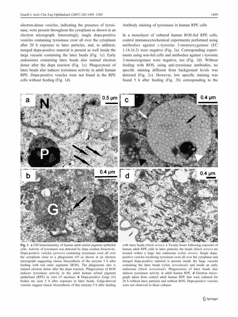
electron-dense vesicles, indicating the presence of tyrosi-nase, were present throughout the cytoplasm as shown in anelectron micrograph. Interestingly, single dopa-positivevesicles containing tyrosinase exist all over the cytoplasmafter 20 h exposure to latex particles, and, in addition,merged dopa-positive material is present as well inside thelarge vacuole containing the latex beads (Fig. 1c). Earlyendosomes containing latex beads also stained electrondense after the dopa reaction (Fig. 1c). Phagocytosis oflatex beads also induces tyrosinase activity in adult humanRPE. Dopa-positive vesicles were not found in the RPEcells without feeding (Fig. 1d).
Antibody staining of tyrosinase in human RPE cells
In a monolayer of cultured human ROS-fed RPE cells,control immunocytochemical experiments performed usingantibodies against L-tyrosine 3-monooxygenase (EC1.14.16.2) were negative (Fig. 2a). Corresponding experi-ments using non-fed cells and antibodies against L-tyrosine3-monooxigenase were negative, too (Fig. 2d). Withoutfeeding with ROS, using anti-tyrosinase antibodies, nospecific staining different from background levels wasdetected (Fig. 2c). However, low specific staining wasfound 5 h after feeding (Fig. 2b) corresponding to the
Fig. 1 a EM histochemistry of human adult retinal pigment epithelialcells. Activity of tyrosinase was detected by dopa oxidase bioactivity.Dopa-positive vesicles (arrows) containing tyrosinase exist all overthe cytoplasm close to a phagosome (P) as shown in an electronmicrograph suggesting classic biosynthesis of the enzyme 5 h afterfeeding with rod outer segments (ROS). The phagosome also isstained electron dense after the dopa reaction. Phagocytosis of ROSinduces tyrosinase activity in the adult human retinal pigmentepithelium (RPE) in vitro (N nucleus). b Dopa-positive Golgi (G)bodies are seen 5 h after exposure to latex beads. Golgi-derivedvesicles suggest classic biosynthesis of this enzyme 5 h after feeding
with latex beads (black arrow). c Twenty hours following exposure ofhuman adult RPE cells to latex particles the beads (black arrow) arelocated within a large late endosome (white arrow). Single dopa-positive vesicles localizing tyrosinase exist all over the cytoplasm andmerged dopa-positive material is present inside the large vacuolecontaining the latex beads (white arrowhead ) and inside an earlyendosome (black arrowhead ). Phagocytosis of latex beads alsoinduces tyrosinase activity in adult human RPE. d Electron micro-graph taken from control adult human RPE that were cultured for20 h without latex particles and without ROS. Dopa-positive vesicleswere not observed in these cultures
Graefe’s Arch Clin Exp Ophthalmol (2007) 245:1495–1505 1499
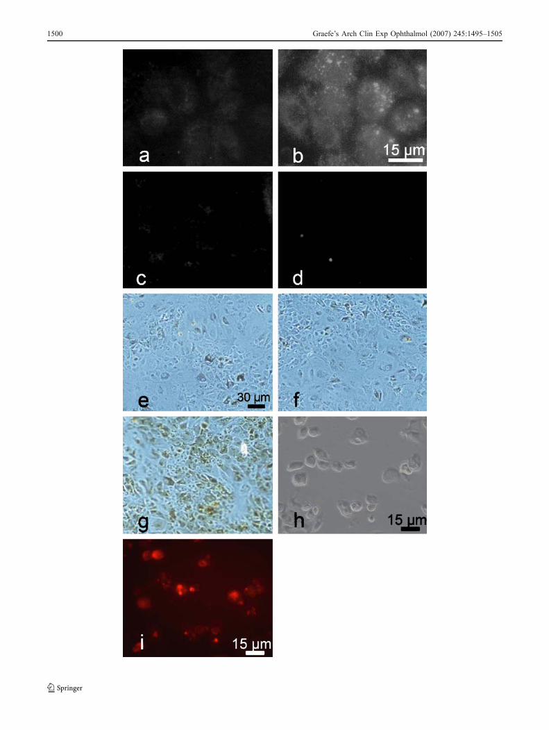
1500 Graefe’s Arch Clin Exp Ophthalmol (2007) 245:1495–1505
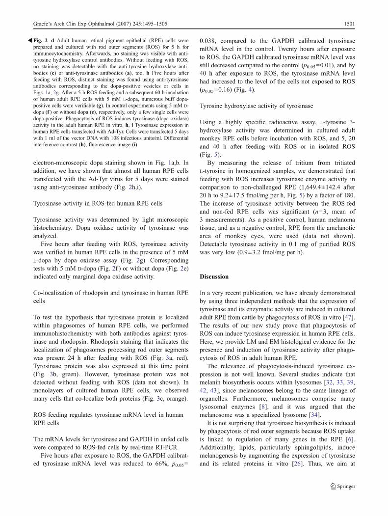
electron-microscopic dopa staining shown in Fig. 1a,b. Inaddition, we have shown that almost all human RPE cellstransfected with the Ad-Tyr virus for 5 days were stainedusing anti-tyrosinase antibody (Fig. 2h,i).
Tyrosinase activity in ROS-fed human RPE cells
Tyrosinase activity was determined by light microscopichistochemistry. Dopa oxidase activity of tyrosinase wasanalyzed.
Five hours after feeding with ROS, tyrosinase activitywas verified in human RPE cells in the presence of 5 mML-dopa by dopa oxidase assay (Fig. 2g). Correspondingtests with 5 mM D-dopa (Fig. 2f ) or without dopa (Fig. 2e)indicated only marginal dopa oxidase activity.
Co-localization of rhodopsin and tyrosinase in human RPEcells
To test the hypothesis that tyrosinase protein is localizedwithin phagosomes of human RPE cells, we performedimmunohistochemistry with both antibodies against tyros-inase and rhodopsin. Rhodopsin staining that indicates thelocalization of phagosomes processing rod outer segmentswas present 24 h after feeding with ROS (Fig. 3a, red).Tyrosinase protein was also expressed at this time point(Fig. 3b, green). However, tyrosinase protein was notdetected without feeding with ROS (data not shown). Inmonolayers of cultured human RPE cells, we observedmany cells that co-localize both proteins (Fig. 3c, orange).
ROS feeding regulates tyrosinase mRNA level in humanRPE cells
The mRNA levels for tyrosinase and GAPDH in unfed cellswere compared to ROS-fed cells by real-time RT-PCR.
Five hours after exposure to ROS, the GAPDH calibrat-ed tyrosinase mRNA level was reduced to 66%, p0.05=
0.038, compared to the GAPDH calibrated tyrosinasemRNA level in the control. Twenty hours after exposureto ROS, the GAPDH calibrated tyrosinase mRNA level wasstill decreased compared to the control (p0.05=0.01), and by40 h after exposure to ROS, the tyrosinase mRNA levelhad increased to the level of the cells not exposed to ROS(p0.05=0.16) (Fig. 4).
Tyrosine hydroxylase activity of tyrosinase
Using a highly specific radioactive assay, L-tyrosine 3-hydroxylase activity was determined in cultured adultmonkey RPE cells before incubation with ROS, and 5, 20and 40 h after feeding with ROS or in isolated ROS(Fig. 5).
By measuring the release of tritium from tritiatedL-tyrosine in homogenized samples, we demonstrated thatfeeding with ROS increases tyrosinase enzyme activity incomparison to non-challenged RPE (1,649.4±142.4 after20 h to 9.2±17.5 fmol/mg per h, Fig. 5) by a factor of 180.The increase of tyrosinase activity between the ROS-fedand non-fed RPE cells was significant (n=3, mean of3 measurements). As a positive control, human melanomatissue, and as a negative control, RPE from the amelanoticarea of monkey eyes, were used (data not shown).Detectable tyrosinase activity in 0.1 mg of purified ROSwas very low (0.9±3.2 fmol/mg per h).
Discussion
In a very recent publication, we have already demonstratedby using three independent methods that the expression oftyrosinase and its enzymatic activity are induced in culturedadult RPE from cattle by phagocytosis of ROS in vitro [47].The results of our new study prove that phagocytosis ofROS can induce tyrosinase expression in human RPE cells.Here, we provide LM and EM histological evidence for thepresence and induction of tyrosinase activity after phago-cytosis of ROS in adult human RPE.
The relevance of phagocytosis-induced tyrosinase ex-pression is not well known. Several studies indicate thatmelanin biosynthesis occurs within lysosomes [32, 33, 39,42, 43], since melanosomes belong to the same lineage oforganelles. Furthermore, melanosomes comprise manylysosomal enzymes [8], and it was argued that themelanosome was a specialized lysosome [34].
It is not surprising that tyrosinase biosynthesis is inducedby phagocytosis of rod outer segments because ROS uptakeis linked to regulation of many genes in the RPE [6].Additionally, lipids, particularly sphingolipids, inducemelanogenesis by augmenting the expression of tyrosinaseand its related proteins in vitro [26]. Thus, we aim at
Fig. 2 d Adult human retinal pigment epithelial (RPE) cells wereprepared and cultured with rod outer segments (ROS) for 5 h forimmunocytochemistry. Afterwards, no staining was visible with anti-tyrosine hydroxylase control antibodies. Without feeding with ROS,no staining was detectable with the anti-tyrosine hydroxylase anti-bodies (c) or anti-tyrosinase antibodies (a), too. b Five hours afterfeeding with ROS, distinct staining was found using anti-tyrosinaseantibodies corresponding to the dopa-positive vesicles or cells inFigs. 1a, 2g. After a 5-h ROS feeding and a subsequent 60-h incubationof human adult RPE cells with 5 mM L-dopa, numerous buff dopa-positive cells were verifiable (g). In control experiments using 5 mM D-dopa (f ) or without dopa (e), respectively, only a few single cells weredopa-positive. Phagocytosis of ROS induces tyrosinase (dopa oxidase)activity in the adult human RPE in vitro. h, i Tyrosinase expression inhuman RPE cells transfected with Ad-Tyr. Cells were transfected 5 dayswith 1 ml of the vector DNA with 108 infectious units/ml. Differentialinterference contrast (h), fluorescence image (i)
�
Graefe’s Arch Clin Exp Ophthalmol (2007) 245:1495–1505 1501
researching in subsequent studies whether ROS lipidsinduce expression of tyrosinase and melanogenesis.
The electron-microscopic results of tyrosinase activationafter feeding with ROS match the classic model ofmelanogenesis with Golgi-derived dopa-positive vesicleswidespread in the cytoplasm of pigment cells. Vesiclesincorporating tyrosinase may fuse with phagosomes.Tyrosinase may detoxify reactive oxygen species insidephagosomes, e.g. superoxide anions [51], or may haveanother as yet unknown function. This model complies withresults of this and recent studies, in which tyrosinase wasfound in partly degraded phagosomes [42]. In addition, thereis an interrelation between phagosomal degradation path-ways and melanosome biogenesis in RPE cells due to thefact that phagosomal material is transported to the melano-somes or melanogenesis occurs inside phagosomes [46].
Recent experimental results and the findings of thisstudy are in line with the current model of tyrosinasetrafficking in melanocytes assuming that phagosomes arerelated to endosomes. Tyrosinase is transported from thetrans-Golgi network to early endosomes and attains via lateendosomes lysosomes and premelanosomes in melanocytes[16, 35].
There is a causal interrelationship between the phagocy-tosis of ROS and the production of reactive oxygenintermediates such as superoxide anion radicals O�
�2
� �,
hydrogen peroxide (H2O2) and hydroxyl radicals (OH·) [9].Hydrogen peroxide increases tyrosinase activity in culturedhuman melanoma cells [19], is a competitive inhibitor oftyrosinase [54] and induces tyrosinase [15, 19]. Moreover,superoxide anion radicals augment activity of humantyrosinase [54]. Tyrosinase may protect against oxidativestress by diverse mechanisms: first, by oxidation oftetrahydroisoquinones, tyrosinase may promote their con-version into inert insoluble polymers; second, by utilizingthe superoxide anion as substrate. Hence, the L-dopa:oxygen oxidoreductase could act as a free radical trappingenzyme [51]. Moreover, it has been shown in retinaldiseases with an oxidative stress component like age-relatedmacular degeneration that black people are more protected
Fig. 3 Co-staining of rhodopsin and tyrosinase in adult human retinalpigment epithelial (RPE) cells in vitro 24 h after incubation with rodouter segments (ROS). a The fluorescence microscopy-based imagetaken from the RPE monolayer shows immunostaining with an anti-rhodopsin antibody detected with a Cy3-conjugated secondaryantibody (red). Rhodopsin was highly expressed in ROS phagosomes24 h after feeding with ROS. b The same fluorescent image afterlabelling of tyrosinase with a monoclonal antibody and a Cy2-conjugated secondary antibody (green). c A merged image of a+b thatdemonstrates colocalization (large arrow, orange fluorescence) ofrhodopsin (arrowhead) and tyrosinase (small arrow) expression inROS phagosomes. The nuclear staining was performed with Hoechstdye (blue). No expression of tyrosinase was found without feedingwith ROS (d)
�
1502 Graefe’s Arch Clin Exp Ophthalmol (2007) 245:1495–1505
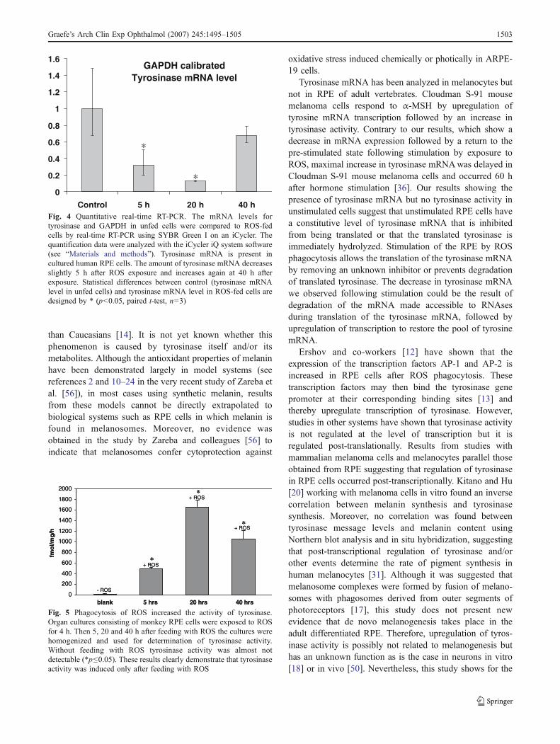
than Caucasians [14]. It is not yet known whether thisphenomenon is caused by tyrosinase itself and/or itsmetabolites. Although the antioxidant properties of melaninhave been demonstrated largely in model systems (seereferences 2 and 10–24 in the very recent study of Zareba etal. [56]), in most cases using synthetic melanin, resultsfrom these models cannot be directly extrapolated tobiological systems such as RPE cells in which melanin isfound in melanosomes. Moreover, no evidence wasobtained in the study by Zareba and colleagues [56] toindicate that melanosomes confer cytoprotection against
oxidative stress induced chemically or photically in ARPE-19 cells.
Tyrosinase mRNA has been analyzed in melanocytes butnot in RPE of adult vertebrates. Cloudman S-91 mousemelanoma cells respond to α-MSH by upregulation oftyrosine mRNA transcription followed by an increase intyrosinase activity. Contrary to our results, which show adecrease in mRNA expression followed by a return to thepre-stimulated state following stimulation by exposure toROS, maximal increase in tyrosinase mRNAwas delayed inCloudman S-91 mouse melanoma cells and occurred 60 hafter hormone stimulation [36]. Our results showing thepresence of tyrosinase mRNA but no tyrosinase activity inunstimulated cells suggest that unstimulated RPE cells havea constitutive level of tyrosinase mRNA that is inhibitedfrom being translated or that the translated tyrosinase isimmediately hydrolyzed. Stimulation of the RPE by ROSphagocytosis allows the translation of the tyrosinase mRNAby removing an unknown inhibitor or prevents degradationof translated tyrosinase. The decrease in tyrosinase mRNAwe observed following stimulation could be the result ofdegradation of the mRNA made accessible to RNAsesduring translation of the tyrosinase mRNA, followed byupregulation of transcription to restore the pool of tyrosinemRNA.
Ershov and co-workers [12] have shown that theexpression of the transcription factors AP-1 and AP-2 isincreased in RPE cells after ROS phagocytosis. Thesetranscription factors may then bind the tyrosinase genepromoter at their corresponding binding sites [13] andthereby upregulate transcription of tyrosinase. However,studies in other systems have shown that tyrosinase activityis not regulated at the level of transcription but it isregulated post-translationally. Results from studies withmammalian melanoma cells and melanocytes parallel thoseobtained from RPE suggesting that regulation of tyrosinasein RPE cells occurred post-transcriptionally. Kitano and Hu[20] working with melanoma cells in vitro found an inversecorrelation between melanin synthesis and tyrosinasesynthesis. Moreover, no correlation was found betweentyrosinase message levels and melanin content usingNorthern blot analysis and in situ hybridization, suggestingthat post-transcriptional regulation of tyrosinase and/orother events determine the rate of pigment synthesis inhuman melanocytes [31]. Although it was suggested thatmelanosome complexes were formed by fusion of melano-somes with phagosomes derived from outer segments ofphotoreceptors [17], this study does not present newevidence that de novo melanogenesis takes place in theadult differentiated RPE. Therefore, upregulation of tyros-inase activity is possibly not related to melanogenesis buthas an unknown function as is the case in neurons in vitro[18] or in vivo [50]. Nevertheless, this study shows for the
GAPDH calibratedTyrosinase mRNA level
0
0.2
0.4
0.6
0.8
1
1.2
1.4
1.6
Control 5 h 20 h 40 h
∗
∗
Fig. 4 Quantitative real-time RT-PCR. The mRNA levels fortyrosinase and GAPDH in unfed cells were compared to ROS-fedcells by real-time RT-PCR using SYBR Green I on an iCycler. Thequantification data were analyzed with the iCycler iQ system software(see “Materials and methods”). Tyrosinase mRNA is present incultured human RPE cells. The amount of tyrosinase mRNA decreasesslightly 5 h after ROS exposure and increases again at 40 h afterexposure. Statistical differences between control (tyrosinase mRNAlevel in unfed cells) and tyrosinase mRNA level in ROS-fed cells aredesigned by * (p<0.05, paired t-test, n=3)
0
200
400
600
800
1000
1200
1400
1600
1800
2000
- ROS
+ ROS
+ ROS
+ ROS
fmo
l/mg
/h
blank 5 hrs 20 hrs 40 hrs
*
*
*
0
200
400
600
800
1000
1200
1400
1600
1800
2000
- ROS
+ ROS
+ ROS
+ ROS
fmo
l/mg
/h
blank 5 hrs 20 hrs 40 hrs
*
*
*
Fig. 5 Phagocytosis of ROS increased the activity of tyrosinase.Organ cultures consisting of monkey RPE cells were exposed to ROSfor 4 h. Then 5, 20 and 40 h after feeding with ROS the cultures werehomogenized and used for determination of tyrosinase activity.Without feeding with ROS tyrosinase activity was almost notdetectable (*p≤0.05). These results clearly demonstrate that tyrosinaseactivity was induced only after feeding with ROS
Graefe’s Arch Clin Exp Ophthalmol (2007) 245:1495–1505 1503

first time using several independent methods that tyrosinaseactivity as well as its expression is induced by phagocytosisof rod outer segments in adult human RPE cells.
Acknowledgements This project was supported by the DeutscheForschungsgemeinschaft, Project SCHR 436/12-2. The authors thankHanna Janicki, Sigrid Schultheiss, Jolanta Koslowski and Judith Birchfor expert technical assistance.
References
1. Abul-Hassan K, Walmsley R, Tombran-Tink J, Boulton M (2000)Regulation of tyrosinase expression and activity in culturedhuman retinal pigment epithelial cells. Pigment Cell Res13:436–441
2. Aronson JF (1983) Human retinal pigment cell culture. In Vitro19:642–650
3. Basu PK, Sarkar P, Menon I, Carre F, Persad S (1983) Bovineretinal pigment epithelial cells cultured in vitro: growth character-istics, morphology, chromosomes, phagocytosis ability, tyrosinaseactivity and effect of freezing. Exp Eye Res 36:671–683
4. Brichard V, Van Pel A, Wolfel C, De Plaen E, Lethe B, Coulie P,Boon T (1993) The tyrosinase gene codes for an antigenrecognized by autologous cytolytic T lymphocytes on HLA-A2melanomas. J Exp Med 178:489–495
5. Carr RE, Siegel IM (1979) The retinal pigment epithelium inocular albinism. In: KM Zinn, Marmor MF (eds) The retinalpigment epithelium. Harvard University Press, Cambridge, MA,pp 413–423
6. Chowers I, Kim Y, Farkas RH, Gunatilaka TL, Hackam AS,Campochiaro PA, Finnemann SC, Zack DJ (2004) Changes inretinal pigment epithelial gene expression induced by rod outersegment uptake. Invest Ophthalmol Vis Sci 45:2098–2106
7. Cuomo M, Nicotra MR, Apollonj C, Fraioli R, Giacomini P,Natali PG (1991) Production and characterization of the murinemonoclonal antibody 2G10 to a human T4-tyrosinase epitope.J Invest Dermatol 96:446–451
8. Diment S, Eidelman M, Rodriguez GM, Orlow SJ (1995)Lysosomal hydrolases are present in melanosomes and areelevated in melanizing cells. J Biol Chem 270:4213–4215
9. Dorey CK, Khouri GG, Syniuta LA, Curran SA, Weiter JJ (1989)Superoxide production by porcine retinal pigment epithelium invitro. Invest Ophthalmol Vis Sci 30:1047–1054
10. Dorey CK, Torres X, Swart T (1990) Evidence of melanogenesisin porcine retinal pigment epithelial cells in vitro. Exp Eye Res50:1–10
11. Dryja TP, O’Neil-Dryja M, Pawelek JM, Albert DM (1978)Demonstration of tyrosinase in the adult bovine uveal tract andretinal pigment epithelium. Invest Ophthalmol Vis Sci 17:511–514
12. Ershov AV, Lukiw WJ, Bazan NG (1996) NG Selectivetranscription factor induction in retinal pigment epithelial cellsduring photoreceptor phagocytosis. J Biol Chem 271:28458–28462
13. Ferguson CA, Kidson SH (1997) The regulation of tyrosinasegene transcription. Pigment Cell Res 10:127–138
14. Friedman DS, Katz J, Bressler NM, Rahmani B, Tielsch JM(1999) Racial differences in the prevalence of age-related maculardegeneration: the Baltimore Eye Survey. Ophthalmology106:1049–1055
15. Gomez Sarosi LA, Rieber MS, Rieber M (2003) Hydrogenperoxide increases a 55-kDa tyrosinase concomitantly withinduction of p53-dependent p21 waf1 expression and a greater
Bax/Bcl-2 ratio in pigmented melanoma. Biochem Biophys ResCommun 312:355–359
16. Hearing VJ (2005) Biogenesis of pigment granules: a sensitiveway to regulate melanocyte function. J Dermatol Sci 37:3–14
17. Herman KG, Steinberg RH (1982) Melanosome metabolism in theretinal pigmented epithelium of the opossum. Cell Tissue Res227:485–507
18. Higashi Y, Asanuma M, Miyazaki I, Ogawa N (2000) Inhibitionof tyrosinase reduces cell viability in catecholaminergic neuronalcells. J Neurochem 75:1771–1774
19. Karg E, Odh G, Wittbjer A, Rosengren E, Rorsman H (1993)Hydrogen peroxide as an inducer of elevated tyrosinase level inmelanoma cells. J Invest Dermatol 100:209S–213S
20. Kitano Y, Hu F (1970) DNA, RNA, and protein synthesis ofpigment cells in culture. J Invest Dermatol 54:463–471
21. Korner A, Pawelek J (1982) Mammalian tyrosinase catalyzesthree reactions in the biosynthesis of melanin. Science 217:1163–1165
22. Kreppel F, Biermann V, Kochanek S, Schiedner G (2002) ADNA-based method to assay total and infectious particle contentsand helper virus contamination in high-capacity adenoviral vectorpreparations. Hum Gene Ther 13:1151–1156
23. LaVail MM (1980) Circadian nature of rod outer segment discshedding in the rat. Invest Ophthalmol Vis Sci 19:407–411
24. Lerch K (1987) Monophenol monooxygenase from Neurosporacrassa. Methods Enzymol 142:165–169
25. Lowry OH, Rosebrough NJ, Farr AL, Randall RJ (1951) Proteinmeasurement with the Folin phenol reagent. J Biol Chem193:265–275
26. Mallick S, Singh SK, Sarkar C, Saha B, Bhadra R (2005) Humanplacental lipid induces melanogenesis by increasing the expres-sion of tyrosinase and its related proteins in vitro. Pigment CellRes 18:25–33
27. Marks MS, Seabra MC (2001) The melanosome: membranedynamics in black and white. Nat Rev Mol Cell Biol 2:738–748
28. Mirica LM, Vance M, Rudd DJ, Hedman B, Hodgson KO,Solomon EI, Stack TD (2005) Tyrosinase reactivity in a modelcomplex: an alternative hydroxylation mechanism. Science308:1890–1892
29. Miyamoto M, Fitzpatrick TB (1957) On the nature of pigment inretinal pigment epithelium. Science 126:449–450
30. Morrison TB, Weiss JJ, Wittwer CT (1998) Quantification of low-copy transcripts by continuous SYBR Green monitoring duringamplification. Biotechniques 24:954–958, 960, 962
31. Naeyaert JM, Eller M, Gordon PR, Park HY, Gilchrest BA (1991)Pigment content of cultured human melanocytes does not correlatewith tyrosinase message level. Br J Dermatol 125:297–303
32. Nakagawa H, Rhodes AR, Momtaz T, Fitzpatrick TB (1984)Morphologic alterations of epidermal melanocytes and melano-somes in PUVA lentigines: a comparative ultrastructural investi-gation of lentigines induced by PUVA and sunlight. J InvestDermatol 82:101–107
33. Novikoff AB, Leuenberger PM, Novikoff PM, Quintana N (1979)Retinal pigment epithelium. Interrelations of endoplasmic reticu-lum and melanolysosomes in the black mouse and its beigemutant. Lab Invest 40:155–165
34. Orlow SJ (1995) Melanosomes are specialized members of thelysosomal lineage of organelles. J Invest Dermatol 105:3–7
35. Raposo G, Marks MS (2002) The dark side of lysosome-relatedorganelles: specialization of the endocytic pathway for melano-some biogenesis. Traffic 3:237–248
36. Rungta D, Corn TD, Fuller BB (1996) Regulation of tyrosinasemRNA in mouse melanoma cells by alpha-melanocyte-stimulatinghormone. J Invest Dermatol 107:689–693
37. Sarna T (1992) Properties and function of the ocular melanin-aphotobiophysical view. J Photochem Photobiol B 12:215–258
1504 Graefe’s Arch Clin Exp Ophthalmol (2007) 245:1495–1505
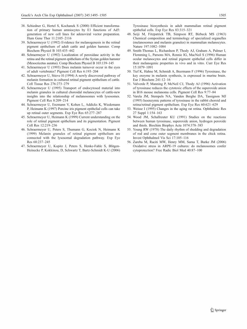
38. Schiedner G, Hertel S, Kochanek S (2000) Efficient transforma-tion of primary human amniocytes by E1 functions of Ad5:generation of new cell lines for adenoviral vector preparation.Hum Gene Ther 11:2105–2116
39. Schraermeyer U (1992) Evidence for melanogenesis in the retinalpigment epithelium of adult cattle and golden hamster. CompBiochem Physiol B 103:435–442
40. Schraermeyer U (1992) Localization of peroxidase activity in theretina and the retinal pigment epithelium of the Syrian golden hamster(Mesocricetus auratus). Comp Biochem Physiol B 103:139–145
41. Schraermeyer U (1993) Does melanin turnover occur in the eyesof adult vertebrates? Pigment Cell Res 6:193–204
42. Schraermeyer U, Stieve H (1994) A newly discovered pathway ofmelanin formation in cultured retinal pigment epithelium of cattle.Cell Tissue Res 276:273–279
43. Schraermeyer U (1995) Transport of endocytosed material intomelanin granules in cultured choroidal melanocytes of cattle-newinsights into the relationship of melanosomes with lysosomes.Pigment Cell Res 8:209–214
44. Schraermeyer U, Enzmann V, Kohen L, Addicks K, WiedemannP, Heimann K (1997) Porcine iris pigment epithelial cells can takeup retinal outer segments. Exp Eye Res 65:277–287
45. Schraermeyer U, Heimann K (1999) Current understanding on therole of retinal pigment epithelium and its pigmentation. PigmentCell Res 12:219–236
46. Schraermeyer U, Peters S, Thumann G, Kociok N, Heimann K(1999) Melanin granules of retinal pigment epithelium areconnected with the lysosomal degradation pathway. Exp EyeRes 68:237–245
47. Schraermeyer U, Kopitz J, Peters S, Henke-Fahle S, Blitgen-Heinecke P, Kokkinou, D, Schwartz T, Bartz-Schmidt K-U (2006)
Tyrosinase biosynthesis in adult mammalian retinal pigmentepithelial cells. Exp Eye Res 83:315–321
48. Seiji M, Fitzpatrick TB, Simpson RT, Birbeck MS (1963)Chemical composition and terminology of specialized organelles(melanosomes and melanin granules) in mammalian melanocytes.Nature 197:1082–1084
49. Smith-Thomas L, Richardson P, Thody AJ, Graham A, Palmer I,Flemming L, Parsons MA, Rennie IG, MacNeil S (1996) Humanocular melanocytes and retinal pigment epithelial cells differ intheir melanogenic properties in vivo and in vitro. Curr Eye Res15:1079–1091
50. Tief K, Hahne M, Schmidt A, Beermann F (1996) Tyrosinase, thekey enzyme in melanin synthesis, is expressed in murine brain.Eur J Biochem 241:12–16
51. Valverde P, Manning P, McNeil CJ, Thody AJ (1996) Activationof tyrosinase reduces the cytotoxic effects of the superoxide anionin B16 mouse melanoma cells. Pigment Cell Res 9:77–84
52. Varela JM, Stempels NA, Vanden Berghe DA, Tassignon MJ(1995) Isoenzymic patterns of tyrosinase in the rabbit choroid andretina/retinal pigment epithelium. Exp Eye Res 60:621–629
53. Weisse I (1995) Changes in the aging rat retina. Ophthalmic Res27 Suppl 1:154–163
54. Wood JM, Schallreuter KU (1991) Studies on the reactionsbetween human tyrosinase, superoxide anion, hydrogen peroxideand thiols. Biochim Biophys Acta 1074:378–385
55. Young RW (1978) The daily rhythm of shedding and degradationof rod and cone outer segment membranes in the chick retina.Invest Ophthalmol Vis Sci 17:105–116
56. Zareba M, Raciti MW, Henry MM, Sarna T, Burke JM (2006)Oxidative stress in ARPE-19 cultures: do melanosomes confercytoprotection? Free Radic Biol Med 40:87–100
Graefe’s Arch Clin Exp Ophthalmol (2007) 245:1495–1505 1505
1
The classical pathway of melanogenesis is not essential for melanin synthesis in the adult retinal pigment epithelium
Antje Biesemeier1, Florian Kreppel2, Stefan Kochanek2, Ulrich Schraermeyer1 1 Department for Experimental Vitreoretinal Surgery, University Eye Hospital Tuebingen,
Tuebingen, Germany; 2 Division for Gene Therapy, University of Ulm, Ulm, Germany Summary
Premelanosomes are presumed to be essential for melanogenesis in melanocytes and pre-natal RPE cells. In this study, we analysed melanin synthesis in adenoviral-transduced, tyrosinase gene expressing ARPE 19 cells to find out whether premelanosome formation is needed for post-natal melanogenesis. Synthesis of melanogenic proteins and melanin granules was investigated with immunocytochemistry, light and electron microscopy. The occurrence of tyrosinase was analysed ultrastructurally, using DOPA histochemistry. The viability of transduced cell cultures was analysed using an MTT assay. We found active tyrosinase in small granule-like vesicles all over the cytoplasm and in the ER and the nuclear membrane. Tyrosinase was also associated with multi-vesicular and multi-lamellar organelles. Typical premelanosomes, the structural protein PMEL17, and tyrosinase-related protein 1, as well as the melanosomal stages I-IV, as described in the literature, were not detected. Instead, melanogenesis took place inside multi-vesicular and multi-lamellar bodies of unknown origin. Viability was not affected up to ten days after transduction. Here we demonstrate a pathway of melanin formation, which lacks the typical hallmarks of melanogenesis, as they are described in the literature. Keywords: ARPE 19, tyrosinase, premelanosome, melanogenesis, alternative pathway
Introduction
Many differences between melanocyte and RPE melanisation are due to their different embryonic origin and development (Bharti et al. 2006; Bharti et al. 2008; Murisier et al. 2007), and affect, for example, the regulation of melanogenesis and the transcription of melanogenic genes by using different transcription and growth factors. While skin melanocytes produce melanosomes continuously, RPE cells are presumed to create melanosomes only prenatally. In addition, the shape and size of mature RPE melanosomes differs from that of melanocytes (Schraermeyer and Heimann 1999). Despite this, melanogenesis in RPE cells has been described as sharing a common pathway with melanocytes. They use the same melanogenic proteins and the ultrastructural characterisation of morphologically distinct melanosomal stages I to IV has been confirmed for both melanocytes and pre-natal RPE cells (Lopes et al. 2007). The formation of melanosomes in both cell types has been described as follows: An amorphous vesicle, called the premelanosome, which accumulates typical intraluminal striations, is believed to build the first phase of melanogenesis (stage I). After the intraluminal arrangement is completed (stage II), tyrosinase-filled coated vesicles, which have emerged from the Golgi apparatus, can enter the premelanosome. Tyrosinase and other melanising proteins are integrated into the melanosomal membrane and synthesise melanin, which is deposited along the internal striations (stage III). When the whole organelle is filled with melanin, it is termed stage IV or a mature melanosome (Seiji et al. 1963). There are two criteria essential for melanogenesis in melanocytes and pre-natal RPE cells: the presence of premelanosomes and the synthesis of tyrosinase and other melanogenic proteins. Without the enzymatic action of tyrosinase, the formation of melanin is not possible
2
(Oetting and King 1999). Since tyrosinase and melanin precursors have a high oxidative potential and can facilitate radical building and oxidative stress reactions in the cell, they have to be separated inside a closed organelle, the premelanosome (Theos et al. 2006). The premelanosome is characterised by having typical internal striations, which are used for melanin deposition. These striations are composed of the structural protein PMEL17 (Berson et al. 2001; Berson et al. 2003) and probably other components, as yet unknown. The absence of PMEL17 leads to the loss of premelanosome formation, to mistargeting and degradation of melanogenic enzymes and to defective pigmentation in mutant individuals (Theos et al. 2006). While some authors have found tyrosinase in adult RPE (Schraermeyer et al. 2006; Julien et al. 2007; Basu et al. 1983; Abul-Hassan et al. 2000; Varela et al. 1995), the presence of PMEL17 has not yet been reported (Lopes et al. 2007). Therefore, a melanin renewal in the adult RPE has long been doubted (Carr and Siegel 1979; Dorey et al. 1990; Miyamoto and Fitzpatrick 1957; Sarna 1992). In the few publications existing about post-natal RPE melanogenesis (Herman and Steinberg 1982; Thumann et al. 1999; Schraermeyer et al. 1999; Peters et al. 2000; Schraermeyer and Stieve 1994; Dorey et al. 1990), opinions differ concerning the occurrence of typical striated premelanosomes. Goldman-Herman and Steinberg (Herman and Steinberg 1982) only found the melanogenic stages II-IV in the RPE of opossums. Others showed that striated premelanosomes were associated with phago-lysosomal organelles (Thumann et al. 1999; Schraermeyer et al. 1999; Peters et al. 2000; Schraermeyer and Stieve 1994; Young 1977) or observed melanogenesis taking place in organelles of the degradative pathway inside multi-vesicular and multi-lamellar bodies (Novikoff et al. 1979) and without the formation of striated premelanosomes (Dorey et al. 1990). The morphological diversity of post-natal melanogenic stages observed indicates the presence of an alternative pathway of melanosome formation in adult RPE cells, which might lack typical PMEL17 striations or which might at least be morphologically different to the classical model. Here we demonstrate that melanogenesis can occur without the formation of typical premelanosomes inside multi-vesicular and multi-lamellar organelles of ARPE 19 cells.
Methods
Human amelanotic retinal pigment epithelial cells Human amelanotic retinal pigment epithelial cells (ARPE 19 cell line) were obtained from the American Type Culture Collection (ATCC, Manassas, VA, USA) and used between passages three and thirteen. Since ARPE 19 cells are a highly differentiated, immortalized RPE cell line, lacking an observable pigmentation due to passaging and the very low ability to form new pigments (Dunn et al. 1996), they provide the ideal system to study new pigment synthesis of adult RPE cells. Cells were grown in DMEM medium with 10% FCS and 1% antibiotics (containing 5 units/ml Penicillin G and 5 mg/ml Streptomycin) in 250 ml culture flasks (Greiner, Frickenhausen, Germany). All cell culture reagents were obtained from GIBCO (Invitrogen, Karlsruhe, Germany). The experiments were performed in 24-well culture plates (Costar Corning, Bodenheim, Germany) for the light and electron microscopy and in 16-well chamber slides (nunc, Wiesbaden) for fluorescence immunocytochemistry. MNT-1 melanoma cells The MNT-1 cells were a kind gift from Prof. P.G. Natali, Dep. of Immunology and Molecular Pathology Laboratories, "Regina Elena" National Cancer Institute, Rome, Italy. They were cultured in RPMI 1640 medium +10% FCS without the supplementation of antibiotics. Additional material: Chemicals were purchased from Sigma, Deisenhofen, Germany; dimethylsulfoxid (DMSO) and paraformaldehyde (PFA) from Merck, Darmstadt, Germany. Generation of AdTyr The adenoviral vector AdTyr carries the human tyrosinase insert in its E1 deleted region. It was generated using a method that has been described previously (see (Julien et al. 2007)).
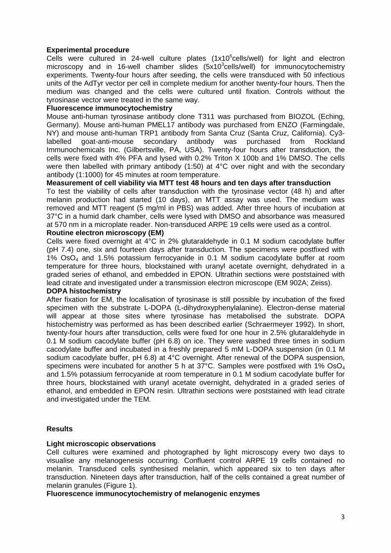
3
Experimental procedure Cells were cultured in 24-well culture plates (1x10⁵cells/well) for light and electron microscopy and in 16-well chamber slides (5x10³cells/well) for immunocytochemistry experiments. Twenty-four hours after seeding, the cells were transduced with 50 infectious units of the AdTyr vector per cell in complete medium for another twenty-four hours. Then the medium was changed and the cells were cultured until fixation. Controls without the tyrosinase vector were treated in the same way. Fluorescence immunocytochemistry Mouse anti-human tyrosinase antibody clone T311 was purchased from BIOZOL (Eching, Germany). Mouse anti-human PMEL17 antibody was purchased from ENZO (Farmingdale, NY) and mouse anti-human TRP1 antibody from Santa Cruz (Santa Cruz, California). Cy3-labelled goat-anti-mouse secondary antibody was purchased from Rockland Immunochemicals Inc. (Gilbertsville, PA, USA). Twenty-four hours after transduction, the cells were fixed with 4% PFA and lysed with 0.2% Triton X 100b and 1% DMSO. The cells were then labelled with primary antibody (1:50) at 4°C over night and with the secondary antibody (1:1000) for 45 minutes at room temperature. Measurement of cell viability via MTT test 48 hours and ten days after transduction To test the viability of cells after transduction with the tyrosinase vector (48 h) and after melanin production had started (10 days), an MTT assay was used. The medium was removed and MTT reagent (5 mg/ml in PBS) was added. After three hours of incubation at 37°C in a humid dark chamber, cells were lysed with DMSO and absorbance was measured at 570 nm in a microplate reader. Non-transduced ARPE 19 cells were used as a control. Routine electron microscopy (EM) Cells were fixed overnight at 4°C in 2% glutaraldehyde in 0.1 M sodium cacodylate buffer (pH 7.4) one, six and fourteen days after transduction. The specimens were postfixed with 1% OsO4 and 1.5% potassium ferrocyanide in 0.1 M sodium cacodylate buffer at room temperature for three hours, blockstained with uranyl acetate overnight, dehydrated in a graded series of ethanol, and embedded in EPON. Ultrathin sections were poststained with lead citrate and investigated under a transmission electron microscope (EM 902A; Zeiss). DOPA histochemistry After fixation for EM, the localisation of tyrosinase is still possible by incubation of the fixed specimen with the substrate L-DOPA (L-dihydroxyphenylalanine). Electron-dense material will appear at those sites where tyrosinase has metabolised the substrate. DOPA histochemistry was performed as has been described earlier (Schraermeyer 1992). In short, twenty-four hours after transduction, cells were fixed for one hour in 2.5% glutaraldehyde in 0.1 M sodium cacodylate buffer (pH 6.8) on ice. They were washed three times in sodium cacodylate buffer and incubated in a freshly prepared 5 mM L-DOPA suspension (in 0.1 M sodium cacodylate buffer, pH 6.8) at 4°C overnight. After renewal of the DOPA suspension, specimens were incubated for another 5 h at 37°C. Samples were postfixed with 1% OsO4 and 1.5% potassium ferrocyanide at room temperature in 0.1 M sodium cacodylate buffer for three hours, blockstained with uranyl acetate overnight, dehydrated in a graded series of ethanol, and embedded in EPON resin. Ultrathin sections were poststained with lead citrate and investigated under the TEM.
Results
Light microscopic observations Cell cultures were examined and photographed by light microscopy every two days to visualise any melanogenesis occurring. Confluent control ARPE 19 cells contained no melanin. Transduced cells synthesised melanin, which appeared six to ten days after transduction. Nineteen days after transduction, half of the cells contained a great number of melanin granules (Figure 1). Fluorescence immunocytochemistry of melanogenic enzymes
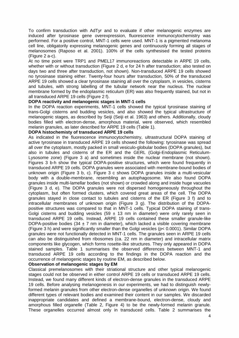
4
To confirm transduction with AdTyr and to evaluate if other melanogenic enzymes are induced after tyrosinase gene overexpression, fluorescence immunocytochemistry was performed. For a positive control, MNT-1 cells were used. MNT-1 is a pigmented melanoma cell line, obligatorily expressing melanogenic genes and continuously forming all stages of melanosomes (Raposo et al. 2001). 100% of the cells synthesised the tested proteins (Figure 2 a-c). At no time point were TRP1 and PMEL17 immunoreactions detectable in ARPE 19 cells, whether with or without transduction (Figure 2 d, e for 24 h after transduction; also tested on days two and three after transduction, not shown). Non-transduced ARPE 19 cells showed no tyrosinase staining either. Twenty-four hours after transduction, 50% of the transduced ARPE 19 cells showed a clear tyrosinase staining all over the cytoplasm, in vesicles, cisterns and tubules, with strong labelling of the tubular network near the nucleus. The nuclear membrane formed by the endoplasmic reticulum (ER) was also frequently stained, but not in all transduced ARPE 19 cells (Figure 2 f). DOPA reactivity and melanogenic stages in MNT-1 cells In the DOPA reaction experiments, MNT-1 cells showed the typical tyrosinase staining of trans-Golgi cisterns and budding vesicles, and also showed the typical ultrastructure of melanogenic stages, as described by Seiji (Seiji et al. 1963) and others. Additionally, cloudy bodies filled with electron-dense, amorphous material, were observed, which resembled melanin granules, as later described for ARPE 19 cells (Table 1). DOPA histochemisty of transduced ARPE 19 cells As indicated in the fluorescence immunocytochemistry, ultrastructural DOPA staining of active tyrosinase in transduced ARPE 19 cells showed the following: tyrosinase was spread all over the cytoplasm, mostly packed in small vesiculo-globular bodies (DOPA granules), but also in tubules and cisterns of the ER and the GERL (Golgi-Endoplasmic Reticulum-Lysosome zone) (Figure 3 a) and sometimes inside the nuclear membrane (not shown). Figures 3 b-h show the typical DOPA-positive structures, which were found frequently in transduced ARPE 19 cells. DOPA granules were associated with membrane-bound bodies of unknown origin (Figure 3 b, c). Figure 3 c shows DOPA granules inside a multi-vesicular body with a double-membrane, resembling an autophagosome. We also found DOPA granules inside multi-lamellar bodies (not shown) or crowded along and inside huge vacuoles (Figure 3 d, e). The DOPA granules were not dispersed homogeneously throughout the cytoplasm, but often formed clusters, which covered great areas of the cell. The DOPA granules stayed in close contact to tubules and cisterns of the ER (Figure 3 f) and to intracellular membranes of unknown origin (Figure 3 g). The distribution of the DOPA-positive structures was compared to that in MNT-1 cells. Typical DOPA staining of trans-Golgi cisterns and budding vesicles (59 ± 13 nm in diameter) were only rarely seen in transduced ARPE 19 cells. Instead, ARPE 19 cells contained these smaller granule-like DOPA-positive bodies (34 ± 7 nm in diameter), which lacked a visible covering membrane (Figure 3 h) and were significantly smaller than the Golgi vesicles (p< 0.0001). Similar DOPA granules were not functionally detected in MNT-1 cells. The granules seen in ARPE 19 cells can also be distinguished from ribosomes (ca. 22 nm in diameter) and intracellular matrix components like glycogen, which forms rosette-like structures. They only appeared in DOPA stained samples. Table 1 summarises the observed differences between MNT-1 and transduced ARPE 19 cells according to the findings in the DOPA reaction and the occurrence of melanogenic stages by routine EM, as described below. Observation of melanogenic stages by EM Classical premelanosomes with their striational structure and other typical melanogenic stages could not be observed in either control ARPE 19 cells or transduced ARPE 19 cells. Instead, we found many different kinds of electron-dense granules in the transduced ARPE 19 cells. Before analysing melanogenesis in our experiments, we had to distinguish newly-formed melanin granules from other electron-dense organelles of unknown origin. We found different types of relevant bodies and examined their content in our samples. We discarded inappropriate candidates and defined a membrane-bound, electron-dense, cloudy and amorphous filled organelle (Table 2, Figure 4) to be the newly-formed melanin granule. These organelles occurred almost only in transduced cells. Table 2 summarises the

5
occurrence of melanogenic organelles as they are observed at different time points. Organelles, which were filled to at least 1/5 with electron-dense amorphous material, but also contained other material, were defined as untypical melanosomal stages 1 to 4 (in contrast to the classical stages I to IV). If less than 1/5 of the granule was filled with the electron-dense material, the organelle was attributed to the mixed bodies, containing lamellar, vesicular and other shaped material. Additionally, Table 2 shows the typical distribution of multi-vesicular bodies (MVBs) and multi-lamellar bodies (MLBs), often seen in all samples independent of transduction. We found a chronological sequence, demonstrating that in the first days after transduction no melanised stages were observable. Instead, MVBs and MLBs and a few mixed bodies (enclosing a potpourri of vesicles, lamellae and electron-dense material) were found. Later on (after 6 days), the material inside the MVBs, MLBs and mixed bodies of transduced samples appeared to merge together, harbouring melanin-like material to form melanogenic stages 1-3, showing lamellae and vesicles, but also higher amounts of electron-dense and amorphous material inside. Mixed vesicles were found particularly in six-day-old cells. After fourteen days, most of the organelles were filled completely with melanin (stage 3-4). The melanin granules were 0.3-1 µm in diameter, resembling the size of MVBs and MLBs, and related to them. Control cells (c in Table 2, day 6) contained only MLBs and some MVBs and mixed bodies, but the latter were less frequently observed. Viability of transduced cells Two and ten days after transduction, the viability of control ARPE 19 cells and cells transduced with AdTyr was analysed using an MTT assay. Two days after transduction, tyrosinase-transduced ARPE 19 cells (101 +/- 16%) showed the same viability as ARPE 19 cells without transduction (100 +/- 11%; Figure 5). After ten days, the tyrosinase-transduced cells (91 +/- 17%) showed a small, but insignificant decrease in viability compared to ARPE 19 cells (100 +/- 12%; p>0.05; n.s.).
Discussion:
Most publications dealing with RPE and melanocyte melanogenesis agree that the enzyme tyrosinase emerges from the trans-faced cisterns of the Golgi apparatus and travels inside coated vesicles to stage II melanosomes (reviewed by (Hearing 2005), but the origin of the amorphous vesicle forming the stage I premelanosome is still not understood. Findings from the late 60s to the early 80s proposed that the premelanosome of melanocytes (reviewed by (Jimbow et al. 1976) and pre-natal RPE cells (Mishima et al. 1978; Eppig and Dumont 1972; Eppig 1970; Stanka et al. 1981; Stanka 1971) is formed out of the ER or tubules of the associated GERL (Novikoff et al. 1968). Novikoff was the first who assumed that the RPE melanosome is a specialised lysosome, emerging from acid phosphatase-positive cisterns of the ER (Novikoff et al. 1979). In the 90s, further scientists confirmed that melanosomes of melanocytes (Schraermeyer 1995), pre-natal RPE (reviewed by (Orlow 1995) and post-natal RPE, reviewed by (Schraermeyer and Heimann 1999; Mishima 1994), are lysosome-related organelles, since they are closely related to phagosomes and lysosomes and have lysosomal enzymes (Diment et al. 1995). New techniques, using immuno-gold labelled antibodies, are able to clearly identify proteins and organelles in ultrastructural samples. Thus, recent studies have clarified that the premelanosome of melanocytes and melanoma cells is an endosomal structure (Raposo et al. 2001), in which the structural protein PMEL17 builds internal striations forming the stage II melanosome (Raposo and Marks 2002). For RPE cells, corresponding studies are still lacking. However, the results of Raposo and colleagues are in accordance with the findings from studies in non-pigmented cells: HELA cells that were forced to synthesise PMEL17 (e.g. by transfection), transported it to MVBs, where it formed premelanosomal striations (Berson et al. 2001). Transfection of fibroblasts (Winder et al. 1993; Ando et al. 1993) and different cell lines (Luo et al. 1994) with tyrosinase induced melanogenesis within lysosomes. Meanwhile, it is accepted that all melanosomes are at least lysosome-related organelles (Bonifacino 2004).
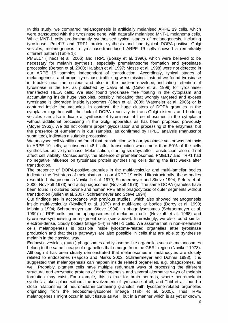
6
In this study, we compared melanogenesis in artificially melanised ARPE 19 cells, which were transduced with the tyrosinase gene, with naturally melanised MNT-1 melanoma cells. While MNT-1 cells predominantly synthesised typical stages of melanogenesis, including tyrosinase, Pmel17 and TRP1 protein synthesis and had typical DOPA-positive Golgi vesicles, melanogenesis in tyrosinase-transduced ARPE 19 cells showed a remarkably different pattern (Table 1): PMEL17 (Theos et al. 2006) and TRP1 (Boissy et al. 1996), which were believed to be necessary for melanin synthesis, especially premelanosome formation and tyrosinase processing (Berson et al. 2000; Halaban et al. 1997; Mosse et al. 1998) were not detected in our ARPE 19 samples independent of transduction. Accordingly, typical stages of melanogenesis and proper tyrosinase trafficking were missing. Instead we found tyrosinase in tubules near the nucleus and also in the nuclear envelope, indicating retention of tyrosinase in the ER, as published by Calvo et al. (Calvo et al. 1999) for tyrosinase-transfected HELA cells. We also found tyrosinase free floating in the cytoplasm and accumulating inside huge vacuoles, possibly indicating that wrongly targeted and excess tyrosinase is degraded inside lysosomes (Chen et al. 2009; Wasmeier et al. 2006) or is captured inside the vacuoles. In contrast, the huge clusters of DOPA granules in the cytoplasm together with the lack of DOPA reactivity in trans-Golgi cisterns and budding vesicles can also indicate a synthesis of tyrosinase at free ribosomes in the cytoplasm without additional processing in the Golgi apparatus as has been proposed previously (Moyer 1963). We did not confirm proper glycosilation and processing of the enzymes, but the presence of eumelanin in our samples, as confirmed by HPLC analysis (manuscript submitted), indicates a suitable processing. We analysed cell viability and found that transduction with our tyrosinase vector was not toxic to ARPE 19 cells, as observed 48 h after transduction when more than 50% of the cells synthesised active tyrosinase. Melanisation, starting six days after transduction, also did not affect cell viability. Consequently, the absence of premelanosomes, PMEL17 and TRP1 had no negative influence on tyrosinase protein synthesising cells during the first weeks after transduction. The presence of DOPA-positive granules in the multi-vesicular and multi-lamellar bodies indicates the first steps of melanisation in our ARPE 19 cells. Ultrastructurally, these bodies resembled phagosomes (Novikoff et al. 1979; Schraermeyer and Stieve 1994; Peters et al. 2000; Novikoff 1973) and autophagosomes (Novikoff 1973). The same DOPA granules have been found in cultured bovine and human RPE after phagocytosis of outer segments without transduction (Julien et al. 2007; Schraermeyer and Stieve 1994). Our findings are in accordance with previous studies, which also showed melanogenesis inside multi-vesicular (Novikoff et al. 1979) and multi-lamellar bodies (Dorey et al. 1990; Mishima 1994; Schraermeyer and Stieve 1994), in phago-lysosomes (Schraermeyer et al. 1999) of RPE cells and autophagosomes of melanoma cells (Novikoff et al. 1968) and tyrosinase-synthesising non-pigment cells (see above). Interestingly, we also found similar electron-dense, cloudy bodies (stage 1-4) in MNT-1 cells. We assume that in non-melanised cells melanogenesis is possible inside lysosome-related organelles after tyrosinase production and that these pathways are also possible in cells that are able to synthesise melanin in the classical way. Endocytic vesicles, (auto-) phagosomes and lysosome-like organelles such as melanosomes belong to the same lineage of organelles that emerge from the GERL region (Novikoff 1973). Although it has been clearly demonstrated that melanosomes in melanocytes are closely related to endosomes (Raposo and Marks 2002; Schraermeyer and Dohms 1993), it is suggested that melanogenesis can happen inside related organelles, e.g. phagosomes, as well. Probably, pigment cells have multiple redundant ways of processing the different structural and enzymatic proteins of melanogenesis and several alternative ways of melanin formation may exist. For example, this is true for brain neurons, where neuromelanin synthesis takes place without the involvement of tyrosinase at all, and Tribl et al. found a close relationship of neuromelanin-containing granules with lysosome-related organelles originating from the endosome-lysosome lineage (Tribl et al. 2005). Thus, RPE melanogenesis might occur in adult tissue as well, but in a manner which is as yet unknown.

7
Latest findings (Gargiulo et al. 2009) show that subretinal injection of an AVV- mediated tyrosinase vector led to repigmentation and the recovery of visual function in albino mice, suggesting that both young and adult RPE cells and choroidal melanocytes are able to restore melanisation.
Acknowledgements
The authors are indebted to Prof. P. G. Natali (Dep. of Immunology and Molecular Pathology Laboratories, "Regina Elena" National Cancer Institute, Rome) for providing the MNT-1 cells. We would also like to thank Monika Rittgarn and Sigrid Schultheiss for excellent technical assistance and Judith Birch for excellent editorial assistance. This work was supported by grants to U. Schraermeyer from the Deutsche Forschungsgemeinschaft (DFG SCHR436/12-2).
References
Abul-Hassan K, Walmsley R, Tombran-Tink J and Boulton M (2000) Regulation of tyrosinase expression and activity in cultured human retinal pigment epithelial cells. Pigment Cell Res 13:436-441
Ando O, Mishima Y, Hanada S, Suemoto Y, Atobe J and Kurimoto M (1993) Analyses of mixed melanogenesis in tyrosinase cDNA-transfected human amelanotic melanoma cells. J Invest Dermatol 101:864-870
Basu PK, Sarkar P, Menon I, Carre F and Persad S (1983) Bovine retinal pigment epithelial cells cultured in vitro: growth characteristics, morphology, chromosomes, phagocytosis ability, tyrosinase activity and effect of freezing. Exp Eye Res 36:671-683
Berson JF, Frank DW, Calvo PA, Bieler BM and Marks MS (2000) A common temperature-sensitive allelic form of human tyrosinase is retained in the endoplasmic reticulum at the nonpermissive temperature. J Biol Chem 275:12281-12289
Berson JF, Harper DC, Tenza D, Raposo G and Marks MS (2001) Pmel17 initiates premelanosome morphogenesis within multivesicular bodies. Mol Biol Cell 12:3451-3464
Berson JF, Theos AC, Harper DC, Tenza D, Raposo G and Marks MS (2003) Proprotein convertase cleavage liberates a fibrillogenic fragment of a resident glycoprotein to initiate melanosome biogenesis. J Cell Biol 161:521-533
Bharti K, Liu W, Csermely T, Bertuzzi S and Arnheiter H (2008) Alternative promoter use in eye development: the complex role and regulation of the transcription factor MITF. Development 135:1169-1178
Bharti K, Nguyen MT, Skuntz S, Bertuzzi S and Arnheiter H (2006) The other pigment cell: specification and development of the pigmented epithelium of the vertebrate eye. Pigment Cell Res 19:380-394
Boissy RE, Zhao H, Oetting WS, Austin LM, Wildenberg SC, Boissy YL, Zhao Y, Sturm RA, Hearing VJ, King RA and Nordlund JJ (1996) Mutation in and lack of expression of tyrosinase-related protein-1 (TRP-1) in melanocytes from an individual with brown oculocutaneous albinism: a new subtype of albinism classified as "OCA3". Am J Hum Genet 58:1145-1156
Bonifacino JS (2004) Insights into the biogenesis of lysosome-related organelles from the study of the Hermansky-Pudlak syndrome. Ann N Y Acad Sci 1038:103-114
Calvo PA, Frank DW, Bieler BM, Berson JF and Marks MS (1999) A cytoplasmic sequence in human tyrosinase defines a second class of di-leucine-based sorting signals for late endosomal and lysosomal delivery. J Biol Chem 274:12780-12789
Carr RE and Siegel IM (1979) The retinal pigment epitehlium in ocular albinism;. In: Zinn KM and Mamor MF (eds) The retinal pigment epithelium. Harvard University Press, Cambridge, pp 413-423
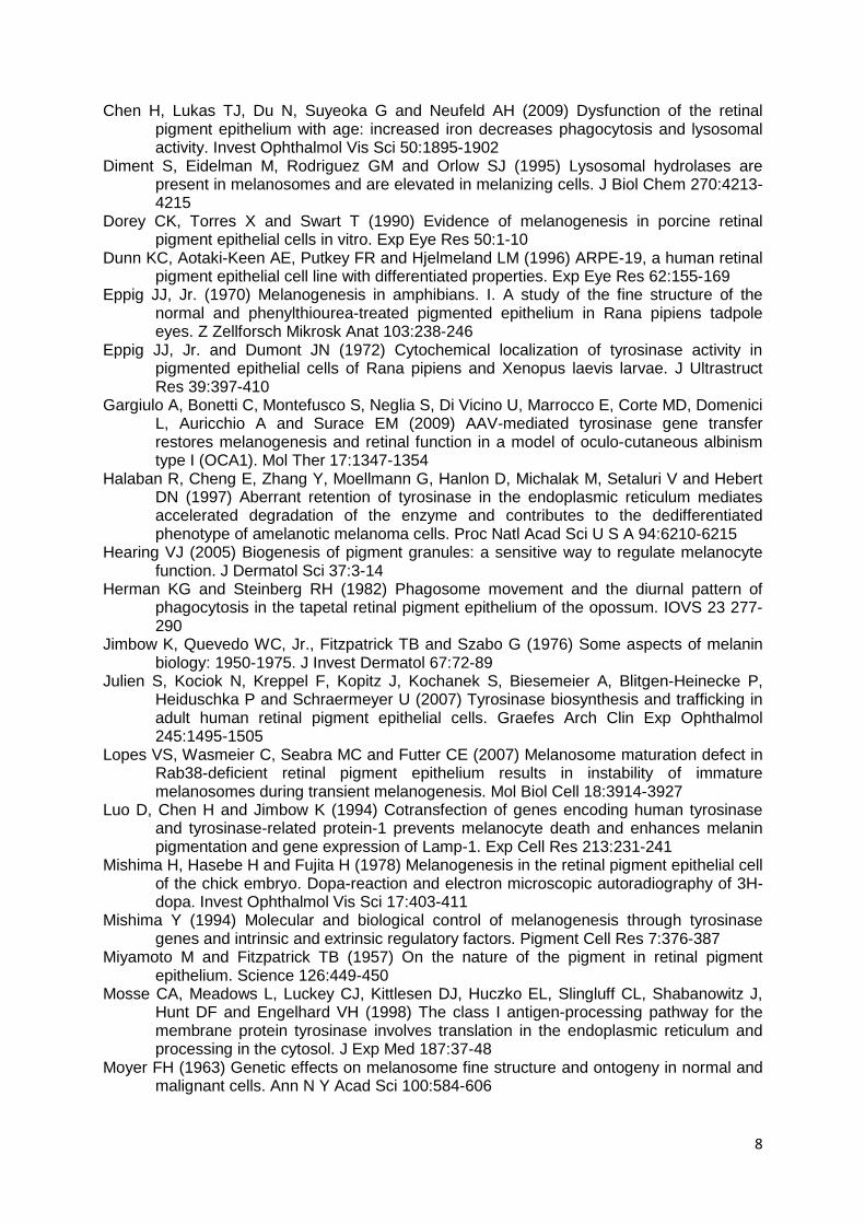
8
Chen H, Lukas TJ, Du N, Suyeoka G and Neufeld AH (2009) Dysfunction of the retinal pigment epithelium with age: increased iron decreases phagocytosis and lysosomal activity. Invest Ophthalmol Vis Sci 50:1895-1902
Diment S, Eidelman M, Rodriguez GM and Orlow SJ (1995) Lysosomal hydrolases are present in melanosomes and are elevated in melanizing cells. J Biol Chem 270:4213-4215
Dorey CK, Torres X and Swart T (1990) Evidence of melanogenesis in porcine retinal pigment epithelial cells in vitro. Exp Eye Res 50:1-10
Dunn KC, Aotaki-Keen AE, Putkey FR and Hjelmeland LM (1996) ARPE-19, a human retinal pigment epithelial cell line with differentiated properties. Exp Eye Res 62:155-169
Eppig JJ, Jr. (1970) Melanogenesis in amphibians. I. A study of the fine structure of the normal and phenylthiourea-treated pigmented epithelium in Rana pipiens tadpole eyes. Z Zellforsch Mikrosk Anat 103:238-246
Eppig JJ, Jr. and Dumont JN (1972) Cytochemical localization of tyrosinase activity in pigmented epithelial cells of Rana pipiens and Xenopus laevis larvae. J Ultrastruct Res 39:397-410
Gargiulo A, Bonetti C, Montefusco S, Neglia S, Di Vicino U, Marrocco E, Corte MD, Domenici L, Auricchio A and Surace EM (2009) AAV-mediated tyrosinase gene transfer restores melanogenesis and retinal function in a model of oculo-cutaneous albinism type I (OCA1). Mol Ther 17:1347-1354
Halaban R, Cheng E, Zhang Y, Moellmann G, Hanlon D, Michalak M, Setaluri V and Hebert DN (1997) Aberrant retention of tyrosinase in the endoplasmic reticulum mediates accelerated degradation of the enzyme and contributes to the dedifferentiated phenotype of amelanotic melanoma cells. Proc Natl Acad Sci U S A 94:6210-6215
Hearing VJ (2005) Biogenesis of pigment granules: a sensitive way to regulate melanocyte function. J Dermatol Sci 37:3-14
Herman KG and Steinberg RH (1982) Phagosome movement and the diurnal pattern of phagocytosis in the tapetal retinal pigment epithelium of the opossum. IOVS 23 277-290
Jimbow K, Quevedo WC, Jr., Fitzpatrick TB and Szabo G (1976) Some aspects of melanin biology: 1950-1975. J Invest Dermatol 67:72-89
Julien S, Kociok N, Kreppel F, Kopitz J, Kochanek S, Biesemeier A, Blitgen-Heinecke P, Heiduschka P and Schraermeyer U (2007) Tyrosinase biosynthesis and trafficking in adult human retinal pigment epithelial cells. Graefes Arch Clin Exp Ophthalmol 245:1495-1505
Lopes VS, Wasmeier C, Seabra MC and Futter CE (2007) Melanosome maturation defect in Rab38-deficient retinal pigment epithelium results in instability of immature melanosomes during transient melanogenesis. Mol Biol Cell 18:3914-3927
Luo D, Chen H and Jimbow K (1994) Cotransfection of genes encoding human tyrosinase and tyrosinase-related protein-1 prevents melanocyte death and enhances melanin pigmentation and gene expression of Lamp-1. Exp Cell Res 213:231-241
Mishima H, Hasebe H and Fujita H (1978) Melanogenesis in the retinal pigment epithelial cell of the chick embryo. Dopa-reaction and electron microscopic autoradiography of 3H-dopa. Invest Ophthalmol Vis Sci 17:403-411
Mishima Y (1994) Molecular and biological control of melanogenesis through tyrosinase genes and intrinsic and extrinsic regulatory factors. Pigment Cell Res 7:376-387
Miyamoto M and Fitzpatrick TB (1957) On the nature of the pigment in retinal pigment epithelium. Science 126:449-450
Mosse CA, Meadows L, Luckey CJ, Kittlesen DJ, Huczko EL, Slingluff CL, Shabanowitz J, Hunt DF and Engelhard VH (1998) The class I antigen-processing pathway for the membrane protein tyrosinase involves translation in the endoplasmic reticulum and processing in the cytosol. J Exp Med 187:37-48
Moyer FH (1963) Genetic effects on melanosome fine structure and ontogeny in normal and malignant cells. Ann N Y Acad Sci 100:584-606
9
Murisier F, Guichard S and Beermann F (2007) Distinct distal regulatory elements control tyrosinase expression in melanocytes and the retinal pigment epithelium. Dev Biol 303:838-847
Novikoff AB (1973) Lysosomes, a personal account. In: Hers G. VHF (ed) Lysosomes abd Storage diseases, New York, pp 1
Novikoff AB, Albala A and Biempica L (1968) Ultrastructural and cytochemical observations on B-16 and Harding-Passey mouse melanomas. The origin of premelanosomes and compound melanosomes. J Histochem Cytochem 16:299-319
Novikoff AB, Leuenberger PM, Novikoff PM and Quintana N (1979) Retinal pigment epithelium. Interrelations of endoplasmic reticulum and melanolysosomes in the black mouse and its beige mutant. Lab Invest 40:155-165
Oetting WS and King RA (1999) Molecular basis of albinism: mutations and polymorphisms of pigmentation genes associated with albinism. Hum Mutat 13:99-115
Orlow SJ (1995) Melanosomes are specialized members of the lysosomal lineage of organelles. J Invest Dermatol 105:3-7
Peters S, Kayatz P, Heimann K and Schraermeyer U (2000) Subretinal injection of rod outer segments leads to an increase in the number of early-stage melanosomes in retinal pigment epithelial cells. Ophthalmic Res 32:52-56
Raposo G and Marks MS (2002) The dark side of lysosome-related organelles: specialization of the endocytic pathway for melanosome biogenesis. Traffic 3:237-248
Raposo G, Tenza D, Murphy DM, Berson JF and Marks MS (2001) Distinct protein sorting and localization to premelanosomes, melanosomes, and lysosomes in pigmented melanocytic cells. J Cell Biol 152:809-824
Sarna T (1992) Properties and function of the ocular melanin--a photobiophysical view. J Photochem Photobiol B 12:215-258
Schraermeyer U (1992) Evidence for melanogenesis in the retinal pigment epithelium of adult cattle and golden hamster. Comp Biochem Physiol B 103:435-442
Schraermeyer U (1995) Transport of endocytosed material into melanin granules in cultured choroidal melanocytes of cattle--new insights into the relationship of melanosomes with lysosomes. Pigment Cell Res 8:209-214
Schraermeyer U and Dohms M (1993) Atypical granules in the eyes of the white mutant of Drosophila melanogaster are lysosome-related organelles. Pigment Cell Res 6:73-84
Schraermeyer U and Heimann K (1999) Current understanding on the role of retinal pigment epithelium and its pigmentation. Pigment Cell Res 12:219-236
Schraermeyer U, Kopitz J, Peters S, Henke-Fahle S, Blitgen-Heinecke P, Kokkinou D, Schwarz T and Bartz-Schmidt KU (2006) Tyrosinase biosynthesis in adult mammalian retinal pigment epithelial cells. Exp Eye Res 83:315-321
Schraermeyer U, Peters S, Thumann G, Kociok N and Heimann K (1999) Melanin granules of retinal pigment epithelium are connected with the lysosomal degradation pathway. Exp Eye Res 68:237-245
Schraermeyer U and Stieve H (1994) A newly discovered pathway of melanin formation in cultured retinal pigment epithelium of cattle. Cell Tissue Res 276:273-279
Seiji M, Shimao K, Birbeck MS and Fitzpatrick TB (1963) Subcellular localization of melanin biosynthesis. Ann N Y Acad Sci 100:497-533
Stanka P (1971) [Electron microscopic study of the origin of premelanosomes of the retinal pigment epithelium in chick embryos]. Z Zellforsch Mikrosk Anat 112:120-128
Stanka P, Rathjen P and Sahlmann B (1981) Evidence of membrane transformation during melanogenesis. Electron microscopic study on the retinal pigment epithelium of chick embryos. Cell Tissue Res 214:343-353
Theos AC, Berson JF, Theos SC, Herman KE, Harper DC, Tenza D, Sviderskaya EV, Lamoreux ML, Bennett DC, Raposo G and Marks MS (2006) Dual loss of ER export and endocytic signals with altered melanosome morphology in the silver mutation of Pmel17. Mol Biol Cell 17:3598-3612
Thumann G, Bartz-Schmidt KU, Kociok N, Kayatz P, Heimann K and Schraermeyer U (1999) Retinal damage by light in the golden hamster: an ultrastructural study in the retinal pigment epithelium and Bruch's membrane. J Photochem Photobiol B 49:104-111

10
Tribl F, Gerlach M, Marcus K, Asan E, Tatschner T, Arzberger T, Meyer HE, Bringmann G and Riederer P (2005) "Subcellular proteomics" of neuromelanin granules isolated from the human brain. Mol Cell Proteomics 4:945-957
Varela JM, Stempels NA, Vanden Berghe DA and Tassignon MJ (1995) Isoenzymic patterns of tyrosinase in the rabbit choroid and retina/retinal pigment epithelium. Exp Eye Res 60:621-629
Wasmeier C, Romao M, Plowright L, Bennett DC, Raposo G and Seabra MC (2006) Rab38 and Rab32 control post-Golgi trafficking of melanogenic enzymes. J Cell Biol 175:271-281
Winder AJ, Wittbjer A, Rosengren E and Rorsman H (1993) The mouse brown (b) locus protein has dopachrome tautomerase activity and is located in lysosomes in transfected fibroblasts. J Cell Sci 106 ( Pt 1):153-166
Young RW (1977) The daily rhythm of shedding and degradation of cone outer segment membranes in the lizard retina. J Ultrastruct Res 61:172-185
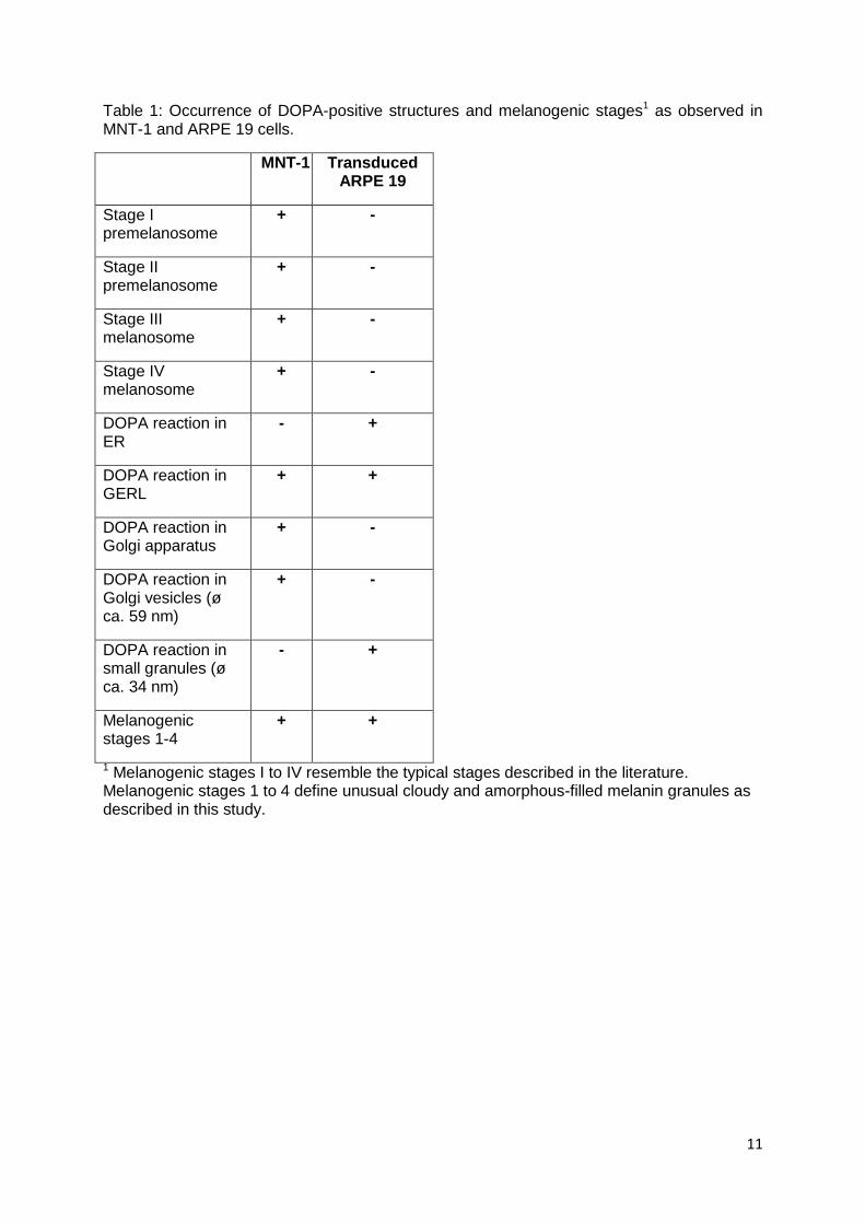
11
Table 1: Occurrence of DOPA-positive structures and melanogenic stages1 as observed in MNT-1 and ARPE 19 cells.
MNT-1 Transduced ARPE 19
Stage I premelanosome
+ -
Stage II premelanosome
+ -
Stage III melanosome
+ -
Stage IV melanosome
+ -
DOPA reaction in ER
- +
DOPA reaction in GERL
+ +
DOPA reaction in Golgi apparatus
+ -
DOPA reaction in Golgi vesicles (ø ca. 59 nm)
+ -
DOPA reaction in small granules (ø ca. 34 nm)
- +
Melanogenic stages 1-4
+ +
1 Melanogenic stages I to IV resemble the typical stages described in the literature. Melanogenic stages 1 to 4 define unusual cloudy and amorphous-filled melanin granules as described in this study.

12
Table 2: Occurrence of melanogenic stages 1 to 4 in control cells (c) and at different time points after transduction:
– Rarely seen (less than 3% of counted organelles), + present (5-7%), ++ frequently seen (-15%), +++ often seen (-25%), ++++ mainly seen (>26%). In addition, multi-vesicular bodies (MVB), multi-lamellar bodies (MLB) and mixed bodies are included, which are assumed to be the precursors of melanogenesis in ARPE 19 cells.
13
Figure legends Fig 1 Light microscopic image, showing ARPE 19 cells, 19 days after transduction: Newly formed melanin granules can be observed all over the cytoplasm. n nucleus, * high melanised cell, o low melanised cell; scale bar 25 µm
Fig 2 Fluorescence immunocytochemistry of MNT-1 (a-c) and transduced ARPE 19 cells (d-f): a+d PMEL17; b+e TRP1; c+f tyrosinase. All MNT-1 cells show the tested proteins all over the cytoplasm. An exact localisation is not possible in these micrographs, but the proteins are accumulating in central parts of some cells (arrows in a) and at dendrite tips (arrow heads in b, c). However, only tyrosinase is present in transduced ARPE 19 cells. Inset in f: magnification, showing tyrosinase-immunoreactivity in the perinuclear space, suggesting accumulation in the ER; scale bars (a-e) 25 µm; f) 5 µm
Fig 3 DOPA-histochemistry of transduced ARPE 19 cells: a) A cell showing DOPA-positive granules spread all over the cytoplasm. They are associated with membrane-bound bodies (arrows) and cisterns (c) of unknown origin. Structures, resembling ER or GERL tubules are also stained positive (asterisk), but Golgi cisterns are stained negative (G); scale bar 1 µm. (b-h) Detailed magnifications of DOPA-positive structures, as they are frequently found in transduced ARPE 19 cells; scale bars 200 nm: b) Magnification from a: DOPA granules fuse with the membrane of a vesicular organelle of unknown origin. First granules have already arrived inside the organelle. c) A multi-vesicular organelle with a double membrane, resembling an autophagosome, has also incorporated DOPA granules (arrow). d) Hundreds of DOPA granules are clustered together and accumulate close to a huge membrane-bound vacuole of unknown origin. e) As in d: The granules seem to fuse with the covering membrane of a vacuole. f) Tubules, resembling an endoplasmic reticulum, are DOPA-positive and associated with several DOPA-positive granules. g) Often, DOPA granules accumulate around intracellular membranes of unknown origin. Here, it can be seen that the DOPA granules lack a covering membrane, while the double membrane of the mitochondrion at the upper right can be clearly seen. h) The electron micrograph shows DOPA granules (black arrows) and Golgi vesicles (white arrow). It can be clearly observed that the DOPA granules are smaller (approx. 34 nm) and lack a covering membrane. Thus they can easily be distinguished from typical membrane-bound Golgi vesicles (approx. 59 nm)
Fig 4 Melanogenic organelles as observed by EM 6 days after transduction: All relevant organelles of melanin formation can be observed. Here, multi-lamellar bodies (MLBs), a mixed body (MB) and a newly formed melanin granule (M) are presented in one cell. Scale bar 500 nm
Fig 5 Viability of ARPE 19 cells is not affected by tyrosinase (d 2) and melanin synthesis (d 10). Mean +/- SEM control: ARPE cells; +AdTyr ARPE cells, transduced with the tyrosinase vector AdTyr (p= n.s.)

14
Figure 1
Figure 2

15
Figure 3

16
Figure 4
Figure 5
1
Tyrosinase in conjunction with phagocytosis of retinal outer segments influences the morphology and melanogenesis in cultured human ARPE 19 cells
A. Biesemeier1, P. Blitgen-Heinecke1, F. Kreppel2, S. Kochanek2, U. Schraermeyer1 1 Department for experimental vitreoretinal Surgery, University Eye Hospital Tuebingen,
Schleichstr. 12/1, 72076 Tuebingen, Germany; 2 Devision for Gene Therapy, Helmholtzster. 8/1, University of Ulm, Ulm, Germany
Corresponding author: [email protected]; Tel: +49/70712984015; Fax: +49/7071294554 Email addresses: Petra Blitgen-Heinecke: [email protected] [email protected] [email protected] [email protected] Abstract: We investigated whether tyrosinase expression has an effect on cultured adult RPE morphology and function. In addition, the effect of phagocytosis on tyrosinase expression and melanisation in the same system was studied. Cells of the non-pigmented ARPE 19 cell line, which lack tyrosinase, were transduced with an adenoviral tyrosinase vector and analysed for changes in morphology, specific and unspecific phagocytic activity and melanin synthesis with light and electron microscopy, fluorescence immunohistochemistry, DOPA histochemistry and HPLC analysis. Cultured cells without any treatment were used as controls. Control ARPE 19 cells had a dedifferentiated, spindle-shaped morphology and a low phagocytosis rate. In contrast, transduced cells showed a typical epithelial appearance, produced melanin and were able to phagocytose four times more ROS than the controls. Furthermore, tyrosinase expression and DOPA-oxidase activity were found to be significantly higher in transduced cells additionally fed with ROS than in non-fed transduced cells. Fed transduced cells were also able to synthesise melanin more rapidly. They contained twice as many melanised cells after one week of transduction than cells which were only transduced but not fed. Phagocytosis of latex beads had no effect on tyrosinase action and vice versa, showing that only the specific phagocytosis of ROS was able to increase the amount of melanin formation. In conclusion, tyrosinase expression enhanced phagocytic uptake of retinal outer segments, led to repigmentation and induced a hexagonal rearrangement of the ARPE 19 cell layer, resembling an in vivo morphology. Additionally, the phagocytosis of retinal outer segments resulted in a faster tyrosinase expression and melanisation in tyrosinase transduced ARPE 19 cells. Keywords: RPE, retinal outer segment, phagocytosis, tyrosinase, ultrastructure, melanogenesis Abbreviations: AMD age-related macular degeneration; ARPE 19 amelanotic retinal pigment epithelial cells: A control ARPE 19 cell; AT ARPE+ AdTyr vector; AR ARPE+ ROS; ATR ARPE +AdTyr+ROS; DMSO dimethylsulfoxid; EM electron microscopy; FCS foetal calf serum, IHC immunohistochemistry; L-DOPA L-dihydroxyphenylalanine; LM light microscopy;
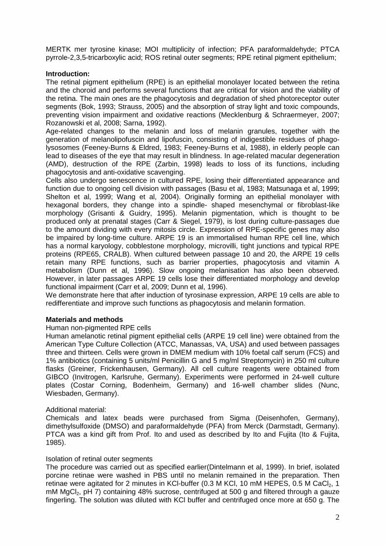
2
MERTK mer tyrosine kinase; MOI multiplicity of infection; PFA paraformaldehyde; PTCA pyrrole-2,3,5-tricarboxylic acid; ROS retinal outer segments; RPE retinal pigment epithelium; Introduction: The retinal pigment epithelium (RPE) is an epithelial monolayer located between the retina and the choroid and performs several functions that are critical for vision and the viability of the retina. The main ones are the phagocytosis and degradation of shed photoreceptor outer segments (Bok, 1993; Strauss, 2005) and the absorption of stray light and toxic compounds, preventing vision impairment and oxidative reactions (Mecklenburg & Schraermeyer, 2007; Rozanowski et al, 2008; Sarna, 1992). Age-related changes to the melanin and loss of melanin granules, together with the generation of melanolipofuscin and lipofuscin, consisting of indigestible residues of phago-lysosomes (Feeney-Burns & Eldred, 1983; Feeney-Burns et al, 1988), in elderly people can lead to diseases of the eye that may result in blindness. In age-related macular degeneration (AMD), destruction of the RPE (Zarbin, 1998) leads to loss of its functions, including phagocytosis and anti-oxidative scavenging. Cells also undergo senescence in cultured RPE, losing their differentiated appearance and function due to ongoing cell division with passages (Basu et al, 1983; Matsunaga et al, 1999; Shelton et al, 1999; Wang et al, 2004). Originally forming an epithelial monolayer with hexagonal borders, they change into a spindle- shaped mesenchymal or fibroblast-like morphology (Grisanti & Guidry, 1995). Melanin pigmentation, which is thought to be produced only at prenatal stages (Carr & Siegel, 1979), is lost during culture-passages due to the amount dividing with every mitosis circle. Expression of RPE-specific genes may also be impaired by long-time culture. ARPE 19 is an immortalised human RPE cell line, which has a normal karyology, cobblestone morphology, microvilli, tight junctions and typical RPE proteins (RPE65, CRALB). When cultured between passage 10 and 20, the ARPE 19 cells retain many RPE functions, such as barrier properties, phagocytosis and vitamin A metabolism (Dunn et al, 1996). Slow ongoing melanisation has also been observed. However, in later passages ARPE 19 cells lose their differentiated morphology and develop functional impairment (Carr et al, 2009; Dunn et al, 1996). We demonstrate here that after induction of tyrosinase expression, ARPE 19 cells are able to redifferentiate and improve such functions as phagocytosis and melanin formation. Materials and methods Human non-pigmented RPE cells Human amelanotic retinal pigment epithelial cells (ARPE 19 cell line) were obtained from the American Type Culture Collection (ATCC, Manassas, VA, USA) and used between passages three and thirteen. Cells were grown in DMEM medium with 10% foetal calf serum (FCS) and 1% antibiotics (containing 5 units/ml Penicillin G and 5 mg/ml Streptomycin) in 250 ml culture flasks (Greiner, Frickenhausen, Germany). All cell culture reagents were obtained from GIBCO (Invitrogen, Karlsruhe, Germany). Experiments were performed in 24-well culture plates (Costar Corning, Bodenheim, Germany) and 16-well chamber slides (Nunc, Wiesbaden, Germany). Additional material: Chemicals and latex beads were purchased from Sigma (Deisenhofen, Germany), dimethylsulfoxide (DMSO) and paraformaldehyde (PFA) from Merck (Darmstadt, Germany). PTCA was a kind gift from Prof. Ito and used as described by Ito and Fujita (Ito & Fujita, 1985). Isolation of retinal outer segments The procedure was carried out as specified earlier(Dintelmann et al, 1999). In brief, isolated porcine retinae were washed in PBS until no melanin remained in the preparation. Then retinae were agitated for 2 minutes in KCl-buffer (0.3 M KCl, 10 mM HEPES, 0.5 M CaCl2, 1 mM MgCl2, pH 7) containing 48% sucrose, centrifuged at 500 g and filtered through a gauze fingerling. The solution was diluted with KCl buffer and centrifuged once more at 650 g. The
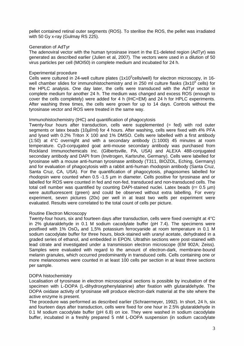
3
pellet contained retinal outer segments (ROS). To sterilise the ROS, the pellet was irradiated with 50 Gy x-ray (Gulmay RS 225). Generation of AdTyr The adenoviral vector with the human tyrosinase insert in the E1-deleted region (AdTyr) was generated as described earlier (Julien et al, 2007). The vectors were used in a dilution of 50 virus particles per cell (MOI50) in complete medium and incubated for 24 h. Experimental procedure Cells were cultured in 24-well culture plates (1x10⁵cells/well) for electron microscopy, in 16-well chamber slides for immunohistochemistry and in 250 ml culture flasks (3x10⁶ cells) for the HPLC analysis. One day later, the cells were transduced with the AdTyr vector in complete medium for another 24 h. The medium was changed and excess ROS (enough to cover the cells completely) were added for 4 h (IHC+EM) and 24 h for HPLC experiments. After washing three times, the cells were grown for up to 14 days. Controls without the tyrosinase vector and ROS were treated in the same way. Immunohistochemistry (IHC) and quantification of phagocytosis Twenty-four hours after transduction, cells were supplemented (= fed) with rod outer segments or latex beads (10µl/ml) for 4 hours. After washing, cells were fixed with 4% PFA and lysed with 0.2% Triton X 100 and 1% DMSO. Cells were labelled with a first antibody (1:50) at 4°C overnight and with a secondary antibody (1:1000) 45 minutes at room temperature. Cy3-conjugated goat anti-mouse secondary antibody was purchased from Rockland Immunochemicals Inc. (Gilbertsville, PA, USA) and ALEXA 488-conjugated secondary antibody and DAPI from (Invitrogen, Karlsruhe, Germany). Cells were labelled for tyrosinase with a mouse anti-human tyrosinase antibody (T311, BIOZOL, Eching, Germany) and for evaluation of phagocytosis with a rabbit anti-human rhodopsin antibody (Santa Cruz, Santa Cruz, CA, USA). For the quantification of phagocytosis, phagosomes labelled for rhodopsin were counted when 0.5 -1.5 µm in diameter. Cells positive for tyrosinase and or labelled for ROS were counted in fed and non-fed, transduced and non-transduced cells. The total cell number was quantified by counting DAPI-stained nuclei. Latex beads (r= 0.5 µm) were autofluorescent (green) and could be observed without extra labelling. For every experiment, seven pictures (20x) per well in at least two wells per experiment were evaluated. Results were correlated to the total count of cells per picture. Routine Electron Microscopy Twenty-four hours, six and fourteen days after transduction, cells were fixed overnight at 4°C in 2% glutaraldehyde in 0.1 M sodium cacodylate buffer (pH 7.4). The specimens were postfixed with 1% OsO4 and 1,5% potassium ferrocyanide at room temperature in 0.1 M sodium cacodylate buffer for three hours, block-stained with uranyl acetate, dehydrated in a graded series of ethanol, and embedded in EPON. Ultrathin sections were post-stained with lead citrate and investigated under a transmission electron microscope (EM 902A; Zeiss). Samples were evaluated with regard to the amount of electron-dark, membrane-bound melanin granules, which occurred predominantly in transduced cells. Cells containing one or more melanosomes were counted in at least 100 cells per section in at least three sections per sample. DOPA histochemistry Localisation of tyrosinase in electron microscopical sections is possible by incubation of the specimen with L-DOPA (L-dihydroxyphenylalanine) after fixation with glutaraldehyde. The DOPA oxidase activity of tyrosinase will produce electron-dark material at the site where the active enzyme is present. The procedure was performed as described earlier (Schraermeyer, 1992). In short, 24 h, six and fourteen days after transduction, cells were fixed for one hour in 2.5% glutaraldehyde in 0.1 M sodium cacodylate buffer (pH 6.8) on ice. They were washed in sodium cacodylate buffer, incubated in a freshly prepared 5 mM L-DOPA suspension (in sodium cacodylate
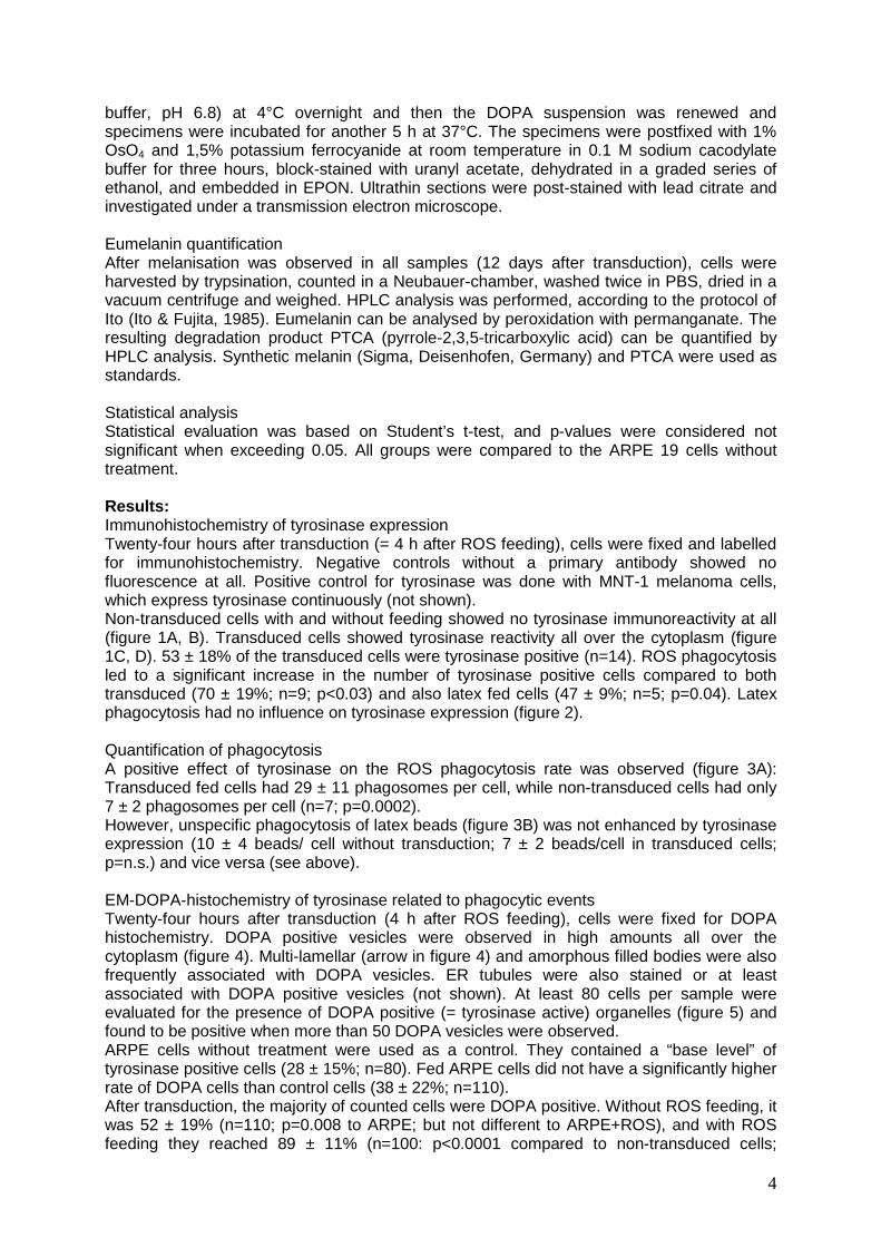
4
buffer, pH 6.8) at 4°C overnight and then the DOPA suspension was renewed and specimens were incubated for another 5 h at 37°C. The specimens were postfixed with 1% OsO4 and 1,5% potassium ferrocyanide at room temperature in 0.1 M sodium cacodylate buffer for three hours, block-stained with uranyl acetate, dehydrated in a graded series of ethanol, and embedded in EPON. Ultrathin sections were post-stained with lead citrate and investigated under a transmission electron microscope. Eumelanin quantification After melanisation was observed in all samples (12 days after transduction), cells were harvested by trypsination, counted in a Neubauer-chamber, washed twice in PBS, dried in a vacuum centrifuge and weighed. HPLC analysis was performed, according to the protocol of Ito (Ito & Fujita, 1985). Eumelanin can be analysed by peroxidation with permanganate. The resulting degradation product PTCA (pyrrole-2,3,5-tricarboxylic acid) can be quantified by HPLC analysis. Synthetic melanin (Sigma, Deisenhofen, Germany) and PTCA were used as standards. Statistical analysis Statistical evaluation was based on Student’s t-test, and p-values were considered not significant when exceeding 0.05. All groups were compared to the ARPE 19 cells without treatment. Results: Immunohistochemistry of tyrosinase expression Twenty-four hours after transduction (= 4 h after ROS feeding), cells were fixed and labelled for immunohistochemistry. Negative controls without a primary antibody showed no fluorescence at all. Positive control for tyrosinase was done with MNT-1 melanoma cells, which express tyrosinase continuously (not shown). Non-transduced cells with and without feeding showed no tyrosinase immunoreactivity at all (figure 1A, B). Transduced cells showed tyrosinase reactivity all over the cytoplasm (figure 1C, D). 53 ± 18% of the transduced cells were tyrosinase positive (n=14). ROS phagocytosis led to a significant increase in the number of tyrosinase positive cells compared to both transduced (70 ± 19%; n=9; p<0.03) and also latex fed cells (47 ± 9%; n=5; p=0.04). Latex phagocytosis had no influence on tyrosinase expression (figure 2). Quantification of phagocytosis A positive effect of tyrosinase on the ROS phagocytosis rate was observed (figure 3A): Transduced fed cells had 29 ± 11 phagosomes per cell, while non-transduced cells had only 7 ± 2 phagosomes per cell (n=7; p=0.0002). However, unspecific phagocytosis of latex beads (figure 3B) was not enhanced by tyrosinase expression (10 ± 4 beads/ cell without transduction; 7 ± 2 beads/cell in transduced cells; p=n.s.) and vice versa (see above). EM-DOPA-histochemistry of tyrosinase related to phagocytic events Twenty-four hours after transduction (4 h after ROS feeding), cells were fixed for DOPA histochemistry. DOPA positive vesicles were observed in high amounts all over the cytoplasm (figure 4). Multi-lamellar (arrow in figure 4) and amorphous filled bodies were also frequently associated with DOPA vesicles. ER tubules were also stained or at least associated with DOPA positive vesicles (not shown). At least 80 cells per sample were evaluated for the presence of DOPA positive (= tyrosinase active) organelles (figure 5) and found to be positive when more than 50 DOPA vesicles were observed. ARPE cells without treatment were used as a control. They contained a “base level” of tyrosinase positive cells (28 ± 15%; n=80). Fed ARPE cells did not have a significantly higher rate of DOPA cells than control cells (38 ± 22%; n=110). After transduction, the majority of counted cells were DOPA positive. Without ROS feeding, it was 52 ± 19% (n=110; p=0.008 to ARPE; but not different to ARPE+ROS), and with ROS feeding they reached 89 ± 11% (n=100: p<0.0001 compared to non-transduced cells;
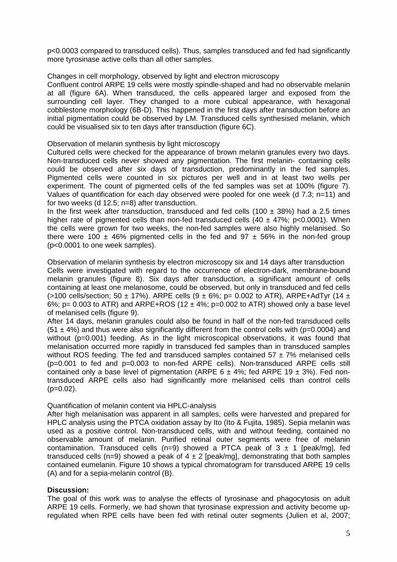
5
p<0.0003 compared to transduced cells). Thus, samples transduced and fed had significantly more tyrosinase active cells than all other samples. Changes in cell morphology, observed by light and electron microscopy Confluent control ARPE 19 cells were mostly spindle-shaped and had no observable melanin at all (figure 6A). When transduced, the cells appeared larger and exposed from the surrounding cell layer. They changed to a more cubical appearance, with hexagonal cobblestone morphology (6B-D). This happened in the first days after transduction before an initial pigmentation could be observed by LM. Transduced cells synthesised melanin, which could be visualised six to ten days after transduction (figure 6C). Observation of melanin synthesis by light microscopy Cultured cells were checked for the appearance of brown melanin granules every two days. Non-transduced cells never showed any pigmentation. The first melanin- containing cells could be observed after six days of transduction, predominantly in the fed samples. Pigmented cells were counted in six pictures per well and in at least two wells per experiment. The count of pigmented cells of the fed samples was set at 100% (figure 7). Values of quantification for each day observed were pooled for one week (d 7.3; n=11) and for two weeks (d 12.5; n=8) after transduction. In the first week after transduction, transduced and fed cells (100 ± 38%) had a 2.5 times higher rate of pigmented cells than non-fed transduced cells (40 ± 47%; p<0.0001). When the cells were grown for two weeks, the non-fed samples were also highly melanised. So there were 100 ± 46% pigmented cells in the fed and 97 ± 56% in the non-fed group (p<0.0001 to one week samples). Observation of melanin synthesis by electron microscopy six and 14 days after transduction Cells were investigated with regard to the occurrence of electron-dark, membrane-bound melanin granules (figure 8). Six days after transduction, a significant amount of cells containing at least one melanosome, could be observed, but only in transduced and fed cells (>100 cells/section; 50 ± 17%). ARPE cells (9 ± 6%; p= 0.002 to ATR), ARPE+AdTyr (14 ± 6%; p= 0.003 to ATR) and ARPE+ROS (12 ± 4%; p=0.002 to ATR) showed only a base level of melanised cells (figure 9). After 14 days, melanin granules could also be found in half of the non-fed transduced cells (51 ± 4%) and thus were also significantly different from the control cells with (p=0.0004) and without (p=0.001) feeding. As in the light microscopical observations, it was found that melanisation occurred more rapidly in transduced fed samples than in transduced samples without ROS feeding. The fed and transduced samples contained 57 ± 7% melanised cells (p=0.001 to fed and p=0.003 to non-fed ARPE cells). Non-transduced ARPE cells still contained only a base level of pigmentation (ARPE 6 ± 4%; fed ARPE 19 ± 3%). Fed non-transduced ARPE cells also had significantly more melanised cells than control cells (p=0.02). Quantification of melanin content via HPLC-analysis After high melanisation was apparent in all samples, cells were harvested and prepared for HPLC analysis using the PTCA oxidation assay by Ito (Ito & Fujita, 1985). Sepia melanin was used as a positive control. Non-transduced cells, with and without feeding, contained no observable amount of melanin. Purified retinal outer segments were free of melanin contamination. Transduced cells (n=9) showed a PTCA peak of 3 ± 1 [peak/mg], fed transduced cells (n=9) showed a peak of 4 ± 2 [peak/mg], demonstrating that both samples contained eumelanin. Figure 10 shows a typical chromatogram for transduced ARPE 19 cells (A) and for a sepia-melanin control (B). Discussion: The goal of this work was to analyse the effects of tyrosinase and phagocytosis on adult ARPE 19 cells. Formerly, we had shown that tyrosinase expression and activity become up-regulated when RPE cells have been fed with retinal outer segments (Julien et al, 2007;
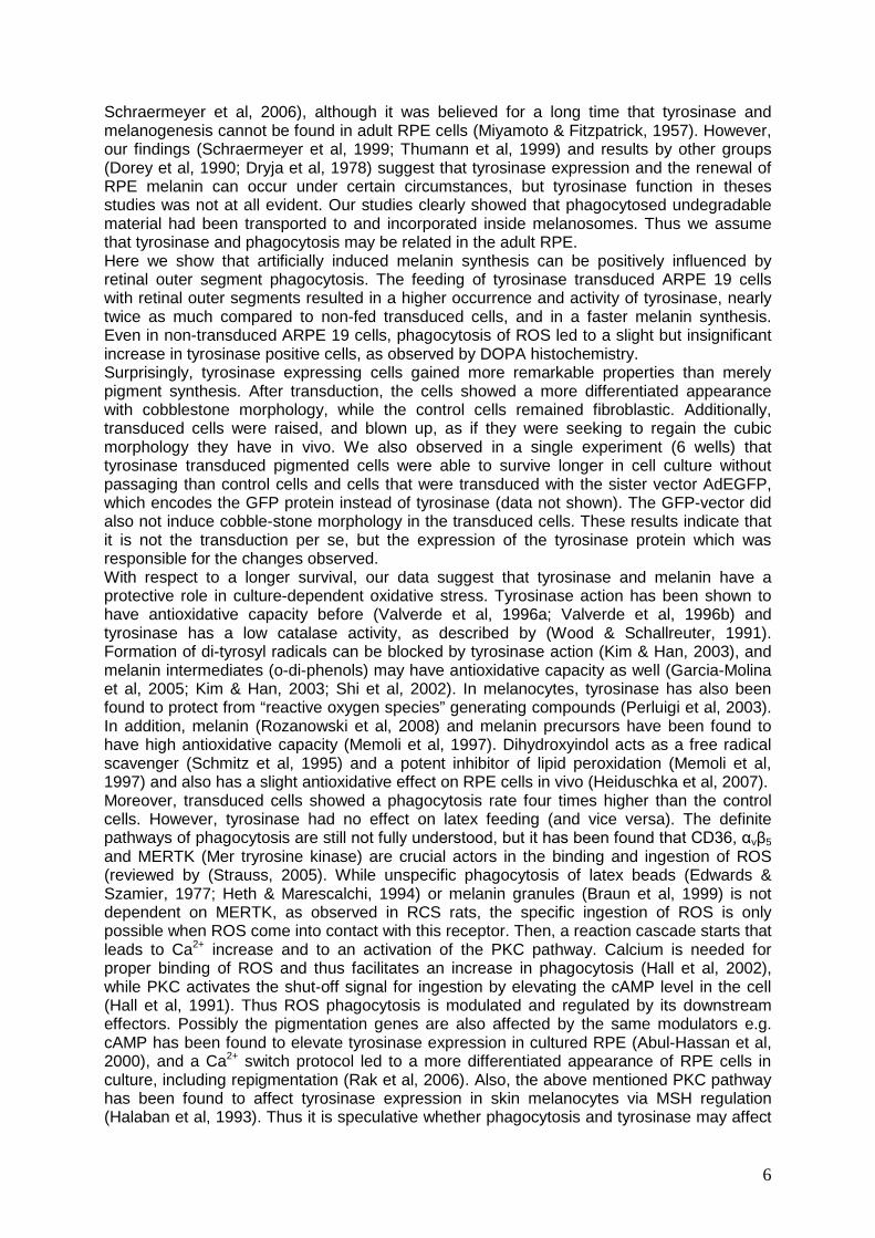
6
Schraermeyer et al, 2006), although it was believed for a long time that tyrosinase and melanogenesis cannot be found in adult RPE cells (Miyamoto & Fitzpatrick, 1957). However, our findings (Schraermeyer et al, 1999; Thumann et al, 1999) and results by other groups (Dorey et al, 1990; Dryja et al, 1978) suggest that tyrosinase expression and the renewal of RPE melanin can occur under certain circumstances, but tyrosinase function in theses studies was not at all evident. Our studies clearly showed that phagocytosed undegradable material had been transported to and incorporated inside melanosomes. Thus we assume that tyrosinase and phagocytosis may be related in the adult RPE. Here we show that artificially induced melanin synthesis can be positively influenced by retinal outer segment phagocytosis. The feeding of tyrosinase transduced ARPE 19 cells with retinal outer segments resulted in a higher occurrence and activity of tyrosinase, nearly twice as much compared to non-fed transduced cells, and in a faster melanin synthesis. Even in non-transduced ARPE 19 cells, phagocytosis of ROS led to a slight but insignificant increase in tyrosinase positive cells, as observed by DOPA histochemistry. Surprisingly, tyrosinase expressing cells gained more remarkable properties than merely pigment synthesis. After transduction, the cells showed a more differentiated appearance with cobblestone morphology, while the control cells remained fibroblastic. Additionally, transduced cells were raised, and blown up, as if they were seeking to regain the cubic morphology they have in vivo. We also observed in a single experiment (6 wells) that tyrosinase transduced pigmented cells were able to survive longer in cell culture without passaging than control cells and cells that were transduced with the sister vector AdEGFP, which encodes the GFP protein instead of tyrosinase (data not shown). The GFP-vector did also not induce cobble-stone morphology in the transduced cells. These results indicate that it is not the transduction per se, but the expression of the tyrosinase protein which was responsible for the changes observed. With respect to a longer survival, our data suggest that tyrosinase and melanin have a protective role in culture-dependent oxidative stress. Tyrosinase action has been shown to have antioxidative capacity before (Valverde et al, 1996a; Valverde et al, 1996b) and tyrosinase has a low catalase activity, as described by (Wood & Schallreuter, 1991). Formation of di-tyrosyl radicals can be blocked by tyrosinase action (Kim & Han, 2003), and melanin intermediates (o-di-phenols) may have antioxidative capacity as well (Garcia-Molina et al, 2005; Kim & Han, 2003; Shi et al, 2002). In melanocytes, tyrosinase has also been found to protect from “reactive oxygen species” generating compounds (Perluigi et al, 2003). In addition, melanin (Rozanowski et al, 2008) and melanin precursors have been found to have high antioxidative capacity (Memoli et al, 1997). Dihydroxyindol acts as a free radical scavenger (Schmitz et al, 1995) and a potent inhibitor of lipid peroxidation (Memoli et al, 1997) and also has a slight antioxidative effect on RPE cells in vivo (Heiduschka et al, 2007). Moreover, transduced cells showed a phagocytosis rate four times higher than the control cells. However, tyrosinase had no effect on latex feeding (and vice versa). The definite pathways of phagocytosis are still not fully understood, but it has been found that CD36, αvβ5 and MERTK (Mer tryrosine kinase) are crucial actors in the binding and ingestion of ROS (reviewed by (Strauss, 2005). While unspecific phagocytosis of latex beads (Edwards & Szamier, 1977; Heth & Marescalchi, 1994) or melanin granules (Braun et al, 1999) is not dependent on MERTK, as observed in RCS rats, the specific ingestion of ROS is only possible when ROS come into contact with this receptor. Then, a reaction cascade starts that leads to Ca2+ increase and to an activation of the PKC pathway. Calcium is needed for proper binding of ROS and thus facilitates an increase in phagocytosis (Hall et al, 2002), while PKC activates the shut-off signal for ingestion by elevating the cAMP level in the cell (Hall et al, 1991). Thus ROS phagocytosis is modulated and regulated by its downstream effectors. Possibly the pigmentation genes are also affected by the same modulators e.g. cAMP has been found to elevate tyrosinase expression in cultured RPE (Abul-Hassan et al, 2000), and a Ca2+ switch protocol led to a more differentiated appearance of RPE cells in culture, including repigmentation (Rak et al, 2006). Also, the above mentioned PKC pathway has been found to affect tyrosinase expression in skin melanocytes via MSH regulation (Halaban et al, 1993). Thus it is speculative whether phagocytosis and tyrosinase may affect
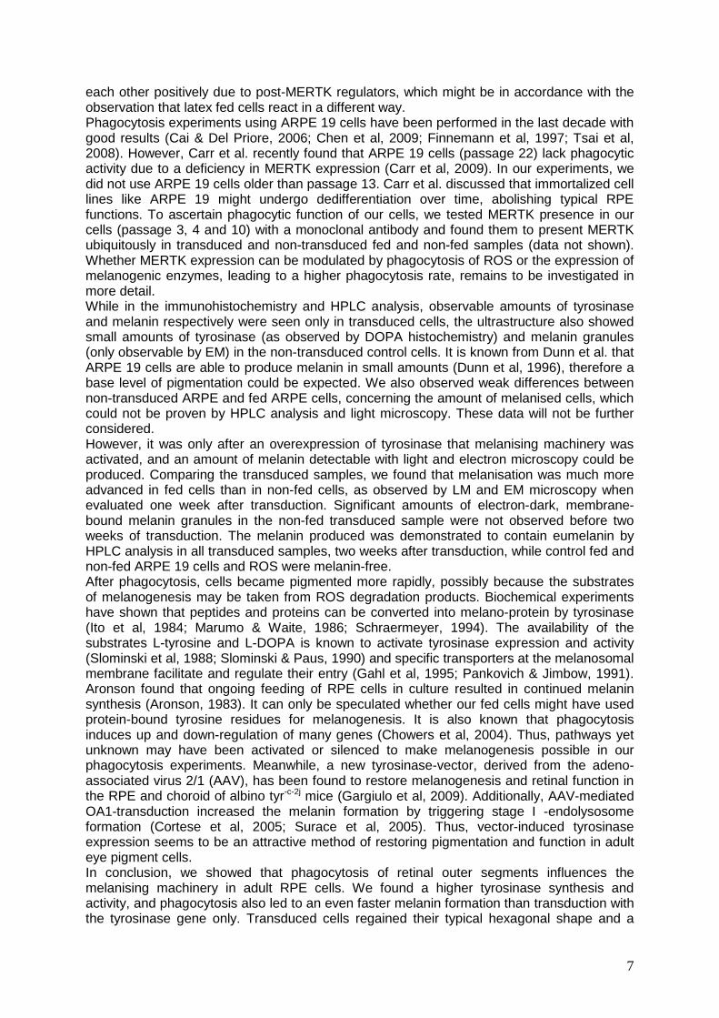
7
each other positively due to post-MERTK regulators, which might be in accordance with the observation that latex fed cells react in a different way. Phagocytosis experiments using ARPE 19 cells have been performed in the last decade with good results (Cai & Del Priore, 2006; Chen et al, 2009; Finnemann et al, 1997; Tsai et al, 2008). However, Carr et al. recently found that ARPE 19 cells (passage 22) lack phagocytic activity due to a deficiency in MERTK expression (Carr et al, 2009). In our experiments, we did not use ARPE 19 cells older than passage 13. Carr et al. discussed that immortalized cell lines like ARPE 19 might undergo dedifferentiation over time, abolishing typical RPE functions. To ascertain phagocytic function of our cells, we tested MERTK presence in our cells (passage 3, 4 and 10) with a monoclonal antibody and found them to present MERTK ubiquitously in transduced and non-transduced fed and non-fed samples (data not shown). Whether MERTK expression can be modulated by phagocytosis of ROS or the expression of melanogenic enzymes, leading to a higher phagocytosis rate, remains to be investigated in more detail. While in the immunohistochemistry and HPLC analysis, observable amounts of tyrosinase and melanin respectively were seen only in transduced cells, the ultrastructure also showed small amounts of tyrosinase (as observed by DOPA histochemistry) and melanin granules (only observable by EM) in the non-transduced control cells. It is known from Dunn et al. that ARPE 19 cells are able to produce melanin in small amounts (Dunn et al, 1996), therefore a base level of pigmentation could be expected. We also observed weak differences between non-transduced ARPE and fed ARPE cells, concerning the amount of melanised cells, which could not be proven by HPLC analysis and light microscopy. These data will not be further considered. However, it was only after an overexpression of tyrosinase that melanising machinery was activated, and an amount of melanin detectable with light and electron microscopy could be produced. Comparing the transduced samples, we found that melanisation was much more advanced in fed cells than in non-fed cells, as observed by LM and EM microscopy when evaluated one week after transduction. Significant amounts of electron-dark, membrane-bound melanin granules in the non-fed transduced sample were not observed before two weeks of transduction. The melanin produced was demonstrated to contain eumelanin by HPLC analysis in all transduced samples, two weeks after transduction, while control fed and non-fed ARPE 19 cells and ROS were melanin-free. After phagocytosis, cells became pigmented more rapidly, possibly because the substrates of melanogenesis may be taken from ROS degradation products. Biochemical experiments have shown that peptides and proteins can be converted into melano-protein by tyrosinase (Ito et al, 1984; Marumo & Waite, 1986; Schraermeyer, 1994). The availability of the substrates L-tyrosine and L-DOPA is known to activate tyrosinase expression and activity (Slominski et al, 1988; Slominski & Paus, 1990) and specific transporters at the melanosomal membrane facilitate and regulate their entry (Gahl et al, 1995; Pankovich & Jimbow, 1991). Aronson found that ongoing feeding of RPE cells in culture resulted in continued melanin synthesis (Aronson, 1983). It can only be speculated whether our fed cells might have used protein-bound tyrosine residues for melanogenesis. It is also known that phagocytosis induces up and down-regulation of many genes (Chowers et al, 2004). Thus, pathways yet unknown may have been activated or silenced to make melanogenesis possible in our phagocytosis experiments. Meanwhile, a new tyrosinase-vector, derived from the adeno-associated virus 2/1 (AAV), has been found to restore melanogenesis and retinal function in the RPE and choroid of albino tyr-c-2j mice (Gargiulo et al, 2009). Additionally, AAV-mediated OA1-transduction increased the melanin formation by triggering stage I -endolysosome formation (Cortese et al, 2005; Surace et al, 2005). Thus, vector-induced tyrosinase expression seems to be an attractive method of restoring pigmentation and function in adult eye pigment cells. In conclusion, we showed that phagocytosis of retinal outer segments influences the melanising machinery in adult RPE cells. We found a higher tyrosinase synthesis and activity, and phagocytosis also led to an even faster melanin formation than transduction with the tyrosinase gene only. Transduced cells regained their typical hexagonal shape and a

8
higher phagocytosis rate than non-transduced cells. Thus tyrosinase expression switched cultured RPE cells closer to the in vivo situation. Acknowledgements The authors are indebted to Prof. S. Ito (Dep. of Dermatology, Sapporo, Japan) for providing the PTCA standard and Prof. H.P. Rodemann and Dr. K. Dittmann (Dep. of Radiation Oncology, Tübingen) for the use of their X-ray application device. They would also like to thank Monika Rittgarn and Sigrid Schultheiss for technical assistance. This work was supported by grants to U. Schraermeyer from the Deutsche Forschungsgemeinschaft (DFG SCHR436/12-2). Figure legends Fig. 1: Fluorescence light micrographs taken from ARPE 19 cells, 24h after transduction (magnification 20x): A) Control ARPE 19 cells, B) ARPE 19 cells fed with retinal outer segments, C) ARPE 19 cells, transduced with the tyrosinase gene, and D) ARPE 19 cells, which were fed with retinal outer segments and transduced with tyrosinase are shown. Tyrosinase labelling (red) can be seen all over the cytoplasm, but only in the transduced cells. Phagocytic events were visualised by rhodopsin labelling (green). Phagosomes (0.5-1.5µm) are spread all over the cytoplasm.
Fig. 2: Quantification of tyrosinase immunoreactivity, 24h after transduction: Transduced ARPE cells that were additionally fed with ROS (ATR, n=9), had significantly more tyrosinase expressing cells than cells that were only transduced (AT, n=14; *p<0.03) or cells transduced and fed with latex beads (ATL; n=5; p=0.04). Latex feeding had no additional effect on tyrosinase immuno-reactivity.
Fig. 3: A) Quantification of ROS phagocytosis: Non-transduced ARPE cells (AR) phagocytosed four times less ROS than transduced (ATR) cells (n=7; p<0.0002). B) Quantification of latex phagocytosis: There were no significant differences in the phagocytic activity of latex fed cells (AL) and transduced, latex fed (ATL) cells (n=5).
Fig. 4: Electron micrograph taken from DOPA histochemistry of a transduced ARPE 19 cell, 6 days after transduction: The cell shows tyrosinase filled granule-like vesicles throughout the cytoplasm, sometimes in association with multi-lamellar (black arrow) and amorphous filled bodies of unknown origin (*). C: neighbouring cell, lacking DOPA positive reaction.
Fig. 5: Quantification of DOPA histochemistry, 24h after transduction: Transduced cells with (ATR) and without feeding (AT) showed significantly more DOPA positive cells than non-transduced non-fed cells (A; op<0.008 to AT; *p<0.0001 A + AR to ATR). ATR had even more DOPA positive cells than AT and non-transduced fed cells (AR •p<0.0001).
Fig. 6: Morphological observations: A-C light, D electron microscopy: A) Control ARPE 19 cells had a mostly spindle-shaped, non-pigmented appearance (here: day 7). B) In the first days after transduction, cells developed a hexagonal morphology, grew larger and appeared more prominent than control cells (here: day 6). C) After one week of transduction, first melanin granules were observed. The light micrograph shows a transduced and fed sample, 11 days after transduction. D) Electron micrograph of DOPA reaction at low magnification: the cell in the middle is transduced since DOPA positive vesicles can clearly be recognised. It is up to 1/3 higher than neighbouring cells, which lack DOPA staining and thus might not be transduced.
Fig. 7: Quantification of melanin synthesis observed one and two weeks after transduction (LM): Samples were checked for melanisation every two days. After one week of transduction, first melanised cells were observed in the transduced samples by light microscopy (ATR = 100%): Quantification of melanised cells shows that fed transduced samples (ATR) became melanised more rapidly than cells that were only transduced (AT) (n=8; *p=<0.0001). After two weeks, most cells in the non-fed samples were also pigmented (op<0.0001 to AT at one week). Non-transduced cells did not show any pigmentation when observed under the light microscope (not shown).
9
Fig. 8: Electron micrograph, obtained two weeks after transduction: A typical melanosome with covering membrane can be seen. About 50% of the transduced cells have synthesised melanin granules, which were 0.3 -1 µm in diameter.
Fig. 9: Quantification of melanin synthesis observed one and two weeks after transduction (EM): While transduced and fed cells (ATR) had developed a significant amount of melanised cells after one (*p<0.03 to A, AR, AT) and two (*p<0.003 to A and AR) weeks of transduction, non-fed transduced cells (AT) needed two weeks to gather enough melanin to outnumber the basal pigmentation of control cells (op<0.001 to A and AR and to AT at one week).
Fig. 10: Chromatogram of HPLC analysis, showing the peak of PTCA produced (black arrow) in A) transduced ARPE 19 cells and B) Sepia melanin control.
References Abul-Hassan K, Walmsley R, Tombran-Tink J, Boulton M (2000) Regulation of tyrosinase expression and activity in cultured human retinal pigment epithelial cells. Pigment Cell Res 13(6): 436-441
Aronson JF (1983) Human retinal pigment cell culture. In Vitro 19(8): 642-650
Basu PK, Sarkar P, Menon I, Carre F, Persad S (1983) Bovine retinal pigment epithelial cells cultured in vitro: growth characteristics, morphology, chromosomes, phagocytosis ability, tyrosinase activity and effect of freezing. Exp Eye Res 36(5): 671-683
Bok D (1993) The retinal pigment epithelium: a versatile partner in vision. J Cell Sci Suppl 17: 189-195
Braun M, Kage A, Heimann K, Schraermeyer U (1999) Retinal pigment epithelial cells from Royal College of Surgeons dystrophic rats can take up melanin granules. Graefes Arch Clin Exp Ophthalmol 237(1): 67-71
Cai H, Del Priore LV (2006) Gene expression profile of cultured adult compared to immortalized human RPE. Mol Vis 12: 1-14
Carr AJ, Vugler A, Lawrence J, Chen LL, Ahmado A, Chen FK, Semo M, Gias C, da Cruz L, Moore HD, Walsh J, Coffey PJ (2009) Molecular characterization and functional analysis of phagocytosis by human embryonic stem cell-derived RPE cells using a novel human retinal assay. Mol Vis 15: 283-295
Carr RE, Siegel IM (1979) The retinal pigment epitehlium in ocular albinism;. In The retinal pigment epithelium, Zinn KM, Mamor MF (eds), pp 413-423. Cambridge: Harvard University Press
Chen H, Lukas TJ, Du N, Suyeoka G, Neufeld AH (2009) Dysfunction of the retinal pigment epithelium with age: increased iron decreases phagocytosis and lysosomal activity. Invest Ophthalmol Vis Sci 50(4): 1895-1902 Chowers I, Kim Y, Farkas RH, Gunatilaka TL, Hackam AS, Campochiaro PA, Finnemann SC, Zack DJ (2004) Changes in retinal pigment epithelial gene expression induced by rod outer segment uptake. Invest Ophthalmol Vis Sci 45(7): 2098-2106
Cortese K, Giordano F, Surace EM, Venturi C, Ballabio A, Tacchetti C, Marigo V (2005) The ocular albinism type 1 (OA1) gene controls melanosome maturation and size. Invest Ophthalmol Vis Sci 46(12): 4358-4364
Dintelmann TS, Heimann K, Kayatz P, Schraermeyer U (1999) Comparative study of ROS degradation by IPE and RPE cells in vitro. Graefes Arch Clin Exp Ophthalmol 237(10): 830-839
Dorey CK, Torres X, Swart T (1990) Evidence of melanogenesis in porcine retinal pigment epithelial cells in vitro. Exp Eye Res 50(1): 1-10
Dryja TP, O'Neil-Dryja M, Pawelek JM, Albert DM (1978) Demonstration of tyrosinase in the adult bovine uveal tract and retinal pigment epithelium. Invest Ophthalmol Vis Sci 17(6): 511-514
10
Dunn KC, Aotaki-Keen AE, Putkey FR, Hjelmeland LM (1996) ARPE-19, a human retinal pigment epithelial cell line with differentiated properties. Exp Eye Res 62(2): 155-169
Edwards RB, Szamier RB (1977) Defective phagocytosis of isolated rod outer segments by RCS rat retinal pigment epithelium in culture. Science 197(4307): 1001-1003
Feeney-Burns L, Eldred GE (1983) The fate of the phagosome: conversion to 'age pigment' and impact in human retinal pigment epithelium. Trans Ophthalmol Soc U K 103 ( Pt 4): 416-421
Feeney-Burns L, Gao CL, Berman ER (1988) The fate of immunoreactive opsin following phagocytosis by pigment epithelium in human and monkey retinas. Invest Ophthalmol Vis Sci 29(5): 708-719
Finnemann SC, Bonilha VL, Marmorstein AD, Rodriguez-Boulan E (1997) Phagocytosis of rod outer segments by retinal pigment epithelial cells requires alpha(v)beta5 integrin for binding but not for internalization. Proc Natl Acad Sci U S A 94(24): 12932-12937
Gahl WA, Potterf B, Durham-Pierre D, Brilliant MH, Hearing VJ (1995) Melanosomal tyrosine transport in normal and pink-eyed dilution murine melanocytes. Pigment Cell Res 8(5): 229-233
Garcia-Molina F, Hiner AN, Fenoll LG, Rodriguez-Lopez JN, Garcia-Ruiz PA, Garcia-Canovas F, Tudela J (2005) Mushroom tyrosinase: catalase activity, inhibition, and suicide inactivation. J Agric Food Chem 53(9): 3702-3709
Gargiulo A, Bonetti C, Montefusco S, Neglia S, Di Vicino U, Marrocco E, Corte MD, Domenici L, Auricchio A, Surace EM (2009) AAV-mediated Tyrosinase Gene Transfer Restores Melanogenesis and Retinal Function in a Model of Oculo-cutaneous Albinism Type I (OCA1). Mol Ther
Grisanti S, Guidry C (1995) Transdifferentiation of retinal pigment epithelial cells from epithelial to mesenchymal phenotype. Invest Ophthalmol Vis Sci 36(2): 391-405
Halaban R, Tyrrell L, Longley J, Yarden Y, Rubin J (1993) Pigmentation and proliferation of human melanocytes and the effects of melanocyte-stimulating hormone and ultraviolet B light. Ann N Y Acad Sci 680: 290-301
Hall MO, Abrams TA, Mittag TW (1991) ROS ingestion by RPE cells is turned off by increased protein kinase C activity and by increased calcium. Exp Eye Res 52(5): 591-598
Hall MO, Obin MS, Prieto AL, Burgess BL, Abrams TA (2002) Gas6 binding to photoreceptor outer segments requires gamma-carboxyglutamic acid (Gla) and Ca(2+) and is required for OS phagocytosis by RPE cells in vitro. Exp Eye Res 75(4): 391-400
Heiduschka P, Blitgen-Heinecke P, Tura A, Kokkinou D, Julien S, Hofmeister S, Bartz-Schmidt KU, Schraermeyer U (2007) Melanin precursor 5,6-dihydroxyindol: protective effects and cytotoxicity on retinal cells in vitro and in vivo. Toxicol Pathol 35(7): 1030-1038
Heth CA, Marescalchi PA (1994) Inositol triphosphate generation in cultured rat retinal pigment epithelium. Invest Ophthalmol Vis Sci 35(2): 409-416
Ito S, Fujita K (1985) Microanalysis of eumelanin and pheomelanin in hair and melanomas by chemical degradation and liquid chromatography. Anal Biochem 1(144(2)): 527-536
Ito S, Kato T, Shinpo K, Fujita K (1984) Oxidation of tyrosine residues in proteins by tyrosinase. Formation of protein-bonded 3,4-dihydroxyphenylalanine and 5-S-cysteinyl-3,4-dihydroxyphenylalanine. Biochem J 222(2): 407-411
Julien S, Kociok N, Kreppel F, Kopitz J, Kochanek S, Biesemeier A, Blitgen-Heinecke P, Heiduschka P, Schraermeyer U (2007) Tyrosinase biosynthesis and trafficking in adult human retinal pigment epithelial cells. Graefes Arch Clin Exp Ophthalmol 245(10): 1495-1505
Kim SM, Han S (2003) Tyrosinase scavenges tyrosyl radical. Biochem Biophys Res Commun 312(3): 642-649
Marumo K, Waite JH (1986) Optimization of hydroxylation of tyrosine and tyrosine-containing peptides by mushroom tyrosinase. Biochim Biophys Acta 872(1-2): 98-103
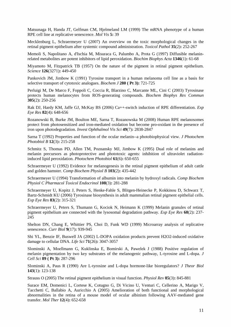
11
Matsunaga H, Handa JT, Gelfman CM, Hjelmeland LM (1999) The mRNA phenotype of a human RPE cell line at replicative senescence. Mol Vis 5: 39
Mecklenburg L, Schraermeyer U (2007) An overview on the toxic morphological changes in the retinal pigment epithelium after systemic compound administration. Toxicol Pathol 35(2): 252-267
Memoli S, Napolitano A, d'Ischia M, Misuraca G, Palumbo A, Prota G (1997) Diffusible melanin-related metabolites are potent inhibitors of lipid peroxidation. Biochim Biophys Acta 1346(1): 61-68
Miyamoto M, Fitzpatrick TB (1957) On the nature of the pigment in retinal pigment epithelium. Science 126(3271): 449-450
Pankovich JM, Jimbow K (1991) Tyrosine transport in a human melanoma cell line as a basis for selective transport of cytotoxic analogues. Biochem J 280 ( Pt 3): 721-725
Perluigi M, De Marco F, Foppoli C, Coccia R, Blarzino C, Marcante ML, Cini C (2003) Tyrosinase protects human melanocytes from ROS-generating compounds. Biochem Biophys Res Commun 305(2): 250-256
Rak DJ, Hardy KM, Jaffe GJ, McKay BS (2006) Ca++-switch induction of RPE differentiation. Exp Eye Res 82(4): 648-656
Rozanowski B, Burke JM, Boulton ME, Sarna T, Rozanowska M (2008) Human RPE melanosomes protect from photosensitized and iron-mediated oxidation but become pro-oxidant in the presence of iron upon photodegradation. Invest Ophthalmol Vis Sci 49(7): 2838-2847
Sarna T (1992) Properties and function of the ocular melanin--a photobiophysical view. J Photochem Photobiol B 12(3): 215-258
Schmitz S, Thomas PD, Allen TM, Poznansky MJ, Jimbow K (1995) Dual role of melanins and melanin precursors as photoprotective and phototoxic agents: inhibition of ultraviolet radiation-induced lipid peroxidation. Photochem Photobiol 61(6): 650-655
Schraermeyer U (1992) Evidence for melanogenesis in the retinal pigment epithelium of adult cattle and golden hamster. Comp Biochem Physiol B 103(2): 435-442
Schraermeyer U (1994) Transformation of albumin into melanin by hydroxyl radicals. Comp Biochem Physiol C Pharmacol Toxicol Endocrinol 108(3): 281-288
Schraermeyer U, Kopitz J, Peters S, Henke-Fahle S, Blitgen-Heinecke P, Kokkinou D, Schwarz T, Bartz-Schmidt KU (2006) Tyrosinase biosynthesis in adult mammalian retinal pigment epithelial cells. Exp Eye Res 83(2): 315-321
Schraermeyer U, Peters S, Thumann G, Kociok N, Heimann K (1999) Melanin granules of retinal pigment epithelium are connected with the lysosomal degradation pathway. Exp Eye Res 68(2): 237-245
Shelton DN, Chang E, Whittier PS, Choi D, Funk WD (1999) Microarray analysis of replicative senescence. Curr Biol 9(17): 939-945
Shi YL, Benzie IF, Buswell JA (2002) L-DOPA oxidation products prevent H2O2-induced oxidative damage to cellular DNA. Life Sci 71(26): 3047-3057
Slominski A, Moellmann G, Kuklinska E, Bomirski A, Pawelek J (1988) Positive regulation of melanin pigmentation by two key substrates of the melanogenic pathway, L-tyrosine and L-dopa. J Cell Sci 89 ( Pt 3): 287-296
Slominski A, Paus R (1990) Are L-tyrosine and L-dopa hormone-like bioregulators? J Theor Biol 143(1): 123-138
Strauss O (2005) The retinal pigment epithelium in visual function. Physiol Rev 85(3): 845-881
Surace EM, Domenici L, Cortese K, Cotugno G, Di Vicino U, Venturi C, Cellerino A, Marigo V, Tacchetti C, Ballabio A, Auricchio A (2005) Amelioration of both functional and morphological abnormalities in the retina of a mouse model of ocular albinism following AAV-mediated gene transfer. Mol Ther 12(4): 652-658
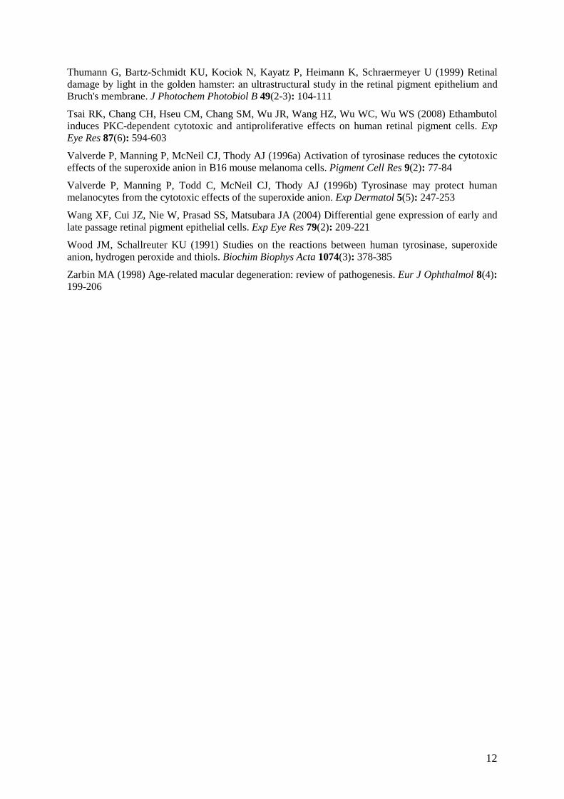
12
Thumann G, Bartz-Schmidt KU, Kociok N, Kayatz P, Heimann K, Schraermeyer U (1999) Retinal damage by light in the golden hamster: an ultrastructural study in the retinal pigment epithelium and Bruch's membrane. J Photochem Photobiol B 49(2-3): 104-111
Tsai RK, Chang CH, Hseu CM, Chang SM, Wu JR, Wang HZ, Wu WC, Wu WS (2008) Ethambutol induces PKC-dependent cytotoxic and antiproliferative effects on human retinal pigment cells. Exp Eye Res 87(6): 594-603
Valverde P, Manning P, McNeil CJ, Thody AJ (1996a) Activation of tyrosinase reduces the cytotoxic effects of the superoxide anion in B16 mouse melanoma cells. Pigment Cell Res 9(2): 77-84
Valverde P, Manning P, Todd C, McNeil CJ, Thody AJ (1996b) Tyrosinase may protect human melanocytes from the cytotoxic effects of the superoxide anion. Exp Dermatol 5(5): 247-253
Wang XF, Cui JZ, Nie W, Prasad SS, Matsubara JA (2004) Differential gene expression of early and late passage retinal pigment epithelial cells. Exp Eye Res 79(2): 209-221
Wood JM, Schallreuter KU (1991) Studies on the reactions between human tyrosinase, superoxide anion, hydrogen peroxide and thiols. Biochim Biophys Acta 1074(3): 378-385
Zarbin MA (1998) Age-related macular degeneration: review of pathogenesis. Eur J Ophthalmol 8(4): 199-206

13
Figure 1
Figure 2
14
Figure 3
Figure 4
Figure 5
15
Figure 6
Figure 7
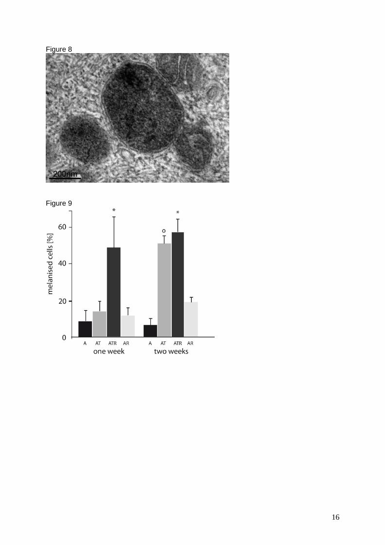
16
Figure 8
Figure 9
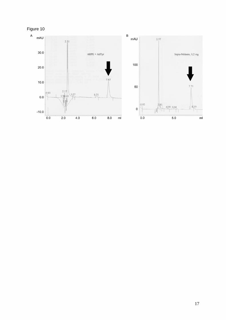
17
Figure 10