Seal gammaherpesviruses: identification, characterisation and epidemiology
7
Seal gammaherpesviruses: identification, characterisation and epidemiology B.E.E. Martina a , G.M.G.M. Verjans b , T.C. Harder c , T. Kuiken a,b , M.W.G. van de Bildt a , P.A.C. van Bergen b , A.D.M.E. Osterhaus a,b, * a Seal Rehabilitation and Research Centre, Hoofdstraat 94a, 9968 AG Pieterburen, Netherlands b Institute of Virology, Erasmus MC Rotterdam, Dr Molewaterplein 50, P.O. Box 1738, 3015 GE Rotterdam, Netherlands c Food, Veterinary and Environmental Diagnostic Laboratory of Schleswig-Holstein, Max-Eyth Str. 5, D-24537 Neumuenster, Germany Received 5 December 2002; received in revised form 10 April 2003; accepted 12 April 2003 Abstract Phocid herpesvirus type 2 (PhHV-2), tentatively classified as a gammaherpesvirus, has been isolated from European and American harbour seals (Phoca vitulina ). Here we describe the isolation and the molecular as well as biological characterisation of different PhHV-2 isolates from harbour seals and grey seals (Halichoerus grypus ). Of 522 harbour seals and 231 grey seals that had been admitted to the seal research and rehabilitation centre in Pieterburen, The Netherlands, between 1992 and 2000, 38 and 18%, respectively, proved to have PhHV-2 neutralising antibodies. PhHV-2 was isolated from peripheral blood mononuclear cells (PBMCs) of 12 and 28% of these seropositive animals, respectively, and 26 and 56% of these cell samples, respectively, were positive by PCR analysis. Analysis of amino acid sequences of PCR products and of the growth characteristics of different PhHV-2 isolates indicated that harbour and grey seals are infected with distinct g-herpesviruses, which however, may co-circulate between the two species. # 2003 Published by Elsevier Science B.V. Keywords: Phocid herpesvirus type 2; Seal gammaherpesviruses; Grey seals; Harbour seals; Replication rate; UL52 sequence 1. Introduction To date two herpesviruses of seals, phocid herpesvirus type 1 (PhHV-1) and phocid herpesvirus type 2 (PhHV- 2), have been described (Harder et al., 1996; Osterhaus et al., 1985). PhHV-2 was first isolated from peripheral blood mononuclear cells (PBMCs) of a harbour seal (Phoca vitulina) suffering from severe respiratory dis- ease (Lebich et al., 1994) and subsequently from harbour seals in the USA (Harder et al., 1996). Based on nucleotide sequence data, PhHV-2 has been identi- fied as a putative member of the gamma (g)-Herpesvir- inae sub-family (Harder et al., 1996). Limited data are available about the prevalence of g-herpesvirus infec- tions in pinnipeds. The prevalence of PhHV-2 specific antibodies in free-ranging marine mammals along the coasts of Alaska and Russia varied between 17 and 42%, with the highest prevalence among harbour seals (Zarnke et al., 1997). Grey seals (Halichorus grypus ) were not included in that study. The present paper describes the isolation, partial genetic and biological characterisation of g-herpesviruses of harbour seals (PV) and grey seals (HG) admitted to the Seal Research and Rehabilitation Centre (SRRC) in Pieterburen the Neth- erlands, from 1992 to 2000. In addition, the prevalence of PhHV-2 neutralising serum antibodies in these animals is presented. 2. Materials and methods 2.1. Virus isolation In December of 1999, a sick harbour seal (PV 9912) with severe respiratory illness found at the coast of France, south of the Seine mouth, was admitted to the * Corresponding author. Tel.: /31-10-408-8066/8067; fax: /31-10- 408-9485. E-mail address: [email protected] (A.D.M.E. Osterhaus). Virus Research 94 (2003) 25 /31 www.elsevier.com/locate/virusres 0168-1702/03/$ - see front matter # 2003 Published by Elsevier Science B.V. doi:10.1016/S0168-1702(03)00120-5
Transcript of Seal gammaherpesviruses: identification, characterisation and epidemiology

Seal gammaherpesviruses: identification, characterisation andepidemiology
B.E.E. Martina a, G.M.G.M. Verjans b, T.C. Harder c, T. Kuiken a,b,M.W.G. van de Bildt a, P.A.C. van Bergen b, A.D.M.E. Osterhaus a,b,*
a Seal Rehabilitation and Research Centre, Hoofdstraat 94a, 9968 AG Pieterburen, Netherlandsb Institute of Virology, Erasmus MC Rotterdam, Dr Molewaterplein 50, P.O. Box 1738, 3015 GE Rotterdam, Netherlands
c Food, Veterinary and Environmental Diagnostic Laboratory of Schleswig-Holstein, Max-Eyth Str. 5, D-24537 Neumuenster, Germany
Received 5 December 2002; received in revised form 10 April 2003; accepted 12 April 2003
Abstract
Phocid herpesvirus type 2 (PhHV-2), tentatively classified as a gammaherpesvirus, has been isolated from European and
American harbour seals (Phoca vitulina ). Here we describe the isolation and the molecular as well as biological characterisation of
different PhHV-2 isolates from harbour seals and grey seals (Halichoerus grypus ). Of 522 harbour seals and 231 grey seals that had
been admitted to the seal research and rehabilitation centre in Pieterburen, The Netherlands, between 1992 and 2000, 38 and 18%,
respectively, proved to have PhHV-2 neutralising antibodies. PhHV-2 was isolated from peripheral blood mononuclear cells
(PBMCs) of 12 and 28% of these seropositive animals, respectively, and 26 and 56% of these cell samples, respectively, were positive
by PCR analysis. Analysis of amino acid sequences of PCR products and of the growth characteristics of different PhHV-2 isolates
indicated that harbour and grey seals are infected with distinct g-herpesviruses, which however, may co-circulate between the two
species.
# 2003 Published by Elsevier Science B.V.
Keywords: Phocid herpesvirus type 2; Seal gammaherpesviruses; Grey seals; Harbour seals; Replication rate; UL52 sequence
1. Introduction
To date two herpesviruses of seals, phocid herpesvirus
type 1 (PhHV-1) and phocid herpesvirus type 2 (PhHV-
2), have been described (Harder et al., 1996; Osterhaus
et al., 1985). PhHV-2 was first isolated from peripheral
blood mononuclear cells (PBMCs) of a harbour seal
(Phoca vitulina) suffering from severe respiratory dis-
ease (Lebich et al., 1994) and subsequently from
harbour seals in the USA (Harder et al., 1996). Based
on nucleotide sequence data, PhHV-2 has been identi-
fied as a putative member of the gamma (g)-Herpesvir-
inae sub-family (Harder et al., 1996). Limited data are
available about the prevalence of g-herpesvirus infec-
tions in pinnipeds. The prevalence of PhHV-2 specific
antibodies in free-ranging marine mammals along the
coasts of Alaska and Russia varied between 17 and 42%,
with the highest prevalence among harbour seals
(Zarnke et al., 1997). Grey seals (Halichorus grypus )
were not included in that study. The present paper
describes the isolation, partial genetic and biological
characterisation of g-herpesviruses of harbour seals (PV)
and grey seals (HG) admitted to the Seal Research andRehabilitation Centre (SRRC) in Pieterburen the Neth-
erlands, from 1992 to 2000. In addition, the prevalence
of PhHV-2 neutralising serum antibodies in these
animals is presented.
2. Materials and methods
2.1. Virus isolation
In December of 1999, a sick harbour seal (PV 9912)
with severe respiratory illness found at the coast of
France, south of the Seine mouth, was admitted to the
* Corresponding author. Tel.: �/31-10-408-8066/8067; fax: �/31-10-
408-9485.
E-mail address: [email protected] (A.D.M.E. Osterhaus).
Virus Research 94 (2003) 25�/31
www.elsevier.com/locate/virusres
0168-1702/03/$ - see front matter # 2003 Published by Elsevier Science B.V.
doi:10.1016/S0168-1702(03)00120-5

Dutch Seal Research and Rehabilitation centre at
Pieterburen (SSRC; The Netherlands) where it died
with a pneumonia. Approximately 500 ml pleural
effusion as well as different organs was collected forroutine virological investigations. A lung homogenate
(10%) was made and inoculated on Crandell Rees feline
kidney (CrFK), Vero, and seal kidney (SeKC; from PV
origin) cultures. The pleural fluid was centrifuged for 10
min at 1000�/g and the supernatant was inoculated on
CrFK, SeKC and concanavalin A (ConA, Sigma, St.
Louis, CA) stimulated PV PBMCs. Cultures displaying
cytopathic effect (CPE) were analysed by negativecontrast electron microscopy. Further confirmation for
the isolation of a herpesvirus was achieved by perfor-
mance of immunofluorescence (IFA), using a panel of
discriminatory monoclonal antibodies (moAb) raised
against PhHV-1 strain PB 84 (2F2, 1H3) and PhHV-2
strain 7848 (3E5, 6C5 and 7C8) (Harder et al., 1996).
PBMC, isolated from heparinised blood samples of
different PV and HG admitted to the SSRC as describedpreviously (de Swart et al., 1993), were stored at �/
135 8C. In order to isolate virus from PBMC of seals,
PBMC were cultured in RPMI 1640 medium (BioWhit-
taker, Belgium) supplemented with antibiotics and 5%
heat inactivated foetal bovine serum (FBS), or stimu-
lated with 5 mg/ml ConA, or alternatively with a mixture
of 20 ng/ml phorbol myristate acetate (PMA; Sigma�/
Aldrich), 30 mM n-butyrate (Sigma�/Aldrich) and 1 mg/ml cyclosporin A (FK506; Sigma�/Aldrich) (PB-FK).
Fourteen days after stimulation, 106 cells were co-
cultivated with CrFK and SeKC monolayer cultures
for 6 weeks in Dulbecco’s Modified Eagle’s Medium
(DMEM) containing 5% FBS. Cultures that did not
show CPE were confirmed as being PhHV-2 negative by
IFA using the moAb 6C5 and by a PhHV-2-specific
PCR as described below.
2.2. PhHV-2-specific polymerase chain reaction (PCR)
DNA was extracted with silica from PBMC samples
used for virus isolation as described previously (Boom et
al., 1990). The primers used for the PhHV-2-specific
PCR, based on the PhHV-2 strain 7848 UL52 gene,
were: 5?-TGTTCAGATGCCAGTTCC-3? (forward pri-mer) and 5?-TTCATCCAAGTCCCACTC-3? (reverse
primer). PCR reactions were performed for 40 cycles
comprising 30 s at 94 8C, 1 min at 55 8C and 1 min at
72 8C. A final elongation phase of 10 min at 72 8C was
allowed. PCR products were size-fractionated on 2%
agarose gel, blotted onto a hybond N�/ membrane
(Amersham, Stad, UK) and subsequently hybridised
with a PhHV-2-specific biotin-conjugated probe (5?-CTTGGTGTGAAAGGTCTCACTC-3?) and visualised
using chemiluminescence. The PhHV-2 strain 7848 and
the PhHV-1 strain PB84 (Harder et al., 1996) or
uninfected cell cultures, were used as positive and
negative control samples, respectively.
2.3. Sequence analyses of UL52 amplicons of different
PhHV-2 isolates
The UL52-specific amplicons, purified from gel using
the Qiagene kit (Westburg, Stad, Belgium), were
sequenced directly with the aforementioned PCR pri-
mers on a 373A-automated sequencer (Applied Biosys-
tems, Stad, Land) according to the manufacturer’s
instructions. The UL52 DNA and corresponding pro-tein sequences were aligned with ‘‘CLUSTALW’’ (Bioedit
software package). Phylogenetic trees were constructed
using the ‘‘DNAML’’ software program of ‘‘PHYLIP’’
(version 3.5, department of Genetics, University of
Washington, State) using 100 bootstraps. The ‘‘CON-
SENSE’’ package of the program was used to generate the
tree.
2.4. Serological analyses
2.4.1. Serum samples
A serum panel of 753 seals, collected from 522 PV and
231 HG between 1992 and 2000 in the Dutch Wadden
Sea, was used to determine the prevalence of PhHV-2
neutralising serum antibodies in seals admitted to the
SRRC. All sera were taken approximately 6�/8 weeks
after admission to the SRRC.Sequential serum samples were available from 80
PhHV-2 seropositive PVs and 42 PhHV-2 seropositive
HG (see below) starting with a sample obtained at the
day of admission and ending with samples just before
release (12�/24 weeks after admission). Serial titration
analyses of these samples were performed to discrimi-
nate between maternal antibodies, past, or active PhHV-
2 infection. Animals PhHV-2 seropositive at admissionthat became seronegative during rehabilitation were
considered to have maternal antibodies. Active infection
during rehabilitation was defined by a 4-fold increase in
neutralisation antibody titres.
2.4.2. PhHV-2-specific serum IgG ELISA
The PhHV-2 strains PV 7848 and PV 9912 were used
to develop a whole virus-based ELISA system to detectserum IgG against PhHV-2. Both virus strains were
propagated in CrFK cells at a moi of 10�2. When more
than 90% of the monolayer showed CPE, cells and
supernatants were harvested and freeze-thawed twice.
Virus was purified by ultra-centrifugation through a
sucrose gradient. Virus containing bands were collected,
dialysed, pooled, and treated with 4% (w/v) Mega-10
(Sigma). The preparation was sonicated twice for 15 s at9 mM. Microtitre plates (96-well flatbottom) with high
protein binding capacities (Costar, USA) were coated
with 50 ng/well viral proteins for 1 h at 37 8C. Sera were
B.E.E. Martina et al. / Virus Research 94 (2003) 25�/3126

tested in a single dilution of 1:30. Further processing
was by means of a protein-A ELISA as previously
described (Martina et al., 2001).
2.4.3. Virus neutralisation assay
A virus neutralisation (VN) assay was performed as
previously described (Martina et al., 2001). Briefly, four
2-fold serum dilutions (starting dilution 1:20) were made
and incubated with 100 TCID50 of PhHV-2 strains PV
7848 or PV 9912. In some cases higher dilutions were
tested. Neutralising titres were determined after 14 days
microscopically on the basis of suppression of CPE.Titres were expressed as the reciprocal of the highest
dilution still giving 100% suppression of CPE.
3. Results
3.1. Isolation of a g-herpesvirus like virus from a harbour
seal
Four to six weeks after inoculation of lung homo-
genate and pleural fluid, obtained from the diseased
harbour seal (PV 9912), in CrFK, Vero, and SeKC
cultures, plaques of rounded and syncytial cells were
observed. The pleural fluid was also inoculated in PV-
PBMC cultures. Approximately 5 weeks after inocula-
tion, ballooning and extensive cell death was observed in
PBMC. Examination of supernatant from these cell-
cultures by negative contrast EM revealed the presence
herpesvirus like particles (results not shown). IFA
performed with a panel of phocine herpesvirus-specific
moAbs showed that the isolate did not react with the
PhHV-1-specific moAb 1H3, whereas a strong fluores-
cence was seen on viral infected cells with the PhHV-2-
specific moAbs 6C5 and 7C8. Interestingly, moAb 3E5,
generated against PhHV-2 strain PV 7848, did not
recognise cells infected with the PhHV-2 like strain PV
9912. These results indicated that the isolate is a PhHV-
2 like virus and is antigenically distinct from the PhHV-
2 strain PV 7848. To further confirm that PV 9912 was a
Table 1
Seals with g-herpesvirus (PhHV-2) specific serum antibodies or PhHV-
2 infection in the Netherlands between 1992 and 2000
Species Protein-A ELISA
(%)a
PV 7848 VN
(%)
PV 9912 VN
(%)
(a ) PhHV-2 seropositive seals
Harbour seal 208/522 (40) 198/522 (38) 154/522 (30)
Grey seal 74/231 (32) 34/231 (15) 42/231 (18)
(b ) PhHV-2 in PBMC
Virus isolation
(%)
PCR analysis (%)
6/50 (12) 13/50 (26)
5/18 (28) 10/18 (56)
a ELISA was based on an antigen mixture from both isolates.
Table 2
Age and clinical signs of PhHV-2 infected sealsa
Isolate Age (month) Clinical signs Virus isolation (yes/no)
PV 1 3 Cough and nasal discharge Yes
PV 2 6 Healthy Yes
PV 3 4 Nasal discharge No
PV 4 5 Severe respiratory disease Yes
PV 5 6 Data not available No
PV 6 12 Respiratory disease Yes
PV 7 8 Healthy Yes
PV 8 4 Data not available No
PV 9 3 Healthy No
PV 10 8 Healthy No
PV 11 5 Lethargy No
PV 12 5 Healthy No
PV 13 4 Data not available Yes
HG 1 1 Healthy Yes
HG 2 3 Cough Yes
HG 3 2 Healthy No
HG 4 2 Data not available No
HG 5 1 Cough Yes
HG 6 B/1 Healthy No
HG 7 B/1 Healthy Yes
HG 8 B/1 Epistaxis and coughing No
HG 9 3 Healthy No
HG 10 3 Data not available Yes
PV 7848 �/12 Moribund seal with pneumonia Yes
PV 9912 6 Pneumonia Yes
a Infection defined as PBMC positive in UL52 PCR.
B.E.E. Martina et al. / Virus Research 94 (2003) 25�/31 27

PhHV-2 like virus, PCR analysis was carried out using
PhHV-2 specific primers. An amplicon of the expected
size was obtained and was recognised by UL52-specific
probe, indicating that PV 9912 was indeed a PhHV-2
like virus.
3.2. Seroprevalence of PhHV-2-like viruses in seals
The prevalence of serum antibodies reacting with
PhHV-2 were determined by using a protein-A basedELISA system. In 208 out of 522 PV (40%) and 74 out
of 231 HG (32%) sera were positive when tested against
an antigenic protein preparation of both isolates (Table
1a). To determine whether seroprevalences against the
PhHV-2 strains PV 7848 and PV 9912 differed between
the seal populations, all sera were subjected to VN
assays using the respective viruses. Among the 522 PV
sera, 198 (38%) and 154 (30%) had VN antibodies (titre]/20) against PhHV-2 strain PV 7848 and PV 9912,
respectively (Table 1a). The 44 PV that did not
neutralise PV 9912 all had a PV 7848-specific VN titre
of 20. Among the 231 HG sera, 34 (15%) and 42 (18%)
neutralised PV 7848 and PV 9912, respectively (Table
1a). The 8 HG sera, not neutralising PV 7848, had a PV
9912-specific VN titre of 40. Overall, serum VN titresranged from 20 to 80 in both seal populations. These
sera were all obtained 6�/8 weeks post-admission. To
investigate whether the observed antibodies could have
been from maternal origin, serial VN assays performed
on sequential serum samples of PhHV-2 seropositive
animals showed that 43% (34 out of 80) of PV and 88%
(37 out of 42) of HG seal sera were positive on the day
of admission. Analysis of sequentially obtained sera ofthe same animals indicated that seven PV (21%) and five
HG (14%) had maternal antibodies rather than a past or
active PhHV-2 infection, as shown by disappearance of
antibodies 8�/10 weeks after admission. Based on a more
than 4-fold antibody titre rise, active infection was
detected in ten out of 26 (38%) PV and in 12 out of 32
(38%) HG tested, with neutralising titres rising from 20
up to 320.
3.3. Isolation and genetic characterisation of PhHV-2-
like viruses in seals
PhHV-2 has tentatively been classified as a lympho-
tropic g-herpesvirus (Harder et al., 1996). Therefore,
PBMC from 50 PV and 18 HG seals seropositive at
admission were cultured in presence of ConA, PB-FK,or plain culture medium. Viruses were isolated from
cultured PBMC, obtained from six PV (12%) (PV isolate
1, 2, 4, 6, 7, 13) and five HG (28%) (HG isolate 1, 2, 5, 7,
10). All these seals showed serological evidence of active
infection (see above). The HG 7 isolate was recovered
from PBMC cultured in PB-FK. No virus was recovered
after ConA stimulation of seal PBMC fractions. Cul-
tures of the PBMC samples that did not show CPE after3 weeks maintenance were co-cultivated for 6 weeks
with CrFK and SeK cells. IFA analyses conducted on
these cells showed no evidence for virus production in
these PBMC cultures.
However, when PCR was performed directly on all
unstimulated PBMC, using a PhHV-2 UL52-specific
PCR, 13 (26%) PV and 10 (56%) HG were found
positive (Table 1b). All PBMC from which virus wasisolated proved to be positive in this PCR. All UL52
PCR positive samples, including the reference PhHV-2
isolates PV 9912 and PV 7848 (see Table 2), were
sequenced and a phylogenetic tree was constructed. The
phylogenetic relationship between the 430 nucleotides of
the different UL52 amplicons was determined on
aligned sequences and maximum likelihood analysis
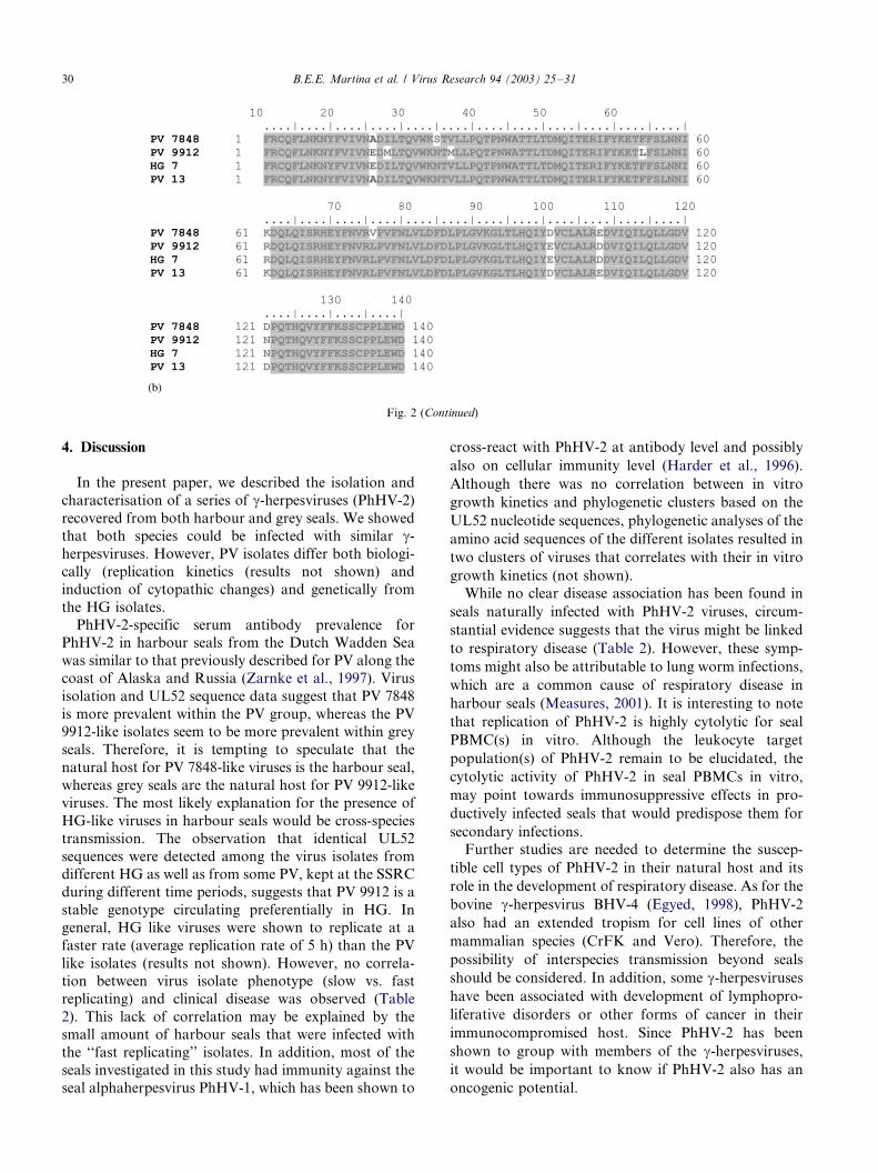
was performed. Four clusters of viruses were distin-guishable (Fig. 1). When the deduced amino acid
sequences were compared, two isolates were identical
to PV 7848, 11 were identical to PV 9912, three identical
Fig. 1. Unrooted phylogenetic tree of UL52 equivalent gene sequences
of different PhHV-2 isolates. Nucleotide sequences (430 bp long) were
aligned and subjected to bootstrapped (n�/100) maximum likelihood
analysis using the PHYLIP package. The consensus tree is shown with
bootstrap values indicated at tree branchings.
B.E.E. Martina et al. / Virus Research 94 (2003) 25�/3128

to HG 7 and seven had a sequence identical to that of
PV 13 (Figs. 1 and 2).
3.4. Growth characteristics of different isolates
CrFK cells infected with HG like viruses showed large
syncytial cells and ballooning, whereas slow-replicating
viruses induced plaques of rounded cells but did not
result in large syncytia formation and ballooning.
Interestingly, the isolates PV 2, 6, and 9912 (all isolates
recovered from harbour seal PBMC) had the same
growth characteristics and induced the same cytopathic
changes as the HG viruses, in agreement with their
genetic similarity to the HG viruses.
Fig. 2. Nucleotide (a) and deduced amino acid (b) sequence alignment of UL52 PCR fragments of different PhHV-2 isolates. Alignment was
performed with the CLUSTALW program of bioedit software package. Identical amino acids are represented on dark background and boxes indicate
similar amino acids (BLOSUM62 matrix).
B.E.E. Martina et al. / Virus Research 94 (2003) 25�/31 29
4. Discussion
In the present paper, we described the isolation and
characterisation of a series of g-herpesviruses (PhHV-2)
recovered from both harbour and grey seals. We showed
that both species could be infected with similar g-
herpesviruses. However, PV isolates differ both biologi-
cally (replication kinetics (results not shown) and
induction of cytopathic changes) and genetically from
the HG isolates.
PhHV-2-specific serum antibody prevalence for
PhHV-2 in harbour seals from the Dutch Wadden Sea
was similar to that previously described for PV along the
coast of Alaska and Russia (Zarnke et al., 1997). Virus
isolation and UL52 sequence data suggest that PV 7848
is more prevalent within the PV group, whereas the PV
9912-like isolates seem to be more prevalent within grey
seals. Therefore, it is tempting to speculate that the
natural host for PV 7848-like viruses is the harbour seal,
whereas grey seals are the natural host for PV 9912-like
viruses. The most likely explanation for the presence of
HG-like viruses in harbour seals would be cross-species
transmission. The observation that identical UL52
sequences were detected among the virus isolates from
different HG as well as from some PV, kept at the SSRC
during different time periods, suggests that PV 9912 is a
stable genotype circulating preferentially in HG. In
general, HG like viruses were shown to replicate at a
faster rate (average replication rate of 5 h) than the PV
like isolates (results not shown). However, no correla-
tion between virus isolate phenotype (slow vs. fast
replicating) and clinical disease was observed (Table
2). This lack of correlation may be explained by the
small amount of harbour seals that were infected with
the ‘‘fast replicating’’ isolates. In addition, most of the
seals investigated in this study had immunity against the
seal alphaherpesvirus PhHV-1, which has been shown to
cross-react with PhHV-2 at antibody level and possibly
also on cellular immunity level (Harder et al., 1996).
Although there was no correlation between in vitro
growth kinetics and phylogenetic clusters based on the
UL52 nucleotide sequences, phylogenetic analyses of the
amino acid sequences of the different isolates resulted in
two clusters of viruses that correlates with their in vitro
growth kinetics (not shown).
While no clear disease association has been found in
seals naturally infected with PhHV-2 viruses, circum-
stantial evidence suggests that the virus might be linked
to respiratory disease (Table 2). However, these symp-
toms might also be attributable to lung worm infections,
which are a common cause of respiratory disease in
harbour seals (Measures, 2001). It is interesting to note
that replication of PhHV-2 is highly cytolytic for seal
PBMC(s) in vitro. Although the leukocyte target
population(s) of PhHV-2 remain to be elucidated, the
cytolytic activity of PhHV-2 in seal PBMCs in vitro,
may point towards immunosuppressive effects in pro-
ductively infected seals that would predispose them for
secondary infections.
Further studies are needed to determine the suscep-
tible cell types of PhHV-2 in their natural host and its
role in the development of respiratory disease. As for the
bovine g-herpesvirus BHV-4 (Egyed, 1998), PhHV-2
also had an extended tropism for cell lines of other
mammalian species (CrFK and Vero). Therefore, the
possibility of interspecies transmission beyond seals
should be considered. In addition, some g-herpesviruses
have been associated with development of lymphopro-
liferative disorders or other forms of cancer in their
immunocompromised host. Since PhHV-2 has been
shown to group with members of the g-herpesviruses,
it would be important to know if PhHV-2 also has an
oncogenic potential.
Fig. 2 (Continued)
B.E.E. Martina et al. / Virus Research 94 (2003) 25�/3130

Acknowledgements
We thank Dr Ron Fouchier helpful suggestions and
Theo Bestebroer for technical assistance.
References
Boom, R., Sol, C.J., Salimans, M.M., Jansen, C.L., Wertheim-van
Dillen, P.M., van der, N.J., 1990. Rapid and simple method for
purification of nucleic acids. J. Clin. Microbiol. 28, 495�/503.
de Swart, R.L., Kluten, R.M., Huizing, C.J., Vedder, L.J., Reijnders,
P.J., Visser, I.K., UytdeHaag, F.G., Osterhaus, A.D., 1993.
Mitogen and antigen induced B and T cell responses of peripheral
blood mononuclear cells from the harbour seal (Phoca vitulina ).
Vet. Immunol. Immunopathol. 37, 217�/230.
Egyed, L., 1998. Replication of bovine herpesvirus type 4 in human
cells in vitro. J. Clin. Microbiol. 36, 2109�/2111.
Harder, T.C., Harder, M., Vos, H., Kulonen, K., Kennedy-Stoskopf,
S., Liess, B., Appel, M.J., Osterhaus, A.D., 1996. Characterization
of phocid herpesvirus-1 and -2 as putative alpha- and gamma-
herpesviruses of North and European pinnipeds. J. Gen. Virol. 77
(Pt 1), 27�/35.
Lebich, M., Harder, T.C., Frey, H.R., Visser, I.K., Osterhaus, A.D.,
Liess, B., 1994. Comparative immunological characterization of
type-specific and conserved B-cell epitopes of pinniped, felid and
canid herpesviruses. Arch. Virol. 136, 335�/347.
Martina, B.E., Airikkala, M.I., Harder, T.C., Amerongen, G.,
Osterhaus, A.D., 2001. A candidate phocid herpesvirus vaccine
that provides protection against feline herpesvirus infection.
Vaccine 20, 943�/948.
Measures, L.N., 2001. Lungworms of marine mammals. In: Samuel,
W.M., Pybus, M.J., Kocan, A.A. (Eds.), Parasitic Diseases of Wild
Mammals, second ed. Iowa State University Press, Ames, Iowa, pp.
279�/300.
Osterhaus, A.D., Yang, H., Spijkers, H.E., Groen, J., Teppema, J.S.,
van Steenis, G., 1985. The isolation and partial characterization of
a highly pathogenic herpesvirus from the harbor seal (Phoca
vitulina ). Arch. Virol. 86, 239�/251.
Zarnke, R.L., Harder, T.C., Vos, H.W., Ver Hoef, J.M., Osterhaus,
A.D., 1997. Serologic survey for phocid herpesvirus-1 and -2 in
marine mammals from Alaska and Russia. J. Wildl. Dis. 33, 459�/
465.
B.E.E. Martina et al. / Virus Research 94 (2003) 25�/31 31