The tumor suppressor semaphorin 3B triggers a prometastatic program mediated by interleukin 8 and...
17
The Journal of Experimental Medicine ARTICLE © 2008 Rolny et al. The Rockefeller University Press $30.00 J. Exp. Med. Vol. 205 No. 5 1155-1171 www.jem.org/cgi/doi/ 1155 10.1084/jem.20072509 Semaphorins are a highly conserved family of molecular signals originally identified as guid- ance cues for axon navigation in neural develop- ment (1, 2). Moreover, several semaphorins have been implicated in angiogenesis, immune function, and cancer (for reviews see refer- ences 3, 4). The functional role of semaphorins in tumor progression is still controversial. For example, semaphorin 3B ( SEMA3B) and the homologous gene SEMA3F have been indicated as puta- tive tumor suppressors because they are both located in the chromosomal region 3p21.3, a locus frequently deleted or hypermethylated in lung cancers (5, 6). SEMA3B and SEMA3F can furthermore inhibit tumor proliferation in vitro and in vivo (7–9) Moreover, SEMA3F has been shown to inhibit angiogenesis and metastatic dissemination (10, 11). We have previously demonstrated that semaphorin function is mediated by a family of plasma membrane receptors, the plexins (12). However, most secreted semaphorins (including SEMA3B) do not bind directly to plexins but instead interact with plexin-associated corecep- tors, denoted neuropilin (NP) 1 and 2. More- over, NPs directly bind vascular endothelial growth factor (VEGF) family members and as- sociate in complex with VEGF receptors (13), CORRESPONDENCE Luca Tamagnone: [email protected] Abbreviations used: ATF-2, activating transcription factor 2; CDKI, cytokine-dependent kinase inhibitor; CM, condi- tioned medium; EV, empty vector; HUVEC, human um- bilical vein endothelial cell; MAPK, mitogen-activated protein kinase; MCP-1, mono- cyte chemotactic protein 1; NP, neuropilin; RNAi, RNA inter- ference; SEMA3B, semaphorin 3B; shRNA, short hairpin RNA; TAM, tumor-associated macrophage; TUNEL, Tdt- mediated dUTP-biotin nick- end labeling; VEGF, vascular endothelial growth factor. L. Capparuccia and A. Casazza contributed equally to this work. C. Rolny’s present address is Dept. of Genetics and Pathology, Unit of Vascular Biology, Rudbeck Laboratory, Uppsala University, S-751 85 Uppsala, Sweden. The online version of this article contains supplemental material. The tumor suppressor semaphorin 3B triggers a prometastatic program mediated by interleukin 8 and the tumor microenvironment Charlotte Rolny , 1 Lorena Capparuccia, 1 Andrea Casazza, 1 Massimiliano Mazzone, 1,2 Antonella Vallario , 1 Alessandro Cignetti, 1 Enzo Medico, 1 Peter Carmeliet, 2 Paolo M. Comoglio, 1 and Luca Tamagnone 1 1 Institute for Cancer Research and Treatment (IRCC), University of Turin, School of Medicine, 10060 Candiolo, Italy 2 Department of Transgene Technology and Gene Therapy (Flanders Institute for Biotechnology) and Center for Transgene Technology and Gene Therapy, Katholieke Universiteit Leuven, 3000 Leuven, Belgium Semaphorins are a large family of evolutionarily conserved morphogenetic molecules originally identified for their repelling role in axonal guidance. Intriguingly, semaphorins have recently been implicated in cancer progression (Neufeld, G., T. Lange, A. Varshavsky, and O. Kessler. 2007. Adv. Exp. Med. Biol. 600:118–131). In particular, semaphorin 3B (SEMA3B) is considered a putative tumor suppressor, and yet we found that it is ex- pressed at high levels in many invasive and metastatic human cancers. By investigating experimental tumor models, we confirmed that SEMA3B expression inhibited tumor growth, whereas metastatic dissemination was surprisingly increased. We found that SEMA3B induced the production of interleukin (IL) 8 by tumor cells by activating the p38–mitogen-activated protein kinase pathway in a neuropilin 1–dependent manner. Silencing the expression of endogenous SEMA3B in tumor cells impaired IL-8 transcrip- tion. The release of IL-8, in turn, induced the recruitment of tumor-associated macro- phages and metastatic dissemination to the lung, which could be rescued by blocking IL-8 with neutralizing antibodies. In conclusion, we report that SEMA3B exerts unexpected functions in cancer progression by fostering a prometastatic environment through elevated IL-8 secretion and recruitment of macrophages coupled to the suppression of tumor growth. on June 2, 2016 jem.rupress.org Downloaded from Published May 5, 2008 http://jem.rupress.org/content/suppl/2008/05/04/jem.20072509.DC1.html Supplemental Material can be found at:
-
Upload
independent -
Category
Documents
-
view
4 -
download
0
Transcript of The tumor suppressor semaphorin 3B triggers a prometastatic program mediated by interleukin 8 and...

The
Journ
al o
f Exp
erim
enta
l M
edic
ine
ARTICLE
© 2008 Rolny et al.
The Rockefeller University Press $30.00
J. Exp. Med. Vol. 205 No. 5 1155-1171 www.jem.org/cgi/doi/
1155
10.1084/jem.20072509
Semaphorins are a highly conserved family of molecular signals originally identifi ed as guid-ance cues for axon navigation in neural develop-ment ( 1, 2 ). Moreover, several semaphorins have been implicated in angiogenesis, immune function, and cancer (for reviews see refer-ences 3, 4 ).
The functional role of semaphorins in tumor progression is still controversial. For example, semaphorin 3B ( SEMA3B ) and the homologous gene SEMA3F have been indicated as puta-tive tumor suppressors because they are both
located in the chromosomal region 3p21.3, a locus frequently deleted or hypermethylated in lung cancers ( 5, 6 ). SEMA3B and SEMA3F can further more inhibit tumor proliferation in vitro and in vivo ( 7 – 9 ) Moreover, SEMA3F has been shown to inhibit angiogenesis and metastatic dissemination ( 10, 11 ).
We have previously demonstrated that semaphorin function is mediated by a family of plasma membrane receptors, the plexins ( 12 ). However, most secreted semaphorins (including SEMA3B) do not bind directly to plexins but instead interact with plexin-associated corecep-tors, denoted neuropilin (NP) 1 and 2. More-over, NPs directly bind vascular endothelial growth factor (VEGF) family members and as-sociate in complex with VEGF receptors ( 13 ),
CORRESPONDENCE
Luca Tamagnone:
Abbreviations used: ATF-2,
activating transcription factor 2;
CDKI, cytokine-dependent
kinase inhibitor; CM, condi-
tioned medium; EV, empty
vector; HUVEC, human um-
bilical vein endothelial cell;
MAPK, mitogen-activated
protein kinase; MCP-1, mono-
cyte chemotactic protein 1; NP,
neuropilin; RNAi, RNA inter-
ference; SEMA3B, semaphorin
3B; shRNA, short hairpin
RNA; TAM, tumor-associated
macrophage; TUNEL, Tdt-
mediated dUTP-biotin nick-
end labeling; VEGF, vascular
endothelial growth factor.
L. Capparuccia and A. Casazza contributed equally to this work.
C. Rolny ’ s present address is Dept. of Genetics and Pathology,
Unit of Vascular Biology, Rudbeck Laboratory,
Uppsala University, S-751 85 Uppsala, Sweden.
The online version of this article contains supplemental material.
The tumor suppressor semaphorin 3B triggers a prometastatic program mediated by interleukin 8 and the tumor microenvironment
Charlotte Rolny , 1 Lorena Capparuccia , 1 Andrea Casazza , 1 Massimiliano Mazzone , 1,2 Antonella Vallario , 1 Alessandro Cignetti , 1 Enzo Medico , 1 Peter Carmeliet , 2 Paolo M. Comoglio , 1 and Luca Tamagnone 1
1 Institute for Cancer Research and Treatment (IRCC), University of Turin, School of Medicine, 10060 Candiolo, Italy
2 Department of Transgene Technology and Gene Therapy (Flanders Institute for Biotechnology) and Center for Transgene
Technology and Gene Therapy, Katholieke Universiteit Leuven, 3000 Leuven, Belgium
Semaphorins are a large family of evolutionarily conserved morphogenetic molecules
originally identifi ed for their repelling role in axonal guidance. Intriguingly, semaphorins
have recently been implicated in cancer progression (Neufeld, G., T. Lange, A. Varshavsky,
and O. Kessler. 2007. Adv. Exp. Med. Biol. 600:118 – 131). In particular, semaphorin 3B
(SEMA3B) is considered a putative tumor suppressor, and yet we found that it is ex-
pressed at high levels in many invasive and metastatic human cancers. By investigating
experimental tumor models, we confi rmed that SEMA3B expression inhibited tumor
growth, whereas metastatic dissemination was surprisingly increased. We found that
SEMA3B induced the production of interleukin (IL) 8 by tumor cells by activating the
p38 – mitogen-activated protein kinase pathway in a neuropilin 1 – dependent manner.
Silencing the expression of endogenous SEMA3B in tumor cells impaired IL-8 transcrip-
tion. The release of IL-8, in turn, induced the recruitment of tumor-associated macro-
phages and metastatic dissemination to the lung, which could be rescued by blocking IL-8
with neutralizing antibodies. In conclusion, we report that SEMA3B exerts unexpected
functions in cancer progression by fostering a prometastatic environment through
elevated IL-8 secretion and recruitment of macrophages coupled to the suppression
of tumor growth.
on June 2, 2016jem
.rupress.orgD
ownloaded from
Published May 5, 2008
http://jem.rupress.org/content/suppl/2008/05/04/jem.20072509.DC1.html Supplemental Material can be found at:
1156 SEMA3B PROMOTES METASTASIS VIA THE MICROENVIRONMENT | Rolny et al.
RESULTS
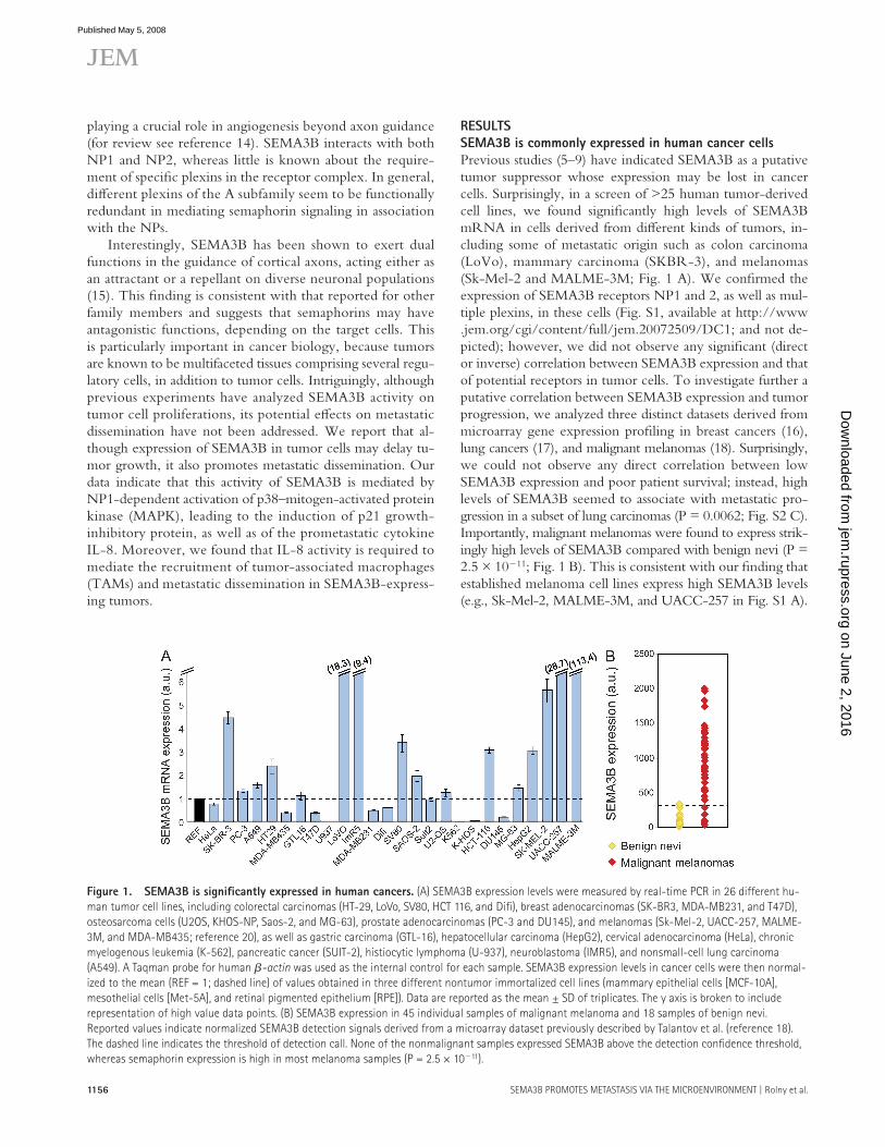
SEMA3B is commonly expressed in human cancer cells
Previous studies ( 5 – 9 ) have indicated SEMA3B as a putative tumor suppressor whose expression may be lost in cancer cells. Surprisingly, in a screen of > 25 human tumor-derived cell lines, we found signifi cantly high levels of SEMA3B mRNA in cells derived from diff erent kinds of tumors, in-cluding some of metastatic origin such as colon carcinoma (LoVo), mammary carcinoma (SKBR-3), and melanomas (Sk-Mel-2 and MALME-3M; Fig. 1 A ). We confi rmed the expression of SEMA3B receptors NP1 and 2, as well as mul-tiple plexins, in these cells (Fig. S1, available at http://www.jem.org/cgi/content/full/jem.20072509/DC1; and not de-picted); however, we did not observe any signifi cant (direct or inverse) correlation between SEMA3B expression and that of potential receptors in tumor cells. To investigate further a putative correlation between SEMA3B expression and tumor progression, we analyzed three distinct datasets derived from microarray gene expression profi ling in breast cancers ( 16 ), lung cancers ( 17 ), and malignant melanomas ( 18 ). Surprisingly, we could not observe any direct correlation between low SEMA3B expression and poor patient survival; instead, high levels of SEMA3B seemed to associate with metastatic pro-gression in a subset of lung carcinomas (P = 0.0062; Fig. S2 C). Importantly, malignant melanomas were found to express strik-ingly high levels of SEMA3B compared with benign nevi (P = 2.5 × 10 � 11 ; Fig. 1 B ). This is consistent with our fi nding that established melanoma cell lines express high SEMA3B levels (e.g., Sk-Mel-2, MALME-3M, and UACC-257 in Fig. S1 A).
playing a crucial role in angiogenesis beyond axon guidance (for review see reference 14 ). SEMA3B interacts with both NP1 and NP2, whereas little is known about the require-ment of specifi c plexins in the receptor complex. In general, diff erent plexins of the A subfamily seem to be functionally redundant in mediating semaphorin signaling in association with the NPs.
Interestingly, SEMA3B has been shown to exert dual functions in the guidance of cortical axons, acting either as an attractant or a repellant on diverse neuronal populations ( 15 ). This fi nding is consistent with that reported for other family members and suggests that semaphorins may have antagonistic functions, depending on the target cells. This is particularly important in cancer biology, because tumors are known to be multifaceted tissues comprising several regu-latory cells, in addition to tumor cells. Intriguingly, although previous experiments have analyzed SEMA3B activity on tumor cell proliferations, its potential eff ects on metastatic dissemination have not been addressed. We report that al-though expression of SEMA3B in tumor cells may delay tu-mor growth, it also promotes metastatic dissemination. Our data indicate that this activity of SEMA3B is mediated by NP1-dependent activation of p38 – mitogen-activated protein kinase (MAPK), leading to the induction of p21 growth-inhibitory protein, as well as of the prometastatic cytokine IL-8. Moreover, we found that IL-8 activity is required to mediate the recruitment of tumor-associated macrophages (TAMs) and metastatic dissemination in SEMA3B-express-ing tumors.
Figure 1. SEMA3B is signifi cantly expressed in human cancers. (A) SEMA3B expression levels were measured by real-time PCR in 26 different hu-
man tumor cell lines, including colorectal carcinomas (HT-29, LoVo, SV80, HCT 116, and Difi ), breast adenocarcinomas (SK-BR3, MDA-MB231, and T47D),
osteosarcoma cells (U2OS, KHOS-NP, Saos-2, and MG-63), prostate adenocarcinomas (PC-3 and DU145), and melanomas (Sk-Mel-2, UACC-257, MALME-
3M, and MDA-MB435; reference 20 ), as well as gastric carcinoma (GTL-16), hepatocellular carcinoma (HepG2), cervical adenocarcinoma (HeLa), chronic
myelogenous leukemia (K-562), pancreatic cancer (SUIT-2), histiocytic lymphoma (U-937), neuroblastoma (IMR5), and nonsmall-cell lung carcinoma
(A549). A Taqman probe for human � -actin was used as the internal control for each sample. SEMA3B expression levels in cancer cells were then normal-
ized to the mean (REF = 1; dashed line) of values obtained in three different nontumor immortalized cell lines (mammary epithelial cells [MCF-10A],
mesothelial cells [Met-5A], and retinal pigmented epithelium [RPE]). Data are reported as the mean ± SD of triplicates. The y axis is broken to include
representation of high value data points. (B) SEMA3B expression in 45 individual samples of malignant melanoma and 18 samples of benign nevi.
Reported values indicate normalized SEMA3B detection signals derived from a microarray dataset previously described by Talantov et al. (reference 18 ).
The dashed line indicates the threshold of detection call. None of the nonmalignant samples expressed SEMA3B above the detection confi dence threshold,
whereas semaphorin expression is high in most melanoma samples (P = 2.5 × 10 � 11 ).
on June 2, 2016jem
.rupress.orgD
ownloaded from
Published May 5, 2008

JEM VOL. 205, May 12, 2008
ARTICLE
1157
Figure 2. SEMA3B-expressing tumors display reduced growth but increased metastatic dissemination. (A) MDA-MB435 (MDA) and A549 cells trans-
duced to express SEMA3B (3B) or a noncoding EV were grown in culture for 4 d in 1% FBS. Cell growth was evaluated daily in separate dishes by staining cells
with crystal violet and measuring absorbance. Data shown represent the mean and SD of triplicates.(B and C) SEMA3B-transduced and EV control tumor cells
were injected subcutaneously into nude mice. Graphs display tumor volume (measured externally during the experiment; B) and tumor weight (measured after
excision at the end of the experiment; C), respectively. Data shown are the mean ± SEM from nine mice per each experimental group. ***, P < 0.0005. (D) The
expression of SEMA3B (Myc tagged) was detected in tumors by staining tissue sections with anti-Myc antibodies (green), whereas nuclear counterstaining was
done with DAPI (blue). Bar, 100 μ m. SEMA3B was similarly detected in tumor lysates by Western blotting (bottom). (E) At the end of the experiment, the lungs of
mice carrying tumor xenografts (as described above) were contrasted by airway perfusion with India ink, and superfi cial metastases were counted under a ste-
reoscopic microscope. Numbers of metastases are given as the mean ± SD from nine mice for each experimental group (left). We further determined the meta-
static index of SEMA3B-expressing and control tumors (right) by calculating the ratio between the number of metastatic foci and tumor weight. **, P < 0.01; ***,
P < 0.001. (F) A549 cells expressing SEMA3B under control of a doxycycline-inducible promoter were injected subcutaneously in nude mice. Tumor cells express-
ing the transactivator rTTA alone were used as controls. After 46 d, the mice were killed, and we measured tumor burden, counted lung metastasis, and calculated
the metastatic index as described. Data shown are the mean ± SEM. Western blot analysis on protein lysates from tumor samples (top) revealed a minimum level
of recombinant SEMA3B expression in the absence of the doxycycline, which was strongly induced by treatment with the drug. *, P < 0.05; **, P < 0.01.
on June 2, 2016jem
.rupress.orgD
ownloaded from
Published May 5, 2008

1158 SEMA3B PROMOTES METASTASIS VIA THE MICROENVIRONMENT | Rolny et al.
inducible promoter in A549 cells. This system allowed us to obtain drug-regulated expression of SEMA3B in tumor xe-nografts in vivo. Notably, low levels of SEMA3B were de-tected in tumors in the absence of the drug, whereas they greatly increased upon treatment with doxycycline ( Fig. 2 F , top). Semaphorin levels in xenografts correlated directly with the metastatic potential and inversely with the tumor burden ( Fig. 2 F , bottom).
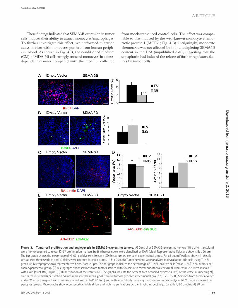
Tumor cell proliferation and angiogenesis in
SEMA3B-expressing tumors
We hypothesized that the inhibition of tumor growth by SEMA3B could be accounted for by a primary eff ect on cell proliferation, consistent with our data in vitro. To reliably answer this question, we analyzed tumors of comparable size taken 15 d after transplant. Consistent with in vitro observa-tions, SEMA3B-expressing tumor cells showed a > 60% re-duction in proliferation rate when transplanted in vivo (as assessed by anti – human Ki-67 staining; Fig. 3 A ), whereas apoptosis was only moderately increased (detected by Tdt-mediated dUTP-biotin nick-end labeling [TUNEL]; Fig. 3 B ); these results were further confi rmed by analyzing tumors at a later stage (30 d; not depicted). Double-staining experiments with markers of endothelia and TAMs did not detect a sig-nifi cant number of mouse stromal cells undergoing apoptosis (unpublished data), consistent with those that were essentially represented by tumor cells. Because it has been shown pre-viously that other semaphorins can inhibit VEGF-induced angiogenesis (for review see reference 21 ), we asked whether SEMA3B could similarly mediate inhibition of tumor angio-genesis. We could not fi nd any signifi cant diff erence in vessel area, as measured by lectin-perfused vessels, between SEMA3B xenografts and controls in tumors of comparable size (15 d after transplant); notably, the number of vessels was slightly increased in SEMA3B-expressing xenografts compared with controls, suggesting that vessel structure was altered ( Fig. 3, C and D ). However reduced angiogenesis was unlikely to be primarily responsible for delayed tumor growth. We ob-served a 25% reduction of angiogenesis in tumors expressing SEMA3B at later stages (day 30), when they had become re-markably smaller than controls (unpublished data). Moreover, in SEMA3B-expressing tumors, we observed notable defects of pericyte recruitment to blood vessels compared with con-trol xenografts ( Fig. 3 E ).
Macrophage recruitment in SEMA3B tumors
Several important factors regulating tumor angiogenesis and metastatic dissemination are released by TAMs ( 22 – 24 ). In-terestingly, by staining our cryostat sections with the selective antibody F4/80 for macrophages, we observed a twofold in-duction of macrophage recruitment into the tumor periphery ( Fig. 4 A , top row) in SEMA3B xenografts, as well as an in-creased infi ltration in the center of the tumor ( Fig. 4 A , bot-tom row). In contrast, tumor-infi ltrating granulocytes did not increase upon SEMA3B expression in vivo (based on Gr1 immunostaining; unpublished data).
Moreover, Hoek et al. reported a sixfold induction of SEMA3B gene expression in primary malignant melanoma cells com-pared with normal melanocytes ( 19 ).
These fi ndings seemed to challenge the idea that SEMA3B functions as a tumor suppressor and prompted us to experi-mentally test in vivo the functional role of SEMA3B in can-cer progression and metastasis. For our experiments, we thus selected MDA-MB435 human tumor cells (of melanoma ori-gin) ( 20 ), which are characterized by very low expression of SEMA3B ( Fig. 1 A ), a fast growth rate, and low metastatic potential, and A549 nonsmall-cell lung carcinoma cells that express intermediate levels of SEMA3B and form slowly growing metastatic tumors in nude mice.
SEMA3B inhibits tumor growth but induces metastasis in vivo
We transduced MDA-MB435 and A549 cells with lentiviral vectors either encoding SEMA3B or a noncoding empty vec-tor (EV). SEMA3B-transduced cells did not undergo any clear morphological change compared with controls (unpublished data). Consistent with what has been previously shown in other tumor cell lines ( 7 – 9 ), we observed growth inhibition in MDA-MB435 and A549 cells upon SEMA3B expression ( Fig. 2 A ). Moreover, in accordance with previous data on MDA-MB435 cells ( 9 ), we found that NP1 receptor expressed in tumor cells was likely implicated to mediate this autocrine eff ect, because NP1-blocking antibodies could rescue SEMA3B-induced growth inhibition, whereas RNA interference (RNAi) – me-diated suppression of NP2 could not (Fig. S3, available at http://www.jem.org/cgi/content/full/jem.20072509/DC1). Notably, we found no evidence of increased cell apoptosis in vitro upon SEMA3B expression, nor that SEMA3B expression signifi cantly aff ected tumor cell motility or cell scattering in vitro (unpublished data).
The tumorigenic potential of SEMA3B-expressing cells was analyzed in vivo by subcutaneous transplantation in nude mice. Tumor volume was periodically measured, and tumor weight was determined upon dissection at the end of the ex-periment. Tumor xenografts expressing SEMA3B displayed a signifi cant reduction in volume and tumor burden ( Fig. 2, B and C ), yet they were surprisingly associated with a remark-able increase of spontaneous metastatic dissemination to the lungs ( Fig. 2 E ). The metastatic potential was quantifi ed by scoring macrometastasis in the lungs ( Fig. 2 E , left). Moreover, we found that the ratio between the number of metastatic foci and the size of respective primary tumors was increased > 10 times in SEMA3B-expressing tumors versus controls ( Fig. 2 E , right), indicating a strong increase in the metastatic poten-tial of tumor cells. SEMA3B expression was confi rmed in pri-mary tumors by the immunostaining of cryostat sections ( Fig. 2 D , top) and by Western blot analysis of protein lysates from tumor samples ( Fig. 2 D , bottom). SEMA3B expression was also detected in metastatic cells by immunostaining (unpub-lished data), ruling out that they represented a selected sub-population of cells that have lost SEMA3B expression.
To confi rm the specifi city of these eff ects, we expressed SEMA3B under the control of an rTTA- and doxycycline-
on June 2, 2016jem
.rupress.orgD
ownloaded from
Published May 5, 2008
JEM VOL. 205, May 12, 2008
ARTICLE
1159
These fi ndings indicated that SEMA3B expression in tumor cells induces their ability to attract monocytes/macrophages. To further investigate this eff ect, we performed migration assays in vitro with monocytes purifi ed from human periph-eral blood. As shown in Fig. 4 B , the conditioned medium (CM) of MDA-3B cells strongly attracted monocytes in a dose-dependent manner compared with the medium collected
from mock-transduced control cells. The eff ect was compa-rable to that induced by the well-known monocyte chemo-tactic protein 1 (MCP-1; Fig. 4 B ). Intriguingly, monocyte chemotaxis was not aff ected by immunodepleting SEMA3B content in the CM (unpublished data), suggesting that the semaphorin had induced the release of further regulatory fac-tors by tumor cells.
Figure 3. Tumor cell proliferation and angiogenesis in SEMA3B-expressing tumors. (A) Control or SEMA3B-expressing tumors (15 d after transplant)
were immunostained to reveal KI-67 proliferation markers (red), whereas nuclei were visualized by DAPI (blue). Representative fi elds are shown. Bar, 20 μ m.
The bar graph shows the percentage of Ki-67 – positive cells (mean ± SD) in six tumors per each experimental group. For all quantifi cations shown in this fi g-
ure, at least three sections and 10 fi elds were counted for each tumor. **, P < 0.01. (B) Tumor sections were analyzed to reveal apoptotic cells using TUNEL
green kit. Micrographs show representative fi elds. Bars, 20 μ m. The bar graph indicates the percentage of TUNEL-positive cells (mean ± SD) in six tumors per
each experimental group. (C) Micrographs show sections from tumors stained with SA-lectin to reveal endothelial cells (red), whereas nuclei were marked
with DAPI (blue). Bar, 60 μ m. (D) Quantifi cation of the results in C. The graphs indicate the percent area occupied by vessels (left) or the vessel number (right),
calculated in six fi elds per section. Values represent the mean ± SD from six tumors per each experimental group. *, P < 0.05. (E) Sections from tumors excised
at day 21 after transplant were immunostained with anti-CD31 (red) and with an antibody revealing the chondroitin proteoglycan NG2 that is expressed on
pericytes (green). Micrographs show representative fi elds at low and high magnifi cations (left and right, respectively). Bars: (left) 60 μ m; (right) 20 μ m.
on June 2, 2016jem
.rupress.orgD
ownloaded from
Published May 5, 2008

1160 SEMA3B PROMOTES METASTASIS VIA THE MICROENVIRONMENT | Rolny et al.
lated by treatment with doxycycline in A549 carcinoma cells expressing SEMA3B under the control of a drug-inducible promoter ( Fig. 5 D ).
Monocyte/macrophage recruitment induced by SEMA3B
depends on IL-8
Consistent with previous reports ( 28 – 30 ), we observed a signifi cant dose-dependent increase of monocyte migration in vitro in response to purifi ed IL-8 (Fig. S4 A, available at http://www.jem.org/cgi/content/full/jem.20072509/DC1). This monocyte-attracting activity of purifi ed IL-8 was com-parable to that induced by the CM of SEMA3B-expressing cells (Fig. S4 A); moreover, it was effi ciently blocked by mono-clonal anti – IL-8 neutralizing antibodies (MAB208; Fig. S4 B). Therefore, to test whether monocyte recruitment induced by SEMA3B-CM could actually depend on increased IL-8
SEMA3B induces IL-8 expression in tumor cells
By performing microarray analysis of gene expression on mRNA derived from MDA-MB435 cells transduced with SEMA3B or EV, we found that the most signifi cantly regu-lated gene was IL-8 (P < 0.001; Fig. 5 A ). Interestingly, IL-8 is known to be a chemoattractant for leucocytes and endo-thelial and smooth muscle cells, which are further implicated in human cancer biology ( 25 – 27 ).
We thus confi rmed microarray data by real-time quanti-tative PCR in tumor cell lines transduced with SEMA3B (i.e., A549 lung carcinoma, HeLa cervical carcinoma, HT29 colon carcinoma, and GTL16 gastric carcinoma; Fig. 5 B ). In addition, the CM of MDA-3B and control cells was collected and analyzed by Western blotting, revealing that IL-8 secre-tion was strongly induced upon SEMA3B expression ( Fig. 5 C ). Moreover, IL-8 protein levels were specifi cally up-regu-
Figure 4. SEMA3B expression in tumor cells induces macrophage recruitment. (A) Sections from tumors excised at day 15 after transplant were
immunostained with anti-F4/80 antibody to selectively detect macrophages (red), whereas nuclei were revealed with DAPI (blue). Macrophages were
quantifi ed in the periphery and in the center of the tumors. Bars, 60 μ m. The graphs represent the mean ± SD from eight tumors for each experimental
group (at least three sections and 10 fi elds occupied by macrophages were counted from each tumor). ***, P < 0.001; **, P < 0.01. (B) The migration of
monocytes purifi ed from human peripheral blood was analyzed in a Transwell migration assay. Increasing concentrations of CM (1x, 3x, or 10x, as indi-
cated) derived from tumor cells transduced with SEMA3B (3B) or EV were included in the lower chamber (or alternatively in the upper chamber, as speci-
fi ed). Migrated monocytes were stained with Giemsa and counted in low magnifi cation micrographs of the fi lters, as reported previously (reference 27 ).
Values in the graph represent the mean ± SD of three independent experiments. 100 ng/ml MCP-1 in the lower chamber provided an internal positive
control. n.t., nontreated cells.
on June 2, 2016jem
.rupress.orgD
ownloaded from
Published May 5, 2008

JEM VOL. 205, May 12, 2008
ARTICLE
1161
knock down of SEMA3B expression led to reduced levels of IL-8 mRNA ( Fig. 6 A ). Furthermore, three diff erent shRNA sequences for SEMA3B were tested with consistent results, ruling out off -target eff ects (Fig. S5, available at http://www.jem.org/cgi/content/full/jem.20072509/DC1).
To investigate the role of endogenous SEMA3B in mono-cyte recruitment by tumor cells, we performed migration assays with the CM of Sk-Mel-2 melanoma cells that basally se-crete high levels of IL-8 (unpublished data). The medium collected from control cells strongly attracted monocytes compared with that of SEMA3B-defi cient cells (shSEMA3B; Fig. 6 B ). Importantly, this functional diff erence was entirely accounted for by the loss of IL-8 expression in shSEMA3B cells, because including IL-8 neutralizing antibodies abro-gated the diff erence between monocyte migration induced by control CM and that of SEMA3B-defi cient melanoma cells ( Fig. 6 B ).
secretion by tumor cells, we repeated migration assays, as in Fig. 4 B , in the presence of anti – IL-8 neutralizing antibodies. Fig. 5 E shows that this treatment abolished the functional diff erence between the chemotactic activities of SEMA3B-CM and control EV-CM, whereas an irrelevant antibody ( � -CD19) was ineff ective. These data clearly indicated that the cytokine IL-8, released by tumor cells in response to an auto-crine loop of SEMA3B, was crucially implicated to increase monocyte recruitment.
Consistent with our evidence that SEMA3B regulates IL-8 production, we asked whether IL-8 levels might be af-fected upon knocking down endogenous SEMA3B in cancer cells. We thus stably transduced various tumor cells expressing medium to high levels of SEMA3B (i.e., A549, HeLa, LoVo, and Sk-Mel-2 cells) with constructs expressing short hairpin RNAs (shRNAs) targeting SEMA3B (shSEMA3B) or control unrelated sequences (shCTR). Importantly, we found that the
Figure 5. SEMA3B induces IL-8 expression in tumor cells, which accounts for increased monocyte attraction. (A) mRNA from transduced
MDA-MB435 cells was used to probe a microarray representative of the human genome. Results are shown as a dot plot of individual genes, compar-
ing the expression levels found in EV – and SEMA3B-transduced cells. A selected panel of 622 genes, including cytokines and factors mediating infl am-
matory response (identifi ed by keywords as described in Materials and methods), is highlighted in red. IL-8 is the only gene in the panel signifi cantly
regulated more than twofold. ***, P < 0.001) (B) Real-time quantitative PCR mRNA expression analysis of SEMA3B- and EV-transduced tumor cells us-
ing Taqman probes for IL-8 and for the housekeeping gene � -actin (internal reference). Reported values indicate fold induction of IL-8 expression in
SEMA3B-expressing cells relative to the respective EV control for each cell line, and values correspond to the mean ± SD of three individual experi-
ments. *, P < 0.05. (C) The CM from MDA-MB435 EV control and SEMA3B-expressing cells was collected and concentrated 300 times. 10 μ l of this prep-
aration was analyzed by Western blotting with anti – IL-8 antibodies. The indicated amounts of purifi ed IL-8 were loaded for internal reference. The
concentration of IL-8 in the original CM of MDA-3B cells was thus estimated to be in the range of 5 – 7 ng/ml. (D) Western blot analysis of A549 cells
expressing SEMA3B under a drug-inducible promoter; control cells (EV) expressed the doxycycline-dependent transactivator rTTA alone. Upon doxycy-
cline treatment, SEMA3B expression in tumor cells (revealed by anti-Myc tag antibodies) and IL-8 levels in the CM (revealed with MAB208) were in-
creased; purifi ed IL8 was loaded as an internal reference. (E) The CM of SEMA3B tumor cells and EV controls were assayed in monocyte migration
experiments, similar to those shown in Fig. 4 B . Where indicated, IL-8 neutralizing antibodies (MAB208) or an unrelated control antibody (i.e., anti-
CD19; C Ab), or 100 ng/ml of purifi ed IL-8 were added in the lower well. The number of migrated monocytes was scored as in Fig. 4 B , and the graph
shows the mean of three independent experiments. IL-8 is responsible for the increased monocyte-attracting activity released by SEMA3B-expressing
tumor cells. **, P < 0.01.
on June 2, 2016jem
.rupress.orgD
ownloaded from
Published May 5, 2008

1162 SEMA3B PROMOTES METASTASIS VIA THE MICROENVIRONMENT | Rolny et al.
residual macrophage infi ltration ( Fig. 7, D and E ). In con-trast, there was no signifi cant reduction in control tumors. These fi ndings suggested a putative antiangiogenic activity of SEMA3B, masked in vivo by increased IL-8 release. Notably, other class 3 semaphorins have been shown to repel endo-thelial cells and to inhibit angiogenesis both in vitro and in vivo ( 10, 36 ). In fact, we could demonstrate a repelling activ-ity of SEMA3B on human umbilical vein endothelial cells (HUVECs) in co-culture experiments and migration assays in vitro, even if this inhibitory eff ect was partly mitigated by the autocrine secretion of IL-8 induced by the semaphorin (Fig. S6, available at http://www.jem.org/cgi/content/full/jem.20072509/DC1). Consistent with the reduced vascular-ization in SEMA3B-expressing tumors treated with MAB208, we also found a fourfold induction of apoptosis ( Fig. 7 F ), whereas tumors in all other conditions were not signifi cantly aff ected. Importantly, we ruled out any direct detrimental ef-fect of the antibody on the viability or proliferation of tumor cells by further examining the growth rate in the absence or presence of MAB208 in vitro (unpublished data).
SEMA3B-dependent IL-8 induction is mediated by NP1
and p38 – MAPK activation
In an eff ort to identify the molecular mechanisms driving SEMA3B-dependent IL-8 induction in tumor cells, we inves-tigated NF- � B and p38 – MAPK, two known regulators of IL-8 expression at transcriptional and posttranscriptional levels ( 25 ). The nuclear translocation of NF- � B was not signifi cantly increased in MDA-3B cells (unpublished data). However, SEMA3B-expressing cells displayed a striking increase in p38 phosphorylation upon serum starvation compared with con-trols ( Fig. 8 A ). Moreover, by treating MDA-MB435 or A549 cells with CM containing SEMA3B, we rapidly induced the phosphorylation of p38 and its major substrate, activating tran-scription factor 2 (ATF-2), whereas MAPK phosphorylation was not induced ( Fig. 8, B and C ). To investigate the require-ment of p38 – MAPK signaling for SEMA3B-dependent IL-8 induction, we cultured transduced cells in the presence of se-lective p38 inhibitors. As shown in Fig. 8 D , blocking p38 sig-naling abrogated IL-8 expression in SEMA3B-expressing cells.
It has been previously described that p38 regulates the expression of the cytokine-dependent kinase inhibitor (CDKI) p21-Waf ( 37 ). We therefore investigated p21 protein levels in SEMA3B-transduced cells and found that they were increased compared with controls (staurosporin was used as a positive control for p21 induction; Fig. 8 E ). Because we had found that NP1 is required for SEMA3B-dependent growth in-hibition (Fig. S3 A), we asked whether the same receptor was involved in SEMA3B-induced p38 – MAPK activation. In fact, by incubating cells with anti-NP1 blocking antibodies, we demonstrated that the activation of this pathway requires NP1 ( Fig. 8 F ). Furthermore we exploited RNAi to knock down NP1 in tumor cells. Interestingly, SEMA3B overexpression in NP1-defi cient cells could not up-regulate IL-8 levels ( Fig. 8 G ), indicating that the signaling pathway depends on this receptor. In contrast, RNAi-mediated knock down of NP2 did not
IL-8 mediates macrophage infi ltration and metastasis
in SEMA3B-expressing tumors
Elevated IL-8 expression in tumors is a poor prognostic indi-cator correlating with invasive/metastatic progression ( 31 – 33 ); thus, we hypothesized that this cytokine could account for the metastatic progression observed in SEMA3B-expressing tumors. To this end, we exploited neutralizing antibodies (MAB208) to inhibit IL-8 function in vivo, as successfully done in previous studies ( 34, 35 ). MDA-EV and MDA-3B tumor xenografts were treated with MAB208 or with an ir-relevant control antibody from the same day of transplant and twice a week for a period of 2 wk by direct injection at the tumor site. Tumor volume was not signifi cantly altered by MAB208 treatment ( Fig. 7 A ). In contrast, the increased re-cruitment of macrophages into tumors and the metastatic dis-semination induced by SEMA3B were totally prevented by MAB208 ( Fig. 7, C and B , respectively), whereas tumors treated with control antibody were not aff ected. Moreover, tumor angiogenesis in SEMA3B-expressing tumors was strongly reduced upon MAB208 treatment and associated with sites of
Figure 6. SEMA3B knock down in tumor cells affects IL-8 produc-
tion and monocyte recruitment. (A) RNA was collected from LoVo,
A549, HeLa, and Sk-Mel-2 cells transduced with shRNA against SEMA3B
(to suppress gene expression; shSEMA3B) and unrelated sequences
(shCTR). Real-time PCR was performed using Taqman probes for SEMA3B
(results shown at the bottom) and IL-8 (in the graph). Expression levels
were normalized to controls for each cell line and are represented as fold
changes. Data shown are the mean ± SEM. (B) Monocyte migration to CM
derived from Sk-Mel-2 melanoma cells transduced with shRNAs. IL-8
neutralizing antibodies (MAB208) or unrelated control antibodies (anti-
CD19) were included in the lower chamber. Knock down of SEMA3B ex-
pression in Sk-Mel-2 cells abolished IL-8 – dependent monocyte-attracting
activity in the CM. The number of migrated monocytes represents the
mean ± SEM of two independent experiments. **, P < 0.01.
on June 2, 2016jem
.rupress.orgD
ownloaded from
Published May 5, 2008

JEM VOL. 205, May 12, 2008
ARTICLE
1163
DISCUSSION
Semaphorins, beyond their well-characterized function in neuronal guidance, have also been implicated in tumorigene-sis. In particular, SEMA3B is considered a putative tumor
aff ect SEMA3B-dependent IL-8 induction (unpublished data). Thus, the NP1 receptor and p38 – MAPK signaling are required in the pathway mediating SEMA3B-dependent growth in-hibition and IL-8 up-regulation.
Figure 7. Neutralizing IL-8 in SEMA3B-expressing tumors blocks macrophage recruitment and metastasis. (A) Nude mice were injected subcu-
taneously with either MDA-EV or MDA-3B tumor cells together with 100 μ g of neutralizing IL-8 antibody (MAB208) or 100 μ g of control antibody (anti-
CD19). Antibodies were administered locally into tumors again at days 2, 5, 8, and 11 after tumor transplant. Data shown are the mean tumor volume ±
SD from 10 tumors per each experimental group at the day of excision (day 15). (B) To reveal metastatic dissemination from the tumors, the lungs of mice
described in A were analyzed by real-time RT-PCR using species-specifi c Taqman probes for mouse HPRT and human HPRT transcripts. Plotted values
indicate the mean ratio between human and mouse RNA levels in the lungs of fi ve mice per each experimental group ± SD. *, P < 0.05. (C) Tissue sections
from SEMA3B and EV tumors, treated with control antibody or MAB208 (from the same experiment as in A), were stained with anti-F4/80 to detect mon-
ocytes/macrophages (red), whereas nuclei were revealed with DAPI (blue). Bar, 60 μ m. Each data point represents the mean percentage of the macro-
phage-occupied area ± SD from eight tumors per each experimental group (10 fi elds were counted from at least three different sections for each tumor).
**, P < 0.01; ***, P < 0.001. (D) Blood vessels and macrophages in sections of MDA-EV or MDA-3B tumors treated as described were revealed with anti – VE-
cadherin (red) and anti-F4/80 (green); nuclei were marked with DAPI. Bar, 60 μ m. (E) Blood vessels in sections of MDA-EV or MDA-3B tumors treated as
described were revealed with anti-CD31 antibody (red), whereas nuclei were marked by DAPI (blue). Bar, 60 μ m. The graph shows the mean vessel area ±
SD from eight tumors for each experimental group (at least three sections and 10 fi elds were quantifi ed per tumor). ***, P < 0.0005. (F) Sections from
MDA-EV or MDA-3B tumors treated as described were subjected to TUNEL staining to reveal apoptotic cells. Bar, 60 μ m. The graph shows the mean per-
centage of TUNEL-positive cells ( ± SEM) from fi ve tumors for each experimental group in at least three sections and 10 fi elds. *, P < 0.05.
on June 2, 2016jem
.rupress.orgD
ownloaded from
Published May 5, 2008

1164 SEMA3B PROMOTES METASTASIS VIA THE MICROENVIRONMENT | Rolny et al.
Figure 8. p38 – MAPK signaling elicited by SEMA3B mediates IL-8 induction. (A) Immunoblotting analysis to reveal phospho-p38 protein levels in
MDA-MB435 tumor cells expressing SEMA3B (or EV controls), grown in the presence or absence of 10% FBS for 48 h. Results are displayed as fold induc-
tion of the phosphorylated/total p38 ratio in SEMA3B cells versus controls, as measured by densitometric analysis. (B) Serum-starved MDA-MB435 cells
were treated for 30 min with CM collected from SEMA3B or EV control cells. The levels of phospho-p38 and phospho – ATF-2 were analyzed in protein
lysates by immunoblotting. The same fi lter was furthermore decorated to reveal vinculin, providing loading controls. Plotted values indicate fold increase
of the phosphorylated p38/vinculin or ATF2/vinculin ratios in SEMA3B-treated cells versus controls. (C) Immunoblotting analysis of A549 cells treated as
in B with SEMA3B or EV control CM. Filters were probed to reveal phospho-p38, phospho-p42/44 – MAPK, and vinculin levels (as loading control). The
graph shows fold increase of the phosphorylated p38/vinculin or phospho-p42/44 – MAPK/vinculin ratios in SEMA3B-treated cells versus controls.
(D) MAB208 antibody was used to detect IL-8 expression by immunofl uorescence (top) or by Western blotting (bottom) in SEMA3B-expressing and control
cells grown in the absence of serum for 48 h, in the presence or absence of 10 μ M of the selective p38 – MAPK inhibitor SB202190. Micrographs were ac-
quired with a bioimager (Pathway; BD Biosciences) and quantifi ed by Autovision 1.5 (10 images per each data point). Results are displayed as mean ± SD.
***, P < 0.001. (E) CDKI p21 expression was detected by Western blotting in A549 cells, transduced with SEMA3B or EV control, upon serum starvation
on June 2, 2016jem
.rupress.orgD
ownloaded from
Published May 5, 2008
JEM VOL. 205, May 12, 2008
ARTICLE
1165
suppressor gene. In fact, consistent with previous reports ( 7 – 9 ), we observed that SEMA3B is able to inhibit the prolifera-tion of tumor cells in vitro and delay the growth of tumor xenografts in nude mice ( Fig. 2, A – C ). Based on these premises, high expression of SEMA3B in tumors may be thought to correlate with good patient prognosis and vice versa. How-ever, by analyzing large datasets of human tumor samples, we could not fi nd any statistically signifi cant correlation between SEMA3B expression and patient survival. In contrast, our data indicated that subsets of tumors with elevated SEMA3B levels are associated with metastatic progression and poor prognosis. These unexpected fi ndings prompted us to experi-mentally test in vivo the functional role of SEMA3B in can-cer progression, especially because previous studies had not investigated its potential relevance in tumor invasion and metastasis. Interestingly, we found that an elevated expression of SEMA3B in MDA-MB435 and A549 human cancer cells induced spontaneous metastasis from subcutaneous tumors in mice. This eff ect was even more striking considering that SEMA3B-expressing primary tumors were remarkably smaller ( Fig. 2 E ). Moreover, unlike what was described for SEMA3E ( 38 ), tumor cells expressing SEMA3B did not show an increased ability to form metastatic lung colonies upon direct injection in the blood circulation (Fig. S7, available at http://www.jem.org/cgi/content/full/jem.20072509/DC1), indicating that SEMA3B acts by promoting cancer cell dissemination from the primary tumor, and not by increased cell survival or homing from the circulation.
Because SEMA3B expression in tumor cells did not seem to aff ect cell adhesion, cell migration, or cell scattering in vitro in a cell-autonomous manner (unpublished data), we postu-lated that the increased metastatic potential observed in vivo may implicate regulatory cells in the tumor microenvironment. In fact, we found that SEMA3B expression increased the re-cruitment of monocytes/macrophages into the tumor micro-environment ( Fig. 4 ); notably these TAMs are well-known mediators of tumor invasion and metastasis ( 39, 24 ). More-over, SEMA3B expression induced the secretion of IL-8 by tumor cells ( Fig. 5 and Fig. 6 ), a cytokine also associated with tumor progression and metastasis ( 40 ). By means of neutraliz-ing antibodies, we demonstrated that the activity of IL-8 was responsible for macrophage recruitment and for the prometa-static eff ect mediated by SEMA3B ( Fig. 7 ). Finally, we identi-fi ed the receptor and intracellular pathway, namely NP1 and p38 – MAPK, required to mediate SEMA3B activities in cancer
(48 h in 1% FCS). Cells treated with 20 nM staurosporin (STS) for 24 h provided a positive control for p21 activation. � -Actin levels provided a reference
for protein loading. Plotted values indicate fold induction of the p21/ � -actin ratio. (F) A549 cells were serum starved for 24 h and then treated for 30 min
with CM from SEMA3B or EV control cells in the presence of 20 ng/ml anti-NP1 blocking antibody or the unrelated antibody anti – VSV-G. Cells treated
with 20 μ m cisplatin for 2 h provided a positive control for p38 activation. The levels of phospho-p38 and vinculin (as loading control) were analyzed in
protein lysates by immunoblotting. (G) Consistent with a previous report, many tumor cells could not survive RNAi-mediated knock down of NP1 (refer-
ence 65 ); however, we managed to stably express NP1-targeted shRNAs (shNP1) in HeLa cells previously transduced with SEMA3B or EV ( Fig. 5 B ). Real-
time PCR analysis indicated that NP1 expression levels were knocked down to 20% compared with cells transduced with unrelated sequences (shCTR; not
depicted). Notably, SEMA3B autocrine stimulation was unable to up-regulate IL-8 levels in NP1-defi cient cells, indicating the requirement for this receptor
in the signaling pathway. The graph shows fold increase of expression relative to control cells. Data shown are the mean ± SEM.
cells, leading to increased expression of IL-8 and the cell-cycle inhibitor p21-Waf ( Fig. 8 ).
IL-8 is a pleiotropic cytokine produced by tumor cells and cells of the tumor microenvironment ( 40 – 44 ). IL-8 can also act as chemoattractant for neutrophils and monocytes ( 26 – 28, 35 ). Moreover, it has been shown to promote endo-thelial cell survival and angiogenesis in vivo, as well as tumor cell migration and survival ( 42, 43 ). Elevated IL-8 expression in tumors is a poor prognostic indicator, correlated with in-creased metastasis and to remarkable eff ects on the tumor microenvironment (e.g., leukocyte recruitment and tumor angiogenesis) ( 45, 46 ). Studies have shown that IL-8 levels are up-regulated through the activation of p38 – MAPK and/or NF- � B independent pathways ( 25 ), as well as in response to RAS signaling ( 34 ). We now show for the fi rst time that SEMA3B induces IL-8 expression in tumor and endothelial cells, thereby supporting a new and unexpected function of this semaphorin to promote tumor progression and metastasis. The specifi city of SEMA3B-dependent regulation of IL-8 was confi rmed in diff erent ways: (a) SEMA3B overexpression in a variety of tumor cells — under constitutive or drug-regu-lated promoters — correlated with induced IL-8 levels ( Fig. 5 ); (b) RNAi-mediated suppression of endogenous SEMA3B in tumor cells led to reduced levels of IL-8 ( Fig. 6 ); and (c) one of the two SEMA3B receptors, NP1, was found to be re-quired for SEMA3B-dependent activation of p38 – MAPK and IL-8 induction ( Fig. 8 ).
By means of IL-8-blocking antibodies, we have demon-strated that the functional diff erence between SEMA3B-ex-pressing tumor cells and their respective controls, in terms of monocyte/macrophage-attracting activity and metastatic progression in vivo, was entirely dependent on the release of IL-8 ( Fig. 5 E ; Fig. 6 B ; and Fig. 7, B and C ). As expected, the CM of tumor cells basally contained additional monocyte chemoattractants, independently from SEMA3B regulation. This was consistent with our microarray expression analysis; moreover, we directly investigated by real-time PCR the expression levels of two major monocyte chemoattractants capable of mediating invasive/metastatic tumor progression, such as CSF-1 and MCP-1, in four diff erent tumor cell lines transduced with SEMA3B. We found that these factors are basally expressed; however, unlike what was seen for IL-8, there was no consistent induction of MCP-1 or CSF-1 levels in cancer cells overexpressing SEMA3B (unpublished data). Moreover, by analyzing gene expression in a dataset derived
on June 2, 2016jem
.rupress.orgD
ownloaded from
Published May 5, 2008

1166 SEMA3B PROMOTES METASTASIS VIA THE MICROENVIRONMENT | Rolny et al.
Intriguingly, despite the observation that SEMA3B ex-pression in cancer cells induced high levels of IL-8 and increased the recruitment of infi ltrating macrophages, we found that the tumor-associated vessel area was not signifi -cantly aff ected. However, upon IL-8 neutralization, not only was SEMA3B-mediated macrophage recruitment inhibited, but vessel area was also markedly decreased compared with control tumors. These data suggested that IL-8 and the pro-angiogenic signals released by TAMs in SEMA3B tumors were in balance with some antiangiogenic activity. In fact, con-sistent with what was reported for other class 3 semaphorins ( 36, 10 – 11 ), we found that SEMA3B is capable of repelling HUVECs and inhibiting their migration in vitro, an activity that is probably balanced in vivo by the proangiogenic factors released by TAMs. Notably, although it was recently shown that another semaphorin, SEMA3F, inhibits VEGF expression in tumor cells ( 52 ), we demonstrated that SEMA3B does not aff ect VEGF levels in cancer cells in vitro or in vivo (Fig. S9, available at http://www.jem.org/cgi/content/full/jem.20072509/DC1).
Blood vessels in SEMA3B-expressing tumors further revealed defects in pericyte recruitment, a mechanism that has been implicated in metastatic dissemination ( 53 ). In fact, although normal blood vessels get stabilized and remodeled by associated pericytes and other “ mural ” cells, pericyte
from human melanoma samples, we found that SEMA3B expression is directly correlated with IL-8 levels and with the expression of the macrophage marker CD14 (Fig. S8, available at http://www.jem.org/cgi/content/full/jem.20072509/DC1) ( 47 ). Intriguingly, we did not observe an increased recruit-ment of neutrophils in SEMA3B-expressing xenografts in vivo, although SEMA3B-CM increased the basal migration of neutrophils in vitro (by 1.5-fold) because of its content in IL-8 (unpublished data). This discrepancy may be explained by the concept that the recruitment of diff erent leucocytes into growing tumors in vivo is further regulated by the activity of additional factors released in the microenvironment by cancer and stromal cells.
TAMs recruited from the peripheral blood play a crucial role in tumor angiogenesis and tumor progression ( 24 ), espe-cially by releasing proangiogenic and proinvasive factors such as VEGF or epidermal growth factor ( 48 – 51 ). Notably, we have evidence in vitro that some of the factors released by TAMs, such as epidermal growth factor and VEGF, induce the migration of the tumor cells used in our studies (unpub-lished data). None of these mechanisms is likely to act alone to mediate invasion and metastasis in vivo. Thus, we propose that the prometastatic function of SEMA3B seen in vivo de-pends on the activity of IL-8 and on additional factors released by macrophages recruited in the tumor microenvironent.
Figure 9. Proposed mechanisms for autocrine SEMA3B signaling in tumor progression. Schematic diagram depicting our current model to explain
multiple functional activities of SEMA3B in tumor cells. Autocrine SEMA3B signals, mediated by NP1 receptor (in association with plexins or other receptor
subunits to be identifi ed) leads to p38 – MAPK activation. This results in growth inhibition (via CDKI p21 induction), as well as transcriptional activation of
IL-8 expression (via ATF-2). In turn, the activity of IL-8 on stromal cells of the tumor microenvironment (including TAM) promotes invasion and metastasis.
on June 2, 2016jem
.rupress.orgD
ownloaded from
Published May 5, 2008

JEM VOL. 205, May 12, 2008
ARTICLE
1167
dence implicates p38 – MAPK in tumor progression and metastasis (for review see reference 57 ) This may be explained by a mechanism called “ tumor dormancy, ” by which cancer cells slow down proliferation to escape environmental stresses while maintaining a high invasive and metastatic potential. Consistent with this scenario, SEMA3B-induced p38 activa-tion may feature an “ escape ” response of tumor cells, based on slow cell growth and gene induction promoting invasion and metastasis. Interestingly, there are other examples of putative tumor suppressors found to promote cancer progression, e.g., STAT1 ( 58 ) and the SMADs ( 59 ). It was also shown that hypoxic conditions put a brake on tumor growth, whereas they may in turn foster cancer cells that are more aggressive and metastatic ( 60 ). Moreover, it was recently reported that IL-8 induces angiogenesis and rescues tumor cells from under-going apoptosis in colon cancer cells that lack HIF1- � ex-pression ( 35 ). Notably, although hypoxic conditions have been reported to regulate the expression of IL-8 and its receptors ( 61, 62 ), we detected comparable levels of tissue hypoxia in SEMA3B-expressing tumors and in controls (by pimonidazol staining; unpublished data).
In conclusion, our results indicate that tumors express-ing high SEMA3B levels grow slowly because of reduced proliferation of tumor cells, consistent with our fi nding that SEMA3B induces p38 – MAPK activation. In addition, SEMA3B triggers an escape program from cancer suppression, mediated by p38-dependent IL-8 production by tumor cells, and by a stromal response that fosters cancer progression and metastatic dissemination ( Fig. 9 ). These unexpected fi ndings expand our knowledge of the functional activities mediated by SEMA3B in tumors and suggest a reconsideration of semaphorins as multifaceted regulators of cancer progression.
MATERIALS AND METHODS Cell lines. Cell lines were obtained from American Type Culture Collec-
tion, except for the following: GTL-16 (provided by S. Giordano, IRCC,
Torino, Italy), SUIT-2 (provided by M.F. Di Renzo, IRCC, Torino, Italy),
and IMR5 (provided by V. Pistoia, Giannina Gaslini Institute, Genoa, Italy).
Cell lines were grown in standard medium supplemented with l -glutamine
and 10% FBS (Sigma-Aldrich).
Antibodies and reagents. Myc-tagged proteins were detected by a biotin-
conjugated anti-Myc antibody (9E10; Santa Cruz Biotechnology, Inc.).
Anti-CD31 and anti-F4/80 were purchased from BD Biosciences. Other
antibodies were as follows: anti – VE-cadherin (R & D Systems), anti – human
Ki-67 antibody (Dako), anti-NG2 (Chemicon), and anti – IL-8 (catalog no.
MAB208 [clone 6217]; R & D Systems). Antibodies to detect p38 – MAPK,
phospho-p38 (Thr180/Tyr182), and phospho – ATF-2 (Thr71) were from
obtained from Cell Signaling Technology. Anti-p21 (F-5) was purchased
from Santa Cruz Biotechnology, Inc., and anti – smooth muscle actin was
purchased from Sigma-Aldrich. Anti-NP1 antibodies ( 63 ) were provided by
A. Kolodkin (Johns Hopkins University, Baltimore, MD). The p38 – MAPK
inhibitor SB202190, staurosporin, cisplatin, and any other reagents, if not
otherwise specifi ed, were obtained from Sigma-Aldrich.
Gene expression in mammalian cells. Mouse SEMA3B cDNA was
subcloned, fused with an Myc epitope tag at the C terminus, into the lenti-
viral transfer plasmid pRRLsin.cPPT.hCMV.GFP.Wpre. Nonreplicating
viral particles containing SEMA3B transfer plasmid (or an EV noncoding
plasmid as control) were produced in 293T packaging cells and incubated
recruitment is often aff ected in tumor vasculature; endo-thelial cell survival and morphogenesis thereby becomes de-pendent on the angiogenic factors released by tumor cells and by infi ltrating TAMs ( 54 ). Notably, we found that most of the vessels in SEMA3B-expressing tumors were in close association with macrophages ( Fig. 7 D ). In conclusion, the metastatic potential of SEMA3B-expressing tumors seems to involve multiple coordinated mechanisms dependent on IL-8 secretion and on remarkable changes in the tumor microenvironment.
In our study, we also investigated receptors and signaling pathways implicated in SEMA3B-mediated functions in tu-mor cells. SEMA3B was reported to bind both NP1 and NP2. Our unpublished data show that SEMA3B cannot bind directly to any of the plexins, whereas several plexins are re-dundantly capable to associate with NP1 and mediate a func-tional response to SEMA3B in COS cells similar to that known for most secreted class 3 semaphorins ( 55 ). We thus focused on NPs as required receptors to mediate SEMA3B activity. Notably, NPs are widely expressed in cancer cells and in the tumor microenvironment (e.g., in endothelial cells). It was previously shown that SEMA3B and VEGF have antagonistic functions in regulating tumor growth, and this was explained by binding competition of the two factors for NP1 ( 9 ). We have obtained analogous results in A549 cells (unpublished data). However recent fi ndings seem to refute the assumption that semaphorin and VEGF binding sites on NP1 are over-lapping ( 56 ). In fact, none of these data ruled out the pos-sibility that SEMA3B and VEGF may independently elicit two antagonistic signaling pathways. In this respect, we have demonstrated that NP1 is responsible for SEMA3B-depen-dent growth inhibition by blunting this eff ect with NP1 blocking antibodies (Fig. S3 A). In addition, we showed that SEMA3B triggers p38 – MAPK activation and IL-8 induction in an NP1-dependent manner ( Fig. 8, F and G ). Notably, p38 signaling is associated with cell-cycle inhibition via the induc-tion of the CDKI p21 ( 37 ), which was also up-regulated in response to SEMA3B in our experiments ( Fig. 8 E ). Thus, p38 – MAPK activation and the consequent induction of the CDKI p21 could have a major role in growth inhibition in-duced by SEMA3B. By using RNAi, we could furthermore rule out the requirement of NP2 to mediate SEMA3B func-tions in tumor cells. For instance, upon RNAi-mediated knock down of NP2, the antiproliferatory eff ect of SEMA3B in A549 carcinoma cells was preserved or even increased (Fig. S3 B). These data suggest that NP1 and NP2 are alterna-tive receptors for SEMA3B and trigger independent signal-ing pathways.
We therefore focused on the intracellular mediators lead-ing to IL-8 induction by SEMA3B in tumor cells. Previous data have shown that IL-8 expression is up-regulated through the activation of p38 – MAPK (for review see reference 25 ). In this study, we show that p38 – MAPK activity is signifi cantly induced by SEMA3B; moreover, IL-8 induction was blocked by treatment with p38 inhibitors ( Fig. 8 ). Interestingly, in ad-dition to its role in cell-cycle arrest, a growing body of evi-
on June 2, 2016jem
.rupress.orgD
ownloaded from
Published May 5, 2008
1168 SEMA3B PROMOTES METASTASIS VIA THE MICROENVIRONMENT | Rolny et al.
tion, the cells were cultured in DMEM supplemented with l -glutamine and
1% FBS for 1, 2, 3, and 4 d. Cells were fi xed with 11% glutheraldehyde and
stained with 0.5% crystal violet in 20% methanol. The dye was eluted with
10% vol/vol glacial acetic acid, and the absorbance at 562 nm was deter-
mined by using a Multiskan reader (Titertek).
SDS-PAGE and Western immunoblotting. Cellular proteins were sol-
ubilized in 1% Triton X-100 – containing extraction buff er (20 mM Tris-
HCl [pH 7.4], 150 mM NaCl, 10% glycerol) containing 1 mM Na 3 VO 4 ,
100 mM NaF, 1 mM PMSF, 10 μ g/ml leupeptin, 10 μ g/ml aprotinin, and
1 μ g/ml pepstatin. The amount of proteins was quantifi ed by bicinchoninic
assay, and proteins were separated by SDS-PAGE, transferred to nitrocellu-
lose membranes, and blocked in phosphate-buff ered saline, 0.1% Tween 20,
5% BSA. The membrane was incubated with appropriate dilutions of pri-
mary antibody, followed by the appropriate peroxidase-conjugated second-
ary antibody (Bio-Rad Laboratories). Final detection was done by enhanced
chemiluminescence (GE Healthcare). Detected signals were digitally mea-
sured using QuantityOne (Bio-Rad Laboratories).
Monocyte migration assays. PBMCs were isolated from heparinized
blood from healthy volunteers by density gradient centrifugation using Lym-
phoprep (Nycomed). Monocytes were positively purifi ed with a magneti-
cally labeled monoclonal antibody to CD14 (Miltenyi Biotec). Purity of the
CD14 + fraction was always > 94%; contaminating granulocytes were be-
tween 2 – 3%. To test migration, a Transwell system (8- μ m pores; Costar) was
used as previously described ( 68 – 70 ). In brief, 2 × 10 5 monocytes from
healthy donors were placed in the upper chamber of the insert (in the pres-
ence of 1% FCS) and were allowed to migrate through a semipermeable
membrane toward CM in the lower chamber. Unless otherwise specifi ed in
the fi gures, we used 10 × concentrated CM (containing approximately 60
ng/ml IL-8; see legend to Fig. 5 C for quantifi cation). When indicated in the
fi gures, 0.5 – 1 μ g/ml anti-IL8 antibody (MAB208), 1 μ g/ml anti-CD19 con-
trol antibody, or the specifi ed cytokines were included in the lower chamber.
After 90 min of migration, cells adherent to the upper side of the membrane
were mechanically removed. To score for monocyte migration, Transwell
inserts were fi xed in methanol, and the cells migrated to the lower side of the
membrane were stained with Giemsa. By microscopic analysis, we confi rmed
that > 90% of these cells were clearly mononuclear. Low-power microscopic
images of the Transwell membranes were then analyzed using QuantityOne
to score the number of migrated cells. Because granulocytes did not adhere
to the membrane, migrated neutrophils were identifi ed and counted by fl ow
cytometry in the medium from the lower chamber.
RNA isolation and real-time PCR. Total RNA from tumor cell lines
or tissues was isolated by using the RNeasy Protect Mini Kit (QIAGEN)
according to the manufacturer ’ s instructions. cDNA preparation was per-
formed according to standard procedures using M-MLV RT (Promega) and
oligo-dT (Sigma-Aldrich) primers. The expression of SEMA3B, IL-8, NPs,
and plexins were analyzed using Taqman gene-specifi c probes from Applied
Biosystems (Table S1, available at http://www.jem.org/cgi/content/full/
jem.20072509/DC1). Moreover, we used SYBR green real-time PCR prim-
ers to measure the expression of h-MCP1 (forward, 5 � -TCTGTGCCTGCT-
GCTCATAG-3 � ; reverse, 5 � -GCTTCTTTGGGACACTTGCT-3 � ; 168 bp),
H-CSF-1 (forward, 5 � -GCAAGAACTGCAACAACAGC-3 � ; reverse,
5 � -CAGAGTCCTCCCAGGTCAAG-3 � ; 177 bp), and VEGF-A (forward,
5 � -CCTTGCTGCTCTACCTCCACC-3 � ; reverse, 5 � -TCCTCCTTCT-
GCCATGGG-3 � ) . R eal-time PCR analysis was performed (7900HT Fast
Real-time PCR System; Applied Biosystems).
RNA extraction and processing for microarray analysis. Total RNA
from tumor cell lines was isolated by using RNeasy Protect Mini Kit accord-
ing to the manufacturer ’ s instructions. RT and biotinylated cRNA synthesis
were performed using the Illumina TotalPrep RNA Amplifi cation Kit
(Ambion), according to the manufacturer ’ s protocol. Hybridization of the
cRNAs was performed for 18 h on Human-6 Expression BeadChips (48K
with cultured human cells in the presence of 8 μ g/ml polybrene, as previ-
ously described ( 64 ). This method ensured stable gene transfer with a very
high effi ciency ( > 95% transgene-positive cells, as determined by immuno-
staining), without need to select individual cell clones. In addition, to rule
out the variability of biological responses, we tested at least three indepen-
dent batches for each construct that was used to transduce cells. Regulated
expression of SEMA3B was achieved by transducing tumor cells with a
lentiviral expression construct under the control of an rTTA- and doxycy-
cline-regulated promoter (the cloning plasmid was provided by E. Vigna,
IRCC, Torino, Italy; reference 65 ). Semaphorin expression was induced
by adding 0.1 – 1 mg/ml doxycycline in the cell-culture medium, and 1 mg/ml
in the drinking water of mice. The CM harvested from transduced cells
grown in the absence of fetal serum were concentrated and size fractionated
to enrich for SEMA3B and other regulatory proteins using 20 U of Centricon
Plus (Millipore).
Suppression of gene expression by RNAi. SEMA3B expression was sup-
pressed in tumor cells by lentiviral-mediated expression of shRNAs specifi -
cally targeting the SEMA3B transcript. To this end, we used three diff erent
shRNA-SEMA3B sequences with consistent results (Fig. S5): two were
derived from the Sigma-Aldrich Mission library under accession no. NM_
004636 (sequence 1, TRCN0000062898; and sequence 5, TRCN0000062899),
whereas the other (sequence 3) was identifi ed in our laboratory by computer-
assisted analysis (5 � -ACGTCCAAGTCTCCGAACA-3 � ). Results shown in
the fi gures refer to sequence 1. NP1 expression was knocked down by lenti-
viral-mediated expression of gene-targeted shRNA sequences that were pre-
viously described ( 66 ), and lentiviral vectors expressing shRNA against NP2
were provided by S. Rizzolio (IRCC, Torino, Italy).
Xenograft tumor model. All mice studies were conducted with 6 – 8-wk-
old immunocompromised CD1 � / � nude athymic female mice (Charles River
Laboratories. 2 × 10 6 MDA-MB435 or 10 7 A549 cells were injected subcuta-
neously into the fl ank of anaesthetized animals. For the treatment with IL-8
neutralizing antibodies, four groups of fi ve mice were injected on both sides
with either MDA-EV or MDA-3B cells, together with 100 μ g/mouse of ei-
ther IL-8 neutralizing monoclonal antibody (MAB208) or the control anti-
body CD19 (Antigenix America). Tumor size was monitored every 2 d using
calipers, and the length (l) and width (w) of the developing tumor was con-
verted to volume (v) using the equation v = (w 2 × l)/2. Mice were killed 15 –
30 d after transplant, and tumors were weighted after dissection. Superfi cial
pulmonary metastases were contrasted by black India ink airway infusion be-
fore excision, and were counted on dissected lung lobes under a stereoscopic
microscope, as previously described ( 67 ). Histological analysis was performed
on paraffi n-embedded sections stained with hematoxylin and eosin. In other
experiments, lungs were snap frozen in liquid nitrogen for RNA analysis. All
animal procedures were approved by the Ethical Commission of the Univer-
sity of Torino and by the Italian Ministry of Health.
Tissue analysis. Samples were cut into 10- μ m-thick sections and immuno-
stained with the appropriate antibodies. Streptavidin and all secondary anti-
bodies used were conjugated with Alexa Fluor 488 or Alexa Fluor 546
fl uorochromes (Invitrogen). Cell nuclei were labeled with DAPI (1:2,000;
Invitrogen). To detect apoptotic cells in tumor sections, we used an indirect
TUNEL assay using the ApopTag Green In Situ Apoptosis Detection Kit
(Chemicon). Slides were mounted with Fluoromount-G (SouthernBio-
tech). Independent fi elds were quantifi ed for every section (as indicated
specifi cally for each experiment in the corresponding fi gure legend), and
diff erent sections of every tumor (as indicated in the fi gure legends) were
analyzed using a microscope (DM IRB; Leica) and quantifi ed by ImageJ
software (available at http://rsb.info.nih.gov/ij/). A t test was performed for
all quantifi cations, and p-values were indicated for each experiment in the
corresponding fi gure legend.
Growth curve assay. Tumor cells were plated in multiple 96-well dishes
(Costar) at an initial density of 10 3 cells per well. After 24 h of serum starva-
on June 2, 2016jem
.rupress.orgD
ownloaded from
Published May 5, 2008

JEM VOL. 205, May 12, 2008
ARTICLE
1169
7 . Tse , C. , R.H. Xiang , T. Bracht , and S.L. Naylor . 2002 . Human Semaphorin 3B (SEMA3B) located at chromosome 3p21.3 suppresses tu-mor formation in an adenocarcinoma cell line. Cancer Res. 62 : 542 – 546 .
8 . Tomizawa , Y. , Y. Sekido , M. Kondo , B. Gao , J. Yokota , J. Roche , H. Drabkin , M.I. Lerman , A.F. Gazdar , and J.D. Minna . 2001 . Inhibition of lung cancer cell growth and induction of apoptosis after reexpression of 3p21.3 candidate tumor suppressor gene SEMA3B. Proc. Natl. Acad. Sci. USA . 98 : 13954 – 13959 .
9 . Castro-Rivera , E. , S. Ran , P. Thorpe , and J.D. Minna . 2004 . Semaphorin 3B (SEMA3B) induces apoptosis in lung and breast cancer, whereas VEGF165 antagonizes this eff ect. Proc. Natl. Acad. Sci. USA . 101 : 11432 – 11437 .
10 . Bielenberg , D.R. , Y. Hida , A. Shimizu , A. Kaipainen , M. Kreuter , C.C. Kim , and M. Klagsbrun . 2004 . Semaphorin 3F, a chemorepulsant for endothelial cells, induces a poorly vascularized, encapsulated, non-metastatic tumor phenotype. J. Clin. Invest. 114 : 1260 – 1271 .
11 . Kessler , O. , N. Shraga-Heled , T. Lange , N. Gutmann-Raviv , E. Sabo , L. Baruch , M. Machluf , and G. Neufeld . 2004 . Semaphorin-3F is an inhibitor of tumor angiogenesis. Cancer Res. 64 : 1008 – 1015 .
12 . Tamagnone , L. , S. Artigiani , H. Chen , Z. He , G.I. Ming , H. Song , A. Chedotal , M.L. Winberg , C.S. Goodman , M. Poo , et al . 1999 . Plexins are a large family of receptors for transmembrane, secreted, and GPI-anchored semaphorins in vertebrates. Cell . 99 : 71 – 80 .
13 . Soker , S. , H.Q. Miao , M. Nomi , S. Takashima , and M. Klagsbrun . 2002 . VEGF165 mediates formation of complexes containing VEGFR-2 and neuropilin-1 that enhance VEGF165-receptor binding. J. Cell. Biochem. 85 : 357 – 368 .
14 . Guttmann-Raviv , N. , O. Kessler , N. Shraga-Heled , T. Lange , Y. Herzog , and G. Neufeld . 2006 . The neuropilins and their role in tu-morigenesis and tumor progression. Cancer Lett. 231 : 1 – 11 .
15 . Falk , J. , A. Bechara , R. Fiore , H. Nawabi , H. Zhou , C. Hoyo-Becerra , M. Bozon , G. Rougon , M. Grumet , A.W. Puschel , et al . 2005 . Dual functional activity of semaphorin 3B is required for positioning the an-terior commissure. Neuron . 48 : 63 – 75 .
16 . van ‘ t Veer , L.J. , H. Dai , M.J. van de Vijver , Y.D. He , A.A. Hart , M. Mao , H.L. Peterse , K. van der Kooy , M.J. Marton , A.T. Witteveen , et al . 2002 . Gene expression profi ling predicts clinical outcome of breast cancer. Nature . 415 : 530 – 536 .
17 . Bhattacharjee , A. , W.G. Richards , J. Staunton , C. Li , S. Monti , P. Vasa , C. Ladd , J. Beheshti , R. Bueno , M. Gillette , et al . 2001 . Classifi cation of human lung carcinomas by mRNA expression profi ling reveals distinct adenocarcinoma subclasses. Proc. Natl. Acad. Sci. USA . 98 : 13790 – 13795 .
18 . Talantov , D. , A. Mazumder , J.X. Yu , T. Briggs , Y. Jiang , J. Backus , D. Atkins , and Y. Wang . 2005 . Novel genes associated with malig-nant melanoma but not benign melanocytic lesions. Clin. Cancer Res. 11 : 7234 – 7242 .
19 . Hoek , K.S. , N.C. Schlegel , P. Braff ord , A. Sucker , S. Ugurel , R. Kumar , B.L. Weber , K.L. Nathanson , D.J. Phillips , M. Herlyn , et al . 2006 . Metastatic potential of melanomas defi ned by specifi c gene expression profi les with no BRAF signature. Pigment Cell Res. 19 : 290 – 302 .
20 . Rae , J.M. , C.J. Creighton , J.M. Meck , B.R. Haddad , and M.D. Johnson . 2007 . MDA-MB-435 cells are derived from M14 melanoma cells – a loss for breast cancer, but a boon for melanoma research. Breast Cancer Res. Treat. 104 : 13 – 19 .
21 . Neufeld , G. , T. Lange , A. Varshavsky , and O. Kessler . 2007 . Sema-phorin signaling in vascular and tumor biology. Adv. Exp. Med. Biol. 600 : 118 – 131 .
22 . Porta , C. , B. Subhra Kumar , P. Larghi , L. Rubino , A. Mancino , and A. Sica . 2007 . Tumor promotion by tumor-associated macrophages. Adv. Exp. Med. Biol. 604 : 67 – 86 .
23 . Condeelis , J. , and J.W. Pollard . 2006 . Macrophages: obligate partners for tumor cell migration, invasion, and metastasis. Cell . 124 : 263 – 266 .
24 . Lin , E.Y. , A.V. Nguyen , R.G. Russell , and J.W. Pollard . 2001 . Colony-stimulating factor 1 promotes progression of mammary tumors to malig-nancy. J. Exp. Med. 193 : 727 – 740 .
25 . Hoff mann , E. , O. Dittrich-Breiholz , H. Holtmann , and M. Kracht . 2002 . Multiple control of interleukin-8 gene expression. J. Leukoc. Biol. 72 : 847 – 855 .
v1.0; Illumina). Hybridized arrays were stained and scanned in a Beadstation
500 (Illumina) according to the manufacturer ’ s protocols.
Microarray data analysis. MIAME-compliant microarray data are publicly
available at the National Center for Biotechnology Information Gene Acces-
sion Omnibus under accession no. GSE10431 . BeadStudio software (Illumina)
was used to analyze raw data grouped by experimental condition. After rank-
invariant normalization, genes were fi ltered for detection ( > 0.95 in at least one
of the two experimental groups) and assessed for statistically signifi cant diff er-
ential expression using the Illumina custom test (iterative robust least squares fi t).
To select a panel of genes related to infl ammation and leukocyte chemotaxis,
a series of keywords were used to search the Gene Ontology annotation of the
analyzed probes. The keywords were as follows: interleukin, chemo*, cytokine,
extracell*, solub*, chemota*, adhesi*, motility, infl ammat*, and immun*.
Online supplemental material. Fig. S1 shows NP1 and NP2 expression
in human cancer cells. Fig. S2 shows SEMA3B expression in human tumors.
Fig. S3 demonstrates that NP1, but not NP2, is required for SEMA3B-me-
diated growth inhibition. Fig. S4 shows that IL-8 stimulates the migration of
monocytes. Fig. S5 demonstrates that RNAi-mediated knock down of
endogenous SEMA3B suppresses IL-8 production in tumor cells. Fig. S6
shows SEMA3B repelling activity on endothelial cells. Fig. S7 demonstrates
that SEMA3B expression does not increase the formation of lung colonies
upon tail-vein injection of transduced tumor cells. Fig. S8 shows that SEMA3B
expression in human melanoma samples correlates with the expression of
CD14 macrophage marker and elevated IL-8 levels. Fig. S9 demonstrates
that VEGF expression in tumor cells is not aff ected by SEMA3B. Sup-
plemental materials and methods describes MTT assays, HUVEC co-culture
experiments, and endothelial cell migration. Table S1 shows Taqman probes
used for real-time quantitative PCR. Online supplemental material is avail-
able at http://www.jem.org/cgi/content/full/jem.20072509/DC1.
We are grateful to A. Kolodkin for providing anti-NP1 antibodies, and to E. Vigna
and S. Rizzolio for the lentiviral tet-inducible expression vector and lentiviral
vectors expressing shRNA to NP2, respectively. We would like to thank S. Artigiani,
P. Fazzari, J. Penachioni, D. Cantarella, B. Martinoglio, E. Tenaglia, A. Bertotti,
C. Isella, A. Elia, A. Camperi, L. Palmas, and R. Albano, as well as L. Naldini and M. De
Palma, for help and support.
The fi nancial support of the Italian Association for Cancer Research (to L.
Tamagnone, P.M. Comoglio, and E. Medico) and of Regione Piemonte (to A. Casazza
and L. Tamagnone) is acknowledged. C. Rolny is a Fellow of the European Union –
Marie Curie Association.
The authors have no confl icting fi nancial interests.
Submitted: 26 November 2007
Accepted: 7 April 2008
REFERENCES 1 . Luo , Y. , D. Raible , and J.A. Raper . 1993 . Collapsin: a protein in brain
that induces the collapse and paralysis of neuronal growth cones. Cell . 75 : 217 – 227 .
2 . Kolodkin , A.L. , D.J. Matthes , and C.S. Goodman . 1993 . The semapho-rin genes encode a family of transmembrane and secreted growth cone guidance molecules. Cell . 75 : 1389 – 1399 .
3 . Tamagnone , L. , and P.M. Comoglio . 2004 . To move or not to move? Semaphorin signalling in cell migration. EMBO Rep. 5 : 356 – 361 .
4 . Kruger , R.P. , J. Aurandt , and K.L. Guan . 2005 . Semaphorins command cells to move. Nat. Rev. Mol. Cell Biol. 6 : 789 – 800 .
5 . Sekido , Y. , S. Bader , F. Latif , J.Y. Chen , F.M. Duh , M.H. Wei , J.P. Albanesi , C.C. Lee , M.I. Lerman , and J.D. Minna . 1996 . Human sema-phorins A(V) and IV reside in the 3p21.3 small cell lung cancer deletion region and demonstrate distinct expression patterns. Proc. Natl. Acad. Sci. USA . 93 : 4120 – 4125 .
6 . Kuroki , T. , F. Trapasso , S. Yendamuri , A. Matsuyama , H. Alder , N.N. Williams , L.R. Kaiser , and C.M. Croce . 2003 . Allelic loss on chro-mosome 3p21.3 and promoter hypermethylation of semaphorin 3B in non-small cell lung cancer. Cancer Res. 63 : 3352 – 3355 .
on June 2, 2016jem
.rupress.orgD
ownloaded from
Published May 5, 2008

1170 SEMA3B PROMOTES METASTASIS VIA THE MICROENVIRONMENT | Rolny et al.
45 . Ivarsson , K. , A. Ekerydh , I.M. Fyhr , P.O. Janson , and M. Brannstrom . 2000 . Upregulation of interleukin-8 and polarized epithelial expression of interleukin-8 receptor A in ovarian carcinomas. Acta Obstet. Gynecol. Scand. 79 : 777 – 784 .
46 . Shi , Q. , J.L. Abbruzzese , S. Huang , I.J. Fidler , Q. Xiong , and K. Xie . 1999 . Constitutive and inducible interleukin 8 expression by hypoxia and acidosis renders human pancreatic cancer cells more tumorigenic and metastatic. Clin. Cancer Res. 5 : 3711 – 3721 .
47 . Taylor , P.R. , L. Martinez-Pomares , M. Stacey , H.H. Lin , G.D. Brown , and S. Gordon . 2005 . Macrophage receptors and immune recognition. Annu. Rev. Immunol. 23 : 901 – 944 .
48 . Coussens , L.M. , C.L. Tinkle , D. Hanahan , and Z. Werb . 2000 . MMP-9 supplied by bone marrow-derived cells contributes to skin carcinogenesis. Cell . 103 : 481 – 490 .
49 . Bergers , G. , R. Brekken , G. McMahon , T.H. Vu , T. Itoh , K. Tamaki , K. Tanzawa , P. Thorpe , S. Itohara , Z. Werb , et al . 2000 . Matrix me-talloproteinase-9 triggers the angiogenic switch during carcinogenesis. Nat. Cell Biol. 2 : 737 – 744 .
50 . Wyckoff , J. , W. Wang , E.Y. Lin , Y. Wang , F. Pixley , E.R. Stanley , T. Graf , J.W. Pollard , J. Segall , and J. Condeelis . 2004 . A paracrine loop between tumor cells and macrophages is required for tumor cell migra-tion in mammary tumors. Cancer Res. 64 : 7022 – 7029 .
51 . De Palma , M. , M.A. Venneri , R. Galli , L. Sergi Sergi , L.S. Politi , M. Sampaolesi , and L. Naldini . 2005 . Tie2 identifi es a hematopoietic lineage of proangiogenic monocytes required for tumor vessel formation and a mes-enchymal population of pericyte progenitors. Cancer Cell . 8 : 211 – 226 .
52 . Potiron , V.A. , G. Sharma , P. Nasarre , J.A. Clarhaut , H.G. Augustin , R.M. Gemmill , J. Roche , and H. Drabkin . 2007 . Semaphorin SEMA3F aff ects multiple signaling pathways in lung cancer cells. Cancer Res. 67 : 8708 – 8715 .
53 . Xian , X. , J. Hakansson , A. Stahlberg , P. Lindblom , C. Betsholtz , H. Gerhardt , and H. Semb . 2006 . Pericytes limit tumor cell metastasis. J. Clin. Invest. 116 : 642 – 651 .
54 . Benjamin , L.E. , and E. Keshet . 1997 . Conditional switching of vascular endothelial growth factor (VEGF) expression in tumors: induction of endothelial cell shedding and regression of hemangioblastoma-like ves-sels by VEGF withdrawal. Proc. Natl. Acad. Sci. USA . 94 : 8761 – 8766 .
55 . Yaron , A. , P.H. Huang , H.J. Cheng , and M. Tessier-Lavigne . 2005 . Diff erential requirement for Plexin-A3 and -A4 in mediating responses of sensory and sympathetic neurons to distinct class 3 Semaphorins. Neuron . 45 : 513 – 523 .
56 . Appleton , B.A. , P. Wu , J. Maloney , J. Yin , W.C. Liang , S. Stawicki , K. Mortara , K.K. Bowman , J.M. Elliott , W. Desmarais , et al . 2007 . Structural studies of neuropilin/antibody complexes provide insights into semaphorin and VEGF binding. EMBO J. 26 : 4902 – 4912 .
57 . Engelberg , D. 2004 . Stress-activated protein kinases-tumor suppressors or tumor initiators? Semin. Cancer Biol. 14 : 271 – 282 .
58 . Kovacic , B. , D. Stoiber , R. Moriggl , E. Weisz , R.G. Ott , R. Kreibich , D.E. Levy , H. Beug , M. Freissmuth , and V. Sexl . 2006 . STAT1 acts as a tumor promoter for leukemia development. Cancer Cell . 10 : 77 – 87 .
59 . Kang , Y. , W. He , S. Tulley , G.P. Gupta , I. Serganova , C.R. Chen , K. Manova-Todorova , R. Blasberg , W.L. Gerald , and J. Massague . 2005 . Breast cancer bone metastasis mediated by the Smad tumor suppressor pathway. Proc. Natl. Acad. Sci. USA . 102 : 13909 – 13914 .
60 . Pennacchietti , S. , P. Michieli , M. Galluzzo , M. Mazzone , S. Giordano , and P.M. Comoglio . 2003 . Hypoxia promotes invasive growth by tran-scriptional activation of the met protooncogene. Cancer Cell . 3 : 347 – 361 .
61 . Kim , K.S. , V. Rajagopal , C. Gonsalves , C. Johnson , and V.K. Kalra . 2006 . HIF-1 and NF-kappaB-mediated upregulation of CXCR1 and CXCR2 expression promotes cell survival in hypoxic prostate cancer cells. J. Immunol. 177 : 7211 – 7224 .
62 . Maxwell , P.J. , R. Gallagher , A. Seaton , C. Wilson , P. Scullin , J. Pettigrew , I.J. Stratford , K.J. Williams , P.G. Johnston , and D.J. Waugh . 2007 . Oncogene . 26 : 7333 – 7345 .
63 . Kolodkin , A.L. , D.V. Levengood , E.G. Rowe , Y.T. Tai , R.J. Giger , and D.D. Ginty . 1997 . Neuropilin is a semaphorin III receptor. Cell . 90 : 753 – 762 .
64 . Follenzi , A. , and L. Naldini . 2002 . HIV-based vectors. Preparation and use. Methods Mol. Med. 69 : 259 – 274 .
26 . Mukaida , N. 2000 . Interleukin-8: an expanding universe beyond neu-trophil chemotaxis and activation. Int. J. Hematol. 72 : 391 – 398 .
27 . Xie , K. 2001 . Interleukin-8 and human cancer biology. Cytokine Growth Factor Rev. 12 : 375 – 391 .
28 . Gerszten , R.E. , E.A. Garcia-Zepeda , Y.C. Lim , M. Yoshida , H.A. Ding , M.A. Gimbrone Jr ., A.D. Luster , F.W. Luscinskas , and A. Rosenzweig . 1999 . MCP-1 and IL-8 trigger fi rm adhesion of monocytes to vascular endothelium under fl ow conditions. Nature . 398 : 718 – 723 .
29 . Smythies , L.E. , A. Maheshwari , R. Clements , D. Eckhoff , L. Novak , H.L. Vu , L.M. Mosteller-Barnum , M. Sellers , and P.D. Smith . 2006 . Mucosal IL-8 and TGF-beta recruit blood monocytes: evidence for cross-talk between the lamina propria stroma and myeloid cells. J. Leukoc. Biol. 80 : 492 – 499 .
30 . Liu-Bryan , R. , S. Pay , I.U. Schraufstatter , and D.M. Rose . 2005 . The CXCR1 tail mediates beta1 integrin-dependent cell migration via MAP kinase signaling. Biochem. Biophys. Res. Commun. 332 : 117 – 125 .
31 . Kubo , F. , S. Ueno , K. Hiwatashi , M. Sakoda , K. Kawaida , K. Nuruki , and T. Aikou . 2005 . Interleukin 8 in human hepatocellular carcinoma correlates with cancer cell invasion of vessels but not with tumor angio-genesis. Ann. Surg. Oncol. 12 : 800 – 807 .
32 . Chen , J.J. , P.L. Yao , A. Yuan , T.M. Hong , C.T. Shun , M.L. Kuo , Y.C. Lee , and P.C. Yang . 2003 . Up-regulation of tumor interleukin-8 expression by infi ltrating macrophages: its correlation with tumor angiogenesis and pa-tient survival in non-small cell lung cancer. Clin. Cancer Res. 9 : 729 – 737 .
33 . Arenberg , D.A. , S.L. Kunkel , P.J. Polverini , M. Glass , M.D. Burdick , and R.M. Strieter . 1996 . Inhibition of interleukin-8 reduces tumori-genesis of human non-small cell lung cancer in SCID mice. J. Clin. Invest. 97 : 2792 – 2802 .
34 . Sparmann , A. , and D. Bar-Sagi . 2004 . Ras-induced interleukin-8 ex-pression plays a critical role in tumor growth and angiogenesis. Cancer Cell . 6 : 447 – 458 .
35 . Mizukami , Y. , W.S. Jo , E.M. Duerr , M. Gala , J. Li , X. Zhang , M.A. Zimmer , O. Iliopoulos , L.R. Zukerberg , Y. Kohgo , et al . 2005 . Induction of interleukin-8 preserves the angiogenic response in HIF-1alpha-defi cient colon cancer cells. Nat. Med. 11 : 992 – 997 .
36 . Serini , G. , D. Valdembri , S. Zanivan , G. Morterra , C. Burkhardt , F. Caccavari , L. Zammataro , L. Primo , L. Tamagnone , M. Logan , et al . 2003 . Class 3 semaphorins control vascular morphogenesis by inhibiting integrin function. Nature . 424 : 391 – 397 .
37 . Mikule , K. , B. Delaval , P. Kaldis , A. Jurcyzk , P. Hergert , and S. Doxsey . 2007 . Loss of centrosome integrity induces p38-p53-p21-dependent G1-S arrest. Nat. Cell Biol. 9 : 160 – 170 .
38 . Christensen , C. , N. Ambartsumian , G. Gilestro , B. Thomsen , P. Comoglio , L. Tamagnone , P. Guldberg , and E. Lukanidin . 2005 . Proteolytic processing converts the repelling signal Sema3E into an in-ducer of invasive growth and lung metastasis. Cancer Res. 65 : 6167 – 6177 .
39 . Wyckoff , J.B. , Y. Wang , E.Y. Lin , J.F. Li , S. Goswami , E.R. Stanley , J.E. Segall , J.W. Pollard , and J. Condeelis . 2007 . Direct visualization of macrophage-assisted tumor cell intravasation in mammary tumors. Cancer Res. 67 : 2649 – 2656 .
40 . Yuan , A. , J.J. Chen , P.L. Yao , and P.C. Yang . 2005 . The role of inter-leukin-8 in cancer cells and microenvironment interaction. Front. Biosci. 10 : 853 – 865 .
41 . Salcedo , R. , M. Martins-Green , B. Gertz , J.J. Oppenheim , and W.J. Murphy . 2002 . Combined administration of antibodies to human inter-leukin 8 and epidermal growth factor receptor results in increased anti-metastatic eff ects on human breast carcinoma xenografts. Clin. Cancer Res. 8 : 2655 – 2665 .
42 . Charalambous , C. , L.B. Pen , Y.S. Su , J. Milan , T.C. Chen , and F.M. Hofman . 2005 . Interleukin-8 diff erentially regulates migration of tu-mor-associated and normal human brain endothelial cells. Cancer Res. 65 : 10347 – 10354 .
43 . Varney , M.L. , A. Li , B.J. Dave , C.D. Bucana , S.L. Johansson , and R.K. Singh . 2003 . Expression of CXCR1 and CXCR2 receptors in malig-nant melanoma with diff erent metastatic potential and their role in in-terleukin-8 (CXCL-8)-mediated modulation of metastatic phenotype. Clin. Exp. Metastasis . 20 : 723 – 731 .
44 . Ramjeesingh , R. , R. Leung , and C.H. Siu . 2003 . Interleukin-8 secreted by endothelial cells induces chemotaxis of melanoma cells through the chemokine receptor CXCR1. FASEB J. 17 : 1292 – 1294 .
on June 2, 2016jem
.rupress.orgD
ownloaded from
Published May 5, 2008

JEM VOL. 205, May 12, 2008
ARTICLE
1171
65 . Vigna , E. , S. Cavalieri , L. Ailles , M. Geuna , R. Loew , H. Bujard , and L. Naldini . 2002 . Robust and effi cient regulation of transgene expression in vivo by improved tetracycline-dependent lentiviral vectors. Mol. Ther. 5 : 252 – 261 .
66 . Bachelder , R.E. , E.A. Lipscomb , X. Lin , M.A. Wendt , N.H. Chadborn , B.J. Eickholt , and A.M. Mercurio . 2003 . Competing autocrine path-ways involving alternative neuropilin-1 ligands regulate chemotaxis of carcinoma cells. Cancer Res. 63 : 5230 – 5233 .
67 . Michieli , P. , M. Mazzone , C. Basilico , S. Cavassa , A. Sottile , L. Naldini , and P.M. Comoglio . 2004 . Targeting the tumor and its mi-croenvironment by a dual-function decoy Met receptor. Cancer Cell . 6 : 61 – 73 .
68 . Cignetti , A. , A. Vallario , I. Roato , P. Circosta , G. Strola , C. Scielzo , B. Allione , L. Garetto , F. Caligaris-Cappio , and P. Ghia . 2003 . The characterization of chemokine production and chemokine receptor expression reveals possible functional cross-talks in AML blasts with monocytic diff erentiation. Exp. Hematol. 31 : 495 – 503 .
69 . Matsushima , K. , C.G. Larsen , G.C. DuBois , and J.J. Oppenheim . 1989 . Purifi cation and characterization of a novel monocyte chemotactic and activating factor produced by a human myelomonocytic cell line. J. Exp. Med. 169 : 1485 – 1490 .
70 . Traves , S.L. , S.J. Smith , P.J. Barnes , and L.E. Donnelly . 2004 . Specifi c CXC but not CC chemokines cause elevated monocyte migration in COPD: a role for CXCR2. J. Leukoc. Biol. 76 : 441 – 450 .
on June 2, 2016jem
.rupress.orgD
ownloaded from
Published May 5, 2008