Improved production of fuel oxygenates via glycerol acetylation with acetic acid
Gcn5 and Sirtuins Regulate
Current Biology 23, 1638–1648, September 9, 2013 ª2013 Elsevier Ltd All rights reserved http://dx.doi.org/10.1016/j.cub.2013.06.050
Article
Acetylation of the RibosomalProtein Transcription Factor Ifh1
Michael Downey,1 Britta Knight,3 Ajay A. Vashisht,2
Charles A. Seller,1 James A. Wohlschlegel,2 David Shore,3
and David P. Toczyski1,*1Department of Biochemistry and Biophysics, Helen DillerFamily Comprehensive Cancer Center, University of California,San Francisco, 1450 3rd Street, San Francisco, CA 94158, USA2Department of Biological Chemistry, University of California,Los Angeles, 615 Charles E. Young Drive South BSRB 377A,Los Angeles, CA 90095, USA3Department of Molecular Biology, University of Geneva, 30,quai Ernest Ansermet, 1211 Geneva 4, Switzerland
Summary
Background: In eukaryotes, ribosome biosynthesis involvesthe coordination of ribosomal RNA and ribosomal protein(RP) production. In S. cerevisiae, the regulation of ribosomebiosynthesis occurs largely at the level of transcription. Thetranscription factor Ifh1 binds at RP genes and promotes theirtranscription when growth conditions are favorable. AlthoughIfh1 recruitment to RP genes has been characterized, little isknown about the regulation of promoter-bound Ifh1.Results: We used a novel whole-cell-extract screeningapproach to identify Spt7, amember of the SAGA transcriptioncomplex, and the RP transactivator Ifh1 as highly acetylatednonhistone species. We report that Ifh1 is modified by acetyla-tion specifically in an N-terminal domain. These acetylationsrequire the Gcn5 histone acetyltransferase and are reversedby the sirtuin deacetylases Hst1 and Sir2. Ifh1 acetylation isregulated by rapamycin treatment and stress and limits theability of Ifh1 to act as a transactivator at RP genes.Conclusions: Our data suggest a novel mechanism of regula-tion whereby Gcn5 functions to titrate the activity of Ifh1following its recruitment to RP promoters to provide morethan an all-or-nothing mode of transcriptional regulation. Weprovide insights into how the action of histone acetylationmachineries converges with nutrient-sensing pathways toregulate important aspects of cell growth.
Introduction
Growth and cell division are tightly coupled such that cellsmust reach a size threshold prior to irreversible commitmentto a new cell cycle [1]. Growth potential, in turn, dependslargely on the ability of a cell to increase its translationalcapacity by synthesizing new ribosomes. In budding yeast,a group of over 200 coregulated genes, termed the ribosomebiogenesis cluster, must coordinate the assembly of fourribosomal RNA (rRNA) molecules transcribed by RNA pol Iand RNA pol III with 79 ribosomal proteins (RPs) whosemessenger RNAs (mRNAs) are transcribed by RNA pol IIfrom 138 open reading frames (ORFs) scattered throughoutthe genome. This complex process of ribosome productionis coupled to nutrient availability and is downregulated at
*Correspondence: [email protected]
multiple levels both during starvation and under conditionsof cellular stress [1–4].RP transcription accounts for up to 50% of all RNA pol II-
mediated transcription and is regulated in large part by theessential transcriptional activator Ifh1 [5]. Ifh1 is recruitedalmost exclusively to RP promoters. This recruitment is medi-ated by an interaction with the fork-head-associated (FHA)domain of Fhl1, which remains constitutively bound at pro-moter sites [6–9]. Target of rapamycin (TOR) kinase activitypromotes Ifh1 recruitment when nutrients are available [6]. Incontrast, Ifh1 is not bound to RP promoters during periodsof starvation or stress [6–9]. Fhl1-dependent Ifh1 recruitmentis insufficient to drive transcription on its own [10], and otherfactors, such as promoter-bound Rap1 or the transcriptionfactors Sfp1 and Hmo1, may function in a pathway requiredfor Ifh1 function [3, 10, 11].Acetylation of lysine residues in histone tails modifies chro-
matin structure both directly, by neutralizing the positivecharge of these residues, and indirectly, by creating bindingsites for acetyllysine-binding bromodomains [12]. Proteincomplexes recruited via acetyllysine-dependent interactionsmay participate in chromatin remodeling by sliding or evictingnucleosomes from DNA at promoters to provide access tosite-specific regulators of transcription [12]. Two histone ace-tyltransferases (HATs) bind RP promoters. First, the essentialHAT Esa1 positively regulates RP transcription [13, 14]. Thisregulation is thought to occur via acetylation of the N-terminaltails of histone H4 and is opposed by the action of the Rpd3deacetylase [13, 15]. Esa1 recruitment to RP promoters corre-lates with favorable growth conditions and occurs in partthrough a direct interaction with Rap1, which, like Fhl1, is aconstitutive resident at RP promoters [15, 16]. Chromatinimmunoprecipitation studies suggest that the SAGA complex,which contains the Gcn5 HAT, also localizes to RP genes[14, 17]. In contrast to Esa1, however, SAGA recruitment toRP genes does not appear to be significantly regulated bystress in logarithmically growing cultures [18] and the relevanttarget(s) of Gcn5 at RP promoters are not fully understood.Here, we provide new mechanistic insights into RP tran-
scriptional regulation by showing that Gcn5 acetylates theIfh1 transcription factor. Acetylation of Ifh1 occurs predomi-nantly in an N-terminal acidic region and is negatively regu-lated by the sirtuin class of deacetylases. Furthermore, wefind that acetylation is regulated by the TOR nutrient-sensingkinase and cellular stress. Analysis of nonacetylatablemutantssuggests that Ifh1 acetylation negatively regulates its functionat RP promoters. We suggest a model whereby unacetylatedIfh1 is recruited to promoters in response to nutrients and pro-vides an initial burst of activity that is subsequently restrainedby Gcn5-mediated acetylation.
Results
Ifh1 and SAGA Subunits Are Highly Acetylated Proteins
in YeastRecent work suggests that yeast HATs may regulate cell func-tion in part though the modification of nonhistone substrates[19, 20]. To study nonhistone acetylation in yeast, we probed
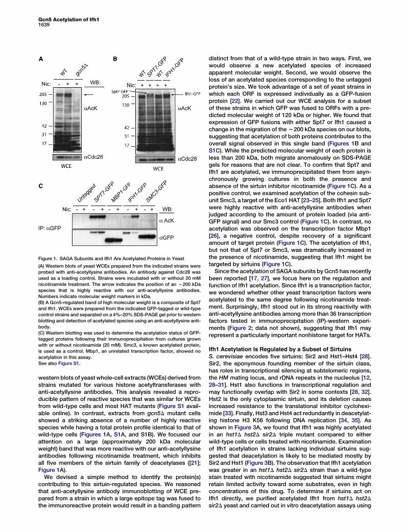
Figure 1. SAGA Subunits and Ifh1 Are Acetylated Proteins in Yeast
(A) Western blots of yeast WCEs prepared from the indicated strains were
probed with anti-acetyllysine antibodies. An antibody against Cdc28 was
used as a loading control. Strains were incubated with or without 20 mM
nicotinamide treatment. The arrow indicates the position of an w200 kDa
species that is highly reactive with our anti-acetyllysine antibodies.
Numbers indicate molecular weight markers in kDa.
(B) A Gcn5-regulated band of high molecular weight is a composite of Spt7
and Ifh1. WCEs were prepared from the indicated GFP-tagged or wild-type
control strains and separated on a 4%–20% SDS-PAGE gel prior to western
blotting and detection of acetylated species using an anti-acetyllysine anti-
body.
(C) Western blotting was used to determine the acetylation status of GFP-
tagged proteins following their immunoprecipitation from cultures grown
with or without nicotinamide (20 mM). Smc3, a known acetylated protein,
is used as a control. Mbp1, an unrelated transcription factor, showed no
acetylation in this assay.
See also Figure S1.
Gcn5 Acetylation of Ifh11639
western blots of yeastwhole-cell extracts (WCEs) derived fromstrains mutated for various histone acetyltransferases withanti-acetyllysine antibodies. This analysis revealed a repro-ducible pattern of reactive species that was similar for WCEsfrom wild-type cells and most HAT mutants (Figure S1 avail-able online). In contrast, extracts from gcn5D mutant cellsshowed a striking absence of a number of highly reactivespecies while having a total protein profile identical to that ofwild-type cells (Figures 1A, S1A, and S1B). We focused ourattention on a large (approximately 200 kDa molecularweight) band that was more reactive with our anti-acetyllysineantibodies following nicotinamide treatment, which inhibitsall five members of the sirtuin family of deacetylases ([21];Figure 1A).
We devised a simple method to identify the protein(s)contributing to this sirtuin-regulated species. We reasonedthat anti-acetyllysine antibody immunoblotting of WCE pre-pared from a strain in which a large epitope tag was fused tothe immunoreactive protein would result in a banding pattern
distinct from that of a wild-type strain in two ways. First, wewould observe a new acetylated species of increasedapparent molecular weight. Second, we would observe theloss of an acetylated species corresponding to the untaggedprotein’s size. We took advantage of a set of yeast strains inwhich each ORF is expressed individually as a GFP-fusionprotein [22]. We carried out our WCE analysis for a subsetof these strains in which GFP was fused to ORFs with a pre-dicted molecular weight of 120 kDa or higher. We found thatexpression of GFP fusions with either Spt7 or Ifh1 caused achange in the migration of thew200 kDa species on our blots,suggesting that acetylation of both proteins contributes to theoverall signal observed in this single band (Figures 1B andS1C). While the predicted molecular weight of each protein isless than 200 kDa, both migrate anomalously on SDS-PAGEgels for reasons that are not clear. To confirm that Spt7 andIfh1 are acetylated, we immunoprecipitated them from asyn-chronously growing cultures in both the presence andabsence of the sirtuin inhibitor nicotinamide (Figure 1C). As apositive control, we examined acetylation of the cohesin sub-unit Smc3, a target of the Eco1HAT [23–25]. Both Ifh1 and Spt7were highly reactive with anti-acetyllysine antibodies whenjudged according to the amount of protein loaded (via anti-GFP signal) and our Smc3 control (Figure 1C). In contrast, noacetylation was observed on the transcription factor Mbp1[26], a negative control, despite recovery of a significantamount of target protein (Figure 1C). The acetylation of Ifh1,but not that of Spt7 or Smc3, was dramatically increased inthe presence of nicotinamide, suggesting that Ifh1 might betargeted by sirtuins (Figure 1C).Since the acetylation of SAGA subunits byGcn5 has recently
been reported [17, 27], we focus here on the regulation andfunction of Ifh1 acetylation. Since Ifh1 is a transcription factor,we wondered whether other yeast transcription factors wereacetylated to the same degree following nicotinamide treat-ment. Surprisingly, Ifh1 stood out in its strong reactivity withanti-acetyllysine antibodies among more than 36 transcriptionfactors tested in immunoprecipitation (IP)-western experi-ments (Figure 2; data not shown), suggesting that Ifh1 mayrepresent a particularly important nonhistone target for HATs.
Ifh1 Acetylation Is Regulated by a Subset of Sirtuins
S. cerevisiae encodes five sirtuins: Sir2 and Hst1–Hst4 [28].Sir2, the eponymous founding member of the sirtuin class,has roles in transcriptional silencing at subtelomeric regions,the HM mating locus, and rDNA repeats in the nucleolus [12,28–31]. Hst1 also functions in transcriptional regulation andmay functionally overlap with Sir2 in some contexts [28, 32].Hst2 is the only cytoplasmic sirtuin, and its deletion causesincreased resistance to the translational inhibitor cyclohexi-mide [33]. Finally, Hst3 and Hst4 act redundantly in deacetylat-ing histone H3 K56 following DNA replication [34, 35]. Asshown in Figure 3A, we found that Ifh1 was highly acetylatedin an hst1D hst2D sir2D triple mutant compared to eitherwild-type cells or cells treated with nicotinamide. Examinationof Ifh1 acetylation in strains lacking individual sirtuins sug-gested that deacetylation is likely to be mediated mostly bySir2 and Hst1 (Figure 3B). The observation that Ifh1 acetylationwas greater in an hst1D hst2D sir2D strain than a wild-typestain treated with nicotinamide suggested that sirtuins mightretain limited activity toward some substrates, even in highconcentrations of this drug. To determine if sirtuins act onIfh1 directly, we purified acetylated Ifh1 from hst1D hst2Dsir2D yeast and carried out in vitro deacetylation assays using

Figure 2. Nicotinamide Does Not Induce Global Acetylation of Transcription
Factors
The indicated GFP-tagged transcription factors were immunopurified from
log-phase cultures treated with nicotinamide (20 mM) using an aGFP anti-
body, and reactivity of recovered proteins with anti-acetyllysine antibodies
was tested following separation on 4%–20% SDS-PAGE gradient gels and
western blotting. Even when Ifh1 levels are adjusted to that of the lowest
abundant protein recovered, its acetylation stood out among all candidates
tested, which showed only background reactivity with our antibodies. The
asterisk indicates immunoglobulin G bands from immunoprecipitations.
Current Biology Vol 23 No 171640
Sir2. Sir2 readily deacetylated Ifh1 in the presence of itscofactor nicotinamide adenine dinucleotide (NAD+), and thisdeacetylation was inhibited by inclusion of nicotinamide inthe reaction (Figure 3C). Together, our data suggest that Ifh1is acetylated in vivo and that its acetylations are reversed bya subset of sirtuins.
The Acetylation of Ifh1 Is Mediated by Gcn5
Esa1 has been previously implicated in the transcription of RPgenes through the regulation of histone H4 acetylation [13, 15].To test for a role of Esa1 in Ifh1 acetylation, we immunoprecip-itated Ifh1 from strains carrying the temperature-sensitiveesa1-414 allele [36] and examined acetylation of the recoveredprotein. While global H4 acetylation was lost when esa1-414strains were incubated at the restrictive temperature for 2 hr,Ifh1 acetylation remained largely unaffected (Figure S2A).These data suggest that Esa1 does not have a direct role inacetylating Ifh1 at RP promoters. We also found that the dele-tion of GCN4, which has been shown to inhibit Esa1 recruit-ment to RP promoters [16], had no impact on Ifh1 acetylation(Figure S2B).
Our initial screen suggested that Gcn5 may be required forIfh1 acetylation (Figure S1A). Indeed, Gcn5, with the SAGAcomplex, localizes to RP promoters [14, 17]. In contrast toprotein from wild-type cells, Ifh1 from cells lacking GCN5did not react with anti-acetyllysine antibodies (Figure 3D).Gcn5 was also required for the increased acetylation of Ifh1observed following treatment of cells with nicotinamide (Fig-ure 3E). Gcn5 purified from bacteria, but not Gcn5-E173Q,carrying a mutation in its catalytic domain [37], was able toacetylate Ifh1 in vitro (Figure 3F), suggesting that Gcn5 actson Ifh1 directly. In vivo, however, Ifh1 acetylation requiredthe SAGA structural component Spt7, suggesting that SAGArather than free Gcn5 mediates Ifh1 acetylation in cells(Figure 3G).
Ifh1 Acetylation Is Regulated by Nutrient Levels and
Temperature StressSince RP genes are regulated by stress and nutrient status [2],we examined whether Ifh1 acetylation is also regulated undersuch circumstances. We first tested the effect of rapamycin, aTOR inhibitor that results in a rapid downregulation of RP genetranscription [38]. As an independent measure of the efficacy
of drug treatment, we monitored the status of Gln3-GFPphosphorylation, which is dependent on TOR function and islost following rapamycin treatment [39]. Twenty minutesafter rapamycin treatment, Gln3 hyperphosphorylation wasdecreased relative to a control treatment (Figure 4A, inputs).The level of Ifh1 protein remained constant for both treatedand control cells for the course of the experiment (Figure 4A).However, while Ifh1 acetylation also remained constant incontrol cells, TOR inhibition immediately reduced this acetyla-tion (Figure 4A). We also measured Ifh1 acetylation during amild temperature shock, which is associated with a temporarydownregulation of RP mRNA levels [40]. While Ifh1 levelsremained constant during a heat shock from 23�C to 37�C,its acetylation rapidly decreased and remained low for10–20 min before eventually recovering by 60 min (Figure 4B).To determine if loss of Ifh1 acetylation after stress is due to theaction of sirtuins, we analyzed Ifh1 from cells treated simulta-neously with both rapamycin and nicotinamide. Nicotinamidehad no effect on the loss of TOR-dependent Gln3 phosphory-lation after rapamycin treatment (Figure 4C, inputs). However,nicotinamide treatment prevented the rapid deacetylation ofIfh1 observed in cells treatedwith rapamycin alone (Figure 4C),suggesting that sirtuins are required for deacetylation of Ifh1after stress.The interaction of Ifh1 with promoter-bound Fhl1 is thought
to be a critical step in regulating the transcription of RP genesand is assumed to be inhibited in growth conditions that allowfor only minimal RP transcription. Indeed, as reported previ-ously [9], we found that the interaction of Ifh1 with Fhl1 wasabolished following treatment with rapamycin (Figure 4D).Surprisingly, however, only limited reduction of the Fhl1-Ifh1interaction was observed upon transfer of cells either to richmedia lacking any source of carbon or to water (FiguresS2C). We also found that the amount of Ifh1 bound to Fhl1was virtually unchanged following the addition of glucose tocells growing in a poor carbon source (Figure 4E). However,in all cases, acetylation of Ifh1 was increased upon a returnto rich media (Figures 4E and S2C). These results suggestthat acetylation of promoter-bound Ifh1-Fhl1 molecules mayplay a role in regulating RP transcription during a recoveryfrom stress.Ifh1 is thought to be essential, due to both its importance in
activating RP transcription directly and its role in blockingFHL1 repression of RP genes, such that an fhl1D is viableand epistatic to ifh1D [41]. Rudra et al. previously reportedthat Ifh1 could still interact with Rap1 in an fhl1 mutant strain[42], despite the fact that Fhl1 seems to be required forlocalization of Ifh1 to RP promoters by chromatin immunopre-cipitation and for transcription from these promoters [9].Accordingly, we found that Ifh1 was still acetylated in anfhl1D strain, although this acetylation remained sensitive to ra-pamycin and nicotinamide treatments (Figures 4F and S2D).These data are consistent with a model wherein Ifh1 is looselybound at RP promoters in fhl1D strains in a manner that isnonpermissive for DNA crosslinking while remaining amend-able to acetylation by SAGA. They further suggest that addi-tional protein contacts are required for Ifh1 recruitment andfunction at RP promoters.
Ifh1 Is Acetylated at Multiple Lysine Residues in an
N-Terminal DomainTo identify the sites of Ifh1 acetylation in vivo, we purified flag-tagged Ifh1 from sir2D hst1D hst2D cells (Figure 5A, top panel)and identified acetylated lysine residues in peptides generated

Figure 3. Regulation of Ifh1 Acetylation
(A) Flag-tagged Ifh1 was immunoprecipitated from asynchronously growing cultures of the indicated genotypes and analyzed with an antibody directed
toward acetylated lysine or the flag epitope. Two isolates of the sirtuin triple mutant are shown.
(B) Flag-tagged Ifh1 was immunoprecipitated from asynchronously growing cultures of the indicated genotypes and analyzed as in (A).
(C) In vitro deacetylation of Ifh1. Partially purified GST-Sir2 was incubated with acetylated Ifh1 purified from sir2D hst1D hst2D cells under the reaction con-
ditions indicated for 1 hr at 30�C. Reactionswere stoppedwith the addition of 33SDS-PAGE sample buffer and separated on a 4%–20%gradient gel prior to
western blotting and detection of acetyllysine using an anti-acetyllysine antibody. Nic, inclusion of 5 mM nicotinamide in the reaction as a sirtuin inhibitor.
(D) Gcn5 regulation of Ifh1 acetylation was examined using an IP-western protocol. The abundant Yap1 transcription factor was used to gauge general
cross-reactivity of the aAcK antibody.
(E) Gcn5 is required for the increased acetylation observed in nicotinamide-treated samples. Samples were processed as in (D), except that 33 more IPed
material was loaded from slow-growing gcn5D strains relative to wild-type control strains to properly judge the level of acetylation.
(F) Gcn5 acetylation of Ifh1 in vitro. Bacterially purified Gcn5 or catalytic-deadmutant Gcn5 (Gcn5 E173Q) was incubatedwith Ifh1 purified from gcn5D yeast
under the reaction conditions specified for 1 hr at 30�C. Reactions were stopped with the addition of 33 SDS-PAGE loading buffer and were separated on a
4%–20% gradient gel prior to western blotting and detection of reaction products with the indicated antibodies.
(G)Acetylation levelsof Ifh1 IPed fromwild-typeor spt7Dstrainswerecomparedusingananti-acetyllysineantibody followingSDS-PAGEandwesternblotting.
See also Figure S2.
Gcn5 Acetylation of Ifh11641
by trypsin or chymotrypsin cleavage (see the SupplementalInformation). We identified seven acetylation sites of mediumor high confidence as judged by spectra quality and peptideabundance (Figure 5A, bottom). Intriguingly, all seven of thesesites map to an acidic region in the N-terminal half of Ifh1 (Fig-ure 5B). We tested the relative contribution of these sites to
overall Ifh1 acetylation by expressing mutant versions of theseproteins, in which identified sites are mutated to arginine,which maintains the charge of a lysine residue but cannot beacetylated, or to glutamine, which structurally mimics anacetylated lysine residue. We found that mutation of lysines180 and 254 to arginine severely diminished the level of Ifh1
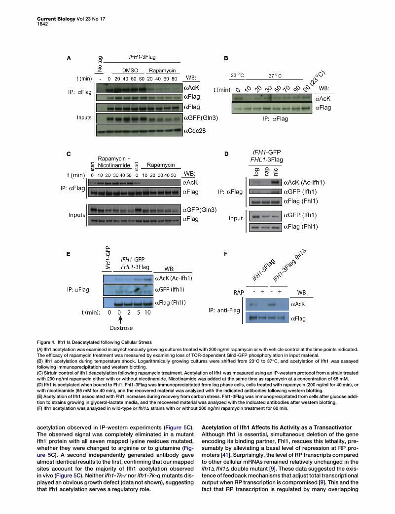
Figure 4. Ifh1 Is Deacetylated following Cellular Stress
(A) Ifh1 acetylation was examined in asynchronously growing cultures treated with 200 ng/ml rapamycin or with vehicle control at the time points indicated.
The efficacy of rapamycin treatment was measured by examining loss of TOR-dependent Gln3-GFP phosphorylation in input material.
(B) Ifh1 acetylation during temperature shock. Logarithmically growing cultures were shifted from 23�C to 37�C, and acetylation of Ifh1 was assayed
following immunoprecipitation and western blotting.
(C) Sirtuin control of Ifh1 deacetylation following rapamycin treatment. Acetylation of Ifh1 was measured using an IP-western protocol from a strain treated
with 200 ng/ml rapamycin either with or without nicotinamide. Nicotinamide was added at the same time as rapamycin at a concentration of 65 mM.
(D) Ifh1 is acetylated when bound to Fhl1. Fhl1-3Flag was immunoprecipitated from log phase cells, cells treated with rapamycin (200 ng/ml for 40 min), or
with nicotinamide (65 mM for 40 min), and the recovered material was analyzed with the indicated antibodies following western blotting.
(E) Acetylation of Ifh1 associated with Fhl1 increases during recovery from carbon stress. Fhl1-3Flag was immunoprecipitated from cells after glucose addi-
tion to strains growing in glycerol-lactate media, and the recovered material was analyzed with the indicated antibodies after western blotting.
(F) Ifh1 acetylation was analyzed in wild-type or fhl1D strains with or without 200 ng/ml rapamycin treatment for 60 min.
Current Biology Vol 23 No 171642
acetylation observed in IP-western experiments (Figure 5C).The observed signal was completely eliminated in a mutantIfh1 protein with all seven mapped lysine residues mutated,whether they were changed to arginine or to glutamine (Fig-ure 5C). A second independently generated antibody gavealmost identical results to the first, confirming that ourmappedsites account for the majority of Ifh1 acetylation observedin vivo (Figure 5C). Neither ifh1-7k-r nor ifh1-7k-qmutants dis-played an obvious growth defect (data not shown), suggestingthat Ifh1 acetylation serves a regulatory role.
Acetylation of Ifh1 Affects Its Activity as a TransactivatorAlthough Ifh1 is essential, simultaneous deletion of the geneencoding its binding partner, Fhl1, rescues this lethality, pre-sumably by alleviating a basal level of repression at RP pro-moters [41]. Surprisingly, the level of RP transcripts comparedto other cellular mRNAs remained relatively unchanged in theifh1D fhl1D double mutant [9]. These data suggested the exis-tence of feedbackmechanisms that adjust total transcriptionaloutput when RP transcription is compromised [9]. This and thefact that RP transcription is regulated by many overlapping
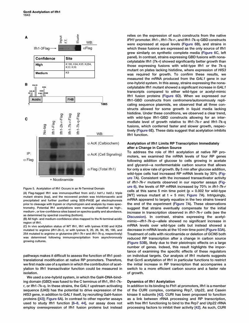
Figure 5. Acetylation of Ifh1 Occurs in an N-Terminal Domain
(A) Flag-tagged Ifh1 was immunopurified from sir2D hst1D hst2D triple
mutant strains (top), and the recovered protein was trichloroacetic acid
precipitated and further purified using SDS-PAGE gel electrophoresis
prior to cleavage with trypsin or chymotrypsin and analysis by mass spec-
trometry. Potential Ifh1 acetylations were manually classified as high-,
medium-, or low-confidence sites based on spectra quality and abundance,
as determined by spectral counting (bottom).
(B) All high- and medium-confidence sites mapped to the N-terminal acidic
region of Ifh1.
(C) In vivo acetylation status of WT Ifh1, Ifh1 with lysines K180 and K254
mutated to arginine (ifh1-2k-r), or with lysines 9, 20, 26, 94, 95, 180, and
254 mutated to arginine or glutamine (ifh1-7k-r and ifh1-7k-q, respectively)
was determined following immunoprecipitation from asynchronously
growing cultures.
Gcn5 Acetylation of Ifh11643
pathways makes it difficult to assess the function of Ifh1 post-translational modification at native RP promoters. Therefore,we firstmade use of a system inwhich the contribution of acet-ylation to Ifh1 transactivator function could be measured inisolation.
We used a one-hybrid system, in which the Gal4-DNA-bind-ing domain (GBD) was fused to the C terminus of Ifh1, Ifh1-7k-r, or Ifh1-7k-q. In these strains, the GAL1 upstream activatingsequence (UAS) has the potential to drive expression of theHIS3 gene, in addition to GAL1 itself, by recruiting Gal4-fusionproteins ([43]; Figure 6A). In contrast to other reporter assaysused to study Ifh1 function [6–8, 44], our assay does notemploy overexpression of Ifh1 fusion proteins but instead
relies on the expression of such constructs from the nativeIFH1 promoter. Ifh1-, Ifh1-7k-r-, and Ifh1-7k-q-GBD constructswere expressed at equal levels (Figure 6B), and strains inwhich these fusions are expressed as the only source of Ifh1grew similarly on synthetic complete media (Figure 6C, leftpanel). In contrast, strains expressing GBD fusions with nona-cetylatable Ifh1 (7k-r) showed significantly better growth thanthose expressing fusions with wild-type Ifh1 or the 7k-qmutant on plates lacking histidine, where expression of HIS3was required for growth. To confirm these results, wemeasured the mRNA produced from the GAL1 gene in ourone-hybrid system. In this assay, strains expressing the nona-cetylatable Ifh1 mutant showed a significant increase in GAL1transcripts compared to either wild-type or acetyl-mimicIfh1 fusion proteins (Figure 6D). When we expressed ourIfh1-GBD constructs from centromere/autonomously repli-cating sequence plasmids, we observed that all three con-structs allowed for some growth in liquid media lackinghistidine. Under these conditions, we observed a clear trend,with wild-type Ifh1-GBD constructs allowing for an inter-mediate level of growth relative to Ifh1-7k-r and Ifh1-7k-qfusions, which conferred faster and slower growth, respec-tively (Figure 6E). These data suggest that acetylation inhibitsIfh1 function.
Acetylation of Ifh1 Limits RP Transcription Immediatelyafter a Change in Carbon Source
To address the role of Ifh1 acetylation at native RP pro-moters, we examined the mRNA levels of four RP genesfollowing addition of glucose to cells growing in acetateand glycerol—a nonfermentable carbon source that allowsfor only a slow rate of growth. By 5 min after glucose addition,wild-type cells had increased RP mRNA levels by 30% (Fig-ure 7A). Consistent with the increased transactivator activityof ifh1-7k-r mutants observed in our reporter assays (Fig-ure 6), the levels of RP mRNA increased by 70% in ifh1-7k-rcells at this same 5 min time point (p = 0.002 for wild-type[WT] versus mutant at t = 5 min; Figure 7A). Intriguingly,mRNA appeared to largely equalize in the two strains towardthe end of the experiment (Figure 7A). These observationssuggest that strains eventually compensate for the initialincrease in transcription observed in ifh1-7k-r cells (see theDiscussion). In contrast, strains expressing the acetyl-mimic—ifh1-7k-q—allele showed no significant increase inmRNA levels over wild-type cells but instead showed adecrease in mRNA levels at the 10 min time point (Figure S3A).Treatment of cells with nicotinamide or deletion of GCN5 bothreduced RP transcription after a change in carbon source(Figure S3B), likely due to their pleiotropic effects on a largenumber of genes. Indeed, this result highlights the impor-tance of examining the specific effects of these regulatorson individual targets. Our analysis of Ifh1 mutants suggeststhat Gcn5 acetylation of Ifh1 in particular functions to restrictthe initial increase in RP transcription that accompanies aswitch to a more efficient carbon source and a faster rateof growth.
Dynamics of Ifh1 Acetylation
In addition to its binding to Fhl1 at promoters, Ifh1 is amemberof the CURI complex, containing Rrp7, Utp22, and Caseinkinase II subunits [42]. CURI has been proposed to functionas a link between rRNA processing and RP transcription,with free Ifh1 functioning to bind to the Rrp7 and Utp22 rRNAprocessing factors to inhibit their activity [42]. As such, CURI

Figure 6. Ifh1 Acetylation Inhibits Its Transactivator Activity
(A) Experimental design. Ifh1 or the indicatedmutants were expressed under the Ifh1 promoter as fusion proteins with the GBD in a strain in which theGAL1
UAS drives transcription of the HIS3 gene in addition to endogenous GAL genes.
(B) Expression of Ifh1 fusion proteins in the strains described in (A). Myc-tagged GBD fusions are the only source of Ifh1 in these strains.
(C) Five-fold serial dilutions of strains expressing the indicated Ifh1 fusions on completemedia or completemedia lacking histidine (C-histidine). Plates were
imaged after 2 days’ growth at 30�C.(D) GAL1 mRNA level (versus ACT1 control) in strains expressing the Ifh1 fusion proteins described in (A), as determined by quantitative PCR (qPCR) anal-
ysis. Error bars represent the SEM. The asterisk indicates significantly different fromWT (p < 0.03), with p values calculated using a two-tailed Student’s t test
(n = 4 for each strain type indicated).
(E) The indicated strains were grown to midlog phase in minimal media lacking tryptophan before being washed in water and transferred to media lacking
both tryptophan and histidine. Growth, which requires expression of the HIS3 gene, was assayed using OD 600 readings at the indicated time points. n = 3,
with error bars indicating the SEM.
Current Biology Vol 23 No 171644
is thought to provide a mechanism through which the cell cantitrate both rRNA and RP production by regulating Ifh1 avail-ability. We wondered whether CURI might play a role in theacetylation-deacetylation cycle of Ifh1. We found that a signif-icant fraction of the total acetylated Ifh1 in the cell is containedwithin the CURI complex (Figure 7B). The acetylation of CURI-bound Ifh1 was increased by nicotinamide treatment andeliminated with rapamycin, and these effects could not be
explained by changes in Ifh1 binding to CURI (Figure 7B).These data suggest the possibility that Ifh1 acetylated at thepromoter may subsequently accumulate in the CURI complexfor some period of time after its release. Interestingly, Ifh1expressed from the strong TEF promoter did not result in anincrease in acetylated Ifh1, suggesting that overexpressionresults in a large free pool of Ifh1 that is not targeted bySAGA (Figure S3C).
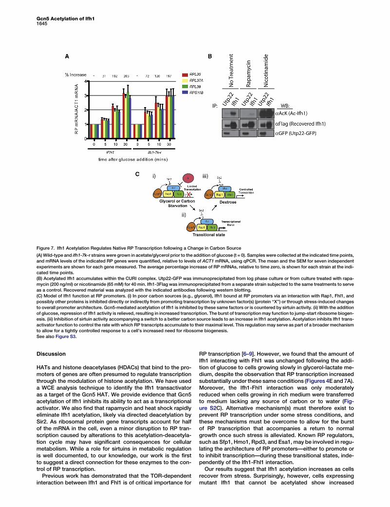
Figure 7. Ifh1 Acetylation Regulates Native RP Transcription following a Change in Carbon Source
(A)Wild-type and ifh1-7k-r strains were grown in acetate/glycerol prior to the addition of glucose (t = 0). Samples were collected at the indicated time points,
and mRNA levels of the indicated RP genes were quantified, relative to levels of ACT1 mRNA, using qPCR. The mean and the SEM for seven independent
experiments are shown for each gene measured. The average percentage increase of RP mRNAs, relative to time zero, is shown for each strain at the indi-
cated time points.
(B) Acetylated Ifh1 accumulates within the CURI complex. Utp22-GFP was immunoprecipitated from log phase culture or from culture treated with rapa-
mycin (200 ng/ml) or nicotinamide (65 mM) for 40 min. Ifh1-3Flag was immunoprecipitated from a separate strain subjected to the same treatments to serve
as a control. Recovered material was analyzed with the indicated antibodies following western blotting.
(C) Model of Ifh1 function at RP promoters. (i) In poor carbon sources (e.g., glycerol), Ifh1 bound at RP promoters via an interaction with Rap1, Fhl1, and
possibly other proteins is inhibited directly or indirectly from promoting transcription by unknown factor(s) (protein ‘‘X’’) or through stress-induced changes
to overall promoter architecture. Gcn5-mediated acetylation of Ifh1 is inhibited by these same factors or is countered by sirtuin activity. (ii) With the addition
of glucose, repression of Ifh1 activity is relieved, resulting in increased transcription. The burst of transcription may function to jump-start ribosome biogen-
esis. (iii) Inhibition of sirtuin activity accompanying a switch to a better carbon source leads to an increase in Ifh1 acetylation. Acetylation inhibits Ifh1 trans-
activator function to control the rate with which RP transcripts accumulate to their maximal level. This regulation may serve as part of a broader mechanism
to allow for a tightly controlled response to a cell’s increased need for ribosome biogenesis.
See also Figure S3.
Gcn5 Acetylation of Ifh11645
Discussion
HATs and histone deacetylases (HDACs) that bind to the pro-moters of genes are often presumed to regulate transcriptionthrough the modulation of histone acetylation. We have useda WCE analysis technique to identify the Ifh1 transactivatoras a target of the Gcn5 HAT. We provide evidence that Gcn5acetylation of Ifh1 inhibits its ability to act as a transcriptionalactivator. We also find that rapamycin and heat shock rapidlyeliminate Ifh1 acetylation, likely via directed deacetylation bySir2. As ribosomal protein gene transcripts account for halfof the mRNA in the cell, even a minor disruption to RP tran-scription caused by alterations to this acetylation-deacetyla-tion cycle may have significant consequences for cellularmetabolism. While a role for sirtuins in metabolic regulationis well documented, to our knowledge, our work is the firstto suggest a direct connection for these enzymes to the con-trol of RP transcription.
Previous work has demonstrated that the TOR-dependentinteraction between Ifh1 and Fhl1 is of critical importance for
RP transcription [6–9]. However, we found that the amount ofIfh1 interacting with Fhl1 was unchanged following the addi-tion of glucose to cells growing slowly in glycerol-lactate me-dium, despite the observation that RP transcription increasedsubstantially under these same conditions (Figures 4E and 7A).Moreover, the Ifh1-Fhl1 interaction was only moderatelyreduced when cells growing in rich medium were transferredto medium lacking any source of carbon or to water (Fig-ure S2C). Alternative mechanism(s) must therefore exist toprevent RP transcription under some stress conditions, andthese mechanisms must be overcome to allow for the burstof RP transcription that accompanies a return to normalgrowth once such stress is alleviated. Known RP regulators,such as Sfp1, Hmo1, Rpd3, and Esa1, may be involved in regu-lating the architecture of RP promoters—either to promote orto inhibit transcription—during these transitional states, inde-pendently of the Ifh1-Fhl1 interaction.Our results suggest that Ifh1 acetylation increases as cells
recover from stress. Surprisingly, however, cells expressingmutant Ifh1 that cannot be acetylated show increased
Current Biology Vol 23 No 171646
mRNA production during recovery from carbon starvation,suggesting an inhibitory role for Ifh1 acetylation. We proposea model wherein promoter-bound and hypoacetylated Ifh1could act as a strong transactivator to provide an initial burstof RP transcription following recovery from stress or starva-tion (Figure 7C). This rapid increase to transcriptional outputcould jump-start ribosome biogenesis in the first minutesfollowing nutrient addition. Our data suggest that SAGA-mediated acetylation of Ifh1 may normally function to limitthe strength of this burst of RP transcription. We found thatRP mRNA levels increased 70% 5 min after glucose additionin the ifh1-7k-r mutant, as opposed to 30% in wild-type cells(Figure 7A). Given that RP mRNAs account for up to half ofall RNA pol II-derived messages in the cell [5], this differenceamounts to a very large increase in total cellular mRNAtranscripts.
Despite a greater increase in RP transcription in ifh1-7k-rmutants relative to wild-type controls early during carbon-shiftexperiments, the levels of RP mRNAs were equalized in wild-type and mutant ifh1 strains after 30 min (Figure 7A). Thisequalization may involve a negative feedback mechanism trig-gered by the initial burst of RP transcription and may functionby affecting RPpromoter architecture. Controlling the strengthand timing of the initial response to the addition of nutrientsmay help cells to coordinate RP transcription with rRNA pro-cessing and other growth-related processes.
How acetylation of Ifh1’s acidic domain might inhibit itstransactivator function is unclear. The transactivator do-main(s) of Ifh1 have been proposed to reside in the C terminusof the protein, although it is not obvious how these domainsfunction to promote RNA polymerase II recruitment and/oractivity [44]. Paradoxically, it has both been reported thatdeletion of the N terminus of Ifh1 can dramatically increase[44] or slightly decrease [6] transactivator activity of Ifh1 inone-hybrid assays. The difference between these assaysappears to be whether Ifh1 was recruited to reporter genesdirectly or indirectly via a GBD fusion with the Fhl1 FHAdomain. Moreover, a third study found that full-length Fhl1fused to a DNA-binding domain was incapable of stimulatingtranscription, even though this construct promoted Ifh1recruitment [10]. These data suggest that Fhl1 may have aregulatory role in RP transcription, in addition to its role ininitial Ifh1 recruitment. Acetylation in Ifh1’s acidic domainmay regulate this function. Acetylation may also alter Ifh1’sinteraction with other promoter-bound proteins, such asRap1. In this context, it is noteworthy that overexpression ofa construct containing an N-terminal Ifh1 fragment disruptsSir2-mediated telomere silencing, which also requires theRap1 protein [45].
Ifh1 within the CURI complex and Ifh1 bound to Fhl1 at RPpromoters are deacetylated after stress, and this deacetyla-tion requires the action of sirtuins. While Sir2 and Hst2 havebeen previously implicated in the deacetylation of two nonhis-tone proteins, Pck1 [19] and Snf2 [46], respectively, Ifh1 is, toour knowledge, the first such protein known to be regulatedredundantly by multiple yeast sirtuins. Purified Hst1 was notactive against acetylated Ifh1 in our in vitro reactions, althoughalso it appears to act as a poor enzyme in vitro on histone sub-strates [47]. Hst1-mediated Ifh1 deacetylation may be facili-tated in vivo by additional factors, or Hst1 may regulate Ifh1acetylation indirectly.
During carbon starvation, most Ifh1 localizes to the nucle-olus [3], where Sir2 and Hst1 have been shown to function[28]. Since the bulk of Ifh1 appears to be bound within the
CURI complex [42], it is tempting to speculate that relocaliza-tion of CURI to the nucleolus may facilitate Ifh1 deacetylation.The accumulation of hypoacetylated Ifh1 at RP promoters inparticular may also be facilitated by the inhibition of Gcn5 ac-tivity toward Ifh1. Although significant changes in the recruit-ment of the SAGA complex to RP promoters have not beendescribed for logarithmically growing cells, even followingstresses, such as heat shock [18], the inhibition of Gcn5’saction on Ifh1 could result from the same mechanisms thatprevent transcription in the presence of Fhl1-bound Ifh1 mole-cules. Deacetylation of Ifh1 may reset its ability to act as astrong activator once starvation or stress conditions arerelieved.
Experimental Procedures
Details regarding specific experiments are contained in the Supplemental
Information.
Yeast Strains, Plasmids, and Growth Conditions
Yeast strains and plasmids were generated using standard techniques and
are described in Tables S1 and S2 and the Supplemental Experimental
Procedures.
Immunoprecipitations
Cells were lysed using a bead-beating protocol, and WCEs were clarified
via centrifugation at 4�C (see the Supplemental Experimental Procedures
for specific conditions). Immunoprecipitations were carried out in volumes
of 500 ml with 0.5 ml of AB290 anti-GFP antibody for 2 hr. Proteins were
then recovered with 20 ml Protein A beads (Dynabeads, Invitrogen) for
40 min. For anti-Flag purifications, magnetic anti-Flag M2 beads (Sigma)
were used for 2 hr. Beads were washed with lysis buffer three times and
protein complexes were eluted in 60 ml SDS-PAGE sample buffer with
0.1 M dithiothreitol (DTT) at 65�C for 10 min. Eluates were boiled prior to
SDS-PAGE.
HAT Assays
HAT assays were carried out in a final volume of 50 ml with 3 ml of 6His-TRX-
Gcn5 or 6His-TRX-Gcn5-E173Q (approximately 3 mg), 800 mM acetyl coen-
zyme A, and 25 ml 2X HAT buffer (100 mM NaCl, 10% glycerol, 100 mM
Tris-HCl pH 8.0, and 0.2 mM EDTA supplemented with 2 mM sodium buty-
rate and 2 mM DTT). Ifh1-3flag protein was purified from gcn5D yeast.
Reactions were carried out at 30�C for 1 hr, stopped with the addition of
33 SDS-PAGE sample buffer containing 0.1M DTT, and boiled prior to
SDS-PAGE.
HDAC Assays
Ifh1 purified from hst1D hst2D sir2D cells was used in a final volume of 25 ml
with 5 ml 5X HDAC reaction buffer (250mMTris HCl, pH 8.0, 2.5mMDTT, one
Roche Protease inhibitor tablet without EDTA per 10 ml), 10 ml GST-Sir2
(approximately 0.5 mg total), and 100 mM NAD. Nicotinamide was used at
a final concentration of 5 mM. Reactions were incubated for 1 hr at 30�C.Reactions were stopped with the addition of SDS-PAGE sample buffer
with 0.1 M DTT and boiled to remove Ifh1 from beads.
Supplemental Information
Supplemental Information includes Supplemental Experimental Proce-
dures, three figures, and two tables and can be found with this article online
at http://dx.doi.org/10.1016/j.cub.2013.06.050.
Acknowledgments
WethankStanFieldsandJoachimLi for strains andSongTan, TonyBedalov,
andBrenda Andrews for plasmids. This work was supported by NIHGeneral
Medical Sciences grants to D.P.T. (GM059691) and J.A.W. (GM89778). D.P.T
is an Ellison Senior Scholar in aging. J.A.W. is supported with funds from
Jonsson Cancer Center at UCLA. D.S. would like to acknowledge support
from the Swiss National Fund (grant 31003A_143457 and theNCCRprogram
‘‘Frontiers in Genetics’’) and from the Canton and Republic of Geneva. M.D.
was supported by a Human Frontiers Long-term Postdoctoral Fellowship

Gcn5 Acetylation of Ifh11647
and currently by the UCSF Program for Breakthrough Biomedical Research,
which is funded in part by the Sandler Foundation.
Received: January 29, 2013
Revised: May 31, 2013
Accepted: June 19, 2013
Published: August 22, 2013
References
1. Jorgensen, P., and Tyers, M. (2004). How cells coordinate growth and
division. Curr. Biol. 14, R1014–R1027.
2. Lempiainen, H., and Shore, D. (2009). Growth control and ribosome
biogenesis. Curr. Opin. Cell Biol. 21, 855–863.
3. Jorgensen, P., Rupes, I., Sharom, J.R., Schneper, L., Broach, J.R., and
Tyers, M. (2004). A dynamic transcriptional network communicates
growth potential to ribosome synthesis and critical cell size. Genes
Dev. 18, 2491–2505.
4. Singh, J., and Tyers, M. (2009). A Rab escort protein integrates the
secretion system with TOR signaling and ribosome biogenesis. Genes
Dev. 23, 1944–1958.
5. Warner, J.R. (1999). The economics of ribosome biosynthesis in yeast.
Trends Biochem. Sci. 24, 437–440.
6. Martin, D.E., Soulard, A., and Hall, M.N. (2004). TOR regulates ribosomal
protein gene expression via PKA and the Forkhead transcription factor
FHL1. Cell 119, 969–979.
7. Schawalder, S.B., Kabani, M., Howald, I., Choudhury, U., Werner, M.,
and Shore, D. (2004). Growth-regulated recruitment of the essential
yeast ribosomal protein gene activator Ifh1. Nature 432, 1058–1061.
8. Wade, J.T.,Hall, D.B., andStruhl, K. (2004). The transcription factor Ifh1 is
a key regulator of yeast ribosomal protein genes. Nature 432, 1054–1058.
9. Rudra, D., Zhao, Y., and Warner, J.R. (2005). Central role of Ifh1p-Fhl1p
interaction in the synthesis of yeast ribosomal proteins. EMBO J. 24,
533–542.
10. Zhao, Y., McIntosh, K.B., Rudra, D., Schawalder, S., Shore, D., and
Warner, J.R. (2006). Fine-structure analysis of ribosomal protein gene
transcription. Mol. Cell. Biol. 26, 4853–4862.
11. Berger, A.B., Decourty, L., Badis, G., Nehrbass, U., Jacquier, A., and
Gadal, O. (2007). Hmo1 is required for TOR-dependent regulation of
ribosomal protein gene transcription. Mol. Cell. Biol. 27, 8015–8026.
12. Shahbazian, M.D., and Grunstein, M. (2007). Functions of site-specific
histone acetylation and deacetylation. Annu. Rev. Biochem. 76, 75–100.
13. Reid, J.L., Iyer, V.R., Brown, P.O., and Struhl, K. (2000). Coordinate regu-
lation of yeast ribosomal protein genes is associated with targeted
recruitment of Esa1 histone acetylase. Mol. Cell 6, 1297–1307.
14. Robert, F., Pokholok, D.K., Hannett, N.M., Rinaldi, N.J., Chandy, M.,
Rolfe, A., Workman, J.L., Gifford, D.K., and Young, R.A. (2004). Global
position and recruitment of HATs and HDACs in the yeast genome.
Mol. Cell 16, 199–209.
15. Rohde, J.R., and Cardenas, M.E. (2003). The tor pathway regulates gene
expression by linking nutrient sensing to histone acetylation. Mol. Cell.
Biol. 23, 629–635.
16. Joo, Y.J., Kim, J.H., Kang, U.B., Yu, M.H., and Kim, J. (2011). Gcn4p-
mediated transcriptional repression of ribosomal protein genes under
amino-acid starvation. EMBO J. 30, 859–872.
17. Cai, L., Sutter, B.M., Li, B., and Tu, B.P. (2011). Acetyl-CoA induces cell
growth and proliferation by promoting the acetylation of histones at
growth genes. Mol. Cell 42, 426–437.
18. Ghosh, S., and Pugh, B.F. (2011). Sequential recruitment of SAGA and
TFIID in a genomic response to DNA damage in Saccharomyces cerevi-
siae. Mol. Cell. Biol. 31, 190–202.
19. Lin, Y.Y., Lu, J.Y., Zhang, J., Walter, W., Dang, W., Wan, J., Tao, S.C.,
Qian, J., Zhao, Y., Boeke, J.D., et al. (2009). Protein acetylation microar-
ray reveals that NuA4 controls key metabolic target regulating gluco-
neogenesis. Cell 136, 1073–1084.
20. Kaluarachchi Duffy, S., Friesen, H., Baryshnikova, A., Lambert, J.P.,
Chong, Y.T., Figeys, D., and Andrews, B. (2012). Exploring the yeast
acetylome using functional genomics. Cell 149, 936–948.
21. Starai, V.J., Takahashi, H., Boeke, J.D., and Escalante-Semerena, J.C.
(2003). Short-chain fatty acid activation by acyl-coenzyme A synthe-
tases requires SIR2 protein function in Salmonella enterica and
Saccharomyces cerevisiae. Genetics 163, 545–555.
22. Huh, W.K., Falvo, J.V., Gerke, L.C., Carroll, A.S., Howson, R.W.,
Weissman, J.S., and O’Shea, E.K. (2003). Global analysis of protein
localization in budding yeast. Nature 425, 686–691.
23. Rolef Ben-Shahar, T., Heeger, S., Lehane, C., East, P., Flynn, H., Skehel,
M., and Uhlmann, F. (2008). Eco1-dependent cohesin acetylation during
establishment of sister chromatid cohesion. Science 321, 563–566.
24. Unal, E., Heidinger-Pauli, J.M., Kim, W., Guacci, V., Onn, I., Gygi, S.P.,
and Koshland, D.E. (2008). A molecular determinant for the establish-
ment of sister chromatid cohesion. Science 321, 566–569.
25. Zhang, J., Shi, X., Li, Y., Kim, B.J., Jia, J., Huang, Z., Yang, T., Fu, X.,
Jung, S.Y., Wang, Y., et al. (2008). Acetylation of Smc3 by Eco1 is
required for S phase sister chromatid cohesion in both human and
yeast. Mol. Cell 31, 143–151.
26. Koch, C., Moll, T., Neuberg,M., Ahorn, H., and Nasmyth, K. (1993). A role
for the transcription factors Mbp1 and Swi4 in progression from G1 to S
phase. Science 261, 1551–1557.
27. Mischerikow, N., Spedale, G., Altelaar, A.F., Timmers, H.T., Pijnappel,
W.W., and Heck, A.J. (2009). In-depth profiling of post-translational
modifications on the related transcription factor complexes TFIID and
SAGA. J. Proteome Res. 8, 5020–5030.
28. Brachmann, C.B., Sherman, J.M., Devine, S.E., Cameron, E.E., Pillus, L.,
and Boeke, J.D. (1995). The SIR2 gene family, conserved from bacteria
to humans, functions in silencing, cell cycle progression, and chromo-
some stability. Genes Dev. 9, 2888–2902.
29. Pillus, L., and Rine, J. (1989). Epigenetic inheritance of transcriptional
states in S. cerevisiae. Cell 59, 637–647.
30. Rine, J., and Herskowitz, I. (1987). Four genes responsible for a position
effect on expression from HML and HMR in Saccharomyces cerevisiae.
Genetics 116, 9–22.
31. Shogren-Knaak, M., and Peterson, C.L. (2006). Switching on chromatin:
mechanistic role of histone H4-K16 acetylation. Cell Cycle 5, 1361–1365.
32. Hickman, M.A., and Rusche, L.N. (2007). Substitution as a mechanism
for genetic robustness: the duplicated deacetylases Hst1p and Sir2p
in Saccharomyces cerevisiae. PLoS Genet. 3, e126.
33. Wilson, J.M., Le, V.Q., Zimmerman, C., Marmorstein, R., and Pillus, L.
(2006). Nuclear export modulates the cytoplasmic Sir2 homologue
Hst2. EMBO Rep. 7, 1247–1251.
34. Maas, N.L., Miller, K.M., DeFazio, L.G., and Toczyski, D.P. (2006). Cell
cycle and checkpoint regulation of histone H3 K56 acetylation by Hst3
and Hst4. Mol. Cell 23, 109–119.
35. Celic, I., Masumoto, H., Griffith, W.P., Meluh, P., Cotter, R.J., Boeke,
J.D., and Verreault, A. (2006). The sirtuins hst3 and Hst4p preserve
genome integrity by controlling histone h3 lysine 56 deacetylation.
Curr. Biol. 16, 1280–1289.
36. Clarke, A.S., Lowell, J.E., Jacobson, S.J., and Pillus, L. (1999). Esa1p is
an essential histone acetyltransferase required for cell cycle progres-
sion. Mol. Cell. Biol. 19, 2515–2526.
37. Wang, L., Liu, L., and Berger, S.L. (1998). Critical residues for histone
acetylation by Gcn5, functioning in Ada and SAGA complexes,
are also required for transcriptional function in vivo. Genes Dev. 12,
640–653.
38. Hardwick, J.S., Kuruvilla, F.G., Tong, J.K., Shamji, A.F., and Schreiber,
S.L. (1999). Rapamycin-modulated transcription defines the subset of
nutrient-sensitive signaling pathways directly controlled by the Tor
proteins. Proc. Natl. Acad. Sci. USA 96, 14866–14870.
39. Beck, T., and Hall, M.N. (1999). The TOR signalling pathway controls
nuclear localization of nutrient-regulated transcription factors. Nature
402, 689–692.
40. Herruer, M.H., Mager, W.H., Raue, H.A., Vreken, P., Wilms, E., and
Planta, R.J. (1988). Mild temperature shock affects transcription of
yeast ribosomal protein genes as well as the stability of their mRNAs.
Nucleic Acids Res. 16, 7917–7929.
41. Cherel, I., and Thuriaux, P. (1995). The IFH1 gene product interacts with
a fork head protein in Saccharomyces cerevisiae. Yeast 11, 261–270.
42. Rudra, D., Mallick, J., Zhao, Y., and Warner, J.R. (2007). Potential inter-
face between ribosomal protein production and pre-rRNA processing.
Mol. Cell. Biol. 27, 4815–4824.
43. James, P., Halladay, J., and Craig, E.A. (1996). Genomic libraries and a
host strain designed for highly efficient two-hybrid selection in yeast.
Genetics 144, 1425–1436.
44. Zhong, P., and Melcher, K. (2010). Identification and characterization of
the activation domain of Ifh1, an activator of model TATA-less genes.
Biochem. Biophys. Res. Commun. 392, 77–82.

Current Biology Vol 23 No 171648
45. Singer, M.S., Kahana, A., Wolf, A.J., Meisinger, L.L., Peterson, S.E.,
Goggin, C., Mahowald, M., and Gottschling, D.E. (1998). Identification
of high-copy disruptors of telomeric silencing in Saccharomyces cere-
visiae. Genetics 150, 613–632.
46. Kim, J.H., Saraf, A., Florens, L., Washburn, M., and Workman, J.L.
(2010). Gcn5 regulates the dissociation of SWI/SNF from chromatin by
acetylation of Swi2/Snf2. Genes Dev. 24, 2766–2771.
47. Sutton, A., Heller, R.C., Landry, J., Choy, J.S., Sirko, A., and Sternglanz,
R. (2001). A novel form of transcriptional silencing by Sum1-1
requires Hst1 and the origin recognition complex. Mol. Cell. Biol. 21,
3514–3522.
Copyright © 2022 FDOKUMEN