Characterization of the ovine ribosomal protein SA gene and its pseudogenes
12
RESEARCH ARTICLE Open Access Characterization of the ovine ribosomal protein SA gene and its pseudogenes Alice Van den Broeke 1 , Mario Van Poucke 1 , Ane Marcos-Carcavilla 2 , Karine Hugot 3 , Hélène Hayes 3 , Maud Bertaud 3 , Alex Van Zeveren 1 , Luc J Peelman 1* Abstract Background: The ribosomal protein SA (RPSA), previously named 37-kDa laminin receptor precursor/67-kDa laminin receptor (LRP/LR) is a multifunctional protein that plays a role in a number of pathological processes, such as cancer and prion diseases. In all investigated species, RPSA is a member of a multicopy gene family consisting of one full length functional gene and several pseudogenes. Therefore, for studies on RPSA related pathways/ pathologies, it is important to characterize the whole family and to address the possible function of the other RPSA family members. The present work aims at deciphering the RPSA family in sheep. Results: In addition to the full length functional ovine RPSA gene, 11 other members of this multicopy gene family, all processed pseudogenes, were identified. Comparison between the RPSA transcript and these pseudogenes shows a large variety in sequence identities ranging from 99% to 74%. Only one of the 11 pseudogenes, i.e. RPSAP7, shares the same open reading frame (ORF) of 295 amino acids with the RPSA gene, differing in only one amino acid. All members of the RPSA family were annotated by comparative mapping and fluorescence in situ hybridization (FISH) localization. Transcription was investigated in the cerebrum, cerebellum, spleen, muscle, lymph node, duodenum and blood, and transcripts were detected for 6 of the 11 pseudogenes in some of these tissues. Conclusions: In the present work we have characterized the ovine RPSA family. Our results have revealed the existence of 11 ovine RPSA pseudogenes and provide new data on their structure and sequence. Such information will facilitate molecular studies of the functional RPSA gene taking into account the existence of these pseudogenes in the design of experiments. It remains to be investigated if the transcribed members are functional as regulatory non-coding RNA or as functional proteins. Background The ribosomal protein SA (RPSA), previously named 37-kDa laminin receptor precursor/67-kDa laminin receptor (LRP/LR) is a multifunctional protein. In the nucleus it binds to DNA via the histones H2A, H2B and H4 [1], in the cytoplasm it is associated with the 40S ribosomal subunit [2], and at the cell surface it acts as a receptor for a number of components i.e. laminin, elas- tin, the green tea catechin epigallocatechin-3-gallate (EGCG), carbohydrates, the prion protein, different viruses like Dengue virus, Sindbis virus, Venezuelean Equine Encephalitis virus and Adeno-associated-viruses and various bacteria like Streptococcus pneumoniae, Neisseria meningitidis and Haemophilus influenza [2,3]. The receptor is involved in many pathological pro- cesses. It is upregulated in cancer and its expression is positively correlated with metastasis and the aggressive- ness of tumour cells in breast, ovary, lung, prostate and cervical carcinomas [2]. In the context of prion disease, RPSA is needed for the internalization and propagation of prion proteins [2]. Several therapeutic approaches based on down-regulation (e.g. via RNA interference) and/or blocking (e.g. with specific antibodies or trans- dominant negative mutants) of the receptor result in reduced adhesion, migration and invasion of tumour cells [4-7], and reduced accumulation of the pathogenic isoform of the prion protein in many organs involved in the pathogenesis of transmissible spongiform * Correspondence: [email protected] 1 Department of Nutrition, Genetics and Ethology, Faculty of Veterinary Medicine, Ghent University, Heidestraat 19, B-9820 Merelbeke, Belgium Van den Broeke et al. BMC Genomics 2010, 11:179 http://www.biomedcentral.com/1471-2164/11/179 © 2010 Van den Broeke et al; licensee BioMed Central Ltd. This is an Open Access article distributed under the terms of the Creative Commons Attribution License (http://creativecommons.org/licenses/by/2.0), which permits unrestricted use, distribution, and reproduction in any medium, provided the original work is properly cited.
-
Upload
independent -
Category
Documents
-
view
0 -
download
0
Transcript of Characterization of the ovine ribosomal protein SA gene and its pseudogenes
RESEARCH ARTICLE Open Access
Characterization of the ovine ribosomal proteinSA gene and its pseudogenesAlice Van den Broeke1, Mario Van Poucke1, Ane Marcos-Carcavilla2, Karine Hugot3, Hélène Hayes3, Maud Bertaud3,Alex Van Zeveren1, Luc J Peelman1*
Abstract
Background: The ribosomal protein SA (RPSA), previously named 37-kDa laminin receptor precursor/67-kDalaminin receptor (LRP/LR) is a multifunctional protein that plays a role in a number of pathological processes, suchas cancer and prion diseases. In all investigated species, RPSA is a member of a multicopy gene family consisting ofone full length functional gene and several pseudogenes. Therefore, for studies on RPSA related pathways/pathologies, it is important to characterize the whole family and to address the possible function of the other RPSAfamily members. The present work aims at deciphering the RPSA family in sheep.
Results: In addition to the full length functional ovine RPSA gene, 11 other members of this multicopy genefamily, all processed pseudogenes, were identified. Comparison between the RPSA transcript and thesepseudogenes shows a large variety in sequence identities ranging from 99% to 74%. Only one of the 11pseudogenes, i.e. RPSAP7, shares the same open reading frame (ORF) of 295 amino acids with the RPSA gene,differing in only one amino acid. All members of the RPSA family were annotated by comparative mapping andfluorescence in situ hybridization (FISH) localization. Transcription was investigated in the cerebrum, cerebellum,spleen, muscle, lymph node, duodenum and blood, and transcripts were detected for 6 of the 11 pseudogenes insome of these tissues.
Conclusions: In the present work we have characterized the ovine RPSA family. Our results have revealed theexistence of 11 ovine RPSA pseudogenes and provide new data on their structure and sequence. Such informationwill facilitate molecular studies of the functional RPSA gene taking into account the existence of thesepseudogenes in the design of experiments. It remains to be investigated if the transcribed members are functionalas regulatory non-coding RNA or as functional proteins.
BackgroundThe ribosomal protein SA (RPSA), previously named37-kDa laminin receptor precursor/67-kDa lamininreceptor (LRP/LR) is a multifunctional protein. In thenucleus it binds to DNA via the histones H2A, H2B andH4 [1], in the cytoplasm it is associated with the 40Sribosomal subunit [2], and at the cell surface it acts as areceptor for a number of components i.e. laminin, elas-tin, the green tea catechin epigallocatechin-3-gallate(EGCG), carbohydrates, the prion protein, differentviruses like Dengue virus, Sindbis virus, VenezueleanEquine Encephalitis virus and Adeno-associated-viruses
and various bacteria like Streptococcus pneumoniae,Neisseria meningitidis and Haemophilus influenza [2,3].The receptor is involved in many pathological pro-
cesses. It is upregulated in cancer and its expression ispositively correlated with metastasis and the aggressive-ness of tumour cells in breast, ovary, lung, prostate andcervical carcinomas [2]. In the context of prion disease,RPSA is needed for the internalization and propagationof prion proteins [2]. Several therapeutic approachesbased on down-regulation (e.g. via RNA interference)and/or blocking (e.g. with specific antibodies or trans-dominant negative mutants) of the receptor result inreduced adhesion, migration and invasion of tumourcells [4-7], and reduced accumulation of the pathogenicisoform of the prion protein in many organs involvedin the pathogenesis of transmissible spongiform
* Correspondence: [email protected] of Nutrition, Genetics and Ethology, Faculty of VeterinaryMedicine, Ghent University, Heidestraat 19, B-9820 Merelbeke, Belgium
Van den Broeke et al. BMC Genomics 2010, 11:179http://www.biomedcentral.com/1471-2164/11/179
© 2010 Van den Broeke et al; licensee BioMed Central Ltd. This is an Open Access article distributed under the terms of the CreativeCommons Attribution License (http://creativecommons.org/licenses/by/2.0), which permits unrestricted use, distribution, andreproduction in any medium, provided the original work is properly cited.
encephalopathies [8-12], leading to a significant prolon-gation of the pre-clinical phase or survival time after theoccurrence of the first symptoms [10-12].In addition, it has been shown that binding of green
tea catechin EGCG to RPSA causes anti-thrombotic,anti-allergic and anti-obesity effects and mediates cancerprevention by inhibiting cell growth [13-16], thus RPSAis a target in new therapies against this large group ofdiseases.However, in order to unravel the multiple pathways in
which RPSA is involved and to develop RPSA-baseddiagnostic/therapeutic tools, it is necessary first to char-acterize in full detail the complex genetic background ofRPSA. Indeed, previous studies have shown that in mostinvestigated species thus far, RPSA is a member of amulticopy gene family consisting of one full length func-tional gene and several pseudogenes (e.g. at least 63 inman; Table 1). Moreover, the presence of pseudogenesin a genome can interfere with molecular studies of thecorresponding functional gene (i.e. sequencing, mapping,polymorphism detection, genotyping, association analy-sis, mRNA expression studies, ...) and transcribed pseu-dogenes can produce endogenous small interferingRNAs that regulate the expression of the functionalgene or other genes [17].Previously, Marcos-Carcavilla et al. [18] have postu-
lated the existence of an ovine RPSA pseudogene. Thepresent work aims at providing a genetic basis for futurestudies on RPSA related pathways/pathologies in sheepby identifying and characterizing the complex RPSAgene family.
Results and DiscussionBAC screening and STS content mappingEight different primer pairs were designed in conservedovine RPSA regions identified by aligning previouslydescribed mRNA and expressed sequence tag (EST)sequences, representing each exon at least once. Using
these primers, 34 bacterial artificial chromosome (BAC)clones, containing members of the RPSA family, wereisolated by PCR screening of the INRA sheep BAClibrary [19], with an annealing temperature (Ta) thatwas at least 8°C lower than the melting temperature(Tm) of the primers to allow primer mismatches (Addi-tional file 1). By sequence tagged site (STS) contentmapping, performed with 54 unique STS primer pairsthat were designed from the 68 BAC end sequences(BES) [GenBank:GS375851-GS375918], 6 mini-contigscould be constructed and another 6 single BAC clonescould be identified, each containing a different familymember of the ovine RPSA family (Figure 1 and 2;Additional file 2).
Characterization of the 12 RPSA gene family membersEach member of the RPSA gene family was sequencedby direct sequencing on BAC DNA starting with thePCR primers as sequencing primers and finishing by pri-mer walking. The sequences were assembled with theCAP3 program [20] and annotated with BLAST [21].The full length functional gene, that was first
described by Marcos-Carcavilla et al. [18], was presentin one of the contigs composed of 6 BAC clones [Gen-bank:GQ202529]. We have sequenced for the first time,the complete intron 3, comprising 8846 bp, which likethe other introns, has consensus acceptor and donorsplice sites. The full length ovine RPSA gene consistthus of 13287 bp.Besides the full length functional gene, 11 other RPSA
gene family members were sequenced [GenBank:GQ202530-GQ202540]. A schematic representation ofall the family members, based on sequence alignmentswith the full length functional gene (Additional file 3), isincluded in Figure 3. They all are considered as pro-cessed pseudogenes and in accordance with RPSA pseu-dogenes described in other species, they have beenassigned the names RPSAP1-RPSAP11. Pseudogenes
Table 1 Number of RPSA pseudogenes in different species identified so far
Species Processed pseudogenes/transcribed Duplicated pseudogenes Reference
Homo sapiens 63(a)/1(b) / Balasubramanian et al. (2009) [45](a)
Asano et al. (2004)[46](b)
Bos taurus 60(c)/1(b) / Germerodt et al. (2004) [32](b)
Sus scrofa 2(b) 1(b) Knorr et al. (2007) [47](b)
Mus musculus 45(a)/2(b) / Balasubramanian et al. (2009) [45](a)
Fernandez et al. (1991) [48](b)
Gallus gallus / / Bignon et al. (1991) [49](b)
Ovis aries / 1(b)? Marcos-Carcavilla et al. (2008)[18](b)
Pan troglodytes 52(a) Balasubramanian et al. (2009) [45](a)
Rattus norvegicus 45(a) Balasubramanian et al. (2009)[45](a)
(a) In silico genome-wide screening studies in species with fully sequenced genomes. (b) In vitro studies screening genomic or cDNA library, (c) In silico genome-wide screening study carried out in this paper.
Van den Broeke et al. BMC Genomics 2010, 11:179http://www.biomedcentral.com/1471-2164/11/179
Page 2 of 12

arise in 2 different manners: either by retrotranspositionof the mRNA of the ancestral gene into the genome orby duplication of genomic DNA [22]. The first class isknown as processed pseudogenes, the second one asnon processed pseudogenes. The majority of the pseu-dogenes are processed and originate from housekeepinggenes, with ribosomal protein genes as largest subgroup[22,23]. As processed pseudogenes are inserted withoutinternal promoter, they are released from selection pres-sure and accumulate mutations during evolution leadingto frameshift mutations and/or premature stopcodonswhich prevents them of encoding a functional protein
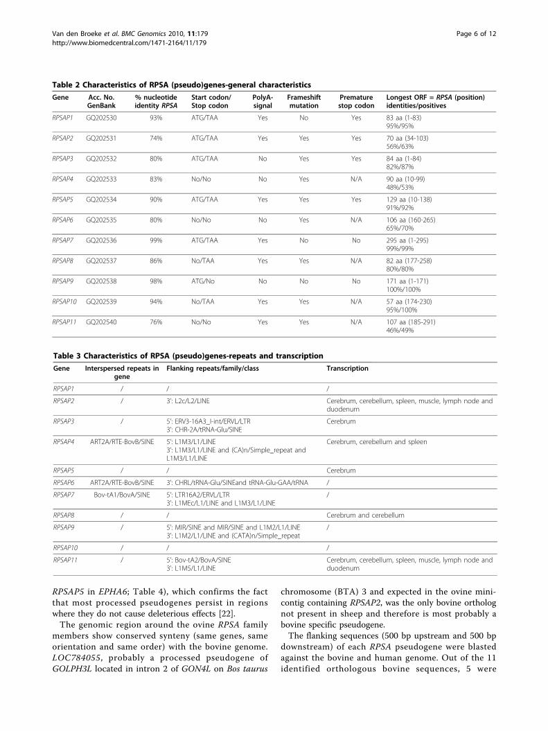
[24]. In some cases nevertheless, they have obtained a(regulatory) function [17].To investigate this possibility, all the RPSA pseudogeneswere further characterized in silico and their main char-acteristics are listed in detail in Table 2 and 3. Compari-son with the full length RPSA gene transcript showsthat the pseudogenes vary greatly both in structure andsequence identity. These differences range from structu-rally identical pseudogenes sharing 99% sequence iden-tity (RPSAP7) to pseudogenes lacking half of the gene(RPSAP8, RPSAP9 and RPSAP10) or containing manydeletions throughout the whole gene sharing a sequenceidentity of only 74% (RPSAP2).Analysis of the primer binding sites in the pseudo-
genes showed that in our experimental design thescreening primers could anneal to targets down to 83%sequence identity, even in the case of RPSAP2.All BAC clones and thus all RPSA family members
were isolated with at least 2 primer pairs and there wasno concordance between the number of BACs in amini-contig and the level of sequence identity withRPSA. We conclude that it is most likely that we haveisolated all the ovine RPSA pseudogenes sharing a highlevel of sequence identity and that therefore can inter-fere with the functional RPSA gene in genetic studies.To obtain a first indication of possible functionality,
in silico ORF and promoter prediction analysis were car-ried out.The pseudogene RPSAP7 is the only member sharing
almost an identical ORF with the full length RPSA gene.The only one amino acid difference (amino acid 31:D ® G) is located in the intracellular part of the recep-tor that does not belong to any binding site. All theother pseudogenes either lack the start codon or containa premature stop codon due to nonsense or frameshiftmutations. The size of the potential ORF of the otherpseudogenes varies and the largest reaches 171 aminoacids sharing 100% identity with RPSA (Table 2). MostORFs lie in the intracellular region of RPSA (amino acid1-101). In case of RPSAP6, RPSAP8, RPSAP10 andRPSAP11, the ORF contains a part of the binding sitesof RPSA with PrP (direct binding aa 161-180; indirectbinding aa 180-285 [25]), but most of them have a lowlevel of amino acid identity.In silico promoter analysis predicted a possible promo-
ter for RPSAP1, RPSAP2, RPSAP4, RPSAP8, RPSAP9 andRPSAP10 (Additional file 4). A consensus polyadenyla-tion signal is present in 7 of the 11 pseudogenes(including RPSAP7).Repeated sequences were identified with Repeatmasker
[26] and showed that 4 pseudogenes are disrupted byinterspersed repeats belonging to the class/family SINE/RTE-BovB, SINE/BovA, tRNA and SINE/tRNA-Glu, andthat 7 pseudogenes were flanked by repeats belonging to
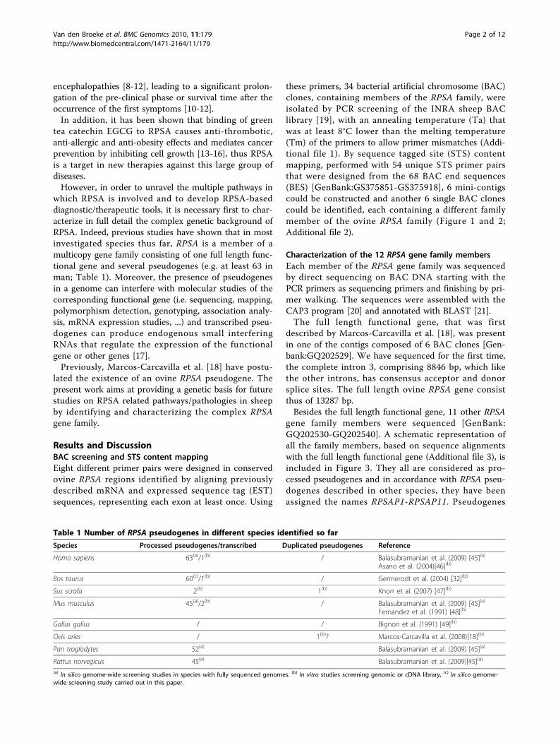
Figure 1 Comparative mapping of the region of RPSA in sheepand cattle. The ovine BAC mini-contig is drawn in part A. Trianglesrepresent BAC end sequences; pointing towards the 3’-end of theBAC clone. Black triangles represent BES from which primers weredesigned to construct the mini-contig. White triangles are BES fromwhich it was impossible to design STS-primers. Black squares showoverlaps between BES and other BAC clones. Black circles representgenes annotated by PCR. Annotated sequences are shown in aplane map in part B. The position and orientation of the genespresent in the syntenic region of Bos taurus are represented witharrows (C).
Van den Broeke et al. BMC Genomics 2010, 11:179http://www.biomedcentral.com/1471-2164/11/179
Page 3 of 12
the SINE, LINE, tRNA, LTR and simple repeat classes.According to Zhang et al. [27], processed pseudogenesare mostly found in genomic regions with a relativelylow GC content, as do LINE repeats. Thus, it is not sur-prising that such repeats are present in the regionsflanking many of the RPSA pseudogenes.A remarkable observation is that part of the RPSA intron
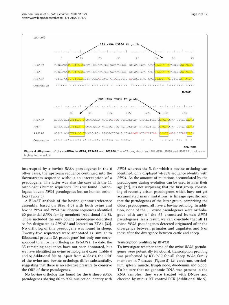
4, containing the small nucleolar RNA (snoRNA)SNORA62, is present in the RPSAP8 and RPSAP9
pseudogenes. Therefore, these pseudogenes can be consid-ered as semi-processed pseudogenes, which are very rarelyreported and defined by Zhang et al. as “pseudogenes thatcontain remnant introns, which suggests that they werederived from semi-processed RNA transcripts” [28].SnoRNAs are encoded in introns of ribosomal protein
genes and other housekeeping genes [29,30], and areresponsible for both sequence-specific methylation andpseudouridilation of RNA [31]. SNORA62 isan H/ACA
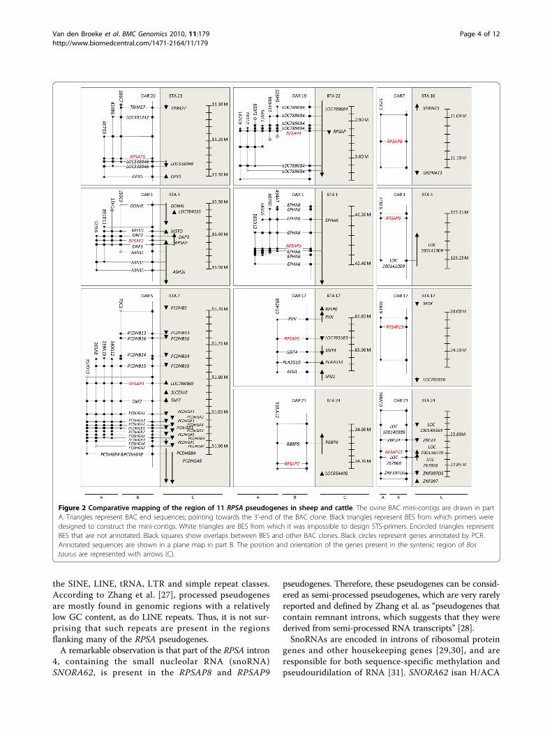
Figure 2 Comparative mapping of the region of 11 RPSA pseudogenes in sheep and cattle. The ovine BAC mini-contigs are drawn in partA. Triangles represent BAC end sequences; pointing towards the 3’-end of the BAC clone. Black triangles represent BES from which primers weredesigned to construct the mini-contigs. White triangles are BES from which it was impossible to design STS-primers. Encircled triangles representBES that are not annotated. Black squares show overlaps between BES and other BAC clones. Black circles represent genes annotated by PCR.Annotated sequences are shown in a plane map in part B. The position and orientation of the genes present in the syntenic region of Bostaurus are represented with arrows (C).
Van den Broeke et al. BMC Genomics 2010, 11:179http://www.biomedcentral.com/1471-2164/11/179
Page 4 of 12
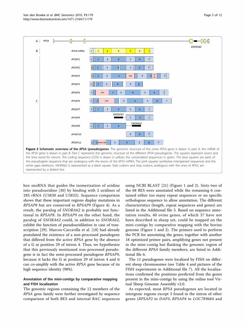
box snoRNA that guides the isomerization of uridineinto pseudouridine [30] by binding with 2 uridines of28S rRNA (U3830 and U3832). Sequence comparisonshows that these important regions display mutations inRPSAP8 but are conserved in RPSAP9 (Figure 4). As aresult, the paralog of SNORA62 is probably not func-tional in RPSAP8. In RPSAP9 on the other hand, theparalog of SNORA62 could, in addition to SNORA62,exhibit the function of pseudouridilation in case of tran-scription [29]. Marcos-Carcavilla et al. [18] had alreadypostulated the existence of a non-processed pseudogenethat differed from the active RPSA gene by the absenceof a G at position 29 of intron 4. Thus, we hypothesizethat this previously mentioned non-processed pseudo-gene is in fact the semi-processed pseudogene RPSAP9,because it lacks the G at position 29 of intron 4 and itcan co-amplify with the active RPSA gene because of itshigh sequence identity (98%).
Annotation of the mini-contigs by comparative mappingand FISH localizationThe genomic regions containing the 12 members of theRPSA gene family were further investigated by sequencecomparison of both BES and internal BAC sequences
using NCBI BLAST [21] (Figure 1 and 2). Sixty-two ofthe 68 BES were annotated while the remaining 6 con-tained either too many repeat sequences or no specificorthologous sequence to allow annotation. The differentcharacteristics (length, repeat sequences and genes) arelisted in the Additional file 5. Based on sequence anno-tation results, 40 ovine genes, of which 37 have notbeen described in sheep yet, could be mapped on themini-contigs by comparative mapping with the bovinegenome (Figure 1 and 2). The primers used to performthe PCR for annotating the genes, together with another18 optimized primer pairs, amplifying genes not presentin the mini-contig but flanking the genomic region ofthe different RPSA family members, are listed in Addi-tional file 6.The 11 pseudogenes were localized by FISH on differ-
ent sheep chromosomes (see Table 4 and pictures of theFISH experiments in Additional file 7). All the localiza-tions confirmed the positions predicted from the genespresent in the mini-contigs by using the online tool Vir-tual Sheep Genome Assembly v2.0.As expected, most RPSA pseudogenes are located in
intergenic regions except 3 found in the intron of othergenes (RPSAP2 in DAP3; RPSAP4 in LOC789684 and
Figure 3 Schematic overview of the RPSA (pseudo)genes. The genomic structure of the ovine RPSA gene is drawn in part A, the mRNA ofthe RPSA gene is drawn in part B. Part C represents the genomic structure of the different RPSA pseudogenes. The squares represent exons andthe lines stand for introns. The coding sequence (CDS) is drawn in yellow; the untranslated sequences in green. The blue squares are parts ofthe pseudogene sequence that are analogous with the exons of the RPSA mRNA. The pink squares symbolize interspersed sequences and thewhite gaps deletions. SNORA62 is represented as a black square. Start codons and stop codons, analogous with the ones of RPSA, arerepresented by a dotted line.
Van den Broeke et al. BMC Genomics 2010, 11:179http://www.biomedcentral.com/1471-2164/11/179
Page 5 of 12
RPSAP5 in EPHA6; Table 4), which confirms the factthat most processed pseudogenes persist in regionswhere they do not cause deleterious effects [22].The genomic region around the ovine RPSA family
members show conserved synteny (same genes, sameorientation and same order) with the bovine genome.LOC784055, probably a processed pseudogene ofGOLPH3L located in intron 2 of GON4L on Bos taurus
chromosome (BTA) 3 and expected in the ovine mini-contig containing RPSAP2, was the only bovine ortholognot present in sheep and therefore is most probably abovine specific pseudogene.The flanking sequences (500 bp upstream and 500 bp
downstream) of each RPSA pseudogene were blastedagainst the bovine and human genome. Out of the 11identified orthologous bovine sequences, 5 were
Table 2 Characteristics of RPSA (pseudo)genes-general characteristics
Gene Acc. No.GenBank
% nucleotideidentity RPSA
Start codon/Stop codon
PolyA-signal
Frameshiftmutation
Prematurestop codon
Longest ORF = RPSA (position)identities/positives
RPSAP1 GQ202530 93% ATG/TAA Yes No Yes 83 aa (1-83)95%/95%
RPSAP2 GQ202531 74% ATG/TAA Yes Yes Yes 70 aa (34-103)56%/63%
RPSAP3 GQ202532 80% ATG/TAA No Yes Yes 84 aa (1-84)82%/87%
RPSAP4 GQ202533 83% No/No No Yes N/A 90 aa (10-99)48%/53%
RPSAP5 GQ202534 90% ATG/TAA Yes Yes Yes 129 aa (10-138)91%/92%
RPSAP6 GQ202535 80% No/No No Yes N/A 106 aa (160-265)65%/70%
RPSAP7 GQ202536 99% ATG/TAA Yes No No 295 aa (1-295)99%/99%
RPSAP8 GQ202537 86% No/TAA Yes Yes N/A 82 aa (177-258)80%/80%
RPSAP9 GQ202538 98% ATG/No No No No 171 aa (1-171)100%/100%
RPSAP10 GQ202539 94% No/TAA Yes Yes N/A 57 aa (174-230)95%/100%
RPSAP11 GQ202540 76% No/No Yes Yes N/A 107 aa (185-291)46%/49%
Table 3 Characteristics of RPSA (pseudo)genes-repeats and transcription
Gene Interspersed repeats ingene
Flanking repeats/family/class Transcription
RPSAP1 / / /
RPSAP2 / 3’: L2c/L2/LINE Cerebrum, cerebellum, spleen, muscle, lymph node andduodenum
RPSAP3 / 5’: ERV3-16A3_I-int/ERVL/LTR3’: CHR-2A/tRNA-Glu/SINE
Cerebrum
RPSAP4 ART2A/RTE-BovB/SINE 5’: L1M3/L1/LINE3’: L1M3/L1/LINE and (CA)n/Simple_repeat andL1M3/L1/LINE
Cerebrum, cerebellum and spleen
RPSAP5 / / Cerebrum
RPSAP6 ART2A/RTE-BovB/SINE 3’: CHRL/tRNA-Glu/SINEand tRNA-Glu-GAA/tRNA /
RPSAP7 Bov-tA1/BovA/SINE 5’: LTR16A2/ERVL/LTR3’: L1MEc/L1/LINE and L1M3/L1/LINE
/
RPSAP8 / / Cerebrum and cerebellum
RPSAP9 / 5’: MIR/SINE and MIR/SINE and L1M2/L1/LINE3’: L1M2/L1/LINE and (CATA)n/Simple_repeat
/
RPSAP10 / / /
RPSAP11 / 5’: Bov-tA2/BovA/SINE3’: L1M5/L1/LINE
Cerebrum, cerebellum, spleen, muscle, lymph node andduodenum
Van den Broeke et al. BMC Genomics 2010, 11:179http://www.biomedcentral.com/1471-2164/11/179
Page 6 of 12
interrupted by a bovine RPSA pseudogene; in the 6other cases, the upstream sequence continued into thedownstream sequence without an interruption of apseudogene. The latter was also the case with the 11orthologous human sequences. Thus we found 5 ortho-logous bovine RPSA pseudogenes but no human ortho-logs (Table 5).A BLAST analysis of the bovine genome (reference
assembly, based on Btau_4.0) with both ovine andbovine RPSA and RPSA pseudogene sequences identified60 potential RPSA family members (Additional file 8).These included the only bovine pseudogene describedso far, designated as RPSAP1 and located on BTA4 [32].No ortholog of this pseudogene was found in sheep.Twenty-five sequences were annotated as ‘similar toRibosomal protein SA pseudogene’ but only one corre-sponded to an ovine ortholog i.e. RPSAP11. To date, the35 remaining sequences have not been annotated, butwe have identified an ovine ortholog in 4 cases (Table 4and 5; Additional file 8). Apart from RPSAP3, the ORFof the ovine and bovine orthologs differ substantially,suggesting that there is no selective pressure to conservethe ORF of these pseudogenes.No bovine ortholog was found for the 6 sheep RPSA
pseudogenes sharing 86 to 99% nucleotide identity with
RPSA whereas the 5, for which a bovine ortholog wasidentified, only displayed 74-83% sequence identity withRPSA. As the amount of mutations accumulated by thepseudogenes during evolution can be used to infer theirage [27], it’s not surprising that the first group, consist-ing of recently arisen pseudogenes which have not yetaccumulated many mutations, is lineage specific andthat the pseudogenes of the latter group, comprising theoldest pseudogenes, all have a bovine ortholog. In addi-tion, none of the 11 ovine pseudogenes were ortholo-gous with any of the 63 annotated human RPSApseudogenes. As a result, we can conclude that all 11ovine RPSA pseudogenes detected originated after thedivergence between primates and ungulates and 6 ofthese after the divergence between cattle and sheep.
Transcription profiling by RT-PCRTo investigate whether some of the ovine RPSA pseudo-genes were potentially functional, transcription profilingwas performed by RT-PCR for all sheep RPSA familymembers in 7 tissues (Figure 5) i.e. cerebrum, cerebel-lum, spleen, muscle, lymph node, duodenum and blood.To be sure that no genomic DNA was present in theRNA samples, they were treated with DNase andchecked by minus RT control PCR (Additional file 9).
Figure 4 Alignment of the snoRNAs in RPSA, RPSAP8 and RPSAP9. The ACA-box, H-box and 28S rRNA U3830 and U3832 PU guide arehighlighted in yellow.
Van den Broeke et al. BMC Genomics 2010, 11:179http://www.biomedcentral.com/1471-2164/11/179
Page 7 of 12

For 8 members of the RPSA family, gene-specific pri-mers could be designed and their specificity was provenby checking that the primers did not amplify any otherRPSA family member using the respective unique BACclones as template (Additional file 10). Because RPSA,RPSAP1, RPSAP7 and RPSAP9 share a high level ofsequence identity, no specific primers could be designedfor these RPSA family members, they were tested withaspecific primers. All generated amplicons weresequenced. RPSA was expressed in all tested tissues.This agrees with the results of Marcos-Carcavilla et al.and Qiao et al. [18,33]. None of the pseudogenes wastranscribed in blood. RPSAP2 and RPSAP11 were tran-scribed in all other tested tissues, while RPSAP3,RPSAP5 and RPSAP8 were only transcribed in one ormore brain regions and RPSAP4 was transcribed inbrain regions and spleen. RPSAP6 and RPSAP10 werenot expressed in any of the tested tissues. In the case ofRPSAP1, RPSAP7 and RPSAP9, tested with aspecific pri-mers which all could also amplify RPSA, we generated
amplicons which, after sequencing, turned out to be allRPSA transcripts. Thus we can conclude that RPSAP1,RPSAP7 and RPSAP9 are not expressed or at a very lowlevel compared to the active RPSA gene. Therefore itwould be interesting to do RT-qPCR with specificprobes in order to be sure if the pseudogenes areexpressed at very low levels or not at all. No clear rela-tionship between the transcription profile of the variouspseudogenes and the in silico prediction of possible pro-moters was observed. For instance, RPSAP10 is notexpressed in any tissue tested although we did predict apromoter in the upstream sequence. Thus it may bepossible that RPSAP10 is expressed in other tissues notexamined in this study or that it has a low level of tran-scription. In addition, the in silico predicted promotermight not act as a cis-regulatory element in vivo. Incontrast, RPSAP3 is transcribed in certain brain regionsalthough we did not predict any promoter, probablybecause the promoter is located more upstream thanthe region analyzed here.
Table 4 Location of RPSA (pseudo)genes
Gene Chromosomal location Ortholog Bos taurus
RPSA OAR19q13 intergenic between LOC515736 and MOBP ortholog BTA22: GeneID: 281898
RPSAP1 OAR20q22 intergenic between LOC401242 and LOC538046 no
RPSAP2 OAR1p13 in intron 2 DAP3 ortholog BTA3: not annotated yet
RPSAP3 OAR 5q22.3 intergenic between PCDHB15 and TAF7 ortholog BTA7: not annotated yet
RPSAP4 OAR19q12 in intron 1 LOC789684 ortholog BTA22: not annotated yet
RPSAP5 OAR1q21-q22 in intron 2 EPHA6 no
RPSAP6 OAR17q26prox intergenic between PXN and SIRT4 ortholog BTA17: not annotated yet
RPSAP7 OAR23q23prox intergenic between RBBP8 and LOC100138286 no
RPSAP8 OAR7q12-q13 intergenic between SERINC5 and GNPNAT1 no
RPSAP9 OAR1p37 intergenic between LOC100141009 and LOC522241 no
RPSAP10 OAR17q21prox intergenic between MDK and LOC783956 no
RPSAP11 OAR23q21 intergenic between ZNF24 and LOC767868 ortholog BTA24: GeneID: 100138378
Table 5 Bovine orthologs
Ovineortholog
Chromosomallocation
GenBankAcc. No.
Range Nucleic acid identity withbovine RPSA
Nucleic acid identity withovine ortholog
Features in sequence
RPSA BTA22 NC_007320.3 12885045-12898467
100% CDS 96%Gene 87%
RPSA: 12886519-12898467
RPSAP2 BTA3 NC_007301.3 16411109-16410143
75% 91% DAP3 intron 2:16430465-16408264
RPSAP3 BTA7 NC_007305.3 51802201-51801057
81% 95% LOC786980: 51801077-51802181
RPSAP4 BTA22 NC_007320.3 2925069-2925867
78% 91% LOC789684: 2840530-3050478
RPSAP6 BTA17 NC_007315.3 65883767-65884909
81% 92% LOC783583: 65881027-65884941
RPSAP11 BTA24 NC_007325.3 22831525-22830341
77% 91% LOC100138378:22830405-22834810
Van den Broeke et al. BMC Genomics 2010, 11:179http://www.biomedcentral.com/1471-2164/11/179
Page 8 of 12

ConclusionsIn addition to the already described ovine RPSA gene,we have identified 11 members of the ovine RPSA genefamily, and designated them RPSAP1-RPSAP11 sincethey are all considered to be processed pseudogenes.The flanking genomic regions of each RPSA familymember was analyzed by annotating the constructedBAC contigs, which revealed 40 genes (of which 37 hadnot been previously described in sheep) based on com-parative mapping. All these regions show conserved syn-teny with the orthologous bovine counterparts and thelocations were confirmed by FISH. Five pseudogeneshave a bovine counterpart. In silico analysis predictedthe presence of 55 more RPSA pseudogenes in thebovine genome.
Compared to the RPSA transcript, RPSA pseudogenesdiffer significantly both in structure and sequence iden-tity, ranging from structurally identical pseudogenessharing 99% sequence identity to pseudogenes lackinghalf of the gene or containing many deletions through-out the whole gene, sharing only 74% sequence identity.A remarkable result is that at least 6 of the 11 pseudo-genes are transcriptionally active. However, whetherthese transcripts are functional as regulatory non-coding RNA or as functional proteins remains to beinvestigated.In previous studies, 1 to 3 RPSA pseudogenes per spe-
cies, discovered while screening with the intention toisolate the full length functional RPSA gene, were char-acterized. Furthermore, the number of RPSA pseudo-genes in 4 species with fully sequenced genomes wasdetermined by genome-wide in silico screening butthose pseudogenes were not characterized (Table 1).Here we report in detail the characterization of theRPSA gene family in a species. A strategy was developedto isolate all the ovine RPSA pseudogenes sharing a highlevel of sequence identity with RPSA. We screened with8 different primers representing each exon at least onceand with a Ta that was at least 8°C lower than the Tm.All BAC clones were positive for at least 2 primer pairsand there was no concordance between the number ofBAC in a mini-contig and the level of sequence identitywith RPSA. Therefore, we conclude that it is most likelythat we have isolated all the ovine RPSA pseudogenesthat could interfere with the functional RPSA gene ingenetic studies. The discrepancy between the numbersof ovine RPSA pseudogenes found (11) and the numbersdescribed in genome-wide screenings (45-61) might beexplained by the low sequence identity of most pseudo-genes found in silico. In Bos taurus for instance, 51 ofthe 60 pseudogenes share an overall nucleic acid identitywith the bovine RPSA gene beneath 80% (Additional file8). Due to our experimental design, pseudogenes with alow sequence identity were not isolated since it is notlikely that those pseudogenes would interfere with mole-cular studies on the functional full length RPSA gene.In conclusion, we describe 11 ovine processed RPSA
pseudogenes. This knowledge on their structure andsequence will facilitate the molecular genetic studies ofthe functional gene since it will now be possible to takeinto account the existence of the pseudogenes in thedesign of such studies.
MethodsConstruction BAC mini-contigsThe ovine INRA BAC library, consisting of 90.000clones with an average insert length of 123 kb and agenome equivalent of 3.4, was screened by PCR [19].The primers were designed using Primer3, based on
Figure 5 Transcription profile of the RPSA gene familymembers. Marker (M) is the Hyperladder V or IV (Bioline). Samplesare cerebrum (Cbu), cerebellum (Cbe), spleen (Sp), muscle (Mu),lymph node (Ln), duodenum (Dd), blood (Bl) genomic or BAC DNA(+) and water (-).
Van den Broeke et al. BMC Genomics 2010, 11:179http://www.biomedcentral.com/1471-2164/11/179
Page 9 of 12
conserved regions in the sheep RPSA gene [34]. Theconserved regions were detected by comparison of allovine ESTs available in GenBank that shared similaritywith the published ovine mRNA sequence of RPSA[GenBank:EF649775] with BLAST and ClustalW [21,35].PCR was conducted with Faststart Taq DNA Polymerase(Roche). PCR conditions were 5 min at 95°C, 40 cyclesof 30 s at 95°C, 30 s at 50°C and 1 min at 72°C, and afinal 10-min elongation step at 72°C. Thirty-nine super-pools, each consisting of 44 pools (24 plates, 8 rows and12 columns), were screened. Each positive combinationwas verified by colony PCR.All isolated BACs were grown in a 200 ml culture from
which DNA was purified with the Qiagen Plasmid Midikit (Qiagen) according to the manufacturer’s instructions.The BAC ends were sequenced with the universal primer(UP) (5’-CGACGTTGTAAAACGACGGCCAG-3’) andreverse primer (RP) (5’-CACAGGAAACAGCTATGAC-CATGATTACG-3’) primers with 1 μg of purified BACDNA as template. Unique STS primer pairs (Additionalfile 2), based on the BESs, were used to screen all isolatedBACs and to construct mini-contigs.All sequencing was performed with the Big Dye Ter-
minator mix (Applied Biosystems) and analyzed on anABI-3730xl Analyser (Applied Biosystems).
Characterization of RPSA gene family membersThe primers used to screen the INRA BAC library wereused as initial sequence primers to sequence the RPSAfamily member in one BAC of each mini-contig bydirect sequencing. The obtained sequence was then usedto develop new sequencing primers until the whole geneand an additional ± 500 bp upstream and ± 500 bpdownstream of the sequence showing similarity withRPSA, was sequenced. If the screening primer did notwork as sequencing primer, the amplicon generatedwith the screening primer was cloned into a pCR 2.1vector with the TA Cloning Kit (Invitrogen) and thevector was transformed in DH5a Competent Cells (Invi-trogen). The insert was then sequenced with UP and RPprimers. All sequences were assembled into continuoussequences with CAP3 and analyzed with FGENESH andNCBI ORF Finder [20,36,37]. Promoter sequences weresearched with CISTER, Neural Network Promoter Pre-diction, FPROM and TFsearch [36,38-40].
Annotation of the mini-contigs by comparative mappingAll mini-contigs were annotated by comparing the BESsagainst bovine and human genomic sequences withNCBI BLAST [21]. Internal sequences were also anno-tated by PCR with primers based on bovine sequencesof genes that were expected to be present in the mini-contig. All amplicons were verified and repeats weredetected with Repeatmasker [26].
FISHFluorescent in situ hybridization was performed atINRA in Jouy-en-Josas (France). To prepare the probes,BAC DNA was extracted according to standard proto-cols and purified with the S.N.A.P. K1900-01 Miniprepkit (Invitrogen). DNA was then nick-translated with bio-tin-14-dATP (BioNick 18247-015 labeling system, Invi-trogen) and mixed with 100× total sonicated herringsperm DNA and 100× total sonicated sheep DNA. Sub-sequently, it was precipitated with ethanol, slightly driedand resuspended in hybridization buffer.For R-banded sheep chromosomes, embryo fibroblast
cell cultures were synchronized with an excess of thymi-dine and treated with 5-bromo-2’-deoxyuridine duringthe second half of S phase [41].FISH, signal detection and R-banding were performed
as previously described [42]. Briefly, labeled probes weredenatured at 100°C for 10 min and pre-hybridized at 37°C for 30 to 60 min before hybridization to the chromo-somes. Chromosome identification and band numberingfollowed the standard sheep ideogram reported inISCNDB2000 [43].
Transcription profilingFresh tissue samples were obtained from a commercialsheep slaughterhouse, frozen in liquid nitrogen immedi-ately after slaughtering, crushed into powder and frozenat -80°C. Total RNA was isolated with the Aurum TotalRNA Fatty and Fibrous Tissue kit (Bio-Rad) as describedin the instruction manual. Subsequently, a minusRT-PCR was performed with actin, beta (ACTB) pri-mers on 1 μl RNA to confirm the absence of any DNAcontamination (Additional file 9) as previously described[44]. If DNA was still present in the sample, an addi-tional DNase treatment with RQ1 RNase-free DNase(Promega) and a spin column purification with Micro-con YM-100 (Millipore) were carried out.The RNA concentration and purity of the samples
were measured with the Nanodrop ND-1000 Spectro-photometer (Isogen) and the RNA quality was deter-mined by evaluation of the 28S and 18S ribosomalbands on a 0.8% agarose gel.Then, 0.2-1 μg RNA was converted into cDNA with
iScript cDNA synthesis kit (Bio-Rad) using random andoligo dT primers. A confirmation PCR on 10× dilutedcDNA with ACTB primers (giving a different ampliconlength on gDNA and cDNA) was performed.Specific primers, based on the aligned sequences of
the different RPSA family members (Additional file 3)were designed for 8 members of the RPSA family andspecificity was proven (see above).Due to the high level of nucleotide sequence identity
among RPSA, RPSAP1, RPSAP7 and RPSAP9, it was notpossible to develop specific primers for these, but we
Van den Broeke et al. BMC Genomics 2010, 11:179http://www.biomedcentral.com/1471-2164/11/179
Page 10 of 12
were able to develop several primer pairs which ampli-fied different combinations of 2 to 6 RPSA family mem-bers. One primer for instance amplifies RPSA, RPSAP1,RPSAP7 and RPSAP11; another RPSA and RPSAP7 anda third one RPSA, RPSAP1, RPSAP5 RPSAP7 andRPSAP11.The obtained amplicons were sequenced to deter-
mine/confirm which (pseudo)gene was transcribed.
Additional file 1: Primers used for screening of the INRA BAClibrary. Characteristics of primers used for screening of the INRA BAClibrary: sequence, location, melting temperature and amplicon length.Click here for file[ http://www.biomedcentral.com/content/supplementary/1471-2164-11-179-S1.XLSX ]
Additional file 2: STS primers BES. Characteristics of primers used forSTS content mapping: sequence, annealing temperature and ampliconlength.Click here for file[ http://www.biomedcentral.com/content/supplementary/1471-2164-11-179-S2.XLSX ]
Additional file 3: Alignment of the mRNA of the ovine RPSA genewith 11 RPSA pseudogenes. The startcodon, stopcodon, poly-adenylation signal exon-exon junctions and interspersed repeats arehighlighted in yellow. The primers used to screen the INRA BAC libraryare highlighted in green.Click here for file[ http://www.biomedcentral.com/content/supplementary/1471-2164-11-179-S3.PDF ]
Additional file 4: In silico promoter prediction. The in silico predictedpromoter sequence is shown with the transcription start site highlightedin red. Furthermore, the score of the prediction, identification of thepromoter and location of the transcription start site in the sequence isshown.Click here for file[ http://www.biomedcentral.com/content/supplementary/1471-2164-11-179-S4.XLSX ]
Additional file 5: Characteristics BES. Characteristics of the 68 BES: BESlength, orthologous sequences and genes, and repeats present in theBES.Click here for file[ http://www.biomedcentral.com/content/supplementary/1471-2164-11-179-S5.XLSX ]
Additional file 6: Primers used to annotate the mini-contigs.Characteristics of primers used to annotate the mini-contig: sequence,annealing temperature, amplicon size and annotation information.Click here for file[ http://www.biomedcentral.com/content/supplementary/1471-2164-11-179-S6.XLSX ]
Additional file 7: FISH experiments. Pictures of FISH experiments ofthe 11 RPSA pseudogenes.Click here for file[ http://www.biomedcentral.com/content/supplementary/1471-2164-11-179-S7.PDF ]
Additional file 8: Bovine RPSA pseudogenes. Characteristics of bovineRPSA pseudogenes predicted in silico by BLAST analysis of the bovinegenome (reference assembly, based on Btau_4.0). In yellow are thepseudogenes that are annotated as ‘similar to Ribosomal protein SApseudogene’. The bovine pseudogene RPSAP1 is highlighted in blue andthe pseudogenes that aren’t annotated yet in white.Click here for file[ http://www.biomedcentral.com/content/supplementary/1471-2164-11-179-S8.XLSX ]
Additional file 9: minus-RT PCR control on RNA isolated from blood.RT-PCR with ACTB primers. Marker (M) is the Hyperladder V (Bioline).Samples are RNA isolated from blood, genomic DNA and water (-).Click here for file[ http://www.biomedcentral.com/content/supplementary/1471-2164-11-179-S9.PDF ]
Additional file 10: Specificity of the expression primer for RPSAP6.PCR with expression primer for RPSAP6. Marker (M) is the Hyperladder IV(Bioline). Samples are the respective unique BAC clones of all RPSA familymembers, genomic DNA and water (-).Click here for file[ http://www.biomedcentral.com/content/supplementary/1471-2164-11-179-S10.PDF ]
AcknowledgementsThe authors wish to thank Caroline Rogiers and Robin Maene for excellenttechnical assistance and Dr. Malena Serrano for her continuous help. AliceVan den Broeke is supported with a grant of the Institute for the Promotionof Innovation by Science and Technology in Flanders (IWT). Dr. Marcos-Carcavilla was supported by RTA2006-00104 457 INIA project.
Author details1Department of Nutrition, Genetics and Ethology, Faculty of VeterinaryMedicine, Ghent University, Heidestraat 19, B-9820 Merelbeke, Belgium.2Departamento de Mejora Genética Animal, INIA, Ctra La Coruña Km 7.5,Madrid 28040, Spain. 3INRA, UMR 1313 Génétique Animale et BiologieIntégrative, F78350 Jouy-en-Josas, France.
Authors’ contributionsAVDB carried out the BAC library screening, mini-contig building,sequencing of the genes, annotation by comparative mapping, thetranscription profiling and drafted this manuscript. MVP participated in thedesign of the study, participated in the screening of the BAC library andprovided experimental support. AMC participated in the screening of theBAC library. KH supervised the BAC library screening. HH and MB carried outthe FISH mapping experiments. AVZ supervised the study. LJP participatedin the study design and supervised the study. All authors read and approvedthe final manuscript.
Received: 9 December 2009 Accepted: 16 March 2010Published: 16 March 2010
References1. Kinoshita K, Kaneda Y, Sato M, Saeki Y, Wataya-Kaneda M, Hoffmann A: LBP-
p40 binds DNA tightly through associations with histones H2A, H2B,and H4. Biochemical and biophysical research communications 1998,253(2):277-282.
2. Nelson J, McFerran NV, Pivato G, Chambers E, Doherty C, Steele D,Timson DJ: The 67 kDa laminin receptor: structure, function and role indisease. Bioscience reports 2008, 28(1):33-48.
3. Orihuela CJ, Mahdavi J, Thornton J, Mann B, Wooldridge KG, Abouseada N,Oldfield NJ, Self T, Ala’Aldeen DA, Tuomanen EI: Laminin receptor initiatesbacterial contact with the blood brain barrier in experimental meningitismodels. J Clin Invest 2009, 119(6):1638-1646.
4. Chen FX, Qian YR, Duan YH, Ren WW, Yang Y, Zhang CC, Qiu YM, Ji YH:Down-regulation of 67LR reduces the migratory activity of humanglioma cells in vitro. Brain Res Bull 2009, 79(6):402-408.
5. Zhou L, Xie M, Zhou JQ, Tao L: 67-kDa laminin receptor in humanlaryngeal squamous cell carcinoma. Laryngoscope 2006, 116(1):28-32.
6. Li D, Chen J, Gao Z, Li X, Yan X, Xiong Y, Wang S: 67-kDa laminin receptorin human bile duct carcinoma. Eur Surg Res 2009, 42(3):168-173.
7. Zuber C, Knackmuss S, Zemora G, Reusch U, Vlasova E, Diehl D, Mick V,Hoffmann K, Nikles D, Frohlich T, Arnold GJ, Brenig B, Wolf E, Lahm H,Little M, Weiss S: Invasion of tumorigenic HT1080 cells is impeded by
Van den Broeke et al. BMC Genomics 2010, 11:179http://www.biomedcentral.com/1471-2164/11/179
Page 11 of 12

blocking or downregulating the 37-kDa/67-kDa laminin receptor. Journalof molecular biology 2008, 378(3):530-539.
8. Zuber C, Knackmuss S, Rey C, Reusch U, Rottgen P, Frohlich T, Arnold GJ,Pace C, Mitteregger G, Kretzschmar HA, Little M, Weiss S: Single chain Fvantibodies directed against the 37 kDa/67 kDa laminin receptor astherapeutic tools in prion diseases. Molecular immunology 2008,45(1):144-151.
9. Zuber C, Mitteregger G, Schuhmann N, Rey C, Knackmuss S, Rupprecht W,Reusch U, Pace C, Little M, Kretzschmar HA, Hallek M, Buning H, Weiss S:Delivery of single-chain antibodies (scFvs) directed against the 37/67kDa laminin receptor into mice via recombinant adeno-associated viralvectors for prion disease gene therapy. The Journal of general virology2008, 89(Pt 8):2055-2061.
10. Pflanz H, Vana K, Mitteregger G, Pace C, Messow D, Sedlaczek C, Nikles D,Kretzschmar HA, Weiss SF: Microinjection of lentiviral vectors expressingsmall interfering RNAs directed against laminin receptor precursormRNA prolongs the pre-clinical phase in scrapie-infected mice. TheJournal of general virology 2009, 90(Pt 1):269-274.
11. Zuber C, Mitteregger G, Pace C, Zerr I, Kretzschmar HA, Weiss S: Anti-LRP/LR antibody W3 hampers peripheral PrPSc propagation in scrapieinfected mice. Prion 2007, 1(3):207-212.
12. Pflanz H, Vana K, Mitteregger G, Renner-Muller I, Pace C, Kuchenhoff H,Kretzschmar HA, Wolf E, Weiss S: Scrapie-infected transgenic miceexpressing a laminin receptor decoy mutant reveal a prolongedincubation time associated with low levels of PrPres. Journal of molecularbiology 2009, 388(4):721-729.
13. Holy EW, Stampfli SF, Akhmedov A, Holm N, Camici GG, Luscher TF,Tanner FC: Laminin receptor activation inhibits endothelial tissue factorexpression. J Mol Cell Cardiol 2009.
14. Fujimura Y, Umeda D, Yamada K, Tachibana H: The impact of the 67 kDalaminin receptor on both cell-surface binding and anti-allergic action oftea catechins. Arch Biochem Biophys 2008, 476(2):133-138.
15. Ku HC, Chang HH, Liu HC, Hsiao CH, Lee MJ, Hu YJ, Hung PF, Liu CW,Kao YH: Green tea (-)-epigallocatechin gallate inhibits insulin stimulationof 3T3-L1 preadipocyte mitogenesis via the 67-kDa laminin receptorpathway. Am J Physiol Cell Physiol 2009, 297(1):C121-132.
16. Umeda D, Yano S, Yamada K, Tachibana H: Green tea polyphenolepigallocatechin-3-gallate signaling pathway through 67-kDa lamininreceptor. The Journal of biological chemistry 2008, 283(6):3050-3058.
17. Khachane AN, Harrison PM: Assessing the genomic evidence forconserved transcribed pseudogenes under selection. BMC Genomics2009, 10:435.
18. Marcos-Carcavilla A, Calvo JH, Gonzalez C, Serrano C, Moazami-Goudarzi K,Laurent P, Bertaud M, Hayes H, Beattie AE, Lyahyai J, Martin-Burriel I,Torres JM, Serrano M: Structural and functional analysis of the ovinelaminin receptor gene (RPSA): Possible involvement of the LRP/LRprotein in scrapie response. Mamm Genome 2008, 19(2):92-105.
19. Vaiman D, Billault A, Tabet-Aoul K, Schibler L, Vilette D, Oustry-Vaiman A,Soravito C, Cribiu EP: Construction and characterization of a sheep BAClibrary of three genome equivalents. Mamm Genome 1999, 10(6):585-587.
20. Huang X, Madan A: CAP3: A DNA sequence assembly program. Genomeresearch 1999, 9(9):868-877.
21. Altschul SF, Gish W, Miller W, Myers EW, Lipman DJ: Basic local alignmentsearch tool. Journal of molecular biology 1990, 215(3):403-410.
22. Mighell AJ, Smith NR, Robinson PA, Markham AF: Vertebrate pseudogenes.FEBS Lett 2000, 468(2-3):2-3.
23. Zhang Z, Carriero N, Gerstein M: Comparative analysis of processedpseudogenes in the mouse and human genomes. Trends Genet 2004,20(2):62-67.
24. Ding W, Lin L, Chen B, Dai J: L1 elements, processed pseudogenes andretrogenes in mammalian genomes. IUBMB Life 2006, 58(12):677-685.
25. Hundt C, Peyrin JM, Haik S, Gauczynski S, Leucht C, Rieger R, Riley ML,Deslys JP, Dormont D, Lasmezas CI, Weiss S: Identification of interactiondomains of the prion protein with its 37-kDa/67-kDa laminin receptor.The EMBO journal 2001, 20(21):5876-5886.
26. Repeatmasker Webserver. [http://www.repeatmasker.org].27. Zhang Z, Harrison P, Gerstein M: Identification and analysis of over 2000
ribosomal protein pseudogenes in the human genome. Genome research2002, 12(10):1466-1482.
28. Zhang ZD, Cayting P, Weinstock G, Gerstein M: Analysis of nuclearreceptor pseudogenes in vertebrates: how the silent tell their stories.Molecular biology and evolution 2008, 25(1):131-143.
29. Zemann A, op de Bekke A, Kiefmann M, Brosius J, Schmitz J: Evolution ofsmall nucleolar RNAs in nematodes. Nucleic acids research 2006,34(9):2676-2685.
30. Lestrade L, Weber MJ: snoRNA-LBME-db, a comprehensive database ofhuman H/ACA and C/D box snoRNAs. Nucleic acids research 2006, 34Database: D158-162.
31. Reichow SL, Hamma T, Ferre-D’Amare AR, Varani G: The structure andfunction of small nucleolar ribonucleoproteins. Nucleic acids research2007, 35(5):1452-1464.
32. Germerodt M, Knorr C, Beck J, Drogemuller C, Williams JL, Habermann F,Fries R, Brenig B: Characterization and chromosome localization of aprocessed pseudogene related to the bovine laminin receptor genefamily. Cytogenet Genome Res 2004, 107(1-2):1-2.
33. Qiao J, Su X, Wang Y, Yang J, Kouadir M, Zhou X, Yin X, Zhao D: Cloningand characterization of full-length coding sequence (CDS) of the ovine37/67-kDa laminin receptor (RPSA). Mol Biol Rep 2009, 36(8):2131-2137.
34. Rozen S, Skaletsky H: Primer3 on the WWW for general users and forbiologist programmers. Methods Mol Biol 2000, 132:365-386.
35. ClustalW Webserver. [http://www.ebi.ac.uk/Tools/clustalw].36. FGENESH Webserver. [http://linux1.softberry.com].37. NCBI ORF Finder Webserver. [http://www.ncbi.nlm.nih.gov/projects/gorf/].38. Cister Webserver. [http://zlab.bu.edu/~mfrith/cister.shtml].39. Neural Network Promoter Prediction Webserver. [http://www.fruitfly.org/
seq_tools/promoter.html].40. TFsearch Webserver. [http://www.cbrc.jp/research/db/TFSEARCH.html].41. Hayes H, Petit E, Dutrillaux B: Comparison of RBG-banded karyotypes of
cattle, sheep, and goats. Cytogenet Cell Genet 1991, 57(1):51-55.42. Hayes H, Petit E, Lemieux N, Dutrillaux B: Chromosomal localization of the
ovine beta-casein gene by non-isotopic in situ hybridization and R-banding. Cytogenet Cell Genet 1992, 61(4):286-288.
43. Cribiu EP, Di Berardino D, Di Meo GP, Eggen A, Gallagher DS, Gustavsson I,Hayes H, Iannuzzi L, Popescu CP, Rubes J, Schmutz S, Stranzinger G,Vaiman A, Womack J: International System for ChromosomeNomenclature of Domestic Bovids (ISCNDB 2000). Cytogenet Cell Genet2001, 92(3-4):3-4.
44. Lampo E, Van Poucke M, Hugot K, Hayes H, Van Zeveren A, Peelman LJ:Characterization of the genomic region containing the Shadow of PrionProtein (SPRN) gene in sheep. BMC Genomics 2007, 8:138.
45. Balasubramanian S, Zheng D, Liu YJ, Fang G, Frankish A, Carriero N,Robilotto R, Cayting P, Gerstein M: Comparative analysis of processedribosomal protein pseudogenes in four mammalian genomes. GenomeBiol 2009, 10(1):R2.
46. Asano Y, Takashima S, Asakura M, Shintani Y, Liao Y, Minamino T,Asanuma H, Sanada S, Kim J, Ogai A, Fukushima T, Oikawa Y, Okazaki Y,Kaneda Y, Sato M, Miyazaki J, Kitamura S, Tomoike H, Kitakaze M, Hori M:Lamr1 functional retroposon causes right ventricular dysplasia in mice.Nat Genet 2004, 36(2):123-130.
47. Knorr C, Beuermann C, Beck J, Brenig B: Characterization of the porcinemulticopy ribosomal protein SA/37-kDa laminin receptor gene family.Gene 2007, 395(1-2):1-2.
48. Fernandez MT, Castronovo V, Rao CN, Sobel ME: The high affinity murinelaminin receptor is a member of a multicopy gene family. Biochemicaland biophysical research communications 1991, 175(1):84-90.
49. Bignon C, Roux-Dosseto M, Zeigler ME, Mattei MG, Lissitzky JC, Wicha MS,Martin PM: Genomic analysis of the 67-kDa laminin receptor in normaland pathological tissues: circumstantial evidence for retroposonfeatures. Genomics 1991, 10(2):481-485.
doi:10.1186/1471-2164-11-179Cite this article as: Van den Broeke et al.: Characterization of the ovineribosomal protein SA gene and its pseudogenes. BMC Genomics 201011:179.
Van den Broeke et al. BMC Genomics 2010, 11:179http://www.biomedcentral.com/1471-2164/11/179
Page 12 of 12