Assembly constraints drive co-evolution among ribosomal constituents

Phytol. (1996), 133, 103-ill
Diversity of the ribosomal internaltranscribed spacers within and amongisolates of Glomus mosseae and relatedmycorrhizal fungi
BY S. A. LLOYD-MACGILPi, S. M. CHAMBERS*, J. C. DODD^A. H. FITTER!, Q WALKER* AND J. P. W. YOUNGS
^Department of Biology, PO Box 373, University of York, York YOl 5YW, UK^International Institute of Biotechnology, PO Box 228, Canterbury,Kent CT2 7YW, UK^Forestry Commission, The Forestry Authority, Northern Research Station, Roslin,Midlothian EH25 9SY, UK
{Received 13 November 1995)
SUMM.4RY
The internal transcribed spacer (ITS) region of the nuclear ribosomal RNA was amplified, cloned and sequencedfrom spores of five isolates of the arbuscular mycorrhizal fungus Glomus mosseae and one isolate each of G.fasciculatum, G. dimorphicum and G. coronatiim. The sequences comprised ITSl (113—121 base pairs), 5.SS rRNAgene (154 base pairs) and ITS2 (222-230 base pairs). The ITS sequences were at least 84% identical, but onlydistantly related to other published sequences. Their identification as Glomus sequences was confirmed bysequencing part of the adjoining SSU rRNA gene from one isolate; it was 98 °o identical to the published G.mosseae sequence. Two to four clones were sequenced from each isolate, and in many instances these weresubstantially different (up to 6 % divergence) even when they were obtained from a single spore. Sequences froma single isolate were generally, but not always, more similar to each other than to those from different isolates. Inaddition to base substitutions, many ITS sequences differed slightly in length because of the insertion or deletionof up to three nucleotides within short runs of a single base, generally A or T. These changes were found at 22separate sites within the ITS, and were more common between than within isolates. Phylogenetic relationshipsdeduced from length variation and from base substitution were similar. The variation among isolates fromdifferent continents (Europe, Asia, South America) was no greater than among those from a smaller geographicrange. The ITSl and 2 sequences from G. coronatum were clearly distinct from the other isolates (11-16%divergence), but those of G. fasciculatum and G. dimorphicum fell w-ithin the range of variation exhibited by G.mosseae.
Key words: Arbuscular mycorrhizal fungi, Glomus, rRN.^, internal transcribed spacer (ITS).
hypbal stage is dependent on the association withI N T R O D U C T I O N , m • u .4 'U .] u
plant roots. I axonomists nave described threeFungi iti the order Glomaies form arbuscular families, six genera and approx. ISO species, most ofmycorrbizas, which are intimate symbioses with which are in the genus Glomus (Walker, 1992;plant root systems. They are associated with the Morton & Bentivenga, 1994). This taxonomy ismajority of land plant species and are found based on morphological characters, and especially onworldwide in virtually all habitats. They form large features of the spores. A single morphospecies, e.g.multinucleate spores in the soil, but growth of the G. mosseae, can have a very wide distribution both
ecologically and geographically.The study of arbuscular mycorrhizai (AM) fungi
• Current address: Department of Biological Sciences, Uu.- ^ ^^^^ impeded by their obligate biotrophic natureversity of Western Sydney, Nepean, PO Box 10, Kingswood J. , . '. , . , .„ . „NSW 2747 Australia.' ' 3"'! by difficulties m the identihcation of spores.
104 S. A. Lloyd-MacGilp and others
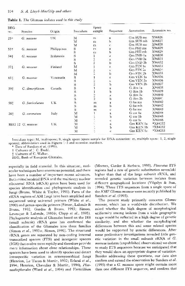
Table 1. The Glomus isolates used in this study
BEGno.
25t
551
54t
571
61J
59J
58t
2 8 |
BEG
Species
G. mosseae
G. mosseae
G. mosseae
G. mosseae
G. mosseae
G. dimorphietim
G. fasciculatum
G. coronatum
12 G. mosseae
Origin
UK
Philippines
Indonesia
Finland
Venezuela
Canada
U K
Italy
U K
Inoculum
MMMSS
sssMMMSSssssssssMMMMMM
Sporesample
mmmmm1221221221122mmm111111
Sequence
abcabaabaabaababababcabcabc
Annotation
Gm SUS maGm SUS mbGm SUS meGm PHI maGm PHI mbGmlNDlaGm IND 2aGm IND 2bGm FIN laGm FIN 2aGm FIN 2bGm VEN laGm VEN 2aGm VEN 2bG dim laG dim 1 bG dim 2aG dim 2bG fas maG fas mbG fas meG cot laG corlbG cor lcGmKENlaGmKEN lbGmKENlc
Accession no.
X96826X96827X96828X96829X96829X96830X96831X96832X96833X96834X96835X96836X96836X96837X96838X96839X96840X96841X96842X96842X96843X96844X96845X96846*X84232•X84232*X84233
Inoculum tj'pe: M, multispore; S, single spore (spore sample for DNA extraction: m, multiple spore; 1, 2, singlespores); abbreviation used in Figures 1-3 and accession numbers.
* Data of Sanders et al. (1995).t Cultures of J. C. Dodd.J Cultures of C. Walker.BEG, Bank of European Glomales.
especially in field material. In this situation, mol-ecular techniques have enormous potential, and therehave been a number of important recent advances.Both mitochondrial DNA and the multicopy nuclearribosomal RNA (rRNA) genes have been used forspecies identification and pbylogenetic analysis infungi (Bruns, White & Taylor, 1991). Parts of therRNA regions of AM fungi have been amplified andsequenced using universal primers (White et al.,1990) and genus-specific primers (Simon, Lalonde &Bruns, 1992, Gardes & Bruns, 1993; Simon,Levesque & Lalonde, 19936; Clapp et al, 1995).Phylogenetic analysis of Glomales based on the 18S(small subunit) rRNA gene has confirmed theclassification of the Glomales into three families(Simon et al, 1993a; Simon, 1996). The structuralrRNA genes are separated by non-coding internaltranscribed spacers (ITS) and intergenic spacers(IGS) that evolve more rapidly and therefore providemore information about close relationships. Thesespacers have been used to identify species and detectintraspecific variation in ectomycorrhizal fungi(Henrion, Le Tacon & Martin, 1992; Erland et al,1994; Henrion, Chevaher & Martin, 1994), Plas-modiophorales (Ward et al., 1994) and Verticillium
(Morton, Carder & Barbara, 1995). Pleurotus ITSregions had a rate of genetic substitution sevenfoldhigher than that of the large subunit rRNA, andrevealed genetic variation between isolates fromdifferent geographical locations (Vilgalys & Sun,1994). Three ITS sequences frotn a single spore ofthe AMF Glomus mosseae were recently published bySanders et a/. (1995).
The present study primarily concerns Glomusmosseae, which has a worldwide distribution. Weasked whether the high degree of morphologicalunifortnitj' atnong isolates from a wide geographicrange would be refiected in a high degree of geneticsimilarity, and also whether the morphologicaldifferences between this and some related specieswould be supported by genetic diflFerences. Aftersome preliminary investigations revealed little gen-etic variation in the small subunit rRNA of G.mosseae isolates (unpublished observations) we choseto study ITS sequences because we anticipated thatthey would show an appropriate degree of variation.Besides addressing these questions, our data alsoconfirm and extend the observation by Sanders et al.(1995) that a G. mosseae spore might contain morethan one different ITS sequence, and confirm that

ITS diversity in Glomus mosseae 105
all these ITS sequences are from Glomus itself ratberthan from associated fungi.
MATERIALS AND METHODS
Etingal isolates and culture conditions
Spores of G. dimorphicum Boyetchko & Te-wari, G.fasciculatum (Thaxter) Gerdemann & Trappeemend. Walker & Koske, G. coronatum Giovannettiand isolates of G. mosseae (Nicbolson & Gerdemann)Gerdemann & Trappe, were collected from es-tablished pot cultures and are presented in Table 1.
DNA extraction procedures
Spores vs'ere separated from pot culture medium byrepeated washing and re-suspension in water, andcollected using a 60 /im sieve. The remainingmaterial and spores were washed from the sieve intoa Petri dish. Spores were visualized with a lightmicroscope and collected using fine forceps. DNAwas extracted from the spores using either hexade-cyltrimethylammonium bromide (CTAB, Sigma) orInstagene* (6%, Bio-Rad). DNA from G. mosseae(BEG 25, BEG 55), and G. fasciculatum was extrac-ted using CTAB extraction buffer. DNA wasextracted from single spores of G. mosseae (BEG 57,BEG 54, BEG 61), G. dimorphicum and G. coronatumwith Instagene*.
(1) Multispore extraction. Eifty spores were crushedin liquid nitrogen within a microcentrifuge tubeusing a micropestle, re-suspended in 750 /i\ CTABextraction buffer (2% w/v CTAB, 1-4M NaCl,20 mM EDTA, lOOmM Tris-HCl pH 8-0, 0-2%(v/v) 2-mercaptoethanol) and incubated at 65 °C for30 min. The sample was extracted twice with anequal volume of phenol: chloroform: isoamyl alcohol(25:24:1 v/v/v, Sigma) and once with an equalvolume of chloroform (Sambrook, Fritsch &Maniatis, 1989). DNA was precipitated using 1/10volume 3 M Na acetate pH 5-2 and an equal volumeof isopropanol, incubated at —20 °C for 15 min andcentrifuged at 12000 jr for 15 min. The DNA pelletwas washed with 100 fil 70% (v/v) ethanol, driedunder vacuum and re-suspended in 20 /t! TE bufferpH 8 (10 mM Tris-HCl, 1 mM EDTA ). Sampleswere stored at —20 °C until use.
(2) Single spore extraction. A single spore was crushedin 20 /A Instagene using a micropestle and incubatedat 56 °C for 30 min. The sample was vortexed for10 s, incubated at 100 °C for 10 min, vortexed againfor 10 s and centrifuged at lOOOOj- for 3 min. Thesupernatant was removed and transferred into afresh microcentrifuge tube. Samples were stored at— 20 °C and used within a month.
PCR amplification
The 18S (3' part) and ITS regions of G. mosseae(BEG 25) were amplified using the primer pairNS5/ITS4 (White et al., 1990). The ITS regions ofthe other G. mosseae isolates, G. dimorphicum, G.fasciculatum and G. coronatum were amplified withprimer pairs ITS1/ITS4 (White et aL, 1990) orITSIE/ITS4 (Gardes & Bruns, 1993). Ampli-fications in a 50 /tl volume contained 2-5 /tl template,200 /tm dNTPs (Pharmacia), approx. 20 pmol eachprimer (Pharmacia), 2-5 units Taq DNA polymerase(Promega), 50 mM KCl, 10 mM Tris-HCl pH 9-0,0-1% Triton* X-lOO and 1-5 mM MgCl^. PCRamplification was carried out in a PTC-100 thermalcycler (MJ Research) with cycles as described byGardes & Bruns (1993). Negative controls withouttemplate were included.
Cloning procedures
PCR products were cloned into pGEM-T (VectorSystem 1, Promega) at a 1:3 molar ratio of vector toinsert and transformed into competent DH5a cells{Escherichia coli) using standard techniques andprocedures (Sambrook et al., 1989). Recombinantcolonies were selected and screened by PCR to checkfor the presence of inserts of c. 650 base pairs.Amplification reactions were carried out essentiallyas descibed above but in 16 /A total reaction volume,with T7 and SP6 primers (Promega), and bacterialcolonies as templates. Following the initial dena-turation step at 98 °C for 120 s, 25 cycles wereperformed at 98 °C for 20 s, 48 °C for 30 s, 72 °C for120 s, with a final extension time of 10 min at 72 °C..
DNA sequencing
Recombinant plasmid DNA was isolated and puri-fied using Wizard mini-prep DNA columns(Promega). Plasmid templates were sequenced usingthe T7 sequencing kit (Pharmacia) with a com-bination of the following primers; T7 (Promega),SP6 (Promega), ITSIE, ITS4, ITS2 and ITS3(White et al., 1990) primers. Sequence productswere electropboresed through a 6% acrylamide gel(Sequagel System,, Flowgen). Approximately 50 %of the 5.8S and ITS regions were sequenced in bothorientations and no discrepancies were observedbetween the strands. G. mosseae (BEG 25) wasfurther sequenced using primers NS5f(GCTTAACATTGTTAGGTCG) and ITSb(CTCACTAAGCCATTCAATC), generated fromthe sequence data, to complete the IBS and ITSregions. Sequence data were assembled usingDNASTAR (DNASTAR Inc) for the AppleMacintosh, and submitted to the EMBL databank(accession numbers in Table 1).
10'6 S. A. Lloyd-MacGilp and others
la r ^ M _ f-L pmL 1 ^ K 4 , '.J.I r i., r.L, r~i, '^J., "^ i^^ '-*, i"^ i-^ i-^ r-<4 " ^ "-.• '^^ " H '^- '^^ ' ^ i^"^
: : :".<< : :<g[: : : :S3S : : : : : i-^gSS
88BB888 8 888888B8B888888 8 888 S § ' s
888888888888 S 88888888888888 | j_
FQ. t^ PJ. F^ ' ^ " ^ '-i. ^^ ^J W | fHi "J. •-*- " ^ 1" 1^ '^'' '"N 1^ " ^ "U " ^ "^^ ^ * ^ * ^ ^ ^
88S8 8S88B,8 8 8B888388B8S8B888
? r r r ^ ^ . ? ^ . ^ . ^ - r r r - . r . r . r . r . r . r ,

ITS diversity in Glomus mosseae 107
U U U O U U U U C J U O U O L f U U U U O a U - U U U U U U
, - --' EH
r 3 L 5 o u u o t 5 o d t 3 d o d c i o c 5 o c i d w t 3 o o c u c 5 L ?
U U U U U O U D U O U U U U U U U U U U U U U O U U U
C i U U O C J C D O U C D ' O O D O C l l l J b C ' O O O O C i a O C i ' i l S C Dc u u o o L D O c D O O O o a o o u u i a a u O ' U O o c ; ! ; - " -U C J C J U U O ' ^ O U U U U U O O u O O c O U L l ' O U H EO U U U U U U L - ' U U f J ' L l U O U U U U C s U U U O p U L _< < < : < < f f C i < < < ; r t ; i < : < < < ' < < < t ; < ; r t : a : ; < < ; < S < : < ; <L > v - I U U L I U U U - L ; U U U U U J J L ; U U U U U U U C I & * E - f r «
• < < <
^ ^ F - E - ' H H B - i E H i ^ ^ E - i t ^ E - ' E - i t - i E - ' t - i E H E H E - ' E - i E - i H E - ' F - E - i H E -
1 u b tl b u b b u u u u u O O O u u LJ u u o c; u u bl I ' O C J C J - O D D U C J O O O C l U C - C i J C I U C J C S C J C D U C I C J t D L ?_ __ __ __ — __ __ — — _ _ _ _ _ . . , _ _ - _^ , ^ _- , j _ji f^ ^ _- 1 ^ - ^ „ - I - _P
d <; < < .
g g § p g | : | g g g g g g i g g g g g g g | g g g
TD- c d c c i:; d; a -iH -H -H a 01 fu .e g g I
rC ra-rci O 0 O« n ' ( i u i U U O
j B G B B e e f e B B B e B ^
siU LI
H tHC i .p - ,P_ i ^ f_ , ^ f -H^E- iE~ 'E^E- - ^E - *E - 'E - 'B^E- 'E - ^i b a u u o u t j - o u u u u u u u u o u u tj U U [
<: < < 'F- E- H E
U ^^ CjJ LJ ' ^ ' ^ ' ^ ' ^ '.-.' •—' - .. ^^ ^^ — -
U L I U U U U U U U U U U U U U C
S S ^ E
J d d C 5 C i C ! U D O O U O C i a O C ! > C 5 U U O D C J O Ou O - L : i j u u u o u u u o u u o u u o u o o u u u u a u88S8888888888S88888888888888888888888888S888888888 8 888

108 S. A. Lloyd-MacGilp and others
Phylogenetic analysis
Sequences were aligned using Clustal (DNASTAR)and further aligned manually to produce the mostplausible alignment. ClustalW (Thompson, Higgins& Gibson, 1994) was used to calculate distancematrices and to reconstruct phylogenies by theNeighbor-joining Method (Saitou & Nei, 1987).Trees were drawn using Phylip (Felsenstein, 1993).Two independent phylogenetic analyses were carriedout: firstly a conventional analysis based on nucleo-tide substitutions alone, ignoring positions withgaps, and secondly, an analysis based on lengthvariation, because small insertions and deletions(indels) were very frequent and therefore potentiallyinformative. For the latter analysis, the number ofgap symbols in each sequence at each indel site wasrecorded manually from the alignment (Fig. 1) andconverted into a pseudosequence by arbitrarilycoding 0,1 >2, and 3 gap symbols as A,T,G and Crespectively. The effect of this is that all differencesin length are weighted equally. Distances werecalculated from these pseudosequences without theusual correction for multiple hits, which would bebased on inappropriate assun:iptions.
RESULTS
Amplification and sequencing of ITS regions
The region between the nuclear SSU and LSUrRNA genes (c. 600 bp of DNA comprising ITSl ,the 5.8S rRNA gene, and ITS2) was amplified byPCR, cloned and sequenced from eight Glomusisolates (Table 1). Initial attempts at direct sequen-cing of PCR products from multiple spores andsingle spores yielded uninterpretable results becausethey generated mixed sequences, therefore all sequen-ces were obtained from cloned DNA. At least threesequences were obtained from each isolate, eitherfrom a multispore DNA preparation or from one ortwo individual spores. For G. mosseae (BEG 25), alonger sequence was obtained, including an ad-ditional 600 bp at the 5' end. This additional DNA,the 3' portion of the SSU rRNA gene, showed a98 % homology with the corresponding publishedsequence from G. mosseae (Simon et al., 1993 a),providing confirmation that the corresponding ITSsequence was indeed from G. mosseae rather thansome contaminating fungus.
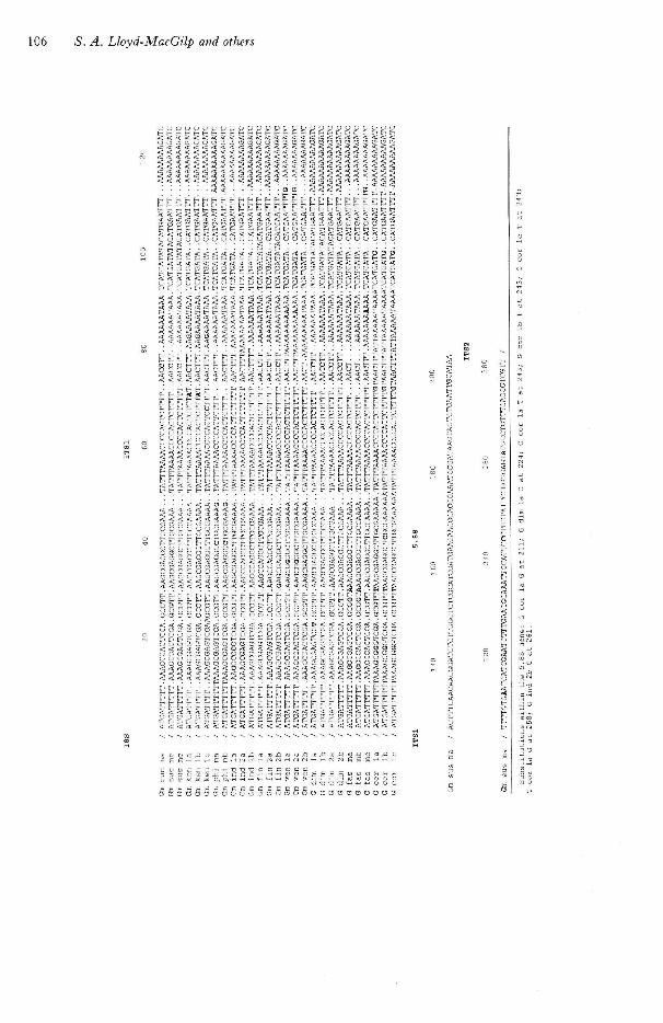
Alignment of I TS sequence data
A sequence alignment is presented in Figure 1,which also includes the sequences from G. mosseae(BEG 12) obtained previously by Sanders et al.(1995). The Glomus ITS sequences are all clearlyrelated to each other, but are only very distantlyrelated to any fungal ITS sequences in the sequence
59
100
Gdim Ib
G dim 2a
— GmFIN2a
— Gdimi2b
•GmFIN2b
-G dim la
GmFIN la
Gm SUS ma
Gm SUS mb
Gm SUS me
-GmlND2b
GmVENZb
Gm KEN la
Gm KEN lb
-GmKEN lc
75jGm VEN la
.96M Gm VEN 2a
'— G fas me
99 iGm PHI ma
' mb
54 Gm IND la
Gm IND 2a
100 G fas ma
G fas mb
53 -G cor la
0-005
I'Gcor lc
-G cor lb
Figure 2. Phylogenetic tree of the ITS and 5.8S regions;analysis based on nucleotide substitutions. Refer to Table1 for annotations. Bootstrap values greater than 50 % areshown.
databanks. There are numerous small diflFerences inlength which arise from small indels, i.e. insertionsor deletions of a few bases. Most of these indels areassociated with short runs of a single base (usually Aor T) and result from a change in the number ofrepeats of that base. In the ITS regions of thesequences in Figure 1 there are indels of one, two orthree bases at 22 distinct sites, while there are basesubstitutions at 71 sites out of 348 in the alignment.Comparing ITSl with iTS2, neither the ratio ofindels to substitutions (9:19 and 13:52) nor theoverall proportion of varying sites (24 % and 28 %) issignificantly diflFerent.
In contrast to the two non-coding ITS regions,the 5.8S rRNA gene that lies between them is muchmore conserved, with no indels and only six basesubstitutions (mostly unique to single sequences) in154 bp.
Sequence heterogeneity within isolates
The replicate sequences cloned from a single isolateare sometimes identical, but in most cases they arediflFerent even when they were derived from a singlespore. In some instances, sequences from diflFerent

ITS diversity in Qlomus mosseae 109
G dim la
Gdhm lb
G dim: 2a
75|Gm FIN la
Gm FIN 2b
Gm SUS me
g6|GmSUS ma
Gm SUS mb
KEN la
Gm KEN 1c
Gm VEN 2b
GmiNDla
831 Gm VEN la
Gm VEN 2a
G fas me
98 I G fas ma
G fas mb
Gm IND2a
Gm IND2b
jGmiPHI ma
iGm PHI mb
0-05
Figure 3. Phylogenetic tree of the ITS and S.8S regions;analysis based on insertions and deletions onl)'. Refer toTable 1 for annotations. Bootstrap values greater than50% are shown.
isolates are more similar than those from a singlespore (Figs 2, 3). Within isolates, the ratio of indelsto substitutions is lower than it is between isolates.Of the 71 sites at which substitutions occur, 44 \'aryonly within a single isolate, 17 vary only betweenisolates, and 10 vary both within and between. Thecorresponding figures for the 22 indel sites are 3, 8and 11, respectively, which are significantly differentproportions (x' = 18-3, df = 2, P < 0-005). The threeiodels that vary exclusively within an isolate are all inthe G. mosseae (BEG 12) sequences published bySanders et al. (1995).
Phylogeny based on nucleotide substitutions
A phylogenetic tree for all the sequences, based inthe conventional way on nucleotide substitutiondistances, is shown in Figure 2. The sequences fronaS. coronatum are very distinct from all the others11-16% divergence) and were used as an outgroup0 root the remainder of the tree. All the othersequences are fairly closely related (up to 10%iivergence), and few of the branches are stronglyupported by bootstrap analysis. Two importantacts are clear, though: the sequences from single
isolate do not necessarily cluster together, and thesequences from G. fasciculatum. and G. dimorphicumare embedded within the cluster of G. mosseaesequences.
Phylogeny based on nucleotide insertions anddeletions
Although substutions occur at 71 sites in the ITSregions, only 35 of these are phylogeneticallyinformative; in the other cases variants are confinedto a single sequence. By contrast, the indel sites arenearly all phylogenetically informative (19 out of 22),so they could contribute a significant independentset of data for the reconstruction of the tree. Adistance matrix was therefore calculated from thelength differences at the indel sites, and used toconstruct a tree (Fig. 3). This independent analysisgives strikingly similar results, confirming the dis-tinctness of G. coronatum and the lack of separationof G. fasciculatum, G. dimorphicum and G. mosseae.Furthermore, some branches receive support fromboth data sets, such as the grouping of G. dimor-phicum with G. mosseae isolates from Sussex(BEG 25) and Finland (BEG 57), and the associationof the G. fasciculatum c sequence with the la and 2asequences from the Venezuelan G. mosseae (BEG 61)isolate.
DISCUSSION
The sequences are from Glomus
The ITS sequences that we have obtained are notsimilar to any sequences in the databanks, except forthose reported by Sanders et al. (1995) from oneGlomus mosseae (BEG 12) spore. It is common forthe spores of AM fungi to be contaminated byhyphae of other soil fungi, and sequences from avariety of such fungi can be amplified by the primersthat we used (unpublished observations). It istherefore important to establish that the distinctix'egroup of ITS sequences reported here are genuinelythose of Glomus. Our sequences from G. mosseaeBEG 25 prove this point, as they encompass in asingle clone both the ITS regions and a diagnosticpart of the SSU gene,, which was 98 % identical tO'the SSU sequences ascribed to Glomus in theliterature (Simon et al., 1993 o). We can therefore beconfident that ITS sequences described here, andthose of Sanders et al. (1995), are indeed fromGlomus.
The sequences are sufficiently reliable
Tag polymerase was used for the polymerase chainreaction, because this enzyme adds an overhanging Ato the 3' end of the amplification products, whichallows rehable cloning into the pGEM-T vector.

110 S. A. Lloyd-MacGilp and others
However, it is well known that Taq polymeraseintroduces base substitution errors into the amplifiedsequences at a low but detectable rate. This has beenestimated at 0-25 % within a 30-cycle amplification(Saiki et aL, 1988), which would lead us to expectapprox. one error from this source in each of thesequences reported here. Clearly, even if there weresuch errors, they would not affect significantly any ofthe conclusions that we can draw. Furthermore,amplification errors are independent of the biologicalfunction of the sequence and should therefore occurwith equal frequency in the 5.8S rRNA gene and inthe ITS regions. In the 5.8S structural genes, thereare five base substitutions affecting single sequencesin the 27 sequences in Figure 1, a frequency of 0-1 %of nucleotides sequenced. A sinnilar frequency in theITS regions would only explain about 10 sub-stitutions, so the probability that two sequences haveindependently acquired the same error at the samesite, and therefore contribute false information to thephylogenetic analysis, is negligible.
Single spores have multiple ITS sequences
Sanders et al. (1995) published the sequences ofthree ITS clones from a single spore of G. mosseae(BEG 12); two were identical but the third differed.We have confirmed that ITS sequence heterogeneityis normal within Glomus spores. In fact, we obtainedpairs of sequences from single spores of threedifferent G. mossseae isolates and one isolate of G.dimorphicum, and three sequences from a single G.coronatum spore; in no instance were the sequencesidentical. The number of possible different sequen-ces per spore must be at least three, therefore, butthe pool of distinct sequences is not arbitrarily largebecause we did obtain identical sequences fromdifferent spores of one G. mosseae isolate, and alsofronn multispore preparations of several other iso-lates. It has been estimated that a Glomus spore may-contain from 1000 to 5000 nuclei (Giovannetti &Gianinazzi-Pearson, 1994) and it is normal for afungal nucleus to ha\'e multiple copies of theribosomal RNA genes, so it wilt be of great interestto determine whether the sequence heterogeneityarises because copies differ within a single nucleargenome or because the spore includes nuclei ofdifferent genotypes. Microheterogeneity has beenreported among the ribosomal gene copies inDrosophila (Schlotterer et aL, 1994), although thelevels of divergence that we find in Glomus arehigher. If, on the other hand, there are differencesbetween nuclei, then some mechanism or selectionmust exist to ensure that the diversity is not lost.
Glomus mosseae is similar worldwide
We examined isolates of G. mosseae that wereoriginally isolated in the UK (Sussex, BEG 25),
Finland (BEG 57), the Philippines (BEG 55), Indo-nesia (BEG 54) and Venezuela (BEG 61); and thepublished sequences of Sanders et al. (1995) are fromanother UK isolate from Kent, a county adjacent toSussex, (BEG 12). There is, however, no indicationof any grouping of the sequences hy geographicalorigin (Figs 2, 3). Indeed, the divergence ofsequences between isolates from distant continents isscarcely greater than the divergence found withinsingle spores. We conclude that the sequencevariants evolve on a time-scale that is long relative tothe rate at which these fungi spread across the world.
Glomus dimorphicum and G. fasciculatum are notdistinct from G. mosseae
The molecular data, both from base substitutionsand indels, clearly distinguish the isolate of G.coronatum from all the others in the study. However,the isolates of G. dimorphicum and G. fasciculatumhave sequences that place them firmly within therange of diversity found in G. mosseae, allying themwith G. mosseae isolates from Sussex (BEG 25) andFinland (BEG 57) on the one hand, and fromVenezuela (BEG 61) and Asia (BEG 55, BEG 54) onthe other. This is surprising because spore mor-phology, characterized hy colour, size, and occlusiontype, indicates that G. mosseae and G. fasciculatumare very distinct. However, the ITS sequence of asecond isolate of G fasciculatum (from Canada) isequally similar to those of G. mosseae (unpublishedobservations). G. mosseae, G. dimorphicum and G.coronatum have very similar spore morphologies,indicating that they are closely related. It has beenproposed that G. mosseae and G. dimorphicum mighthe conspecific (Walker, 1992), whereas G. coronatumis distinguished only by its darker spores (Dodd etal., 1996). Our sequence data therefore conflict withthe classification based on spore morphology in theirplacement of G. coronatum and G. fasciculatumrelative to G. mosseae. The relationship betweengenetic divergence and spore morphology is clearlynot straightforward, and data from a wider range ofspecies and genes are needed in order to resolve thisapparent conflict.
ACKNOWLEDGEMENTS
We thank James Merryweather, Jean Denison, AliceBroome and Edwin Lloyd-Jones for maintaining the potcultures. This work was supported by the BBSRC.
REFERENCES
Bruns TD, White TJ, Taylor JW. 1991. Fungal molecularsystematics. Annual Review of Ecology and Systematics 22:525-564.
Clapp JP, Young JPW, Merryweather JW, Fitter AH. 1995.Dii'ersity of fungal symbionts in arbuscular mycorrhizas froma natural community. Nas Phytologist 130: 59-265.
Dodd JC, Rosendahl S, Giovannetti M, Broome A,

ITS diversity in frlomus mosseae 111
Lanfranco L, Walker C 1996. Inter- and intraspecificvariation within the morphologically-similar arbuscular mycor-rhizal fungi Glomus mosseae/Giomus coronatum. Nezv Phy-toiogist, this volume: 000-000.
Erland S, Henrion B, Martin F, Glover LA, Alexander IJ.1994. Identiiication of the ectomycorrhizal basidiomyceteTylospora fibrillosa Donk by RFLP analysis of the PCR-amphiied ITS and IGS regions of ribosomal DKA, I^eivPhytologist 126: 525-532.
Felsenstein J. 1993 Phylip. (Phylogeny inference package)version 3.5c. Department of Genetics, University ofWashington, Seattle, USA.
Gardes M, Bruns TD. 1993. ITS primers with enhancedspecificity? for basidiomycetes - application to the identificationof mycorrhizae and rusts. Molecular Ecology 2: 113-18.
Giovannetti M, Gianinazzi-Pearson V. 1994. Biodiversity inarbuscular mycorrhizal fungi. Mycological Research 98:705-715.
Henrion B, Chevalier G, Martin F. 1994. Typing truffle speciesby PCR amphfication of tbe ribosomal DNA spacers. Myco-iogical Research 98: 3 7 ^ 3 .
Henrion B, Le Tacon F, Martin F. 1992. Rapid identification ofgenetic variation of ectomycorrhizal fungi by ampli6catjon ofribosmal RNA genes. New Phytologist 122: 289-29S.
Morton A, Carder JH, Barbara DJ. 1995. Sequences of theinternal transcribed spacers of the ribosomal RNA genes andrelationships between isolates of Verticillium alboatrum and V.dahliae. Plant Pathology 44: 183-190,
Morton JB, Bentivenga SP, 1994. Levels of diversisity inendomycorrhizal fungi (Glomales, Zygom^^cetes) and their rolem defining taxornomic and non-taxonomic groups, Plant andSoil 159: 47-59.
Saiki RK, Gelfand DH, Stoffel S, Scharf SJ, Higuchi R, HornGT, Mullis KB, Eriich HA. 1988. Primer-directed enzymaticamplification of DNA with a thermostable DNA polymerase.Science 239: 487^91 .
Saitou N, Nei M. 1987. The Keighbor-joining method: a newmethod for reconstructing phylogenetic trees, Molecular Si-ology of Evolution 4: 406^25..
Sambrook J, Fritsch EF, Maniatis T. 1989. Molecular cloning.A laboratory manual. (2nd edition). Cold Spring Harbor, USA:Cold Spring Harbor Laborator\^ Press.
Sanders IR, Alt M, Groppe K, Boiler T, Wiemken A. 1995.Identification of ribosoma! DNA polymorphisms among andwhhin spores of the Glomales: application to studies on thegenetic diversity of arbuscular mycorrhizal fungal communities.New Phytologist 130: 419—1-27.
Schlotterer C, Hauser M, von Haeseler A, Tautz D. 1994.Comparative evolutionary analysis of rDNA ITS regions inDrosophila. Molecular Biology of Evotutton 11: 513-522.
Simon L. 1996. Phylogeny of the Glomales: deciphering the pastto understand the present. New Phvtologist, this volume:000-000.
Simon L, Lalonde M, Bruns TD. 1992. Specific amplification of18S fungal ribosomal genes from vesicular-arbuscular endo-mycorrhizal fungt colonizing roots. Applied and EnvironmentalMicrobiology 58: 291-295.
Simon L, Bousquet J, Levesque RC, Lalonde M. 1993a.Origin and diversification of endoniycorrhizal fungi andcoincidence with • ^ascular land plants. Nature 363: 67-69.
Simon L, Levesque RC, Lalonde M. 19936. Identification ofendomycorrhizal fungi colonizing roots bv fluorescent single-strand conformation polymorphism-polymerase chain reaction.Applied and Environinental Microbiology 59: 4211-4215.
Thompson JD, Higgins DG, Gibson TJ. 1994. CLUSTAL W:improving the sensitivity of multiple sequence alignmentthrough sequence weighting, position-specific gap penalties andweight matrix choice. Nucleic Acids Research 2.2: 4673-4680.
Vilgalys R, Sun BL. 1994. Ancient and recent patterns ofgeographic speciation in the oyster mushroom Pleurotusrevealed by phylogenetic analysis of nbosomal DNA sequences.Proceedings of the National Academv of Science USA 91:4599^603.
"Walker C. 1992.. Systematics and taxonomy of the arbuscularendomycorrhi:aa] fungi (Glo'imales) - a possible way forward.Agronomie 12: 887-897.
White TJ, Bruns T, Lee S, Taylor J. 1990. Amplification anddirect sequencing of fungal ribosomal RNA genes for phylo-genetics. In: Innis MA, Gelfand DH, Snmsky JJ, White TJ,eds. PCR Protocols : a Guide to Methods and Applications. NewYork: Academic Press, 315-322.
Ward E, Adams MJ, Mutasa ES, Collier CR, Asher MJC.1994. Characterization of Polytnyxa species by restrictionanalysis of PCR-amplified ribosomal DNA. Plant Pathology 43:872-^877.

Copyright © 2022 FDOKUMEN