Capsaicin-enhanced Ribosomal Protein P2 Expression in Human Intestinal Caco-2 Cells

proteinsSTRUCTURE O FUNCTION O BIOINFORMATICS
STRUCTURE NOTE
Solution NMR structure of the ribosomalprotein RP-L35Ae from Pyrococcus furiosus
David A. Snyder,1,2 James M. Aramini,2 Bomina Yu,2 Yuanpeng J. Huang,2 Rong Xiao,2
John R. Cort,3 Ritu Shastry,2 Li-Chung Ma,2 Jinfeng Liu,4 Burkhard Rost,4 Thomas B. Acton,2
Michael A. Kennedy,5 and Gaetano T. Montelione2,6*1Department of Chemistry, College of Science and Health, William Paterson University, Wayne, New Jersey 07470
2Department of Molecular Biology and Biochemistry, Center for Advanced Biotechnology and Medicine, and The Northeast Structural Genomics Consortium,
Rutgers, The State University of New Jersey, Piscataway, New Jersey 08854
3 Biological Sciences Division, Pacific Northwest National Laboratory, and The Northeast Structural Genomics Consortium, Richland, Washington 99352
4Department of Biochemistry and Molecular Biophysics, Center for Computational Biology and Bioinformatics, and
The Northeast Structural Genomics Consortium, Columbia University, New York, New York 10032
5Department of Chemistry and Biochemistry, and The Northeast Structural Genomics Consortium, Miami University, Oxford, Ohio 45056
6Department of Biochemistry, Robert Wood Johnson Medical School, University of Medicine and Dentistry of New Jersey, Piscataway, New Jersey 08854
INTRODUCTION
Ribosomes are ubiquitous macromolecular complexescomposed of two subunits, each of which are made up ofseveral proteins and RNA molecules, hundreds of nucleo-tides in length. Ribosomes are responsible for proteinsynthesis in all living cells. The catalytic core of ribo-somes—where the information encoded on mRNA istranslated, tRNAs are bound, and peptide bonds areformed—is highly conserved throughout evolution. Pe-ripheral activities of the ribosome are less conserved,reflecting differences in the mechanisms of protein
Additional Supporting Information may be found in the online version of this
article.
Grant sponsor: National Institute of General Medical Sciences Protein Structure
Initiative (PSI); Grant numbers: U54-GM074958 and U54-GM094597 (Gaetano
T. Montelione).
Partial support was also provided to David A. Snyder as Assigned Release Time
from William Paterson University.
*Correspondence to: Gaetano T. Montelione, CABM, Rutgers University, 679 Hoes
Lane, Piscataway, NJ 08854. E-mail: [email protected]
Received 7 February 2012; Accepted 3 March 2012
Published online 16 March 2012 in Wiley Online Library (wileyonlinelibrary.com).
DOI: 10.1002/prot.24071
ABSTRACT
The ribosome consists of small and large subunits each composed of dozens of proteins and RNA molecules. However, the
functions of many of the individual protomers within the ribosome are still unknown. In this article, we describe the solu-
tion NMR structure of the ribosomal protein RP-L35Ae from the archaeon Pyrococcus furiosus. RP-L35Ae is buried within
the large subunit of the ribosome and belongs to Pfam protein domain family PF01247, which is highly conserved in eukar-
yotes, present in a few archaeal genomes, but absent in bacteria. The protein adopts a six-stranded anti-parallel b-barrelanalogous to the ‘‘tRNA binding motif ’’ fold. The structure of the P. furiosus RP-L35Ae presented in this article constitutes
the first structural representative from this protein domain family.
Proteins 2012; 00:00–00.VVC 2012 Wiley Periodicals, Inc.
Key words: ribosomal protein; L35Ae; PF01247; tRNA binding; EF-Tu/eEF-1A; solution NMR; structural genomics
.
Published 2012 WILEY PERIODICALS, INC. PROTEINS 1

synthesis. Eukaryotic ribosomes are substantially larger
and more complex than those of eubacteria. Homologous
eukaryotic ribosomal proteins often have extra protein
fragments and the RNA molecules have expansion seg-
ments, which, together with the �25 unique eukaryotic
ribosomal proteins, contribute to the observed 40%
increase in size.1
The RP-L35Ae domain family includes ribosomal pro-
teins known to be buried within the large subunit of the
ribosome2 and to bind both initiator and elongator
tRNA molecules.3 Eukaryotic homologs also appear to
function extra-ribosomally as inhibitors of apoptosis4
and are over-expressed in certain cancer cell lines, which
contribute to resistance to chemotherapeutic agents.5
This contrasts with many other proteins associated with
translation that function extra-ribosomally as promoters
of apoptosis.6 However, the precise roles of RP-L35Ae
proteins in the mechanism of translation and their
function(s) outside the ribosome are not known.
RP-L35Ae proteins belong to the highly conserved Pfam
PF01247 protein domain family7 [Fig. 1(a)]. Absent in
bacterial genomes, homologs of RP-L35Ae are found in
only four classes of archaea (www.orthology.org). The crys-
tal structure of the large ribosomal subunit of Haloarcula
marismortui lacks an RP-L35Ae homolog,8 further demon-
strating that this ribosomal protein is not required in at
least some archaeal ribosomes. In fact, the ribosomes of
many ‘‘crown’’ euryarchaeotes lack homologs for up to 10
eukaryotic ribosomal proteins, apparently a result of
‘‘reductive evolution.’’ In most cases, the collection of arch-
aea lacking a homolog of a particular eukaryotic ribosomal
protein form a monophyletic group (clade), however, the
organisms lacking RP-L35Ae homologs do not constitute a
single clade.9 Studies of isolated ribosomal proteins play a
critical role in elucidating the structure of ribosomes as a
whole and can provide valuable insights into the extra-ri-
bosomal functions of these proteins as well as into the
mechanisms of ribosomal reductive evolution.
In this article, we present the solution NMR structure
of RP-L35Ae from Pyrococcus furiosus. This protein was
selected for three-dimensional structure determination by
the Northeast Structural Genomics Consortium (NESG;
http://www.nesg.org) as part of the NIH Protein Struc-
ture Initiative on structural coverage of large, broadly
conserved protein domain families. RP-L35Ae is an 87-
residue basic protein (UniProtKB/Swiss-Prot ID, RL35A_-
PYRFU; NESG ID, PfR48). When deposited and released
in the PDB, its structure was the first structural represen-
tative of this important protein domain family.
MATERIALS AND METHODS
P. furiosus RP-L35Ae was cloned, expressed, and puri-
fied following standard NESG protocols.10 Briefly, the
rpl35Ae gene (UniProtKB/Swiss-Prot ID, RL35A_PYRFU;
NESG ID, PfR48; hereafter referred to as RP-L35Ae)
from P. furiosus was amplified from genomic DNA and
cloned into the pET21_NESG vector (Novagen) in frame
with a C-terminal affinity tag (LEHHHHHH), trans-
formed into E. coli BL21(DE3) pMGK cells, and
expressed overnight at 178C in MJ9 minimal media.11
Isotopically enriched samples were produced using U-
(15NH4)2SO4 and U-13C-glucose as the sole nitrogen and
carbon sources. Proteins were purified using an AKTAx-
press system (GE Healthcare) with a two-step protocol
consisting of IMAC (HisTRAP HP) and gel filtration
(HiLoad 26/60 Superdex 75) chromatography. Final
yields of 6 mg of [U-13C,15N]-RP-L35Ae and 25 mg of
[U-5%-13C,100%-15N]-RP-L35Ae were obtained per liter
of cell culture and concentrated to 0.3–1.1 mM in 90%
H2O/10% 2H2O, 20 mM MES, 100 mM NaCl, 5 mM
CaCl2, 10 mM DTT, 50 lM DSS, pH 6.5. The pET
expression vector for P. furiosus RP-L35Ae (NESG PfR48-
21.1), has been deposited in the PSI Materials Repository
(http://psimr.asu.edu/).
All NMR data for resonance assignment and structure
determination were collected at 293 K on Varian INOVA
600, 750, and 800 MHz and Varian UNITY 600 MHz
spectrometers equipped with 5 mm HCN probes, proc-
essed with NMRPipe 2.112 and visualized using SPARKY
3.106.13 All spectra were referenced to internal DSS.
Complete 1H, 13C, and 15N resonance assignments for
RP-L35Ae were determined using conventional triple res-
onance NMR methods. Backbone resonance assignments
were made by AutoAssign 1.914 using peak lists for 2D1H–15N HSQC and 3D HNCO, CBCA(CO)NH, and
HNCACB spectra. Side chain assignment was completed
manually using 3D HBHA(CO)NH, HCCH-COSY,
(H)CCH-TOCSY, and CC(CO)NH-TOCSY experiments.
Stereospecific isopropyl methyl resonance assignments for
all Val and Leu residues were determined from character-
istic cross-peak fine structures in high resolution 2D1H–13C HSQC spectra of [U-5%-13C,100%-15N]-RP-
L35Ae.15 Resonance assignments were validated using
the Assignment Validation Suite (AVS) software pack-
age.16 Three-bond 3J(HN-Ha) scalar couplings were
determined using the 3D HNHA experiment.17
The solution NMR structure of RP-L35Ae was calcu-
lated using CYANA 3.0,18,19 supplied with peak inten-
sities from the following NOESY spectra (with sm 5 140
ms and in 10% 2H2O, unless otherwise specified): 3D15N-edited NOESY (sm 5 100 ms), 3D 13C-edited ali-
phatic NOESY, 3D 13C-edited aliphatic NOESY (in 100%2H2O), and 3D 13C-edited aromatic NOESY, together
with broad dihedral angle constraints computed by
TALOS120 (/, w � 308) for ordered residues with con-
fidence scores of 10, and hydrogen bond constraints. The
20 structures with lowest target function out of 100 cal-
culated in the final cycle were further refined by
restrained molecular dynamics in explicit water using
CNS 1.321,22 and the PARAM19 force field, supplied
D.A. Snyder Et al.
2 PROTEINS
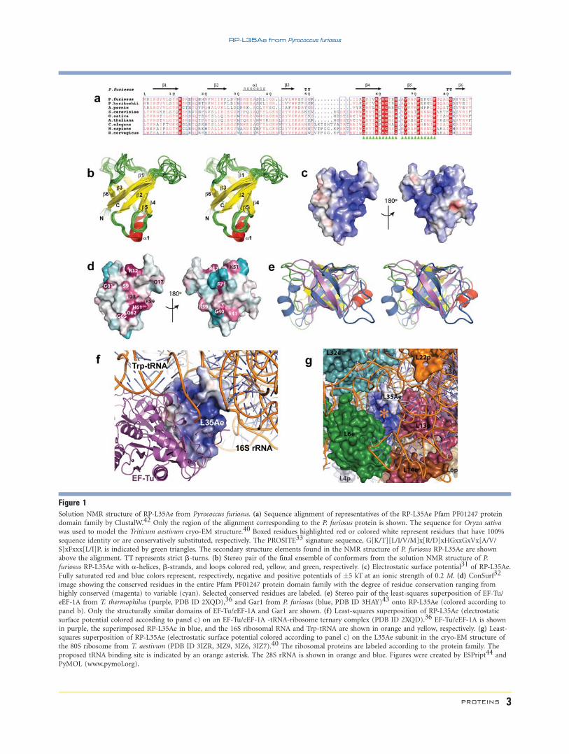
Figure 1Solution NMR structure of RP-L35Ae from Pyrococcus furiosus. (a) Sequence alignment of representatives of the RP-L35Ae Pfam PF01247 proteindomain family by ClustalW.42 Only the region of the alignment corresponding to the P. furiosus protein is shown. The sequence for Oryza sativa
was used to model the Triticum aestivum cryo-EM structure.40 Boxed residues highlighted red or colored white represent residues that have 100%
sequence identity or are conservatively substituted, respectively. The PROSITE33 signature sequence, G[K/T][L/I/V/M]x[R/D]xHGxxGxVx[A/V/
S]xFxxx[L/I]P, is indicated by green triangles. The secondary structure elements found in the NMR structure of P. furiosus RP-L35Ae are shown
above the alignment. TT represents strict b-turns. (b) Stereo pair of the final ensemble of conformers from the solution NMR structure of P.
furiosus RP-L35Ae with a-helices, b-strands, and loops colored red, yellow, and green, respectively. (c) Electrostatic surface potential31 of RP-L35Ae.
Fully saturated red and blue colors represent, respectively, negative and positive potentials of �5 kT at an ionic strength of 0.2 M. (d) ConSurf32
image showing the conserved residues in the entire Pfam PF01247 protein domain family with the degree of residue conservation ranging from
highly conserved (magenta) to variable (cyan). Selected conserved residues are labeled. (e) Stereo pair of the least-squares superposition of EF-Tu/
eEF-1A from T. thermophilus (purple, PDB ID 2XQD),36 and Gar1 from P. furiosus (blue, PDB ID 3HAY)43 onto RP-L35Ae (colored according to
panel b). Only the structurally similar domains of EF-Tu/eEF-1A and Gar1 are shown. (f) Least-squares superposition of RP-L35Ae (electrostatic
surface potential colored according to panel c) on an EF-Tu/eEF-1A -tRNA-ribosome ternary complex (PDB ID 2XQD).36 EF-Tu/eEF-1A is shown
in purple, the superimposed RP-L35Ae in blue, and the 16S ribosomal RNA and Trp-tRNA are shown in orange and yellow, respectively. (g) Least-
squares superposition of RP-L35Ae (electrostatic surface potential colored according to panel c) on the L35Ae subunit in the cryo-EM structure of
the 80S ribosome from T. aestivum (PDB ID 3IZR, 3IZ9, 3IZ6, 3IZ7).40 The ribosomal proteins are labeled according to the protein family. The
proposed tRNA binding site is indicated by an orange asterisk. The 28S rRNA is shown in orange and blue. Figures were created by ESPript44 and
PyMOL (www.pymol.org).
RP-L35Ae from Pyrococcus furiosus
PROTEINS 3

with the final NOE-derived distance, TALOS1 dihedral
angle, and hydrogen bond constraints. In this final stage
of the structure determination, rotamer states of specific-
ordered residues were constrained (v1, v2 �208) based
on MolProbity23,24 and PROCHECK25 analyses.
The final refined ensemble of 20 structures (excluding
the four C-terminal unassigned histidines) was deposited
into the Protein Data Bank (PDB ID 2LP6) and BioMa-
gResDB (BMRB accession number 6173). Structural sta-
tistics and global structure quality factors, including Veri-
fy3D,26 ProsaII,27 PROCHECK,25 and MolProbity23,24
raw and statistical Z-scores were computed using the
PSVS 1.4 software package.28 The global goodness-of-fit
of the final structure ensemble with the NOESY peak list
data and resonance assignments was determined using
the RPF analysis program.29
RESULTS AND DISCUSSION
The solution structure of RP-L35Ae reported in this
article corresponds to the first structural representative
from the Pfam PF01247 (Ribosomal_L35Ae) protein do-
main family.7 The protein eluted from the gel-filtration
column as a monomer during purification. The NMR
structure consists of a six-stranded anti-parallel b-barrelwith an additional a-helix between strands b2 and b3 of
the barrel [Fig. 1(b)]. Several lines of spectroscopic evi-
dence, analyzed automatically by AutoStructure30 and
manually validated, confirm the observed secondary
structure and barrel topology of b1-b2-b5-b4-b3-b6.These include the chemical shift values, 3J(HN–Ha) scalar
coupling constants, and the observed pattern of sequen-
tial and medium range NOEs (Supporting Information
Figure S1). Table I presents statistics that characterize the
quality of the resulting i.e., RP-L35Ae structure. Similar
structures and structure quality scores were obtained
using AutoStructure30 for automated NOESY spectral
analysis.
Electrostatic surface potential31 [Fig. 1(c)] and Con-
Surf32 [Fig. 1(d)] images reveal that one face of the RP-
L35Ae b-barrel is particularly positively charged and that
the PROSITE33 signature sequence for the RP-L35Ae fam-
ily [Fig. 1(a)] lies alongside a very cationic pocket on this
positively charged face. SCOP34 and CATH35 classify the
RP-L35Ae structure to a fold/topology class including
such tRNA binding domains as the b-barrel domains of
EF-Tu/eEF-1A36 and Gar137 [Fig. 1(e)]. Although, there
is no significant sequence similarity between RP-L35Ae
family members and other domains having the same
b-barrel structure, a structural superimposition of RP-
L35Ae into the structure of a ternary EF-Tu/eEF-1A—
tRNA—ribosome complex36 reveals that L35Ae and
EF-Tu/eEF-1A may share a similar tRNA binding site
[Fig. 1(f)]; in particular, when superimposed onto eEF-1A
in this protein-tRNA complex, the basic face of RP-L35Ae
is juxtaposed with the Trp-tRNA, indicating a tRNA-bind-
ing function for RP-L35Ae. This putative tRNA binding
site of RP-L35Ae proteins is corroborated by its sequence
conservation across the RP-L35Ae family. Other conserved
surface patches on the RP-L35Ae b-barrel may be involved
in binding other ribosomal proteins, as well as ribosomal
Table ISummary of NMR and Structural Statisticsa
RP-L35Ae
Completeness of resonance assignmentsb
Backbone (%) 98.2Side chain (%) 90.9Aromatic (%) 100Stereospecific methyl (%) 84.2
Conformationally restricting constraintsc
Distance constraintsTotal 1402Intra-residue (i 5 j) 390Sequential (|i-j| 5 1) 322Medium range (1 < |i 2 j| < 5) 122Long range (|i 2 j| � 5) 568
Dihedral angle constraints 153Hydrogen bond constraints 56No. of constraints per residue 18.3No. of long range constraints per residue 7.0
Residual constraint violationsc
Average No. of distance violationsper structure0.1–0.2 � 3.600.2–0.5 � 0.05>0.5 � 0
Average No. of dihedral angle violationsper structure1–108 12.55>108 0
Model qualityc
RMSD backbone atoms (�)d 0.5RMSD heavy atoms (�)d 1.0RMSD bond lengths (�) 0.018RMSD bond angles (8) 1.2
MolProbity Ramachandran statisticsc,d
Most favored regions (%) 97.0Allowed regions (%) 3.0Disallowed regions (%) 0
Global quality scores (raw/Z-score)c
Verify3D 0.39 21.12ProsaII 0.47 20.74Procheck (phi–psi)d 20.56 21.89Procheck (all) d 20.26 21.54MolProbity clash 15.13 21.07
RPF scorese
Recall/precision 0.96 0.87F-measure/DP-score 0.92 0.78
BMRB accession code 6173PDB accession codef 2LP6
aStructural statistics computed for the ensemble of 20 deposited structures.bComputed using AVS software16 from the expected number of peaks, excluding:
highly exchangeable protons (N-terminal and Lys amino and Arg guanido groups,
hydroxyls of Ser, Thr, Tyr), carboxyls of Asp and Glu, nonprotonated aromatic
carbons, and the C-terminal 6-His tag.cCalculated using PSVS 1.428. Average distance violations were calculated using
the sum over r26.dBased on ordered residue ranges [S(phi) 1 S(psi) > 1.8], 2-12,21-76,79-87.eRPF scores as defined by Huang et al.29.fPDB ID 2LP6 is a refined structure replacing PDB ID 1SQR.
D.A. Snyder Et al.
4 PROTEINS
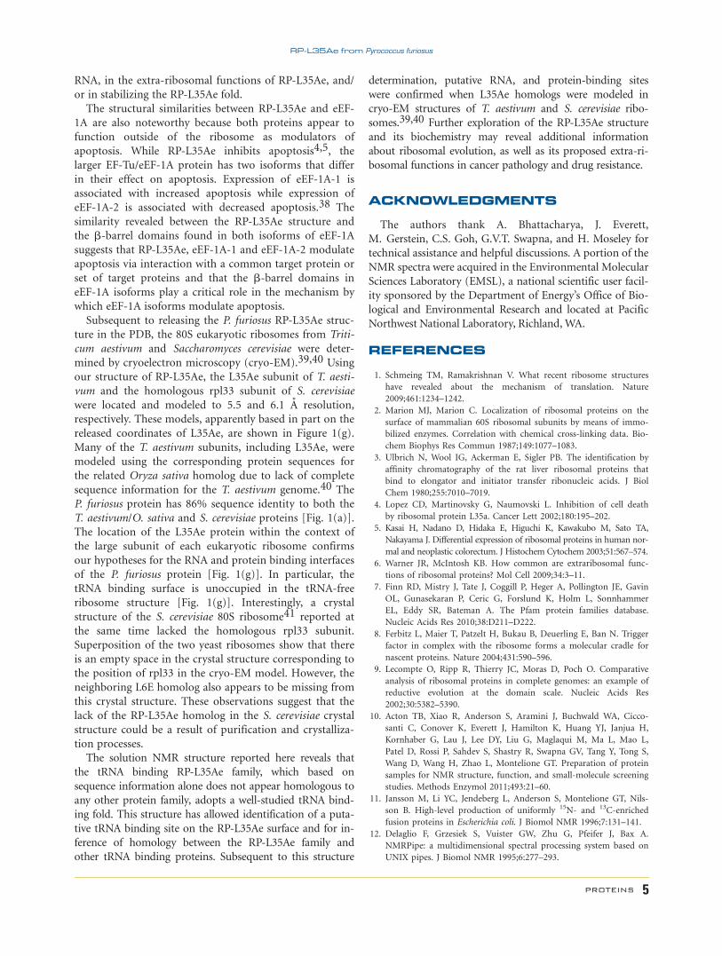
RNA, in the extra-ribosomal functions of RP-L35Ae, and/
or in stabilizing the RP-L35Ae fold.
The structural similarities between RP-L35Ae and eEF-
1A are also noteworthy because both proteins appear to
function outside of the ribosome as modulators of
apoptosis. While RP-L35Ae inhibits apoptosis4,5, the
larger EF-Tu/eEF-1A protein has two isoforms that differ
in their effect on apoptosis. Expression of eEF-1A-1 is
associated with increased apoptosis while expression of
eEF-1A-2 is associated with decreased apoptosis.38 The
similarity revealed between the RP-L35Ae structure and
the b-barrel domains found in both isoforms of eEF-1A
suggests that RP-L35Ae, eEF-1A-1 and eEF-1A-2 modulate
apoptosis via interaction with a common target protein or
set of target proteins and that the b-barrel domains in
eEF-1A isoforms play a critical role in the mechanism by
which eEF-1A isoforms modulate apoptosis.
Subsequent to releasing the P. furiosus RP-L35Ae struc-
ture in the PDB, the 80S eukaryotic ribosomes from Triti-
cum aestivum and Saccharomyces cerevisiae were deter-
mined by cryoelectron microscopy (cryo-EM).39,40 Using
our structure of RP-L35Ae, the L35Ae subunit of T. aesti-
vum and the homologous rpl33 subunit of S. cerevisiae
were located and modeled to 5.5 and 6.1 A resolution,
respectively. These models, apparently based in part on the
released coordinates of L35Ae, are shown in Figure 1(g).
Many of the T. aestivum subunits, including L35Ae, were
modeled using the corresponding protein sequences for
the related Oryza sativa homolog due to lack of complete
sequence information for the T. aestivum genome.40 The
P. furiosus protein has 86% sequence identity to both the
T. aestivum/O. sativa and S. cerevisiae proteins [Fig. 1(a)].
The location of the L35Ae protein within the context of
the large subunit of each eukaryotic ribosome confirms
our hypotheses for the RNA and protein binding interfaces
of the P. furiosus protein [Fig. 1(g)]. In particular, the
tRNA binding surface is unoccupied in the tRNA-free
ribosome structure [Fig. 1(g)]. Interestingly, a crystal
structure of the S. cerevisiae 80S ribosome41 reported at
the same time lacked the homologous rpl33 subunit.
Superposition of the two yeast ribosomes show that there
is an empty space in the crystal structure corresponding to
the position of rpl33 in the cryo-EM model. However, the
neighboring L6E homolog also appears to be missing from
this crystal structure. These observations suggest that the
lack of the RP-L35Ae homolog in the S. cerevisiae crystal
structure could be a result of purification and crystalliza-
tion processes.
The solution NMR structure reported here reveals that
the tRNA binding RP-L35Ae family, which based on
sequence information alone does not appear homologous to
any other protein family, adopts a well-studied tRNA bind-
ing fold. This structure has allowed identification of a puta-
tive tRNA binding site on the RP-L35Ae surface and for in-
ference of homology between the RP-L35Ae family and
other tRNA binding proteins. Subsequent to this structure
determination, putative RNA, and protein-binding sites
were confirmed when L35Ae homologs were modeled in
cryo-EM structures of T. aestivum and S. cerevisiae ribo-
somes.39,40 Further exploration of the RP-L35Ae structure
and its biochemistry may reveal additional information
about ribosomal evolution, as well as its proposed extra-ri-
bosomal functions in cancer pathology and drug resistance.
ACKNOWLEDGMENTS
The authors thank A. Bhattacharya, J. Everett,
M. Gerstein, C.S. Goh, G.V.T. Swapna, and H. Moseley for
technical assistance and helpful discussions. A portion of the
NMR spectra were acquired in the Environmental Molecular
Sciences Laboratory (EMSL), a national scientific user facil-
ity sponsored by the Department of Energy’s Office of Bio-
logical and Environmental Research and located at Pacific
Northwest National Laboratory, Richland,WA.
REFERENCES
1. Schmeing TM, Ramakrishnan V. What recent ribosome structures
have revealed about the mechanism of translation. Nature
2009;461:1234–1242.
2. Marion MJ, Marion C. Localization of ribosomal proteins on the
surface of mammalian 60S ribosomal subunits by means of immo-
bilized enzymes. Correlation with chemical cross-linking data. Bio-
chem Biophys Res Commun 1987;149:1077–1083.
3. Ulbrich N, Wool IG, Ackerman E, Sigler PB. The identification by
affinity chromatography of the rat liver ribosomal proteins that
bind to elongator and initiator transfer ribonucleic acids. J Biol
Chem 1980;255:7010–7019.
4. Lopez CD, Martinovsky G, Naumovski L. Inhibition of cell death
by ribosomal protein L35a. Cancer Lett 2002;180:195–202.
5. Kasai H, Nadano D, Hidaka E, Higuchi K, Kawakubo M, Sato TA,
Nakayama J. Differential expression of ribosomal proteins in human nor-
mal and neoplastic colorectum. J Histochem Cytochem 2003;51:567–574.
6. Warner JR, McIntosh KB. How common are extraribosomal func-
tions of ribosomal proteins? Mol Cell 2009;34:3–11.
7. Finn RD, Mistry J, Tate J, Coggill P, Heger A, Pollington JE, Gavin
OL, Gunasekaran P, Ceric G, Forslund K, Holm L, Sonnhammer
EL, Eddy SR, Bateman A. The Pfam protein families database.
Nucleic Acids Res 2010;38:D211–D222.
8. Ferbitz L, Maier T, Patzelt H, Bukau B, Deuerling E, Ban N. Trigger
factor in complex with the ribosome forms a molecular cradle for
nascent proteins. Nature 2004;431:590–596.
9. Lecompte O, Ripp R, Thierry JC, Moras D, Poch O. Comparative
analysis of ribosomal proteins in complete genomes: an example of
reductive evolution at the domain scale. Nucleic Acids Res
2002;30:5382–5390.
10. Acton TB, Xiao R, Anderson S, Aramini J, Buchwald WA, Cicco-
santi C, Conover K, Everett J, Hamilton K, Huang YJ, Janjua H,
Kornhaber G, Lau J, Lee DY, Liu G, Maglaqui M, Ma L, Mao L,
Patel D, Rossi P, Sahdev S, Shastry R, Swapna GV, Tang Y, Tong S,
Wang D, Wang H, Zhao L, Montelione GT. Preparation of protein
samples for NMR structure, function, and small-molecule screening
studies. Methods Enzymol 2011;493:21–60.
11. Jansson M, Li YC, Jendeberg L, Anderson S, Montelione GT, Nils-
son B. High-level production of uniformly 15N- and 13C-enriched
fusion proteins in Escherichia coli. J Biomol NMR 1996;7:131–141.
12. Delaglio F, Grzesiek S, Vuister GW, Zhu G, Pfeifer J, Bax A.
NMRPipe: a multidimensional spectral processing system based on
UNIX pipes. J Biomol NMR 1995;6:277–293.
RP-L35Ae from Pyrococcus furiosus
PROTEINS 5

13. Goddard TD, Kneller DG. SPARKY 3: University of California, San
Francisco. See also: http://www.cgl.ucsf.edu/home/sparky/.
14. Moseley HN, Monleon D, Montelione GT. Automatic determination
of protein backbone resonance assignments from triple resonance nu-
clear magnetic resonance data. Methods Enzymol 2001;339:91–108.
15. Neri D, Szyperski T, Otting G, Senn H, Wuthrich K. Stereospecific
nuclear magnetic resonance assignments of the methyl groups of
valine and leucine in the DNA-binding domain of the 434 repressor
by biosynthetically directed fractional 13C labeling. Biochemistry
1989;28:7510–7516.
16. Moseley HN, Sahota G, Montelione GT. Assignment validation soft-
ware suite for the evaluation and presentation of protein resonance
assignment data. J Biomol NMR 2004;28:341–355.
17. Vuister GW, Bax A. Quantitative J correlation: a new approach for
measuring homonuclear three-bond J(HNHa) coupling constants in15N-enriched proteins. J Am Chem Soc 1993;115:7772–7777.
18. Guntert P, Mumenthaler C, Wuthrich K. Torsion angle dynamics
for NMR structure calculation with the new program DYANA.
J Mol Biol 1997;273:283–298.
19. Herrmann T, Guntert P, Wuthrich K. Protein NMR structure deter-
mination with automated NOE assignment using the new software
CANDID and the torsion angle dynamics algorithm DYANA. J Mol
Biol 2002;319:209–227.
20. Shen Y, Delaglio F, Cornilescu G, Bax A. TALOS1: a hybrid
method for predicting protein backbone torsion angles from NMR
chemical shifts. J Biomol NMR 2009;44:213–223.
21. Brunger AT, Adams PD, Clore GM, DeLano WL, Gros P, Grosse-
Kunstleve RW, Jiang JS, Kuszewski J, Nilges M, Pannu NS, Read RJ,
Rice LM, Simonson T, Warren GL. Crystallography & NMR system:
a new software suite for macromolecular structure determination.
Acta Crystallogr D Biol Crystallogr 1998;54:905–921.
22. Linge JP, Williams MA, Spronk CA, Bonvin AM, Nilges M. Refinement
of protein structures in explicit solvent. Proteins 2003;50:496–506.
23. Lovell SC, Davis IW, Arendall WB, 3rd, de Bakker PI, Word JM,
Prisant MG, Richardson JS, Richardson DC. Structure validation by
Ca geometry: f,w and Cb deviation. Proteins 2003;50:437–450.
24. Davis IW, Leaver-Fay A, Chen VB, Block JN, Kapral GJ, Wang X,
Murray LW, Arendall WB, 3rd, Snoeyink J, Richardson JS, Richard-
son DC. MolProbity: all-atom contacts and structure validation for
proteins and nucleic acids. Nucleic Acids Res 2007;35:W375–W383.
25. Laskowski RA, MacArthur MW, Moss DS, Thornton JM. PRO-
CHECK: a program to check the stereochemical quality of protein
structures. J Appl Crystallogr 1993;26:283–291.
26. Luthy R, Bowie JU, Eisenberg D. Assessment of protein models
with three-dimensional profiles. Nature 1992;356:83–85.
27. Sippl MJ. Recognition of errors in three-dimensional structures of
proteins. Proteins 1993;17:355–362.
28. Bhattacharya A, Tejero R, Montelione GT. Evaluating protein struc-
tures determined by structural genomics consortia. Proteins
2007;66:778–795.
29. Huang YJ, Powers R, Montelione GT. Protein NMR recall, preci-
sion, and F-measure scores (RPF scores): structure quality assess-
ment measures based on information retrieval statistics. J Am
Chem Soc 2005;127:1665–1674.
30. Huang YJ, Tejero R, Powers R, Montelione GT. A topology-con-
strained distance network algorithm for protein structure determi-
nation from NOESY data. Proteins 2006;62:587–603.
31. Baker NA, Sept D, Joseph S, Holst MJ, McCammon JA. Electro-
statics of nanosystems: application to microtubules and the ribo-
some. Proc Natl Acad Sci USA 2001;98:10037–10041.
32. Glaser F, Pupko T, Paz I, Bell RE, Bechor-Shental D, Martz E,
Ben-Tal N. ConSurf: identification of functional regions in proteins
by surface-mapping of phylogenetic information. Bioinformatics
2003;19:163–164.
33. Falquet L, Pagni M, Bucher P, Hulo N, Sigrist CJ, Hofmann K,
Bairoch A. The PROSITE database, its status in 2002. Nucleic Acids
Res 2002;30:235–238.
34. Murzin AG, Brenner SE, Hubbard T, Chothia C. SCOP: a structural
classification of proteins database for the investigation of sequences
and structures. J Mol Biol 1995;247:536–540.
35. Orengo CA, Michie AD, Jones S, Jones DT, Swindells MB, Thorn-
ton JM. CATH – hierarchic classification of protein domain struc-
tures. Structure 1997;5:1093–1108.
36. Voorhees RM, Schmeing TM, Kelley AC, Ramakrishnan V. The
mechanism for activation of GTP hydrolysis on the ribosome. Sci-
ence 2010;330:835–838.
37. Rashid R, Liang B, Baker DL, Youssef OA, He Y, Phipps K, Terns
RM, Terns MP, Li H. Crystal structure of a Cbf5-Nop10-Gar1 com-
plex and implications in RNA-guided pseudouridylation and dys-
keratosis congenita. Mol Cell 2006;21:249–260.
38. Mateyak MK, Kinzy TG. eEF1A: thinking outside the ribosome. J
Biol Chem 2010;285:21209–21213.
39. Armache JP, Jarasch A, Anger AM, Villa E, Becker T, Bhushan S,
Jossinet F, Habeck M, Dindar G, Franckenberg S, Marquez V,
Mielke T, Thomm M, Berninghausen O, Beatrix B, Soding J, West-
hof E, Wilson DN, Beckmann R. Cryo-EM structure and rRNA
model of a translating eukaryotic 80S ribosome at 5.5-A resolution.
Proc Natl Acad Sci USA 2010;107:19748–19753.
40. Armache JP, Jarasch A, Anger AM, Villa E, Becker T, Bhushan S,
Jossinet F, Habeck M, Dindar G, Franckenberg S, Marquez V,
Mielke T, Thomm M, Berninghausen O, Beatrix B, Soding J, West-
hof E, Wilson DN, Beckmann R. Localization of eukaryote-specific
ribosomal proteins in a 5.5-A cryo-EM map of the 80S eukaryotic
ribosome. Proc Natl Acad Sci USA 2010;107:19754–19759.
41. Ben-Shem A, Jenner L, Yusupova G, Yusupov M. Crystal structure
of the eukaryotic ribosome. Science 2010;330:1203–1209.
42. Chenna R, Sugawara H, Koike T, Lopez R, Gibson TJ, Higgins DG,
Thompson JD. Multiple sequence alignment with the Clustal series
of programs. Nucleic Acids Res 2003;31:3497–3500.
43. Duan J, Li L, Lu J, Wang W, Ye K. Structural mechanism of sub-
strate RNA recruitment in H/ACA RNA-guided pseudouridine syn-
thase. Mol Cell 2009;34;427–439.
44. Gouet P, Courcelle E, Stuart DI, Metoz F. ESPript: analysis of multiple
sequence alignments in PostScript. Bioinformatics 1999;15:305–308.
D.A. Snyder Et al.
6 PROTEINS
Copyright © 2022 FDOKUMEN