Rapid disassembly of dynamic microtubules upon activation of the capsaicin receptor TRPV1
Upload
independentCategory
view
2download
0
-1
Erratum
Due to an unfortunate turn of events the uncorrected version of this article was published inCytotechnology 47:89–96.5
Capsaicin-enhanced ribosomal protein P2 expression in human intestinal
Caco-2 cells
Junkyu Han, Mitsuaki Akutsu, Terence P.N. Talorete, Takaaki Maekawa,Toshiyuki Tanaka and Hiroko Isoda*
Graduate School of Life and Environmental Sciences, University of Tsukuba, Tennodai 1-1-1, Tsukuba,Ibaraki 305-8572, Japan; *Author for correspondence (e-mail: [email protected]; phone: +81-29-853-5775; fax: +81-29-853-5776)
The following pages contain the corrected article and should therefore be treated as definitive by the reader.
Cytotechnology (2005) 49:77–85 � Springer 2005
DOI 10.1007/s10616-005-6709-0
-1
Capsaicin-enhanced ribosomal protein P2 expression in human intestinal
Caco-2 cells
Junkyu Han, Mitsuaki Akutsu, Terence P.N. Talorete, Takaaki Maekawa,Toshiyuki Tanaka and Hiroko Isoda*Graduate School of Life and Environmental Sciences, University of Tsukuba, Tennodai 1-1-1, Tsukuba,Ibaraki 305-8572, Japan; *Author for correspondence (e-mail: [email protected]; phone: +81-29-853-5775; fax: +81-29-853-5776)
Received 1 December 2004; accepted in revised form 25 January 2005
Key words: Caco-2 Cells, Capsaicin, Elongation factor 2, F-actin, Proteome analysis, Ribosomal proteinP2, Tight-junction permeability
Abstract
On the basis of transepithelial electrical resistance (TER) measurements, we found that capsaicin (100 lM)-treated human intestinal Caco-2 cells show a momentary increase in tight-junction (TJ) permeability (de-crease in TER) followed by a complete recovery. We used proteome analysis to search for proteins that areassociated with the recovery of TJ permeability in capsaicin-treated Caco-2 cells. A protein with a relativemolecular mass of 14 kDa was found to be expressed more highly in capsaicin-treated cells than in non-treated cells. Mass spectrometry and sequence analyses revealed that the protein that is expressed signifi-cantly upon capsaicin treatment is the ribosomal protein P2; its cDNA sequence was identical to that foundin the human genome database. An increase in the amount of cellular filamentous actin (F-actin) wasshown after 8 h of incubation with capsaicin. It has been reported that P2 activates elongation factor 2,which stabilizes F-actin filaments, and that the depolymerization of F-actin is associated with the increasein TJ permeability (decrease in TER). Consequently, these results suggest that P2 plays an important role inthe recovery of the TJ permeability in capsaicin-treated human intestinal cells.
Introduction
Human intestinal Caco-2 cells (Fogh et al. 1977)have been widely used as in vitro models toevaluate the transport of absorbed water, ions,and nutrients across the intestinal epithelial bar-rier (Satsu et al. 2003). The tight-junction (TJ)permeability increase in Caco-2 cells is regulatedby various factors, such as food factors (forexample, capsaicin and capsianoside.) and chem-icals (such as alkylphenolic compounds andEDTA). Such factors can alter the epithelialtransport by and barrier function of humanintestinal epithelial cells using various mechanisms
(cytoskeletal reorganization, redistribution ofZO-1 and occludin, exertion of Rho A, Rac 1,and Cdc 42, etc.) (Nusrat et al. 1995; Yap et al.1998; Han et al. 2002; Bruewer et al. 2004).
In previous studies (Isoda et al. 2001; Han et al.2002), we showed that the TJ permeability increase(decrease in transepithelial electrical resistance(TER)) in capsaicin-treated (45 min) humanintestinal Caco-2 cells is through binding of cap-saicin to a capsaicin receptor-like protein. We alsosuggested that the increase in TJ permeabilityupon capsaicin treatment occurs due to acytoskeletal reorganization of actin filaments,particularly due to the decrease in the amount
Cytotechnology (2005) 47:89–96 � Springer 2005
DOI 10.1007/s10616-005-3756-5
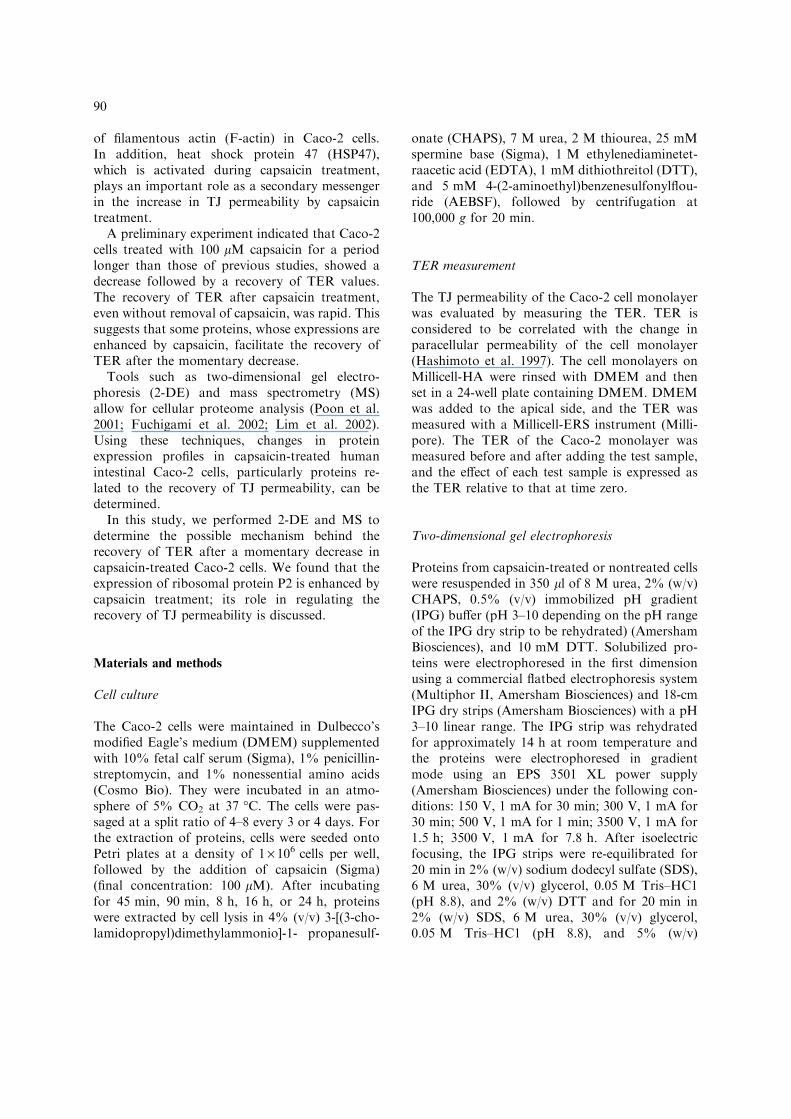
of filamentous actin (F-actin) in Caco-2 cells.In addition, heat shock protein 47 (HSP47),which is activated during capsaicin treatment,plays an important role as a secondary messengerin the increase in TJ permeability by capsaicintreatment.
A preliminary experiment indicated that Caco-2cells treated with 100 lM capsaicin for a periodlonger than those of previous studies, showed adecrease followed by a recovery of TER values.The recovery of TER after capsaicin treatment,even without removal of capsaicin, was rapid. Thissuggests that some proteins, whose expressions areenhanced by capsaicin, facilitate the recovery ofTER after the momentary decrease.
Tools such as two-dimensional gel electro-phoresis (2-DE) and mass spectrometry (MS)allow for cellular proteome analysis (Poon et al.2001; Fuchigami et al. 2002; Lim et al. 2002).Using these techniques, changes in proteinexpression profiles in capsaicin-treated humanintestinal Caco-2 cells, particularly proteins re-lated to the recovery of TJ permeability, can bedetermined.
In this study, we performed 2-DE and MS todetermine the possible mechanism behind therecovery of TER after a momentary decrease incapsaicin-treated Caco-2 cells. We found that theexpression of ribosomal protein P2 is enhanced bycapsaicin treatment; its role in regulating therecovery of TJ permeability is discussed.
Materials and methods
Cell culture
The Caco-2 cells were maintained in Dulbecco’smodified Eagle’s medium (DMEM) supplementedwith 10% fetal calf serum (Sigma), 1% penicillin-streptomycin, and 1% nonessential amino acids(Cosmo Bio). They were incubated in an atmo-sphere of 5% CO2 at 37 �C. The cells were pas-saged at a split ratio of 4–8 every 3 or 4 days. Forthe extraction of proteins, cells were seeded ontoPetri plates at a density of 1 · 106 cells per well,followed by the addition of capsaicin (Sigma)(final concentration: 100 lM). After incubatingfor 45 min, 90 min, 8 h, 16 h, or 24 h, proteinswere extracted by cell lysis in 4% (v/v) 3-[(3-cho-lamidopropyl)dimethylammonio]-1- propanesulf-
onate (CHAPS), 7 M urea, 2 M thiourea, 25 mMspermine base (Sigma), 1 M ethylenediaminetet-raacetic acid (EDTA), 1 mM dithiothreitol (DTT),and 5 mM 4-(2-aminoethyl)benzenesulfonylflou-ride (AEBSF), followed by centrifugation at100,000 g for 20 min.
TER measurement
The TJ permeability of the Caco-2 cell monolayerwas evaluated by measuring the TER. TER isconsidered to be correlated with the change inparacellular permeability of the cell monolayer(Hashimoto et al. 1997). The cell monolayers onMillicell-HA were rinsed with DMEM and thenset in a 24-well plate containing DMEM. DMEMwas added to the apical side, and the TER wasmeasured with a Millicell-ERS instrument (Milli-pore). The TER of the Caco-2 monolayer wasmeasured before and after adding the test sample,and the effect of each test sample is expressed asthe TER relative to that at time zero.
Two-dimensional gel electrophoresis
Proteins from capsaicin-treated or nontreated cellswere resuspended in 350 ll of 8 M urea, 2% (w/v)CHAPS, 0.5% (v/v) immobilized pH gradient(IPG) buffer (pH 3–10 depending on the pH rangeof the IPG dry strip to be rehydrated) (AmershamBiosciences), and 10 mM DTT. Solubilized pro-teins were electrophoresed in the first dimensionusing a commercial flatbed electrophoresis system(Multiphor II, Amersham Biosciences) and 18-cmIPG dry strips (Amersham Biosciences) with a pH3–10 linear range. The IPG strip was rehydratedfor approximately 14 h at room temperature andthe proteins were electrophoresed in gradientmode using an EPS 3501 XL power supply(Amersham Biosciences) under the following con-ditions: 150 V, 1 mA for 30 min; 300 V, 1 mA for30 min; 500 V, 1 mA for 1 min; 3500 V, 1 mA for1.5 h; 3500 V, 1 mA for 7.8 h. After isoelectricfocusing, the IPG strips were re-equilibrated for20 min in 2% (w/v) sodium dodecyl sulfate (SDS),6 M urea, 30% (v/v) glycerol, 0.05 M Tris–HC1(pH 8.8), and 2% (w/v) DTT and for 20 min in2% (w/v) SDS, 6 M urea, 30% (v/v) glycerol,0.05 M Tris–HC1 (pH 8.8), and 5% (w/v)
90
iodoacetamide. The strip was placed on a gradientSDS-PAGE gel (12–14% (w/v) polyacrylamide)and run at 1000 V, 20 mA for 45 min and at1000 V, 40 mA for 160 min (Poon et al. 2001). Theproteins were visualized by Coomassie BrilliantBlue (CBB) staining (using PhastGel Blue R-350,Amersham Biosciences) for spot analysis. The spotanalysis software, ImageMaster 2D Elite (Amer-sham Biosciences), allows for the magnification ofspecific fields of view and assists greatly in the vi-sual comparison of proteins between control andexperimental gels.
Mass spectrometry
Protein spots of interest were excised from the gel,washed and digested in-gel with trypsin (sequenc-ing grade, Boehringer Mannheim). All matrix-as-sisted laser desorption/ionization time-of-flight(MALDI-TOF) mass spectra were acquired usinga Voyager-DE STR mass spectrometer (AppliedBiosystems). The matrix solution was prepared bydissolving 10 mg of a -cyano-4-hydroxycinnamicacid (Sigma) in 1 ml of 50% acetonitorile and0.1% trifluoroacetic acid in deionized water. Theobtained peptide sequence tags were used toidentify proteins by searching databases usingBLAST (http://www.ncbi.nlm.nih.gov:80/blast) orthe Prospector software MS-Tag (http://prospec-tor.ucsf.edu/).
Preparation of RNA and the cDNA library
Total RNA was extracted from Caco-2 cellsincubated with 100 lM capsaicin for 16 h usingISOGEN (Nippon Gene), and poly(A) + RNA
was isolated using the Poly(A) + RNA IsolationKit (Nippon Gene). Double-stranded cDNA wassynthesized from Poly(A) + RNA using the Uni-versal RiboClone cDNA Synthesis System (Pro-mega).
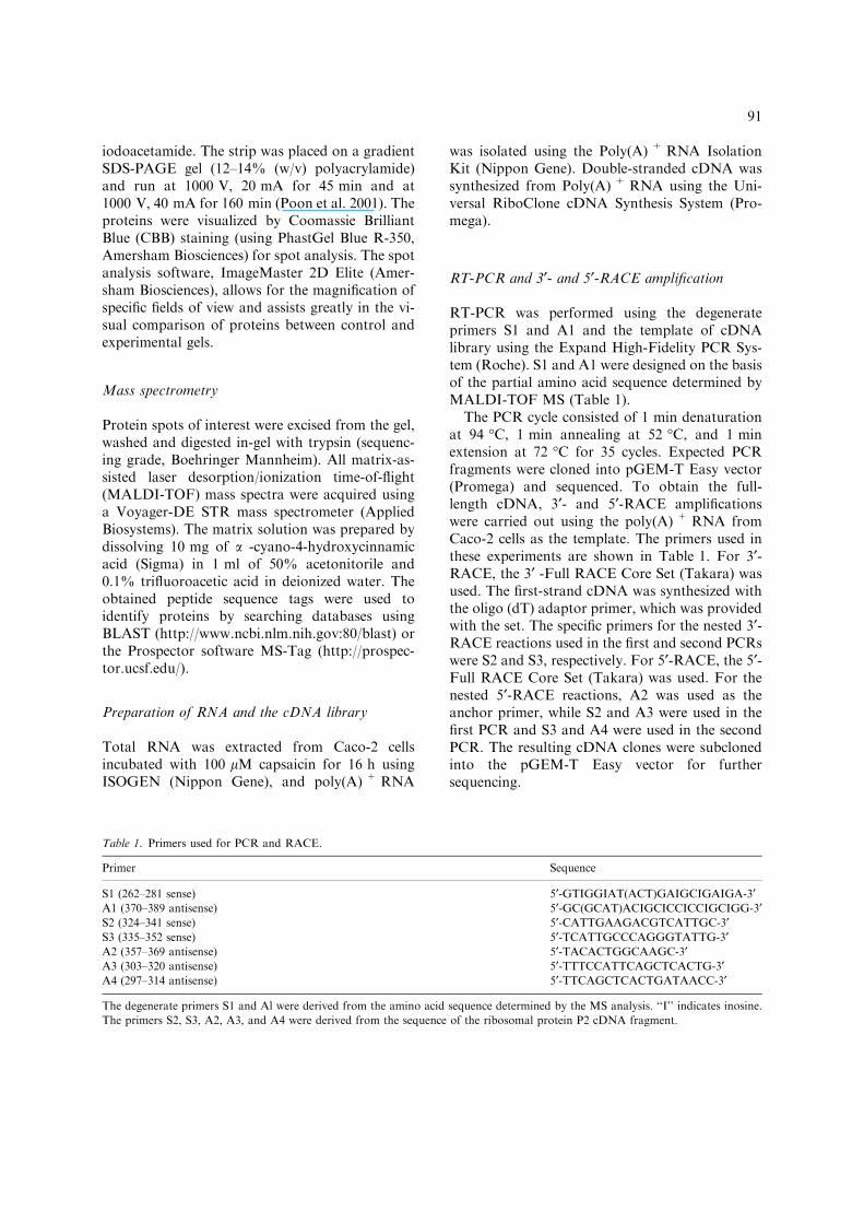
RT-PCR and 3¢- and 5¢-RACE amplification
RT-PCR was performed using the degenerateprimers S1 and A1 and the template of cDNAlibrary using the Expand High-Fidelity PCR Sys-tem (Roche). S1 and A1 were designed on the basisof the partial amino acid sequence determined byMALDI-TOF MS (Table 1).
The PCR cycle consisted of 1 min denaturationat 94 �C, 1 min annealing at 52 �C, and 1 minextension at 72 �C for 35 cycles. Expected PCRfragments were cloned into pGEM-T Easy vector(Promega) and sequenced. To obtain the full-length cDNA, 3¢- and 5¢-RACE amplificationswere carried out using the poly(A) + RNA fromCaco-2 cells as the template. The primers used inthese experiments are shown in Table 1. For 3¢-RACE, the 3¢ -Full RACE Core Set (Takara) wasused. The first-strand cDNA was synthesized withthe oligo (dT) adaptor primer, which was providedwith the set. The specific primers for the nested 3¢-RACE reactions used in the first and second PCRswere S2 and S3, respectively. For 5¢-RACE, the 5¢-Full RACE Core Set (Takara) was used. For thenested 5¢-RACE reactions, A2 was used as theanchor primer, while S2 and A3 were used in thefirst PCR and S3 and A4 were used in the secondPCR. The resulting cDNA clones were subclonedinto the pGEM-T Easy vector for furthersequencing.
Table 1. Primers used for PCR and RACE.
Primer Sequence
S1 (262–281 sense) 5¢-GTIGGIAT(ACT)GAIGCIGAIGA-3¢A1 (370–389 antisense) 5¢-GC(GCAT)ACIGCICCICCIGCIGG-3¢S2 (324–341 sense) 5¢-CATTGAAGACGTCATTGC-3¢S3 (335–352 sense) 5¢-TCATTGCCCAGGGTATTG-3¢A2 (357–369 antisense) 5¢-TACACTGGCAAGC-3¢A3 (303–320 antisense) 5¢-TTTCCATTCAGCTCACTG-3¢A4 (297–314 antisense) 5¢-TTCAGCTCACTGATAACC-3¢
The degenerate primers S1 and Al were derived from the amino acid sequence determined by the MS analysis. ‘‘I’’ indicates inosine.
The primers S2, S3, A2, A3, and A4 were derived from the sequence of the ribosomal protein P2 cDNA fragment.
91
Measurement of cellular F-actin
The relative content of F-actin was determined bya fluorescent phalloidin binding assay. Caco-2 cellsthat had been incubated on a slide chamber for3 days were rinsed with phosphate-buffered saline(PBS) and then incubated with capsaicin (100 lM)for 0, 8, 16, or 24 h. After incubation, the cellmonolayer was fixed with acetone/methanol (1/1 = v/v), and then the actin was stained for20 min with rhodamine-phalloidin (Wako) dilutedtenfold with PBS. The stained cells were extractedwith 2 ml of methanol, and the fluorescenceintensity of the extract was determined using aFluoroscan Ascent FL fluorescence spectrophoto-meter (Labsystems) with excitation-emissionwavelengths of 545–578 nm.
Statistical analysis
Statistical analysis was carried out using Student’st-test, and relationships were considered statisti-cally significant at p<0.05.
Results and discussion
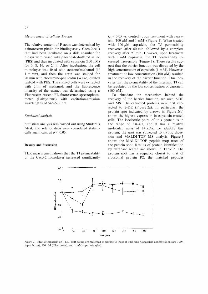
TER measurement shows that the TJ permeabilityof the Caco-2 monolayer increased significantly
(p<0.05 vs. control) upon treatment with capsa-icin (100 lM and 1 mM) (Figure 1). When treatedwith 100 lM capsaicin, the TJ permeabilityrecovered after 60 min, followed by a completerecovery after 90 min. However, upon treatmentwith 1 mM capsaicin, the TJ permeability in-creased irreversibly (Figure 1). These results sug-gest that the barrier function was disrupted by thehigh concentration of capsaicin (1 mM). However,treatment at low concentration (100 lM) resultedin the recovery of the barrier function. This indi-cates that the permeability of the intestinal TJ canbe regulated by the low concentration of capsaicin(100 lM).
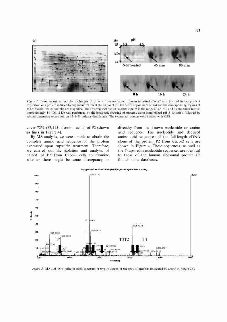
To elucidate the mechanism behind therecovery of the barrier function, we used 2-DEand MS. The extracted proteins were first sub-jected to 2-DE (Figure 2a). In particular, theprotein spot indicated by arrows in Figure 2(b)shows the highest expression in capsaicin-treatedcells. The isoelectric point of this protein is inthe range of 3.8–4.3, and it has a relativemolecular mass of 14 kDa. To identify thisprotein, the spot was subjected to tryptic diges-tion and MALDI-TOF MS analysis. Figure 3shows the MALDI-TOF peptide map trace ofthe protein spot. Results of protein identificationby database search are shown in Table 2. Theprotein spot has a sequence closest to that ofribosomal protein P2; the matched peptides
Figure 1. Effect of capsaicin on TER. TER values are presented as relative to those at time zero. Capasaicin concentrations are 0 lM(open boxes), 100 lM (filled boxes), and 1 mM (open triangles).
92
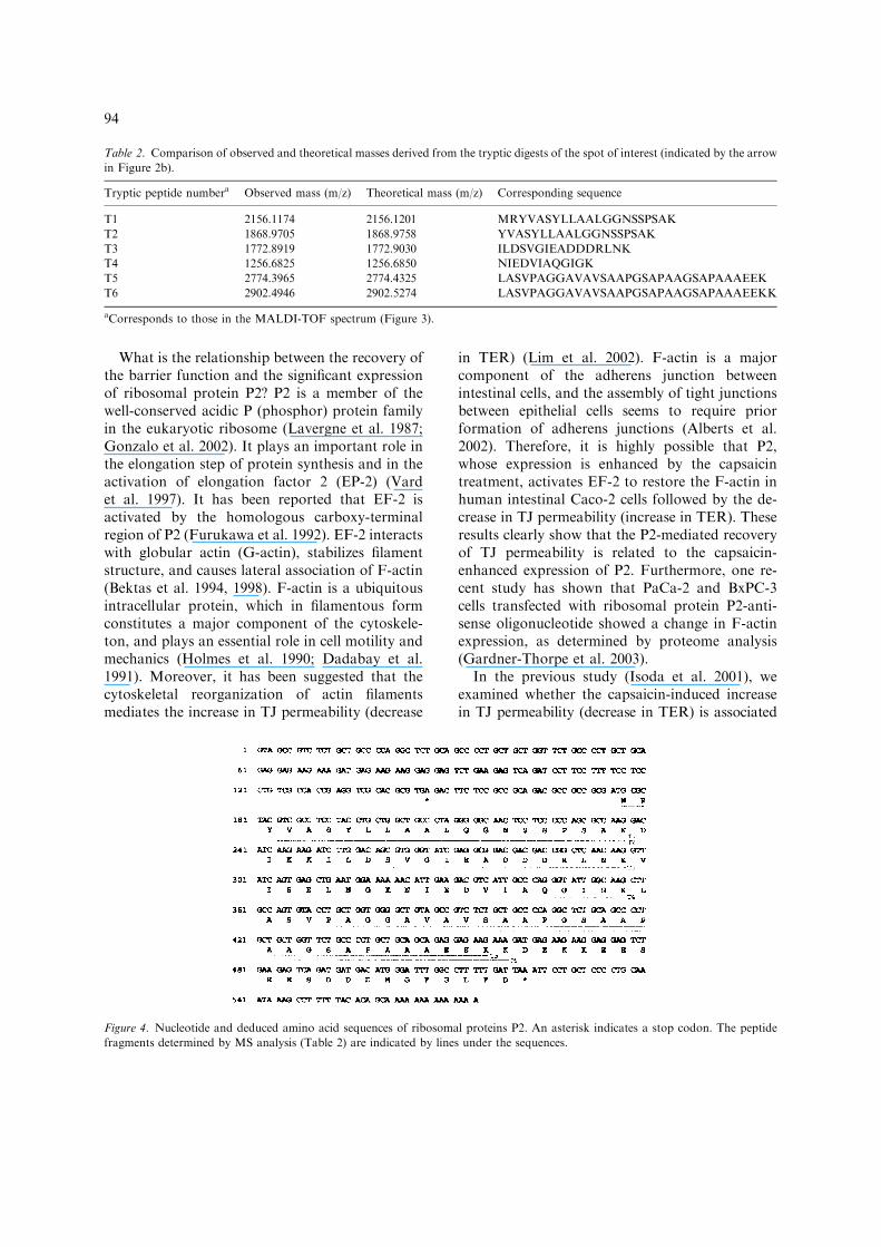
cover 72% (83/115 of amino acids) of P2 (shownas lines in Figure 4).
By MS analysis, we were unable to obtain thecomplete amino acid sequence of the proteinexpressed upon capsaicin treatment. Therefore,we carried out the isolation and analysis ofcDNA of P2 from Caco-2 cells to examinewhether there might be some discrepancy or
diversity from the known nucleotide or aminoacid sequence. The nucleotide and deducedamino acid sequences of the full-length cDNAclone of the protein P2 from Caco-2 cells areshown in Figure 4. These sequences, as well asthe 5¢-upstream nucleotide sequence, are identicalto those of the human ribosomal protein P2found in the databases.
Figure 3. MALDI-TOF reflector mass spectrum of tryptic digests of the spot of interests (indicated by arrow in Figure 2b).
Figure 2. Two-dimensional gel electrophoresis of protein from nontreated human intestinal Caco-2 cells (a) and time-dependent
expression of a protein induced by capsaicin treatment (b). In panel (b), the boxed region in panel (a) and the corresponding regions of
the capsaicin-treated samples are magnified. The arrowed spot has an isoelectric point in the range of 3.8–4.3, and its molecular mass is
approximately 14 kDa. 2-De was performed by the isoelectric focusing of proteins using immobilized pH 3–10 strips, followed by
second-dimension separation on 12–14% polyacrylamide gels. The separated proteins were stained with CBB.
93
What is the relationship between the recovery ofthe barrier function and the significant expressionof ribosomal protein P2? P2 is a member of thewell-conserved acidic P (phosphor) protein familyin the eukaryotic ribosome (Lavergne et al. 1987;Gonzalo et al. 2002). It plays an important role inthe elongation step of protein synthesis and in theactivation of elongation factor 2 (EP-2) (Vardet al. 1997). It has been reported that EF-2 isactivated by the homologous carboxy-terminalregion of P2 (Furukawa et al. 1992). EF-2 interactswith globular actin (G-actin), stabilizes filamentstructure, and causes lateral association of F-actin(Bektas et al. 1994, 1998). F-actin is a ubiquitousintracellular protein, which in filamentous formconstitutes a major component of the cytoskele-ton, and plays an essential role in cell motility andmechanics (Holmes et al. 1990; Dadabay et al.1991). Moreover, it has been suggested that thecytoskeletal reorganization of actin filamentsmediates the increase in TJ permeability (decrease
in TER) (Lim et al. 2002). F-actin is a majorcomponent of the adherens junction betweenintestinal cells, and the assembly of tight junctionsbetween epithelial cells seems to require priorformation of adherens junctions (Alberts et al.2002). Therefore, it is highly possible that P2,whose expression is enhanced by the capsaicintreatment, activates EF-2 to restore the F-actin inhuman intestinal Caco-2 cells followed by the de-crease in TJ permeability (increase in TER). Theseresults clearly show that the P2-mediated recoveryof TJ permeability is related to the capsaicin-enhanced expression of P2. Furthermore, one re-cent study has shown that PaCa-2 and BxPC-3cells transfected with ribosomal protein P2-anti-sense oligonucleotide showed a change in F-actinexpression, as determined by proteome analysis(Gardner-Thorpe et al. 2003).
In the previous study (Isoda et al. 2001), weexamined whether the capsaicin-induced increasein TJ permeability (decrease in TER) is associated
Table 2. Comparison of observed and theoretical masses derived from the tryptic digests of the spot of interest (indicated by the arrow
in Figure 2b).
Tryptic peptide numbera Observed mass (m/z) Theoretical mass (m/z) Corresponding sequence
T1 2156.1174 2156.1201 MRYVASYLLAALGGNSSPSAK
T2 1868.9705 1868.9758 YVASYLLAALGGNSSPSAK
T3 1772.8919 1772.9030 ILDSVGIEADDDRLNK
T4 1256.6825 1256.6850 NIEDVIAQGIGK
T5 2774.3965 2774.4325 LASVPAGGAVAVSAAPGSAPAAGSAPAAAEEK
T6 2902.4946 2902.5274 LASVPAGGAVAVSAAPGSAPAAGSAPAAAEEKK
aCorresponds to those in the MALDI-TOF spectrum (Figure 3).
Figure 4. Nucleotide and deduced amino acid sequences of ribosomal proteins P2. An asterisk indicates a stop codon. The peptide
fragments determined by MS analysis (Table 2) are indicated by lines under the sequences.
94
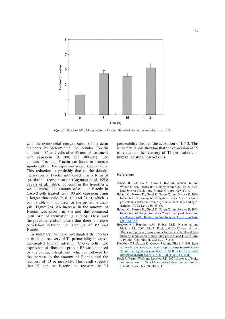
with the cytoskeletal reorganization of the actinfilaments by determining the cellular F-actinamount in Caco-2 cells after 45 min of treatmentwith capsaicin (0, 200, and 300 lM). Theamount of cellular F-actin was found to decreasesignificantly in the capsaicin-treated Caco-2 cells.This reduction is probably due to the depoly-merization of F-actin into G-actin as a form ofcytoskeletal reorganization (Rayment et al. 1993;Savala et al. 1998). To confirm the hypothesis,we determined the amount of cellular F-actin inCaco-2 cells treated with 100 lM capsaicin usinga longer time scale (0, 8, 16, and 24 h), which iscomparable to that used for the proteome anal-ysis (Figure 2b). An increase in the amount ofF-actin was shown in 8 h and this continueduntil 24 h of incubation (Figure 5). These andthe previous results indicate that there is a closecorrelation between the amounts of P2 andF-actin.
In summary, we have investigated the mecha-nism of the recovery of TJ permeability in capsa-icin-treated human intestinal Caco-2 cells. Theexpression of ribosomal protein P2 was enhancedby the capsaicin-treatment, which is followed bythe increase in the amount of F-actin and therecovery of TJ permeability. This result suggeststhat P2 stabilizes F-actin and recovers the TJ
permeability through the activation of EF-2. Thisis the first report showing that the expression of P2is related to the recovery of TJ permeability inhuman intestinal Caco-2 cells.
References
Alberts B., Johnson A., Lewis J., Raff M., Roberts K. and
Walter P. 2002. Molecular Biology of the Cell, 4th ed. Gar-
land Science (Taylor and Francis Group), New York.
Bektas M., Nurten R., Gurel Z., Sayers Z. and Bermek E. 1994.
Interaction of eukaryotic elongation factor 2 with actin: a
possible link between protein synthetic machinery and cyto-
skeleton. FEBS Lett. 356: 89–93.
Bektas M., Nurten R., Gurel Z., Sayers Z. and Bermek E. 1998.
Interaction of elongation factor 2 with the cytoskeleton and
interference with DNase I binding to actin. Eur. J. Biochem.
256: 142–147.
Bruewer M., Hopkins A.M., Hobert M.E., Nusrat A. and
Madara J.L. 2004. RhoA, Racl, and Cdc42 exert distinct
effects on epithelial barrier via selective structural and bio-
chemical modulation of junctional proteins and F-actin. Am.
J. Physiol. Cell Physiol. 287: C327–C335.
Dadabay C.J., Patton E., Cooper J.A. and Pike L.J. 1991. Lack
of correlation between changes in polyphosphoinositide lev-
els and actin/gelsolin complexes in A431 cells treated with
epidermal growth factor. J. Cell Biol. 112: 1151–1156.
Fogh J., Wright W.C. and Loveless J.D. 1977. Absence of HeLa
contamination in 169 cell lines derived from human tumors.
J. Natl. Cancer Inst 58: 209–214.
Figure 5. Effect of 100 lM capsaicin on F-actin. Standard deviations were less than 10%.
95
Fuchigami T., Misumi S., Takamune N., Takahashi I., Takama
M. and Shoji S. 2002. Acid-labile formylation of amino ter-
minal proaline of human immunodeficiency virus type 1
p24gag was found by proteomics using two-dimensional gel
electrophoresis and matrix-assisted laser desorption/ioniza-
tion-time-of-flight mass spectrometry. Biochem. Biophys.
Res. Commun. 293: 1107–1113.
Furukawa T., Uchiumi T., Tokunaga R. and Taketani S. 1992.
Ribosomal protein P2, a novel iron-binding protein. Arch.
Biochem. Biophys. 298: 182–186.
Gardner-Thorpe J., Ito H., Ashley S. and Whang E.E. 2003.
Ribosomal protein P2: a potential molecular target for anti-
sense therapy of human malignancies. Anticancer Res. 23:
4549–4560.
Gonzalo P., Lavergne J.P. and Reboud J.P. 2002. Pivotal role
of Pl N-terminal domain in the assembly of the mammalian
ribosomal stalk and in the proteosynthetic activity. J. Biol.
Chem. 276: 19762–19769.
Han J.K., Isoda H. and Maekawa T. 2002. Analysis of the
mechanism of the tight junctional permeability increase by
capsaicin treatment on the Caco-2 cells. Cytotechnology 40:
93–98.
Hashimoto K., Kawagishi H., Nakayama T. and Shimizu M.
1997. Effect of capsianoside, a diterpene glycoside, on tight-
junctional permeability. Biochim. Biophys. Acta 1323: 281–
290.
Holmes K.C., Popp D., Gebhard W. and Kabsch W. 1990.
Atomic model of the actin filament. Nature 347: 44–49.
Isoda H., Han J.K., Tominaga M. and Maekawa T. 2001. Ef-
fect of capsaicin on human intestinal cell line Caco-2. Cyto-
technology 36: 155–161.
Lavergne J.P., Conquet F., Reboud J.P. and Reboud A.M.
1987. Role of acidic phosphoprotein in the parartical recon-
stitution of the active 60S ribosomal subunit. FEBS Lett. 216:
83–88.
Lim S.O., Park S.J., Kim W., Park S.G., Kim H.J., Kim Y.I.,
Sohn T.S., Noh J.H. and Jung G. 2002. Proteome analysis of
hepatocellular carcinoma. Biochem. Biophys. Res. Commun.
291: 1031–1037.
Nusrat A., Giry M., Turner J.R., Colgan S.P., Parkos C.A.,
Carnes D., Lemichez E., Boquet P. and Madara J.L. 1995.
Rho protein regulates tight junctions and perijunctional actin
organization in polarized epithelia. Proc. Natl. Acad. Sci.
U.S.A. 92: 10629–10633.
Poon T.C. and Johnson P.J. 2001. Proteome analysis and its
impact on the discovery of serological tumor markers. Clinica
Chimica Acta 313: 231–239.
Rayment I., Holden H.M., Whittaker M., Yohn C.B., Lorenz
M., Holmes K.C. and Milligan R.A. 1993. Structure of the
actin-myosin complex and its implications for muscle con-
traction. Science 261: 58–65.
Satsu H., Yokoyama T., Ogawa N., Fujiwara-Hatano Y. and
Shimizu M. 2003. Effect of neuronal PC12 cells on the
functional properties of intestinal epithelial Caco-2 cells.
Biosci. Biotechnol. Biochem. 67: 1312–1318.
Savala U. and Waters C.M. 1998. Barrier function of airway
epithelium: effects of radiation and protection by keratino-
cyte growth factor. Radiat. Res. 150: 195–203.
Vard C., Guillot D., Bargis P., Lavergne J.P. and Reboud J.P.
1997. Specific role for the phosphorylation of mammalian
acidic ribosomal protein P2. J. Biol. Chem. 272: 20259–
20262.
Yap A.S., Mullin J.M. and Stevenson B.R. 1998. Molecular
analyses of tight junction physiology: insights and paradoxes.
J. Membr. Biol. 163: 159–167.
96
Copyright © 2022 FDOKUMEN