The role of human ribosomal proteins in the maturation of rRNA and ribosome production
12
The role of human ribosomal proteins in the maturation of rRNA and ribosome production SARA ROBLEDO, 1,3 RACHEL A. IDOL, 1,3 DAN L. CRIMMINS, 2 JACK H. LADENSON, 2 PHILIP J. MASON, 1,4 and MONICA BESSLER 1,4 1 Department of Internal Medicine, Division of Hematology, Washington University School of Medicine, St. Louis, Missouri 63110, USA 2 Department of Pathology and Immunology, Division of Laboratory and Genomic Medicine, Washington University School of Medicine, St. Louis, Missouri 63110, USA ABSTRACT Production of ribosomes is a fundamental process that occurs in all dividing cells. It is a complex process consisting of the coordinated synthesis and assembly of four ribosomal RNAs (rRNA) with about 80 ribosomal proteins (r-proteins) involving more than 150 nonribosomal proteins and other factors. Diamond Blackfan anemia (DBA) is an inherited red cell aplasia caused by mutations in one of several r-proteins. How defects in r-proteins, essential for proliferation in all cells, lead to a human disease with a specific defect in red cell development is unknown. Here, we investigated the role of r-proteins in ribosome biogenesis in order to find out whether those mutated in DBA have any similarities. We depleted HeLa cells using siRNA for several individual r-proteins of the small (RPS6, RPS7, RPS15, RPS16, RPS17, RPS19, RPS24, RPS25, RPS28) or large subunit (RPL5, RPL7, RPL11, RPL14, RPL26, RPL35a) and studied the effect on rRNA processing and ribosome production. Depleting r-proteins in one of the subunits caused, with a few exceptions, a decrease in all r-proteins of the same subunit and a decrease in the corresponding subunit, fully assembled ribosomes, and polysomes. R-protein depletion, with a few exceptions, led to the accumulation of specific rRNA precursors, highlighting their individual roles in rRNA processing. Depletion of r-proteins mutated in DBA always compromised ribosome biogenesis while affecting either subunit and disturbing rRNA processing at different levels, indicating that the rate of ribosome production rather than a specific step in ribosome biogenesis is critical in patients with DBA. Keywords: Diamond Blackfan anemia; ribosome biogenesis; siRNA; ribosome profile INTRODUCTION The production of ribosomes, the sites of protein synthesis, is a process of fundamental importance in cell biology. It has been estimated that in a rapidly growing yeast cell, the majority of transcription is devoted to ribosomal RNA (rRNA) and about half of RNA polymerase II transcription to ribosomal proteins (r-proteins) (Warner 1999). Tandem arrays of ribosomal genes are transcribed by RNA poly- merase I in the nucleolus and the transcripts are cleaved, modified, and assembled with r-proteins to form the mature 40S and 60S subunits of the mature ribosome. Ribosome production in the nucleolus and export to the cytoplasm involve the coordinated synthesis of four rRNAs, about 80 r-proteins, more than 150 other proteins, and about 70 snoRNAs. The 60S subunit contains 28S, 5.8S, and 5S RNAs, while the 40S subunit contains 18S RNA. 5S RNA is transcribed by RNA polymerase III and imported into the nucleolus during ribosome maturation. Most of our knowledge about ribosome biogenesis derives from genetic studies in yeast and bacteria. While there is a high level of conservation between the components of ribosome biogenesis in yeast and mammalian cells, studies of mammalian ribosome production are still in their infancy. Interest in mammalian ribosome biosynthesis has been stimulated by the finding that the pathogenesis of a number of inherited and acquired diseases involves mutations in genes encoding components of the ribosome or proteins involved in ribosome maturation, assembly, or export. Diamond Blackfan anemia (DBA, OMIM#105650) is a rare inherited bone marrow failure syndrome that affects mainly 3 These authors contributed equally to this work. 4 Joint senior authors. Reprint requests to: Philip J. Mason, Department of Internal Medicine, Division of Hematology, Washington University School of Medicine, 660 South Euclid Avenue, Campus Box 8118, St. Louis, MO 63110, USA; e-mail: [email protected]; fax: (314) 362-8826. Article published online ahead of print. Article and publication date are at http://www.rnajournal.org/cgi/doi/10.1261/rna.1132008. 1918 RNA (2008), 14:1918–1929. Published by Cold Spring Harbor Laboratory Press. Copyright Ó 2008 RNA Society.
-
Upload
independent -
Category
Documents
-
view
1 -
download
0
Transcript of The role of human ribosomal proteins in the maturation of rRNA and ribosome production

The role of human ribosomal proteins in the maturation
of rRNA and ribosome production
SARA ROBLEDO,1,3 RACHEL A. IDOL,1,3 DAN L. CRIMMINS,2 JACK H. LADENSON,2
PHILIP J. MASON,1,4 and MONICA BESSLER1,4
1Department of Internal Medicine, Division of Hematology, Washington University School of Medicine, St. Louis, Missouri 63110, USA2Department of Pathology and Immunology, Division of Laboratory and Genomic Medicine, Washington University School of Medicine,St. Louis, Missouri 63110, USA
ABSTRACT
Production of ribosomes is a fundamental process that occurs in all dividing cells. It is a complex process consisting of thecoordinated synthesis and assembly of four ribosomal RNAs (rRNA) with about 80 ribosomal proteins (r-proteins) involvingmore than 150 nonribosomal proteins and other factors. Diamond Blackfan anemia (DBA) is an inherited red cell aplasia causedby mutations in one of several r-proteins. How defects in r-proteins, essential for proliferation in all cells, lead to a humandisease with a specific defect in red cell development is unknown. Here, we investigated the role of r-proteins in ribosomebiogenesis in order to find out whether those mutated in DBA have any similarities. We depleted HeLa cells using siRNA forseveral individual r-proteins of the small (RPS6, RPS7, RPS15, RPS16, RPS17, RPS19, RPS24, RPS25, RPS28) or large subunit(RPL5, RPL7, RPL11, RPL14, RPL26, RPL35a) and studied the effect on rRNA processing and ribosome production. Depletingr-proteins in one of the subunits caused, with a few exceptions, a decrease in all r-proteins of the same subunit and a decreasein the corresponding subunit, fully assembled ribosomes, and polysomes. R-protein depletion, with a few exceptions, led to theaccumulation of specific rRNA precursors, highlighting their individual roles in rRNA processing. Depletion of r-proteinsmutated in DBA always compromised ribosome biogenesis while affecting either subunit and disturbing rRNA processing atdifferent levels, indicating that the rate of ribosome production rather than a specific step in ribosome biogenesis is critical inpatients with DBA.
Keywords: Diamond Blackfan anemia; ribosome biogenesis; siRNA; ribosome profile
INTRODUCTION
The production of ribosomes, the sites of protein synthesis,is a process of fundamental importance in cell biology. Ithas been estimated that in a rapidly growing yeast cell,the majority of transcription is devoted to ribosomal RNA(rRNA) and about half of RNA polymerase II transcriptionto ribosomal proteins (r-proteins) (Warner 1999). Tandemarrays of ribosomal genes are transcribed by RNA poly-merase I in the nucleolus and the transcripts are cleaved,modified, and assembled with r-proteins to form themature 40S and 60S subunits of the mature ribosome.
Ribosome production in the nucleolus and export to thecytoplasm involve the coordinated synthesis of four rRNAs,about 80 r-proteins, more than 150 other proteins, andabout 70 snoRNAs. The 60S subunit contains 28S, 5.8S,and 5S RNAs, while the 40S subunit contains 18S RNA.5S RNA is transcribed by RNA polymerase III andimported into the nucleolus during ribosome maturation.Most of our knowledge about ribosome biogenesis derivesfrom genetic studies in yeast and bacteria. While there is ahigh level of conservation between the components ofribosome biogenesis in yeast and mammalian cells, studiesof mammalian ribosome production are still in theirinfancy.
Interest in mammalian ribosome biosynthesis has beenstimulated by the finding that the pathogenesis of a numberof inherited and acquired diseases involves mutations ingenes encoding components of the ribosome or proteinsinvolved in ribosome maturation, assembly, or export.Diamond Blackfan anemia (DBA, OMIM#105650) is a rareinherited bone marrow failure syndrome that affects mainly
rna11320 Robledo et al. ARTICLE RA
3These authors contributed equally to this work.4Joint senior authors.Reprint requests to: Philip J. Mason, Department of Internal Medicine,
Division of Hematology, Washington University School of Medicine, 660South Euclid Avenue, Campus Box 8118, St. Louis, MO 63110, USA;e-mail: [email protected]; fax: (314) 362-8826.
Article published online ahead of print. Article and publication date areat http://www.rnajournal.org/cgi/doi/10.1261/rna.1132008.
1918 RNA (2008), 14:1918–1929. Published by Cold Spring Harbor Laboratory Press. Copyright � 2008 RNA Society.
JOBNAME: RNA 14#9 2008 PAGE: 1 OUTPUT: Friday August 8 17:34:50 2008
csh/RNA/164293/rna11320

the terminal stages of red cell differentiation (Ohene-Abuakwa et al. 2005; Lipton 2006). DBA is caused bymutations in one of several genetic loci, all of which codefor r-proteins in both the small and large ribosomal subunit(Draptchinskaia et al. 1999; Willig et al. 1999; Gazda et al.2006; Cmejla et al. 2007; Farrar et al. 2008; H. Gazda,pers. comm.). A major shift in our perception of the role ofr-proteins came in 2000 when it was found that the cata-lytic molecule in ribosomes is rRNA and the r-proteins arelikely to be responsible for the correct folding of rRNA,needed for accurate cleavage and processing, and for theassembly of the ribosome (Steitz and Moore 2003). The pre-cise role of the 80 or so r-proteins in ribosome biogenesis isnot fully understood, especially in mammals. In addition totheir role in ribosome biogenesis, extraribosomal functionshave been postulated for many of the r-proteins (Wool 1996).
DBA is usually diagnosed as a severe anemia in earlychildhood caused by a defect in red cell maturation (redcell aplasia). As well as failure of red cell development,the disorder may be associated with a variety of congenitalabnormalities and a predisposition to malignancy (Lipton2006). Mutations causing DBA have been found in RPS19(accounting for 25% of cases) (Draptchinskaia et al. 1999;Willig et al. 1999); RPS24 (2%) (Gazda et al. 2006); RPS17(one case) (Cmejla et al. 2007); RPL35a (2%) (Farrar et al.2008); and RPL5 and RPL11 (10% each) (H. Gazda, pers.comm.). In all cases the inheritance is autosomal dominant;haploinsufficiency for the r-protein is thought to be themechanism of disease (Gazda et al. 2004). However,the mechanism whereby haploinsufficiency for one of severalr-proteins leads specifically to red cell aplasia while onlyminimally affecting other tissues is unknown. Mice hetero-zygous for an Rps19 gene deletion develop normally withno abnormalities in red cell development (Matsson et al.2004, 2006). In contrast, cell culture studies using bonemarrow cells or peripheral blood hematopoietic progenitorcells from patients with DBA show that the defect isintrinsic to the red cell progenitors, which fail to completedifferentiation in response to erythropoietin (Ohene-Abuakwa et al. 2005). Depleting the amount of RPS19 inhuman CD34+ primitive hematopoietic progenitors causesa reduction in the proliferation of immature red cells (Ebertet al. 2005; Flygare et al. 2005). It has recently been shownthat the depletion of RPS19 in human cell lines by siRNAcauses decreased ribosome synthesis with accumulationof some specific rRNA precursors (Choesmel et al. 2007;Flygare et al. 2007; Idol et al. 2007). The same precursorswere found to accumulate to some extent in patients’fibroblasts, lymphocyte cell lines, and, in one case, inprimary cells from bone marrow (Flygare et al. 2007).Similarly, depletion of RPS24 causes decreased rRNAsynthesis due to failure of an earlier step in 18S rRNAprocessing (Choesmel et al. 2008). Since the only knowngenetic cause of DBA is mutations in genes coding forr-proteins, we were interested to test whether the possible
molecular mechanism would be a specific defect in thesynthesis of ribosomes shared by the r-proteins affected inDBA.
To investigate this hypothesis and to gain insight intothe molecular mechanism responsible for disease, we de-pleted HeLa cells for the mRNA coding for a selection ofr-proteins, including several known to be involved in DBApathogenesis. We found that depletion of r-proteinsinvolved in the pathogenesis of DBA may affect eithersubunit, but always was associated with a decrease in theproduction of ribosomes. We further found that depletionof any r-proteins involved in the pathogenesis of DBA alsodecreased the amount of all r-proteins from the samesubunit, and caused a decreased production of the corre-sponding subunit. The alterations in rRNA processingvaried amongst the various DBA-associated r-proteinsand did not correlate with disease association. We thereforeconclude that any of the r-proteins, when mutated, com-promise ribosome biogenesis, is a potential candidate forDBA, and that the common factor is the decreased pro-duction of ribosomes rather than a specific step inribosome biogenesis or the accumulation of a specificrRNA or ribosome subunit precursor.
RESULTS
In order to investigate whether there are common featuresin the function of r-proteins mutated in DBA, we investi-gated the role of 15 r-proteins in ribosome biosynthesis andrRNA processing, including six r-proteins that are currentlyknown to be affected in DBA. At the time the study wasinitiated, only r-proteins of the small subunit RPS19(Draptchinskaia et al. 1999), RPS24 (Gazda et al. 2006),and RPS17 (Cmejla et al. 2007) were known to be mu-tated in DBA. During the course of the study, the numberof r-proteins responsible for DBA increased, including pro-teins belonging to the large r-subunit, RPL35a (Farrar et al.2008), and RPL5 and RPL11 (H. Gazda, pers. comm.).
Depletion of r-protein mRNAs in HeLa cellsusing siRNA
We and others have previously reported on the effect ofr-protein RPS19 depletion on ribosome biosynthesis andrRNA processing. Here, we used a similar approachdepleting individual r-proteins in growing HeLa cells usingsiRNA. In addition to the proteins RPS17, RPS19, RPS24,RPL5, RPL11, and RPL35a known to be associated withDBA, we also studied the depletion of RPS6, RPS7, RPS15,RPS16, RPS25, RPS28, RPL7, RPL14, and RPL26. To date,these r-proteins have not been associated with DBA, buttheir involvement is not ruled out, as the causativemutation has not been found in about half of the knownDBA patients. The efficiency of r-protein depletion wasassessed by Western blotting using either commercially
Ribosomal proteins in ribosome biogenesis
www.rnajournal.org 1919
JOBNAME: RNA 14#9 2008 PAGE: 2 OUTPUT: Friday August 8 17:34:50 2008
csh/RNA/164293/rna11320

available or in-house antibodies and by Northern analysisusing cDNA probes.
Figure 1 is an example of the data showing the depletionof mRNA for the r-proteins in our study. In all cases, thesiRNA reduces the abundance of the mRNA to almostundetectable levels. The percentage knockdowns shown inFigure 1 are those from one typical experiment obtainedfrom phosphorimaging of Northern blots. Notably, thetarget mRNA was the only one that was decreased inabundance. No evidence for reduction in one mRNAcausing consistent and significant reduction in any of theothers was found.
Depletion of r-proteins causes decreased abundanceof other r-proteins belonging to the sameribosomal subunit
We next examined the effect of the mRNA knockdown onthe steady-state level of r-proteins. Protein was extractedfrom the cells 72 h after the cells had been transfected withthe siRNAs, and r-proteins were examined by Westernblotting (Fig. 2). For this we generated peptide antibodiesfor RPS7, RPS12, RPS15, RPS24, RPS25, and RPS28. Wehave previously reported the generation of peptide anti-bodies against RPS19. Antibodies against RPS6, RPS16,
RPL7, and RPL26 were from commercial sources. Westernanalysis showed that the steady-state level of the siRNAtargeted r-protein was decreased by 30%–60% (Fig. 2). Inseveral experiments, there was considerable variation in thelevel of protein knockdown achieved. The knockdown ofone of the r-proteins always resulted in a decrease of thesteady-state levels of the other r-proteins belonging to thesame ribosomal subunit (Fig. 2). The exception to this wasRPS25. When RPS25 mRNA was reduced in abundance toalmost zero, RPS6, RPS12, RPS16, RPS19, and RPS24 didnot significantly change in abundance. Using an antibodyto RPS25 shows that cells treated with siRNA to RPS25were indeed depleted for RPS25 and that no reduction inthe steady-state level of any of the other r-protein of thesmall subunit (RPS) proteins was apparent. For r-proteinsbelonging to the large ribosomal subunit, RPL7, RPL14,RPL26, and RPL35a (RPLs), we used antibodies againstRPL7 and RPL26. Figure 2 shows that, as expected, therewas a reduction in the steady-state levels of RPL7 andRPL26 when the level of their mRNAs was reduced. Inkeeping with our results from the RPS knockdowns, thelevels of RPL7 and RPL26 proteins were reduced to asimilar extent when RPL5, RPL7, RPL11, RPL14, orRPL35a were knocked down. Interestingly, though RPS19was unaffected by depletion of RPL5, RPL7, RPL11, RPL14,
or RPL35a, depletion of RPL26 causedan approximate 30% reduction inRPS19 level. Depletion of the RPSproteins had no effect on the levels ofRPL26.
Depletion of r-proteins causesdecreased production of matureribosomes and functionallyactive polysomes
We next investigated the effect of thedepletion of individual r-proteins onthe ribosome profile in the HeLa cells72 h after siRNA treatment. HeLa cellsthat were mock transfected showed aclassical profile of ribosomes and poly-somes, with the 40S and 60S ribosomalsubunits and a large 80S peak represent-ing the mature assembled ribosome(monosome). Depletion of any of theRPSs caused a reduction in the amountof free 40S subunits, a great reductionin the amount of mature 80S ribosomes,and an increase in the amount of free60S subunits (Fig. 3). The exception wasagain RPS25. Here, the ribosome profileshowed an intermediate phenotype witha reduction in the amount of 80S ribo-somes and an increase in the amount of
FIGURE 1. Efficient depletion of r-protein mRNA in HeLa using siRNA. Cells weretransfected with scrambled negative control siRNA or siRNAs targeting the RPS6, RPS7,RPS15, RPS16, RPS17, RPS19, RPS24, RPS25, RPS28, RPL7, RPL14, RL26, or RPL35a mRNAs.After 72 h, total RNA was extracted and subjected to Northern blotting analysis. Membraneswere hybridized with cDNA probes for one of each r-protein; mouse b-actin was used as aloading control. The percentage decrease in the targeted mRNA by the siRNA is indicated(right side for each panel). (A) Efficient depletion of targeted small subunit (RPS) mRNAs.A single experiment where equivalent amounts of RNAs were analyzed on three separate blots.Note RNA from the RPS6 depletion experiment is overloaded in this experiment. (B) Efficientdepletion of targeted large subunit (RPL) mRNAs.
Robledo et al.
1920 RNA, Vol. 14, No. 9
JOBNAME: RNA 14#9 2008 PAGE: 3 OUTPUT: Friday August 8 17:34:51 2008
csh/RNA/164293/rna11320

60S subunits, but no significant changes in the amount ofthe 40S subunit. In the polysome region of the profiles, wenote a variation in the extent to which polysomes aredecreased with a trend toward a decrease in the relativenumber of large polysomes, perhaps reflecting a decrease inthe number of initiations caused by the deficiency in thenumber of 40S subunits. A somewhat different pattern wasseen when RPLs were depleted (Fig. 3). In the case of RPL5,RPL7, RPL11, RPL14, RPL26, and RPL35a depletioncompared with the normal ribosome profile, the amountof the 60S subunit was greatly reduced, as was the peakrepresenting the levels of 80S ribosomes. There was aconcomitant increase in the amount of free 40S subunit,although for the RPL26 depletion the free 40S subunit didnot show this increase. In these profiles, the 80S andpolysome peaks are split, an appearance characteristic of‘‘halfmers,’’ which are monosomes and polysomes to whichan isolated 40S subunit is bound (Helser et al. 1981). In the
RPL knockdowns, there appears to be a decrease in theoverall number of polysomes.
Depletion of r-proteins blocks rRNA processingat specific steps
The r-proteins not only serve as a scaffold for the rRNA andensure the correct structure of the ribosome, but are alsoinvolved in the maturation of the rRNA precursors. Havingfound that depleting any r-protein decreases the produc-tion of one ribosomal subunit and, consequently, pro-duction of mature ribosomes, we were interested to knowthe effect of each depletion on pre-rRNA processing. Sinceribosome biogenesis involves a characteristic series of rRNAprocessing events, we can investigate the extent, and atwhich level, pre-rRNA processing is affected by the deple-tion of individual r-proteins. RNA was extracted fromHeLa cells 72 h after the cells had been transfected withthe various siRNAs. The RNA was subjected to Northernblotting using a series of probes from within the rRNA
FIGURE 3. Depletion of r-proteins impairs the synthesis of newribosomes. After 72 h of treatment with siRNA, cells were treated withcycloheximide, lysed, and the cytoplasmic fraction was layered over10%–45% sucrose gradients. Mock or scramble treated cells show atypical profile of free 40S, free 60S, 80S, and polysomes. Cells depletedfor RPS6, RPS7, RPS15, RPS16, RPS17, RPS19, RPS24, and RPS28have significantly decreased levels of free 40S and 80S, with a dramaticincrease in free 60S. The polysome levels are decreased. RPS25depleted cells show an intermediate phenotype, with a slight decreasein free 40S, an increase in free 60S, and partial decrease in 80S.Depleting cells of RPL5, RPL7, RPL11, RPL14, and RPL35a show anincrease in free 40S subunits, a substantial decrease in free 60Ssubunits, and a decrease in the 80S. Polysome profiles also demon-strate the presence of halfmers and a decrease in polysomes,particularly the larger polysomes. Depletion of RPL26 also decreasedthe free 60S subunit and the 80S and produced halfmer polysomes,although the free 40S levels did not increase as dramatically as wasseen with other RPL knockdowns.
FIGURE 2. Knocking down ribosomal proteins decreases the levels ofother ribosomal proteins of the same subunit. Western blot analysisof r-protein levels after siRNA treatment. Total cellular protein wasextracted 72 h after treatment with siRNA specific to each target or ascrambled sequence. (A) Western blotting was performed to examinethe levels of RPS6, RPS12, RPS16, RPS19, RPS24, RPS25, RPL26;GAPDH was used as a loading control. Knocking down all RPS pro-teins analyzed decreased the levels of other RPS proteins, with the ex-ception of RPS25. RPS25 protein levels were decreased by treatmentwith siRNA against RPS25, although the levels of the other r-proteinsof the small subunit examined did not change. The levels of RPLproteins did not decrease. (B) Western blot analysis of RPL7, RPL26,and RPS19 protein levels after depletion of RPL proteins; GAPDH wasused as a loading control. Knocking down all RPL proteins analyzeddecreased the levels of other RPL proteins. The levels of r-proteins ofthe small subunit did not decrease, except in the RPL26 knockdownwhere a slight reduction of RPS19 levels was observed.
Ribosomal proteins in ribosome biogenesis
www.rnajournal.org 1921
JOBNAME: RNA 14#9 2008 PAGE: 4 OUTPUT: Friday August 8 17:34:57 2008
csh/RNA/164293/rna11320
locus. The location of the probes andthe rRNA processing pathway as it isthought to occur in HeLa cells (Hadjiolovaet al. 1993) is schematically shownin Figure 4A. Figure 4B shows theeffects on pre-rRNA processing of thedepletion of r-proteins of the smallsubunit. Depletion of RPSs affectedthe formation of the mature 18S rRNA,while the formation of the mature 28Sand 5.8S RNA was not significantlyaffected (Fig. 5; data not shown). Thedefects in rRNA processing detected byNorthern blotting after the depletion ofindividual r-proteins of the small sub-unit fell into three district categories.The most frequently observed defect inpre-rRNA processing, seen after deplet-ing RPS6, RPS7, RPS16, RPS24, andRPS28, was characterized by the de-crease of the 41S rRNA precursor andan accumulation of a 30S rRNA pro-cessing intermediate, and a decrease inthe 21S and 18SE precursors. Thisimplies that the respective r-proteinsare essential for the processing at the59-ETS, which leads to the decrease inthe 41S, the accumulation of the 30Sprecursor, and a decrease of the 21Sr-RNA and 18S-E precursors. ForRPS24, similar findings were reportedby Choesmel et al. (2008). A differentpre-rRNA processing defect was ob-served for RPS17 and RPS19 depletion,which caused accumulation of the 21Sand 20S precursors, implying that thesetwo r-proteins are required for a laterstep in pre-rRNA processing duringwhich the 21S molecule is processed toform the 18S-E nuclear precursor. Thethird defect is shown after the depletionof RPS15 and also after depletion ofRPS25. Here, all the rRNA precursorsare present at levels similar to those inuntreated cells or cells treated with thescrambled control siRNA, and 18S-E isexported into the cytoplasm in bothcases. Interestingly, results from theribosome profiles obtained fromRPS15-depleted cells show that the pro-duction of 40S subunits is defective inthese cells, whereas in RPS25-depletedcells the production of the 40S subunitis only impaired, leading us to concludethat RPS15 may play a role late in
FIGURE 4. Ribosomal proteins of the small subunit control distinct rRNA processing stepsimportant for the maturation of the 18S rRNA. (A) Schematic diagram of pre-rRNAprocessing pathways in HeLa cells. (Inset) Structure of the primary 47S rRNA transcriptcontaining two external transcriber spacers at its 59 and 39 ends (59ETS and 39ETS,respectively), as well two internal transcriber spacers (ITS1 and ITS2). (Arrows) Position ofmajor processing sites (0–4). The 47S pre-rRNA is processed through intermediate precursorsdesignated according to their relative sedimentation coefficients (S) to mature 18S, 28S, and5.8S rRNAs. In HeLa cells, 45S pre-rRNA can be processed by two alternative pathways.Pathway A is initiated with the removal of 59-ETS by cleavage at site 1, while pathway B startswith cleavage in ITS1, probably at site 2 (Hadjiolova et al. 1993; Rouquette et al. 2005; Idolet al. 2007). The sequence locations of the probes used in Northern blot analysis are shown as aline above the primary transcript scheme (bottom). (B) Northern hybridization of total RNAfrom HeLa cells after depletion of r-proteins of the small subunit. Cells were transfected withscrambled negative control siRNA or siRNAs targeting RPS6, RPS7, RPS15, RPS16, RPS17,RPS19, RPS24, RPS25, or RPS28 mRNAs. After 72 h, total RNA was extracted and subjected toNorthern blotting analysis for pre-rRNA species. Membranes were hybridized with probes forthe 59 end of ITS1 (probe i, upper panel), 59 ends of ITS2 (probe f, lower panel), or b-actin.(Left panel) Short exposure, (right panel) long exposure. Depletion of r-proteins of the smallsubunit block rRNA processing at specific steps. The sizes of pre-rRNA species are indicatedbetween the left and right panels. (C) Northern hybridization of total (T), nuclear (N), andcytoplasmic (C) rRNA isolated from HeLa cells after depletion of r-proteins of RPS15 andRPS25. Cells were transfected with scrambled negative control siRNA or siRNAs targetingRPS15 and RPS25 mRNAs. Membranes were hybridized with probes for the 59 end of ITS1(probe i).
Robledo et al.
1922 RNA, Vol. 14, No. 9
JOBNAME: RNA 14#9 2008 PAGE: 5 OUTPUT: Friday August 8 17:35:11 2008
csh/RNA/164293/rna11320
ribosome biogenesis involving the final stages of matura-tion in the cytoplasm or interaction with the large subunit,whereas RPS25 is not essential for 40S subunit production.Metabolic labeling of cells with 3H-methylmethionine afterdepletion of r-proteins of the small subunit similarlyconfirmed the decrease in the production of the mature18S rRNA, while 28S rRNA was not affected. This was thecase in all knockdown cells with the exception of RPS15and RPS25 depleted cells, which seemed to synthesize boththe 18S and 28S rRNA and all precursors at a level and ratethat were indistinguishable from normal or mock trans-fected cells (Fig. 5).
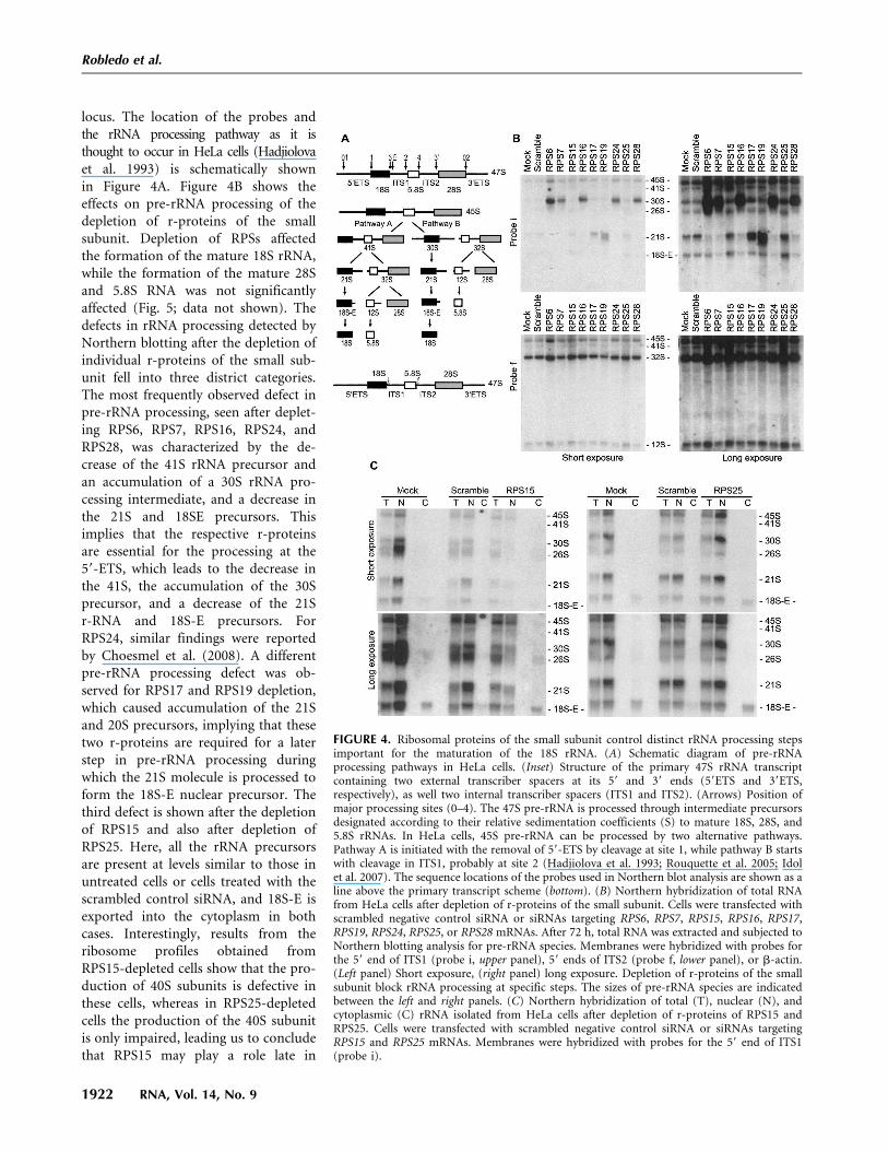
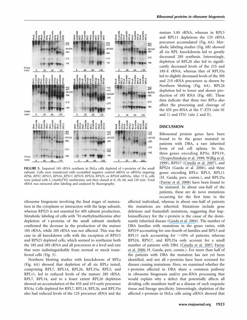
Northern blotting studies with knockdowns of RPLs(Fig. 6A) showed that depletion of all six RPLs tested,comprising RPL7, RPL14, RPL26, RPL35a, RPL5, andRPL11, led to reduced levels of the mature 28S rRNA.RPL7, RPL14, and to a lesser extent RPL26 depletionshowed an accumulation of the 45S and 41S early precursorRNAs. Cells depleted for RPL7, RPL14, RPL26, and RPL35aalso had reduced levels of the 12S precursor rRNA and the
mature 5.8S rRNA, whereas in RPL5and RPL11 depletions the 12S rRNAprecursor accumulated (Fig. 6A). Met-abolic labeling studies (Fig. 6B) showedall six RPL knockdowns led to greatlydecreased 28S synthesis. Interestingly,depletion of RPL26 also led to signifi-cantly decreased levels of the 21S and18S-E rRNA, whereas that of RPL35aled to slightly decreased levels of the 30Sand 21S rRNA precursors as shown byNorthern blotting (Fig. 6A). RPL26depletion led to lower and slower pro-duction of 18S RNA (Fig. 6B). Thesedata indicate that these two RPLs alsoaffect the processing and cleavage ofthe 45S pre-RNA at the 59-ETS (site 01and 1) and ITS1 (site 2 and E).
DISCUSSION
Ribosomal protein genes have beenfound to be the genes mutated inpatients with DBA, a rare inheritedform of red cell aplasia. So far,three genes encoding RPSs: RPS19,(Draptchinskaia et al. 1999; Willig et al.1999), RPS17 (Cmejla et al. 2007), andRPS24 (Gazda et al. 2006), and threegenes encoding RPLs: RPL5, RPL11(H. Gazda, pers. comm.), and RPL35a(Farrar et al. 2008) have been found tobe mutated. In about one-half of thepatients, these are de novo mutationsoccurring for the first time in the
affected individual, whereas in about one-half of patientsthe mutations are inherited. Mutations include genedeletions and frameshift mutations, suggesting that hap-loinsufficency for the r-protein is the cause of the domi-nantly inherited disease (Gazda et al. 2004). The number ofDBA families with mutations in the genes varies, withRPS19 accounting for one-fourth of families and RPL5 andRPL11 each accounting for z10% of patients; whereasRPS24, RPS17, and RPL35a only account for a smallnumber of patients with DBA (Cmejla et al. 2007; Farraret al. 2008; H. Gazda, pers. comm.). For more than half ofthe patients with DBA the mutation has not yet beenidentified, and not all r-proteins have been screened fordisease causing mutations. Here, we examined whether ther-proteins affected in DBA share a common pathwayin ribosome biogenesis and/or pre-RNA processing thatwould explain why a defect that potentially affects alldividing cells manifests itself as a disease of such exquisitetissue and lineage specificity. Interestingly, depletion of theaffected r-proteins in HeLa cells using siRNA showed that
FIGURE 5. Impaired 18S rRNA synthesis in HeLa cells depleted of r-proteins of the smallsubunit. Cells were transfected with scrambled negative control siRNA or siRNAs targetingRPS6, RPS7, RPS15, RPS16, RPS17, RPS19, RPS24, RPS25, or RPS28 mRNAs. After 72 h, cellswere pulsed with L-[methyl3H] methionine and then chased at 0, 30, 60, and 120 min. TotalrRNA was extracted after labeling and analyzed by fluorography.
Ribosomal proteins in ribosome biogenesis
www.rnajournal.org 1923
JOBNAME: RNA 14#9 2008 PAGE: 6 OUTPUT: Friday August 8 17:35:21 2008
csh/RNA/164293/rna11320
the only shared feature was that their depletion impairedthe production of ribosomes, as assayed by examinationof the ribosome profile on sucrose gradients, a character-istic we found to be shared with most, although not all,
other r-proteins. Furthermore, ourresults demonstrated that the depletionof r-proteins associated with DBA couldaffect the biosynthesis of either the largeor small ribosomal subunit. Finally, wefound that pre-RNA cleavage wasimpaired at various steps of rRNAprocessing, leading to the accumulationof different rRNA precursors. Interest-ingly, RPS19, RPL5, and RPL11, whichaccount for almost 35% of cases ofDBA, show defects in late nucleolarmaturation of the pre-40S and pre-60Sribosomal particles.
With the exception of RPS25 andRPS15, depletion of RPSs always led todecreased levels of mature 18S rRNA,decreased levels of the small 40S ribo-somal subunit, and decreased levels ofmature 80S ribosomes. RPS15 did notvisibly affect rRNA processing, export ofthe 18S-E RNA precursor, or the accu-mulation of the mature 18S RNA inmetabolic labeling experiments. How-ever, the levels of 40S ribosomal sub-units and 80S ribosomes were decreased,indicating that RPS15 is essential for alate maturation step of the 40S subunit.In contrast, RPS25 seemed to have noeffect on rRNA processing, 18S-E ex-port, or mature 18S rRNA accumula-tion, and was not essential for 40S and80S ribosome biosynthesis, though for-mation of small subunits was reduced inits absence. In all of these cases, impair-ment of the formation of the smallribosome subunit did not affect thematuration of the 28S and 5.8S rRNAand was associated with excess produc-tion of the 60S large subunit.
Ribosome biogenesis has been stud-ied extensively in yeast. Several recentstudies have documented the role in ribo-some biogenesis of specific r-proteinsby depleting proteins using the powerand elegance of yeast genetics (vanBeekvelt et al. 2001; Leger-Silvestreet al. 2004, 2005; West et al. 2005;Ferreira-Cerca et al. 2007; Rosadoet al. 2007). In both human and yeast,RPS6, RPS16, and RPS24 depletion all
impair the processing of the 59 ETS, leading to theaccumulation of an early precursor and decrease of the20S (yeast) 21S (human) rRNA molecule that is a precursorto 18S rRNA. Similarly in both yeast and human, RPS19
FIGURE 6. Ribosomal proteins of the large subunit control distinct rRNA processing steps, inparticular, those important for the maturation of the 28S rRNA and 5.8S rRNA. (A) Northernhybridization of total RNA from HeLa cells after depletion of r-proteins of the large subunit.Cells were transfected with scrambled negative control siRNA or siRNAs targeting RPL5, RPL7,RPL11, RPL14, RPL26, or RPL35a mRNAs. After 72 h, total RNA was extracted and subjectedto Northern blotting analysis for pre-rRNA species. Membranes were hybridized with probesfor the 59 end of ITS1 (probe i, upper panel) and 59 end of ITS2 (probe f, lower panel). Pre-rRNA products after hybridization with probe i (upper panel) and probe f (lower panel). (Leftpanel) Short exposure, (right panel) long exposure. The sizes of pre-rRNA species are indicatedbetween the left and right panels. This short run is shown to emphasize the characteristicdifference in 5.8S and 12S accumulation. (B) Analysis of the rRNA synthesis of 18S and 28S inHeLa cells depleted of r-proteins of the large subunit by pulse-chase analysis with L-[methyl3H] methionine labeling. Cells were transfected with scrambled negative controlsiRNA or siRNAs targeting RPL5, RPL7, RPL11, RPL14, RL26, or RPL35a mRNAs. After 72 h,cells were pulsed with L-[methyl3H] methionine and then chased at 0, 30, 60, and 120 min.Total rRNA was extracted after labeling and analyzed by fluorography.
Robledo et al.
1924 RNA, Vol. 14, No. 9
JOBNAME: RNA 14#9 2008 PAGE: 7 OUTPUT: Friday August 8 17:35:29 2008
csh/RNA/164293/rna11320

depletion leads to the accumulation of the 20S/21S rRNAspecies, which is not processed further. Finally, in bothyeast and humans, RPS25 was not essential for ribosomebiosynthesis. However, we also identified distinct differ-ences in the role of r-proteins in humans and yeast. Incontrast to yeast, in HeLa cells the ribosome profile wasaltered after the depletion of RPS25 (see Fig. 3), suggestingthat the depletion of RPS25 allows the assembly of the 40Ssubunit but impairs its association with the 60S subunit toform the mature 80S ribosome or that the 80S ribosome isunstable and is degraded. Furthermore, we found thatRPS15 depletion in human cell lines did not visibly impairthe processing of the rRNA, and that the 18S-E rRNAprecursor was exported into the cytoplasm as in mocktransfected cells or cells transfected with a scrambledsiRNA. However, the levels of 40S subunit and 80S matureribosomes were greatly reduced, suggesting that in humansRPS15 is essential for the final cytoplasmic maturation andassembly of the 40S subunit. In contrast, in yeast, depletionof RPS15 leads to the accumulation of the 20S in thenucleus, suggesting that RPS15 in yeast is essential forexport from the nucleus (Leger-Silvestre et al. 2004).Finally, in HeLa cells, the depletion of RPS7 and RPS28led to the accumulation of the early 30S rRNA precursor,whereas in yeast, RPS7 and RPS28 affect the cytoplasmicmaturation of the 18S rRNA and terminal assembly of the40S subunit. The defect in HeLa cells was assessed for atleast two independent siRNAs for each r-protein, indicatingthat off-target effects were unlikely to account for thedifferences observed. RPS17 depletion was not analyzed inyeast. Further analysis will show whether these are realdifferences in the yeast and human r-proteins and when inevolution these proteins might have adopted a differentrole in ribosome biogenesis.
Depletion of RPLs similarly primarily affected produc-tion of the mature 28S and 5.8S rRNA, the assembly of the60S large ribosomal subunit, the mature 80S ribosome, andled to the production of ‘‘halfmers,’’ due to the binding ofa 40S subunit to a mRNA without a 60S subunit. Theexception was the depletion of RPL26 that impaired boththe production of the mature 18S and 28S rRNA with areduction of both the 40S and 60S subunits, a reduction ofthe 80S mature ribosome, and the generation of halfmers.RPLs associated with DBA showed defects in either early orlate processing of the 28S and 5.8S rRNA, whereas the mostfrequent forms of RPL mutations in DBA, RPL5, andRPL11 affected the late steps in 28S and 5.8S maturationoccurring in the nucleolus. In contrast to RPSs, RPLs havenot been studied in a systematic manner in yeast; thus, acomparison of the role of RPLs in ribosome biogenesis inyeast and humans is not possible. Interestingly, RPL5 andRPL11 share two other properties as well as their connec-tion with DBA. Both have been implicated in negativelyregulating MDM2, a nucleolar protein that causes rapidturnover of the oncoprotein p53 (Marechal et al. 1994;
Lohrum et al. 2003; Zhang et al. 2003). Moreover, both areassociated, at least in yeast, with 5S rRNA at an early stagein ribosome biogenesis (Zhang et al. 2007).
One of the most interesting observations of our study isthat depleting a cell of a r-protein leads not only to adecrease in the abundance of that protein as expectedbut also to a concomitant decrease in the abundance of allproteins from the same subunit. The most likely explana-tion for this observation is that the lack of one r-proteinprevents formation of a stable subunit, and other proteinsof that subunit with no structure to be incorporated intoare degraded. This explanation is compatible with earlyresults that showed excess ribosomal proteins, which arenot assembled into ribosomes, are rapidly degraded(Warner 1977, 1979). This point has been reinforced by arecent study showing that r-proteins are synthesized at ratesmuch higher than required for ribosome biogenesis, andthat excess proteins not incorporated into ribosomes arerapidly degraded in the nucleoplasm (Lam et al. 2007). Aspointed out by Lam et al., this suggests a simple way forthe cell to solve the problem of coordinated synthesis ofr-proteins for ribosome production; i.e., they are simplymade in vast excess, and those not needed are degraded. Inthe experiments presented here, when one protein becomeslimiting there is a rapid decrease in the other proteins ofthat subunit. We observe this concerted decrease inproteins of one subunit for all the knockdowns we havecarried out except for that of RPS25. Alternatively, whenone r-protein is under-produced, there may be someassembly of other proteins into nascent subunits followedby partial disassembly and turnover in the absence ofstoichiometric amounts of ribosomal components (Moritzet al. 1990). This scheme may be more compatible with thedecrease in r-proteins and accumulation of rRNA precur-sors that we observe.
To what extent are our observations relevant to thepathogenesis of DBA? At first glance there is a conundrumsince, if r-proteins are made in vast excess over require-ments, then reducing the production of an r-protein by50% as happens in DBA should not have a drastic effect.We must therefore hypothesize that in developing red cells,and possibly in cells of other tissues affected in DBA, thereis a requirement for maximal ribosome synthesis at a timewhen DNA transcription is turned off as part of erythroidmaturation or in a key developmental stage elsewhere.Erythroid precursors in response to erythropoietin doindeed have a very high rate of ribosome production inorder to undergo the final red cell divisions and to be ableto produce the massive amounts of globin needed in themature circulating red cell (Rifkind et al. 1964; Flygare andKarlsson 2007). If the rate of ribosome production requiredwas more than half of the maximum rate governed byproduction of r-proteins, then haploinsufficiency for anyr-protein would slow ribosome production. In red cellmaturation, this may lead to the cell cycle arrest or cell
Ribosomal proteins in ribosome biogenesis
www.rnajournal.org 1925
JOBNAME: RNA 14#9 2008 PAGE: 8 OUTPUT: Friday August 8 17:35:37 2008
csh/RNA/164293/rna11320

death that is the hallmark of DBA. That some r-proteinsmay be limiting such that a 50% reduction in quantity mayaffect ribosome production in red cells, whereas for othersa reduction of 50% may not be limiting, or may also belimiting in other tissues to the extent that it would interferewith embryogenesis, might explain why mutations incertain r-proteins account more frequently for DBA thanothers (Ellis and Massey 2006). There are alternativeschemes that may underlie DBA pathogenesis. It is possiblethat certain cell types such as the developing red cell may bemore sensitive to an imbalance in ribosome subunit pro-duction, and this may be more crucial in cells producingribosomes at a high rate. In our HeLa cell model,the overproduction/degradation mechanism ensures thatr-proteins not incorporated into ribosome subunits do notaccumulate, but it is not clear whether this surveillancemechanism is present in the maturing red cell. Further-more, there does not appear to be a mechanism forensuring that large or small subunits do not accumulatein the absence of the other subunit.
In summary, our results support the notion that a failurein the production of ribosomes is the factor that triggersthe process leading to the red cell aplasia characteristic ofDBA. Presumably, any r-protein produced in limiting quan-tities and whose haploinsufficiency is not lethal in the em-bryo would be a candidate. Whether the crude number ofribosomes or the disruption of the nucleolar structurecausing nucleolar stress and the activation of p53 arekeys to pathogenesis in DBA is subject to our futureinvestigations.
MATERIALS AND METHODS
Cell culture
HeLa cells were cultured in Dulbecco’s modified Eagle’s mediumcontaining 15% fetal bovine serum, antibiotics (100 mg ofpenicillin and 50 mg of streptomycin sulfate/mL), and 2 mMglutamine at 37°C, 5% CO2.
RNA preparation
Total RNA was extracted using Trizol (Invitrogen) following themanufacturer’s instructions. Nuclear and cytoplasmic fractionswere prepared using Cell fractionation buffer (Ambion), and thennuclear or cytoplasmic RNA was extracted from each fraction withTrizol.
Oligonucleotides
The oligonucleotide sequences complementary to the knownhuman rDNA used in this paper were:
(Probe i) 20-ITS1: 59-CCTCGCCCTCCGGGCTCCGTTAATTGATC-39 (Rouquette et al. 2005); and
(Probe f) 15-ITS2: 59-CGCACCCCGAGGAGCCCGGAGGCACCCCCGG-39 (Hadjiolova et al. 1993).
Complementary DNA was synthesized using SuperScript IIIreverse transcriptase (Invitrogen) following the manufacturer’sinstructions. The following primers were used for PCR:
RPS6: 59-ATGAAGCTGAACATCTCCTTC-39 and 59-TTTCTGACTGGATTCAGACTTAG-39;
RPS7: 59-ATGTTCAGTTCGAGCGCC-39 and 59-CAATTGAAACTCTGGGAA-39;
RPS15: 59-ATGGCAGAAGTAGAGCAGAAG-39 and 59-CTTGAGAGGGATGAAGCG-39;
RPS16: 59-ATGCCGTCCAAGGGCCCGCTGCAG-39 and 59-TCGGTAGGATTTCTGGTAGCG-39;
RPS17: 59-ATGGGCCGCGTTCGCACCAAA-39 and 59-TCAAACAGGTCCCCGAGGCGT-39;
RPS19: 59-ATGCCTGGAGTTACTGTAAAA-39 and 59-CTAATGCTTCTTGTTGGCAGC-39;
RPS24: 59-ATGAACGACACCGTAACT-39 and 59-CTCCTTCGGCTTTTTGCC-39
RPS25: 59-ATGCCGCCTAAGGACGAC-39 and 59-TCATGCATCTTCACCAGC-39;
RPS28: 59-ATGGACACCAGCCGTGTG-39 and 59-TCAGCGCAACCTCCGGGCTT-39;
RPL7: 59-ATGGAGGGTGTAGAAGAG-39 and 59-GTTCATTCTTCTAATAAGCCTG-39;
RPL14: 59-ATGGTGTTCAGGCGCTTC-39 and 59-TGCTTTCTTGCCAGATGC-39;
RPL26: 59-ATGAAGTTTAATCCCTTTGTGAXTTCCG-39 and 59-TTCCTGCATCTTCTCAATGGT-39; and
RPL35a: 59-ATGTCTGGAAGGCTGTGGTCC-39 and 59AATCCTTGAGGGGTACAGCAT-39.
The first strand cDNA was synthesized by RT-PCR fromtotal RNA using the SuperScript First-Strand Synthesis Sys-tem (Invitrogen). After synthesis, target cDNA was amplified withspecific primers by PCR with Platinum Taq DNA polymerasehigh fidelity (Invitrogen) for 35 cycles under the followingconditions: 15 sec, 94°C; 30 sec, 55°C; and 60 sec, 68°C. PCRproducts were resolved by electrophoresis on 1% agarose.
rRNA analysis
Pre-rRNA analysis by Northern blotting was adapted fromHadjiolova et al. (1993) using synthetic oligonucleotide DNAand cDNA as probes. RNA (10 mg) was electrophoresed in 1.25%(w/v) agarose–formaldehyde gels and blotted to Hybond-N+
membranes (GE Healthcare) overnight. Oligonucleotides wereend-labeled with [g-32P]ATP and T4 polynucleotide kinase(Fisher), and cDNA probes were labeled with [32P]dCTP usingthe Rediprime II random prime labeling system (AmershamBiosciences).
Membranes were hybridized overnight in hybridizationbuffer (0.75 M NaCl, 0.075 M Na citrate pH 7.0, 13 Denhardt’ssolution, 150 mg/mL tRNA) and 1% SDS (when oligonucleotideprobes were used), or 0.25 M Na/phosphate buffer, 1.0 g/LBSA, and 7% SDS (when cDNA probes were used). Temperatureof hybridization was 55°C (probe i) and 65°C (probe f andcDNA probes for all different RPSs, RPLs, and b-actin). Washingconditions included 33 SSC, 0.1% SDS for 15 min; 23 SSC, 0.1%SDS for 30 min; and 0.23 SSC, 0.1% SDS for 30 min at 55°C or65°C (for oligonucleotide probes i and f, respectively), or 33 SSC,
Robledo et al.
1926 RNA, Vol. 14, No. 9
JOBNAME: RNA 14#9 2008 PAGE: 9 OUTPUT: Friday August 8 17:35:37 2008
csh/RNA/164293/rna11320

0.3% SDS for 15 min; 23 SSC, 0.3% SDS for 30 min; and 0.23
SSC, 0.3% SDS for 45 min at 65°C (for cDNA probes).Membranes were both exposed to a storage phosphor screen(GE Healthcare) for further analysis using a phosphor imager(Typhoon trio, GE Healthcare) and X-ray film.
Labeling of rRNA
Cells were seeded in 12-well plates at 5 3 106 cells per welland pre-incubated for 45 min in methionine-free medium andthen incubated for 30 min with 50 mCi L-[methyl3H] methionine.Cells were then chased in nonradioactive medium containing15 mg/mL methionine for 0, 30, 60, and 120 min, after which totalRNA was isolated using Trizol (Invitrogen) and label incorpora-tion for each sample was measured by scintillation counting.An equal number of counts were loaded in each lane of a1.25% agarose–formaldehyde gel and then transferred toHybond-N+ (GE Healthcare). Membranes were exposed to X-ray film.
Antibodies
Anti-peptide RPS rabbit antibodies were generated from purifiedsynthetic peptides obtained from Biomolecules Midwest,Inc..Three versions of each peptide were synthesized (native,N-terminal cysteine, and N-terminal long-chain-biotin) anda KLH-peptide was used to immunize two rabbits with theresulting antiserum purified over a peptide-affinity column (Idolet al. 2007). The peptides corresponding to human RPS sequenceswere:
K7IVKPNGEKPDEFESG22 (RPS7);N74LIKVDDNKKLGEWV88 (RPS12);A1EVEQKKKRTFRKFTY16 (RPS15);T102SRKQRKERKNRMKKVRGT120 (RPS24); andK33KWSKGKVRDKLNNLVLFD51 (RPS25).
Commercially available rabbit polyclonal antibodies against RPS6,RPL7, and RPL26 (Bethyl Laboratories) and RPS16 (GeneTX,Inc.) were obtained and used following the manufacturers’protocols. Rabbit polyclonal anti-GAPDH (Abcam) was used asa loading control.
Sucrose gradient centrifugation
Cells were washed in ice-cold PBS and incubated with 100 mg/mLcycloheximide in PBS for 10 min at 4°C prior to lysis. Allsubsequent steps were performed on ice. Cells were resuspendedin hypotonic buffer (1.5 mM KCl, 2.5 mM MgCl2, and 5.0 mMTris-Cl at pH 7.4) followed by the addition of 80 units of RNaseinhibitor (Invitrogen). Hypotonic lysis buffer (1.5 mM KCl, 2.5mM MgCl2, and 5.0 mM Tris-Cl pH 7.4, 2% sodium deoxycho-late, 2% Triton X-100, and 2.5 mM DTT) were added and cellswere gently homogenized with 10 strokes of a Dounce homoge-nizer. Cell lysates were centrifuged at 8000g for 10 min at 4°C andthe supernatant was adjusted to 2 mg/mL heparin. Lysates werestored at �80°C. Linear 10%–45% sucrose gradients (80 mMNaCl, 5 mM MgCl2, 20 mM Tris-Cl at pH 7.4, and 1 mM DTT)were poured using a Gradient Master (BioComp). Gradients werecentrifuged at 38,000 rpm for 3 h at 4°C and analyzed with anISCO fractionator (ISCO).
siRNA
A 21-bp siRNA targeting RPS19 was synthesized, annealed,and purified (Ambion): 59-GAUGGCGGCCGCAAACUGUCA-39
(sense) and 59-UCAGUUUGCGGCCGCCAUCTT-39 (antisense),named RPS19 siRNA #2. One additional siRNA targeting RPS19was obtained from Ambion (siRNA ID# 2801). The followingsiRNAs were purchased from Ambion to target ribosomal proteinsof interest:
RPS6 (siRNA ID #45962);RPS7 (siRNA ID #142206 and 142207);RPS15 (siRNA ID #142212);RPS17 (siRNA ID #142215);RPS24 (siRNA ID #45965);RPS25 (siRNA ID #14220);RPS28 (siRNA ID #41629 and 284684);RPL5 (siRNA ID #142169);RPL7 (siRNA ID #142173);RPL11 (siRNA ID #9352);RPL14 (siRNA ID #13949);RPL26 (siRNA ID #9179); andRPL35a (siRNA ID #45958).
Silencer negative control #1 (Ambion) was used as a negativecontrol, and Silencer GAPDH (Ambion) was used as a positivecontrol. Cells were transfected with siPORT NeoFX (Ambion)according to manufacturer’s protocols using 30 nM siRNA perwell.
Western blots
Total cell extracts were made using Cellytic Reagent(Sigma) supplemented with a protease inhibitor cocktail (Roche),and protein concentration was determined by a modifiedBradford assay (Bio-Rad). Proteins were separated on a 15%SDS-PAGE gel and transferred to Hybond nitrocellulose(GE Healthcare). Membranes were blocked overnight at 4°Cin Odyssey blocking buffer (LI-COR). Membranes were incubatedfor 1 h at room temperature with primary antibodies in Odysseyblocking buffer containing 0.05% Tween-20, and washedthree times with PBST. The secondary antibody, either goatanti-rabbit IRDYE 680 (LI-COR) or goat anti-mouse IRDYE680 (LI-COR), was added to Odyssey blocking buffer (LI-COR)containing 0.05% Tween-20, and membranes were incubated for1 h at room temperature. Membranes were washed twice withPBST, rinsed with PBS, and signals were detected with theOdyssey (LI-COR). All signal quantitation was performed withthe Odyssey (LI-COR).
ACKNOWLEDGMENTS
This work was supported by NIH DK075443 (to M.B.), CA106995(to P.J.M.), and the Daniella Maria Arturi Foundation. Wethank Mary Jane Eichenseer for antibody purification, and ananonymous reviewer for helpful suggestions. We thank RobertArceci and Hannah Gazda for permission to quote unpublisheddata.
Received April 10, 2008; accepted June 6, 2008.
Ribosomal proteins in ribosome biogenesis
www.rnajournal.org 1927
JOBNAME: RNA 14#9 2008 PAGE: 10 OUTPUT: Friday August 8 17:35:38 2008
csh/RNA/164293/rna11320

REFERENCES
Choesmel, V., Bacqueville, D., Rouquette, J., Noaillac-Depeyre, J.,Fribourg, S., Cretien, A., Leblanc, T., Tchernia, G., Da Costa, L.,and Gleizes, P.E. 2007. Impaired ribosome biogenesis in Diamond-Blackfan anemia. Blood 109: 1275–1283.
Choesmel, V., Fribourg, S., Aguissa-Toure, A.H., Pinaud, N.,Legrand, P., Gazda, H.T., and Gleizes, P.E. 2008. Mutationof ribosomal protein RPS24 in Diamond-Blackfan anemia resultsin a ribosome biogenesis disorder. Hum. Mol. Genet. 17: 1253–1263.
Cmejla, R., Cmejlova, J., Handrkova, H., Petrak, J., andPospisilova, D. 2007. Ribosomal protein S17 gene (RPS17) ismutated in Diamond-Blackfan anemia. Hum. Mutat. 28: 1178–1182.
Draptchinskaia, N., Gustavsson, P., Andersson, B., Pettersson, M.,Willig, T.N., Dianzani, I., Ball, S., Tchernia, G., Klar, J.,Matsson, H., et al. 1999. The gene encoding ribosomal proteinS19 is mutated in Diamond-Blackfan anemia. Nat. Genet. 21: 169–175.
Ebert, B.L., Lee, M.M., Pretz, J.L., Subramanian, A., Mak, R.,Golub, T.R., and Sieff, C.A. 2005. An RNA interference model ofRPS19 deficiency in Diamond-Blackfan anemia recapitulatesdefective hematopoiesis and rescue by dexamethasone: Identifica-tion of dexamethasone-responsive genes by microarray. Blood 105:4620–4626.
Ellis, S.R. and Massey, A.T. 2006. Diamond Blackfan anemia: Aparadigm for a ribosome-based disease. Med. Hypotheses 66: 643–648.
Farrar, J.E., Nater, M., Caywood, E., McDevitt, M.A., Kowalski, J.,Takemoto, C.M., Talbot, C.C.J., Meltzer, P., Esposito, D.,Beggs, A.H., et al. 2008. Abnormalities of the large ribosomalsubunit protein, Rpl35A, in Diamond-Blackfan Anemia. Blooddoi: 10.1182/blood-2008-02-140012.
Ferreira-Cerca, S., Poll, G., Kuhn, H., Neueder, A., Jakob, S.,Tschochner, H., and Milkereit, P. 2007. Analysis of the in vivoassembly pathway of eukaryotic 40S ribosomal proteins. Mol. Cell28: 446–457.
Flygare, J. and Karlsson, S. 2007. Diamond-Blackfan anemia: Eryth-ropoiesis lost in translation. Blood 109: 3152–3154.
Flygare, J., Kiefer, T., Miyake, K., Utsugisawa, T., Hamaguchi, I., DaCosta, L., Richter, J., Davey, E.J., Matsson, H., Dahl, N., et al.2005. Deficiency of ribosomal protein S19 in CD34+ cellsgenerated by siRNA blocks erythroid development and mimicsdefects seen in Diamond-Blackfan anemia. Blood 105: 4627–4634.
Flygare, J., Aspesi, A., Bailey, J.C., Miyake, K., Caffrey, J.M.,Karlsson, S., and Ellis, S.R. 2007. Human RPS19, the gene mutatedin Diamond-Blackfan anemia, encodes a ribosomal proteinrequired for the maturation of 40S ribosomal subunits. Blood109: 980–986.
Gazda, H.T., Zhong, R., Long, L., Niewiadomska, E., Lipton, J.M.,Ploszynska, A., Zaucha, J.M., Vlachos, A., Atsidaftos, E.,Viskochil, D.H., et al. 2004. RNA and protein evidence forhaplo-insufficiency in Diamond-Blackfan anaemia patients withRPS19 mutations. Br. J. Haematol. 127: 105–113.
Gazda, H.T., Grabowska, A., Merida-Long, L.B., Latawiec, E.,Schneider, H.E., Lipton, J.M., Vlachos, A., Atsidaftos, E.,Ball, S.E., Orfali, K.A., et al. 2006. Ribosomal protein S24 gene ismutated in Diamond-Blackfan anemia. Am. J. Hum. Genet. 79:1110–1118.
Hadjiolova, K.V., Nicoloso, M., Mazan, S., Hadjiolov, A.A., andBachellerie, J.P. 1993. Alternative pre-rRNA processing pathwaysin human cells and their alteration by cycloheximide inhibition ofprotein synthesis. Eur. J. Biochem. 212: 211–215.
Helser, T.L., Baan, R.A., and Dahlberg, A.E. 1981. Characterization ofa 40S ribosomal subunit complex in polyribosomes of Saccharo-myces cerevisiae treated with cycloheximide. Mol. Cell. Biol. 1: 51–57.
Idol, R.A., Robledo, S., Du, H.Y., Crimmins, D.L., Wilson, D.B.,Ladenson, J.H., Bessler, M., and Mason, P.J. 2007. Cellsdepleted for RPS19, a protein associated with Diamond BlackfanAnemia, show defects in 18S ribosomal RNA synthesis andsmall ribosomal subunit production. Blood Cells Mol. Dis. 39:35–43.
Lam, Y.W., Lamond, A.I., Mann, M., and Andersen, J.S. 2007.Analysis of nucleolar protein dynamics reveals the nuclear degra-dation of ribosomal proteins. Curr. Biol. 17: 749–760.
Leger-Silvestre, I., Milkereit, P., Ferreira-Cerca, S., Saveanu, C.,Rousselle, J.C., Choesmel, V., Guinefoleau, C., Gas, N., andGleizes, P.E. 2004. The ribosomal protein Rps15p is required fornuclear exit of the 40S subunit precursors in yeast. EMBO J. 23:2336–2347.
Leger-Silvestre, I., Caffrey, J.M., Dawaliby, R., Alvarez-Arias, D.A.,Gas, N., Bertolone, S.J., Gleizes, P.E., and Ellis, S.R. 2005. Specificrole for yeast homologs of the Diamond Blackfan Anemia-associated Rps19 protein in ribosome synthesis. J. Biol. Chem.280: 38177–38185.
Lipton, J.M. 2006. Diamond Blackfan anemia: New paradigms for a‘‘not so pure’’ inherited red cell aplasia. Semin. Hematol. 43: 167–177.
Lohrum, M.A., Ludwig, R.L., Kubbutat, M.H., Hanlon, M., andVousden, K.H. 2003. Regulation of HDM2 activity by theribosomal protein L11. Cancer Cell 3: 577–587.
Marechal, V., Elenbaas, B., Piette, J., Nicolas, J.C., and Levine, A.J.1994. The ribosomal L5 protein is associated with mdm-2 andmdm-2-p53 complexes. Mol. Cell. Biol. 14: 7414–7420.
Matsson, H., Davey, E.J., Draptchinskaia, N., Hamaguchi, I., Ooka, A.,Leveen, P., Forsberg, E., Karlsson, S., and Dahl, N. 2004. Targeteddisruption of the ribosomal protein S19 gene is lethal prior toimplantation. Mol. Cell. Biol. 24: 4032–4037.
Matsson, H., Davey, E.J., Frojmark, A.S., Miyake, K.,Utsugisawa, T., Flygare, J., Zahou, E., Byman, I., Landin, B.,Ronquist, G., et al. 2006. Erythropoiesis in the Rps19 disruptedmouse: Analysis of erythropoietin response and biochemicalmarkers for Diamond-Blackfan anemia. Blood Cells Mol. Dis.36: 259–264.
Moritz, M., Paulovich, A.G., Tsay, Y.F., and Woolford Jr., J.L. 1990.Depletion of yeast ribosomal proteins L16 or rp59 disruptsribosome assembly. J. Cell Biol. 111: 2261–2274.
Ohene-Abuakwa, Y., Orfali, K.A., Marius, C., and Ball, S.E. 2005.Two-phase culture in Diamond Blackfan anemia: Localization oferythroid defect. Blood 105: 838–846.
Rifkind, R.A., Danon, D., and Marks, P.A. 1964. Alterations inpolyribosomes during erythroid cell maturation. J. Cell Biol. 22:599–611.
Rosado, I.V., Kressler, D., and de la Cruz, J. 2007. Functional analysisof Saccharomyces cerevisiae ribosomal protein Rpl3p in ribosomesynthesis. Nucleic Acids Res. 35: 4203–4213.
Rouquette, J., Choesmel, V., and Gleizes, P.E. 2005. Nuclearexport and cytoplasmic processing of precursors to the 40Sribosomal subunits in mammalian cells. EMBO J. 24: 2862–2872.
Steitz, T.A. and Moore, P.B. 2003. RNA, the first macromolecularcatalyst: The ribosome is a ribozyme. Trends Biochem. Sci. 28: 411–418.
van Beekvelt, C.A., de Graaff-Vincent, M., Faber, A.W., van’t Riet, J.,Venema, J., and Raue, H.A. 2001. All three functional domains ofthe large ribosomal subunit protein L25 are required for both earlyand late pre-rRNA processing steps in Saccharomyces cerevisiae.Nucleic Acids Res. 29: 5001–5008.
Warner, J.R. 1977. In the absence of ribosomal RNA synthesis, theribosomal proteins of HeLa cells are synthesized normally anddegraded rapidly. J. Mol. Biol. 115: 315–333.
Warner, J.R. 1979. Distribution of newly formed ribosomal proteinsin HeLa cell fractions. J. Cell Biol. 80: 767–772.
Warner, J.R. 1999. The economics of ribosome biosynthesis in yeast.Trends Biochem. Sci. 24: 437–440.
Robledo et al.
1928 RNA, Vol. 14, No. 9
JOBNAME: RNA 14#9 2008 PAGE: 11 OUTPUT: Friday August 8 17:35:39 2008
csh/RNA/164293/rna11320

West, M., Hedges, J.B., Chen, A., and Johnson, A.W. 2005. Definingthe order in which Nmd3p and Rpl10p load onto nascent 60Sribosomal subunits. Mol. Cell. Biol. 25: 3802–3813.
Willig, T.N., Draptchinskaia, N., Dianzani, I., Ball, S., Niemeyer, C.,Ramenghi, U., Orfali, K., Gustavsson, P., Garelli, E., Brusco, A.,et al. 1999. Mutations in ribosomal protein S19 gene and DiamondBlackfan anemia: Wide variations in phenotypic expression. Blood94: 4294–4306.
Wool, I.G. 1996. Extraribosomal functions of ribosomal proteins.Trends Biochem. Sci. 21: 164–165.
Zhang, Y., Wolf, G.W., Bhat, K., Jin, A., Allio, T., Burkhart, W.A.,and Xiong, Y. 2003. Ribosomal protein L11 negativelyregulates oncoprotein MDM2 and mediates a p53-dependentribosomal-stress checkpoint pathway. Mol. Cell. Biol. 23: 8902–8912.
Zhang, J., Harnpicharnchai, P., Jakovljevic, J., Tang, L., Guo, Y.,Oeffinger, M., Rout, M.P., Hiley, S.L., Hughes, T., and WoolfordJr., J.L. 2007. Assembly factors Rpf2 and Rrs1 recruit 5S rRNA andribosomal proteins rpL5 and rpL11 into nascent ribosomes. Genes& Dev. 21: 2580–2592.
Ribosomal proteins in ribosome biogenesis
www.rnajournal.org 1929
JOBNAME: RNA 14#9 2008 PAGE: 12 OUTPUT: Friday August 8 17:35:40 2008
csh/RNA/164293/rna11320