Puromycin–rRNA interaction sites at the peptidyl transferase center
12
2000 6: 744-754 RNA C Rodriguez-Fonseca, H Phan, K S Long, et al. Puromycin-rRNA interaction sites at the peptidyl transferase center. References http://rnajournal.cshlp.org/content/6/5/744#related-urls Article cited in: service Email alerting click here top right corner of the article or Receive free email alerts when new articles cite this article - sign up in the box at the http://rnajournal.cshlp.org/subscriptions go to: RNA To subscribe to © 2000 RNA Society Cold Spring Harbor Laboratory Press on July 13, 2011 - Published by rnajournal.cshlp.org Downloaded from
-
Upload
foreignpolicy -
Category
Documents
-
view
2 -
download
0
Transcript of Puromycin–rRNA interaction sites at the peptidyl transferase center

2000 6: 744-754RNA C Rodriguez-Fonseca, H Phan, K S Long, et al. Puromycin-rRNA interaction sites at the peptidyl transferase center.
References http://rnajournal.cshlp.org/content/6/5/744#related-urls
Article cited in:
serviceEmail alerting
click heretop right corner of the article orReceive free email alerts when new articles cite this article - sign up in the box at the
http://rnajournal.cshlp.org/subscriptions go to: RNATo subscribe to
© 2000 RNA Society
Cold Spring Harbor Laboratory Press on July 13, 2011 - Published by rnajournal.cshlp.orgDownloaded from

Puromycin–rRNA interaction sitesat the peptidyl transferase center
CRISTINA RODRIGUEZ-FONSECA,1,2 HIEN PHAN,1 KATHERINE S. LONG, 1 BO T. PORSE,1
STANISLAV V. KIRILLOV, 3 RICARDO AMILS, 2 and ROGER A. GARRETT 1
1RNA Regulation Centre, Institute of Molecular Biology, University of Copenhagen, Sølvgade 83H,DK-1307 Copenhagen K, Denmark
2Centro de Biología Molecular, Universidad Autónoma de Madrid, Campus de Cantoblanco, E-28049 Madrid, Spain3Petersburg Nuclear Physics Institute, Russian Academy of Sciences, 188350 Gatchina, St+ Petersburg, Russia
ABSTRACT
The binding site of puromycin was probed chemically in the peptidyl-transferase center of ribosomes from Esche-richia coli and of puromycin-hypersensitive ribosomes from the archaeon Haloferax gibbonsii. Several nucleotides ofthe 23S rRNAs showed altered chemical reactivities in the presence of puromycin. They include A2439, G2505, andG2553 for E. coli, and G2058, A2503, G2505, and G2553 for Hf. gibbonsii (using the E. coli numbering system).Reproducible enhanced reactivities were also observed at A508 and A1579 within domains I and III, respectively, ofE. coli 23S rRNA. In further experiments, puromycin was shown to produce a major reduction in the UV-inducedcrosslinking of deacylated-(2N 3A76)tRNA to U2506 within the P 9 site of E. coli ribosomes. Moreover, it stronglystimulated the putative UV-induced crosslink between a streptogramin B drug and m 2A2503/C2504 at an adjacent sitein E. coli 23S rRNA. These data strongly support the concept that puromycin, along with other peptidyl-transferaseantibiotics, in particular the streptogramin B drugs, bind to an RNA structural motif that contains several conservedand accessible base moieties of the peptidyl transferase loop region. This streptogramin motif is also likely to providebinding sites for the 39 termini of the acceptor and donor tRNAs. In contrast, the effects at A508 and A1579, which arelocated at the exit site of the peptide channel, are likely to be caused by a structural effect transmitted along thepeptide channel.
Keywords: 23S rRNA; peptide channel; peptidyl transferase; puromycin
INTRODUCTION
The antibiotic puromycin has played a central role inour understanding of the mechanism of peptide elon-gation in the ribosome+ This is because it is partiallycostructural with the 39 terminus of aminoacyl-tRNA(Harris & Symons, 1973) (Fig+ 1) and can, therefore,function as an amino acid acceptor substrate (Traut &Monro, 1964; Smith et al+, 1965)+ This property of thedrug has been exploited in assays for peptide-bondformation and elongation+ These have yielded impor-tant, albeit superficial, insight into the mechanisms ofinhibition of peptide-bond formation by many other anti-biotics (reviewed in Pestka, 1977; Vázquez, 1979;Galeet al+, 1981)+
Several antibiotics have been footprinted on free ri-bosomes within the peptidyl-transferase loop of 23SrRNA, including some that bind competitively with pu-romycin (reviewed in Garrett & Rodriguez-Fonseca,1995)+ Moreover, for some of the antibiotics, but notpuromycin, drug-resistant mutants have been isolatedfrom bacteria and haloarchaea that carry single-sitemutations in the peptidyl-transferase loop (Garrett &Rodriguez-Fonseca, 1995)+ This, together with recentUV-induced crosslinking data for sparsomycin (Porseet al+, 1999b) and for the streptogramin B, pristinamy-cin IA (Porse et al+, 1999a), within this rRNA regionsuggest that most, if not all, peptidyl-transferase anti-biotics bind there (reviewed in Porse et al+, 2000)+
Localization of the puromycin-binding site is im-portant for defining the position of the 39 end ofaminoacyl-tRNA immediately prior to peptide-bond for-mation (Porse et al+, 1995; Kirillov et al+, 1997)+ How-ever, these experiments are difficult to execute forseveral reasons+ First, puromycin binds very weakly to
Reprint requests to: Professor R+A+ Garrett, RNA RegulationCentre, Institute of Molecular Biology, University of Copenhagen,Sølvgade 83H, DK-1307 Copenhagen K, Denmark; e-mail:garrett@mermaid+molbio+ku+dk+
RNA (2000), 6:744–754+ Cambridge University Press+ Printed in the USA+Copyright © 2000 RNA Society+
744
Cold Spring Harbor Laboratory Press on July 13, 2011 - Published by rnajournal.cshlp.orgDownloaded from

free ribosomes (Pestka, 1970; Fernandez-Muñoz &Vázquez, 1973)+ In addition, because the drug is ananalog of the aminoacyl-acceptor substrate, it is alsoa potential analog of the donor substrate, and maytherefore bind weakly in the P9 site (Kirillov et al+, 1997)of free ribosomes (Bourd et al+, 1983)+ Furthermore,probing in the presence of an active donor substratewill result in peptide bond formation, which will occurcoincidentally with the probable movement of thepuromycin-peptide complex (Odom et al+, 1990)+
Several crosslinking investigations with different pu-romycin derivatives have provided useful informationon the location of the puromycin-binding site+ In oneexperiment, the derivative [3H]-p-azidopuromycin wasbound to free ribosomes and, upon irradiation with UVlight, crosslinked to positions G2502 and U2504, at thebase of the peptidyl-transferase loop+ The derivativewas also crosslinked to protein L23, which binds todomain III of 23S rRNA (Nicholson et al+, 1982a, 1982b;Hall et al+, 1988)+ In another experiment, an attemptwas made to locate the equivalent of C-74 ofaminoacyl-tRNA by attaching an affinity label to puro-mycin (Green et al+, 1998)+A crosslink was produced toposition 2553 outside of the peptidyl-transferase loop,some 10 Å from the acceptor group of puromycin+ Onlyin the latter experiment was evidence provided for thederivatized puromycin occupying a functional acceptor-substrate site+
In the present work, three strategies are used to lo-cate the puromycin-binding site and its effects on 23SrRNA+ First, an rRNA-footprinting approach was em-ployed on puromycin complexed with ribosomes fromEscherichia coli and the haloarchaeon Haloferax gib-bonsii+ The latter ribosomes were included to providecomplementary data from another domain of life and,moreover, they are hypersensitive to puromycin (Sanzet al+, 1993)+ Second, a study was made of the effect ofpuromycin on the crosslinking of deacylated-(2N3A76)tRNA bound to the P9 site of 70S ribosome–poly(U)complexes+ Finally, the effect of puromycin on theputative UV-induced crosslinking of pristinamycin IAwithin the peptidyl-transferase loop of 23S rRNA wasexamined+
RESULTS
In vitro rRNA footprinting of puromycincomplexed to E. coli andHf. gibbonsii ribosomes
Puromycin–ribosome complexes were formed by incu-bating ribosomes from E. coli (Makhno et al+, 1988) andHf. gibbonsii with increasing concentrations of puromy-cin (0+1, 0+5, 1, 2, and 4 mM)+ The complexes andcontrol samples of free ribosomes were then treatedwith the base-specific reagents dimethylsulfate (DMS)[G (N-7) . A (N-1) . C (N-3)], kethoxal [G (N-1, N-2)],and CMCT (1-cyclohexyl-3(2-morpholinoethyl)-carbo-diimide metho-p-toluene sulfonate) [U (N-3) . G (N-1)]+After removal of excess reagent and proteins, deoxy-oligonucleotide primers were hybridized to 23S rRNA,39 from the RNA region to be investigated, subjected toprimer extension, and analyzed on polyacrylamide se-quencing gels+
The most extensive work was done on E. coli ribo-somes where we studied puromycin complexes sys-tematically with 70S ribosomes, 50S subunits, andwith polysomes, and for each of the complexes, thewhole range of puromycin concentrations was tested+Moreover, puromycin aminonucleoside was includedin all of these experiments in the concentration range0+1 to 2 mM as a control+ For the halophile ribo-somes, only puromycin complexes with 70S ribo-somes were examined+ For the 70S ribosomes of bothorganisms, altered nucleotide reactivities, includingprotections and enhancements, were observed+Puromycin-induced effects were not detected at thelowest puromycin concentration (0+1 mM), in accor-dance with an earlier observation for E. coli ribo-somes, which was attributed to the low bindingconstant of puromycin (Moazed & Noller, 1987)+ Auto-radiograms of gels illustrating some of the changesfor DMS- and kethoxal-modified samples, includingcontrol samples modified in the absence of antibiot-ics for E. coli and Hf. gibbonsii, are depicted in Fig-ures 2 and 3, respectively+ No reactivity changes wereobserved for CMCT-modified samples+
Reactivity changes were quantified by microdensito-metry relative to adjacent control bands+ Representa-tive scans are illustrated for the reactions at positionsA508 and G2505 at different puromycin concentrationsin Figure 4+ All the effects are summarized in Table 1,where horizontally aligned nucleotides occur at corre-sponding positions in the two secondary structures of23S rRNA (Figs+ 5 and 6)+ The intensity changes be-tween 0 and 2 mM puromycin, for all of the nucleotides,were in the range 30–70%+ There is no reason to ex-pect that the total intensity change should be similar forthe different nucleotides, as in total they represent dif-ferences in the accessibility of two chemicals at variouspositions in the ribosome in the presence of puromycin+
FIGURE 1. Chemical structures of A: puromycin, and B: the 39-endof a phenylalanyl-tRNA molecule+
Puromycin–rRNA interactions 745
Cold Spring Harbor Laboratory Press on July 13, 2011 - Published by rnajournal.cshlp.orgDownloaded from

Most of the altered reactivities were localized in, oradjoining, the peptidyl-transferase loop of domain V(Fig+ 5)+ This domain, together with domain IV, wasscreened many times (.20) for altered nucleotide re-activities in both organisms+ The remainder of E. coli23S rRNA was screened at least twice, and reproduc-ible enhanced reactivities were observed in domain I(Fig+ 6A) and domain III (Fig+ 6B)+ The remainder ofthe extreme halophile 23S rRNA was also screened,but additional altered nucleotide reactivities were notobserved+
Similar protection effects were observed for both or-ganisms at nt G2505 (Figs+ 2D, 3B, and 4B) and G2553(Fig+ 2E), using E. coli numbering (Table 1)+ However,effects were also detected that were particular to eachorganism+ In E. coli, enhanced reactivities were seen atA2439 (Fig+ 2C), in addition to A508 (Figs+ 2A and 4A)
and A1579 (Fig+ 2 B), in domains I and III, respectively+Reactivity changes in Hf. gibbonsii were observed atG2058 (G2084) (Fig+ 3A) and A2503 (A2521) (Fig+ 3B),that correspond in identity to A2058 and m2A2503, re-spectively, in E. coli+ In vitro footprinting experimentswere also carried out systematically on 70S ribosomesof another extreme halophile, Hf. mediterranei+ Bothhalophiles yielded indistinguishable results (data notshown) for the effects summarized in Table 1, that didnot reflect the 3+5-fold higher sensitivity of the Hf. gib-bonsii ribosomes to puromycin measured in in vitroassays (Sanz et al+, 1993)+
Variable reactivity changes were observed at posi-tion 2062 in both E. coli and the haloarchaeal ribo-somes+ These weak effects are not included in Table 1because not all of the criteria for significance (outlinedin Materials and methods) were met+ A weak enhance-
FIGURE 2. Autoradiograms showing puromycin-induced changes in nucleotide reactivities of 23S rRNA isolated from 70Sribosomes of E. coli+Altered reactivities are indicated with arrows+ 70S ribosomes and their complexes with puromycin weremodified with DMS or kethoxal (Keth)+ Unmodified ribosomes (Ru), ribosomes modified in the absence of drug (Rm), andribosomes modified in the presence of puromycin (P—0+1, 0+5, 1, or 2 mM) were coelectrophoresed in the differentexperiments+ Identical changes in the modification patterns were also observed for puromycin complexes with 50S subunitsand with polysomes+ Lanes A, C, G, and U represent dideoxy-sequencing reactions+ The reverse transcription terminationeffects are offset by one residue from the corresponding positions in the sequencing tracks+
746 C. Rodriguez-Fonseca et al.
Cold Spring Harbor Laboratory Press on July 13, 2011 - Published by rnajournal.cshlp.orgDownloaded from
ment was observed at 2062 of E. coli relative to theeffects at A2058 and A2059 that could not be quantifiedbecause of the lack of strong adjacent control bands+Moreover, a very weak protection effect was observedat 2062 for the extreme halophiles (Fig+ 3A)+
The E. coli experiments were also done on 50S sub-units and polysomes, and for the latter, two types ofexperiments were performed+ In one, a mixture of poly-some peaks was isolated from a gradient before theaddition of puromycin and chemical modification+ In theother, the samples were rerun on a gradient, after in-cubation with puromycin and chemical modification, andthe stable polysome peaks were again pooled and pre-cipitated+ For each of the ribosomal complexes, thepatterns of altered reactivities were indistinguishablefrom those of the 70S ribosomal particles (Fig+ 2;Table 1)+ In all of these experiments, puromycin amino-nucleoside was included, which is an analog of puro-mycin with an amino group replacing the modifiedaminoacyl group at the 39 position+ No altered reactiv-ities were detected in the 23S rRNAs of any of the E.coli ribosomal particles (data not shown)+ Control foot-printing experiments were also carried out on mixturesof puromycin and free 23S rRNA for E. coli, and on
puromycin complexes with E. coli ribosomes from whichthe 5S rRNA was subsequently extracted and exam-ined for chemical modification+ No altered nucleotidereactivities were observed at a puromycin concentra-tion of 2 mM+
Effect of puromycin on the crosslinkingof a P-site bound tRNA substrate
It has been demonstrated that tRNA substrates substi-tuted with an azido group at the 2 position of the 39-
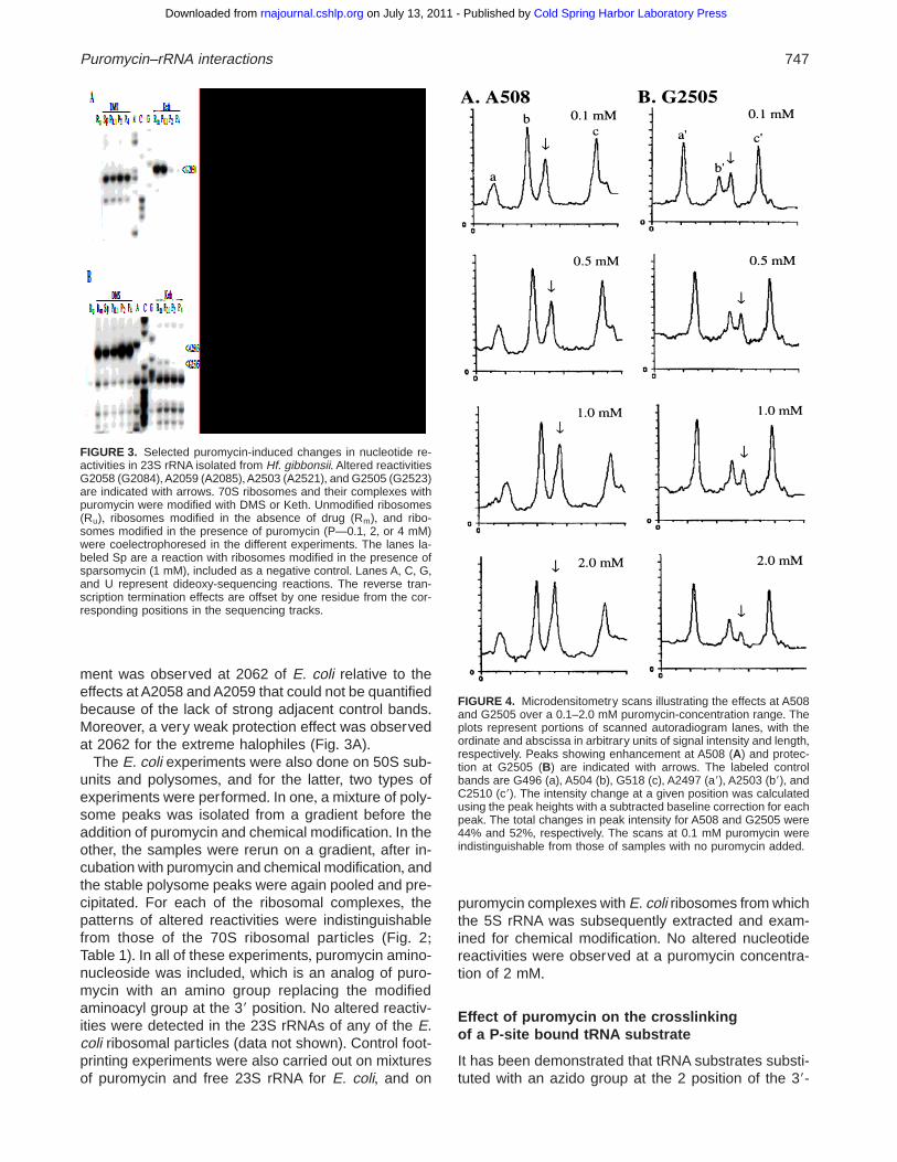
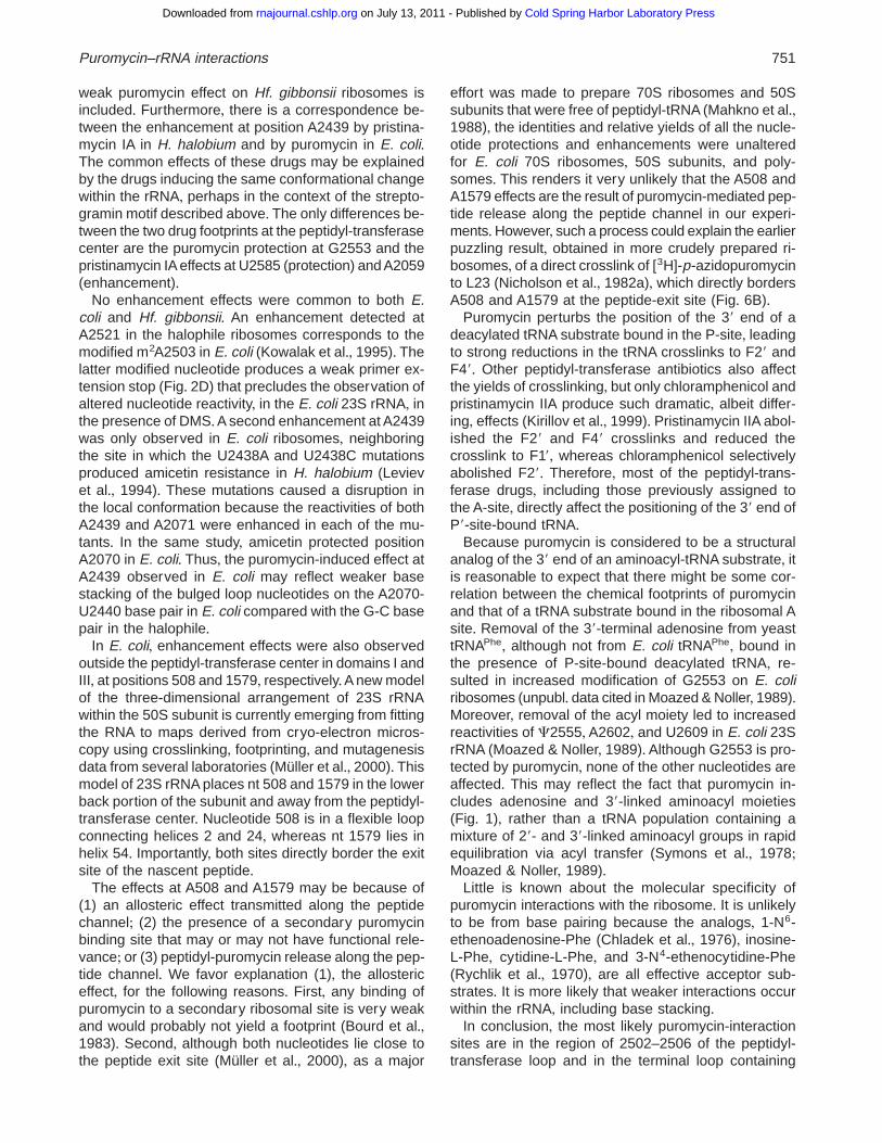
FIGURE 3. Selected puromycin-induced changes in nucleotide re-activities in 23S rRNA isolated from Hf. gibbonsii+ Altered reactivitiesG2058 (G2084),A2059 (A2085),A2503 (A2521), and G2505 (G2523)are indicated with arrows+ 70S ribosomes and their complexes withpuromycin were modified with DMS or Keth+ Unmodified ribosomes(Ru), ribosomes modified in the absence of drug (Rm), and ribo-somes modified in the presence of puromycin (P—0+1, 2, or 4 mM)were coelectrophoresed in the different experiments+ The lanes la-beled Sp are a reaction with ribosomes modified in the presence ofsparsomycin (1 mM), included as a negative control+ Lanes A, C, G,and U represent dideoxy-sequencing reactions+ The reverse tran-scription termination effects are offset by one residue from the cor-responding positions in the sequencing tracks+
FIGURE 4. Microdensitometry scans illustrating the effects at A508and G2505 over a 0+1–2+0 mM puromycin-concentration range+ Theplots represent portions of scanned autoradiogram lanes, with theordinate and abscissa in arbitrary units of signal intensity and length,respectively+ Peaks showing enhancement at A508 (A) and protec-tion at G2505 (B) are indicated with arrows+ The labeled controlbands are G496 (a), A504 (b), G518 (c), A2497 (a9), A2503 (b9), andC2510 (c9)+ The intensity change at a given position was calculatedusing the peak heights with a subtracted baseline correction for eachpeak+ The total changes in peak intensity for A508 and G2505 were44% and 52%, respectively+ The scans at 0+1 mM puromycin wereindistinguishable from those of samples with no puromycin added+
Puromycin–rRNA interactions 747
Cold Spring Harbor Laboratory Press on July 13, 2011 - Published by rnajournal.cshlp.orgDownloaded from

terminal adenosine residue, and bound at the ribosomalP-site, generate crosslinks with 23S rRNA and ribo-somal proteins L27 and L33 upon UV irradiation at365 nM (Wower et al+, 1995)+ The RNA crosslinks havebeen localized on previously characterized 23S rRNAfragments F19, F29, and F49 at nt C2601/A2602 andU2584/U2585 (F19), U2506 (F29), and A2062/C2063(F49) (Kirillov et al+, 1999)+ Puromycin was tested forits ability to alter the yields or identities of thesecrosslinks+ It was added to 70S ribosome-poly (U) com-plexes before addition of azidoadenosine tRNA sub-strate and irradiation+ Unlike experiments performedwith other antibiotics (Kirillov et al+, 1999), those withpuromycin were done only in the presence of deacyl-ated tRNA substrate+ The use of an active peptidyl-tRNAdonor substrate was avoided, as this, in conjunctionwith puromycin, would complicate the interpretation ofresults owing to peptide-bond formation and the sub-sequent dissociation of peptidyl-puromycin+
Puromycin did not change the level of tRNA bindingto the ribosome, but it did produce significant decreasesin crosslinking yields at two of three rRNA sites+ Inaddition, no new crosslinks were detected with puro-mycin+ The changes in crosslinking yields for each rRNAfragment are depicted in Figure 7 and the quantifiedyields are summarized in Table 2+ In the presence ofthe drug, no crosslink was formed with F49, and thecrosslink to F29 was reduced by about 50%+ The cross-link to F19 was not significantly affected by puromycinaveraged over four independent experiments (Table 2)+
Moreover, no significant changes in the crosslinkingyields to ribosomal proteins were observed in the pres-ence of the drug (data not shown)+
Effect of puromycin on pristinamycinIA-dependent rRNA modifications
The streptogramin B drug pristinamycin IA producesmodifications in E. coli 23S rRNA at m2A2503/C2504and G2061/A2062 upon irradiation with 365 nM light,where the former sites, at least, are likely to involve adrug–rRNA crosslink (Porse et al+, 1999a)+ Given theproximity of the footprinting sites to m2A2503/C2504,the ability of puromycin to influence these pristinamy-cin IA-dependent rRNA modifications was investigated(Fig+ 8)+ Puromycin was added to a preformed complexof pristinamycin IA and 70S ribosome–poly (U) com-plexes before irradiating and analyzing the peptidyl-transferase-loop region by primer extension+ Puromycinenhanced the pristinamycin IA-dependent reverse tran-scriptase stops at m2A2503/C2504 by 2+5-fold (Fig+ 8,lanes 2 and 3), but had no detectable effect on those at
TABLE 1+ Nucleotide positions exhibiting altered reactivities in thepresence of 0 and 2 mM puromycin within E. coli and Hf. gibbonsii23S rRNAs+
E. coli Hf. gibbonsii
Nucleotide 2 puromycin 1 puromycin 2 puromycin 1 puromycin
A508 1 11A1579 1 11A2058 (G2084) 11 (1)A2439 (A2458) (1) 1m2A2503 (A2521) 11 111G2505 (G2523) 1 (1) (1) 2G2553 (G2571) 1 (1) 1 (1)
The intensities of the autoradiographed bands were quantified bymicrodensitometry and are denoted as:111 strong,11 medium,1weak, and (1) very weak+ Intensities were assigned through visualinspection of several sets of films in combination with microdensito-metry+ The total intensity changes summarized above represent min-imum total intensity changes of 25–30% (see legend to Fig+ 4 andMaterials and methods)+ The total percentage changes observed forE. coli ribosomes, between 0 and 2 mM puromycin,were A508 (44%),A1579 (71%), A2439 (37%), G2505 (52%), and G2553 (32%)+ Errorlimits were estimated at 610% of the number given+ Experimentswere performed with 70S ribosomes, from which 23S rRNA wasisolated (see Materials and methods)+ For E. coli, similar reactivitychanges were observed with 50S subunits and polysomes+ Nucleo-tide positions are given using E. coli 23S rRNA numbering (Egebjerget al+, 1990), with the corresponding H. halobium numbering given inparentheses+ FIGURE 5. The location of puromycin-induced effects in the peptidyl-
transferase loop and adjoining regions of domain V of 23S rRNA+The sequence shown is that of E. coli 23S rRNA+ Nucleotides af-fected by puromycin in E. coli and Hf. gibbonsii 23S rRNAs areboxed and labeled+ Altered reactivities are indicated with circles andsquares, for E. coli and Hf. gibbonsii, respectively+ Protections aredenoted with filled symbols,whereas open symbols indicate enhance-ments+Where corresponding nucleotides in the two rRNAs differ, theidentity and numbering of the halophile nucleotide is given in paren-theses+ Nucleotides joined by lines are base paired, and posttran-scriptional modifications are indicated (Smith et al+, 1992)+
748 C. Rodriguez-Fonseca et al.
Cold Spring Harbor Laboratory Press on July 13, 2011 - Published by rnajournal.cshlp.orgDownloaded from

G2061/A2062 (data not shown)+ Control experimentsin which the order of drug addition was reversed yieldedidentical results (data not shown)+ The yields of therRNA modifications were enhanced another twofold inthe presence of both drugs with a P-site bound deacyl-ated tRNAPhe substrate (Fig+ 8, lanes 5 and 6)+ This isconsistent with deacylated tRNA producing a threefoldincrease in the pristinamycin IA-dependent modifica-tion yields at m2A2503/C2504 (Porse et al+, 1999a)+Moreover, the puromycin-induced enhancement of thePIA modification in both the presence and absence ofa P-site bound tRNA suggests that puromycin residesin the A-site in both ribosomal complexes+
DISCUSSION
For both the bacterial and archaeal ribosomes, most ofthe puromycin-induced effects lie within, or adjacent to,the peptidyl-transferase loop of domain V of 23S rRNA(Fig+ 9)+ Of these effects, all except one are at univer-sally conserved nucleotides, which is consistent withpuromycin-inhibiting bacterial, archaeal, and eukary-otic ribosomes+ Moreover, the fact that the effects ap-peared within the same concentration range suggeststhat they derive from a single drug-binding site+
The protection effects common to E. coli and Hf. gib-bonsii occur at the universally conserved nt G2505 andG2553+ G2505 is the site of altered nucleotide reactiv-ity in the presence of several other antibiotics, includ-ing the macrolides carbomycin and tylosin, and thestreptogramin A drugs (Garrett & Rodriguez-Fonseca,1995; Porse & Garrett, 1999)+ In a recent investigation,G2553 was specifically crosslinked to the A-site tRNAanalog, 4-thio-dT-p-C-p-puromycin (Green et al+, 1998)+In another study, all possible mutations made at G2553in E. coli resulted in dominant growth defects in vivo, aswell as reduced peptidyl transferase activity in vitro(Kim & Green,1999)+ In addition, complementation analy-sis of mutant A-site substrate analogs and ribosomesestablished a specific interaction between C75 of theaminoacyl–tRNA and G2553 of 23S rRNA (Kim & Green,1999)+ An important role for this rRNA loop in puromy-cin binding is also suggested by mutagenesis anddamage-selection experiments, in which changes at2550, 2552, 2555, and 2557 interfered with the transfer
FIGURE 6. Putative allosteric effects dependent on puromycin in domains I and III of E. coli 23S rRNA+ Nucleotidesexhibiting enhanced reactivities in the presence of puromycin are boxed in (A) domain I and (B) domain III+ The shadedregions are binding sites for ribosomal proteins L24 (Egebjerg et al+, 1987) and L23 (Egebjerg et al+, 1991)+
FIGURE 7. The effect of puromycin on the crosslinking yields ofdeacylated (2N3A76)tRNAPhe, bound at the ribosomal P site, tofragments of 23S rRNA+ Complexes were prepared in the absence(control) or presence of puromycin (1 mM), before binding of[32P](2N3A76)tRNAPhe and irradiation (see Materials and methods)+The specific activity of [32P](2N3A76)tRNAPhe was 3,000 dpm/pmol+The crosslinked fragments were generated by RNase H digestionwith a set of deoxyoligonucleotides (see Materials and methods),and analyzed on polyacrylamide gels+ The positions of the threemain labeled fragments F19, F29, and F49 are indicated+ Although theintensity of the F19 crosslink appears reduced in the puromycin lane,when the counts are adjusted for the degree of tRNA binding toribosomes, the differences are minimal (Table 2)+
TABLE 2 + Summary of the crosslinking yields of deacylated(2N3A76)tRNAPhe to rRNA fragments F19, F29, and F49+
Antibiotic F19 F29 F49
none 13 6 2 25 6 3 7 6 1puromycin 10 6 1 13 6 2 n+b+a
Data are presented as counts per minute of crosslinked deacyl-ated (2N3A76)tRNAPhe per picomole of ribosome-bound deacylated(2N3A76)tRNAPhe+ The specific activity of the (2N3A76)tRNAPhe wasnormalized to an initial activity of 10,000 dpm/pmol or about 1,800cpm/pmol estimated in an Instant Imager+ The data were averagedfrom four separate experiments+
an+b+: no band detected+
Puromycin–rRNA interactions 749
Cold Spring Harbor Laboratory Press on July 13, 2011 - Published by rnajournal.cshlp.orgDownloaded from

of peptidyl moiety to puromycin (Porse & Garrett, 1995;Bocchetta et al+, 1998)+ Taken together, the data sug-gest that the loop containing G2553 folds into the cat-alytic center and is in close proximity with the 39 end ofthe aminoacyl-tRNA substrate+
The region between 2058 and 2062 of the peptidyl-transferase loop is an area of diverse reactivity changesfor many antibiotics and is known to be conformation-ally labile+ In the case of puromycin, there is strongprotection of G2058 (G2084) in the halophiles but noeffect at A2058 in E. coli+ Differences in the chemicalfootprint of the streptogramin A drugs, bound to ar-chaeal and bacterial ribosomes, have also been ob-
served at this position (Rodriguez-Fonseca et al+, 1995;Porse & Garrett, 1999)+ In the presence of pristinamy-cin IIA, protection at G2058 (G2084) is observed inHalobacteria halobium, whereas enhancements areseen at A2058 in Bacillus megaterium, both in vitro andin vivo (Porse & Garrett, 1999)+ The conformationalflexibility of this region is underscored by the variableweak effects at position 2062 that were observed inE. coli and Hf. gibbonsii in the presence of puromycin+
A working model called the streptogramin motif hasbeen proposed based on footprinting and mutagenesisdata (Porse & Garrett, 1999)+ The model juxtaposesthe upper (2058–2062) and lower (2500–2506) re-gions of the peptidyl-transferase center, that are linkedin many antibiotic footprints, in an irregular helix+ Themodel includes A2060•G2505 and G2061•U2504 basepairings that are supported by several lines of muta-genesis evidence (see Porse & Garrett, 1999)+Anotherfeature of the model is that U2506 can form base pairswith either A/G2058 or A2059+ Thus, the differences inthe footprints between the two organisms may reflecttwo alternative, and perhaps functional, RNA conform-ers in this region+
The puromycin footprint obtained in Hf. gibbonsii ri-bosomes is similar to that obtained with pristinamycinIA complexed to H. halobium ribosomes (Porse & Gar-rett, 1999)+ Identical effects include an enhancement at2503 and protections at 2058 and 2505+ In addition,protection of position 2062 is a common feature if the
FIGURE 8. The effect of puromycin (puro) on UV-induced modifica-tions of E. coli 70S ribosomes that are dependent on pristinamycin IA(PIA)+ Pristinamycin IA (10 mM) was complexed to 70S ribosome(0+15 mM) –poly(U) complexes in the absence or presence of de-acylated tRNA and was incubated at 37 8C for 10 min+ Puromycin(1 mM) was added and incubation was continued for 10 min+ Thesamples were then irradiated at 365 nM for 20 min+ Primer extensionanalyses were performed by using EC2621 (59-CAGTTCTCCAGCGCCCAC-39), a primer complementary to positions 2638–2621 ofE. coli 23S rRNA+ The primer extension stops at 2503 and 2504 werequantified in an Instant Imager, using the stop at 2497 as a reference,and the results are shown below the gel, relative to a control samplein the absence of puromycin+ G, A, U, and C represent dideoxy-sequencing reactions+
FIGURE 9. Summary of the puromycin effects in the peptidyl-transferase loop of 23S rRNA+ The sites of altered nucleotide reac-tivities for E. coli and Hf. gibbonsii are boxed on the E. coli 23S rRNAsequence (see Fig+ 5)+ Sites of drug-dependent UV-induced cross-links or modifications are labelled with boxed crosses+ The sites ofcrosslinks with P-site bound tRNA substrate are denoted with open-headed arrows+ Filled arrows indicate sites of crosslinking withp-azidopuromycin+
750 C. Rodriguez-Fonseca et al.
Cold Spring Harbor Laboratory Press on July 13, 2011 - Published by rnajournal.cshlp.orgDownloaded from
weak puromycin effect on Hf. gibbonsii ribosomes isincluded+ Furthermore, there is a correspondence be-tween the enhancement at position A2439 by pristina-mycin IA in H. halobium and by puromycin in E. coli+The common effects of these drugs may be explainedby the drugs inducing the same conformational changewithin the rRNA, perhaps in the context of the strepto-gramin motif described above+ The only differences be-tween the two drug footprints at the peptidyl-transferasecenter are the puromycin protection at G2553 and thepristinamycin IA effects at U2585 (protection) and A2059(enhancement)+
No enhancement effects were common to both E.coli and Hf. gibbonsii+ An enhancement detected atA2521 in the halophile ribosomes corresponds to themodified m2A2503 in E. coli (Kowalak et al+, 1995)+ Thelatter modified nucleotide produces a weak primer ex-tension stop (Fig+ 2D) that precludes the observation ofaltered nucleotide reactivity, in the E. coli 23S rRNA, inthe presence of DMS+A second enhancement at A2439was only observed in E. coli ribosomes, neighboringthe site in which the U2438A and U2438C mutationsproduced amicetin resistance in H. halobium (Levievet al+, 1994)+ These mutations caused a disruption inthe local conformation because the reactivities of bothA2439 and A2071 were enhanced in each of the mu-tants+ In the same study, amicetin protected positionA2070 in E. coli+ Thus, the puromycin-induced effect atA2439 observed in E. coli may reflect weaker basestacking of the bulged loop nucleotides on the A2070-U2440 base pair in E. coli compared with the G-C basepair in the halophile+
In E. coli, enhancement effects were also observedoutside the peptidyl-transferase center in domains I andIII, at positions 508 and 1579, respectively+A new modelof the three-dimensional arrangement of 23S rRNAwithin the 50S subunit is currently emerging from fittingthe RNA to maps derived from cryo-electron micros-copy using crosslinking, footprinting, and mutagenesisdata from several laboratories (Müller et al+, 2000)+ Thismodel of 23S rRNA places nt 508 and 1579 in the lowerback portion of the subunit and away from the peptidyl-transferase center+ Nucleotide 508 is in a flexible loopconnecting helices 2 and 24, whereas nt 1579 lies inhelix 54+ Importantly, both sites directly border the exitsite of the nascent peptide+
The effects at A508 and A1579 may be because of(1) an allosteric effect transmitted along the peptidechannel; (2) the presence of a secondary puromycinbinding site that may or may not have functional rele-vance; or (3) peptidyl-puromycin release along the pep-tide channel+ We favor explanation (1), the allostericeffect, for the following reasons+ First, any binding ofpuromycin to a secondary ribosomal site is very weakand would probably not yield a footprint (Bourd et al+,1983)+ Second, although both nucleotides lie close tothe peptide exit site (Müller et al+, 2000), as a major
effort was made to prepare 70S ribosomes and 50Ssubunits that were free of peptidyl-tRNA (Mahkno et al+,1988), the identities and relative yields of all the nucle-otide protections and enhancements were unalteredfor E. coli 70S ribosomes, 50S subunits, and poly-somes+ This renders it very unlikely that the A508 andA1579 effects are the result of puromycin-mediated pep-tide release along the peptide channel in our experi-ments+However, such a process could explain the earlierpuzzling result, obtained in more crudely prepared ri-bosomes, of a direct crosslink of [3H]-p-azidopuromycinto L23 (Nicholson et al+, 1982a), which directly bordersA508 and A1579 at the peptide-exit site (Fig+ 6B)+
Puromycin perturbs the position of the 39 end of adeacylated tRNA substrate bound in the P-site, leadingto strong reductions in the tRNA crosslinks to F29 andF49+ Other peptidyl-transferase antibiotics also affectthe yields of crosslinking, but only chloramphenicol andpristinamycin IIA produce such dramatic, albeit differ-ing, effects (Kirillov et al+, 1999)+ Pristinamycin IIA abol-ished the F29 and F49 crosslinks and reduced thecrosslink to F19, whereas chloramphenicol selectivelyabolished F29+ Therefore, most of the peptidyl-trans-ferase drugs, including those previously assigned tothe A-site, directly affect the positioning of the 39 end ofP9-site-bound tRNA+
Because puromycin is considered to be a structuralanalog of the 39 end of an aminoacyl-tRNA substrate, itis reasonable to expect that there might be some cor-relation between the chemical footprints of puromycinand that of a tRNA substrate bound in the ribosomal Asite+ Removal of the 39-terminal adenosine from yeasttRNAPhe, although not from E. coli tRNAPhe, bound inthe presence of P-site-bound deacylated tRNA, re-sulted in increased modification of G2553 on E. coliribosomes (unpubl+ data cited in Moazed & Noller, 1989)+Moreover, removal of the acyl moiety led to increasedreactivities of C2555, A2602, and U2609 in E. coli 23SrRNA (Moazed & Noller, 1989)+ Although G2553 is pro-tected by puromycin, none of the other nucleotides areaffected+ This may reflect the fact that puromycin in-cludes adenosine and 39-linked aminoacyl moieties(Fig+ 1), rather than a tRNA population containing amixture of 29- and 39-linked aminoacyl groups in rapidequilibration via acyl transfer (Symons et al+, 1978;Moazed & Noller, 1989)+
Little is known about the molecular specificity ofpuromycin interactions with the ribosome+ It is unlikelyto be from base pairing because the analogs, 1-N6-ethenoadenosine-Phe (Chladek et al+, 1976), inosine-L-Phe, cytidine-L-Phe, and 3-N4-ethenocytidine-Phe(Rychlik et al+, 1970), are all effective acceptor sub-strates+ It is more likely that weaker interactions occurwithin the rRNA, including base stacking+
In conclusion, the most likely puromycin-interactionsites are in the region of 2502–2506 of the peptidyl-transferase loop and in the terminal loop containing
Puromycin–rRNA interactions 751
Cold Spring Harbor Laboratory Press on July 13, 2011 - Published by rnajournal.cshlp.orgDownloaded from

G2553 (Fig+ 9)+ Given that puromycin can act as astructural analog of an aminoacyl-tRNA substrate, bothof these RNA regions must be closely juxtaposed at thecatalytic center+ Evidence supporting this hypothesisnot only includes the common protections at G2505and G2553 in bacteria and archaea, but also the stronglyenhanced reactivity at A2503 (A2521) in the halophilethat would have remained undetected in the E. coli 23SrRNA+ Moreover, the effects of puromycin on the F29crosslink of deacylated (2N3A76)tRNA at U2506 andon the putative UV-induced crosslinking yields of pris-tinamycin IA at m2A2503/C2504 further support thebinding of puromycin to this putative “streptogramin mo-tif” of the peptidyl-transferase loop (Fig+ 9)+ In addition,these results correlate directly with the earlier findingthat G2502 and U2504 are the principal crosslinkingsites of p-azidopuromycin with rRNA (Hall et al+, 1988)+
MATERIALS and METHODS
Preparation of ribosomes
Tight-couple ribosomes and 50S subunits were prepared fromE. coli MRE 600 as described (Makhno et al+, 1988), exceptthat particles were harvested at all preparative steps by cen-trifugation in a fixed angle rotor at 100,000 3 g+ The Makhnoet al+ (1988) procedure aims to remove endogenous mRNAand peptidyl-tRNA through dissociation of the ribosomes intosubparticles, followed by reassociation+ Briefly, tight-coupleribosomes, defined as those that remain associated between4–6 mM Mg21, are initially purified in an associated state+The complexes are heated at 37 8C for 15 min in 3 mM MgCl2and 300 mM NH4Cl, followed by sedimentation in a sucrosegradient to dissociate the particles into subunits+ Equimolaramounts of subunits are then combined and subjected torepeated sedimentation in the above buffer used for isolationof purified 30S and 50S subunits+ Alternatively, they are re-sedimented through a sucrose gradient in 5 mM MgCl2 and50 mM NH4Cl for final selection of tight-couple 70S ribo-somes+ Polysomes were isolated essentially as described byPowers and Noller (1991)+
Hf. gibbonsii (ATCC 33959) and Hf. mediterranei (ATCC33500) ribosomes were prepared from cells that were grownto A660 5 0+5, spun down, and lysed by alumina grinding(1+5 g alumina/g cells) in high-salt buffer (20 mM Tris-HCl,pH 7+4, 60 mM magnesium acetate, 3 M KCl, and 6 mM2-mercaptoethanol)+ The lysate was centrifuged at 15,000rpm for 15 min in a Sorvall SS34 rotor+ Because of the inac-tivation of DNAse I by the high salt concentration used, theDNA was pelleted by centrifugation at 45,000 rpm for 30 min+The supernatant from the second centrifugation was thenrecentrifuged at 48,000 rpm for 2+5 h in a Beckman Ti 65rotor+ The ribosomal pellet was gently resuspended with aglass rod and washed twice in high-salt buffer before repel-leting by ultracentrifugation for 3 h at 45,000 rpm, and 15 h at30,000 rpm+ The ribosomes were clarified by centrifugation at15,000 rpm for 10 min, divided into small aliquots and storedat 280 8C+ All ribosomal preparations were highly active inpoly(U)-dependent poly(Phe)-synthesis, where an assay op-
timized for halo archaea was used for both halophiles (Sanzet al+, 1988)+
RNA footprinting of puromycin-ribosomecomplexes
Puromycin-ribosome complexes were prepared as follows+Ribosomes (20 mg) were incubated at 30 8C for 20 min in100 mL of the appropriate buffer (E. coli: 50 mM HEPES-KOH, pH 7+8, 10 mM MgCl2, 15 mM KCl, 15 mM NH4Cl,1 mM DTT, and 0+1 mM EDTA; Hf. gibbonsii and Hf. medi-terranei : 70 mM HEPES-KOH, pH 7+8, 60 mM magnesiumacetate, 3 M KCl, and 6 mM 2-mercaptoethanol)+ Puromy-cin (Sigma) was then added to a final concentration of 0+1,0+5, 1, 2, or 4 mM, and the mixture was incubated at 30 8Cfor 30 min before cooling slowly and placing on ice+ Ali-quots of ribosomes and puromycin-ribosome complexes(100 mL) were then reacted with the following chemicalprobes as described earlier (Christiansen et al+, 1990): 1 mLDMS (1:1 dilution in ethanol), 5 mL kethoxal (35 mg/mL in20% ethanol), and 100 mL CMCT (42 mg/mL in the appro-priate buffer)+ For the DMS and kethoxal reactions, sam-ples were incubated at 30 8C for 10 min, and the CMCTreaction mixture was incubated at 30 8C for 20 min+ Reac-tions were stopped by standard procedures (Christiansenet al+, 1990), and the RNA was precipitated by ethanol+Modified sites were identified by extension from 32P-end-labeled oligodeoxynucleotide primers using reverse tran-scriptase (Life Sciences, Florida)+ The primers werehybridized to sites localized 150–200 nt apart along each23S rRNA+ Altered nucleotide reactivities were quantifiedby microdensitometry (Hoeffer, California)+ Autoradiogramswere scanned and peak heights and areas determined+ Thetotal intensity change at a particular nucleotide position wascalculated from the peak heights with a subtracted baselinecorrection for each peak+
The criteria for evaluating the significance of the footprint-ing results included the following: (1) reproducible observa-tion of the effects in several different sets of experiments; (2)for E. coli, identical effects were observed for 70S ribosomes,50S subunits, and polysomes; (3) the halophile effects wereobserved for two different organisms: Hf. gibbonsii and Hf.mediterranei ; (4) the effects considered to be significant hada minimum total intensity change of 25–30%,whereas weakertotal intensity changes were not accepted as significant; and(5) minor variations in the intensities of certain peaks ob-served from experiment to experiment were judged to beinsignificant+
Binding and crosslinking of tRNAto ribosomes
The 2-azidoadenosine tRNA substrates and the poly (U)templates were prepared as described previously (Kirillovet al+, 1999, and references therein)+ Tight-couple 70S ribo-somes (2 mM) were incubated with poly (U) (1 mg/pmolribosomes) in 20 mM Tris-Cl (pH 7+5), 50 mM NH4Cl, 10 mMMgCl2, and 0+5 mM EDTA for 5 min at 37 8C+ Puromycin(1 mM) was added and incubated for 5 min at 37 8C beforeadding the azido-tRNA derivative at molar ratios of 0+5–1+1and incubating another 30 min at 37 8C+ The level of tRNA
752 C. Rodriguez-Fonseca et al.
Cold Spring Harbor Laboratory Press on July 13, 2011 - Published by rnajournal.cshlp.orgDownloaded from

binding was measured by nitrocellulose filter binding as de-scribed earlier (Kirillov et al+, 1999)+ Crosslinks were gen-erated by diluting the samples in cold buffer, and irradiatingwith 365 nM light at 0 8C, at a distance of 5 cm from theUV source+ Analysis of the crosslinking products, includingpreparation of the rRNA fragments F19, F29, and F49 byoligonucleotide-directed RNase H digestion, is detailed inKirillov et al+ (1999)+
Interaction of PIA with 70S ribosomes
Pristinamycin IA (10 mM) was incubated with 70S ribosomes(150 nM) in 20 mM Tris-chloride, pH 7+5, 50 mM NH4Cl, and10 mM MgCl2 at 37 8C for 10 min+ Where indicated, deacyl-ated tRNA (1+2 mol/mol ribosomes) was included in the pres-ence of poly(U) (1 mg/pmol ribosomes)+ Puromycin (1 mM)was added either to preformed PIA–ribosome complexes ordirectly to 70S ribosomes before adding PIA+ Following ad-dition of puromycin or PIA, the samples were incubated at37 8C for 10 min+ Samples were maintained as droplets on amicrotiter tray, placed on an ice-water bath, and subjected toUV irradiation in a Stratalinker 1800 (5 3 8 W bulbs, Strata-gene, California) for 20 min at 365 nM+ Samples were ex-tracted with phenol, (1:1) phenol:chloroform, and chloroform,prior to ethanol precipitation to remove protein and antibiot-ics+ The rRNA was subjected to primer extension using AMVreverse transcriptase (Life Sciences, Florida) and deoxyoligo-nucleotide primers complementary to rRNA 39 to the modifi-cations+The extension products were separated on denaturingpolyacrylamide gels and autoradiographed+
ACKNOWLEDGMENTS
The research and B+T+ Porse were supported by grants fromthe Danish Natural Science Research Council and the Dan-ish Biotek II program+ C+ Rodriguez-Fonseca was a fellow ofthe Comunidad de Madrid and received EMBO short-termfellowships and a Danish Research Academy studentship+K+S+ Long received an investigator fellowship from the AlfredBenzon Foundation+Richard Brimacombe is thanked for help-ful discussions on the peptide exit site+
Received January 14, 2000; returned for revision February25, 2000; revised manuscript received March 16, 2000
REFERENCES
Bocchetta M, Xiong L, Mankin AS+ 1998+ 23S rRNA positions essen-tial for tRNA binding in ribosomal functional sites+ Proc Natl AcadSci USA 95:3525–3530+
Bourd SB, Kukhanova MK, Gottikh BP, Krayevsky AA+ 1983+ Coop-erative effects in the peptidyl transferase center of Escherichiacoli ribosomes+ Eur J Biochem 135:465–469+
Chladek S, Ringer D, Abraham EM+ 1976+ Fluorescent 29(39)-O-aminoacylnucleosides-acceptor substrates for ribosomal pepti-dyltransferase1+ Nucleic Acids Res 3:1215–1224+
Christiansen J, Egebjerg J, Larsen N, Garrett RA+ 1990+ Analysis ofRNA structure: Experimental and theoretical considerations+ In:Spedding G, ed+ Ribosomes and protein synthesis: A practicalapproach+ Oxford: Oxford University Press+ pp 229–252+
Egebjerg J, Christiansen J, Garrett RA+ 1991+ Attachment sites ofprimary binding proteins L1, L2, and L23 on 23S ribosomal RNAof Escherichia coli+ J Mol Biol 222:251–264+
Egebjerg J, Larsen N,Garrett RA+ 1990+ Structural map of 23S rRNA+In: Hill W, Dahlberg A, Garrett RA, Moore PB Schlessinger D,Warner J, eds+ The ribosome: Structure, function, and evolution+Washington, DC: American Society for Microbiology Press+ pp168–179+
Egebjerg J, Leffers H, Christensen A, Andersen H, Garrett RA+ 1987+Structure and accessibility of domain I of E. coli 23S rRNA in freeRNA, in the L24 RNA complex and in 50S subunits: Implicationsfor ribosomal assembly+ J Mol Biol 196:125–136+
Fernandez-Muñoz R, Vázquez D+ 1973+ Binding of puromycin to E.coli ribosomes+ Effects of puromycin analogues and peptide bondformation inhibitors+ Mol Biol Rep 1:27–32+
Gale EF, Cundliffe E, Reynolds PE, Richmond RA, Waring MJ,eds+ 1981+ Antibiotic inhibitors of ribosome function+ In: The mo-lecular basis of antibiotic action+ London: Wiley & Sons, Ltd+ pp402–457+
Garrett RA, Rodriguez-Fonseca C+ 1995+ The peptidyl transferasecenter+ In: Zimmermann RA, Dahlberg A, eds+ Ribosomal RNA:Structure, evolution, processing and function+ Boca Raton, Flor-ida: CRC Press+ pp 327–355.
Green R, Switzer C, Noller HF+ 1998+ Ribosome-catalyzed peptide-bond formation with an A-site substrate covalently linked to 23Sribosomal RNA+ Science 280:286–289+
Hall CC, Johnson D, Cooperman BS+ 1988+ [3H]-p-Azidopuromycinphotoaffinity labeling of Escherichia coli ribosomes: Evidence forsite-specific interaction at U-2504 and G-2502 in domain V of 23Sribosomal RNA+ Biochemistry 27:3983–3990+
Harris RJ, Symons RH+ 1973+ On the molecular mechanism of actionof certain substrates and inhibitors of ribosomal peptidyltransfer-ase+ Bioorg Chem 2:266–285+
Kim DF, Green R+ 1999+ Base-pairing between 23S rRNA and tRNAin the ribosomal A site+ Mol Cell 4:859–864+
Kirillov S, Porse B, Vester B,Woolley P, Garrett RA+ 1997+Movementof the 39-end of tRNA through the peptidyl transferase center andits inhibition by antibiotics+ FEBS Lett 406:223–233+
Kirillov SV, Porse BT, Garrett RA+ 1999+ Peptidyl transferase antibi-otics perturb the relative positioning of the 39-terminal adenosineof P/P9-site-bound tRNA and 23S rRNA in the ribosome+ RNA5:1003–1013+
Kowalak JA, Bruenger E, McCloskey JA+ 1995+ Posttranscriptionalmodification of the central loop of domain V in Escherichia coli23S ribosomal RNA+ J Biol Chem 270:17758–17764+
Leviev IG, Rodriguez-Fonseca C, Phan H, Garrett RA, Heilek G,Noller HF, Mankin AS+ 1994+ A conserved secondary structuralmotif in 23S rRNA defines the site of interaction of amicetin, auniversal inhibitor of peptide bond formation+ EMBO J 13:1682–1686+
Makhno VI, Peshin NN, Semenkov YP, Kirillov SV+ 1988+ Modifiedprocedure of producing “tight” 70S ribosomes from E. coli, highlyactive in individual stages of the elongation cycle+ Mol Biol 22:528–537+
Moazed D, Noller HF+ 1987+ Chloramphenicol, erythromycin, carbo-mycin, and vernamycin B protect overlapping sites in the peptidyltransferase region of 23S rRNA+ Biochimie 69:879–884+
Moazed D, Noller HF+ 1989+ Interaction of tRNA with 23S rRNA in theribosomal A, P, and E sites+ Cell 57:585–597+
Müller F, Sommer I, Baranov P, Stark H, van Heel M, Rodnina M,Wintermeyer W, Brimacombe R+ 2000+ The 3D arrangement ofthe RNA in the E. coli 50S ribosomal subunit+ II+ Distribution offunctionally important sites+ J Mol Biol+ In press+
Nicholson AW,Hall CC, Strycharz WA,Cooperman BS+ 1982a+ Photo-affinity labeling of Escherichia coli ribosomes by an aryl azideanalogue of puromycin+ On the identification of the major cova-lently labeled ribosomal proteins and on the mechanism of photo-incorporation+ Biochemistry 21:3797–3808+
Nicholson AW,Hall CC, Strycharz WA,Cooperman BS+ 1982b+ Photo-affinity labeling of Escherichia coli ribosomes by an aryl azideanalogue of puromycin+ Evidence for the functional site specificityof labeling+ Biochemistry 21:3809–3817+
Odom OW, Picking WD, Hardesty B+ 1990+ Movement of tRNA butnot the nascent peptide during peptide bond formation on theribosome+ Biochemistry 29:10734–10744+
Pestka S+ 1970+ Studies on the formation of transfer ribonucleic acid-ribosome complexes+ 8+ Survey of the effect of antibiotics on
Puromycin–rRNA interactions 753
Cold Spring Harbor Laboratory Press on July 13, 2011 - Published by rnajournal.cshlp.orgDownloaded from

N-acetyl-phenylalanyl-puromycin formation: Possible mecha-nism of chloramphenicol action+Arch Biochem Biophys 136:80–88+
Pestka S+ 1977+ Inhibitors of protein synthesis+ In: Weissbach H,Pestka S, eds+ Molecular mechanisms of protein biosynthesis+New York: Academic Press+ pp 467–553+
Porse BT, Garrett RA+ 1995+ Mapping important nucleotides in thepeptidyl transferase center of 23S rRNA using a random muta-genesis approach+ J Mol Biol 249:1–20+
Porse BT, Rodriguez-Fonseca C, Leviev I, Garrett RA+ 1995+ Antibi-otic inhibition of the movement of tRNA substrates through apeptidyl transferase cavity+ Biochem Cell Biol 73:877–885+
Porse BT, Garrett RA+ 1999+ Sites of interaction of streptogramin Aand B antibiotics in the peptidyl transferase loop of 23S rRNA andthe synergism of their inhibitory mechanisms+ J Mol Biol 286:375–387+
Porse BT, Kirillov SV, Awayez MJ, Garrett RA+ 1999a+ UV-inducedmodifications in the peptidyl transferase loop of 23S rRNA de-pendent on binding of the streptogramin B antibiotic, pristinamy-cin IA+ RNA 5:585–595+
Porse BT, Kirillov SV,Awayez MJ,Ottenheijm HCJ,Garrett RA+ 1999b+Direct crosslinking of the antitumor antibiotic sparsomycin, and itsderivatives, to A2602 in the peptidyl transferase center of 23S-like rRNA within ribosome-tRNA complexes+ Proc Natl Acad SciUSA 96:9003–9008+
Porse BT, Kirillov SV, Garrett RA+ 2000+ Antibiotics and the peptidyl-transferase center+ In:Garrett RA,Douthwaite SR, Liljas A,Mathe-son AT, Moore PB, Noller HF, eds+ The ribosome: Structure,function, antibiotics, and cellular interactions+ Washington, DC:American Society for Microbiology Press+ pp 441–449+
Powers T, Noller HF+ 1991+ A functional pseudoknot in 16S rRNA+EMBO J 10:2203–2214+
Rodriguez-Fonseca C, Amils R, Garrett RA+ 1995+ Fine structure ofthe peptidyl transferase center on 23S-like rRNAs deduced fromchemical probing of antibiotic-ribosome complexes+ J Mol Biol247:224–235+
Rychlik I, Cerna J, Chladek S, Pulkrabek P, Zemlicka J+ 1970+ Sub-strate specificity of ribosomal peptidyltransferase+ The effect ofthe nature of the amino acid side chain on the acceptor activity of29(39)-O-aminoacyladenosines+ Eur J Biochem 16:136–142+
Sanz JL, Marín I, Balboa MA, Ureña D, Amils R+ 1988+ An NH41
dependent protein synthesis cell-free system for halobacteria+Biochemistry 27:8194–8199+
Sanz JL, Marín I, Ureña D, Amils R+ 1993+ Functional analysis ofseven ribosomal systems from extreme halophilic archaea+ Can JMicrobiol 39:311–317+
Smith JD, Traut RR, Blackburn GM, Monro RE+ 1965+ Action of pu-romycin in polyadenylic acid-directed polylysine synthesis+ J MolBiol 13:617–628+
Smith JE,Cooperman BS,Mitchell P+ 1992+Methylation sites in Esch-erichia coli rRNA: Localization and identification of four new sitesof methylation in 23S rRNA+ Biochemistry 31:10825–10834+
Symons RH, Harris RJ, Greenwell P, Eckermann DJ, Vanin EF+ 1978+The use of puromycin analogs and related compounds to probethe active center of peptidyl transferase on Escherichia coli ribo-somes+ Bioorg Chem (Suppl+) 4:409–436+
Traut RR, Monro RE+ 1964+ The puromycin reaction and its relation-ship to protein synthesis+ J Mol Biol 10:63–72+
Vázquez D+ 1979+ Inhibitors of protein synthesis+ Berlin: Springer-Verlag+
Wower J, Wower IK, Kirillov SV, Rosen KV, Hixon SS, ZimmermannRA+ 1995+ Peptidyl transferase and beyond+ Biochem Cell Biol73:1041–1047+
754 C. Rodriguez-Fonseca et al.
Cold Spring Harbor Laboratory Press on July 13, 2011 - Published by rnajournal.cshlp.orgDownloaded from