Inhibition of cytosolic glutathione S-transferase activity from rat liver by copper
10
Chemico-Biological Interactions 164 (2006) 39–48 Inhibition of cytosolic glutathione S-transferase activity from rat liver by copper M.E. Letelier ∗ , M. Mart´ ınez, V. Gonz´ alez-Lira, M. Fa´ undez, P. Aracena-Parks Laboratory of Pharmacology, Department of Pharmacological and Toxicological Chemistry, Chemical and Pharmaceutical Sciences School, Universidad de Chile, Olivos 1007, Independencia, Santiago, Chile Received 16 May 2006; received in revised form 12 August 2006; accepted 16 August 2006 Available online 25 August 2006 Abstract H 2 O 2 inactivation of particular GST isoforms has been reported, with no information regarding the overall effect of other ROS on cytosolic GST activity. The present work describes the inactivation of total cytosolic GST activity from liver rats by the oxygen radical-generating system Cu 2+ /ascorbate. We have previously shown that this system may change some enzymatic activities of thiol proteins through two mechanisms: ROS-induced oxidation and non-specific Cu 2+ binding to protein thiol groups. In the present study, we show that nanomolar Cu 2+ in the absence of ascorbate did not modify total cytosolic GST activity; the same concentrations of Cu 2+ in the presence of ascorbate, however, inhibited this activity. Micromolar Cu 2+ in either the absence or presence of ascorbate inhibited cytosolic GST activity. Kinetic studies show that GSH but no 1-chloro-2,4-dinitrobenzene prevent the inhibition on cytosolic GST induced by micromolar Cu 2+ either in the absence or presence of ascorbate. On the other hand, NEM and mersalyl acid, both thiol-alkylating agents, inhibited GST activity with differential reactivity in a dose-dependent manner. Taken together, these results suggest that an inhibitory Cu 2+ -binding effect is likely to be negligible on the overall inhibition of cytosolic GST activity observed by the Cu 2+ /ascorbate system. We discuss how modification of GST-thiol groups is related to the inhibition of cytosolic GST activity. © 2006 Elsevier Ireland Ltd. All rights reserved. Keywords: Cytosolic GST–Cu; Cytosolic GST–Cu/ascorbate; GST-thiols 1. Introduction Glutathione S-transferases (GSTs; EC 2.5.1.18) are a family of isoenzymes involved in the xenobiotic detox- ication. GSTs catalyze the nucleophilic attack of glu- tathione (GSH) on electrophilic/lipophilic substrates, thereby decreasing their reactivity with cellular macro- molecules [1]. These substrates display an electrophilic carbon as the site of attack by GSH; the relative lack of ∗ Corresponding author. Tel.: +56 2 6782885; fax: +56 2 7378920. E-mail address: [email protected] (M.E. Letelier). specificity of the enzyme however, permit that atoms other than carbon could serve as the point of nucle- ophilic attack such as the nitrogen atom of organic nitrate esters or the sulfur atom of organic thiocyanates [2–4]. Although these isoenzymes display low speci- ficity towards their substrates, GSTs show an absolute specificity towards its endogenous cofactor, GSH [5]. Several roles in cellular processes have been described for mammalian liver GST isoenzymes, between them drug metabolism [1,4,6–8], intracellu- lar transport [5,9–11], antioxidant defense [12–18], leukotriene biosynthesis [19–21], cell survival [22–24] and drug multi-resistance [11,25,26]. As many as eight 0009-2797/$ – see front matter © 2006 Elsevier Ireland Ltd. All rights reserved. doi:10.1016/j.cbi.2006.08.013
-
Upload
independent -
Category
Documents
-
view
1 -
download
0
Transcript of Inhibition of cytosolic glutathione S-transferase activity from rat liver by copper

A
ortpcptNTci©
K
1
fittmc
0
Chemico-Biological Interactions 164 (2006) 39–48
Inhibition of cytosolic glutathione S-transferaseactivity from rat liver by copper
M.E. Letelier ∗, M. Martınez, V. Gonzalez-Lira, M. Faundez, P. Aracena-ParksLaboratory of Pharmacology, Department of Pharmacological and Toxicological Chemistry, Chemical and Pharmaceutical
Sciences School, Universidad de Chile, Olivos 1007, Independencia, Santiago, Chile
Received 16 May 2006; received in revised form 12 August 2006; accepted 16 August 2006Available online 25 August 2006
bstract
H2O2 inactivation of particular GST isoforms has been reported, with no information regarding the overall effect of other ROSn cytosolic GST activity. The present work describes the inactivation of total cytosolic GST activity from liver rats by the oxygenadical-generating system Cu2+/ascorbate. We have previously shown that this system may change some enzymatic activities ofhiol proteins through two mechanisms: ROS-induced oxidation and non-specific Cu2+ binding to protein thiol groups. In theresent study, we show that nanomolar Cu2+ in the absence of ascorbate did not modify total cytosolic GST activity; the sameoncentrations of Cu2+ in the presence of ascorbate, however, inhibited this activity. Micromolar Cu2+ in either the absence orresence of ascorbate inhibited cytosolic GST activity. Kinetic studies show that GSH but no 1-chloro-2,4-dinitrobenzene preventhe inhibition on cytosolic GST induced by micromolar Cu2+ either in the absence or presence of ascorbate. On the other hand,EM and mersalyl acid, both thiol-alkylating agents, inhibited GST activity with differential reactivity in a dose-dependent manner.
aken together, these results suggest that an inhibitory Cu2+-binding effect is likely to be negligible on the overall inhibition ofytosolic GST activity observed by the Cu2+/ascorbate system. We discuss how modification of GST-thiol groups is related to thenhibition of cytosolic GST activity.2006 Elsevier Ireland Ltd. All rights reserved.
iols
eywords: Cytosolic GST–Cu; Cytosolic GST–Cu/ascorbate; GST-th. Introduction
Glutathione S-transferases (GSTs; EC 2.5.1.18) are aamily of isoenzymes involved in the xenobiotic detox-cation. GSTs catalyze the nucleophilic attack of glu-athione (GSH) on electrophilic/lipophilic substrates,
hereby decreasing their reactivity with cellular macro-olecules [1]. These substrates display an electrophilicarbon as the site of attack by GSH; the relative lack of
∗ Corresponding author. Tel.: +56 2 6782885; fax: +56 2 7378920.E-mail address: [email protected] (M.E. Letelier).
009-2797/$ – see front matter © 2006 Elsevier Ireland Ltd. All rights reservdoi:10.1016/j.cbi.2006.08.013
specificity of the enzyme however, permit that atomsother than carbon could serve as the point of nucle-ophilic attack such as the nitrogen atom of organicnitrate esters or the sulfur atom of organic thiocyanates[2–4]. Although these isoenzymes display low speci-ficity towards their substrates, GSTs show an absolutespecificity towards its endogenous cofactor, GSH [5].
Several roles in cellular processes have beendescribed for mammalian liver GST isoenzymes,
between them drug metabolism [1,4,6–8], intracellu-lar transport [5,9–11], antioxidant defense [12–18],leukotriene biosynthesis [19–21], cell survival [22–24]and drug multi-resistance [11,25,26]. As many as eighted.

ologica
40 M.E. Letelier et al. / Chemico-Biclasses of cytosolic GSTs, designated Alpha, Mu, Pi,Sigma, Theta, Zeta, Kappa and Omega have been iden-tified in mammals [27]. In rat liver, GSTs occur as fourcytosolic and one microsomal isoforms. These isoen-zymes display differences in their apparent molecularweights, induction by xenobiotics, and immunochemi-cal properties. Remarkably, these isoenzymes are dis-tinguishable by their response towards detergents orsulfhydryl group-reducing agents [14,28]. The activeform of rat liver microsomal GST is thought to be adimeric conformation of two identical protein chainsbound by their unique cysteine residue [29–31]. On theother hand, it has been reported that rat glutathionetransferase P-form is inactivated by hydrogen perox-ide (H2O2). The authors suggest that cysteine 47 andcysteine-101 may be involved in the formation of intra-or inter-subunit disulfides and these residues may belocated in an important region for GSH binding, disul-phide bond formation between these residues resultingin steric hindrance [32]. Similar results were observedwith human placenta GSH-transferase � form andthe authors demonstrated that the oxidative inactiva-tion was reversed by 2,4-dithiotreitol (DTT) treatment[33]. Thus, it has been generally accepted that reac-tive oxygen species (ROS) lead to the activation ofmicrosomal GST and to the inactivation of cytosolicisoenzymes.
Copper toxicity is thought to be a consequence of thegeneration of reactive oxygen species (ROS) via Fen-ton and/or Haber–Weiss reactions, in which copper ionscatalyze the formation of ROS such as hydroxyl (HO•)and superoxide anion radicals (O2
•−) [34]. Copper ions,however, display high affinity for thiol and amino groupsoccurring in proteins. We have recently been able toidentify two different mechanisms for copper-inducedmodification of thiol groups in microsomal proteins,i.e. oxidation and protein binding [35]. In the presentwork, therefore, we investigated the effects of Cu2+
alone and the oxygen free radical-generating systemCu2+/ascorbate on the overall GST activity from rat livercytosol.
Treatment of the cytosolic fraction with micromo-lar copper concentrations either in the absence or inthe presence of ascorbate inhibited the cytosolic GSTactivity; the major component of this inhibitory effectwas, apparently, the pro-oxidant effect of copper ions.Further study of cytosolic GST inhibition by copperin terms of changes in kinetic parameters showed a
decrease in Vmax, an increase in the Km for GSH with-out significant changes for the xenobiotic 1-chloro-2,4-dinitrobenzene. We also showed a clear relationshipbetween thiol group’s loss and GST activity inhibi-l Interactions 164 (2006) 39–48
tion induced by copper. We discuss the mechanismsby which copper display inhibitory effects on the over-all cytosolic GST activity, in relation to conformationalchanges induced by the pro-oxidant effect of copperand the binding of this metal per se to GST thiolgroups.
2. Materials and methods
2.1. Chemicals
1-Chloro-2,4-dinitrobenzene was purchased fromACROS Organics (New Jersey, NJ, USA). Glutathione(GSH); N-ethylmaleimide (NEM); mersalyl acid;5,5′-dithiobis(2-nitrobenzoic acid) (Ellman’s reagent,DTNB); 2,4-dithiothreitol (DTT) and iminodiacetic acidsodium form in polystyrene matrix (CHELEX-100),were obtained from Sigma Chemical Co. (St. Louis, MO,USA). CuSO4·5H2O was obtained from Merck, Chile.All other chemicals used were of analytical grade. Allthese compounds were prepared in buffer solution pre-viously treated with CHELEX-100.
2.2. Animals
Adult male Sprague–Dawley rats (200–250 g),derived from a stock maintained at the Universityof Chile, were used. They were allowed free accessto pelleted food, maintained with controlled tempera-ture (22 ◦C) and photoperiod (lights on from 07:00 to19:00 h). All procedures were performed using proto-cols approved by the Institutional Ethical Committee ofthe Chemical and Pharmaceutical Sciences School andMedicine School, University of Chile.
2.3. Liver cytosolic fractionation
Rats were fasted for 15 h with water ad libitum, andsacrificed by decapitation. Livers were perfused in situwith four volumes of 25 ml 0.9% (w/v) NaCl, excised,and placed on ice. All homogenization and fractiona-tion procedures were performed at 4 ◦C and all cen-trifugations were performed using either a Suprafuge22 Heraeus centrifuge or an XL-90 Beckmann ultra-centrifuge. Liver tissue (9–11 g wet weight), devoidof connective and vascular tissue, was homogenizedwith five volumes of 0.154 M KCl, with eight strokesin a Dounce Wheaton B homogenizer. Homogenates
were centrifuged at 9000 × g for 15 min and sedi-ments were discarded. Supernatants were then cen-trifuged at 105,000 × g for 60 min. Supernatants (cytosolfraction) were stored at −80 ◦C until use. Protein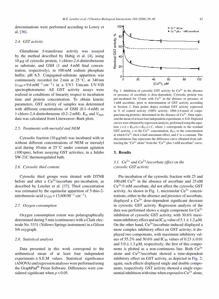
ological Interactions 164 (2006) 39–48 41
da
2
b1atbc(srtpw1d
2
wa(S
2
bdwn
2
dt5
2
ae(ts
Fig. 1. Inhibition of cytosolic GST activity by Cu2+ in the absenceor presence of ascorbate is dose-dependent. Cytosolic protein waspre-incubated for 15 min with Cu2+ in the absence or presence of1 mM ascorbate, prior to determination of GST activity accordingto Section 2. Data points depict residual GST activity expressedin % of control activity (100% activity: 1004 ± 6 nmol of conju-gate/min mg protein), determined in the absence of Cu2+. Data repre-sent the mean of at least four independent experiments ± S.D. Depictedcurves were obtained by regression analysis, performed using the equa-tion y = A × K0.5/(x + K0.5) + C, where y corresponds to the residualGST activity, x to the Cu2+ concentration, K to the concentration
M.E. Letelier et al. / Chemico-Bi
eterminations were performed according to Lowry etl. [36].
.4. GST activity
Glutathione S-transferase activity was assayedy the method described by Habig et al. [4], using0 �g of cytosolic protein, 1-chloro-2,4-dinitrobenzenes substrate, and GSH (1 and 4 mM final concen-ration, respectively), in 100 mM sodium phosphateuffer, pH 6.5. Conjugated-substrate apparition wasontinuously recorded for 2 min at 25 ◦C, at 340 nmε340 = 9.6 mM−1 cm−1) in a UV3 Unicam UV-VISpectrophotometer. All GST activity assays wereealized in conditions of linearity respect to incubationime and protein concentration. To obtain kineticarameters, GST activity of samples was determinedith different concentrations of GSH (0.1–4 mM) or-chloro-2,4-dinitrobenzene (0.2–2 mM). Km and Vmaxata was calculated from Lineweaver–Burk plots.
.5. Treatments with mersalyl and NEM
Cytosolic fraction (10 �g/ml) was incubated with orithout different concentrations of NEM or mersalyl
cid during 10 min at 25 ◦C under constant agitation100 rpm), before assaying GST activities, in a JulaboW-21C thermoregulated bath.
.6. Cytosolic thiol content
Cytosolic thiol groups were titrated with DTNBefore and after a Cu2+/ascorbate pre-incubation, asescribed by Letelier et al. [37]. Thiol concentrationas estimated by the equimolar apparition of 5-thio-2-itrobenzoic-acid (ε410 = 13,600 M−1 cm−1).
.7. Oxygen consumption
Oxygen consumption extent was polarographicallyetermined during 5 min (continuous) with a Clark elec-rode No. 5331 (Yellows Springs instrument) in a Gilson/6 oxygraph.
.8. Statistical analysis
Data presented in this work correspond to the
rithmetical mean of at least four independentxperiments ± S.E.M values. Statistical significanceANOVA) and regression analyses were performed usinghe GraphPad® Prism Software. Differences were con-idered significant when p < 0.05.0.5
at which Cu2+ elicit a half-maximum effect, and C to a constant. Thediscontinuous line represents the difference curve obtained from sub-tracting the “Cu2+ alone” from the “Cu2+ plus 1 mM ascorbate” curve.
3. Results
3.1. Cu2+ and Cu2+/ascorbate effect on thecytosolic GST activity
Pre-incubation of the cytosolic fraction with 25 and100 nM Cu2+ in the absence of ascorbate and 25 nMCu2+/1 mM ascorbate, did not affect the cytosolic GSTactivity. As shown in Fig. 1, micromolar Cu2+ concen-trations, either in the absence and presence of ascorbate,displayed a Cu2+ dose-dependent significant decreasein cytosolic GST activity. Regression analysis of thedata was performed shows a single component for Cu2+
inhibition of cytosolic GST activity, with 30.6% maxi-mum inhibitory effect and an IC50 value of 5.1 ± 1.2 �M.On the other hand, Cu2+/ascorbate-induced displayed amore complex inhibitory effect on GST activity; it dis-played two components, with maximum inhibitory val-ues of 55.2% and 30.6% and IC50 values of 0.23 ± 0.01and 5.0 ± 1.3 �M, respectively; the first of this compo-nents is plotted as a non-continuous line. Both Cu2+
alone and Cu2+/ascorbate showed a time-dependent
inhibitory effect on GST activity, as depicted in Fig. 2;again, such effects displayed single and double compo-nents, respectively. GST activity showed a single expo-nential inhibition with time when exposed to Cu2+ alone,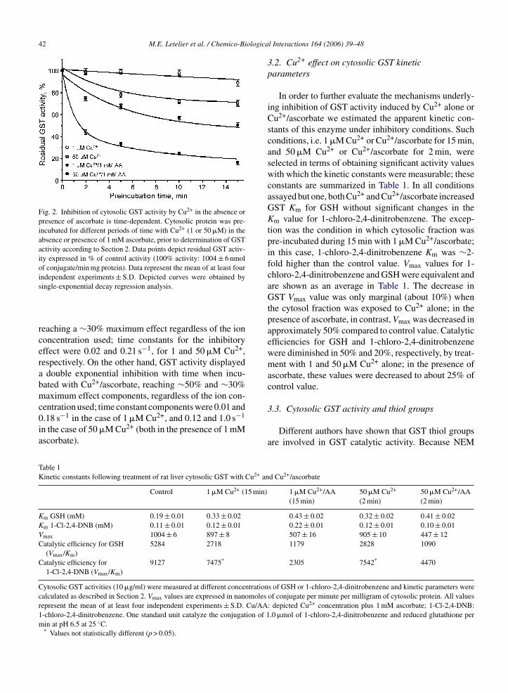
42 M.E. Letelier et al. / Chemico-Biologica
Fig. 2. Inhibition of cytosolic GST activity by Cu2+ in the absence orpresence of ascorbate is time-dependent. Cytosolic protein was pre-incubated for different periods of time with Cu2+ (1 or 50 �M) in theabsence or presence of 1 mM ascorbate, prior to determination of GSTactivity according to Section 2. Data points depict residual GST activ-ity expressed in % of control activity (100% activity: 1004 ± 6 nmolof conjugate/min mg protein). Data represent the mean of at least fourindependent experiments ± S.D. Depicted curves were obtained by
3.3. Cytosolic GST activity and thiol groups
single-exponential decay regression analysis.
reaching a ∼30% maximum effect regardless of the ionconcentration used; time constants for the inhibitoryeffect were 0.02 and 0.21 s−1, for 1 and 50 �M Cu2+,respectively. On the other hand, GST activity displayeda double exponential inhibition with time when incu-bated with Cu2+/ascorbate, reaching ∼50% and ∼30%maximum effect components, regardless of the ion con-centration used; time constant components were 0.01 and0.18 s−1 in the case of 1 �M Cu2+, and 0.12 and 1.0 s−1
in the case of 50 �M Cu2+ (both in the presence of 1 mMascorbate).
Table 1Kinetic constants following treatment of rat liver cytosolic GST with Cu2+ an
Control 1 �M Cu2+ (15 min)
Km GSH (mM) 0.19 ± 0.01 0.33 ± 0.02Km 1-Cl-2,4-DNB (mM) 0.11 ± 0.01 0.12 ± 0.01Vmax 1004 ± 6 897 ± 8Catalytic efficiency for GSH
(Vmax/Km)5284 2718
Catalytic efficiency for1-Cl-2,4-DNB (Vmax/Km)
9127 7475*
Cytosolic GST activities (10 �g/ml) were measured at different concentrationcalculated as described in Section 2. Vmax values are expressed in nanomolesrepresent the mean of at least four independent experiments ± S.D. Cu/AA1-chloro-2,4-dinitrobenzene. One standard unit catalyze the conjugation of 1min at pH 6.5 at 25 ◦C.
* Values not statistically different (p > 0.05).
l Interactions 164 (2006) 39–48
3.2. Cu2+ effect on cytosolic GST kineticparameters
In order to further evaluate the mechanisms underly-ing inhibition of GST activity induced by Cu2+ alone orCu2+/ascorbate we estimated the apparent kinetic con-stants of this enzyme under inhibitory conditions. Suchconditions, i.e. 1 �M Cu2+ or Cu2+/ascorbate for 15 min,and 50 �M Cu2+ or Cu2+/ascorbate for 2 min, wereselected in terms of obtaining significant activity valueswith which the kinetic constants were measurable; theseconstants are summarized in Table 1. In all conditionsassayed but one, both Cu2+ and Cu2+/ascorbate increasedGST Km for GSH without significant changes in theKm value for 1-chloro-2,4-dinitrobenzene. The excep-tion was the condition in which cytosolic fraction waspre-incubated during 15 min with 1 �M Cu2+/ascorbate;in this case, 1-chloro-2,4-dinitrobenzene Km was ∼2-fold higher than the control value. Vmax values for 1-chloro-2,4-dinitrobenzene and GSH were equivalent andare shown as an average in Table 1. The decrease inGST Vmax value was only marginal (about 10%) whenthe cytosol fraction was exposed to Cu2+ alone; in thepresence of ascorbate, in contrast, Vmax was decreased inapproximately 50% compared to control value. Catalyticefficiencies for GSH and 1-chloro-2,4-dinitrobenzenewere diminished in 50% and 20%, respectively, by treat-ment with 1 and 50 �M Cu2+ alone; in the presence ofascorbate, these values were decreased to about 25% ofcontrol value.
Different authors have shown that GST thiol groupsare involved in GST catalytic activity. Because NEM
d Cu2+/ascorbate
1 �M Cu2+/AA(15 min)
50 �M Cu2+
(2 min)50 �M Cu2+/AA(2 min)
0.43 ± 0.02 0.32 ± 0.02 0.41 ± 0.020.22 ± 0.01 0.12 ± 0.01 0.10 ± 0.01507 ± 16 905 ± 10 447 ± 121179 2828 1090
2305 7542* 4470
s of GSH or 1-chloro-2,4-dinitrobenzene and kinetic parameters wereof conjugate per minute per milligram of cytosolic protein. All values: depicted Cu2+ concentration plus 1 mM ascorbate; 1-Cl-2,4-DNB:.0 �mol of 1-chloro-2,4-dinitrobenzene and reduced glutathione per
M.E. Letelier et al. / Chemico-Biological Interactions 164 (2006) 39–48 43
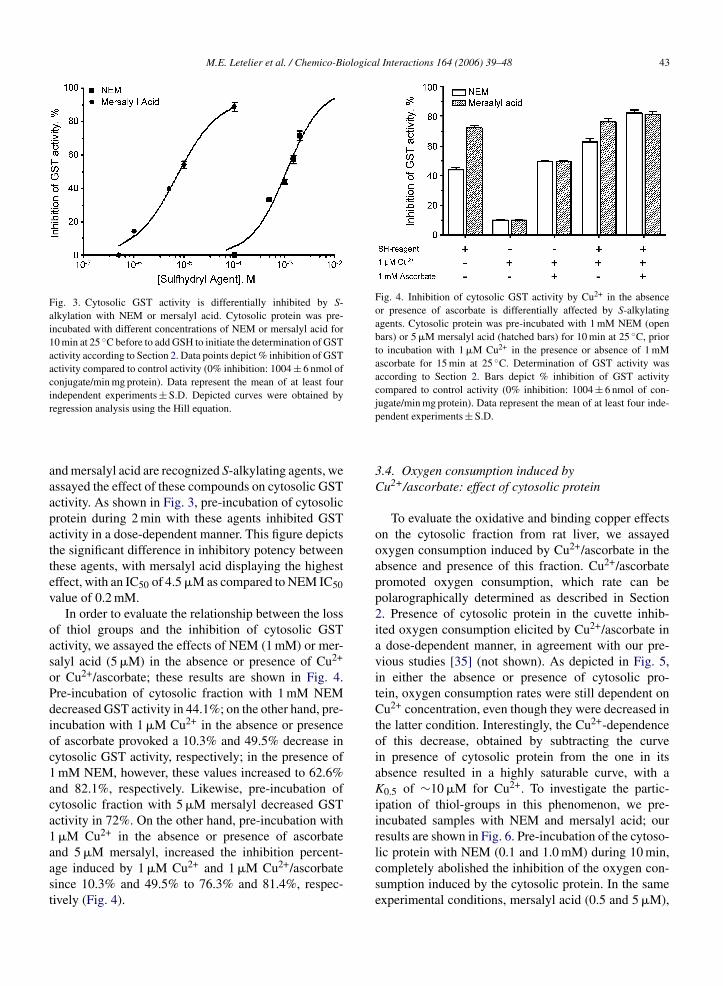
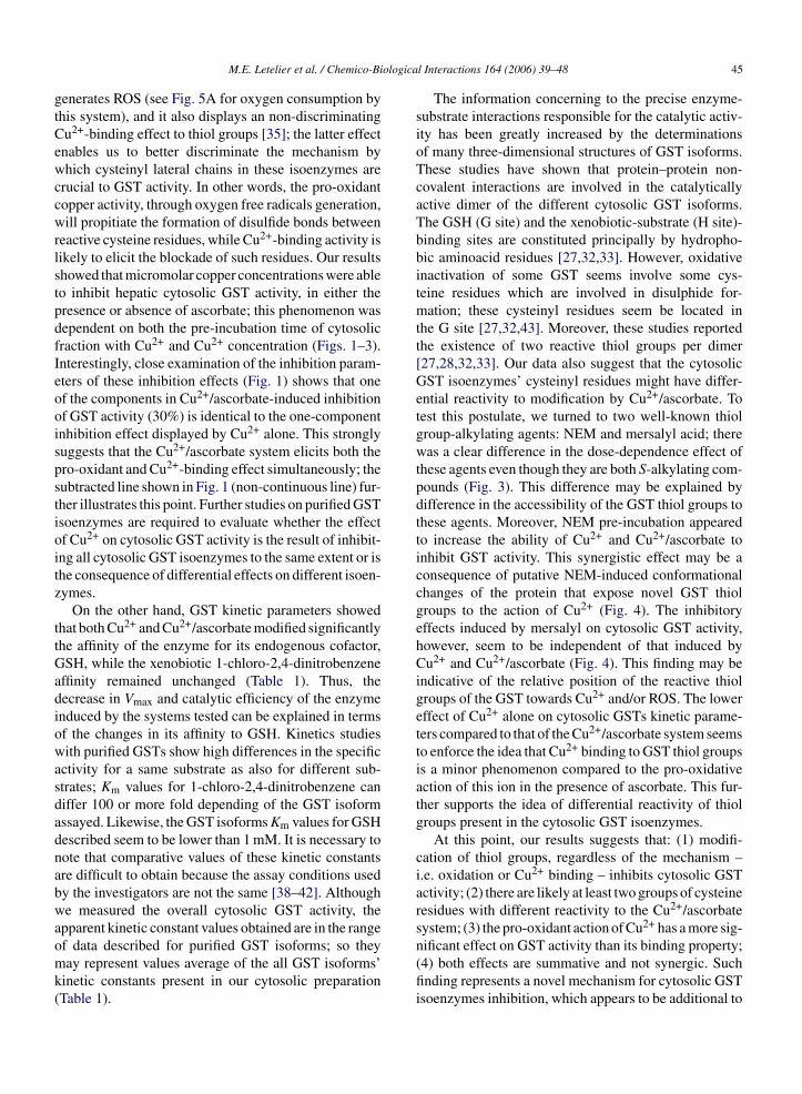
Fig. 3. Cytosolic GST activity is differentially inhibited by S-alkylation with NEM or mersalyl acid. Cytosolic protein was pre-incubated with different concentrations of NEM or mersalyl acid for10 min at 25 ◦C before to add GSH to initiate the determination of GSTactivity according to Section 2. Data points depict % inhibition of GSTactivity compared to control activity (0% inhibition: 1004 ± 6 nmol ofconjugate/min mg protein). Data represent the mean of at least fourindependent experiments ± S.D. Depicted curves were obtained byr
aaapattev
oasoPdioc1aca1aast
Fig. 4. Inhibition of cytosolic GST activity by Cu2+ in the absenceor presence of ascorbate is differentially affected by S-alkylatingagents. Cytosolic protein was pre-incubated with 1 mM NEM (openbars) or 5 �M mersalyl acid (hatched bars) for 10 min at 25 ◦C, priorto incubation with 1 �M Cu2+ in the presence or absence of 1 mMascorbate for 15 min at 25 ◦C. Determination of GST activity wasaccording to Section 2. Bars depict % inhibition of GST activitycompared to control activity (0% inhibition: 1004 ± 6 nmol of con-
egression analysis using the Hill equation.
nd mersalyl acid are recognized S-alkylating agents, wessayed the effect of these compounds on cytosolic GSTctivity. As shown in Fig. 3, pre-incubation of cytosolicrotein during 2 min with these agents inhibited GSTctivity in a dose-dependent manner. This figure depictshe significant difference in inhibitory potency betweenhese agents, with mersalyl acid displaying the highestffect, with an IC50 of 4.5 �M as compared to NEM IC50alue of 0.2 mM.
In order to evaluate the relationship between the lossf thiol groups and the inhibition of cytosolic GSTctivity, we assayed the effects of NEM (1 mM) or mer-alyl acid (5 �M) in the absence or presence of Cu2+
r Cu2+/ascorbate; these results are shown in Fig. 4.re-incubation of cytosolic fraction with 1 mM NEMecreased GST activity in 44.1%; on the other hand, pre-ncubation with 1 �M Cu2+ in the absence or presencef ascorbate provoked a 10.3% and 49.5% decrease inytosolic GST activity, respectively; in the presence ofmM NEM, however, these values increased to 62.6%nd 82.1%, respectively. Likewise, pre-incubation ofytosolic fraction with 5 �M mersalyl decreased GSTctivity in 72%. On the other hand, pre-incubation with�M Cu2+ in the absence or presence of ascorbate
nd 5 �M mersalyl, increased the inhibition percent-ge induced by 1 �M Cu2+ and 1 �M Cu2+/ascorbateince 10.3% and 49.5% to 76.3% and 81.4%, respec-ively (Fig. 4).jugate/min mg protein). Data represent the mean of at least four inde-pendent experiments ± S.D.
3.4. Oxygen consumption induced byCu2+/ascorbate: effect of cytosolic protein
To evaluate the oxidative and binding copper effectson the cytosolic fraction from rat liver, we assayedoxygen consumption induced by Cu2+/ascorbate in theabsence and presence of this fraction. Cu2+/ascorbatepromoted oxygen consumption, which rate can bepolarographically determined as described in Section2. Presence of cytosolic protein in the cuvette inhib-ited oxygen consumption elicited by Cu2+/ascorbate ina dose-dependent manner, in agreement with our pre-vious studies [35] (not shown). As depicted in Fig. 5,in either the absence or presence of cytosolic pro-tein, oxygen consumption rates were still dependent onCu2+ concentration, even though they were decreased inthe latter condition. Interestingly, the Cu2+-dependenceof this decrease, obtained by subtracting the curvein presence of cytosolic protein from the one in itsabsence resulted in a highly saturable curve, with aK0.5 of ∼10 �M for Cu2+. To investigate the partic-ipation of thiol-groups in this phenomenon, we pre-incubated samples with NEM and mersalyl acid; ourresults are shown in Fig. 6. Pre-incubation of the cytoso-
lic protein with NEM (0.1 and 1.0 mM) during 10 min,completely abolished the inhibition of the oxygen con-sumption induced by the cytosolic protein. In the sameexperimental conditions, mersalyl acid (0.5 and 5 �M),
44 M.E. Letelier et al. / Chemico-Biological Interactions 164 (2006) 39–48
Fig. 5. Oxygen consumption elicited by Cu2+/ascorbate is inhib-ited by the presence of cytosolic proteins. Oxygen consumption wasdetermined as described in Section 2. Data points depict oxygen-consumption rates from experiments in the absence (open circles) orpresence (closed circles) of cytosolic protein (0.1 mg/ml). Solid curveswere obtained by hyperbolic regression analysis; the discontinuouscurve was obtained by subtracting the curve in the presence of cytoso-
Fig. 7. GSH but not 1-chloro-2,4-dinitrobenzene protects cytosolicGST activity from inhibition by Cu2+ in the absence or presenceof ascorbate. Cytosolic fractions (10 �g/ml) were pre-incubated inthe absence or presence of either 4 mM GSH or 1 mM 1-chloro-2,4-dinitrobenzene for 20 min at 25 ◦C prior to incubation with either 1 �MCu2+ plus 1 mM ascorbate or 50 �M Cu2+ alone for 10 min at 25 ◦C.GST activity was determined as described in Section 2. Bars depictresidual GST activity expressed in % of control activity (100% activ-
lic protein from the curve in its absence. Determination of oxygenconsumption was according to Section 2. Data represent the mean ofat least four independent experiments ± S.D.
however, did not display the expected analogous effect(Fig. 6).
3.5. Cytosolic GST activity in the presence ofCu2+/ascorbate and Cu2+: effect of pre-incubation
with GSH and 1-chloro-2,4-dinitrobenzeneIt has been suggested that critical thiol groupsin cytosolic GST enzymes can be protected by pre-
Fig. 6. Oxygen consumption elicited by Cu2+/ascorbate-induced isnot inhibited by cytosolic proteins treated with S-alkylating agents.Cytosolic fraction was pre-incubated in the absence or presence ofNEM (0.1 or 1 mM) or mersalyl acid (0.5 or 5 �M) for 10 min at 25 ◦C.Then, oxygen consumption elicited by 50 �M Cu2+ and 1 mM ascor-bate was determined in the absence or presence of cytosolic proteinsamples, as described in Section 2. Data represent the mean of at leastfour independent experiments ± S.D.
ity: 1004 ± 6 nmol of conjugate/min mg protein). Data represent themean of at least four independent experiments ± S.D.
incubation with it cofactor GSH, because the redox-reactive cysteine residues may be located in the regionfor GSH binding [27,32]. Thus, to further evaluate thisprotective effect, we investigated whether pre-incubation(20 min) of the cytosolic fraction with the cofactor GSHand the xenobiotic 1-chloro-2,4-dinitrobenzene wereable to protect cytosolic GST activity from Cu2+-inducedinhibition. As shown in Fig. 7, the pre-incubation ofcytosolic fraction with GSH completely prevented theCu2+-induced inhibition on cytosolic GST induced by1 �M Cu2+/ascorbate and 50 �M Cu2+. On the otherhand, pre-incubation of cytosolic fraction with the sub-strate 1-chloro-2,4-dinitrobenzene did not modify theCu2+-induced inhibition on cytosolic GST (Fig. 7).
4. Discussion
4.1. Inhibition of GST activity by copper
It has been reported that H2O2 leads to the formationof intra- or inter-subunit disulfide bonds between partic-ular cysteine residues within GST amino acid sequence,leading to the inactivation of the cytosolic enzymes[27,28,32,33]. The effect of other ROS on this activ-ity, however, has not been thoroughly investigated; thus,
we set to explore the effects of oxygen free radicalsuperoxide anion (O2•−) and hydroxyl radical (HO•)on the overall cytosolic GST activity. With this pur-pose, we chose the Cu2+/ascorbate system because it
ologica
gtCewccwrlstpdfIeooispstioitz
ttGadiowasdadnabwaomk(
M.E. Letelier et al. / Chemico-Bi
enerates ROS (see Fig. 5A for oxygen consumption byhis system), and it also displays an non-discriminatingu2+-binding effect to thiol groups [35]; the latter effectnables us to better discriminate the mechanism byhich cysteinyl lateral chains in these isoenzymes are
rucial to GST activity. In other words, the pro-oxidantopper activity, through oxygen free radicals generation,ill propitiate the formation of disulfide bonds between
eactive cysteine residues, while Cu2+-binding activity isikely to elicit the blockade of such residues. Our resultshowed that micromolar copper concentrations were ableo inhibit hepatic cytosolic GST activity, in either theresence or absence of ascorbate; this phenomenon wasependent on both the pre-incubation time of cytosolicraction with Cu2+ and Cu2+ concentration (Figs. 1–3).nterestingly, close examination of the inhibition param-ters of these inhibition effects (Fig. 1) shows that onef the components in Cu2+/ascorbate-induced inhibitionf GST activity (30%) is identical to the one-componentnhibition effect displayed by Cu2+ alone. This stronglyuggests that the Cu2+/ascorbate system elicits both thero-oxidant and Cu2+-binding effect simultaneously; theubtracted line shown in Fig. 1 (non-continuous line) fur-her illustrates this point. Further studies on purified GSTsoenzymes are required to evaluate whether the effectf Cu2+ on cytosolic GST activity is the result of inhibit-ng all cytosolic GST isoenzymes to the same extent or ishe consequence of differential effects on different isoen-ymes.
On the other hand, GST kinetic parameters showedhat both Cu2+ and Cu2+/ascorbate modified significantlyhe affinity of the enzyme for its endogenous cofactor,SH, while the xenobiotic 1-chloro-2,4-dinitrobenzene
ffinity remained unchanged (Table 1). Thus, theecrease in Vmax and catalytic efficiency of the enzymenduced by the systems tested can be explained in termsf the changes in its affinity to GSH. Kinetics studiesith purified GSTs show high differences in the specific
ctivity for a same substrate as also for different sub-trates; Km values for 1-chloro-2,4-dinitrobenzene caniffer 100 or more fold depending of the GST isoformssayed. Likewise, the GST isoforms Km values for GSHescribed seem to be lower than 1 mM. It is necessary toote that comparative values of these kinetic constantsre difficult to obtain because the assay conditions usedy the investigators are not the same [38–42]. Althoughe measured the overall cytosolic GST activity, the
pparent kinetic constant values obtained are in the range
f data described for purified GST isoforms; so theyay represent values average of the all GST isoforms’inetic constants present in our cytosolic preparationTable 1).
l Interactions 164 (2006) 39–48 45
The information concerning to the precise enzyme-substrate interactions responsible for the catalytic activ-ity has been greatly increased by the determinationsof many three-dimensional structures of GST isoforms.These studies have shown that protein–protein non-covalent interactions are involved in the catalyticallyactive dimer of the different cytosolic GST isoforms.The GSH (G site) and the xenobiotic-substrate (H site)-binding sites are constituted principally by hydropho-bic aminoacid residues [27,32,33]. However, oxidativeinactivation of some GST seems involve some cys-teine residues which are involved in disulphide for-mation; these cysteinyl residues seem be located inthe G site [27,32,43]. Moreover, these studies reportedthe existence of two reactive thiol groups per dimer[27,28,32,33]. Our data also suggest that the cytosolicGST isoenzymes’ cysteinyl residues might have differ-ential reactivity to modification by Cu2+/ascorbate. Totest this postulate, we turned to two well-known thiolgroup-alkylating agents: NEM and mersalyl acid; therewas a clear difference in the dose-dependence effect ofthese agents even though they are both S-alkylating com-pounds (Fig. 3). This difference may be explained bydifference in the accessibility of the GST thiol groups tothese agents. Moreover, NEM pre-incubation appearedto increase the ability of Cu2+ and Cu2+/ascorbate toinhibit GST activity. This synergistic effect may be aconsequence of putative NEM-induced conformationalchanges of the protein that expose novel GST thiolgroups to the action of Cu2+ (Fig. 4). The inhibitoryeffects induced by mersalyl on cytosolic GST activity,however, seem to be independent of that induced byCu2+ and Cu2+/ascorbate (Fig. 4). This finding may beindicative of the relative position of the reactive thiolgroups of the GST towards Cu2+ and/or ROS. The lowereffect of Cu2+ alone on cytosolic GSTs kinetic parame-ters compared to that of the Cu2+/ascorbate system seemsto enforce the idea that Cu2+ binding to GST thiol groupsis a minor phenomenon compared to the pro-oxidativeaction of this ion in the presence of ascorbate. This fur-ther supports the idea of differential reactivity of thiolgroups present in the cytosolic GST isoenzymes.
At this point, our results suggests that: (1) modifi-cation of thiol groups, regardless of the mechanism –i.e. oxidation or Cu2+ binding – inhibits cytosolic GSTactivity; (2) there are likely at least two groups of cysteineresidues with different reactivity to the Cu2+/ascorbatesystem; (3) the pro-oxidant action of Cu2+ has a more sig-
nificant effect on GST activity than its binding property;(4) both effects are summative and not synergic. Suchfinding represents a novel mechanism for cytosolic GSTisoenzymes inhibition, which appears to be additional to
ologica
46 M.E. Letelier et al. / Chemico-Bithat of intra- or inter-molecular cross-linking, mediatedby modifications of it thiols residues.
To elucidate whether the effects shown in our studywere due only to modification of different thiol, we per-formed additional studies. First, we addressed the oxy-gen consumption pattern generated by Cu2+/ascorbate(Fig. 5); cytosolic protein inhibited the oxygen con-sumption elicited by Cu2+/ascorbate in a dose-dependentmanner (Fig. 5). In agreement with our previous study,Cu2+ binding to thiol groups in proteins is likely todecrease the availability of Cu2+ ions to undergo Haber-Weiss and/or Fenton reactions [34,35]. Indeed, whenpre-incubated with NEM, but not with mersalyl acid, thecytosolic fraction is no longer able to decrease the oxy-gen consumption elicited by Cu2+/ascorbate; this clearlydemonstrate the critical property of thiol groups, reac-tive only to NEM, in Cu2+ binding. Taken together,these results show that although both NEM and mer-salyl acid inhibited GST activity (Fig. 3), these agentmay S-alkylate different thiol groups in the cytosolicGST isoenzymes. Furthermore, this evidence suggeststhat NEM, and not mersalyl acid, may target solely thiolgroups of GSTs which will display Cu2+ binding. Asmentioned above, our results hinted the possibility thatcritical thiol groups may be located close to the GSH-binding site of the cytosolic GSTs. This is suggested bythe fact that Cu2+/ascorbate (and Cu2+ alone to a lesserextent) decreased the affinity of cytosolic GST isoen-zymes to GSH, without changing their affinity for thexenobiotic substrate (Table 1). This postulate was furtherstrengthened by the total protection of cytosolic GSTactivity towards both Cu2+ or Cu2+/ascorbate by pre-incubating the cytosolic fraction with GSH, while pre-incubation with 1-chloro-2,4-dinitrobenzene displayednegligible effects (Fig. 7). According with other authors,our results seem indicate that binding of GSH to the GSTactive site seem to hinder this type of critical cysteinylresidues preventing either its oxidation or Cu2+ binding,thus protecting GST activity [27,28,32,33]. Since freeGSH may reduce Cu2+ ions like those that ascorbatedoes, our results also suggest that binding of GSH to theGSTs active site may also abolish the redox reactivity ofGSH; this phenomenon has also been reported in othersystems [44] and requires further testing.
In summary, our data suggest that the major mech-anism underlying copper-induced inhibitory effect oncytosolic GST activity is likely to reside in this ion pro-oxidant properties, leading to the oxidation of protein
thiol groups. Binding of Cu2+ to cytosolic GST thiolgroups of the monomeric or dimeric isoforms also mayoccur, but only micromolar copper concentrations mayinduce a significant inhibition of overall cytosolic GSTl Interactions 164 (2006) 39–48
activity. Thus, it is likely that the cytosolic GSTs thiolgroups capable of binding copper ions are not exposedto the environment.
In humans, physiological copper plasma concentra-tions range between 14 and 19 �M [45–47]; in hepaticcells, copper ions are sequestered principally by metal-lotionein and GSH [47]. High cellular copper concentra-tion however, such as those propitiated in copper-relatedpathologies, can saturate the capacity of these moleculesto bind copper ions; in this condition, copper may induceon one side, oxidative stress, and on the other, bindingof this metal to critical cysteinyl residues of cellularproteins, leading to cytotoxicity. Our experimental Kmof cytosolic GST for GSH was 0.19 mM; since cytoso-lic GSH concentration ranges between 0.5 and 10 mMand hepatic concentration ∼5 �mol/g of liver [48–50],GSH may be saturating GST molecules under physiolog-ical conditions, protecting its activity from Cu2+-induceddamage. GST represents 10% of total content of cytoso-lic protein and its role in detoxication mechanisms iswell acknowledged [12–16,19,22,23]. The existence ofthis putative GST–GSH complex may represent a physi-ological mechanism to protect this remarkable cytosolicenzyme – and also GSH – from the oxidative stress-related damage.
References
[1] R.N. Armstrong, Structure, catalytic mechanism, and evolutionof the glutathione transferases, Chem. Res. Toxicol. 10 (1) (1997)2–18.
[2] L.A. Heppel, R.J. Hilmoe, Metabolism of inorganic nitrite andnitrate esters. II. The enzymatic reduction of nitroglycerin anderythritol tetranitarte by glutathione, J. Biol. Chem. 183 (1950)129–138.
[3] H. Ohkawa, R. Ohkawa, I. Yamamoto, J.F. Casida, Enzymaticmechanism and toxicological significance of hydrogen cyanideliberation from various organothiocyanates and organonitrilesin mice and houseflies, Pestic. Biochem. Physiol. 2 (1972) 95–112.
[4] W. Habig, M. Pabst, W. Jakoby, Glutathione S-transferases. Thefirst enzymatic step in mercapturic acid formation, J. Biol. Chem.249 (22) (1974) 7130–7139.
[5] N. Kaplowitz, Physiological significance of glutathione S-transferases, Am. J. Physiol. 239 (6) (1980) G439–G444.
[6] D.L. Eaton, T.K. Bammler, Concise review of the glutathione S-transferases and their significance to toxicology, Toxicol. Sci. 49(2) (1999) 156–164.
[7] K. Thyagaraju, B. Hemavathi, K. Vasundhara, A.D. Rao, K.N.Devi, Comparative study on glutathione transferases of ratbrain and testis under the stress of phenobarbitol and beta-
methylcholanthrene, J. Zhejiang Univ. Sci. 6 (8) (2005) 759–769.[8] K. Holovska Jr., A. Sobekova, K. Holovska, V. Lenartova, P.Javorsky, J. Legath, L. Legath, M. Maretta, Antioxidant anddetoxifying enzymes in the liver of rats after subchronic inhala-

ologica
[
[
[
[
[
[
[
[
[
[
[
[
[
[
[
[
[
[
[
[
[
[
[
[
[
[
M.E. Letelier et al. / Chemico-Bi
tion of the mixture of cyclic hydrocarbons, Exp. Toxicol. Pathol.56 (6) (2005) 377–383.
[9] J. Ketley, W. Habig, W. Jakoby, Binding of non-substrate ligandsto the glutathione S-transferases, J. Biol. Chem. 250 (22) (1975)8670–8673.
10] M. Lo Bello, M. Nuccetelli, A.M. Caccuri, L. Stella, M.W. Parker,J. Rossjohn, W.J. McKinstry, A.F. Mozzi, G. Federici, F. Polizio,J.Z. Pedersen, G. Ricci, Human glutathione transferase P1-1 andnitric oxide carriers; a new role for an old enzyme, J. Biol. Chem.276 (45) (2001) 42138–42145.
11] C. Peklak-Scott, A.J. Townsend, C.S. Morrow, Dynamics of glu-tathione conjugation and conjugate efflux in detoxification ofthe carcinogen, 4-nitroquinoline 1-oxide: contributions of glu-tathione, glutathione S-transferase, and MRP1, Biochemistry 44(11) (2005) 4426–4433.
12] E. Mosialou, R. Morgenstern, Activity of rat liver microsomalglutathione transferase toward products of lipid peroxidation andstudies of the effect of inhibitors on glutathione-dependent pro-tection against lipid peroxidation, Arch. Biochem. Biophys. 275(1) (1989) 289–294.
13] J.D. Hayes, D.J. Pulford, The glutathione S-transferase supergenefamily: regulation of GST and the contribution of the isoen-zymes to cancer chemoprotection and drug resistance, Crit. Rev.Biochem. Mol. Biol. 30 (6) (1995) 445–600.
14] H. Fiander, H. Schneider, Compounds that induce isoforms ofglutathione S-transferase with properties of a critical enzyme indefense against oxidative stress, Biochem. Biophys. Res. Com-mun. 262 (3) (1999) 591–595.
15] S.P. Singh, A.J. Janecki, S.K. Srivastava, S. Awasthi, Y.C.Awasthi, S.J. Xia, P. Zimniak, Membrane association of glu-tathione S-transferase mGSTA4-4, an enzyme that metabolizeslipid peroxidation products, J. Biol. Chem. 277 (6) (2002)4232–4239.
16] T. Zhao, S.S. Singhal, J.T. Piper, J. Cheng, U. Pandya, J. Clark-Wronski, S. Awasthi, Y.C. Awasthi, The role of human glu-tathione S-transferases hGSTA1-1 and hGSTA2-2 in protectionagainst oxidative stress, Arch. Biochem. Biophys. 367 (2) (1999)216–224.
17] S.K. Srivastava, S.C. Watkins, E. Schuetz, S.V. Singh, Roleof glutathione conjugate efflux in cellular protection againstbenzo[a]pyrene-7,8-diol-9,10-epoxide-induced DNA damage,Mol. Carcinog. 33 (3) (2002) 156–162.
18] B.P. Patel, U.M. Rawal, P.M. Shah, J.A. Prajapati, R.M. Rawal,T.K. Dave, P.S. Patel, Study of tobacco habits and alterations inenzymatic antioxidant system in oral cancer, Oncology 68 (4–6)(2005) 511–519.
19] P.J. Jakobsson, J.A. Mancini, A.W. Ford-Hutchinson, Identifi-cation and characterization of a novel human microsomal glu-tathione S-transferase with leukotriene C4 synthase activity andsignificant sequence identity to 5-lipoxygenase-activating pro-tein and leukotriene C4 synthase, J. Biol. Chem. 271 (36) (1996)22203–22210.
20] K.A. Scoggan, P.J. Jakobsson, A.W. Ford-Hutchinson, Productionof leukotriene C4 in different human tissues is attributable todistinct membrane bound biosynthetic enzymes, J. Biol. Chem.272 (15) (1997) 10182–10187.
21] D. Anuradha, K.V. Reddy, T.C. Kumar, S. Neeraja, P.R. Reddy,
P. Reddanna, Purification and characterization of rat testicularglutathione S-transferases: role in the synthesis of eicosanoids,Asian J. Androl. 2 (4) (2000) 277–282.22] S.C. Kampranis, R. Damianova, M. Atallah, G. Toby, G.Kondi, P.N. Tsichlis, A.M. Makris, A novel plant glutathione S-
[
[
l Interactions 164 (2006) 39–48 47
transferase/peroxidase suppresses Bax lethality in yeast, J. Biol.Chem. 275 (38) (2000) 29207–29216.
23] S.G. Cho, Y.H. Lee, H.S. Park, K. Ryoo, K.W. Kang, J. Park,S.J. Eom, M.J. Kim, T.S. Chang, S.Y. Choi, J. Shim, Y. Kim,M.S. Dong, M.J. Lee, S.G. Kim, H. Ichijo, E.J. Choi, GlutathioneS-transferase mu modulates the stress-activated signals by sup-pressing apoptosis signal-regulating kinase 1, J. Biol. Chem. 276(16) (2001) 12749–12755.
24] T. Sakurai, C. Kojima, M. Ochiai, T. Ohta, M.H. Sakurai, M.P.Waalkes, K. Fujiwara, Cellular glutathione prevents cytolethalityof monomethylarsonic acid, Toxicol. Appl. Pharmacol. 195 (2)(2004) 129–141.
25] S. Lien, A.K. Larsson, B. Mannervik, The polymorphic humanglutathione transferase T1-1, the most efficient glutathione trans-ferase in the denitrosation and inactivation of the anticancerdrug 1,3-bis(2-chloroethyl)-1-nitrosourea, Biochem. Pharmacol.63 (2) (2002) 191–197.
26] M.A. Harkey, M. Czerwinski, J. Slattery, H.P. Kiem, Overex-pression of glutathione-S-transferase, MGSTII, confers resistanceto busulfan and melphalan, Cancer Invest. 23 (1) (2005) 19–25.
27] H. Cheng, T. Tchaikovskaya, Y. Tu, J. Chapman, B. Qian, W.Ching, M. Tien, J.D. Rowe, Y.V. Patskovsky, I. Listowsky, C.D.Tu, Rat glutathione S-transferase M4-4: an isoenzymes withunique structural features including a redox-reactive cysteine-115residue that forms mixed disulphides with glutathione, Biochem.J. 356 (2001) 403–414.
28] M. Lo Bello, R. Petruzzelli, E. De Stefano, C. Tenedini, D. Barra,G. Federico, Identification of a highly reactive sulphydryl groupin human placental glutathione transferase by a site-directed flu-orescent reagent, FEBS 263 (1990) 389–391.
29] R. Morgenstern, J. DePierre, Microsomal glutathione transferase.Purification in unactivated form and further characterization of theactivation process, substrate specificity and amino acid composi-tion, Eur. J. Biochem. 134 (3) (1983) 591–597.
30] Y. Aniya, A. Naito, Oxidative stress-induced activation micro-somal glutathione S-transferase in isolated rat liver, Biochem.Pharmacol. 45 (1) (1993) 37–42.
31] Y. Aniya, M. Anders, Activation of rat liver microsomal glu-tathione S-transferase by hydrogen peroxide: role protein-dimerformation, Arch. Biochem. Biophys. 296 (2) (1992) 611–616.
32] H. Shen, S. Tsuchida, K. Tamai, K. Sato, Identification of cys-teine residues involved in disulfide formation in the inactivationof glutathione transferase P-form by hydrogen peroxide, Arch.Biochem. Biophys. 300 (1) (1993) 137–141.
33] G. Ricci, G. Del Boccio, A. Pennelli, M. Lo Bello, R. Petruzzelli,A.M. Caccuri, D. Barra, G. Federico, Redox forms of humanplacenta glutathione transferase, J. Biol. Chem. 206 (32) (1991)21409–21415.
34] B. Haliwell, C.E. Cross, Oxygen-derived species: their relationto human disease and environmental stress, Environ. Health Per-spect. 102 (Suppl. 10) (1994) 5–12.
35] M.E. Letelier, A.M. Lepe, M. Faundez, J. Salazar, R. Marın, P.Aracena, H. Speisky, Possible mechanisms underlying copper-induced damage in biological membranes leading to cellulartoxicity, Chem. Biol. Interact. 151 (2005) 71–82.
36] O.H. Lowry, N.J. Rosenbrough, A.L. Farr, R.J. Randall, Proteinmeasurement with the Folin phenol reagent, J. Biol. Chem. 193(1) (1951) 265–275.
37] M.E. Letelier, P. Izquierdo, L. Godoy, A.M. Lepe, M. Faundez,Liver microsomal biotransformation of nitro-aryl drugs: mecha-

ologica
[
[
[
[
[
[
[
[
[
[
[
[
48 M.E. Letelier et al. / Chemico-Bi
nism for potential oxidative stress induction, J. Appl. Toxicol. 24(2004) 519–525.
38] E.M. van der Aar, D. Buikema, J.N. Commandeur, J.M. te Kop-pele, B. van Ommen, P.J. van Bladeren, N.P. Vermeulen, Enzymekinetics and substrate selectivities of rat glutathione S-transferaseisoenzymes towards a series of new 2-substituted 1-chloro-4-nitrobenzenes, Xenobiotica 26 (2) (1996) 143–155.
39] E.T. Adman, I. Le Trong, R.E. Stenkamp, B.S. Nieslanik, E.C.Dietze, G. Tai, C. Ibarra, W.M. Atkins, Localization of the C-terminus of rat glutathione S-transferase A1-1: crystal structure ofmutants W21F and W21F/F220Y, Proteins: Struct. Funct. Genet-ics 42 (2001) 192–200.
40] P. Hoarau, G. Garello, M. Gnassia-Barelli, M. Romeo, J.P. Girard,Purification and partial characterization of seven glutathione S-transferase isoforms from the clam Ruditapes decussates, Eur. J.Biochem. 269 (2002) 4359–4366.
41] A. Shokeer, A.K. Larsson, B. Mannervik, Residue 234 in glu-tathione transferase T1-1 plays a pivotal role in the catalytic activ-ity and the selectivity against alternative substrates, Biochem. J.
388 (2005) 387–392.42] P.D. Fortin, G.P. Horsman, H.M. Yang, L.D. Eltis, A glutathioneS-transferase catalyzes the dehalogenation of inhibitory metabo-lites of polychlorinated biphenyls, J. Bacteriol. 188 (12) (2006)4424–4430.
[
l Interactions 164 (2006) 39–48
43] Y. Ji, V. Toader, B.M. Bennett, Regulation of microsomal andcytosolic glutathione S-transferase activities by S-nitrosylation,Biochem. Pharmacol. 63 (2002) 1397–1404.
44] I. Jimenez, H. Speisky, Effects of copper ions on the free radical-scavenging properties of reduced glutathione: implications ofa complex formation, Trace Elem. Biol. Med. 14 (2) (2000)161–167.
45] G.J. Brewer, Wilson disease and canine copper toxicosis, Am. J.Clin. Nutr. 67 (Suppl.) (1998) 1087S–1090S.
46] A. Cordano, Clinical manifestations of nutritional copper defi-ciency in infants and children, Am. J. Clin. Nutr. 67 (Suppl.)(1998) 1012S–1016S.
47] M.C. Linder, L. Wooten, P. Cerveza, S. Cotton, R. Shulze, N.Lomeli, Copper transport, Am. J. Clin. Nutr. 67 (Suppl.) (1998)965S–971S.
48] C. Dameron, M. Harrison, Mechanisms for protection againstcopper toxicity, Am. J. Clin. Nutr. 67 (5) (1998) 1091s–1097s.
49] A. Meister, S. Tate, Glutahione and related gamma-glutamyl com-pounds: biosynthesis and utilization, Annu. Rev. Biochem. 45
(1976) 559–604.50] B.H. Lauterburg, J.R. Mitchell, Regulation of hepatic glutathioneturnover in rats in vivo and evidence for kinetic homogeneityof the hepatic glutathione pool, J. Clin. Invest. 67 (5) (1981)1415–1424.