Structured mRNAs Regulate Translation Initiation by Binding to the Platform of the Ribosome
Upload
independentCategory
view
1download
0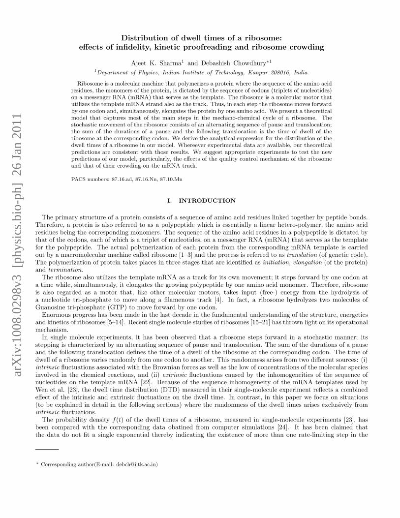
arX
iv:1
008.
0298
v3 [
phys
ics.
bio-
ph]
26
Jan
2011
Distribution of dwell times of a ribosome:
effects of infidelity, kinetic proofreading and ribosome crowding
Ajeet K. Sharma1 and Debashish Chowdhury∗1
1Department of Physics, Indian Institute of Technology, Kanpur 208016, India.
Ribosome is a molecular machine that polymerizes a protein where the sequence of the amino acidresidues, the monomers of the protein, is dictated by the sequence of codons (triplets of nucleotides)on a messenger RNA (mRNA) that serves as the template. The ribosome is a molecular motor thatutilizes the template mRNA strand also as the track. Thus, in each step the ribosome moves forwardby one codon and, simultaneously, elongates the protein by one amino acid. We present a theoreticalmodel that captures most of the main steps in the mechano-chemical cycle of a ribosome. Thestochastic movement of the ribosome consists of an alternating sequence of pause and translocation;the sum of the durations of a pause and the following translocation is the time of dwell of theribosome at the corresponding codon. We derive the analytical expression for the distribution of thedwell times of a ribosome in our model. Whereever experimental data are available, our theoreticalpredictions are consistent with those results. We suggest appropriate experiments to test the newpredictions of our model, particularly, the effects of the quality control mechanism of the ribosomeand that of their crowding on the mRNA track.
PACS numbers: 87.16.ad, 87.16.Nn, 87.10.Mn
I. INTRODUCTION
The primary structure of a protein consists of a sequence of amino acid residues linked together by peptide bonds.Therefore, a protein is also referred to as a polypeptide which is essentially a linear hetero-polymer, the amino acidresidues being the corresponding monomers. The sequence of the amino acid residues in a polypeptide is dictated bythat of the codons, each of which is a triplet of nucleotides, on a messenger RNA (mRNA) that serves as the templatefor the polypeptide. The actual polymerization of each protein from the corresponding mRNA template is carriedout by a macromolecular machine called ribosome [1–3] and the process is referred to as translation (of genetic code).The polymerization of protein takes places in three stages that are identified as initiation, elongation (of the protein)and termination.The ribosome also utilizes the template mRNA as a track for its own movement; it steps forward by one codon at
a time while, simultaneously, it elongates the growing polypeptide by one amino acid monomer. Therefore, ribosomeis also regarded as a motor that, like other molecular motors, takes input (free-) energy from the hydrolysis ofa nucleotide tri-phosphate to move along a filamenous track [4]. In fact, a ribosome hydrolyzes two molecules ofGuanosine tri-phosphate (GTP) to move forward by one codon.Enormous progress has been made in the last decade in the fundamental understanding of the structure, energetics
and kinetics of ribosomes [5–14]. Recent single molecule studies of ribosomes [15–21] has thrown light on its operationalmechanism.In single molecule experiments, it has been observed that a ribosome steps forward in a stochastic manner; its
stepping is characterized by an alternating sequence of pause and translocation. The sum of the durations of a pauseand the following translocation defines the time of a dwell of the ribosome at the corresponding codon. The time ofdwell of a ribosome varies randomly from one codon to another. This randomness arises from two different sources: (i)intrinsic fluctuations associated with the Brownian forces as well as the low of concentrations of the molecular speciesinvolved in the chemical reactions, and (ii) extrinsic fluctuations caused by the inhomogeneities of the sequence ofnucleotides on the template mRNA [22]. Because of the sequence inhomogeneity of the mRNA templates used byWen et al. [23], the dwell time distribution (DTD) measured in their single-molecule experiment reflects a combinedeffect of the intrinsic and extrinsic fluctuations on the dwell time. In contrast, in this paper we focus on situations(to be explained in detail in the following sections) where the randomness of the dwell times arises exclusively fromintrinsic fluctuations.The probability density f(t) of the dwell times of a ribosome, measured in single-molecule experiments [23], has
been compared with the corresponding data obatined from computer simulations [24]. It has been claimed thatthe data do not fit a single exponential thereby indicating the existence of more than one rate-limiting step in the
∗ Corresponding author(E-mail: [email protected])
2
mechano-chemical cycle of each ribosome. In fact, the best fit to the simulation data was achieved with five differentrate-determining steps [24].A systematic analytical derivation of the DTD of the ribosomes was presented recently [25] from a kinetic theory
of translation [26] that also involves essentially five steps in the mechano-chemical cycle of a ribosome. However,the model developed in ref.[26] ignores some of the key features of the mechano-chemical cycle of an individualribosome during the elongation stage. For example, a ribosome deploys an elaborate proofreading mechanism toselect the correct amino acid dictated by the template (and to reject the incorrect ones) to ensure high translationalfidelity. Nevertheless, occasionally, translational errors take place. In this paper we extend Basu and Chowdhury’soriginal model [26] by capturing the processes of proofreading and allowing for the possibility of imperfect fidelity oftranslation. Moreover, the identification of the mechano-chemical states as well as the nature of the transitions amongthese states is revised in the light of the empirical facts established in the last couple of years. Using this revised andextended kinetic model of translation [27], we analytically calculate the probability density f(t) of the dwell times ofribosomes.The DTD derived in this paper is qualitatively similar to that observed by Wen et al. in their single molecule
experiments [23]. However, because of the sequence inhomogeneity of the template mRNA used in the experiment ofWen et al.[23], their data cannot be compared quantitatively with our analytical expression for f(t). Therefore, wepropose a concrete experimental set up required for a quantitative testing of our theoretical predictions. However,till such experiments are actually carried out, the results reported here will continue to provide insight into themechanistic origin of the qualitative features of the DTD. Moreover, attempts are being made to extend our modelto capture sequence inhomogeneities of mRNAs and to calculate the corresponding DTDs of ribosomes analytically.Interestingly, the inverse of the mean dwell time is the average velocity V of the ribosome motor. Since, in our
model, the ribosome is not allowed to back track on the mRNA template, V is also the mean rate of elongation ofthe polypeptide. We show that V satisfies an equation that resembles the Michaelis-Menten equation for the averagerate of simple enzymatic reactions [28].It is well known that most often a large number of ribosomes simultaneously move on the same mRNA track each
polymerizing one copy of the same protein. This phenomenon is usually referred to as ribosome traffic because ofits superficial similarity with vehicular traffic on highways [26, 29–41] In this paper we also report the effect of stericinteractions of the ribosomes in ribosome traffic on their DTD.
II. THE MODEL
We develope here a theoretical model for the polymerization of a protein by ribosomes using an artificially synthe-sized mRNA template that consists of a homogeneous sequence (i.e., all the codons of which are identical). In thesurrounding medium, two species of amino-acids are available only one of which is the correct one according to thegenetic code. The ribosome deploys a quality control system that rejects the amino acid monomer if it is incorrect. Ifthis quality control system never fails, perfect fidelity of translation would result in a homopolymer whose constituentamino acid monomers are all identical. However, occasionally, wrong amino acid monomer escapes the quality controlsystem. Such translational errors, whereby a wrong amino acid monomer is incorporated in the elongating protein,results in a hetero-polymer. We’ll study the effects of the quality control system on the DTD of the ribosomes.A ribosome consists of two interconnected subunits which are designated as “large” and “small” (see fig.1). The
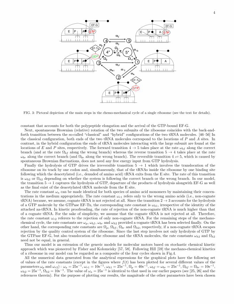
small subunit binds with the mRNA track and decodes the genetic message of the codon whereas the polymerization ofthe protein takes place in the large subunit. The operations of the two subunits are coordinated by a class of adaptermolecules, called tRNA (see fig.1). One end of a tRNA helps in the decoding process by matching its anti-codon withthe codon on the mRNA while its other end carries an amino acid; in this form the complex is called an amino-acyltRNA (aa-tRNA).The three main stages in each mechano-chemical cycle of a ribosome, shown in fig.2, are as follows: (i) selection
of the cognate (i.e., correct) aa-tRNA, (ii) formation of the peptide bond between the amino acid brought in by theselected aa-tRNA and the elongating protein, (iii) translocation of the ribosome by one codon on its track. However,some of these steps consist of important sub-steps. Moreover, the aa-tRNA selected (erroneously) by the ribosomemay not be the cognate tRNA; this leads to a branching of pathways. Such branching, in turn, gives rise to thepossibility of more than one cyclic pathway for a ribosome in a given cycle.Let us begin with the state labelled by “1”; both the E and A sites are empty while the site P is occupied as shown
in fig.3. An incoming aa-tRNA molecule, bound to an elongation factor EF-Tu, occupies the site A; the resulting stateis labelled by “2”. The transition 1 → 2 takes place at the rate ωa. However, not all incoming aa-tRNA moleculesare automatically selected by the ribosome. In order to ensure translational fidelity [42–45] the ribosome deploys aquality control mechanism to ensure that the aa-tRNA selected is, indeed, cognate (i.e., carries the correct amino acidas dictated by the mRNA template). A non-cognate tRNA is rejected on the basis of codon-anticodon mismatch; the
3
FIG. 1: Cartoons representing the main molecules and machinery involved in the process of translation.
FIG. 2: Pictoral depiction of the three main stages in the chemo-mechanical cycle of a single ribosome (see the text for details).
corresponding transition 2 → 1 takes place at the rate ωr1. However, if the aa-tRNA is not a non-cognate one, theGTP molecule bound to it is then hydrolyzed to GDP causing the irreversible transition 2 → 3 at the rate ωh1. Atthis stage, kinetic proofreading can reject the tRNA if it is a near-cognate one; the transition 3 → 1 captures thisphenomenon and takes place at the rate ωr2. In our model we do not explicitly treat those sub-steps in which EF-TUand the products of the hydrolysis of GTP leave the ribosome. We’ll later utilize the fact that ωa is proportional tothe concentration of the aa-tRNA molecules in the surrounding medium.If the selected aa-tRNA is not rejected, it does not necessarily imply that it is cognate because, occasionally, even
non-cognate or near-cognate aa-tRNA escapes the ribosomes quality control mechanism. The amino acid supplied bythe selected aa-tRNA is then linked to the growing polypeptide by a peptide bond; this peptidyl transferase activityof the ribosome elongates the polypeptide by one monomer. The correct amino acid is incorporated at a rate ωp
whereas a wrong amino acid is incorporated at a rate Ωp thereby giving rise to the two different branches of themechano-chemical cycle of the ribosome; the third branch 3 → 1, however, ends a “futile” cycle. The only differencebetween the states 4 and 4∗ is that the last amino acid in the polypeptide is correct in 4 but incorrect in 4∗. While thepolypeptide gets elongated by one amino acid, a fresh molecule of GTP enters bound with an elongation factor EF-G.The arrival of GTP-bound EF-G is not treated explicitly in our model. Alternatively, ωp (and Ωp) is an effective rate
4
FIG. 3: Pictoral depiction of the main steps in the chemo-mechanical cycle of a single ribosome (see the text for details).
constant that accounts for both the polypeptide elongation and the arrival of the GTP-bound EF-G.Next, spontaneous Brownian (relative) rotation of the two subunits of the ribosome coincides with the back-and-
forth transition between the so-called “classical” and “hybrid” configurations of the two tRNA molecules. [46–56] Inthe classical configuration, both ends of the two tRNA molecules correspond to the locations of P and A sites. Incontrast, in the hybrid configuration the ends of tRNA molecules interacting with the large subunit are found at thelocations of E and P sites, respectively. The forward transition 4 → 5 takes place at the rate ωbf along the correctbranch (and at the rate Ωbf along the wrong branch) whereas the reverse transition 5 → 4 takes place at the rateωbr along the correct branch (and Ωbr along the wrong branch). The reversible transition 4 5, which is caused byspontaneous Brownian fluctuations, does not need any free energy input from GTP hydrolysis.Finally the hydrolysis of GTP drives the irreversible transition 5 → 1 which involves the translocation of the
ribosome on its track by one codon and, simultaneously, that of the tRNAs inside the ribosome by one binding sitefollowing which the deacetylated (i.e., denuded of amino acid) tRNA exits from the E site. The rate of this transitionis ωh2 or Ωh2 depending on whether the system is following the correct branch or the wrong branch. In our model,the transition 5 → 1 captures the hydrolysis of GTP, departure of the products of hydrolysis alongwith EF-G as wellas the final exist of the deacetylated tRNA molecule from the E site.The rate constant ωa can be made identical for both species of amino acid monomers by maintaining their concen-
trations in the medium appropriately. The rate constant ωr1 refers only to the wrong amino acids (i.e., non-cognatetRNA) because, we assume, cognate tRNA is not rejected at all. Since the transition 2 → 3 accounts for the hydrolysisof a GTP molecule by the GTPase EF-Tu, the corresponding rate constant is ωh1, irrespective of the identity of theattached aa-tRNA. In kinetic proofreading, the rate of rejection of the non-cognate tRNA is much higher than thatof a cognate tRNA. For the sake of simplicity, we assume that the cognate tRNA is not rejected at all. Therefore,the rate constant ωr2 referers to the rejection of only non-cognate tRNA. For the remaining steps of the mechano-chemical cycle, the rate constants are ωp, ωbf , ωbr and ωh2 provided a cognate tRNA has been selected finally. On theother hand, the corresponding rate constants are Ωp, Ωbf , Ωbr and Ωh2, respectively, if a non-cognate tRNA escapesrejection by the quality control system of the ribosome. Since the last step involves not only hydrolysis of GTP bythe GTPase EF-G, but also translocation of the ribosome and the tRNA molecules, the rate constants ωh2 and Ωh2
need not be equal, in general.Thus our model is an extension of the generic models for molecular motors based on stochastic chemical kinetic
approach which was pioneered by Fisher and Kolomeisky [57, 58]. Following Hill [59] the mechano-chemical kineticsof a ribosome in our model can be regarded as a composite of the four cycles shown in Fig.4.All the numerical data generated from the analytical expressions for the graphical plots have the following set
of values of the rate constants (except in the figures where f(t) has been plotted for several different values of theparameters ωp and ωr2) : ωa = 25s−1, ωr1 = 10s−1, ωh1 = 25s−1, Ωp = 40s−1, ωbf = ωbr = 25s−1, Ωbf = Ωbr = 10s−1,ωh2 = 25s−1, Ωh2 = 10s−1. The value of ωa = 25s−1 is identical to that used in our earlier papers (see [25, 26] and thereferences therein). For the purpose of plotting our results, the magnitude of the other parameters have been chosen

5
FIG. 4: The full mechano-chemical kinetic of a ribosome during the elongation state, as modelled in Fig.3, can be regardedas a composite of at least four different cycles shown in (a)-(d). Among these, both (a) and (b) are unproductive in the sensethat these lead to neither elongation of the polypeptide nor forward stepping of the ribosome. However, (b) is “futile” becauseit dissipates the free energy input from the hydrolysis of a GTP molecule whereas no fuel is wasted in (a) . In contrast, both(c) and (d) lead to elongation of the polypeptide and forward stepping of the ribosome by hydrolysing two molecules of GTP;however, (c) incorporates the correct amino acid whereas (d) incorporated a wrong amino acid into the growing polypeptide.
to be comparable to that of ωa. The values of some of the parameters, and the ranges over which some of these havebeen varied, may be unrealistically high or low. But, this deliberate choice has been motivated by our intention todemonstrate graphically the interplay of various kinetic processes in translation.
III. DWELL TIME DISTRIBUTION
For the convenience of mathematical calculations, we assume that, after reaching the state 5 (or 5∗) at location j
the system makes a transition to a hypothetical state 1 at location j + 1 which, then, relaxes to the state 1, at thesame location, at the rate δ. At the end of the calculation our model is recovered by setting δ → ∞.Suppose Pµ(j, t) denote the probability at time t that the ribosome is in the “chemical” state µ and is decoding the
j-th codon. Let us use the symbol P1(j + 1, t) to denote the probability of finding the ribosome in the hypothetical
state 1 at time t while decoding the j + 1-th codon. The time taken by the ribosome to reach the state 1 at j + 1,starting from the initial state 1 at j, defines its time of dwell at the j-th codon. Since, in this context, all the“chemical” states, except 1 refer to the j-th codon while 1 corresponds to the j + 1-th codon, from now onwards wedrop the site index j (and j + 1) to keep the notations simple.The master equations governing the time evolution of the probabilities Pµ(t) can be written as
dP1(t)
dt= −ωaP1(t) + ωr1P2(t) + ωr2P3(t) (1)
dP2(t)
dt= ωaP1(t)− (ωr1 + ωh1)P2(t) (2)
dP3(t)
dt= ωh1P2(t)− (ωp +Ωp + ωr2)P3(t) (3)
dP4(t)
dt= ωpP3(t)− ωbfP4(t) + ωbrP5(t) (4)

6
0 0.25 0.5 0.75 1 1.25
Dwell Time (s)0
1
2
3
4
Prob
abili
ty D
ensi
ty
Simulation ωp= 50 s
-1
Simulation ωp= 100 s
-1
Simulation ωp= 200 s
-1
Formula ωp=50 s
-1
Formula ωp= 100 s
-1
Formula ωp= 200 s
-1
FIG. 5: The probability density of the dwell times are plotted for a few different values of the parameter ωp.
dP5(t)
dt= ωbfP4(t)− (ωh2 + ωbr)P5(t) (5)
dP ∗
4 (t)
dt= ΩpP3(t)− ΩbfP
∗
4 (t) + ΩbrP∗
5 (t) (6)
dP ∗
5 (t)
dt= ΩbfP
∗
4 (t)− (Ωh2 +Ωbr)P∗
5 (t) (7)
dP1(t)
dt= ωh2P5(t) + Ωh2P
∗
5 (t) (8)
Because of the normalization condition
5∑
µ=1
Pµ(t) + P ∗
4 (t) + P ∗
5 (t) + P1(t) = 1 (9)
not all the equations (1)-(8) above are independent of each other.For the calculation of the dwell time, we impose the initial conditions
P1(0) = 1, and P2(0) = P3(0) = P4(0) = P5(0) = P ∗
4 (0) = P ∗
5 (0) = P1(0) = 0 (10)
Suppose, f(t) is the probability density of the dwell times. Then, the probability of adding one amino acid to thegrowing polypeptide in the time interval between t and t+∆t is f(t)∆t where
f(t) =∆P1(t)
∆t= ωh2P5(t) + Ωh2P
∗
5 (t) (11)
The calculation of the DTD [60–65] is essentially that of a distribution of first-passage times [66].
7
0 0.25 0.5 0.75 1 1.25 1.5
Dwell Time (s)0
0.5
1
1.5
2
2.5
3
3.5
Prob
abili
ty D
ensi
ty
Simulation ωr2
=0 s-1
Simulation ωr2
=50 s-1
Simulation ωr2
=150 s-1
Formula ωr2
=0 s-1
Formula ωr2
=50 s-1
Formula ωr2
=150 s-1
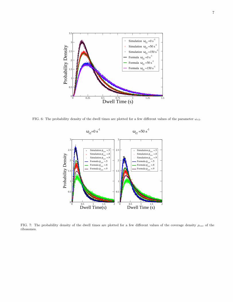
FIG. 6: The probability density of the dwell times are plotted for a few different values of the parameter ωr2.
0 0.5 1 1.5 2
Dwell Time(s)0
0.5
1
1.5
2
2.5
3
Prob
abili
ty D
ensi
ty
Simulation ρcov
=.5
Simulation ρcov
=.8
Simulation ρcov
=.9
Formula ρcov
=.5
Formula ρcov
=.8
Formula ρcov
=.9
ωr2
=0 s-1
0 0.5 1 1.5 2
Dwell Time (s)0
0.5
1
1.5
2
2.5
3
Simulation ρcov
=.5
Simulation ρcov
=.8
Simulation ρcov
=.9
Formula ρcov
=.5
Formula ρcov
=.8
Formula ρcov
=.9
ωr2
=50 s-1
FIG. 7: The probability density of the dwell times are plotted for a few different values of the coverage density ρcov of theribosomes.

8
The exact probability density of the dwell times is given by
f(t) =
[ωh2ωbfωpωh1ωa
(ω1 − ω2)(ω1 − ω3)(ω1 − ω4)(ω1 − ω5)
]e−ω1t +
[Ωh2ΩbfΩpωh1ωa
(ω1 − ω2)(ω1 − ω3)(ω1 − Ω4)(ω1 − Ω5)
]e−ω1t
+
[ωh2ωbfωpωh1ωa
(ω2 − ω3)(ω2 − ω4)(ω2 − ω5)(ω2 − ω1)
]e−ω2t +
[Ωh2ΩbfΩpωh1ωa
(ω2 − ω3)(ω2 − Ω4)(ω2 − Ω5)(ω2 − ω1)
]e−ω2t
+
[ωh2ωbfωpωh1ωa
(ω3 − ω4)(ω3 − ω5)(ω3 − ω1)(ω3 − ω2)
]e−ω3t +
[Ωh2ΩbfΩpωh1ωa
(ω3 − Ω4)(ω3 − Ω5)(ω3 − ω1)(ω3 − ω2)
]e−ω3t
+
[ωh2ωbfωpωh1ωa
(ω4 − ω5)(ω4 − ω1)(ω4 − ω2)(ω4 − ω3)
]e−ω4t +
[Ωh2ΩbfΩpωh1ωa
(Ω4 − Ω5)(Ω4 − ω1)(Ω4 − ω2)(Ω4 − ω3)
]e−Ω4t
+
[ωh2ωbfωpωh1ωa
(ω5 − ω1)(ω5 − ω2)(ω5 − ω3)(ω5 − ω4)
]e−ω5t +
[Ωh2ΩbfΩpωh1ωa
(Ω5 − ω1)(Ω5 − ω2)(Ω5 − ω3)(Ω5 − Ω4)
]e−Ω5t
(12)
where ω1,ω2 and ω3 are solution of the cubic equation
ω3 − ω2(ωr1 + ωh1 + ωa + ωr2 + ωp +Ωp) + ω(ωh1ωa + ωr2ωr1 + ωr2ωh1 + ωr2ωa
+ ωpωr1 + ωpωh1 + ωpωa +Ωpωr1 +Ωpωh1 +Ωpωa)− Ωpωh1ωa + ωpωh1ωa = 0, (13)
ω4 and ω5 are the solution of the quadratic equation
ω2 − ω(ωh2 + ωbr + ωbf ) + ωh2ωbf = 0 (14)
and Ω4 and Ω5 are the solution of the quadratic equation
Ω2 − Ω(Ωh2 +Ωbr +Ωbf ) + Ωh2Ωbf = 0 (15)
Some of the details of this derivation are given in the appendix. Note that the problem of determining the ratesωi (i = 1, 2, 3, 4, 5) in terms of the rate constants of the model is similar to that of expressing the normal modes ofvibration of a set of coupled harmonic oscillators. It is the “backward” reactions in the mechano-chemical cycle ofour model which play the role of coupling of the harmonic oscillators. For example, if Ωp = 0 and simultaneously,ωr1 = ωr2 = ωbr = 0, the expressions for ωi (i = 1, 2, 3, 4, 5) reduce to the simple form ω1 = ωa, ω2 = ωh1, ω3 = ωp,ω4 = ωbf , and ω5 = ωh2.The distribution (12) is plotted in Fig.5 for a few different values of the parameter ωp. Note that
φ = ωp/(ωp + Ωp) (16)
is a measure of the fidelity of translation. Therefore, increasing ωp, keeping Ωp fixed, enhances translational fidelity.Moreover, a higher ωp also corresponds to faster peptidyl transferase reaction. Therefore, the trend of variation ofthe most probable dwell time with ωp is consistent with the intuitive expectation that the slower is the peptidyltransferase reaction, the longer is the dwell time.The kinetic parameter ωr2 is a measure of the rate of rejection of the aa-tRNA by kinetic proofreading. Consequently,
the effect of the variation of the ωr2 on the most probable dwell time is opposite to that of ωp (see Fig.6); the higheris the frequency of tRNA rejection by kinetic proofreading, the longer is the dwell time.
IV. MEAN RATE OF POLYMERIZATION: A MICHAELIS-MENTEN-LIKE EQUATION
The average Dwell time can be calculated by substituting (12) into the definition
〈t〉 =
∫ +∞
0
tf(t) dt. (17)
Hence, the expression for 〈t〉
〈t〉 =1
ω1
+1
ω2
+1
ω3
+ωp
ωp +Ωp
(1
ω4
+1
ω5
)+
Ωp
ωp +Ωp
(1
Ω4
+1
Ω5
)(18)
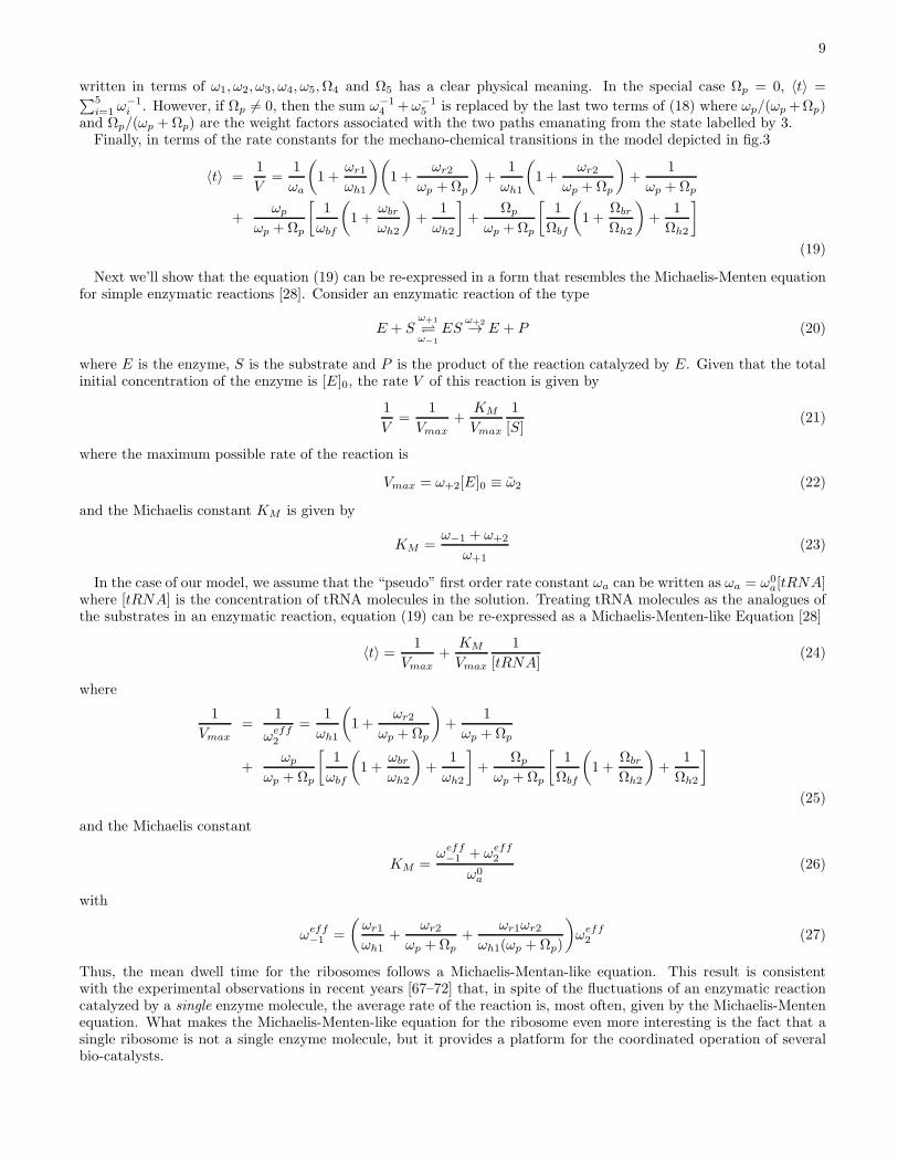
9
written in terms of ω1, ω2, ω3, ω4, ω5,Ω4 and Ω5 has a clear physical meaning. In the special case Ωp = 0, 〈t〉 =∑5
i=1ω−1i . However, if Ωp 6= 0, then the sum ω−1
4 +ω−15 is replaced by the last two terms of (18) where ωp/(ωp +Ωp)
and Ωp/(ωp +Ωp) are the weight factors associated with the two paths emanating from the state labelled by 3.Finally, in terms of the rate constants for the mechano-chemical transitions in the model depicted in fig.3
〈t〉 =1
V=
1
ωa
(1 +
ωr1
ωh1
)(1 +
ωr2
ωp +Ωp
)+
1
ωh1
(1 +
ωr2
ωp +Ωp
)+
1
ωp +Ωp
+ωp
ωp +Ωp
[1
ωbf
(1 +
ωbr
ωh2
)+
1
ωh2
]+
Ωp
ωp +Ωp
[1
Ωbf
(1 +
Ωbr
Ωh2
)+
1
Ωh2
]
(19)
Next we’ll show that the equation (19) can be re-expressed in a form that resembles the Michaelis-Menten equationfor simple enzymatic reactions [28]. Consider an enzymatic reaction of the type
E + Sω+1
ω
−1
ESω+2
→ E + P (20)
where E is the enzyme, S is the substrate and P is the product of the reaction catalyzed by E. Given that the totalinitial concentration of the enzyme is [E]0, the rate V of this reaction is given by
1
V=
1
Vmax
+KM
Vmax
1
[S](21)
where the maximum possible rate of the reaction is
Vmax = ω+2[E]0 ≡ ω2 (22)
and the Michaelis constant KM is given by
KM =ω−1 + ω+2
ω+1
(23)
In the case of our model, we assume that the “pseudo” first order rate constant ωa can be written as ωa = ω0a[tRNA]
where [tRNA] is the concentration of tRNA molecules in the solution. Treating tRNA molecules as the analogues ofthe substrates in an enzymatic reaction, equation (19) can be re-expressed as a Michaelis-Menten-like Equation [28]
〈t〉 =1
Vmax
+KM
Vmax
1
[tRNA](24)
where
1
Vmax
=1
ωeff2
=1
ωh1
(1 +
ωr2
ωp +Ωp
)+
1
ωp +Ωp
+ωp
ωp +Ωp
[1
ωbf
(1 +
ωbr
ωh2
)+
1
ωh2
]+
Ωp
ωp +Ωp
[1
Ωbf
(1 +
Ωbr
Ωh2
)+
1
Ωh2
]
(25)
and the Michaelis constant
KM =ωeff−1 + ωeff
2
ω0a
(26)
with
ωeff−1 =
(ωr1
ωh1
+ωr2
ωp +Ωp
+ωr1ωr2
ωh1(ωp +Ωp)
)ωeff2 (27)
Thus, the mean dwell time for the ribosomes follows a Michaelis-Mentan-like equation. This result is consistentwith the experimental observations in recent years [67–72] that, in spite of the fluctuations of an enzymatic reactioncatalyzed by a single enzyme molecule, the average rate of the reaction is, most often, given by the Michaelis-Mentenequation. What makes the Michaelis-Menten-like equation for the ribosome even more interesting is the fact that asingle ribosome is not a single enzyme molecule, but it provides a platform for the coordinated operation of severalbio-catalysts.
10
V. EFFECTS OF CROWDING ON THE DWELL TIME DISTRIBUTION
It is well known that most often a large number of ribosomes simultaneously move on the same mRNA track eachpolymerizing one copy of the same protein. This phenomenon is usually referred to as ribosome traffic because of itssuperficial similarity with vehicular traffic on highways [26, 29–41] . Suppose, ℓ denotes the number of codons that aribosome can cover simultaneously. Extending the prescription used in our earlier works on ribosome traffic [26] forcapturing the steric interactions of the ribosomes, we replace the equations (5), (7 and (8) by
dP5(i, t)
dt= ωbfP4(i, t)− ωbrP5(i, t)− ωh2P5(i, t)Q(i|i+ ℓ) (28)
dP ∗
5 (i, t)
dt= ΩbfP
∗
4 (i, t)− ΩbrP∗
5 (i, t)− Ωh2P∗
5 (i, t)Q(i|i+ ℓ) (29)
dP1(i, t)
dt=
(ωh2P5(i− 1, t) + Ωh2P
∗
5 (i− 1, t)
)Q(i-1|i − 1 + ℓ) (30)
where Q(i|j) = 1 − P (i|j) is the conditional probability that, given a ribosome in site i, site j is empty. All theother equations for Pµ(i, t) remain unchanged. Note that these equations have been written under the mean-field
approximation.In the limit L → ∞, all the sites are treated on the same footing so that the site-dependence of Pµ(i, t) drops out.
In this limit, Q takes the simple form [26]
Q(i|i + ℓ) =1− ρℓ
1 + ρ− ρℓ. (31)
where ρ is the number density of the ribosomes (i.e., number of ribosomes per unit length of the mRNA track).Therefore, in ribosome traffic the distribution of the dwell times of the ribosomes is given by the expression (12)
where ωh2 and Ωh2 are replaced by ωh2Q and Ωh2Q, respectively. Using the expression (31), we get the DTD for thegiven number density ρ of the ribosomes. For the purpose of graphical demostration of the effects of ribosome crowdingon the DTD, we use the coverage density ρcov = ρ ℓ. In Fig.7 we plot the DTD for a few different values of ρcov. Thehigher is the density, the stronger is the hindrance and longer is the dwell time. Moreover, higher coverage densityintroduces stronger correlations which our mean-field equations ignore. Consequently, our analytical predictions,based on equations written under the mean-field approximation, deviate more and more from the correspondingsimulation data as the coverage density increases.
VI. COMPARISON WITH EXPERIMENTAL DATA
The distribution of dwell times involves essentially five different rate-determining parameters ωi (i = 1, 2, 3, 4, 5) ifthe translational fidelity is perfect. This is consistent with the earlier observation [24] that the best fit to the simulationdata was obtained with five rate-determining parameters. However, the dwell time distribution obtained from thesingle-molecule experiments (more precisely, single-ribosome manipulation) fit reasonably well with a difference of justtwo exponentials [23]. This strongly indicates the possibility that only two of the five rate-determining parameterswere rate-limiting under the conditions maintained in those experiments. However, for a quantitative testing of thepredictions of our theoretical model, one should use a mRNA template with a homogeneous codon sequence and onlytwo species of aa-tRNA one of which is cognate (corresponding to the codons in the coding sequence of the mRNA)and the other is either near-cognate or non-cognate.In order to clarify the proposed experimental set up, let us consider the concrete example shown schematically in fig.
8. This example is essentially the protocols used by Uemura et al. [20] in their single-molecule studies of translationin real time. The actual coding sequence to be translated consists of nc number of identical codons; in fig.8 nc = 6and each codon is UUU which codes for the amino acid Phenylalanine (abbreviated Phe or F). The coding sequenceis preceeded by a start codon AUG and is followed by a stop codon UAA. The start codon itself is preceeded by anuntranslated region (UTR) at the 5’-end of the mRNA; this is required for assembling the ribosome and for stabilizingthe pre-initiation complex. At the 3’-end, the stop codon is followed by a sequence of nnc non-coding codons UUU(nnc = 4 in fig.8); this region merely ensures the absence of any “edge effect”, i.e., the translation is not affectedwhen the ribosome approaches the 3’-end of the codong sequence. A good choice for the corresponding near-cognate

11
FIG. 8: A schematic description of a mRNA with homogeneous (poly-U) coding sequence (adapted from refs.[20, 21]).
tRNA would be tRNALeu because it is cognate for the codon CUU which codes for Leucine (abbreviated L). The twodistinct species of aa-tRNA should be labelled by fluorescent dye molecules of two different colors which could be, forexample, Cy5 (red) and Cy3 (green). Equal concentrations of Cy5-labelled ternary complexe aa-tRNAPhe-EF-Tu-GTPand Cy3-labelled ternary complex aa-tRNALeu-EF-Tu-GTP should be made available in the medium surrounding theribosome to avoid any bias arising from difference in their concentrations. The color of the fluorescence pulse identifiesthe amino acid monomer species that elongates the polypeptide by one unit; monitoring the colors of the fluorescencepulses one would get an estimate of the translational fidelity φ. Moreover, the time interval between the arrival ofthe successive aa-tRNA molecules provides an estimate of the dwell times of the ribosome. Thus, using this opticaltechnique one would get not only the DTD, but also the fidelity of translation. Usually the coding sequence of suchpoly-U mRNA strands is quite short. Therefore, for collecting enough data to extract the DTD, the experiment hasto be repeated sufficiently large number of times.The effects of kinetic proofreading on the rate of translational error has been investigated experimentally by several
different methods in the last three decades. However, most of those methods (see, for example, ref.[73, 74]) requirebulk samples. But, the more recent single-molecule FRET technique used in the study of tRNA selection [19] seemsto be more suitable for testing our predictions on the effects of kinetic proofreading on the DTD.The cluster of ribosomes translating the same mRNA simultaneously is usually referred to as a polysome. For
studying the effects of ribosome crowding on the DTD, one has to measure simultaneously the DTD and the polysomesize [75].
VII. SUMMARY AND CONCLUSION
In this paper we have presented a theoretical model of translation that captures all the main steps in the mechano-chemical cycle of a ribosome during the elongation stage. This model also accounts for translational fidelity, kineticproofreading and the crowding of the ribosomes on the same mRNA track. In principle, this model can be extended,by increasing the number of “chemical” states, to account for some of the sub-steps which have not been treatedexplicitly in the version of the model presented here.In spite of the details already incorporated in this model, we have suceeded in carrying out an analytical calculation
of the distribution of the dwell times of the ribosomes at the successive codons. We have compared this theoreticalestimate of the DTD with the corresponding numerical data which we have obtained from direct computer simulationof the model. If the motion of the ribosome is not hindered by the presence of any other ribosome on the samemRNA track, our analytical treatment yields the exact expression for the distribution for the times of its dwell at thesuccessive codons. In this case, excellent agreement between theoretical prediction and simulation data is observedprovided the data are averaged over sufficiently large number of samples.However, because of the mean-field approximation made in capturing the effects of crowding of the ribosomes the
corresponding analytical expression is approximate. Therefore, the higher is the coverage density, the larger is thedeviation of the analytical estimate from the simulation data which are averaged over many samples.We have analyzed the dependence of the DTD on some of the crucially important kinetic parameters of the
model to elucidate the physical implications of the result. Our results are in good qualitative agreement with theexperimental data reported in the literature [23, 24]. However, for the reasons explained in the sections I and II, itis not possible to compare these experimental data quantitatively with the analytical expressions of DTD which wehave reported in this paper. We hope our theoretical predictions will stimulate further experimental studies along thelines suggested briefly in section VI although some technical hurdles may hinder quick progress. A combination ofthe single-ribosome experiments and bulk measurements may be required for comparing the theoretically predictedvariation of the DTD with the concentrations of tRNA, GTP and other key molecules involved in translation as wellas with the increase of futile cycles and crowding.
It would also be desirable to extend our model in future to account for some important features of translation; someof these possible extentions are listed below. (i) Capturing the sequence heterogeneity of real mRNA templates will
12
open up larger number of branched pathways in Fig.3, each corresponding to a distinct species (cognate, non-cognateor near-cognate) of amino acid. Moreover, at each step of the ribosome, the rate constants for the correct andincorrect pathways will also depend on the codon under consideration. (ii) The interaction of the growing polypeptidewith the exit tunnel in the ribosome and the spontaneous folding of the nascent protein as it comes out of the tunnelmay affect the rate of translation. It may be possible to capture these effects by making some of the relevant rateconstants, e.g., ωh2, Ωh2, etc. on the instantaneous length of the growing polypeptide which, in turn, is identical tothe length of the codon sequence already translated by the ribosome. (iii) Maintaining correct reading frame on asequence-homogeneous mRNA (e.g., poly-U) may be more difficult than on an inhomogeneous sequence. It may bedesirable to extend our model allowing the possibility of frameshift errors [76] although, at present, the prescriptionfor this extension is not obvious. The DTD for such a “realistic model” may be obtained numerically because ananalytical derivation of the general expression may not be possible in the forseeable future.
Acknowledgements: One of the authors (DC) thanks Joachim Frank and Joseph D. Puglisi for useful correspon-dences.Appendix
Solving the equations (1)-(7) under the initial condition (10) we get
P1(t) = C1e−ω1t + C2e
−ω2t + C3e−ω3t (32)
P2(t) =C1ωae
−ω1t
ωr1 + ωh1 − ω1
+C2ωae
−ω2t
ωr1 + ωh1 − ω2
+C3ωae
−ω3t
ωr1 + ωh1 − ω3
(33)
P3(t) =C1ωh1ωae
−ω1t
(ωr1 + ωh1 − ω1)(ωr2 + ωp +Ωp − ω1)+
C2ωh1ωae−ω2t
(ωr1 + ωh1 − ω2)(ωr2 + ωp +Ωp − ω2)
+C3ωh1ωae
−ω3t
(ωr1 + ωh1 − ω3)(ωr2 + ωp +Ωp − ω3)(34)
P4(t) =C1ωpωh1ωa(ωh2 + ωbr − ω1)e
−ω1t
(ωr1 + ωh1 − ω1)(ωr2 + ωp +Ωp − ω1)(ω4 − ω1)(ω5 − ω1)
+C2ωpωh1ωa(ωh2 + ωbr − ω2)e
−ω2t
(ωr1 + ωh1 − ω2)(ωr2 + ωp +Ωp − ω2)(ω4 − ω2)(ω5 − ω2)
+C3ωpωh1ωa(ωh2 + ωbr − ω3)e
−ω3t
(ωr1 + ωh1 − ω3)(ωr2 + ωp +Ωp − ω3)(ω4 − ω3)(ω5 − ω3)
+ C4e−ω4t + C5e
−ω5t
(35)
P5(t) =C1ωbfωpωh1ωae
−ω1t
(ωr1 + ωh1 − ω1)(ωr2 + ωp +Ωp − ω1)(ω4 − ω1)(ω5 − ω1)
+C2ωbfωpωh1ωae
−ω2t
(ωr1 + ωh1 − ω2)(ωr2 + ωp +Ωp − ω2)(ω4 − ω2)(ω5 − ω2)
+C3ωbfωpωh1ωae
−ω3t
(ωr1 + ωh1 − ω3)(ωr2 + ωp +Ωp − ω3)(ω4 − ω3)(ω5 − ω3)
+C4ωbfe
−ω4t
ωh2 + ωbr − ω4
+C5ωbfe
−ω5t
ωh2 + ωbr − ω5
(36)
P ∗
4 (t) =C1Ωpωh1ωa(Ωh2 +Ωbr − ω1)e
−ω1t
(ωr1 + ωh1 − ω1)(ωr2 + ωp +Ωp − ω1)(Ω4 − ω1)(Ω5 − ω1)
+C2Ωpωh1ωa(Ωh2 +Ωbr − ω2)e
−ω2t
(ωr1 + ωh1 − ω2)(ωr2 + ωp +Ωp − ω2)(Ω4 − ω2)(Ω5 − ω2)
+C3Ωpωh1ωa(Ωh2 +Ωbr − ω3)e
−ω3t
(ωr1 + ωh1 − ω3)(ωr2 + ωp +Ωp − ω3)(Ω4 − ω3)(Ω5 − ω3)
+ D4e−Ω4t +D5e
−Ω5t
(37)
13
P ∗
5 (t) =C1ΩbfΩpωh1ωae
−ω1t
(ωr1 + ωh1 − ω1)(ωr2 + ωp +Ωp − ω1)(Ω4 − ω1)(Ω5 − ω1)
+C2ΩbfΩpωh1ωae
−ω2t
(ωr1 + ωh1 − ω2)(ωr2 + ωp +Ωp − ω2)(Ω4 − ω2)(Ω5 − ω2)
+C3ΩbfΩpωh1ωae
−ω3t
(ωr1 + ωh1 − ω3)(ωr2 + ωp +Ωp − ω3)(Ω4 − ω3)(Ω5 − ω3)
+D4Ωbfe
−Ω4t
Ωh2 + Ωbr − Ω4
+D5Ωbfe
−Ω5t
Ωh2 +Ωbr − Ω5
(38)
where ω1, ω2 and ω3 are the solutions of the cubic equation (13) while ω4, ω5 are the solutions of the quadraticequation (14) and Ω4, Ω5 are the solutions of the quadratic equation (15). The coefficients Cν (ν = 1, 2, ..., 5) andD4, D5, which are determined by the initial condition (10), are as follows:
C1 =(ωr1 + ωh1 − ω1)(ωr2 + ωp +Ωp − ω1)
(ω2 − ω1)(ω3 − ω1)(39)
C2 =(ωr1 + ωh1 − ω2)(ωr2 + ωp +Ωp − ω2)
(ω3 − ω2)(ω1 − ω2)(40)
C3 =(ωr1 + ωh1 − ω3)(ωr2 + ωp +Ωp − ω3)
(ω1 − ω3)(ω2 − ω3)(41)
C4 =(ωh2 + ωbr − ω4)ωpωh1ωa
(ω5 − ω4)(ω1 − ω4)(ω2 − ω4)(ω3 − ω4)(42)
C5 =(ωh2 + ωbr − ω5)ωpωh1ωa
(ω1 − ω5)(ω2 − ω5)(ω3 − ω5)(ω4 − ω5)(43)
D4 =(Ωh2 +Ωbr − Ω4)Ωpωh1ωa
(Ω5 − Ω4)(ω1 − Ω4)(ω2 − Ω4)(ω3 − Ω4)(44)
D5 =(Ωh2 +Ωbr − Ω5)Ωpωh1ωa
(ω1 − Ω5)(ω2 − Ω5)(ω3 − Ω5)(Ω4 − Ω5)(45)
[1] A. S. Spirin, Ribosomes, (Springer, 2000).[2] A.S. Spirin, FEBS Lett. 514, 2 (2002).[3] A. S. Spirin, J. Biol. Chem. 284, 211203 (2009).[4] R. A. Cross, Nature 385, 18 (1997).[5] V. Ramakrishnan, Angew. Chem. Int. Ed. 49, 4355 (2010).[6] A. Yonath, Angew. Chem. Int. Ed. 49, 4340 (2010).[7] T. A. Steitz, Angew. Chem. Int. Ed. 49, 4381 (2010).[8] T. A. Steitz, Nat. Rev. Mol. Cell Biol. 9, 242 (2008).[9] T. M. Schmeing and V. Ramakrishnan, Nature 461, 1234 (2009).
[10] V. Ramakrishnan, Cell 108, 557 (2002).[11] J. Frank and C.M.T. Spahn, Rep. Prog. Phys. 69, 1383 (2006).[12] K. Mitra and J. Frank, Annu. Rev. Biophys. Biomol. Struct. 35, 299 (2006).[13] X. Agirrezabala and J. Frank, Quart. Rev. Biophys. 42, 159 (2009).[14] J. Frank and R. L. Gonzales, Annu. Rev. Biochem. 79, 381 (2010).[15] R.A. Marshall, C.E. Aitken, M. Dorywalska and J.D. Puglisi, Annu. Rev. Biochem. 77, 177 (2008).[16] J.B. Munro, A. Vaiana, K.Y. Sanbonmatsu and S.C. Blanchard, Biopolymers 89, 565 (2008).[17] J. B. Munro, K. Y. Sanbonmatsu, C.M.T. Spahn and S.C. Blanchard, Trends in Biochem. Sci. 34, 390 (2009).
14
[18] S.C. Blanchard, Curr. Opin. Struct. Biol. 19, 103 (2009).[19] S. Blanchard, R.L. Gonzalez Jr., H.D. Kim, S. Chu and J.D. Puglisi, Nat. Str. & Mol. Biol. 11, 1008 (2004).[20] S. Uemura, C. E. Aitken, B.A. Flusberg, S. W. Turner and J.D. Puglisi, Nature 464, 1012 (2010).[21] C. E. Aitken and J. D. Puglisi, Nat. Struct. Mol. Biol. 17, 793 (2010).[22] J. R. Buchan and I. Stansfield, Biol. Cell 99, 475-487 (2007).[23] J.D. Wen, L. Lancaster, C. Hodges, A.C. Zeri, S.H. Yoshimura, H.F. Noller, C. Bustamante and I. Tinoco Jr., Nature 452,
598 (2008).[24] I. Tinoco Jr. and J. D. Wen, Phys. Biol. 6, 025006 (2009).[25] A. Garai, D. Chowdhury, D. Chowdhury and T. V. Ramakrishnan, Phys. Rev. E 80, 011908 (2009).[26] A. Basu and D. Chowdhury, Phys. Rev. E 75, 021902 (2007).[27] A. K. Sharma and D. Chowdhury, Phys. Rev. E 82, 031912 (2010).[28] M. Dixon and E.C. Webb, Enzymes (Academic Press, 1979).[29] C. MacDonald, J. Gibbs and A. Pipkin, Biopolymers, 6, 1 (1968).[30] C. MacDonald and J. Gibbs, Biopolymers, 7, 707 (1969).[31] G. Lakatos and T. Chou, J. Phys. A 36, 2027 (2003).[32] L.B. Shaw, R.K.P. Zia and K.H. Lee, Phys. Rev. E 68, 021910 (2003).[33] L.B. Shaw, J.P. Sethna and K.H. Lee, Phys. Rev. E 70, 021901 (2004).[34] L.B. Shaw, A.B. Kolomeisky and K.H. Lee, J. Phys. A 37, 2105 (2004).[35] T. Chou, Biophys. J., 85, 755 (2003).[36] T. Chou and G. Lakatos, Phys. Rev. Lett. 93, 198101 (2004).[37] J.J. Dong, B. Schmittmann and R.K.P. Zia, J. Stat. Phys. 128, 21 (2006).[38] J.J. Dong, B. Schmittmann and R.K.P. Zia, Phys. Rev. E 76, 051113 (2007).[39] L. J. Cook, R. K. P. Zia and B. Schmittmann, Phys. Rev. E 80, 031142 (2009).[40] L. Ciandrini, I. Stansfield and M. C. Romano, Phys. Rev. E 81, 051904 (2010).[41] N. Mitarai, K. Sneppen and S. Pedersen, J. Mol. Biol. 382, 236 (2008).[42] M. V. Rodnina and W. Wintermeyer, Annu. Rev. Biochem. 70, 415 (2001).[43] T. Daviter, K. B. Gromadski and M. V. Rodnina, Biochimie 88, 1001 (2006).[44] J. M. Ogle and V. Ramakrishnan, Annu. Rev. Biochem. 74, 129 (2005).[45] H. S. Zaher and R. Green, Cell 136, 746 (2009).[46] D. Moazed and H. F. Noller, Nature 342, 142 (1989).[47] K. S. Wilson and H. F. Noller, Cell 92, 337 (1998).[48] H. F. Noller, FEBS Lett. 514, 11 (2002).[49] L. H. Horan and H. F. Noller, PNAS 104, 4881 (2007).[50] A. Korostelev, D. N. Ermolenko and H. F. Noller, Curr. Opin. Chem. Biol. 12, 674 (2008).[51] J. Frank and R. K. Agrawal, Nature 406, 318 (2000).[52] M. Valle, A. Zavialov, J. Sengupta, U. Rawat, M. Ehrenberg and J. Frank, Cell 114, 123 (2003).[53] J. Frank, H. Gao, J. Sengupta, N. Gao and D. J. Taylor, PNAS 104, 19671 (2007).[54] D. Pan, S.V. Kirillov and B.S. Cooperman, Mol. Cell 25, 519 (2007).[55] S.J. Moran, J. F. Flanagan, O. Namy, D.I. Stuart, I. Brierley and R.J.C. Gilbert, Structure 16, 664 (2008).[56] S. Shoji, S. E. Walker and K. Fredrick, ACS Chem. Biol. 4, 93 (2009).[57] M.E. Fisher and A.B. Kolomeisky, PNAS 96, 6597 (1999).[58] M.E. Fisher and A.B. Kolomeisky, Annu. Rev. Phys. Chem. 58, 675 (2007).[59] T. L. Hill, Free energy transduction and biochemical cycle kinetics (Dover, 2005).[60] Y.R. Chemla, J.R. Moffitt and C. Bustamante, J. Phys. Chem. B 112, 6025 (2008).[61] J.W. Shaevitz, S.M. Block and M.J. Schnitzer, Biophys. J. 89, 2277 (2005).[62] J.C. Liao, J.A. Spudich, D. Parker, S.L. Delp, PNAS 104, 3171 (2007).[63] M. Linden and M. Wallin, Biophys. J. 92, 3804 (2007).[64] D. Tsygankov, M. Linden, and M. E. Fisher, Phys. Rev. E 75, 021909 (2007).[65] A. Garai and D. Chowdhury, Submitted for publication.[66] S. Redner, A Guide to First-Passage Processes, (Cambridge University Press, 2001).[67] H. Qian and E.L. Elson, Biophys. Chem. 101-102, 565 (2002).[68] B.P. English, W. min, A.M. van Oijen, K.T. Lee, G. Luo, H. Sun, B.J. Cherayil, S.C. Kou and X.S. Xie, Nat. Chem. Biol.
2, 87 (2006).[69] S.C. Kou, B.J. Cherayil, W. Min, B.P. English and X.S. Xie, J. Phys. Chem. B 109, 19068 (2005).[70] W. Min, B.P. English, G. Luo, B.J. Cherayil, S.C. Kou and X. S. Xie, Acc. Chem. Res. 38, 923 (2005).[71] W. Min, I.V. Gopich, B.P. English, S.C. Kou, X.S. Xie and A. Szabo, J. Phys. Chem. B 110, 20093-20097 (2006).[72] M. Basu and P.K. Mohanty, arxiv.0901.2844 (2009).[73] J. J. Hopfield, T. Yamane, V. Yue and S.M. Coutts, Proc. Nat. Acad. Sci. USA 73, 1164 (1976).[74] T. Yamane and J. J. Hopfield, Proc. Nat. Acad. Sci. USA 74, 2246 (1977).[75] Y. Arava, Y. Wang, J.D. Storey, C.L. Liu, P.O. Brown and D. Herschlag, PNAS 100, 3889 (2003).[76] P J. Farabaugh, Prog. Nucl. Acid Res. Mol. Biol. 64, 131 (2000).
Copyright © 2022 FDOKUMEN