Dynamically interdependent business model for airline–airport ...
Upload
independentCategory
view
3download
0
www.elsevier.com/locate/yexcr
Experimental Cell Research
The mitochondrial ribosome-specific MrpL55 protein is essential in
Drosophila and dynamically required during development
Timofey V. Tselykha,*,1, Christophe Roosb, Tapio I. Heinoa,c,*
aInstitute of Biotechnology, Developmental Biology Program, University of Helsinki, FIN-00014 Helsinki, FinlandbMedicel Oy, Huopalahdentie 24, FIN-00350 Helsinki, Finland
cDepartment of Biological and Environmental Sciences, University of Helsinki, FIN-00014 Helsinki, Finland
Received 22 November 2004, revised version received 22 March 2005
Available online 13 May 2005
Abstract
We report on the essential Drosophila mRpL55 gene conserved exclusively in metazoans. Null mRpL55 mutants did not grow after
hatching, moved slowly and died as first instar larvae. MrpL55 is similar to mammalian MRPL55, a protein that, in a large-scale mass
spectrometry study, has been found as a mitoribosome-specific large subunit protein. We showed that MrpL55 was localised to the
mitochondrion in S2 cells and tissues and was enriched in cells with a higher protein synthesis activity. The MrpL55 protein contains a KOW-
like motif present in proteins with a role in transcriptional anti-termination and regulation of translation. Modulation of mRpL55 expression
level is critical for development. Somatic clonal analysis showed that MrpL55 was not required in larval eye imaginal discs but required in
pupal discs apparently during the second mitotic wave. Therefore, our results showed that the MrpL55 protein acts dynamically in the cell
during development. We propose that MrpL55 is involved in Drosophila mitochondrial biogenesis and G2/M phase cell cycle progression.
D 2005 Elsevier Inc. All rights reserved.
Keywords: CG14283; Mitochondria; Mitoribosome; Ribosomal protein; Mitochondrial biogenesis; KOW motif; Cell cycle; SIN3; E2F/RB
Introduction
The mitochondrion is a double-membrane cytoplasmic
organelle that plays a major role as an energy factory of the
cell. It contains its own specific set of mitochondrial
ribosomes, the mitoribosomes. All mitochondrial ribosomal
proteins (MRPs) are encoded by nuclear genes and
synthesised in the cytoplasm. Mitoribosomes are respon-
sible for the biosynthesis of the proteins, which produce
about 90% of ATP in the eukaryotic cell. In mammals, the
mitochondrial ribosome takes part in the synthesis of all 13
0014-4827/$ - see front matter D 2005 Elsevier Inc. All rights reserved.
doi:10.1016/j.yexcr.2005.03.037
* Corresponding authors. Fax: +358 9 191 59366.
E-mail addresses: [email protected] (T.V. Tselykh),
[email protected] (T.I. Heino).1 Present address: Molecular and Cancer Biology Research Program,
Institute of Biomedicine, Biomedicum Helsinki, P.O. Box 63, University of
Helsinki, FIN-00014 Helsinki, Finland.
proteins of the inner mitochondrial membrane involved in
oxidative phosphorylation [1,2].
The bovine mitochondrial ribosome has been used as a
model system for the study of human mitochondrial
ribosomes. The mitochondrial ribosomes represent the most
diverse group of ribosomes studied. Mammalian mitoribo-
somes have a lower percentage of rRNAs in comparison to
bacterial and eukaryotic cytoplasmic ribosomes and display
instead a compensating increase in the number of ribosomal
proteins [3,4]. Analysis of all the protein components of the
mammalian mitoribosome performed by different laborato-
ries revealed 78 proteins in the small (28S) and the large
(39S) subunits of the 55S mitochondrial ribosome [5–8]. Of
them, 15 proteins in the small subunit and 20 proteins in the
large subunit of the mammalian mitoribosome seem to be
specific to mitochondrial ribosome and display no homol-
ogy to other known types of ribosomal proteins (bacterial or
cytoplasmic). In addition, 20 of the specific mitoribosomal
proteins (9 in 28S small and 11 in 39S large subunit) have
307 (2005) 354 – 366

T.V. Tselykh et al. / Experimental Cell Research 307 (2005) 354–366 355
no homologs in yeast, and their role in translation remains to
be determined (reviewed in [9]).
Mitoribosomes are responsible for production of the
oxidative phosphorylation system proteins. Therefore, many
proteins involved in the mitoribosome function are expected
to be essential. Mutations in the genes encoding mitoribo-
somal proteins may lead to lethality or severe abnormalities.
A number of human nuclear genes encoding mitochondrial
ribosomal proteins have been mapped to the chromosomes
to investigate the possible involvement of mitochondrial
ribosomal defects in human disease [10]. The most
extensively studied human mitoribosomal gene MRPS12
[11–14] has been characterised as a candidate gene for
autosomal dominant sensorineural hearing loss [15]. Addi-
tionally, it has been found that certain mitoribosomal genes
are located in candidate regions for disorders involving
neural dysfunction such as Moebius syndrome (MRPL3)
and Hallervorden–Spatz syndrome (MRPS26) as well as for
retinitis pigmentosa (MRPL9, MRPS23) and Usher syn-
drome (MRPL39) [10]. The majority of the mammalian
MRPs have never been characterised in the laboratory. The
function of many mitoribosomal proteins in mammals has
been predicted only in accordance with the known functions
of homologous ribosomal proteins existing in bacteria or
yeast.
To date, only a small number of genes encoding for
mitoribosomal proteins have been extensively studied in
model organisms of higher eukaryotes. The fruit fly,
Drosophila melanogaster, offers a powerful environment
to study genes encoding mitochondrial ribosomal proteins.
The technical knockout (tko, mRpS12) gene has been
studied in detail in Drosophila. In tko mutants, the
mitochondrial synthesis apparatus is affected. The tko
mutant phenotype exhibits deficiency in respiratory chain
and various features of human mitochondrial disease
(developmental retardation, deafness etc.) [14]. In the
Drosophila bonsai (mRpS15) mutants, growth rate is
reduced. The mutant phenotype is characterised by a strong
reduction of mitochondrial activity in the gut and growth
retardation [16]. Also the null mutant cells for the
Drosophila ortholog of mammalian MRPL12, mRpL12,
show cell-autonomous growth defects [17].
The class of mitoribosome-specific ribosomal proteins
has not been much studied so far, but their functions are
expected to be quite intriguing. A few studies report that
some of the mitoribosomal proteins appear to be bifunc-
tional. For example, the MRPS29 protein has been shown to
be a GTP-binding protein identical to the death-associated
protein 3, DAP3 [18,19], while the MRPL41 protein
(BMRP) has been demonstrated to be a Bcl-2-binding
protein that induces apoptosis [20]. Finally, to our knowl-
edge, there have been only two studies related to character-
isation of metazoan MRPs, which are absent in yeast.
Mammalian MRPS30 has been implicated in apoptosis and
has been shown to be identical to the programmed cell death
protein 9 (PDCD9) [19,21]. Another protein, MRPS34,
interacts with the human homolog of the Drosophila disc
large tumor suppressor protein (hDLG) prior to its entry into
the mitochondria [22]. Therefore, there is a tendency for the
mitoribosome-specific proteins to function in the regulation
of cell death and interact with tumor suppressor proteins. To
our knowledge, none of the 11 large subunit MRPs, which
are specific exclusively for mitoribosome of multicellular
eukaryotes, have been characterised so far.
Here, we report the identification and characterisation of
the Drosophila mitochondrial ribosomal protein L55
(mRpL55) gene, which encodes 1 of 11 previously
uncharacterised large subunit MRPs specific for mitoribo-
some in multicellular organisms. Our study shows that the
mRpL55 gene is essential in Drosophila and conserved
exclusively in multicellular animals. This study also
demonstrates that the mRpL55 gene is dynamically
required in the cell during development and links mito-
chondrial biogenesis to the cell cycle. The present report is
the first in vivo characterisation of the mRpL55 gene and
protein using biochemical, cell biology and molecular
genetics techniques.
Materials and methods
Molecular methods
Screening of cDNA embryonic library. A 0.5 kb BamHI
fragment of genomic DNA from the enhancer trap strain
s2248 was used to probe Northern blots and isolate clones
from both the Drosophila EMBL4 genomic library (from R.
Blackman) and from a E-Zap embryonic (0–22 h after egg
laying) library. Isolated cDNA clones were subcloned into
the Bluescript SK vector.
The Northern blot was performed according to standard
protocol. Embryos and larvae were homogenised in Trizol
(Sigma), and total RNA was extracted using an RNeasy
Mini Kit (Qiagen) followed by poly (A)+ mRNA purifica-
tion using an mRNA DIRECT Kit (Dynabeads). 5 Ag of
mRNA for each sample was loaded and run on a 1.2%
agarose gel. RNA was transferred on Hybond-N membrane
(Amersham Pharmacia Biotech). A full cDNA sequence
corresponding to CG14283 (mRpL55) was radioactively
labelled and used as a probe for hybridisation.
Cloning, expression and purification of recombinant
proteins
The mRpL55 gene was cloned into pQE-30 (Qiagen)
and expressed in Escherichia coli strain M15[pREP4].
Isopropylthio-h-galactoside-induced bacterial cells express-
ing (His)6MrpL55 protein were grown overnight at 16-C,collected by centrifugation, and purification of the His-
tagged protein was performed in denaturing conditions using
HisTrap Chelating 1 ml columns (Amersham Pharmacia
Biotech AB, Sweden). The recombinant (His)6MrpL55 was
T.V. Tselykh et al. / Experimental Cell Research 307 (2005) 354–366356
used to raise polyclonal antiserum in two rabbits. Specificity
of anti-MrpL55 serum was confirmed by immunoblot
analysis on wild type and the mutant strain, mRpL55tmb13,
which totally lacks the coding sequence for the protein. This
serum was then used for detection of MrpL55 on Western
blots and for immunocytochemistry in S2 cells and live
tissues. Pre-immune serum was processed similarly and
used as a negative control in all experiments. V5- and His-
tagged MrpL55 for overexpression in S2 cells was
constructed by first amplifying the mRpL55 coding
sequence using primers designed to remove the stop codon
then subcloned directly into the pMT/V5-His TOPO vector
from a DES TOPO TA Cloning Kit (Version D, Invitrogen).
To bring the mRpL55 coding sequence in phase with the V5
and His epitope coding sequences, an extra amino acid,
alanine, was encoded in a designed forward primer directly
following Kozak sequence and methionine:
Forward: 5VACCATGGCCTTGCTGAAACAGTTGCCCC 3VReverse: 5V CTTCTTTTTGATGTACTTCATGTACTT 3V
All expression constructs described were sequenced to
confirm correct fusion of the open reading frames.
Cellular fractionation and larval protein extracts
preparation
For cell fractions preparations, S2 cells were sedimented
by centrifugation. Nuclear fraction was prepared from S2
cells following the protocol presented at http://www.
lamondlab.com/f5nucleolarprotocol.htm, which is a varia-
tion on a method described by Muramatsu and co-workers
in 1963 [23], skipping the final nucleolar separation step.
After the first nuclei-pull-down centrifugation (220 � g),
the supernatant was carefully transferred into a clean tube,
centrifuged again (700 � g) and used for the heavy
membrane (HM, mitochondria enriched) fraction isolation
according to the protocol published by Igaki et al. [24],
skipping the light membrane fraction isolation. Drosophila
larvae were homogenised with a Dounce homogeniser in
phosphate-buffered saline (PBS) on ice.
SDS-PAGE and Western blotting
All protein samples were solubilised at room temper-
ature in water and 1� SDS-PAGE sample buffer (2% (w/v)
SDS, 5% 2-mercaptoethanol, 10% glycerol, 0.05 M Tris–
HCl, pH 6.8), heated to 95-C and loaded on 15% (w/v)
SDS-polyacrylamide gels. For quantification experiments,
either loading controls were used or the total protein
amount in samples was measured by Bio-Rad DC Protein
Assay (Bio-Rad Laboratories). Standard immunoblotting
was performed onto polyvinylidene diflouride membranes
(Immubilon-P, Millipore Corp.). Membranes were blocked
for 2 h in phosphate-buffered saline containing 0.1%
Tween 20 and 5% (w/v) non-fat dry milk. Blots were
incubated with primary antibody in blocking solution
overnight at 4-C. Rabbit polyclonal anti-MrpL55 serum
was diluted 1/500, monoclonal mouse anti-V5 tag antibody
(Invitrogen) 1/5000, mouse anti-fibrillarin antibody (EnCor
Biotechnology Inc.) 1/1000, rabbit polyclonal anti-mtSSB
serum [25] (a gift from Dr. Laurie Kaguni) 1/500 and rabbit
polyclonal anti-twinfilin serum 1/2000 [26]. Horseradish-
peroxidase-coupled anti-rabbit IgG (diluted 1/5000) and
anti-mouse IgG (diluted 1/5000) were used to detect
primary antibodies. Immobilised proteins were visualised
on membranes with an ECL Western Blotting Analysis
System (Amersham Biosciences). Digital images were
obtained using a LAS 3000 CCD camera and edited in
Adobe Photoshop.
Generation of mRpL55 null mutations
mRpL55 was originally identified in a genomic analysis
of open reading frames flanking the insertion of a lethal
enhancer trap line s2248 at the cytological position 91F. The
lethality of the strain was not caused by the P(lacW)
insertion, and the lethal element was removed by recombi-
nation, and a homozygous viable strain (Q29) was
generated. Excisions of Q29 were generated by crossing
in a third chromosome carrying D2–3 to supply trans-
posase. 130 white-eyed (w) males from individual excision
events were crossed with Df(3R)Cha7/TM3 Sb lacZ females
to test lethality of the excision mutants. Genomic DNA
isolated from heterozygous flies was digested with various
restriction enzymes, separated on agarose gels, blotted and
probed with selected genomic fragments to define the limits
of the deletions. After this initial analysis, the exact limits of
deletions of the alleles mRpL55tmb13 and mRpL55tmb50 were
obtained by sequencing of the PCR products from the
genomic DNA of the mutants.
Generation of transformant fly stocks, ectopic expression
and rescue experiments
The 484 bp cDNA containing the full-length mRpL55
coding sequence was cloned with EcoRI and XhoI
restriction enzymes into a pUAST vector. The construct
was transformed into white-eyed flies by standard P-element
transformation technique, and 2 independent strains (UAS-
mRpL553 and UAS-mRpL5512) were obtained. The inser-
tion sites were determined on polytene chromosomes with
in situ hybridisation [27].
To rescue the first instar larval lethality, we used the
ubiquitously expressing daG79-GAL4 driver (kindly pro-
vided by Helen Skaer), which was recombined into the
mRpL55CL13 chromosome. Also the UAS-mRpL553 was
independently recombined into the mRpL55tmb13 chromo-
some. In the crosses between UAS-mRpL55; mRpL55tmb13/
TM6 Tb and daG79-GAL4; mRpL55tmb13/TM6 Tb, the
appearance of wild type third instar larvae and pupae was
monitored. Rescue experiments were done at 18-C, 23-C,

T.V. Tselykh et al. / Experimental Cell Research 307 (2005) 354–366 357
25-C and 29-C. The sgs3-GAL4 and tub-GAL4 lines were
used in the ectopic expression experiments.
Analysis of mosaic clones
Germline mosaics were generated using the autosoQ
mal FLP-DFS technique. For this purpose, we created
mRpL55tmb13 FRT82B recombinant chromosome, and the
generation of mosaics was done according to Chou and
Perrimon [28]. Somatic clones were generated into adult
eyes and eye imaginal discs using ey-GAL4 and UAS-FLP.
Cell culture and transfection
For intracellular protein localisation, Schneider 2 cells
(S2) were grown at room temperature in complete Schneid-
er’s Drosophila medium (Gibco) containing 10% (w/v) heat
inactivated fetal calf serum (Sigma), 50 units/ml of
penicillin G and 50 Ag/ml streptomycin sulfate (Sigma).
The recombinant mRpL55 construct in pMT/V5-His TOPO
vector was either transiently or stably transfected into S2
cells using standard calcium transfection protocol supplied
with a DES Expression System manual (Invitrogen). The
lacZ gene supplied with the DES TOPO TA Cloning Kit
and cloned into the pMT/V5-His TOPO vector was used as
a control in all overexpression experiments performed in
cell culture.
Immunohistochemistry analysis
S2 cells were fixed on cover slips using 4% (w/v)
paraformaldehyde (PFA) in phosphate-buffered saline
(PBS), washed and blocked in standard Dulbecco buffer
containing 0.2% (w/v) mammalian serum albumin. Cells
were permeabilised with 0.1% (w/v) Triton X-100 in PBS
(PBX) for 10 min and consequently incubated with primary
and secondary antibodies in 0.2% BSA Dulbecco buffer for
1 h at room temperature. Polyclonal anti-MrpL55 serum was
used at a 1/500 dilution both in S2 cells and tissue stainings,
and monoclonal anti-V5 antibodies in S2 cells were diluted
1/1000. Stained cells were placed in VectaShield mounting
medium for fluorescence (Vector Laboratories Inc.) con-
taining 0.5 Ag/ml Hoechst. For visualisation of mitochon-
dria, cells and tissues were stained for 45 min at 37-C before
fixation with MitoTracker Red CMXRos (Molecular
Probes). MitoTracker Red 1 mM stock solution was diluted
1/1000 in complete cell culture medium for S2 cells and 1/
200 in PBS for tissue stainings. Drosophila tissue was first
quickly dissected in PBS and fixed in 4% PFA for 30 min at
room temperature. Samples were then washed 3 � 10 min in
PBX and blocked for 2 h at room temperature in PBX
containing 1% (w/v) BSA. Tissues were incubated with
primary and secondary antibodies overnight and for 3 h,
respectively. Samples were washed 4 � 15 min in 1% BSA
PBX between primary and secondary antibodies treatment.
Finally, tissues were washed 4 � 15 min in PBX and
mounted in the VectaShield medium containing 0.5 Ag/ml
Hoechst. Secondary antibodies were pre-absorbed against
wild type tissues. Pre-immune serum was always used as
negative control at dilutions specified for anti-MrpL55
serum. Immunofluorescence analysis of stained samples
was performed using Olympus AX70 PROVIS microscope.
Cells and tissues were photographed using high resolution
Olympus DP70 camera. Eye imaginal discs were analysed
using a Bio-Rad Laser Confocal System equipped with a
Zeiss Axiovert 135 M microscope. Digital images were
manually edited with PhotoShop software.
Bioinformatics methods
Sequence similarity searches were performed using the
NCBI-BLAST server (http://www.ncbi.nlm.nih.gov/blast)
and the EBI WU-Blast server (http://www.ebi.ac.uk/blast2).
Motif searches were performed using the MEME/MAST
system at the San Diego supercomputing centre http://meme.
sdsc.edu). The secondary structure predictor Jpred was used
at the University of Dundee (http://www.compbio.dundee.
ac.uk/~www-jpred/).
Results
Cloning, structure and null mutant phenotype of the
mRpL55 gene
The mRpL55 (CG14283) gene is localised at 91F1 on the
third chromosome between the center divider (cdi ,
CG6027) and the ATP synthase subunit d (ATPsyn-d,
CG6030) genes. The cdi gene plays a role in embryonic
CNS midline cell development [29] and spermatogenesis
[30]. The ATPsyn-d is involved in hydrogen-exporting
ATPase activity and phosphorylative mechanisms in the
mitochondria [31]. Two cDNAs corresponding to the
mRpL55 gene were isolated from an embryonic cDNA
library using a genomic fragment as a probe. Sequencing of
the cDNAs revealed the presence of two exons separated by
a 70 bp intron. The longest open reading frame codes for a
protein of 107 amino acids (Fig. 1A).
We isolated deletion mutants of the mRpL55 gene by
excision of a P element (P[3R]Q29) inserted into the 3V endof mRpL55. Two deletions were analysed in more detail,
mRpL55tmb50, spanning the whole mRpL55 gene and the
first untranslated exon of cdi (data not shown), and a smaller
deletion, mRpL55tmb13, removing the mRpL55 gene and
only the beginning of the cdi first exon (Fig. 1A). The
mRpL55tmb50 and mRpL55tmb13 mutants were both larval
lethal at early larval stage. These alleles showed equivalent
levels of lethality, indicating that they are genetic null
alleles. This study focused on mRpL55tmb13 that did not
affect the coding regions of neighbouring genes. Null
mutant larvae did not grow, moved slowly and died as very
tiny first instar larvae 4–7 days after hatching. Because of
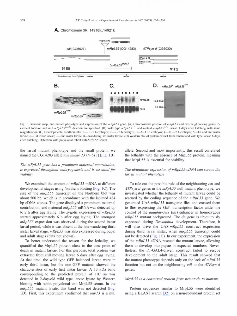
Fig. 1. Genomic map, null mutant phenotype and expression of the mRpL55 gene. (A) Chromosomal position of mRpL55 and two neighbouring genes. P-
element location and null mRpL55tmb13 deletion are specified. (B) Wild type mRpL55+/+ and mutant mRpL55�/� larvae 3 days after hatching with same
magnification. (C) Developmental Northern blot: 1— 0–2 h embryos; 2—2–6 h embryos; 3—6–11 h embryos; 4—11–22 h embryos; 5—1st and 2nd instar
larvae; 6—1st instar larvae; 7—2nd instar larvae; 8—wandering 3rd instar larvae. (D) Western blot of protein extract from mutant and wild type larvae 6 days
after hatching. Detection with polyclonal rabbit anti-MrpL55 serum.
T.V. Tselykh et al. / Experimental Cell Research 307 (2005) 354–366358
the larval mutant phenotype and the small protein, we
named the CG14283 allele tom thumb 13 (tmb13) (Fig. 1B).
The mRpL55 gene has a prominent maternal contribution,
is expressed throughout embryogenesis and is essential for
viability
We examined the amount of mRpL55 mRNA at different
developmental stages using Northern blotting (Fig. 1C). The
size of the mRpL55 transcript on the Northern blot was
about 500 bp, which is in accordance with the isolated 484
bp cDNA clones. The gene displayed a prominent maternal
contribution, and maternal mRpL55 mRNAwas detected up
to 2 h after egg laying. The zygotic expression of mRpL55
started approximately 6 h after egg laying. The strongest
mRpL55 expression was observed during the second instar
larval period, while it was absent at the late wandering third
instar larval stage. mRpL55 was also expressed during pupal
and adult stages (data not shown).
To better understand the reason for the lethality, we
quantified the MrpL55 protein close to the time point of
death in mutant larvae. For this purpose, total protein was
extracted from still moving larvae 6 days after egg laying.
At that time, the wild type GFP balanced larvae were in
early third instar, but the non-GFP mutants showed the
characteristics of early first instar larvae. A 13 kDa band
corresponding to the predicted protein of 107 aa was
detected in 2-day-old wild type larvae lysate by Western
blotting with rabbit polyclonal anti-MrpL55 serum. In the
mRpL55 mutant lysate, this band was not detected (Fig.
1D). First, this experiment confirmed that tmb13 is a null
allele. Second and most importantly, this result correlated
the lethality with the absence of MrpL55 protein, meaning
that MrpL55 is essential for viability.
The ubiquitous expression of mRpL55 cDNA can rescue the
larval mutant phenotype
To rule out the possible role of the neighbouring cdi and
ATPsyn-d genes in the mRpL55 null mutant phenotype, we
investigated whether the lethality of mutant larvae could be
rescued by the coding sequence of the mRpL55 gene. We
generated UAS-mRpL55 transgenic flies and crossed them
to flies expressing the Gal4 transcription factor under the
control of the daughterless (da) enhancer in homozygous
mRpL55 mutant background. The da gene is ubiquitously
expressed during Drosophila development. Therefore, it
will also drive the UAS-mRpL55 construct expression
during third larval instar, when mRpL55 transcript could
not be detected (Fig. 1C). In our experiment, the expression
of the mRpL55 cDNA rescued the mutant larvae, allowing
them to develop into pupae in expected numbers. Never-
theless, the da-GAL4-driven construct failed to rescue
development to the adult stage. This result showed that
the mutant phenotype depends only on the lack of mRpL55
function but not on the neighbouring cdi or the ATPsyn-d
genes.
MrpL55 is a conserved protein from nematode to humans
Protein sequences similar to MrpL55 were identified
using a BLAST search [32] on a non-redundant protein set

Fig. 3. Comparison of the most conserved part of MrpL55-related proteins.
The figure includes sequences with high similarity to MrpL55 (putative
orthologs) as well as ribosomal L26-related proteins. (A) The DGSTI motif
and its surroundings as discovered with the MEME tool. The histogram
shows the significance of every position measured in bits along the motif.
The colours of the amino acids reflect their physico-chemical properties.
(B) Alignment of the secondary structure prediction by Jpred emphasising
the pivotal position of glycine (G) in both the structure and the motif.
T.V. Tselykh et al. / Experimental Cell Research 307 (2005) 354–366 359
at NCBI and on species-specific genomes at ENSEMBL.
Putative orthologs (Blast E-value < 1e-8) were found (in
decreasing order of significance) in malaria mosquito
(ENSANGG00000017941 gene), in human (MRPL55),
in mouse (Mrpl55), in rat (ENSRNOG00000002943), in
cattle (BE237145), in chicken (LOC428400), in zebra fish
(ENSDARG00000008358), in honey bee (ENSAPMP00
000001344), in Caenorhabditis briggsae (CBG13496) and
in Caenorhabditis elegans (Y66H1A.3). In the frog
(Xenopus), only a sequence fragment similar to MrpL55
was found. This may be due to the fact that only a
preliminary genome draft was available. No similarity to
plant or yeast proteins was found at this level of
significance. All putative orthologs are in the same size
range as MrpL55 with the exception of the chicken protein,
which is 275 aa long. This peptide has been predicted by
automated computational analysis at NCBI and is derived
from an annotated genomic sequence (NW_060264) using
the GNOMON gene prediction method, so it might be
erroneous. The Drosophila MrpL55 has 42% identity and
71% similarity to the human MRPL55 protein over its full
107 aa length (Fig. 2).
Multiple sequence analyses were performed using both
ClustalW [33] as well as MEME, a tool for discovering
motifs in a group of related sequences [34]. They high-
lighted a conserved motif centered on the FDGSTI_sequence (Figs. 2, 3) perfectly conserved in all species with
the exception of Anopheles and Xenopus.
All the similar proteins were examined with the PSORT
II software (http://www.psort.ims.u-tokyo.ac.jp/), [35] and
putative nuclear localisation signals (NLS) were identified
in all of them (Fig. 2). The fruit fly MrpL55 protein contains
several conserved overlapping NLSs, and the mammalian
proteins were characterised by the presence of an absolutely
identical NLS of seven amino acids. The positions of the
predicted NLSs in the different species are conserved.
Using MitoProt and TargetP software [36,37], we
revealed that MrpL55 and its mammalian orthologs also
Fig. 2. Multiple sequence alignment of some putative MrpL55 orthologs. The mitoc
Putatively phosphorylated tyrosines are marked with stars. The most conserved pa
(CK II) site is marked. The conserved position of the nuclear localisation signal (N
is boxed with a dotted line.
contain an N-terminal mitochondrial targeting sequences
(MTS) (Fig. 2). Interestingly, SMART searches (http://
www.smart.embl-heidelberg.de) [38,39] show that the frog,
rat, mouse and bovine proteins also have a signal sequence
embedded in the MTS (data not shown).
PSORT II revealed that all analysed proteins contain one
positionally conserved casein kinase II (CKII) site close to
the NLS. Although some MrpL55 orthologs have additional
CKII sites, only one has been conserved in the process of
evolution (Fig. 2). MrpL55 and its orthologs also have a
number of conserved tyrosine residues available for
hondrial targeting sequence (MTS) position is boxed with a continuous line.
rt of the protein is emphasised with squares. The conserved casein kinase II
LS) is boxed with a dashed line, and the ER membrane retention signal (EP)
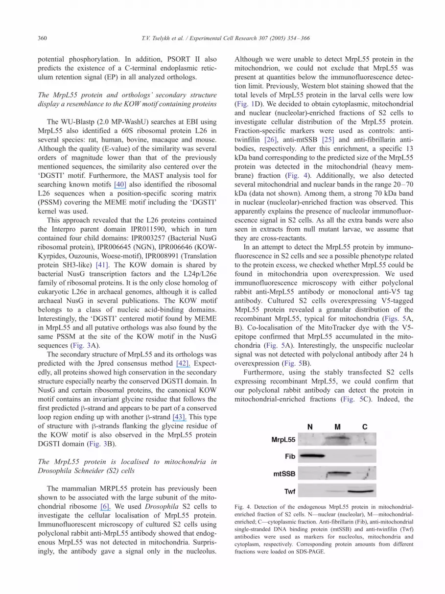
Fig. 4. Detection of the endogenous MrpL55 protein in mitochondrial-
enriched fraction of S2 cells. N—nuclear (nucleolar), M—mitochondrial-
enriched; C—cytoplasmic fraction. Anti-fibrillarin (Fib), anti-mitochondrial
single-stranded DNA binding protein (mtSSB) and anti-twinfilin (Twf)
antibodies were used as markers for nucleolus, mitochondria and
cytoplasm, respectively. Corresponding protein amounts from different
fractions were loaded on SDS-PAGE.
T.V. Tselykh et al. / Experimental Cell Research 307 (2005) 354–366360
potential phosphorylation. In addition, PSORT II also
predicts the existence of a C-terminal endoplasmic retic-
ulum retention signal (EP) in all analyzed orthologs.
The MrpL55 protein and orthologs’ secondary structure
display a resemblance to the KOW motif containing proteins
The WU-Blastp (2.0 MP-WashU) searches at EBI using
MrpL55 also identified a 60S ribosomal protein L26 in
several species: rat, human, bovine, macaque and mouse.
Although the quality (E-value) of the similarity was several
orders of magnitude lower than that of the previously
mentioned sequences, the similarity also centered over the
FDGSTI_ motif. Furthermore, the MAST analysis tool for
searching known motifs [40] also identified the ribosomal
L26 sequences when a position-specific scoring matrix
(PSSM) covering the MEME motif including the FDGSTI_kernel was used.
This approach revealed that the L26 proteins contained
the Interpro parent domain IPR011590, which in turn
contained four child domains: IPR003257 (Bacterial NusG
ribosomal protein), IPR006645 (NGN), IPR006646 (KOW-
Kyrpides, Ouzounis, Woese-motif), IPR008991 (Translation
protein SH3-like) [41]. The KOW domain is shared by
bacterial NusG transcription factors and the L24p/L26e
family of ribosomal proteins. It is the only close homolog of
eukaryotic L26e in archaeal genomes, although it is called
archaeal NusG in several publications. The KOW motif
belongs to a class of nucleic acid-binding domains.
Interestingly, the FDGSTI_ centered motif found by MEME
in MrpL55 and all putative orthologs was also found by the
same PSSM at the site of the KOW motif in the NusG
sequences (Fig. 3A).
The secondary structure of MrpL55 and its orthologs was
predicted with the Jpred consensus method [42]. Expect-
edly, all proteins showed high conservation in the secondary
structure especially nearby the conserved DGSTI domain. In
NusG and certain ribosomal proteins, the canonical KOW
motif contains an invariant glycine residue that follows the
first predicted h-strand and appears to be part of a conservedloop region ending up with another h-strand [43]. This type
of structure with h-strands flanking the glycine residue of
the KOW motif is also observed in the MrpL55 protein
DGSTI domain (Fig. 3B).
The MrpL55 protein is localised to mitochondria in
Drosophila Schneider (S2) cells
The mammalian MRPL55 protein has previously been
shown to be associated with the large subunit of the mito-
chondrial ribosome [6]. We used Drosophila S2 cells to
investigate the cellular localisation of MrpL55 protein.
Immunofluorescent microscopy of cultured S2 cells using
polyclonal rabbit anti-MrpL55 antibody showed that endog-
enous MrpL55 was not detected in mitochondria. Surpris-
ingly, the antibody gave a signal only in the nucleolus.
Although we were unable to detect MrpL55 protein in the
mitochondrion, we could not exclude that MrpL55 was
present at quantities below the immunofluorescence detec-
tion limit. Previously, Western blot staining showed that the
total levels of MrpL55 protein in the larval cells were low
(Fig. 1D). We decided to obtain cytoplasmic, mitochondrial
and nuclear (nucleolar)-enriched fractions of S2 cells to
investigate cellular distribution of the MrpL55 protein.
Fraction-specific markers were used as controls: anti-
twinfilin [26], anti-mtSSB [25] and anti-fibrillarin anti-
bodies, respectively. After this enrichment, a specific 13
kDa band corresponding to the predicted size of the MrpL55
protein was detected in the mitochondrial (heavy mem-
brane) fraction (Fig. 4). Additionally, we also detected
several mitochondrial and nuclear bands in the range 20–70
kDa (data not shown). Among them, a strong 70 kDa band
in nuclear (nucleolar)-enriched fraction was observed. This
apparently explains the presence of nucleolar immunofluor-
escence signal in S2 cells. As all the extra bands were also
seen in extracts from null mutant larvae, we assume that
they are cross-reactants.
In an attempt to detect the MrpL55 protein by immuno-
fluorescence in S2 cells and see a possible phenotype related
to the protein excess, we checked whether MrpL55 could be
found in mitochondria upon overexpression. We used
immunofluorescence microscopy with either polyclonal
rabbit anti-MrpL55 antibody or monoclonal anti-V5 tag
antibody. Cultured S2 cells overexpressing V5-tagged
MrpL55 protein revealed a granular distribution of the
recombinant MrpL55, typical for mitochondria (Figs. 5A,
B). Co-localisation of the MitoTracker dye with the V5-
epitope confirmed that MrpL55 accumulated in the mito-
chondria (Fig. 5A). Interestingly, the unspecific nucleolar
signal was not detected with polyclonal antibody after 24 h
overexpression (Fig. 5B).
Furthermore, using the stably transfected S2 cells
expressing recombinant MrpL55, we could confirm that
our polyclonal rabbit antibody can detect the protein in
mitochondrial-enriched fractions (Fig. 5C). Indeed, the

Fig. 5. Immunostainings of stably transfected S2 cells and detection of V5-tagged MrpL55 in mitochondrial-enriched fractions. (A) Using a V5 tag-specific
antibody, MrpL55 was detected in the mitochondria after 24 h of overexpression, as confirmed with MitoTracker dye. (B) Also using a polyclonal anti-MrpL55
serum, the overexpressed protein was detected in the mitochondria. (C) In opposition to pre-immune serum (lane 4), a single specific 13 kDa band was
observed in non-transfected control cells (lane 1), and 16 kDa V5-tagged MrpL55 band was detected with both polyclonal anti-MrpL55 (lane 2) and
monoclonal anti-V5 (lane 3) antibodies in stably transfected cells. (D) Pre-immune serum gave no signal in immunofluorescence stainings. Note: only a single
cell is presented on panels (A) and (B), but a number of cells at lower magnification is analysed on panel (D).
T.V. Tselykh et al. / Experimental Cell Research 307 (2005) 354–366 361
recombinant V5-tagged protein overexpressed for 24 h was
detected on the SDS-PAGE as a higher molecular weight 16
kDa band in comparison with the 13 kDa endogenous
MrpL55 protein (Fig. 5C, lanes 1–2). Moreover, the 16 kDa
band was also detected in mitochondrial-enriched fraction
with monoclonal anti-V5 antibody (Fig. 5C, lane 3).
The overexpression of MrpL55 did not seem to affect the
cell survival in general. S2 cells constantly expressing
MrpL55 in a second transfected cell line stayed alive for at
least 3 weeks.
The MrpL55 protein is localised to mitochondria in
Drosophila tissues
Salivary gland cells of the third instar larvae have a
number of advantages for assaying subcellular protein
localisation due to their large cytoplasm. Drosophila larvae
spend most of their time inside the food substrate as
foraging larvae. During the third instar stage, in response
to increased levels of ecdysone, they leave the food as
wandering larvae and search for an adequate site to pupate,
which in laboratory cultures are the walls of the culture
vial. To identify the localisation of MrpL55 in Drosophila
tissue cells, we performed immunohistochemical staining
using anti-MrpL55 antibody on foraging and wandering
larval salivary glands. In foraging third instar larvae
salivary glands, we could not detect any mitochondrial
signal. Similar to the case with S2 cells, the antibody
cross-reacted with some components in the nucleolus of
foraging larvae salivary glands. In wandering larvae, a
mitochondrial signal could be detected, but only in the
proximal cells of the salivary glands (Fig. 6A). Co-
localisation of the MitoTracker dye with the anti-MrpL55
antibody confirmed that the protein accumulated in the
mitochondria in secretory cells (Fig. 6A). This experiment
clearly showed that the intracellular localisation of MrpL55
protein was similar in tissues isolated from wandering
larvae and S2 cells.
We also checked expression of the MrpL55 protein in
the cells of other third instar larval tissues such as gut, fat
body, imaginal discs, brain, trachea as well as adult
ovaries. In all tissues examined, the MrpL55 protein was
found in the cytoplasm and had a faint granular,
mitochondrial-like distribution (data not shown). The
results demonstrate that MrpL55 is expressed in multiple
tissues.
Modulation of ectopic expression of the mRpL55 gene leads
to different responses during development
To get insights into the role of mRpL55, the gene was
overexpressed under three different tissue-specific en-
hancers using the UAS-Gal4 system. Using the salivary
gland secretion-3 (sgs-3) enhancer, the overexpression of
MrpL55 leads to accumulation of the protein in all salivary
gland cells of wandering larvae (Fig. 6B). This accumu-
lation of the protein had no effect on fly development.
Using the tubulin (tub) enhancer, the mRpL55 gene was
expressed ubiquitously, and mitochondrial accumulation of
the protein could be observed in cells of larval fat body,
imaginal discs, trachea, brain as well as adult ovaries (data
not shown). However, these animals (tub-Gal4 � UAS-
mRpL55) died as late third instar larvae or pupae. Using the
daughterless (da) enhancer, ubiquitous expression was also
achieved, and the mitochondrial accumulation of the
protein could again be observed in all studied cells.

Fig. 7. Somatic mosaic clones induced with FLP-FRT recombination: (A) in
anterior part of eye imaginal disc. Homozygous mutant clones are marked
with a star. Heterozygous and wild type clones carry one or two copies of a
GFP-balancer (TM3, Ser GFP) and are seen stained (left). There is no
difference in cell size as outlined using a phalloidin staining (middle); (B) in
adult eye. (Left) Clones of normal red eye colour were of genotype w+/w+;
mRpL55+/mRpL55+, while the pale red patches were w+/w�; mRpL55+/
mRpL55�. No white clones corresponding to the genotype w�/w�;
mRpL55�/mRpL55� were seen. (Middle) When no recombination takes
place, all cells were w+/w�; mRpL55+/mRpL55� and appeared pale red.
(Right) In a control experiment using Stubble (Sb) instead of mRpL55,
patches of all three types were generated, including mutant white clones.
Fig. 6. Immunohistochemical stainings of salivary gland cells from 3rd instar larvae. (A) MrpL55 was detected with a polyclonal anti-MrpL55 serum in
mitochondria of proximal secretory salivary gland cells of wandering larvae. The localisation was confirmed with the specific mitochondrial marker
MitoTracker Red. Proximal is on the left, distal is on the right. (B) sgs-3-driven mRpL55 overexpression. The MrpL55 protein was detected in mitochondria of
all salivary gland cells, as confirmed with MitoTracker Red. Note: a lower magnification was used for images on panel (A) to show proximal and distal parts of
the salivary gland.
T.V. Tselykh et al. / Experimental Cell Research 307 (2005) 354–366362
Interestingly, the da-Gal4 � UAS-mRpL55 animals sur-
vived and were fertile. Since the mRpL55 gene is normally
not expressed in third instar larvae (Fig. 1C), these results
show that the modulation of expression is a critical issue
for animal development.
Clonal analysis shows a dynamic autonomous requirement
for mRpL55 activity in the cell during development
Since we observed germline expression of mRpL55, we
wanted to study whether the gene had a function during
oogenesis. Therefore, we applied the FLP-FRT recombina-
tion technique to generate germline mosaic clones where
the maternal mRpL55 expression is eliminated. Upon
recombination, the females became sterile. This shows that
mRpL55 seems to be required for germline cell survival.
Based on our initial observations of tiny mRpL55 mutant
larvae arrested in growth, we postulated that mRpL55 could
be involved in cell growth or cell proliferation. However,
due to the possible cell-autonomous requirement for
mRpL55 in germline cells, we could not get any information
on the requirement for maternal mRpL55 during early
embryogenesis.
To understand the cellular basis for mRpL55 effects on
cell growth and proliferation, we also conducted a somatic
mosaic analysis with the FLP-FRT recombination techni-
que. We created patches of mosaic clones in eye imaginal
discs. FLP was expressed under the control of the eye-
specific eyeless (ey) enhancer. Clones of both mRpL55+/+
and mRpL55�/� genotypes were observed in anterior parts
of eye discs (Fig. 7A). Additionally, mutant clones were
sometimes detected in the area just posterior to the
morphogenetic furrow, a physical constriction in the apical
surface of the eye disc epithelium (data not shown). This

T.V. Tselykh et al. / Experimental Cell Research 307 (2005) 354–366 363
result shows that mRpL55 is not required in eye imaginal
disc cells at third instar larval stage. Indeed, the Northern
blotting results showed that the mRpL55 gene is not
expressed during this stage (Fig. 1C). Moreover, the
mRpL55�/� clones did not appear to be smaller than
mRpL55+/+ twin spot clones.
Knowing that mRpL55 is expressed during pupal and
adult stages, we extended the somatic clonal analyses to
adult eyes. No mutant clones appeared in the adult eyes,
and the heterozygous mRpL55+/� patches were smaller
than the mRpL55+/+ clones (Fig. 7B). Overall shape and
size of adult eyes were not affected. This experiment
showed that mRpL55 is essential for the survival of
developing eye cells at pupal stage. Taken together, these
results indicate that there is a dynamic requirement for
mRpL55 activity in the cell.
Discussion
In this study, we report the characterisation of the
essential Drosophila mRpL55 gene, which encodes 1 of
11 previously uncharacterised large subunit MRPs specific
for mitoribosome in multicellular organisms. The mRpL55
larval mutant phenotype was characterised by developmen-
tal arrest at early larval stage. The Drosophila MrpL55
protein is orthologous to the mammalian mitochondrial
ribosomal protein MRPL55, which belongs to a class of
proteins specific only for mitoribosomes and expressed
exclusively in metazoans. The Drosophila protein and its
orthologs also contain an N-terminal mitochondrial target-
ing sequence (MTS). Interestingly, the MrpL55 protein also
contains a nuclear localisation signal (NLS), the position of
which is highly conserved in MrpL55 orthologs from other
species. However, it is not clear whether this NLS has any
relation to the function of the protein. The pattern of
conservation in MrpL55 orthologs indicates that this protein
has evolved relatively recently.
Using biochemical, cell biology and genetics techniques,
we showed that the Drosophila MrpL55 protein is localised
to the mitochondrion both in S2 cells and various
Drosophila tissues. The localisation of MrpL55 in mito-
chondria is in agreement with the presence of a mitochon-
drial targeting sequence (MTS) in the protein. Our
observations of intracellular localisation of the protein are
in agreement with a report that the mammalian MrpL55
ortholog, MRPL55, is associated with the large subunit of
the mitochondrial ribosome [6].
The Drosophila MrpL55 protein and its orthologs display
a high similarity in secondary structure and amino acid
composition to the KOW motif [43]. The KOW (Kyrpides,
Ouzounis, Woese) motif is found in a variety of ribosomal
proteins and the bacterial transcription anti-termination
NusG proteins. It encodes a nucleic acid interaction motif
(Interpro entry IPR006646). The KOW-motif-containing
proteins have been shown to play a role in transcriptional
anti-termination and in the regulation of translation [44–47].
The crystal structure of a NusG transcription factor from
Aquifex aeolicus indicates that the KOWmotif is found to be
the core of an RNA-binding domain [48]. The most
conserved peptide sequence of the MrpL55 protein ortho-
logs, DGSTI, is found in the middle of the protein and
includes the conservation of a glycine amino acid separating
the two h-strands in the NusG protein. The KOW motif is
present in proteins with nucleic acid binding function:
bacterial NusG proteins are transcription factors, and L24/
26 are ribosomal proteins. The presence of a KOW-like motif
in MrpL55 indicates that the protein may bind mitochondrial
DNA (mtDNA), mitochondrial ribosomal RNAs (rRNAs) or
other RNA types located in the mitochondria. We suggest
that MrpL55 plays a role in the regulation of mitochondrial
rRNA transcription, protein translation or mitoribosomal
saturation.
The modulation of the mRpl55 gene expression level is
critical for development. Both absence and overexpression
of the gene can lead to lethality in vivo. The mutant larvae
without any endogenous zygotic expression could be
rescued until third instar/pupal stage using the daughterless
enhancer. Is the dosage of the gene expression especially
critical at the third instar larval stage where mRpL55 is
normally not expressed? Ectopic expression of the gene
using the daughterless enhancer on top of the endogenous
expression did not disturb the development of the animals,
showing that there is a certain tolerance for overexpression.
Thus, it appears that expression under the daughterless
enhancer is not strong enough (1) to bring the rescue of the
null phenotype beyond the early pupal stage or (2) to disturb
a normal development when cumulated to endogenous
expression. However, ectopic expression using the stronger
tubulin enhancer on top of endogenous expression exceeds
the tolerated expression level, and the animals die during
third instar/pupal stage. Therefore, it is possible that rescue
of mutants using tubulin-driven expression would extend
the rescue to a later stage than daughterless-driven
expression.
The requirement for MrpL55 in the cell varies during
larval development. We showed that mRpL55 expression
drops at third instar larval stage. However, the MrpL55
protein was detected enriched in the mitochondria of
secretory proximal salivary gland cells towards the very
end of the third larval instar stage (wandering larvae). It
appears that MrpL55 is enriched in the mitochondria of
cells with a higher protein synthesis activity (Fig. 6A).
Indeed, towards the end of larval development, the distal
salivary gland cells enter a phase of reduced protein
synthesis and less intense endomitosis. Thus, the amount
of MrpL55 protein changes dynamically depending on the
status of the cell. Taking into account that MrpL55 is a
mitoribosomal protein and that most larval tissues consist
of metabolically active polytene cells, the growth retarda-
tion and tiny size of mRpL55 mutant larvae might be a
consequence of disturbances in the mitochondrial produc-

T.V. Tselykh et al. / Experimental Cell Research 307 (2005) 354–366364
tion of ATP, which is required for general protein synthesis
in the cell.
The concept of dynamic requirement for MrpL55 in the
cell is further strengthened by somatic mosaic experiments.
We showed that mRpL55�/� cells do not survive in adult
eyes, while they do survive in eye imaginal discs of third
instar larvae. In wandering third instar larvae, mRpL55
transcripts are absent, but the protein is still present. The
survival of mRpL55�/� clones in eye imaginal discs could
be explained either by the possibility that the protein is
dispensable or is still present in sufficient amounts due to
the relatively long protein half-life. However, the absence of
mutant clones in adult eyes showed that MrpL55 was again
required during pupal stage.
In a set of experiments, Pile and colleagues have identified
genes, the expression of which is modified by the SIN3
deacetylase complex, essential for G2 phase cell cycle
progression [49]. The mRpL55 gene was identified as a
SIN3 target of repression together with other mitochondrial
ribosomal genes. In another high throughput study, per-
formed by Dimova et al., mRpL55 was, among other genes,
described as a likely direct target for E2F/RB transcription
factor regulation [50]. The E2F and pRB families are pivotal
regulators in cell division control (for a review, see [51,52]).
The E2F/RB transcription factors regulation of mRpL55
suggests that the MrpL55 protein links mitochondrial bio-
genesis to cell cycle progression. Nuclear and mitochondrial
genome functions have been shown to be interconnected in
several independent studies. For example, mitochondria can
modify nuclear gene expression levels by direct signalling to
nuclei [53,54] and, in Drosophila, a molecular link between
nuclear and mitochondrial DNA replication has been
established through the role of the mitochondrial single-
stranded DNA-binding protein gene (mtSSB) [55]. Finally,
the Drosophila mitoribosomal protein MrpL12 has been
shown to be required for Cyclin D/Cdk4-driven growth,
building a link between cellular growth rates and mitochon-
drial activity [17]. A possible involvement of the mRpL55
gene in cell cycle regulation is also supported by the fact that
about three quarter of rescued mRpL55 mutant third instar
larvae developed pigment-encapsulated cell clusters, which
lost their cellular appearance and transformed into black
inclusion bodies as seen in black pearl (blp) mutants [56].
The melanotic tumor-like bodies were found in mRpL55
rescue experiments almost in all dead pupae (Tselykh T.V.,
unpublished data). Inappropriate level of MrpL55 during the
third instar larval stage, when the mRpL55 gene is normally
not expressed (Fig. 1C), could have an influence upon cell
division, leading to melanotic Ftumor_ formation and,
consequently, death.
Dimova et al. [50] also proposed that the mRpL55 gene,
among other genes analysed, plays a role in progression
through G2/M cell cycle transition, which in Drosophila is
crucial during the patterning and specification of adult
tissues, when the precise number of cells may be important
[57]. Development of the Drosophila adult eye is an
example where G2/M cell cycle regulation is of paramount
importance.
Each Drosophila adult eye develops from the eye
imaginal disc and is composed of about 800 ommatidia,
special eye units. Differentiation and patterning of omma-
tidia begins at late third instar larval stage and continues in
pupa. Cell differentiation in eye discs is coordinated with
cell proliferation and cell death (for detailed review, see
[57]). The total cell number in adult eye depends on the
balance of these processes. The differentiation process in
eye imaginal disc initiates posterior to the morphogenetic
furrow (MF). All cells ahead of the MF are undiffer-
entiated and divide asynchronously. The cells just anterior
to the MF are transiently arrested in G1 phase. The
differentiation progresses as a wave marked by the MF
moving anteriorly. The process is followed by a ‘‘Second
Mitotic Wave’’ (SMW) cell cycle that is regulated at the
G2/M phase transition. Importantly, determination of exact
cell number in each adult eye ommatidia occurs at the G2/
M transition of the SMW cell cycle. The regulation of the
terminal cell divisions is coordinated by local intercellular
signals [58]. Dimova et al. [50] showed that there is a class
of genes with E2F-dependent transcription that requires
additional transcription factors and that is activated later in
the cell cycle. The mRpL55 gene belongs to this class.
Interestingly, the string (stg) gene also belongs to the same
class. In Drosophila, the stg gene is a limiting factor for
the cell cycle during Drosophila development and one of
the major components during G2/M in the SMW [59,60].
Interestingly, Pile and colleagues [49] reported that loss of
SIN3 in the cell repressed the string gene whereas it
induced mRpL55 expression. Our results reveal that
MrpL55 is not required in asynchronously dividing and
G1 phase arrested cells. However, the fact that mRpL55�/�
clones were not observed in adult eye suggests that G2/M
transition in the SMW at pupal stage was affected in
mutant cells. Therefore, our results suggest that mRpL55
has a role in the G2/M cell cycle transition, the more
precise molecular function, however, will be the subject of
future studies.
Acknowledgments
We thank Martyn James and Konstantin I. Ivanov for
critical review of the manuscript. We are grateful to
Laurie Kaguni (Michigan State University, USA) for
providing us with a rabbit polyclonal anti-mtSSB anti-
body. The authors are also grateful to Tiina Immonen,
Marja Mikkola, Johan Peranen, Mari Palgi, Mari Palviai-
nen and Gudrun Wahlstrom (University of Helsinki,
Finland) for technical support and especially to Christos
Samakovlis (Stockholm University, Sweden) for his help
at the early stages of this study. This work was supported
in part by a grant from the Centre for International
Mobility (CIMO, Finland).

T.V. Tselykh et al. / Experimental Cell Research 307 (2005) 354–366 365
References
[1] G. Attardi, Animal mitochondrial DNA: an extreme example of
genetic economy, Int. Rev. Cytol. 93 (1985) 93–145.
[2] A. Chomyn, M.W. Cleeter, C.I. Ragan, M. Riley, R.F. Doolittle, G.
Attardi, URF6, last unidentified reading frame of human mtDNA,
codes for an NADH dehydrogenase subunit, Science 234 (1986)
614–618.
[3] H. de Vries, R. Koogh-Schuuring, Physicochemical characteristics of
isolated 55-S mitochondrial ribosomes from rat-liver, Biochem.
Biophys. Res. Commun. 54 (1973) 308–314.
[4] T.W. O’Brien, D.E. Mathews, N.D. Denslow, in: T.I. Bucher, W.
Neupert, W.S. Sebald, S. Werner (Eds.), Genetics and Biogenesis of
Chloroplasts and Mitochondria, 1976, pp. 741–748.
[5] E.C. Koc, W. Burkhart, K. Blackburn, A. Moseley, L.L. Spremulli,
The small subunit of the mammalian mitochondrial ribosome,
Identification of the full complement of ribosomal proteins present,
J. Biol. Chem. 276 (2001) 19363–19374.
[6] E.C. Koc, W. Burkhart, K. Blackburn, M.B. Moyer, D.M. Schlatzer,
A. Moseley, L.L. Spremulli, The large subunit of the mammalian
mitochondrial ribosome. Analysis of the complement of ribosomal
proteins present, J. Biol. Chem. 276 (2001) 43958–43969.
[7] T. Suzuki, M. Terasaki, C. Takemoto-Hori, T. Hanada, T. Ueda, A.
Wada, K. Watanabe, Structural compensation for the deficit of rRNA
with proteins in the mammalian mitochondrial ribosome, Systematic
analysis of protein components of the large ribosomal subunit from
mammalian mitochondria, J. Biol. Chem. 276 (2001) 21724–21736.
[8] T. Suzuki, M. Terasaki, C. Takemoto-Hori, T. Hanada, T. Ueda, A.
Wada, K. Watanabe, Proteomic analysis of the mammalian mitochon-
drial ribosome, Identification of protein components in the 28 S small
subunit, J. Biol. Chem. 276 (2001) 33181–33195.
[9] T.W. O’Brien, Evolution of a protein-rich mitochondrial ribosome:
implications for human genetic disease, Gene 286 (2002) 73–79.
[10] N. Kenmochi, T. Suzuki, T. Uechi, M. Magoori, M. Kuniba, S. Higa,
K. Watanabe, T. Tanaka, The human mitochondrial ribosomal protein
genes: mapping of 54 genes to the chromosomes and implications for
human disorders, Genomics 77 (2001) 65–70.
[11] D.F. Johnson, M. Hamon, N. Fischel-Ghodsian, Characterization of
the human mitochondrial ribosomal S12 gene, Genomics 52 (1998)
363–368.
[12] P. Mariottini, Z.H. Shah, J.M. Toivonen, C. Bagni, J.N. Spelbrink, F.
Amaldi, H.T. Jacobs, Expression of the gene for mitoribosomal protein
S12 is controlled in human cells at the levels of transcription, RNA
splicing, and translation, J. Biol. Chem. 274 (1999) 31853–31862.
[13] Z.H. Shah, K.M. O’Dell, S.C. Miller, X. An, H.T. Jacobs, Metazoan
nuclear genes for mitoribosomal protein S12, Gene 204 (1997) 55–62.
[14] J.M. Toivonen, K.M. O’Dell, N. Petit, S.C. Irvine, G.K. Knight, M.
Lehtonen, M. Longmuir, K. Luoto, S. Touraille, Z. Wang, S. Alziari,
Z.H. Shah, H.T. Jacobs, Technical knockout, a Drosophila model of
mitochondrial deafness, Genetics 159 (2001) 241–254.
[15] Z.H. Shah, V. Migliosi, S.C. Miller, A. Wang, T.B. Friedman, H.T.
Jacobs, Chromosomal locations of three human nuclear genes
(RPSM12, TUFM, and AFG3L1) specifying putative components of
the mitochondrial gene expression apparatus, Genomics 48 (1998)
384–388.
[16] M. Galloni, Bonsai, a ribosomal protein S15 homolog, involved in gut
mitochondrial activity and systemic growth, Dev. Biol. 264 (2003)
482–494.
[17] C. Frei, M. Galloni, E. Hafen, B.A. Edgar, The Drosophila
mitochondrial ribosomal protein mRpL12 is required for Cyclin
D/Cdk4-driven growth, EMBO J. 24 (2005) 623–634.
[18] J.L. Kissil, O. Cohen, T. Raveh, A. Kimchi, Structure– function
analysis of an evolutionary conserved protein, DAP3, which mediates
TNF-alpha- and Fas-induced cell death, EMBO J. 18 (1999) 353–362.
[19] K.E. Cavdar, A. Ranasinghe, W. Burkhart, K. Blackburn, H. Koc, A.
Moseley, L.L. Spremulli, A new face on apoptosis: death-associated
protein 3 and PDCD9 are mitochondrial ribosomal proteins, FEBS
Lett. 492 (2001) 166–170.
[20] S.R. Chintharlapalli, M. Jasti, S. Malladi, K.V. Parsa, R.P. Ballestero,
M. Gonzalez-Garcia, BMRP is a Bcl-2 binding protein that induces
apoptosis, J. Cell. Biochem. 94 (2005) 611–626.
[21] L. Sun, Y. Liu, M. Fremont, S. Schwarz, M. Siegmann, R. Matthies,
J.P. Jost, A novel 52 kDa protein induces apoptosis and concurrently
activates c-Jun N-terminal kinase 1 (JNK1) in mouse C3H10T1/2
fibroblasts, Gene 208 (1998) 157–166.
[22] F. Ogawa, S. Adachi, K. Kohu, K. Shige, T. Akiyama, Binding of the
human homolog of the Drosophila discs large tumor suppressor
protein to the mitochondrial ribosomal protein MRP-S34, Biochem.
Biophys. Res. Commun. 300 (2003) 789–792.
[23] M. Muramatsu, H. Bush, Studies on nucleolar RNA of the Walker
256 carcinosarcoma and the liver of the rat, Cancer Res. 24 (1964)
1028–1034.
[24] T. Igaki, H. Kanuka, N. Inohara, K. Sawamoto, G. Nunez, H. Okano,M.
Miura, Drob-1, a Drosophila member of the Bcl-2/CED-9 family that
promotes cell death, Proc. Natl. Acad. Sci. U. S. A. 97 (2000) 662–667.
[25] C.L. Farr, Y. Wang, L.S. Kaguni, Functional interactions of mitochon-
drial DNA polymerase and single-stranded DNA-binding protein.
Template-primer DNA binding and initiation and elongation of DNA
strand synthesis, J. Biol. Chem. 274 (1999) 14779–14785.
[26] G. Wahlstrom, M. Vartiainen, L. Yamamoto, P.K. Mattila, P. Lappalai-
nen, T.I. Heino, Twinfilin is required for actin-dependent developmen-
tal processes in Drosophila, J. Cell Biol. 155 (2001) 787–796.
[27] T.I. Heino, Polytene chromosomes from ovarian pseudonurse cells of
the Drosophila melanogaster otu mutant: II. Photographic map of the
X chromosome, Chromosoma 103 (1994) 4–15.
[28] T.B. Chou, N. Perrimon, The autosomal FLP-DFS technique for
generating germline mosaics in Drosophila melanogaster, Genetics
144 (1996) 1673–1679.
[29] B.B. Matthews, S.T. Crews, Drosophila center divider gene is
expressed in CNS midline cells and encodes a developmentally
regulated protein kinase orthologous to human TESK1, DNA Cell
Biol. 18 (1999) 435–448.
[30] K. Raymond, E. Bergeret, A. Avet-Rochex, R. Griffin-Shea, M.O.
Fauvarque, A screen for modifiers of RacGAP(84C) gain-of-function
in the Drosophila eye revealed the LIM kinase Cdi/TESK1 as a
downstream effector of Rac1 during spermatogenesis, J. Cell Sci. 117
(2004) 2777–2789.
[31] C. Caggese, G. Ragone, B. Perrini, R. Moschetti, V. De Pinto, R.
Caizzi, P. Barsanti, Identification of nuclear genes encoding mitochon-
drial proteins: isolation of a collection of D. melanogaster cDNAs
homologous to sequences in the Human Gene Index database, Mol.
Gen. Genet. 261 (1999) 64–70.
[32] S.F. Altschul, T.L. Madden, A.A. Schaffer, J. Zhang, Z. Zhang, W.
Miller, D.J. Lipman, Gapped BLAST and PSI-BLAST: a new
generation of protein database search programs, Nucleic Acids Res.
25 (1997) 3389–3402.
[33] J.D. Thompson, D.G. Higgins, T.J. Gibson, CLUSTALW: improving
the sensitivity of progressive multiple sequence alignment through
sequence weighting, position-specific gap penalties and weight matrix
choice, Nucleic Acids Res. 22 (1994) 4673–4680.
[34] T.J. Bailey, C. Elkan, Fitting a mixture model by expectation
maximization to discover motifs in biopolymers, Proceedings of the
Second International Conference on Intelligent Systems for Molecular
Biology, AAAI Press, Menlo Park, CA, 1994.
[35] K. Nakai, P. Horton, PSORT: a program for detecting sorting signals in
proteins and predicting their subcellular localization, Trends Biochem.
Sci. 24 (1999) 34–36.
[36] M.G. Claros, P. Vincens, Computational method to predict mitochond-
rially imported proteins and their targeting sequences, Eur. J.
Biochem. 241 (1996) 779–786.
[37] O. Emanuelsson, H. Nielsen, S. Brunak, G. von Heijne, Predicting
subcellular localization of proteins based on their N-terminal amino
acid sequence, J. Mol. Biol. 300 (2000) 1005–1016.
T.V. Tselykh et al. / Experimental Cell Research 307 (2005) 354–366366
[38] I. Letunic, R.R. Copley, S. Schmidt, F.D. Ciccarelli, T. Doerks,
J. Schultz, C.P. Ponting, P. Bork, SMART 4.0: towards genomic
data integration, Nucleic Acids Res. 32 (2004) D142–D144
(database issue).
[39] J. Schultz, F. Milpetz, P. Bork, C.P. Ponting, SMART, a simple
modular architecture research tool: identification of signaling domains,
Proc. Natl. Acad. Sci. U. S. A. 95 (1998) 5857–5864.
[40] T.L. Bailey, M. Gribskov, Combining evidence using p-values:
application to sequence homology searches, Bioinformatics 14
(1998) 48–54.
[41] N.J. Mulder, R. Apweiler, T.K. Attwood, A. Bairoch, D. Barrell, A.
Bateman, D. Binns, M. Biswas, P. Bradley, P. Bork, P. Bucher, R.R.
Copley, E. Courcelle, U. Das, R. Durbin, L. Falquet, W. Fleischmann,
S. Griffiths-Jones, D. Haft, N. Harte, N. Hulo, D. Kahn, A. Kanapin,
M. Krestyaninova, R. Lopez, I. Letunic, D. Lonsdale, V. Silventoinen,
S.E. Orchard, M. Pagni, D. Peyruc, C.P. Ponting, J.D. Selengut, F.
Servant, C.J. Sigrist, R. Vaughan, E.M. Zdobnov, The InterPro
Database, 2003 brings increased coverage and new features, Nucleic
Acids Res. 31 (2003) 315–318.
[42] J.A. Cuff, M.E. Clamp, A.S. Siddiqui, M. Finlay, G.J. Barton, JPred: a
consensus secondary structure prediction server, Bioinformatics 14
(1998) 892–893.
[43] N.C. Kyrpides, C.R. Woese, C.A. Ouzounis, KOW: a novel motif
linking a bacterial transcription factor with ribosomal proteins, Trends
Biochem. Sci. 21 (1996) 425–426.
[44] G.O. Bylund, B.C. Persson, L.A. Lundberg, P.M. Wikstrom, A novel
ribosome-associated protein is important for efficient translation in
Escherichia coli, J. Bacteriol. 179 (1997) 4567–4574.
[45] C.L. Squires, J. Greenblatt, J. Li, C. Condon, C.L. Squires, Ribosomal
RNA antitermination in vitro: requirement for Nus factors and one or
more unidentified cellular components, Proc. Natl. Acad. Sci. U. S. A.
90 (1993) 970–974.
[46] M. Torres, J.M. Balada, M. Zellars, C. Squires, C.L. Squires, In vivo
effect of NusB and NusG on rRNA transcription antitermination,
J. Bacteriol. 186 (2004) 1304–1310.
[47] M. Zellars, C.L. Squires, Antiterminator-dependent modulation of
transcription elongation rates by NusB and NusG, Mol. Microbiol. 32
(1999) 1296–1304.
[48] T. Steiner, J.T. Kaiser, S. Marinkovic, R. Huber, M.C. Wahl, Crystal
structures of transcription factor NusG in light of its nucleic acid- and
protein-binding activities, EMBO J. 21 (2002) 4641–4653.
[49] L.A. Pile, P.T. Spellman, R.J. Katzenberger, D.A. Wassarman, The
SIN3 deacetylase complex represses genes encoding mitochondrial
proteins: implications for the regulation of energy metabolism, J. Biol.
Chem. 278 (2003) 37840–37848.
[50] D.K. Dimova, O. Stevaux, M.V. Frolov, N.J. Dyson, Cell cycle-
dependent and cell cycle-independent control of transcription by the
Drosophila E2F/RB pathway, Genes Dev. 17 (2003) 2308–2320.
[51] N. Dyson, The regulation of E2F by pRB-family proteins, Genes Dev.
12 (1998) 2245–2262.
[52] J.M. Trimarchi, J.A. Lees, Sibling rivalry in the E2F family, Nat. Rev.,
Mol. Cell Biol. 3 (2002) 11–20.
[53] V.S. Parikh, M.M. Morgan, R. Scott, L.S. Clements, R.A. Butow, The
mitochondrial genotype can influence nuclear gene expression in
yeast, Science 235 (1987) 576–580.
[54] A. Traven, J.M. Wong, D. Xu, M. Sopta, C.J. Ingles, Interorganellar
communication. Altered nuclear gene expression profiles in a yeast
mitochondrial dna mutant, J. Biol. Chem. 276 (2001) 4020–4027.
[55] D.M. Ruiz, I.E. Lefai, R. Garesse, L.S. Kaguni, Regulation of
mitochondrial single-stranded DNA-binding protein gene expression
links nuclear and mitochondrial DNA replication in Drosophila,
J. Biol. Chem. 275 (2000) 13628–13636.
[56] S. Becker, A. Gehrsitz, P. Bork, S. Buchner, E. Buchner, The
black-pearl gene of Drosophila defines a novel conserved protein
family and is required for larval growth and survival, Gene 262
(2001) 15–22.
[57] N.E. Baker, Cell proliferation, survival, and death in the Drosophila
eye, Semin. Cell Dev. Biol. 12 (2001) 499–507.
[58] N.E. Baker, S.Y. Yu, The EGF receptor defines domains of cell cycle
progression and survival to regulate cell number in the developing
Drosophila eye, Cell 104 (2001) 699–708.
[59] B.A. Edgar, P.H. O’Farrell, Genetic control of cell division patterns in
the Drosophila embryo, Cell 57 (1989) 177–187.
[60] D.A. Lehman, B. Patterson, L.A. Johnston, T. Balzer, J.S. Britton, R.
Saint, B.A. Edgar, Cis-regulatory elements of the mitotic regulator,
string/Cdc25, Development 126 (1999) 1793–1803.
Copyright © 2022 FDOKUMEN