Stage-specific changes in the Krebs cycle network regulate ...
Upload
independentCategory
view
3download
0
Structured mRNAs Regulate TranslationInitiation by Bindingto the Platform of the RibosomeStefano Marzi,1,2 Alexander G. Myasnikov,1 Alexander Serganov,3 Chantal Ehresmann,2 Pascale Romby,2
Marat Yusupov,1 and Bruno P. Klaholz1,*1IGBMC (Institute of Genetics and of Molecular and Cellular Biology), Department of Structural Biology and Genomics, Illkirch,
F-67404 France; Inserm, U596, Illkirch, F-67400 France; CNRS, UMR7104, Illkirch, F-67400 France; Universite Louis Pasteur,
Strasbourg, F-67000 France2Architecture et Reactivite de l’ARN, CNRS, IBMC (Institute of Molecular and Cellular Biology), 15 rue R. Descartes,
67084 Strasbourg, France; Universite Louis Pasteur de Strasbourg, Strasbourg, F-67000 France3Structural Biology program, Memorial Sloan-Kettering Cancer Center, New York, NY 10021, USA*Correspondence: [email protected]
DOI 10.1016/j.cell.2007.07.008
SUMMARY
Gene expression can be regulated at the level ofinitiation of protein biosynthesis via structuralelements present at the 50 untranslated regionof mRNAs. These folded mRNA segments maybind to the ribosome, thus blocking translationuntil the mRNA unfolds. Here, we report a seriesof cryo-electron microscopy snapshots of ribo-somal complexes directly visualizing either themRNA structure blocked by repressor proteinS15 or the unfolded, active mRNA. In the stalledstate, the folded mRNA prevents the start co-don from reaching the peptidyl-tRNA (P) site in-side the ribosome. Upon repressor release, themRNA unfolds and moves into the mRNA chan-nel allowing translation initiation. A comparativestructure and sequence analysis suggests theexistence of a universal stand-by site on theribosome (the 30S platform) dedicated forbinding regulatory 50 mRNA elements. Differenttypes of mRNA structures may be accommo-dated during translation preinitiation and regu-late gene expression by transiently stalling theribosome.
INTRODUCTION
Structures presented in the translation initiation region of
both prokaryotic and eukaryotic mRNAs play a direct
role in regulating gene expression. They may grant two
possible regulatory effects, repression or activation. The
modulation of the stability of folded 50 mRNA segments
allows temporal translational regulation in response to
growth condition changes or to specific needs when fast
Cell 1
adaptation is required. Translation initiation regions of
bacterial mRNAs that undergo conformational changes
leading to either repression or activation are typical of ri-
boswitches (Tucker and Breaker, 2005). These conforma-
tional changes can be induced by environmental cues
(temperature; Johansson et al., 2002), by trans-acting li-
gands such as small metabolites (Tucker and Breaker,
2005), noncoding RNAs (Gottesman, 2005) and regulatory
proteins (Schlax and Worhunsky, 2003; Romby and
Springer, 2003). Many of these 50 regulatory elements
act mainly as translational repressors (Schlax and Wo-
rhunsky, 2003; de Smit and van Duin, 2003; Gebauer
and Hentze, 2004) by inhibiting the formation of an active
initiation complex. In some cases, alleviation of such inhi-
bition requires melting of the folded structure (Schlax and
Worhunsky, 2003) and assembly of a stable translation ini-
tiation complex (reviewed in Gualerzi and Pon, 1990).
Repression of translation may occur through either
competition or entrapment. These two alternative mecha-
nisms are used, for instance, by prokaryotic ribosomal
proteins for adjustment of their synthesis to the level of ri-
bosomal RNA (Nomura et al., 1984; Zengel and Lindahl,
1994). The competition mechanism has been addressed
by biochemical and structural studies of several proteins
(Springer and Portier, 2003; Merianos et al., 2004; Jenner
et al., 2005; Scott and Williamson, 2005) and metabolites
interacting with mRNAs (Serganov et al., 2004; Batey
et al., 2004; Thore et al., 2006; Serganov et al., 2006;
Fuchs et al., 2006). The mechanism involves competitive
binding of a repressor or a ribosome to overlapping/
adjacent mRNA binding sites leading to the formation of
mutually exclusive repressor-mRNA or ribosome-mRNA
complexes (Romby and Springer, 2003). In contrast, the
entrapment mechanism is less well described at the struc-
ture-function level. It was proposed that formation of
a mRNA-repressor-complex may cause ribosome stalling
at the preinitiation stage (Schlax and Worhunsky, 2003;
Ehresmann et al., 2004). A model system for the
30, 1019–1031, September 21, 2007 ª2007 Elsevier Inc. 1019
1020 Cell 130, 1019–1031, September 21, 2007 ª2007 Elsevier Inc.

entrapment mechanism is the autoregulation of the E. coli
ribosomal protein S15 (Philippe et al., 1993). S15 protein
plays a pivotal role in the assembly of the central domain
of the small ribosomal subunit (Agalarov et al., 2000).
When S15 protein is synthesized in excess over rRNA,
the protein can repress translation of its own mRNA via
binding and stabilization of a pseudoknot-containing motif
in the 50 UTR of mRNA (Ehresmann et al., 2004). In the ab-
sence of S15, the mRNA may adopt either the pseudoknot
or an alternative double hairpin conformation in which the
SD sequence is buried, but the mRNA can bind the ribo-
some only in the pseudoknot conformation (Philippe
et al., 1993). The rpsO mRNA contains a further 50-hairpin
loop (domain 1, upstream of the pseudoknot) that is not
essential for regulation.
Although ribosome stalling appears to be a general
mechanism of gene expression regulation (e.g., Spedding
et al., 1993; Anderson and Kedersha, 2006; de Smit and
van Duin, 2003), the molecular details governing the stall-
ing have remained elusive. Indeed, the protein repressor-
regulatory mRNA complexes docked on the ribosome
have never been directly visualized. Such blocked com-
plexes would correspond to a stand-by preinitiation state
that precedes mRNA adaptation, and in the absence of
structural information, the complexes have only been hy-
pothesized based on biochemical and kinetics data (de
Smit and van Duin, 2003; Studer and Joseph, 2006; Dar-
feuille et al., 2007). Biochemical evidences showed that
the folded state of the mRNA is stabilized in the presence
of S15 (Philippe et al., 1993; Ehresmann et al., 2004), but
the binding site for the S15-mRNA on the ribosome has
remained unknown – it was even thought to be possibly lo-
cated in the A-site inside the ribosome, blocking tRNA ac-
cess. In this context, it is unknown whether a common
binding site is used for all mRNAs, independently of their
regulatory mechanism. Furthermore, the role of the ribo-
some in molecular recognition, unfolding and activation
of structured mRNAs during the initiation phase remains
unclear. Therefore, in the current study, we have pro-
duced a series of snapshots of the reaction intermediates
that describes the docking of the structured mRNA-S15
complex on the ribosome, and - once S15 release and
mRNA unfolding have occurred - the adaptation of the
mRNA into its channel thus providing productive start
codon-initiator tRNA interactions. These structures reveal
the molecular details of the ribosome entrapment and
show the structural changes accompanying its relief.
Moreover, a comparative structure and sequence analysis
suggests the existence of a dedicated preinitiation site on
the platform of the small ribosomal subunit that serves
Cell 1
for binding structured mRNAs regulating the translation
initiation.
RESULTS
Cryo-Electron Microscopy Visualizationof Ribosomal Complexes in DifferentTransition States during Translation InitiationIn order to address the molecular role of 50 folded mRNA
structures in translation initiation, cryo-electron micros-
copy (cryo-EM) structures of six ribosome complexes rep-
resenting initiation intermediates have been determined
(Figures 1 and 2). For this study, we prepared the complex
between the E. coli ribosome, the rpsO mRNA coding for
protein S15 and the repressor protein S15 (preinitiation
complex). The rpsOSD mRNA, used in this study, contains
the Ribosome Binding Site (RBS) (nucleotides �12 to �5)
and spans the region of the rpsO mRNA between nucleo-
tides�110 and +65 (Figure 3, central part, and Figure S1 in
the Supplemental Data available with this article online).
The complexes formed (Figure 1) include the ‘‘stalled
preinitiation’’ complex rpsOSD-S15-70S, the ‘‘D1-preini-
tiation’’ complex D1rpsOSD (lacking domain 1)-S15-70S
(domains are annotated in Figure 3; see also Figure S1),
the ‘‘fMet-preinitiation’’ complex rpsOSD-S15-70S-fMet-
tRNAfMet, the ‘‘fMet-initiation’’ complex rpsOSD-70S-
fMet-tRNAfMet and the vacant 70S. Comparison of maps
for the stalled preinitiation complexes and the vacant
70S ribosome locates the rpsO translational operator be-
tween the head and platform of the 30S subunit (Figures
1A and 1B). The density corresponding to the mRNA-
S15 complex shows a dual-domain organization, in agree-
ment with in vitro probing and mutational analysis of the
complex (Philippe et al., 1994, 1995; Serganov et al.,
2002). Assignment of the stem-loop (domain 1) and the
regulatory pseudoknot structures was performed based
on the difference between the cryo-EM maps of the ‘‘pre-
initiation’’ complexes prepared with either the entire
mRNA RBS (rpsOSD) or the mRNA lacking domain 1
(D1rpsOSD) (Figures 1B–1C and S1).
Mostly 70S ribosome complexes were used in order to
obtain more homogeneous complexes better suited for
cryo-EM analysis. However, we have also determined
the structure of the 30S preinitiation complex with rpsOSD
mRNA/S15 bound, albeit at lower resolution (22 A rather
than 10-14 A for the 70S complexes, according to the
0.14 (Rosenthal and Henderson, 2003) and one-half-bit
criteria (van Heel and Schatz, 2005; see Experimental
Procedures). This structure confirms the binding site of
the regulatory pseudoknot structure of the mRNA on the
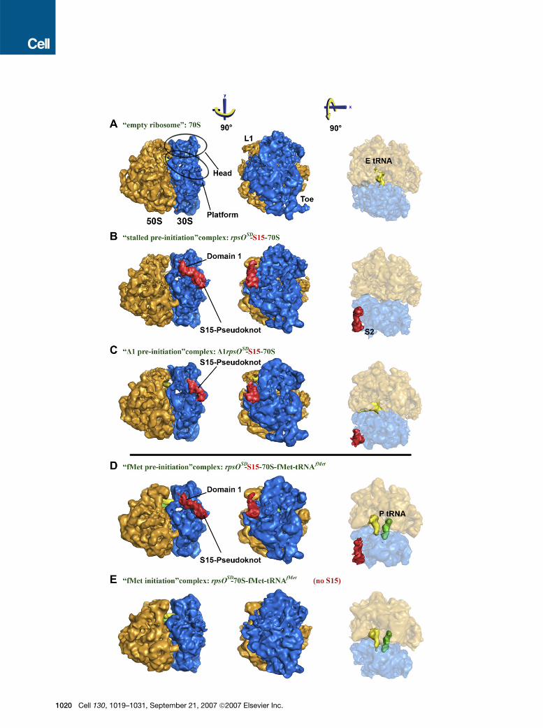
Figure 1. Cryo-EM Structures of Five Different 70S Complexes Illustrating the Entrapment Mechanism and Its Relief
Views from the L1 side (left), from the 30S side (middle) and from the top (right). Localization and domain assignment (domain 1 and pseudoknot) by
comparison of (A) empty 70S ribosome, (B) ‘‘stalled preinitiation’’ rpsOSD-S15-70S complex, and (C) ‘‘D1-preinitiation’’ D1rpsOSD-S15-70S (mRNA
mutant lacking the 50 hairpin loop domain 1). Entrapment relief by comparison of (B) ‘‘stalled preinitiation’’ complex and (D) ‘‘fMet-preinitiation’’
rpsOSD-S15-70S-fMet-tRNAfMet complex, with (E) ‘‘fMet-initiation’’ rpsOSD-70S-fMet-tRNAfMet complex. The 30S and 50S subunits are shown in
blue and orange, respectively. The S15-mRNA part is shown in red, the E-site tRNA in yellow, and the P-site initiator fMet-tRNAfMet in green. The
L1-stalk, the toe, the head, the platform and ribosomal protein S2 are indicated.
30, 1019–1031, September 21, 2007 ª2007 Elsevier Inc. 1021
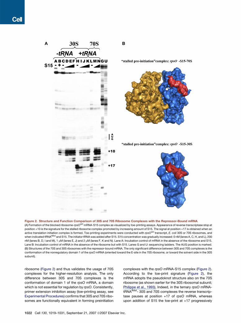
Figure 2. Structure and Function Comparison of 30S and 70S Ribosome Complexes with the Repressor-Bound mRNA
(A) Formation of the blocked ribosome-rpsOSD mRNA-S15 complex as visualized by toe-printing assays. Appearance of reverse transcriptase stop at
position +10 is the signature for the stalled ribosome complex promoted by increasing amount of S15. The signal at position +17 is obtained when an
active translation initiation complex is formed. Toe-printing experiments were conducted with rpsOSD transcript, E. coli 30S or 70S ribosomes, and
when indicated tRNAfMet and S15. The initiator tRNA was added after S15. S15 concentration was gradually increased: 0 nM (lanes A, C, H, and L), 200
nM (lanes B, D, I and M), 1 mM (lanes E, J) and 2 mM (lanes F, K and N). Lane A: Incubation control of mRNA in the absence of the ribosome and S15.
Lane B: Incubation control of mRNA in the absence of the ribosome but with S15. Lanes G and U: sequencing ladders. The AUG position is marked.
(B) Structures of the 70S and 30S ribosomes with the repressor-bound mRNA. The only significant difference between 30S and 70S complexes is the
conformation of the nonregulatory domain 1 of the rpsO mRNA (oriented toward the E-site in the 70S ribosome, or toward the solvent side in the 30S
subunit).
ribosome (Figure 2) and thus validates the usage of 70S
complexes for the higher-resolution analysis. The only
difference between 30S and 70S complexes is the
conformation of domain 1 of the rpsO mRNA, a domain
which is not essential for regulation by rpsO. Consistently,
primer extension inhibition assay (toe-printing assay, see
Experimental Procedures) confirms that 30S and 70S ribo-
somes are functionally equivalent in forming preinitiation
1022 Cell 130, 1019–1031, September 21, 2007 ª2007 Elsevie
complexes with the rpsO mRNA-S15 complex (Figure 2).
According to the toe-print signature (Figure 2), the
mRNA adopts the pseudoknot structure also on the 70S
ribosome (as shown earlier for the 30S ribosomal subunit;
Philippe et al., 1993). Indeed, in the ternary rpsO mRNA-
tRNAfMet- 30S and 70S complexes the reverse transcrip-
tase pauses at position +17 of rpsO mRNA, whereas
upon addition of S15 the toe-print at +17 progressively
r Inc.

Figure 3. Detail View of rpsO-S15 Com-
plex and Its Interactions with the Ribo-
some
The model of the rpsO-S15 complex (Mathy
et al., 2004) fitted to the extracted cryo-EM
density (light pink surface) is shown in the rib-
bon representation. Top, view from the solvent
side; bottom, view from the top; domain 1 in
blue, pseudoknot in red, protein S15 in green,
and SD sequence in magenta. The position of
the AUG start codon is indicated in cyan; the
possible path of the connecting RNA-loops is
in gray. The ribosomal proteins surrounding
the rpsO-S15 complex are indicated and the
anti-SD (aSD) sequence is in orange. The sec-
ondary structure of the rpsO mRNA is shown in
the middle, residues crucial for interaction with
S15 are shown in red (see also Figure S1). The
two inserts describe the stalled preinitiation
complex with the fitted mRNA/S15 model in
the same orientation as the corresponding
panels.
decreases and concomitantly intensity of a pause at posi-
tion +10 increases (Figure 2). The +10 pause coincides
with the 30 terminus of the pseudoknot structure, which
is the signature of the binary 30S-rpsO mRNA complex
that lacks productive rpsO mRNA-tRNAfMet interactions
(Philippe et al., 1993). These data clearly indicate that
S15 prevents the formation of the active initiation complex
by trapping the 30S subunit in a preinitiation complex even
in the context of the 70S ribosome. The fact that the 50S
subunit has no effect on the toe-printing assay is in line
with the observation that also in the 70S complexes, the
rpsO mRNA solely interacts with the 30S subunit.
Deriving Molecular Models of the InitiationComplexes from the Cryo-EM MapsIn order to analyze the molecular interactions between the
rpsO mRNA-S15 complex and the ribosome, the E. coli
and T. thermophilus ribosome crystal structures (Yusupov
et al., 2001; Schuwirth et al., 2005) were fitted into the ex-
perimental map. Since the crystal structure of the pseudo-
knot-S15 complex is not available, a three-dimensional
Cell 1
model of this sub-complex was used for fitting into the ex-
perimental density (Figure 3). The model was derived from
the crystal structures of ribosomal protein S15 bound
either to its 16S rRNA binding site (Nikulin et al., 2001 or
to the 30S (Wimberly et al., 2000) and allowed positioning
S15 with respect to the pseudoknot. It also takes into ac-
count data of the S15 binding site on the pseudoknot
structure obtained from foot-printing experiments and
site-directed mutagenesis performed both on the protein
and on the mRNA (Philippe et al., 1995; Serganov et al.,
2002; Serganov et al., 2003; Mathy et al., 2004). The model
features specific contacts between conserved amino
acids of S15 with the bulged A-45 and the minor groove
of the G-U/G-C motif in the pseudoknot shown to be es-
sential for the S15-mediated repression in vivo (Mathy
et al., 2004 and Figure S1). These contacts provide further
constraints for the orientation of S15 with respect to the
pseudoknot. An unambiguous localization of the protein
part of the pseudoknot-S15 model in the cryo-EM maps
was obtained based on density-contouring levels, reveal-
ing that S15 is oriented toward the solvent side whereas
30, 1019–1031, September 21, 2007 ª2007 Elsevier Inc. 1023

the pseudoknot contacts the ribosome (Figures 3 and S3).
The present structures show that S15 recognizes and sta-
bilizes the pseudoknot fold on the ribosome and that the
ribosome and S15 protect distinct regions of rpsO
mRNA consistent with previous foot-printing data (Phil-
ippe et al., 1993; Serganov et al., 2002, 2003). Further-
more, based on the accessibility of nucleotides toward
chemical probes (Philippe et al., 1993), domain 1 was
modeled as a stem-loop derived from a tRNA anti-codon
stem (see Supplementary Information) and was fitted into
the cryo-EM density taking into account the constraints
imposed by the connectivity to the pseudoknot domain.
The Preinitiation mRNA Binding Siteon the RibosomeThe binding site of the rpsO-S15 complex, which we call
here the platform-binding center, is located next to the
mRNA exit site, between the head and the platform of
the 30S subunit (Figure 1A). It extends from protein S11,
located close to the tRNA exit (E)-site, to protein S2 posi-
tioned on the solvent side of the 30S subunit (Figure 3).
This site also comprises the tips of helices h26 and h40,
and the anti Shine and Dalgarno (anti-SD) area at the 30-
end of the 16S rRNA. Ribosomal proteins S7 and S18 con-
tribute to the edge of the mRNA ‘‘nest.’’ Domain 1 inter-
acts with ribosomal proteins S11 and S21 (in both 30S
and 70S complexes). The rpsO pseudoknot is close to
the tip of helix h40 and interacts with the N-terminal do-
main of protein S2 (Figure 3), which moves away from helix
h26 (compared to the 70S E. coli crystal structure (Schu-
wirth et al., 2005)). Repressor protein S15 interacts with ri-
bosomal protein S2 on one side, and with the tip of helix
h26 on the other side. In addition, helix h26 contacts the
junction between domain 1 and the pseudoknot. The
density for the SD helix positioned next to this junction
(Figure 3) strongly suggests the existence of base pairing
between the SD sequence of rpsO mRNA and the 30-end
of 16S rRNA, in agreement with previous foot-printing
experiments (Philippe et al., 1993). Interestingly, the SD
sequence is accessible for docking to the ribosome since
it is located in the large connecting loop L2 of the pseudo-
knot structure (Figure 3). Consistently, the stability of the
entrapped complex increases with the length of the com-
plementary region between the SD and the anti-SD se-
quences (Table S1). The SD/anti-SD helix is slightly shifted
outwards from its classical position (Yusupova et al., 2001
and Figure 3). Since in the ‘‘fMet-initiation’’ complex, i.e. in
the absence of S15, this shift is not observable, we believe
that in the preinitiation complex the position of the SD/
anti-SD helix is determined by the absence of interactions
with the P-site tRNA that normally help to phase the mRNA
(Yusupova et al., 2006), and by the tense constraints that
impose the SD and anti-SD interactions.
Comparative Structure and Sequence Analysisof the Platform-Binding CenterThe binding site discovery of the mRNA/repressor com-
plex on the ribosome prompted us to examine other ribo-
1024 Cell 130, 1019–1031, September 21, 2007 ª2007 Elsevier
somal mRNA complexes. Strikingly, the binding site for
rpsO partially overlaps with that of the prokaryotic thrS
50-UTR (Jenner et al., 2005) and with that of several
IRES elements located in the 50 leader region of mRNAs
from hepatitis C virus (HCV) and cricket paralysis virus
(CrPV) that bind to eukaryotic ribosomes (Spahn et al.,
2001a, 2004; Boehringer et al., 2005). This suggests the
existence of a common binding site for initiator mRNAs.
We therefore decided to further characterize the plat-
form-binding center by performing a systematic structure
and sequence analysis of all currently known ribosomal
complexes that carry folded mRNAs. Both in prokaryotes
and eukaryotes, these leader mRNAs carry a folded do-
main used for translation initiation regulation. In the HCV
and CrPV IRESes, as well as in the rpsO and thrS mRNAs,
structural elements of the 50 mRNA regions are located on
the small ribosomal subunit platform in vicinity to ribo-
somal proteins S7 and S11 (respectively S5 and S14 in
eukaryotic ribosomes), and to helix h26, which is only
a few residues longer in yeast and human rRNAs than in
E. coli rRNA. In rpsO - ribosome and other ribosome -
mRNA complexes (SD-polyU and SD-polyA) (Yusupova
et al., 2006), the mRNAs also contact protein S2, which
may contribute to the stabilization of the mRNA structure.
When analyzing other ribosome cryo-EM maps (http://
www.ebi.ac.uk/msd/index.html), we detected another 50
mRNA extension located close to protein S2 as seen in
the structure of a translation initiation complex in E. coli
(entry EMD-1248, Allen et al., 2005). Interestingly, the se-
quence encompassing 25 nucleotides upstream of the SD
sequence is predicted to fold into a hairpin structure,
which would nicely fit into the cryo-EM density (Allen
et al., 2005) extending from the SD/anti-SD helix to the
C-terminal domain of protein S2.
Among the ribosomal proteins contributing to the plat-
form-binding center, S2, S7 and S11 proteins are found
across all species, whereas proteins S18 and S21 have
no homologs in eukaryotes (Lecompte et al., 2002) even
though they appear to be replaced by other proteins
(see below). Comparative sequence analysis of S2, S7
and S11 proteins (see Experimental Procedures) reveals
patches of highly conserved residues located on the pro-
tein surface in direct vicinity of the folded mRNAs (Figures
4 and S2). These conserved residues are not critical for the
protein structure and do not interact with other ribosomal
components. These regions, present in all organisms and
notably very rich in positively charged residues, comprise
residues in the N-terminal part of S2, in the C-terminal part
of S7 and in S11; the conserved patches of S7 and S11 are
adjacent to each other in space. In the case of the rspO-
S15 complex, a conformational change, positioning the
N-terminus of S2 away from helix h26 (compared to the
E. coli 70S ribosome crystal structure), provides additional
conserved amino acids for mRNA binding. Notably, ac-
cording to the yeast and human ribosome cryo-EM
maps (Spahn et al., 2001a, 2001b, 2004; Boehringer
et al., 2005), nonassigned protein densities are observed
at positions analogous to those corresponding to the
Inc.

Figure 4. The Platform-Binding Center
for Folded 50-UTR mRNAs
The platform-binding center for structured
mRNAs is shown on the small ribosomal sub-
unit (Schuwirth et al., 2005) (viewing angle as
in Figure 3 top panel). Ribosomal proteins
and RNA helices are labeled and color-coded
as in Figure 3. Conserved surface residues of
S2, S7, and S11, adjacent to the position of
the folded mRNAs, are highlighted by cyan
van der Waals spheres. In the case of the
rspO-S15 complex, a conformational change
positions S2 away from helix h26 (compared
to the crystal structure) providing additional
conserved residues (highlighted in red) for
mRNA binding.
prokaryotic S18 and S21, thus possibly providing struc-
tural similarity of the prokaryotic and eukaryotic sites. In
turn, the replacement of S18 and S21 by other proteins
would appear to be the basis for IRES-specificity toward
eukaryotes, rather than the sole absence of S18 and S21
(Spahn et al., 2001b). Taken together, the sequence and
structural conservation of the RNA and protein compo-
nents of the platform-binding center together with the sim-
ilar localization of the folded 50-UTR of mRNAs bound to
the ribosome suggests that this mRNA binding site is func-
tionally conserved and thus used across species for trans-
lation regulation by folded mRNAs.
The Entrapment MechanismThe precise positioning of the operator-repressor com-
plex on the ribosome elucidates the molecular mechanism
by which the mRNA-S15 complex represses translation
(Figure 5). Within active initiation complexes, i.e. in the ab-
sence of S15, the mRNA adopts a single-stranded confor-
Cell 1
mation in the mRNA channel thus providing the initiation
codon to the P-site tRNA inside the ribosome (Yusupova
et al., 2001 and Figure 5B). In contrast, in the stalled pre-
initiation complex, the initiator AUG codon located next to
the SD sequence in the large connecting loop 2 (Figure 3)
is in close vicinity to the pseudoknot structure on the ribo-
somal platform. As a consequence, the 30 end of the
mRNA rests on the surface of the ribosome rather than in-
side the mRNA channel and prevents the initiator tRNA
from reaching the start codon by keeping it almost 90 A
away from the P-site. Therefore, S15 exerts its inhibitory
function by preventing the pseudoknot to unfold and enter
the mRNA channel, thus blocking the transition of the pre-
initiation complex to the productive initiation complex.
mRNA Adaptation into the Channel: Transitionfrom Preinitiation to InitiationThe synthesis of ribosomal components responds to
the growth-rate and requires the co-ordination of the
30, 1019–1031, September 21, 2007 ª2007 Elsevier Inc. 1025

Figure 5. Schematic Representation of the Entrapment Mechanism and Its Relief
Comparison of the folded rpsO mRNA position (A) with the normal mRNA path (B) obtained after S15 release and fMet-tRNAfMet binding. The 50S
(orange) and the 30S (light blue) subunits are represented as a projection shape seen from the top of the ribosome. In the active 70S initiation complex
(B), the initiation codon is located in the P-site and interacts with the fMet-tRNAfMet (Yusupova et al., 2001). In the inactive 70S complex (A), repressor
protein S15 stabilizes the rpsO mRNA in a folded conformation and prevents the start codon from entering the mRNA channel. The SD sequence of
the mRNA (in magenta) interacts with the anti-SD sequence at the 30 end of the 16S rRNA (in orange) forming a short helix, which is shifted outwards in
the 70S-rpsO-S15 complex. SD/anti-SD interactions are present in the docked and unfolded states.
synthesis of the individual ribosomal rRNAs and proteins.
The ‘stand-by’ S15-mRNA complex can be alleviated by
an increased level of 16S rRNA (Mathy et al., 2004). This
occurs since S15 can recognize either rpsO mRNA or
16S rRNA. In order to address the mRNA adaptation
mechanism, we have determined the cryo-EM structures
of rpsO-70S complexes in the absence of the regulatory
S15 protein, but in the presence of the initiator fMet-
tRNAfMet (Figures 1E and 5B). Under these conditions,
the mRNA pseudoknot is not anymore stabilized by the re-
pressor protein S15. Rather, the mRNA unfolds and enters
its channel around the small ribosomal subunit neck (Yu-
supova et al., 2001). Consistently, this conformational
re-arrangement can be assessed by the disappearance
of the entire density for the rpsO pseudoknot on the plat-
form, as well as the maintenance of the SD/anti-SD helix
and S2 densities. In addition, a concomitant closure of
the mRNA channel ‘‘entry’’ is formed by the ribosomal pro-
teins S3, S4 and S5, characteristic for the presence of an
mRNA in the channel (Movie S1). These data indicate that
the mRNA adopts a single-stranded conformation in the
mRNA channel extending from the SD/anti-SD helix on
the platform to the mRNA ‘‘entry.’’ The new mRNA confor-
mation is in good agreement with the toe-printing data
(Figure 2), which show the typical signature (toe-print at
position +17) for the formation of the active 70S initiation
complex. The presence of the initiator tRNA is necessary
(Philippe et al., 1993) but per se not sufficient for induction
of mRNA unfolding. This is illustrated by the fact that in the
presence of the repressor protein S15, the density corre-
sponding to the S15-rpsO complex is still present in the
platform of the ribosome even after the addition of fMet-
tRNAfMet to the entrapped complex (Figure 1D and Movie
1026 Cell 130, 1019–1031, September 21, 2007 ª2007 Elsevie
S1). Thus, S15 prevents the transition of the preinitiation
complex toward the initiation complex and exerts its inhib-
itory action independently of fMet-tRNAfMet binding. The
docking and adaptation process of rpsO appears similar
to the association of structured mRNAs to 30S subunit
that has been shown by kinetic studies to present two dis-
tinct phases: (1) the first binding and (2) the mRNA unfold-
ing (Studer and Joseph, 2006). In the absence of the re-
pressor S15, the delay time between these phases will
be decreased, but the overall docking and adaptation
mechanism is likely to remain similar. Importantly, in our
system, the SD/anti-SD interactions are seen in both re-
pressed and activated initiation complexes, suggesting
that SD interactions are maintained as a common anchor
point during the docking and adaptation process (Fig-
ure 5). After S15 release and initiator tRNA binding, the for-
mation of the initiation complex becomes irreversible.
DISCUSSION
The present cryo-EM structures of different functional
complexes within the translation initiation pathway ad-
dress the key role of folded 50 mRNA structures in regulat-
ing protein synthesis by stalling the ribosomal machinery
at the preinitiation step. The study represents the first ex-
ample of a ribosome-bound mRNA whose folded struc-
ture is stabilized by a repressor protein thus preventing
the message to enter the ribosome channel. Previous bio-
chemical studies provided only indirect information on the
ribosome entrapment mechanism, whereas the present
study directly illustrates the simultaneous binding of the
repressor protein and the ribosome on the mRNA, thus
validating the concept of entrapment. Using a long,
r Inc.

natural mRNA, our results give key insights into the trans-
lation regulation by a 50-folded mRNA, revealing the mech-
anism of transient ribosome entrapment during the initia-
tion phase. In particular, the individual 3D structures
visualize key intermediates of the mRNA docking and ad-
aptation process. The recognition of structured mRNAs
can be divided into three phases: the docking of the folded
mRNA on the ribosome, the unfolding of mRNA structures
and the final mRNA adaptation into the mRNA channel.
The delay time between docking and unfolding reflects
the stability of the mRNA riboswitch structures thus allow-
ing translation modulation. Ligands that strongly stabilize
the folded state of the mRNA (such as protein S15) block
the ribosome at the preinitiation stage by preventing the
initiator codon from reaching the decoding site inside
the ribosome (Figure 5A). This hampers codon-anticodon
interactions with the initiator tRNA in the P-site. Upon
repressor release and initiator tRNA binding, the mRNA
unfolds and adopts the classical path in the mRNA chan-
nel thus leading to the formation of an active translation
initiation complex (Figure 5B). Although it cannot be prin-
cipally ruled out that the mRNA binds, dissociates, unfolds
and rebinds, there are some indirect evidences in favor of
unfolding occurring on or in close vicinity to the 30S plat-
form. The SD helix, which is maintained in blocked and ac-
tive states, may favor the transition to the active complex
rather than a dissociation / re-association mechanism
which is unlikely also for entropic reasons. Furthermore,
it was previously shown that the pseudoknot structure is
the structure recognized by the ribosome (even in ab-
sence of S15; Figure 2 and Philippe et al., 1993). In solu-
tion, co-existing structures have been detected that corre-
spond to either the pseudoknot structure or two hairpin
motifs. This latter structure cannot bind as such to the
30S subunit because the SD-sequence is sequestered,
and conversely, the 30S subunit alone stabilizes the
pseudoknot structure unless initiator tRNA is present
(Philippe et al., 1994).
Obtaining the structure of the rpsO/S15 ribosome com-
plex was the key for a comprehensive comparison of
all known ribosome complexes with folded mRNAs.
These isolated experimental data (currently 7 structures) -
combined with a large-scale sequence analysis of the
site - have been integrated into a new concept proposing
a common binding site dedicated for transiently binding
regulatory mRNAs during the translation initiation pro-
cess. As is evident from ribosome-mRNA complexes
in both prokaryotic and eukaryotic systems, the
platform-binding center is not specific for particular
mRNA secondary structures. Indeed, it can accommo-
date different types of RNA structures (hairpin loop or
pseudoknots) as seen for rpsO, and thrS (Jenner et al.,
2005), poly-A and poly-U mRNA or other 50 UTR se-
quences (Allen et al., 2005; Yusupova et al., 2006), and
the IRES (HCV and CrPV; Spahn et al., 2001a, 2004; Boeh-
ringer et al., 2005) mRNA’s on the ribosome. Since the ma-
jority of 50 extensions are structured to some degree, this
binding mode might be a common feature of translation
Cell 1
regulation. The fact that even poly-A and poly-U se-
quences form a folded structure on the ribosome strongly
suggests that the platform-binding site is not sequence-
specific but rather a general binding site for anchoring
mRNA regions at the initiation step. Consistently, the plat-
form site is not limited to SD interactions (IRESes also bind
although they do not contain any SD sequence) and many
of the conserved amino acids are positively charged and
may contribute to mRNA binding before the bacteria-spe-
cific SD interactions are established (Studer and Joseph,
2006; Darfeuille et al., 2007). Future experiments using
for instance genetics analysis may address the contribu-
tion of individual (conserved) residues of the ribosomal
components of the platform-binding center in docking
folded mRNAs or in promoting their unfolding. Melting of
mRNA structures may be promoted by the ribosome itself
or by some accessory protein factors. Ribosomal protein
S1 is a particularly interesting candidate since it was
shown to possess nucleic acid helix-unwinding properties
(Kolb et al., 1977) and to promote initial binding of many
structured mRNAs in E. coli (e.g., Boni et al., 1991; Ring-
quist et al., 1995; Tedin et al., 1997; Sorensen et al.,
1998) although it is weakly bound to the ribosome.
Importantly, our data provide insights into an important
concept in translation initiation: the mRNA exit site (named
like this because of its role during the elongation phase;
Yusupova et al., 2001) in fact serves as a docking and entry
site during the initiation regulation process. Other folded
mRNA structures, for instance present within the trans-
lated 30 mRNA regions, may transiently bind to a distinct,
albeit specific site on the ribosome during elongation, ter-
mination or frame-shift events. This site is probably located
close to the 30 end of the mRNA channel (opposite to the
platform-binding center), and is surrounded by proteins
S3, S4 and S5. The involvement of proteins S3 and S4
in unfolding 30structured mRNAs during the elongation
phase has been shown recently (Takyar et al., 2005).
The regulatory mechanism of S15 is probably shared by
most of the gram-negative bacteria since the pseudoknot
structure and its position relative to the SD and AUG se-
quences in the rpsO genes are conserved in these organ-
isms (Figure S1). Other mRNAs with 50-UTR pseudoknots
may function in a similar way. For example, the entrap-
ment mechanism was also shown for ribosomal protein
S4, which represses translation of the a operon encoding
proteins S13, S11, S4, and L17 (reviewed in Schlax and
Worhunsky, 2003). S4 acts as an allosteric riboswitch
ligand by favoring an inactive conformation of the mRNA
that contains a pseudoknot structure with the SD and
the AUG codon located in the connecting loop (Deckman
and Draper, 1987; Tang and Draper, 1989). The similarity
of translation regulation by proteins S4 and S15 suggests
that the S4-mRNA complex binds to the same ribosomal
environment and hinders the conformational change of
the mRNA required for formation of the productive initia-
tion complex. Many more mRNAs might be regulated in
a similar way if they have 50 secondary structures close
enough to the RBS. In contrast to the displacement/
30, 1019–1031, September 21, 2007 ª2007 Elsevier Inc. 1027

competition mechanism, the entrapment mechanism has
the advantage that no high-affinity repressor-mRNA com-
plex is required, but rather a transient, unproductive initi-
ation complex is used for efficient repression of translation
(Schlax and Worhunsky, 2003; Mathy et al., 2004). In this
context, it is remarkable that the S15 binding site is ex-
posed on the accessible surface of the folded rpsO
mRNA bound to the ribosome, thus allowing regulatory
on and off binding of S15 to its own message on the ribo-
some. This may be the way riboswitch ligands access/
leave the ribosome-bound mRNA they regulate.
The finding of a specific site on the ribosome dedicated
for docking regulatory mRNA has profound implications
for the regulation of gene expression. Nature has found
an elegant way to temporarily stall ribosomes at the
mRNA binding step, right before protein synthesis starts,
until cellular conditions request the activation of an al-
ready transcribed gene (e.g., stress bodies in eukaryotes),
independently from transcription (and splicing) events.
These transcripts are ready to be rescued and translated
when needed for the cell (Anderson and Kedersha,
2006). Taken together, controlling translation via ribosome
stalling might be more widespread than expected.
EXPERIMENTAL PROCEDURES
Design of mRNAs
In order to get homogeneous particles for cryo-EM analysis, we opti-
mized the formation of the ribosomal entrapped complex. For this pur-
pose, we have prepared wild-type rpsO transcript (�110 to +65, +1
being the A of the rpsO translational initiation codon, WT), and
mRNA derivatives where three mutations were introduced in the SD
sequence to increase the length of the region complementary to the
30 end of 16S rRNA (�110 to +65, rpsOSD) and to delete the stem-
loop structure of domain 1 (�60 to +65, D1rpsOSD) (Supplementary
Fig. S1). The D1rpsOSD transcript was designed in order to precisely
locate the pseudoknot structure and the stem-loop structure of
domain 1 in the cryo-EM maps. The rpsO RNA fragments were syn-
thesized by in vitro transcription with T7 RNA polymerase from
HindIII-linearized plasmids pBSM13 (rpsOSD) and pTZ18R (D1rpsOSD)
containing rpsO gene (Serganov et al., 2002).
Preparation of Ribosome Complexes
Purification of S15 from E. coli and preparation of fMet-tRNAfMet was
performed as described previously (Serganov et al., 2001 and Rodnina
et al., 1994, respectively). Before use, S15 was reactivated for 30 min
at 37�C in a buffer containing 50 mM Tris-HCl (pH 7.5), 270 mM KCl,
3 mM DTT, 20 mM MgCl2 and BSA 0.002%. The two rpsO mRNA frag-
ments (�110 to +65 (rpsOSD), and �60 to +65 (D1rpsOSD)) were puri-
fied by gel electrophoresis under denaturing conditions followed by
a Mono Q anion exchange column. Before use, mRNAs were renatured
as follows: incubation at 90�C for 1 min in RNase-free water, at 4�C for
1 min, and at 25�C for 30 min in the TKM buffer (20 mM Tris-HCl pH 7.5,
60 mM KCl, 1 mM DTT, 10 mM MgCl2). The 70S ribosomes were
isolated from Escherichia coli MRE600 strain as previously described
(Yusupov et al., 2001). Prior the formation of the entrapped complex,
ribosomes were first incubated at 37�C for 15 min in a buffer containing
20 mM Tris-HCl (pH 7.5), 150 mM KCl, 20 mM MgCl2 and 1 mM DTT.
MgCl2 concentration was then decreased to 2 mM by dilution and the
samples were incubated for additional 15 min at 37�C to promote the
‘‘breathing’’ of the 70S ribosomes in order to make them more recep-
tive for ligands. The ribosomal complexes were formed at 37�C for
15 min in TKM buffer containing 7.5 mM MgCl2 in the presence of
1028 Cell 130, 1019–1031, September 21, 2007 ª2007 Elsevie
the E. coli 70S ribosomes (0.4 mM), rpsO mRNA (0.6 mM), and when re-
quired S15 (3 mM). When present, the fMet-tRNAfMet (3 mM) has been
added to the assembled mRNA-ribosome complexes with a further
incubation time of 5 min at 37�C. MgCl2 concentration was then
increased to 15.6 mM and the sample was incubated 30 min at
37�C. Formation of the complexes was followed by sucrose gradient
(see Supplementary Information) and by toe-printing experiments.
Toe-Printing Experiments
Toe-printing approach devised by Gold and coworkers (Hartz et al.,
1988) allows to monitor the binding of the 30S subunit on the mRNA
in the presence of the initiator tRNA. This approach is based on the in-
hibition of reverse transcription from a labeled primer annealed to the
mRNA, by the formation of the ribosomal initiation complex. The toe-
print represents the reverse transcriptase pause, which corresponds
to the 30 edge of the 30S-binding site on the mRNA usually occurring
at position +16 (+1 being the adenine of the AUG codon). Experimental
conditions were derived from (Philippe et al., 1993). In a standard ex-
periment, rpsO mRNA (WT or rpsOSD, 24 nM), the labeled primer (com-
plementary to nucleotides +38 to +50), E. coli 30S subunits or 70S
ribosomes (200 nM), all four dNTPs (each at 50 mM), and S15 (from
200 nM to 2 mM) were added to 10 ml of TKM buffer incubated for
10 min at 20�C and 5 min at 37�C. tRNAfMet was added at a final
concentration of 2 mM, and the assays were incubated for 5 min at
37�C. In order to promote efficient rpsO mRNA binding to 70S ribo-
somes, the ribosomes were preincubated in TKM buffer containing
low MgCl2 concentration (2 mM) to facilitate ligand binding. MgCl2concentration was then increased (10 mM) after the addition of S15
to favor the stabilization of the 70S ribosome. Primer extension was
conducted with 1 unit of Moloney Murine Leukemia Virus (MMLV) re-
verse transcriptase for 15 min at 20�C. The reactions were stopped
by phenol/chloroform RNA extraction prior to ethanol precipitation of
the mRNA. The samples were loaded on 8% polyacrylamide-8 M
urea slab gel in 10 ml of loading buffer after heating to 90�C for 2 min.
Cryo-EM Structure Determination
3.5 ml of the ribosomal complexes were applied to cryo-EM holey car-
bon grids as described in (Klaholz et al., 2003, 2004). As a control, we
used the vacant 70S ribosomes. Low-dose images were taken on
Kodak SO-163 films with the in-house FEI Tecnai20 field emission
gun transmission electron microscope using a magnification of
50,000 and a defocus range of 0.8-3.5 mm. The best micrographs
were digitized on a drum scanner (Heidelberger Druckmaschinen)
with a step size of 5 mm. The digitized images were further selected ac-
cording to their calculated power spectra and were coarsened by a fac-
tor of 3, resulting in a pixel size corresponding to 3.0 A at the specimen
level. Single-molecule images (10,212 for the empty 70S ribosome;
34,477 for the ‘‘D1-preinitiation complex’’ S15-D1rpsOSD mRNA-ribo-
some reference complex; 102,928 for the ‘‘stalled preinitiation com-
plex’’ S15-rpsOSD mRNA-ribosome entrapped complex; 20,324 for
the ‘‘fMet-preinitiation complex’’ S15-rpsOSD mRNA-fMet-tRNAfMet-
ribosome entrapped complex; 18,450 for ‘‘fMet-initiation complex’’
the rpsOSD mRNA-fMet-tRNAfMet-ribosome relieved complex and
5,630 for the S15-rpsOSD mRNA-30S entrapped complex) were
selected semi-automatically using the BOXER subroutine of EMAN
(Ludtke et al., 1999). Correction of the phase-contrast-transfer func-
tion was performed by phase flipping on the raw data using the
IMAGIC-5 software system, which was used for all further image pro-
cessing and structure refinement as described previously (van Heel
et al., 2000); some 3D reconstructions were done with the software im-
plemented by Orlov (Orlov et al., 2006). The resolution of the final three-
dimensional structures was estimated with the Fourier shell correlation
according to the 0.14 (Rosenthal and Henderson, 2003) and one-half-
bit (van Heel and Schatz, 2005) criteria rather than to the 3s and 0.5
criteria since 0.14 and one-half-bit criteria appear to be more consis-
tent with the correlation obtained from crystal structure fits (Myasnikov
et al., 2005). The respective estimated resolutions were as follows:
r Inc.

10 A for the S15-rpsOSD mRNA-ribosome complex; 13.5 A for the S15-
D1rpsOSD mRNA-ribosome complex; 14.0 A for the S15-rpsOSD
mRNA-fMet-tRNAfMet-ribosome complex; 14.0 A for the rpsOSD
mRNA-fMet-tRNAfMet-ribosome complex; 12.0 A for the empty 70S
ribosome reference complex; 22 A for the S15-rpsOSD mRNA-30S
complex. Details on the cryo-EM structure interpretation can be found
in the Supplementary Information.
Comparative Structure and Sequence Analysis
of the Platform-Binding Center
Individual sequence alignments of ribosomal proteins S2, S7, S11 (S18
and S21 are not present in eukaryotes (Lecompte et al., 2002)) was
done with PipeAlign (http://bips.u-strasbg.fr/PipeAlign/ Plewniak
et al., 2003) including 200 sequences for each protein. Figure S2
shows excerpts of these alignments. Using the E. coli 70S crystal
structure (Schuwirth et al., 2005), the side-chain interactions of resi-
dues highly conserved across kingdoms were individually checked
and grouped into three categories according to residues involved in ei-
ther (i) contacts with ribosomal RNA, (ii) stabilization of the protein ar-
chitecture itself, or (iii) neither, but located on the ribosome surface and
thus exposed on the solvent side for potential interactions with nonri-
bosomal components. The latter are labeled in Figure 4. None of them
form the E-site or the insides of the mRNA channel. Structures of other
known ribosomal mRNA complexes were downloaded from the PDB
http://www.rcsb.org/pdb/ and the EBI Macromolecular Structure
Database http://www.ebi.ac.uk/msd/index.html (see also Supplemen-
tary Information) and 16S and 18S RNA sequences were taken from
http://www.rna.ccbb.utexas.edu/ (Cannone et al., 2002).
Figures were prepared using Pymol and Nuccyl (Jovine, L., nuccyl
(2003) www.biosci.ki.se/groups/ljo/software/nuccyl.html), the movie
was produced using the UCSF Chimera package (Pettersen et al.,
2004).
Supplemental Data
Supplemental Data include supplemental text, Supplemental Refer-
ences, three figures, one table, and one movie and can be found
with this article online at http://www.cell.com/cgi/content/full/130/6/
1019/DC1/.
ACKNOWLEDGMENTS
We are grateful to Patrick Schultz, Bernard Ehresmann, Dino Moras,
Jean-Claude Thierry, Veronique Mallouh, Angelita Simonetti and Igor
Orlov for their constant support and interest, to Eric Westhof for
discussions and the help during the modeling of the structure of the
domain 1, to Anne-Catherine Helfer for the toe-printing assays, and
to Michael Schatz and Ralf Schmidt for support with the IMAGIC-5
software. We thank Christian Spahn for the map of the 40S/HCV
IRES complex. This work was supported by grants from the Centre
National pour la Recherche Scientifique (CNRS), the Ministere de la
Recherche et de la Technologie (ACI BCMS), the European Molecular
Biology Organization (EMBO) Young Investigator Programme (YIP),
the Institut du Developpement et des Ressources en Informatique
Scientifique (IDRIS, France), and the European Commission as
SPINE2-complexes (contract n� LSHG-CT-2006-031220) and the
BacRNA (contract LSHG-CT-2005-018618). S.M. was supported by
post-doctoral fellowships from the University Louis Pasteur, CNRS
and from the Fondation de la Recherche Medicale (FRM), and A.G.M.
was a recipient of postdoctoral fellowships from CNRS and FRM. The
electron microscope facility is supported by the Alsace Region, the
Institut National de la Sante et de la Recherche Medicale (INSERM),
the CNRS and the Association pour la Recherche sur le Cancer (ARC).
Received: January 26, 2007
Revised: May 18, 2007
Accepted: July 6, 2007
Published: September 20, 2007
Cell
REFERENCES
Agalarov, S.C., Sridhar Prasad, G., Funke, P.M., Stout, C.D., and
Williamson, J.R. (2000). Structure of the S15,S6,S18-rRNA complex:
assembly of the 30S ribosome central domain. Science 288, 107–113.
Allen, G.S., Zavialov, A., Gursky, R., Ehrenberg, M., and Frank, J.
(2005). The cryo-EM structure of a translation initiation complex from
Escherichia coli. Cell 121, 703–712.
Anderson, P., and Kedersha, N. (2006). RNA granules. J. Cell Biol. 172,
803–808.
Batey, R.T., Gilbert, S.D., and Montange, R.K. (2004). Structure of
a natural guanine-responsive riboswitch complexed with the metabo-
lite hypoxanthine. Nature 18, 411–415.
Boehringer, D., Thermann, R., Ostareck-Lederer, A., Lewis, J.D., and
Stark, H. (2005). Structure of the hepatitis C Virus IRES bound to the
human 80S ribosome: remodeling of the HCV IRES. Structure 13,
1695–1706.
Boni, I.V., Isaeva, D.M., Musychenko, M.L., and Tzareva, N.V. (1991).
Ribosome-messenger recognition: mRNA target sites for ribosomal
protein S1. Nucleic Acids Res. 19, 155–162.
Cannone, J.J., Subramanian, S., Schnare, M.N., Collett, J.R., D’Souza,
L.M., Du, Y., Feng, B., Lin, N., Madabusi, L.V., Muller, K.M., et al.
(2002). The Comparative RNA Web (CRW) Site: An Online Database
of Comparative Sequence and Structure Information for Ribosomal,
Intron, and other RNAs. BMC Bioinformatics 3. Published online Jan-
uary 17, 2002. 10.1186/1471-2105-3-2.
Darfeuille, F., Unoson, C., Vogel, J., and Wagner, E.G.H. (2007). An an-
tisense RNA inhibits translation by competing with standby ribosomes.
Mol. Cell 26, 381–392.
de Smit, M.H., and van Duin, J. (2003). Translational standby sites: how
ribosomes may deal with the rapid folding kinetics of mRNA. J. Mol.
Biol. 331, 737–743.
Deckman, I.C., and Draper, D.E. (1987). S4-alpha mRNA translation
regulation complex. II. Secondary structures of the RNA regulatory
site in the presence and absence of S4. J. Mol. Biol. 196, 323–332.
Ehresmann, C., Ehresmann, B., Ennifar, E., Dumas, P., Garber, M.,
Mathy, N., Nikulin, A., Portier, C., Patel, D., and Serganov, A. (2004).
Molecular mimicry in translational regulation: the case of ribosomal
protein S15. RNA Biol. 1, 66–73.
Fuchs, R.T., Grundy, F.J., and Henkin, T.M. (2006). The S(MK) box is
a new SAM-binding RNA for translational regulation of SAM synthe-
tase. Nat. Struct. Mol. Biol. 13, 226–233.
Gebauer, F., and Hentze, M.W. (2004). Molecular mechanisms of
translational control. Nat. Rev. Mol. Cell Biol. 5, 827–835.
Gottesman, S. (2005). Micros for microbes: non-coding regulatory
RNAs in bacteria. Trends Genet. 21, 399–404.
Gualerzi, C.O., and Pon, C.L. (1990). Initiation of mRNA translation in
prokaryotes. Biochemistry 29, 5881–5889.
Hartz, D., McPheeters, D.S., Traut, R., and Gold, L. (1988). Extension
inhibition analysis of translation initiation complexes. Methods Enzy-
mol. 164, 419–425.
Jenner, L., Romby, P., Rees, B., Schulze-Briese, C., Springer, M.,
Ehresmann, C., Ehresmann, B., Moras, D., Yusupova, G., and Yusu-
pov, M. (2005). Translational operator of mRNA on the ribosome:
how repressor proteins exclude ribosome binding. Science 308,
120–123.
Johansson, J., Mandin, P., Renzoni, A., Chiaruttini, C., Springer, M.,
and Cossart, P. (2002). An RNA thermosensor controls expression of
virulence genes in Listeria monocytogenes. Cell 110, 551–561.
Klaholz, B.P., Pape, T., Zavialov, A.V., Myasnikov, A.G., Orlova, E.V.,
Vestergaard, B., Ehrenberg, M., and van Heel, M. (2003). Structure
of the Escherichia coli ribosomal termination complex with release fac-
tor 2. Nature 421, 90–94.
130, 1019–1031, September 21, 2007 ª2007 Elsevier Inc. 1029

Klaholz, B.P., Myasnikov, A.G., and Van Heel, M. (2004). Visualization
of release factor 3 on the ribosome during termination of protein syn-
thesis. Nature 427, 862–865.
Kolb, A., Hermoso, J.M., Thomas, J.O., and Szer, W. (1977). Nucleic
acid helix-unwinding properties of ribosomal protein S1 and the role
of S1 in mRNA binding to ribosomes. Proc. Natl. Acad. Sci. USA 74,
2379–2383.
Lecompte, O., Ripp, R., Thierry, J.C., Moras, D., and Poch, O. (2002).
Comparative analysis of ribosomal proteins in complete genomes: an
example of reductive evolution at the domain scale. Nucleic Acids Res.
30, 5382–5390.
Ludtke, S.J., Baldwin, P.R., and Chiu, W. (1999). EMAN: semiauto-
mated software for high-resolution single-particle reconstructions.
J. Struct. Biol. 128, 82–97.
Mathy, N., Pellegrini, O., Serganov, A., Patel, D.J., Ehresmann, C., and
Portier, C. (2004). Specific recognition of rpsO mRNA and 16S rRNA by
Escherichia coli ribosomal protein S15 relies on both mimicry and site
differentiation. Mol. Microbiol. 52, 661–675.
Merianos, H.J., Wang, J., and Moore, P.B. (2004). The structure of a
ribosomal protein S8/spc operon mRNA complex. RNA 10, 954–964.
Myasnikov, A.G., Marzi, S., Simonetti, A., Giuliodori, A.M., Gualerzi,
C.O., Yusupova, G., Yusupov, M., and Klaholz, B.P. (2005). Conforma-
tional transition of initiation factor 2 from the GTP- to GDP-bound state
visualized on the ribosome. Nat. Struct. Mol. Biol. 12, 1145–1149.
Nikulin, A., Serganov, A., Ennifar, E., Tishchenko, S., Nevskaya, N.,
Shepard, W., Portier, C., Garber, M., Ehresmann, B., Ehresmann, C.,
et al. (2001). Crystal structure of the S15-rRNA complex. Nat. Struct.
Mol. Biol. 7, 273–277.
Nomura, M., Gourse, R., and Baughman, G. (1984). Regulation of the
synthesis of ribosomes and ribosomal components. Annu. Rev. Bio-
chem. 53, 75–117.
Orlov, I.M., Morgan, D.G., and Cheng, R.H. (2006). Efficient implemen-
tation of a filtered back-projection algorithm using a voxel-by-voxel
approach. J. Struct. Biol. 154, 287–296.
Pettersen, E.F., Goddard, T.D., Huang, C.C., Couch, G.S., Greenblatt,
D.M., Meng, E.C., and Ferrin, T.E. (2004). UCSF Chimera–a visualiza-
tion system for exploratory research and analysis. J. Comput. Chem.
25, 1605–1612.
Philippe, C., Eyermann, F., Benard, L., Portier, C., Ehresmann, B., and
Ehresmann, C. (1993). Ribosomal protein S15 from Escherichia coli
modulates its own translation by trapping the ribosome on the
mRNA initiation loading site. Proc. Natl. Acad. Sci. USA 90, 4394–
4398.
Philippe, C., Benard, L., Eyermann, F., Cachia, C., Kirillov, S.V., Portier,
C., Ehresmann, B., and Ehresmann, C. (1994). Structural elements of
rps0 mRNA involved in the modulation of translational initiation and
regulation of E. coli ribosomal protein S15. Nucleic Acids Res. 22,
2538–2546.
Philippe, C., Benard, L., Portier, C., Westhof, E., Ehresmann, B., and
Ehresmann, C. (1995). Molecular dissection of the pseudoknot govern-
ing the translational regulation of Escherichia coli ribosomal protein
S15. Nucleic Acids Res. 23, 18–28.
Plewniak, F., Bianchetti, L., Brelivet, Y., Carles, A., Chalmel, F.,
Lecompte, O., Mochel, T., Moulinier, L., Muller, A., Muller, J., et al.
(2003). PipeAlign: A new toolkit for protein family analysis. Nucleic
Acids Res. 31, 3829–3832.
Ringquist, S., Jones, T., Snyder, E.E., Gibson, T., Boni, I., and Gold, L.
(1995). High-affinity RNA ligands to Escherichia coli ribosomes and
ribosomal protein S1: comparison of natural and unnatural binding
sites. Biochemistry 34, 3640–3648.
Rodnina, M.V., Semenkov, Y.P., and Wintermeyer, W. (1994). Purifica-
tion of fMet-tRNA(fMet) by fast protein liquid chromatography. Anal.
Biochem. 219, 380–381.
1030 Cell 130, 1019–1031, September 21, 2007 ª2007 Elsevie
Romby, P., and Springer, M. (2003). Bacterial translational control at
atomic resolution. Trends Genet. 19, 155–161.
Rosenthal, P.B., and Henderson, R. (2003). Optimal determination of
particle orientation, absolute hand, and contrast loss in single-particle
electron cryomicroscopy. J. Mol. Biol. 333, 721–745.
Schlax, P.J., and Worhunsky, D.J. (2003). Translational repression
mechanisms in prokaryotes. Mol. Microbiol. 48, 1157–1169.
Schuwirth, B.S., Borovinskaya, M.A., Hau, C.W., Zhang, W., Vila-
Sanjurjo, A., Holton, J.M., and Cate, J.H. (2005). Structures of the
bacterial ribosome at 3.5 A resolution. Science 310, 827–834.
Scott, L.G., and Williamson, J.R. (2005). The binding interface between
Bacillus stearothermophilus ribosomal protein S15 and its 50-transla-
tional operator mRNA. J. Mol. Biol. 351, 280–290.
Serganov, A., Benard, L., Portier, C., Ennifar, E., Garber, M., Ehres-
mann, B., and Ehresmann, C. (2001). Role of conserved nucleotides
in building the 16 S rRNA binding site for ribosomal protein S15.
J. Mol. Biol. 305, 785–803.
Serganov, A., Ennifar, E., Portier, C., Ehresmann, B., and Ehresmann,
C. (2002). Do mRNA and rRNA binding sites of E.coli ribosomal protein
S15 share common structural determinants? J. Mol. Biol. 320, 963–
978.
Serganov, A., Polonskaia, A., Ehresmann, B., Ehresmann, C., and
Patel, D.J. (2003). Ribosomal protein S15 represses its own translation
via adaptation of an rRNA-like fold within its mRNA. EMBO J. 22, 1898–
1908.
Serganov, A., Yuan, Y.R., Pikovskaya, O., Polonskaia, A., Malinina, L.,
Phan, A.T., Hobartner, C., Micura, R., Breaker, R.R., and Patel, D.J.
(2004). Structural basis for discriminative regulation of gene expres-
sion by adenine- and guanine-sensing mRNAs. Chem. Biol. 11,
1729–1741.
Serganov, A., Polonskaia, A., Phan, A.T., Breaker, R.R., and Patel, D.J.
(2006). Structural basis for gene regulation by a thiamine pyrophos-
phate-sensing riboswitch. Nature 29, 1167–1171.
Sorensen, M.A., Fricke, J., and Pedersen, S. (1998). Ribosomal protein
S1 is required for translation of most, if not all, natural mRNAs in Es-
cherichia coli in vivo. J. Mol. Biol. 280, 561–569.
Spahn, C.M., Kieft, J.S., Grassucci, R.A., Penczek, P.A., Zhou, K.,
Doudna, J.A., and Frank, J. (2001a). Hepatitis C virus IRES RNA-
induced changes in the conformation of the 40s ribosomal subunit.
Science 291, 1959–1962.
Spahn, C.M., Beckmann, R., Eswar, N., Penczek, P.A., Sali, A., Blobel,
G., and Frank, J. (2001b). Structure of the 80S ribosome from
Saccharomyces cerevisiae–tRNA-ribosome and subunit-subunit
interactions. Cell 107, 373–386.
Spahn, C.M., Jan, E., Mulder, A., Grassucci, R.A., Sarnow, P., and
Frank, J. (2004). Cryo-EM visualization of a viral internal ribosome entry
site bound to human ribosomes: the IRES functions as an RNA-based
translation factor. Cell 118, 465–475.
Spedding, G., Gluick, T.C., and Draper, D.E. (1993). Ribosome initia-
tion complex formation with the pseudoknotted alpha operon messen-
ger RNA. J. Mol. Biol. 229, 609–622.
Springer, M., and Portier, C. (2003). More than one way to skin a cat:
translational autoregulation by ribosomal protein S15. Nat. Struct.
Biol. 10, 420–422.
Studer, S.M., and Joseph, S. (2006). Unfolding of mRNA secondary
structure by the bacterial translation initiation complex. Mol. Cell 22,
105–115.
Takyar, S., Hickerson, R.P., and Noller, H.F. (2005). mRNA helicase
activity of the ribosome. Cell 120, 49–58.
Tang, C.K., and Draper, D.E. (1989). Unusual mRNA pseudoknot struc-
ture is recognized by a protein translational repressor. Cell 57, 531–
536.
r Inc.

Tedin, K., Resch, A., and Blasi, U. (1997). Requirements for ribosomal
protein S1 for translation initiation of mRNAs with and without a 50
leader sequence. Mol. Microbiol. 25, 189–199.
Thore, S., Leibundgut, M., and Ban, N. (2006). Structure of the
eukaryotic thiamine pyrophosphate riboswitch with its regulatory
ligand. Science 26, 1208–1211.
Tucker, B.J., and Breaker, R.R. (2005). Riboswitches as versatile gene
control elements. Curr. Opin. Struct. Biol. 15, 342–348.
van Heel, M., Gowen, B., Matadeen, R., Orlova, E.V., Finn, R., Pape, T.,
Cohen, D., Stark, H., Schmidt, R., Schatz, M., et al. (2000). Single-
particle electron cryo-microscopy: towards atomic resolution.
Q. Rev. Biophys. 33, 307–369.
van Heel, M., and Schatz, M. (2005). Fourier shell correlation threshold
criteria. J. Struct. Biol. 151, 250–262.
Wimberly, B.T., Brodersen, D.E., Clemons, W.M., Jr., Morgan-Warren,
R.J., Carter, A.P., Vonrhein, C., Hartsch, T., and Ramakrishnan, V.
(2000). Structure of the 30S ribosomal subunit. Nature 407, 327–339.
Cell
Yusupov, M.M., Yusupova, G.Z., Baucom, A., Lieberman, K., Earnest,
T.N., Cate, J.H., and Noller, H.F. (2001). Crystal structure of the ribo-
some at 5.5 A resolution. Science 292, 883–896.
Yusupova, G.Z., Yusupov, M.M., Cate, J.H., and Noller, H.F. (2001).
The path of messenger RNA through the ribosome. Cell 106, 233–241.
Yusupova, G., Jenner, L., Rees, B., Moras, D., and Yusupov, M. (2006).
Structural basis for messenger RNA movement on the ribosome.
Nature 444, 391–394, Epub 2006 Oct 18.
Zengel, J.M., and Lindahl, L. (1994). Diverse mechanisms for regulat-
ing ribosomal protein synthesis in Escherichia coli. Prog. Nucleic
Acid Res. Mol. Biol. 47, 331–370.
Accession Numbers
The electron density map of the entrapped rpsOmRNA-S15-70S
complex has been deposited to the EM Data Bank with the accession
number EMD-1391; model coordinates have been deposited to the
protein data bank under PDB ID 2vaz.
130, 1019–1031, September 21, 2007 ª2007 Elsevier Inc. 1031
Copyright © 2022 FDOKUMEN