Molecular signaling pathways that regulate prostate gland development
31
Molecular signaling pathways that regulate prostate gland development Gail S. Prins and Department of Urology, College of Medicine, University of Illinois at Chicago, Chicago, IL 606012, USA, Tel: +1 312 413 9766, Fax: +1 312 996 1291 Oliver Putz Graduate Theological Union, Berkeley, CA, USA Gail S. Prins: [email protected]; Oliver Putz: Abstract Prostate gland development is a complex process that involves coordination of multiple signaling pathways including endocrine, paracrine, autocrine, juxtacrine and transcription factors. To put this into proper context, the present manuscript will begin with a brief overview of the stages of prostate development and a summary of androgenic signaling in the developing prostate, which is essential for prostate formation. This will be followed by a detailed description of other transcription factors and secreted morphogens directly involved in prostate formation and branching morphogenesis. Except where otherwise indicated, results from rodent models will be presented since studies that examine molecular signaling in the developing human prostate gland are sparse at the present time. Keywords prostate development; androgen receptor; Hox genes; Nkx3.1; FoxA1; Notch; Fgf10; Shh; Bmp4; Bmp7; TgfB1; Wnt genes Stages of prostatic development In contrast to most male accessory sex glands, which develop embryologically from the Wolffian ducts (mesodermal), the prostate gland originates from the urogenital sinus (UGS) and is an endodermal structure. Although the developmental process is continuous, it can be categorized in five distinct stages involving determination, initiation or budding, branching morphogenesis, differentiation, and pubertal maturation (Fig. 1). Determination of the prostate occurs before clear morphological evidence of a developing structure and involves expression of molecular signals that commit a specific field of UGS cells to a prostatic cell fate. Phenotypic prostate development commences as UGS epithelial cells form outgrowths or buds that penetrate into the surrounding UGS mesenchyme in the ventral, dorsal, and lateral directions caudal to the bladder. In humans, prostate development occurs during the second and third trimester and is complete at the time of birth (Lowsley, 1912;Prins, 1993). This contrasts with the rodent prostate gland, which is rudimentary at birth and undergoes the majority of its development during the first 15 days of life. In the mouse, the initial outgrowth of epithelial buds occurs between fetal days 16.5–17.5 (f16.5–17.5) in a 19 day gestation strain (Sugimura et al., 1986), while in the rat it occurs at f18.5 in a 21 day gestation strain (Hayashi et al., © 2008, Copyright the Authors, Journal compilation © 2008, International Society of Differentiation Correspondence to: Gail S. Prins, [email protected]. NIH Public Access Author Manuscript Differentiation. Author manuscript; available in PMC 2010 February 18. Published in final edited form as: Differentiation. 2008 July ; 76(6): 641–659. doi:10.1111/j.1432-0436.2008.00277.x. NIH-PA Author Manuscript NIH-PA Author Manuscript NIH-PA Author Manuscript
-
Upload
independent -
Category
Documents
-
view
0 -
download
0
Transcript of Molecular signaling pathways that regulate prostate gland development

Molecular signaling pathways that regulate prostate glanddevelopment
Gail S. Prins andDepartment of Urology, College of Medicine, University of Illinois at Chicago, Chicago, IL 606012,USA, Tel: +1 312 413 9766, Fax: +1 312 996 1291
Oliver PutzGraduate Theological Union, Berkeley, CA, USAGail S. Prins: [email protected]; Oliver Putz:
AbstractProstate gland development is a complex process that involves coordination of multiple signalingpathways including endocrine, paracrine, autocrine, juxtacrine and transcription factors. To put thisinto proper context, the present manuscript will begin with a brief overview of the stages of prostatedevelopment and a summary of androgenic signaling in the developing prostate, which is essentialfor prostate formation. This will be followed by a detailed description of other transcription factorsand secreted morphogens directly involved in prostate formation and branching morphogenesis.Except where otherwise indicated, results from rodent models will be presented since studies thatexamine molecular signaling in the developing human prostate gland are sparse at the present time.
Keywordsprostate development; androgen receptor; Hox genes; Nkx3.1; FoxA1; Notch; Fgf10; Shh; Bmp4;Bmp7; TgfB1; Wnt genes
Stages of prostatic developmentIn contrast to most male accessory sex glands, which develop embryologically from theWolffian ducts (mesodermal), the prostate gland originates from the urogenital sinus (UGS)and is an endodermal structure. Although the developmental process is continuous, it can becategorized in five distinct stages involving determination, initiation or budding, branchingmorphogenesis, differentiation, and pubertal maturation (Fig. 1). Determination of the prostateoccurs before clear morphological evidence of a developing structure and involves expressionof molecular signals that commit a specific field of UGS cells to a prostatic cell fate. Phenotypicprostate development commences as UGS epithelial cells form outgrowths or buds thatpenetrate into the surrounding UGS mesenchyme in the ventral, dorsal, and lateral directionscaudal to the bladder. In humans, prostate development occurs during the second and thirdtrimester and is complete at the time of birth (Lowsley, 1912;Prins, 1993). This contrasts withthe rodent prostate gland, which is rudimentary at birth and undergoes the majority of itsdevelopment during the first 15 days of life. In the mouse, the initial outgrowth of epithelialbuds occurs between fetal days 16.5–17.5 (f16.5–17.5) in a 19 day gestation strain (Sugimuraet al., 1986), while in the rat it occurs at f18.5 in a 21 day gestation strain (Hayashi et al.,
© 2008, Copyright the Authors, Journal compilation © 2008, International Society of DifferentiationCorrespondence to: Gail S. Prins, [email protected].
NIH Public AccessAuthor ManuscriptDifferentiation. Author manuscript; available in PMC 2010 February 18.
Published in final edited form as:Differentiation. 2008 July ; 76(6): 641–659. doi:10.1111/j.1432-0436.2008.00277.x.
NIH
-PA Author Manuscript
NIH
-PA Author Manuscript
NIH
-PA Author Manuscript

1991). At birth, the ventral, dorsal, and lateral rodent prostate lobes primarily consist ofunbranched, solid elongating buds or ducts and subsequent outgrowth and patterning occurpostnatally. During this time, proliferation of epithelial cells occurs primarily at the leadingedge of the ducts (i.e. distal tips) (Prins et al., 1992). Branching morphogenesis begins whenthe elongating UGS epithelial buds contact the prostate mesenchymal pads that are peripheralto the periurethral smooth muscle. At that point, secondary, tertiary, and further branch pointsare established with continued proximal-to-distal outgrowth and complexity (Timms et al.,1994). Branching patterns are lobe-specific with ventral branching preceding that in thedorsolateral lobes by 3–4 days (Hayashi et al., 1991). Morphogenesis of the entire complex iscompleted between postnatal days 15 and 30. Final growth and maturation occur at puberty(days 25–40) when circulating androgens levels rise sharply.
Epithelial and mesenchymal cell differentiation is co-ordinated with branching morphogenesisand occurs in the proximal-to-distal direction (Prins and Birch, 1995; Hayward et al., 1996a,1996b). Epithelial differentiation from progenitor cells into differentiated basal and luminalcells has been documented in the rat prostate with changing patterns of cytokeratins as well asalterations in androgen receptor (AR) expression, an early marker of epithelial celldifferentiation (Prins and Birch, 1995; Hayward et al., 1996b). The process initiates betweendays 3 and 5 in the rat ventral prostate and ~ 2 days later in the dorsal and lateral lobes.Lumenization of the solid epithelial cords is concomitant with differentiation into basal andluminal cell layers and initiates in the proximal ducts around postnatal day 5, extending to thedistal tips by ~ day 12. Between days 10 and 15, functional differentiation commences, asdefined by the synthesis of secretory products by differentiated luminal epithelial cells (Prinsand Birch, 1995). Concomitant with epithelial differentiation, the prostatic mesenchymeundergoes differentiation postnatally. As UGS epithelial ducts penetrate into the prostatemesenchymal pads, mesenchymal cells condensate around the tip and form a distinctive patternalong the length of the basement membrane. Between days 3 and 5, cells adjacent to the ductsform a periductal layer of smooth muscle cells while interductal cells differentiate into maturefibroblasts (Prins and Birch, 1995; Hayward et al., 1996a). Lying between the basementmembrane and the periductal smooth muscle is an extremely thin single cell layer ofdifferentiated fibroblasts. As the proximal ducts branch and grow, the periductal cell layertapers in the distal direction forming a single layer of smooth muscle cells at the distal tips ofthe mature prostate (Chang et al., 1999b). As the prostate undergoes branching morphogenesis,a branched vascular bed forms in parallel with neovascularization forming within the prostaticstromal elements and capillary beds extending to the ductal basement membrane (Shabsigh etal., 1999).
Hormonal regulation of prostate developmentThe determination and initiation of prostatic development in the human and rodent fetus isentirely dependent upon androgens produced by the fetal testes. Surgical or chemical castration(i.e. anti-androgen administration) of rodents during critical periods of fetal life results ininhibition of prostate development (Price, 1936; Jost, 1953; Price and Williams-Ashman,1961; Cunha, 1973; Lasnitzki and Mizuno, 1977). However, the extent of inhibition dependson the timing of androgen ablation relative to bud initiation. To study this in detail, Cunhaemployed organ culture of murine UGS explants from male mice which typically initiateprostate budding at f17 in vivo (Cunha, 1973). UGS retrieved from f12–13 mice, before fetaltestes production of testosterone at f14, did not produce prostatic buds when cultured for 6–8days in the absence of androgens. However, UGS explants removed on f14 or f15, i.e. aftertestosterone production began in vivo, and cultured for 6 days without androgens producedbuds in 15% and 53% of the tissues, respectively. By f16, 100% of male UGS explants grewprostatic buds when cultured further in the absence of androgens. Using a similar study designwith male rats that normally initiate prostate budding at f18.5, Lasnitzki and Mizuno, (1977)
Prins and Putz Page 2
Differentiation. Author manuscript; available in PMC 2010 February 18.
NIH
-PA Author Manuscript
NIH
-PA Author Manuscript
NIH
-PA Author Manuscript
observed comparable results. Explants of male rat UGS removed between f14.5 and f16.5 failedto bud when cultured in the absence of androgen, while UGS removed on f17.5–18.5, that wereexposed in vivo to endogenous testosterone, developed prostate buds when cultured withoutandrogens, albeit at half the normal number. The rat explant budding response to androgensin vitro was dose-dependent, and dihydrotestosterone (DHT) showed greater inductive capacitythan testosterone. Furthermore, a single day exposure of the f16.5 rat UGS explant totestosterone followed by culture in the absence of androgens was sufficient to drive budformation, albeit it at a lower number, and continued exposure to testosterone was required toachieve maximal bud number (Lasnitzki and Mizuno, 1977; Takeda et al., 1986). Togetherthese findings indicate that while androgens are essential for prostate determination and formaximal bud number and length during initiation, budding can continue to a large degree inthe absence of testosterone due to irreversible commitment of the tissue. Rodent neonatalprostate explant cultures have further shown that while branching morphogenesis can occur inthe absence of exogenous androgens, maximal organ growth with full branching as well ascomplete cellular cytodifferentiation are only realized with the addition of exogenoustestosterone (Lipschutz et al., 1997).
In the 1970s, it was determined that the primary androgen responsible for prostatic developmentis DHT, the reduced metabolite of testosterone (Wilson and Gloyna, 1970). DHT is formedintracellularly in the prostate epithelium by 5α-reductase and has been shown to have higheraffinity for the AR as compared with the parent compound, testosterone (Fang et al., 1969).Human males with 5α-reductase deficiency syndrome have complete absence of prostatemorphogenesis with normal development of the seminal vesicles and vas deferens which emptyinto a blind vagina (Sitteri and Wilson, 1974). Similarly, treatment of pregnant female rats witha 5α-reductase inhibitor from f14–22 obliterated the formation of prostatic buds, an effect thatcould be reversed with concomitant administration of DHT (Wilson and Lasnitzki, 1971).However, more recent studies challenge the absolute requirement of DHT for bud inductionafter the determination stage because testosterone and non-reducible synthetic androgens werecapable of inducing equivalent bud numbers as DHT (Foster and Cunha, 1999).
Androgen action is mediated through interaction with nuclear AR which are members of asuperfamily of transcription factors (Liao and Fang, 1969; Committee, 1999). Evidence for theabsolute necessity of AR for prostate development comes from the observation of prostaticabsence in mice or humans with complete dysfunctional AR (Bardin et al., 1973; Brown,1995). As shown in Fig. 2, AR are highly expressed in the UGS mesenchyme before and duringprostate morphogenesis whereas epithelial AR expression is induced after budding andbranching morphogenesis has begun (Shannon and Cunha, 1983; Takeda et al., 1985; Husmannet al., 1991; Prins and Birch, 1995). Classical tissue recombinant studies by Cunhademonstrated that AR in the mesenchyme, and not epithelial AR, are responsible for prostaticmorphogenesis (Cunha and Chung, 1981; Cunha et al., 1987). When wild-type murine UGSmesenchyme was recombined with AR-deficient murine UGS epithelium and grafted underthe renal capsule, the AR-deficient epithelium underwent androgen-dependent ductalmorphogenesis, epithelial proliferation, and columnar cytodifferentiation forming glandularepithelium that resembled normal prostate. On the contrary, when AR-deficient UGSmesenchyme was recombined with wild-type UGS epithelium, vaginal-like differentiationoccurred. Although further analysis revealed that epithelial AR are required for expression ofsecretory proteins in mouse (Donjacour and Cunha, 1993) and rat prostates (Prins and Birch,1995), epithelial proliferation and cytodifferentiation appear to be largely driven by paracrinefactors under mesenchymal AR control. Mesenchymal cell differentiation into periductalsmooth muscle in the prostate also requires a signal from the epithelium (Cunha et al., 1992).Because we have shown that AR induction in prostate epithelium begins as early as postnataldays 1–2 (before cytodifferentiation of the epithelium and mesenchyme) (Prins and Birch,1995), it is possible that androgen-driven epithelial signals contribute to morphogenesis of the
Prins and Putz Page 3
Differentiation. Author manuscript; available in PMC 2010 February 18.
NIH
-PA Author Manuscript
NIH
-PA Author Manuscript
NIH
-PA Author Manuscript

prostate by affecting the differentiation of adjacent mesenchymal cells. Recent evidence withAR inactivation restricted to murine prostate epithelial cells confirms the above model and alsoprovides evidence that epithelial AR regulates basal cell proliferation (Simanainen et al.,2007). It is noteworthy that AR expression does not vary along the proximal–distal axis of thedeveloping and adult prostate (Prins et al., 1992; Prins and Birch, 1995), thus differential geneexpression along this axis is likely driven by factors other than androgens.
There is also clear evidence for a role of other steroids including estrogens and retinoids duringprostate development and the readers are referred to recent reviews (Prins et al., 2001; Huanget al., 2004; Prins and Korach, 2008). Studies from our laboratory have identified specificreceptors for these steroids during rat prostate morphogenesis, which vary in a time and cell-specific manner (Prins and Birch, 1997; Prins et al., 1998, 2002; Pu et al., 2003). While thesetranscription factors are not essential for prostate development, we propose that they modulateexpression of specific genes that are involved in differentiated function and homeostasis. Aschematic summarizing steroid action through specific receptors during prostatic developmentis shown in Fig. 2.
Developmental genesAppendicular patterning (proximal-to-distal outgrowth), as seen during continuous branchingmorphogenesis of glandular structures, is dictated by time-specific and region/cell-specificexpression of master regulatory genes that are evolutionarily conserved throughout the animalkingdom. So common are signal pathways across species and between organs of a singlespecies that it is envisioned there is a conserved “morphogenetic code” or common set of rulesthat is used repeatedly in different combinations to effect formation of separate organs (Hogan,1999). Although common morphoregulatory genes are expressed by all branched structures,the critical difference is that spatial and temporal combinations of these as well as organ-specific genes give rise to unique structures. Precise coordination of these events implies tightfeedback interactions and, for the prostate, androgenic regulation at some level. In this review,we will consider two major categories of morphoregulatory genes involved in prostatedevelopment: (1) Nuclear transcription factors that include common and organ-specifichomeobox genes and (2) secreted signaling ligands encoded by a small number of conservedmultigene families including Hedgehogs, Wnts, Fgfs, and Bmps/Tgfβ/activin (Hogan, 1999).These latter positive and negative regulatory molecules communicate paracrine and autocrinesignals between epithelial and mesenchymal cells via their cognate receptors. Importantly,while specific genes may drive cell determination, proliferation, differentiation, or spatialpatterning, the interpretation of new signals will always be determined by a cell’s history. Inrecent years, a marked number of studies in rodent models has permitted formation of a“prostatic morphogenetic code” (Fig. 3). Based on work from our laboratory, we haveschematized the temporal expression pattern of several of these key genes over the differentstages of rat prostate development (Fig. 1) and these results as well as studies from multiplelaboratories will be highlighted below.
There are several critical points that must be borne in mind regarding prostate development.The adult prostate gland is a heterogeneous ductal structure with defined proximal, central,and distal regions (Lee et al., 1990). Similarly, during morphogenesis of the prostate,expression of developmental genes, and secretion of paracrine factors is heterogeneous acrossregions and cell types along the proximal–distal axis. This positional specification must beincorporated into models that describe molecular regulation of prostate development.Frequently, regional expression is most complex at the distal tip and sites of branchpoints wheredifferential and reciprocal signaling is essential for morphogenetic changes. We have recentlytermed this region the “distal signaling center” similar in nature to distal regions in the limband lungs (Pu et al., 2003). Regional expression of developmental genes by a subpopulation
Prins and Putz Page 4
Differentiation. Author manuscript; available in PMC 2010 February 18.
NIH
-PA Author Manuscript
NIH
-PA Author Manuscript
NIH
-PA Author Manuscript
of cells also results in gradients of secreted morphogens. Complexity is added to this modelwhen interpretation of the morphogen by receiving cells is non-linear due to differingsensitivity thresholds (e.g. presence/absence of cognate receptors). Another level of complexityarises when positive and negative morphogenetic signals as well as their secreted inhibitorsoverlap in specific regions. Finally, while specific studies typically focus on the nature androle of individual morphoregulatory genes, it is important to appreciate the signaling networksthat arise due to cross-regulation in gene expression, a topic that will be discussed at the endof this review.
HOMEOBOX GENES AND TRANSCRIPTION FACTORSAxis positioning and tissue determination involve expression of specific members of thehomeobox gene superfamily (Gehring, 1994). These master regulatory genes encodetranscription factors that contain a highly conserved ~ 60 amino acid peptide segment, theDNA-binding Ahomeodomain, which recognizes specific regulatory regions of target genes.Specific homeobox genes have been identified within developing prostate tissue and arethought to, in part, account for prostate determination, budding and morphogenesis. Theseinclude members of the Hox gene family (Warot et al., 1997) and the NK gene family (Bieberichet al., 1996; Bhatia-Gaur et al., 1999).
Hox genesThe largest and most extensively studied members of the homeobox superfamily are the Hoxgenes, which determine patterning in body regions from Drosophila to humans. In mammals,gene duplication has led to four Hox clusters (A, B, C, and D) on separate chromosomesencoding a total of 39 Hox genes (Krumlauf, 1994). Similar genes in the separate clusters areconsidered paralogs and are largely, although not always, redundant. Expression of these genes,from the 3′ to 5′ end of each cluster, follows a strict pattern of spatial and temporal colinearityduring embryogenesis. The 3′ genes designate anterior regions while the 5′ genes encodeposterior regions. A generalized model for regional tissue specification is that nested, partiallyoverlapping expression domains of several genes in a Hox cluster determine segment identity.As a rule, the most 5′Hox genes expressed in a given tissue have specification dominance overthe more anterior Hox genes that are co-expressed in that tissue.
As the prostate is one of the most posterior organs in the male, the most posterior genes of theHox clusters are involved in prostate gland identity. Hoxa13 and Hoxd13 have similarexpression profiles and patterns in the developing rodent prostates and are believed to havefunctional redundancy (Podlasek et al., 1997, 1999b). Studies with null mutant mice haveshown essential roles for Hoxa13 in prostate growth and Hoxd13 in prostate growth andbranching with compound mutants exhibiting severely hypoplastic prostate rudiments despitenormal testis (Podlasek et al., 1997, 1999b; Warot et al., 1997). Bushman and colleaguescharacterized the expression of Hoxa-13 and Hoxd-13 in the mouse prostate and observed thatlevels are highest during fetal life and decline postnatally (Oefelein et al., 1996; Podlasek etal., 1999b). While expression is strongest in the mesenchyme, epithelial expression is alsofound at lower levels during fetal and neonatal life. Expression patterns for Hoxa13 andHoxd13 differ in the rat prostate where levels are lower during the budding and morphogenesisstages and rise to higher expression levels in the adult prostate (Huang et al., 2007). Thesignificance of these differences is unclear.
In contrast to the A and D paralogs, Hoxb13 localizes exclusively to epithelial cells in themurine and rat prostate (Sreenath et al., 1999; Economides and Capecchi, 2003; Huang et al.,2007). Furthermore, levels rapidly rise during the postnatal period as the epitheliumdifferentiates with expression localized to central duct and distal tip epithelial cells (Huang etal., 2007). A clear increasing anterior-to-posterior expression gradient is observed with highest
Prins and Putz Page 5
Differentiation. Author manuscript; available in PMC 2010 February 18.
NIH
-PA Author Manuscript
NIH
-PA Author Manuscript
NIH
-PA Author Manuscript

levels in the rat ventral lobe, declining expression in the lateral and dorsal lobes, and minimaldetection in the anterior prostate (coagulating gland). It is notable that while Hoxa13 andHoxd13 are expressed in the seminal vesicles, Hoxb13 is restricted to UGS-derivedreproductive tract structures, which suggests that Hoxb13 is important in prostate identity(Huang et al., 2007). Studies with Hoxb13 null mutant mice revealed an essential role inepithelial cell differentiation, because loss of secretory gene production and cell polarity wereobserved in the ventral lobe (Economides and Capecchi, 2003). This was supported by recentstudies from our laboratory in which lentiviral vectors expressing Hoxb13 in undifferentiatedrat prostate cells were capable of driving differentiation to a luminal cell phenotype (Huang etal., 2007). Of particular interest, HOXB13 is expressed in normal adult human prostates andin all specimens of prostate cancer where levels are frequently elevated. Based on thisubiquitous HOXB13 expression, it has been suggested that the rodent ventral lobe, typicallyregarded as having no human homolog, is in fact most representative of the human prostatewith regards to Hox gene expression (Edwards et al., 2005).
When examining posterior Hox gene expression in the male accessory glands including theseparate rat prostate lobes, we observed that each structure has a unique Hox gene profile,which we propose contributes to the separate lobe branching patterns and functional identity(Huang et al., 2007). This includes the more anterior Hoxa 9, Hoxa 10, and Hoxa 11 geneswhich are also expressed in the developing rat prostate, although at levels 10-fold lower thanthe Hox13 genes. [Note: To date, Hoxc13 has not been found in the prostate gland (Takahashiet al., 2004).] Organ culture studies using newborn rat prostate lobes revealed that the posteriorHox genes expressed during prostate development, including Hoxa13, Hoxd13, and Hoxb13,are up-regulated by testosterone (Huang et al., 2007). Interestingly, this was specific to theventral lobe because lateral lobe Hox genes were not affected by androgens. Furthermore,androgens had limited effects on Hox gene expression in the adult prostate where they onlyup-regulated Hoxb13 levels. Together, these findings suggest that androgenic regulation ofHox genes may contribute to prostate gland morphogenesis and maintenance of epithelialdifferentiated status.
Studies on human prostate HOX gene expression have been confined to adult tissues and cellswhich show expression of all HOX13 paralogs as well as several anterior HOX genes (Milleret al., 2003; Jung et al., 2004b; Takahashi et al., 2004). While there are no reports on theexpression or roles for HOX genes during human prostate development, studies on humanprostate cancer have identified the potential involvement of HOX gene dysregulation in humanprostate cancers (Waltregny et al., 2002; Miller et al., 2003; Jung et al., 2004a; Edwards et al.,2005). Based on these reports, it has been suggested that normal HOX expression is necessaryfor homeostasis of the human gland.
Nkx3.1A novel member of the NK homeobox gene family, Nkx3.1, the mammalian homolog ofDrosophila NK-3 (bagpipe), was identified in 1996, and its expression in the male reproductivetract was restricted to UGS-derived prostate and bulbourethral gland epithelium (Bieberich etal., 1996; Schiavolino et al., 1997). Importantly, this gene is expressed in the fetal mouse UGSepithelium at bud sites before bud formation suggesting a role for Nkx3.1 in prostatedetermination (Bieberich et al., 1996; Bhatia-Gaur et al., 1999). Expression of Nkx3.1 continuesduring ductal outgrowth and branching morphogenesis and is highest at the distal regions ofthe elongating and branching structures. In the rat prostate lobes, we observed a sharp peak inNkx3.1 expression between days 6 and 15 as the epithelium undergoes cytodifferentiation witha marked decline to relatively lower steady-state levels thereafter (Prins et al., 2006). EpithelialNkx3.1 expression is maintained throughout life and is believed to be important for epithelialhomeostasis. Null mutant Nkx3.1−/− mice exhibit defective branching patterns, perturbed
Prins and Putz Page 6
Differentiation. Author manuscript; available in PMC 2010 February 18.
NIH
-PA Author Manuscript
NIH
-PA Author Manuscript
NIH
-PA Author Manuscript

functional differentiation and adult onset of prostatic intraepithelial neoplasia (PIN) indicatingroles for Nkx3.1 in prostate branching morphogenesis and differentiation (Bhatia-Gaur et al.,1999; Schneider et al., 2000; Tanaka et al., 2000; Kim et al., 2002). The importance of Nkx3.1in maintaining epithelial homeostasis is strongly supported by multiple studies in animalmodels and humans, which show that Nkx3.1 acts as a tumor suppressor and that loss ofexpression is involved in prostate carcinogenesis and progression (Bowen et al., 2000; Shenand Abate-Shen, 2003; Bethel et al., 2006; Guan et al., 2007).
Nkx3.1 expression in the adult mouse prostate and the human prostate LNCaP cell line isdirectly up-regulated by androgens at the transcriptional level (Bieberich et al., 1996; Prescottet al., 1998). We recently demonstrated that androgens strongly and rapidly increase Nkx3.1expression in the developing rat prostate lobes, which provides another pathway wherebyandrogens influence prostate development (Pu et al., 2007). However, because epithelial ARexpression is absent in the fetal UGS epithelial cells when Nkx3.1 is initially expressed, it isunlikely that prostate development is initiated through direct androgen action on Nkx3.1 genetranscription. Importantly, Nkx3.1 expression during prostate formation has been shown to bestrictly dependent on epithelial Shh expression (Schneider et al., 2000). Multiple cis-regulatoryelements that mediate distinct expression domains of Nkx3.1 have been identified and keyelements important for prostatic expression are contained in a distal 5Kb region located > 7Kbdownstream from the coding sequence (Chen et al., 2005). Further deletion analysis is requiredto identify transcription factors that act through this 5Kb region to regulate prostatic expressionduring early development.
Fox A1 and A2Forkhead box genes (Fox), formerly known as hepatocyte nuclear factor or HNF genes, encodea superfamily of winged-helix transcription factors from Drosophila to mammals (Kaestner etal., 2000). Multiple Fox genes have been identified that are specific to endodermal-derivedstructures and several are involved in organ development (Clevidence et al., 1993). Of these,FoxA1 (formerly Hnf3) localizes to the developing prostate epithelium where it plays animportant role in ductal morphogenesis and epithelial cell maturation (Kopachik et al., 1998).FoxA1 expression is observed in rat and mouse f18 UGS epithelial buds and levels increasewith prostatic development and are maintained throughout adult life (Kopachik et al., 1998;Gao et al., 2005). Sustained expression of FoxA1 in the rodent and human prostate is requiredfor probasin and PSA expression, respectively, through direct interactions with both FoxAcis-regulatory elements and AR on gene promoters (Gao et al., 2005). In contrast, FoxA2 isonly expressed in prostate epithelial cells at the mesenchymal interface during the earlybudding stage and rapidly declines thereafter (Mirosevich et al., 2004). Null mutantFoxA1−/− mice are neonatal lethal, and Matusik and colleagues determined the prostatephenotype using renal capsule organ rescue and tissue recombination (Gao et al., 2005). Atbirth and postnatal day 1, prostate rudiments at the budding stage were identical to wild-typeprostates indicating that FoxA1 loss does not affect prostate bud initiation. After renal grafting,prostate growth was reduced, lumen formation was incomplete, epithelial cells weredisorganized, their differentiation was arrested at the intermediate stage, and they failed toexpress secretory gene products. Although FoxA1 expression is restricted to epithelial cells,periductal smooth muscle layers were expanded in size in FoxA1−/− rescued prostates, perhapsdue to persistent expression of paracrine-acting Shh by the developmentally arrested basal-type epithelial cells. It is noteworthy that expression of several mesenchymal expressedparacrine factors including Fgf10, Fgf7, and Bmp4 were markedly increased in FoxA1−/−
rescued prostates indicating indirect regulation of these genes by this epithelial celltranscription factor. Importantly, AR expression was not affected by loss of FoxA1. In all,these detailed studies demonstrate an essential role for FoxA1 in prostate epithelial celldifferentiation and continued function in both AR-independent and AR-dependent manners.
Prins and Putz Page 7
Differentiation. Author manuscript; available in PMC 2010 February 18.
NIH
-PA Author Manuscript
NIH
-PA Author Manuscript
NIH
-PA Author Manuscript

Notch1/delta/jaggedThe Notch signaling pathway is a highly conserved cell-cell signaling system involved in cellfate specification and patterning in developing tissues (Bolos et al., 2007). It consists of asingle-pass transmembrane Notch receptor glycoprotein that interacts with Jagged/Deltamembrane proteins on adjacent cells to initiate activation. Activation involves proteolyticcleavage of Notch, releasing the intracellular domain, which translocates to the nucleus whereit interacts with transcription factors and regulates gene expression including Hes1, a knowndownstream target. Nuclear localization of the cytoplasmic domain of Notch1 as well asHes1 expression is observed in mouse UGS epithelium and in early prostatic buds on f18indicating its activation at that early stage (Grishina et al., 2005). Delta-like ligand1 (Dll1) isexpressed in epithelial cell clusters adjacent to mesenchyme where buds emerge whilejagged1 expression localizes to UGS epithelium and proximal mesenchyme at that time(Grishina et al., 2005). Furthermore, Maniac Fringe, which glycosolates Notch1 andpotentiates its interaction with Dll1, also localizes to the epithelium of the initial prostatic buds.Together, these localization patterns place an active Notch1 signaling pathway at the sight ofbud initiation in the developing prostate suggesting a potential role in that process.
Gao and colleagues have characterized the role of Notch signaling in postnatal murine prostatedevelopment. Notch1 is highly expressed in the mouse prostate epithelial compartment at thetime of birth, remains high throughout morphogenesis and declines to low expression in adults(Shou et al., 2001). While Notch1 is initially expressed by all progenitor cells, uponcytodifferentiation Notch1 localizes to only basal epithelial cells (Wang et al., 2004). Inhibitionof Notch cleavage using secretase inhibitors in a neonatal rat ventral prostate organ culturesystem markedly reduced branching morphogenesis and interfered with epithelialdifferentiation (Wang et al., 2006b). After six days of organ culture, the majority of epithelialcells co-expressed basal (CK14) and luminal (CK8) cell cytokeratins rather than distinct cellpopulations as seen in control cultures. Furthermore, these intermediate cells were highlyproliferative. Because null mutant Notch1−/− mice are embryonic lethal, mice homologous forloxP-flanked Notch1 and positive for interferon-inducing Mx-Cre transgene in the prostateepithelium were used to examine phenotypes over time (Wang et al., 2006b). Afterdevelopment was completed (d15–25), deletion of Notch1 was induced and three weeks later,ventral prostates exhibited reduced secretions, enhanced epithelial proliferation, increasedepithelial infoldings with occasional bridging and clusters of predifferentiation epithelial cellsco-expressing CK8 and CK14. Hyperplastic phenotypes were observed as the animals aged.Together, these findings provide strong support that Notch signaling inhibits expansion ofprostatic progenitor cells and facilitates epithelial differentiation during development and thatcontinued pathway activation plays a role in maintaining homeostasis.
To determine gene pathways affected by Notch1 signaling, prostate-specific Notch1 wasdeleted by crossing loxP-Notch1 mice with Nkx3.1+/−Cre mice and prostate gene expressionwas examined by microarrays (Wang et al., 2006b). Networks containing c-Fos and c-Jun werethe most affected pathway supporting a critical role for Notch1 in cell specification anddifferentiation. Interestingly, despite known interactions in other developing structures, genesinvolved in Shh and Wntsignaling pathways were not affected by Notch1 deletion in theprostate.
SECRETED SIGNALING MOLECULESIn addition to developmental determination by homeobox genes and other transcription factors,branching morphogenesis is driven by a complex interplay between epithelial andmesenchymal cells through secretion of paracrine and autocrine factors. While many secretedepithelial–mesenchymal signals have been characterized, a small number of highly conservedsignaling molecules have been found to be critical during embryogenesis (Hogan, 1999). In
Prins and Putz Page 8
Differentiation. Author manuscript; available in PMC 2010 February 18.
NIH
-PA Author Manuscript
NIH
-PA Author Manuscript
NIH
-PA Author Manuscript

particular, combinations of Hedgehogs, Wnts, Fgfs, and Bmps/Tgfβ/Activins to a large extentcontrol soft tissue development. These positive and negative regulatory molecules are spatiallyand temporally regulated and communicate signals between cells via their cognate receptors.Below we review members of these key families that are known to be involved in prostategland development.
Sonic hedgehog (Shh)Sonic hedgehog (Shh) is a member of the conserved Hedgehog family, which also includesIndian hedgehog (Ihh) and Desert hedgehog (Dhh) and is expressed in developing tissues fromDrosophila to mammals. Shh is a secreted glycoprotein produced by epithelial cells at themesenchymal interface in developing structures where it is involved in determination of cellfate, proliferation and embryonic patterning (see review by Ingham and McMahon, 2001). Thissecreted morphogen binds to membrane-bound patched (ptc) receptors on adjacentmesenchymal cells and establishes epithelial-mesenchymal cell cross-talk. Liganding of ptcby Shh relieves its inhibition on smoothened (smo) resulting in activation of Gli transcriptionfactors, the downstream effectors. In vertebrates, there are 3 known Gli transcripts; gli1, gli2and gli3, which have both redundant and unique actions. Importantly, GLI1 and gli2 aretranscriptional activators, while gli3 is believed to be a transcriptional repressor (Walterhouseet al., 1999; Meyer and Roelink, 2003), which permits tight regulation of Shh actions. Bothshort-range and long-range actions of Shh have been described which differ as a function ofconcentration gradients (Gritli-Linde et al., 2001). Shh is considered to be a master regulatorymorphogen because it regulates the expression of other secreted morphogens and homeoboxgenes in several structures including the prostate (Goyette et al., 2000; Schneider et al.,2000; Haraguchi et al., 2001; Perriton et al., 2002; Chuang and McMahon, 2003; Pu et al.,2004). It also induces ptc and gli1 expression thus establishing an autoregulatory loop (Marigoand Babin, 1996).
Shh is expressed in prostate epithelial cells at the earliest stages of prostate bud induction inthe rodent prostate gland and rapidly declines over the next several days as morphogenesis iscompleted (Podlasek et al., 1999a; Lamm et al., 2002; Berman et al., 2004; Pu et al., 2004).As with other organs, Shh is expressed in a spatially defined manner. During the initial buddingphase, Shh has a broad epithelial expression along the ductal length, which rapidly transitionsinto a distal tip pattern as the ducts elongate and branch (Fig. 5). Of note, expression patternsare heterogeneous at the distal signaling center with foci of high Shh expression at specificsites, which may permit highly localized actions in those cells resulting in differential growthand branchpoint formation (Pu et al., 2004). In developing mouse and rat prostates, ptcexpression localizes to the condensed mesenchymal cells adjacent to the elongating epithelialducts with strongest expression surrounding the distal tips (Fig. 5) (Lamm et al., 2002; Pu etal., 2004). A weaker ptc signal is also found within the epithelial cells in the distal but notcentral or proximal regions of the branching ducts, which provides an opportunity for autocrineShh action in the distal epithelium. Gli1, gli2, and gli3 are all expressed in mesenchymal cellsat the distal signaling center (Lamm et al., 2002; Pu et al., 2004) with some noteworthydifferences. While gli1 and gli2, the transcription activators, are also expressed in periductalmesenchyme along the ductal length, gli3, the transcription repressor, is restricted to the distaltips (Pu et al., 2004). Additionally, gli1 and gli3, but not gli2, are expressed in distal tipepithelial cells immediately adjacent to mesenchyme. Finally, a potential Shh signalingmodulator, Scube1, was recently identified as highly expressed in UGM and distal-tip prostatemesenchyme during morphogenesis (Vanpoucke et al., 2008). In total, this complex pattern oftranscription activators and repressors may permit differential Shh actions at specific sites inboth epithelial and mesenchymal cells during prostate development with the highest Shh signalstransmitted at the distal signaling center.
Prins and Putz Page 9
Differentiation. Author manuscript; available in PMC 2010 February 18.
NIH
-PA Author Manuscript
NIH
-PA Author Manuscript
NIH
-PA Author Manuscript
The human fetal prostate expresses SHH with a strong increase in expression between fetalweeks 10 and 13, the time when active morphogenesis is underway (Barnett et al., 2002).Recent analysis has shown the full component of all SHH signaling pathway genes in the humanfetal prostate including SMO, PTC1 and GLI1 as well as DHH, which supports an active rolefor this pathway in human prostate development (Zhu et al., 2007). In addition to the peakbetween weeks 10 and 16, there was a subsequent decline followed by a second expressionpeak at week 28 corresponding with elevated proliferation, which indicates a potential activerole for SHH during third trimester prostate growth and differentiation.
Specific roles for Shh during prostate development have been examined in rodent models bya number of laboratories using a variety of approaches. Studies using anti-Shh antibodies(Podlasek et al., 1999a) and cyclopamine to block Shh signaling showed that a functionalShh pathway was required for bud initiation and ductal elongation placing Shh as a criticalinducer for prostate formation. However, subsequent studies using UGS tissues from nullmutant Shh−/− mice were able to demonstrate bud initiation in organ culture (Freestone et al.,2003; Berman et al., 2004) and differentiated prostate formation when grafted in whole or asrecombinants of Shh−/− epithelium with wild-type UGS mesenchyme under the renal capsule(Berman et al., 2004). These studies led to the conclusion that Shh is not required for prostatebud induction or elongation. This was more recently challenged by additional examination ofthe Shh−/− mouse prostate that showed compensatory expression of Ihh, which is not normallyexpressed in the prostate (Doles et al., 2006). Further, this Ihh was capable of activating thehedgehog signaling pathway in Shh−/− prostates thus indicating that prostatic budding,elongation and differentiation in Shh−/− prostates are possible due to continued hedgehogpathway signaling. Additional studies with Gli−/− mice revealed the requirement of Gli2 fornormal budding as well as considerable functional redundancy between these transcriptionfactors (Shaw and Bushman, 2007). In total, the weight of these recent studies leaves theoriginal antibody and inhibitor experiments more reliable in predicting an essential role forhedgehog signaling for prostate initiation and development, although a more definitiveassessment will be required for an absolute determination.
Beyond the issue of Shh’s role in prostate budding, other studies have demonstrated a role forShh in maintenance of progenitor cells within the prostate for normal ductal patterning (Bermanet al., 2004) and for epithelial proliferation, differentiation and branching (Freestone et al.,2003; Pu et al., 2004). Excess Shh added to an organ culture system resulted in reduced ductalgrowth and branching (Freestone et al., 2003; Wang et al., 2003). However, this was shown tobe a function of Fgf10 down-regulation and Bmp4 up-regulation throughout the prostateresulting in growth restraint (Pu et al., 2004). In contrast, localized Shh delivery withmicrobeads at the distal tips revealed localized Fgf10 down-regulation concomitant withlocalized growth inhibition. Based on these findings, we propose that focal expression ofShh within the distal signaling center results in localized gradients of growth inhibitory andstimulatory factors that combined, permit differential growth and branching during thebranching morphogenesis stage (Fig. 4). This will be discussed further in the section onsignaling networks.
While evidence has shown that androgens are capable of up-regulating prostatic Shh expression(Podlasek et al., 1999a; Freestone et al., 2003; Pu et al., 2004), similar Shh levels in male andfemale f17 UGS tissues and continued expression of Shh following AR blockade cast doubton its absolute necessity (Freestone et al., 2003; Pu et al., 2007). Furthermore, Shh is expressedin late fetal UGS epithelium and budding prostate ducts when epithelial AR expression islimited or absent. Blockade of Fgf10 signaling in short-term rat prostate organ culture was ableto block androgen stimulation of Shh expression indicating that androgen regulation of prostateShh expression during early development is indirectly mediated through the mesenchyme (Pu
Prins and Putz Page 10
Differentiation. Author manuscript; available in PMC 2010 February 18.
NIH
-PA Author Manuscript
NIH
-PA Author Manuscript
NIH
-PA Author Manuscript

et al., 2007). Currently, the factors or signals that induce and drive Shh expression in the UGSepithelium during early prostate gland initiation remain unresolved.
Fibroblast growth factor-10Fgf10 is a member of the fibroblast growth factor (Fgf) family of secreted morphogens whichconsists of 23 known members (Ornitz and Itoh, 2001; Raman et al., 2003). Fgfs have a highaffinity for heparin and glycosaminoglycans (GAGs) which position them for interaction withmembrane associated, tyrosine kinase Fgf receptors (FgfR) on target cells (Uematsu et al.,2000). Developmental studies have shown a critical role for Fgf-10 in initiation and directionaloutgrowth of buds as well as ductal branching in many branched structures including theprostate gland (Bellusci et al., 1997; Thomson and Cunha, 1999). In the prostate, as in otherbranched structures, Fgf10 expression is spatially restricted to the distal aspects of the glandswhere it is believed to function as a chemoattractant for elongating ducts and an inducer ofductal branching through stimulation of epithelial cell proliferation (Lu et al., 1999; Thomsonand Cunha, 1999; Donjacour et al., 2003). As previously shown (Huang et al., 2005), theexpression pattern of Fgf10 in the mesenchymal pad is broad during early stages of ductalbudding and elongation and subsequently condenses tightly around the elongating ducts withstrongest expression at the distal tips during active branching morphogenesis (Fig. 5). IdenticalFgf10 patterning is observed in the separate rat prostate lobes. There are four Fgf receptors(FgfR) with multiple splice variants that have varying affinities for the Fgf ligands, which addsconsiderable complexity during development. The splice variant of FgfR2, FgfR2iiib, is thespecific transmembrane receptor for Fgf10 as well as for Fgf7 (Finch et al., 1995). It isexpressed by prostatic epithelial cells thus establishing an important stromal-epithelial cellparacrine pathway during development. We observed a spatially restricted pattern forFgfR2iiib in the distal tips of elongating rat prostate ducts at day 1 (Fig. 5) with continueddistal expression during branching morphogenesis that results in restricted epithelial cellproliferation at these distal sites (Huang et al., 2005).
Studies with null mutant Fgf10−/− mice established an essential role for Fgf10 in prostateinitiation and branching morphogenesis, because prostates were rudimentary with limited budnumbers, and growth was severely restricted (Donjacour et al., 2003). Renal grafts ofFgf10−/− prostatic rudiments in intact wild-type hosts showed little growth with limiteddifferentiation. Importantly, while Fgf10 plus testosterone could partially restore prostaticgrowth of Fgf10−/− rudiments, Fgf10 alone was ineffective suggesting that Fgf10 is essentialbut not sufficient for prostate bud development and that Fgf10 must interact with othertestosteroneinduced genes for prostate formation. Short-term culture of mesenchyme-free,epithelial ducts isolated from newborn rat ventral prostates revealed that while testosteronealone was incapable of initiating ductal branching, this could be induced by Fgf10 alone, whichdemonstrates that Fgf10 is sufficient for branch point formation (Huang et al., 2005).Furthermore, Fgf-10 induced branching was blocked by a Mek1/2 inhibitor, showing thatFgf10/FgfR2iiib acts through the ras/raf/Mek/Erk1/2-signaling pathway in the developingprostate. This was recently confirmed in organ cultures of f18 murine UGS which showed thatthe Mek/Erk1/2 pathway was essential for Fgf10 stimulated bud induction and elongation inthe presence of testosterone (Kuslak and Marker, 2007). Tissue and cell-specific requirementfor FgfR2 in murine prostate morphogenesis was also demonstrated using a Cre-LoxP systemto delete FgfR2 in UGS epithelium (Lin et al., 2007). Interestingly, the ventral lobe was moresensitive to epithelial FgfR2 deletion because it was absent in FgfR2cn mice while small dorsaland lateral lobes developed. In those regions, ducts were underdeveloped with impairedbranching and epithelial aberrations indicating that the FgfR2 pathway is also necessary forterminal differentiation. Furthermore, androgen dependency for prostatic homeostasis wasdisturbed in the absence of epithelial FgfR2, which supports a role for FgfR2 in mediatingandrogenic action in the prostate gland.
Prins and Putz Page 11
Differentiation. Author manuscript; available in PMC 2010 February 18.
NIH
-PA Author Manuscript
NIH
-PA Author Manuscript
NIH
-PA Author Manuscript

Fgf10 was initially described as a prostatic andromedin defined as a paracrine-acting,androgen-regulated, secreted mesenchymal factor that drives proliferation or differentiation ofthe epithelium (Lu et al., 1999). This was challenged in other studies that failed to show robustandrogen regulation of Fgf10 gene expression (Thomson and Cunha, 1999; Thomson, 2001).However, more recent experiments using shorter-term cultures of developing prostates (Pu etal., 2007) and earlier time points (Kuslak and Marker, 2007) have shown that Fgf10 expressionin the newborn rat ventral and lateral lobes and the fetal mouse UGS mesenchyme is indeedup-regulated by exogenous androgens. We have also shown that FgfR2iiib is up-regulated inthe ventral prostate by testosterone which can further amplify Fgf10-mediated action as afunction of androgen levels in the developing tissue. Using an FgfR2 antagonist and a Mekinhibitor, Fgf10 signaling was shown to be essential for testosterone stimulation of epithelialShh and Hoxb13 expression in the ventral prostate thus establishing that Fgf10 functions as anandrogen-regulated paracrine factor that influences epithelial cell gene expression of othermorphoregulatory genes (Pu et al., 2007).
Most branched structures expressing Fgf10, including the prostate, also express endogenousregulators of Fgf action to maintain tight regulation of its proliferative signals. This includesthe Sprouty proteins that modulate receptor tyrosine kinases including epidermal growth factors(Egfs) and Fgfs. While the role of Sprouty proteins has not been examined for the developingprostate gland to date, it deserves mention that Sprouty 1, 2 and 4 are expressed in the humanadult prostate and levels are down-regulated in prostate cancer (Wang et al., 2006a). Of interest,a novel variant of Sprouty1 that represents a fetal isoform is also observed in prostate tumourcells and tissues (Fritzsche et al., 2006) suggesting that dysregulation of developmentalSprouty genes may contribute to abnormal growth with disease. In addition to down-regulationof Fgf10 expression by Shh as described above, stromalTgfβ1 has also been shown to directlysuppress Fgf10 expression at the proximal promoter in the developing rat prostate (Tomlinsonet al., 2004b). These regulatory networks are further highlighted in the last section.
Bmps/Tgf β/activinsBone morphogenetic proteins (Bmps) are members of the Tgfβ gene superfamily and, in generalact as inhibitors of proliferation during development (Hogan, 1996). Secreted Bmps initiatecell signaling by binding transmembrane Type II receptors (BmpRII or ActRII), which complexwith Type I receptors (Alk3 and Alk6) and activate intracellular pathways involving Smads 1,3 and 5. In the mouse and rat prostate, Bmp-4 is broadly expressed in the UGM and prostatemesenchymal pads before and during bud initiation and levels decline postnatally withexpression localized to periductal mesenchyme along the length of the elongating andbranching ducts (Lamm et al., 2001; Prins et al., 2006). BmpR1 are expressed by bothmesenchymal and epithelial cells in mouse prostate indicating that Bmp actions may bemediated on both cell types during development (Lamm et al., 2001). While targeted disruptionof Bmp-4 is embryonic lethal, Bushman and colleagues found that Bmp4+/− heterozygotespossess an increased number of branching tips in the murine ventral prostate indicating that itfunctions as a prostatic growth inhibitor (Lamm et al., 2001). This conclusion was furthersupported by organ culture studies with exogenous Bmp4, which prevented ductal budding andoutgrowths. Based upon its localization pattern, actions in organ culture systems, increasedgrowth in Bmp4+/− prostates and rapid decline in expression as morphogenesis proceeds, it isbelieved that Bmp4 restricts ductal outgrowths and that clearing of its expression is requiredfor bud initiation. Continued Bmp4 expression along the ductal length is thought to play anactive role in branching morphogenesis by limiting epithelial cell proliferation at restrictedsites as modeled in Fig. 4 (Lamm et al., 2001; Pu et al., 2004; Huang et al., 2005; Prins et al.,2006). Because Bmp4 expression is regulated by other prostatic growth factors including up-regulation by Shh and down-regulation by Fgf10, we propose this results in Bmp4 expressiongradients at distinct sites that contributes to ductal branching (Pu et al., 2004; Huang et al.,
Prins and Putz Page 12
Differentiation. Author manuscript; available in PMC 2010 February 18.
NIH
-PA Author Manuscript
NIH
-PA Author Manuscript
NIH
-PA Author Manuscript

2005). In contrast to secreted growth stimulators, we found that androgens decrease theexpression of Bmp4 in the developing prostate and propose that repression of the growthrepressors contributes to androgenic regulation of prostate development (Pu et al., 2007).
Bmp7 is another Bmp family member expressed in the prostate gland that plays an inhibitoryrole in prostate development (Grishina et al., 2005). Expression of Bmp7 has been localized toUGM before bud initiation in the mouse prostate and to epithelial cells during postnatal life(Grishina et al., 2005). In the rat prostate, we observe increasing expression of epithelialBmp7 between days 1 and 5 with localization restricted to the distal signaling center in theseparate lobes (Huang et al., 2005). Bmp7 ligands to BmpRII and Type I receptors Alk2 andAlk6 and activates Smad1. Interestingly, while Alk2, 3 and 6 are expressed in mouse f18 UGSepithelial cells, Alk6 alone is found in newborn proximal mesenchyme while Alk2 and Alk3are present in the distal mesenchyme which may provide differential responses at these distinctsites (Grishina et al., 2005). To determine a role for Bmp7 during development, null mutantBmp7−/− mice were analyzed and their prostates exhibited a twofold increase in branching.Furthermore, addition of recombinant Bmp7 to organ cultures inhibited morphogenesis whichtogether indicates that similar to Bmp4, Bmp7 functions to restrict prostate growth duringdevelopment (Grishina et al., 2005). However, due to expression in distinctly different cellcompartments, the mechanisms of growth inhibition are likely to differ. Of particular interest,Notch1 signaling was derepressed in Bmp7 null prostates resulting in widespread Notch activitythroughout the epithelium. It is proposed that Bmp7 may restrict ductal branching duringprostate development by limiting epithelial domains with Notch1 activity (Grishina et al.,2005).
Like many secreted growth regulators, actions of Bmp4 and, to a lesser degree, Bmp7 aremodulated by a secreted endogenous inhibitor termed noggin. Noggin functions by binding toavailable Bmp ligands in the extracellular regions thus blocking their interactions withtransmembrane receptors. A recent study by Bushman and colleagues demonstrated the criticalimportance of noggin during prostate development using null mutant noggin−/− mice, whichshowed complete absence of the ventral mesenchymal pad and loss of ventral prostate buddingwith restricted budding in the dorsolateral regions (Cook et al., 2007). This find reinforces theconcept that unopposed Bmp action in the UGM will inhibit prostate formation. Further, organculture studies revealed that Bmp4 inhibited proliferation of p63+epithelial cells at the distaltips while noggin addition blocked this action. The authors propose that mesenchymal-expressed noggin interacts with secreted Bmp4/7 to create a gradient of Bmp signaling alongthe ductal axis that restricts and stimulates outgrowth at specific sites.
Tgfβ1 has also been shown to have a growth inhibitory role during prostate gland development.Both TgfβRI and TgfβRII are found in developing prostate stromal and epithelial cells thuspermitting Tgfβ action in both cell populations (Chang et al., 1999a). While Tgfβ2 and Tgfβ3protein localize to rat prostate epithelium upon differentiation, active Tgfβ1 localizes to thepostnatal periductal mesenchymal cells as they differentiate into smooth muscle cells (Changet al., 1999a). Similarly, in mice, high levels of Tgfβ1 mRNA were observed in mesenchymesurrounding areas of active epithelial duct formation (Timme et al., 1994). Organ culturestudies with newborn mouse (Tanji et al., 1994) and rat (Itoh et al., 1998; Tomlinson et al.,2004a) prostates demonstrated that exogenous Tgfβ1 inhibited testosterone-induced growthand branching morphogenesis. This may be mediated in part through induction of epithelialp21cip1/waf1, a known downstream gene of prostatic Tgfβ1, which drives epithelial cells into aterminal differentiation pathway, effectively limiting their proliferation (Chang et al., 1999a).It is noteworthy that Tgfβ1 actions varied along the proximal–distal axis with suppression ofepithelial and stromal cell proliferation in the proximal ducts yet stimulation of epithelial cellproliferation at the younger and less differentiated distal tips (Tomlinson et al., 2004a). Thegrowth inhibitory actions of Tgfβ1 may also be mediated, in part, by Tgfβ1-induced
Prins and Putz Page 13
Differentiation. Author manuscript; available in PMC 2010 February 18.
NIH
-PA Author Manuscript
NIH
-PA Author Manuscript
NIH
-PA Author Manuscript
redistribution of nuclear AR to the cytoplasm in prostate smooth muscle cells effectivelysuppressing androgen action in those cells (Gerdes et al., 1998). In addition, Tgfβ1 was shownto repress prostatic Fgf10 expression which may further contribute to its growth inhibitoryeffects (Tomlinson et al., 2004a).
Activins, also members of the Tgfβ gene superfamily, have been shown to influence prostategland development. Activins are comprised of homo- and heterodimers of βA and βB subunits,which form activins A, AB and B. They have been shown to play important roles indevelopment of multiple structures including the mammary glands (Welt and Crowley,1998). Studies by Risbridger and colleagues demonstrated a specific role for activins in ratprostate gland development (Cancilla et al., 2001). Activin βA localized to mesenchymal cellssurrounding the distal tips of branching ducts, while activin βB was found in somemesenchymal and fibroblastic stromal cells but not in smooth muscle. As the epitheliumdifferentiated into luminal cells, activin βB was strongly expressed. Specific receptors ActRIAand ActRIIA were found throughout the epithelium of developing glands implicating them asdirect targets. Addition of exogenous activin A inhibited ductal branching and elongation innewborn rat ventral lobes by limiting epithelial cell proliferation at the distal tips. It alsosuppressed stromal differentiation of smooth muscle cells towards the distal duct regions.Actions of activins in tissues are counteracted by activin-binding follistatin proteins. Follistatinwas expressed throughout the epithelium of developing prostates and maintained into themature glands. In newborn prostate explants grown in the absence of testosterone, addition offollistatin increased growth and branching. Together these findings support a balancedinteraction between activins and follistatins in regulating ductal growth and branching withconcentrated action at the distal tips (Cancilla et al., 2001).
Wnt genes and signaling regulatorsThe Wnt genes encode a large, highly conserved family of secreted glycoproteins that playimportant roles in controlling tissue patterning, cell fate and proliferation within a broad rangeof embryonic contexts (Cadigan and Nusse, 1997; Nelson and Nusse, 2004). Wnt genes are themammalian homologues of the Drosophila polarity gene, wingless. The 19 mammalian Wntproteins identified to date associate with ECM proteogylcans and bind to frizzled (Fzd) cellsurface receptors thus mediating cross-talk between cells (Cadigan and Nusse, 1997).Vertebrate Wnts have been divided into two functional groups by reference to their downstreamsignaling pathways. In short, the canonical Wnts signal through nuclear β-catenin/TCF-LEFwhile the noncanonical Wnts function through alternate pathways that include Ca1/PKC orRhoA/JNK (Bejsovec, 2005). While both pathways involve initial liganding to Fzd receptors,the canonical pathway includes recruitment of a coreceptor, LRP5/6 on the cell membrane,while the non-canonical pathway does not involve this molecule. In addition to Wnt ligands,receptors and downstream signaling molecules, the Wnt network also includes a number ofextracellular secreted regulators that antagonize Wnt actions. Secreted frizzled-related proteins1–5 (sFrps) have a cysteine-rich domain similar to Fzd receptor ligand-binding domain anddampen Wnt actions by competitive binding for available Wnts. Wnt inhibitory factors (Wif 1and 2) also bind to secreted Wnts and block their ability to interact with Fzd receptors on thecell membrane (Hsieh et al., 1999). The dickkopf (Dkk) proteins 1–4 bind to the canonicalcoreceptor LRP 5/6 and interfere with canonical Wnt signaling specifically (Mao et al.,2001). The large number of secreted inhibitory molecules stresses the critical importance oftight control of Wnt signaling to effect normal development and maintain tissue homeostasis.
Despite a large number of studies that have demonstrated a role for aberrant Wnt signalingduring prostate carcinogenesis and progression (Chesire and Isaacs, 2003; Yardy and Brewster,2005), there is little published work on the role(s) of Wnt genes during prostate development.The numerous functions of Wnt signaling in animal development include crucial morphogenic
Prins and Putz Page 14
Differentiation. Author manuscript; available in PMC 2010 February 18.
NIH
-PA Author Manuscript
NIH
-PA Author Manuscript
NIH
-PA Author Manuscript
roles of many, if not most, organs and thus it is expected that this includes the prostate gland.Constitutive expression of stable β-catenin in prostate epithelium of transgenic mice resultedin epithelial hyperplasia and squamous metaplasia by 8 weeks of age with transdifferentiationto epidermal-like cell lineages suggesting that canonical Wnt signaling plays a key role in celldetermination, differentiation and proliferation in the prostate (Bierie et al., 2003). A role forβ-catenin is also suggested for normal prostate epithelial proliferation because nuclear β-catenin increases in proliferating prostate epithelium of castrated rat prostates followingandrogen replacement (Chesire et al., 2002). SAGE libraries of adult and developing mouseprostates, UGE and UGM were screened and revealed strong expression of several Wnt genefamily members during early development, which was confirmed by quantitative reverse-transcriptase polymerase chain reaction (qRT-PCR) for Wnt 4, Wnt11, Fzd1, Fzd7, LRP,Lef1 and sFrp2 (Zhang et al., 2006).
We have screened for expression of Wnt genes and signaling components in the developingrat ventral prostate using DNA arrays and a PCR array and have identified expression of severalWnts, Fzds, Dshs, most intracellular signaling molecules involved in canonical and non-canonical Wnt signaling as well as extracellular regulators (unpublished findings). As shownin Fig. 6A, six Wnt genes were expressed with high signal intensity in Day 3 ventral lobesincluding three canonical Wnts (Wnt2, Wnt2b and Wnt7b), three non-canonical Wnts (Wnt4,Wnt5a and Wnt11), Fzd2 and 4 and Dsh1. Temporal Wnt gene expression profiles were gatheredby qRT-PCR and all, except Wnt7b, showed high expression at birth with levels decliningduring and after the completion of morphogenesis (Fig. 1, unpublished findings). In contrast,Wnt7b expression was relatively low during early development and expression rapidlyincreased upon functional cytodifferentiation. Along with spatially restricted expression, thesedynamic temporal expression profiles suggest important roles for these morphogens duringprostate gland development. Detailed studies with Wnt5a demonstrated that this noncanonicalWnt is a growth and branching repressor during morphogenesis (Huang et al., 2006). Ofparticular interest, we observed with explant cultures that androgens repress Wnt5a expressionin the ventral prostate (Pu et al., 2007), again supporting our proposal that androgens repressthe growth repressor genes to drive prostate gland development.
Expression and functions for the secreted Wnt regulators has also been examined in thedeveloping mouse and rat prostate glands. SFrp1 (Joesting et al., 2005) and sFrp2 (Zhang etal., 2006) were found to be highly expressed in developing mouse prostates using SAGElibraries and Affimetrix DNA arrays, respectfully, which was confirmed by RT-PCR. For bothgenes, signal was found in the early UGM and prostate mesenchyme, but with buddevelopment, signal was concentrated in the ductal epithelium. Addition of recombinantsFrp1 to rat ventral prostate explant cultures resulted in increased growth over 5 days relativeto BSA-treated controls. Similarly, we observed that Dkk1 protein added to newborn rat ventrallobes stimulated growth and branching over a four-day period (Fig. 6B). Because Dkk1antagonism is specific to canonical signaling, these findings suggest that canonical Wntsignaling may play an inhibitory role with regards to epithelial cell proliferation duringdevelopment.
Cross-talk between developmental genesAs we have mentioned throughout this review, it is clear there are complex signaling networksin the developing prostate gland that involve cross-regulation of morphoregulatory geneexpression. We propose that these gene regulatory networks organize normal prostatedevelopment through a temporal series of reciprocal signals and feedback loops that tightlyregulate proliferation, differentiation, ductal outgrowth and branchpoint formation. This isschematized in Fig. 7 where we highlight known interactions of developmental genes in thefetal and newborn prostate gland. Androgen action during prostate development includes
Prins and Putz Page 15
Differentiation. Author manuscript; available in PMC 2010 February 18.
NIH
-PA Author Manuscript
NIH
-PA Author Manuscript
NIH
-PA Author Manuscript

stimulation or repression of several genes and this action may be potentiated through a resultantcascade of cross-regulatory networks that work together to drive prostate gland development.Mesenchymal Fgf10, acting via epithelial FgfR2iiib, directly up-regulates epithelial Shhexpression resulting in up-regulation of mesenchymal ptc and glis which down-regulatemesenchymal Fgf10 expression, thus establishing a negative feedback loop to provide tightcontrol of branching (Pu et al., 2004;Huang et al., 2005). Fgf10 also down-regulates expressionof Bmp4, an established restrictor of growth and branching in the prostate gland (Huang et al.,2005). Because Fgf10 and Bmp4 have opposing actions with regards to prostatic ductaloutgrowth, localized down-regulation of Bmp4 expression by Fgf10 may contribute to Fgf10’sstimulatory effects. Furthermore, because Shh up-regulates mesenchymal Bmp4 expression atfocal sites in prostatic ductal tips (Pu et al., 2004), down-regulation by Fgf10 will contributeto the reciprocal regulation necessary to sculpture the prostatic form. Similar upregulation ofmesenchymal Wnt5a by Shh may further contribute to focal growth at the ductal tips (Huanget al., 2006). Both mesenchymal Fgf10 and epithelial Shh stimulate expression of the epithelialhomeobox genes, Hoxb13 and Nkx3.1, that drive epithelial differentiation. Shh also hasreciprocal stimulatory action with the early epithelial transcription factor FoxA2, which itselfis repressed by FoxA1 as ducts elongate (Gao et al., 2005). FoxA1 stimulates Nkx3.1 expressionthus in addition to its own role in promoting epithelial differentiation, FoxA1 maintainsdifferentiation by networking with Nkx3.1 (Gao et al., 2005). Fgf10 increases epithelialBmp7 expression that in turn blocks epithelial Notch1 expression (Grishina et al., 2005), whichmay serve to enhance proliferation and suppress premature differentiation during the earlygrowth phase. Mesenchymal Tgfβ1, which becomes functional as periductal mesenchymedifferentiates into smooth muscle (Chang et al., 1999a), down-regulates Fgf10 expression(Tomlinson et al., 2004a), which will serve to brake prostatic growth as development iscompleted.
Undoubtedly, there are other yet uncharacterized morphoregulatory genes with additionalinteractions that together contribute to the growth, branching and differentiation of the prostategland during development. We look forward to learning of these actions in the coming yearsand predict this will eventually lead to a thorough understanding of the prostate developmentalprocesses. In addition to providing a more complete developmental picture, this informationwill be of tremendous value towards understanding dysgenesis in growth and differentiationthat occurs in benign prostatic hyperplasia and prostate cancer upon aging.
AcknowledgmentsThe authors gratefully acknowledge the contributions of Drs. William Chang, Carl Woodham, Liwei Huang, YongbingPu, David Hepps and Shumyle Alam as well as Ms. Lynn Birch for their research findings that are presented throughoutthis review. This work was supported in part by NIH grants DK40890, ES12282 (G.S.P.), DK09873 (W.C.), DK09653(C.W.) and AFUD fellowships to W.C. and L.H.
ReferencesBardin CW, Bullock LP, Sherins RJ, Mowszowicz I, Blackburn WR. Androgen metabolism and
mechanism of action in male pseudohermaphroditism: a study of testicular feminization. Rec ProgHorm Res 1973;29:65–109. [PubMed: 4356278]
Barnett DH, Huang HY, Wu XR, Laciak R, Shapiro E, Bushman W. The human prostate expresses sonichedgehog during fetal development. J Urol 2002;168:2206–2210. [PubMed: 12394760]
Bejsovec A. Wnt pathway activation: new relations and locations. Cell 2005;120:11–14. [PubMed:15652476]
Bellusci S, Grindley J, Emoto H, Itoh N, Hogan B. Fibroblast growth factor 10 (FGF10) and branchingmorphogenesis in the embryonic mouse lung. Development 1997;124:4867–4878. [PubMed:9428423]
Prins and Putz Page 16
Differentiation. Author manuscript; available in PMC 2010 February 18.
NIH
-PA Author Manuscript
NIH
-PA Author Manuscript
NIH
-PA Author Manuscript
Berman DM, Desai N, Wang X, Karhadkar SS, Reynon M, Abate-Shen C, Beachy PA, Shen MM. Rolesfor hedgehog signaling in androgen production and prostate ductal morphogenesis. Dev Biol2004;267:387–398. [PubMed: 15013801]
Bethel CR, Faith D, Li X, Guan B, Hicks JL, Lan F, Jenkins RB, Bieberich CJ, De Marzo AM. DecreasedNKX3.1 protein expression in focal prostatic atrophy, prostatic intraepithelial neoplasia, andadenocarcinoma: association with gleason score and chromosome 8p deletion. Cancer Res2006;66:10683–10690. [PubMed: 17108105]
Bhatia-Gaur R, Donjacour AA, Sciavolino PJ, Kim M, Desai N, Young P, Norton CR, Gridley T, CardiffRD, Cunha GR, Abate-Shen C, Shen MM. Roles for Nkx3.1 in prostate development and cancer. GenDev 1999;13:966–977.
Bieberich CJ, Fujita K, He WW, Jay G. Prostate-specific and androgen-dependent expression of a novelhomeobox gene. J Biol Chem 1996;271:31779–31782. [PubMed: 8943214]
Bierie B, Nozawa M, Renou JP, Shillingford JM, Morgan F, Oka T, Taketo MM, Cardiff R, Miyoshi K,Wagner K, Robinson GW, Huang L. Activation of b-catenin in prostate epithelium induces hyperplasiaand squamous transdifferentiation. Oncogene 2003;22:3875–3887. [PubMed: 12813461]
Bolos V, Grego-Bessa J, de la Pompa JL. Notch signaling in development and cancer. Endocrine Rev2007;28:339–363. [PubMed: 17409286]
Bowen C, Bubendorf L, Voeller HJ, Slack R, Willi N, Sauter G, Gasser TC, Koivisto P, Lack EE, KononenJ, Kallioniemi OP, Gelmann EP. Loss of NKX3.1 expression in human prostate cancers correlateswith tumor progression. Cancer Res 2000;60:6111–6115. [PubMed: 11085535]
Brown TR. Human androgen insensitivity syndrome. J Androl 1995;16:299–303. [PubMed: 8537246]Cadigan KM, Nusse R. Wnt signaling: a common theme in animal development. Genes Dev
1997;11:3286–3305. [PubMed: 9407023]Cancilla B, Jarred RA, Wang H, Mellow SL, Cunha GR, Risbridger GP. Regulation of prostate branching
morphogenesis by Activin A and Follistatin. Dev Biol 2001;237:145–158. [PubMed: 11518512]Chang WY, Birch L, Woodham C, Gold LI, Prins GS. Neonatal estrogen exposure alters the transforming
growth factor-β signaling system in the developing rat prostate and blocks the transient p21cip1/wafl expression associated with epithelial differentiation. Endocrinology 1999a;140:2801–2813.[PubMed: 10342871]
Chang WY, Wilson MJ, Birch L, Prins GS. Neonatal estrogen stimulates proliferation of periductalfibroblasts and alters the extracellular matrix composition in the rat prostate. Endocrinology 1999b;140:405–415. [PubMed: 9886852]
Chen H, Mutton LN, Prins GS, Bieberich CJ. Distinct regulatory elements mediate the dynamicexpression pattern of Nkx3.1. Dev Dyn 2005;234:961–973. [PubMed: 16245334]
Chesire DR, Ewing CM, Gage WR, Isaacs WB. In vitro evidence for complex modes of nuclear beta-catenin signaling during prostate growth and tumorigenesis. Oncogene 2002;21:2679–2694.[PubMed: 11965541]
Chesire DR, Isaacs WB. Beta-catenin signaling in prostate cancer: an early perspective. EndocrineRelated Cancers 2003;10:537–560.
Chuang P-T, McMahon AP. Branching morphogenesis of the lung: new molecular insights into an oldproblem. Trends Cell Biol 2003;13:86–91. [PubMed: 12559759]
Clevidence DE, Overdier DG, Tao W, Aian X, Pani L, Lai E, Costa RH. Identification of nine tissue-specific transcription factors of the hepatocyte nuclear factor 3/forkhead DNA-binding-domainfamily. Proc Natl Acad Sci USA 1993;90:3948–3952. [PubMed: 7683413]
Committee NRN. A unified nomenclature system for the nuclear receptor superfamily. Cell 1999;97:161–163. [PubMed: 10219237]
Cook C, Vezina CM, Allgeier SH, Shaw A, Yu M, Peterson R, Bushman W. Noggin is required fornormal lobe patterning and ductal budding in the mouse prostate. Dev Biol 2007;312:217–230.[PubMed: 18028901]
Cunha GR. The role of androgens in the epithelio-mesenchymal interactions involved in prostaticmorphogenesis in embryonic mice. Anat Rec 1973;175:87–96. [PubMed: 4734188]
Cunha GR, Battle E, Young P, Brody J, Donjacour A, Hayashi N, Kinbarra H. Role of epithelial-mesenchymal interactions in the differentiation and spatial organization of visceral smooth muscle.Epithelial Cell Biol 1992;1:76–83. [PubMed: 1307941]
Prins and Putz Page 17
Differentiation. Author manuscript; available in PMC 2010 February 18.
NIH
-PA Author Manuscript
NIH
-PA Author Manuscript
NIH
-PA Author Manuscript

Cunha GR, Chung LWK. Stromal-epithelial interactions: I. Induction of prostatic phenotype inurothelium of testicular feminized (Tfm/Y) mice. J Steroid Biochem 1981;14:1317–1321. [PubMed:6460136]
Cunha GR, Donjacour AA, Cooke PS, Mee S, Bigsby RM, Higgins SJ, Sugimura Y. The endocrinologyand developmental biology of the prostate. Endo Rev 1987;8:338–363.
Doles J, Cook C, Shi X, Valosky J, Lipinski R, Bushman W. Functional compensation in hedgehogsignaling during mouse prostate development. Dev Biol 2006;295:13–25. [PubMed: 16707121]
Donjacour AA, Cunha GR. Assessment of prostatic protein secretion in tissue recombinants made ofurogenital sinus mesenchyme and urothelium from normal or androgen-insensitive mice.Endocrinology 1993;132:2342–2350. [PubMed: 7684975]
Donjacour AA, Thomson AA, Cunha G. FGF-10 plays an essential role in the growth of the fetal prostate.Dev Biol 2003;261:39–54. [PubMed: 12941620]
Economides KD, Capecchi MR. Hoxb13 is required for normal differentiation and secretory function ofthe ventral prostate. Development 2003;130:2061–2069. [PubMed: 12668621]
Edwards S, Campbell C, Flohr P, Shipley J, Giddings I, Te-Poele R, Dodson A, Foster C, Clark J, JhavarS, Kovacs G, Cooper CS. Expression analysis onto microarrays of randomly selected cDNA cloneshighlights HOXB13 as a marker of human prostate cancer. Br J Cancer 2005;92:376–381. [PubMed:15583692]
Fang S, Anderson KM, Liao S. Receptor proteins for androgens. J Biochem 1969;244:6584–6595.Finch P, Cunha G, Rubin J, Wong J, Ron D. Pattern of keratinocyte growth factor and keratinocyte growth
factor receptor expression during mouse fetal development suggests a role in mediatingmorphogenetic mesenchymal-epithelial interactions. Dev Dyn 1995;203:223–240. [PubMed:7655084]
Foster BA, Cunha GR. Efficacy of various natural and synthetic androgens to induce ductal branchingmorphogenesis in the developing anterior rat prostate. Endocrinology 1999;140:318–328. [PubMed:9886841]
Freestone SH, Marker P, Grace OC, Tomlinson DC, Cunha GR, Harnden P, Thomson AA. Sonichedgehog regulates prostatic growth and epithelial differentiation. Dev Biol 2003;264:352–362.[PubMed: 14651923]
Fritzsche S, Kenzelmann M, Hoffmann MJ, Müller M, Engers R, Gröne HJ, Schulz WA. Concomitantdown-regulation of SPRY1 and SPRY2 in prostate carcinoma. Endocrine Related Cancer2006;13:839–849. [PubMed: 16954433]
Gao N, Ishii K, Mirosevich J, Kuwajima S, Oppenheimer SR, Roberts RL, Jiang M, Yu X, Shappell SB,Caprioli RM, Stoffel M, Hayward SW, Matusik RJ. Forkhead box A1 regulates prostate ductalmorphogenesis and promotes epithelial cell maturation. Development 2005;132:4331–4343.
Gehring, W. The discovery of the homeobox. In: Duboule, D., editor. Guidebook to the homeobox genes.New York: Oxford University press; 1994. p. 3-10.
Gerdes M, Dang T, Larsen M, Rowley D. Transforming growth factor-b1 induces nuclear to cytoplasmicdistribution of androgen receptor and inhibits androgen response in prostate smooth muscle ells.Endocrinology 1998;139:3569–3577. [PubMed: 9681509]
Goyette P, Allan D, Peschard P, Chen CF, Wang W, Lohnes D. Regulation of Gli activity by all-transretinoic acid in mouse keratinocytes. Cancer Res 2000;60:5386–5389. [PubMed: 11034076]
Grishina IB, Kinm SY, Ferrara C, Makarenkova HP, Walden PD. BMP7 inhibits branchingmorphogenesis in the prostate gland and interferes with Notch signaling. Dev Biol 2005;288:334–347. [PubMed: 16324690]
Gritli-Linde A, Lewis P, McMahon AP, Linde A. The whereabouts of a morphogen: direct evidence forshort-and graded long-range activity of hedgehog signaling peptides. Dev Biol 2001;236:364–386.[PubMed: 11476578]
Guan B, Pungaliya P, Li X, Uquillas C, Mutton LN, Rubin EH, Bieberich CJ. Ubiquitination by TOPORSregulates the prostate tumor suppressor NKX3.1. J Biol Chem 2007;283:4834–4840. [PubMed:18077445]
Haraguchi R, Mo R, Hui C, Motoyama J, Makino S, Shiroishi T, Gaffield W, Yamada G. Unique functionsof sonic hedgehog signaling during external genitalia development. Development 2001;128:4241–4250. [PubMed: 11684660]
Prins and Putz Page 18
Differentiation. Author manuscript; available in PMC 2010 February 18.
NIH
-PA Author Manuscript
NIH
-PA Author Manuscript
NIH
-PA Author Manuscript
Hayashi N, Sugimura Y, Kawamura J, Donjacour AA, Cunha GR. Morphological and functionalheterogeneity in the rat prostatic gland. Biol Reprod 1991;45:308–321. [PubMed: 1786296]
Hayward S, Baskin L, Haughney P, Foster B, Bakiya R, Prins G, Cunha G. Stromal development in theventral prostate, anterior prostate and seminal vesicle of the rat. Acta Anat 1996a;155:94–103.[PubMed: 8828707]
Hayward SW, Baskin LS, Haughney PC, Foster BA, Prins GS, Dahiya R, Cunha GR. Epithelialdevelopment in the rat ventral prostate, anterior prostate and seminal vesicle. Acta Anat 1996b;155:81–93. [PubMed: 8828706]
Hogan BLM. Bone morphogenetic proteins in development. Curr Opin Gen Dev 1996;6:432–438.Hogan BLM. Morphogenesis. Cell 1999;96:225–233. [PubMed: 9988217]Hsieh JC, Kodjabachian L, Rebbert ML, Rattner A, Smallwood PM, Samos CH, Nusse R, Dawid IB,
Nathans J. A new secreted protein that binds to Wnt proteins and inhibits their activities. Nature1999;398:431–436. [PubMed: 10201374]
Huang L, Pu Y, Alam S, Birch L, Prins GS. Estrogenic regulation of signaling pathways and homeoboxgenes during rat prostate development. J Androl 2004;25:330–337. [PubMed: 15064308]
Huang L, Pu Y, Alam S, Birch L, Prins GS. The role of Fgf10 signaling in branching morphogenesis andgene expression in the rat prostate gland: lobe-specific supression by neonatal estrogens. Dev Biol2005;278:396–414. [PubMed: 15680359]
Huang L, Pu Y, Birch L, Belmonte J, Prins GS. Wnt5a is required for prostate morphogenesis. J Androl2006;27:85. #115.
Huang L, Pu Y, Birch L, Prins GS. Posterior Hox gene expression and differential androgen regulationin the developing and adult rat prostate lobes. Endocrinology 2007;148:1235–1245. [PubMed:17138648]
Husmann DA, McPhaul MJ, Wilson JD. Androgen receptor expression in the developing rat prostate isnot altered by castration, flutamide, or suppression of the adrenal axis. Endocrinology1991;128:1902–1906. [PubMed: 2004609]
Ingham PW, McMahon AP. Review: hedgehog signaling in animal development: paradigms andprinciples. Genes Dev 2001;15:3059–3087. [PubMed: 11731473]
Itoh N, Patel U, Cupp A, Skinner M. Developmental and hormonal regulation of transforming growthfactor-β1 (TGF β1), -2, and -3 gene expression in isolated prostatic epithelial and stromal cells:epidermal growth factor and TGFβ interactions. Endocrinology 1998;139:1378–1388. [PubMed:9492075]
Joesting MS, Perrin S, Elenbaas B, Fawell SE, Rubin JS, Franco OE, Hayward SW, Cunha GR, MarkerPC. Identification of SFRP1 as a candidate mediator of stromal-to-epithelial signaling in prostatecancer. Cancer Res 2005;65:10423–10430. [PubMed: 16288033]
Jost A. Problems of fetal endocrinology: the gonadal and hypophyseal hormones. Rec Prog Horm Res1953;8:379–418.
Jung C, Kim RS, Lee SJ, Wang C, Jeng MH. HOXB13 homeodomain protein suppresses the growth ofprostate cancer cells by the negative regulation of T-cell factor 4. Cancer Res 2004a;64:3046–3051.[PubMed: 15126340]
Jung C, Kim RS, Zhang H, Lee SJ, Jeng MH. HOXB13 induces growth suppression of prostate cancercells as a repressor of hormone-activated androgen receptor signaling. Cancer Res 2004b;64:9185–9192. [PubMed: 15604291]
Kaestner KH, Knochel W, Martinez DE. Unified nomenclature for the winged helix/forkheadtranscription factors. Genes Dev 2000;14:142–146. [PubMed: 10702024]
Kim MJ, Bhatia-Gaur R, Banach-Petrosky WA, Desai N, Wang Y, Hayward SW, Cunha GR, CardiffRD, Shen MM, Abate-Shen C. Nkx3.1 mutant mice recapitulate early stages of prostatecarcinogenesis. Cancer Res 2002;62:2999–3004. [PubMed: 12036903]
Kopachik W, Hayward SW, Cunha GR. Expression of hepatocyte nuclear factor-3α in rat prostate,seminal vesicle, and bladder. Dev Dyn 1998;211:131–140. [PubMed: 9489767]
Krumlauf R. Hox genes in vertebrate development. Cell 1994;78:191–201. [PubMed: 7913880]Kuslak SL, Marker PC. Fibroblast growth factor receptor signaling through MEK-ERK is required for
prostate bud induction. Differentiation 2007;75:638–651. [PubMed: 17309601]
Prins and Putz Page 19
Differentiation. Author manuscript; available in PMC 2010 February 18.
NIH
-PA Author Manuscript
NIH
-PA Author Manuscript
NIH
-PA Author Manuscript
Lamm ML, Catbagan WS, Laciak RJ, Barnett DH, Hebner CM, Gaffield W, Walterhouse D, IannacconeP, Bushman W. Sonic hedgehog activates mesenchymal Gli1 expression during prostate ductal budformation. Dev Biol 2002;249:349–366. [PubMed: 12221011]
Lamm ML, Podlasek CA, Barnett DH, Lee J, Clemens JQ, Hebner CM, Bushman W. Mesenchymalfactor bone morphogenetic protein 4 restricts ductal budding and branching morphogenesis in thedeveloping prostate. Dev Biol 2001;232:301–314. [PubMed: 11401393]
Lasnitzki I, Mizuno T. Induction of the rat prostate gland by androgens in organ culture. J Endocrinol1977;74:47–55. [PubMed: 874417]
Lee C, Sensibar JA, Dudek SM, Hiipakka RA, Liao S. Prostatic ductal system in rats: regional variationin morphological and functional activities. Biol Reprod 1990;43:1079–1086. [PubMed: 2291926]
Liao S, Fang S. Receptor-proteins for androgens and the mode of action of androgens on gene transcriptionin ventral prostate. Vitamins and Hormones 1969;27:17–90. [PubMed: 4911119]
Lin Y, Liu G, Zhang Y, Hu YP, Yu K, Lin C, McKeehan K, Xuan JW, Ornitz DM, Shen MM, GreenbergN, McKeehan WL, Wang F. Fibroblast growth factor receptor 2 tyrosine kinase is required forprostatic morphogenesis and the acquisition of strict androgen dependency for adult tissuehomeostasis. Development 2007;134:723–734. [PubMed: 17215304]
Lipschutz J, Foster B, Cunha G. Differentiation of rat neonatal ventral prostates grown in a serum-freeorgan culture system. Prostate 1997;32:35–42. [PubMed: 9207955]
Lowsley OS. The development of the human prostate gland with reference to the development of otherstructures at the neck of the urinary bladder. Am J Anat 1912;13:299–348.
Lu W, Luo Y, Kan M, McKeehan WL. Fibroblast growth factor-10: a second candidate stromal toepithelial cell andromedin in prostate. J Biol Chem 1999;274:12827–12834. [PubMed: 10212269]
Mao B, Wu W, Li Y, Hoppe D, Stannek P, Glinka A, Niehrs C. LDL-receptor-related protein 6 is areceptor for Dickkopf proteins. Nature 2001;411:423–424. [PubMed: 11373654]
Marigo V, Babin CJ. Regulation of patched by sonic hedgehog in the developing neural tube. Proc NatlAcad Sci USA 1996;93:9347–9351.
Meyer NP, Roelink H. The amino-terminal region of Gli3 antagonizes the Shh response and acts indorsoventral fate specification in the developing spinal cord. Dev Biol 2003;257:343–355. [PubMed:12729563]
Miller G, Miller HL, van Bokhoven A, Lambert J, Werahera P, Schirripa O, Luccia M, Nordeen S.Aberrant HOXC expression accompanies the malignant phenotype in prostate cancer. Cancer Res2003;63:5879–5888. [PubMed: 14522913]
Mirosevich J, Gao N, Matusik RJ. Expression of Foxa transcription factors in the developing an adultMurine prostate. Prostate 2004;62:339–352. [PubMed: 15389796]
Nelson WJ, Nusse R. Convergance of Wnt, β-catenin and cadherin pathways. Science 2004;303:1483–1487. [PubMed: 15001769]
Oefelein M, Chin-Chance C, Bushman W. Expression of the homeotic gene Hox-d13 in the developingand adult mouse prostate. J Urol 1996;155:342–346. [PubMed: 7490883]
Ornitz DM, Itoh N. Fibroblast growth factors. Genome Biol 2001;2:1–12.Perriton CL, Powles N, Chiang C, Maconochie MK, Cohn MJ. Sonic hedgehog signaling from the urethral
epithelium controls external genital development. Dev Biol 2002;247:26–46. [PubMed: 12074550]Podlasek CA, Barnett DH, Clemens JQ, Bak PM, Bushman W. Prostate development requires sonic
hedgehog expressed by the urogenital sinus epithelium. Dev Biol 1999a;209:28–39. [PubMed:10208740]
Podlasek CA, Clemens JQ, Bushman W. Hoxa-13 gene mutation results in abnormal seminal vesicle andprostate development. J Urol 1999b;161:1655–1661. [PubMed: 10210434]
Podlasek CA, Duboule D, Bushman W. Male accessory sex organ morphogenesis is altered by loss offunction of Hoxd-13. Dev Dyn 1997;208:454–465. [PubMed: 9097018]
Prescott J, Blok L, Tindall D. Isolation and androgen regulation of the human homeobox cDNA, NKX3.1.Prostate 1998;35:71–80. [PubMed: 9537602]
Price D. Normal development of the prostate and seminal vesicles of the rat with a study of experimentalpostnatal modifications. Am J Anat 1936;60:79–127.
Prins and Putz Page 20
Differentiation. Author manuscript; available in PMC 2010 February 18.
NIH
-PA Author Manuscript
NIH
-PA Author Manuscript
NIH
-PA Author Manuscript
Price, D.; Williams-Ashman, HG. The accessory reproductive glands of mammals. 3rd edition.Baltimore: Williams & Wilkens Co.; 1961. p. 366-448.
Prins, GS. Development of the prostate. In: Haseltine, F.; Paulsen, C.; Wang, C., editors. Reproductiveissues and the aging male. New York: Embryonic Inc.; 1993. p. 101-112.
Prins GS, Birch L. The developmental pattern of androgen receptor expression in rat prostate lobes isaltered after neonatal exposure to estrogen. Endocrinology 1995;136:1303–1314. [PubMed:7867585]
Prins GS, Birch L. Neonatal estrogen exposure upregulates estrogen receptor expression in the developingand adult rat prostate lobes. Endocrinology 1997;138:1801–1809. [PubMed: 9112371]
Prins GS, Birch L, Habermann H, Chang WY, Tebeau C, Putz O, Bieberich C. Influence of neonatalestrogens on rat prostate development. Reprod Fertil Dev 2001;13:241–252. [PubMed: 11800163]
Prins GS, Chang WY, Wang Y, van Breemen RB. Retinoic acid receptors and retinoids are up-regulatedin the developing and adult rat prostate by neonatal estrogen exposure. Endocrinology2002;143:3628–3640. [PubMed: 12193579]
Prins GS, Cooke PS, Birch L, Donjacour AA, Yalcinkaya TM, Siiteri PK, Cunha GR. Androgen receptorexpression and 5a-reductase activity along the proximal-distal axis of the rat prostatic duct.Endocrinology 1992;130:3066–3073. [PubMed: 1572313]
Prins GS, Huang L, Birch L, Pu Y. The role of estrogens in normal and abnormal development of theprostate gland. Ann NY Acad Sci 2006;1089:1–13. [PubMed: 17261752]
Prins GS, Korach KS. The role of estrogens and estrogen receptors in normal prostate growth and disease.Steroids 2008;73:233–244. [PubMed: 18093629]
Prins GS, Marmer M, Woodham C, Chang WY, Kuiper G, Gustafsson JA, Birch L. Estrogen receptor-β messenger ribonucleic acid ontogeny in the prostate of normal and neonatally estrogenized rats.Endocrinology 1998;139:874–883. [PubMed: 9492016]
Pu, Y.; Deng, L.; Davies, PJP.; Prins, GS. Retinoic acid metabolizing enzymes, binding proteins andRXRs are differentially expressed in the developing and adult rat prostate lobes and are altered byneonatal estrogens in a lobe-specific manner. The Endocrine Society’s 85th Annual Meeting; ofConference; Philadelphia, PA. 2003. p. 530Abstract # P3-236
Pu Y, Huang L, Birch L, Prins GS. Androgen regulation of prostate morphoregulatory gene expression:Fgf10-dependent and -independent pathways. Endocrinology 2007;148:1697–1706. [PubMed:17218409]
Pu Y, Huang L, Prins GS. Sonic hedgehog-patchedgli signaling in the developing rat prostate gland: lobe-specific suppression by neonatal estrogens reduces ductal growth and branching. Dev Biol2004;273:257–275. [PubMed: 15328011]
Raman R, Venkataraman G, Ernst S, Sasisekharan V, Sasisekharan R. Structural specificty of heparinbinding growth factor family of proteins. Proc Natl Acad Sci 2003;100:2357–2362. [PubMed:12604799]
Schiavolino PJ, Abrams EW, Yang L, Austenberg LP, Shen MM, Abate-Shen C. Tissue-specificexpression of murine Nkx3.1 in the male urogenital system. Dev Dyn 1997;209:127–138. [PubMed:9142502]
Schneider A, Brand T, Zweigerdt R, Arnold H. Targeted disruption of the Nkx3.1 gene in mice resultsin morphogenetic defects of minor salivary glands: parallels to glandular duct morphogenesis inprostate. Mech Dev 2000;95:163–174. [PubMed: 10906459]
Shabsigh A, Tanji N, D’Agati V, Burchardt T, Burchardt M, Hayek O, Shabsigh R, Buttyan R. Vascularanatomy of the rat ventral prostate. Anat Rec 1999;256:403–411. [PubMed: 10589026]
Shannon JM, Cunha GR. Autoradiographic localization of androgen binding in the developing mouseprostate. Prostate 1983;4:367–373. [PubMed: 6866851]
Shaw A, Bushman W. Hedgehog signaling in the prostate. J Urol 2007;177:832–838. [PubMed:17296352]
Shen MM, Abate-Shen C. Roles of the Nkx3.1 homeobox genes in prostate organogenesis andcarcinogenesis. Dev Dyn 2003;228:767–778. [PubMed: 14648854]
Shou J, Ross S, Koeppen H, de Sauvage FJ, Gao WQ. Dynamics of notch expression during murineprostate development and tumorigenesis. Cancer Res 2001;61:7291–7297. [PubMed: 11585768]
Prins and Putz Page 21
Differentiation. Author manuscript; available in PMC 2010 February 18.
NIH
-PA Author Manuscript
NIH
-PA Author Manuscript
NIH
-PA Author Manuscript
Simanainen U, Allan CM, Lim P, McPherson S, Jimenez M, Zajac JD, Davey RA, Handelsman DJ.Disruption of prostate epithelial androgen receptor impedes prostate lobe-specific growth andfunction. Endocrinology 2007;148:2264–2272. [PubMed: 17317769]
Sitteri PK, Wilson JD. Testosterone formation and metabolism during male sexual differentiation in thehuman embryo. J Clin Endocrinol Metab 1974;38:113–125. [PubMed: 4809636]
Sreenath T, Orosz A, Fujita K, Bieberich CJ. Androgen-independent expression of hoxb-13 in the mouseprostate. Prostate 1999;41:203–207. [PubMed: 10517879]
Sugimura Y, Cunha GR, Donjacour AA. Morphogenesis of ductal networks in the mouse prostate. BiolReprod 1986;34:961–971. [PubMed: 3730488]
Takahashi Y, Hamada J, Murakawa K, Takada M, Tada M, Nogami I, Hayashi N, Nakamori S, MondenM, Miyamoto M, Katoh H, Moriuchi T. Expression profiles of 39 HOX genes in normal humanadult organs and anaplastic thyroid cancer cell lines by quantitative real-time RT-PCR system. ExpCell Res 2004;293:144–153. [PubMed: 14729064]
Takeda H, Lasnitzki I, Mizuno T. Analysis of prostatic bud induction by brief androgen treatment in thefetal rat urogenital sinus. J Endocrinol 1986;110:467–470. [PubMed: 3639118]
Takeda H, Mizuno T, Lasnitzki I. Autoradiographic studies of androgen-binding sites in the rat urogenitalsinus and postnatal prostate. J Endocrinol 1985;104:87–92. [PubMed: 3968508]
Tanaka M, Komuro I, Inagaki H, Jenkins NA, Copeland NG, Izumo S. Nkx3.1, a murine homolog ofDdrosophila bagpipe, regulates epithelial ductal branching and proliferation of the prostate andpalatine glands. Dev Dyn 2000;219:248–260. [PubMed: 11002344]
Tanji N, Tsuji M, Terada N, Takeuchi M, Cunha G. Inhibitory effects of transforming growth factor-β 1on androgen-induced development of neonatal mouse seminal vesicles in vitro. Endocrinology1994;134:1155–1162. [PubMed: 8119154]
Thomson AA. Role of androgens and fibroblast growth factors in prostatic development. Reproduction2001;121:187–195. [PubMed: 11226043]
Thomson AA, Cunha GR. Prostatic growth and development are regulated by FGF10. Development1999;126:3693–3701. [PubMed: 10409514]
Timms BG, Mohs TJ, Didio LJ. Ductal budding and branching patterns in the developing prostate. J Urol1994;151:1427–1432. [PubMed: 8158800]
Timme TL, Truong LD, Merz VW, Krebs T, Kadmon D, Flanders KC, Park SH, Thompson TC.Mesenchymal-epithelial interactions and transforming growth factor-βexpression during mouseprostate morphogenesis. Endocrinology 1994;134:1039–1045. [PubMed: 8119140]
Tomlinson DC, Freestone SH, Grace OC, Thomson AA. Differential effects of transforming growthfactor-β1 on cellular proliferation in the developing prostate. Endocrinology 2004a;145:4292–4300.[PubMed: 15192047]
Tomlinson DC, Gindley JC, Thomson AA. Regulation of Fgf10 gene expression in the prostate:identification of transforming growth factor-β1 and promoter elements. Endocrinology 2004b;145:1988–1995. [PubMed: 14726452]
Uematsu F, Kan M, Wang F, Jang JH, Luo Y, McKeehan WL. Ligand binding properties of binarycomplexes of heparin and immunoglobulin-like modules of FGF receptor 2. Biochem Biophys ResCommun 2000;272:830–836. [PubMed: 10860838]
Vanpoucke G, Orr B, Grace OC, Chan R, Ashley GR, Williams K, Franco OE, Hayward SW, ThomsonAA. Transcriptional profiling of inductive mesenchyme to identify molecules involved in prostatedevelopment and disease. Genome Biol 2008;8:R213.1–R213.14. [PubMed: 17922897]
Walterhouse D, Yoon J, Iannaccone P. Developmental pathways: sonic hedgehog-patched-GLI. EnvirHealth Persp 1999;107:167–171.
Waltregny D, Alami Y, Clausse N, de Leval J, Castronovo V. Overexpression of the homeobox geneHOXC8 in human prostate cancer correlates with loss of tumor differentiation. Prostate2002;50:162–169. [PubMed: 11813208]
Wang B, Shou J, Ross S, Koeppen H, de Sauvage FJ, Gao W-Q. Inhibition of epithelial ductal branchingin the prostate by sonic hedgehog is indirectly mediated by stromal cells. J Biol Chem2003;278:18506–18513. [PubMed: 12626524]
Prins and Putz Page 22
Differentiation. Author manuscript; available in PMC 2010 February 18.
NIH
-PA Author Manuscript
NIH
-PA Author Manuscript
NIH
-PA Author Manuscript

Wang J, Thompson B, Ren C, Ittmann M, Kwabi-Addo B. Sprouty4, a suppressor of tumor cell motility,is down regulated by DNA methylation in human prostate cancer. Prostate 2006a;66:613–624.[PubMed: 16388505]
Wang XD, Leow CC, Zha J, Tang Z, Modrusan Z, Radtke F, Aguet M, de Sauvage FJ, Gao WQ. Notchsignaling is required for normal prostatic epithelial cell proliferation and differentiation. Dev Biol2006b;290:66–80. [PubMed: 16360140]
Wang XD, Shou J, Wong P, French DM, Gao WQ. Notch1-expressing cells are indispensable for prostaticbranching morphogenesis during development and re-growth following castration and androgenreplacement. J Biol Chem 2004;279:24733–24744. [PubMed: 15028713]
Warot X, Fromental-Ramain C, Fraulob V, Chambon P, Dolle P. Gene dosage-dependent effects of theHoxa-13 and Hoxd-13 mutations on morphogenesis of the terminal parts of the digestive andurogenital tracts. Development 1997;124:4781–4791. [PubMed: 9428414]
Welt CK, Crowley WF. Activin: an endocrine or paracrine agent? Eur J Endocrinol 1998;139:469–471.[PubMed: 9849807]
Wilson J, Gloyna R. The intranuclear metabolism of testosterone in the accessory organs of reproduction.Rec Prog Horm Res 1970;26:309–336. [PubMed: 4990424]
Wilson JD, Lasnitzki I. Dihydrotestsoterone formation in the fetal tissue of the rabbit and rat.Endocrinology 1971;89:659. [PubMed: 5566386]
Yardy GW, Brewster SF. Wnt signalling and prostate cancer. Prostate Cancer Prostatic Dis 2005;8:119–126. [PubMed: 15809669]
Zhang TJ, Hoffman BG, Ruiz de Algara T, Helgason CD. SAGE reveals expression of Wnt signallingpathway members during mouse prostate development. Gene Expr Patterns 2006;6:310–324.[PubMed: 16378759]
Zhu G, Zhau HE, He H, Zhang L, Shehata B, Wang X, Cerwinka WH, Elmore J, He D. Sonic and deserthedgehog signaling in human fetal prostate development. Prostate 2007;67:6.
Prins and Putz Page 23
Differentiation. Author manuscript; available in PMC 2010 February 18.
NIH
-PA Author Manuscript
NIH
-PA Author Manuscript
NIH
-PA Author Manuscript

Fig. 1.Rat prostate developmental stages, timeline and morphoregulatory gene expression. The daysof fetal and postnatal life are shown at the bottom. Ventral prostate morphology anddevelopmental stages (top) are sequentially aligned to the corresponding days that they appear.Note that cellular differentiation occurs during the later days of branching morphogenesis asindicated by blue-striped lines. Temporal patterns of morphoregulatory genes expression areshown in black-gray-white bars representing relative levels of gene expression as determinedby quantitative real-time reverse-transcriptase polymerase chain reaction.
Prins and Putz Page 24
Differentiation. Author manuscript; available in PMC 2010 February 18.
NIH
-PA Author Manuscript
NIH
-PA Author Manuscript
NIH
-PA Author Manuscript
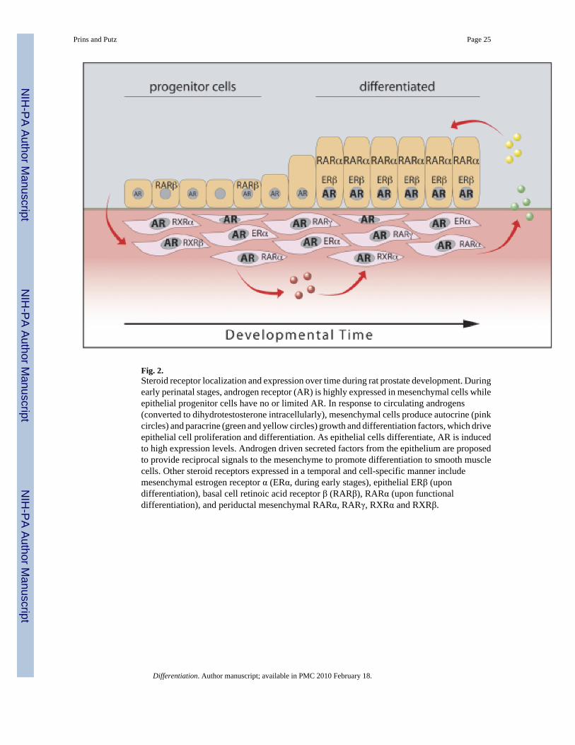
Fig. 2.Steroid receptor localization and expression over time during rat prostate development. Duringearly perinatal stages, androgen receptor (AR) is highly expressed in mesenchymal cells whileepithelial progenitor cells have no or limited AR. In response to circulating androgens(converted to dihydrotestosterone intracellularly), mesenchymal cells produce autocrine (pinkcircles) and paracrine (green and yellow circles) growth and differentiation factors, which driveepithelial cell proliferation and differentiation. As epithelial cells differentiate, AR is inducedto high expression levels. Androgen driven secreted factors from the epithelium are proposedto provide reciprocal signals to the mesenchyme to promote differentiation to smooth musclecells. Other steroid receptors expressed in a temporal and cell-specific manner includemesenchymal estrogen receptor α (ERα, during early stages), epithelial ERβ (upondifferentiation), basal cell retinoic acid receptor β (RARβ), RARα (upon functionaldifferentiation), and periductal mesenchymal RARα, RARγ, RXRα and RXRβ.
Prins and Putz Page 25
Differentiation. Author manuscript; available in PMC 2010 February 18.
NIH
-PA Author Manuscript
NIH
-PA Author Manuscript
NIH
-PA Author Manuscript
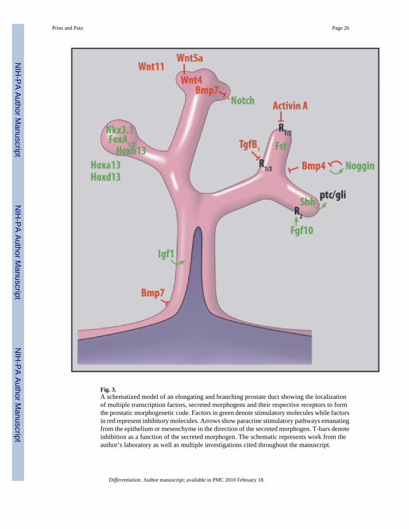
Fig. 3.A schematized model of an elongating and branching prostate duct showing the localizationof multiple transcription factors, secreted morphogens and their respective receptors to formthe prostatic morphogenetic code. Factors in green denote stimulatory molecules while factorsin red represent inhibitory molecules. Arrows show paracrine stimulatory pathways emanatingfrom the epithelium or mesenchyme in the direction of the secreted morphogen. T-bars denoteinhibition as a function of the secreted morphogen. The schematic represents work from theauthor’s laboratory as well as multiple investigations cited throughout the manuscript.
Prins and Putz Page 26
Differentiation. Author manuscript; available in PMC 2010 February 18.
NIH
-PA Author Manuscript
NIH
-PA Author Manuscript
NIH
-PA Author Manuscript

Fig. 4.A proposed model for dichotomous branching of the developing rat prostate ducts as controlledby localized expression and cross-talk of secreted morphoregulatory factors. The distal ducttips express Shh in discreet focal areas (for simplicity, only one shown in red) while the distalmesenchyme expresses Fgf10 (green dots) and Bmp4 (blue dots). As these cells make contactwith each other (b), the secreted Shh (red arrow) activates ptc on mesenchymal cells, locallydown-regulates Fgf10 (loss of green dots) and up-regulates Bmp4 (blue dots) expression. Thefocal downregulation of Fgf10 results in lateral subdomains of higher Fgf10 expressionadjacent to the Shh foci, which in turn, down-regulates Bmp4 in that region and activates (greenarrow) higher epithelial proliferation via epithelial FgfR2iiib (c). The disparate epithelial
Prins and Putz Page 27
Differentiation. Author manuscript; available in PMC 2010 February 18.
NIH
-PA Author Manuscript
NIH
-PA Author Manuscript
NIH
-PA Author Manuscript

proliferation rates in the lateral domains results in the sprouting of two buds on each side ofthe Shh foci (blue arrows) which initiates a branchpoint (d). Further, the elevated Fgf10 in thelateral domains up-regulates Shh and ptc expression (d), which allows repetition of the abovesteps and results in complex branching patterns (Based on results published by Huang et al.,2005).
Prins and Putz Page 28
Differentiation. Author manuscript; available in PMC 2010 February 18.
NIH
-PA Author Manuscript
NIH
-PA Author Manuscript
NIH
-PA Author Manuscript
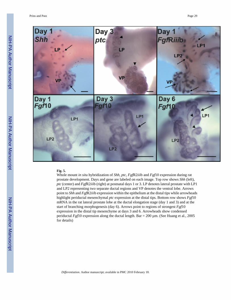
Fig. 5.Whole mount in situ hybridization of Shh, ptc, FgfR2iiib and Fgf10 expression during ratprostate development. Days and gene are labeled on each image. Top row shows Shh (left),ptc (center) and FgfR2iiib (right) at postnatal days 1 or 3. LP denotes lateral prostate with LP1and LP2 representing two separate ductal regions and VP denotes the ventral lobe. Arrowspoint to Shh and FgfR2iiib expression within the epithelium at the distal tips while arrowheadshighlight periductal mesenchymal ptc expression at the distal tips. Bottom row shows Fgf10mRNA in the rat lateral prostate lobe at the ductal elongation stage (day 1 and 3) and at thestart of branching morphogenesis (day 6). Arrows point to regions of strongest Fgf10expression in the distal tip mesenchyme at days 3 and 6. Arrowheads show condensedperiductal Fgf10 expression along the ductal length. Bar = 200 µm. (See Huang et al., 2005for details)
Prins and Putz Page 29
Differentiation. Author manuscript; available in PMC 2010 February 18.
NIH
-PA Author Manuscript
NIH
-PA Author Manuscript
NIH
-PA Author Manuscript
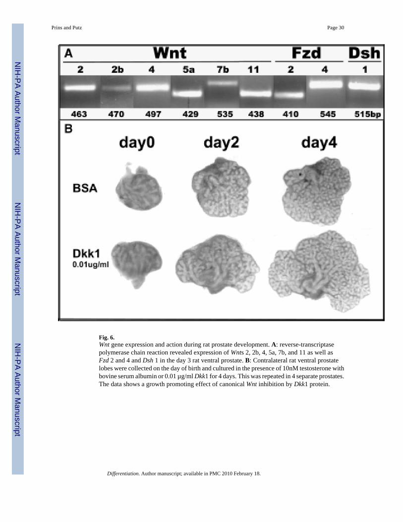
Fig. 6.Wnt gene expression and action during rat prostate development. A: reverse-transcriptasepolymerase chain reaction revealed expression of Wnts 2, 2b, 4, 5a, 7b, and 11 as well asFzd 2 and 4 and Dsh 1 in the day 3 rat ventral prostate. B: Contralateral rat ventral prostatelobes were collected on the day of birth and cultured in the presence of 10nM testosterone withbovine serum albumin or 0.01 µg/ml Dkk1 for 4 days. This was repeated in 4 separate prostates.The data shows a growth promoting effect of canonical Wnt inhibition by Dkk1 protein.
Prins and Putz Page 30
Differentiation. Author manuscript; available in PMC 2010 February 18.
NIH
-PA Author Manuscript
NIH
-PA Author Manuscript
NIH
-PA Author Manuscript

Fig. 7.A schematic representation of regulatory networks between secreted morphogens andtranscription factors in the epithelial and mesenchymal cells at the distal signaling center ofthe developing prostate gland. Fgf10 (mesenchymal) and FgfR2iiib (epithelial) upregulate(green arrows) epithelial expression of Shh and Bmp7 involved in branching morphogenesisas well as Hoxb13 and Nkx3.1 involved in epithelial differentiation. Shh up-regulates ptc andgli in adjacent mesenchymal cells which down-regulates (red lines) Fgf10 expression thusestablishing a negative feedback loop for controlled growth. Shh-ptc-gli also up-regulates thegrowth inhibitory Wnt5a and Bmp4 molecules in the mesenchyme while Fgf10 down-regulatestheir expression which further serves to tightly control localized tissue growth. FoxA1stimulates expression of Nkx3.1 and inhibits FoxA2 expression which has reciprocal up-regulation with Shh. Fgf10/FgfR2iiib up-regulates Notch expression, which drives ductalgrowth while Bmp7 down-regulates Notch and suppresses regional growth. Tgfβ1 suppressesFgf10 expression, which may serve as a brake for growth as development nears completion.
Prins and Putz Page 31
Differentiation. Author manuscript; available in PMC 2010 February 18.
NIH
-PA Author Manuscript
NIH
-PA Author Manuscript
NIH
-PA Author Manuscript