bacterial endospore detection using photoluminescence from ...
Upload
khangminh22Category
view
4download
0
HAL Id: tel-01753129https://tel.archives-ouvertes.fr/tel-01753129
Submitted on 29 Mar 2018
HAL is a multi-disciplinary open accessarchive for the deposit and dissemination of sci-entific research documents, whether they are pub-lished or not. The documents may come fromteaching and research institutions in France orabroad, or from public or private research centers.
L’archive ouverte pluridisciplinaire HAL, estdestinée au dépôt et à la diffusion de documentsscientifiques de niveau recherche, publiés ou non,émanant des établissements d’enseignement et derecherche français ou étrangers, des laboratoirespublics ou privés.
Inhibition of the bacterial ribosome by nascent andantimicrobial peptides
Alexandra Seefeldt
To cite this version:Alexandra Seefeldt. Inhibition of the bacterial ribosome by nascent and antimicrobial peptides. Bio-chemistry, Molecular Biology. Université de Bordeaux, 2017. English. �NNT : 2017BORD0856�.�tel-01753129�
THÈSE PRÉSENTÉE
POUR OBTENIR LE GRADE DE
DOCTEUR DE
L’UNIVERSITÉ DE BORDEAUX
ÉCOLE DOCTORALE SCIENCES DE LA VIE ET DE LA SANTÉ
SPÉCIALITÉ BIOCHIMIE et BIOLOGIE STRUCTURALE
Par Alexandra Carolin SEEFELDT
Master of Science in Biochemistry (LMU, Munich, Germany)
Née le 20. Août 1988 à Heidelberg, Allemagne
Inhibition of the bacterial ribosome by nascent and
antimicrobial peptides
Sous la direction du: Dr C Axel INNIS
Soutenue le 14. Décembre 2017
Membres du jury :
Prof. KRAMER, Ijsbrand Professeur, Université de Bordeaux Président
Dr. SCHMITT, Emmanuelle Directeur de recherche 2, École polytechnique (Paris) Rapporteur
Prof. SUGA, Hiroaki Professeur, University of Tokyo Rapporteur
Dr. FRONZES, Rémi Directeur de recherche 2, Université de Bordeaux Examinateur
Dr. HASHEM, Yaser Chargé de recherche, Université de Strasbourg Examinateur


To
my mom Bärbel Seefeldt,
my grandma Trude Kurz,
my grandfather Rolf Kurz,
my uncle Frieder Kurz,
and music.
“The way to get good ideas is to get lots of ideas and throw the bad ones away.”
Linus Pauling

Acknowledgments
Firstly, I would like to thank all members of the jury, Prof Ijsbrand Kramer, Dr. Emmanuelle
Schmitt, Prof Hiroaki Suga, Dr. Rémi Fronzes and Dr. Yaser Hashem, for taking the time to
read and evaluate my thesis. My research was funded with a pre-doctoral fellowship from
INSERM and the region Aquitaine.
During the last three years, I was often asked the question why I decided to come to Bordeaux
and my answer was always the same, I wanted to work on ribosomes and Structural Biology
and that I chose the project not the city. By now, I know that this city and its location are just
incredible and I had a great opportunity to get to know the South-West of France. The last
three years have been a wonderful journey and I had the honor to work with so many inspiring
people and here I would like to take the opportunity to acknowledge everyone.
Firstly, I would like to express my sincere gratitude to my thesis supervisor Axel Innis for giving
me this great opportunity to come to Bordeaux and to work on these wonderful projects. It
has been my pleasure and honor to be your first PhD student. Your passion for science was
inspiring. Your constant guidance and our knowledge exchanges helped me during the whole
of my research and writing and inspired me to pursue an academic career.
Next, I am especially grateful to Britta Seip. I will never forget your welcoming smile when I
first arrived in Bordeaux. From the start, you were there for me to discuss with me and giving
me constant input throughout the entire time of my thesis and during the writing process. You
were there when I needed a push to finish an experiment or to post-pone it to the next day. I
would like to thank you for being an inspiring colleague and more importantly becoming a
wonderful and close friend. It was a lot of fun to discover France with you.
I owe a special thanks to Kishore Inampudi for establishing the flexizyme reaction and tRNAiMet
purification, for cloning tRNAPro into pUC19 and cloning the proline-tRNA synthetase. Thanks
to your work, I had a huge advantage when I started my thesis. A special thank goes to
Natacha Pérébaskine for ribosome and tRNA purification and teaching me how to crystallize
ribosomes. Our synchrotron trips were always special, and we came back to Bordeaux often
with more than one exciting story to tell. I had a great time working with you.
I would like to thank Alba Herrero del Valle for her support and positive energy. We had a lot
to talk and laugh about. You always found a new way to improve my figures and slides (😊)
make them look better. I had a great time working with you and I am happy to call you my
friend. I want to thank Justine Charon for helping me write the French sections and bringing
a great spirit into the lab. I want to thank Guénaël Sacheau for his help with sequencing
reactions, correcting my French sections of my thesis and for providing a good mood including
OOOOOh yeah, tons of despacito and “Je peux pas, j’ai aqua cloning”. A special thanks to
Mélanie Gillard, Elodie Leroy and Aitor Manteca. I know you just arrived but it has been a
fun time working with you and spent time with you during lunch and coffee breaks. A big
thanks to Elodie Leroy for her help with the summary in French.
I want to acknowledge Gilles Guichard and his group for fruitful collaboration over the last
years that resulted directly in many positive results. I want to thank in particular Caterina
Lombardo and Christophe André for the synthesis of the flexizyme compounds and constant
discussions to increase the yield of the peptidylation and to overcome problems with proline.
Furthermore, I would like to thank Céline Douat and Stéphanie Antunes for their work during
the Onc112 project. It was a great pleasure to work with you.
6 Acknowledgements
Next, I want thank Daniel Wilson and his group for constant input, knowledge exchanges and
sending the plasmid expressing EF-P and EF-G from E. coli. Additionally, I want to thank Fabian
Nguyen, Michael Graf and Stefan Arenz for their work during the PrAMP project. A special
thanks to Marco Scocchi and Mario Mardirossian for working with us on the further
characterization of PrAMPs.
I owe also thanks to Alexander Shura Mankin and his team for their help in input. I especially
want to thank Nora Vazquez-Laslop. It was always inspiring discussing with you during
conferences. A special thanks to Tanja Florin and James Marks III for your ideas to improve
my toeprinting experiments. My sincere thanks to Thomas Steitz and his group for providing
plasmids, protocols and advice for tRNA purification.
I want to thank Derek McCusker and his team for providing material and Emmaunelle
Schmitt for providing the pBSTNAV plasmids for tRNA purification. A special thanks to Brice
Kaufmann and Stéphane Massip for their support during crystal freezing and fishing. It was
always a very exciting moment after weeks of sample preparation. During crystal screening and
data collection, I want to acknowledge Pierre Legrand and Leonard Chavas from PX1 and
William Shepard and Martin Savko from PX2a beamline at the Soleil synchrotron for their
advice and support during data processing.
I learned a lot from all of you. I started with cryo-EM in January 2017 without experience in
sample preparation, data collection and data processing. I want to thank Armel Bézault for
training me in sample preparation and data collection. A special thanks to Rémi Fronzes and
his team for sharing the cluster for data processing and constant discussion through-out all
the steps. In particular, I want to thank Chiara Rapisdara for long and extensive discussions
about structural biology and life and for giving me tons of positive energy. I want to thank
Valerie Gabélica and Frédéric Rosu for mass spectrometry analysis of tRNAPro and for their
support during data interpretation.
My longest and most constant lunch group member Lionel Beaurepaire had more than once
the one or the other idea to get me further with my experiments. Sometimes a lunch break can
solve most of the problems with experiments. More importantly, thank you for listening and
teaching me about France. Nearly every morning (except when she was on holidays) I was
greeted with a warm smile. Thank you Mariline Yot for being there and for being my best
French teacher. I want to acknowledge Myriam Mederic for her positive energy every time she
came to our lab. A special thanks to Patricia Martin and Kati Ba-Pierozzi for helping me with
all the administration, refunding and mission order. Special thanks to Gerald Canet and Eric
Roubin for solving all the computer problems, I was facing during the last years. The printer
was always happy to see you, too.
I want thank the whole unit Inserm U1212, CNRS UMR5320 under the direction of Jean-Louis
Mergny. It was a pleasure to discuss and exchange knowledge due to different expertise. In
particular, I want to thank Fabien Darfeuille, Cameron Mackereth and Denis Dupuy for
answering a lot of questions. In addition, I want to thank the team of Martin Teichmann in
particular Stéphanie Durrieu, Camila Perrot and Wiebke Bretting for their help improving
protocols.
I would like to acknowledge all members of the JJC organization committee 2015 and 2016. I
was a great experience to work together and organize two exciting conferences. I want to thank
in particular Diane Bécart for a wonderful friendship, 1000000 PhD student dinners, long
discussions about life and science. I loved playing music with you and just being around you.
7 Acknowledgements
Thank you for convincing me to start dancing Batchata, it was each time a great experience
even if I am not the most talented dancer. I want to thank Birgit Habenstein and Joséphine
Abi-Ghanam for their advice, wonderful discussions and constant support. Furthermore, I want
to thank Laura Mauran for hours of surfing and wonderful times on the Sunday market in
Pessac. A special thanks to Sonia Cuidad, Martí Ninot, Eduard Puig and Thomas Perry for
their constant support, encouragement and for increasing my knowledge about penguins. It
was always lots of fun to do things together.
Ein großes Dankeschön geht an meine Familie, die mich immer ermutigt und unterstützt hat
meinen eigenen Weg zu finden und mich gegen jeglichen Wiederstand durchzusetzen. Meiner
Mama Bärbel Seefeldt möchte ich für ihr beeindruckendes Druchhaltevermögen und offenes
Ohr danken auch wenn ich meinen eigenen Kopf durchsetzen wollte. Ich möchte meiner Oma
Trude Kurz dafür danken, dass sie immer wieder die richtigen Worte findet egal ob auf Deutsch
oder Französisch. Meinen Großvater Rolf Kurz möchte ich danken, dass er die Begeisterung
für Naturwissenschaften mit mir teilt und mir sehr viel beigebracht hat. Zusätzlich möchte ich
meinem Onkel Frieder Kurz danken, dass er immer einen Rat für mich übrighatte. Ohne euch
wäre diese Arbeit nicht möglich gewesen. Ich möchte zusätzlich meiner längsten Freundin
Anna Neubauer, danken, dass sie mich durch alle Höhen und Tiefen bisher begleitet hat und
die sich einfach nur freut, wenn ich anrufe.
Studying chemistry and biochemistry made it possible to meet Thomas Leissing, Michaela
Wipper, Katharina Essig, Stefanie Herrmann, Kerstin Lippl and Janina Andreß. I want to
acknowledge them for their constant support throughout the complete time of my studies but
more importantly for their friendship. In the three years you showed me no matter where we
will live we will find our way to stay in touch. Five years ago, I went to Riverside California and
was lucky to meet Tamara Mielke. I want to thank you for being my best friend ever since. I
can thank you enough for listening through all the up and downs of a thesis, for going back
with me to California. You are the person to be hiking on a volcano in a volcano, finding our
inner sunflower as well as being in a kayak while being surrounded by sea otters, sea lions and
humpback whales. I also want to thank Raissa and Jarren Kay for their constant support and
Nicola Cellini for reminding me about my personal reasons to keep on going with science.
Je tiens à remercier tous les membres de la chorale croq'notes d'être la meilleure classe de
français et une merveilleuse compagnie à chanter avec beaucoup de joie tous les mardis soirs
et de voyager à travers l'Italie. En particulier, je tiens à remercier Anne-Marie Garcia pour être
l'une des meilleures chefs de chorale avec qui j'ai travaillé et pour nous avoir motivé à participer
deux fois au projet Tutti.
A special thanks to the whole team of the Black Valvet Bar, Barry, Will, Marta, Angi, Nick and
Langi for the best entertainment on Wednesdays nights increasing important knowledge
about penguins and constant training for blind test. The Black Valvet Bar is the best Irish pub
in Bordeaux and there is only one real catzapplin. I want to acknowledge Ian Ruigrok for
introducing me to this particular pub and for the one and other great discussion.
Table of content
List of Figures and Tables .................................................................................................................................... 14
Conventions .............................................................................................................................................................. 20
Abbreviations ........................................................................................................................................................... 21
1. Introduction ......................................................................................................................................................... 26
1.1 Bacterial translation ................................................................................................................................... 26
1.1.1 Gene expression and the bacterial ribosome .......................................................................... 26
1.1.2 The prokaryotic translation cycle .................................................................................................. 29
1.2 The prokaryotic ribosome as a target for antibiotics .................................................................... 35
1.2.1 Antibiotics targeting the PTC ......................................................................................................... 36
1.2.2 Macrolides and ketolides binding to the ribosomal tunnel ............................................... 37
1.3 Proline-rich antimicrobial peptides (PrAMPs) as possible new therapeutics ....................... 39
1.4 Nascent chain-mediated translational arrest ................................................................................... 42
1.4.1 Ligand-independent arrest peptides ........................................................................................... 44
1.4.2 Ligand-dependent arrest peptides .............................................................................................. 47
1.4.3 Short ligand-dependent ribosomal arrest peptides in bacteria........................................ 50
1.4.4 Polyproline induced arrest is relieved by the protein factor EF-P .................................... 52
1.5 Flexizyme as a tool to study translation ............................................................................................. 55
1.5.1 Identification and sequence optimization ................................................................................. 55
1.5.2 Case studies .......................................................................................................................................... 59
1.6 Aims ................................................................................................................................................................. 60
9 Table of Content
2. Materials ................................................................................................................................................................ 61
2.1 Chemicals ....................................................................................................................................................... 61
2.2 Antibiotics ...................................................................................................................................................... 68
2.3 Enzymes .......................................................................................................................................................... 68
2.4 Kits .................................................................................................................................................................... 70
2.5 Equipment ..................................................................................................................................................... 70
2.5.1 Columns ................................................................................................................................................. 70
3. Methods ................................................................................................................................................................ 71
3.1 General methods......................................................................................................................................... 71
3.1.1 Microbiological handling ................................................................................................................. 71
3.1.2 Extraction of genomic DNA from E. coli and T. thermophilus ........................................... 73
3.2 Analytical procedures ................................................................................................................................ 74
3.2.1 Standard gel electrophoresis ......................................................................................................... 74
3.2.2 Northern Blot ....................................................................................................................................... 77
3.2.3 Macromolecule concentration determination ......................................................................... 79
3.2.4 Native mass spectrometry ............................................................................................................... 80
3.3 Cloning ............................................................................................................................................................ 80
3.3.1 Amplification of gene of interest from genomic DNA ......................................................... 80
3.3.2 Amplification of gene of interest by oligo assembly ............................................................ 82
3.3.3 Vector restriction and ligation ....................................................................................................... 83
3.3.4 Mutagenesis of Thermus thermophilus Elongation Factor P ............................................. 85
3.3.5 Preparation of chemically competent cells ............................................................................... 86
3.3.6 Plasmid transformation into chemically competent cells .................................................... 86
3.3.7 Colony identification ......................................................................................................................... 87
3.3.8 Plasmid extraction and sequencing ............................................................................................. 88
3.4 Protein expression and purification ..................................................................................................... 88
3.4.1 Expression vectors .............................................................................................................................. 89
10 Table of Content
3.4.2 Expression and purification of aminoacyl tRNA synthetases, T7 RNAP and EF-G ..... 89
3.4.3 Expression and purification of Elongation factor-P ............................................................... 90
3.5 RNA handling ............................................................................................................................................... 91
3.5.1 In vitro transcription and purification in vitro transcribed RNA ........................................ 91
3.5.2 Phenol-Chloroform extraction ....................................................................................................... 92
3.5.3 Precipitation of RNA .......................................................................................................................... 92
3.6 tRNA expression and purification ......................................................................................................... 93
3.6.1 Test expression of tRNAPro overexpression ............................................................................... 94
3.6.2 Large-scale expression of tRNAPro and total tRNA extraction ............................................ 95
3.6.3 Chromatography of tRNAPro ........................................................................................................... 97
3.6.4 CCA end modification of tRNA ...................................................................................................... 98
3.6.5 Aminoacylation with aminoacyl-tRNA synthetases ............................................................... 99
3.6.6 Peptidylation of tRNAiMet with flexizyme technique ........................................................... 100
3.7 Ribosome purification ............................................................................................................................ 101
3.8 Toeprinting ................................................................................................................................................. 103
3.8.1 In vitro translation ........................................................................................................................... 103
3.8.2 Sanger sequencing for toeprinting experiment ................................................................... 105
3.8.3 Analysis and interpretation .......................................................................................................... 106
3.9 X-ray crystallography and structure determination .................................................................... 107
3.9.1 Complex formation and crystallography ................................................................................ 108
3.9.2 Data collection and processing .................................................................................................. 109
3.9.3 Phasing, refinement and model building ............................................................................... 111
3.10 Cryo-Electron microscopy (cryo-EM) ............................................................................................. 113
3.10.1 Sample preparation ...................................................................................................................... 113
3.10.2 Data collection................................................................................................................................ 114
3.10.3 Single particle reconstruction using RELION gui .............................................................. 115
3.10.4 Model building ............................................................................................................................... 117
11 Table of Content
3.11 Bioinformatic databases and programs ........................................................................................ 118
4. Results ................................................................................................................................................................. 119
4.1 Proline-rich antimicrobial peptides inhibit the bacterial ribosome ...................................... 119
4.2 A novel strategy for the structural characterization of arrested ribosomal complexes
featuring short nascent peptides .............................................................................................................. 122
4.3 Peptidylation of tRNAiMet using flexizyme ...................................................................................... 126
4.3.1 Screening reactivity and time courses ..................................................................................... 126
4.3.2 Purification of peptidylated tRNAiMet ....................................................................................... 128
4.4 Investigations of fM+X(+) nascent chain-mediated translational arrest in the presence of
erythromycin ..................................................................................................................................................... 133
4.4.1 Complexes to study fM+X(+) in the presence of erythromycin .................................... 133
4.4.2 Toeprinting to validate that fM+F(+) arrests the ribosome in the presence of
erythromycin ................................................................................................................................................ 134
4.4.3 Toeprinting: fMKF-tRNAiMet arrests the ribosome in the presence of erythromycin
........................................................................................................................................................................... 138
4.4.5 Structural studies of an MKFR-ribosome complex arrested in the presence of
erythromycin ................................................................................................................................................ 140
4.4.6 Single particle reconstruction using RELION......................................................................... 141
4.4.7 Model building, refinement, and validation .......................................................................... 147
4.4.9 Structure interpretation ................................................................................................................. 150
4.5 Polyproline-mediated arrest ................................................................................................................ 159
4.5.1 Complexes to study arrest along consecutive proline motifs ......................................... 159
4.5.2 tRNAPro expression and purification ......................................................................................... 160
4.5.3 Overexpression tests of tRNAPro ................................................................................................. 162
4.5.4 tRNAPro purification ......................................................................................................................... 164
4.5.5 tRNAPro characterization ................................................................................................................ 166
4.5.6 Purification and activity of Elongation Factor P (EF-P) ...................................................... 169
12 Table of Content
4.5.7 Comparison of EF-P from T. thermophilus and E. coli ....................................................... 171
4.5.8 Studies of T. thermophilus EF-P mutants containing the flexible loop region from
E. coli EF-P ..................................................................................................................................................... 174
4.5.9 Toeprint to study polyproline-mediated arrest and its release by EF-P ..................... 177
4.5.10 EF-P does not release other well-characterized nascent chain-mediated translation
arrest peptides ............................................................................................................................................. 180
5. Discussion and perspectives ....................................................................................................................... 183
5.1 Proline-rich antimicrobial peptides and implications for further drug development ... 183
5.2 The flexizyme methodology to study nascent chain-mediated translational arrest ...... 188
5.3 fMKF(R) arrests the 70S E. coli ribosome in the presence of erythromycin ....................... 189
5.4 Polyproline-mediated arrest ................................................................................................................ 194
6. Conclusion ......................................................................................................................................................... 199
7. Résumé français............................................................................................................................................... 200
7.1 Général ......................................................................................................................................................... 200
7.2 Peptides antimicrobiens riches en proline (PrAMPs) ................................................................. 200
7.3 L'arrêt de la traduction induit par le peptide naissant .............................................................. 201
7.3.1 M+X(+) arrête le ribosome bactérien en présence d'érythromycine .......................... 202
7.3.2 L'arrêt médié par la polyproline est soulagé par le facteur d'élongation P .............. 204
7.4 Conclusion .................................................................................................................................................. 205
8. References ......................................................................................................................................................... 206
9. Curriculum vitae .............................................................................................................................................. 230
13 Table of Content
10. Supplemental information ........................................................................................................................ 233
10.1 DNA primer lists ..................................................................................................................................... 233
10.2 Canonical base orientations .............................................................................................................. 236
10.3 tRNAPro modifications .......................................................................................................................... 237
10.4 Ribosome purification ......................................................................................................................... 238
10.5 MKF-CME flexizyme reaction ............................................................................................................ 238
10.6 Protein purifications ............................................................................................................................. 239
10.7 Sequencing results ................................................................................................................................ 241
10.8 Purity of flexizyme compounds ....................................................................................................... 244
List of Figures and Tables
Figure 1: tRNA positions and correct labeling of the nascent chain, as used in this work. ........ 20
Figure 2: Structure of 70S ribosome from E. coli. ....................................................................................... 27
Figure 3: Schematic view of the ribosomal exit tunnel. ........................................................................... 28
Figure 4: Overview of the bacterial translation cycle as reviewed by Schmeing and
Ramakrishnan, 2009. .................................................................................................................................... 29
Figure 5: Chemical structure of Erythromycin (macrolide) and Telithromycin (ketolide). ........... 37
Figure 6: Binding site of erythromycin within the bacterial ribosome. .............................................. 38
Figure 7: Sequence alignments of PrAMPs isolated from different species (insects and
mammalian) or derived from drug development. ............................................................................ 40
Figure 8: fM+X(+) arrests the ribosome in the presence of the antibiotic erythromycin. .......... 51
Figure 9: Summary of the sequence variants that can induce polyproline-mediated ribosomal
arrest. ................................................................................................................................................................. 53
Figure 10: Sequence alignments of the different in vitro selected flexizymes Fx3, eFx, dFx, and
aFx. ...................................................................................................................................................................... 57
Figure 11: Different flexizymes (eFx, dFx, and aFx) were selected in vitro to recognize different
leaving groups to increase reactivity and complexity in the substrate..................................... 57
Figure 12: Assembly of Northern Blot, the RNA was separated in the TBU-PAGE prior to the
blotting. ............................................................................................................................................................. 77
Figure 13: Summary of tRNAPro extraction and purification. .................................................................. 96
Figure 14: Ribosomal domains for rigid-body fitting. ........................................................................... 111
Figure 15: PrAMPs bind to the ribosomal exit tunnel. ........................................................................... 119
Figure 16: Mechanism of action of proline-rich antimicrobial peptides. ....................................... 121
Figure 17: General workflow to study short peptides that mediated nascent chain-dependent
translational arrest using the flexizyme methodology. ................................................................ 123
Figure 18: Workflow to obtain the arrest complex using one round of translocation. ............. 125
Figure 19: Time course of flexizyme reactions for fMK-CBT (A), AcRP-CBT (B), AcRA-CBT (C)
and AcRD-CBT (D) onto tRNAiMet. ........................................................................................................ 127
Figure 20: Time course following the peptidylation reaction of AcRAP-CBT peptide with
microhelix over six days. .......................................................................................................................... 128
15 List of Figures and Tables
Figure 21: RP-HPLC purification of dipetidylated-tRNAiMet after flexizyme reaction for 8 days
on ice. .............................................................................................................................................................. 129
Figure 22: Purification of AcRAP-tRNAiMet after flexizyme reaction for 7 days on ice by C4-RP-
HPLC. ............................................................................................................................................................... 131
Figure 23: After buffer exchange and lyophilization of the peptidylated tRNAs did not
hydrolyze. ...................................................................................................................................................... 131
Figure 24: Complex to study fM+X(+)-mediated arrest in the presence of erythromycin. ..... 133
Figure 25: MRFR, MKFR, MRFK and MKFK arrest the E. coli ribosome in the presence of
erythromycin. ............................................................................................................................................... 135
Figure 26: In the presence of erythromycin, ErmDL arrests 70S T. thermophilus ribosomes at
the same position as the 70S E. coli ribosomes. ............................................................................. 136
Figure 27: Toeprint of MRFRI in the presence or absence of erythromycin/telithromycin using
70S T. thermophilus ribosomes.. .......................................................................................................... 137
Figure 28 fMKF-tRNAiMet arrests E. coli 70S ribosomes in vitro in the presence of erythromycin
and Arg-tRNAArg. ......................................................................................................................................... 139
Figure 29: Grids prepared with a ribosome concentration of 480 nM showed a good
distribution of particles on micrographs. .......................................................................................... 142
Figure 30: Representative 2D classes of the sample sorted in order of decreasing abundance.
........................................................................................................................................................................... 143
Figure 31: Masked 3D classification sorting according to the occupancy of A-, P- and E-site
tRNA. ............................................................................................................................................................... 144
Figure 32: Local resolution distribution and FSC curve. ........................................................................ 146
Figure 33: Examples of density display around different parts of the model ............................... 147
Figure 34: The reconstructed density contains density for P-site (green) and E-site tRNA (dark
blue) but no density for an A-site tRNA. ........................................................................................... 150
Figure 35: The fMKF-peptide does not form direct contact with the drug while the
penultimate lysine side chain points towards the A-site crevice. ............................................ 151
Figure 36: In the MKF-70S E. coli structure, U2506 is a universally conserved residue within
the PTC that adopts a novel conformation different from one during translational cycle
and arrest....................................................................................................................................................... 153
Figure 37: The base of U2585, moving during tRNA accommodation, adopts a specific
conformation similar to the one in intrinsic arrested ribosomes. ............................................ 154
16 List of Figures and Tables
Figure 38: 23S rRNA residue A2062 is a universally conserved base within the ribosomal exit
tunnel that acts as a sensor for arrest peptides (Vázquez‐Laslop et al., 2010) that points
towards the PTC in the MKF-70S structure (green). ...................................................................... 155
Figure 39: Proposed mechanism of MKF-mediated translational arrest in the presence of
erythromycin.. .............................................................................................................................................. 157
Figure 40: Complexes to study nascent chain-mediated translational arrest along consecutive
proline motifs. .............................................................................................................................................. 159
Figure 41: Schematic view of tRNAPro overexpression strategies ...................................................... 161
Figure 42: Comparison of the different overexpression strategies using pBSTNAV vectors and
pUC19-proK vector. ................................................................................................................................... 162
Figure 43: Similar amounts of tRNAPro in E. coli DH5α and HB101 were expressed per cell
using the pBSTNAV vectors.. .................................................................................................................. 163
Figure 44: Chromatographic steps of tRNAPro purification.. ................................................................ 164
Figure 45: tRNAPro purification steps analyzed by denaturing PAGE and Northern blotting. 166
Figure 46: Mass spectrogram from native mass spectrometry of tRNAPro. ................................... 167
Figure 47: Activity test of purified tRNAPro using toeprinting ............................................................. 168
Figure 48: Screening of different toeprinting templates to study polyproline-mediated arrest
and its release using purified E. coli EF-P. ........................................................................................ 170
Figure 49: Sequence alignment of T. thermophilus EF-P (Tth_wt) and E. coli EF-P (Eco_wt). . 171
Figure 50: Unmodified T. thermophilus EF-P does not release E. coli ribosomes arrested on
polyproline motifs. ..................................................................................................................................... 172
Figure 51: Positive density for minimally biased difference maps (Fo-Fc) of T. thermophilus and
E. coli EF-P. .................................................................................................................................................... 173
Figure 52: Amino acid sequence alignment of T. thermophilus EF-P and the two mutants... 175
Figure 53: SDS-PAGE showing purified EF-P variants.. .......................................................................... 175
Figure 54: The mutants generated do not release E. coli ribosomes arrested at consecutive
prolines. .......................................................................................................................................................... 176
Figure 55: Polyproline-mediated arrest and its release by E. coli EF-P was studied using
flexizyme-peptidylated tRNAs. .............................................................................................................. 178
Figure 56: E. coli EF-P releases polyproline-mediated arrested ribosomes but does not release
ribosomes stalled while translating other well-characterized arrest peptides.................... 181
Figure 57: Overlay of the binding sites of Onc112 and clinically used antibiotics. .................... 183
17 List of Figures and Tables
Figure 58: Comparison of the binding sites of Bac7(1-16), Api137 and Klebsazolicin (KLB). 185
Figure 59: Peptide modifications to get a greater understanding of the chemical properties
needed for nascent chain mediated translational arrest. ............................................................ 190
Figure 60: Composition of in vitro translation systems for arrested complex formation for
structural biology. ...................................................................................................................................... 194
Figure 61: Complexes to study the molecular mechanism of EF-P. ................................................. 195
Figure 62: The position of the Cγ-atom influences the isomeric form of the peptide bond. . 196
Figure 63: The variety of modifications essential for EF-P activity in different species. ........... 197
Figure 64: Additional conformations of U2506 (A), U2585 (B) and A2062 (C) during pre-
accommodation, catalysis and translocation. .................................................................................. 236
Figure 65: Base modification found in proK. ............................................................................................. 237
Figure 66: Sucrose gradients of the 70S E. coli ribosome purification. ........................................... 238
Figure 67: fMKF-CME flexizyme reaction performed by Dr. K. Kishore Inampudi. ..................... 238
Figure 68: ProS purification steps analyzed by SDS-PAGE. ................................................................. 239
Figure 69: PheST purification steps analyzed by SDS-PAGE. .............................................................. 239
Figure 70: E. coli and T. thermophilus EF-P purification steps analyzed by SDS-PAGE. ........... 240
Figure 71: Purification of the T. thermophilus mutants by SDS-PAGE. ........................................... 240
Figure 72: Sequencing results for proK-pBSTNAV2OK ......................................................................... 241
Figure 73: Sequencing results for tRNAPro into pBSTNAV3S vector. ................................................ 241
Figure 74: sequencing result of Tth EF-P HQ mutant ............................................................................ 242
Figure 75: Sequencing result from mutagenesis to generate Tth EF-P mutant incl ecoli loop.
........................................................................................................................................................................... 243
Table 1: Summary of abbreviations used in this work. ............................................................................. 21
Table 2: Peptide sequences that lead to a nascent chain-mediated translational arrest in
bacteria. ............................................................................................................................................................. 44
Table 3: Summary of well-characterized ligand-dependent arrest peptides in bacteria. ........... 47
Table 4: List of chemicals ..................................................................................................................................... 61
Table 5: List of antibiotics .................................................................................................................................... 68
Table 6: Enzymes used for cloning, toeprinting and RNA purification .............................................. 68
Table 7: List of Kits ................................................................................................................................................. 70
Table 8: List of used columns for RNA and protein purification .......................................................... 70
18 List of Figures and Tables
Table 9: Media composition of LB and TB .................................................................................................... 71
Table 10: List of used E. coli strains with specifics on genotypes and usage .................................. 71
Table 11: 689 media composition .................................................................................................................... 72
Table 12: Composition of a native nucleic acid (TBE)-PAGE .................................................................. 74
Table 13: Composition of a denaturing nucleic (TBU)-PAGE ................................................................. 75
Table 14: Composition of acidic denaturing PAGE .................................................................................... 76
Table 15: Composition of SDS-PAGE .............................................................................................................. 76
Table 16: List of used probes ............................................................................................................................. 78
Table 17: List of proteins purified in this thesis, their corresponding extinction coefficients and
molecular weight were determined using the bioinformatic platform ProtParam. ............. 79
Table 18: Pipetting scheme for gene amplification from genomic DNA .......................................... 81
Table 19: PCR program for gene extraction ................................................................................................. 81
Table 20: Pipetting scheme for primer annealing PCR ............................................................................. 82
Table 21: PCR program for primer annealing .............................................................................................. 82
Table 22: List of vectors with their corresponding restriction enzymes and downstream
application ....................................................................................................................................................... 83
Table 23: Pipetting scheme for insertion of gene into pUC19 or pBAT4 .......................................... 84
Table 24: PCR program for MEGAWHOP ...................................................................................................... 84
Table 25: Pipetting scheme for PCR based mutagenesis ........................................................................ 85
Table 26: PCR program for PCR based mutagenesis ................................................................................ 85
Table 27: Pipetting scheme for Colony PCR ................................................................................................. 87
Table 28: Reaction scheme for Colony PCR .................................................................................................. 87
Table 29: List of expression vectors used for protein purification ....................................................... 89
Table 30: List of tRNA constructs for purification and expression ....................................................... 94
Table 31: List of used PURExpress systems ................................................................................................ 103
Table 32: Reaction mixture for toeprinting control reactions............................................................. 104
Table 33: List of 3x ddNTP stock solutions ................................................................................................ 105
Table 34: Mastermix for sequencing reaction .......................................................................................... 105
Table 35: Program for sequencing reaction .............................................................................................. 106
Table 36: Gel mix for sequencing PAGE ...................................................................................................... 106
Table 37: List of synthetic mRNAs for structural biology studies ...................................................... 108
Table 38: Refinement and model statistics single molecular reconstruction. .............................. 149
19 List of Figures and Tables
Table 39:Primer list for aminoacyl-tRNA synthetase cloning .............................................................. 233
Table 40: DNA primers for mutagenesis for T. thermophilus EF-P ................................................... 233
Table 41: DNA primers for tRNA cloning ................................................................................................... 234
Table 42: DNA Primer for toeprinting templates..................................................................................... 235
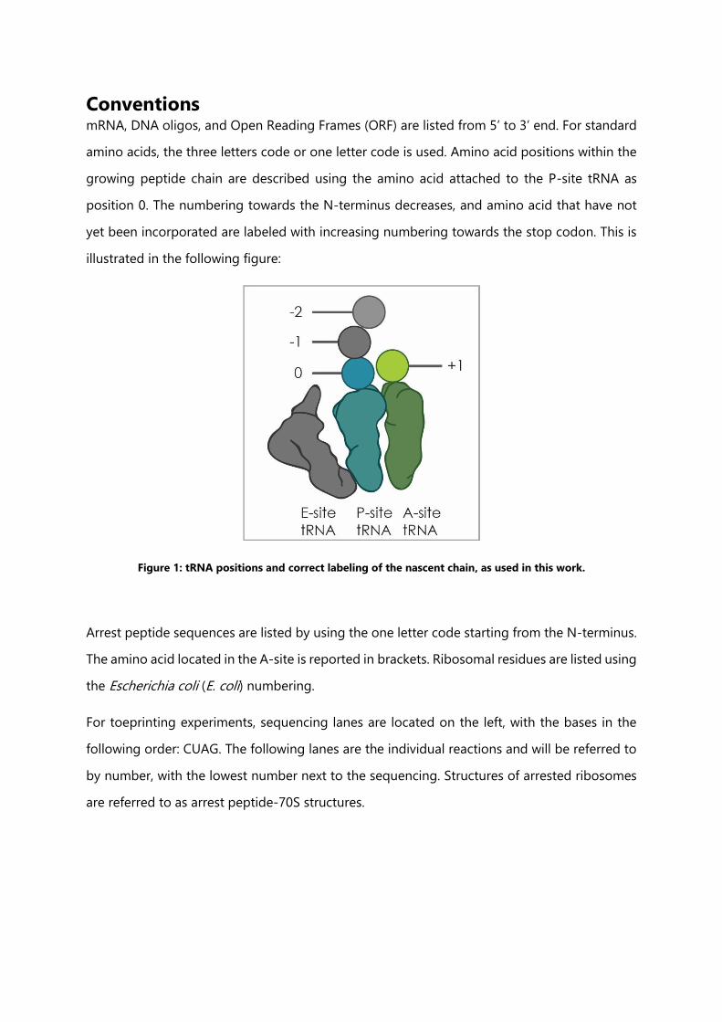
Conventions mRNA, DNA oligos, and Open Reading Frames (ORF) are listed from 5’ to 3’ end. For standard
amino acids, the three letters code or one letter code is used. Amino acid positions within the
growing peptide chain are described using the amino acid attached to the P-site tRNA as
position 0. The numbering towards the N-terminus decreases, and amino acid that have not
yet been incorporated are labeled with increasing numbering towards the stop codon. This is
illustrated in the following figure:
Figure 1: tRNA positions and correct labeling of the nascent chain, as used in this work.
Arrest peptide sequences are listed by using the one letter code starting from the N-terminus.
The amino acid located in the A-site is reported in brackets. Ribosomal residues are listed using
the Escherichia coli (E. coli) numbering.
For toeprinting experiments, sequencing lanes are located on the left, with the bases in the
following order: CUAG. The following lanes are the individual reactions and will be referred to
by number, with the lowest number next to the sequencing. Structures of arrested ribosomes
are referred to as arrest peptide-70S structures.
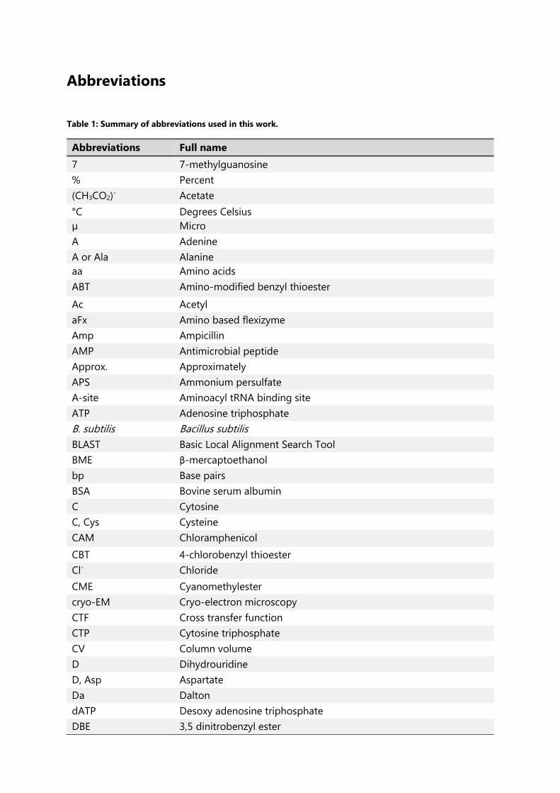
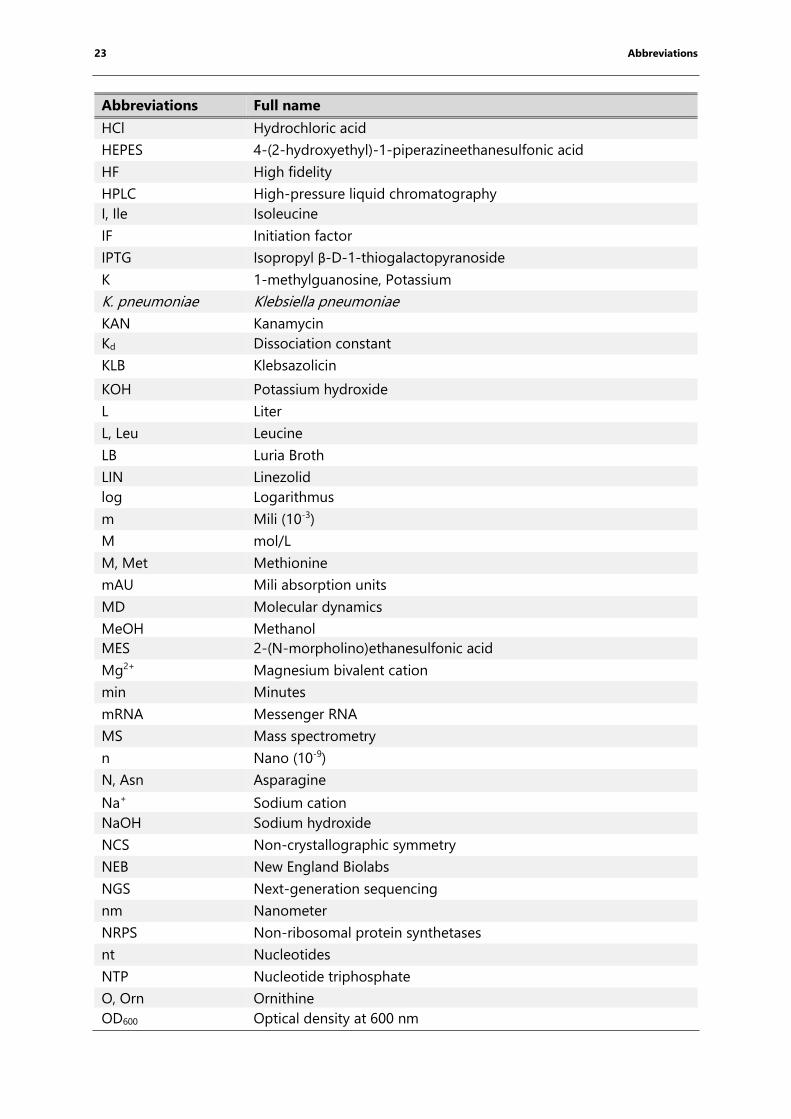
Abbreviations
Table 1: Summary of abbreviations used in this work.
Abbreviations Full name
7 7-methylguanosine
% Percent
(CH3CO2)- Acetate
°C Degrees Celsius
µ Micro
A Adenine
A or Ala Alanine
aa Amino acids
ABT Amino-modified benzyl thioester
Ac Acetyl
aFx Amino based flexizyme
Amp Ampicillin
AMP Antimicrobial peptide
Approx. Approximately
APS Ammonium persulfate
A-site Aminoacyl tRNA binding site
ATP Adenosine triphosphate
B. subtilis Bacillus subtilis
BLAST Basic Local Alignment Search Tool
BME β-mercaptoethanol
bp Base pairs
BSA Bovine serum albumin
C Cytosine
C, Cys Cysteine
CAM Chloramphenicol
CBT 4-chlorobenzyl thioester
Cl- Chloride
CME Cyanomethylester
cryo-EM Cryo-electron microscopy
CTF Cross transfer function
CTP Cytosine triphosphate
CV Column volume
D Dihydrouridine
D, Asp Aspartate
Da Dalton
dATP Desoxy adenosine triphosphate
DBE 3,5 dinitrobenzyl ester

22 Abbreviations
Abbreviations Full name
dCTP Desoxy cytosine triphosphate
ddATP Didesoxy adenosine triphosphate
ddCTP Didesoxy cytosine triphosphate
ddGTP Dideoxy guanosine triphosphate
ddNTP Dideoxynucleotide triphosphate
ddTTP Dideoxy thymine triphosphate
DEPC Diethyl pyrocarbonate
dFx Dinitro flexizyme
dGTP Desoxy guanosine triphosphate
DMSO Dimethyl sulfoxide
DNA Deoxyribonucleic acid
DNAP DNA polymerase
dNTP Desoxy nucleotide triphosphate
DT Drift tube
dTTP Desoxy thymine triphosphate
E, Glu Glutamate
E. coli Escherichia coli
e.g. For example
EDTA Ethylenediaminetetraacetic acid
EF Elongation factor
eFx Enhanced flexizyme
erm Erythromycin resistance methylase
ERY Erythromycin
ESI Electrospray ionization
E-site Exit, deaminoacyl tRNA binding site
ESRF European Synchrotron Radiation Facility
EtOH Ethanol
ExPASy The Expert Protein Analysis System
f Formyl, femto (10-15)
F, Phe Phenylalanine
FRET Fluorescence (Förster) resonance energy transfer
FSC Fourier shell correlation
Fx Flexizyme
g Gramm
G Guanine
G, Gly Glycine
GTP Guanosine triphosphate
gu N,N,N',N'-tetramethylguarnidino
h Hours
H, His Histidine
H2O Water
23 Abbreviations
Abbreviations Full name
HCl Hydrochloric acid
HEPES 4-(2-hydroxyethyl)-1-piperazineethanesulfonic acid
HF High fidelity
HPLC High-pressure liquid chromatography
I, Ile Isoleucine
IF Initiation factor
IPTG Isopropyl β-D-1-thiogalactopyranoside
K 1-methylguanosine, Potassium
K. pneumoniae Klebsiella pneumoniae
KAN Kanamycin
Kd Dissociation constant
KLB Klebsazolicin
KOH Potassium hydroxide
L Liter
L, Leu Leucine
LB Luria Broth
LIN Linezolid
log Logarithmus
m Mili (10-3)
M mol/L
M, Met Methionine
mAU Mili absorption units
MD Molecular dynamics
MeOH Methanol
MES 2-(N-morpholino)ethanesulfonic acid
Mg2+ Magnesium bivalent cation
min Minutes
mRNA Messenger RNA
MS Mass spectrometry
n Nano (10-9)
N, Asn Asparagine
Na+ Sodium cation
NaOH Sodium hydroxide
NCS Non-crystallographic symmetry
NEB New England Biolabs
NGS Next-generation sequencing
nm Nanometer
NRPS Non-ribosomal protein synthetases
nt Nucleotides
NTP Nucleotide triphosphate
O, Orn Ornithine
OD600 Optical density at 600 nm
24 Abbreviations
Abbreviations Full name
ORF Open reading frame
P Pseudouridine
p Pico
P, Pro Proline
PAGE Polyacrylamid gel elecetrophoresis
PCR Polymerase chain reaction
PDB Protein data bank
pH Log10 of the proton concentration
PHENIX Python-based Hierarchical ENvironment for Integrated Xtallography
pKa Logarithmic acid constant
PMSF Phenylmethanesulfonyl fluoride
PrAMP Proline-rich antimicrobial peptide
P-Site Peptidyl-tRNA binding site
px Pixel
Q, Gln Glutamine
r D-arginine
R, Arg Arginine
RBS Ribosomal binding site
RCF Relative centrifugal force
RELION REgularized LIkelihood OptimizatioN
RF Release factor
rmsd Root-mean-square deviation
RNA Ribonucleic acid
RNAP RNA polymerase
RNase RNA degradation enzyme
RNC Ribosome nascent-chain complexes
RP Reverse phase
rpm Revolutions per minute
RRF Ribosomal recycling factor
rRNA Ribosomal RNA
RT Room temperature
s Seconds
S Svedberg
SD Shine Dalgarno sequence
SDS Sodium dodecyl sulfate
SILAC Stable isotopic labeling of amino acids in cell culture
SLIC Sequence and ligation independent cloning
SPAR Sparsomycin
Str Streptomycin
T Thymine
T, Thr Threonine
25 Abbreviations
Abbreviations Full name
T. thermophilus Thermus thermophilus
TAE Tris Acetate EDTA
TB Terrific Broth
TBE Tris Boric acid EDTA
TBU Tris Boric acid Urea
TCA Trichloroacetic acid
TEMED Tetramethylethylenediamine
Tet Tetracycline
tmRNA Transfer-messenger RNA
ToF Time of flight
Tris Trizma Base
tRNA Transfer RNA
Tu Temperature unstable
U Uracil
UTP Uracil triphosphate
V Volt, Volume
V, Val Valine
V. alginolyticus Vibrio alginolyticus
v/v Volume per volume
VdW Van der Waals
Vol Volumes
W Watt
W, Trp Tryptophane
w/v Weight per volume
wt Wild-type
X Any amino acid
ʎ Wavelength
Δ Deletion
ε Extinction coefficient
1. Introduction
1.1 Bacterial translation
1.1.1 Gene expression and the bacterial ribosome
The central dogma of molecular biology was elucidated by Francis Crick in 1958 (Crick, 1958)
and summarizes the general steps from a gene to a functional protein. The genetic material is
encoded as deoxyribonucleic acid (DNA) and is transcribed into messenger ribonucleic acid
(mRNA) by RNA polymerases (RNAP). Subsequently, the mRNA is translated into the
corresponding amino acid (aa) sequence by the ribosome (Crick, 1970). Over the years,
discoveries like for example non-coding RNAs (Blattner et al., 1997) or post-transcriptional and
post-translational modifications revealed the complexity of this process (Mohanty and
Kushner, 2006).
Ribosomes were first observed in electron microscopy (EM) micrographs of mammalian cell
cross sections and were described as dense particles or granules (Palade, 1955). Further
investigations showed that ribosomes are abundant in all kingdoms of life and are the key
players in protein biosynthesis as reviewed by Schmeing and Ramakrishnan, 2009. In
prokaryotes, ribosomes are 2.5 megadaltons (MDa) ribonucleoprotein complexes, termed the
70S (Svedberg) ribosomes, which dissociate into two subunits at low magnesium bivalent
cations (Mg2+) concentrations (Chao, 1957). In Escherichia coli, the large (50S) subunit consists
of 34 proteins, 23S ribosomal RNA (rRNA) and 5S rRNA. The length of the 23S rRNA is approx.
2900 nucleotides (nt) while the length of the 5S rRNA is approx. 120 nt. The small (30S) subunit
consists of an approx. 1500 nt long 16S rRNA and 21 proteins (Ban et al., 2014; Kurland, 1960;
Schuwirth et al., 2005; Wilson and Nierhaus, 2005, (Figure 2). Among those 55 proteins, 34 are
conserved in all kingdoms of life and 44 proteins are conserved among bacteria (study included
995 bacterial genomes) (Yutin et al., 2012).
During translation, the mRNA sequence is translated into the corresponding amino acid
sequence utilizing amino acid-specific transfer RNAs (tRNA) as adaptors (Crick, 1958; Hoagland
et al., 1958). The ribosome has three distinct binding positions for tRNAs: the aminoacyl-site
(A-site) is the binding site for the aminoacylated tRNA, the peptidyl-site (P-site) is the position
27 1. Introduction
in which the tRNA is connected to the nascent chain and the exit site (E-site) is the position
from which the deacylated tRNA is released (Agrawal et al., 1996). The decoding process is
carried out by the small subunit (Ogle et al., 2003; Wimberly et al., 2000) whereas peptide bond
formation is catalyzed in the large subunit within the peptidyl transferase center (PTC) (Maden
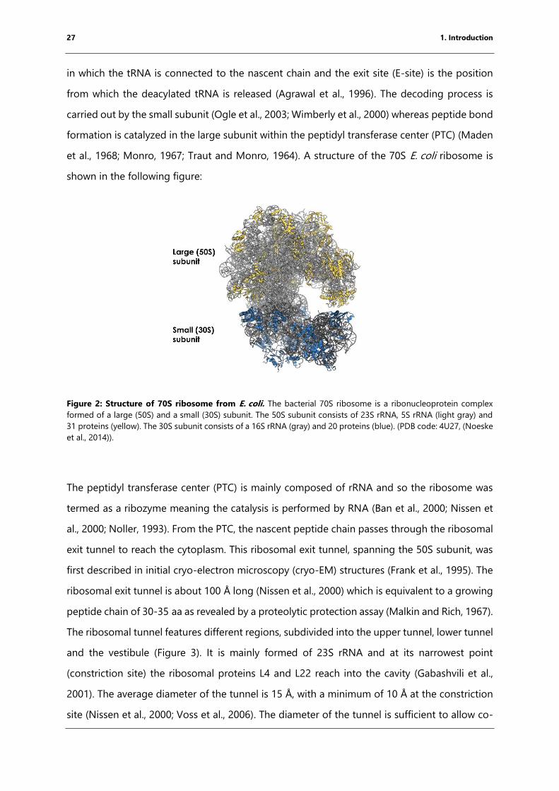
et al., 1968; Monro, 1967; Traut and Monro, 1964). A structure of the 70S E. coli ribosome is
shown in the following figure:
Figure 2: Structure of 70S ribosome from E. coli. The bacterial 70S ribosome is a ribonucleoprotein complex
formed of a large (50S) and a small (30S) subunit. The 50S subunit consists of 23S rRNA, 5S rRNA (light gray) and
31 proteins (yellow). The 30S subunit consists of a 16S rRNA (gray) and 20 proteins (blue). (PDB code: 4U27, (Noeske
et al., 2014)).
The peptidyl transferase center (PTC) is mainly composed of rRNA and so the ribosome was
termed as a ribozyme meaning the catalysis is performed by RNA (Ban et al., 2000; Nissen et
al., 2000; Noller, 1993). From the PTC, the nascent peptide chain passes through the ribosomal
exit tunnel to reach the cytoplasm. This ribosomal exit tunnel, spanning the 50S subunit, was
first described in initial cryo-electron microscopy (cryo-EM) structures (Frank et al., 1995). The
ribosomal exit tunnel is about 100 Å long (Nissen et al., 2000) which is equivalent to a growing
peptide chain of 30-35 aa as revealed by a proteolytic protection assay (Malkin and Rich, 1967).
The ribosomal tunnel features different regions, subdivided into the upper tunnel, lower tunnel
and the vestibule (Figure 3). It is mainly formed of 23S rRNA and at its narrowest point
(constriction site) the ribosomal proteins L4 and L22 reach into the cavity (Gabashvili et al.,
2001). The average diameter of the tunnel is 15 Å, with a minimum of 10 Å at the constriction
site (Nissen et al., 2000; Voss et al., 2006). The diameter of the tunnel is sufficient to allow co-
28 1. Introduction
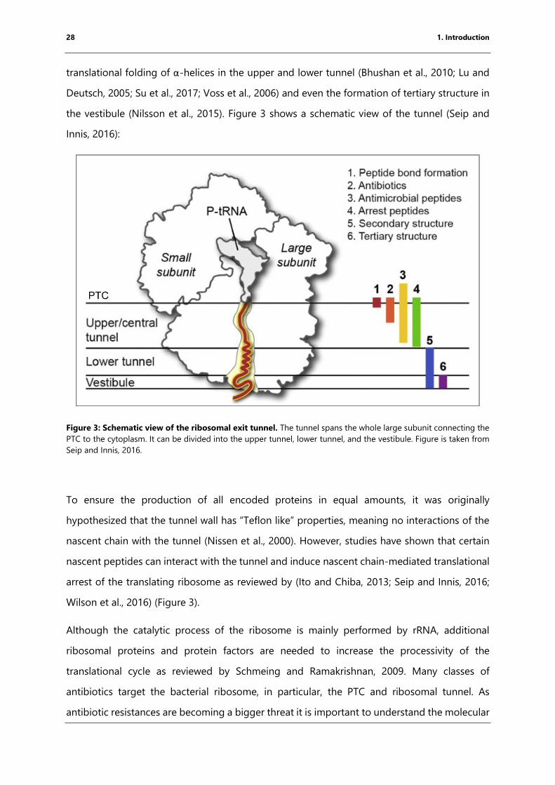
translational folding of α-helices in the upper and lower tunnel (Bhushan et al., 2010; Lu and
Deutsch, 2005; Su et al., 2017; Voss et al., 2006) and even the formation of tertiary structure in
the vestibule (Nilsson et al., 2015). Figure 3 shows a schematic view of the tunnel (Seip and
Innis, 2016):
Figure 3: Schematic view of the ribosomal exit tunnel. The tunnel spans the whole large subunit connecting the
PTC to the cytoplasm. It can be divided into the upper tunnel, lower tunnel, and the vestibule. Figure is taken from
Seip and Innis, 2016.
To ensure the production of all encoded proteins in equal amounts, it was originally
hypothesized that the tunnel wall has “Teflon like” properties, meaning no interactions of the
nascent chain with the tunnel (Nissen et al., 2000). However, studies have shown that certain
nascent peptides can interact with the tunnel and induce nascent chain-mediated translational
arrest of the translating ribosome as reviewed by (Ito and Chiba, 2013; Seip and Innis, 2016;
Wilson et al., 2016) (Figure 3).
Although the catalytic process of the ribosome is mainly performed by rRNA, additional
ribosomal proteins and protein factors are needed to increase the processivity of the
translational cycle as reviewed by Schmeing and Ramakrishnan, 2009. Many classes of
antibiotics target the bacterial ribosome, in particular, the PTC and ribosomal tunnel. As
antibiotic resistances are becoming a bigger threat it is important to understand the molecular
29 1. Introduction
regulation of the bacterial ribosome as well as the action of antibiotics for further drug
development as reviewed (Wilson, 2009, 2014).
1.1.2 The prokaryotic translation cycle
The bacterial translation cycle can be divided into initiation, elongation and termination phases,
the latter being divided into the release of the produced protein and the recycling of the
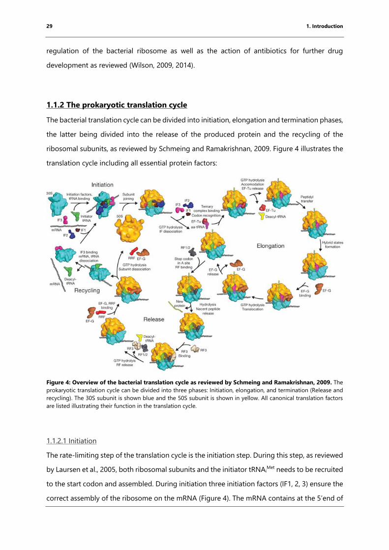
ribosomal subunits, as reviewed by Schmeing and Ramakrishnan, 2009. Figure 4 illustrates the
translation cycle including all essential protein factors:
Figure 4: Overview of the bacterial translation cycle as reviewed by Schmeing and Ramakrishnan, 2009. The
prokaryotic translation cycle can be divided into three phases: Initiation, elongation, and termination (Release and
recycling). The 30S subunit is shown blue and the 50S subunit is shown in yellow. All canonical translation factors
are listed illustrating their function in the translation cycle.
1.1.2.1 Initiation
The rate-limiting step of the translation cycle is the initiation step. During this step, as reviewed
by Laursen et al., 2005, both ribosomal subunits and the initiator tRNAiMet needs to be recruited
to the start codon and assembled. During initiation three initiation factors (IF1, 2, 3) ensure the
correct assembly of the ribosome on the mRNA (Figure 4). The mRNA contains at the 5’end of
30 1. Introduction
the open reading frame (ORF) a ribosomal binding site (RBS), termed in bacteria
Shine-Dalgarno (SD) sequence which is six to seven nucleotides (nts) upstream from the start
codon (Ringquist et al., 1992; Shine and Dalgarno, 1974) The consensus SD sequence is
AGGAGG. The 3’ end of the 16S rRNA in the small ribosomal subunit contains an anti-SD-
sequence forming base pairs with the SD-sequence thus resulting in right positioning of the
small subunit (Shine and Dalgarno, 1974).
In E. coli, 82.6% genes start with an “AUG”, encoding Met, as a start codon and are initiated by
tRNAiMet. Alternative start codons in E. coli are GUG (14.7%, Val), UUG (3.0%, Leu) and two
possible rare ones (Blattner et al., 1997). In order to prevent premature subunit joining IF3 is
bound to the E-site of the small subunit (Carter et al., 2001; Dallas and Noller, 2001; Karimi et
al., 1999; Shine and Dalgarno, 1974). Binding of IF1 to the A-site of the small subunit prevents
the binding of elongator tRNA as well as premature subunit joining (Antoun et al., 2006a;
Antoun et al., 2006b). The initiator-tRNA has a different sequence than the elongator-tRNAMet,
resulting in higher rigidity and affinity for the P-site. Furthermore, the methionine attached to
the tRNAiMet is formylated. The fMet-tRNAi
Met is delivered to the initiation complex by the
translational GTPase (trGTPase) IF2 in its GTP-bound state. (Antoun et al., 2003). Once fMet-
tRNAiMet is positioned properly, GTP hydrolysis is triggered and IF1 and IF3 are released from
the 30S subunit followed by the recruitment of the 50S subunit and the release of IF2 (Simonetti
et al., 2013).
1.1.2.2 Elongation
The following step, elongation, is the most repeated one in the translational cycle as the
ribosome has to go through it for the incorporation of each amino acid. During elongation, the
mRNA is decoded by the aid of tRNAs and the 30S subunit. Base triplets encode for a particular
amino acid termed genetic code (Crick et al., 1961). Amino acids are attached to their cognate
tRNA through the activity of specific aminoacyl-tRNA synthetases in an ATP-dependent
process. Aminoacylation is a process with high enzymatic selectivity. Next, to their synthetase
activity these enzymes have also and editing domain for the specific removal of wrongly
attached amino acids as a proof-reading domain (Ibba et al., 1999). Specificity is ensured by
e.g. a double sieve mechanism (Ibba et al., 1997; Nureki et al., 1998; Schmidt and Schimmel,
31 1. Introduction
1994) or the presence of cations (Dock-Bregeon et al., 2000) ensuring the discrimination of the
chemical properties of similar amino acids.
Aminoacylated-tRNAs are recognized and bound by Elongation Factor (EF)-Tu in its GTP bound
state (Nissen et al., 1995). The genetic code contains 64 possible codon combinations for only
20 amino acids and two release factors. The consequence is that multiple codons encode the
same amino acid. To reduce the number of tRNAs within the cell, only the first two bases of
the codon form Watson-Crick base pairs with the mRNA while the third base can form wobble
base pairs (Ebel et al., 1973; Speyer et al., 1963). The aminoacylated-tRNA in complex with EF-
Tu is then recruited to the ribosome allowing the tRNA to form codon-anticodon interactions
with the mRNA. The decoding process involves the small subunit detecting the right base
pairing by flipping the bases A1493 and A1492 of helix 44, resulting in conformational changes
within the L7/12 stalk and sarcin-ricin loop, triggering GTP hydrolysis within EF-Tu and its
release from the ribosome (Diaconu et al., 2005; Ogle et al., 2003; Schmeing et al., 2009). For
peptide bond formation to occur, the amino acid attached to the 3’CCA end of the A-site tRNA
needs to be well positioned in the A-crevice within the PTC. During A-site tRNA
accommodation, the tRNA has to go through large conformational changes rendering it the
rate-limiting step of peptide bond formation (Pape et al., 1998; Valle et al., 2002).
Processes studied during the work of this thesis are known to target and influence peptide
bond formation and so the following paragraphs discuss this process in detail. During A-site
tRNA accommodation, the residues of the 23S rRNA undergo large conformational changes
switching from the uninduced to the induced state (Schmeing et al., 2005c). In the uninduced
state, the ester bond between the P-site tRNA and the peptide needs to be protected from
premature hydrolysis. The ester bond is sequestered by the bases of the 23S rRNA U2585,
A2451, and C2063 (Schmeing et al., 2005c). A-site tRNA accommodation induces a
rearrangement of 23S ribosomal RNA residues due to base pairing of C75 of the A-site tRNA
with G2553 resulting in movement of U2585 and the reorientation of the ribose of A76 of the
P-site tRNA (induced fit) (Schmeing et al., 2005c). The induced fit brings the carbonyl ester
carbon of the P-site tRNA in close proximity of the α-amino (NH2) group of the following amino
acid (Schmeing et al., 2005c). As a consequence, a peptide bond can be formed by the
nucleophilic attack of the α-amino (NH2) group of the A-site amino acid to the carbonyl ester
carbon of the peptidyl-tRNA. The nucleophilic attack leads to a tetrahedral oxyanion transition
32 1. Introduction
state that breaks down leaving the peptide chain attached to the A-site tRNA. The ribosome
acts as an entropic trap that changes binding affinities for the different binding sites thus
enhancing the reaction by a factor of 107 (Sievers et al., 2004).
To enhance the nucleophilicity of the α-NH2 group of the A-site amino acid, it needs to be
deprotonated. Early X-ray crystallography structures showed that no ribosomal protein was
observed within 15 Å of the PTC (Ban et al., 2000; Nissen et al., 2000). Consequently, the
ribosome is deemed to act as a ribozyme which was postulated first in 1993 (Noller, 1993). The
resulting possible reaction mechanisms that can be catalyzed by RNA are acid-base catalysis,
metal catalysis and a substrate-induced mechanism as reviewed by (Leung et al., 2011). The
acid-base mechanism based on a crystal structure of the Haloarcula martismortui 50S subunit
in complex with the Yarus inhibitor that mimics the transition state of peptide bond formation.
It was proposed due to the close proximity of the N3 of A2451 to the Yarus inhibitor (Nissen
et al., 2000). A2451 is one of the five universally conserved bases located in the PTC (Thompson
et al., 2001; Youngman et al., 2004). It was also hypothesized that the base of G2447 lowers the
pKa of A2451 due to hydrogen bonding increasing the basicity of the N3 atom and this will
facilitate the deprotonation of the α-amino group (Nissen et al., 2000). Mutation of these two
bases led to a growth defect but mutant ribosomes are still capable of peptide bond formation
(Beringer et al., 2003; Beringer et al., 2005; Polacek et al., 2001; Thompson et al., 2001). The
pH independence of the peptide bond formation was demonstrated by replacing α-amino
acids by α-hydroxy acids allowing to perform the reaction at different pH ranges. The result
was that the reaction rate of the ribosome is independent of the pH of the solution (Bieling et
al., 2006). This makes an acid-base catalyzed mechanism unlikely (Bieling et al., 2006) and is
consistent with results from molecular dynamics studies (Trobro and Åqvist, 2005). X-ray
crystallography structures containing substrate and transition state mimics showed that no
Mg2+ or other cations could be localized within the PTC and that instead well-ordered water
molecules could be detected (Schmeing et al., 2005b). This fact makes a metal-catalyzed
mechanism also unlikely.
This leaves substrate induced mechanism as a mechanism that can be catalyzed by RNA. In this
case, tRNAs deliver the amino acids to the A-site and are attached to the growing peptide
chain in the P-site. Biochemical and molecular dynamics studies have shown that the 2’OH
group of A76 is necessary for peptide bond formation when the tRNA is located in the P-site
33 1. Introduction
(Dorner et al., 2003; Dorner et al., 2002; Trobro and Åqvist, 2005; Weinger et al., 2004). The
2’OH group of A76 could be a part of a six-membered proton shuttle between the nascent
chain and the α-NH2 group of A-site amino acid ensuring its deprotonation and stabilization
of the oxyanion transition state (Dorner et al., 2002). The well-ordered water molecule in close
proximity to the 2’OH group of A76 of the P-site tRNA could possibly form an eight-membered
proton shuttle with a second water molecule that stabilizes potentially the oxyanion transition
state (Schmeing et al., 2005b). Molecular dynamics showed that the eight-membered proton
shuttle is preferred over the six-membered proton shuttle (Wallin and Åqvist, 2010)
Lacking of the 2’OH group of A2451 leads to reduction in peptide bond formation by a factor
of 1000 fold as shown biochemically (Erlacher et al., 2005; Erlacher et al., 2006; Lang et al., 2008)
and in molecular dynamics studies (Trobro and Åqvist, 2005) which was not a part of the proton
shuttle models. A recent study involving high-resolution X-ray structures of the PRE- and POST-
catalysis state of the 70S T. thermophilus ribosome with full-length tRNAs revealed three
trapped water molecules within the PTC. This led to the postulation of the proton wire model,
which takes into account the involvement of the 2’OH of A2451 in contrast to previous models
(Polikanov et al., 2014).
After the peptide bond is formed, the nascent peptide is attached to the A-site tRNA and the
P-site tRNA is deaminoacylated. The consequence is that the binding affinities of the tRNAs for
their current positions are decreased. This results in movement of the tRNAs between two PRE-
translocation states: The classical PRE-state (A/A, P/P) and the hybrid state (A/P, P/E) meaning
that the CCA-ends of the tRNAs move towards the next codon leading to the rotation of the
50S subunit compared to the 30S subunit by 6° (Frank and Agrawal, 2000; Munro et al., 2007).
As the last step of the elongation cycle, the ribosome needs to be translocated exactly one
codon further towards the 3’ end of the ORF. This step is catalyzed by EF-G, which recognizes
the hybrid state of the ribosome in its GTP-bound form (Agrawal et al., 1998). Time-resolved
cryo-EM as well as high-resolution structures of EF-G bound to the bacterial ribosome and
trapped in PRE-, intermediated and POST-translocation states gave great insights into
movements of the protein factor, tRNAs, mRNA and the ribosome Due to GTP hydrolysis
triggered by the L7/12 stalk and sarcin-ricin loop, EF-G undergoes a large conformational
change (Chen et al., 2013; Fischer et al., 2010; Gao et al., 2009; Lin et al., 2015; Pulk and Cate,
2013; Tourigny et al., 2013; Zhou et al., 2013). EF-G was reported to act as Brownian ratchet
34 1. Introduction
directing the movement of the ribosome along the mRNA as reviewed in detail by Rodnina
and Wintermeyer, 2011 while a recent X-ray crystallographic study suggests that the
conformational change of EF-G applies a power stroke resulting in translocation of the bacterial
ribosome (Lin et al., 2015). A recent high resolution single molecular study led to the
observation that translocation is driven by EF-G acting as a Brownian ratchet and by using a
power stroke (Chen et al., 2016). The elongation cycle is a very fast step due to the fact that
the bacterial ribosome incorporates 10-20 aa per second (s) (Sørensen et al., 1989).
1.1.2.3 Peptide release and recycling
Termination is the last step of the translation cycle. Therefore, the stop codon has to be
recognized, the peptide has to be released and the ribosome has to be disassembled into its
subunits as reviewed by (Korostelev, 2011). The three stop codons (UGA, UAA, UAG) (Brenner
et al., 1967; Brenner et al., 1965) are recognized by class I peptide termination factors. The UAA
stop codon can be recognized by the two release factors (RF1, 2) found in E. coli. Due to a
slight difference in the primary structure of the release factors, RF1 recognizes the UAG and
RF2 recognizes the UGA stop codon (Klaholz et al., 2003; Petry et al., 2008; Weixlbaumer et al.,
2008). The peptide is released by the positioning of a water molecule by the conserved GGQ
motif of RF1/2 reaching into the PTC changing its activity to an esterase (Jin et al., 2010;
Korostelev et al., 2008; Weixlbaumer et al., 2008). Subsequently, the trGTPase RF3, a class II
peptide termination factor, releases RF1 or RF2 from the ribosome under GTP hydrolysis
(Freistroffer et al., 1997; Goldstein and Caskey, 1970; Jin et al., 2011). The GTP hydrolysis is
triggered by the L7/12 stalk and the sarcin-ricin loop (Diaconu et al., 2005; Zavialov et al., 2001).
As the last step, the deaminoacylated P-site tRNA is released and the 70S ribosome is
disassembled into the individual subunits. Therefore, the ribosome recycling factor (RRF) and
EF-G work together under NTP hydrolysis (Barat et al., 2007; Pavlov et al., 1997; Weixlbaumer
et al., 2007). To prevent the large subunit from reassociation to the small subunit, IF3 binds to
its previously described position and the translation cycle can be initiated again (Antoun et al.,
2006b).
In summary, peptide bond formation is independent of energy use and catalyzed by rRNA,
whereas the steps of subunit assembly, tRNA aminoacylation, and delivery, translocation, the
35 1. Introduction
release of the release factors 1 or 2 and subunit dissociation require the NTP hydrolysis and
protein factors (Schmeing and Ramakrishnan, 2009).
1.2 The prokaryotic ribosome as a target for antibiotics
Antibiotics target macromolecular complexes involved in cell wall synthesis, genome
maintenance as well as protein synthesis as reviewed by (Kohanski et al., 2010). More than 50%
of known antibiotics target the bacterial ribosome affecting all steps of the translational cycle.
Known antibiotic binding sites include the decoding center (e.g. Aminoglycosides), sarcin-ricin
loop (e.g. Thiopeptides), factor binding (e.g. Fusidic acid), peptidyl transferase center (e.g.
Phenicols) and the ribosomal exit tunnel (e.g. Macrolides) as reviewed in detail (Wilson, 2009,
2011, 2014).
The large increase of antibiotic resistance, which is a consequence of excessive antibiotic use
over the last decades, represents a global problem. Studies have shown that resistance against
antibiotics arises rapidly after their introduction into medical use. The macrolide Erythromycin
was introduced into clinical use in 1952 and the first resistant bacterial strain was isolated in
1955 as reviewed by (Lewis, 2013). Strategies of antibiotic resistance can be classified into the
following categories as reviewed in detail by (Roberts, 2008; Wilson, 2014): disruption of the
binding site by mutation or modification, enzymatic modification/degradation of the drug or
active export of the drug.
The following chapter will focus on antibiotics that target specifically the PTC and macrolides
targeting the ribosomal exit tunnel. In addition, they can act as ligands during nascent-chain
mediated translational arrest (chapter 1.3.2).
36 1. Introduction
1.2.1 Antibiotics targeting the PTC
The PTC is a major target for antibiotics. Classes that target specifically the PTC are Phenicols
(Chloramphenicol, CAM), Oxazolidinones (Linezolid, LIN), Lincosamides (Clindamycin, CLN),
Sparsomycin (SPAR), Puromycins, Pleuromutilins (Tiamulin, TIA), Hygromycin A, A203A and
Blasticidin S (BlaS) as reviewed (Arenz and Wilson, 2016; Wilson, 2011).
Most of these classes bind to the A-site within the PTC. Puromycin is an antibiotic that reacts
covalently with the nascent chain and results in premature termination as reviewed (Darken,
1964). Puromycin is active against eukaryotic as well as prokaryotic ribosomes meaning it
cannot be used for treatments (Darken, 1964). Nevertheless, puromycin or its derivatives are
used in several biochemical, kinetic and structural studies to study peptide bond formation and
nascent chain-mediated translational arrest (Muto et al., 2006; Schmeing et al., 2002; Sievers et
al., 2004; Traut and Monro, 1964). Other antibiotics that bind to the A-site, like chloramphenicol
and clindamycin, bind to or in close proximity to the A-site crevice, the binding pocket for the
A-site aminoacyl moiety, thus preventing the accommodation of the aminoacyl-tRNA as shown
in X-ray crystal structures (Bulkley et al., 2010; Dunkle et al., 2010; Wilson et al., 2008). The
antibiotic Hygromycin A binds to the CCA binding pocket of the A-site resulting in the
inhibition of A-site tRNA accommodation (Polikanov et al., 2015b).
Sparsomycin is an antibiotic that, like puromycin, is potent against eukaryotic as well as
prokaryotic ribosomes. It was reported that Sparsomycin induces precise translocation of the
ribosome in the absence of EF-G (Fredrick and Noller, 2003). In addition, Sparsomycin stabilizes
the growing peptide chain and prevents further A-site accommodation. This particular property
is used in structural biology to force the binding short peptidyl-RNA fragments, that mimic
peptidyl chain and the 3’ CCA end of tRNAs, to the P-site (Melnikov et al., 2016a; Schmeing et
al., 2005a). Pleuromutilins like Tiamulin bind to the A as well as the P-site blocking the
elongation of peptide growth due to steric hindrance (Gürel et al., 2009). Blasticidin S binds to
the P-site and deforms the 3’ CCA-end of the P-site tRNA. This protects the peptide chain from
elongation and release (Svidritskiy et al., 2013).
All in all, antibiotics that target the PTC inhibit peptide bond formation by hydrolysis of the
peptide chain, stabilization of the peptide chain, preventing A-site tRNA accommodation or
steric hindrance of prolongation of the growing peptide chain (Wilson, 2011).
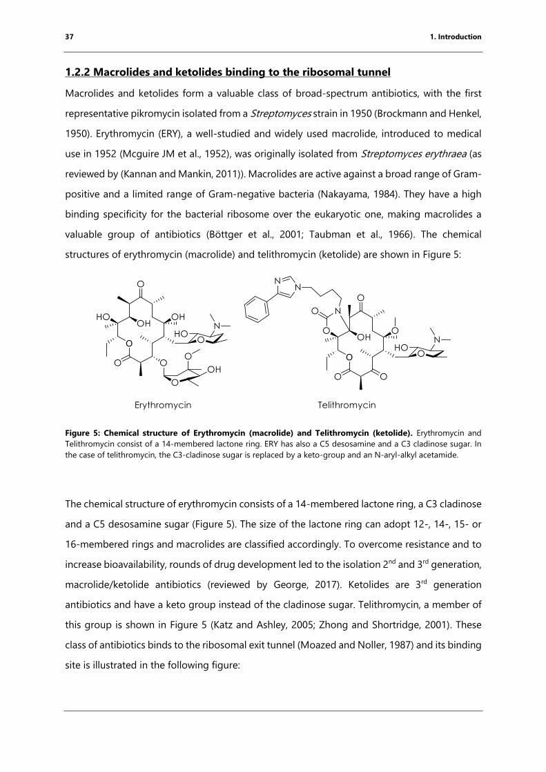
37 1. Introduction
1.2.2 Macrolides and ketolides binding to the ribosomal tunnel
Macrolides and ketolides form a valuable class of broad-spectrum antibiotics, with the first
representative pikromycin isolated from a Streptomyces strain in 1950 (Brockmann and Henkel,
1950). Erythromycin (ERY), a well-studied and widely used macrolide, introduced to medical
use in 1952 (Mcguire JM et al., 1952), was originally isolated from Streptomyces erythraea (as
reviewed by (Kannan and Mankin, 2011)). Macrolides are active against a broad range of Gram-
positive and a limited range of Gram-negative bacteria (Nakayama, 1984). They have a high
binding specificity for the bacterial ribosome over the eukaryotic one, making macrolides a
valuable group of antibiotics (Böttger et al., 2001; Taubman et al., 1966). The chemical
structures of erythromycin (macrolide) and telithromycin (ketolide) are shown in Figure 5:
Figure 5: Chemical structure of Erythromycin (macrolide) and Telithromycin (ketolide). Erythromycin and
Telithromycin consist of a 14-membered lactone ring. ERY has also a C5 desosamine and a C3 cladinose sugar. In
the case of telithromycin, the C3-cladinose sugar is replaced by a keto-group and an N-aryl-alkyl acetamide.
The chemical structure of erythromycin consists of a 14-membered lactone ring, a C3 cladinose
and a C5 desosamine sugar (Figure 5). The size of the lactone ring can adopt 12-, 14-, 15- or
16-membered rings and macrolides are classified accordingly. To overcome resistance and to
increase bioavailability, rounds of drug development led to the isolation 2nd and 3rd generation,
macrolide/ketolide antibiotics (reviewed by George, 2017). Ketolides are 3rd generation
antibiotics and have a keto group instead of the cladinose sugar. Telithromycin, a member of
this group is shown in Figure 5 (Katz and Ashley, 2005; Zhong and Shortridge, 2001). These
class of antibiotics binds to the ribosomal exit tunnel (Moazed and Noller, 1987) and its binding
site is illustrated in the following figure:

38 1. Introduction
Figure 6: Binding site of erythromycin within the bacterial ribosome. Overlay of the 70S E. coli ribosome
(turquoise; PDB code: 4V7U) and 70S T. thermophilus ribosome (blue; PDB code: 4V7X) in complex with
erythromycin illustrates the similar conformations of the drug bound to the ribosomal tunnel. The bases of the 23S
rRNA that form the binding site are shown in gray. For simplification reasons, only the bases from the
T. thermophilus 70S ribosome structure are shown. A2059 and U2611 form a hydrophobic surface resulting in
stacking interactions with the lactone ring (Bulkley et al., 2010; Dunkle et al., 2010).
Extensive biochemical studies identified the upper ribosomal exit tunnel as the binding site for
erythromycin allowing the accommodation of the A-site tRNA (Celma et al., 1971; Moazed and
Noller, 1987). Several X-ray crystallography studies identified and proved the biochemical
binding sites but different orientations of the lactone ring were observed (Berisio et al., 2003;
Schlunzen et al., 2001; Tu et al., 2005). More recent studies have shown that different
macrolides and telithromycin bound to the 70S E. coli (Dunkle et al., 2010) and T. thermophilus
(Bulkley et al., 2010) ribosome have similar conformation of the lactone ring independently
from drug and species (Figure 6). Erythromycin forms stable interactions with the ribosomal
exit tunnel via base A2058 while the lactone ring stacks against the hydrophobic surface
formed by A2059 and U2611 (Bulkley et al., 2010; Dunkle et al., 2010; Figure 6).
This binding site within the ribosomal tunnel originally suggested that macrolides work like a
plug, inhibiting protein synthesis in the early elongation phase and leading to peptidyl-tRNA
drop-off (Menninger, 1995; Menninger and Otto, 1982; Tenson et al., 2003). Further
investigations identified nascent chain peptide sequences that lead to the arrest of the
ribosome in the presence of the macrolides (Gryczan et al., 1980; Horinouchi and Weisblum,
1980) (more detailed chapter 2). More recent studies have shown that cells, which are exposed
39 1. Introduction
to erythromycin or telithromycin, can still produce a subset of proteins. The by-passing of the
drug is possible due to a specific amino acid composition of these proteins in their N-terminus
(ILNNIR or IGQQVR) (Kannan et al., 2012). Another effect is drug-induced frameshifting that
can be observed in the presence of ketolides like telithromycin (Gupta et al., 2013).
1.3 Proline-rich antimicrobial peptides (PrAMPs) as possible new
therapeutics
All organisms produce substances to protect themselves against competitors for nutrients and
against pathogens. Peptides or glycopeptides like Vancomycin represent a large proportion of
these defense substances. They can be produced by the ribosome or by large multiprotein
complexes called non-ribosomal synthetases, which are capable of incorporating
nonproteinogenic amino acids into peptides or macrocycles (Hur et al., 2012). Antimicrobial
peptides (AMPs) can be classified by their structure, such as α-helices, β-sheets or macrocycles,
by their mode of action, like lytic or non-lytic, or by their amino acid composition, like
hydrophobic, amphiphilic or specific amino acid-rich as reviewed in detail by (Epand and Vogel,
1999; Otvos, 2005).
One class of AMPs are proline-rich antimicrobial peptides (PrAMPs) which are produced as a
part of the innate immune system of higher eukaryotes (Otvos, 2002). Representative members
have been isolated and characterized from the milkweed bug (Oncopeltus fasciatus, (Schneider
and Dorn, 2001)), honey bees (Apis mellifera, (Casteels et al., 1990)), green shield bug
(Palomena prasina, (Chernysh et al., 1996)), Firebug (Pyrrhocoris apterus, (Cociancich et al.,
1994)) and even domestic mammals like cows (Bos taurus, (Scocchi et al., 1994)). As PrAMPs
are incapable of entering the eukaryotic cell (Hansen et al., 2012) and therefore, represent a
promising class of antimicrobials. Additionally, these peptides can also be used to treat
infections affecting the brain as they can cross the blood-brain barrier (Stalmans et al., 2014).
A representative list of sequences is shown in Figure 7:
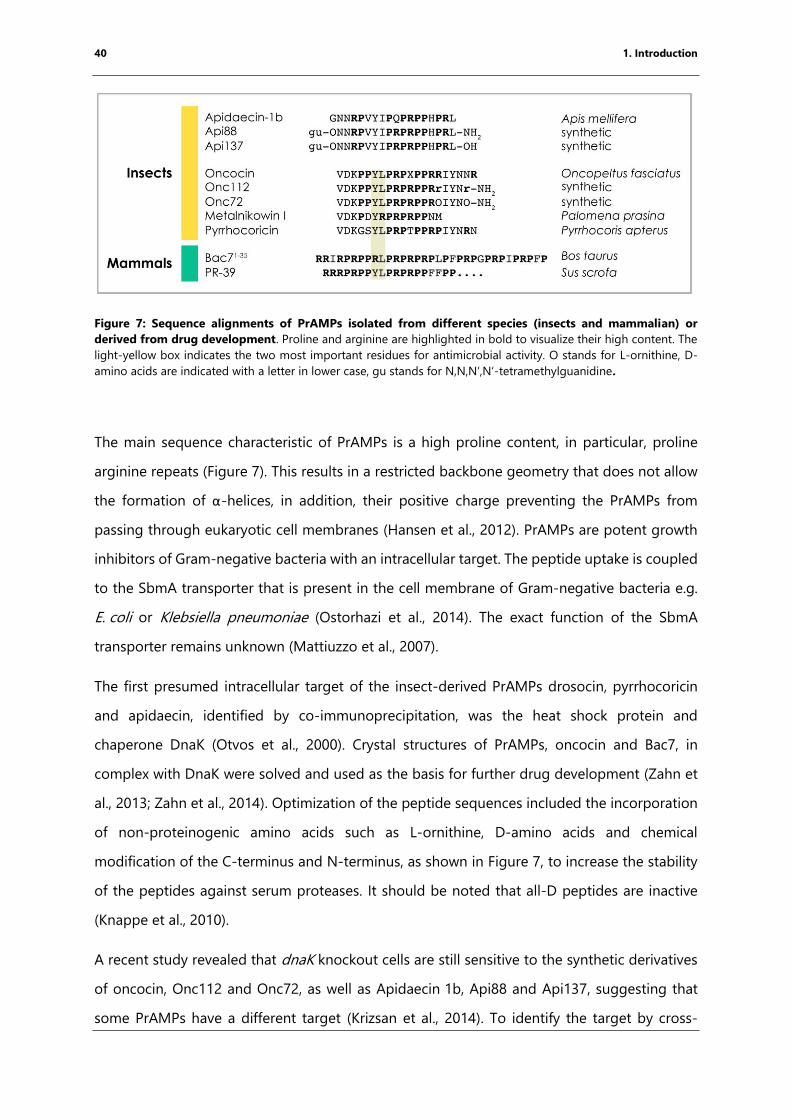
40 1. Introduction
Figure 7: Sequence alignments of PrAMPs isolated from different species (insects and mammalian) or
derived from drug development. Proline and arginine are highlighted in bold to visualize their high content. The
light-yellow box indicates the two most important residues for antimicrobial activity. O stands for L-ornithine, D-
amino acids are indicated with a letter in lower case, gu stands for N,N,N‘,N‘-tetramethylguanidine.
The main sequence characteristic of PrAMPs is a high proline content, in particular, proline
arginine repeats (Figure 7). This results in a restricted backbone geometry that does not allow
the formation of α-helices, in addition, their positive charge preventing the PrAMPs from
passing through eukaryotic cell membranes (Hansen et al., 2012). PrAMPs are potent growth
inhibitors of Gram-negative bacteria with an intracellular target. The peptide uptake is coupled
to the SbmA transporter that is present in the cell membrane of Gram-negative bacteria e.g.
E. coli or Klebsiella pneumoniae (Ostorhazi et al., 2014). The exact function of the SbmA
transporter remains unknown (Mattiuzzo et al., 2007).
The first presumed intracellular target of the insect-derived PrAMPs drosocin, pyrrhocoricin
and apidaecin, identified by co-immunoprecipitation, was the heat shock protein and
chaperone DnaK (Otvos et al., 2000). Crystal structures of PrAMPs, oncocin and Bac7, in
complex with DnaK were solved and used as the basis for further drug development (Zahn et
al., 2013; Zahn et al., 2014). Optimization of the peptide sequences included the incorporation
of non-proteinogenic amino acids such as L-ornithine, D-amino acids and chemical
modification of the C-terminus and N-terminus, as shown in Figure 7, to increase the stability
of the peptides against serum proteases. It should be noted that all-D peptides are inactive
(Knappe et al., 2010).
A recent study revealed that dnaK knockout cells are still sensitive to the synthetic derivatives
of oncocin, Onc112 and Onc72, as well as Apidaecin 1b, Api88 and Api137, suggesting that
some PrAMPs have a different target (Krizsan et al., 2014). To identify the target by cross-
41 1. Introduction
linking, Tyr7 of Api88 was replaced by p-benzoylphenyl alanine, a photoactive derivative. In
addition, Api88 was biotinylated for the purification of the crosslinking products. The direct
result was that Api88 was cross-linked to the ribosomal protein L10 (Krizsan et al., 2014).
Follow-up in vitro protein synthesis assays revealed that these peptides inhibit protein
biosynthesis and show a lower dissociation constant (Kd) for the bacterial 70S ribosome than
for DnaK (Krizsan et al., 2014). A different study, working with the mammalian-derived PrAMP
Bac71-35, the shortened form of the normally 60 aa long Bac7 peptide, identified by pulse-
labeling experiments that the peptide is transported into the cell and blocks the production of
proteins. Additionally, this Bac71-35 was co-sedimented with 70S ribosomes (Mardirossian et al.,
2014). However, the molecular mechanism of action remains unknown.
42 1. Introduction
1.4 Nascent chain-mediated translational arrest
During protein synthesis, the translating ribosome can stall along the mRNA due to various
reasons including rare codons, amino acid starvation, antibiotic binding, mRNA secondary
structure or the amino acid sequence of the nascent peptide. A stalling event that is mediated
by the translated amino acid sequence is termed nascent chain-mediated translational arrest.
This arrest is induced by the particular interactions of the nascent chain with the tunnel wall
resulting in a rearrangement of ribosomal bases and can result in a specific path or fold of the
nascent chain within the tunnel. This process can be aided by the binding of a small ligand,
antibiotic or metabolite. Thus, the nascent chain acts as a sensor for the presence or absence
of this particular ligand within the cell and so gene expression can be modulated in prokaryotes
as well as eukaryotes (reviewed in detail Chiba et al., 2011; Ramu et al., 2009; Seip and Innis,
2016; Wilson et al., 2016). This section will focus on nascent chain-mediated translational arrest
in bacteria.
After the identification of the first arrest peptide, ermCL, in 1980 (Gryczan et al., 1980;
Horinouchi and Weisblum, 1980) different methods were developed to study nascent chain-
mediated translational arrest in vitro as well as in vivo. In vitro methods include toeprinting,
which uses in vitro translation systems that give at nucleotide resolution of the position of the
ribosome along the mRNA under stalling and nonstalling conditions using primer extension
(detailed description chapter 3.8) (Hartz et al., 1988; Orelle et al., 2013a). Arrest peptides are
less sensitive to puromycin treatment and can be analyzed by acidic gel electrophoresis (Muto
and Ito, 2008). The nature of the P-site tRNA can be identified using northern blotting (Chiba
and Ito, 2012; Muto et al., 2006). Fluorescence resonance energy transfer (FRET) studies can
give great insights into conformational changes of the ribosomes while arrest complex
formation (Johansson et al., 2014; Tsai et al., 2014).
In vivo methods include the expression of a reporter gene, in many studies lacZ that encodes
for β-galactosidase. The arrest can be investigated by fusing the arrest peptide directly to the
reporter gene, meaning if the arrest occurs LacZ is not produced (Martínez et al., 2014; Ude et
al., 2013). Another approach uses the mechanism of translational regulation by replacing the
downstream gene by the reporter gene. In contrast to the fusion protein approach, LacZ is
produced only if the arrest occurs (Gupta et al., 2016).
43 1. Introduction
Ribosome profiling is a high-throughput method to analyze translation events occurring on a
genome-wide scale in vivo (Ingolia et al., 2009) and has shown to be a valuable tool to study
nascent chain-mediated translational arrest. Total mRNA bound to ribosomes is isolated from
cells and digested by nucleases. The remaining mRNA fragments are purified, reverse
transcribed, analyzed by next-generation sequencing (NGS) and mapped to the genome
(Ingolia et al., 2009).
Another method to gain a deepened understanding of the molecular mechanism of nascent
chain-mediated translation arrest is solving the structure of arrested ribosomes using cryo-EM
(Arenz et al., 2016; Arenz et al., 2014a; Arenz et al., 2014b; Bhushan et al., 2011; Bischoff et al.,
2014; Seidelt et al., 2009; Sohmen et al., 2015; Su et al., 2017; Zhang et al., 2015).
In prokaryotes, one mRNA often encodes multiple genes and is thus termed as a polycistronic
mRNA. ORFs regulated by nascent chain-mediated arrest peptides are organized following:
The arrest peptide, termed leader peptide, is encoded at the 5’ end of the mRNA followed by
the ORFs of the regulated genes. The expression of downstream genes can be regulated
through transcriptional or translational mechanisms (Gong and Yanofsky, 2002; Vazquez-
Laslop et al., 2008).
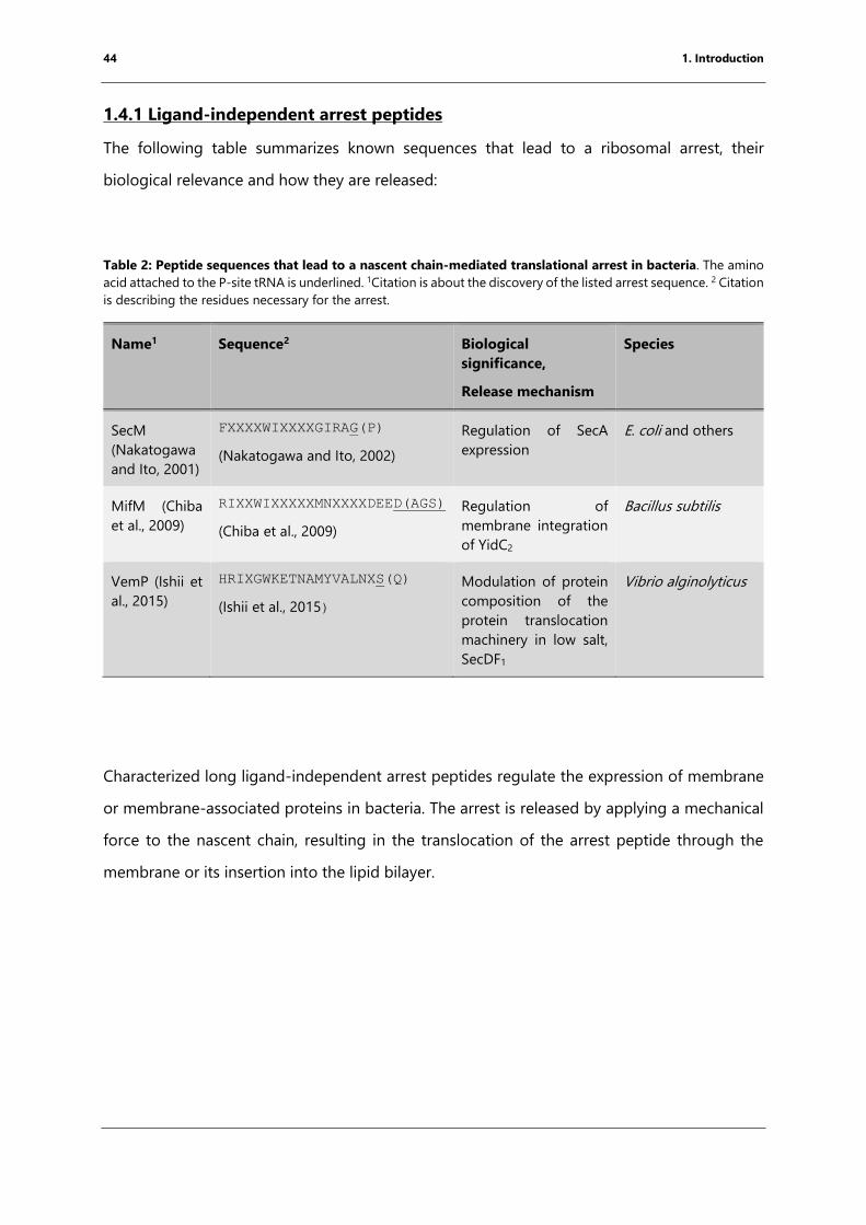
44 1. Introduction
1.4.1 Ligand-independent arrest peptides
The following table summarizes known sequences that lead to a ribosomal arrest, their
biological relevance and how they are released:
Table 2: Peptide sequences that lead to a nascent chain-mediated translational arrest in bacteria. The amino
acid attached to the P-site tRNA is underlined. 1Citation is about the discovery of the listed arrest sequence. 2 Citation
is describing the residues necessary for the arrest.
Name1 Sequence2 Biological
significance,
Release mechanism
Species
SecM
(Nakatogawa
and Ito, 2001)
FXXXXWIXXXXGIRAG(P)
(Nakatogawa and Ito, 2002)
Regulation of SecA
expression
E. coli and others
MifM (Chiba
et al., 2009)
RIXXWIXXXXXMNXXXXDEED(AGS)
(Chiba et al., 2009)
Regulation of
membrane integration
of YidC2
Bacillus subtilis
VemP (Ishii et
al., 2015)
HRIXGWKETNAMYVALNXS(Q)
(Ishii et al., 2015)
Modulation of protein
composition of the
protein translocation
machinery in low salt,
SecDF1
Vibrio alginolyticus
Characterized long ligand-independent arrest peptides regulate the expression of membrane
or membrane-associated proteins in bacteria. The arrest is released by applying a mechanical
force to the nascent chain, resulting in the translocation of the arrest peptide through the
membrane or its insertion into the lipid bilayer.
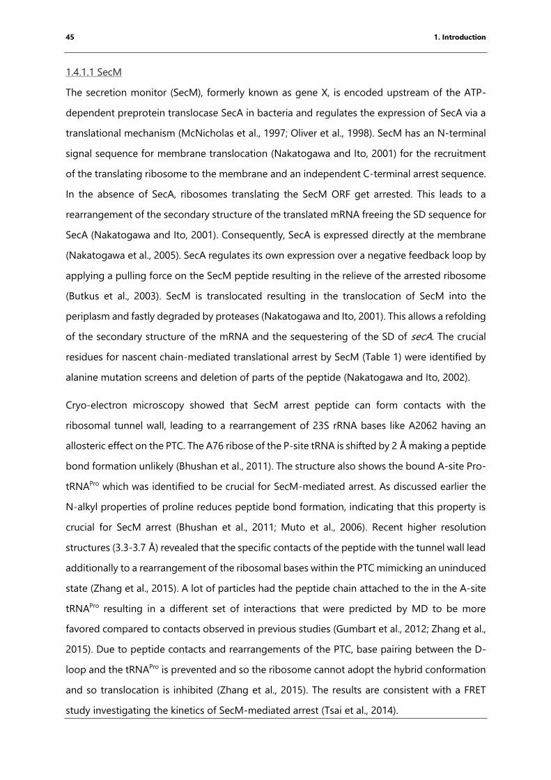
45 1. Introduction
1.4.1.1 SecM
The secretion monitor (SecM), formerly known as gene X, is encoded upstream of the ATP-
dependent preprotein translocase SecA in bacteria and regulates the expression of SecA via a
translational mechanism (McNicholas et al., 1997; Oliver et al., 1998). SecM has an N-terminal
signal sequence for membrane translocation (Nakatogawa and Ito, 2001) for the recruitment
of the translating ribosome to the membrane and an independent C-terminal arrest sequence.
In the absence of SecA, ribosomes translating the SecM ORF get arrested. This leads to a
rearrangement of the secondary structure of the translated mRNA freeing the SD sequence for
SecA (Nakatogawa and Ito, 2001). Consequently, SecA is expressed directly at the membrane
(Nakatogawa et al., 2005). SecA regulates its own expression over a negative feedback loop by
applying a pulling force on the SecM peptide resulting in the relieve of the arrested ribosome
(Butkus et al., 2003). SecM is translocated resulting in the translocation of SecM into the
periplasm and fastly degraded by proteases (Nakatogawa and Ito, 2001). This allows a refolding
of the secondary structure of the mRNA and the sequestering of the SD of secA. The crucial
residues for nascent chain-mediated translational arrest by SecM (Table 1) were identified by
alanine mutation screens and deletion of parts of the peptide (Nakatogawa and Ito, 2002).
Cryo-electron microscopy showed that SecM arrest peptide can form contacts with the
ribosomal tunnel wall, leading to a rearrangement of 23S rRNA bases like A2062 having an
allosteric effect on the PTC. The A76 ribose of the P-site tRNA is shifted by 2 Å making a peptide
bond formation unlikely (Bhushan et al., 2011). The structure also shows the bound A-site Pro-
tRNAPro which was identified to be crucial for SecM-mediated arrest. As discussed earlier the
N-alkyl properties of proline reduces peptide bond formation, indicating that this property is
crucial for SecM arrest (Bhushan et al., 2011; Muto et al., 2006). Recent higher resolution
structures (3.3-3.7 Å) revealed that the specific contacts of the peptide with the tunnel wall lead
additionally to a rearrangement of the ribosomal bases within the PTC mimicking an uninduced
state (Zhang et al., 2015). A lot of particles had the peptide chain attached to the in the A-site
tRNAPro resulting in a different set of interactions that were predicted by MD to be more
favored compared to contacts observed in previous studies (Gumbart et al., 2012; Zhang et al.,
2015). Due to peptide contacts and rearrangements of the PTC, base pairing between the D-
loop and the tRNAPro is prevented and so the ribosome cannot adopt the hybrid conformation
and so translocation is inhibited (Zhang et al., 2015). The results are consistent with a FRET
study investigating the kinetics of SecM-mediated arrest (Tsai et al., 2014).
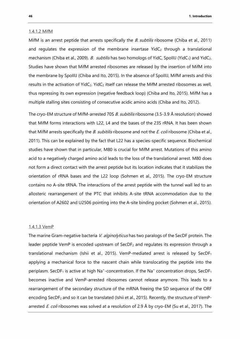
46 1. Introduction
1.4.1.2 MifM
MifM is an arrest peptide that arrests specifically the B. subtilis ribosome (Chiba et al., 2011)
and regulates the expression of the membrane insertase YidC2 through a translational
mechanism (Chiba et al., 2009). B. subtilis has two homologs of YidC, SpoIIIJ (YidC1) and YidC2.
Studies have shown that MifM arrested ribosomes are released by the insertion of MifM into
the membrane by SpoIIIJ (Chiba and Ito, 2015). In the absence of SpoIIIJ, MifM arrests and this
results in the activation of YidC2. YidC2 itself can release the MifM arrested ribosomes as well,
thus repressing its own expression (negative feedback loop) (Chiba and Ito, 2015). MifM has a
multiple stalling sites consisting of consecutive acidic amino acids (Chiba and Ito, 2012).
The cryo-EM structure of MifM-arrested 70S B. subitilis ribosome (3.5-3.9 Å resolution) showed
that MifM forms interactions with L22, L4 and the bases of the 23S rRNA. It has been shown
that MifM arrests specifically the B. subitilis ribosome and not the E. coli ribosome (Chiba et al.,
2011). This can be explained by the fact that L22 has a species-specific sequence. Biochemical
studies have shown that in particular, M80 is crucial for MifM arrest. Mutations of this amino
acid to a negatively charged amino acid leads to the loss of the translational arrest. M80 does
not form a direct contact with the arrest peptide but its location indicates that it stabilizes the
orientation of rRNA bases and the L22 loop (Sohmen et al., 2015). The cryo-EM structure
contains no A-site tRNA. The interactions of the arrest peptide with the tunnel wall led to an
allosteric rearrangement of the PTC that inhibits A-site tRNA accommodation due to the
orientation of A2602 and U2506 pointing into the A-site binding pocket (Sohmen et al., 2015).
1.4.1.3 VemP
The marine Gram-negative bacteria V. alginolyticus has two paralogs of the SecDF protein. The
leader peptide VemP is encoded upstream of SecDF2 and regulates its expression through a
translational mechanism (Ishii et al., 2015). VemP-mediated arrest is released by SecDF1
applying a mechanical force to the nascent chain while translocating the peptide into the
periplasm. SecDF1 is active at high Na+-concentration. If the Na+ concentration drops, SecDF1
becomes inactive and VemP-arrested ribosomes cannot release anymore. This leads to a
rearrangement of the secondary structure of the mRNA freeing the SD sequence of the ORF
encoding SecDF2 and so it can be translated (Ishii et al., 2015). Recently, the structure of VemP-
arrested E. coli ribosomes was solved at a resolution of 2.9 Å by cryo-EM (Su et al., 2017). The

47 1. Introduction
VemP peptide forms two helices, one in the upper and one in the lower tunnel. The two helices
are connected over an linking region forming two consecutive turns through the constriction
site (Su et al., 2017). Residues forming the α-helix within the upper tunnel can form interactions
with the tunnel wall, leading to an allosteric rearrangement within the PTC that stabilizes the
uninduced state of the ribosome. Consequently, the accommodation of the A-site tRNA is
prevented by the orientation of U2506 (Su et al., 2017).
1.4.2 Ligand-dependent arrest peptides
In contrast to ligand-independent arrest peptides, ligand-dependent arrest peptides act as
sensors for the presence of ligands. Ligands can be metabolites or antibiotics binding to the
ribosomal exit tunnel (Seip and Innis, 2016). A nascent chain-mediated translational arrest can
also be induced by specific amino acid sequences and the presence of a ligand, such as an
antibiotic or a metabolite. The ribosome-nascent chain complex (RNC) acts as a sensor for the
ligand. The following table includes ligand-dependent arrest peptides, their ligand, biological
significance and their mechanism of inhibition during the translational cycle:
Table 3: Summary of well-characterized ligand-dependent arrest peptides in bacteria. The amino acid located
in the P-site is underlined. 1citation describing the discovery of the listed arrest sequence. 2citation identified
important residues. +No alanine screen is published for ErmDL.
Name1 Sequence2 Biological
significance, ligand
Inhibition
point
TnaC
(Konan and Yanofsky,
1997)
WXXXDXXIXXXXP(*)
(Gong and Yanofsky,
2002)
(Cruz-Vera et al., 2005)
Regulation of the
transcription of the
tnaBC operon,
tryptophan
Termination
Cat86 leader
(Brückner and Matzura,
1985)
MVKTD(K)
(Alexieva et al., 1988) Regulation of
chloramphenicol
resistance (drug
modification
enzymes),
chloramphenicol
Peptidyl
transfer
CmlA leader
(Dorman and Foster,
1985)
MSTSKNAD(K)
(Gu et al., 1994; Gu et al.,
1993)
Peptidyl
transfer
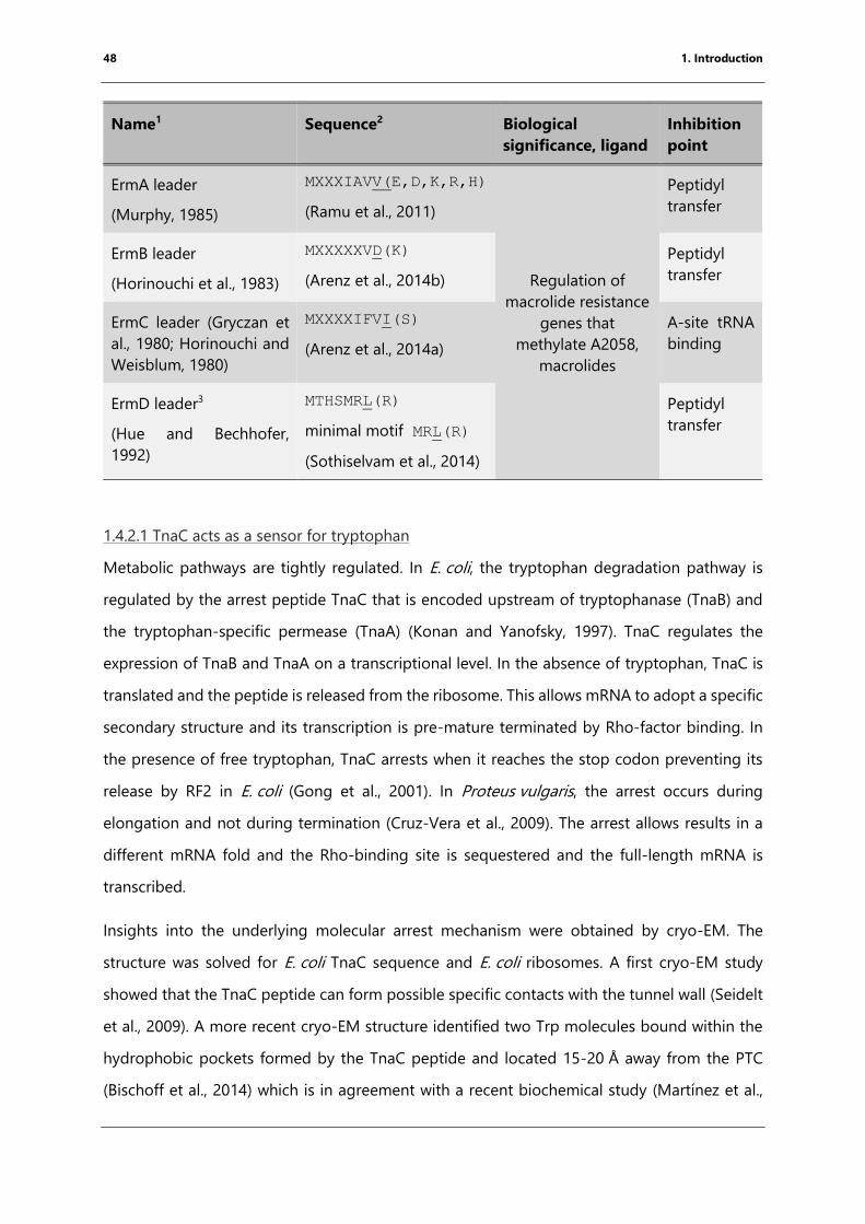
48 1. Introduction
Name1 Sequence2 Biological
significance, ligand
Inhibition
point
ErmA leader
(Murphy, 1985)
MXXXIAVV(E,D,K,R,H)
(Ramu et al., 2011)
Regulation of
macrolide resistance
genes that
methylate A2058,
macrolides
Peptidyl
transfer
ErmB leader
(Horinouchi et al., 1983)
MXXXXXVD(K)
(Arenz et al., 2014b)
Peptidyl
transfer
ErmC leader (Gryczan et
al., 1980; Horinouchi and
Weisblum, 1980)
MXXXXIFVI(S)
(Arenz et al., 2014a)
A-site tRNA
binding
ErmD leader3
(Hue and Bechhofer,
1992)
MTHSMRL(R)
minimal motif MRL(R)
(Sothiselvam et al., 2014)
Peptidyl
transfer
1.4.2.1 TnaC acts as a sensor for tryptophan
Metabolic pathways are tightly regulated. In E. coli, the tryptophan degradation pathway is
regulated by the arrest peptide TnaC that is encoded upstream of tryptophanase (TnaB) and
the tryptophan-specific permease (TnaA) (Konan and Yanofsky, 1997). TnaC regulates the
expression of TnaB and TnaA on a transcriptional level. In the absence of tryptophan, TnaC is
translated and the peptide is released from the ribosome. This allows mRNA to adopt a specific
secondary structure and its transcription is pre-mature terminated by Rho-factor binding. In
the presence of free tryptophan, TnaC arrests when it reaches the stop codon preventing its
release by RF2 in E. coli (Gong et al., 2001). In Proteus vulgaris, the arrest occurs during
elongation and not during termination (Cruz-Vera et al., 2009). The arrest allows results in a
different mRNA fold and the Rho-binding site is sequestered and the full-length mRNA is
transcribed.
Insights into the underlying molecular arrest mechanism were obtained by cryo-EM. The
structure was solved for E. coli TnaC sequence and E. coli ribosomes. A first cryo-EM study
showed that the TnaC peptide can form possible specific contacts with the tunnel wall (Seidelt
et al., 2009). A more recent cryo-EM structure identified two Trp molecules bound within the
hydrophobic pockets formed by the TnaC peptide and located 15-20 Å away from the PTC
(Bischoff et al., 2014) which is in agreement with a recent biochemical study (Martínez et al.,
49 1. Introduction
2014). The peptide forms specific contacts with L22, L4 and 23S rRNA. The PTC is inactivated
due to distinct positions of U2585 and A2602 preventing release factor binding. U2586 forms
stacking interactions with one of the bound Trp-ligands restricting possibly the movement of
U2585. Furthermore, the orientation of the penultimate amino acid clashes with the GGQ motif
of the release factor (Bischoff et al., 2014).
1.4.2.2 Erm leader peptides sense the presence of macrolide antibiotics
The expression of antibiotic resistance genes can be regulated by short leader peptides.
Macrolide and ketolide resistance genes encoding a methyltransferase (erythromycin
resistance methylase, erm), are regulated by upstream encoded leader peptides (Ramu et al.,
2009). Different leader peptide sequences were identified which showed differences in arrest
motifs and could be sorted accordingly. Expression of the downstream erm gene can be
regulated using a translational or transcriptional mechanism. The resistance gene methylates
specifically A2058 resulting in the disruption of the erythromycin binding site (Katz et al., 1987;
Roberts et al., 1999). The first erm leader gene to have been characterized is ermCL (Gryczan
et al., 1980; Horinouchi and Weisblum, 1980). The ErmCL peptide itself has been studied
biochemically and by cryo-EM (Arenz et al., 2014a). The 3.9 Å structure of the arrested 70S
E. coli ribosome reveals stable contacts of the peptide with erythromycin and the ribosomal
tunnel wall resulting in a unique orientation of A2062. This results in a reorientation of bases
within the PTC. U2585 adopts a unique orientation and U2586 forms direct contacts with the
growing peptide chain. A2602 points into the A-site binding pocket prevent A-site tRNA
accommodation (Arenz et al., 2014a).
Another erm leader peptide studied by cryo-EM is ErmBL. The path of the peptide in the
absence of the drug is significantly different from that of ErmCL. ErmBL does not form direct
contacts with the drug but with the tunnel wall (Arenz et al., 2014b). ErmBL adopts a unique
path in the ribosomal tunnel as a consequence of the fact that Asp10 is rotated resulting in a
displacement of A76 of the P-site tRNA. The conformation results in a suboptimal geometry
for the nucleophilic attack of the A-site amino acid (Arenz et al., 2016). The A-site tRNALys is
bound in the structure in a different conformation compared to the fully accommodated state
and U2585 is trapped between uninduced and induced states. Molecular dynamics simulations
have shown that allosteric rearrangement of the PTC due to the presence of erythromycin leads
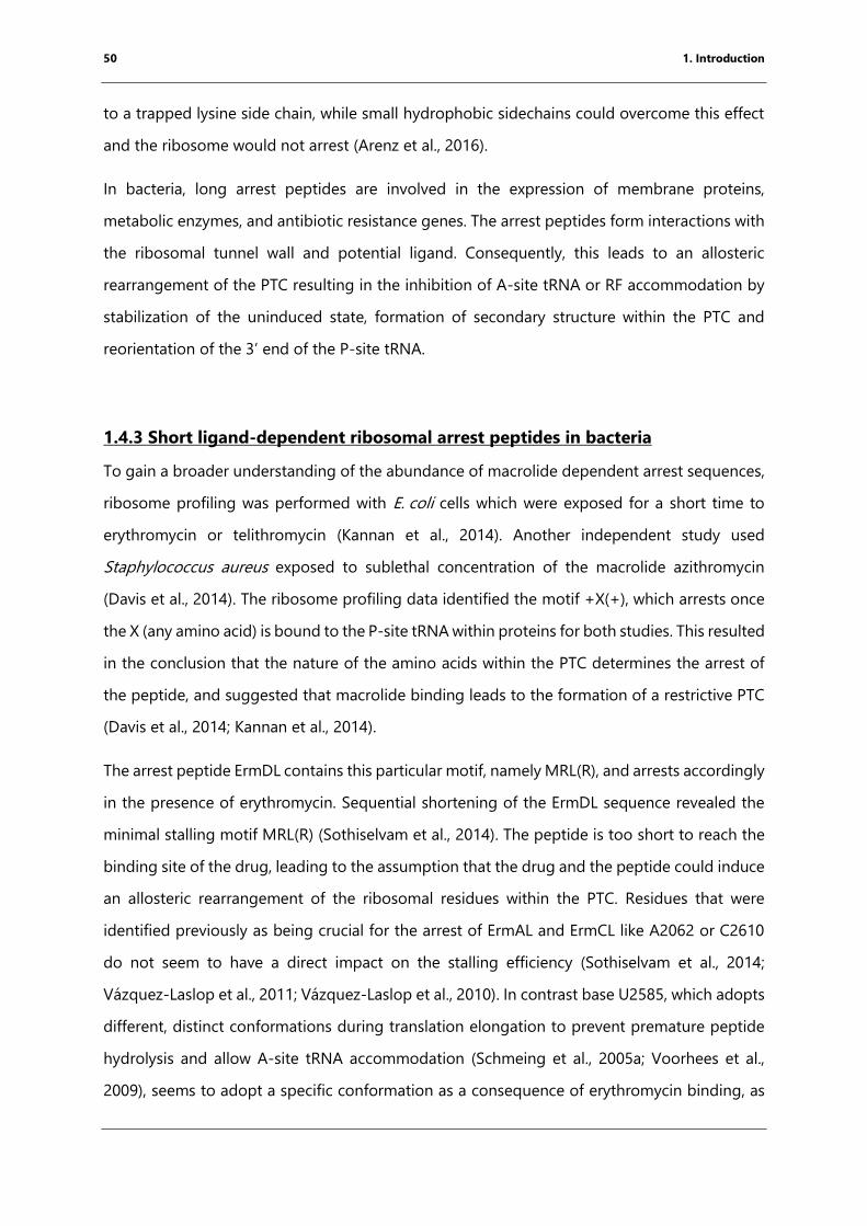
50 1. Introduction
to a trapped lysine side chain, while small hydrophobic sidechains could overcome this effect
and the ribosome would not arrest (Arenz et al., 2016).
In bacteria, long arrest peptides are involved in the expression of membrane proteins,
metabolic enzymes, and antibiotic resistance genes. The arrest peptides form interactions with
the ribosomal tunnel wall and potential ligand. Consequently, this leads to an allosteric
rearrangement of the PTC resulting in the inhibition of A-site tRNA or RF accommodation by
stabilization of the uninduced state, formation of secondary structure within the PTC and
reorientation of the 3’ end of the P-site tRNA.
1.4.3 Short ligand-dependent ribosomal arrest peptides in bacteria
To gain a broader understanding of the abundance of macrolide dependent arrest sequences,
ribosome profiling was performed with E. coli cells which were exposed for a short time to
erythromycin or telithromycin (Kannan et al., 2014). Another independent study used
Staphylococcus aureus exposed to sublethal concentration of the macrolide azithromycin
(Davis et al., 2014). The ribosome profiling data identified the motif +X(+), which arrests once
the X (any amino acid) is bound to the P-site tRNA within proteins for both studies. This resulted
in the conclusion that the nature of the amino acids within the PTC determines the arrest of
the peptide, and suggested that macrolide binding leads to the formation of a restrictive PTC
(Davis et al., 2014; Kannan et al., 2014).
The arrest peptide ErmDL contains this particular motif, namely MRL(R), and arrests accordingly
in the presence of erythromycin. Sequential shortening of the ErmDL sequence revealed the
minimal stalling motif MRL(R) (Sothiselvam et al., 2014). The peptide is too short to reach the
binding site of the drug, leading to the assumption that the drug and the peptide could induce
an allosteric rearrangement of the ribosomal residues within the PTC. Residues that were
identified previously as being crucial for the arrest of ErmAL and ErmCL like A2062 or C2610
do not seem to have a direct impact on the stalling efficiency (Sothiselvam et al., 2014;
Vázquez-Laslop et al., 2011; Vázquez‐Laslop et al., 2010). In contrast base U2585, which adopts
different, distinct conformations during translation elongation to prevent premature peptide
hydrolysis and allow A-site tRNA accommodation (Schmeing et al., 2005a; Voorhees et al.,
2009), seems to adopt a specific conformation as a consequence of erythromycin binding, as
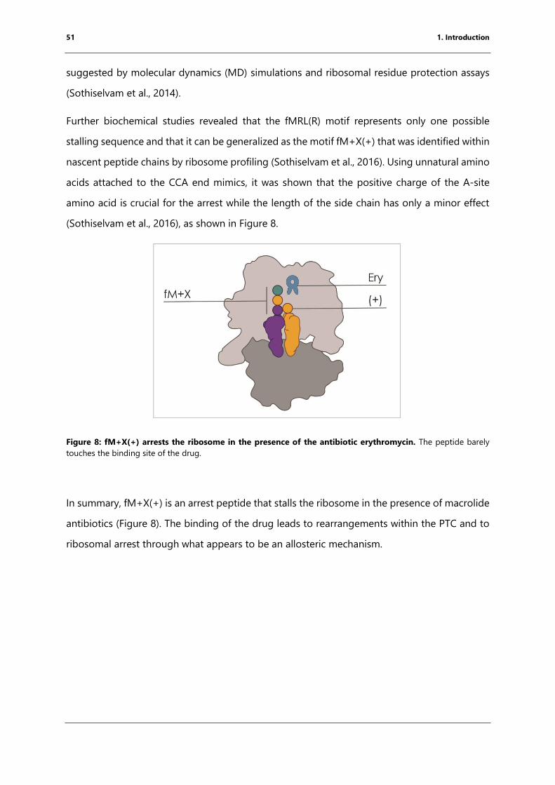
51 1. Introduction
suggested by molecular dynamics (MD) simulations and ribosomal residue protection assays
(Sothiselvam et al., 2014).
Further biochemical studies revealed that the fMRL(R) motif represents only one possible
stalling sequence and that it can be generalized as the motif fM+X(+) that was identified within
nascent peptide chains by ribosome profiling (Sothiselvam et al., 2016). Using unnatural amino
acids attached to the CCA end mimics, it was shown that the positive charge of the A-site
amino acid is crucial for the arrest while the length of the side chain has only a minor effect
(Sothiselvam et al., 2016), as shown in Figure 8.
Figure 8: fM+X(+) arrests the ribosome in the presence of the antibiotic erythromycin. The peptide barely
touches the binding site of the drug.
In summary, fM+X(+) is an arrest peptide that stalls the ribosome in the presence of macrolide
antibiotics (Figure 8). The binding of the drug leads to rearrangements within the PTC and to
ribosomal arrest through what appears to be an allosteric mechanism.
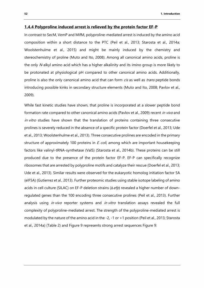
52 1. Introduction
1.4.4 Polyproline induced arrest is relieved by the protein factor EF-P
In contrast to SecM, VemP and MifM, polyproline-mediated arrest is induced by the amino acid
composition within a short distance to the PTC (Peil et al., 2013; Starosta et al., 2014a;
Woolstenhulme et al., 2015) and might be mainly induced by the chemistry and
stereochemistry of proline (Muto and Ito, 2008). Among all canonical amino acids, proline is
the only N-alkyl amino acid which has a higher alkalinity and its imino group is more likely to
be protonated at physiological pH compared to other canonical amino acids. Additionally,
proline is also the only canonical amino acid that can form cis as well as trans peptide bonds
introducing possible kinks in secondary structure elements (Muto and Ito, 2008; Pavlov et al.,
2009).
While fast kinetic studies have shown, that proline is incorporated at a slower peptide bond
formation rate compared to other canonical amino acids (Pavlov et al., 2009) recent in vivo and
in vitro studies have shown that the translation of proteins containing three consecutive
prolines is severely reduced in the absence of a specific protein factor (Doerfel et al., 2013; Ude
et al., 2013; Woolstenhulme et al., 2013). Three consecutive prolines are encoded in the primary
structure of approximately 100 proteins in E. coli, among which are important housekeeping
factors like valinyl-tRNA-synthetase (ValS) (Starosta et al., 2014b). These proteins can be still
produced due to the presence of the protein factor EF-P. EF-P can specifically recognize
ribosomes that are arrested by polyproline motifs and catalyze their rescue (Doerfel et al., 2013;
Ude et al., 2013). Similar results were observed for the eukaryotic homolog initiation factor 5A
(eIF5A) (Gutierrez et al., 2013). Further proteomic studies using stable isotope labeling of amino
acids in cell culture (SILAC) on EF-P deletion strains (Δefp) revealed a higher number of down-
regulated genes than the 100 encoding three consecutive prolines (Peil et al., 2013). Further
analysis using in vivo reporter systems and in vitro translation assays revealed the full
complexity of polyproline-mediated arrest. The strength of the polyproline-mediated arrest is
modulated by the nature of the amino acid in the -2, -1 or +1 position (Peil et al., 2013; Starosta
et al., 2014a) (Table 2) and Figure 9 represents strong arrest sequences Figure 9:
53 1. Introduction
Figure 9: Summary of the sequence variants that can induce polyproline-mediated ribosomal arrest. 0 the
proline and its corresponding tRNA are highlighted in green. The amino acid in the A-site (PP(X)), in the -1 position
(XP(P)) and -2 positions (XPP(P)) directly influence the strength of the arrest. The figure summarizes strong
polyproline arrest motifs identified by (Doerfel et al., 2013; Peil et al., 2013; Starosta et al., 2014a; Woolstenhulme et
al., 2015).
The resulting sequences were validated by recent ribosome profiling experiments comparing
the data obtained from wild-type cells with Δefp (Woolstenhulme et al., 2015). It has been
shown that EF-P and its homologs need to be post-translationally modified to gain activity.
The modification is species-specific and can vary from glycosylation to β-lysinylation (Bailly and
de Crécy-Lagard, 2010; Bullwinkle et al., 2013; Lassak et al., 2015; Park et al., 2012). Bacterial
cells lacking EF-P or its modification enzymes show defects in fitness, mobility, membrane
integrity and virulence (Navarre et al., 2010).
To gain insights into the function of EF-P and its eukaryotic homolog, several structures were
determined by cryo-EM and X-ray crystallography. The first structural information of the
function of EF-P was obtained by co-crystallization of 70S T. thermophilus ribosomes,
unmodified EF-P from T. thermophilus and tRNAiMet from E. coli resulting in a structure at 3.5 Å
resolution (Blaha et al., 2009). The structure was solved under the assumption that EF-P
promotes and enhances the formation of the first peptide bond (An et al., 1980; Glick et al.,
1979; Glick and Ganoza, 1975). The factor binds next to the P-site tRNA stabilizing the
orientation of the 3’CCA end of the P-site tRNA (Blaha et al., 2009).
More recently, the Beckmann group published a cryo-EM structure with 3.9 Å resolution of the
80S yeast (Saccharomyces cerevisiae) ribosome pulled down from cells to study non-stop
decay (NSD) to study the Ski complex (His-tagged). During cryo-EM data processing, a large
number of particles contained two tRNAs and eIF5A, the eukaryotic homolog of EF-P. Like EF-
54 1. Introduction
P, eIF5A needs to be modified to be active. The eukaryotic modification is the unusual amino
acid hypusine, which is formed by transferring a 4-aminobutyl moiety on the corresponding
Lys residue followed by hydroxylation (Dever et al., 2014; Gutierrez et al., 2013). The structure
showed that the hypusine modification of eIF5A forms direct contact with the CCA end of the
P-site tRNA. This aids the correct orientation of the peptide chain for peptide bond formation
(Schmidt et al., 2015).
Recently, a study from the Yusupov group utilized tRNA mimics, a short peptide attached to
the RNA fragment ACCA which mimics the 3’CCA end of the tRNA, in complex with the 80S
S. cerevisiae ribosome giving a greater understanding of the conformation of proline within
the PTC (Melnikov et al., 2016a). The structures solved by X-ray crystallography have a
resolution range of 3.1 to 3.5 Å. To obtain greater insights into the conformation of a diproline
peptide the tRNA mimic was stabilized with the antibiotic Sparsomycin. The backbone of the
dipeptide is bent with the N-terminus pointing towards the tunnel wall while the N-terminus
of the control peptide (Phe-Leu) points into the exit tunnel (Melnikov et al., 2016a). In addition,
proline does not bind in the same orientation like other amino acids to the A-site crevice
leading to wrong orientation for peptide bond formation (Melnikov et al., 2016a). Further
studies, including the eIF5A in complex with the yeast ribosome, showed that eIF5A binds at
the same position even if the P-site tRNA mimic is absent and the hypusine modification points
towards the PTC. In addition, the binding of the factor results in a conformational change of
the ribosome (Melnikov et al., 2016b).
All in all, EF-P is non-canonical translation factor that presents a possible target for antibiotic
research. Even if there are many published structures of EF-P or its eukaryotic homolog in
complex with the ribosome, there is no high-resolution structure containing the native
substrate Pro-tRNAPro as well as the orientation of a prokaryotic modification remains
unknown.
55 1. Introduction
1.5 Flexizyme as a tool to study translation
1.5.1 Identification and sequence optimization
One major aim of this thesis is to study short nascent-chain arrest peptides by structural
characterization. In doing so, the arrest peptide needs to be attached to the 3’CCA end of
tRNAs. To obtain peptidylated tRNA, the flexizyme methodology was used. The flexizyme
methodology, a method to transfer activated amino acids and derivatives specifically to the 3’
CCA end of a tRNA, was developed overcome limitations of genetic reprogramming in vitro
(Goto et al., 2011). These limitations included the high substrate sensitivity of aminoacyl-tRNA
synthetases and so only a limited number of amino acids could be used to produce novel
potential therapeutic peptides by the bacterial ribosome (Hartman et al., 2007; Hartman et al.,
2006).
RNA is involved in many reactions like splicing and peptide bond formation and are indicators
of an “RNA-world” before protein enzymes were evolved. It has been hypothesized that
processes like aminoacylation of tRNAs have been originally catalyzed by ribozymes (as
reviewed by Hager et al., 1996). First attempts to identify RNA sequences that can aminoacylate
and recognize tRNAs resulted in the identification of sequences that can transfer the amino
acid onto their own 5’ end or internally onto 2’OH groups (Lohse and Szostak, 1996; Suga et
al., 1998a; Suga et al., 1998b). Further experiments and optimization steps resulted in the
identification of an RNA sequence that uses activated Biotin-L-glutamine adenylate and Biotin-
L-phenylalanine-adenylate to act in cis (self-amino acylation) or in trans (aminoacylation of a
different RNA) (Lee et al., 2000). The natural leaving group, adenylate, was replaced by a
cyanomethyl ester (CME) to ensure that the chemical properties of the amino acid are forming
the interactions with the ribozyme (Lee et al., 2000).
For incorporation of an amino acid into the nascent chain by the ribosome, the amino acid is
specifically attached to the 3’ CCA end of its tRNA by the aminoacyl-tRNA synthetase (ARS). To
site-direct, the aminoacylation reaction catalyzed ARS like ribozymes a tRNA precursor
molecule was used (Saito et al., 2001a). In cells, tRNAs get produced as long precursors that
are processed post-transcriptionally. In particular, the 5’ end gets processed by the ribozyme
RNase P (Meinnel and Blanquet, 1995). The 5’ end sequence might have had the capability of
aminoacyl synthetase by aminoacylating its own 3’CCA end that got lost after the evolution of
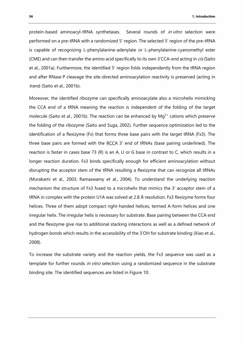
56 1. Introduction
protein-based aminoacyl-tRNA synthetases. Several rounds of in vitro selection were
performed on a pre-tRNA with a randomized 5’ region. The selected 5’ region of the pre-tRNA
is capable of recognizing L-phenylalanine-adenylate or L-phenylalanine-cyanomethyl ester
(CME) and can then transfer the amino acid specifically to its own 3’CCA-end acting in cis (Saito
et al., 2001a). Furthermore, the identified 5’ region folds independently from the tRNA region
and after RNase P cleavage the site-directed aminoacylation reactivity is preserved (acting in
trans) (Saito et al., 2001b).
Moreover, the identified ribozyme can specifically aminoacylate also a microhelix mimicking
the CCA end of a tRNA meaning the reaction is independent of the folding of the target
molecule (Saito et al., 2001b). The reaction can be enhanced by Mg2+ cations which preserve
the folding of the ribozyme (Saito and Suga, 2002). Further sequence optimization led to the
identification of a flexizyme (Fx) that forms three base pairs with the target tRNA (Fx3). The
three base pairs are formed with the RCCA 3’ end of tRNAs (base pairing underlined). The
reaction is faster in cases base 73 (R) is an A, U or G base in contrast to C, which results in a
longer reaction duration. Fx3 binds specifically enough for efficient aminoacylation without
disrupting the acceptor stem of the tRNA resulting a flexizyme that can recognize all tRNAs
(Murakami et al., 2003; Ramaswamy et al., 2004). To understand the underlying reaction
mechanism the structure of Fx3 fused to a microhelix that mimics the 3’ acceptor stem of a
tRNA in complex with the protein U1A was solved at 2.8 Å resolution. Fx3 flexizyme forms four
helices. Three of them adopt compact right-handed helices, termed A-form helices and one
irregular helix. The irregular helix is necessary for substrate. Base pairing between the CCA end
and the flexizyme give rise to additional stacking interactions as well as a defined network of
hydrogen bonds which results in the accessibility of the 3’OH for substrate binding (Xiao et al.,
2008).
To increase the substrate variety and the reaction yields, the Fx3 sequence was used as a
template for further rounds in vitro selection using a randomized sequence in the substrate
binding site. The identified sequences are listed in Figure 10:
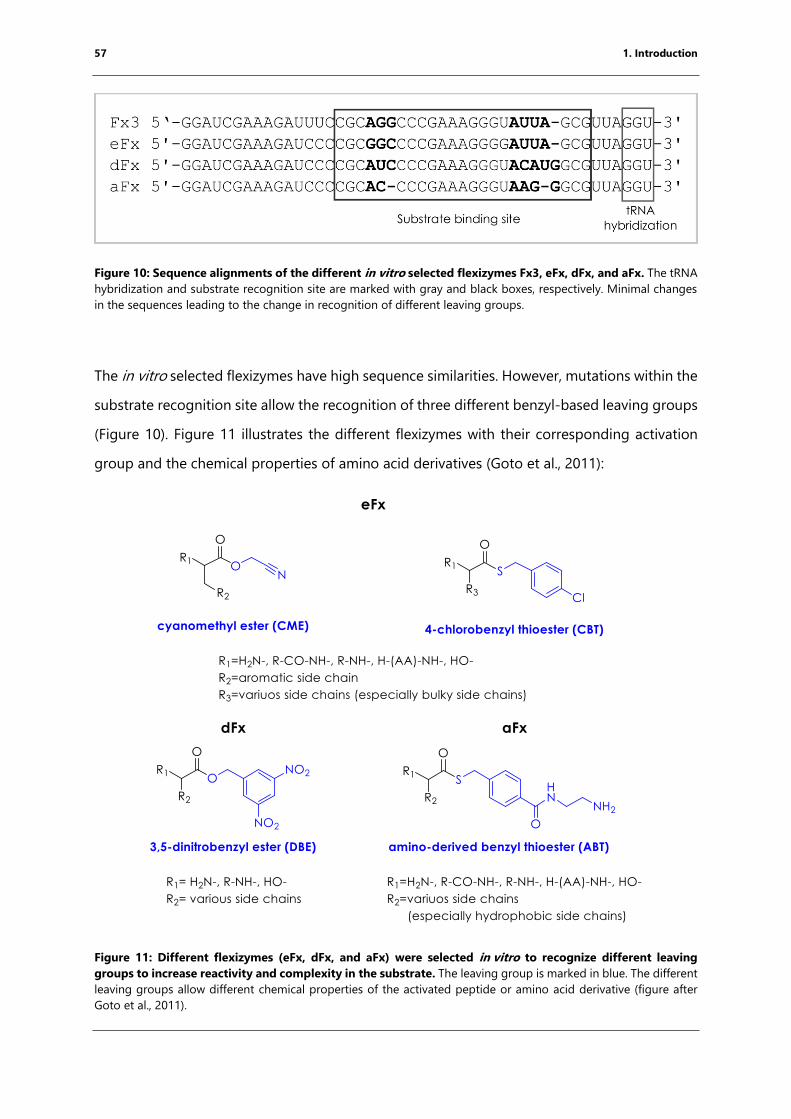
57 1. Introduction
Figure 10: Sequence alignments of the different in vitro selected flexizymes Fx3, eFx, dFx, and aFx. The tRNA
hybridization and substrate recognition site are marked with gray and black boxes, respectively. Minimal changes
in the sequences leading to the change in recognition of different leaving groups.
The in vitro selected flexizymes have high sequence similarities. However, mutations within the
substrate recognition site allow the recognition of three different benzyl-based leaving groups
(Figure 10). Figure 11 illustrates the different flexizymes with their corresponding activation
group and the chemical properties of amino acid derivatives (Goto et al., 2011):
Figure 11: Different flexizymes (eFx, dFx, and aFx) were selected in vitro to recognize different leaving
groups to increase reactivity and complexity in the substrate. The leaving group is marked in blue. The different
leaving groups allow different chemical properties of the activated peptide or amino acid derivative (figure after
Goto et al., 2011).
58 1. Introduction
As shown in Figure 11, enhanced Fx (eFx) is more active than Fx3 and recognizes two different
leaving groups. On one hand aromatic amino acids are activated by CME, the same leaving
group as Fx3, and on the other hand, bulky amino acids can be activated by using 4-chloro
benzyl thioester (CBT). The dinitro flexizyme (dFx) recognizes the leaving group 3,5-
dinitrobenzyl ester (DBE) and transfers various amino acids and activated peptides onto tRNAs
(Murakami et al., 2006a). Already dFx and eFx increased the accessibility to a wider variety of
activated peptides for further experiments whereas hydrophobic amino acids activated with
CBT or DBE are insoluble in water, inducing poor reactivity of the activated peptide. To
overcome this issue of very hydrophobic amino acids, like those with long alkyl side chains,
they can be activated with amino-derived benzyl thioester (ABT) as leaving the group (Niwa et
al., 2009).
59 1. Introduction
1.5.2 Case studies
Flexizyme-mediated charging led to a great variety of substrates that could be incorporated
into a growing nascent peptide. To do so, in vitro translation was performed in a reconstituted
in vitro translation system (Shimizu et al., 2001; Shimizu et al., 2005). Reactions were performed
using an aminoacylated tRNA in an in vitro translation system lacking these tRNAs, the natural
amino acid, and the corresponding aminoacyl-tRNA synthetase. This system is called flexible
in vitro translation (FIT) (Goto et al., 2011) and can be used to initiate translation using any L-
or D-amino acid (Goto et al., 2008; Murakami et al., 2006a), short peptides and peptide-like
structures (Goto and Suga, 2009). Unnatural amino acids can also be incorporated into the
growing peptide chain by charging the molecule onto an elongator tRNA (Murakami et al.,
2006b). To ensure that all native amino acids can be included in the reaction, a tRNASNN library
can be produced by in vitro transcribing individually each tRNA with an SNN anticodon. In
some cases, one amino acid, like alanine or proline, can be delivered by multiple tRNAs to the
ribosome. One of the tRNAs can be aminoacylated with the native amino acid using the
corresponding aminoacyl tRNA synthetase and the others by non-proteinogenic amino acids
or derivatives using flexizyme (Iwane et al., 2016); all charged tRNAs can be mixed and
incorporated while the ribosome is translating an NNS library.
A recent study (Katoh et al., 2016) used the flexizyme approach to identify the nucleotides of
tRNAPro, that are crucial for the recognition of EF-P and the release of polyproline-mediated
translational arrest. Different mutants of tRNAPro, tRNASer, tRNAVal and tRNAiMet were in vitro
transcribed and prolinylated using flexizyme. The reactivity of EF-P was quantified by
measuring the incorporation of downstream radioactive labeled Asp and kinetic parameters
were obtained by measuring Pro incorporation rates in the presence or absence of EF-P. The
major result of this study was that the D-loop structure of tRNAPro and tRNAiMet is crucial for
EF-P recognition and thus needs to be included for further structural studies to understand the
detailed mechanism of EF-P (Katoh et al., 2016).
In summary, extensive studies have shown that flexizymes can be used specifically to transfer
amino acids or small peptides onto the 3’ CCA end of any tRNA and incorporate them into the
growing peptide chain during initiation and elongation.
60
1.6 Aims
The bacterial ribosome is one of the major targets for antibiotics (Wilson, 2009, 2011, 2014).
The rise of multiple bacterial resistant genes against clinically used antibiotics is an increasing
threat and there is need for the development of new therapeutic strategies. Certain peptides
can inhibit bacterial translation, either as free peptides that are produced as a part of defense
mechanisms against other organisms such as PrAMPs or during their own translation (nascent
chain-mediated translational arrest).
PrAMPs are antimicrobial peptides which are produced by insects and mammals as a part of
their innate immune system (Otvos, 2002). Though recent studies have shown that PrAMPs
inhibit protein biosynthesis through binding to the bacterial ribosome (Krizsan et al., 2014;
Mardirossian et al., 2014) the molecular mechanism remains unknown. In order to obtain
detailed insights into the molecular mechanism, the structures of different PrAMPs in complex
with the 70S T. thermophilus ribosome are determined here using X-ray crystallography.
During the nascent-chain mediated translational arrest the bacterial ribosome is inhibited by
the translating peptide. Structural information of long nascent chain arrest peptides such as
SecM (Zhang et al., 2015) and ErmCL (Arenz et al., 2014a) has been obtained recently using
cryo-EM. The underlying mechanisms of how short arrest peptides, including polyproline
motifs as well as fM+X(+)arrest the ribosome in the presence of erythromycin, remain
unknown. To investigate this further, the aim of this work was to study these arrest complexes
using the flexizyme methodology for complex formation for structural and biochemical
characterization. Obtaining high-resolution or near atomic resolution structures will give
insights into the molecular conformation of the nascent chain as well as into their allosteric
effect on the bacterial ribosome. The knowledge to be obtained from this will enable a greater
understanding how peptide bond formation can be inhibited specifically and could lead to the
development of novel highly specific antibiotics targeting this process.
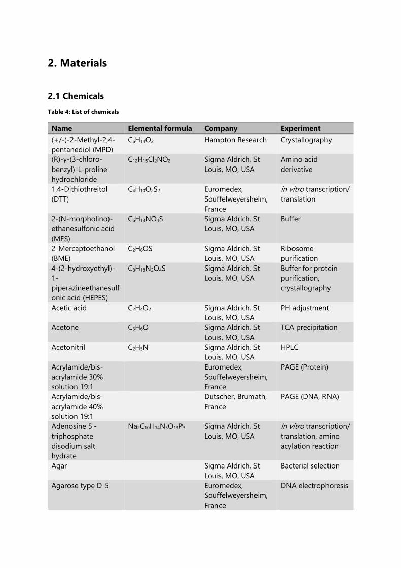
2. Materials
2.1 Chemicals
Table 4: List of chemicals
Name Elemental formula Company Experiment
(+/-)-2-Methyl-2,4-
pentanediol (MPD)
C6H14O2 Hampton Research Crystallography
(R)-γ-(3-chloro-
benzyl)-L-proline
hydrochloride
C12H15Cl2NO2 Sigma Aldrich, St
Louis, MO, USA
Amino acid
derivative
1,4-Dithiothreitol
(DTT)
C4H10O2S2 Euromedex,
Souffelweyersheim,
France
in vitro transcription/
translation
2-(N-morpholino)-
ethanesulfonic acid
(MES)
C6H13NO4S Sigma Aldrich, St
Louis, MO, USA
Buffer
2-Mercaptoethanol
(BME)
C2H6OS Sigma Aldrich, St
Louis, MO, USA
Ribosome
purification
4-(2-hydroxyethyl)-
1-
piperazineethanesulf
onic acid (HEPES)
C8H18N2O4S Sigma Aldrich, St
Louis, MO, USA
Buffer for protein
purification,
crystallography
Acetic acid C2H4O2 Sigma Aldrich, St
Louis, MO, USA
PH adjustment
Acetone C3H6O Sigma Aldrich, St
Louis, MO, USA
TCA precipitation
Acetonitril C2H5N Sigma Aldrich, St
Louis, MO, USA
HPLC
Acrylamide/bis-
acrylamide 30%
solution 19:1
Euromedex,
Souffelweyersheim,
France
PAGE (Protein)
Acrylamide/bis-
acrylamide 40%
solution 19:1
Dutscher, Brumath,
France
PAGE (DNA, RNA)
Adenosine 5'-
triphosphate
disodium salt
hydrate
Na2C10H14N5O13P3 Sigma Aldrich, St
Louis, MO, USA
In vitro transcription/
translation, amino
acylation reaction
Agar Sigma Aldrich, St
Louis, MO, USA
Bacterial selection
Agarose type D-5 Euromedex,
Souffelweyersheim,
France
DNA electrophoresis
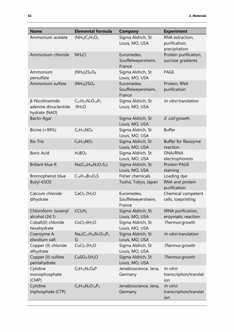
62 2. Materials
Name Elemental formula Company Experiment
Ammonium acetate (NH4)C2H3O2 Sigma Aldrich, St
Louis, MO, USA
RNA extraction,
purification,
precipitation
Ammonium chloride NH4Cl Euromedex,
Souffelweyersheim,
France
Protein purification,
sucrose gradients
Ammonium
persulfate
(NH4)2S2O8 Sigma Aldrich, St
Louis, MO, USA
PAGE
Ammonium sulfate (NH4)2SO4 Euromedex,
Souffelweyersheim,
France
Protein, RNA
purification
β-Nicotinamide
adenine dinucleotide
hydrate (NAD)
C21H27N7O14P2
·XH2O
Sigma Aldrich, St
Louis, MO, USA
In vitro translation
Bacto-Agar Sigma Aldrich, St
Louis, MO, USA
E. coli growth
Bicine (>99%) C6H13NO4 Sigma Aldrich, St
Louis, MO, USA
Buffer
Bis-Tris C8H19NO5 Sigma Aldrich, St
Louis, MO, USA
Buffer for flexizyme
reaction
Boric Acid H3BO3 Sigma Aldrich, St
Louis, MO, USA
DNA/RNA
electrophoresis
Brillant blue R Na(C45H44N3O7S2) Sigma Aldrich, St
Louis, MO, USA
Protein PAGE
staining
Bromophenol blue C19H10Br4O5S Fisher chemicals Loading dye
Butyl-65OS Tosho, Tokyo, Japan RNA and protein
purification
Calcium chloride
dihydrate
CaCl2·2H2O Euromedex,
Souffelweyersheim,
France
Chemical competent
cells, toeprinting
Chloroform: Isoamyl
alcohol (24:1)
CCl2H2 Sigma Aldrich, St
Louis, MO, USA
tRNA purification,
enzymatic reaction
Cobalt(II) chloride
hexahydrate
CoCl2·6H2O Sigma Aldrich, St
Louis, MO, USA
Thermus growth
Coenzyme A
disodium salt
Na2(C21H34N7O16P3
S)
Sigma Aldrich, St
Louis, MO, USA
In vitro translation
Copper (II) chloride
dihydrate
CuCl2·2H2O Sigma Aldrich, St
Louis, MO, USA
Thermus growth
Copper (II) sulfate
pentahydrate
CuSO4·5H2O Sigma Aldrich, St
Louis, MO, USA
Thermus growth
Cytidine
monophosphate
(CMP)
C9H16N3O8P Jenabioscience, Jena,
Germany
In vitro
transcription/translat
ion
Cytidine
triphosphate (CTP)
C9H16N3O14P3 Jenabioscience, Jena,
Germany
In vitro
transcription/translat
ion
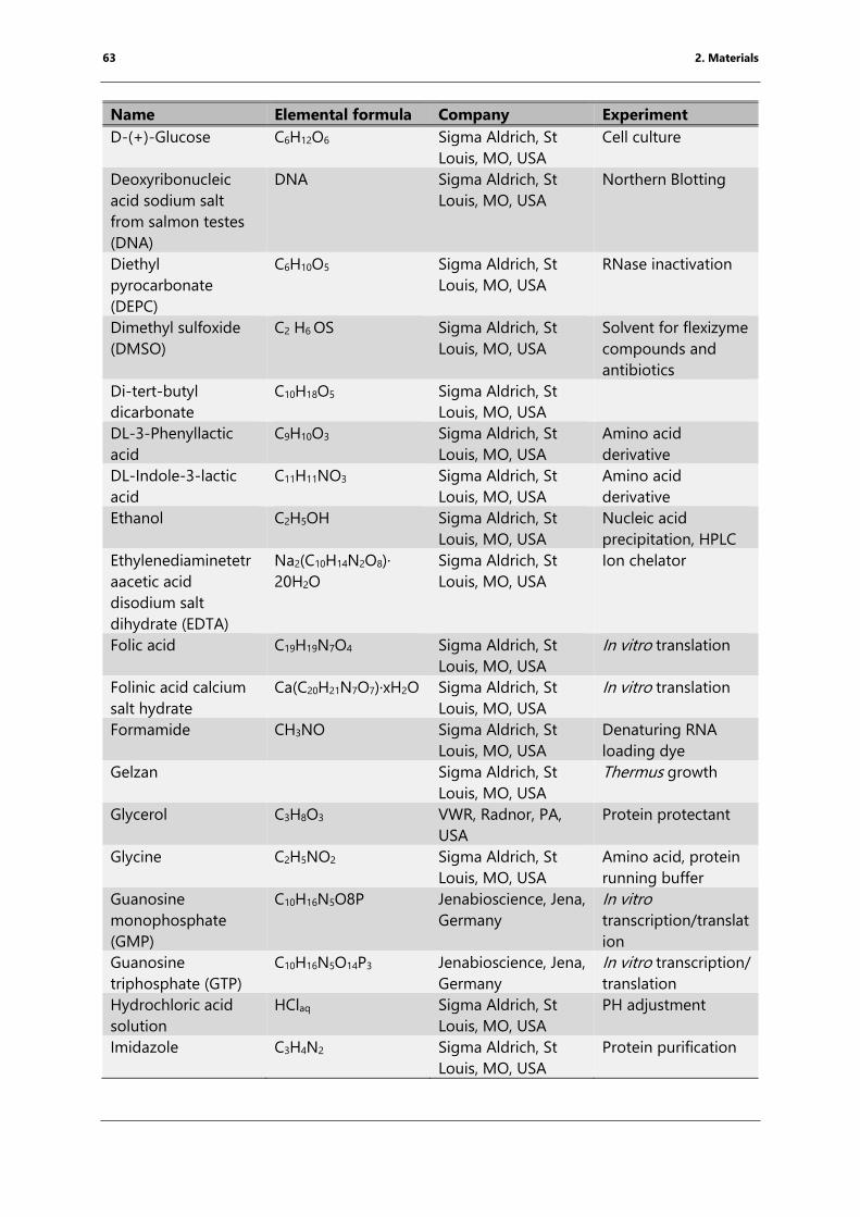
63 2. Materials
Name Elemental formula Company Experiment
D-(+)-Glucose C6H12O6 Sigma Aldrich, St
Louis, MO, USA
Cell culture
Deoxyribonucleic
acid sodium salt
from salmon testes
(DNA)
DNA Sigma Aldrich, St
Louis, MO, USA
Northern Blotting
Diethyl
pyrocarbonate
(DEPC)
C6H10O5 Sigma Aldrich, St
Louis, MO, USA
RNase inactivation
Dimethyl sulfoxide
(DMSO)
C2 H6 OS Sigma Aldrich, St
Louis, MO, USA
Solvent for flexizyme
compounds and
antibiotics
Di-tert-butyl
dicarbonate
C10H18O5 Sigma Aldrich, St
Louis, MO, USA
DL-3-Phenyllactic
acid
C9H10O3 Sigma Aldrich, St
Louis, MO, USA
Amino acid
derivative
DL-Indole-3-lactic
acid
C11H11NO3 Sigma Aldrich, St
Louis, MO, USA
Amino acid
derivative
Ethanol C2H5OH Sigma Aldrich, St
Louis, MO, USA
Nucleic acid
precipitation, HPLC
Ethylenediaminetetr
aacetic acid
disodium salt
dihydrate (EDTA)
Na2(C10H14N2O8)·
20H2O
Sigma Aldrich, St
Louis, MO, USA
Ion chelator
Folic acid C19H19N7O4 Sigma Aldrich, St
Louis, MO, USA
In vitro translation
Folinic acid calcium
salt hydrate
Ca(C20H21N7O7)·xH2O Sigma Aldrich, St
Louis, MO, USA
In vitro translation
Formamide CH3NO Sigma Aldrich, St
Louis, MO, USA
Denaturing RNA
loading dye
Gelzan Sigma Aldrich, St
Louis, MO, USA
Thermus growth
Glycerol C3H8O3 VWR, Radnor, PA,
USA
Protein protectant
Glycine C2H5NO2 Sigma Aldrich, St
Louis, MO, USA
Amino acid, protein
running buffer
Guanosine
monophosphate
(GMP)
C10H16N5O8P Jenabioscience, Jena,
Germany
In vitro
transcription/translat
ion
Guanosine
triphosphate (GTP)
C10H16N5O14P3 Jenabioscience, Jena,
Germany
In vitro transcription/
translation
Hydrochloric acid
solution
HClaq Sigma Aldrich, St
Louis, MO, USA
PH adjustment
Imidazole C3H4N2 Sigma Aldrich, St
Louis, MO, USA
Protein purification
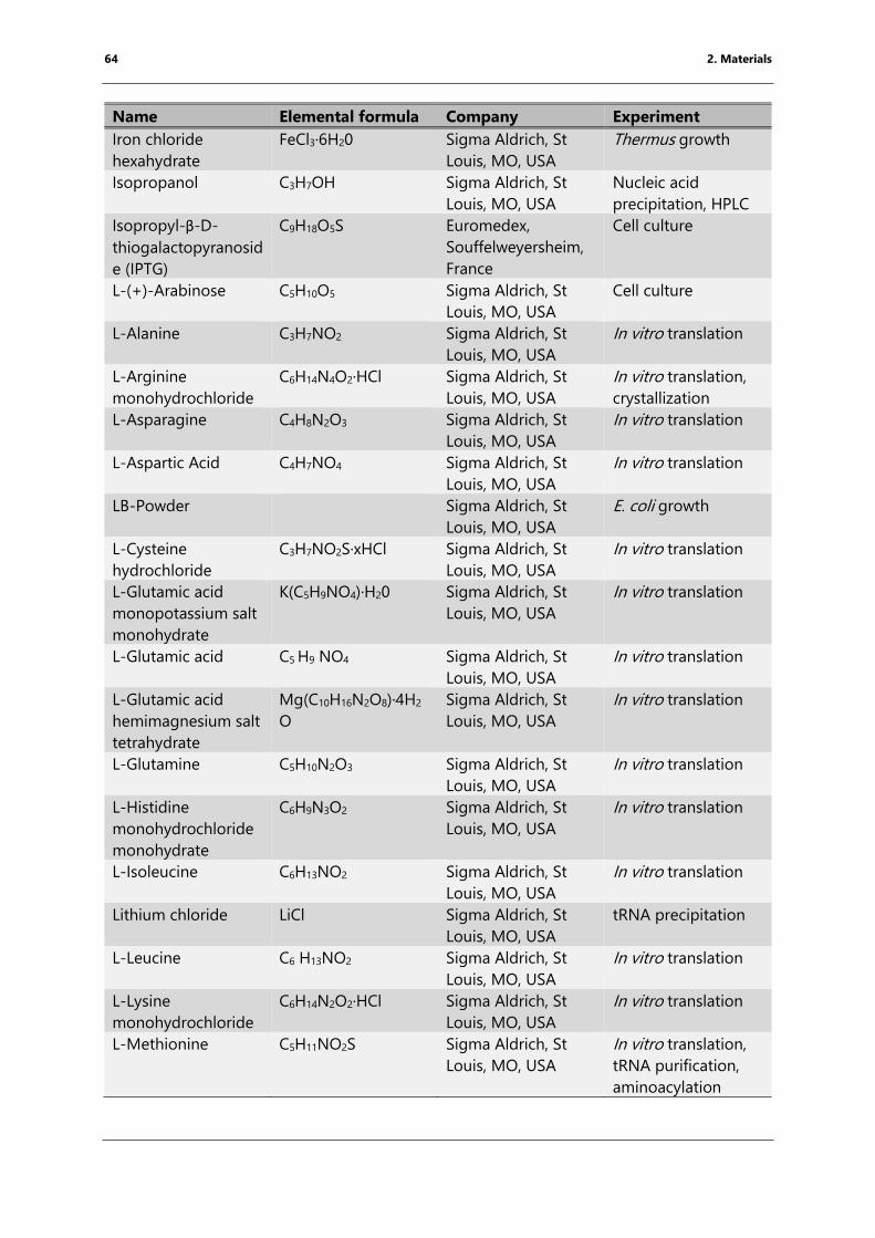
64 2. Materials
Name Elemental formula Company Experiment
Iron chloride
hexahydrate
FeCl3·6H20 Sigma Aldrich, St
Louis, MO, USA
Thermus growth
Isopropanol C3H7OH Sigma Aldrich, St
Louis, MO, USA
Nucleic acid
precipitation, HPLC
Isopropyl-β-D-
thiogalactopyranosid
e (IPTG)
C9H18O5S Euromedex,
Souffelweyersheim,
France
Cell culture
L-(+)-Arabinose C5H10O5 Sigma Aldrich, St
Louis, MO, USA
Cell culture
L-Alanine C3H7NO2 Sigma Aldrich, St
Louis, MO, USA
In vitro translation
L-Arginine
monohydrochloride
C6H14N4O2·HCl Sigma Aldrich, St
Louis, MO, USA
In vitro translation,
crystallization
L-Asparagine C4H8N2O3 Sigma Aldrich, St
Louis, MO, USA
In vitro translation
L-Aspartic Acid C4H7NO4 Sigma Aldrich, St
Louis, MO, USA
In vitro translation
LB-Powder Sigma Aldrich, St
Louis, MO, USA
E. coli growth
L-Cysteine
hydrochloride
C3H7NO2S·xHCl Sigma Aldrich, St
Louis, MO, USA
In vitro translation
L-Glutamic acid
monopotassium salt
monohydrate
K(C5H9NO4)·H20 Sigma Aldrich, St
Louis, MO, USA
In vitro translation
L-Glutamic acid C5 H9 NO4 Sigma Aldrich, St
Louis, MO, USA
In vitro translation
L-Glutamic acid
hemimagnesium salt
tetrahydrate
Mg(C10H16N2O8)·4H2
O
Sigma Aldrich, St
Louis, MO, USA
In vitro translation
L-Glutamine C5H10N2O3 Sigma Aldrich, St
Louis, MO, USA
In vitro translation
L-Histidine
monohydrochloride
monohydrate
C6H9N3O2 Sigma Aldrich, St
Louis, MO, USA
In vitro translation
L-Isoleucine C6H13NO2 Sigma Aldrich, St
Louis, MO, USA
In vitro translation
Lithium chloride LiCl Sigma Aldrich, St
Louis, MO, USA
tRNA precipitation
L-Leucine C6 H13NO2 Sigma Aldrich, St
Louis, MO, USA
In vitro translation
L-Lysine
monohydrochloride
C6H14N2O2·HCl Sigma Aldrich, St
Louis, MO, USA
In vitro translation
L-Methionine C5H11NO2S Sigma Aldrich, St
Louis, MO, USA
In vitro translation,
tRNA purification,
aminoacylation
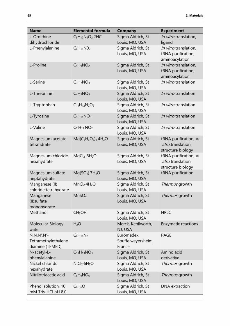
65 2. Materials
Name Elemental formula Company Experiment
L-Ornithine
dihydrochloride
C5H12N2O2·2HCl Sigma Aldrich, St
Louis, MO, USA
In vitro translation,
ligand
L-Phenylalanine C6H11N02 Sigma Aldrich, St
Louis, MO, USA
In vitro translation,
tRNA purification,
aminoacylation
L-Proline C5H9NO2 Sigma Aldrich, St
Louis, MO, USA
In vitro translation,
tRNA purification,
aminoacylation
L-Serine C3H7NO3 Sigma Aldrich, St
Louis, MO, USA
In vitro translation
L-Threonine C4H9NO3 Sigma Aldrich, St
Louis, MO, USA
In vitro translation
L-Tryptophan C11H12N2O2 Sigma Aldrich, St
Louis, MO, USA
In vitro translation
L-Tyrosine C9H11NO3 Sigma Aldrich, St
Louis, MO, USA
In vitro translation
L-Valine C5 H11 NO2 Sigma Aldrich, St
Louis, MO, USA
In vitro translation
Magnesium acetate
tetrahdrate
Mg(C2H3O2)2·4H2O Sigma Aldrich, St
Louis, MO, USA
tRNA purification, in
vitro translation,
structure biology
Magnesium chloride
hexahydrate
MgCl2 ·6H2O Sigma Aldrich, St
Louis, MO, USA
tRNA purification, in
vitro translation,
structure biology
Magnesium sulfate
heptahydrate
Mg(SO4)·7H2O Sigma Aldrich, St
Louis, MO, USA
tRNA purification
Manganese (II)
chloride tetrahydrate
MnCl2·4H2O Sigma Aldrich, St
Louis, MO, USA
Thermus growth
Manganese
(II)sulfate
monohydrate
MnSO4 Sigma Aldrich, St
Louis, MO, USA
Thermus growth
Methanol CH3OH Sigma Aldrich, St
Louis, MO, USA
HPLC
Molecular Biology
water
H2O Merck, Kenilworth,
NJ, USA
Enzymatic reactions
N,N,N',N'-
Tetramethylethylene
diamine (TEMED)
C6H16N2 Euromedex,
Souffelweyersheim,
France
PAGE
N-acetyl-L-
phenylalanine
C11H13NO3 Sigma Aldrich, St
Louis, MO, USA
Amino acid
derivative
Nickel chloride
hexahydrate
NiCl2·6H2O Sigma Aldrich, St
Louis, MO, USA
Thermus growth
Nitrilotriacetic acid C6H9NO6 Sigma Aldrich, St
Louis, MO, USA
Thermus growth
Phenol solution, 10
mM Tris-HCl pH 8.0
C6H6O Sigma Aldrich, St
Louis, MO, USA
DNA extraction
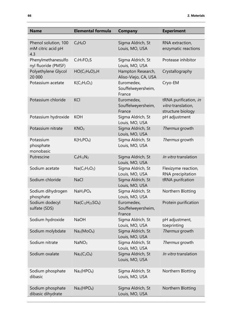
66 2. Materials
Name Elemental formula Company Experiment
Phenol solution, 100
mM citric acid pH
4.3
C6H6O Sigma Aldrich, St
Louis, MO, USA
RNA extraction,
enzymatic reactions
Phenylmethanesulfo
nyl fluoride (PMSF)
C7H7FO2S Sigma Aldrich, St
Louis, MO, USA
Protease inhibitor
Polyethylene Glycol
20 000
HO(C2H4O)nH Hampton Research,
Aliso-Viejo, CA, USA
Crystallography
Potassium acetate K(C2H3O2) Euromedex,
Souffelweyersheim,
France
Cryo-EM
Potassium chloride KCl Euromedex,
Souffelweyersheim,
France
tRNA purification, in
vitro translation,
structure biology
Potassium hydroxide KOH Sigma Aldrich, St
Louis, MO, USA
pH adjustment
Potassium nitrate KNO3 Sigma Aldrich, St
Louis, MO, USA
Thermus growth
Potassium
phosphate
monobasic
K(H2PO4) Sigma Aldrich, St
Louis, MO, USA
Thermus growth
Putrescine C4H12N2 Sigma Aldrich, St
Louis, MO, USA
In vitro translation
Sodium acetate Na(C2H3O2) Sigma Aldrich, St
Louis, MO, USA
Flexizyme reaction,
RNA precipitation
Sodium chloride NaCl Sigma Aldrich, St
Louis, MO, USA
tRNA purifcation
Sodium dihydrogen
phosphate
NaH2PO4 Sigma Aldrich, St
Louis, MO, USA
Northern Blotting
Sodium dodecyl
sulfate (SDS)
Na(C12H25SO4) Euromedex,
Souffelweyersheim,
France
Protein purification
Sodium hydroxide NaOH Sigma Aldrich, St
Louis, MO, USA
pH adjustment,
toeprinting
Sodium molybdate Na2(MoO4) Sigma Aldrich, St
Louis, MO, USA
Thermus growth
Sodium nitrate NaNO3 Sigma Aldrich, St
Louis, MO, USA
Thermus growth
Sodium oxalate
Na2(C2O4) Sigma Aldrich, St
Louis, MO, USA
In vitro translation
Sodium phosphate
dibasic
Na2(HPO4) Sigma Aldrich, St
Louis, MO, USA
Northern Blotting
Sodium phosphate
dibasic dihydrate
Na2(HPO4) Sigma Aldrich, St
Louis, MO, USA
Northern Blotting
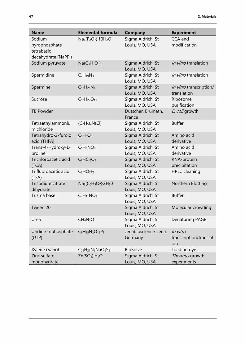
67 2. Materials
Name Elemental formula Company Experiment
Sodium
pyrophosphate
tetrabasic
decahydrate (NaPPi)
Na4(P2O7)·10H2O Sigma Aldrich, St
Louis, MO, USA
CCA end
modification
Sodium pyruvate Na(C3H3O3) Sigma Aldrich, St
Louis, MO, USA
In vitro translation
Spermidine C7H19N3 Sigma Aldrich, St
Louis, MO, USA
In vitro translation
Spermine C10H26N4 Sigma Aldrich, St
Louis, MO, USA
In vitro transcription/
translation
Sucrose C12H22O11 Sigma Aldrich, St
Louis, MO, USA
Ribosome
purification
TB Powder Dutscher, Brumath,
France
E. coli growth
Tetraethylammoniu
m chloride
(C2H5)4N(Cl) Sigma Aldrich, St
Louis, MO, USA
Buffer
Tetrahydro-2-furoic
acid (THFA)
C5H8O3 Sigma Aldrich, St
Louis, MO, USA
Amino acid
derivative
Trans-4-Hydroxy-L-
proline
C5H9NO3 Sigma Aldrich, St
Louis, MO, USA
Amino acid
derivative
Trichloroacetic acid
(TCA)
C2HCl3O2 Sigma Aldrich, St
Louis, MO, USA
RNA/protein
precipitation
Trifluoroacetic acid
(TFA)
C2HO2F3 Sigma Aldrich, St
Louis, MO, USA
HPLC cleaning
Trisodium citrate
dihydrate
Na3(C6H5O7)·2H20 Sigma Aldrich, St
Louis, MO, USA
Northern Blotting
Trizma base C4H11NO3 Sigma Aldrich, St
Louis, MO, USA
Buffer
Tween 20 Sigma Aldrich, St
Louis, MO, USA
Molecular crowding
Urea CH4N2O Sigma Aldrich, St
Louis, MO, USA
Denaturing PAGE
Uridine triphosphate
(UTP)
C9H15N2O15P3 Jenabioscience, Jena,
Germany
In vitro
transcription/translat
ion
Xylene cyanol C25H27N2NaO6S2 BioSolve Loading dye
Zinc sulfate
monohydrate
Zn(SO4)·H2O Sigma Aldrich, St
Louis, MO, USA
Thermus growth
experiments
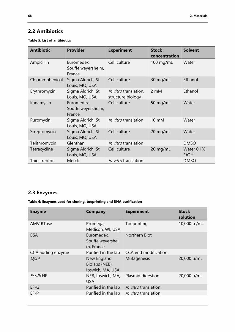
68 2. Materials
2.2 Antibiotics
Table 5: List of antibiotics
Antibiotic Provider Experiment Stock
concentration
Solvent
Ampicillin Euromedex,
Souffelweyersheim,
France
Cell culture 100 mg/mL Water
Chloramphenicol Sigma Aldrich, St
Louis, MO, USA
Cell culture 30 mg/mL Ethanol
Erythromycin Sigma Aldrich, St
Louis, MO, USA
In vitro translation,
structure biology
2 mM Ethanol
Kanamycin Euromedex,
Souffelweyersheim,
France
Cell culture 50 mg/mL Water
Puromycin Sigma Aldrich, St
Louis, MO, USA
In vitro translation 10 mM Water
Streptomycin Sigma Aldrich, St
Louis, MO, USA
Cell culture 20 mg/mL Water
Telithromycin Glenthan In vitro translation DMSO
Tetracycline Sigma Aldrich, St
Louis, MO, USA
Cell culture 20 mg/mL Water 0.1%
EtOH
Thiostrepton Merck In vitro translation DMSO
2.3 Enzymes
Table 6: Enzymes used for cloning, toeprinting and RNA purification
Enzyme Company Experiment Stock
solution
AMV RTase Promega,
Medison, WI, USA
Toeprinting 10,000 u /mL
BSA Euromedex,
Souffelweyershei
m, France
Northern Blot
CCA adding enzyme Purified in the lab CCA end modification
DpnI New England
Biolabs (NEB),
Ipswich, MA, USA
Mutagenesis 20,000 u/mL
EcoRI HF NEB, Ipswich, MA,
USA
Plasmid digestion 20,000 u/mL
EF-G Purified in the lab In vitro translation
EF-P Purified in the lab In vitro translation
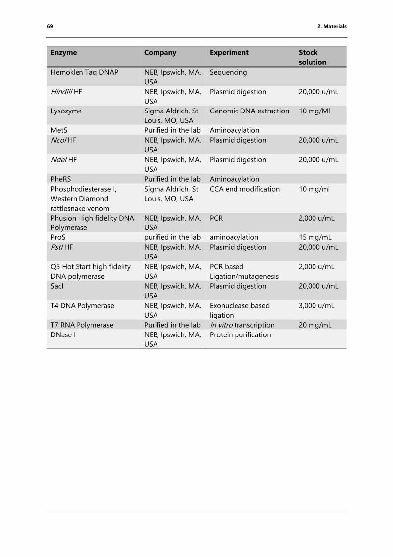
69 2. Materials
Enzyme Company Experiment Stock
solution
Hemoklen Taq DNAP NEB, Ipswich, MA,
USA
Sequencing
HindIII HF NEB, Ipswich, MA,
USA
Plasmid digestion 20,000 u/mL
Lysozyme Sigma Aldrich, St
Louis, MO, USA
Genomic DNA extraction 10 mg/Ml
MetS Purified in the lab Aminoacylation
NcoI HF NEB, Ipswich, MA,
USA
Plasmid digestion 20,000 u/mL
NdeI HF NEB, Ipswich, MA,
USA
Plasmid digestion 20,000 u/mL
PheRS Purified in the lab Aminoacylation
Phosphodiesterase I,
Western Diamond
rattlesnake venom
Sigma Aldrich, St
Louis, MO, USA
CCA end modification 10 mg/ml
Phusion High fidelity DNA
Polymerase
NEB, Ipswich, MA,
USA
PCR 2,000 u/mL
ProS purified in the lab aminoacylation 15 mg/mL
PstI HF NEB, Ipswich, MA,
USA
Plasmid digestion 20,000 u/mL
Q5 Hot Start high fidelity
DNA polymerase
NEB, Ipswich, MA,
USA
PCR based
Ligation/mutagenesis
2,000 u/mL
SacI NEB, Ipswich, MA,
USA
Plasmid digestion 20,000 u/mL
T4 DNA Polymerase NEB, Ipswich, MA,
USA
Exonuclease based
ligation
3,000 u/mL
T7 RNA Polymerase Purified in the lab In vitro transcription 20 mg/mL
DNase I NEB, Ipswich, MA,
USA
Protein purification
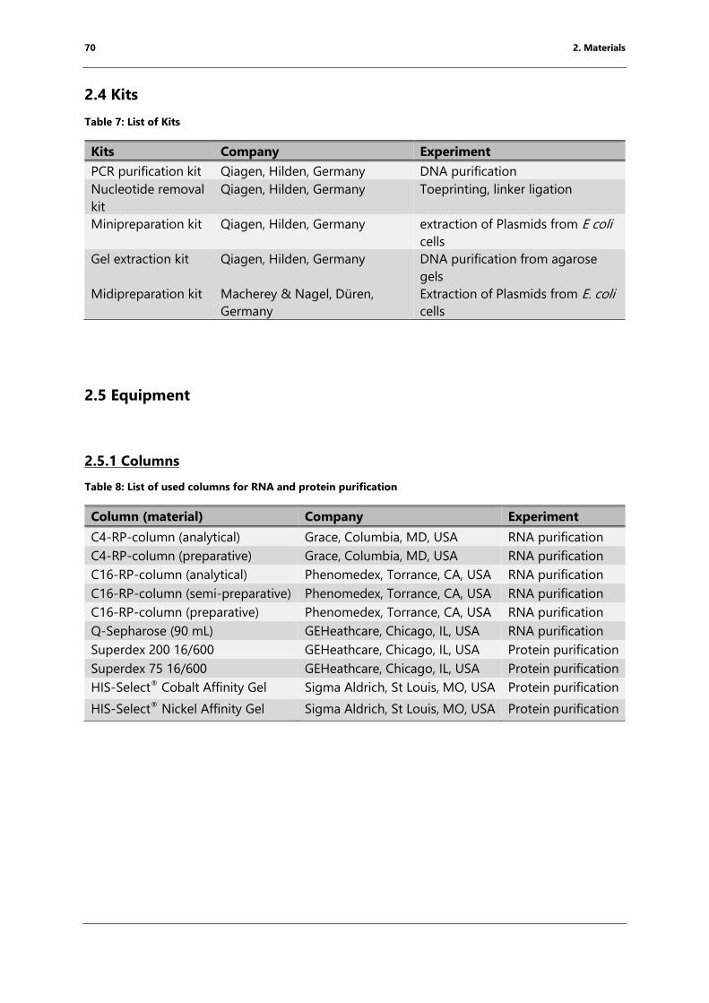
70 2. Materials
2.4 Kits
Table 7: List of Kits
Kits Company Experiment
PCR purification kit Qiagen, Hilden, Germany DNA purification
Nucleotide removal
kit
Qiagen, Hilden, Germany Toeprinting, linker ligation
Minipreparation kit Qiagen, Hilden, Germany extraction of Plasmids from E coli
cells
Gel extraction kit Qiagen, Hilden, Germany DNA purification from agarose
gels
Midipreparation kit Macherey & Nagel, Düren,
Germany
Extraction of Plasmids from E. coli
cells
2.5 Equipment
2.5.1 Columns
Table 8: List of used columns for RNA and protein purification
Column (material) Company Experiment
C4-RP-column (analytical) Grace, Columbia, MD, USA RNA purification
C4-RP-column (preparative) Grace, Columbia, MD, USA RNA purification
C16-RP-column (analytical) Phenomedex, Torrance, CA, USA RNA purification
C16-RP-column (semi-preparative) Phenomedex, Torrance, CA, USA RNA purification
C16-RP-column (preparative) Phenomedex, Torrance, CA, USA RNA purification
Q-Sepharose (90 mL) GEHeathcare, Chicago, IL, USA RNA purification
Superdex 200 16/600 GEHeathcare, Chicago, IL, USA Protein purification
Superdex 75 16/600 GEHeathcare, Chicago, IL, USA Protein purification
HIS-Select® Cobalt Affinity Gel Sigma Aldrich, St Louis, MO, USA Protein purification
HIS-Select® Nickel Affinity Gel Sigma Aldrich, St Louis, MO, USA Protein purification
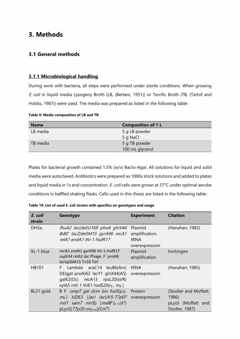
3. Methods
3.1 General methods
3.1.1 Microbiological handling
During work with bacteria, all steps were performed under sterile conditions. When growing
E. coli in liquid media Lysogeny Broth (LB, (Bertani, 1951)) or Terrific Broth (TB, (Tartof and
Hobbs, 1987)) were used. The media was prepared as listed in the following table:
Table 9: Media composition of LB and TB
Name Composition of 1 L
LB media 5 g LB powder
5 g NaCl
TB media 5 g TB powder
100 mL glycerol
Plates for bacterial growth contained 1.5% (w/v) Bacto-Agar. All solutions for liquid and solid
media were autoclaved. Antibiotics were prepared as 1000x stock solutions and added to plates
and liquid media in 1x end concentration. E. coli cells were grown at 37°C under optimal aerobe
conditions in baffled shaking flasks. Cells used in this thesis are listed in the following table:
Table 10: List of used E. coli strains with specifics on genotypes and usage
E. coli
strain
Genotype Experiment Citation
DH5α fhuA2 lac(del)U169 phoA glnV44
Φ80' lacZ(del)M15 gyrA96 recA1
relA1 endA1 thi-1 hsdR17
Plasmid
amplification,
tRNA
overexpression
(Hanahan, 1983)
XL-1 blue recA1 endA1 gyrA96 thi-1 hsdR17 supE44 relA1 lac Phage: F´ proAB lacIqZ∆M15 Tn10 Tetr
Plasmid
amplification
Invitrogen
HB101 F- Lambda- araC14 leuB6(Am)
DE(gpt-proA)62 lacY1 glnX44(AS)
galK2(Oc) recA13 rpsL20(strR)
xylA5 mtl-1 thiE1 hsdS20(rB-, mB
-)
tRNA
overexpression
(Hanahan, 1985)
BL21 gold B F– ompT gal dcm lon hsdSB(rB–
mB–) λ(DE3 [lacI lacUV5-T7p07
ind1 sam7 nin5]) [malB+]K-12(λS)
pLysS[T7p20 orip15A](CmR)
Protein
overexpression
(Studier and Moffatt,
1986)
pLysS (Moffatt and
Studier, 1987)
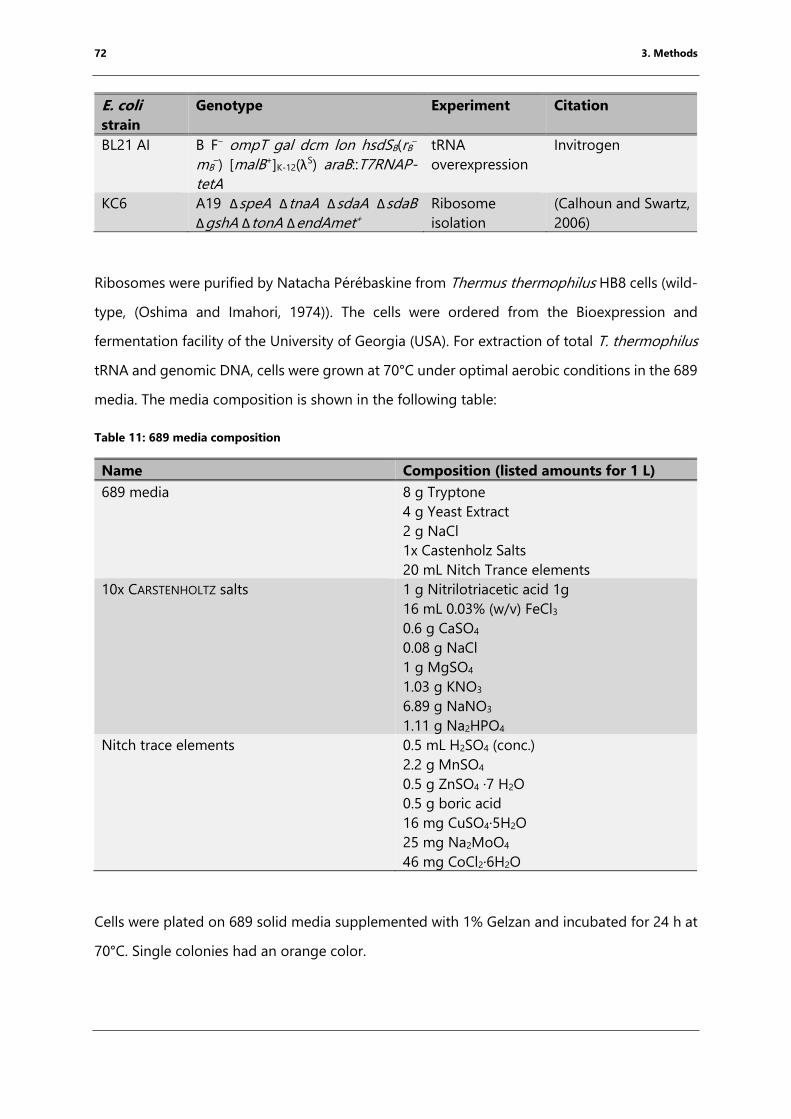
72 3. Methods
E. coli
strain
Genotype Experiment Citation
BL21 AI B F– ompT gal dcm lon hsdSB(rB–
mB–) [malB+]K-12(λ
S) araB::T7RNAP-
tetA
tRNA
overexpression
Invitrogen
KC6 A19 ΔspeA ΔtnaA ΔsdaA ΔsdaB
ΔgshA ΔtonA ΔendAmet+
Ribosome
isolation
(Calhoun and Swartz,
2006)
Ribosomes were purified by Natacha Pérébaskine from Thermus thermophilus HB8 cells (wild-
type, (Oshima and Imahori, 1974)). The cells were ordered from the Bioexpression and
fermentation facility of the University of Georgia (USA). For extraction of total T. thermophilus
tRNA and genomic DNA, cells were grown at 70°C under optimal aerobic conditions in the 689
media. The media composition is shown in the following table:
Table 11: 689 media composition
Name Composition (listed amounts for 1 L)
689 media 8 g Tryptone
4 g Yeast Extract
2 g NaCl
1x Castenholz Salts
20 mL Nitch Trance elements
10x CARSTENHOLTZ salts 1 g Nitrilotriacetic acid 1g
16 mL 0.03% (w/v) FeCl3
0.6 g CaSO4
0.08 g NaCl
1 g MgSO4
1.03 g KNO3
6.89 g NaNO3
1.11 g Na2HPO4
Nitch trace elements 0.5 mL H2SO4 (conc.)
2.2 g MnSO4
0.5 g ZnSO4 ·7 H2O
0.5 g boric acid
16 mg CuSO4·5H2O
25 mg Na2MoO4
46 mg CoCl2·6H2O
Cells were plated on 689 solid media supplemented with 1% Gelzan and incubated for 24 h at
70°C. Single colonies had an orange color.
73 3. Methods
3.1.2 Extraction of genomic DNA from E. coli and T. thermophilus
Genomic DNA was extracted from 5 mL of E. coli or T. thermophilus overnight culture. The cells
were harvested for 10 min at 4000 g in a 5424R Eppendorf centrifuge (Eppendorf, Hamburg,
Germany). The pellet was resuspended in 500 µL TES buffer (50 mM Tris HCl pH 7.4, 1 mM
EDTA, 1% SDS) and cells were lysed by adding 500 µL phenol (pH 8.0): chloroform: isoamyl
alcohol (25:24:1). Phenol denatures proteins and pH 8.0 facilitates the degradation of RNA. The
resuspension was carefully inverted until a homogeneous solution was formed, followed by
spinning at 13000 revolutions per minute (rpm, 18 000 relative centrifugal force (rcf)) in a
tabletop centrifuge (Thermofisher, Waltham, MA, USA) at 4°C for 20 min. This resulted in the
separation into three phases: The aqueous phase containing the DNA, the interphase
containing denatured proteins and the organic phase containing lipids and other hydrophobic
compounds. The aqueous phase, which contains the extracted DNA, was washed twice with
chloroform: isoamyl alcohol (24:1).
The DNA was precipitated by adding end concentrations of 300 mM Na(CH3CO2) pH 4.8 and
50% Isopropanol. The solution was incubated for 1 h at -20°C to enhance the amount of
precipitation. Subsequently, the DNA was pelleted by spinning at full speed for 30 min at 4°C
in a tabletop centrifuge. The pellet was washed with 70% ethanol (EtOH) to remove ions. The
DNA was resuspended in 200 µL TE Buffer (10 mM Tris·HCl pH 8.0, 1 mM EDTA). To enhance
dissolution, the sample was incubated for 1 h at 65°C and overnight at 4°C. The extracted DNA
was used for polymerase chain reaction (PCR, Chapter 3.3).
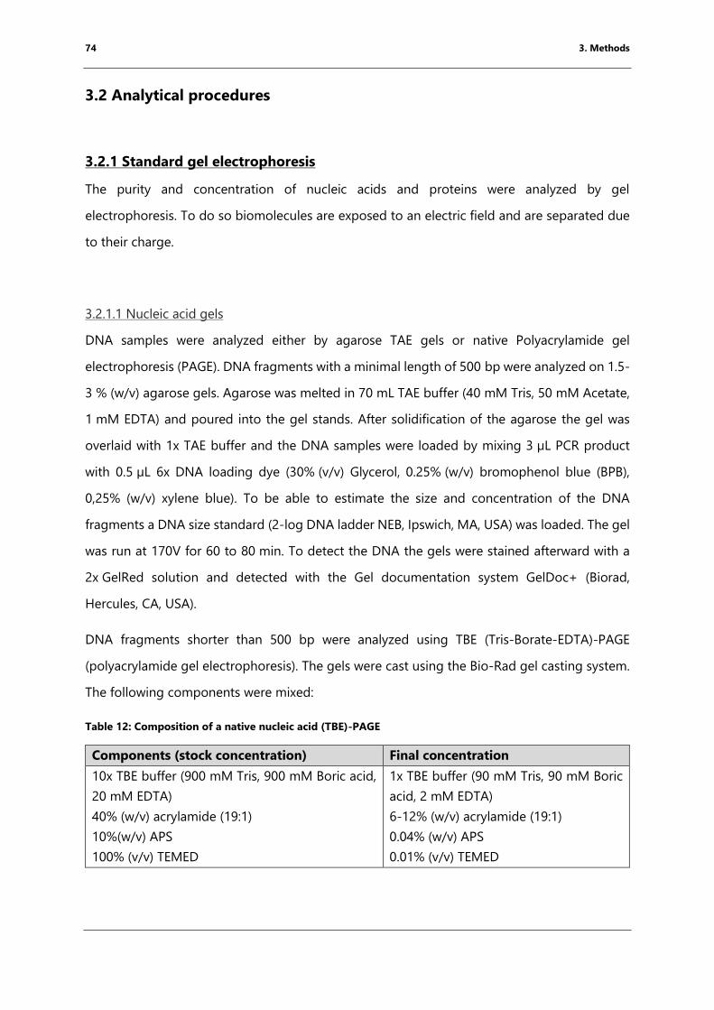
74 3. Methods
3.2 Analytical procedures
3.2.1 Standard gel electrophoresis
The purity and concentration of nucleic acids and proteins were analyzed by gel
electrophoresis. To do so biomolecules are exposed to an electric field and are separated due
to their charge.
3.2.1.1 Nucleic acid gels
DNA samples were analyzed either by agarose TAE gels or native Polyacrylamide gel
electrophoresis (PAGE). DNA fragments with a minimal length of 500 bp were analyzed on 1.5-
3 % (w/v) agarose gels. Agarose was melted in 70 mL TAE buffer (40 mM Tris, 50 mM Acetate,
1 mM EDTA) and poured into the gel stands. After solidification of the agarose the gel was
overlaid with 1x TAE buffer and the DNA samples were loaded by mixing 3 µL PCR product
with 0.5 µL 6x DNA loading dye (30% (v/v) Glycerol, 0.25% (w/v) bromophenol blue (BPB),
0,25% (w/v) xylene blue). To be able to estimate the size and concentration of the DNA
fragments a DNA size standard (2-log DNA ladder NEB, Ipswich, MA, USA) was loaded. The gel
was run at 170V for 60 to 80 min. To detect the DNA the gels were stained afterward with a
2x GelRed solution and detected with the Gel documentation system GelDoc+ (Biorad,
Hercules, CA, USA).
DNA fragments shorter than 500 bp were analyzed using TBE (Tris-Borate-EDTA)-PAGE
(polyacrylamide gel electrophoresis). The gels were cast using the Bio-Rad gel casting system.
The following components were mixed:
Table 12: Composition of a native nucleic acid (TBE)-PAGE
Components (stock concentration) Final concentration
10x TBE buffer (900 mM Tris, 900 mM Boric acid,
20 mM EDTA)
40% (w/v) acrylamide (19:1)
10%(w/v) APS
100% (v/v) TEMED
1x TBE buffer (90 mM Tris, 90 mM Boric
acid, 2 mM EDTA)
6-12% (w/v) acrylamide (19:1)
0.04% (w/v) APS
0.01% (v/v) TEMED
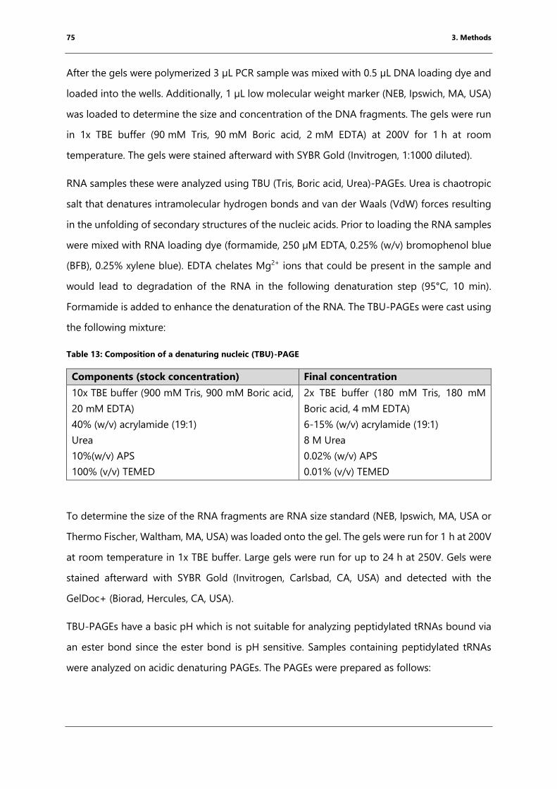
75 3. Methods
After the gels were polymerized 3 µL PCR sample was mixed with 0.5 µL DNA loading dye and
loaded into the wells. Additionally, 1 µL low molecular weight marker (NEB, Ipswich, MA, USA)
was loaded to determine the size and concentration of the DNA fragments. The gels were run
in 1x TBE buffer (90 mM Tris, 90 mM Boric acid, 2 mM EDTA) at 200V for 1 h at room
temperature. The gels were stained afterward with SYBR Gold (Invitrogen, 1:1000 diluted).
RNA samples these were analyzed using TBU (Tris, Boric acid, Urea)-PAGEs. Urea is chaotropic
salt that denatures intramolecular hydrogen bonds and van der Waals (VdW) forces resulting
in the unfolding of secondary structures of the nucleic acids. Prior to loading the RNA samples
were mixed with RNA loading dye (formamide, 250 µM EDTA, 0.25% (w/v) bromophenol blue
(BFB), 0.25% xylene blue). EDTA chelates Mg2+ ions that could be present in the sample and
would lead to degradation of the RNA in the following denaturation step (95°C, 10 min).
Formamide is added to enhance the denaturation of the RNA. The TBU-PAGEs were cast using
the following mixture:
Table 13: Composition of a denaturing nucleic (TBU)-PAGE
Components (stock concentration) Final concentration
10x TBE buffer (900 mM Tris, 900 mM Boric acid,
20 mM EDTA)
40% (w/v) acrylamide (19:1)
Urea
10%(w/v) APS
100% (v/v) TEMED
2x TBE buffer (180 mM Tris, 180 mM
Boric acid, 4 mM EDTA)
6-15% (w/v) acrylamide (19:1)
8 M Urea
0.02% (w/v) APS
0.01% (v/v) TEMED
To determine the size of the RNA fragments are RNA size standard (NEB, Ipswich, MA, USA or
Thermo Fischer, Waltham, MA, USA) was loaded onto the gel. The gels were run for 1 h at 200V
at room temperature in 1x TBE buffer. Large gels were run for up to 24 h at 250V. Gels were
stained afterward with SYBR Gold (Invitrogen, Carlsbad, CA, USA) and detected with the
GelDoc+ (Biorad, Hercules, CA, USA).
TBU-PAGEs have a basic pH which is not suitable for analyzing peptidylated tRNAs bound via
an ester bond since the ester bond is pH sensitive. Samples containing peptidylated tRNAs
were analyzed on acidic denaturing PAGEs. The PAGEs were prepared as follows:
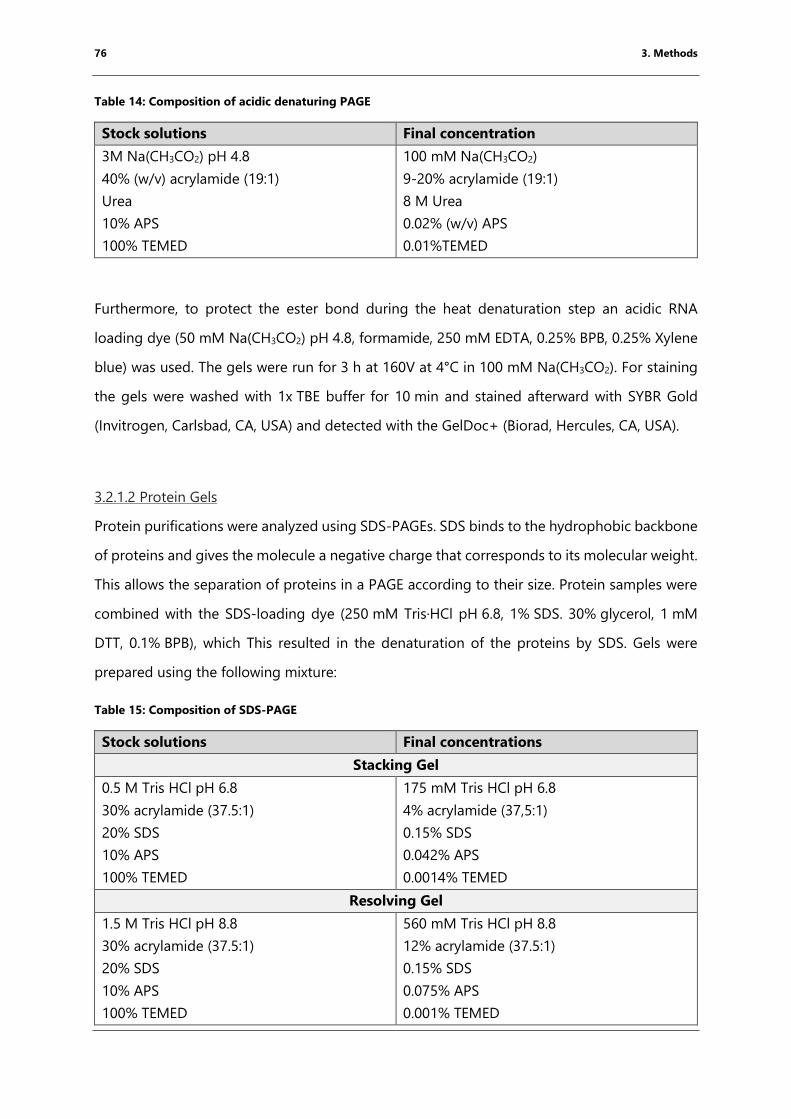
76 3. Methods
Table 14: Composition of acidic denaturing PAGE
Stock solutions Final concentration
3M Na(CH3CO2) pH 4.8
40% (w/v) acrylamide (19:1)
Urea
10% APS
100% TEMED
100 mM Na(CH3CO2)
9-20% acrylamide (19:1)
8 M Urea
0.02% (w/v) APS
0.01%TEMED
Furthermore, to protect the ester bond during the heat denaturation step an acidic RNA
loading dye (50 mM Na(CH3CO2) pH 4.8, formamide, 250 mM EDTA, 0.25% BPB, 0.25% Xylene
blue) was used. The gels were run for 3 h at 160V at 4°C in 100 mM Na(CH3CO2). For staining
the gels were washed with 1x TBE buffer for 10 min and stained afterward with SYBR Gold
(Invitrogen, Carlsbad, CA, USA) and detected with the GelDoc+ (Biorad, Hercules, CA, USA).
3.2.1.2 Protein Gels
Protein purifications were analyzed using SDS-PAGEs. SDS binds to the hydrophobic backbone
of proteins and gives the molecule a negative charge that corresponds to its molecular weight.
This allows the separation of proteins in a PAGE according to their size. Protein samples were
combined with the SDS-loading dye (250 mM Tris·HCl pH 6.8, 1% SDS. 30% glycerol, 1 mM
DTT, 0.1% BPB), which This resulted in the denaturation of the proteins by SDS. Gels were
prepared using the following mixture:
Table 15: Composition of SDS-PAGE
Stock solutions Final concentrations
Stacking Gel
0.5 M Tris HCl pH 6.8
30% acrylamide (37.5:1)
20% SDS
10% APS
100% TEMED
175 mM Tris HCl pH 6.8
4% acrylamide (37,5:1)
0.15% SDS
0.042% APS
0.0014% TEMED
Resolving Gel
1.5 M Tris HCl pH 8.8
30% acrylamide (37.5:1)
20% SDS
10% APS
100% TEMED
560 mM Tris HCl pH 8.8
12% acrylamide (37.5:1)
0.15% SDS
0.075% APS
0.001% TEMED
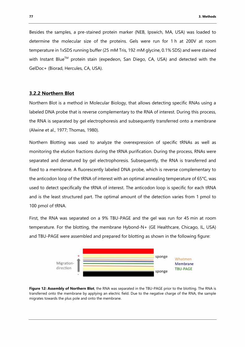
77 3. Methods
Besides the samples, a pre-stained protein marker (NEB, Ipswich, MA, USA) was loaded to
determine the molecular size of the proteins. Gels were run for 1 h at 200V at room
temperature in 1xSDS running buffer (25 mM Tris, 192 mM glycine, 0.1% SDS) and were stained
with Instant BlueTM protein stain (expedeon, San Diego, CA, USA) and detected with the
GelDoc+ (Biorad, Hercules, CA, USA).
3.2.2 Northern Blot
Northern Blot is a method in Molecular Biology, that allows detecting specific RNAs using a
labeled DNA probe that is reverse complementary to the RNA of interest. During this process,
the RNA is separated by gel electrophoresis and subsequently transferred onto a membrane
(Alwine et al., 1977; Thomas, 1980).
Northern Blotting was used to analyze the overexpression of specific tRNAs as well as
monitoring the elution fractions during the tRNA purification. During the process, RNAs were
separated and denatured by gel electrophoresis. Subsequently, the RNA is transferred and
fixed to a membrane. A fluorescently labeled DNA probe, which is reverse complementary to
the anticodon loop of the tRNA of interest with an optimal annealing temperature of 65°C, was
used to detect specifically the tRNA of interest. The anticodon loop is specific for each tRNA
and is the least structured part. The optimal amount of the detection varies from 1 pmol to
100 pmol of tRNA.
First, the RNA was separated on a 9% TBU-PAGE and the gel was run for 45 min at room
temperature. For the blotting, the membrane Hybond-N+ (GE Healthcare, Chicago, IL, USA)
and TBU-PAGE were assembled and prepared for blotting as shown in the following figure:
Figure 12: Assembly of Northern Blot, the RNA was separated in the TBU-PAGE prior to the blotting. The RNA is
transferred onto the membrane by applying an electric field. Due to the negative charge of the RNA, the sample
migrates towards the plus pole and onto the membrane.
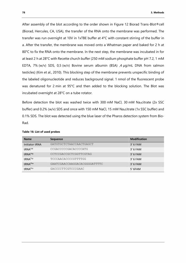
78 3. Methods
After assembly of the blot according to the order shown in Figure 12 Biorad Trans-Blot®cell
(Biorad, Hercules, CA, USA), the transfer of the RNA onto the membrane was performed. The
transfer was run overnight at 10V in 1xTBE buffer at 4°C with constant stirring of the buffer in
a. After the transfer, the membrane was moved onto a Whatman paper and baked for 2 h at
80°C to fix the RNA onto the membrane. In the next step, the membrane was incubated in for
at least 2 h at 28°C with Recette church buffer (250 mM sodium phosphate buffer pH 7.2, 1 mM
EDTA, 7% (w/v) SDS, 0,5 (w/v) Bovine serum albumin (BSA) ,4 µg/mL DNA from salmon
testicles) (Kim et al., 2010). This blocking step of the membrane prevents unspecific binding of
the labeled oligonucleotide and reduces background signal. 1 nmol of the fluorescent probe
was denatured for 2 min at 95°C and then added to the blocking solution. The Blot was
incubated overnight at 28°C on a tube rotator.
Before detection the blot was washed twice with 300 mM NaCl, 30 mM Na3citrate (2x SSC
buffer) and 0.2% (w/v) SDS and once with 150 mM NaCl, 15 mM Na3citrate (1x SSC buffer) and
0.1% SDS. The blot was detected using the blue laser of the Pharos detection system from Bio-
Rad.
Table 16: List of used probes
Name Sequence Modification
Initiator tRNA GATGTGCTCTAACCAACTGAGCT 3' 6 FAM
tRNAVal CCGACCCCCGACACCCCATG 3' 6 FAM
tRNAArg CCTCCGACCGCTCGGTTCGTAG 3' 6 FAM
tRNATrp TCCCAACACCCCGTTTTGG 3' 6 FAM
tRNAPhe GAATCGAACCAAGGACACGGGGATTTTC 3' 6 FAM
tRNAPro GACCCCTTCGTCCCGAAC 5' 6FAM
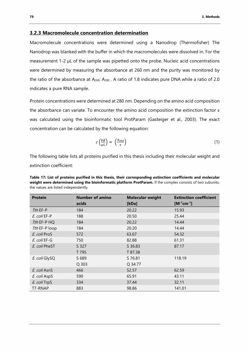
79 3. Methods
3.2.3 Macromolecule concentration determination
Macromolecule concentrations were determined using a Nanodrop (Thermofisher) The
Nanodrop was blanked with the buffer in which the macromolecules were dissolved in. For the
measurement 1-2 µL of the sample was pipetted onto the probe. Nucleic acid concentrations
were determined by measuring the absorbance at 260 nm and the purity was monitored by
the ratio of the absorbance at A260: A280 . A ratio of 1.8 indicates pure DNA while a ratio of 2.0
indicates a pure RNA sample.
Protein concentrations were determined at 280 nm. Depending on the amino acid composition
the absorbance can variate. To encounter the amino acid composition the extinction factor ε
was calculated using the bioinformatic tool ProtParam (Gasteiger et al., 2003). The exact
concentration can be calculated by the following equation:
𝑐 (µ𝑔
µ𝐿) = (
𝐴260
𝜀) (1)
The following table lists all proteins purified in this thesis including their molecular weight and
extinction coefficient:
Table 17: List of proteins purified in this thesis, their corresponding extinction coefficients and molecular
weight were determined using the bioinformatic platform ProtParam. If the complex consists of two subunits,
the values are listed independently.
Protein Number of amino
acids
Molecular weight
[kDa]
Extinction coefficient
[M-1cm-1]
Tth EF-P 184 20.22 15.93
E. coli EF-P 188 20.50 25.44
Tth EF-P HQ 184 20.22 14.44
Tth EF-P loop 184 20.20 14.44
E. coli ProS 572 63.67 54.32
E. coli EF-G 750 82.88 61.31
E. coli PheST S 327
T 795
S 36.83
T 87.38
87.17
E. coli GlySQ S 689
Q 303
S 76.81
Q 34.77
118.19
E. coli AsnS 466 52.57 62.59
E. coli AspS 590 65.91 43.11
E. coli TrpS 334 37.44 32.11
T7-RNAP 883 98.86 141.01
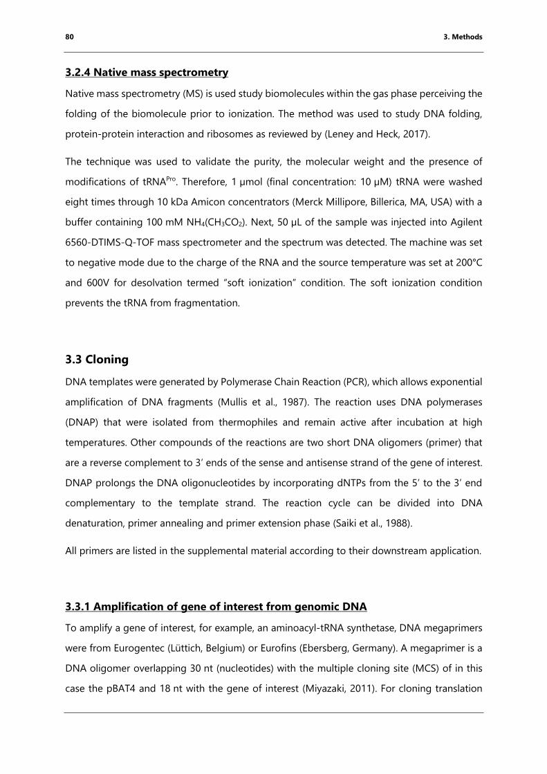
80 3. Methods
3.2.4 Native mass spectrometry
Native mass spectrometry (MS) is used study biomolecules within the gas phase perceiving the
folding of the biomolecule prior to ionization. The method was used to study DNA folding,
protein-protein interaction and ribosomes as reviewed by (Leney and Heck, 2017).
The technique was used to validate the purity, the molecular weight and the presence of
modifications of tRNAPro. Therefore, 1 µmol (final concentration: 10 µM) tRNA were washed
eight times through 10 kDa Amicon concentrators (Merck Millipore, Billerica, MA, USA) with a
buffer containing 100 mM NH4(CH3CO2). Next, 50 µL of the sample was injected into Agilent
6560-DTIMS-Q-TOF mass spectrometer and the spectrum was detected. The machine was set
to negative mode due to the charge of the RNA and the source temperature was set at 200°C
and 600V for desolvation termed “soft ionization” condition. The soft ionization condition
prevents the tRNA from fragmentation.
3.3 Cloning
DNA templates were generated by Polymerase Chain Reaction (PCR), which allows exponential
amplification of DNA fragments (Mullis et al., 1987). The reaction uses DNA polymerases
(DNAP) that were isolated from thermophiles and remain active after incubation at high
temperatures. Other compounds of the reactions are two short DNA oligomers (primer) that
are a reverse complement to 3’ ends of the sense and antisense strand of the gene of interest.
DNAP prolongs the DNA oligonucleotides by incorporating dNTPs from the 5’ to the 3’ end
complementary to the template strand. The reaction cycle can be divided into DNA
denaturation, primer annealing and primer extension phase (Saiki et al., 1988).
All primers are listed in the supplemental material according to their downstream application.
3.3.1 Amplification of gene of interest from genomic DNA
To amplify a gene of interest, for example, an aminoacyl-tRNA synthetase, DNA megaprimers
were from Eurogentec (Lüttich, Belgium) or Eurofins (Ebersberg, Germany). A megaprimer is a
DNA oligomer overlapping 30 nt (nucleotides) with the multiple cloning site (MCS) of in this
case the pBAT4 and 18 nt with the gene of interest (Miyazaki, 2011). For cloning translation
81 3. Methods
factors, histidine6 tags (H6-tag) were introduced at the N- or C-terminus as reported in (Shimizu
et al., 2001; Shimizu et al., 2005).
The amplification was performed by PCR using Phusion DNA polymerase (PhuDNAP). The
pipetting scheme is showing the following table:
Table 18: Pipetting scheme for gene amplification from genomic DNA
Component [stock concentration] Final concentration
HF-Buffer (NEB, Ipswich, MA, USA) [5x] 1x
dNTPs [10 mM each] 200 µM each
Forward primer [10 µM] 200 nM
Reverse primer [10 µM] 200 nM
Genomic DNA 2 µM
PhuDNAP [2 000 u/mL] 0.02 u/µL
Nuclease-free water To final volume
The corresponding PCR program is shown in the following table:
Table 19: PCR program for gene extraction
Temperature Time Function Cycles
98°C 30 s Enzyme activation 1x
98°C 30 s Denaturation of the vector and PCR
fragment 30x
50-60°C 30 s Primer annealing
72°C 2 min Primer extension
4°C ∞ Incubation until further usage 1x
For gene operons like glySQ longer than 750 bp, the temperature during primer extension was
reduced to 68°C and the reaction time was increased to 3 min. The PCR products were analyzed
on 1.5% TAE-agarose gels (chapter 3.8.1) by loading 3 µL per reaction together with the 2-log
DNA ladder (NEB, Ipswich, MA, USA). Depending on its purity, the product was purified by PCR
purification kit (Qiagen, Hilden, Germany) or gel extraction kit (Qiagen, Hilden, Germany)
following the manufacturer's instructions.
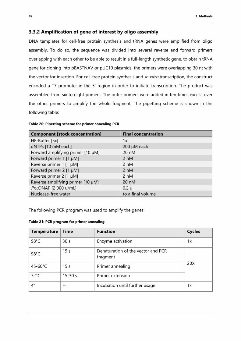
82 3. Methods
3.3.2 Amplification of gene of interest by oligo assembly
DNA templates for cell-free protein synthesis and tRNA genes were amplified from oligo
assembly. To do so, the sequence was divided into several reverse and forward primers
overlapping with each other to be able to result in a full-length synthetic gene. to obtain tRNA
gene for cloning into pBASTNAV or pUC19 plasmids, the primers were overlapping 30 nt with
the vector for insertion. For cell-free protein synthesis and in vitro transcription, the construct
encoded a T7 promoter in the 5’ region in order to initiate transcription. The product was
assembled from six to eight primers. The outer primers were added in ten times excess over
the other primers to amplify the whole fragment. The pipetting scheme is shown in the
following table:
Table 20: Pipetting scheme for primer annealing PCR
Component [stock concentration] Final concentration
HF-Buffer [5x] 1x
dNTPs [10 mM each] 200 µM each
Forward amplifying primer [10 µM] 20 nM
Forward primer 1 [1 µM] 2 nM
Reverse primer 1 [1 µM] 2 nM
Forward primer 2 [1 µM] 2 nM
Reverse primer 2 [1 µM] 2 nM
Reverse amplifying primer [10 µM] 20 nM
PhuDNAP [2 000 u/mL] 0.2 u
Nuclease-free water to a final volume
The following PCR program was used to amplify the genes:
Table 21: PCR program for primer annealing
Temperature Time Function Cycles
98°C 30 s Enzyme activation 1x
98°C 15 s Denaturation of the vector and PCR
fragment
20X 45-60°C 15 s Primer annealing
72°C 15-30 s Primer extension
4° ∞ Incubation until further usage 1x
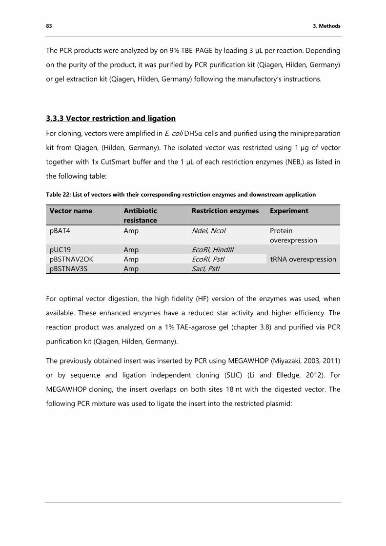
83 3. Methods
The PCR products were analyzed by on 9% TBE-PAGE by loading 3 µL per reaction. Depending
on the purity of the product, it was purified by PCR purification kit (Qiagen, Hilden, Germany)
or gel extraction kit (Qiagen, Hilden, Germany) following the manufactory’s instructions.
3.3.3 Vector restriction and ligation
For cloning, vectors were amplified in E. coli DH5α cells and purified using the minipreparation
kit from Qiagen, (Hilden, Germany). The isolated vector was restricted using 1 µg of vector
together with 1x CutSmart buffer and the 1 µL of each restriction enzymes (NEB,) as listed in
the following table:
Table 22: List of vectors with their corresponding restriction enzymes and downstream application
Vector name Antibiotic
resistance
Restriction enzymes Experiment
pBAT4 Amp NdeI, NcoI Protein
overexpression
pUC19 Amp EcoRI, HindIII
tRNA overexpression pBSTNAV2OK Amp EcoRI, PstI
pBSTNAV3S Amp SacI, PstI
For optimal vector digestion, the high fidelity (HF) version of the enzymes was used, when
available. These enhanced enzymes have a reduced star activity and higher efficiency. The
reaction product was analyzed on a 1% TAE-agarose gel (chapter 3.8) and purified via PCR
purification kit (Qiagen, Hilden, Germany).
The previously obtained insert was inserted by PCR using MEGAWHOP (Miyazaki, 2003, 2011)
or by sequence and ligation independent cloning (SLIC) (Li and Elledge, 2012). For
MEGAWHOP cloning, the insert overlaps on both sites 18 nt with the digested vector. The
following PCR mixture was used to ligate the insert into the restricted plasmid:
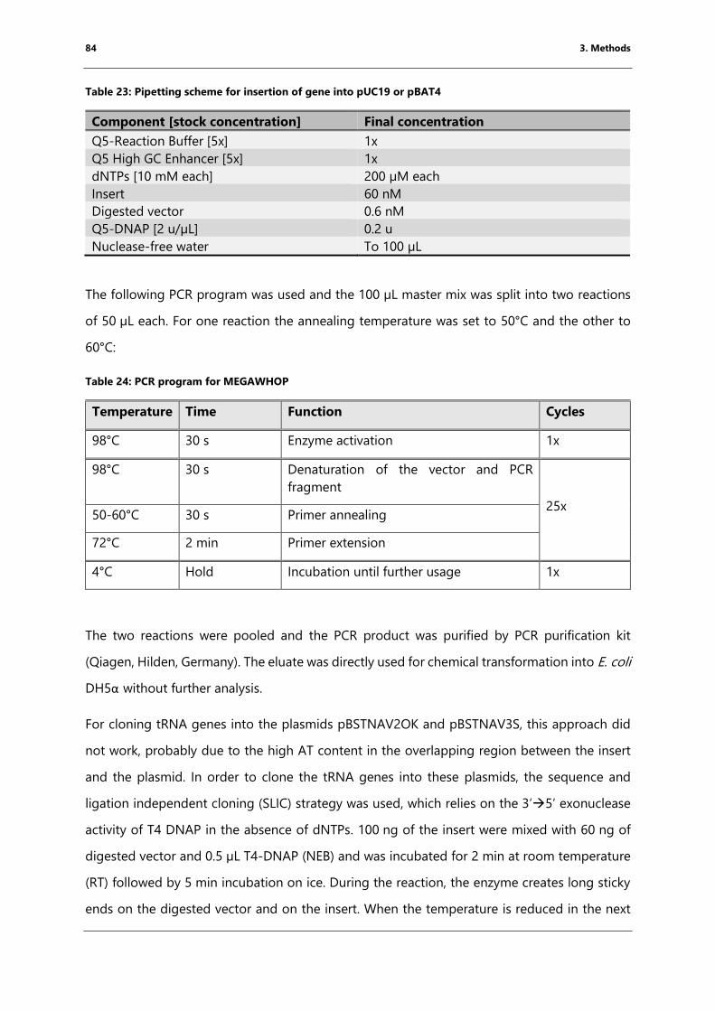
84 3. Methods
Table 23: Pipetting scheme for insertion of gene into pUC19 or pBAT4
Component [stock concentration] Final concentration
Q5-Reaction Buffer [5x] 1x
Q5 High GC Enhancer [5x] 1x
dNTPs [10 mM each] 200 µM each
Insert 60 nM
Digested vector 0.6 nM
Q5-DNAP [2 u/µL] 0.2 u
Nuclease-free water To 100 µL
The following PCR program was used and the 100 µL master mix was split into two reactions
of 50 µL each. For one reaction the annealing temperature was set to 50°C and the other to
60°C:
Table 24: PCR program for MEGAWHOP
Temperature Time Function Cycles
98°C 30 s Enzyme activation 1x
98°C 30 s Denaturation of the vector and PCR
fragment
25x 50-60°C 30 s Primer annealing
72°C 2 min Primer extension
4°C Hold Incubation until further usage 1x
The two reactions were pooled and the PCR product was purified by PCR purification kit
(Qiagen, Hilden, Germany). The eluate was directly used for chemical transformation into E. coli
DH5α without further analysis.
For cloning tRNA genes into the plasmids pBSTNAV2OK and pBSTNAV3S, this approach did
not work, probably due to the high AT content in the overlapping region between the insert
and the plasmid. In order to clone the tRNA genes into these plasmids, the sequence and
ligation independent cloning (SLIC) strategy was used, which relies on the 3’5’ exonuclease
activity of T4 DNAP in the absence of dNTPs. 100 ng of the insert were mixed with 60 ng of
digested vector and 0.5 µL T4-DNAP (NEB) and was incubated for 2 min at room temperature
(RT) followed by 5 min incubation on ice. During the reaction, the enzyme creates long sticky
ends on the digested vector and on the insert. When the temperature is reduced in the next
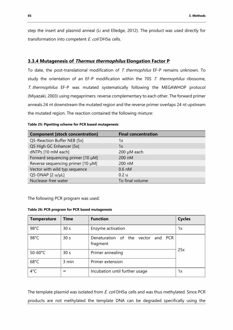
85 3. Methods
step the insert and plasmid anneal (Li and Elledge, 2012). The product was used directly for
transformation into competent E. coli DH5α cells.
3.3.4 Mutagenesis of Thermus thermophilus Elongation Factor P
To date, the post-translational modification of T. thermophilus EF-P remains unknown. To
study the orientation of an EF-P modification within the 70S T. thermophilus ribosome,
T. thermophilus EF-P was mutated systematically following the MEGAWHOP protocol
(Miyazaki, 2003) using megaprimers. reverse complementary to each other. The forward primer
anneals 24 nt downstream the mutated region and the reverse primer overlaps 24 nt upstream
the mutated region. The reaction contained the following mixture:
Table 25: Pipetting scheme for PCR based mutagenesis
Component [stock concentration] Final concentration
Q5-Reaction Buffer NEB [5x] 1x
Q5 High GC Enhancer [5x] 1x
dNTPs [10 mM each] 200 µM each
Forward sequencing primer [10 µM] 200 nM
Reverse sequencing primer [10 µM] 200 nM
Vector with wild typ sequence 0.6 nM
Q5-DNAP [2 u/µL] 0.2 u
Nuclease-free water To final volume
The following PCR program was used:
Table 26: PCR program for PCR based mutagenesis
Temperature Time Function Cycles
98°C 30 s Enzyme activation 1x
98°C 30 s Denaturation of the vector and PCR
fragment
25x 50-60°C 30 s Primer annealing
68°C 3 min Primer extension
4°C ∞ Incubation until further usage 1x
The template plasmid was isolated from E. coli DH5α cells and was thus methylated. Since PCR
products are not methylated the template DNA can be degraded specifically using the
86 3. Methods
restriction enzyme DpnI. DpnI recognizes and cuts specifically methylated and hemimethylated
DNA double strands. To do so, the PCR reaction was mixed with CutSmart buffer (final
concentration 1x) and 10 units DpnI. The reaction was incubated for 2 h at 37°C. The DNA
fragments were purified using the PCR purification kit (Qiagen, Hilden, Germany) and eluted
in30 µL DNase-free water. 15 µL of this DNA was subsequently used for transformation into
competent E. coli XL1-blue cells.
3.3.5 Preparation of chemically competent cells
For cloning, protein and tRNA expression the corresponding plasmids were transformed into
E. coli cells. To enhance uptake of the plasmid DNA the cells were treated with MgCl2, which
leads to permeabilization of the cell wall.
For the preparation of chemically competent cells, a liquid LB culture (containing the required
antibiotic; see Table 10) was inoculated with the respective cells and was grown overnight at
37 °C with 200 rpm shaking. This overnight culture was subsequently used to inoculate 200 mL
LB media to a starting OD600 between 0.05 and 0.1, which was grown at 37 °C until the culture
reached an OD600 of 0.4-0.6. After pelleting the cells (4000g, 10 minutes, 4 °C, Eppendorf
5804R) the pellets were resuspended in 100 mL 100 mM MgCl2 solution and were incubated
for 30 min on ice. Subsequently, the cells were pelleted and resuspended in 10 mL
100 mM CaCl2 and 15% glycerol. The cells were aliquoted in 100 µL and flash-frozen in liquid
nitrogen (N2) and stored at -80°C until further use.
3.3.6 Plasmid transformation into chemically competent cells
For the chemical transformation, the cells were thawed on ice. After adding the plasmid
solution (15 µL of PCR products or SLIC products or 0.5 µL of plasmids) the cells were incubated
on ice for 30 min. After a heat shock at 42°C for 1 min, the cells were placed on ice immediately
and incubated for another 1.5 min. To allow recovery of the cells 500 µL of LB media was added
and the cells were incubated at 37 °C for at least one hour. After the incubation, the cells were
plated on LB agar plates containing the plasmid-specific antibiotic for cell selection. The agar
plates were incubated overnight at 37 °C.
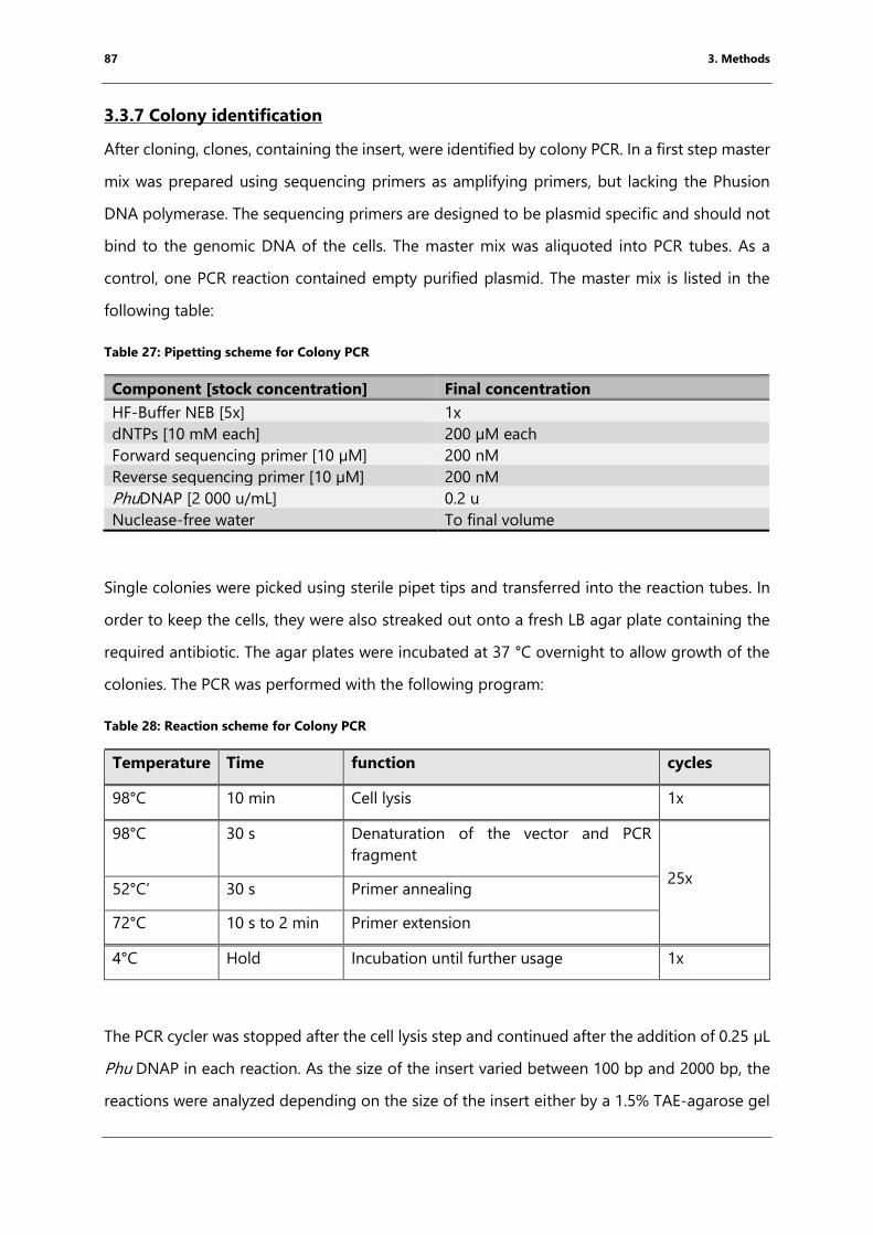
87 3. Methods
3.3.7 Colony identification
After cloning, clones, containing the insert, were identified by colony PCR. In a first step master
mix was prepared using sequencing primers as amplifying primers, but lacking the Phusion
DNA polymerase. The sequencing primers are designed to be plasmid specific and should not
bind to the genomic DNA of the cells. The master mix was aliquoted into PCR tubes. As a
control, one PCR reaction contained empty purified plasmid. The master mix is listed in the
following table:
Table 27: Pipetting scheme for Colony PCR
Component [stock concentration] Final concentration
HF-Buffer NEB [5x] 1x
dNTPs [10 mM each] 200 µM each
Forward sequencing primer [10 µM] 200 nM
Reverse sequencing primer [10 µM] 200 nM
PhuDNAP [2 000 u/mL] 0.2 u
Nuclease-free water To final volume
Single colonies were picked using sterile pipet tips and transferred into the reaction tubes. In
order to keep the cells, they were also streaked out onto a fresh LB agar plate containing the
required antibiotic. The agar plates were incubated at 37 °C overnight to allow growth of the
colonies. The PCR was performed with the following program:
Table 28: Reaction scheme for Colony PCR
Temperature Time function cycles
98°C 10 min Cell lysis 1x
98°C 30 s Denaturation of the vector and PCR
fragment
25x 52°C’ 30 s Primer annealing
72°C 10 s to 2 min Primer extension
4°C Hold Incubation until further usage 1x
The PCR cycler was stopped after the cell lysis step and continued after the addition of 0.25 µL
Phu DNAP in each reaction. As the size of the insert varied between 100 bp and 2000 bp, the
reactions were analyzed depending on the size of the insert either by a 1.5% TAE-agarose gel
88 3. Methods
or a 9% TBE-PAGE (chapter 3.2). Colonies corresponding to reactions giving rise to a band on
the gel with the size of the insert were used for further steps.
3.3.8 Plasmid extraction and sequencing
The potential colonies, containing potentially the insert, were transferred into 5 mL LB
containing the respective antibiotic and grown overnight at 37 °C and shaking at 200 rpm. The
cells were harvested by centrifugation (4000 g, 10 min) and the plasmids purified using the
plasmid purification kit following the manufacturer’s instructions. The plasmid DNA was eluted
in30 µL water and the concentration was determined using a NanoDrop (2000c, Thermofisher,
Waltham, MA, USA).
Plasmids were sequenced by SANGER sequencing (GATC Biotech, Konstanz, Germany) The
resulting sequences were confirmed by comparing with the planned sequences using Basic
Local Alignment Search Tool (BLAST, (Altschul et al., 1990)) and Multalign (Toulouse, France, ).
3.4 Protein expression and purification
All proteins purified in this thesis were overexpressed in E. coli BL21 Gold. The genes encoding
the proteins were cloned to be under the regulation of a T7 RNAP promoter. In E. coli BL21
Gold the T7 RNAP gene is under the control of a LacI repressor and can be induced by the
addition of the lactose derivative, isopropyl β-D-1-thiogalactopyranoside (IPTG).
The process of protein purification can be divided into a capture step, an intermediate step
and a polishing step as reported in (Williams, 2001). To purify the proteins, they were tagged
with an H6-tag. The six consecutive H residues form a stable complex with immobilized Co2+
ions allowing separation of the protein of interest from the cellular proteins (capture step)
(Petty, 2001; Williams, 2001). Since the purity of the protein samples, purified proteins after the
Co2+-NTA column was already high. Subsequently, the buffer was exchanged using a size
exclusion chromatography directly as polishing step. In this step, the sample components are
separated by their overall size (Hagel, 2001; Williams, 2001).
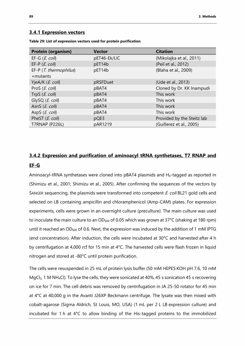
89 3. Methods
3.4.1 Expression vectors
Table 29: List of expression vectors used for protein purification
Protein (organism) Vector Citation
EF-G (E. coli) pET46-Ek/LIC (Mikolajka et al., 2011)
EF-P (E. coli) pET14b (Peil et al., 2012)
EF-P (T. thermophilus)
+mutants
pET14b (Blaha et al., 2009)
YjeA/K (E. coli) pRSFDuet (Ude et al., 2013)
ProS (E. coli) pBAT4 Cloned by Dr. KK Inampudi
TrpS (E. coli) pBAT4 This work
GlySQ (E. coli) pBAT4 This work
AsnS (E. coli) pBAT4 This work
AspS (E. coli) pBAT4 This work
PheST (E. coli) pQE3 Provided by the Steitz lab
T7RNAP (P226L) pAR1219 (Guillerez et al., 2005)
3.4.2 Expression and purification of aminoacyl tRNA synthetases, T7 RNAP and
EF-G
Aminoacyl-tRNA synthetases were cloned into pBAT4 plasmids and H6-tagged as reported in
(Shimizu et al., 2001; Shimizu et al., 2005). After confirming the sequences of the vectors by
SANGER sequencing, the plasmids were transformed into competent E. coli BL21 gold cells and
selected on LB containing ampicillin and chloramphenicol (Amp-CAM) plates. For expression
experiments, cells were grown in an overnight culture (preculture). The main culture was used
to inoculate the main culture to an OD600 of 0.05 which was grown at 37°C (shaking at 180 rpm)
until it reached an OD600 of 0.6. Next, the expression was induced by the addition of 1 mM IPTG
(end concentration). After induction, the cells were incubated at 30°C and harvested after 4 h
by centrifugation at 4,000 rcf for 15 min at 4°C. The harvested cells were flash frozen in liquid
nitrogen and stored at -80°C until protein purification.
The cells were resuspended in 25 mL of protein lysis buffer (50 mM HEPES·KOH pH 7.6, 10 mM
MgCl2, 1 M NH4Cl). To lyse the cells, they were sonicated at 40%, 45 s sonication 45 s recovering
on ice for 7 min. The cell debris was removed by centrifugation in JA 25-50 rotator for 45 min
at 4°C at 40,000 g in the Avanti J26XP Beckmann centrifuge. The lysate was then mixed with
cobalt-agarose (Sigma Aldrich, St Louis, MO, USA) (1 mL per 2 L LB expression culture) and
incubated for 1 h at 4°C to allow binding of the His-tagged proteins to the immobilized
90 3. Methods
Co2+-ions. After binding of the protein, the beads were transferred onto a gravity column and
were washed with 60 mL protein lysis buffer to remove loosely bound proteins from the Cobalt-
agarose beads. The proteins were eluted by adding 5 mL protein elution buffer (protein lysis
buffer with 250 mM imidazole).
The eluate was concentrated using Amicon centrifugation concentrators (Merck Millipore,
Billerica, MA, USA). The molecular weight cut off of the concentrators was chosen depending
on the size of the purified protein. Subsequently, the sample was loaded onto a gel filtration
column to exchange the buffer to protein storage buffer (20 mM HEPES KOH pH 7.6, 10 mM
MgCl2, 50 mM KCl, 50 mM NH4Cl). Depending on the molecular weight of the protein, the gel
filtration was performed using a Superdex 75 (16/600) or Superdex 100 through the NGC
medium pressure liquid chromatography system (Biorad, Hercules, CA, USA) collecting 1 mL
elution fractions. Elution fractions were analyzed by SDS-PAGE. Fractions containing the
protein of interest were pooled and concentrated using centrifugal filters to a concentration of
20 mg/mL. For long-term storage, 100% glycerol was added to a final concentration of
50% glycerol and the samples were flash-frozen in liquid nitrogen and stored at -80°C.
3.4.3 Expression and purification of Elongation factor-P
Elongation factor-P (EF-P) from E. coli was overexpressed as published (Ude et al., 2013). To
ensure that the expressed protein is fully modified the plasmid encoding for H6-tagged EF-P
was co-expressed with the pRSF plasmid encoding for the untagged modification enzymes
YjeA and YjeK. The expression of all three proteins is under the control of a T7 promoter
(Doerfel et al., 2015). 7 T. thermophilus EF-P was expressed as published in (Blaha et al., 2009)
was used. Plasmids encoding mutated versions of T. thermophiles EF-P were also co-expressed
with the modification enzymes for E. coli EF-P. The proteins were overexpressed in E. coli BL21
Gold cells. and the expression was induced by addition of 1mM IPTG (end concentration) at
an OD600 of 0.6. After induction, the cells were grown for 4 h at 30°C. The cells were harvested
by centrifugation at 4,000 g for 15 min at 4°C and flash frozen in liquid nitrogen and stored at
-80 °C until further use.
For EF-P purification the cells were thawed and resuspended protein lysis buffer and lysed as
described in the previous chapter 3.4.2. For the purification of E. coli EF-P, the cell lysate was
91 3. Methods
incubated directly with Co2+-NTA beads. The cell lysate containing the T. thermophilus EF-P or
one of the mutants was first treated with a heat shock to remove E. coli proteins, using the
expected thermal stability of the protein. For the heat shock, the lysate was transferred into an
Erlenmeyer flask that was put in a water bath at a temperature of 60°C. The cell lysate was
incubated for 20 min followed by removal of cell debris and denatured E. coli proteins by
centrifugation in JA 25.50 (Beckmann) rotor for 45 min at 40,000 g at 4°C. The following steps
were performed as described in chapter 3.4.2. Since these proteins were mainly used for
crystallography experiments the proteins were flash-frozen and stored at -80 °C without the
addition of glycerol.
3.5 RNA handling
All reactions containing RNA were performed in DEPC treated water or molecular biology grade
water (Merck, Billerica, MA, USA) to avoid degradation of the RNA by RNases. Glassware used
for these reactions was baked for several hours to degrade potential RNase contaminations.
3.5.1 In vitro transcription and purification in vitro transcribed RNA
Unmodified tRNAs, flexizymes and mRNA templates for toeprinting were transcribed in vitro
using the T7 RNAP mutant P266L (Guillerez et al., 2005). This mutant of the T7 RNAP
synthesized more full-length product compared to the wt T7 RNAP. The DNA templates were
synthesized by PCR as described in chapter 3.2 encoding the genes under the T7 promoter. In
vitro transcription was performed using 5-10 μg/mL of DNA template, 7.5 mM of each NTP,
10 μg/mL of T7 RNAP in Ribomax buffer (80 mM Tris·HCl pH 7.6, 2 mM spermidine, 24 mM
MgCl2),.and was incubated for 2 h at 37°C. The reaction was stopped by phenol-chloroform
extraction (chapter 3.5.2) and nucleic acids and nucleotides were precipitated with 1 M
NH4(CH3CO2) and 70% ethanol end concentration
Unincorporated nucleotides were removed by washing the RNA several times with RNase-free
water over Amicon centrifugation filters (Merck Millipore, Billerica, MA, USA) using a molecular
weight cut-off smaller than the produced RNA. The success of the washing was monitored by
measuring the absorption in the flow-through on the NanoDrop. After recovery of the RNA
92 3. Methods
from the centrifugal filters, the concentration was determined using the Nanodrop 2000c
(Thermofisher, chapter 3.2) and the purity was determined by gel electrophoresis (chapter 3.2).
3.5.2 Phenol-Chloroform extraction
Purification of RNA from enzymatic reactions was performed by phenol-chloroform extraction
using. acidic phenol (pH 4.3, Na3citrate solution, Sigma Aldrich, St Louis, MO, USA). The low pH
prevents hydrolysis of RNA. The reactions were extracted by adding one volume phenol:
chloroform: isoamyl alcohol (25:24:1) and mixing it vigorously by vortexing. The organic phase
was separated from the aqueous phase by centrifugation. The denatured proteins then locate
at the interphase between the organic and the aqueous phase. The aqueous phase was re-
extracted three times with chloroform: isoamyl alcohol (24:1). All phases were washed with
reaction, stabilization buffer or molecular biology water to ensure the recovery of the RNA
sample. To remove residual organic solvents, the RNA was precipitated by NH4Ac/ethanol
precipitation, as described in chapter 3.5.3.
3.5.3 Precipitation of RNA
3.4.3.1 Alcohol precipitation of RNA
An important step in the RNA purification is the removal of salts, buffer exchange as well as
concentrating the sample. Nucleic acids can be precipitated specifically in the presence of
monovalent cations such as Na+ and NH4+. The ions bind to the phosphate-sugar backbone of
the nucleic acid and neutralize its negative charges. By the addition of 2.5 Vol EtOH or one
volume isopropanol, the RNA precipitates at low temperatures due to its decreased solubility.
For precipitation, the samples were incubated for 1 h at -20°C and then spun down for 30 min
at 18,000 rcf in a cooled tabletop centrifuge. The pellet was washed with 70% EtOH to remove
remaining salt and spun down again. The pellet was dried at room temperature for 10-15
minutes and finally. resuspended in the desired buffer.
The salt for RNA precipitation was chosen depending on the downstream application and
properties of the sample. In most cases, NH4(CH3CO2) was used due to its volatile properties.
For aminoacylated and peptidylated tRNA samples Na(CH3CO2) pH 4.8 was used to protect the
93 3. Methods
product from uncontrolled hydrolysis. Chloride as a counterion was avoided since it shows
inhibitory properties in downstream reactions such as in vitro transcription or the initiation of
in vitro translation.
3.5.3.2 TCA precipitation of RNA
tRNA samples from analytical HPLC runs were often too low concentrated for the following
Northern Blot analysis. In order to increase the concentration, the RNA was precipitated using
a 100% (w/v) trichloroacetic acid (TCA) solution to. to obtain a final TCA concentration of 10%.
The samples were incubated for 1 h on ice to ensure complete precipitation. After the
incubation, the RNA was pelleted and washed with 100% acetone to remove residual TCA. After
the washing step, the pellet was dried briefly and resuspended in RNA loading dye. If TCA was
still left in the sample, which was indicated by a color change of the RNA loading dye, small
amounts of 1 M Trizma base were added to ensure a basic pH for optimal separation on TBU-
PAGE.
3.6 tRNA expression and purification
A specific tRNA can either be produced by in vitro transcription or by overexpression of the
corresponding gene in vivo and subsequent purification from cells. tRNAs function as an
adaptor between the mRNA and the amino acid sequence (Crick, 1958) and are modified post-
transcriptionally. These modifications are important decoding capacity, decreasing codon
sensitivity and maintaining open reading frame. The advantage of the purification of tRNAs
from cells is that these tRNAs are modified. In contrast to protein purification, the length of the
tRNA is crucial for the modification, meaning the tRNA cannot be tagged. In order to purify a
specific tRNA species from a crude tRNA purification, the tRNA can be aminoacylated using
the corresponding aminoacyl-tRNA synthetase. This reaction is tRNA specific since the enzyme
recognizes specific features of the tRNA and the chemical properties of the amino acid (Ibba
et al., 1997). The aminoacylation of a tRNA changes its binding properties on a reverse phase
(RP) high-performance liquid chromatography (HPLC) column (Cayama et al., 2000) enabling
separation from the deaminoacylated-tRNAs in the mixture. The column matrix is formed by
hydrophobic residues that bind biomolecules due to hydrophobic residues exposed on the
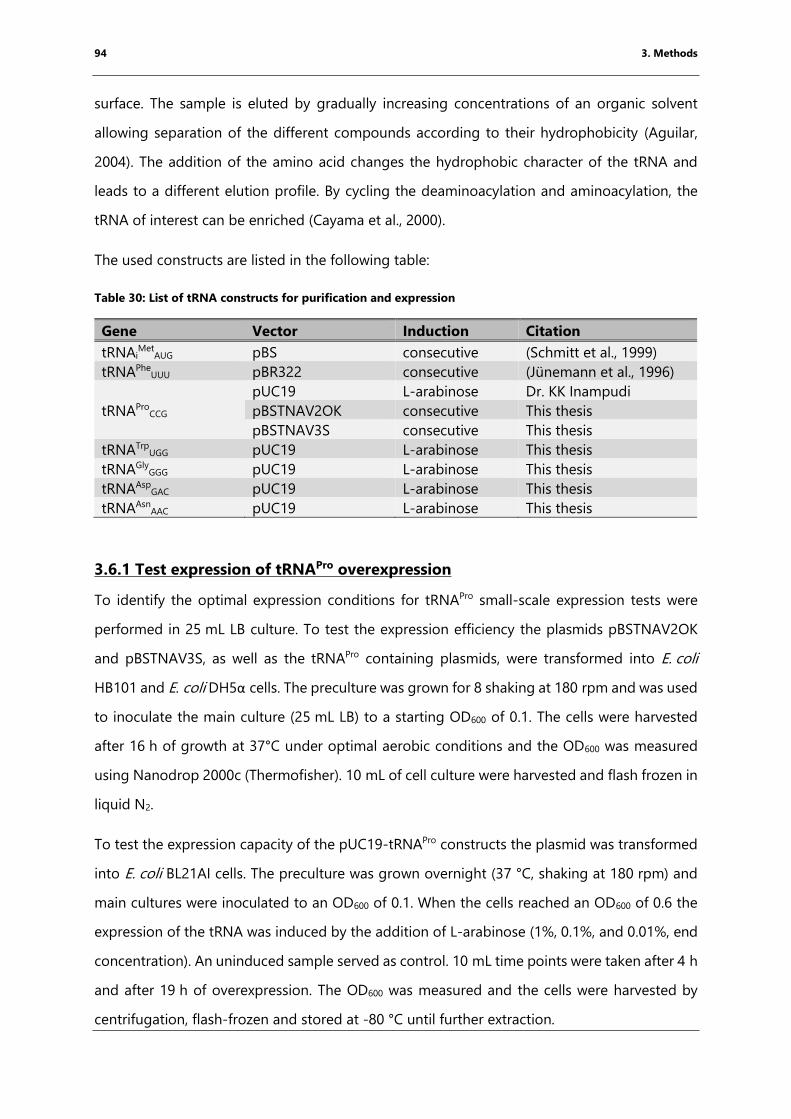
94 3. Methods
surface. The sample is eluted by gradually increasing concentrations of an organic solvent
allowing separation of the different compounds according to their hydrophobicity (Aguilar,
2004). The addition of the amino acid changes the hydrophobic character of the tRNA and
leads to a different elution profile. By cycling the deaminoacylation and aminoacylation, the
tRNA of interest can be enriched (Cayama et al., 2000).
The used constructs are listed in the following table:
Table 30: List of tRNA constructs for purification and expression
Gene Vector Induction Citation
tRNAiMet
AUG pBS consecutive (Schmitt et al., 1999)
tRNAPheUUU pBR322 consecutive (Jünemann et al., 1996)
tRNAProCCG
pUC19 L-arabinose Dr. KK Inampudi
pBSTNAV2OK consecutive This thesis
pBSTNAV3S consecutive This thesis
tRNATrpUGG pUC19 L-arabinose This thesis
tRNAGlyGGG pUC19 L-arabinose This thesis
tRNAAspGAC pUC19 L-arabinose This thesis
tRNAAsnAAC pUC19 L-arabinose This thesis
3.6.1 Test expression of tRNAPro overexpression
To identify the optimal expression conditions for tRNAPro small-scale expression tests were
performed in 25 mL LB culture. To test the expression efficiency the plasmids pBSTNAV2OK
and pBSTNAV3S, as well as the tRNAPro containing plasmids, were transformed into E. coli
HB101 and E. coli DH5α cells. The preculture was grown for 8 shaking at 180 rpm and was used
to inoculate the main culture (25 mL LB) to a starting OD600 of 0.1. The cells were harvested
after 16 h of growth at 37°C under optimal aerobic conditions and the OD600 was measured
using Nanodrop 2000c (Thermofisher). 10 mL of cell culture were harvested and flash frozen in
liquid N2.
To test the expression capacity of the pUC19-tRNAPro constructs the plasmid was transformed
into E. coli BL21AI cells. The preculture was grown overnight (37 °C, shaking at 180 rpm) and
main cultures were inoculated to an OD600 of 0.1. When the cells reached an OD600 of 0.6 the
expression of the tRNA was induced by the addition of L-arabinose (1%, 0.1%, and 0.01%, end
concentration). An uninduced sample served as control. 10 mL time points were taken after 4 h
and after 19 h of overexpression. The OD600 was measured and the cells were harvested by
centrifugation, flash-frozen and stored at -80 °C until further extraction.
95 3. Methods
Independently from the expression strategy, cells were lysed by phenol/chloroform extraction:
In the first step the cells were resuspended in 500 µL tRNA lysis Buffer (50 mM Tris·HCl pH 7.2,
50 mM Mg(CH3CO2)2) and mixed with an equal volume of phenol (pH 5.2): chloroform: isoamyl
alcohol (25:24:1) solution and centrifuged for 15 min at 18,000 rcf to separate the organic from
the aqueous phase. The aqueous phase was washed twice with 500 µL chloroform: isoamyl
alcohol (24:1). Subsequently, small RNA species were precipitated specifically with 1 M
NH4(CH3CO2) and 70% EtOH (end concentration). After pelleting, the samples were
resuspended in RNA loading dye (chapter 3.8) calculated as in the following relation:
𝑉𝑙𝑜𝑎𝑑𝑖𝑛𝑔 𝑑𝑦𝑒 = 𝑂𝐷600 ∙ 16.4 𝜇𝐿 (2)
The expression levels were analyzed by Northern Blot (chapter 3.2).
3.6.2 Large-scale expression of tRNAPro and total tRNA extraction
For large scale expression tRNAPro, eight main cultures of 1 L LB (100 μg/mL ampicillin) were
inoculated with an overnight preculture of E. coli HB101 pBSTNAV3S-tRNAPro. The cultures
were grown overnight (approximately 16 h) at 37°C under aerobe conditions. The cells were
harvested by centrifugation in a JLA 9.100 rotor at 4 000 rcf for 20 min at 4°C (Avanti J26XP
centrifuge, Beckmann, Brea, CA, USA). The cell pellets were washed with tRNA lysis buffer
(20 mM Tris·HCl pH 7.4, 20 mM Mg(CH3CO2)2) and pelleted by a second centrifugation step
using the same conditions. Per 1 L LB culture, the cell pellet weighed approximately 5 g. For
storage, cells were flash-frozen in liquid nitrogen and stored at -80°C. The following figure
highlights the different steps during tRNAPro extraction and purification:
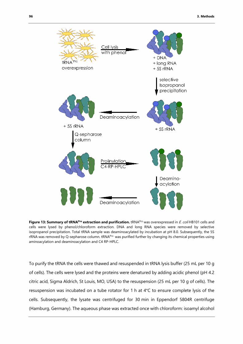
96 3. Methods
Figure 13: Summary of tRNAPro extraction and purification. tRNAPro was overexpressed in E. coli HB101 cells and
cells were lysed by phenol/chloroform extraction. DNA and long RNA species were removed by selective
isopropanol precipitation. Total tRNA sample was deaminoacylated by incubation at pH 8.0. Subsequently, the 5S
rRNA was removed by Q-sepharose column. tRNAPro was purified further by changing its chemical properties using
aminoacylation and deaminoacylation and C4 RP-HPLC.
To purify the tRNA the cells were thawed and resuspended in tRNA lysis buffer (25 mL per 10 g
of cells). The cells were lysed and the proteins were denatured by adding acidic phenol (pH 4.2
citric acid, Sigma Aldrich, St Louis, MO, USA) to the resuspension (25 mL per 10 g of cells). The
resuspension was incubated on a tube rotator for 1 h at 4°C to ensure complete lysis of the
cells. Subsequently, the lysate was centrifuged for 30 min in Eppendorf 5804R centrifuge
(Hamburg, Germany). The aqueous phase was extracted once with chloroform: isoamyl alcohol
97 3. Methods
(24:1, Sigma). The organic phases were washed once with fresh tRNA lysis buffer. The DNA was
removed by precipitation with 300 mM Na(CH3CO2) pH 4.8 and 20% isopropanol for 1 h at 4°C.
After centrifugation for 1 h at 5,170 g (JLA 8.100, Avanti J26XP, Beckmann, Brea, CA, USA) at
4°C, pellets, containing the genomic DNA of the cells, were discarded and the concentration of
isopropanol in the supernatant was increased to 60% to precipitate the RNA. The solution was
incubated overnight at 4°C to ensure precipitation. The RNA was pelleted by centrifugation
and washed with 70% ethanol to remove remaining salts. The pellet was briefly dried on air by
placing the centrifugation bottle under the hood for 20 min.
This extracted tRNA sample contains aminoacylated, deaminoacylated tRNAs and other RNA
species. To deaminoacylate all tRNAs and obtain a homogeneous preparation the pellet was
resuspended in 200 mM Tris·acetate pH 9.0 at 37°C for 1 h. In the last step of the total tRNA
extraction, small RNA species were precipitated specifically by adding 1 M NH4(CH3CO2) and
70% ethanol. The pellet was dissolved in RNase-free water and the concentration was
determined on the NanoDrop (see chapter 3.2).
3.6.3 Chromatography of tRNAPro
The total tRNA extraction (chapter 3.6.2) still contains longer rRNA species that will bind tighter
to the reverse phase (RP) column as they present a larger hydrophobic surface and might
prevent binding of the tRNA to the column material. Therefore, these and other long RNA
species were removed specifically by loading the RNA onto a Q-sepharose (GE Healthcare,
Chicago, IL, USA) column, which is an anion exchange column. Negatively charged
biomolecules bind to the positively charged matrix and can be eluted specifically by increasing
concentrations of Cl- ions in the elution buffer. Due to the lower charge, the shorter tRNAs
elute before the rRNA species. The purification protocol was adopted from the published
protocol (Mechulam et al., 2007). The binding buffer contained 20 mM HEPES·NaOH pH 7.5,
100 µM EDTA, 6 mM MgCl2, 200 mM NaCl. The sample was eluted by increasing the NaCl
concentration over four column volumes slope into 1 M NaCl. The tRNAPro containing fractions
were identified by Northern Blot analysis (chapter 3.8) and pooled for further purification.
In the next purification step, the tRNAPro was prolinylated specificially by using the E. coli prolyl-
tRNA synthetase ProS (purification chapter 3.4.1, reaction chapter 3.6.5). This modification
98 3. Methods
changes the binding behavior of tRNAPro to the C4-RP-HPLC column (Grace, Columbia, MD,
USA). The total tRNA was bound to the column using the RP-buffer A (20 mM NH4(CH3CO2)
pH 5.5, 10 mM Mg(CH3CO2)2, 400 mM NaCl). The different tRNA species were separated by
increasing the amount of methanol (MeOH) within the buffer. RP buffer B contained
20 mM NH4(CH3CO2) pH 5.5, 10 mM Mg(CH3CO2)2, 400 mM NaCl and 15% MeOH. The tRNA
was eluted using a gradient from 0-65% RP-buffer B over 12 column volumes. The tRNAPro
containing fractions were identified by Northern Blotting and in the next step deprolinylated
using 200 mM Tris-acetate pH 8.0 for 1 h at 37°C. The sample was rerun using the same
program since the deaminoacylation of the tRNAPro changed its running behavior. Fractions
containing tRNAPro were pooled together and precipitated with 1 M NH4Ac and 70% EtOH. The
purified tRNAPro was washed several times with H2ODEPC using a 10 kDa centrifugal filter to
remove all sodium cations that potentially interfere with downstream reactions. As a final step,
the sample was lyophilized and resuspended in a small volume of RNase-free water. The
concentration was determined and the tRNA was stored at -80°C.
The protocol for tRNAiMet purification was optimized by Dr. K. Kishore Inampudi following the
same principle. tRNAiMet preparations for this thesis were done by Dr. K. Kishore Inampudi, Dr.
Axel Innis, Natacha Pérébaskine and myself. tRNAPhe was purified by Natacha Pérébaskine.
3.6.4 CCA end modification of tRNA
In the cell, amino acids are linked via an ester bond to the 3’OH of the tRNA. This linkage is pH
sensitive and does hydrolyze during crystallization. In order to prevent deaminoacylation, this
bond was modified to an amide bond, the 3’OH has to be exchanged to a 3’NH2 group. To do
so the purified tRNAs were modified enzymatically by removal and adding back of the 3’CCA
end using enzyme phosphodiesterase and CCA-adding enzyme. The following aminoacylation
reaction was performed using the corresponding amino acyl-tRNA synthetase. This strategy
was used previously to get greater insights into peptide bond formation (Polikanov et al., 2014;
Voorhees et al., 2009).
The modification of the 3’ end was performed as described previously with minor modifications
(Fraser and Rich, 1973; Polikanov et al., 2014; Voorhees et al., 2009). For most tRNAs, the CCA
end was be removed by using phosphodiesterase I (Western Diamond rattlesnake, Sigma
99 3. Methods
Aldrich, St Louis, MO, USA), a 3’-5’ exonuclease that is incapable of degrading double-stranded
RNA. For this reaction 10 µM tRNA were mixed with 0.1 mg/mL of the phosphodiesterase I in
50 mM Tris·HCl pH 7,6, 10 mM MgCl2,1 mM DTT. The reaction was incubated for one hour at
RT and stopped by phenol-chloroform extraction (chapter 3.52).
In tRNAiMet, five bases are not base-paired which is one more than in other modified tRNAs.
Consequently, phosphodiesterase I would remove an additional nucleotide and thus cannot be
used for the removal of the CCA end. To overcome this problem, the CCA-adding enzyme was
used, which, in the presence of pyrophosphate, specifically removes the CCA end. For this
reaction 10 µM tRNA were mixed with 0.1 mg/mL CCA adding enzyme and 250 µM NaPPi in
the same buffer as for the phosphodiesterase I treatment and incubated for 1 h at 37°C. The
reaction was stopped by phenol-chloroform extraction and the tRNA was precipitated with 1 M
NH4(CH3CO2)/70% EtOH precipitation. In the last step, the free nucleotides were removed by
washing the RNA over a 10 kDa Amicon centrifugal concentrator (Merck Millipore).
The CCA-end was added back by reversing the enzymatic activity of the CCA adding an
enzyme, which was done in the presence of CTP and 3’-amino adenosine-triphosphate (3’NH2-
ATP, Biolog Inc, Hayward, CA, USA). As the name indicates, 3’NH2-ATP has an amino group
instead of the 3’ OH group. The reaction to incorporate 3’NH2-ATP into the CCA end of the
tRNA was performed using 10 µg tRNA-CCA together with 30 µM CTP, 40 µM 3’NH2-ATP and 2
µM CCA adding enzyme in 50 mM Tris·HCl pH 7.6, 12 mM MgCl2 and 30 mM KCl (Fraser and
Rich, 1973; Polikanov et al., 2014; Voorhees et al., 2009). The reaction was incubated for 2 h at
37°C and was stopped by phenol-chloroform extraction. Depending on the downstream
application, the product was directly purified by C4-HPLC or was first aminoacylated and then
purified by C4-HPLC. All products of the reactions were analyzed on 15% denaturing TBU-PAGE
to monitor the process.
3.6.5 Aminoacylation with aminoacyl-tRNA synthetases
For solving structures, tRNA purification and biochemical assays it was necessary to
aminoacylate tRNAs using their aminoacyl-tRNA synthetases. The reactions were performed in
100 mM HEPES·KOH pH 7.5, 20 mM MgCl2, 30 mM KCl, 4.15 mM ATP, and 0.42 mM amino acid
using 0.1 mg/mL aminoacyl-tRNA synthetase. The reaction was incubated for 20 to 30 min at
37°C and stopped by phenol-chloroform extraction.
100 3. Methods
3.6.6 Peptidylation of tRNAiMet with flexizyme technique
As described in chapter 1.5, flexizyme can be used to specifically peptidylate and aminoacylate
tRNA on the CCA end forming an ester bond (Goto et al., 2011; Ramaswamy et al., 2004).For
the reaction 75 µM flexizyme and 25 µM tRNAiMet or microhelix, a short tRNA mimic, were
mixed in 50 mM HEPES·KOH buffer (pH 7.5) or 50 mM Bicine·KOH buffer (pH 8.5) and were
incubated for 2 min at 95°C followed by incubation at room temperature for 5 min to allow the
flexizyme to fold. In the next step, the MgCl2 concentration in the reaction was increased to
50-600 mM MgCl2 (sample-specific) and the reaction was incubated for another 5 min at room
temperature followed by an incubation for 3 min on ice (Goto et al., 2011). To start the reaction
5 mM activated peptide solubilized in 100% dimethyl sulfoxide (DMSO) were added, resulting
in a final concentration of 20% DMSO (v/v). The reactions were incubated at 4 °C on ice. The
reaction time and activity depended on the nature of the activated amino acid and varied from
2 h up to several days.
For screening and optimizing the reaction conditions n optimization, the reaction was set up
using a microhelix and 1 µLtime points were taken directly into 20 µL acidic RNA loading dye
over the course of eight days. All samples were analyzed at the end on a 20% acidic denaturing
PAGE (see chapter 3.2). Once the reaction conditions were optimized the reaction size was
scaled-up to a total volume of 200 µL. The reactions were stopped by addition of 300 mM
Na(CH3CO2) pH 4.8 and 70% ethanol. The aminoacylated-tRNA was recovered in 500 µL reverse
phase buffer A (20 mM NH4(CH3CO2) pH 5.5, 10 mM Mg(CH3CO2)2, 400 mM NaCl). The
peptidylated-tRNA was separated by running the sample over an analytical C4 reverse phase
column (Grace) by performing a 14 CV gradient into 50-80% reverse phase buffer B (20 mM
NH4(CH3CO2) pH 5.5, 10 mM Mg(CH3CO2)2, 400 mM NaCl, 40% MeOH). The slope of the
gradient depended on the hydrophobicity of the peptide. Elution fractions were followed by
detecting the UV-Vis profile and analyzed by 9% acidic denaturing PAGE. Fractions containing
the peptidylated tRNA were pooled together and washed multiple times using centrifugal
filters with 20 mM NH4(CH3CO2) pH 5.5 to completely remove Na+ cations. The peptidylated-
tRNA was flash-frozen and stored at -80°C.
101 3. Methods
3.7 Ribosome purification
70S T. thermophilus ribosomes were purified from the strain T. thermophiles HB8 for X-ray
crystallography by Natacha Pérébaskine as published (Selmer et al., 2006).
E. coli ribosomes were isolated for toeprinting and structure determination by cryo-EM from
E. coli KC6 cells. The cells were grown at an OD600 of 0.6 to ensure that the cells are in
exponential growth phase. Using an ice-cooled water bath the cells were cooled down quickly
and then harvested by centrifugation (4500 rpm JLA 9.100 Beckmann rotor, 4°C). The cells were
frozen in liquid nitrogen and stored at -80°C until ribosome purification.
The ribosomes were isolated following the published protocol (Blaha et al., 2000) with minor
modifications. E. coli KC6 cells were thawed and washed once with ribosome resuspension
buffer (50 mM HEPES·KOH pH 7.5, 10 mM Mg(CH3CO2)2, 100 mM NH4Cl and 4 mM β-
mercaptoethanol (BME)). For cell lysis 2 mL ribosome resuspension buffer per 1 g of cells were
added and the cells were lysed using an EmulsiFlex-C5 (Avestin, Ottawa, ON, Canada). The cell
debris was removed by centrifuging the supernatant twice at 4 °C at 48 000 g for 1 h (JA 25.50
rotor Beckmann). The cleared cell lysate was overlaid a sucrose cushion (30% (w/v) sucrose in
ribosome resuspension buffer) and spun by ultra-centrifugation at 40 000 rpm in a 50.2 Ti rotor
(Beckmann) for 20 h at 4°C to pellet the ribosomes. The sucrose was removed by washing the
pellet for 5 min with ribosome dissociation buffer (50 mM HEPES·KOH pH 7.5, 1 mM
Mg(CH3CO2)2, 100 mM NH4Cl) followed by resuspending the pellet in ribosome dissociation
buffer by continuously shaking on ice in the cold room. The low Mg2+ concentration in the
buffer leads to dissociation of the two subunits (Chao, 1957). The absorption was determined
at 260 nm using the Nanodrop (Thermofisher).
To separate the subunits, gradients from 10 to30% sucrose were prepared and overlaid with
200 A260 units of ribosomes. The gradients were centrifuged using a swinging bucket rotor
(SW28) at 19 000 rpm for 17 h (Optima L80XP, Beckmann, Brea, CA, USA). The gradients were
then fractionated using a peristaltic pump combined with the detector and fraction collector
of the NGC medium pressure liquid chromatography system (Bio-Rad). The fraction volume
was set to 1 mL. Fractions for the 50S and 30S subunits were pooled separately and pelleted
by ultracentrifugation in a 50.2 Ti rotor (Beckmann) by spinning at 40 000 rpm for 24 h.
102 3. Methods
The colorless pellets were resuspended in ribosome re-association buffer (50 mM HEPES·KOH
pH 7.5, 10 mM Mg(CH3CO2)2, 30 mM KCl). The concentration of the purified subunits was
determined and the subunits were mixed in an equal ratio of absorption units. This leads to an
excess of the 30S over 50S subunits and thus allows proper separation of re-associated 70S
ribosomes from the 30S subunits in the analysis of the sucrose gradient of these samples. The
mixture was diluted to a concentration of 50 A260 per mL and incubated for 1 h at 40°C to allow
70S ribosome formation. 100 A260 units were loaded per gradient (10-30% sucrose in the re-
association buffer) and centrifuged for 18 h at 18 000 rpm using an SW28 (Beckmann) rotor.
Gradients were collected as before, pooled and pelleted by ultracentrifugation in 50.2 Ti rotor.
The pellets were resuspended in a minimal volume of re-association buffer and incubated for
another 15 min at 40°C to ensure the re-association (Blaha et al., 2000). The absorption was
determined using the Nanodrop 2000c and converted into molar concentration according to
the following equation:
𝑐 (𝑚𝑔
𝑚𝐿) = 𝐴260 ∙ 0.06
𝑚𝑔
𝑚𝐿 (3)
The ribosome solution was aliquoted, flash-frozen and stored at -80°C until further use.
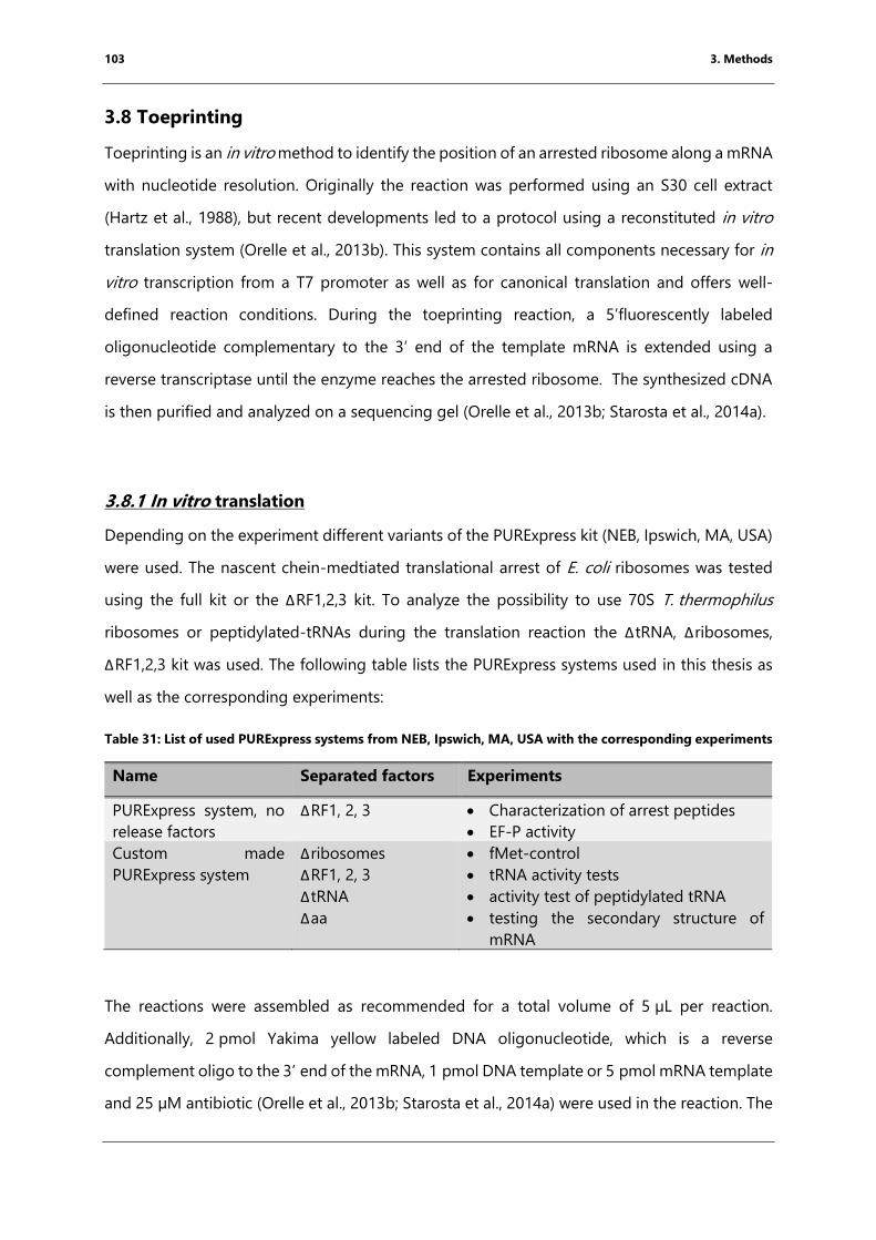
103 3. Methods
3.8 Toeprinting
Toeprinting is an in vitro method to identify the position of an arrested ribosome along a mRNA
with nucleotide resolution. Originally the reaction was performed using an S30 cell extract
(Hartz et al., 1988), but recent developments led to a protocol using a reconstituted in vitro
translation system (Orelle et al., 2013b). This system contains all components necessary for in
vitro transcription from a T7 promoter as well as for canonical translation and offers well-
defined reaction conditions. During the toeprinting reaction, a 5’fluorescently labeled
oligonucleotide complementary to the 3’ end of the template mRNA is extended using a
reverse transcriptase until the enzyme reaches the arrested ribosome. The synthesized cDNA
is then purified and analyzed on a sequencing gel (Orelle et al., 2013b; Starosta et al., 2014a).
3.8.1 In vitro translation
Depending on the experiment different variants of the PURExpress kit (NEB, Ipswich, MA, USA)
were used. The nascent chein-medtiated translational arrest of E. coli ribosomes was tested
using the full kit or the ΔRF1,2,3 kit. To analyze the possibility to use 70S T. thermophilus
ribosomes or peptidylated-tRNAs during the translation reaction the ΔtRNA, Δribosomes,
ΔRF1,2,3 kit was used. The following table lists the PURExpress systems used in this thesis as
well as the corresponding experiments:
Table 31: List of used PURExpress systems from NEB, Ipswich, MA, USA with the corresponding experiments
Name Separated factors Experiments
PURExpress system, no
release factors
ΔRF1, 2, 3 • Characterization of arrest peptides
• EF-P activity
Custom made
PURExpress system
Δribosomes
ΔRF1, 2, 3
ΔtRNA
Δaa
• fMet-control
• tRNA activity tests
• activity test of peptidylated tRNA
• testing the secondary structure of
mRNA
The reactions were assembled as recommended for a total volume of 5 µL per reaction.
Additionally, 2 pmol Yakima yellow labeled DNA oligonucleotide, which is a reverse
complement oligo to the 3’ end of the mRNA, 1 pmol DNA template or 5 pmol mRNA template
and 25 µM antibiotic (Orelle et al., 2013b; Starosta et al., 2014a) were used in the reaction. The
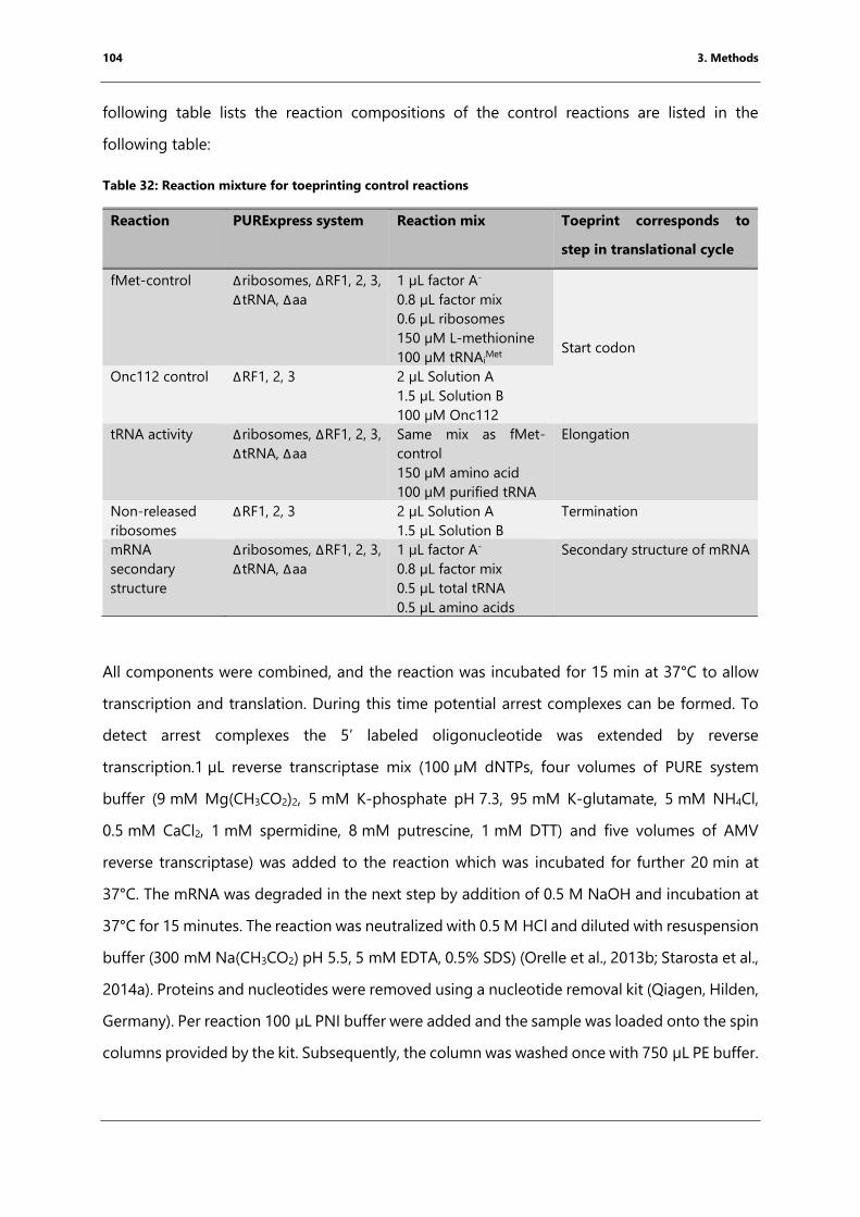
104 3. Methods
following table lists the reaction compositions of the control reactions are listed in the
following table:
Table 32: Reaction mixture for toeprinting control reactions
Reaction PURExpress system Reaction mix Toeprint corresponds to
step in translational cycle
fMet-control Δribosomes, ΔRF1, 2, 3,
ΔtRNA, Δaa
1 µL factor A-
0.8 µL factor mix
0.6 µL ribosomes
150 µM L-methionine
100 µM tRNAiMet
Start codon
Onc112 control ΔRF1, 2, 3 2 µL Solution A
1.5 µL Solution B
100 µM Onc112
tRNA activity Δribosomes, ΔRF1, 2, 3,
ΔtRNA, Δaa
Same mix as fMet-
control
150 µM amino acid
100 µM purified tRNA
Elongation
Non-released
ribosomes
ΔRF1, 2, 3 2 µL Solution A
1.5 µL Solution B
Termination
mRNA
secondary
structure
Δribosomes, ΔRF1, 2, 3,
ΔtRNA, Δaa
1 µL factor A-
0.8 µL factor mix
0.5 µL total tRNA
0.5 µL amino acids
Secondary structure of mRNA
All components were combined, and the reaction was incubated for 15 min at 37°C to allow
transcription and translation. During this time potential arrest complexes can be formed. To
detect arrest complexes the 5’ labeled oligonucleotide was extended by reverse
transcription.1 µL reverse transcriptase mix (100 µM dNTPs, four volumes of PURE system
buffer (9 mM Mg(CH3CO2)2, 5 mM K-phosphate pH 7.3, 95 mM K-glutamate, 5 mM NH4Cl,
0.5 mM CaCl2, 1 mM spermidine, 8 mM putrescine, 1 mM DTT) and five volumes of AMV
reverse transcriptase) was added to the reaction which was incubated for further 20 min at
37°C. The mRNA was degraded in the next step by addition of 0.5 M NaOH and incubation at
37°C for 15 minutes. The reaction was neutralized with 0.5 M HCl and diluted with resuspension
buffer (300 mM Na(CH3CO2) pH 5.5, 5 mM EDTA, 0.5% SDS) (Orelle et al., 2013b; Starosta et al.,
2014a). Proteins and nucleotides were removed using a nucleotide removal kit (Qiagen, Hilden,
Germany). Per reaction 100 µL PNI buffer were added and the sample was loaded onto the spin
columns provided by the kit. Subsequently, the column was washed once with 750 µL PE buffer.
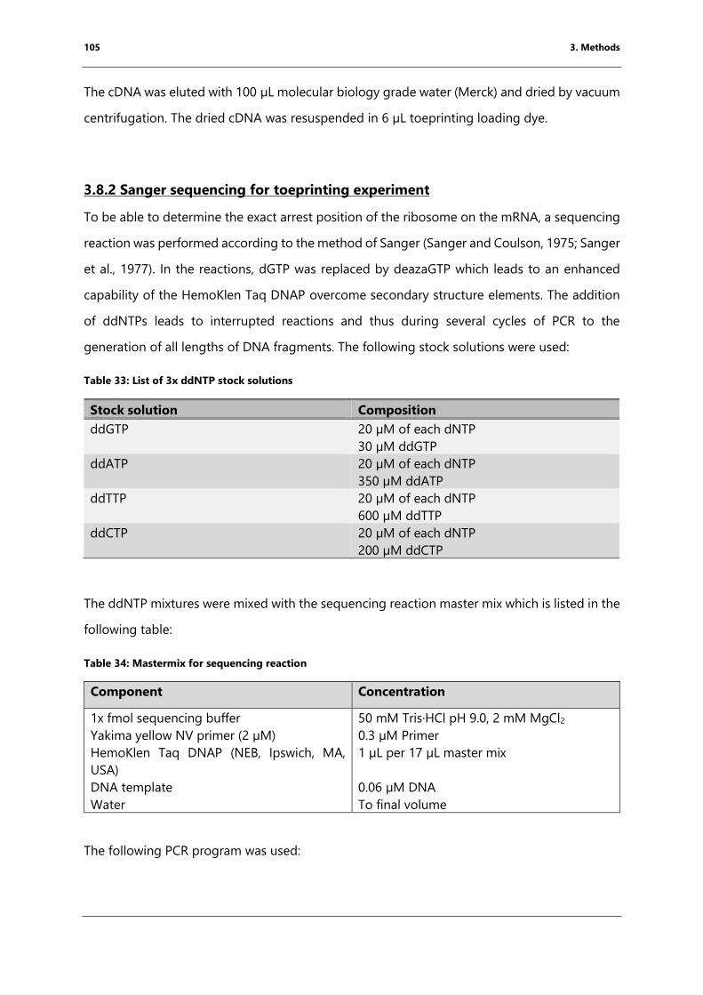
105 3. Methods
The cDNA was eluted with 100 µL molecular biology grade water (Merck) and dried by vacuum
centrifugation. The dried cDNA was resuspended in 6 µL toeprinting loading dye.
3.8.2 Sanger sequencing for toeprinting experiment
To be able to determine the exact arrest position of the ribosome on the mRNA, a sequencing
reaction was performed according to the method of Sanger (Sanger and Coulson, 1975; Sanger
et al., 1977). In the reactions, dGTP was replaced by deazaGTP which leads to an enhanced
capability of the HemoKlen Taq DNAP overcome secondary structure elements. The addition
of ddNTPs leads to interrupted reactions and thus during several cycles of PCR to the
generation of all lengths of DNA fragments. The following stock solutions were used:
Table 33: List of 3x ddNTP stock solutions
Stock solution Composition
ddGTP 20 µM of each dNTP
30 µM ddGTP
ddATP 20 µM of each dNTP
350 µM ddATP
ddTTP 20 µM of each dNTP
600 µM ddTTP
ddCTP 20 µM of each dNTP
200 µM ddCTP
The ddNTP mixtures were mixed with the sequencing reaction master mix which is listed in the
following table:
Table 34: Mastermix for sequencing reaction
Component Concentration
1x fmol sequencing buffer
Yakima yellow NV primer (2 µM)
HemoKlen Taq DNAP (NEB, Ipswich, MA,
USA)
DNA template
Water
50 mM Tris·HCl pH 9.0, 2 mM MgCl2
0.3 µM Primer
1 µL per 17 µL master mix
0.06 µM DNA
To final volume
The following PCR program was used:
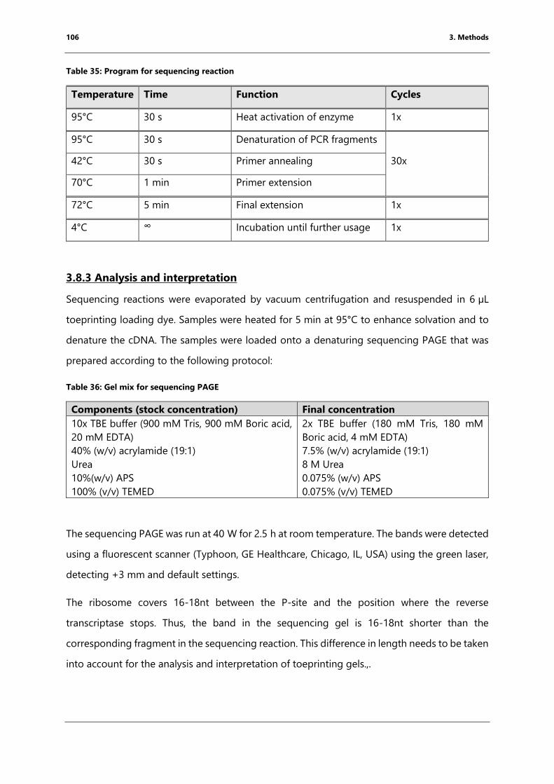
106 3. Methods
Table 35: Program for sequencing reaction
Temperature Time Function Cycles
95°C 30 s Heat activation of enzyme 1x
95°C 30 s Denaturation of PCR fragments
30x 42°C 30 s Primer annealing
70°C 1 min Primer extension
72°C 5 min Final extension 1x
4°C ∞ Incubation until further usage 1x
3.8.3 Analysis and interpretation
Sequencing reactions were evaporated by vacuum centrifugation and resuspended in 6 µL
toeprinting loading dye. Samples were heated for 5 min at 95°C to enhance solvation and to
denature the cDNA. The samples were loaded onto a denaturing sequencing PAGE that was
prepared according to the following protocol:
Table 36: Gel mix for sequencing PAGE
Components (stock concentration) Final concentration
10x TBE buffer (900 mM Tris, 900 mM Boric acid,
20 mM EDTA)
40% (w/v) acrylamide (19:1)
Urea
10%(w/v) APS
100% (v/v) TEMED
2x TBE buffer (180 mM Tris, 180 mM
Boric acid, 4 mM EDTA)
7.5% (w/v) acrylamide (19:1)
8 M Urea
0.075% (w/v) APS
0.075% (v/v) TEMED
The sequencing PAGE was run at 40 W for 2.5 h at room temperature. The bands were detected
using a fluorescent scanner (Typhoon, GE Healthcare, Chicago, IL, USA) using the green laser,
detecting +3 mm and default settings.
The ribosome covers 16-18nt between the P-site and the position where the reverse
transcriptase stops. Thus, the band in the sequencing gel is 16-18nt shorter than the
corresponding fragment in the sequencing reaction. This difference in length needs to be taken
into account for the analysis and interpretation of toeprinting gels.,.
107 3. Methods
3.9 X-ray crystallography and structure determination
Crystallography is a biophysical technique that is commonly used to obtain the structure of
proteins and nucleic acids. During crystallization, the biomolecular complexes adopt well-
ordered, regular orientations. The obtained crystals are exposed to X-rays from a local source
or a synchrotron. The way in which the X-rays interact with the crystal is dependent on the
structure of the macromolecule and thus by analyzing the pattern, the 3-dimensional structure
can be solved.
First high-resolution structures of ribosomal subunits and full 70S T. thermophilus ribosome
complexes gave great insights into the molecular mechanism of translation. These crystals can
diffract to high resolution to obtain information about e.g. ribosomal modifications (Polikanov
et al., 2015a) and or mechanistic insights into peptide bond formation (Polikanov et al., 2014).
This degree of resolution is necessary to understand the underlying mechanism of action of
arrest peptides and proline-rich antimicrobial peptides.
Hindrances of this method include that variation of the sample composition can lead to
decrease in resolution or the sample does not crystallize at all. Another disadvantage includes,
that the obtained structure derived from molecules that are forced into a specific orientation
due to crystal packing and partial dehydration of the biomolecular complex. This might result
in artifacts within the structure. However, high-resolution data can be obtained from well-
diffracting crystals giving great insights into the underlying molecular mechanism of the
biomolecular complex studied. In addition, data sets from multiple experiments can be
obtained in relatively short amounts of time and the data processing involves less
computational power compared to cryo-EM.
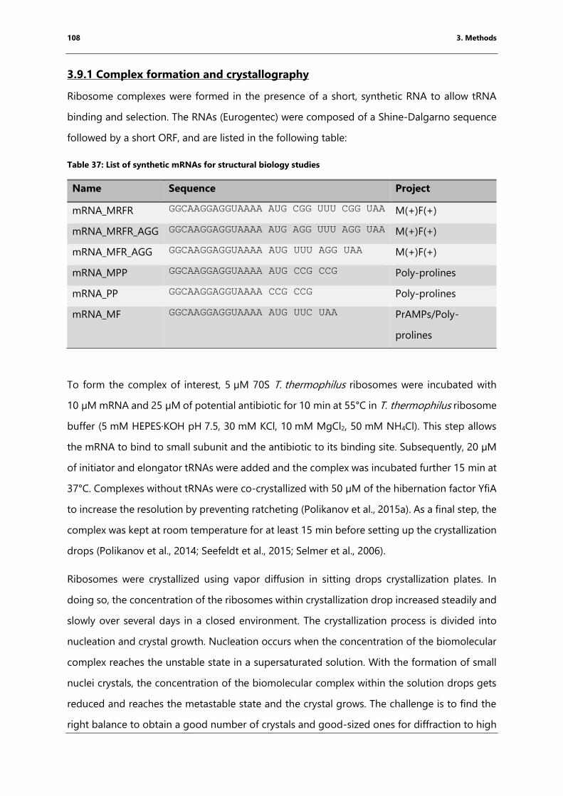
108 3. Methods
3.9.1 Complex formation and crystallography
Ribosome complexes were formed in the presence of a short, synthetic RNA to allow tRNA
binding and selection. The RNAs (Eurogentec) were composed of a Shine-Dalgarno sequence
followed by a short ORF, and are listed in the following table:
Table 37: List of synthetic mRNAs for structural biology studies
Name Sequence Project
mRNA_MRFR GGCAAGGAGGUAAAA AUG CGG UUU CGG UAA M(+)F(+)
mRNA_MRFR_AGG GGCAAGGAGGUAAAA AUG AGG UUU AGG UAA M(+)F(+)
mRNA_MFR_AGG GGCAAGGAGGUAAAA AUG UUU AGG UAA M(+)F(+)
mRNA_MPP GGCAAGGAGGUAAAA AUG CCG CCG Poly-prolines
mRNA_PP GGCAAGGAGGUAAAA CCG CCG Poly-prolines
mRNA_MF GGCAAGGAGGUAAAA AUG UUC UAA PrAMPs/Poly-
prolines
To form the complex of interest, 5 µM 70S T. thermophilus ribosomes were incubated with
10 µM mRNA and 25 µM of potential antibiotic for 10 min at 55°C in T. thermophilus ribosome
buffer (5 mM HEPES·KOH pH 7.5, 30 mM KCl, 10 mM MgCl2, 50 mM NH4Cl). This step allows
the mRNA to bind to small subunit and the antibiotic to its binding site. Subsequently, 20 µM
of initiator and elongator tRNAs were added and the complex was incubated further 15 min at
37°C. Complexes without tRNAs were co-crystallized with 50 µM of the hibernation factor YfiA
to increase the resolution by preventing ratcheting (Polikanov et al., 2015a). As a final step, the
complex was kept at room temperature for at least 15 min before setting up the crystallization
drops (Polikanov et al., 2014; Seefeldt et al., 2015; Selmer et al., 2006).
Ribosomes were crystallized using vapor diffusion in sitting drops crystallization plates. In
doing so, the concentration of the ribosomes within crystallization drop increased steadily and
slowly over several days in a closed environment. The crystallization process is divided into
nucleation and crystal growth. Nucleation occurs when the concentration of the biomolecular
complex reaches the unstable state in a supersaturated solution. With the formation of small
nuclei crystals, the concentration of the biomolecular complex within the solution drops gets
reduced and reaches the metastable state and the crystal grows. The challenge is to find the
right balance to obtain a good number of crystals and good-sized ones for diffraction to high
109 3. Methods
resolution. The reservoir solution consisted of 100 mM Tris·HCl pH 7.6, 2.9% (v/v) polyethylene
glycol (PEG) 20 000, 7-10% v/v) 2-methyl-2,4-pentanediol (MPD) and 175 mM arginine. PEG
20 000 and MPD act as precipitants to reach the supersaturated state of the solution. While
the concentration of PEG was kept constant, the MPD concentration was optimized depending
on the ribosome preparation and the complex composition. Crystallization drops consisted of
3 µL of pre-formed complex and 3-4 µL reservoir solution. The drops contain a lower
concentrated reservoir buffer. During the crystallization process, the concentration differences
are equilibrated by water diffusion from the drop towards the reservoir, increasing the
ribosome concentration slowly within the drop solution resulting in crystallization. Crystals
appeared after 2 to 3 days incubation at 20°C and after approximately a week, crystals grew to
their full sizes of ∼1000 × 100 × 100 μm.
Subsequently, crystals were stabilized to prevent ice formation and drying of the crystals.by
replacing water with MPD. The concentration of MPD was increased stepwise: 25%, 30%, 35%
and 40% (v/v) MPD in 100 mM Tris·HCl, pH 7.6, 2.9% (v/v) PEG 20,000, 50 mM KCl, 10 mM
NH4Cl and 10 mM Mg(CH3COO)2. The reservoir solution was completely removed during this
step. For the first three steps, 5 µL of the cryo-protectant solution was added an incubated for
15 min at room temperature. For the last step, 15 µL of the drop solution was removed and
replaced by 40 µL of the cryo-protectant solution containing 40% MPD. The crystals were
stabilized minimum 1 h. Crystals were fished into nylon loops and flash frozen using a cryo-
stream set to a temperature of 100 K.
3.9.2 Data collection and processing
Data were collected at the Soleil Synchrotron (Saclay, France) at the beamlines Proxima 1 and
Proxima 2A and at the European Synchrotron Radiation Facility (ESRF, Grenoble, France) at the
beamlines ID23 and ID29. The data were collected at 100 K and at a wavelength around 0.98 Å.
Slight differences in wavelength, due to the set-up of the different beamlines, were taken into
account during processing. While collecting the crystal was rotated by 0.1° per image and
exposed for 0.1 s. Data were obtained using helical data collection mode. Alternatively,
individual wedges of 30° were collected and merged at the end into one data set. For each
data set at least 180° were collected.
110 3. Methods
The data were processed using xdsme, which is a collection of python scripts to process the
data with X-ray detector software (XDS) facilitating its usage (Legrand, 2017). XDS is a data
processing program that facilitates processing of data with weak reflections and high mosaicity
(Kabsch, 2010). The processing is divided in the localization of the spots, indexing, integration,
scaling and merging. Ribosome crystals from T. thermophilus have a large number of weak
spots due to large unit cells which were taken into account during spot localization. During this
step, spots were identified and separated from noise. The specific localization of each spot
arises from the orientation of the molecular complexes within the crystal and the position is
crucial for indexing. 70S T. thermophilus ribosomes crystallize in most cases in the point group
P212121 with the cell dimensions of 210 × 450 × 625 Å with the corresponding angles of
α=β=γ=90°.
Spots were integrated over all collected frames to re-create the 3D profile of each spot that
passed through the EWALD sphere. During merging and scaling, the multiple data sets can be
combined into one and converted into *.mtz-format which is necessary for downstream
applications. During this step, 5% of all spots were selected for Rfree calculation. These spots
were excluded from the refinement. Rfree is a factor to prevent over-refinement. To avoid
artifacts during refinement, the same set of spots were used for Rfree calculations for each
processed data set. As a molecular replacement (MR) was used for phasing and so heavy atom
soaking was not necessary for phasing, FRIEDEL’s law was set to true meaning that each spot
has a corresponding spot in point symmetry.
The final resolution was determined using signal to noise ratio (I/σ) of 1.0, significant value for
CC1/2 of > 15 and high completeness (> 95%) for all resolution shells
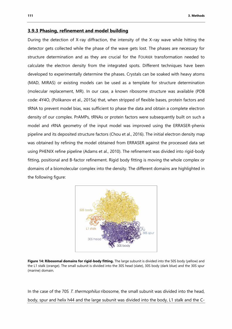
111 3. Methods
3.9.3 Phasing, refinement and model building
During the detection of X-ray diffraction, the intensity of the X-ray wave while hitting the
detector gets collected while the phase of the wave gets lost. The phases are necessary for
structure determination and as they are crucial for the FOURIER transformation needed to
calculate the electron density from the integrated spots. Different techniques have been
developed to experimentally determine the phases. Crystals can be soaked with heavy atoms
(MAD, MIRAS) or existing models can be used as a template for structure determination
(molecular replacement, MR). In our case, a known ribosome structure was available (PDB
code: 4Y4O, (Polikanov et al., 2015a) that, when stripped of flexible bases, protein factors and
tRNA to prevent model bias, was sufficient to phase the data and obtain a complete electron
density of our complex. PrAMPs, tRNAs or protein factors were subsequently built on such a
model and rRNA geometry of the input model was improved using the ERRASER-phenix
pipeline and its deposited structure factors (Chou et al., 2016). The initial electron density map
was obtained by refining the model obtained from ERRASER against the processed data set
using PHENIX refine pipeline (Adams et al., 2010). The refinement was divided into rigid-body
fitting, positional and B-factor refinement. Rigid body fitting is moving the whole complex or
domains of a biomolecular complex into the density. The different domains are highlighted in
the following figure:
Figure 14: Ribosomal domains for rigid-body fitting. The large subunit is divided into the 50S body (yellow) and
the L1 stalk (orange). The small subunit is divided into the 30S head (slate), 30S body (dark blue) and the 30S spur
(marine) domain.
In the case of the 70S T. thermophilus ribosome, the small subunit was divided into the head,
body, spur and helix h44 and the large subunit was divided into the body, L1 stalk and the C-
112 3. Methods
terminus of ribosomal protein L9, which is extended in the crystal structure due to crystal
contacts. Positional refinement was performed applying multiple restraints, including base
pairing restraints and non-crystallographic symmetry (NCS) due to the presence of two
ribosomes per asymmetric unit. The base pairing restraints were generated using the online
interface from the RNA center in Santa Cruz, CA (Laurberg et al., 2008). The last step of each
macrocycle included B-factor refinement. B-factors indicate the possible movement of each
atom. Prior to the first refinement, the B-factor of all atoms was set to the Wilson B-factor as a
starting point. Ribosomal modifications were restrained using cif files created by phenix elbow
describing the chemical properties of modified residues e.g. tRNA or rRNA modification.
In between refinement cycles, the density and the corresponding model were visualized in the
program COOT (Emsley and Cowtan, 2004). Using a minimally biased difference map (Fo-Fc),
missing parts of a model were modeled manually into the density, which was displayed as
positive difference map. Additional information and wrongly modeled parts were surrounded
by a negative density and removed. The model was refined against the processed data set over
several cycles, maintaining values for R and Rfree factor to stay in a different range of under 5%
and making sure that their values decrease with each refinement cycle. The MolProbity server
was used to validate the geometry of the structure (Chen et al., 2010).
113 3. Methods
3.10 Cryo-Electron microscopy (cryo-EM)
Cryo-EM is a method used to study the structure of large biomolecules and has proven to be
a valuable tool for the study the underlying mechanism of translation as reviewed in (Razi et
al., 2017). Early studies provided information on the overall shape of the ribosome including
the ribosomal tunnel and tRNA binding sites as well as its large conformational changes during
the translational cycle at a resolution of 25 Å (Agrawal et al., 1999; Frank et al., 1995) The recent
development of direct electron detectors that allow movie collection mode and the constant
improvement of processing software made it possible to obtain near-atomic resolution
structures from cryo-EM data. The “Resolution revolution”, as it has been called, led to the
solution of structures by cryo-EM with resolutions comparable to X-ray crystallography and
NMR (Kühlbrandt, 2014). Limitations of this technique include long data collection time per
data set and the computational power needed for processing thousands of movies, compared
to X-ray crystallography. Advantages include the low amounts of sample required and the
visualization of the protein in solution which allows seeing different conformations in the same
sample.
The structure is solved by shining electrons through the specimen to be observed. The
electrons that are transmitted through because they have not interacted with the specimen
result in 2D projections in different orientations in the ice of the sample. To prepare cryo-grids,
the protein is quickly frozen at a concentration range of 5 nM to 1 µM depending on the size
of the specimen in its optimal buffer. The images are processed to reconstruct the single
particles followed by the interpretation of the electron density obtained.
3.10.1 Sample preparation
The desired complex was formed using the same concentrations as for the toeprinting assay
(chapter 3.7) and the dilution buffer was substituted with Arg-tRNAArg to ensure that the
dilution has no impact on the occupancy of the A-site tRNA. Therefore, 2.5 µM 70S E. coli
ribosomes were incubated with 5 µM synthetic RNA (Eurogentec, Lüttich, Belgium) and 25 µM
erythromycin. As erythromycin was dissolved in EtOH, the antibiotic was dried into the reaction
tube prior to the complex formation. 10 µM fMKF-tRNAiMet was added together with 20 µM
Arg-tRNAArg. The complex was then incubated for 15 min at 37°C. As a final step, the complex
was diluted to a final concentration of 240-480 nM of ribosomes in cryo-EM dilution buffer
114 3. Methods
(50 mM HEPES·KOH pH 7.5, 20 mM Mg(CH3CO2)2, 100 mM K(CH3CO2)) in the presence of
25 µM erythromycin and 20 µM Arg-tRNAArg.
The sample was placed on a support surface, the grid. For this project, lacey holey carbon grids
were used as the thickness of the vitreous ice was optimal to obtain high-resolution
information (Cho et al., 2013). The grids are carbon coated and its hydrophobicity does not
allow for a homogeneous repartition of the drop onto the grid. The hydrophobicity can be
controlled, treating the grids with a low-energy plasma (Passmore and Russo, 2016). The grids
were glow-discharged for 1 min with an ELMO machine (Cordouan, Pessac, France) at a
pressure of 2*10-2 mbar and a charge of 2 mA.
3.5 µL of the mixture was transferred to a Lacey holey carbon coated grid, blotted for 5 s and
frozen with a Vitrobot™ (FEI, Hillsboro, CA, USA). The Vitrobot™ is an automatic freezing
machine that allows fast plunging of the grids into liquid ethane. The flash-freezing results in
vitreous glass-like ice and prevents the formation of ice crystals. The Vitrobot™ chamber was
set to 4°C and 100% humidity. The humidity ensures that water does not evaporate from the
sample and change the ribosome or salt concentration within the sample. Optimal ice thickness
was obtained using a blotting time of 5 s (Passmore and Russo, 2016). The grid was then
clipped for insertion in an autoloader and stored in liquid nitrogen until data collection.
3.10.2 Data collection
Data were collected on the Talos™ Arctica transmission electron microscope (FEI) equipped
with a Falcon III direct electron detector (FEI) at the IECB (Pessac, France). The Talos™ Arctica
runs at 200 kV and has a spherical aberration of 2.7. The magnification was set to 120 k
resulting in a pixel size of 1.24 Å. The grids were screened to determine the optimal distribution
of the particles and thickness of the ice for data collection. The best squares were identified on
the Atlas of the whole grid collected by the EPU software (FEI-Thermo). Due to the irregularity
of the holes on lacey carbon grids, the software was set up to collect at regular intervals of
2 µm between spots. Regions of the squares having cracks or thick ice were avoided. A total of
5000 movies were collected, each consisting of 20 frames per second. To obtain information
on the whole resolution range, a defocus range between 0.5 µm to 2.5 µm was collected.
115 3. Methods
3.10.3 Single particle reconstruction using RELION gui
During data collection, the electrons interact with the ribosome and those that pass through
are being detected by the camera at the end of the microscope. The resulting micrographs are
a collection of 2D projection of each molecule in solution. The particles are boxed and classified
by the software according to their orientation in ice. To obtain the electron microscopy density,
the 2D projections are used reconstruct the 3D object. To obtain a 3D model, the data were
processed using the REgularized LIkelihood OptimizatioN (RELION 2.1) interface following the
recommended protocol including slight changes (Fernandez-Leiro and Scheres, 2017; Scheres,
2012a, b).
Image processing consists of aligning movie frames by MotionCorr2 (Zheng et al., 2016; Zheng
et al., 2017) and determining of defocus values and astigmatism of the aligned images using
CTFFIND4 (Rohou and Grigorieff, 2015). The electron beam induces movement of the particles
within the grid which leads to blurred images and thus a low-resolution reconstruction. Due to
the development of direct detector, the movement can be detected over the different frames.
To correct for this motion, Motioncor2 was used to align each pixel of the 20 frames per second
collected. The aligned micrographs were converted into the corresponding power spectra by
FOURIER transformation. The background was subtracted using a low pass filter and the
obtained Thon rings were compared to theoretical values to obtain the defocus values for each
image and astigmatism for further processing.
Further processing requires the selection and classification of particles. To generate references
for auto-picking, 1000 to 2000 ribosome particles were selected manually from micrographs
of representative defocus values and classified into 2D classes. The contrast of the images
increases proportionally with increasing defocus values, thus for the auto-picking program to
efficiently recognized particles it is necessary to cover the whole range when manually selecting
the images. The box size was set to 340 Å which is larger than the diameter of the ribosomes.
Auto-picking values were optimized for each data set. The picking threshold used, varied from
0.4-0.8 and maximum standard deviation used was 1.1-1.5 to avoid picking on the carbon. As
Lacey grids were used for the analysis, some of the particles stuck to the carbon resulting in
loss of information. The minimum inner particle distance was set to 120 Å to avoid picking
particles on carbon.
116 3. Methods
The picked particles were extracted and downscaled by a factor of 3 to speed up the processing
time. The picked particles were classified into 100 different 2D classes over 25 iteration cycles.
The alignment and classification were performed using an algorithm based on maximum
likelihood resulting (Scheres, 2010). To further improve the quality of the picked articles, 2D
Classes that appeared to be 70S ribosomes were selected and reclassified into 100 2D classes
as a second cleaning step.
The particles were re-extracted at their original box size of 320 px and used as an input for 3D
auto-refinement using a low-resolution map of a bacterial ribosome. The refinement was
performed using the gold-standard FSC calculations. This means that the data set was split in
half and all reconstruction steps were performed independently. By comparing the
reconstructions, the FOURIER shell correlation is determined and with this the resolution. As
recommended the resolution cut off was set to an FSC value of 0.143. The 3D auto-refinement
was performed without solvent masking to prevent overfitting. However, including the
disordered solvent increases the noise resulting in a lower resolution. By removing solvent
regions with the help of a mask, results directly in an increase of the FSC values and with it the
resolution of the reconstruction. The resulting map was used as an input for masked 3D
classification sorting all particles for the abundance of tRNAs in the A-, P- and E-site (Scheres,
2016). Therefore, a mask around the tRNAs and a mask for the ribosome without tRNAs were
created. By using particle subtraction, particles were classified according to the occupancy of
tRNA in the A, P or E-site These polished particles can be used as an input for further
refinement.
The movement of individual particles was accounted for through movie refinement. Another
important step to improve the final quality of the map is particle polishing. During data
collection, in our case 20 frames per spot, the particles get damaged and loose high-resolution
information. Still, low-resolution information can be necessary to improve reconstruction. Each
frame gets a calculated B-factor and dependent on its value the frame gets taken into account
for the final reconstruction (Scheres, 2016). The shiny particles were used for another round of
3D-refinement using a mask for the 50S subunit as the arrest peptide is located in the
ribosomal exit tunnel, followed by postprocessing and the determination of the local
resolution.
117 3. Methods
3.10.4 Model building
For model building and refinement the resulting *.mrc was renamed to *.ccp4. An initial model,
a 2.8 Å resolution structure of a 70S E. coli ribosome (PDB code: 4U27, (Noeske et al., 2014))
was a rigid-body fitted into the density using the program Situs (Wriggers, 2012). In an initial
fit, the whole structure is fitted into the density setting the resolution to 15 Å. The structure
was divided into the same domains as used for PHENIX refine. The domains are fitted over
multiple cycles into the density while increasing stepwise the resolution. This task is performed
using the program “collage” within the Situs package (Wriggers, 2012).
Flexible bases were rotated into the density, the peptide and the drug were modeled using the
program COOT (Emsley and Cowtan, 2004). The P-site tRNA (PDB code: 4VY5) was a rigid body
fitted to the density using COOT. The backbone geometry of the rRNAs was improved using
the program ERRASER (Chou et al., 2016). The resulting coordinates were refined against the
map using PHENIX real space refinement (Afonine et al., 2013). To increase the amount of
restrains, the input model was used as a reference model. The connection between the peptide
and the tRNA was modeled as a data-link and a corresponding cif file was provided. Base
pairing restraints were applied as geometry restraints. The base pairing restraints were
generated using the online interface from the RNA center in Santa Cruz, CA (Laurberg et al.,
2008).
118 3. Methods
3.11 Bioinformatic databases and programs
The graphics programs, Pymol (DeLano, 2010) and Chimera (Pettersen et al., 2004), were used
to generate figures for structural interpretation. Sequencing data were analyzed using basic
local alignment tool (BLAST, (Altschul et al., 1990)) and Multalign (Corpet, 1988). The DNA
sequence for tRNA cloning was taken from the tRNA database (Leipzig, Germany, Jühling et
al., 2009).
To calculate the theoretical mass spectrum, the sequence containing the modifications proK
were taken from the MODOMICs database (Bujnicki et al., 2013; Dunin-Horkawicz et al., 2006).
Next, the theoretic electrospray series were generated by Mongo Oligo mass calculator
(Rozenski, 1999).
The ExPASy (The Export Protein Analysis System) online tool was used to calculate the
molecular weight, extinction factors and isoelectric points (ProtParam). Nucleic acid sequences
encoding for proteins were translated into the corresponding amino acid sequence using the
Translate tool (Gasteiger et al., 2003).
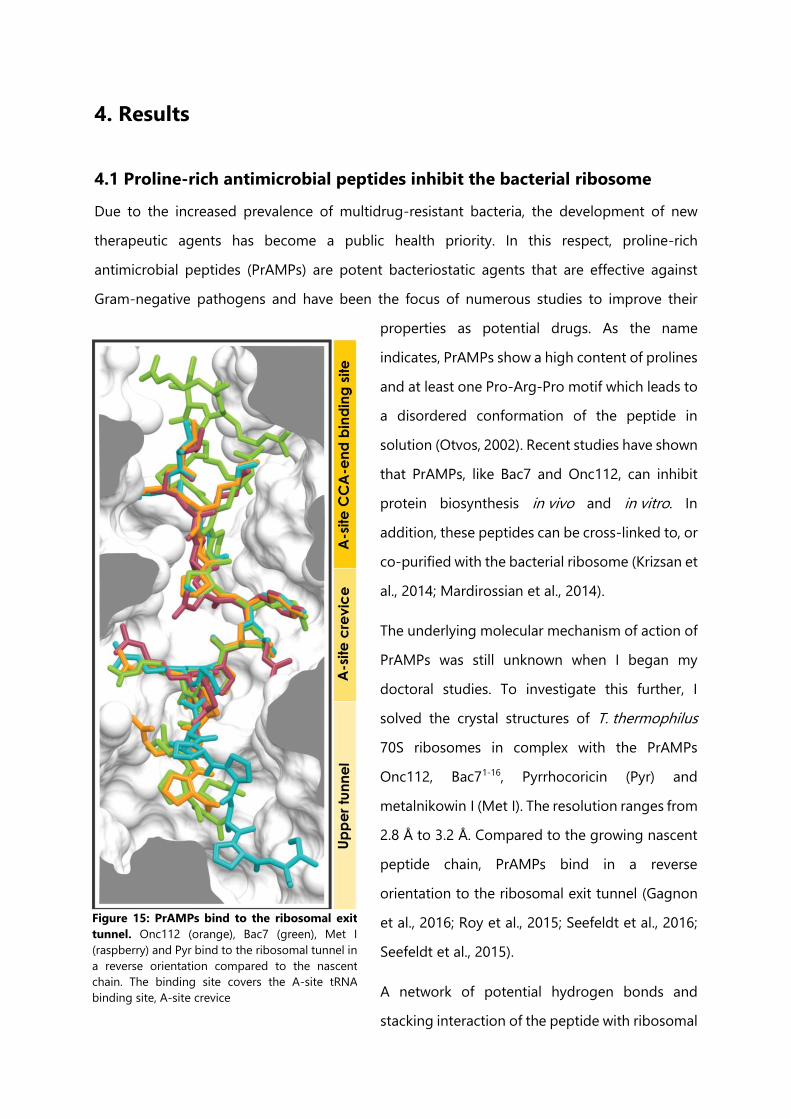
4. Results
4.1 Proline-rich antimicrobial peptides inhibit the bacterial ribosome
Due to the increased prevalence of multidrug-resistant bacteria, the development of new
therapeutic agents has become a public health priority. In this respect, proline-rich
antimicrobial peptides (PrAMPs) are potent bacteriostatic agents that are effective against
Gram-negative pathogens and have been the focus of numerous studies to improve their
properties as potential drugs. As the name
indicates, PrAMPs show a high content of prolines
and at least one Pro-Arg-Pro motif which leads to
a disordered conformation of the peptide in
solution (Otvos, 2002). Recent studies have shown
that PrAMPs, like Bac7 and Onc112, can inhibit
protein biosynthesis in vivo and in vitro. In
addition, these peptides can be cross-linked to, or
co-purified with the bacterial ribosome (Krizsan et
al., 2014; Mardirossian et al., 2014).
The underlying molecular mechanism of action of
PrAMPs was still unknown when I began my
doctoral studies. To investigate this further, I
solved the crystal structures of T. thermophilus
70S ribosomes in complex with the PrAMPs
Onc112, Bac71-16, Pyrrhocoricin (Pyr) and
metalnikowin I (Met I). The resolution ranges from
2.8 Å to 3.2 Å. Compared to the growing nascent
peptide chain, PrAMPs bind in a reverse
orientation to the ribosomal exit tunnel (Gagnon
et al., 2016; Roy et al., 2015; Seefeldt et al., 2016;
Seefeldt et al., 2015).
A network of potential hydrogen bonds and
stacking interaction of the peptide with ribosomal
Figure 15: PrAMPs bind to the ribosomal exit
tunnel. Onc112 (orange), Bac7 (green), Met I
(raspberry) and Pyr bind to the ribosomal tunnel in
a reverse orientation compared to the nascent
chain. The binding site covers the A-site tRNA
binding site, A-site crevice
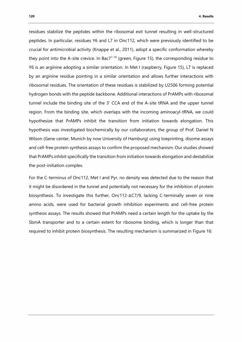
120 4. Results
residues stabilize the peptides within the ribosomal exit tunnel resulting in well-structured
peptides. In particular, residues Y6 and L7 in Onc112, which were previously identified to be
crucial for antimicrobial activity (Knappe et al., 2011), adopt a specific conformation whereby
they point into the A-site crevice. In Bac71-16 (green, Figure 15), the corresponding residue to
Y6 is an arginine adopting a similar orientation. In Met I (raspberry, Figure 15), L7 is replaced
by an arginine residue pointing in a similar orientation and allows further interactions with
ribosomal residues. The orientation of these residues is stabilized by U2506 forming potential
hydrogen bonds with the peptide backbone. Additional interactions of PrAMPs with ribosomal
tunnel include the binding site of the 3’ CCA end of the A-site tRNA and the upper tunnel
region. From the binding site, which overlaps with the incoming aminoacyl-tRNA, we could
hypothesize that PrAMPs inhibit the transition from initiation towards elongation. This
hypothesis was investigated biochemically by our collaborators, the group of Prof. Daniel N
Wilson (Gene center, Munich by now University of Hamburg) using toeprinting, disome assays
and cell-free protein synthesis assays to confirm the proposed mechanism. Our studies showed
that PrAMPs inhibit specifically the transition from initiation towards elongation and destabilize
the post-initiation complex.
For the C-terminus of Onc112, Met I and Pyr, no density was detected due to the reason that
it might be disordered in the tunnel and potentially not necessary for the inhibition of protein
biosynthesis. To investigate this further, Onc112-ΔC7/9, lacking C-terminally seven or nine
amino acids, were used for bacterial growth inhibition experiments and cell-free protein
synthesis assays. The results showed that PrAMPs need a certain length for the uptake by the
SbmA transporter and to a certain extent for ribosome binding, which is longer than that
required to inhibit protein biosynthesis. The resulting mechanism is summarized in Figure 16:
121 4. Results
Figure 16: Mechanism of action of proline-rich antimicrobial peptides. PrAMPs are taken up specifically by
Gram-negative bacteria through the SbmA transporter. C-terminal truncations lead to up-take inhibition. Inside
the cytoplasm, they bind to the ribosomal exit tunnel in the reversed orientation compared to the growing peptide
chain. This results in the inhibition of the transition from initiation towards elongation and destabilizes the initiation
complex. Figure is taken from (Seefeldt et al., 2015).
Comparing the structure Bac71-16-70S T. thermophilus ribosome with mammalian ribosome
showed that the peptide could potentially bind to the mammalian exit tunnel. Using cell extract
from rabbits, we were able to show that Bac71-16 inhibits the eukaryotic protein biosynthesis
(Seefeldt et al., 2016). However, Bac7 has been shown to be not toxic for eukaryotic cells and
might be taken up during endocytosis (Tomasinsig et al., 2006) and stored after its production
in granules (Zanetti et al., 1990). This prevents the contact of the Bac7 with the eukaryotic
ribosome and it is believed that insect-derived PrAMPs might act in a similar way (Seefeldt et
al., 2016). PrAMPs are taken up by Gram-negative bacteria via the SbmA transporter.
The work was published back to back with results from the group of Prof Thomas A. Steitz,
which corroborated our findings.
©20
15N
atu
re A
mer
ica,
Inc.
All
rig
hts
res
erve
d.
NATURE STRUCTURAL & MOLECULAR BIOLOGY ADVANCE ONLINE PUBLICATION 1
A R T I C L E S
Antimicrobial peptides form a diverse group of molecules that are produced as part of the innate immune response of all multicellular organisms1. Among these, PrAMPs have garnered considerable atten-tion as a possible means of countering the rapid increase in bacterial resistance to classical antibiotics2,3. Unlike many peptides that kill bacteria by disrupting their cell membrane, PrAMPs are transported into the cytoplasm by specialized transporters, such as SbmA in Gram-negative bacteria4,5, where they inhibit specific intracellular targets. Given that such transport mechanisms are absent in mammalian cells, and only limited interactions with intracellular eukaryotic proteins have been detected, PrAMPs are generally considered to be nontoxic6 and therefore an attractive alternative to existing antimicrobials. Interestingly, some PrAMPs can cross the blood-brain barrier to selec-tively target brain cells, thus further highlighting their potential for the treatment of cerebral infections or for brain-specific drug delivery7.
Initial efforts to locate bacterial targets for PrAMPs led to the iden-tification of the heat-shock protein DnaK as the prime candidate for inhibition8. Short proline-rich peptides (of 18–20 amino acids (aa)) such as oncocin, drosocin, pyrrhocoricin or apidaecin were previously shown to bind to this bacterial chaperone in a stereospecific manner, thus leading to the development of improved PrAMP derivatives with increased affinity for DnaK9–12. However, subsequent studies into the antimicrobial properties of PrAMPs13 have suggested that these pep-tides are likely to use additional modes of action to inhibit growth. For example, a C-terminally truncated version of the apidaecin 1b peptide results in a loss of antimicrobial activity but no observable decrease
in DnaK binding or cellular uptake13. Similarly, oncocin (Onc72 and Onc112) and apidaecin (Api88 and Api137) derivatives were found to inhibit the growth of a dnaK-deletion strain as efficiently as that of the dnaK-containing parental strain14. Further investigation revealed that these PrAMPs have an additional target within the bacterial cell, namely the ribosome14. Although such PrAMPs have been shown to bind to the ribosome and inhibit translation14, the mechanism by which they inhibit translation has so far not been determined.
Here, we set out to address this issue by obtaining a 3.1-Å- resolution X-ray crystallography structure of the Thermus ther-mophilus 70S ribosome (Tth70S) in complex with a peptidyl (P)-site– bound deacylated tRNAiMet and Onc112, a representative of the oncocin family of PrAMPs produced by the milkweed bug (Oncopeltus fasciatus)15. The structure reveals that the N-terminal residues 1–12 of Onc112 bind to the upper region of the ribosomal exit tunnel, overlapping the binding site for the CCA end of an aminoacyl (A)-site tRNA at the peptidyl transferase center. Consistently with this, we showed biochemically that Onc112 allows translation to initiate but destabilizes the initiation complex and thus prevents subse-quent entry of affected ribosomes into the translation-elongation phase. Moreover, we demonstrated that although truncation of the C-terminal portion of Onc112 is dispensable for ribosome binding, it is essential for antimicrobial activity. We believe that these findings will provide an excellent basis for the design of improved antibacterial compounds, either peptidic or peptidomimetic, that inhibit transla-tion by targeting the ribosomal exit tunnel.
1Institut Européen de Chimie et Biologie, Université de Bordeaux, Pessac, France. 2INSERM U869, Bordeaux, France. 3Gene Center, Department of Biochemistry,
University of Munich, Munich, Germany. 4Université de Bordeaux, CNRS, Institut Polytechnique de Bordeaux, UMR 5248, Institut de Chimie et Biologie des
Membranes et des Nano-objets (CBMN), Pessac, France. 5Center for Integrated Protein Science Munich (CiPSM), University of Munich, Munich, Germany. 6These authors contributed equally to this work. Correspondence should be addressed to C.A.I. ([email protected]) or D.N.W. ([email protected]).
Received 26 February; accepted 22 April; published online 18 May 2015; doi:10.1038/nsmb.3034
The proline-rich antimicrobial peptide Onc112 inhibits translation by blocking and destabilizing the initiation complexA Carolin Seefeldt1,2,6, Fabian Nguyen3,6, Stéphanie Antunes1,4,6, Natacha Pérébaskine1,2, Michael Graf3, Stefan Arenz3, K Kishore Inampudi1,2, Céline Douat1,4, Gilles Guichard1,4, Daniel N Wilson3,5 & C Axel Innis1,2
The increasing prevalence of multidrug-resistant pathogenic bacteria is making current antibiotics obsolete. Proline-rich antimicrobial peptides (PrAMPs) display potent activity against Gram-negative bacteria and thus represent an avenue for antibiotic development. PrAMPs from the oncocin family interact with the ribosome to inhibit translation, but their mode of action has remained unclear. Here we have determined a structure of the Onc112 peptide in complex with the Thermus thermophilus 70S ribosome at a resolution of 3.1 Å by X-ray crystallography. The Onc112 peptide binds within the ribosomal exit tunnel and extends toward the peptidyl transferase center, where it overlaps with the binding site for an aminoacyl-tRNA. We show biochemically that the binding of Onc112 blocks and destabilizes the initiation complex, thus preventing entry into the elongation phase. Our findings provide a basis for the future development of this class of potent antimicrobial agents.
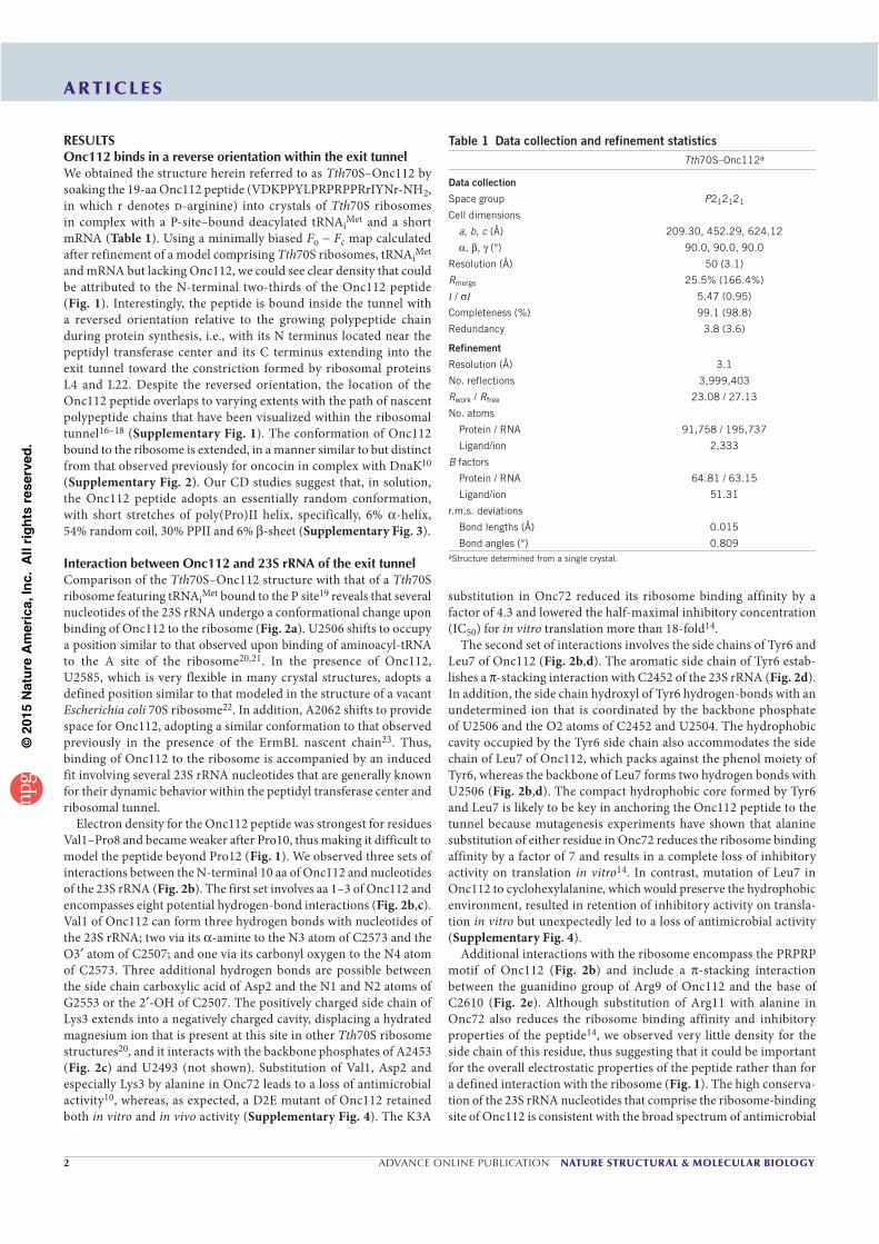
©20
15N
atu
re A
mer
ica,
Inc.
All
rig
hts
res
erve
d.
2 ADVANCE ONLINE PUBLICATION NATURE STRUCTURAL & MOLECULAR BIOLOGY
A R T I C L E S
RESULTSOnc112 binds in a reverse orientation within the exit tunnelWe obtained the structure herein referred to as Tth70S–Onc112 by soaking the 19-aa Onc112 peptide (VDKPPYLPRPRPPRrIYNr-NH2, in which r denotes d-arginine) into crystals of Tth70S ribosomes in complex with a P-site–bound deacylated tRNAiMet and a short mRNA (Table 1). Using a minimally biased Fo − Fc map calculated after refinement of a model comprising Tth70S ribosomes, tRNAiMet and mRNA but lacking Onc112, we could see clear density that could be attributed to the N-terminal two-thirds of the Onc112 peptide (Fig. 1). Interestingly, the peptide is bound inside the tunnel with a reversed orientation relative to the growing polypeptide chain during protein synthesis, i.e., with its N terminus located near the peptidyl transferase center and its C terminus extending into the exit tunnel toward the constriction formed by ribosomal proteins L4 and L22. Despite the reversed orientation, the location of the Onc112 peptide overlaps to varying extents with the path of nascent polypeptide chains that have been visualized within the ribosomal tunnel16–18 (Supplementary Fig. 1). The conformation of Onc112 bound to the ribosome is extended, in a manner similar to but distinct from that observed previously for oncocin in complex with DnaK10 (Supplementary Fig. 2). Our CD studies suggest that, in solution, the Onc112 peptide adopts an essentially random conformation, with short stretches of poly(Pro)II helix, specifically, 6% -helix, 54% random coil, 30% PPII and 6% -sheet (Supplementary Fig. 3).
Interaction between Onc112 and 23S rRNA of the exit tunnelComparison of the Tth70S–Onc112 structure with that of a Tth70S ribosome featuring tRNAiMet bound to the P site19 reveals that several nucleotides of the 23S rRNA undergo a conformational change upon binding of Onc112 to the ribosome (Fig. 2a). U2506 shifts to occupy a position similar to that observed upon binding of aminoacyl-tRNA to the A site of the ribosome20,21. In the presence of Onc112, U2585, which is very flexible in many crystal structures, adopts a defined position similar to that modeled in the structure of a vacant Escherichia coli 70S ribosome22. In addition, A2062 shifts to provide space for Onc112, adopting a similar conformation to that observed previously in the presence of the ErmBL nascent chain23. Thus, binding of Onc112 to the ribosome is accompanied by an induced fit involving several 23S rRNA nucleotides that are generally known for their dynamic behavior within the peptidyl transferase center and ribosomal tunnel.
Electron density for the Onc112 peptide was strongest for residues Val1–Pro8 and became weaker after Pro10, thus making it difficult to model the peptide beyond Pro12 (Fig. 1). We observed three sets of interactions between the N-terminal 10 aa of Onc112 and nucleotides of the 23S rRNA (Fig. 2b). The first set involves aa 1–3 of Onc112 and encompasses eight potential hydrogen-bond interactions (Fig. 2b,c). Val1 of Onc112 can form three hydrogen bonds with nucleotides of the 23S rRNA; two via its -amine to the N3 atom of C2573 and the O3 atom of C2507; and one via its carbonyl oxygen to the N4 atom of C2573. Three additional hydrogen bonds are possible between the side chain carboxylic acid of Asp2 and the N1 and N2 atoms of G2553 or the 2 -OH of C2507. The positively charged side chain of Lys3 extends into a negatively charged cavity, displacing a hydrated magnesium ion that is present at this site in other Tth70S ribosome structures20, and it interacts with the backbone phosphates of A2453 (Fig. 2c) and U2493 (not shown). Substitution of Val1, Asp2 and especially Lys3 by alanine in Onc72 leads to a loss of antimicrobial activity10, whereas, as expected, a D2E mutant of Onc112 retained both in vitro and in vivo activity (Supplementary Fig. 4). The K3A
substitution in Onc72 reduced its ribosome binding affinity by a factor of 4.3 and lowered the half-maximal inhibitory concentration (IC50) for in vitro translation more than 18-fold14.
The second set of interactions involves the side chains of Tyr6 and Leu7 of Onc112 (Fig. 2b,d). The aromatic side chain of Tyr6 estab-lishes a -stacking interaction with C2452 of the 23S rRNA (Fig. 2d). In addition, the side chain hydroxyl of Tyr6 hydrogen-bonds with an undetermined ion that is coordinated by the backbone phosphate of U2506 and the O2 atoms of C2452 and U2504. The hydrophobic cavity occupied by the Tyr6 side chain also accommodates the side chain of Leu7 of Onc112, which packs against the phenol moiety of Tyr6, whereas the backbone of Leu7 forms two hydrogen bonds with U2506 (Fig. 2b,d). The compact hydrophobic core formed by Tyr6 and Leu7 is likely to be key in anchoring the Onc112 peptide to the tunnel because mutagenesis experiments have shown that alanine substitution of either residue in Onc72 reduces the ribosome binding affinity by a factor of 7 and results in a complete loss of inhibitory activity on translation in vitro14. In contrast, mutation of Leu7 in Onc112 to cyclohexylalanine, which would preserve the hydrophobic environment, resulted in retention of inhibitory activity on transla-tion in vitro but unexpectedly led to a loss of antimicrobial activity (Supplementary Fig. 4).
Additional interactions with the ribosome encompass the PRPRP motif of Onc112 (Fig. 2b) and include a -stacking interaction between the guanidino group of Arg9 of Onc112 and the base of C2610 (Fig. 2e). Although substitution of Arg11 with alanine in Onc72 also reduces the ribosome binding affinity and inhibitory properties of the peptide14, we observed very little density for the side chain of this residue, thus suggesting that it could be important for the overall electrostatic properties of the peptide rather than for a defined interaction with the ribosome (Fig. 1). The high conserva-tion of the 23S rRNA nucleotides that comprise the ribosome-binding site of Onc112 is consistent with the broad spectrum of antimicrobial
Table 1 Data collection and refinement statisticsTth70S–Onc112a
Data collection
Space group P212121
Cell dimensions
a, b, c (Å) 209.30, 452.29, 624.12
, , (°) 90.0, 90.0, 90.0
Resolution (Å) 50 (3.1)
Rmerge 25.5% (166.4%)
I / I 5.47 (0.95)
Completeness (%) 99.1 (98.8)
Redundancy 3.8 (3.6)
Refinement
Resolution (Å) 3.1
No. reflections 3,999,403
Rwork / Rfree 23.08 / 27.13
No. atoms
Protein / RNA 91,758 / 195,737
Ligand/ion 2,333
B factors
Protein / RNA 64.81 / 63.15
Ligand/ion 51.31
r.m.s. deviations
Bond lengths (Å) 0.015
Bond angles (°) 0.809
aStructure determined from a single crystal.
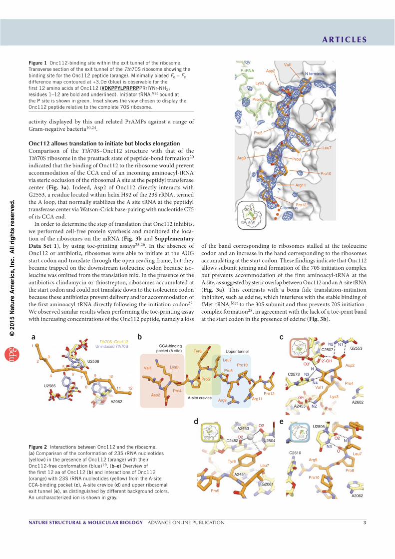
©20
15N
atu
re A
mer
ica,
Inc.
All
rig
hts
res
erve
d.
NATURE STRUCTURAL & MOLECULAR BIOLOGY ADVANCE ONLINE PUBLICATION 3
A R T I C L E S
activity displayed by this and related PrAMPs against a range of Gram-negative bacteria10,24.
Onc112 allows translation to initiate but blocks elongationComparison of the Tth70S–Onc112 structure with that of the Tth70S ribosome in the preattack state of peptide-bond formation20 indicated that the binding of Onc112 to the ribosome would prevent accommodation of the CCA end of an incoming aminoacyl-tRNA via steric occlusion of the ribosomal A site at the peptidyl transferase center (Fig. 3a). Indeed, Asp2 of Onc112 directly interacts with G2553, a residue located within helix H92 of the 23S rRNA, termed the A loop, that normally stabilizes the A site tRNA at the peptidyl transferase center via Watson-Crick base-pairing with nucleotide C75 of its CCA end.
In order to determine the step of translation that Onc112 inhibits, we performed cell-free protein synthesis and monitored the loca-tion of the ribosomes on the mRNA (Fig. 3b and Supplementary Data Set 1), by using toe-printing assays25,26. In the absence of Onc112 or antibiotic, ribosomes were able to initiate at the AUG start codon and translate through the open reading frame, but they became trapped on the downstream isoleucine codon because iso-leucine was omitted from the translation mix. In the presence of the antibiotics clindamycin or thiostrepton, ribosomes accumulated at the start codon and could not translate down to the isoleucine codon because these antibiotics prevent delivery and/or accommodation of the first aminoacyl-tRNA directly following the initiation codon27. We observed similar results when performing the toe-printing assay with increasing concentrations of the Onc112 peptide, namely a loss
of the band corresponding to ribosomes stalled at the isoleucine codon and an increase in the band corresponding to the ribosomes accumulating at the start codon. These findings indicate that Onc112 allows subunit joining and formation of the 70S initiation complex but prevents accommodation of the first aminoacyl-tRNA at the A site, as suggested by steric overlap between Onc112 and an A-site tRNA (Fig. 3a). This contrasts with a bona fide translation-initiation inhibitor, such as edeine, which interferes with the stable binding of fMet-tRNAiMet to the 30S subunit and thus prevents 70S initiation-complex formation28, in agreement with the lack of a toe-print band at the start codon in the presence of edeine (Fig. 3b).
P-tRNA Asp2
Lys3
Pro4
Pro5
Arg9 Pro8
Arg11
Pro10
Pro1230S
50S
P
Leu7
Tyr6
Val1
N terminus
C terminus
Figure 1 Onc112-binding site within the exit tunnel of the ribosome.
Transverse section of the exit tunnel of the Tth70S ribosome showing the
binding site for the Onc112 peptide (orange). Minimally biased Fo − Fc
difference map contoured at +3.0 (blue) is observable for the
first 12 amino acids of Onc112 (VDKPPYLPRPRPPRrIYNr-NH2;
residues 1–12 are bold and underlined). Initiator tRNAiMet bound at
the P site is shown in green. Inset shows the view chosen to display the
Onc112 peptide relative to the complete 70S ribosome.
bCCA-binding
pocket (A site) Upper tunnel
A-site creviceAsp2
Pro4
Lys3
Tyr6
Pro5
Arg9 Arg11Pro12
Pro8Pro10
Leu7
Val1
U2506
U2585
a
A2062
9
8
6
5
3
4
2
1
7 10
11 12
Uninduced Tth70STth70S–Onc112
c
e
G2553C2507
2 -OHO3
OP1
C2573
50S
30S
P
A2453A2602
N2
N3
N4
NZ
N
N1
Asp2
Pro4
Lys3
Val1
C2610
A2062
U2506
O
O250S
30S
P
N3
N
Arg9
Pro8
Leu7
Pro10
A2453
A2451
C2452 U2504
G2061
O2
O2
30S
50SP
Pro5
Leu7Tyr6
d
Figure 2 Interactions between Onc112 and the ribosome.
(a) Comparison of the conformation of 23S rRNA nucleotides
(yellow) in the presence of Onc112 (orange) with their
Onc112-free conformation (blue)19. (b–e) Overview of
the first 12 aa of Onc112 (b) and interactions of Onc112
(orange) with 23S rRNA nucleotides (yellow) from the A-site
CCA-binding pocket (c), A-site crevice (d) and upper ribosomal
exit tunnel (e), as distinguished by different background colors.
An uncharacterized ion is shown in gray.
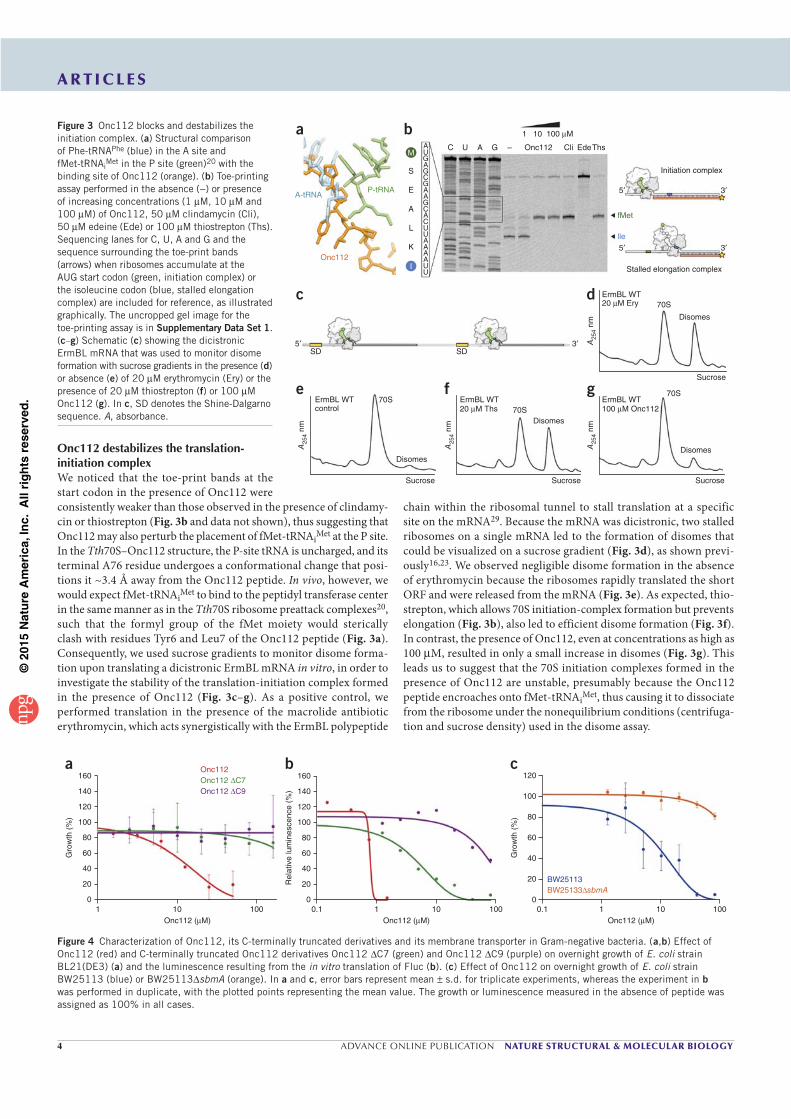
©20
15N
atu
re A
mer
ica,
Inc.
All
rig
hts
res
erve
d.
4 ADVANCE ONLINE PUBLICATION NATURE STRUCTURAL & MOLECULAR BIOLOGY
A R T I C L E S
Onc112 destabilizes the translation-initiation complexWe noticed that the toe-print bands at the start codon in the presence of Onc112 were consistently weaker than those observed in the presence of clindamy-cin or thiostrepton (Fig. 3b and data not shown), thus suggesting that Onc112 may also perturb the placement of fMet-tRNAiMet at the P site. In the Tth70S–Onc112 structure, the P-site tRNA is uncharged, and its terminal A76 residue undergoes a conformational change that posi-tions it ~3.4 Å away from the Onc112 peptide. In vivo, however, we would expect fMet-tRNAiMet to bind to the peptidyl transferase center in the same manner as in the Tth70S ribosome preattack complexes20, such that the formyl group of the fMet moiety would sterically clash with residues Tyr6 and Leu7 of the Onc112 peptide (Fig. 3a). Consequently, we used sucrose gradients to monitor disome forma-tion upon translating a dicistronic ErmBL mRNA in vitro, in order to investigate the stability of the translation-initiation complex formed in the presence of Onc112 (Fig. 3c–g). As a positive control, we performed translation in the presence of the macrolide antibiotic erythromycin, which acts synergistically with the ErmBL polypeptide
chain within the ribosomal tunnel to stall translation at a specific site on the mRNA29. Because the mRNA was dicistronic, two stalled ribosomes on a single mRNA led to the formation of disomes that could be visualized on a sucrose gradient (Fig. 3d), as shown previ-ously16,23. We observed negligible disome formation in the absence of erythromycin because the ribosomes rapidly translated the short ORF and were released from the mRNA (Fig. 3e). As expected, thio-strepton, which allows 70S initiation-complex formation but prevents elongation (Fig. 3b), also led to efficient disome formation (Fig. 3f). In contrast, the presence of Onc112, even at concentrations as high as 100 M, resulted in only a small increase in disomes (Fig. 3g). This leads us to suggest that the 70S initiation complexes formed in the presence of Onc112 are unstable, presumably because the Onc112 peptide encroaches onto fMet-tRNAiMet, thus causing it to dissociate from the ribosome under the nonequilibrium conditions (centrifuga-tion and sucrose density) used in the disome assay.
A25
4 nm
A25
4 nm
A25
4 nm
70S
Disomes
70SDisomes
70S
Disomes
SucroseSucroseSucrose
ErmBL WT100 M Onc112
ErmBL WTErmBL WTcontrol 20 M Ths
A-tRNA
a b
P-tRNA
M
S
A C U A G –
1 10
Onc112 Cli EdeThs
Initiation complex
Stalled elongation complex
fMet
lle
3
3
5
5
100 M
UG
G
G
GC
CUUAAAAUU
AA
A
C
A
E
A
L
K
IOnc112
c
e f g
d
SD SD35 A
254
nm
ErmBL WT70S
Disomes
Sucrose
20 M Ery
Figure 3 Onc112 blocks and destabilizes the
initiation complex. (a) Structural comparison
of Phe-tRNAPhe (blue) in the A site and
fMet-tRNAiMet in the P site (green)20 with the
binding site of Onc112 (orange). (b) Toe-printing
assay performed in the absence (−) or presence
of increasing concentrations (1 M, 10 M and
100 M) of Onc112, 50 M clindamycin (Cli),
50 M edeine (Ede) or 100 M thiostrepton (Ths).
Sequencing lanes for C, U, A and G and the
sequence surrounding the toe-print bands
(arrows) when ribosomes accumulate at the
AUG start codon (green, initiation complex) or
the isoleucine codon (blue, stalled elongation
complex) are included for reference, as illustrated
graphically. The uncropped gel image for the
toe-printing assay is in Supplementary Data Set 1.
(c–g) Schematic (c) showing the dicistronic
ErmBL mRNA that was used to monitor disome
formation with sucrose gradients in the presence (d)
or absence (e) of 20 M erythromycin (Ery) or the
presence of 20 M thiostrepton (f) or 100 M
Onc112 (g). In c, SD denotes the Shine-Dalgarno
sequence. A, absorbance.
160a b c
140
120
100
80
60
40
20
10
160
140
120
100
80
60
40
20
0
120
100
80
60
40
20
0
BW25113BW25133 sbmA
10 100
Gro
wth
(%
)
Gro
wth
(%
)
Rel
ativ
e lu
min
esce
nce
(%)
Onc112
Onc112 C9Onc112 C7
Onc112 ( M)0.1 1 10 100
Onc112 ( M)0.1 1 10 100
Onc112 ( M)
Figure 4 Characterization of Onc112, its C-terminally truncated derivatives and its membrane transporter in Gram-negative bacteria. (a,b) Effect of
Onc112 (red) and C-terminally truncated Onc112 derivatives Onc112 C7 (green) and Onc112 C9 (purple) on overnight growth of E. coli strain
BL21(DE3) (a) and the luminescence resulting from the in vitro translation of Fluc (b). (c) Effect of Onc112 on overnight growth of E. coli strain
BW25113 (blue) or BW25113 sbmA (orange). In a and c, error bars represent mean s.d. for triplicate experiments, whereas the experiment in b
was performed in duplicate, with the plotted points representing the mean value. The growth or luminescence measured in the absence of peptide was
assigned as 100% in all cases.
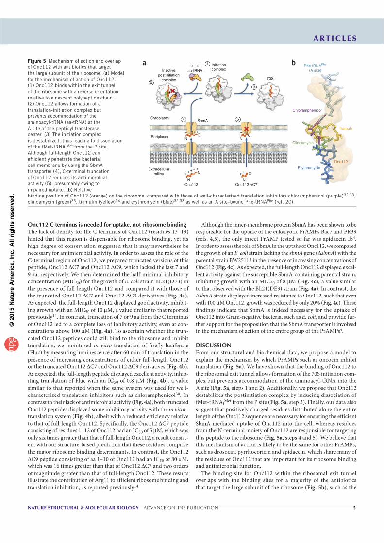
©20
15N
atu
re A
mer
ica,
Inc.
All
rig
hts
res
erve
d.
NATURE STRUCTURAL & MOLECULAR BIOLOGY ADVANCE ONLINE PUBLICATION 5
A R T I C L E S
Onc112 C terminus is needed for uptake, not ribosome bindingThe lack of density for the C terminus of Onc112 (residues 13–19) hinted that this region is dispensable for ribosome binding, yet its high degree of conservation suggested that it may nevertheless be necessary for antimicrobial activity. In order to assess the role of the C-terminal region of Onc112, we prepared truncated versions of this peptide, Onc112 C7 and Onc112 C9, which lacked the last 7 and 9 aa, respectively. We then determined the half-minimal inhibitory concentration (MIC50) for the growth of E. coli strain BL21(DE3) in the presence of full-length Onc112 and compared it with those of the truncated Onc112 C7 and Onc112 C9 derivatives (Fig. 4a). As expected, the full-length Onc112 displayed good activity, inhibit-ing growth with an MIC50 of 10 M, a value similar to that reported previously14. In contrast, truncation of 7 or 9 aa from the C terminus of Onc112 led to a complete loss of inhibitory activity, even at con-centrations above 100 M (Fig. 4a). To ascertain whether the trun-cated Onc112 peptides could still bind to the ribosome and inhibit translation, we monitored in vitro translation of firefly luciferase (Fluc) by measuring luminescence after 60 min of translation in the presence of increasing concentrations of either full-length Onc112 or the truncated Onc112 C7 and Onc112 C9 derivatives (Fig. 4b). As expected, the full-length peptide displayed excellent activity, inhib-iting translation of Fluc with an IC50 of 0.8 M (Fig. 4b), a value similar to that reported when the same system was used for well- characterized translation inhibitors such as chloramphenicol30. In contrast to their lack of antimicrobial activity (Fig. 4a), both truncated Onc112 peptides displayed some inhibitory activity with the in vitro– translation system (Fig. 4b), albeit with a reduced efficiency relative to that of full-length Onc112. Specifically, the Onc112 C7 peptide consisting of residues 1–12 of Onc112 had an IC50 of 5 M, which was only six times greater than that of full-length Onc112, a result consist-ent with our structure-based prediction that these residues comprise the major ribosome binding determinants. In contrast, the Onc112
C9 peptide consisting of aa 1–10 of Onc112 had an IC50 of 80 M, which was 16 times greater than that of Onc112 C7 and two orders of magnitude greater than that of full-length Onc112. These results illustrate the contribution of Arg11 to efficient ribosome binding and translation inhibition, as reported previously14.
Figure 5 Mechanism of action and overlap
of Onc112 with antibiotics that target
the large subunit of the ribosome. (a) Model
for the mechanism of action of Onc112.
(1) Onc112 binds within the exit tunnel
of the ribosome with a reverse orientation
relative to a nascent polypeptide chain.
(2) Onc112 allows formation of a
translation-initiation complex but
prevents accommodation of the
aminoacyl-tRNA (aa-tRNA) at the
A site of the peptidyl transferase
center. (3) The initiation complex
is destabilized, thus leading to dissociation
of the fMet-tRNAiMet from the P site.
Although full-length Onc112 can
efficiently penetrate the bacterial
cell membrane by using the SbmA
transporter (4), C-terminal truncation
of Onc112 reduces its antimicrobial
activity (5), presumably owing to
impaired uptake. (b) Relative
binding position of Onc112 (orange) on the ribosome, compared with those of well-characterized translation inhibitors chloramphenicol (purple)32,33,
clindamycin (green)33, tiamulin (yellow)34 and erythromycin (blue)32,33 as well as an A site–bound Phe-tRNAPhe (ref. 20).
Although the inner-membrane protein SbmA has been shown to be responsible for the uptake of the eukaryotic PrAMPs Bac7 and PR39 (refs. 4,5), the only insect PrAMP tested so far was apidaecin Ib4. In order to assess the role of SbmA in the uptake of Onc112, we compared the growth of an E. coli strain lacking the sbmA gene ( sbmA) with the parental strain BW25113 in the presence of increasing concentrations of Onc112 (Fig. 4c). As expected, the full-length Onc112 displayed excel-lent activity against the susceptible SbmA-containing parental strain, inhibiting growth with an MIC50 of 8 M (Fig. 4c), a value similar to that observed with the BL21(DE3) strain (Fig. 4a). In contrast, the
sbmA strain displayed increased resistance to Onc112, such that even with 100 M Onc112, growth was reduced by only 20% (Fig. 4c). These findings indicate that SbmA is indeed necessary for the uptake of Onc112 into Gram-negative bacteria, such as E. coli, and provide fur-ther support for the proposition that the SbmA transporter is involved in the mechanism of action of the entire group of the PrAMPs4.
DISCUSSIONFrom our structural and biochemical data, we propose a model to explain the mechanism by which PrAMPs such as oncocin inhibit translation (Fig. 5a). We have shown that the binding of Onc112 to the ribosomal exit tunnel allows formation of the 70S initiation com-plex but prevents accommodation of the aminoacyl-tRNA into the A site (Fig. 5a, steps 1 and 2). Additionally, we propose that Onc112 destabilizes the postinitiation complex by inducing dissociation of fMet-tRNAiMet from the P site (Fig. 5a, step 3). Finally, our data also suggest that positively charged residues distributed along the entire length of the Onc112 sequence are necessary for ensuring the efficient SbmA-mediated uptake of Onc112 into the cell, whereas residues from the N-terminal moiety of Onc112 are responsible for targeting this peptide to the ribosome (Fig. 5a, steps 4 and 5). We believe that this mechanism of action is likely to be the same for other PrAMPs, such as drosocin, pyrrhocoricin and apidaecin, which share many of the residues of Onc112 that are important for its ribosome binding and antimicrobial function.
The binding site for Onc112 within the ribosomal exit tunnel overlaps with the binding sites for a majority of the antibiotics that target the large subunit of the ribosome (Fig. 5b), such as the
Inactivepostinitiation
complex2
1
3
5
A
N
C
+ +
+ +++ +++
+++
N
N N
C
C C
++
NC
Onc112 Onc112 ΔC7
P
P
Initiationcomplex
70S
Phe-tRNAPhe
(A site)
ba
Chloramphenicol
Clindamycin
Erythromycin
Onc112
Tiamulin
EF-Tuaa-tRNA
SbmACytoplasm
Periplasm
Extracellularmilieu
4
©20
15N
atu
re A
mer
ica,
Inc.
All
rig
hts
res
erve
d.
6 ADVANCE ONLINE PUBLICATION NATURE STRUCTURAL & MOLECULAR BIOLOGY
A R T I C L E S
chloramphenicols, pleuromutilins (for example, tiamulin) and lincosa-mides (for example, clindamycin), which inhibit peptide-bond forma-tion by preventing the correct positioning of the tRNA substrates, as well as the macrolides (for example, erythromycin), which abort trans-lation by interfering with the movement of the nascent polypeptide chain through the ribosomal exit tunnel27. Given the substantial spa-tial overlap that exists between the binding sites for these antibiotics and the regions of the tunnel that interact with Onc112 (Fig. 5b) and presumably with several other PrAMPs, it appears likely that such antimicrobial peptides represent a vast, untapped resource for the development of new therapeutics. Several strategies have been pursued to design improved or entirely new antimicrobials that target the exit tunnel of the ribosome31. One approach consists of modi-fying existing antibiotics to create semisynthetic compounds that possess enhanced antimicrobial properties, including better affinity for mutated or modified ribosomes, the ability to evade drug modi-fication or degradation pathways, increased solubility, improved uptake and reduced efflux. Other strategies involve designing chi-meric antibiotics from drugs with adjacent binding sites (for example, macrolide-chloramphenicol or linezolid-sparsomycin) or developing entirely new scaffolds, as exemplified by the oxazolidinone linezolid. The ability to produce new scaffolds based on peptides, such as Onc112, that display potent activity against a diverse range of Gram-negative bacteria represents an exciting avenue for the development of future antimicrobials.
METHODSMethods and any associated references are available in the online version of the paper.
Accession codes. Coordinates and structure factors have been depos-ited in the Protein Data Bank under accession code 4ZER.
Note: Any Supplementary Information and Source Data files are available in the online version of the paper.
ACKNOWLEDGMENTSWe thank the staff at the European Synchrotron Radiation Facility (beamline ID-29) for help during data collection and B. Kauffmann and S. Massip at the Institut Européen de Chimie et Biologie for help with crystal freezing and screening. We also thank C. Mackereth for discussions and advice. This research was supported by grants from the Agence Nationale pour la Recherche (ANR-14-CE09-0001 to C.A.I., G.G. and D.N.W.), Région Aquitaine (2012-13-01-009 to C.A.I.), the Fondation pour la Recherche Médicale (AJE201133 to C.A.I.), the European Union (PCIG14-GA-2013-631479 to C.A.I.), the CNRS (C.D.) and the Deutsche Forschungsgemeinschaft (FOR1805, WI3285/4-1 and GRK1721 to D.N.W.). Predoctoral fellowships from the Direction Générale de l’Armement and Région Aquitaine (S. Antunes) and INSERM and Région Aquitaine (A.C.S.) are gratefully acknowledged.
AUTHOR CONTRIBUTIONSA.C.S. performed structure solution, model building and analysis. N.P. prepared and crystallized ribosomes. N.P. and C.A.I. collected X-ray crystallography data. F.N. performed growth and in vitro–translation inhibition assays. S. Antunes and C.D. synthesized the peptides and performed NMR, CD and electrospray ionization high-resolution MS experiments. M.G. performed toe-printing assays. S. Arenz performed disome assays. K.K.I. prepared tRNAiMet. G.G., D.N.W. and C.A.I. designed experiments, interpreted data and wrote the manuscript.
COMPETING FINANCIAL INTERESTSThe authors declare no competing financial interests.
Reprints and permissions information is available online at http://www.nature.com/reprints/index.html.
1. Wang, G. et al. Antimicrobial peptides in 2014. Pharmaceuticals (Basel) 8,
123–150 (2015).
2. Casteels, P., Ampe, C., Jacobs, F., Vaeck, M. & Tempst, P. Apidaecins: antibacterial
peptides from honeybees. EMBO J. 8, 2387–2391 (1989).
3. Li, W. et al. Proline-rich antimicrobial peptides: potential therapeutics against
antibiotic-resistant bacteria. Amino Acids 46, 2287–2294 (2014).
4. Mattiuzzo, M. et al. Role of the Escherichia coli SbmA in the antimicrobial activity
of proline-rich peptides. Mol. Microbiol. 66, 151–163 (2007).
5. Runti, G. et al. Functional characterization of SbmA, a bacterial inner membrane
transporter required for importing the antimicrobial peptide Bac7 (1–35).
J. Bacteriol. 195, 5343–5351 (2013).
6. Hansen, A., Schäfer, I., Knappe, D., Seibel, P. & Hoffmann, R. Intracellular toxicity of
proline-rich antimicrobial peptides shuttled into mammalian cells by the cell-penetrating
peptide penetratin. Antimicrob. Agents Chemother. 56, 5194–5201 (2012).
7. Stalmans, S. et al. Blood-brain barrier transport of short proline-rich antimicrobial
peptides. Protein Pept. Lett. 21, 399–406 (2014).
8. Otvos, L. et al. Interaction between heat shock proteins and antimicrobial peptides.
Biochemistry 39, 14150–14159 (2000).
9. Czihal, P. et al. Api88 is a novel antibacterial designer peptide to treat systemic
infections with multidrug-resistant Gram-negative pathogens. ACS Chem. Biol. 7,
1281–1291 (2012).
10. Knappe, D. et al. Rational design of oncocin derivatives with superior protease
stabilities and antibacterial activities based on the high–resolution structure of the
oncocin–DnaK complex. ChemBioChem 12, 874–876 (2011).
11. Zahn, M. et al. Structural studies on the forward and reverse binding modes of
peptides to the chaperone DnaK. J. Mol. Biol. 425, 2463–2479 (2013).
12. Zahn, M. et al. Structural identification of DnaK binding sites within bovine and
sheep bactenecin Bac7. Protein Pept. Lett. 21, 407–412 (2014).
13. Berthold, N. & Hoffmann, R. Cellular uptake of apidaecin 1b and related analogs
in Gram-negative bacteria reveals novel antibacterial mechanism for proline-rich
antimicrobial peptides. Protein Pept. Lett. 21, 391–398 (2014).
14. Krizsan, A. et al. Insect-derived proline-rich antimicrobial peptides kill bacteria by
inhibiting bacterial protein translation at the 70 S ribosome. Angew. Chem. Int.
Ed. Engl. 53, 12236–12239 (2014).
15. Schneider, M. & Dorn, A. Differential infectivity of two Pseudomonas species and
the immune response in the milkweed bug, Oncopeltus fasciatus (Insecta:
Hemiptera). J. Invertebr. Pathol. 78, 135–140 (2001).
16. Arenz, S. et al. Drug sensing by the ribosome induces translational arrest via active
site perturbation. Mol. Cell 56, 446–452 (2014).
17. Bischoff, L., Berninghausen, O. & Beckmann, R. Molecular basis for the ribosome
functioning as an L-tryptophan sensor. Cell Reports 9, 469–475 (2014).
18. Shao, S. & Hegde, R.S. Reconstitution of a minimal ribosome-associated
ubiquitination pathway with purified factors. Mol. Cell 55, 880–890 (2014).
19. Jenner, L. et al. Structural basis for potent inhibitory activity of the antibiotic
tigecycline during protein synthesis. Proc. Natl. Acad. Sci. USA 110, 3812–3816
(2013).
20. Polikanov, Y.S., Steitz, T.A. & Innis, C.A. A proton wire to couple aminoacyl-tRNA
accommodation and peptide-bond formation on the ribosome. Nat. Struct. Mol.
Biol. 21, 787–793 (2014).
21. Schmeing, T.M., Huang, K.S., Strobel, S.A. & Steitz, T.A. An induced-fit mechanism
to promote peptide bond formation and exclude hydrolysis of peptidyl-tRNA.
Nature 438, 520–524 (2005).
22. Schuwirth, B.S. et al. Structures of the bacterial ribosome at 3.5 A resolution.
Science 310, 827–834 (2005).
23. Arenz, S. et al. Molecular basis for erythromycin-dependent ribosome stalling during
translation of the ErmBL leader peptide. Nat. Commun. 5, 3501 (2014).
24. Knappe, D. et al. Oncocin (VDKPPYLPRPRPPRRIYNR-NH2): a novel antibacterial
peptide optimized against Gram-negative human pathogens. J. Med. Chem. 53,
5240–5247 (2010).
25. Hartz, D., McPheeters, D.S., Traut, R. & Gold, L. Extension inhibition analysis of
translation initiation complexes. Methods Enzymol. 164, 419–425 (1988).
26. Starosta, A.L. et al. Translational stalling at polyproline stretches is modulated
by the sequence context upstream of the stall site. Nucleic Acids Res. 42,
10711–10719 (2014).
27. Wilson, D.N. The A–Z of bacterial translation inhibitors. Crit. Rev. Biochem. Mol.
Biol. 44, 393–433 (2009).
28. Dinos, G. et al. Dissecting the ribosomal inhibition mechanisms of edeine and
pactamycin: the universally conserved residues G693 and C795 regulate P-site
RNA binding. Mol. Cell 13, 113–124 (2004).
29. Vázquez-Laslop, N., Ramu, H., Klepacki, D., Kannan, K. & Mankin, A.S. The key
function of a conserved and modified rRNA residue in the ribosomal response to
the nascent peptide. EMBO J. 29, 3108–3117 (2010).
30. Starosta, A.L. et al. Interplay between the ribosomal tunnel, nascent chain, and
macrolides influences drug inhibition. Chem. Biol. 17, 504–514 (2010).
31. Wilson, D.N. Ribosome-targeting antibiotics and mechanisms of bacterial resistance.
Nat. Rev. Microbiol. 12, 35–48 (2014).
32. Bulkley, D., Innis, C.A., Blaha, G. & Steitz, T.A. Revisiting the structures of several
antibiotics bound to the bacterial ribosome. Proc. Natl. Acad. Sci. USA 107,
17158–17163 (2010).
33. Dunkle, J.A., Xiong, L., Mankin, A.S. & Cate, J.H. Structures of the Escherichia
coli ribosome with antibiotics bound near the peptidyl transferase center explain
spectra of drug action. Proc. Natl. Acad. Sci. USA 107, 17152–17157 (2010).
34. Schlünzen, F., Pyetan, E., Fucini, P., Yonath, A. & Harms, J.M. Inhibition of peptide
bond formation by pleuromutilins: the structure of the 50S ribosomal subunit from
Deinococcus radiodurans in complex with tiamulin. Mol. Microbiol. 54, 1287–1294
(2004).
©20
15N
atu
re A
mer
ica,
Inc.
All
rig
hts
res
erve
d.
NATURE STRUCTURAL & MOLECULAR BIOLOGYdoi:10.1038/nsmb.3034
ONLINE METHODSPeptide synthesis. Commercially available reagents were used throughout without purification. N,N-dimethylformamide (DMF, peptide synthesis– quality grade) was purchased from Carlo Erba, and piperidine and trifluoroacetic acid (TFA) were purchased from Alfa Aesar. Rink amide PS resin was purchased from PolyPeptide Laboratories. N,N -diisopropylcarbodiimide (DIC), Oxyma and all standard N-Fmoc–protected l and d amino acids were purchased from Iris Biotech. N-Fmoc-cyclohexylalanine-OH (Fmoc-Cha-OH) was purchased from PolyPeptide laboratories. RP-HPLC–quality acetonitrile (CH3CN, Sigma-Aldrich) and MilliQ water were used for RP-HPLC analyses and purification. Analytical RP-HPLC analyses were performed on a Dionex U3000SD with a Macherey-Nagel Nucleodur column (4.6 × 100 mm, 3 m) at a flow rate of 1 ml min−1 at 50 °C. The mobile phase was composed of 0.1% (v/v) TFA-H2O (solvent A) and 0.1% TFA-CH3CN (solvent B). Purification was performed on a Gilson GX-281 with a Macherey-Nagel Nucleodur VP250/21 100–5 C18ec column (21 × 250 mm, 5 m) at a flow rate of 20 mL min−1. The solid-phase syntheses of peptides were conducted on an automated Liberty Blue System synthesizer (CEM Waves S.A.S.). 1H NMR spectra were recorded on a DPX-400 NMR spectrometer (Bruker Biospin) with a vertical 9.4T narrow-bore/ultrashield magnet operating at 400 MHz for 1H observation by means of a 5-mm direct QNP 1H/13C/31P/19F probe with gradient capabilities (Supplementary Fig. 5). ESI-MS analyses were carried out on a Thermo Exactive from the Mass Spectrometry Laboratory at the European Institute of Chemistry and Biology (UMS 3033–IECB), Pessac, France (Supplementary Fig. 5).
All peptides were synthesized on Rink Amide PS resin (0.79 mmol/g) with a five-fold excess of reagents for the coupling step (0.2 M N-Fmoc–amino acid solution (in DMF) with 0.5 M DIC (in DMF) and 1.0 M Oxyma (in DMF)). Coupling of N-Fmoc–protected l- and d-arginine-OH was performed twice at 25 °C without microwaves for 1,500 s. Other amino acid couplings were performed first at 90 °C, 170 W, 115 s then at 90 °C, 30 W, 110 s. Fmoc removal was performed with a solution of 20% piperidine in DMF at 75 °C with 155 W for 15 s then 90 °C, 35 W, 50 s. After completion of the synthesis, the pep-tide resin was washed three times with DCM. Cleavage was performed by treatment with 5 mL of a freshly prepared TFA/TIS/H20 solution for 240 min at room temperature. The resin was then filtered off, and the TFA solution was concentrated under reduced pressure. The crude products were precipitated as TFA salts in the presence of Et2O and purified with the appropriate gradient (10–30% of B in 20 min) by semipreparative RP-HPLC. The compounds were freeze dried, and TFA was exchanged with HCl by two repetitive freeze-drying cycles in 0.1 N HCl solution35.
The list of peptides prepared for this study and details concerning their synthesis is as follows:
Onc112. H-Val-Asp-Lys-Pro-Pro-Tyr-Leu-Pro-Arg-Pro-Arg-Pro-Pro-Arg- (d-Arg)-Ile-Tyr-Asn-(d-Arg)-NH2 (2,389.85 g mol−1). Synthesis of Onc112 (0.1-mmol scale): 24 mg (10% yield); RP HPLC tR 4.11 min (gradient 10–50% of B in 10 min); ESI HRMS (m/z): found 1,195.70 [M + 2H]2+, 797.47 [M +3 H]3+, 598.35 [M + 4H]4+, and 478.88 [M + 5H]5+.
Onc112 C7. H-Val-Asp-Lys-Pro-Pro-Tyr-Leu-Pro-Arg-Pro-Arg-Pro-NH2 (1,433.73 g mol−1). Synthesis of Onc112 C7 (0.15-mmol scale): 79.4 mg (37% yield); RP HPLC tR 3.54 min (gradient 10–50% of B in 10 min); ESI HRMS (m/z): [M + H]+ calcd for C67H108H20O15, 1,433.83758 found 1,433.84017, with 717.42 [M + 2H]2+ and 478.61 [M + 3H]3+.
Onc112 C9. H-Val-Asp-Lys-Pro-Pro-Tyr-Leu-Pro-Arg-Pro-NH2 (1,180.42 g mol−1). Synthesis of Onc112 C9. (0.1-mmol scale): 22.6 mg (19% yield); RP HPLC tR 4.78 min (gradient 10–50% of B in 10 min); ESI HRMS (m/z): [M + H]+ calcd for C56H89H15O13, 1,180.63370 found 1,180.68368, with [M + 2H]2+ 590.84 and [M + 3H]3+ 394.23.
Onc112 D2E. H-Val-Glu-Lys-Pro-Pro-Tyr-Leu-Pro-Arg-Pro-Arg-Pro-Pro-Arg-(d-Arg)-Ile-Tyr-Asn-(d-Arg)-NH2 (2,403.88 g mol−1). Synthesis of Onc112 D2E (0.05-mmol scale): 11.6 mg (10% yield); RP HPLC tR 5.75 min (gradient 10–50% of B in 10 min); ESI HRMS (m/z): found 1316.70 [M + 2H]2+, 840.14 [M + 3H]3+ and 601.86 [M + 4H]4+.
Onc112 L7Cha. H-Val-Asp-Lys-Pro-Pro-Tyr-Cha-Pro-Arg-Pro-Arg-Pro-Pro-Arg-(d-Arg)-Ile-Tyr-Asn-(d-Arg)-NH2 (2,429.92 g mol−1). Synthesis of Onc112 L7Cha (0.05-mmol scale): 6.9 mg (6% yield); RP HPLC tR 5.28 min (gradient 10–50% of B in 10 min); ESI HRMS (m/z): found 1,252.18 [M + 2H]2+, 822.80 [M + 3H]3+ and 608.36 [M + 4H]4+.
CD spectroscopy. CD spectra of peptides were recorded on a J-815 Jasco spectropolarimeter (Jasco France). Data are expressed in terms of total molar ellipticity in deg cm2 dmol−1. CD spectra for the Onc112 peptide were acquired at four different concentrations in phosphate buffer (pH 7.6, 10 mM) between 180 and 280 nm with a rectangular quartz cell with a path length of 1 mm (Hellma 110-QS 1 mm) averaging over two runs. Secondary-structure proportion was estimated from the CD spectra with the deconvolution program CDFriend (S. Buchoux (Unité de Génie Enzymatique et Cellulaire, UMR 6022 CNRS-Université de Picardie Jules Verne) and E. Dufourc (Université de Bordeaux, CNRS, Institut Polytechnique de Bordeaux, UMR 5248 Institut de Chimie et Biologie des Membranes et des Nano-objets (CBMN); available upon request), unpublished). This program uses standard curves obtained for each canonical structure ( -helix, -sheet, helix-polyproline type II and random coil) with LiKj (alternated hydrophobic leucine and hydrophilic/charged lysine residues) pep-tides of known length, secondary structure and CD spectra. The program imple-ments a simulated annealing algorithm to get the best combination of -helix,
-sheet, helix-II and random coil that exhibits the lowest normalized r.m.s. devia-tion with respect to the experimental spectrum36–38. The algorithm yielded the following assessment for the Onc112 peptide: 54% random coil, 30% helix-PPII, 6% -helix and 6% -sheet content.
Purification of T. thermophilus 70S ribosomes. Tth70S ribosomes were purified as described previously39 and resuspended in buffer containing 5 mM HEPES-KOH, pH 7.5, 50 mM KCl, 10 mM NH4Cl, and 10 mM Mg(CH3COO)2 to yield a final concentration of 26–32 mg/mL. For storage, Tth70S ribosomes were flash frozen in liquid nitrogen and kept at −80 °C.
Preparation of mRNA and tRNAiMet. Synthetic mRNA with the sequence 5 -GGC AAG GAG GUA AAA AUG CGU UUU CGU-3 was obtained from Eurogentec. This mRNA contains a Shine-Dalgarno sequence and an AUG start codon followed by several additional codons. E. coli tRNAiMet was overexpressed in E. coli HB101 cells and purified as described previously40.
Complex formation. A ternary complex containing Tth70S ribosomes, mRNA and deacylated tRNAiMet was formed by mixing of 5 M Tth70S ribosomes with 10 M mRNA and incubating at 55 °C for 10 min. For the next step, 20 M tRNAiMet was added, and the mixture was incubated at 37 °C for 10 min. Before the complexes for crystallization were used, the sample was incubated at room temperature for at least 15 min. All complexes were centrifuged briefly before use for crystallization. The final buffer conditions were 5 mM HEPES-KOH, pH 7.6, 50 mM KCl, 10 mM NH4Cl and 10 mM Mg(CH3COO)2.
Crystallization. Published conditions were used as a starting point for screen-ing crystallization conditions by vapor diffusion in sitting-drop trays at 20 °C (refs. 20,39). Crystallization drops consisted of 3 l of ternary complex and 3–4 l of reservoir solution containing 100 mM Tris-HCl, pH 7.6, 2.9% (v/v) PEG 20000, 7–10% (v/v) MPD and 175 mM arginine. Crystals appeared within 2–3 d and grew to ~1,000 × 100 × 100 m within 7–8 d. For cryoprotection, the concen-tration of MPD was increased in a stepwise manner to yield a final concentra-tion of 40% (v/v). The ionic composition during cryoprotection was 100 mM Tris-HCl, pH 7.6, 2.9% (v/v) PEG 20000, 50 mM KCl, 10 mM NH4Cl and 10 mM Mg(CH3COO)2. Tth70S–Onc112 complexes were obtained by soaking 10–20 M of Onc112 dissolved in the final cryoprotection solution overnight at 20 °C. Crystals were then flash frozen in a nitrogen cryostream at 80 K for subsequent data collection.
Data collection and processing. Diffraction data were collected at beamline ID29 of the European Synchrotron Radiation Facility (ESRF) in Grenoble, France. A complete data set was obtained by merging 0.1° oscillation data collected at 100 K with a wavelength of 0.97625 Å from multiple regions of the same crystal. Initial data processing, including integration and scaling, were performed with XDS41. All of the data collected could be indexed in the P212121 space group, with unit-cell dimensions around 210 Å × 450 Å × 625 Å and an asymmetric unit containing two copies of the Tth70S ribosome.
Model building and refinement. Initial phases were obtained by molecular replacement performed with Phaser42. The search model was obtained from
©20
15N
atu
re A
mer
ica,
Inc.
All
rig
hts
res
erve
d.
NATURE STRUCTURAL & MOLECULAR BIOLOGY doi:10.1038/nsmb.3034
a high-resolution structure of the Tth70S ribosome (PDB 4Y4O). Restrained crystallographic refinement was carried out with Phenix43 and consisted of a single cycle of rigid-body refinement followed by multiple cycles of positional and individual B-factor refinement. Rigid bodies comprised four domains from the small 30S subunit (head, body, spur and helix h44) and three domains from the large 50S subunit (body, L1 stalk and the C terminus of ribosomal protein L9). Noncrystallographic symmetry restraints between the two copies of the Tth70S ribosome in the asymmetric unit were also applied during refinement. After confirming that a single tRNA was bound to the P site and that additional density corresponding to the Onc112 peptide was visible inside the exit tunnel in a minimally biased Fo − Fc map, a model for Onc112 was built with Rapper44 and Coot45. The models for the tRNA and mRNA were obtained from a high-resolution structure of the Tth70S ribosome preattack complex (PDB 1VY4). Further refinement and model validation were carried out in Phenix and on the MolProbity server46, respectively. In the final model, 0.65% of protein residues were classified as Ramachandran outliers, and 94.38% had favorable backbone conformations.
In vitro–translation assay. The inhibition of firefly luciferase (Fluc) synthesis by Onc112 was assessed with an E. coli lysate–based transcription-translation coupled assay (RTS100, 5Prime) as described previously for other translational inhibitors30. Briefly, 6- L reactions, with or without Onc112/antibiotic were mixed according to the manufacturer’s description and incubated for 1 h at 30 °C with shaking (1,000 r.p.m.). 1 L of each reaction was stopped with 7 L kan-amycin (50 g/ l) and then diluted with 40 L of luciferase assay substrate (Promega) into a white 96-well chimney flat-bottom microtiter plate (Greiner). The luminescence was then measured with a Tecan Infinite M1000 plate reader. Relative values were determined by defining the luminescence value of the sample without inhibitor as 100%.
Growth inhibition assays. Determination of the minimal inhibitory concentration (MIC) of Onc112 was performed as described previously for other antibiotics30. Specifically, an overnight culture of E. coli strain BL21(DE3) (Invitrogen), BW25113 or Keio deletion strain BW25113 sbmA (plate 61, well 10E)47 was diluted 1:100 to an OD600 of ~0.02, and 200 L of the diluted cells was then trans-ferred into individual wells of a 96-well plate (Sarstedt). Either 10 L of Onc112, Onc112 derivative peptide or water was added to each well. Plates were then incub-ated overnight in a thermomixer (Eppendorf) at 37 °C/350 r.p.m. The OD600 was measured in a Tecan Infinite M1000 plate reader, and the relative growth was calculated by defining the growth of samples without antibiotic as 100%.
Toe-printing assay. The position of the ribosome on the mRNA was monitored with a toe-printing assay based on an in vitro–coupled transcription-translation system with the PURExpress in vitro protein synthesis kit (NEB)26. Briefly, each translation reaction consisted of 1 L solution A, 0.5 L isoleucine + tryptophan amino acid mixture, 0.5 L tRNA mixture, 1.5 L solution B, 1 L (0.5 pmol) hns40aa template: (5 -ATTAATACGACTCACTATAGGGATATAAGGAGGAAAACATATGAGCGAAGCACTTAAAATTCTGAACAACATCCGTACTCTTCGTGCGCAGGCAAGAGAATGTACACTTGAAACGCTGGAAGAAATGCTGGAAAAATTAGAAGTTGTTGTTAACGAACGTTGGATTTTGTAA GTGATAGAATTCTATCGTTAATAAGCAAAATTCATTATAACC-3 , with start codon ATG, catch isoleucine codon ATT and stop codon TAA in bold, the hns40aa ORF underlined and primer-binding sites in italics) and 0.5 L additional agents (nuclease-free water, Onc112 or antibiotics). Translation was performed in the absence of isoleucine at 37 °C for 15 min at 500 r.p.m. in 1.5-mL reaction tubes. Ile-tRNA aminoacylation was further prevented by the use of the
Ile-tRNA synthetase inhibitor mupirocin. After translation, 2 pmol Alexa647-labeled NV-1 toe-print primer (5 -GGTTATAATGAATTTTGCTTATTAAC-3 ) was added to each reaction and incubated at 37 °C without shaking for 5 min. Reverse transcription was performed with 0.5 L of AMV RT (NEB), 0.1 L dNTP mix (10 mM) and 0.4 L Pure System Buffer and incubated at 37 °C for 20 min. Reverse transcription was quenched and RNA degraded by addition of 1 L 10 M NaOH and incubation for at least 15 min at 37 °C and then was neutralized with 0.82 L of 12 M HCl. 20 L toe-print resuspension buffer and 200 L PN1 buffer were added to each reaction before treatment with a QIAquick Nucleotide Removal Kit (Qiagen). The Alexa647-labeled DNA was then eluted from the QIAquick columns with 80 L of nuclease-free water. A vacuum concentrator was used to vaporize the solvent, and the Alexa647-labeled DNA was then dissolved into 3.5 L of formamide dye. The samples were heated to 95 °C for 5 min before being applied onto a 6% polyacrylamide (19:1) sequencing gel containing 7 M urea. Gel electrophoresis was performed at 40 W and 2,000 V for 2 h. The GE Typhoon FLA9500 imaging system was subsequently used to scan the polyacrylamide gel.
Disome formation assay. The disome formation assay was performed as described previously16,23. Briefly, in vitro translation of the 2xermBL construct was performed with the Rapid Translation System RTS 100 E. coli HY Kit (Roche). Translations were carried out for 1 h at 30 °C and then analyzed on 10–55% sucrose-density gradients (in a buffer containing 50 mM HEPES-KOH, pH 7.4, 100 mM KOAc, 25 mM Mg(OAc)2 and 6 mM -mercaptoethanol) by centrifuga-tion at 154,693g (SW-40 Ti, Beckman Coulter) for 2.5 h at 4 °C.
Figure preparation. Figures showing electron density and atomic models were generated with PyMOL (http://www.pymol.org/).
35. Brazier, S.P., Ramesh, B., Haris, P.I., Lee, D.C. & Srai, S.K. Secondary structure
analysis of the putative membrane-associated domains of the inward rectifier K+
channel ROMK1. Biochem. J. 335, 375–380 (1998).
36. Jean-François, F. et al. Variability in secondary structure of the antimicrobial peptide
Cateslytin in powder, solution, DPC micelles and at the air-water interface.
Eur. Biophys. J. 36, 1019–1027 (2007).
37. Jobin, M.L. et al. The enhanced membrane interaction and perturbation of a cell
penetrating peptide in the presence of anionic lipids: toward an understanding of
its selectivity for cancer cells. Biochim. Biophys. Acta 1828, 1457–1470 (2013).
38. Khemtémourian, L., Buchoux, S., Aussenac, F. & Dufourc, E.J. Dimerization
of Neu/Erb2 transmembrane domain is controlled by membrane curvature.
Eur. Biophys. J. 36, 107–112 (2007).
39. Selmer, M. et al. Structure of the 70S ribosome complexed with mRNA and tRNA.
Science 313, 1935–1942 (2006).
40. Schmitt, E., Blanquet, S. & Mechulam, Y. Crystallization and preliminary X-ray
analysis of Escherichia coli methionyl-tRNAMetf formyltransferase complexed with
formyl-methionyl-tRNAMetf. Acta Crystallogr. D Biol. Crystallogr. 55, 332–334
(1999).
41. Kabsch, W. Xds. Acta Crystallogr. D Biol. Crystallogr. 66, 125–132 (2010).
42. McCoy, A.J. et al. Phaser crystallographic software. J. Appl. Crystallogr. 40,
658–674 (2007).
43. Adams, P.D. et al. PHENIX: a comprehensive Python-based system for macromolecular
structure solution. Acta Crystallogr. D Biol. Crystallogr. 66, 213–221 (2010).
44. de Bakker, P.I., DePristo, M.A., Burke, D.F. & Blundell, T.L. Ab initio construction
of polypeptide fragments: accuracy of loop decoy discrimination by an all-atom
statistical potential and the AMBER force field with the Generalized Born solvation
model. Proteins 51, 21–40 (2003).
45. Emsley, P. & Cowtan, K. Coot: model-building tools for molecular graphics.
Acta Crystallogr. D Biol. Crystallogr. 60, 2126–2132 (2004).
46. Chen, V.B. et al. MolProbity: all-atom structure validation for macromolecular
crystallography. Acta Crystallogr. D Biol. Crystallogr. 66, 12–21 (2010).
47. Baba, T. et al. Construction of Escherichia coli K-12 in-frame, single-gene knockout
mutants: the Keio collection. Mol. Syst. Biol. 2, 2006.0008 (2006).
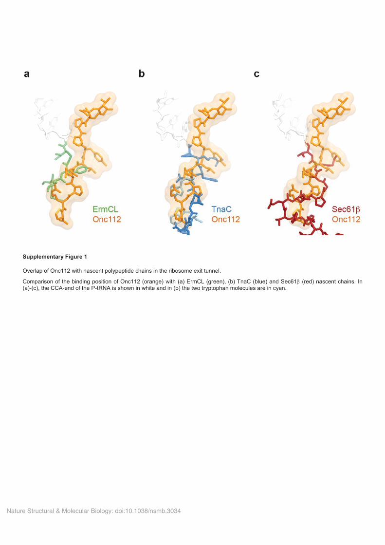
Nature Structural & Molecular Biology: doi:10.1038/nsmb.3034
Supplementary Figure 1
Overlap of Onc112 with nascent polypeptide chains in the ribosome exit tunnel.
Comparison of the binding position of Onc112 (orange) with (a) ErmCL (green), (b) TnaC (blue) and Sec61 (red) nascent chains. In (a)-(c), the CCA-end of the P-tRNA is shown in white and in (b) the two tryptophan molecules are in cyan.
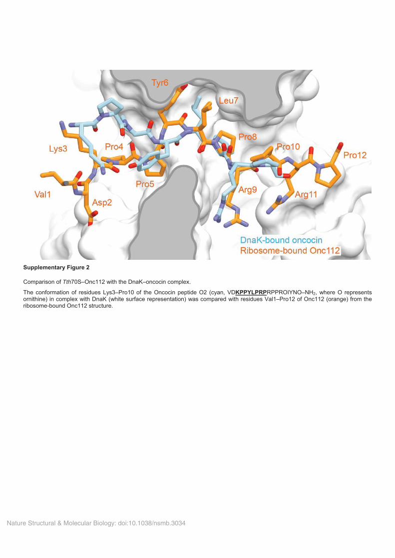
Nature Structural & Molecular Biology: doi:10.1038/nsmb.3034
Supplementary Figure 2
Comparison of Tth70S–Onc112 with the DnaK–oncocin complex.
The conformation of residues Lys3–Pro10 of the Oncocin peptide O2 (cyan, VDKPPYLPRPRPPROIYNO–NH2, where O represents ornithine) in complex with DnaK (white surface representation) was compared with residues Val1–Pro12 of Onc112 (orange) from theribosome-bound Onc112 structure.
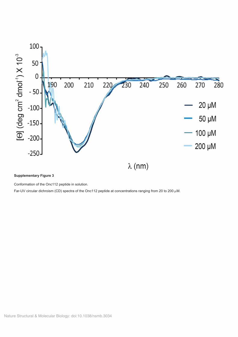
Nature Structural & Molecular Biology: doi:10.1038/nsmb.3034
Supplementary Figure 3
Conformation of the Onc112 peptide in solution.
Far-UV circular dichroism (CD) spectra of the Onc112 peptide at concentrations ranging from 20 to 200 M.
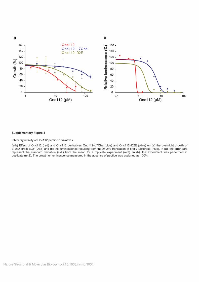
Nature Structural & Molecular Biology: doi:10.1038/nsmb.3034
Supplementary Figure 4
Inhibitory activity of Onc112 peptide derivatives.
(a-b) Effect of Onc112 (red) and Onc112 derivatives Onc112–L7Cha (blue) and Onc112–D2E (olive) on (a) the overnight growth ofE. coli strain BL21(DE3) and (b) the luminescence resulting from the in vitro translation of firefly luciferase (Fluc). In (a), the error bars represent the standard deviation (s.d.) from the mean for a triplicate experiment (n=3). In (b), the experiment was performed induplicate (n=2). The growth or luminescence measured in the absence of peptide was assigned as 100%.
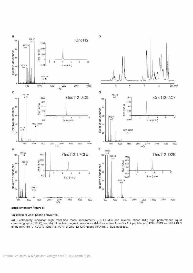
Nature Structural & Molecular Biology: doi:10.1038/nsmb.3034
Supplementary Figure 5
Validation of Onc112 and derivatives.
(a) Electrospray ionization high resolution mass spectrometry (ESI-HRMS) and reverse phase (RP) high performance liquidchromatography (HPLC), and (b) 1H nuclear magnetic resonance (NMR) spectra of the Onc112 peptide. (c-f) ESI-HRMS and RP HPLC of the (c) Onc112– C9, (d) Onc112– C7, (e) Onc112–L7Cha and (f) Onc112–D2E peptides.
Nucleic Acids Research, 2015 1doi: 10.1093/nar/gkv1545
Structure of the mammalian antimicrobial peptideBac7(1–16) bound within the exit tunnel of a bacterialribosomeA. Carolin Seefeldt1,2,3,†, Michael Graf4,†, Natacha Perebaskine1,2,3, Fabian Nguyen4,Stefan Arenz4, Mario Mardirossian5, Marco Scocchi5, Daniel N. Wilson4,6,* and C.Axel Innis1,2,3,*
1Institut Europeen de Chimie et Biologie, University of Bordeaux, Pessac 33607, France, 2U1212, Inserm, Bordeaux33076, France, 3UMR 5320, CNRS, Bordeaux 33076, France, 4Gene Center and Department for Biochemistry,University of Munich, Munich 81377, Germany, 5Department of Life Sciences, University of Trieste, Trieste 34127,Italy and 6Center for integrated Protein Science Munich (CiPSM), University of Munich, Munich 81377, Germany
Received October 13, 2015; Revised December 22, 2015; Accepted December 28, 2015
ABSTRACT
Proline-rich antimicrobial peptides (PrAMPs) pro-duced as part of the innate immune response ofanimals, insects and plants represent a vast, un-tapped resource for the treatment of multidrug-resistant bacterial infections. PrAMPs such as on-cocin or bactenecin-7 (Bac7) interact with the bac-terial ribosome to inhibit translation, but their sup-posed specificity as inhibitors of bacterial rather thanmammalian protein synthesis remains unclear, de-spite being key to developing drugs with low toxic-ity. Here, we present crystal structures of the Ther-mus thermophilus 70S ribosome in complex with thefirst 16 residues of mammalian Bac7, as well as theinsect-derived PrAMPs metalnikowin I and pyrrho-coricin. The structures reveal that the mammalianBac7 interacts with a similar region of the ribosomeas insect-derived PrAMPs. Consistently, Bac7 andthe oncocin derivative Onc112 compete effectivelywith antibiotics, such as erythromycin, which targetthe ribosomal exit tunnel. Moreover, we demonstratethat Bac7 allows initiation complex formation butprevents entry into the elongation phase of trans-lation, and show that it inhibits translation on bothmammalian and bacterial ribosomes, explaining whythis peptide needs to be stored as an inactive pro-peptide. These findings highlight the need to con-sider the specificity of PrAMP derivatives for the bac-terial ribosome in future drug development efforts.
INTRODUCTION
Antimicrobial peptides (AMPs) represent a large and di-verse group of molecules that form part of the innate im-mune response of a variety of invertebrate, plant and ani-mal species (1). While many AMPs kill bacteria by disrupt-ing the bacterial cell membrane, there is growing evidencethat some AMPs have intracellular targets (1). Membersof one such class of non-membranolytic peptides are re-ferred to as proline-rich AMPs (PrAMPs) and are presentin the hemolymph of several species of insects and crus-taceans, as well as in the neutrophils of many mammals(2). PrAMPs exhibit potent antimicrobial activity againsta broad range of bacteria, especially Gram-negative, andare therefore considered as potential lead candidates forthe development of therapeutic antimicrobial agents (3).Well-characterized insect PrAMPs include the apidaecinsproduced by bees (Apis melifera) and wasps (Apis Vesp-idae), pyrrhocoricin from firebugs (Pyrrhocoris apterus),drosocins from fruit flies (Drosophila), metalnikowins fromthe green shield bug (Palomena prasina) and the milkweedbug (Oncopeltus fasciatus) oncocins (2,4,5). PrAMPs aresynthesized as inactive precursors, which undergo prote-olysis to release the active peptide. In contrast to the ac-tive insect peptides, which are generally <21 amino acids inlength, the activemammalianmature forms tend to bemuchlonger; for example, the porcine PR-39 is 39 residues long,whereas the bovine bactenecin-7 (Bac7), which is also foundin sheep and goats, is 60 residues long (2). Nevertheless, C-terminally truncated versions of the mammalian PrAMPsretain antimicrobial activity (6–9) and exhibit high sequencesimilarity with the insect PrAMPs. Indeed, the Bac7(1–16)andBac7(1–35) derivatives corresponding to the first 16 and
*To whom correspondence should be addressed. Tel: +33 540 006 149; Fax: +33 540 002 215; Email: [email protected] may also be addressed to Daniel N. Wilson. Tel: +49 89 2180 76903; Fax: +49 89 2180 76945; Email: [email protected]†These authors contributed equally to the paper as first authors.
C© The Author(s) 2015. Published by Oxford University Press on behalf of Nucleic Acids Research.This is an Open Access article distributed under the terms of the Creative Commons Attribution License (http://creativecommons.org/licenses/by-nc/4.0/), whichpermits non-commercial re-use, distribution, and reproduction in any medium, provided the original work is properly cited. For commercial re-use, please [email protected]
Nucleic Acids Research Advance Access published January 20, 2016
at Universite V
ictor Segalen on January 25, 2016http://nar.oxfordjournals.org/
Dow
nloaded from
2 Nucleic Acids Research, 2015
35 residues of Bac7, respectively, display similar, if not im-proved, antimicrobial activities compared to the full-lengthprocessed Bac7 peptide (6,10,11). For instance, Bac7(1–35)reduces mortality from Salmonella typhimurium in a mousemodel of infection (12) as well as in a rat model for septicshock (13).The insect-derived PrAMPs apidaecin and oncocin, as
well as the mammalian Bac7, penetrate the bacterial cellmembrane mainly via the SbmA transporter present inmany Gram-negative bacteria (10,14). Early studies iden-tified interactions between both insect and mammalianPrAMPs and DnaK, suggesting that this molecular chap-erone was the common intracellular target (2,15). However,subsequent studies questioned the relevance of this inter-action by demonstrating that these PrAMPs also displayan equally potent antimicrobial activity against bacterialstrains lacking the dnaK gene (16–18). Instead, apidaecin,oncocin and Bac7 were shown to bind to the ribosome andinhibit translation (17,19). Subsequent crystal structures ofthe oncocin derivative Onc112 in complex with the bacterial70S ribosome revealed that this peptide binds with a reverseorientation in the ribosomal tunnel and blocks binding ofthe aminoacyl-tRNA to the A-site (20,21). However, thereare no crystal structures to date of a mammalian PrAMP incomplex with the ribosome.Here we present 2.8–2.9 A resolution X-ray struc-
tures of the Thermus thermophilus 70S (Tth70S) ribosomein complex with either the mammalian Bac7 derivativeBac7(1–16) or the insect-derived PrAMPs metalnikowin Ior pyrrhocoricin. The structures reveal that Bac7(1–16),metalnikowin I and pyrrhocoricin bind within the ribo-somal tunnel with a reverse orientation compared to anascent polypeptide chain, as observed previously for on-cocin (20,21). In contrast to the insect PrAMPs oncocin,metalnikowin I and pyrrhocoricin, themammalian Bac7(1–16) utilizes multiple arginine side chains to establish stack-ing interactionswith exposed nucleotide bases of the rRNA,and we show that its unique N-terminal RIRRmotif is crit-ical for inhibiting translation. Like oncocin, metalnikowinI and pyrrhocoricin, the binding site of Bac7 overlaps withthat of the A-tRNA, consistent with our biochemical stud-ies indicating that Bac7(1–16) allows 70S initiation complexformation, but prevents subsequent rounds of translationelongation. Furthermore, we demonstrate that Bac7(1–35)displays activity in amammalian in vitro translation system,providing a possible explanation for why Bac7 is producedas a pre-pro-peptide that is targeted to large granules andphagosomes, thus avoiding direct contact between the ac-tive peptide and the mammalian ribosome.
MATERIALS AND METHODS
Peptide synthesis and purification
The Bac7 N-terminal fragments Bac7(1–16; RRIR-PRPPRLPRPRPR), Bac7(1–35; RRIRPRPPRL-PRPRPRPLPFPRPGPRPIPRPLPFP) and Bac7(5–35;PRPPRLPRPRPRPLPFPRPGPRPIPRPLPFP) weresynthesized on solid phase and purified by reversed-phaseHPLC as described previously (22). Their concentra-tions were determined as reported previously (4). Allpeptides, with a purity of at least 95%, were stored in
milliQ water at −80◦C until use. The Onc112 peptidewas obtained from an earlier study (21). Metalnikowin I(VDKPDYRPRPRPPNM) and pyrrhocoricin (VDKG-SYLPRPTPPRPIYNRN) were synthesized to 97.5 and98.1% purity by NovoPro Bioscience (China).
Purification of T. thermophilus 70S ribosomes
Tth70S ribosomes were purified as described earlier (23)and resuspended in buffer containing 5 mM HEPES-KOH, pH 7.5, 50 mM KCl, 10 mM NH4Cl and 10mM Mg(CH3COO)2 to yield a final concentration of ∼30mg/ml. Tth70S ribosomes were flash frozen in liquid nitro-gen and kept at −80◦C for storage.
Preparation of mRNA, tRNAiMet and YfiA
Synthetic mRNA containing a Shine-Dalgarno sequenceand anAUGstart codon followed by a phenylalanine codon(5′-GGCAAGGAGGUAAAAAUGUUCUAA -3′) waspurchased from Eurogentec. Escherichia coli tRNAi
Met wasoverexpressed in E. coli HB101 cells and purified as de-scribed previously (24). YfiA was overexpressed in BL21Star cells and purified as described previously (25).
Complex formation
A quaternary complex containing Tth70S ribosomes,mRNA, deacylated tRNAi
Met and Bac7(1–16) peptide wasprepared by mixing of 5 �M Tth70S ribosomes with 10 �MmRNA and 50 �MBac7(1–16), and incubating at 55◦C for10 min. After addition of 20 �M tRNAi
Met, the mixturewas incubated at 37◦C for 10 min. The sample was then in-cubated at room temperature for at least 15 min and cen-trifuged briefly prior to use. Ternary complexes containing50 �M metalnikowin I or pyrrhocoricin, 5 �M Tth70S ri-bosomes and 50�MYfiAwere formed by incubation for 30min at room temperature. The final buffer conditions were5 mM HEPES-KOH, pH 7.6, 50 mM KCl, 10 mM NH4Cland 10 mMMg(CH3COO)2.
Crystallization
Published conditions were used as a starting point forscreening crystallization conditions by vapour diffusion insitting-drop trays at 20◦C (23,26). Crystallization dropsconsisted of 3 �l of quaternary or ternary complexes and3–4 �l of reservoir solution containing 100 mM Tris–HCl,pH 7.6, 2.9% (v/v) PEG 20,000, 7–10% (v/v) 2-methyl-2,4-petanediol (MPD) and 175 mM arginine. Crystals ap-peared within 2–3 days and grew to ∼1000 × 100 × 100�m within 7–8 days. For cryoprotection, the concentrationof MPD was increased in a stepwise manner to yield a fi-nal concentration of 40% (v/v). The ionic composition dur-ing cryoprotection was 100 mM Tris–HCl, pH 7.6, 2.9%(v/v) PEG 20,000, 50 mMKCl, 10 mMNH4Cl and 10 mMMg(CH3COO)2. Crystals were flash frozen in a nitrogencryostream at 80 K for subsequent data collection.
at Universite V
ictor Segalen on January 25, 2016http://nar.oxfordjournals.org/
Dow
nloaded from
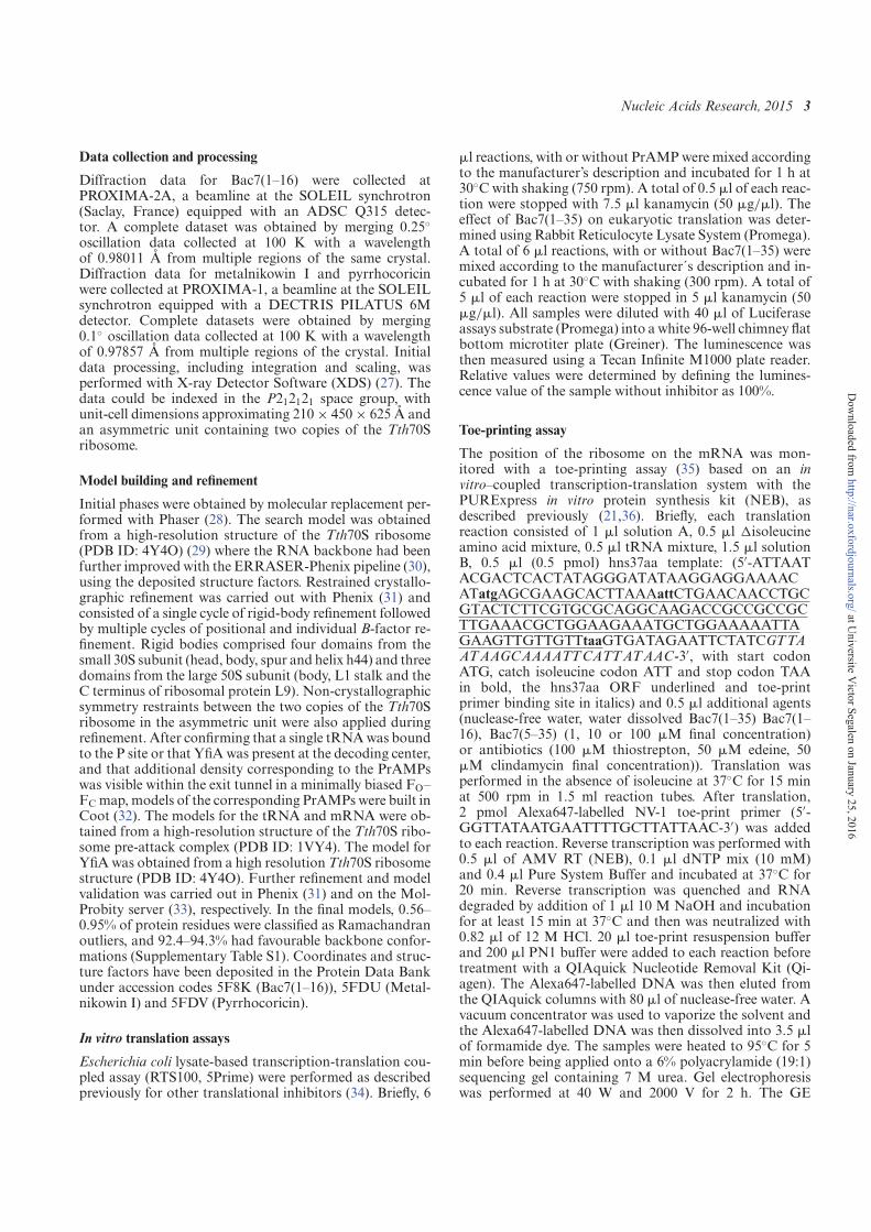
Nucleic Acids Research, 2015 3
Data collection and processing
Diffraction data for Bac7(1–16) were collected atPROXIMA-2A, a beamline at the SOLEIL synchrotron(Saclay, France) equipped with an ADSC Q315 detec-tor. A complete dataset was obtained by merging 0.25◦oscillation data collected at 100 K with a wavelengthof 0.98011 A from multiple regions of the same crystal.Diffraction data for metalnikowin I and pyrrhocoricinwere collected at PROXIMA-1, a beamline at the SOLEILsynchrotron equipped with a DECTRIS PILATUS 6Mdetector. Complete datasets were obtained by merging0.1◦ oscillation data collected at 100 K with a wavelengthof 0.97857 A from multiple regions of the crystal. Initialdata processing, including integration and scaling, wasperformed with X-ray Detector Software (XDS) (27). Thedata could be indexed in the P212121 space group, withunit-cell dimensions approximating 210× 450× 625 A andan asymmetric unit containing two copies of the Tth70Sribosome.
Model building and refinement
Initial phases were obtained by molecular replacement per-formed with Phaser (28). The search model was obtainedfrom a high-resolution structure of the Tth70S ribosome(PDB ID: 4Y4O) (29) where the RNA backbone had beenfurther improved with the ERRASER-Phenix pipeline (30),using the deposited structure factors. Restrained crystallo-graphic refinement was carried out with Phenix (31) andconsisted of a single cycle of rigid-body refinement followedby multiple cycles of positional and individual B-factor re-finement. Rigid bodies comprised four domains from thesmall 30S subunit (head, body, spur and helix h44) and threedomains from the large 50S subunit (body, L1 stalk and theC terminus of ribosomal protein L9). Non-crystallographicsymmetry restraints between the two copies of the Tth70Sribosome in the asymmetric unit were also applied duringrefinement. After confirming that a single tRNAwas boundto the P site or that YfiAwas present at the decoding center,and that additional density corresponding to the PrAMPswas visible within the exit tunnel in a minimally biased FO–FC map, models of the corresponding PrAMPswere built inCoot (32). The models for the tRNA and mRNA were ob-tained from a high-resolution structure of the Tth70S ribo-some pre-attack complex (PDB ID: 1VY4). The model forYfiAwas obtained from a high resolution Tth70S ribosomestructure (PDB ID: 4Y4O). Further refinement and modelvalidation was carried out in Phenix (31) and on the Mol-Probity server (33), respectively. In the final models, 0.56–0.95% of protein residues were classified as Ramachandranoutliers, and 92.4–94.3% had favourable backbone confor-mations (Supplementary Table S1). Coordinates and struc-ture factors have been deposited in the Protein Data Bankunder accession codes 5F8K (Bac7(1–16)), 5FDU (Metal-nikowin I) and 5FDV (Pyrrhocoricin).
In vitro translation assays
Escherichia coli lysate-based transcription-translation cou-pled assay (RTS100, 5Prime) were performed as describedpreviously for other translational inhibitors (34). Briefly, 6
�l reactions, with or without PrAMPwere mixed accordingto the manufacturer’s description and incubated for 1 h at30◦Cwith shaking (750 rpm). A total of 0.5 �l of each reac-tion were stopped with 7.5 �l kanamycin (50 �g/�l). Theeffect of Bac7(1–35) on eukaryotic translation was deter-mined using Rabbit Reticulocyte Lysate System (Promega).A total of 6 �l reactions, with or without Bac7(1–35) weremixed according to the manufacturer´s description and in-cubated for 1 h at 30◦C with shaking (300 rpm). A total of5 �l of each reaction were stopped in 5 �l kanamycin (50�g/�l). All samples were diluted with 40 �l of Luciferaseassays substrate (Promega) into a white 96-well chimney flatbottom microtiter plate (Greiner). The luminescence wasthen measured using a Tecan Infinite M1000 plate reader.Relative values were determined by defining the lumines-cence value of the sample without inhibitor as 100%.
Toe-printing assay
The position of the ribosome on the mRNA was mon-itored with a toe-printing assay (35) based on an invitro–coupled transcription-translation system with thePURExpress in vitro protein synthesis kit (NEB), asdescribed previously (21,36). Briefly, each translationreaction consisted of 1 �l solution A, 0.5 �l �isoleucineamino acid mixture, 0.5 �l tRNA mixture, 1.5 �l solutionB, 0.5 �l (0.5 pmol) hns37aa template: (5′-ATTAATACGACTCACTATAGGGATATAAGGAGGAAAACATatgAGCGAAGCACTTAAAattCTGAACAACCTGCGTACTCTTCGTGCGCAGGCAAGACCGCCGCCGCTTGAAACGCTGGAAGAAATGCTGGAAAAATTAGAAGTTGTTGTTtaaGTGATAGAATTCTATCGTTAATAAGCAAAATTCATTATAAC-3′, with start codonATG, catch isoleucine codon ATT and stop codon TAAin bold, the hns37aa ORF underlined and toe-printprimer binding site in italics) and 0.5 �l additional agents(nuclease-free water, water dissolved Bac7(1–35) Bac7(1–16), Bac7(5–35) (1, 10 or 100 �M final concentration)or antibiotics (100 �M thiostrepton, 50 �M edeine, 50�M clindamycin final concentration)). Translation wasperformed in the absence of isoleucine at 37◦C for 15 minat 500 rpm in 1.5 ml reaction tubes. After translation,2 pmol Alexa647-labelled NV-1 toe-print primer (5′-GGTTATAATGAATTTTGCTTATTAAC-3′) was addedto each reaction. Reverse transcription was performed with0.5 �l of AMV RT (NEB), 0.1 �l dNTP mix (10 mM)and 0.4 �l Pure System Buffer and incubated at 37◦C for20 min. Reverse transcription was quenched and RNAdegraded by addition of 1 �l 10 M NaOH and incubationfor at least 15 min at 37◦C and then was neutralized with0.82 �l of 12 M HCl. 20 �l toe-print resuspension bufferand 200 �l PN1 buffer were added to each reaction beforetreatment with a QIAquick Nucleotide Removal Kit (Qi-agen). The Alexa647-labelled DNA was then eluted fromthe QIAquick columns with 80 �l of nuclease-free water. Avacuum concentrator was used to vaporize the solvent andthe Alexa647-labelled DNA was then dissolved into 3.5 �lof formamide dye. The samples were heated to 95◦C for 5min before being applied onto a 6% polyacrylamide (19:1)sequencing gel containing 7 M urea. Gel electrophoresiswas performed at 40 W and 2000 V for 2 h. The GE
at Universite V
ictor Segalen on January 25, 2016http://nar.oxfordjournals.org/
Dow
nloaded from
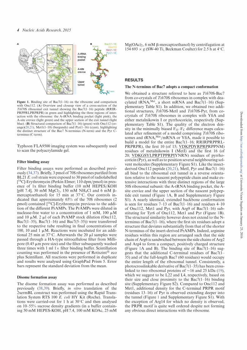
4 Nucleic Acids Research, 2015
Figure 1. Binding site of Bac7(1–16) on the ribosome and comparisonwith Onc112. (A) Overview and closeup view of a cross-section of theTth70S ribosomal exit tunnel showing the Bac7(1–16) peptide (RRIR-PRPPRLPRPRPR) in green and highlighting the three regions of inter-action with the ribosome: the A-tRNA binding pocket (light pink), theA-site crevice (light green) and the upper section of the exit tunnel (lightblue). (B) Structural comparison of Bac7(1–16) (green) with Onc112 (or-ange)(20,21), Met1(1–10) (burgundy) and Pyr(1–16) (cyan), highlightingthe distinct structure of the Bac7 N-terminus (N-term) and the Pyr C-terminus (C-term).
Typhoon FLA9500 imaging system was subsequently usedto scan the polyacrylamide gel.
Filter binding assay
Filter binding assays were performed as described previ-ously (34,37). Briefly, 3 pmol of 70S ribosomes purified fromBL21E. coli strain were exposed to 30 pmol of radiolabelled[14C]-Erythromycin (Perkin Elmer; 110 dpm/pmol) in pres-ence of 1x filter binding buffer (10 mM HEPES/KOH[pH 7.4], 30 mM MgCl2, 150 mM NH4Cl and 6 mM �-mercaptoethanol) for 15 min at 37◦C. Our controls in-dicated that approximately 65% of the 70S ribosomes (2pmol) contained [14C]-Erythromycin previous to the addi-tion of the different PrAMPs. The PrAMPs were diluted innuclease-free water to a concentration of 1 mM, 100 �Mand 10 �M. 2 �l of each PrAMP stock dilution (Onc112,Bac7(1–35), Bac7(1–16) and Bac7(5–35)) were transferredto the respective tube resulting in final concentrations of100, 10 and 1 �M. Reactions were incubated for an addi-tional 25 min at 37◦C. Afterwards the 20 �l samples werepassed through a HA-type nitrocellulose filter from Milli-pore (0.45 �m pore size) and the filter subsequently washedthree times with 1 ml 1× filter binding buffer. Scintillationcounting was performed in the presence of Rotiszint R© ecoplus Scintillant. All reactions were performed in duplicateand results were analysed using GraphPad Prism 5. Errorbars represent the standard deviation from the mean.
Disome formation assay
The disome formation assay was performed as describedpreviously (38,39). Briefly, in vitro translation of the2xermBL construct was performed using the Rapid Trans-lation System RTS 100 E. coli HY Kit (Roche). Transla-tions were carried-out for 1 h at 30◦C and then analysedon 10–55% sucrose density gradients (in a buffer contain-ing 50 mM HEPES-KOH, pH7.4, 100 mM KOAc, 25 mM
Mg(OAc)2, 6 mM �-mercaptoethanol) by centrifugation at154 693 × g (SW-40 Ti, Beckman Coulter) for 2.5 h at 4◦C.
RESULTS
The N-terminus of Bac7 adopts a compact conformation
We obtained a structure referred to here as Tth70S-Bac7from co-crystals of Tth70S ribosomes in complex with dea-cylated tRNAi
Met, a short mRNA and Bac7(1–16) (Sup-plementary Table S1). In addition, we obtained two addi-tional structures, Tth70S-MetI and Tth70S-Pyr, from co-crystals of Tth70S ribosomes in complex with YfiA andeither metalnikowin I or pyrrhocoricin, respectively (Sup-plementary Table S1). The quality of the electron den-sity in the minimally biased FO–FC difference maps calcu-lated after refinement of a model comprising Tth70S ribo-somes and tRNAi
Met/mRNA or YfiA, made it possible tobuild a model for the entire Bac7(1–16; RRIRPRPPRL-PRPRPR), the first 10 (of 15; VDKPDYRPRPRPPNM)residues of metalnikowin I (MetI) and the first 16 (of20; VDKGSYLPRPTPPRPIYNRN) residues of pyrrho-coricin (Pyr), as well as to position several neighbouring sol-vent molecules (Supplementary Figure S1). Like the insect-derived Onc112 peptide (20,21), MetI, Pyr and Bac7(1–16)all bind to the ribosomal exit tunnel in a reverse orienta-tion relative to the nascent polypeptide chain and make ex-tensive interactions with three distinct regions of the large50S ribosomal subunit: the A-tRNA binding pocket, the A-site crevice and the upper section of the nascent polypep-tide exit tunnel (Figure 1A, B and Supplementary FigureS1). A nearly identical, extended backbone conformationis seen for residues 7–13 of Bac7(1–16) and residues 4–10of Onc112, Met1 and Pyr, with Arg9 of Bac7(1–16) sub-stituting for Tyr6 of Onc112, Met1 and Pyr (Figure 1B).The structural similarity however does not extend to the N-terminus of Bac7(1–16), where the first six residues adopt astructure that deviates substantially from that of the shorterN-terminus of the insert-derived PrAMPs. Indeed, arginineresidues within this region are arranged such that the sidechain ofArg6 is sandwiched between the side chains ofArg2and Arg4 to form a compact, positively charged structure(Figure 1A and B). The binding site of Bac7(1–16) sug-gests that the additional C-terminal residues of Bac7(1–35) and of the full-length Bac7 (60 residues) would occupythe entire length of the ribosomal tunnel. Consistently, aphotocrosslinkable derivative of Bac7(1–35) has been cross-linked to two ribosomal proteins of ∼16 and 25 kDa (19),which we suggest to be L22 and L4, respectively, based ontheir size and close proximity to the Bac7(1–16) bindingsite (Supplementary Figure S2). Compared to Onc112 andMet1, additional density for the C-terminal PRPR motif(residues 13–16) of Pyr is observed extending deeper intothe tunnel (Figure 1 and Supplementary Figure S1). Withthe exception of Arg14 for which no density is observed,the PRPR motif is quite well ordered despite not formingany obvious direct interactions with the ribosome.
at Universite V
ictor Segalen on January 25, 2016http://nar.oxfordjournals.org/
Dow
nloaded from
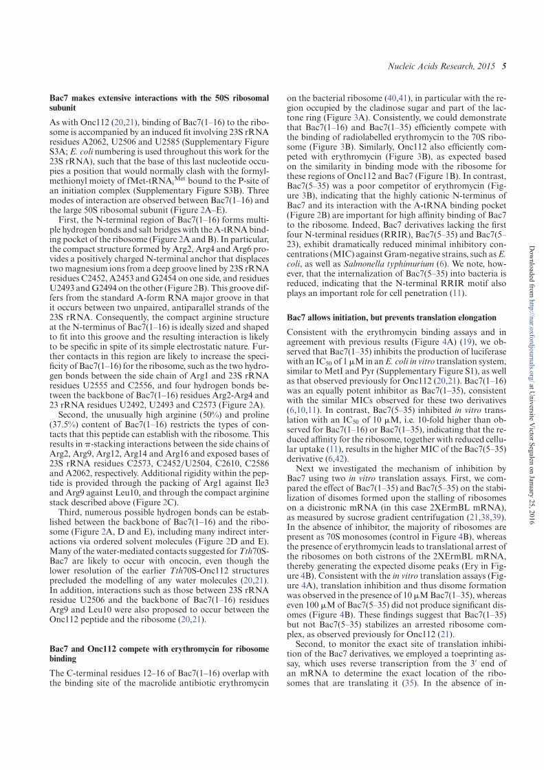
Nucleic Acids Research, 2015 5
Bac7 makes extensive interactions with the 50S ribosomalsubunit
As with Onc112 (20,21), binding of Bac7(1–16) to the ribo-some is accompanied by an induced fit involving 23S rRNAresidues A2062, U2506 and U2585 (Supplementary FigureS3A; E. coli numbering is used throughout this work for the23S rRNA), such that the base of this last nucleotide occu-pies a position that would normally clash with the formyl-methionyl moiety of fMet-tRNAi
Met bound to the P-site ofan initiation complex (Supplementary Figure S3B). Threemodes of interaction are observed between Bac7(1–16) andthe large 50S ribosomal subunit (Figure 2A–E).First, the N-terminal region of Bac7(1–16) forms multi-
ple hydrogen bonds and salt bridges with theA-tRNAbind-ing pocket of the ribosome (Figure 2A and B). In particular,the compact structure formed by Arg2, Arg4 and Arg6 pro-vides a positively charged N-terminal anchor that displacestwomagnesium ions from a deep groove lined by 23S rRNAresidues C2452, A2453 andG2454 on one side, and residuesU2493 andG2494 on the other (Figure 2B). This groove dif-fers from the standard A-form RNA major groove in thatit occurs between two unpaired, antiparallel strands of the23S rRNA. Consequently, the compact arginine structureat the N-terminus of Bac7(1–16) is ideally sized and shapedto fit into this groove and the resulting interaction is likelyto be specific in spite of its simple electrostatic nature. Fur-ther contacts in this region are likely to increase the speci-ficity of Bac7(1–16) for the ribosome, such as the two hydro-gen bonds between the side chain of Arg1 and 23S rRNAresidues U2555 and C2556, and four hydrogen bonds be-tween the backbone of Bac7(1–16) residues Arg2-Arg4 and23 rRNA residues U2492, U2493 and C2573 (Figure 2A).Second, the unusually high arginine (50%) and proline
(37.5%) content of Bac7(1–16) restricts the types of con-tacts that this peptide can establish with the ribosome. Thisresults in �-stacking interactions between the side chains ofArg2, Arg9, Arg12, Arg14 and Arg16 and exposed bases of23S rRNA residues C2573, C2452/U2504, C2610, C2586and A2062, respectively. Additional rigidity within the pep-tide is provided through the packing of Arg1 against Ile3and Arg9 against Leu10, and through the compact argininestack described above (Figure 2C).Third, numerous possible hydrogen bonds can be estab-
lished between the backbone of Bac7(1–16) and the ribo-some (Figure 2A, D and E), including many indirect inter-actions via ordered solvent molecules (Figure 2D and E).Many of the water-mediated contacts suggested forTth70S-Bac7 are likely to occur with oncocin, even though thelower resolution of the earlier Tth70S-Onc112 structuresprecluded the modelling of any water molecules (20,21).In addition, interactions such as those between 23S rRNAresidue U2506 and the backbone of Bac7(1–16) residuesArg9 and Leu10 were also proposed to occur between theOnc112 peptide and the ribosome (20,21).
Bac7 and Onc112 compete with erythromycin for ribosomebinding
The C-terminal residues 12–16 of Bac7(1–16) overlap withthe binding site of the macrolide antibiotic erythromycin
on the bacterial ribosome (40,41), in particular with the re-gion occupied by the cladinose sugar and part of the lac-tone ring (Figure 3A). Consistently, we could demonstratethat Bac7(1–16) and Bac7(1–35) efficiently compete withthe binding of radiolabelled erythromycin to the 70S ribo-some (Figure 3B). Similarly, Onc112 also efficiently com-peted with erythromycin (Figure 3B), as expected basedon the similarity in binding mode with the ribosome forthese regions of Onc112 and Bac7 (Figure 1B). In contrast,Bac7(5–35) was a poor competitor of erythromycin (Fig-ure 3B), indicating that the highly cationic N-terminus ofBac7 and its interaction with the A-tRNA binding pocket(Figure 2B) are important for high affinity binding of Bac7to the ribosome. Indeed, Bac7 derivatives lacking the firstfour N-terminal residues (RRIR), Bac7(5–35) and Bac7(5–23), exhibit dramatically reduced minimal inhibitory con-centrations (MIC) againstGram-negative strains, such asE.coli, as well as Salmonella typhimurium (6). We note, how-ever, that the internalization of Bac7(5–35) into bacteria isreduced, indicating that the N-terminal RRIR motif alsoplays an important role for cell penetration (11).
Bac7 allows initiation, but prevents translation elongation
Consistent with the erythromycin binding assays and inagreement with previous results (Figure 4A) (19), we ob-served that Bac7(1–35) inhibits the production of luciferasewith an IC50 of 1�Min anE. coli in vitro translation system,similar to MetI and Pyr (Supplementary Figure S1), as wellas that observed previously for Onc112 (20,21). Bac7(1–16)was an equally potent inhibitor as Bac7(1–35), consistentwith the similar MICs observed for these two derivatives(6,10,11). In contrast, Bac7(5–35) inhibited in vitro trans-lation with an IC50 of 10 �M, i.e. 10-fold higher than ob-served for Bac7(1–16) or Bac7(1–35), indicating that the re-duced affinity for the ribosome, together with reduced cellu-lar uptake (11), results in the higher MIC of the Bac7(5–35)derivative (6,42).Next we investigated the mechanism of inhibition by
Bac7 using two in vitro translation assays. First, we com-pared the effect of Bac7(1–35) and Bac7(5–35) on the stabi-lization of disomes formed upon the stalling of ribosomeson a dicistronic mRNA (in this case 2XErmBL mRNA),as measured by sucrose gradient centrifugation (21,38,39).In the absence of inhibitor, the majority of ribosomes arepresent as 70S monosomes (control in Figure 4B), whereasthe presence of erythromycin leads to translational arrest ofthe ribosomes on both cistrons of the 2XErmBL mRNA,thereby generating the expected disome peaks (Ery in Fig-ure 4B). Consistent with the in vitro translation assays (Fig-ure 4A), translation inhibition and thus disome formationwas observed in the presence of 10�MBac7(1–35), whereaseven 100 �Mof Bac7(5–35) did not produce significant dis-omes (Figure 4B). These findings suggest that Bac7(1–35)but not Bac7(5–35) stabilizes an arrested ribosome com-plex, as observed previously for Onc112 (21).Second, to monitor the exact site of translation inhibi-
tion of the Bac7 derivatives, we employed a toeprinting as-say, which uses reverse transcription from the 3′ end ofan mRNA to determine the exact location of the ribo-somes that are translating it (35). In the absence of in-
at Universite V
ictor Segalen on January 25, 2016http://nar.oxfordjournals.org/
Dow
nloaded from
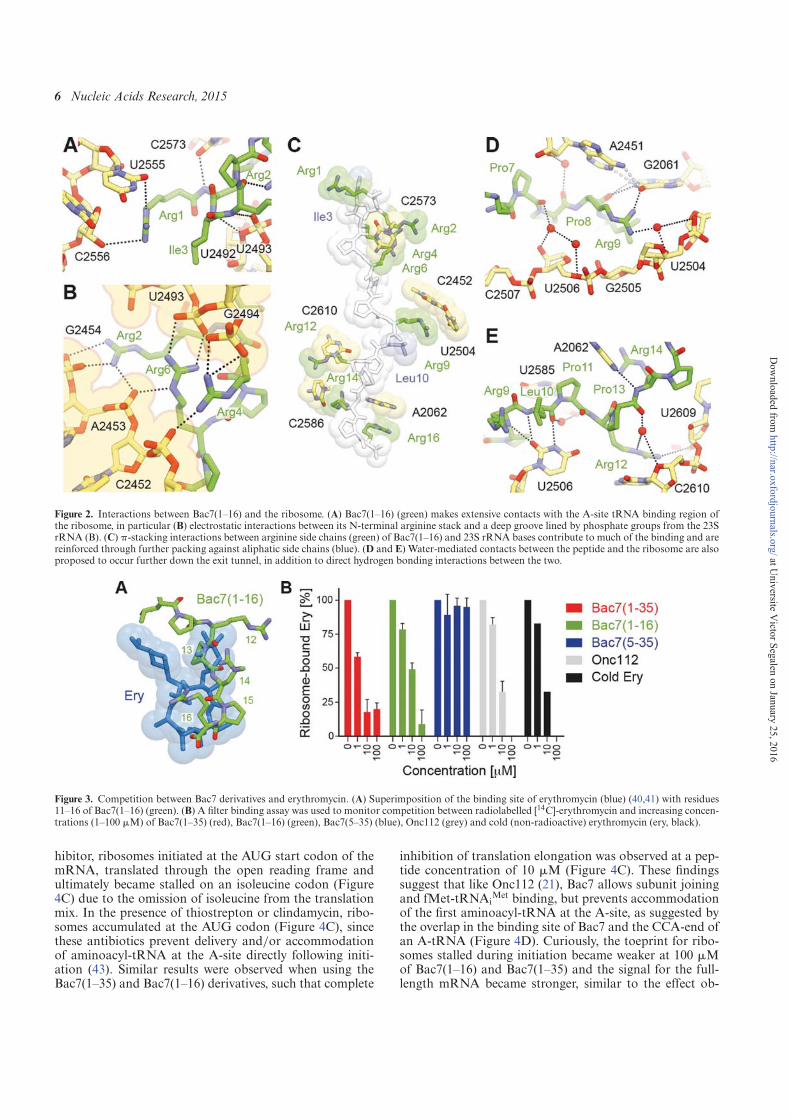
6 Nucleic Acids Research, 2015
Figure 2. Interactions between Bac7(1–16) and the ribosome. (A) Bac7(1–16) (green) makes extensive contacts with the A-site tRNA binding region ofthe ribosome, in particular (B) electrostatic interactions between its N-terminal arginine stack and a deep groove lined by phosphate groups from the 23SrRNA (B). (C) �-stacking interactions between arginine side chains (green) of Bac7(1–16) and 23S rRNA bases contribute to much of the binding and arereinforced through further packing against aliphatic side chains (blue). (D and E) Water-mediated contacts between the peptide and the ribosome are alsoproposed to occur further down the exit tunnel, in addition to direct hydrogen bonding interactions between the two.
Figure 3. Competition between Bac7 derivatives and erythromycin. (A) Superimposition of the binding site of erythromycin (blue) (40,41) with residues11–16 of Bac7(1–16) (green). (B) A filter binding assay was used to monitor competition between radiolabelled [14C]-erythromycin and increasing concen-trations (1–100 �M) of Bac7(1–35) (red), Bac7(1–16) (green), Bac7(5–35) (blue), Onc112 (grey) and cold (non-radioactive) erythromycin (ery, black).
hibitor, ribosomes initiated at the AUG start codon of themRNA, translated through the open reading frame andultimately became stalled on an isoleucine codon (Figure4C) due to the omission of isoleucine from the translationmix. In the presence of thiostrepton or clindamycin, ribo-somes accumulated at the AUG codon (Figure 4C), sincethese antibiotics prevent delivery and/or accommodationof aminoacyl-tRNA at the A-site directly following initi-ation (43). Similar results were observed when using theBac7(1–35) and Bac7(1–16) derivatives, such that complete
inhibition of translation elongation was observed at a pep-tide concentration of 10 �M (Figure 4C). These findingssuggest that like Onc112 (21), Bac7 allows subunit joiningand fMet-tRNAi
Met binding, but prevents accommodationof the first aminoacyl-tRNA at the A-site, as suggested bythe overlap in the binding site of Bac7 and the CCA-end ofan A-tRNA (Figure 4D). Curiously, the toeprint for ribo-somes stalled during initiation became weaker at 100 �Mof Bac7(1–16) and Bac7(1–35) and the signal for the full-length mRNA became stronger, similar to the effect ob-
at Universite V
ictor Segalen on January 25, 2016http://nar.oxfordjournals.org/
Dow
nloaded from
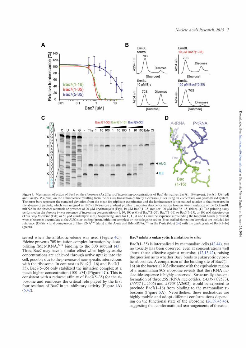
Nucleic Acids Research, 2015 7
Figure 4. Mechanism of action of Bac7 on the ribosome. (A) Effects of increasing concentrations of Bac7 derivatives Bac7(1–16) (green), Bac7(1–35) (red)and Bac7(5–35) (blue) on the luminescence resulting from the in vitro translation of firefly luciferase (Fluc) using an Escherichia coli lysate-based system.The error bars represent the standard deviation from the mean for triplicate experiments and the luminescence is normalized relative to that measured inthe absence of peptide, which was assigned as 100%. (B) Sucrose gradient profiles to monitor disome formation from in vitro translation of the 2XErmBLmRNA in the absence (control) or presence of 20 �M erythromycin (Ery), 10 �M Bac7(1–35) (red) or 100 �M Bac7(5–35) (blue). (C) Toe-printing assayperformed in the absence (−) or presence of increasing concentrations (1, 10, 100 �M) of Bac7(1–35), Bac7(1–16) or Bac7(5–35), or 100 �M thiostrepton(Ths), 50 �M edeine (Ede) or 50 �M clindamycin (Cli). Sequencing lanes for C, U, A and G and the sequence surrounding the toe-print bands (arrowed)when ribosomes accumulate at the AUG start codon (green, initiation complex) or the isoleucine codon (blue, stalled elongation complex) are included forreference. (D) Structural comparison of Phe-tRNAPhe (slate) in the A-site and fMet-tRNAi
Met in the P-site (blue) (26) with the binding site of Bac7(1–16)(green).
served when the antibiotic edeine was used (Figure 4C).Edeine prevents 70S initiation complex formation by desta-bilizing fMet-tRNAi
Met binding to the 30S subunit (43).Thus, Bac7 may have a similar effect when high cytosolicconcentrations are achieved through active uptake into thecell, possibly due to the presence of non-specific interactionswith the ribosome. In contrast to Bac7(1–16) and Bac7(1–35), Bac7(5–35) only stabilized the initiation complex at amuch higher concentration (100 �M) (Figure 4C). This isconsistent with a reduced affinity of Bac7(5–35) for the ri-bosome and reinforces the critical role played by the firstfour residues of Bac7 in its inhibitory activity (Figure 1A)(6,42).
Bac7 inhibits eukaryotic translation in vitro
Bac7(1–35) is internalized by mammalian cells (42,44), yetno toxicity has been observed, even at concentrations wellabove those effective against microbes (12,13,42), raisingthe question as to whether Bac7 binds to eukaryotic cytoso-lic ribosomes. A comparison of the binding site of Bac7(1–16) on the bacterial 70S ribosomewith the equivalent regionof a mammalian 80S ribosome reveals that the rRNA nu-cleotide sequence is highly conserved. Structurally, the con-formation of three 25S rRNA nucleotides, C4519 (C2573),U4452 (U2506) and A3908 (A2602), would be expected topreclude Bac7(1–16) from binding to the mammalian ri-bosome (Figure 5A). Nevertheless, these nucleotides arehighly mobile and adopt different conformations depend-ing on the functional state of the ribosome (26,39,45,46),suggesting that conformational rearrangements of these nu-
at Universite V
ictor Segalen on January 25, 2016http://nar.oxfordjournals.org/
Dow
nloaded from
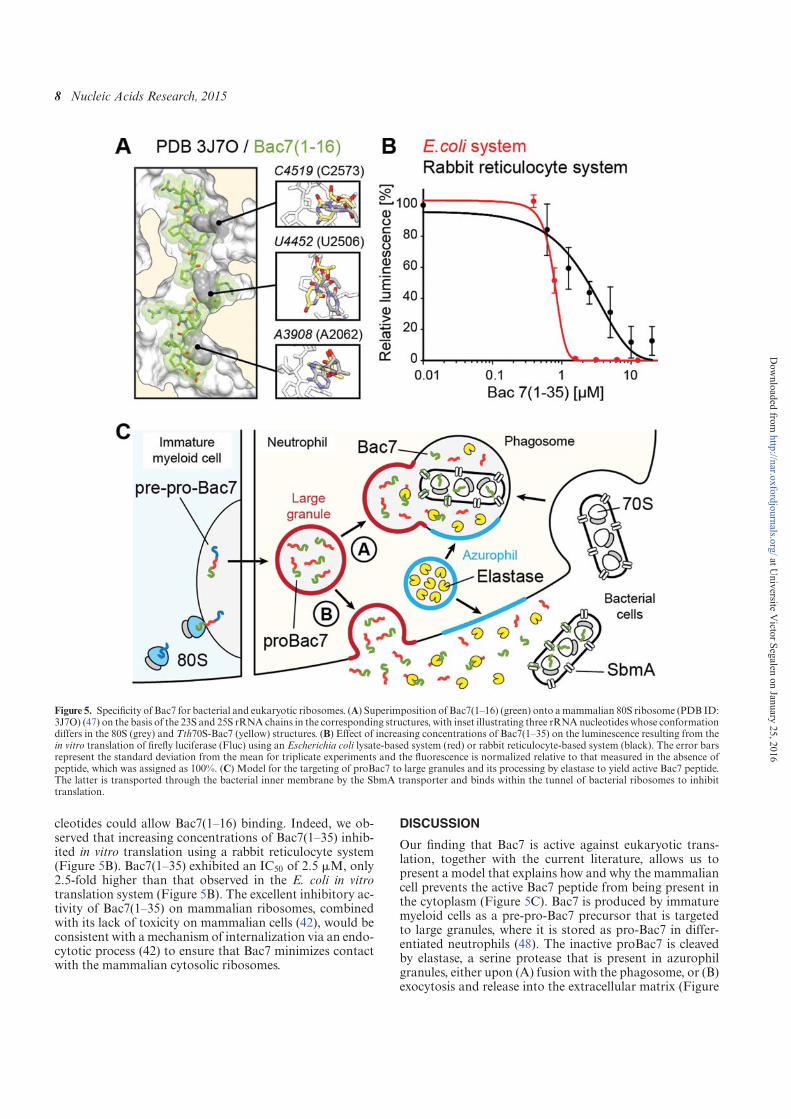
8 Nucleic Acids Research, 2015
Figure 5. Specificity of Bac7 for bacterial and eukaryotic ribosomes. (A) Superimposition of Bac7(1–16) (green) onto amammalian 80S ribosome (PDB ID:3J7O) (47) on the basis of the 23S and 25S rRNA chains in the corresponding structures, with inset illustrating three rRNAnucleotides whose conformationdiffers in the 80S (grey) and Tth70S-Bac7 (yellow) structures. (B) Effect of increasing concentrations of Bac7(1–35) on the luminescence resulting from thein vitro translation of firefly luciferase (Fluc) using an Escherichia coli lysate-based system (red) or rabbit reticulocyte-based system (black). The error barsrepresent the standard deviation from the mean for triplicate experiments and the fluorescence is normalized relative to that measured in the absence ofpeptide, which was assigned as 100%. (C) Model for the targeting of proBac7 to large granules and its processing by elastase to yield active Bac7 peptide.The latter is transported through the bacterial inner membrane by the SbmA transporter and binds within the tunnel of bacterial ribosomes to inhibittranslation.
cleotides could allow Bac7(1–16) binding. Indeed, we ob-served that increasing concentrations of Bac7(1–35) inhib-ited in vitro translation using a rabbit reticulocyte system(Figure 5B). Bac7(1–35) exhibited an IC50 of 2.5 �M, only2.5-fold higher than that observed in the E. coli in vitrotranslation system (Figure 5B). The excellent inhibitory ac-tivity of Bac7(1–35) on mammalian ribosomes, combinedwith its lack of toxicity on mammalian cells (42), would beconsistent with a mechanism of internalization via an endo-cytotic process (42) to ensure that Bac7 minimizes contactwith the mammalian cytosolic ribosomes.
DISCUSSION
Our finding that Bac7 is active against eukaryotic trans-lation, together with the current literature, allows us topresent a model that explains how and why the mammaliancell prevents the active Bac7 peptide from being present inthe cytoplasm (Figure 5C). Bac7 is produced by immaturemyeloid cells as a pre-pro-Bac7 precursor that is targetedto large granules, where it is stored as pro-Bac7 in differ-entiated neutrophils (48). The inactive proBac7 is cleavedby elastase, a serine protease that is present in azurophilgranules, either upon (A) fusion with the phagosome, or (B)exocytosis and release into the extracellular matrix (Figure
at Universite V
ictor Segalen on January 25, 2016http://nar.oxfordjournals.org/
Dow
nloaded from
Nucleic Acids Research, 2015 9
5C) (48,49). The resulting activated Bac7 peptide can thenenter into the bacterial cell through the SbmA transporter(10), where it subsequently binds to the ribosome to inhibittranslation (Figure 5C) (19). Our structure of the Tth70S–Bac7 complex reveals specifically how Bac7 interacts withthe bacterial ribosome (Figures 1 and 2) and inhibits trans-lation by allowing initiation but preventing translation elon-gation (Figure 3). Although the overall mechanism of ac-tion of Bac7 is similar to that of insect-derived AMPs likeoncocin (20,21), the high arginine content of Bac7 leads toa distinct mode of binding to the ribosome, namely throughelectrostatic and stacking interactions with the backboneand bases of 23S rRNA nucleotides, respectively (Figure2C). It will be interesting to see whether such interactionsare the basis for the translational arrest that has been ob-served when the ribosome translates a nascent polypeptidechain bearing positively charged arginine residues (50,51).
ACCESSION NUMBERS
PDB IDs: 5F8K, 5FDU, 5FDV.
SUPPLEMENTARY DATA
Supplementary Data are available at NAR Online.
ACKNOWLEDGEMENT
We thank the staff at the SOLEIL synchrotron (beamlinesPROXIMA-2A and PROXIMA-1) for help during data col-lection and B. Kauffmann and S. Massip at the Institut Eu-ropeen de Chimie et Biologie for help with crystal freezingand screening.
FUNDING
Agence Nationale pour la Recherche [ANR-14-CE09–0001 to C.A.I., D.N.W.]; Conseil Regional d’Aquitaine[2012-13-01-009 to C.A.I.]; EuropeanUnion [PCIG14-GA-2013-631479 to C.A.I.]; Deutsche Forschungsgemeinschaft[FOR1805, WI3285/4-1, GRK1721 to D.N.W.]; FondoRicerca di Ateneo [FRA2014 to M.S.]; Institut National dela Sante et de la Recherche Medicale Pre-doctoral Fellow-ship (toA.C.S.); Conseil Regional d’Aquitaine Pre-doctoralFellowship (to A.C.S.). Funding for open access charge:Institut National de la Sante et de la Recherche Medicale[PCIG14-GA-2013-631479 to C.A.I.].Conflict of interest statement.None declared.
REFERENCES1. Brogden,K.A. (2005) Antimicrobial peptides: pore formers ormetabolic inhibitors in bacteria? Nat. Rev. Microbiol., 3, 238–250.
2. Scocchi,M., Tossi,A. and Gennaro,R. (2011) Proline-richantimicrobial peptides: converging to a non-lytic mechanism ofaction. Cell. Mol. Life Sci., 68, 2317–2330.
3. Li,W., Tailhades,J., O’Brien-Simpson,N.M., Separovic,F., Otvos,L.Jr, Hossain,M.A. and Wade,J.D. (2014) Proline-rich antimicrobialpeptides: potential therapeutics against antibiotic-resistantbacteria. Amino Acids, 46, 2287–2294.
4. Ebbensgaard,A., Mordhorst,H., Overgaard,M.T., Nielsen,C.G.,Aarestrup,F.M. and Hansen,E.B. (2015) Comparative evaluation ofthe antimicrobial activity of different antimicrobial peptides against arange of pathogenic bacteria. PLoS One, 10, e0144611.
5. Chernysh,S., Cociancich,S., Briand,J.P., Hetru,C. and Bulet,P. (1996)The inducible antibacterial peptides of the Hemipteran insectPalomena prasina: identification of a unique family of proline richpeptides and of a novel insect defensin. J. Insect. Physiol., 42, 81–89.
6. Benincasa,M., Scocchi,M., Podda,E., Skerlavaj,B., Dolzani,L. andGennaro,R. (2004) Antimicrobial activity of Bac7 fragments againstdrug-resistant clinical isolates. Peptides, 25, 2055–2061.
7. Chan,Y.R., Zanetti,M., Gennaro,R. and Gallo,R.L. (2001)Anti-microbial activity and cell binding are controlled by sequencedeterminants in the anti-microbial peptide PR-39. J. Invest.Dermatol., 116, 230–235.
8. Frank,R.W., Gennaro,R., Schneider,K., Przybylski,M. andRomeo,D. (1990) Amino acid sequences of two proline-richbactenecins. Antimicrobial peptides of bovine neutrophils. J. Biol.Chem., 265, 18871–18874.
9. Tokunaga,Y., Niidome,T., Hatakeyama,T. and Aoyagi,H. (2001)Antibacterial activity of bactenecin 5 fragments and their interactionwith phospholipid membranes. J. Pept. Sci., 7, 297–304.
10. Mattiuzzo,M., Bandiera,A., Gennaro,R., Benincasa,M., Pacor,S.,Antcheva,N. and Scocchi,M. (2007) Role of the Escherichia coliSbmA in the antimicrobial activity of proline-rich peptides.Mol.Microbiol., 66, 151–163.
11. Podda,E., Benincasa,M., Pacor,S., Micali,F., Mattiuzzo,M.,Gennaro,R. and Scocchi,M. (2006) Dual mode of action of Bac7, aproline-rich antibacterial peptide. Biochim. Biophys. Acta, 1760,1732–1740.
12. Benincasa,M., Pelillo,C., Zorzet,S., Garrovo,C., Biffi,S., Gennaro,R.and Scocchi,M. (2010) The proline-rich peptide Bac7 (1–35) reducesmortality from Salmonella typhimurium in a mouse model ofinfection. BMCMicrobiol., 10, 178.
13. Ghiselli,R., Giacometti,A., Cirioni,O., Circo,R., Mocchegiani,F.,Skerlavaj,B., D’Amato,G., Scalise,G., Zanetti,M. and Saba,V. (2003)Neutralization of endotoxin in vitro and in vivo by Bac7(1–35), aproline-rich antibacterial peptide. Shock, 19, 577–581.
14. Krizsan,A., Knappe,D. and Hoffmann,R. (2015) Influence of yjiLand upstream genes on the antibacterial activity of proline-richantimicrobial peptides overcoming Escherichia coli resistanceinduced by the missing SbmA transporter system. Antimicrob. AgentsChemother., 59, 5992–5998.
15. Otvos,L., O,I., Rogers,M.E., Consolvo,P.J., Condie,B.A., Lovas,S.,Bulet,P. and Blaszczyk-Thurin,M. (2000) Interaction between heatshock proteins and antimicrobial peptides. Biochemistry, 39,14150–14159.
16. Czihal,P., Knappe,D., Fritsche,S., Zahn,M., Berthold,N.,Piantavigna,S., Muller,U., Van Dorpe,S., Herth,N., Binas,A. et al.(2012) Api88 is a novel antibacterial designer peptide to treatsystemic infections with multidrug-resistant gram-negativepathogens. ACS Chem. Biol., 7, 1281–1291.
17. Krizsan,A., Volke,D., Weinert,S., Strater,N., Knappe,D. andHoffmann,R. (2014) Insect-derived proline-rich antimicrobialpeptides kill bacteria by inhibiting bacterial protein translation at the70 S ribosome. Angew. Chem. Int. Ed., 53, 12236–12239.
18. Scocchi,M., Luthy,C., Decarli,P., Mignogna,G., Christen,P. andGennaro,R. (2009) The proline-rich antibacterial peptide Bac7 bindsto and inhibits in vitro the molecular chaperone DnaK. Int. J. Pept.Res. Therapeut., 15, 147–155.
19. Mardirossian,M., Grzela,R., Giglione,C., Meinnel,T., Gennaro,R.,Mergaert,P. and Scocchi,M. (2014) The host antimicrobial peptideBac71–35 binds to bacterial ribosomal proteins and inhibits proteinsynthesis. Chem. Biol., 21, 1639–1647.
20. Roy,R.N., Lomakin,I.B., Gagnon,M.G. and Steitz,T.A. (2015) Themechanism of inhibition of protein synthesis by the proline-richpeptide oncocin. Nat. Struct. Mol. Biol., 22, 466–469.
21. Seefeldt,A.C., Nguyen,F., Antunes,S., Perebaskine,N., Graf,M.,Arenz,S., Inampudi,K.K., Douat,C., Guichard,G., Wilson,D.N. et al.(2015) The proline-rich antimicrobial peptide Onc112 inhibitstranslation by blocking and destabilizing the initiation complex. Nat.Struct. Mol. Biol., 22, 470–475.
22. Guida,F., Benincasa,M., Zahariev,S., Scocchi,M., Berti,F.,Gennaro,R. and Tossi,A. (2015) Effect of size and N-terminal residuecharacteristics on bacterial cell penetration and antibacterial activityof the proline-rich peptide Bac7. J. Med. Chem., 58, 1195–1204.
23. Selmer,M., Dunham,C.M., Murphy,F.V., Weixlbaumer,A., Petry,S.,Kelley,A.C., Weir,J.R. and Ramakrishnan,V. (2006) Structure of the
at Universite V
ictor Segalen on January 25, 2016http://nar.oxfordjournals.org/
Dow
nloaded from
10 Nucleic Acids Research, 2015
70S ribosome complexed with mRNA and tRNA. Science, 313,1935–1942.
24. Schmitt,E., Blanquet,S. and Mechulam,Y. (1999) Crystallization andpreliminary X-ray analysis of Escherichia coli methionyl-tRNAMetfformyltransferase complexed with formyl-methionyl-tRNAMetf.Acta Crystallogr. D, 55, 332–334.
25. Polikanov,Y.S., Blaha,G.M. and Steitz,T.A. (2012) How hibernationfactors RMF, HPF, and YfiA turn off protein synthesis. Science, 336,915–918.
26. Polikanov,Y.S., Steitz,T.A. and Innis,C.A. (2014) A proton wire tocouple aminoacyl-tRNA accommodation and peptide-bondformation on the ribosome. Nat. Struct. Mol. Biol., 21, 787–793.
27. Kabsch,W. (2010) Xds. Acta Crystallogr. D Biol. Crystallogr., 66,125–132.
28. McCoy,A.J., Grosse-Kunstleve,R.W., Adams,P.D., Winn,M.D.,Storoni,L.C. and Read,R.J. (2007) Phaser crystallographic software.J. Appl. Crystallogr., 40, 658–674.
29. Polikanov,Y.S., Melnikov,S.V., Soll,D. and Steitz,T.A. (2015)Structural insights into the role of rRNA modifications in proteinsynthesis and ribosome assembly. Nat. Struct. Mol. Biol., 22,342–344.
30. Chou,F.C., Echols,N., Terwilliger,T.C. and Das,R. (2016) RNAstructure refinement using the ERRASER-phenix pipeline.MethodsMol. Biol., 1320, 269–282.
31. Adams,P.D., Afonine,P.V., Bunkoczi,G., Chen,V.B., Davis,I.W.,Echols,N., Headd,J.J., Hung,L.W., Kapral,G.J.,Grosse-Kunstleve,R.W. et al. (2010) PHENIX: a comprehensivePython-based system for macromolecular structure solution. ActaCrystallogr. D Biol. Crystallogr., 66, 213–221.
32. Emsley,P. and Cowtan,K. (2004) Coot: model-building tools formolecular graphics. Acta Crystallogr. D Biol. Crystallogr., 60,2126–2132.
33. Chen,V.B., Arendall,W.B. 3rd, Headd,J.J., Keedy,D.A.,Immormino,R.M., Kapral,G.J., Murray,L.W., Richardson,J.S. andRichardson,D.C. (2010) MolProbity: all-atom structure validation formacromolecular crystallography. Acta Crystallogr. D Biol.Crystallogr., 66, 12–21.
34. Starosta,A.L., Karpenko,V.V., Shishkina,A.V., Mikolajka,A.,Sumbatyan,N.V., Schluenzen,F., Korshunova,G.A., Bogdanov,A.A.and Wilson,D.N. (2010) Interplay between the ribosomal tunnel,nascent chain, and macrolides influences drug inhibition. Chem.Biol., 17, 504–514.
35. Hartz,D., McPheeters,D.S., Traut,R. and Gold,L. (1988) Extensioninhibition analysis of translation initiation complexes.MethodsEnzymol., 164, 419–425.
36. Starosta,A.L., Lassak,J., Peil,L., Atkinson,G.C., Virumae,K.,Tenson,T., Remme,J., Jung,K. and Wilson,D.N. (2014) Translationalstalling at polyproline stretches is modulated by the sequence contextupstream of the stall site. Nucleic Acids Res., 42, 10711–10719.
37. Polikanov,Y.S., Starosta,A.L., Juette,M.F., Altman,R.B., Terry,D.S.,Lu,W., Burnett,B.J., Dinos,G., Reynolds,K.A., Blanchard,S.C. et al.
(2015) Distinct tRNA accommodation intermediates observed on theribosome with the antibiotics hygromycin A and A201A.Mol. Cell,58, 832–844.
38. Arenz,S., Meydan,S., Starosta,A.L., Berninghausen,O.,Beckmann,R., Vazquez-Laslop,N. and Wilson,D.N. (2014) Drugsensing by the ribosome induces translational arrest via active siteperturbation.Mol. Cell, 56, 446–452.
39. Arenz,S., Ramu,H., Gupta,P., Berninghausen,O., Beckmann,R.,Vazquez-Laslop,N., Mankin,A.S. and Wilson,D.N. (2014) Molecularbasis for erythromycin-dependent ribosome stalling duringtranslation of the ErmBL leader peptide. Nat. Commun., 5, 3501.
40. Bulkley,D., Innis,C.A., Blaha,G. and Steitz,T.A. (2010) Revisiting thestructures of several antibiotics bound to the bacterial ribosome.Proc. Natl. Acad. Sci., 107, 17158–17163.
41. Dunkle,J.A., Xiong,L., Mankin,A.S. and Cate,J.H. (2010) Structuresof the Escherichia coli ribosome with antibiotics bound near thepeptidyl transferase center explain spectra of drug action. Proc. Natl.Acad. Sci. U.S.A., 107, 17152–17157.
42. Tomasinsig,L., Skerlavaj,B., Papo,N., Giabbai,B., Shai,Y. andZanetti,M. (2006) Mechanistic and functional studies of theinteraction of a proline-rich antimicrobial peptide with mammaliancells. J. Biol. Chem., 281, 383–391.
43. Wilson,D.N. (2009) The A-Z of bacterial translation inhibitors. Crit.Rev. Biochem. Mol. Biol., 44, 393–433.
44. Sadler,K., Eom,K.D., Yang,J.L., Dimitrova,Y. and Tam,J.P. (2002)Translocating proline-rich peptides from the antimicrobial peptidebactenecin 7. Biochemistry, 41, 14150–14157.
45. Schmeing,T.M., Huang,K.S., Strobel,S.A. and Steitz,T.A. (2005) Aninduced-fit mechanism to promote peptide bond formation andexclude hydrolysis of peptidyl-tRNA. Nature, 438, 520–524.
46. Schuwirth,B.S., Borovinskaya,M.A., Hau,C.W., Zhang,W.,Vila-Sanjurjo,A., Holton,J.M. and Cate,J.H. (2005) Structures of thebacterial ribosome at 3.5 A resolution. Science, 310, 827–834.
47. Voorhees,R.M., Fernandez,I.S., Scheres,S.H. and Hegde,R.S. (2014)Structure of the mammalian ribosome-Sec61 complex to 3.4 Aresolution. Cell, 157, 1632–1643.
48. Zanetti,M., Litteri,L., Gennaro,R., Horstmann,H. and Romeo,D.(1990) Bactenecins, defense polypeptides of bovine neutrophils, aregenerated from precursor molecules stored in the large granules. J.Cell Biol., 111, 1363–1371.
49. Scocchi,M., Skerlavaj,B., Romeo,D. and Gennaro,R. (1992)Proteolytic cleavage by neutrophil elastase converts inactive storageproforms to antibacterial bactenecins. Eur. J. Biochem., 209, 589–595.
50. Dimitrova,L.N., Kuroha,K., Tatematsu,T. and Inada,T. (2009)Nascent peptide-dependent translation arrest leads toNot4p-mediated protein degradation by the proteasome. J. Biol.Chem., 284, 10343–10352.
51. Lu,J. and Deutsch,C. (2008) Electrostatics in the ribosomal tunnelmodulate chain elongation rates. J. Mol. Biol., 384, 73–86.
at Universite V
ictor Segalen on January 25, 2016http://nar.oxfordjournals.org/
Dow
nloaded from
SUPPLEMENTARY ONLINE MATERIALS
for
Structure of the mammalian antimicrobial peptide Bac7(1-16) bound
within the exit tunnel of a bacterial ribosome
A. Carolin Seefeldt1,2,3
, Michael Graf4, Natacha Pérébaskine
1,2,3, Fabian Nguyen
4, Stefan Arenz
4,
Mario Mardirossian5, Marco Scocchi
5, Daniel N. Wilson
4,6,* and C. Axel Innis
1,2,3,*
1 Institut Européen de Chimie et Biologie, University of Bordeaux, Pessac, 33607, France
2 U1212, Inserm, Bordeaux, 33076, France
3 UMR[---], CNRS, Bordeaux, 33076, France
4 Gene Center and Department for Biochemistry, University of Munich, Munich, 81377, Germany
5 Department of Life Sciences, University of Trieste, Trieste, 34127, Italy
6 Center for integrated Protein Science Munich (CiPSM), University of Munich, Munich, 81377,
Germany
* To whom correspondence should be addressed. Tel: +33 540006149; Fax: +33 540002215; Email:
[email protected]. Correspondence may also be addressed to: [email protected].
The authors wish it to be known that, in their opinion, the first two authors should be regarded as joint First
Authors.
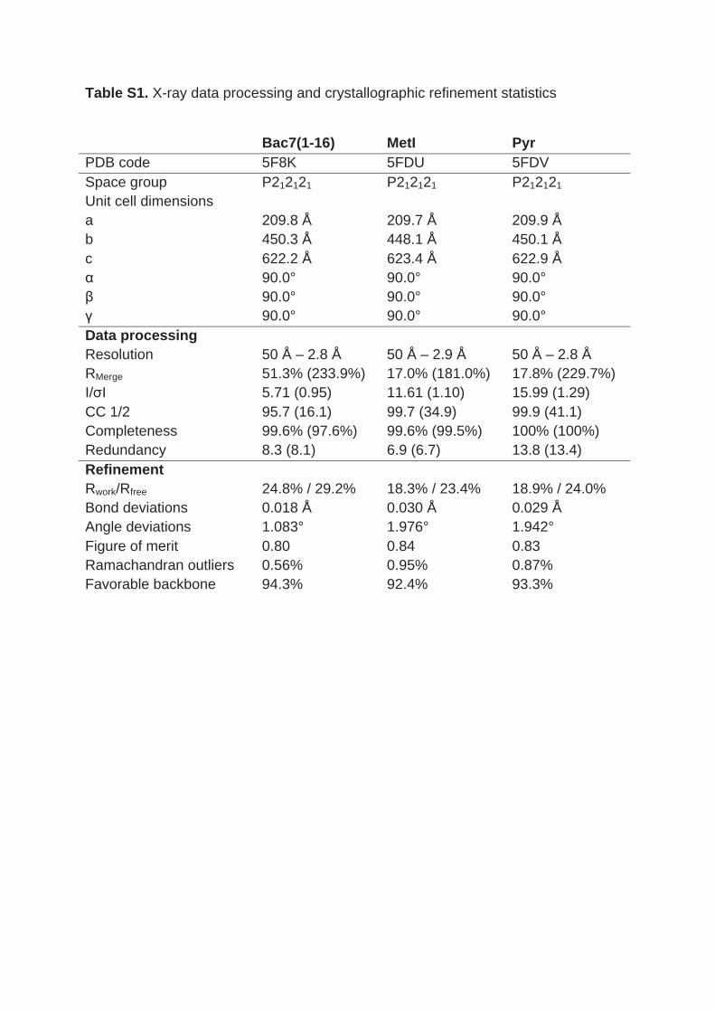
Table S1. X-ray data processing and crystallographic refinement statistics
Bac7(1-16) MetI Pyr
PDB code 5F8K 5FDU 5FDV
Space group P212121 P212121 P212121
Unit cell dimensions
a
b
c
α
β
γ
209.8 Å
450.3 Å
622.2 Å
90.0°
90.0°
90.0°
209.7 Å
448.1 Å
623.4 Å
90.0°
90.0°
90.0°
209.9 Å
450.1 Å
622.9 Å
90.0°
90.0°
90.0°
Data processing
Resolution 50 Å – 2.8 Å 50 Å – 2.9 Å 50 Å – 2.8 Å
RMerge 51.3% (233.9%) 17.0% (181.0%) 17.8% (229.7%)
I/σI 5.71 (0.95) 11.61 (1.10) 15.99 (1.29)
CC 1/2 95.7 (16.1) 99.7 (34.9) 99.9 (41.1)
Completeness 99.6% (97.6%) 99.6% (99.5%) 100% (100%)
Redundancy 8.3 (8.1) 6.9 (6.7) 13.8 (13.4)
Refinement
Rwork/Rfree 24.8% / 29.2% 18.3% / 23.4% 18.9% / 24.0%
Bond deviations 0.018 Å 0.030 Å 0.029 Å
Angle deviations 1.083° 1.976° 1.942°
Figure of merit 0.80 0.84 0.83
Ramachandran outliers 0.56% 0.95% 0.87%
Favorable backbone 94.3% 92.4% 93.3%
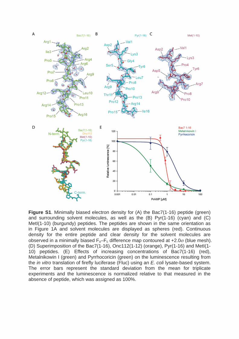
Figure S1. Minimally biased electron density for (A) the Bac7(1-16) peptide (green) and surrounding solvent molecules, as well as the (B) Pyr(1-16) (cyan) and (C) MetI(1-10) (burgundy) peptides. The peptides are shown in the same orientation as in Figure 1A and solvent molecules are displayed as spheres (red). Continuous density for the entire peptide and clear density for the solvent molecules are
observed in a minimally biased Fo–Fc difference map contoured at +2.0σ (blue mesh). (D) Superimposition of the Bac7(1-16), Onc112(1-12) (orange), Pyr(1-16) and MetI(1-10) peptides. (E) Effects of increasing concentrations of Bac7(1-16) (red), Metalnikowin I (green) and Pyrrhocoricin (green) on the luminescence resulting from the in vitro translation of firefly luciferase (Fluc) using an E. coli lysate-based system. The error bars represent the standard deviation from the mean for triplicate experiments and the luminescence is normalized relative to that measured in the absence of peptide, which was assigned as 100%.
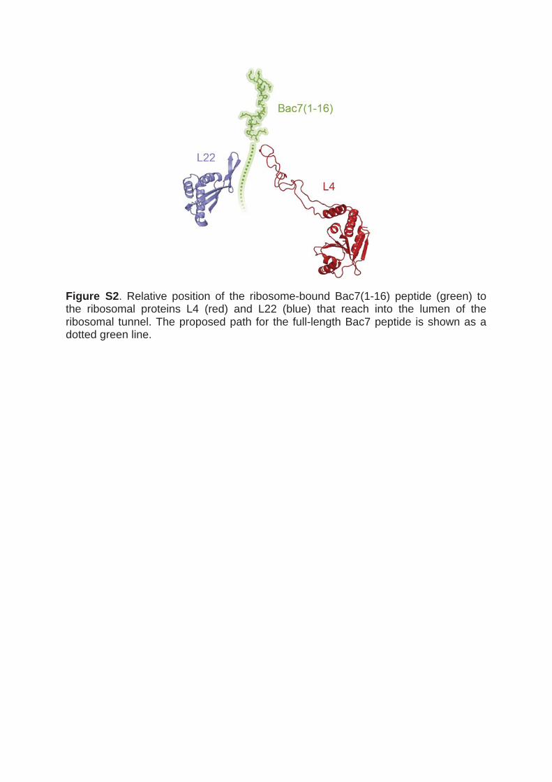
Figure S2. Relative position of the ribosome-bound Bac7(1-16) peptide (green) to the ribosomal proteins L4 (red) and L22 (blue) that reach into the lumen of the ribosomal tunnel. The proposed path for the full-length Bac7 peptide is shown as a dotted green line.
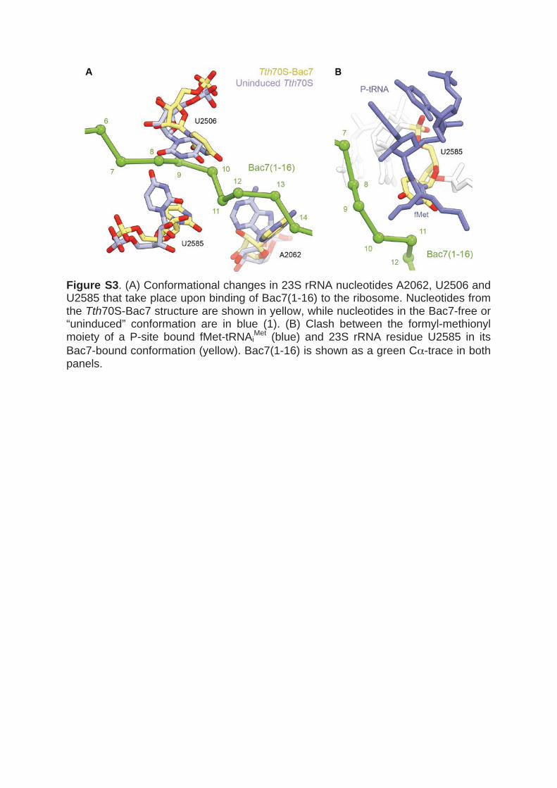
Figure S3. (A) Conformational changes in 23S rRNA nucleotides A2062, U2506 and U2585 that take place upon binding of Bac7(1-16) to the ribosome. Nucleotides from the Tth70S-Bac7 structure are shown in yellow, while nucleotides in the Bac7-free or “uninduced” conformation are in blue (1). (B) Clash between the formyl-methionyl moiety of a P-site bound fMet-tRNAi
Met (blue) and 23S rRNA residue U2585 in its
Bac7-bound conformation (yellow). Bac7(1-16) is shown as a green Cα-trace in both panels.

SUPPLEMENTARY REFERENCES 1. Jenner, L., Starosta, A.L., Terry, D.S., Mikolajka, A., Filonava, L., Yusupov, M.,
Blanchard, S.C., Wilson, D.N. and Yusupova, G. (2013) Structural basis for potent inhibitory activity of the antibiotic tigecycline during protein synthesis. Proceedings of the National Academy of Sciences of the United States of America, 110, 3812-3816.
122 4. Results
4.2 A novel strategy for the structural characterization of arrested
ribosomal complexes featuring short nascent peptides
The second part of this thesis focused on understanding the underlying mechanism of short
peptides that mediated nascent chain-dependent translational arrest. Previously, the M+X(+)
motif was reported to arrest the ribosome in the presence of erythromycin (Sothiselvam et al.,
2014; Sothiselvam et al., 2016) while poly-proline motifs arrest the ribosome without any
additional ligands (Doerfel et al., 2013; Ude et al., 2013). However, far no structural information
had been obtained.
Previously, long nascent chain-mediated translational arrest peptides, like SecM (Bhushan et
al., 2011; Zhang et al., 2015), VemP (Su et al., 2017), MifM (Sohmen et al., 2015), TnaC (Bischoff
et al., 2014; Seidelt et al., 2009), ErmBL (Arenz et al., 2016; Arenz et al., 2014b) and ErmCL (Arenz
et al., 2014a), have been studied by cryo-EM. The nascent chain-mediated translational arrested
ribosome complexes have been obtained using different strategies depending on the length
of the arrest peptide. Arrest peptides including SecM, VemP, MifM, and TnaC are long peptides
with the N-terminus exiting the ribosomal tunnel and they arrest the ribosome-independent
of their N-terminal sequence. The N-terminus was fused to a His8(H8)-tag which allowed
purification after in vitro translation (Sohmen et al., 2015; Su et al., 2017; Zhang et al., 2015) or
the purification of the arrest complex from cells (Bischoff et al., 2014). ErmBL and ErmCL are
shorter arrest peptides in which the nascent chain does not reach the exit of the ribosomal
tunnel and so they cannot be purified using the H8-tag. To still allow solving the structure, the
disome approach was developed (Arenz et al., 2014a; Arenz et al., 2014b). This approach is
based on a bicistronic DNA template that encodes two ORFs of the arrest peptide that is added
into an in vitro translation system. In the absence of the ligand, ribosomes can translate
normally resulting in one ribosome per mRNA on average. In the presence of erythromycin,
the ribosomes are arrested and so two ribosomes, termed disomes, are bound to the same
mRNA molecule. The disomes were separated from the monosomes and through sucrose
gradients. Subsequently, RNase H treatment led to the separation of the disomes and the
obtained monosomes were used for structure determination (Arenz et al., 2014a; Arenz et al.,
2014b).
The original goal of this thesis was to determine the structure of short arrest peptides using X-
ray crystallography. For this approach, a high homogeneity of the arrest complex is necessary
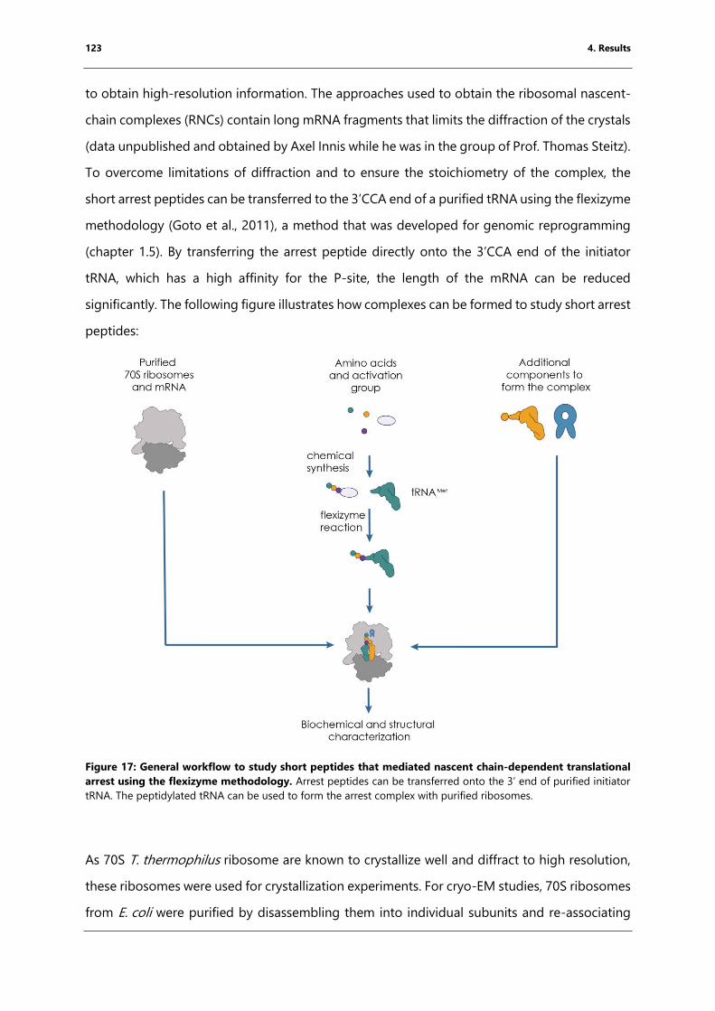
123 4. Results
to obtain high-resolution information. The approaches used to obtain the ribosomal nascent-
chain complexes (RNCs) contain long mRNA fragments that limits the diffraction of the crystals
(data unpublished and obtained by Axel Innis while he was in the group of Prof. Thomas Steitz).
To overcome limitations of diffraction and to ensure the stoichiometry of the complex, the
short arrest peptides can be transferred to the 3’CCA end of a purified tRNA using the flexizyme
methodology (Goto et al., 2011), a method that was developed for genomic reprogramming
(chapter 1.5). By transferring the arrest peptide directly onto the 3’CCA end of the initiator
tRNA, which has a high affinity for the P-site, the length of the mRNA can be reduced
significantly. The following figure illustrates how complexes can be formed to study short arrest
peptides:
Figure 17: General workflow to study short peptides that mediated nascent chain-dependent translational
arrest using the flexizyme methodology. Arrest peptides can be transferred onto the 3’ end of purified initiator
tRNA. The peptidylated tRNA can be used to form the arrest complex with purified ribosomes.
As 70S T. thermophilus ribosome are known to crystallize well and diffract to high resolution,
these ribosomes were used for crystallization experiments. For cryo-EM studies, 70S ribosomes
from E. coli were purified by disassembling them into individual subunits and re-associating
124 4. Results
them (Sucrose gradients, supplements Figure 66, Blaha et al., 2000). This strategy ensures the
removal of ribosome associated factors and tRNAs and increases further the homogeneity of
the complex. A short RNA encoding a Shine-Dalgarno sequence followed by a short open
reading frame (Figure 17) and is necessary for peptidyl tRNA binding to the ribosome. The
arrest peptides were synthesized and activated chemically by Dr. Caterina Lombardo and Dr.
Christophe André from the group of Dr. Gilles Guichard (IECB, Pessac, France) and then
transferred via flexizyme reaction onto the 3’ CCA end of the initiator tRNA. The peptidyl-tRNA
thus obtained was purified using RP-HPLC. We hypothesized that the high P-site binding
affinity of the initiator tRNA is sufficient to force the arrest peptide into the ribosomal exit
tunnel. The arrest complex is formed in the presence of additional compounds such as
aminoacyl-tRNA or an antibiotic. The stalled complex can then be studied structurally and
biochemically. Biochemical assay can be e.g. toeprinting assay, footprinting assay, and
puromycin stability assays. The peptide is normally linked via an ester bond to the 3’CCA end
of the tRNA and this bond is pH sensitive. However, the ester-linked peptidyl-tRNA can be still
used for complex formation to study the RNC with cryo-EM as the sample is frozen directly
after complex formation. The recent development of direct detectors and the constant
improvement of processing software made it possible to obtain near-atomic resolution
structure structures by cryo-EM (Kühlbrandt, 2014). The near-atomic resolution is sufficient to
study short arrest peptides and was used to obtain the structure of the MKF-70S E. coli
ribosome (chapter 4.4).
For X-ray crystallography, crystals were grown for several days at a pH of 7.5 (chapter 3.9) and
so the ester bind might hydrolyze during this process. In order to ensure the stoichiometry of
the complex, the ester linkage can be replaced by an amide linkage (non-hydrolysable bond)
that is pH insensitive and was used in previous studies of the molecular mechanism of peptide
bond formation (Polikanov et al., 2014; Voorhees et al., 2009). This tRNA is obtained by
exchanging the 3’OH of the tRNA by a 3’NH2-group (chapter 3.5.4). Attempts to transfer the
activated peptides onto 3’NH2-tRNAiMet using the flexizyme methodology were so far
unsuccessful. Another strategy to introduce the amide bond between the arrest peptide and
the tRNA is shown in the following figure:
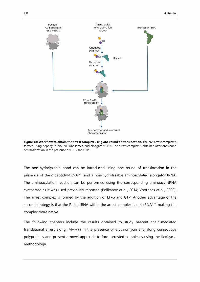
125 4. Results
Figure 18: Workflow to obtain the arrest complex using one round of translocation. The pre-arrest complex is
formed using peptidyl-tRNA, 70S ribosomes, and elongator tRNA. The arrest complex is obtained after one round
of translocation in the presence of EF-G and GTP.
The non-hydrolyzable bond can be introduced using one round of translocation in the
presence of the dipeptidyl-tRNAiMet and a non-hydrolysable aminoacylated elongator tRNA.
The aminoacylation reaction can be performed using the corresponding aminoacyl-tRNA
synthetase as it was used previously reported (Polikanov et al., 2014; Voorhees et al., 2009).
The arrest complex is formed by the addition of EF-G and GTP. Another advantage of the
second strategy is that the P-site tRNA within the arrest complex is not tRNAiMet making the
complex more native.
The following chapters include the results obtained to study nascent chain-mediated
translational arrest along fM+F(+) in the presence of erythromycin and along consecutive
polyprolines and present a novel approach to form arrested complexes using the flexizyme
methodology.
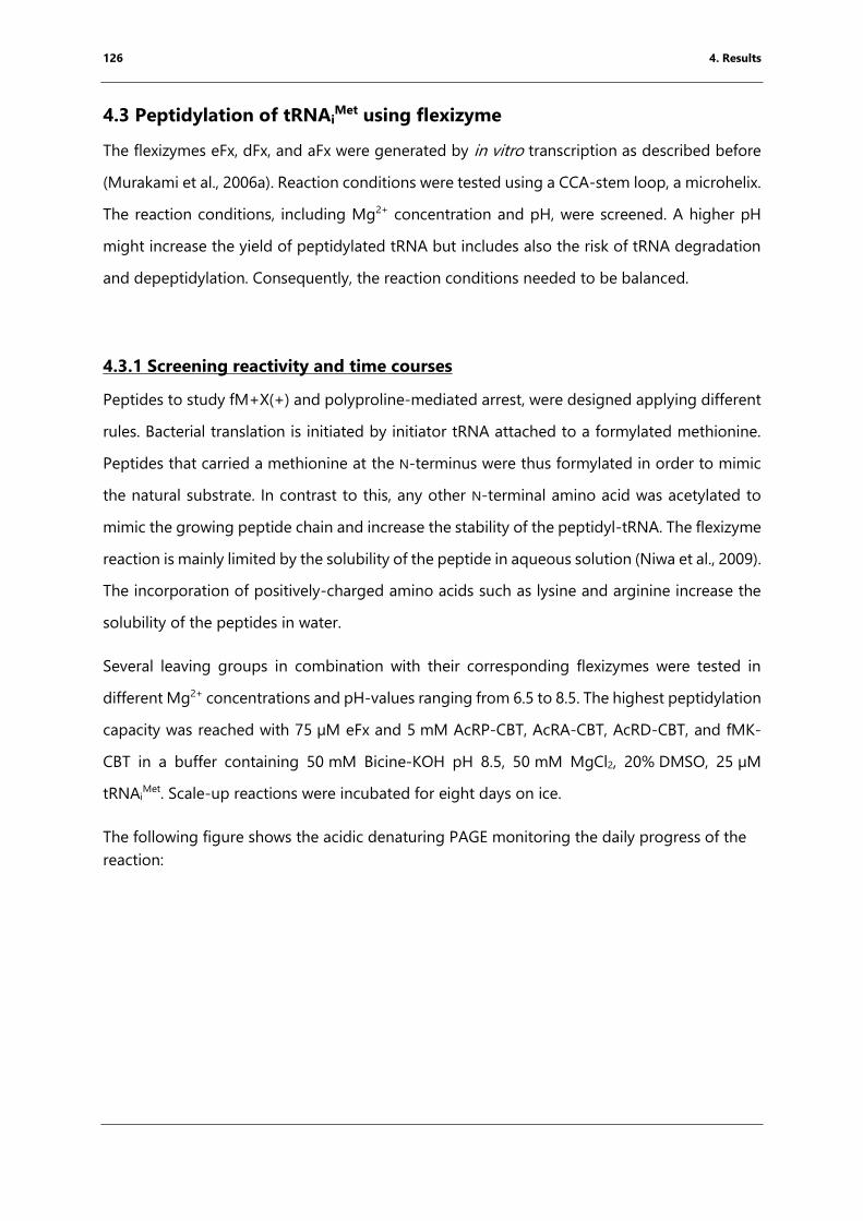
126 4. Results
4.3 Peptidylation of tRNAiMet using flexizyme
The flexizymes eFx, dFx, and aFx were generated by in vitro transcription as described before
(Murakami et al., 2006a). Reaction conditions were tested using a CCA-stem loop, a microhelix.
The reaction conditions, including Mg2+ concentration and pH, were screened. A higher pH
might increase the yield of peptidylated tRNA but includes also the risk of tRNA degradation
and depeptidylation. Consequently, the reaction conditions needed to be balanced.
4.3.1 Screening reactivity and time courses
Peptides to study fM+X(+) and polyproline-mediated arrest, were designed applying different
rules. Bacterial translation is initiated by initiator tRNA attached to a formylated methionine.
Peptides that carried a methionine at the N-terminus were thus formylated in order to mimic
the natural substrate. In contrast to this, any other N-terminal amino acid was acetylated to
mimic the growing peptide chain and increase the stability of the peptidyl-tRNA. The flexizyme
reaction is mainly limited by the solubility of the peptide in aqueous solution (Niwa et al., 2009).
The incorporation of positively-charged amino acids such as lysine and arginine increase the
solubility of the peptides in water.
Several leaving groups in combination with their corresponding flexizymes were tested in
different Mg2+ concentrations and pH-values ranging from 6.5 to 8.5. The highest peptidylation
capacity was reached with 75 µM eFx and 5 mM AcRP-CBT, AcRA-CBT, AcRD-CBT, and fMK-
CBT in a buffer containing 50 mM Bicine-KOH pH 8.5, 50 mM MgCl2, 20% DMSO, 25 µM
tRNAiMet. Scale-up reactions were incubated for eight days on ice.
The following figure shows the acidic denaturing PAGE monitoring the daily progress of the
reaction:
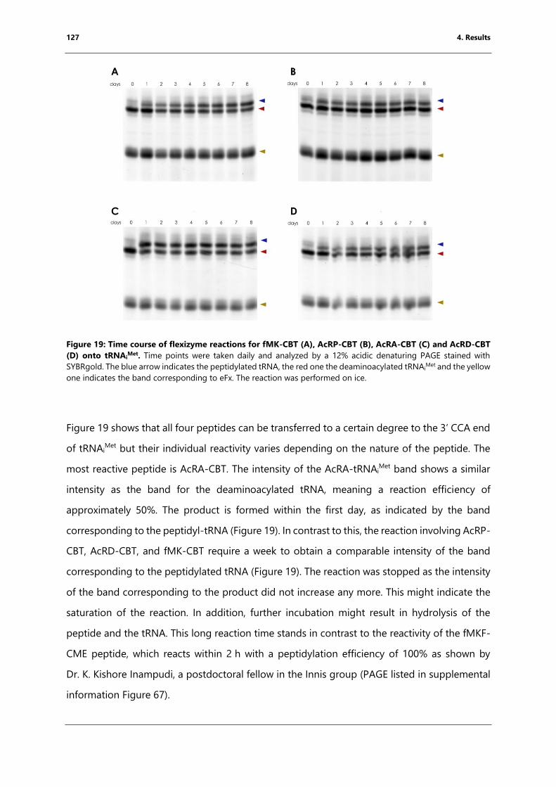
127 4. Results
Figure 19: Time course of flexizyme reactions for fMK-CBT (A), AcRP-CBT (B), AcRA-CBT (C) and AcRD-CBT
(D) onto tRNAiMet. Time points were taken daily and analyzed by a 12% acidic denaturing PAGE stained with
SYBRgold. The blue arrow indicates the peptidylated tRNA, the red one the deaminoacylated tRNAiMet and the yellow
one indicates the band corresponding to eFx. The reaction was performed on ice.
Figure 19 shows that all four peptides can be transferred to a certain degree to the 3’ CCA end
of tRNAiMet but their individual reactivity varies depending on the nature of the peptide. The
most reactive peptide is AcRA-CBT. The intensity of the AcRA-tRNAiMet band shows a similar
intensity as the band for the deaminoacylated tRNA, meaning a reaction efficiency of
approximately 50%. The product is formed within the first day, as indicated by the band
corresponding to the peptidyl-tRNA (Figure 19). In contrast to this, the reaction involving AcRP-
CBT, AcRD-CBT, and fMK-CBT require a week to obtain a comparable intensity of the band
corresponding to the peptidylated tRNA (Figure 19). The reaction was stopped as the intensity
of the band corresponding to the product did not increase any more. This might indicate the
saturation of the reaction. In addition, further incubation might result in hydrolysis of the
peptide and the tRNA. This long reaction time stands in contrast to the reactivity of the fMKF-
CME peptide, which reacts within 2 h with a peptidylation efficiency of 100% as shown by
Dr. K. Kishore Inampudi, a postdoctoral fellow in the Innis group (PAGE listed in supplemental
information Figure 67).
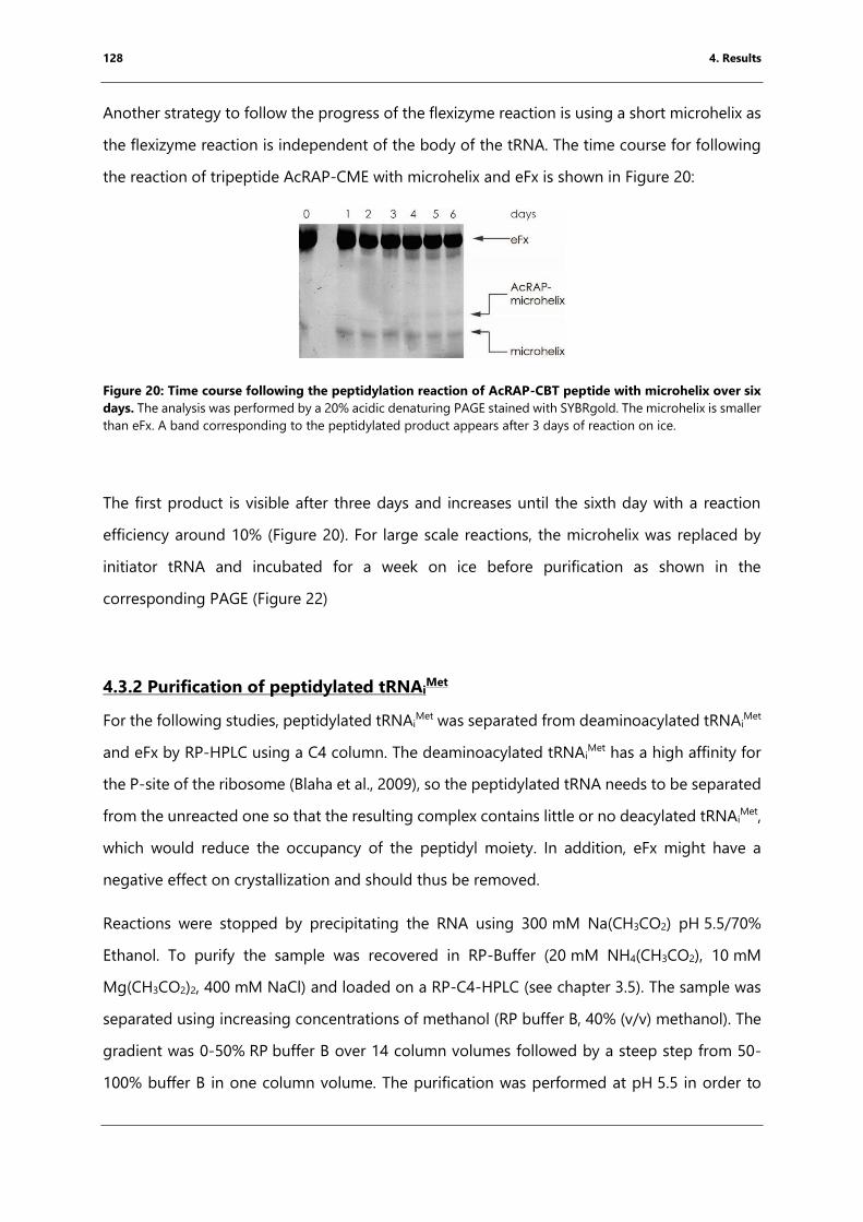
128 4. Results
Another strategy to follow the progress of the flexizyme reaction is using a short microhelix as
the flexizyme reaction is independent of the body of the tRNA. The time course for following
the reaction of tripeptide AcRAP-CME with microhelix and eFx is shown in Figure 20:
Figure 20: Time course following the peptidylation reaction of AcRAP-CBT peptide with microhelix over six
days. The analysis was performed by a 20% acidic denaturing PAGE stained with SYBRgold. The microhelix is smaller
than eFx. A band corresponding to the peptidylated product appears after 3 days of reaction on ice.
The first product is visible after three days and increases until the sixth day with a reaction
efficiency around 10% (Figure 20). For large scale reactions, the microhelix was replaced by
initiator tRNA and incubated for a week on ice before purification as shown in the
corresponding PAGE (Figure 22)
4.3.2 Purification of peptidylated tRNAiMet
For the following studies, peptidylated tRNAiMet was separated from deaminoacylated tRNAi
Met
and eFx by RP-HPLC using a C4 column. The deaminoacylated tRNAiMet has a high affinity for
the P-site of the ribosome (Blaha et al., 2009), so the peptidylated tRNA needs to be separated
from the unreacted one so that the resulting complex contains little or no deacylated tRNAiMet,
which would reduce the occupancy of the peptidyl moiety. In addition, eFx might have a
negative effect on crystallization and should thus be removed.
Reactions were stopped by precipitating the RNA using 300 mM Na(CH3CO2) pH 5.5/70%
Ethanol. To purify the sample was recovered in RP-Buffer (20 mM NH4(CH3CO2), 10 mM
Mg(CH3CO2)2, 400 mM NaCl) and loaded on a RP-C4-HPLC (see chapter 3.5). The sample was
separated using increasing concentrations of methanol (RP buffer B, 40% (v/v) methanol). The
gradient was 0-50% RP buffer B over 14 column volumes followed by a steep step from 50-
100% buffer B in one column volume. The purification was performed at pH 5.5 in order to
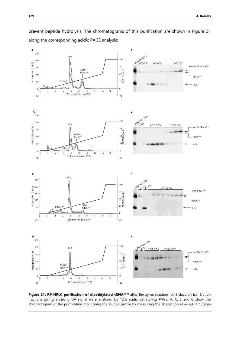
129 4. Results
prevent peptide hydrolysis. The chromatograms of this purification are shown in Figure 21
along the corresponding acidic PAGE analysis:
Figure 21: RP-HPLC purification of dipetidylated-tRNAiMet after flexizyme reaction for 8 days on ice. Elution
fractions giving a strong UV signal were analyzed by 12% acidic denaturing PAGE. A, C, E and G show the
chromatogram of the purification monitoring the elution profile by measuring the absorption at ʎ=260 nm (blue)
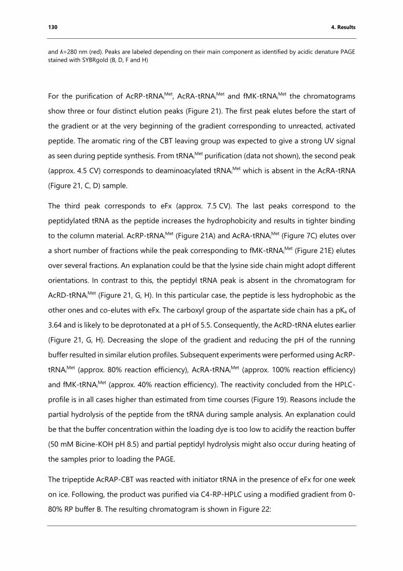
130 4. Results
and ʎ=280 nm (red). Peaks are labeled depending on their main component as identified by acidic denature PAGE
stained with SYBRgold (B, D, F and H)
For the purification of AcRP-tRNAiMet, AcRA-tRNAi
Met and fMK-tRNAiMet the chromatograms
show three or four distinct elution peaks (Figure 21). The first peak elutes before the start of
the gradient or at the very beginning of the gradient corresponding to unreacted, activated
peptide. The aromatic ring of the CBT leaving group was expected to give a strong UV signal
as seen during peptide synthesis. From tRNAiMet purification (data not shown), the second peak
(approx. 4.5 CV) corresponds to deaminoacylated tRNAiMet which is absent in the AcRA-tRNA
(Figure 21, C, D) sample.
The third peak corresponds to eFx (approx. 7.5 CV). The last peaks correspond to the
peptidylated tRNA as the peptide increases the hydrophobicity and results in tighter binding
to the column material. AcRP-tRNAiMet (Figure 21A) and AcRA-tRNAi
Met (Figure 7C) elutes over
a short number of fractions while the peak corresponding to fMK-tRNAiMet (Figure 21E) elutes
over several fractions. An explanation could be that the lysine side chain might adopt different
orientations. In contrast to this, the peptidyl tRNA peak is absent in the chromatogram for
AcRD-tRNAiMet (Figure 21, G, H). In this particular case, the peptide is less hydrophobic as the
other ones and co-elutes with eFx. The carboxyl group of the aspartate side chain has a pKa of
3.64 and is likely to be deprotonated at a pH of 5.5. Consequently, the AcRD-tRNA elutes earlier
(Figure 21, G, H). Decreasing the slope of the gradient and reducing the pH of the running
buffer resulted in similar elution profiles. Subsequent experiments were performed using AcRP-
tRNAiMet (approx. 80% reaction efficiency), AcRA-tRNAi
Met (approx. 100% reaction efficiency)
and fMK-tRNAiMet (approx. 40% reaction efficiency). The reactivity concluded from the HPLC-
profile is in all cases higher than estimated from time courses (Figure 19). Reasons include the
partial hydrolysis of the peptide from the tRNA during sample analysis. An explanation could
be that the buffer concentration within the loading dye is too low to acidify the reaction buffer
(50 mM Bicine-KOH pH 8.5) and partial peptidyl hydrolysis might also occur during heating of
the samples prior to loading the PAGE.
The tripeptide AcRAP-CBT was reacted with initiator tRNA in the presence of eFx for one week
on ice. Following, the product was purified via C4-RP-HPLC using a modified gradient from 0-
80% RP buffer B. The resulting chromatogram is shown in Figure 22:
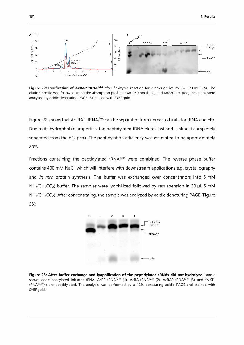
131 4. Results
Figure 22: Purification of AcRAP-tRNAiMet after flexizyme reaction for 7 days on ice by C4-RP-HPLC (A). The
elution profile was followed using the absorption profile at ʎ= 260 nm (blue) and ʎ=280 nm (red). Fractions were
analyzed by acidic denaturing PAGE (B) stained with SYBRgold.
Figure 22 shows that Ac-RAP-tRNAiMet can be separated from unreacted initiator tRNA and eFx.
Due to its hydrophobic properties, the peptidylated tRNA elutes last and is almost completely
separated from the eFx peak. The peptidylation efficiency was estimated to be approximately
80%.
Fractions containing the peptidylated tRNAiMet were combined. The reverse phase buffer
contains 400 mM NaCl, which will interfere with downstream applications e.g. crystallography
and in vitro protein synthesis. The buffer was exchanged over concentrators into 5 mM
NH4(CH3CO2) buffer. The samples were lyophilized followed by resuspension in 20 µL 5 mM
NH4(CH3CO2). After concentrating, the sample was analyzed by acidic denaturing PAGE (Figure
23):
Figure 23: After buffer exchange and lyophilization of the peptidylated tRNAs did not hydrolyze. Lane c
shows deaminoacylated initiator tRNA. AcRP-tRNAiMet (1), AcRA-tRNAi
Met (2), AcRAP-tRNAiMet (3) and fMKF-
tRNAiMet(4) are peptidylated. The analysis was performed by a 12% denaturing acidic PAGE and stained with
SYBRgold.

132 4. Results
All peptidyl-tRNAIMet (AcRP-tRNAi
Met, AcRA-tRNAiMet, AcRAP-tRNAi
Met and fMK-tRNAiMet,) were
still peptidylated after buffer exchange (Figure 23). Samples still contained minor eFx
contaminations which were expected not to interfere with downstream applications.
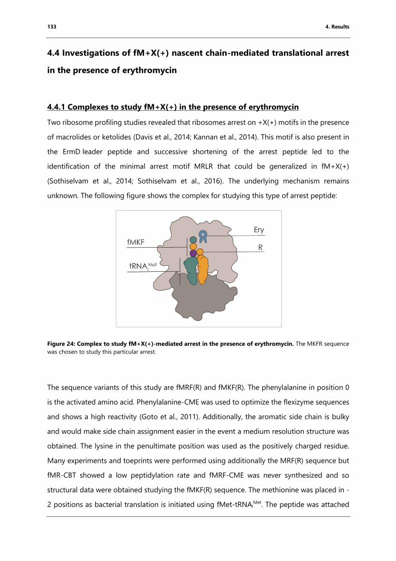
133 4. Results
4.4 Investigations of fM+X(+) nascent chain-mediated translational arrest
in the presence of erythromycin
4.4.1 Complexes to study fM+X(+) in the presence of erythromycin
Two ribosome profiling studies revealed that ribosomes arrest on +X(+) motifs in the presence
of macrolides or ketolides (Davis et al., 2014; Kannan et al., 2014). This motif is also present in
the ErmD leader peptide and successive shortening of the arrest peptide led to the
identification of the minimal arrest motif MRLR that could be generalized in fM+X(+)
(Sothiselvam et al., 2014; Sothiselvam et al., 2016). The underlying mechanism remains
unknown. The following figure shows the complex for studying this type of arrest peptide:
Figure 24: Complex to study fM+X(+)-mediated arrest in the presence of erythromycin. The MKFR sequence
was chosen to study this particular arrest.
The sequence variants of this study are fMRF(R) and fMKF(R). The phenylalanine in position 0
is the activated amino acid. Phenylalanine-CME was used to optimize the flexizyme sequences
and shows a high reactivity (Goto et al., 2011). Additionally, the aromatic side chain is bulky
and would make side chain assignment easier in the event a medium resolution structure was
obtained. The lysine in the penultimate position was used as the positively charged residue.
Many experiments and toeprints were performed using additionally the MRF(R) sequence but
fMR-CBT showed a low peptidylation rate and fMRF-CME was never synthesized and so
structural data were obtained studying the fMKF(R) sequence. The methionine was placed in -
2 positions as bacterial translation is initiated using fMet-tRNAiMet. The peptide was attached
134 4. Results
to tRNAiMet due to the latter’s high affinity for the P-site. Due to its bulky guanidinium group, I
chose Arg-tRNAArg as the incoming aminoacyl-tRNA. This particular tRNA was in vitro
transcribed and aminoacylated by Dr. Axel Innis while he was in the group of Prof. Thomas
Steitz.
For this project, the fMKF-tRNAiMet was generated and purified by Dr. K. Kishore Inampudi and
he used it for X-ray crystallography. I performed the biochemical characterization by
toeprinting and structural studies including X-ray crystallography and cryo-EM.
4.4.2 Toeprinting to validate that fM+F(+) arrests the ribosome in the presence
of erythromycin
By mutating one position at a time, the MRL(R) motif was generalized to fM+X(+) (Sothiselvam
et al., 2016) considering also the published ribosome profiling data that led to the identification
of the +X(+) within long nascent chains (Kannan et al., 2014). In contrast to the MRF(R)
sequence, arrest induced by the sequences MKFR, MRFK and MKFK, which compared to the
MRLR motif encode two substitutions, has not been shown yet. In order to use this sequences
for structural studies I therefore first had to show that they could arrest translation in the
presence of erythromycin. Toeprinting reactions (chapter 3.9) were performed using DNA
templates encoding MRFRI*, MKFRI*, MRFKI* and MKFKI* in the presence or absence of 25 µM
erythromycin within the reaction. A PURExpress system (NEB) lacking release factors was used,
preventing the release of ribosomes that reached the stop codon, which was expected to yield
a toeprint corresponding to ribosomes positioned with their P-site on the preceding isoleucine
codon. The following figure illustrates the nascent chain-mediated translational arrest induced
by these motifs in the presence of erythromycin:
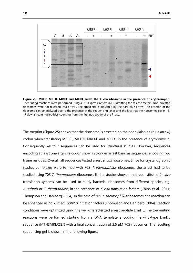
135 4. Results
Figure 25: MRFR, MKFR, MRFK and MKFK arrest the E. coli ribosome in the presence of erythromycin.
Toeprinting reactions were performed using a PURExpress system (NEB) omitting the release factors. Non-arrested
ribosomes were not released (red arrow). The arrest site is indicated by the dark blue arrow. The position of the
ribosome can be analyzed due to the presence of the sequencing lanes and the fact that the ribosomes cover 16-
17 downstream nucleotides counting from the first nucleotide of the P-site.
The toeprint (Figure 25) shows that the ribosome is arrested on the phenylalanine (blue arrow)
codon when translating MRFRI, MKFRI, MRFKI, and MKFKI in the presence of erythromycin.
Consequently, all four sequences can be used for structural studies. However, sequences
encoding at least one arginine codon show a stronger arrest band as sequences encoding two
lysine residues. Overall, all sequences tested arrest E. coli ribosomes. Since for crystallographic
studies complexes were formed with 70S T. thermophilus ribosomes, the arrest had to be
studied using 70S T. thermophilus ribosomes. Earlier studies showed that reconstituted in vitro
translation systems can be used to study bacterial ribosomes from different species, e.g.
B. subtilis or T. thermophilus, in the presence of E. coli translation factors (Chiba et al., 2011;
Thompson and Dahlberg, 2004). In the case of 70S T. thermophilus ribosomes, the reaction can
be enhanced using T. thermophilus initiation factors (Thompson and Dahlberg, 2004). Reaction
conditions were optimized using the well-characterized arrest peptide ErmDL. The toeprinting
reactions were performed starting from a DNA template encoding the wild-type ErmDL
sequence (MTHSMRLRSE*) with a final concentration of 2.5 µM 70S ribosomes. The resulting
sequencing gel is shown in the following figure:
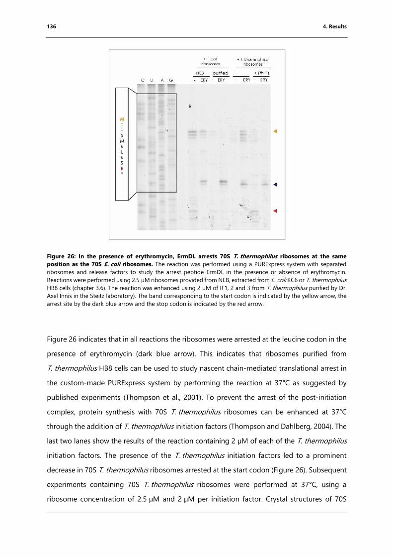
136 4. Results
Figure 26: In the presence of erythromycin, ErmDL arrests 70S T. thermophilus ribosomes at the same
position as the 70S E. coli ribosomes. The reaction was performed using a PURExpress system with separated
ribosomes and release factors to study the arrest peptide ErmDL in the presence or absence of erythromycin.
Reactions were performed using 2.5 µM ribosomes provided from NEB, extracted from E. coli KC6 or T. thermophilus
HB8 cells (chapter 3.6). The reaction was enhanced using 2 µM of IF1, 2 and 3 from T. thermophilus purified by Dr.
Axel Innis in the Steitz laboratory). The band corresponding to the start codon is indicated by the yellow arrow, the
arrest site by the dark blue arrow and the stop codon is indicated by the red arrow.
Figure 26 indicates that in all reactions the ribosomes were arrested at the leucine codon in the
presence of erythromycin (dark blue arrow). This indicates that ribosomes purified from
T. thermophilus HB8 cells can be used to study nascent chain-mediated translational arrest in
the custom-made PURExpress system by performing the reaction at 37°C as suggested by
published experiments (Thompson et al., 2001). To prevent the arrest of the post-initiation
complex, protein synthesis with 70S T. thermophilus ribosomes can be enhanced at 37°C
through the addition of T. thermophilus initiation factors (Thompson and Dahlberg, 2004). The
last two lanes show the results of the reaction containing 2 µM of each of the T. thermophilus
initiation factors. The presence of the T. thermophilus initiation factors led to a prominent
decrease in 70S T. thermophilus ribosomes arrested at the start codon (Figure 26). Subsequent
experiments containing 70S T. thermophilus ribosomes were performed at 37°C, using a
ribosome concentration of 2.5 µM and 2 µM per initiation factor. Crystal structures of 70S
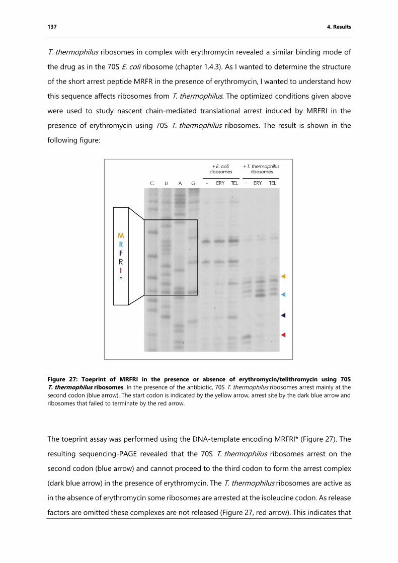
137 4. Results
T. thermophilus ribosomes in complex with erythromycin revealed a similar binding mode of
the drug as in the 70S E. coli ribosome (chapter 1.4.3). As I wanted to determine the structure
of the short arrest peptide MRFR in the presence of erythromycin, I wanted to understand how
this sequence affects ribosomes from T. thermophilus. The optimized conditions given above
were used to study nascent chain-mediated translational arrest induced by MRFRI in the
presence of erythromycin using 70S T. thermophilus ribosomes. The result is shown in the
following figure:
Figure 27: Toeprint of MRFRI in the presence or absence of erythromycin/telithromycin using 70S
T. thermophilus ribosomes. In the presence of the antibiotic, 70S T. thermophilus ribosomes arrest mainly at the
second codon (blue arrow). The start codon is indicated by the yellow arrow, arrest site by the dark blue arrow and
ribosomes that failed to terminate by the red arrow.
The toeprint assay was performed using the DNA-template encoding MRFRI* (Figure 27). The
resulting sequencing-PAGE revealed that the 70S T. thermophilus ribosomes arrest on the
second codon (blue arrow) and cannot proceed to the third codon to form the arrest complex
(dark blue arrow) in the presence of erythromycin. The T. thermophilus ribosomes are active as
in the absence of erythromycin some ribosomes are arrested at the isoleucine codon. As release
factors are omitted these complexes are not released (Figure 27, red arrow). This indicates that
138 4. Results
the T. thermophilus ribosomes seem to have difficulties translating MR at 37°C in the presence
of erythromycin. The presence of erythromycin restricts the PTC and the T. thermophilus
ribosomes might not be able to overcome this at low temperatures and so the experiment
should be repeated using translation factors from T. thermophilus allowing to perform the
reaction at higher temperatures. For crystallographic studies, this issue might be overcome by
forming the complex at a higher temperature or using flexizyme-charged peptide.
4.4.3 Toeprinting: fMKF-tRNAiMet arrests the ribosome in the presence of
erythromycin
fMKF-tRNAiMet was peptidylated and purified by Dr. K. Kishore Inampudi using the flexizyme
eFx and CME activated fMKF. To investigate the stalling activity of the peptide, I performed a
toeprinting assay in the presence or absence of erythromycin using a PURExpress (NEB) system
without tRNAs, amino acids, and ribosomes.
First attempts to study the incorporation of fMKF-tRNAiMet by toeprinting were performed
using 10 µM fMKF-tRNAiMet and the total tRNA solution provided by the kit resulting in
ribosomes arrested at the initiation codon (data were not shown). The tRNA solution provided
by the kit contains total tRNA extracted from cells and used at a concentration of 5 µM end
concentration (Shimizu et al., 2001; Shimizu et al., 2005). Additionally, the encoded arginine
codon CGG (0.5% in E. coli K12) and AGG (0.2% in E. coli K12) are both rare codons in E. coli
K12. the large excess of the initiator tRNA over the rarely represented tRNAArg resulted propably
in a strong signal for the initiation complex but not for the translocated one. The assumption
can be made that the amount of the corresponding tRNA is not enough to form the
translocated complex that was detectable in toeprinting. To overcome this problem, the
experimental set-up was changed to use purified compounds. The initiation complex was pre-
formed by incubating 2.5 µM ribosomes with 5 µM mRNA template (coding sequence: MRPPPI,
AUG AGG CCG CCG CCG AUC UAA), 10 µM peptidylated-tRNAiMet, 25 µM erythromycin, 20 µM
Arg-tRNAArg and the factor mix, containing all protein factors for the canonical translation cycle,
for 5 min at room temperature. Subsequently, solution A-(minus aa, tRNA) provided by the kit,
containing nucleotides, was added, allowing complexes that had not been arrested to be
translocated (further information are provided in chapter 3.9). The following figure shows the
results of these reactions:
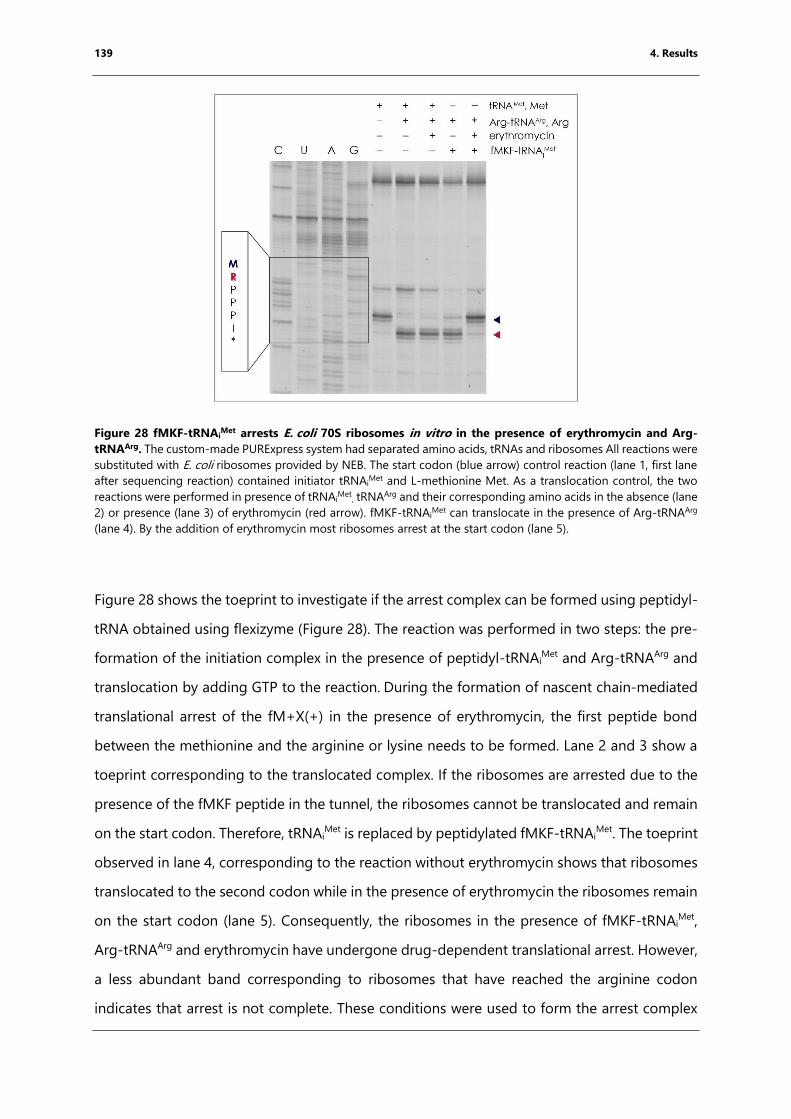
139 4. Results
Figure 28 fMKF-tRNAiMet arrests E. coli 70S ribosomes in vitro in the presence of erythromycin and Arg-
tRNAArg. The custom-made PURExpress system had separated amino acids, tRNAs and ribosomes All reactions were
substituted with E. coli ribosomes provided by NEB. The start codon (blue arrow) control reaction (lane 1, first lane
after sequencing reaction) contained initiator tRNAiMet and L-methionine Met. As a translocation control, the two
reactions were performed in presence of tRNAiMet
, tRNAArg and their corresponding amino acids in the absence (lane
2) or presence (lane 3) of erythromycin (red arrow). fMKF-tRNAiMet can translocate in the presence of Arg-tRNAArg
(lane 4). By the addition of erythromycin most ribosomes arrest at the start codon (lane 5).
Figure 28 shows the toeprint to investigate if the arrest complex can be formed using peptidyl-
tRNA obtained using flexizyme (Figure 28). The reaction was performed in two steps: the pre-
formation of the initiation complex in the presence of peptidyl-tRNAiMet and Arg-tRNAArg and
translocation by adding GTP to the reaction. During the formation of nascent chain-mediated
translational arrest of the fM+X(+) in the presence of erythromycin, the first peptide bond
between the methionine and the arginine or lysine needs to be formed. Lane 2 and 3 show a
toeprint corresponding to the translocated complex. If the ribosomes are arrested due to the
presence of the fMKF peptide in the tunnel, the ribosomes cannot be translocated and remain
on the start codon. Therefore, tRNAiMet is replaced by peptidylated fMKF-tRNAi
Met. The toeprint
observed in lane 4, corresponding to the reaction without erythromycin shows that ribosomes
translocated to the second codon while in the presence of erythromycin the ribosomes remain
on the start codon (lane 5). Consequently, the ribosomes in the presence of fMKF-tRNAiMet,
Arg-tRNAArg and erythromycin have undergone drug-dependent translational arrest. However,
a less abundant band corresponding to ribosomes that have reached the arginine codon
indicates that arrest is not complete. These conditions were used to form the arrest complex
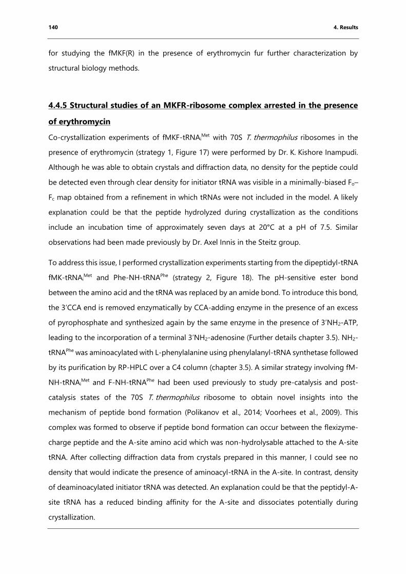
140 4. Results
for studying the fMKF(R) in the presence of erythromycin fur further characterization by
structural biology methods.
4.4.5 Structural studies of an MKFR-ribosome complex arrested in the presence
of erythromycin
Co-crystallization experiments of fMKF-tRNAiMet with 70S T. thermophilus ribosomes in the
presence of erythromycin (strategy 1, Figure 17) were performed by Dr. K. Kishore Inampudi.
Although he was able to obtain crystals and diffraction data, no density for the peptide could
be detected even through clear density for initiator tRNA was visible in a minimally-biased Fo–
Fc map obtained from a refinement in which tRNAs were not included in the model. A likely
explanation could be that the peptide hydrolyzed during crystallization as the conditions
include an incubation time of approximately seven days at 20°C at a pH of 7.5. Similar
observations had been made previously by Dr. Axel Innis in the Steitz group.
To address this issue, I performed crystallization experiments starting from the dipeptidyl-tRNA
fMK-tRNAiMet and Phe-NH-tRNAPhe (strategy 2, Figure 18). The pH-sensitive ester bond
between the amino acid and the tRNA was replaced by an amide bond. To introduce this bond,
the 3’CCA end is removed enzymatically by CCA-adding enzyme in the presence of an excess
of pyrophosphate and synthesized again by the same enzyme in the presence of 3’NH2-ATP,
leading to the incorporation of a terminal 3’NH2-adenosine (Further details chapter 3.5). NH2-
tRNAPhe was aminoacylated with L-phenylalanine using phenylalanyl-tRNA synthetase followed
by its purification by RP-HPLC over a C4 column (chapter 3.5). A similar strategy involving fM-
NH-tRNAiMet and F-NH-tRNAPhe had been used previously to study pre-catalysis and post-
catalysis states of the 70S T. thermophilus ribosome to obtain novel insights into the
mechanism of peptide bond formation (Polikanov et al., 2014; Voorhees et al., 2009). This
complex was formed to observe if peptide bond formation can occur between the flexizyme-
charge peptide and the A-site amino acid which was non-hydrolysable attached to the A-site
tRNA. After collecting diffraction data from crystals prepared in this manner, I could see no
density that would indicate the presence of aminoacyl-tRNA in the A-site. In contrast, density
of deaminoacylated initiator tRNA was detected. An explanation could be that the peptidyl-A-
site tRNA has a reduced binding affinity for the A-site and dissociates potentially during
crystallization.
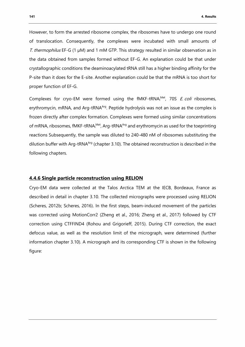
141 4. Results
However, to form the arrested ribosome complex, the ribosomes have to undergo one round
of translocation. Consequently, the complexes were incubated with small amounts of
T. thermophilus EF-G (1 µM) and 1 mM GTP. This strategy resulted in similar observation as in
the data obtained from samples formed without EF-G. An explanation could be that under
crystallographic conditions the deaminoacylated tRNA still has a higher binding affinity for the
P-site than it does for the E-site. Another explanation could be that the mRNA is too short for
proper function of EF-G.
Complexes for cryo-EM were formed using the fMKF-tRNAiMet, 70S E. coli ribosomes,
erythromycin, mRNA, and Arg-tRNAArg. Peptide hydrolysis was not an issue as the complex is
frozen directly after complex formation. Complexes were formed using similar concentrations
of mRNA, ribosomes, fMKF-tRNAiMet, Arg-tRNAArg and erythromycin as used for the toeprinting
reactions Subsequently, the sample was diluted to 240-480 nM of ribosomes substituting the
dilution buffer with Arg-tRNAArg (chapter 3.10). The obtained reconstruction is described in the
following chapters.
4.4.6 Single particle reconstruction using RELION
Cryo-EM data were collected at the Talos Arctica TEM at the IECB, Bordeaux, France as
described in detail in chapter 3.10. The collected micrographs were processed using RELION
(Scheres, 2012b; Scheres, 2016). In the first steps, beam-induced movement of the particles
was corrected using MotionCorr2 (Zheng et al., 2016; Zheng et al., 2017) followed by CTF
correction using CTFFIND4 (Rohou and Grigorieff, 2015). During CTF correction, the exact
defocus value, as well as the resolution limit of the micrograph, were determined (further
information chapter 3.10). A micrograph and its corresponding CTF is shown in the following
figure:
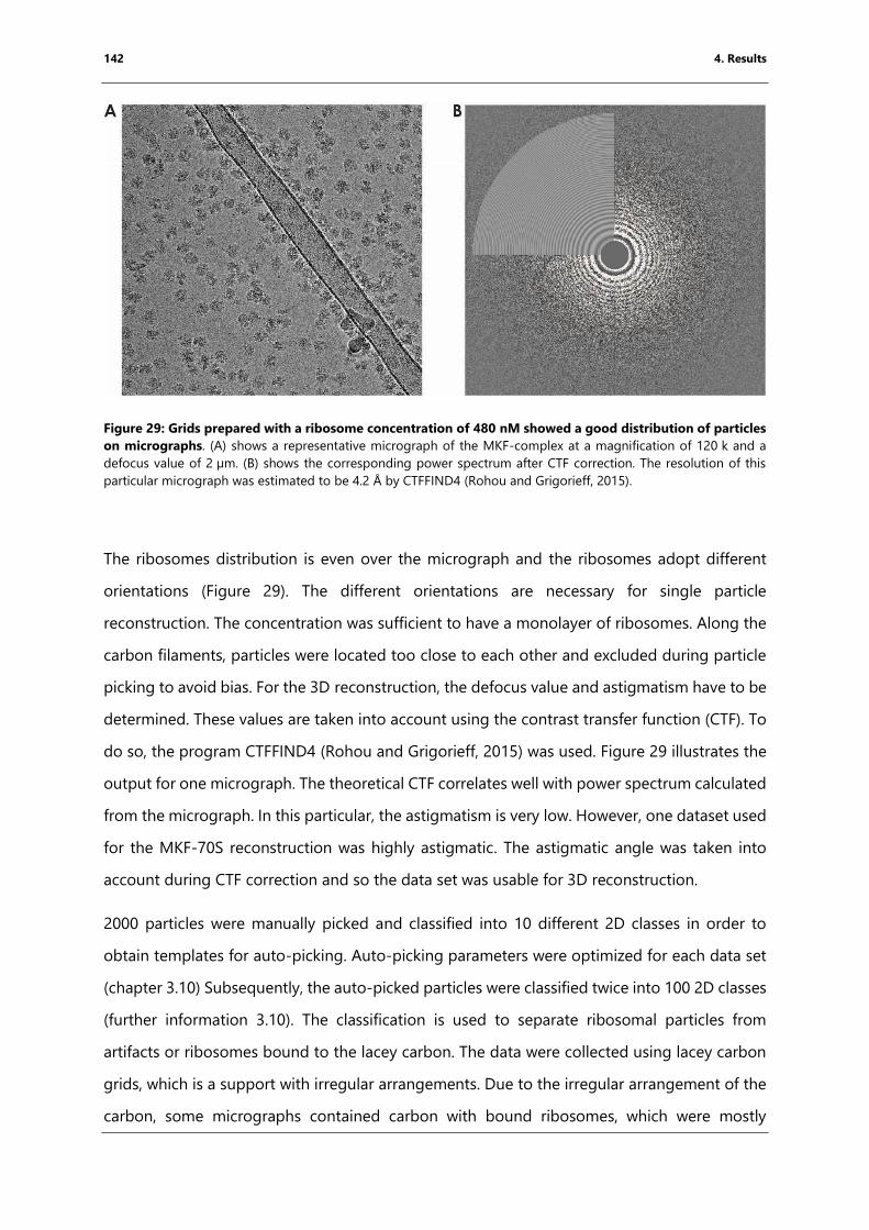
142 4. Results
Figure 29: Grids prepared with a ribosome concentration of 480 nM showed a good distribution of particles
on micrographs. (A) shows a representative micrograph of the MKF-complex at a magnification of 120 k and a
defocus value of 2 µm. (B) shows the corresponding power spectrum after CTF correction. The resolution of this
particular micrograph was estimated to be 4.2 Å by CTFFIND4 (Rohou and Grigorieff, 2015).
The ribosomes distribution is even over the micrograph and the ribosomes adopt different
orientations (Figure 29). The different orientations are necessary for single particle
reconstruction. The concentration was sufficient to have a monolayer of ribosomes. Along the
carbon filaments, particles were located too close to each other and excluded during particle
picking to avoid bias. For the 3D reconstruction, the defocus value and astigmatism have to be
determined. These values are taken into account using the contrast transfer function (CTF). To
do so, the program CTFFIND4 (Rohou and Grigorieff, 2015) was used. Figure 29 illustrates the
output for one micrograph. The theoretical CTF correlates well with power spectrum calculated
from the micrograph. In this particular, the astigmatism is very low. However, one dataset used
for the MKF-70S reconstruction was highly astigmatic. The astigmatic angle was taken into
account during CTF correction and so the data set was usable for 3D reconstruction.
2000 particles were manually picked and classified into 10 different 2D classes in order to
obtain templates for auto-picking. Auto-picking parameters were optimized for each data set
(chapter 3.10) Subsequently, the auto-picked particles were classified twice into 100 2D classes
(further information 3.10). The classification is used to separate ribosomal particles from
artifacts or ribosomes bound to the lacey carbon. The data were collected using lacey carbon
grids, which is a support with irregular arrangements. Due to the irregular arrangement of the
carbon, some micrographs contained carbon with bound ribosomes, which were mostly
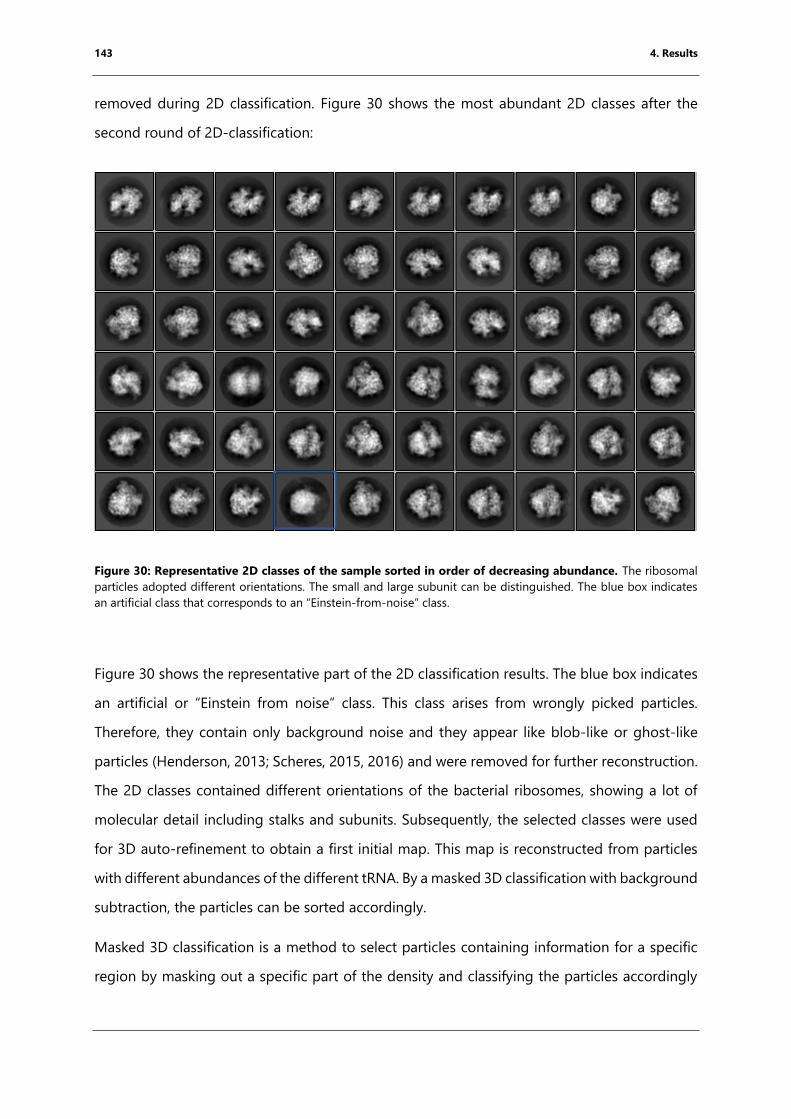
143 4. Results
removed during 2D classification. Figure 30 shows the most abundant 2D classes after the
second round of 2D-classification:
Figure 30: Representative 2D classes of the sample sorted in order of decreasing abundance. The ribosomal
particles adopted different orientations. The small and large subunit can be distinguished. The blue box indicates
an artificial class that corresponds to an “Einstein-from-noise” class.
Figure 30 shows the representative part of the 2D classification results. The blue box indicates
an artificial or “Einstein from noise” class. This class arises from wrongly picked particles.
Therefore, they contain only background noise and they appear like blob-like or ghost-like
particles (Henderson, 2013; Scheres, 2015, 2016) and were removed for further reconstruction.
The 2D classes contained different orientations of the bacterial ribosomes, showing a lot of
molecular detail including stalks and subunits. Subsequently, the selected classes were used
for 3D auto-refinement to obtain a first initial map. This map is reconstructed from particles
with different abundances of the different tRNA. By a masked 3D classification with background
subtraction, the particles can be sorted accordingly.
Masked 3D classification is a method to select particles containing information for a specific
region by masking out a specific part of the density and classifying the particles accordingly
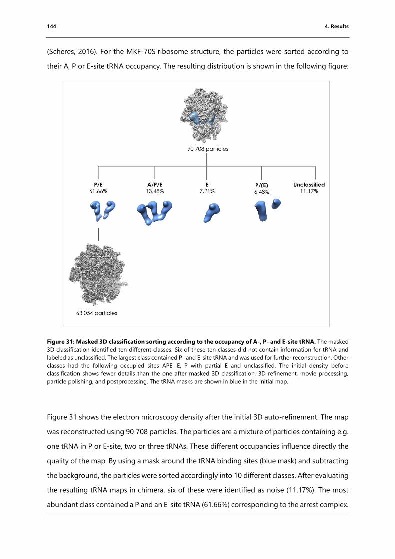
144 4. Results
(Scheres, 2016). For the MKF-70S ribosome structure, the particles were sorted according to
their A, P or E-site tRNA occupancy. The resulting distribution is shown in the following figure:
Figure 31: Masked 3D classification sorting according to the occupancy of A-, P- and E-site tRNA. The masked
3D classification identified ten different classes. Six of these ten classes did not contain information for tRNA and
labeled as unclassified. The largest class contained P- and E-site tRNA and was used for further reconstruction. Other
classes had the following occupied sites APE, E, P with partial E and unclassified. The initial density before
classification shows fewer details than the one after masked 3D classification, 3D refinement, movie processing,
particle polishing, and postprocessing. The tRNA masks are shown in blue in the initial map.
Figure 31 shows the electron microscopy density after the initial 3D auto-refinement. The map
was reconstructed using 90 708 particles. The particles are a mixture of particles containing e.g.
one tRNA in P or E-site, two or three tRNAs. These different occupancies influence directly the
quality of the map. By using a mask around the tRNA binding sites (blue mask) and subtracting
the background, the particles were sorted accordingly into 10 different classes. After evaluating
the resulting tRNA maps in chimera, six of these were identified as noise (11.17%). The most
abundant class contained a P and an E-site tRNA (61.66%) corresponding to the arrest complex.
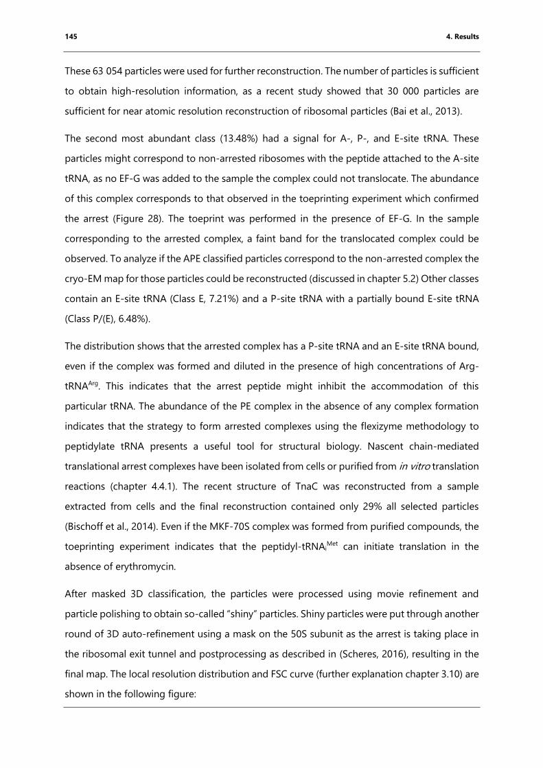
145 4. Results
These 63 054 particles were used for further reconstruction. The number of particles is sufficient
to obtain high-resolution information, as a recent study showed that 30 000 particles are
sufficient for near atomic resolution reconstruction of ribosomal particles (Bai et al., 2013).
The second most abundant class (13.48%) had a signal for A-, P-, and E-site tRNA. These
particles might correspond to non-arrested ribosomes with the peptide attached to the A-site
tRNA, as no EF-G was added to the sample the complex could not translocate. The abundance
of this complex corresponds to that observed in the toeprinting experiment which confirmed
the arrest (Figure 28). The toeprint was performed in the presence of EF-G. In the sample
corresponding to the arrested complex, a faint band for the translocated complex could be
observed. To analyze if the APE classified particles correspond to the non-arrested complex the
cryo-EM map for those particles could be reconstructed (discussed in chapter 5.2) Other classes
contain an E-site tRNA (Class E, 7.21%) and a P-site tRNA with a partially bound E-site tRNA
(Class P/(E), 6.48%).
The distribution shows that the arrested complex has a P-site tRNA and an E-site tRNA bound,
even if the complex was formed and diluted in the presence of high concentrations of Arg-
tRNAArg. This indicates that the arrest peptide might inhibit the accommodation of this
particular tRNA. The abundance of the PE complex in the absence of any complex formation
indicates that the strategy to form arrested complexes using the flexizyme methodology to
peptidylate tRNA presents a useful tool for structural biology. Nascent chain-mediated
translational arrest complexes have been isolated from cells or purified from in vitro translation
reactions (chapter 4.4.1). The recent structure of TnaC was reconstructed from a sample
extracted from cells and the final reconstruction contained only 29% all selected particles
(Bischoff et al., 2014). Even if the MKF-70S complex was formed from purified compounds, the
toeprinting experiment indicates that the peptidyl-tRNAiMet can initiate translation in the
absence of erythromycin.
After masked 3D classification, the particles were processed using movie refinement and
particle polishing to obtain so-called “shiny” particles. Shiny particles were put through another
round of 3D auto-refinement using a mask on the 50S subunit as the arrest is taking place in
the ribosomal exit tunnel and postprocessing as described in (Scheres, 2016), resulting in the
final map. The local resolution distribution and FSC curve (further explanation chapter 3.10) are
shown in the following figure:
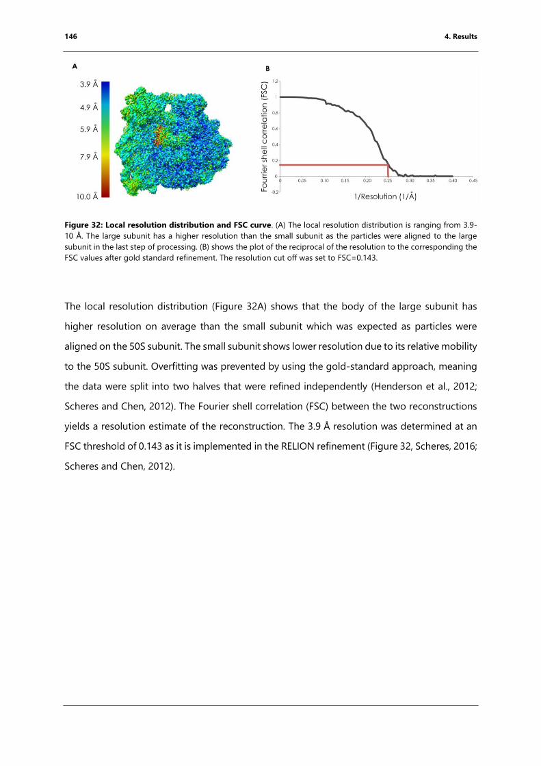
146 4. Results
Figure 32: Local resolution distribution and FSC curve. (A) The local resolution distribution is ranging from 3.9-
10 Å. The large subunit has a higher resolution than the small subunit as the particles were aligned to the large
subunit in the last step of processing. (B) shows the plot of the reciprocal of the resolution to the corresponding the
FSC values after gold standard refinement. The resolution cut off was set to FSC=0.143.
The local resolution distribution (Figure 32A) shows that the body of the large subunit has
higher resolution on average than the small subunit which was expected as particles were
aligned on the 50S subunit. The small subunit shows lower resolution due to its relative mobility
to the 50S subunit. Overfitting was prevented by using the gold-standard approach, meaning
the data were split into two halves that were refined independently (Henderson et al., 2012;
Scheres and Chen, 2012). The Fourier shell correlation (FSC) between the two reconstructions
yields a resolution estimate of the reconstruction. The 3.9 Å resolution was determined at an
FSC threshold of 0.143 as it is implemented in the RELION refinement (Figure 32, Scheres, 2016;
Scheres and Chen, 2012).
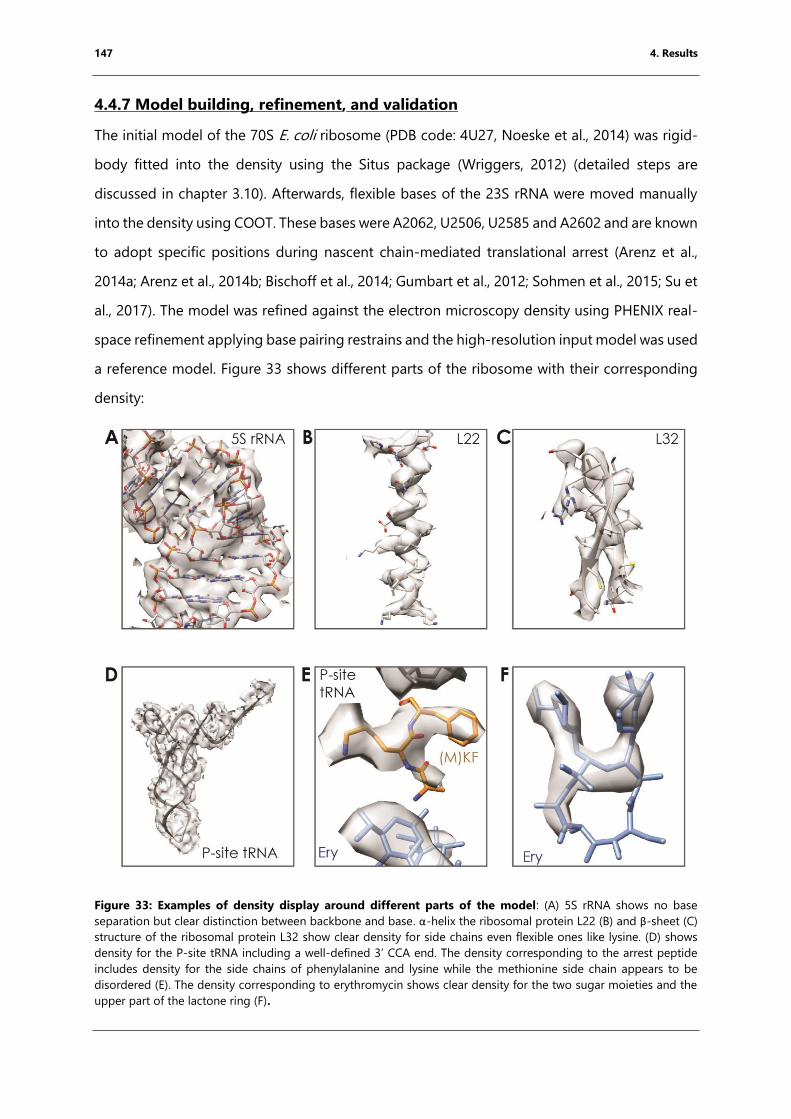
147 4. Results
4.4.7 Model building, refinement, and validation
The initial model of the 70S E. coli ribosome (PDB code: 4U27, Noeske et al., 2014) was rigid-
body fitted into the density using the Situs package (Wriggers, 2012) (detailed steps are
discussed in chapter 3.10). Afterwards, flexible bases of the 23S rRNA were moved manually
into the density using COOT. These bases were A2062, U2506, U2585 and A2602 and are known
to adopt specific positions during nascent chain-mediated translational arrest (Arenz et al.,
2014a; Arenz et al., 2014b; Bischoff et al., 2014; Gumbart et al., 2012; Sohmen et al., 2015; Su et
al., 2017). The model was refined against the electron microscopy density using PHENIX real-
space refinement applying base pairing restrains and the high-resolution input model was used
a reference model. Figure 33 shows different parts of the ribosome with their corresponding
density:
Figure 33: Examples of density display around different parts of the model: (A) 5S rRNA shows no base
separation but clear distinction between backbone and base. α-helix the ribosomal protein L22 (B) and β-sheet (C)
structure of the ribosomal protein L32 show clear density for side chains even flexible ones like lysine. (D) shows
density for the P-site tRNA including a well-defined 3’ CCA end. The density corresponding to the arrest peptide
includes density for the side chains of phenylalanine and lysine while the methionine side chain appears to be
disordered (E). The density corresponding to erythromycin shows clear density for the two sugar moieties and the
upper part of the lactone ring (F).
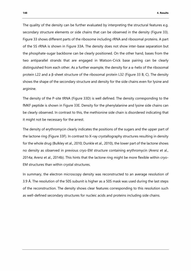
148 4. Results
The quality of the density can be further evaluated by interpreting the structural features e.g.
secondary structure elements or side chains that can be observed in the density (Figure 33).
Figure 33 shows different parts of the ribosome including rRNA and ribosomal proteins. A part
of the 5S rRNA is shown in Figure 33A. The density does not show inter-base separation but
the phosphate-sugar backbone can be clearly positioned. On the other hand, bases from the
two antiparallel strands that are engaged in Watson-Crick base pairing can be clearly
distinguished from each other. As a further example, the density for a α-helix of the ribosomal
protein L22 and a β-sheet structure of the ribosomal protein L32 (Figure 33 B, C). The density
shows the shape of the secondary structure and density for the side chains even for lysine and
arginine.
The density of the P-site tRNA (Figure 33D) is well defined. The density corresponding to the
fMKF peptide is shown in Figure 33E. Density for the phenylalanine and lysine side chains can
be clearly observed. In contrast to this, the methionine side chain is disordered indicating that
it might not be necessary for the arrest.
The density of erythromycin clearly indicates the positions of the sugars and the upper part of
the lactone ring (Figure 33F). In contrast to X-ray crystallography structures resulting in density
for the whole drug (Bulkley et al., 2010; Dunkle et al., 2010), the lower part of the lactone shows
no density as observed in previous cryo-EM structure containing erythromycin (Arenz et al.,
2014a; Arenz et al., 2014b). This hints that the lactone ring might be more flexible within cryo-
EM structures than within crystal structures.
In summary, the electron microscopy density was reconstructed to an average resolution of
3.9 Å. The resolution of the 50S subunit is higher as a 50S mask was used during the last steps
of the reconstruction. The density shows clear features corresponding to this resolution such
as well-defined secondary structures for nucleic acids and proteins including side chains.
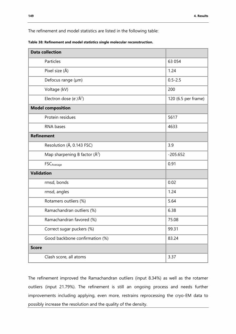
149 4. Results
The refinement and model statistics are listed in the following table:
Table 38: Refinement and model statistics single molecular reconstruction.
Data collection
Particles 63 054
Pixel size (Å) 1.24
Defocus range (µm) 0.5-2.5
Voltage (kV) 200
Electron dose (e-/Å2) 120 (6.5 per frame)
Model composition
Protein residues 5617
RNA bases 4633
Refinement
Resolution (Å, 0.143 FSC) 3.9
Map sharpening B factor (Å2) -205.652
FSCAverage 0.91
Validation
rmsd, bonds 0.02
rmsd, angles 1.24
Rotamers outliers (%) 5.64
Ramachandran outliers (%) 6.38
Ramachandran favored (%) 75.08
Correct sugar puckers (%) 99.31
Good backbone confirmation (%) 83.24
Score
Clash score, all atoms 3.37
The refinement improved the Ramachandran outliers (input 8.34%) as well as the rotamer
outliers (input 21.79%). The refinement is still an ongoing process and needs further
improvements including applying, even more, restrains reprocessing the cryo-EM data to
possibly increase the resolution and the quality of the density.
150 4. Results
4.4.9 Structure interpretation
The complex was formed in the presence of Arg-tRNAArg a short synthetic mRNA encoding for
the amino acid sequence MR. After complex formation, the sample was diluted from 2.5 µM to
240-480 nM. To enhance A-site tRNA binding, the dilution buffer included 20 µM Arg-tRNAArg.
Figure 34 shows the reconstructed density with the P- and E-site tRNAs highlighted:
Figure 34: The reconstructed density contains density for P-site (green) and E-site tRNA (dark blue) but no
density for an A-site tRNA. The final density was reconstructed from 63 054 particles.
The electron microscopy map contained density for the P-site and E-site tRNAs but no density
for the A-site tRNA (Figure 34). The absence of the A-site tRNA can be explained overlying the
resulting model with a model containing an A-site tRNA (Figure 35):
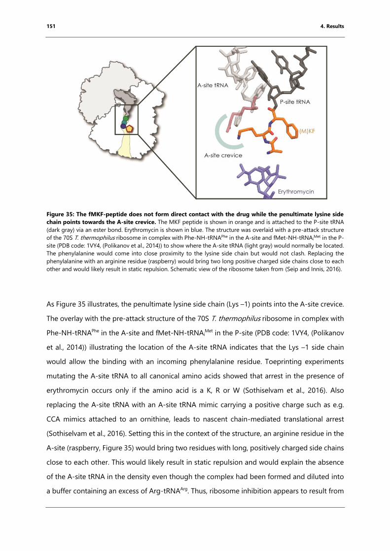
151 4. Results
Figure 35: The fMKF-peptide does not form direct contact with the drug while the penultimate lysine side
chain points towards the A-site crevice. The MKF peptide is shown in orange and is attached to the P-site tRNA
(dark gray) via an ester bond. Erythromycin is shown in blue. The structure was overlaid with a pre-attack structure
of the 70S T. thermophilus ribosome in complex with Phe-NH-tRNAPhe in the A-site and fMet-NH-tRNAiMet in the P-
site (PDB code: 1VY4, (Polikanov et al., 2014)) to show where the A-site tRNA (light gray) would normally be located.
The phenylalanine would come into close proximity to the lysine side chain but would not clash. Replacing the
phenylalanine with an arginine residue (raspberry) would bring two long positive charged side chains close to each
other and would likely result in static repulsion. Schematic view of the ribosome taken from (Seip and Innis, 2016).
As Figure 35 illustrates, the penultimate lysine side chain (Lys –1) points into the A-site crevice.
The overlay with the pre-attack structure of the 70S T. thermophilus ribosome in complex with
Phe-NH-tRNAPhe in the A-site and fMet-NH-tRNAiMet in the P-site (PDB code: 1VY4, (Polikanov
et al., 2014)) illustrating the location of the A-site tRNA indicates that the Lys –1 side chain
would allow the binding with an incoming phenylalanine residue. Toeprinting experiments
mutating the A-site tRNA to all canonical amino acids showed that arrest in the presence of
erythromycin occurs only if the amino acid is a K, R or W (Sothiselvam et al., 2016). Also
replacing the A-site tRNA with an A-site tRNA mimic carrying a positive charge such as e.g.
CCA mimics attached to an ornithine, leads to nascent chain-mediated translational arrest
(Sothiselvam et al., 2016). Setting this in the context of the structure, an arginine residue in the
A-site (raspberry, Figure 35) would bring two residues with long, positively charged side chains
close to each other. This would likely result in static repulsion and would explain the absence
of the A-site tRNA in the density even though the complex had been formed and diluted into
a buffer containing an excess of Arg-tRNAArg. Thus, ribosome inhibition appears to result from
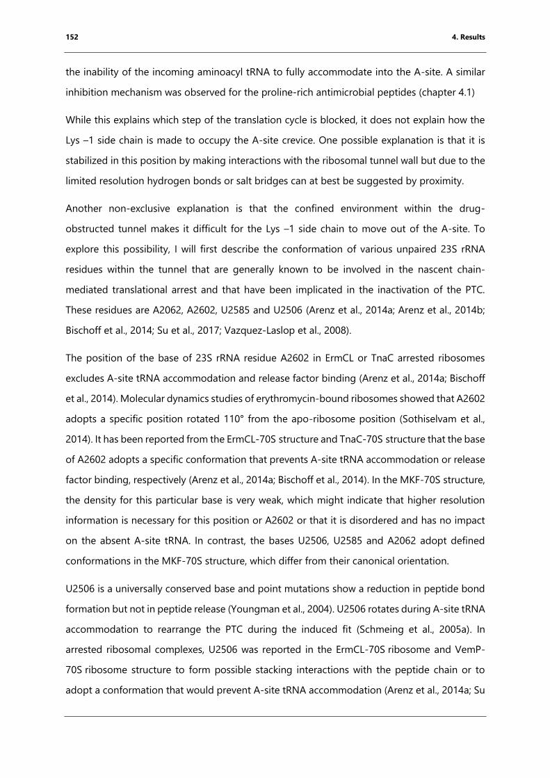
152 4. Results
the inability of the incoming aminoacyl tRNA to fully accommodate into the A-site. A similar
inhibition mechanism was observed for the proline-rich antimicrobial peptides (chapter 4.1)
While this explains which step of the translation cycle is blocked, it does not explain how the
Lys –1 side chain is made to occupy the A-site crevice. One possible explanation is that it is
stabilized in this position by making interactions with the ribosomal tunnel wall but due to the
limited resolution hydrogen bonds or salt bridges can at best be suggested by proximity.
Another non-exclusive explanation is that the confined environment within the drug-
obstructed tunnel makes it difficult for the Lys –1 side chain to move out of the A-site. To
explore this possibility, I will first describe the conformation of various unpaired 23S rRNA
residues within the tunnel that are generally known to be involved in the nascent chain-
mediated translational arrest and that have been implicated in the inactivation of the PTC.
These residues are A2062, A2602, U2585 and U2506 (Arenz et al., 2014a; Arenz et al., 2014b;
Bischoff et al., 2014; Su et al., 2017; Vazquez-Laslop et al., 2008).
The position of the base of 23S rRNA residue A2602 in ErmCL or TnaC arrested ribosomes
excludes A-site tRNA accommodation and release factor binding (Arenz et al., 2014a; Bischoff
et al., 2014). Molecular dynamics studies of erythromycin-bound ribosomes showed that A2602
adopts a specific position rotated 110° from the apo-ribosome position (Sothiselvam et al.,
2014). It has been reported from the ErmCL-70S structure and TnaC-70S structure that the base
of A2602 adopts a specific conformation that prevents A-site tRNA accommodation or release
factor binding, respectively (Arenz et al., 2014a; Bischoff et al., 2014). In the MKF-70S structure,
the density for this particular base is very weak, which might indicate that higher resolution
information is necessary for this position or A2602 or that it is disordered and has no impact
on the absent A-site tRNA. In contrast, the bases U2506, U2585 and A2062 adopt defined
conformations in the MKF-70S structure, which differ from their canonical orientation.
U2506 is a universally conserved base and point mutations show a reduction in peptide bond
formation but not in peptide release (Youngman et al., 2004). U2506 rotates during A-site tRNA
accommodation to rearrange the PTC during the induced fit (Schmeing et al., 2005a). In
arrested ribosomal complexes, U2506 was reported in the ErmCL-70S ribosome and VemP-
70S ribosome structure to form possible stacking interactions with the peptide chain or to
adopt a conformation that would prevent A-site tRNA accommodation (Arenz et al., 2014a; Su
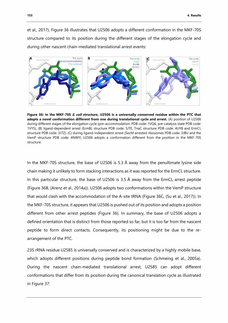
153 4. Results
et al., 2017). Figure 36 illustrates that U2506 adopts a different conformation in the MKF-70S
structure compared to its position during the different stages of the elongation cycle and
during other nascent chain-mediated translational arrest events:
Figure 36: In the MKF-70S E. coli structure, U2506 is a universally conserved residue within the PTC that
adopts a novel conformation different from one during translational cycle and arrest. (A) position of U2506
during different stages of the elongation cycle (pre-accommodation: PDB code: 1VQ6, pre-catalysis state PDB code:
1VY5), (B) ligand-dependent arrest (ErmBL structure PDB code: 5JTE, TnaC structure PDB code: 4UY8 and ErmCL
structure PDB code: 3J7Z). (C) during ligand-independent arrest (SecM arrested ribosomes PDB code: 3JBU and the
VemP structure PDB code: 4NWY) U2506 adopts a conformation different from the position in the MKF-70S
structure.
In the MKF-70S structure, the base of U2506 is 5.3 Å away from the penultimate lysine side
chain making it unlikely to form stacking interactions as it was reported for the ErmCL structure.
In this particular structure, the base of U2506 is 3.5 Å away from the ErmCL arrest peptide
(Figure 36B, (Arenz et al., 2014a)). U2506 adopts two conformations within the VemP structure
that would clash with the accommodation of the A-site tRNA (Figure 36C, (Su et al., 2017)). In
the MKF-70S structure, it appears that U2506 is pushed out of its position and adopts a position
different from other arrest peptides (Figure 36). In summary, the base of U2506 adopts a
defined orientation that is distinct from those reported so far, but it is too far from the nascent
peptide to form direct contacts. Consequently, its positioning might be due to the re-
arrangement of the PTC.
23S rRNA residue U2585 is universally conserved and is characterized by a highly mobile base,
which adopts different positions during peptide bond formation (Schmeing et al., 2005a).
During the nascent chain-mediated translational arrest, U2585 can adopt different
conformations that differ from its position during the canonical translation cycle as illustrated
in Figure 37:
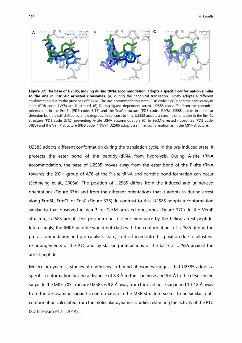
154 4. Results
Figure 37: The base of U2585, moving during tRNA accommodation, adopts a specific conformation similar
to the one in intrinsic arrested ribosomes. (A) during the canonical translation, U2585 adopts a different
conformation due to the presence of tRNAs. The pre-accommodation state (PDB code: 1VQ9) and the post-catalysis
state (PDB code: 1VY5) are illustrated. (B) During ligand-dependent arrest, U2585 can differ from the canonical
orientation. In the ErmBL (PDB code: 5JTE) and the TnaC structure (PDB code: 4UY8) U2585 points in a similar
direction but it is still shifted by a few degrees. In contrast to this, U2582 adopts a specific orientation in the ErmCL
structure (PDB code: 3J7Z) preventing A-site tRNA accommodation. (C) In SecM-arrested ribosomes (PDB code:
3JBU) and the VemP structure (PDB code: 4NWY), U2585 adopts a similar conformation as in the MKF-structure.
U2585 adopts different conformation during the translation cycle. In the pre-induced state, it
protects the ester bond of the peptidyl-tRNA from hydrolysis. During A-site tRNA
accommodation, the base of U2585 moves away from the ester bond of the P-site tRNA
towards the 2’OH group of A76 of the P-site tRNA and peptide bond formation can occur
(Schmeing et al., 2005a). The position of U2585 differs from the induced and uninduced
orientations (Figure 37A) and from the different orientations that it adopts in during arrest
along ErmBL, ErmCL or TnaC (Figure 37B). In contrast to this, U2585 adopts a conformation
similar to that observed in VemP- or SecM-arrested ribosomes (Figure 37C). In the VemP
structure, U2585 adopts this position due to steric hindrance by the helical arrest peptide.
Interestingly, the fMKF-peptide would not clash with the conformations of U2585 during the
pre-accommodation and pre-catalysis state, so it is forced into this position due to allosteric
re-arrangements of the PTC and by stacking interactions of the base of U2585 against the
arrest peptide.
Molecular dynamics studies of erythromycin-bound ribosomes suggest that U2585 adopts a
specific conformation having a distance of 8.3 Å to the cladinose and 9.6 Å to the desosamine
sugar. In the MKF-70Sstructure U2585 is 8.2 Å away from the cladinose sugar and 10-12 Å away
from the desosamine sugar. Its conformation in the MKF-structure seems to be similar to its
conformation calculated from the molecular dynamics studies restricting the activity of the PTC
(Sothiselvam et al., 2014).
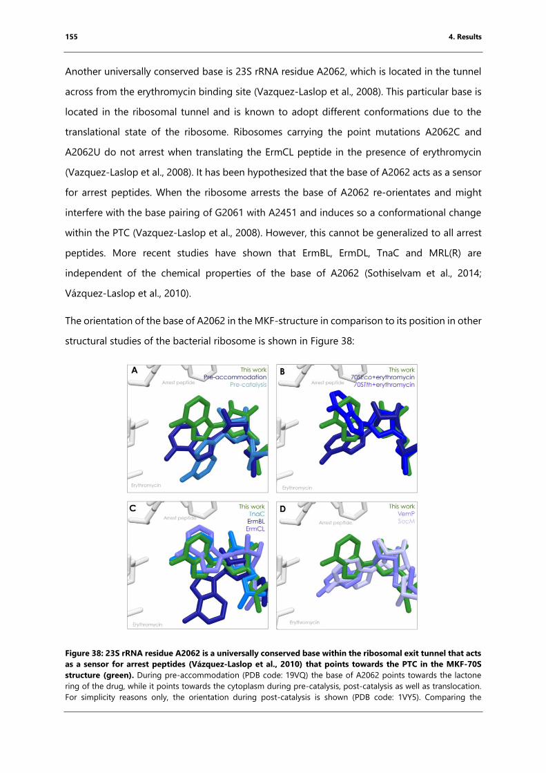
155 4. Results
Another universally conserved base is 23S rRNA residue A2062, which is located in the tunnel
across from the erythromycin binding site (Vazquez-Laslop et al., 2008). This particular base is
located in the ribosomal tunnel and is known to adopt different conformations due to the
translational state of the ribosome. Ribosomes carrying the point mutations A2062C and
A2062U do not arrest when translating the ErmCL peptide in the presence of erythromycin
(Vazquez-Laslop et al., 2008). It has been hypothesized that the base of A2062 acts as a sensor
for arrest peptides. When the ribosome arrests the base of A2062 re-orientates and might
interfere with the base pairing of G2061 with A2451 and induces so a conformational change
within the PTC (Vazquez-Laslop et al., 2008). However, this cannot be generalized to all arrest
peptides. More recent studies have shown that ErmBL, ErmDL, TnaC and MRL(R) are
independent of the chemical properties of the base of A2062 (Sothiselvam et al., 2014;
Vázquez‐Laslop et al., 2010).
The orientation of the base of A2062 in the MKF-structure in comparison to its position in other
structural studies of the bacterial ribosome is shown in Figure 38:
Figure 38: 23S rRNA residue A2062 is a universally conserved base within the ribosomal exit tunnel that acts
as a sensor for arrest peptides (Vázquez‐Laslop et al., 2010) that points towards the PTC in the MKF-70S
structure (green). During pre-accommodation (PDB code: 19VQ) the base of A2062 points towards the lactone
ring of the drug, while it points towards the cytoplasm during pre-catalysis, post-catalysis as well as translocation.
For simplicity reasons only, the orientation during post-catalysis is shown (PDB code: 1VY5). Comparing the
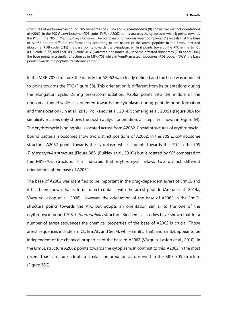
156 4. Results
structures of erythromycin-bound 70S ribosomes of E. coli and T. thermophilus (B) shows two distinct orientations
of A2062. In the 70S E. coli ribosome (PDB code: 4V7U), A2062 points towards the cytoplasm, while it points towards
the PTC in the 70S T. thermophilus ribosome. The comparison of various arrest complexes (C) reveals that the base
of A2062 adopts different conformations according to the nature of the arrest peptide. In the ErmBL arrested
ribosome (PDB code: 5JTE) the base points towards the cytoplasm, while it points towards the PTC in the ErmCL
(PDB code: 3J7Z) and TnaC (PDB code: 4UY8) arrested ribosomes. (D) In SecM-arrested ribosomes (PDB code: 3JBU)
the base points in a similar direction as in MFK-70S while in VemP-arrested ribosomes (PDB code: 4NWY) the base
points towards the peptidyl transferase center.
In the MKF-70S structure, the density for A2062 was clearly defined and the base was modeled
to point towards the PTC (Figure 38). This orientation is different from its orientations during
the elongation cycle. During pre-accommodation, A2062 points into the middle of the
ribosomal tunnel while it is oriented towards the cytoplasm during peptide bond formation
and translocation (Lin et al., 2015; Polikanov et al., 2014; Schmeing et al., 2005a)(Figure 38A for
simplicity reasons only shows the post-catalysis orientation, all steps are shown in Figure 64).
The erythromycin binding site is located across from A2062. Crystal structures of erythromycin-
bound bacterial ribosomes show two distinct positions of A2062. In the 70S E. coli ribosome
structure, A2062 points towards the cytoplasm while it points towards the PTC in the 70S
T. thermophilus structure (Figure 38B, (Bulkley et al., 2010)) but is rotated by 90° compared to
the MKF-70S structure. This indicates that erythromycin allows two distinct different
orientations of the base of A2062.
The base of A2062 was identified to be important in the drug-dependent arrest of ErmCL and
it has been shown that is forms direct contacts with the arrest peptide (Arenz et al., 2014a;
Vazquez-Laslop et al., 2008). However, the orientation of the base of A2062 in the ErmCL
structure points towards the PTC but adopts an orientation similar to the one of the
erythromycin bound 70S T. thermophilus structure. Biochemical studies have shown that for a
number of arrest sequences the chemical properties of the base of A2062 is crucial. Those
arrest sequences include ErmCL, ErmAL, and SecM, while ErmBL, TnaC and ErmDL appear to be
independent of the chemical properties of the base of A2062 (Vázquez‐Laslop et al., 2010). In
the ErmBL structure A2062 points towards the cytoplasm. In contrast to this, A2062 in the most
recent TnaC structure adopts a similar conformation as observed in the MKF-70S structure
(Figure 38C).
157 4. Results
In the most recent SecM structure A2062 adopts a slightly different orientation compared to
the MKF-70S structure but points towards the PTC, while in the VemP structure the base points
into the ribosomal exit tunnel (Figure 38D, (Su et al., 2017; Zhang et al., 2015)).
Like TnaC, it has been shown that MRLR-mediated arrest in the presence of erythromycin is
independent of the nature of the A2062 (Sothiselvam et al., 2014). However, molecular
dynamics studies of SecM and TnaC arrested ribosomes show a reduction of the flexibility of
A2062 (Gumbart et al., 2012). In contrast to ErmCL, the base of A2062 is 4.5 Å away from the
backbone of the MKF peptide and consequently cannot form any hydrogen bonding.
In light of the MKF-70S structure, however, it is possible that even though the nature of residue
2062 is not important for the arrest process, the mere presence of a base within the tunnel at
this location is sufficient to cause arrest. Indeed, the base of A2062 could prevent the necessary
peptide backbone rotation to allow Lys -1out of the A-site crevice, as illustrated in Figure 39.
Figure 39: Proposed mechanism of MKF-mediated translational arrest in the presence of erythromycin. In
the absence of erythromycin A2062 points towards the cytoplasm. This allows the rotation of the peptide backbone
of the MKF peptide and the reorientation of the lysine side chain. Consequently, the following positive A-site tRNA
can bind and peptide bond formation can occur. In contrast to this, the presence of erythromycin limits the flexibility
of A2062 and in the presence of the peptide this particular base points up towards the PTC. This affects directly the
accommodation of the positively charged amino acyl-tRNA and the ribosome undergoes peptide-mediated
translational arrest.
158 4. Results
In summary, the base of A2062 rotates more easily towards the cytoplasm, thus allowing Lys –
1 to leave the A-site crevice in the absence of erythromycin. Consequently, the following amino
acyl-tRNA carrying a positive charge can bind and peptide bond formation can occur (Figure
39). In contrast to this, the presence of erythromycin could favor a conformation of A2062 in
which the base points towards the PTC. The ribosome arrests when A2062 points towards the
PTC inhibiting the reorientation of the arrest peptide and forcing the Lys -1 to point into the
A-site crevice. Consequently, the following A-site tRNA carrying a K, R or W side chain cannot
bind to the PTC due to steric and static hindrance. The ribosome is arrested due to the
prevention of A-site tRNA accommodation (Figure 39).
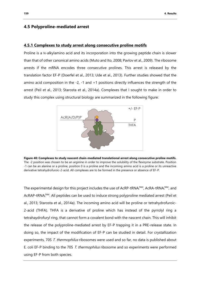
159 4. Results
4.5 Polyproline-mediated arrest
4.5.1 Complexes to study arrest along consecutive proline motifs
Proline is a N-alkylamino acid and its incorporation into the growing peptide chain is slower
than that of other canonical amino acids (Muto and Ito, 2008; Pavlov et al., 2009). The ribosome
arrests if the mRNA encodes three consecutive prolines. This arrest is released by the
translation factor EF-P (Doerfel et al., 2013; Ude et al., 2013). Further studies showed that the
amino acid composition in the -2, -1 and +1 positions directly influences the strength of the
arrest (Peil et al., 2013; Starosta et al., 2014a). Complexes that I sought to make in order to
study this complex using structural biology are summarized in the following figure:
Figure 40: Complexes to study nascent chain-mediated translational arrest along consecutive proline motifs.
The -2 position was chosen to be an arginine in order to improve the solubility of the flexizyme substrate. Position
-1 can be an alanine or a proline, position 0 is a proline and the incoming amino acid is a proline or its unreactive
derivative tetrahydrofuroic-2-acid. All complexes are to be formed in the presence or absence of EF-P.
The experimental design for this project includes the use of AcRP-tRNAiMet, AcRA-tRNAi
Met, and
AcRAP-tRNAiMet. All peptides can be used to induce strong polyproline mediated arrest (Peil et
al., 2013; Starosta et al., 2014a). The incoming amino acid will be proline or tetrahydrofuroic-
2-acid (THFA). THFA is a derivative of proline which has instead of the pyrrolyl ring a
tetrahaydrofuryl ring, that cannot form a covalent bond with the nascent chain. This will inhibit
the release of the polyproline-mediated arrest by EF-P trapping it in a PRE-release state. In
doing so, the impact of the modification of EF-P can be studied in detail. For crystallization
experiments, 70S T. thermophilus ribosomes were used and so far, no data is published about
E. coli EF-P binding to the 70S T. thermophilus ribosome and so experiments were performed
using EF-P from both species.
160 4. Results
A recent study showed that the D-loop of tRNAPro is crucial for EF-P activity (Katoh et al., 2016).
Therefore, E. coli tRNAPro had to be purified from cells and prolinylated in vitro in order to be
used for amino acid delivery. The Pro-tRNAPro can be used for one round of translocation
replacing the initiator tRNA by the tRNAPro and so the “native” complex can be studied. The
complex can be translocated using purified E. coli EF-G overexpressed recombinantly
(Mikolajka et al., 2011).
4.5.2 tRNAPro expression and purification
Recent studies showed that the D-loop of tRNAPro is essential for the function of EF-P (Katoh
et al., 2016). In this study, the authors used flexizymes to prolinylate different in vitro
transcribed tRNAs with varying lengths of the D- loop and base pairing strength depending on
the nucleotide composition of the D-stem. To investigate the molecular mechanism of arrest
along consecutive prolines structurally, tRNAPro was isolated from cells to ensure the presence
of natural nucleotide modifications. E. coli cells have three different genes for tRNAPro, named
proK, proL, and proM. As proK is transcribed as a monocistronic transcript (Mohanty et al.,
2016), this tRNAPro gene was selected for cloning. In addition, the CGG anticodon of proK
results in tRNAPro that is incapable of wobble base pairing. Consequently, proK is the only
tRNAPro that specifically recognizes the CCG codon.
To obtain large amounts of modified proK, two expression strategies were used by cloning the
gene into two different types of vectors, as indicated in Figure 41:
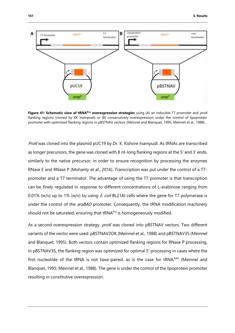
161 4. Results
Figure 41: Schematic view of tRNAPro overexpression strategies using (A) an inducible T7 promoter and proK
flanking regions (cloned by KK Inampudi) or (B) consecutively overexpression under the control of lipoprotein
promoter with optimized flanking regions in pBSTNAV vectors (Meinnel and Blanquet, 1995; Meinnel et al., 1988).
ProK was cloned into the plasmid pUC19 by Dr. K. Kishore Inampudi. As tRNAs are transcribed
as longer precursors, the gene was cloned with 8 nt-long flanking regions at the 5’ and 3’ ends,
similarly to the native precursor, in order to ensure recognition by processing the enzymes
RNase E and RNase P (Mohanty et al., 2016). Transcription was put under the control of a T7-
promoter and a T7 terminator. The advantage of using the T7 promoter is that transcription
can be finely regulated in response to different concentrations of L-arabinose ranging from
0.01% (w/v) up to 1% (w/v) by using E. coli BL21AI cells where the gene for T7 polymerase is
under the control of the araBAD promoter. Consequently, the tRNA modification machinery
should not be saturated, ensuring that tRNAPro is homogeneously modified.
As a second overexpression strategy, proK was cloned into pBSTNAV vectors. Two different
variants of the vector were used: pBSTNAV2OK (Meinnel et al., 1988) and pBSTNAV3S (Meinnel
and Blanquet, 1995). Both vectors contain optimized flanking regions for RNase P processing.
In pBSTNAV3S, the flanking region was optimized for optimal 5’ processing in cases where the
first nucleotide of the tRNA is not base-paired, as is the case for tRNAiMet (Meinnel and
Blanquet, 1995; Meinnel et al., 1988). The gene is under the control of the lipoprotein promoter
resulting in constitutive overexpression.
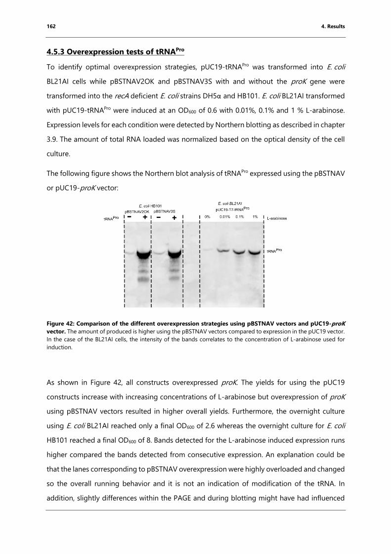
162 4. Results
4.5.3 Overexpression tests of tRNAPro
To identify optimal overexpression strategies, pUC19-tRNAPro was transformed into E. coli
BL21AI cells while pBSTNAV2OK and pBSTNAV3S with and without the proK gene were
transformed into the recA deficient E. coli strains DH5α and HB101. E. coli BL21AI transformed
with pUC19-tRNAPro were induced at an OD600 of 0.6 with 0.01%, 0.1% and 1 % L-arabinose.
Expression levels for each condition were detected by Northern blotting as described in chapter
3.9. The amount of total RNA loaded was normalized based on the optical density of the cell
culture.
The following figure shows the Northern blot analysis of tRNAPro expressed using the pBSTNAV
or pUC19-proK vector:
Figure 42: Comparison of the different overexpression strategies using pBSTNAV vectors and pUC19-proK
vector. The amount of produced is higher using the pBSTNAV vectors compared to expression in the pUC19 vector.
In the case of the BL21AI cells, the intensity of the bands correlates to the concentration of L-arabinose used for
induction.
As shown in Figure 42, all constructs overexpressed proK. The yields for using the pUC19
constructs increase with increasing concentrations of L-arabinose but overexpression of proK
using pBSTNAV vectors resulted in higher overall yields. Furthermore, the overnight culture
using E. coli BL21AI reached only a final OD600 of 2.6 whereas the overnight culture for E. coli
HB101 reached a final OD600 of 8. Bands detected for the L-arabinose induced expression runs
higher compared the bands detected from consecutive expression. An explanation could be
that the lanes corresponding to pBSTNAV overexpression were highly overloaded and changed
so the overall running behavior and it is not an indication of modification of the tRNA. In
addition, slightly differences within the PAGE and during blotting might have had influenced
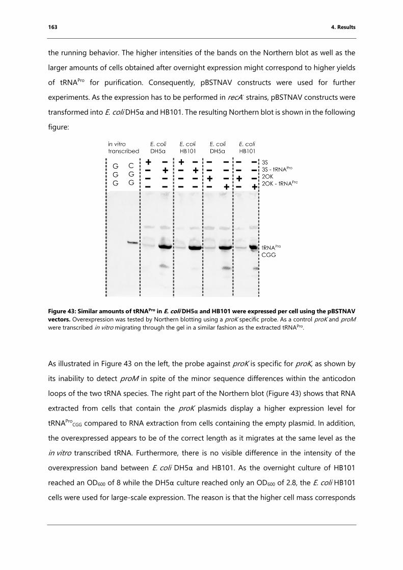
163 4. Results
the running behavior. The higher intensities of the bands on the Northern blot as well as the
larger amounts of cells obtained after overnight expression might correspond to higher yields
of tRNAPro for purification. Consequently, pBSTNAV constructs were used for further
experiments. As the expression has to be performed in recA- strains, pBSTNAV constructs were
transformed into E. coli DH5α and HB101. The resulting Northern blot is shown in the following
figure:
Figure 43: Similar amounts of tRNAPro in E. coli DH5α and HB101 were expressed per cell using the pBSTNAV
vectors. Overexpression was tested by Northern blotting using a proK specific probe. As a control proK and proM
were transcribed in vitro migrating through the gel in a similar fashion as the extracted tRNAPro.
As illustrated in Figure 43 on the left, the probe against proK is specific for proK, as shown by
its inability to detect proM in spite of the minor sequence differences within the anticodon
loops of the two tRNA species. The right part of the Northern blot (Figure 43) shows that RNA
extracted from cells that contain the proK plasmids display a higher expression level for
tRNAProCGG compared to RNA extraction from cells containing the empty plasmid. In addition,
the overexpressed appears to be of the correct length as it migrates at the same level as the
in vitro transcribed tRNA. Furthermore, there is no visible difference in the intensity of the
overexpression band between E. coli DH5α and HB101. As the overnight culture of HB101
reached an OD600 of 8 while the DH5α culture reached only an OD600 of 2.8, the E. coli HB101
cells were used for large-scale expression. The reason is that the higher cell mass corresponds
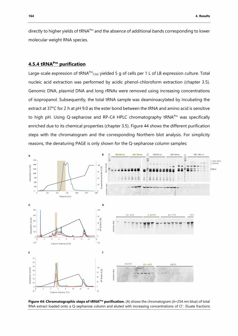
164 4. Results
directly to higher yields of tRNAPro and the absence of additional bands corresponding to lower
molecular weight RNA species.
4.5.4 tRNAPro purification
Large-scale expression of tRNAProCGG yielded 5 g of cells per 1 L of LB expression culture. Total
nucleic acid extraction was performed by acidic phenol-chloroform extraction (chapter 3.5).
Genomic DNA, plasmid DNA and long rRNAs were removed using increasing concentrations
of isopropanol. Subsequently, the total tRNA sample was deaminoacylated by incubating the
extract at 37°C for 2 h at pH 9.0 as the ester bond between the tRNA and amino acid is sensitive
to high pH. Using Q-sepharose and RP-C4 HPLC chromatography tRNAPro was specifically
enriched due to its chemical properties (chapter 3.5). Figure 44 shows the different purification
steps with the chromatogram and the corresponding Northern blot analysis. For simplicity
reasons, the denaturing PAGE is only shown for the Q-sepharose column samples:
Figure 44: Chromatographic steps of tRNAPro purification. (A) shows the chromatogram (ʎ=254 nm blue) of total
RNA extract loaded onto a Q-sepharose column and eluted with increasing concentrations of Cl–. Eluate fractions
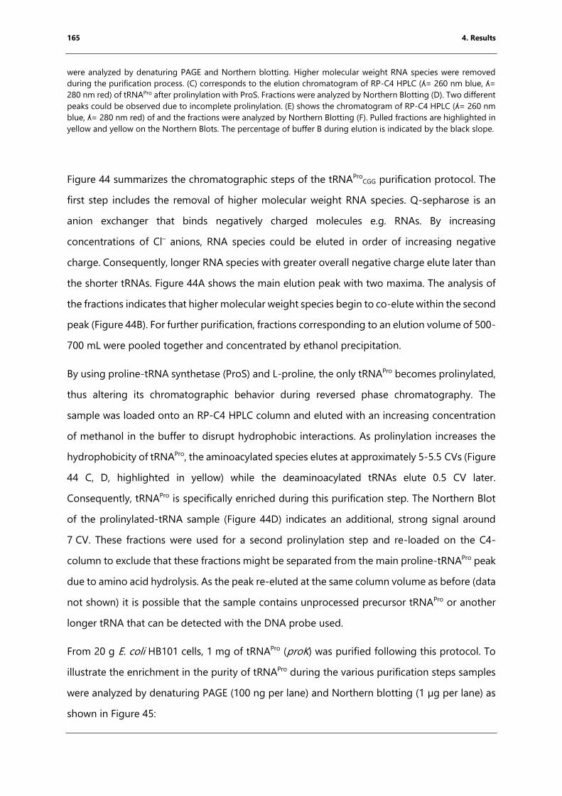
165 4. Results
were analyzed by denaturing PAGE and Northern blotting. Higher molecular weight RNA species were removed
during the purification process. (C) corresponds to the elution chromatogram of RP-C4 HPLC (ʎ= 260 nm blue, ʎ=
280 nm red) of tRNAPro after prolinylation with ProS. Fractions were analyzed by Northern Blotting (D). Two different
peaks could be observed due to incomplete prolinylation. (E) shows the chromatogram of RP-C4 HPLC (ʎ= 260 nm
blue, ʎ= 280 nm red) of and the fractions were analyzed by Northern Blotting (F). Pulled fractions are highlighted in
yellow and yellow on the Northern Blots. The percentage of buffer B during elution is indicated by the black slope.
Figure 44 summarizes the chromatographic steps of the tRNAProCGG purification protocol. The
first step includes the removal of higher molecular weight RNA species. Q-sepharose is an
anion exchanger that binds negatively charged molecules e.g. RNAs. By increasing
concentrations of Cl– anions, RNA species could be eluted in order of increasing negative
charge. Consequently, longer RNA species with greater overall negative charge elute later than
the shorter tRNAs. Figure 44A shows the main elution peak with two maxima. The analysis of
the fractions indicates that higher molecular weight species begin to co-elute within the second
peak (Figure 44B). For further purification, fractions corresponding to an elution volume of 500-
700 mL were pooled together and concentrated by ethanol precipitation.
By using proline-tRNA synthetase (ProS) and L-proline, the only tRNAPro becomes prolinylated,
thus altering its chromatographic behavior during reversed phase chromatography. The
sample was loaded onto an RP-C4 HPLC column and eluted with an increasing concentration
of methanol in the buffer to disrupt hydrophobic interactions. As prolinylation increases the
hydrophobicity of tRNAPro, the aminoacylated species elutes at approximately 5-5.5 CVs (Figure
44 C, D, highlighted in yellow) while the deaminoacylated tRNAs elute 0.5 CV later.
Consequently, tRNAPro is specifically enriched during this purification step. The Northern Blot
of the prolinylated-tRNA sample (Figure 44D) indicates an additional, strong signal around
7 CV. These fractions were used for a second prolinylation step and re-loaded on the C4-
column to exclude that these fractions might be separated from the main proline-tRNAPro peak
due to amino acid hydrolysis. As the peak re-eluted at the same column volume as before (data
not shown) it is possible that the sample contains unprocessed precursor tRNAPro or another
longer tRNA that can be detected with the DNA probe used.
From 20 g E. coli HB101 cells, 1 mg of tRNAPro (proK) was purified following this protocol. To
illustrate the enrichment in the purity of tRNAPro during the various purification steps samples
were analyzed by denaturing PAGE (100 ng per lane) and Northern blotting (1 µg per lane) as
shown in Figure 45:
166 4. Results
Figure 45: tRNAPro purification steps analyzed by denaturing PAGE and Northern blotting. (A) denaturing
PAGE (100 ng RNA loaded per lane) and (B) Northern blot (loaded RNA 1 µg per lane) using a probe against the
tRNAPro anticodon stem. L corresponds to the Ambion RNA ladder. (1) corresponds to the total small RNA extract,
(2) are the pooled fractions from the Q-sepharose column, (3) are the pooled fractions from the C4-RP HPLC run
after prolinylation and (4) are the pooled fractions from the C4-RP HPLC run after deaminoacylation. The amount
of tRNAPro increases while the number of impurities decreases.
Figure 45 shows a summary of all purification steps by denaturing PAGE and Northern blotting.
For denaturing PAGE, 100 ng RNA were loaded and 1 µg RNA was loaded on the Northern blot
to be able to observe tRNAProCCG enrichment over the various purification steps. Denaturing
PAGE (Figure 45A) confirms that during the different purification steps the intensity of the band
corresponding to tRNAPro (proK) is higher while the intensities corresponding to higher
molecular weight RNAs decreases but still remains to a certain extent. Indeed, the tRNAPro
sample shows a double band even at the end of the purification procedure, which could
correspond to a contamination (approx. 20%) by another tRNA. The Northern blot (Figure 45B)
shows that the relative concentration of tRNAPro increases, as indicated by the progressively
greater intensity of the corresponding band over each purification step.
4.5.5 tRNAPro characterization
After purification, the different methods were used to assess the purity, extent of modification
and activity of the purified tRNAPro. First, the purification and modification state of tRNAPro was
analyzed using native mass spectrometry. To do so, a 10 µM tRNAPro solution was prepared in
such a way as to completely remove Na+ ions, which interfere with subsequent data
interpretation. This was achieved by performing repeated washes with 150 mM NH4(CH3CO2)
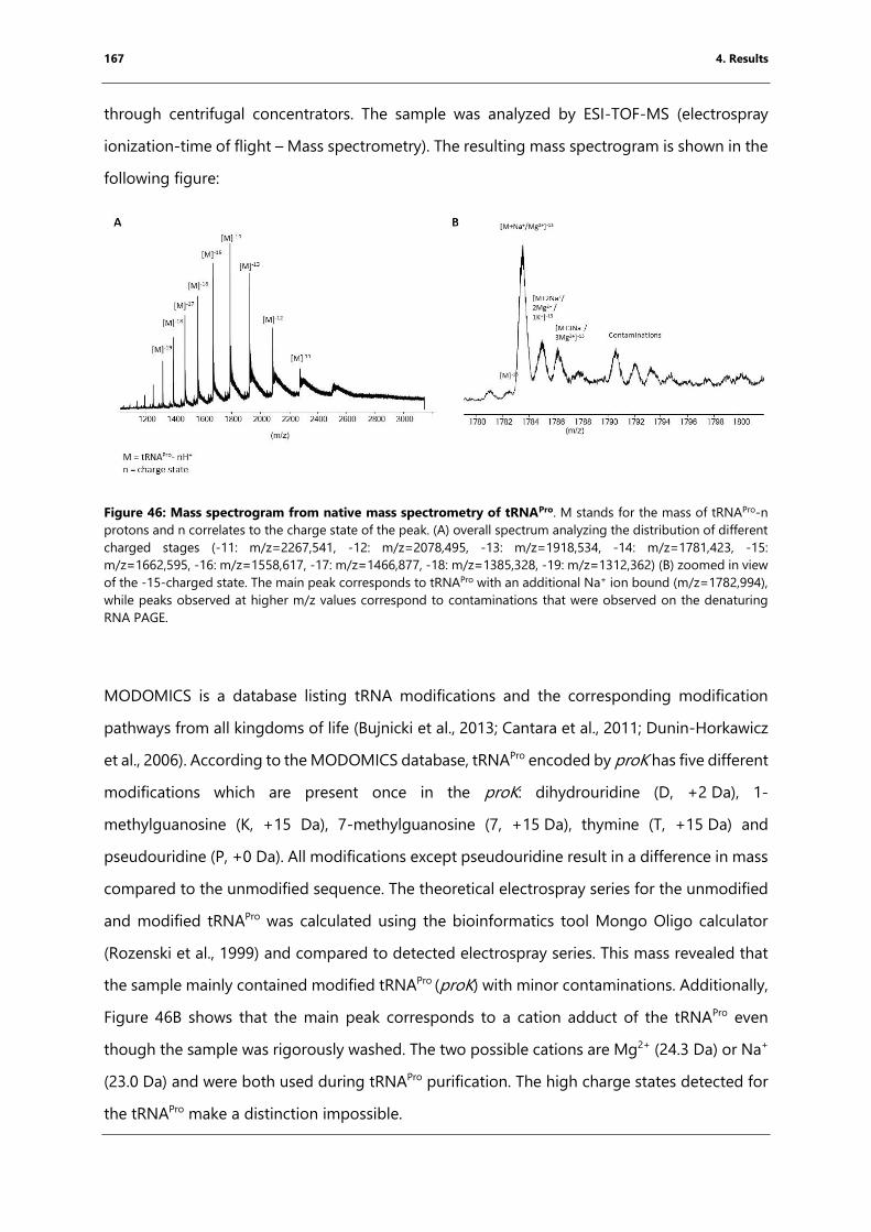
167 4. Results
through centrifugal concentrators. The sample was analyzed by ESI-TOF-MS (electrospray
ionization-time of flight – Mass spectrometry). The resulting mass spectrogram is shown in the
following figure:
Figure 46: Mass spectrogram from native mass spectrometry of tRNAPro. M stands for the mass of tRNAPro-n
protons and n correlates to the charge state of the peak. (A) overall spectrum analyzing the distribution of different
charged stages (-11: m/z=2267,541, -12: m/z=2078,495, -13: m/z=1918,534, -14: m/z=1781,423, -15:
m/z=1662,595, -16: m/z=1558,617, -17: m/z=1466,877, -18: m/z=1385,328, -19: m/z=1312,362) (B) zoomed in view
of the -15-charged state. The main peak corresponds to tRNAPro with an additional Na+ ion bound (m/z=1782,994),
while peaks observed at higher m/z values correspond to contaminations that were observed on the denaturing
RNA PAGE.
MODOMICS is a database listing tRNA modifications and the corresponding modification
pathways from all kingdoms of life (Bujnicki et al., 2013; Cantara et al., 2011; Dunin-Horkawicz
et al., 2006). According to the MODOMICS database, tRNAPro encoded by proK has five different
modifications which are present once in the proK: dihydrouridine (D, +2 Da), 1-
methylguanosine (K, +15 Da), 7-methylguanosine (7, +15 Da), thymine (T, +15 Da) and
pseudouridine (P, +0 Da). All modifications except pseudouridine result in a difference in mass
compared to the unmodified sequence. The theoretical electrospray series for the unmodified
and modified tRNAPro was calculated using the bioinformatics tool Mongo Oligo calculator
(Rozenski et al., 1999) and compared to detected electrospray series. This mass revealed that
the sample mainly contained modified tRNAPro (proK) with minor contaminations. Additionally,
Figure 46B shows that the main peak corresponds to a cation adduct of the tRNAPro even
though the sample was rigorously washed. The two possible cations are Mg2+ (24.3 Da) or Na+
(23.0 Da) and were both used during tRNAPro purification. The high charge states detected for
the tRNAPro make a distinction impossible.
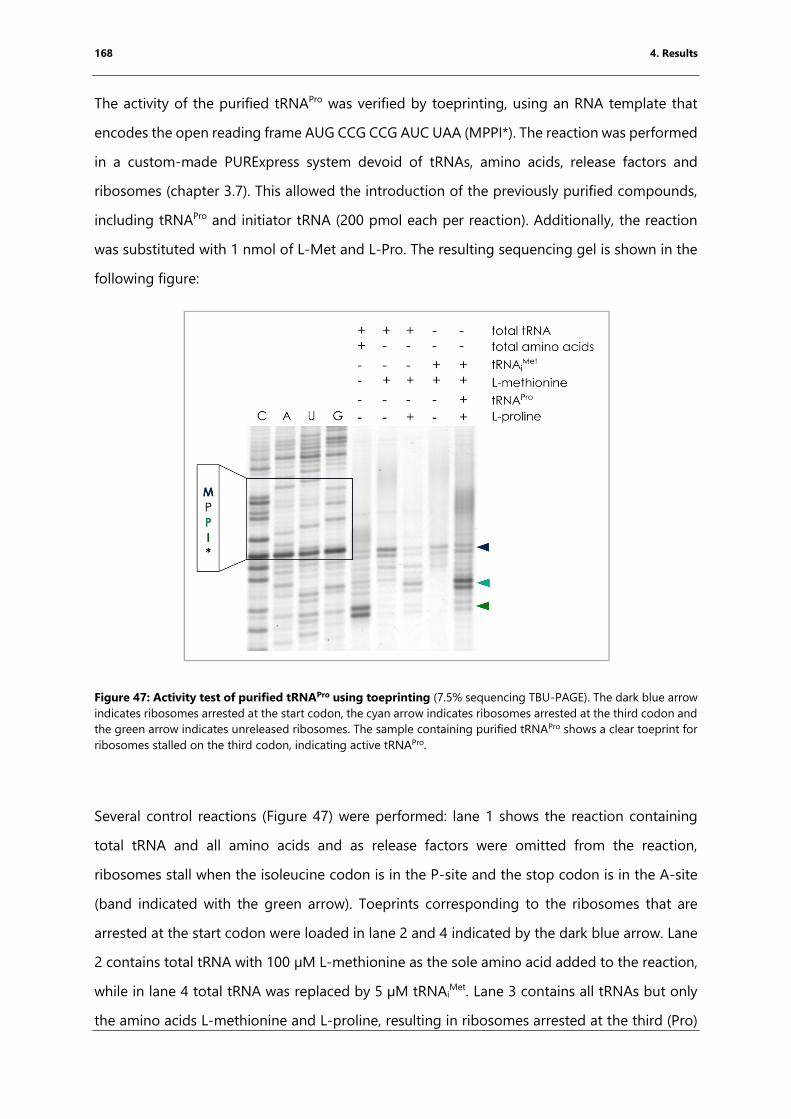
168 4. Results
The activity of the purified tRNAPro was verified by toeprinting, using an RNA template that
encodes the open reading frame AUG CCG CCG AUC UAA (MPPI*). The reaction was performed
in a custom-made PURExpress system devoid of tRNAs, amino acids, release factors and
ribosomes (chapter 3.7). This allowed the introduction of the previously purified compounds,
including tRNAPro and initiator tRNA (200 pmol each per reaction). Additionally, the reaction
was substituted with 1 nmol of L-Met and L-Pro. The resulting sequencing gel is shown in the
following figure:
Figure 47: Activity test of purified tRNAPro using toeprinting (7.5% sequencing TBU-PAGE). The dark blue arrow
indicates ribosomes arrested at the start codon, the cyan arrow indicates ribosomes arrested at the third codon and
the green arrow indicates unreleased ribosomes. The sample containing purified tRNAPro shows a clear toeprint for
ribosomes stalled on the third codon, indicating active tRNAPro.
Several control reactions (Figure 47) were performed: lane 1 shows the reaction containing
total tRNA and all amino acids and as release factors were omitted from the reaction,
ribosomes stall when the isoleucine codon is in the P-site and the stop codon is in the A-site
(band indicated with the green arrow). Toeprints corresponding to the ribosomes that are
arrested at the start codon were loaded in lane 2 and 4 indicated by the dark blue arrow. Lane
2 contains total tRNA with 100 µM L-methionine as the sole amino acid added to the reaction,
while in lane 4 total tRNA was replaced by 5 µM tRNAiMet. Lane 3 contains all tRNAs but only
the amino acids L-methionine and L-proline, resulting in ribosomes arrested at the third (Pro)
169 4. Results
codon (blue arrow). The same pattern is visible in the last lane, which contains the two amino
acids (100 µM each), 5 µM tRNAiMet and 5 µM purified tRNAPro. This indicates that the purified
tRNA is recognized by the proline-tRNA synthetase, recognizes the CCG codon and is
incorporated into the growing polypeptide chain. The additional band indicated with the cyan
arrow corresponds to ribosomes arrested at the stop codon. This indicates that the prior
detected impurity might correspond to tRNAIle. Thus, the purified tRNAPro is active in an in vitro
translation system.
To summarize, the purified tRNA contains a high proportion of active tRNAPro with minor
contaminations. The latter should subsequently be excluded during the correct decoding of
the CCG codon by the ribosome and therefore is unlikely to pose a problem for structural
studies.
4.5.6 Purification and activity of Elongation Factor P (EF-P)
Ribosomal arrest along consecutive proline motifs is released by the protein factor EF-P
(Doerfel et al., 2013; Ude et al., 2013). Various studies have shown that EF-P needs to be
modified post-translationally to be active. EF-P modifying enzyme knockout cells show in a
similar phenotype as efp knockout cells underlying the importance of the modification
(Bullwinkle et al., 2013; Navarre et al., 2010; Park et al., 2012). In E. coli, K34 of EF-P is β-
lysinylated by the enzymes YjeK (EpmA) and YjeA (EpmB). YjeK converts α-lysine into β-lysine
which is then transferred to K34 by YjeA (Bailly and de Crécy-Lagard, 2010; Park et al., 2012). In
a third step, this modified residue is hydroxylated by YfcM (EpmC), which is not essential to
obtain fully functional EF-P (Peil et al., 2012). In order to purify active, His6-tagged E. coli EF-P,
this protein was co-overexpressed with untagged YjeA and YjeK in E. coli BL21gold cells as
described previously (Peil et al., 2012). The activity of the purified E. coli EF-P was tested by
toeprinting and the results are shown in Figure 48:
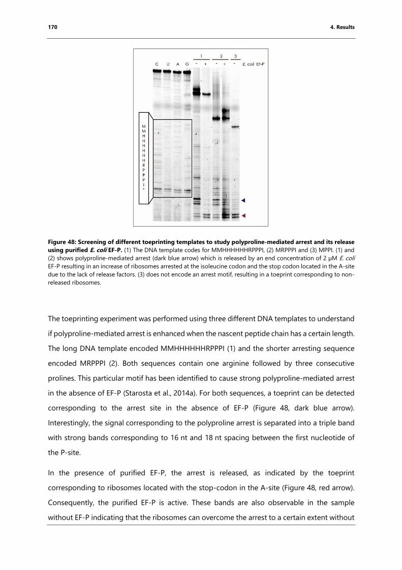
170 4. Results
Figure 48: Screening of different toeprinting templates to study polyproline-mediated arrest and its release
using purified E. coli EF-P. (1) The DNA template codes for MMHHHHHHRPPPI, (2) MRPPPI and (3) MPPI. (1) and
(2) shows polyproline-mediated arrest (dark blue arrow) which is released by an end concentration of 2 µM E. coli
EF-P resulting in an increase of ribosomes arrested at the isoleucine codon and the stop codon located in the A-site
due to the lack of release factors. (3) does not encode an arrest motif, resulting in a toeprint corresponding to non-
released ribosomes.
The toeprinting experiment was performed using three different DNA templates to understand
if polyproline-mediated arrest is enhanced when the nascent peptide chain has a certain length.
The long DNA template encoded MMHHHHHHRPPPI (1) and the shorter arresting sequence
encoded MRPPPI (2). Both sequences contain one arginine followed by three consecutive
prolines. This particular motif has been identified to cause strong polyproline-mediated arrest
in the absence of EF-P (Starosta et al., 2014a). For both sequences, a toeprint can be detected
corresponding to the arrest site in the absence of EF-P (Figure 48, dark blue arrow).
Interestingly, the signal corresponding to the polyproline arrest is separated into a triple band
with strong bands corresponding to 16 nt and 18 nt spacing between the first nucleotide of
the P-site.
In the presence of purified EF-P, the arrest is released, as indicated by the toeprint
corresponding to ribosomes located with the stop-codon in the A-site (Figure 48, red arrow).
Consequently, the purified EF-P is active. These bands are also observable in the sample
without EF-P indicating that the ribosomes can overcome the arrest to a certain extent without
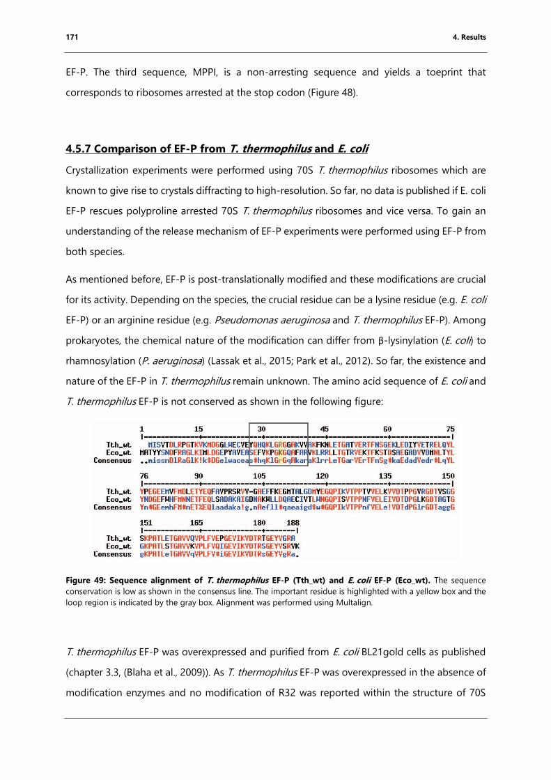
171 4. Results
EF-P. The third sequence, MPPI, is a non-arresting sequence and yields a toeprint that
corresponds to ribosomes arrested at the stop codon (Figure 48).
4.5.7 Comparison of EF-P from T. thermophilus and E. coli
Crystallization experiments were performed using 70S T. thermophilus ribosomes which are
known to give rise to crystals diffracting to high-resolution. So far, no data is published if E. coli
EF-P rescues polyproline arrested 70S T. thermophilus ribosomes and vice versa. To gain an
understanding of the release mechanism of EF-P experiments were performed using EF-P from
both species.
As mentioned before, EF-P is post-translationally modified and these modifications are crucial
for its activity. Depending on the species, the crucial residue can be a lysine residue (e.g. E. coli
EF-P) or an arginine residue (e.g. Pseudomonas aeruginosa and T. thermophilus EF-P). Among
prokaryotes, the chemical nature of the modification can differ from β-lysinylation (E. coli) to
rhamnosylation (P. aeruginosa) (Lassak et al., 2015; Park et al., 2012). So far, the existence and
nature of the EF-P in T. thermophilus remain unknown. The amino acid sequence of E. coli and
T. thermophilus EF-P is not conserved as shown in the following figure:
Figure 49: Sequence alignment of T. thermophilus EF-P (Tth_wt) and E. coli EF-P (Eco_wt). The sequence
conservation is low as shown in the consensus line. The important residue is highlighted with a yellow box and the
loop region is indicated by the gray box. Alignment was performed using Multalign.
T. thermophilus EF-P was overexpressed and purified from E. coli BL21gold cells as published
(chapter 3.3, (Blaha et al., 2009)). As T. thermophilus EF-P was overexpressed in the absence of
modification enzymes and no modification of R32 was reported within the structure of 70S
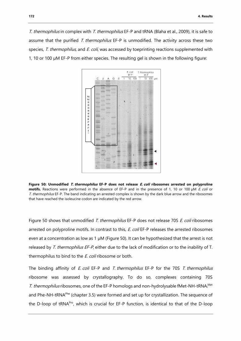
172 4. Results
T. thermophilus in complex with T. thermophilus EF-P and tRNA (Blaha et al., 2009), it is safe to
assume that the purified T. thermophilus EF-P is unmodified. The activity across these two
species, T. thermophilus, and E. coli, was accessed by toeprinting reactions supplemented with
1, 10 or 100 µM EF-P from either species. The resulting gel is shown in the following figure:
Figure 50: Unmodified T. thermophilus EF-P does not release E. coli ribosomes arrested on polyproline
motifs. Reactions were performed in the absence of EF-P and in the presence of 1, 10 or 100 µM E. coli or
T. thermophilus EF-P. The band indicating an arrested complex is shown by the dark blue arrow and the ribosomes
that have reached the isoleucine codon are indicated by the red arrow.
Figure 50 shows that unmodified T. thermophilus EF-P does not release 70S E. coli ribosomes
arrested on polyproline motifs. In contrast to this, E. coli EF-P releases the arrested ribosomes
even at a concentration as low as 1 µM (Figure 50). It can be hypothesized that the arrest is not
released by T. thermophilus EF-P, either due to the lack of modification or to the inability of T.
thermophilus to bind to the E. coli ribosome or both.
The binding affinity of E. coli EF-P and T. thermophilus EF-P for the 70S T. thermophilus
ribosome was assessed by crystallography. To do so, complexes containing 70S
T. thermophilus ribosomes, one of the EF-P homologs and non-hydrolysable fMet-NH-tRNAiMet
and Phe-NH-tRNAPhe (chapter 3.5) were formed and set up for crystallization. The sequence of
the D-loop of tRNAPro, which is crucial for EF-P function, is identical to that of the D-loop
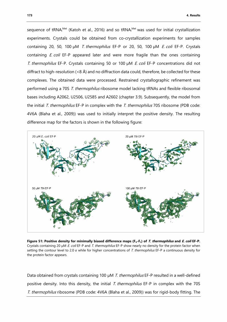
173 4. Results
sequence of tRNAiMet (Katoh et al., 2016) and so tRNAI
Met was used for initial crystallization
experiments. Crystals could be obtained from co-crystallization experiments for samples
containing 20, 50, 100 µM T. thermophilus EF-P or 20, 50, 100 µM E. coli EF-P. Crystals
containing E. coli EF-P appeared later and were more fragile than the ones containing
T. thermophilus EF-P. Crystals containing 50 or 100 µM E. coli EF-P concentrations did not
diffract to high-resolution (<8 Å) and no diffraction data could, therefore, be collected for these
complexes. The obtained data were processed. Restrained crystallographic refinement was
performed using a 70S T. thermophilus ribosome model lacking tRNAs and flexible ribosomal
bases including A2062, U2506, U2585 and A2602 (chapter 3.9). Subsequently, the model from
the initial T. thermophilus EF-P in complex with the T. thermophilus 70S ribosome (PDB code:
4V6A (Blaha et al., 2009)) was used to initially interpret the positive density. The resulting
difference map for the factors is shown in the following figure:
Figure 51: Positive density for minimally biased difference maps (Fo-Fc) of T. thermophilus and E. coli EF-P.
Crystals containing 20 µM E. coli EF-P and T. thermophilus EF-P show nearly no density for the protein factor when
setting the contour level to 2.0 σ while for higher concentrations of T. thermophilus EF-P a continuous density for
the protein factor appears.
Data obtained from crystals containing 100 µM T. thermophilus EF-P resulted in a well-defined
positive density. Into this density, the initial T. thermophilus EF-P in complex with the 70S
T. thermophilus ribosome (PDB code: 4V6A (Blaha et al., 2009)) was for rigid-body fitting. The
174 4. Results
same model was used to display the positive minimal biased difference map for the other
conditions (Figure 51). Complexes co-crystallized in the presence of 20 µM of E. coli or
T. thermophilus EF-P show nearly no density for the factor, which could clearly be seen when
higher concentrations of T. thermophilus EF-P were used (Figure 51).
In summary, no data could be obtained for E. coli EF-P bound to the 70S T. thermophilus
ribosome and polyproline arrested E. coli ribosomes were not released in the presence of
unmodified T. thermophilus EF-P.
4.5.8 Studies of T. thermophilus EF-P mutants containing the flexible loop region
from E. coli EF-P
One main question of this project was to study the impact of the modification of EF-P on the
release of polyproline-mediated arrest. As crystals that were grown in the presence of 70S
T. thermophilus ribosomes and high concentrations of E. coli EF-P, did not diffract to high-
resolution and as the modification for T. thermophilus EF-P remains unknown, a new strategy
was devised to overcome this issue, whereby T. thermophilus EF-P was systematically mutated
to introduce the amino acid sequence from E. coli EF-P that is crucial for the binding of the
modification enzymes without disrupting the binding of T. thermophilus EF-P to the 70S
T. thermophilus ribosome. To do so, the structure of E. coli EF-P in complex with its
modification enzymes ((Yanagisawa et al., 2010), PDB code: 3A5Z) and the crystal structure
containing 70S T. thermophilus ribosome and T. thermophilus EF-P ((Blaha et al., 2009) PDB
code: 4V6A) were analyzed in COOT. This led to the design of two different constructs:
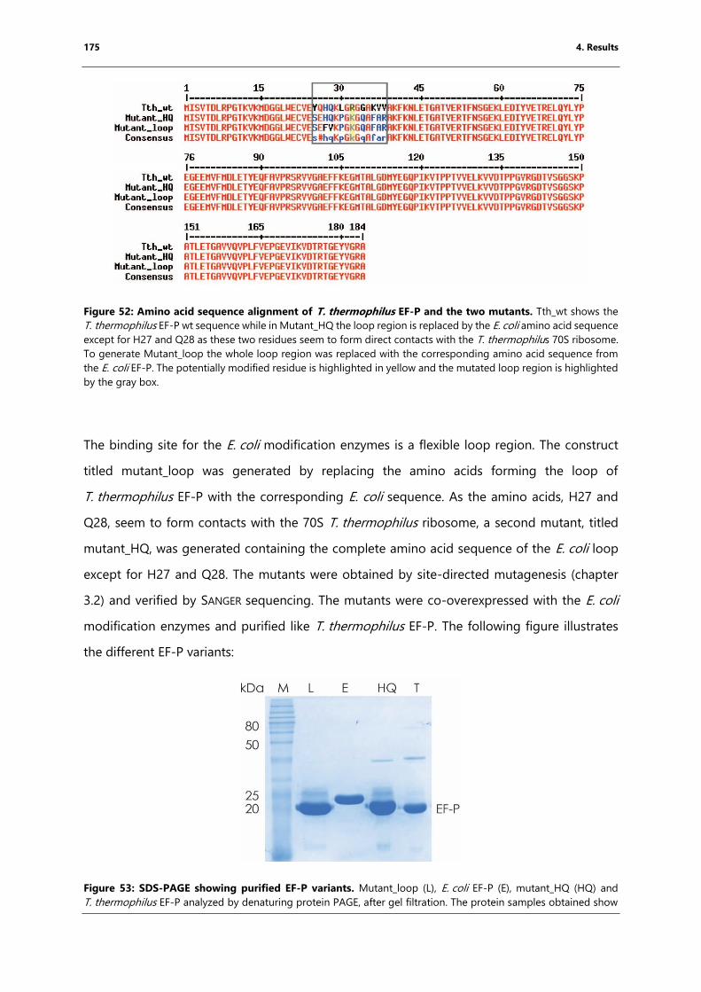
175 4. Results
Figure 52: Amino acid sequence alignment of T. thermophilus EF-P and the two mutants. Tth_wt shows the
T. thermophilus EF-P wt sequence while in Mutant_HQ the loop region is replaced by the E. coli amino acid sequence
except for H27 and Q28 as these two residues seem to form direct contacts with the T. thermophilus 70S ribosome.
To generate Mutant_loop the whole loop region was replaced with the corresponding amino acid sequence from
the E. coli EF-P. The potentially modified residue is highlighted in yellow and the mutated loop region is highlighted
by the gray box.
The binding site for the E. coli modification enzymes is a flexible loop region. The construct
titled mutant_loop was generated by replacing the amino acids forming the loop of
T. thermophilus EF-P with the corresponding E. coli sequence. As the amino acids, H27 and
Q28, seem to form contacts with the 70S T. thermophilus ribosome, a second mutant, titled
mutant_HQ, was generated containing the complete amino acid sequence of the E. coli loop
except for H27 and Q28. The mutants were obtained by site-directed mutagenesis (chapter
3.2) and verified by SANGER sequencing. The mutants were co-overexpressed with the E. coli
modification enzymes and purified like T. thermophilus EF-P. The following figure illustrates
the different EF-P variants:
Figure 53: SDS-PAGE showing purified EF-P variants. Mutant_loop (L), E. coli EF-P (E), mutant_HQ (HQ) and
T. thermophilus EF-P analyzed by denaturing protein PAGE, after gel filtration. The protein samples obtained show
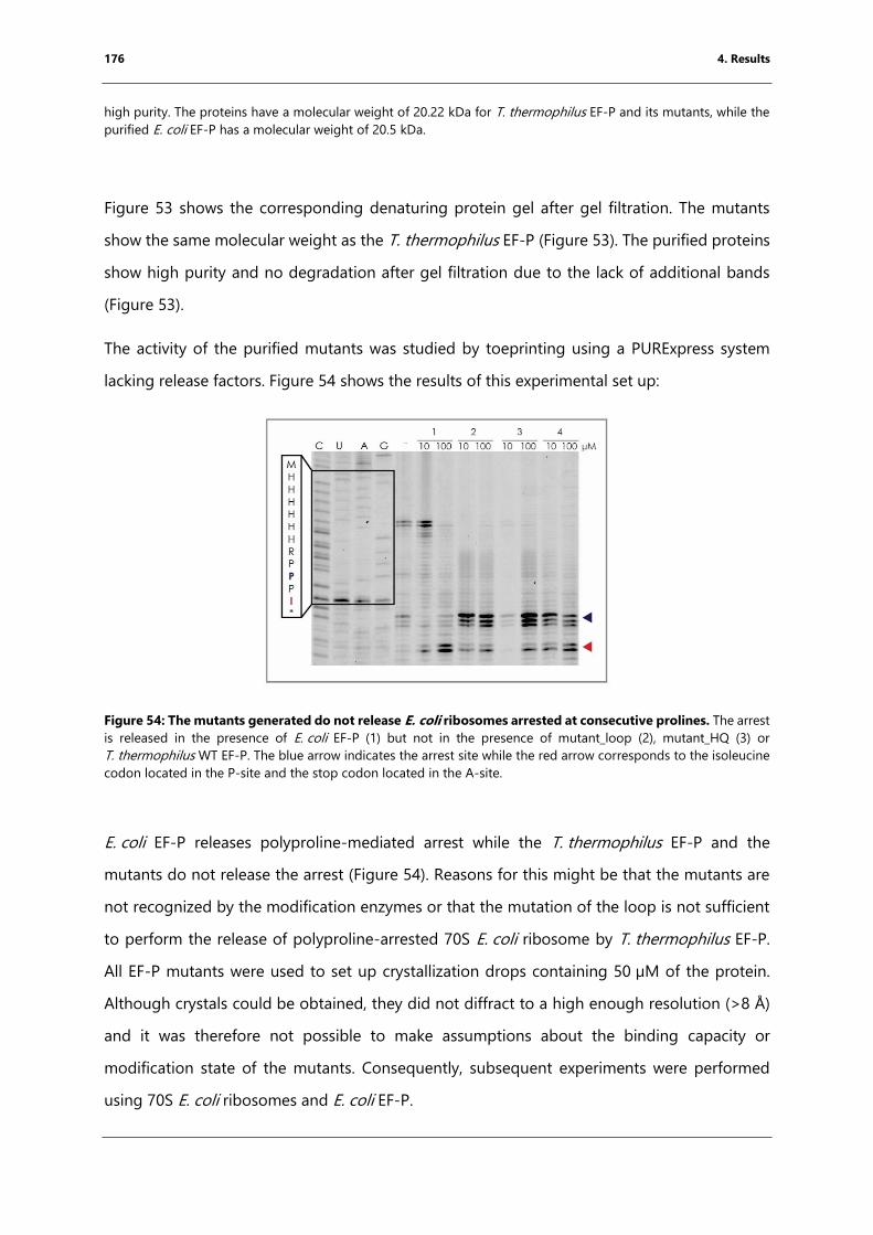
176 4. Results
high purity. The proteins have a molecular weight of 20.22 kDa for T. thermophilus EF-P and its mutants, while the
purified E. coli EF-P has a molecular weight of 20.5 kDa.
Figure 53 shows the corresponding denaturing protein gel after gel filtration. The mutants
show the same molecular weight as the T. thermophilus EF-P (Figure 53). The purified proteins
show high purity and no degradation after gel filtration due to the lack of additional bands
(Figure 53).
The activity of the purified mutants was studied by toeprinting using a PURExpress system
lacking release factors. Figure 54 shows the results of this experimental set up:
Figure 54: The mutants generated do not release E. coli ribosomes arrested at consecutive prolines. The arrest
is released in the presence of E. coli EF-P (1) but not in the presence of mutant_loop (2), mutant_HQ (3) or
T. thermophilus WT EF-P. The blue arrow indicates the arrest site while the red arrow corresponds to the isoleucine
codon located in the P-site and the stop codon located in the A-site.
E. coli EF-P releases polyproline-mediated arrest while the T. thermophilus EF-P and the
mutants do not release the arrest (Figure 54). Reasons for this might be that the mutants are
not recognized by the modification enzymes or that the mutation of the loop is not sufficient
to perform the release of polyproline-arrested 70S E. coli ribosome by T. thermophilus EF-P.
All EF-P mutants were used to set up crystallization drops containing 50 µM of the protein.
Although crystals could be obtained, they did not diffract to a high enough resolution (>8 Å)
and it was therefore not possible to make assumptions about the binding capacity or
modification state of the mutants. Consequently, subsequent experiments were performed
using 70S E. coli ribosomes and E. coli EF-P.
177 4. Results
4.5.9 Toeprint to study polyproline-mediated arrest and its release by EF-P
One of the main aims of this project was to obtain the structure of polyproline arrested
ribosomes and getting a grounded understanding how the arrest is released by EF-P. As the
D-loop of tRNAPro is crucial for the recognition by EF-P, the reaction was initiated from a
dipeptidylated initiator tRNA and so the complex can be formed using strategy 2.
For the poly-proline project, the dipeptides AcRP-CBT and AcRA-CBT, as well as the tripeptide
AcRAP-CBT, were used to obtain the corresponding peptidylated initiator tRNA using the
flexizyme technique (chapter 4.2). The activity of these peptidyl-tRNAsiMet was analyzed by
toeprinting using a mRNA template that encoded the open reading frame AUG CCG CCG AUC
UAA (MPPI*). The mRNA template encodes two proline codons in order to induce stalling only
in the presence of dipeptidylated or tripeptidlyted initiator tRNA on the second or first codon,
respectively. The reaction was performed using a custom-made PURExpress system (NEB,
Ipswich, MA, USA). 10 µM peptidyl-tRNAiMet, 20 µM tRNAPro and 100 µM L-proline were
incubated with 5 µM mRNA, factor mix, and 2.5 µM ribosomes in order to pre-form the
complex (5 min at RT). Subsequently, solution A-(minus aa, tRNA) provided by the kit,
containing nucleotides, was added, allowing complexes that had not been arrested to be
translocated (further information is provided in chapter 3.9). Certain reactions were
supplemented with E. coli EF-P in order to allow the release of the arrested complex. The
reaction was analyzed on a 7.5% sequencing PAGE and the result is shown Figure 55:
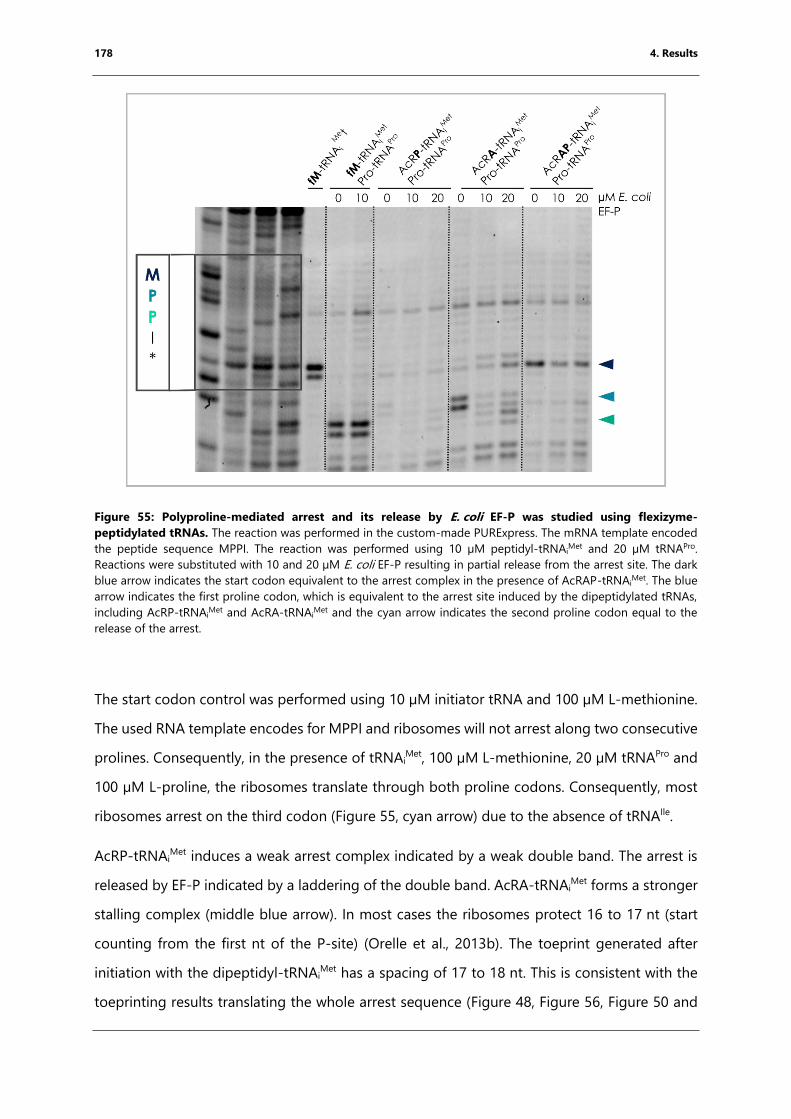
178 4. Results
Figure 55: Polyproline-mediated arrest and its release by E. coli EF-P was studied using flexizyme-
peptidylated tRNAs. The reaction was performed in the custom-made PURExpress. The mRNA template encoded
the peptide sequence MPPI. The reaction was performed using 10 µM peptidyl-tRNAiMet and 20 µM tRNAPro.
Reactions were substituted with 10 and 20 µM E. coli EF-P resulting in partial release from the arrest site. The dark
blue arrow indicates the start codon equivalent to the arrest complex in the presence of AcRAP-tRNAiMet. The blue
arrow indicates the first proline codon, which is equivalent to the arrest site induced by the dipeptidylated tRNAs,
including AcRP-tRNAiMet and AcRA-tRNAi
Met and the cyan arrow indicates the second proline codon equal to the
release of the arrest.
The start codon control was performed using 10 µM initiator tRNA and 100 µM L-methionine.
The used RNA template encodes for MPPI and ribosomes will not arrest along two consecutive
prolines. Consequently, in the presence of tRNAiMet, 100 µM L-methionine, 20 µM tRNAPro and
100 µM L-proline, the ribosomes translate through both proline codons. Consequently, most
ribosomes arrest on the third codon (Figure 55, cyan arrow) due to the absence of tRNAIle.
AcRP-tRNAiMet induces a weak arrest complex indicated by a weak double band. The arrest is
released by EF-P indicated by a laddering of the double band. AcRA-tRNAiMet forms a stronger
stalling complex (middle blue arrow). In most cases the ribosomes protect 16 to 17 nt (start
counting from the first nt of the P-site) (Orelle et al., 2013b). The toeprint generated after
initiation with the dipeptidyl-tRNAiMet has a spacing of 17 to 18 nt. This is consistent with the
toeprinting results translating the whole arrest sequence (Figure 48, Figure 56, Figure 50 and
179 4. Results
Figure 54). The band corresponding to the polyproline-mediated arrest was split into a triple
band corresponding to 16-18 nt (Figure 48, Figure 56, Figure 50 and Figure 54). An explanation
could be that the ribosome might adopt a different ratcheting state while being arrested on
polyproline motifs. The arrest is released in the presence of E. coli EF-P indicated by the release
of the arrest band into laddering.
AcRAP-tRNAiMet carries already the arrest sequence and so the arrest site in the absence of EF-
P is the start codon (dark blue arrow Figure 55). Addition of EF-P decreases the intensity of the
start codon band indicating partial release of the arrested ribosomes.
Overall, the polyproline-mediated arrest can be characterized in toeprint assays using
peptidylated initiator tRNA. The best stalling efficiency and then release by EF-P were observed
initiating the reaction with AcRA-tRNAiMet. A reason could be that the proline within the
flexizyme-charged peptide potentially has a restricted backbone geometry thus resulting in
reduced reactivity compared to the AcRA-tRNAiMet.
In summary, the complex can be formed using the PURExpress system from NEB but the
structure remains unknown. As crystals containing high concentrations of E. coli EF-P did not
diffract and the quality of the cryo-EM map for the MKF-70S structure showed a nice density
for the side chains, is promising to obtain a similar result for the polyproline project. The
strategy to obtain the complexes are discussed in further detail in the discussion (chapter 5.3).
180 4. Results
4.5.10 EF-P does not release other well-characterized nascent chain-mediated
translation arrest peptides
A recent in vivo study using fluorescently labeled E. coli EF-P has shown that EF-P binding
events are more frequent than the occurrence of polyproline-mediated translational arrest
(Mohapatra et al., 2017) raising the question for other functions of EF-P. Furthermore, an
in vitro study investigating the effect of miscoding of the P-site tRNA showed that these
miscoding events lead to a reduction in the translocation rate that the translocation rate can
be enhanced in the presence of EF-P (Alejo and Blanchard, 2017). Both studies illustrate that
EF-P might have multiple functions and can bind to ribosomes with empty E-sites (Alejo and
Blanchard, 2017; Mohapatra et al., 2017) which appears to be more frequent. In E. coli, EF-P is
a part of the ribosome rescue system that recognizes and releases stalled ribosomes.
Ribosomal stalling can be induced by various factors, including the absence of stop codons,
mRNA secondary structures, or amino acid starvation to name only a few examples. The
ribosome rescue system is formed by transfer-messenger RNA (tmRNA) and proteins including
e.g. RelA and LepA as reviewed by (Giudice and Gillet, 2013). The question whether EF-P might
be involved in the release of nascent chain-mediated translational arrested ribosomes stalled
by non-polyproline arrest peptides was addressed by performing toeprinting on the well-
characterized arrest peptides SecM, TnaC, ErmBL, ErmCL, and ErmDL. The results are shown in
Figure 56:
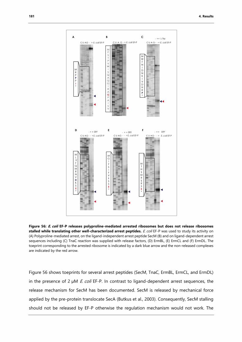
181 4. Results
Figure 56: E. coli EF-P releases polyproline-mediated arrested ribosomes but does not release ribosomes
stalled while translating other well-characterized arrest peptides. E. coli EF-P was used to study its activity on
(A) Polyproline-mediated arrest, on the ligand-independent arrest peptide SecM (B) and on ligand-dependent arrest
sequences including (C) TnaC reaction was supplied with release factors, (D) ErmBL, (E) ErmCL and (F) ErmDL. The
toeprint corresponding to the arrested ribosome is indicated by a dark blue arrow and the non-released complexes
are indicated by the red arrow.
Figure 56 shows toeprints for several arrest peptides (SecM, TnaC, ErmBL, ErmCL, and ErmDL)
in the presence of 2 µM E. coli EF-P. In contrast to ligand-dependent arrest sequences, the
release mechanism for SecM has been documented. SecM is released by mechanical force
applied by the pre-protein translocate SecA (Butkus et al., 2003). Consequently, SecM stalling
should not be released by EF-P otherwise the regulation mechanism would not work. The
182 4. Results
toeprint experiment (Figure 56B) shows that EF-P does not release SecM arrested ribosomes
in vitro and is used as a negative control.
In the presence of tryptophan, TnaC arrests when proline codon 24 is located in the P-site and
the stop codon is located in the A-site and the release of the peptide is inhibited. To be able
to determine arrest conditions from non-arrest conditions the reaction was supplemented with
releases factors provided by the kit. On the other hand, the Erm leader peptides arrest
ribosomes on internal codons in response to erythromycin. Therefore, release factors could be
omitted from the reaction in this case. Ribosomes arrested in the presence of ligands are
indicated by dark blue arrows in Figure 56. In all cases, reactions containing EF-P still show a
strong signal for the arrested complexes (Figure 56C-E) while ribosomes arrested on
polyproline stretches are released (Figure 56A). This means that the arrest peptides SecM, TnaC,
ErmBL, ErmCL, and ErmDL are not released by EF-P in vitro.
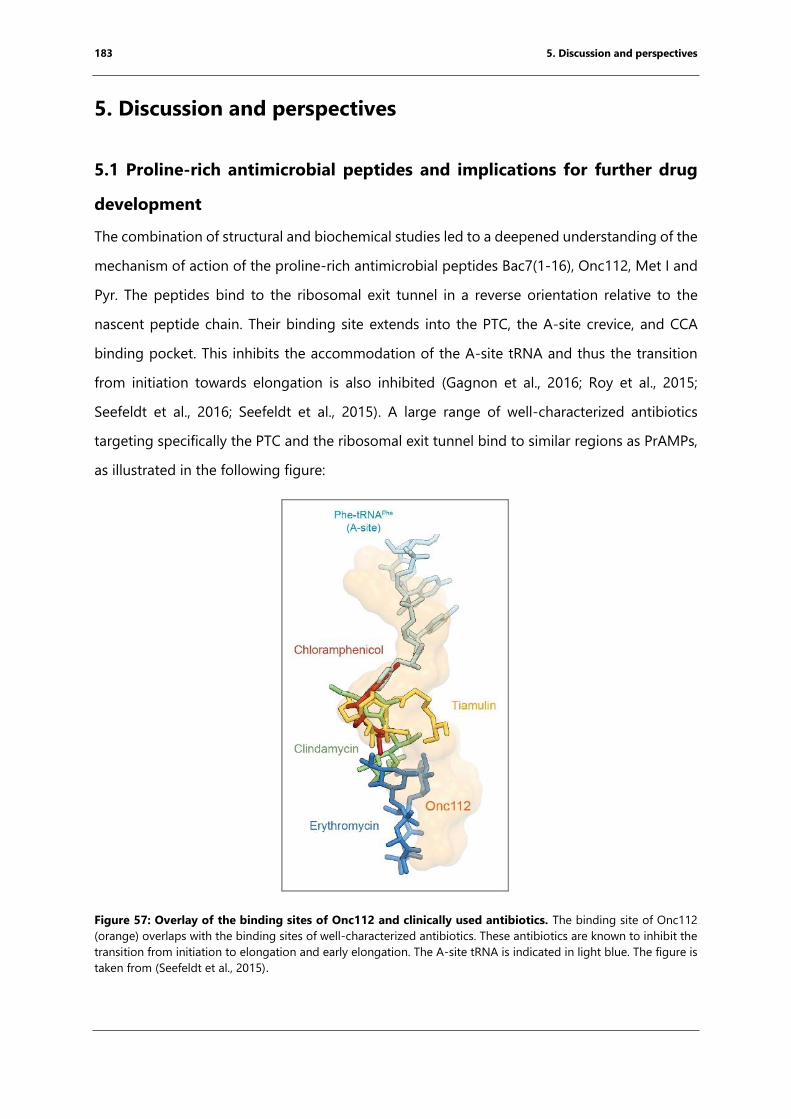
183 5. Discussion and perspectives
5. Discussion and perspectives
5.1 Proline-rich antimicrobial peptides and implications for further drug
development
The combination of structural and biochemical studies led to a deepened understanding of the
mechanism of action of the proline-rich antimicrobial peptides Bac7(1-16), Onc112, Met I and
Pyr. The peptides bind to the ribosomal exit tunnel in a reverse orientation relative to the
nascent peptide chain. Their binding site extends into the PTC, the A-site crevice, and CCA
binding pocket. This inhibits the accommodation of the A-site tRNA and thus the transition
from initiation towards elongation is also inhibited (Gagnon et al., 2016; Roy et al., 2015;
Seefeldt et al., 2016; Seefeldt et al., 2015). A large range of well-characterized antibiotics
targeting specifically the PTC and the ribosomal exit tunnel bind to similar regions as PrAMPs,
as illustrated in the following figure:
Figure 57: Overlay of the binding sites of Onc112 and clinically used antibiotics. The binding site of Onc112
(orange) overlaps with the binding sites of well-characterized antibiotics. These antibiotics are known to inhibit the
transition from initiation to elongation and early elongation. The A-site tRNA is indicated in light blue. The figure is
taken from (Seefeldt et al., 2015).
184 5. Discussion and perspectives
As the binding site of Onc112 overlaps with the binding site of several antibiotics including
chloramphenicol and erythromycin (Bulkley et al., 2010; Dunkle et al., 2010), it may be possible
to modify PrAMPs in order to increase their binding affinity through additional hydrogen-
bonds and stacking interactions. In addition, the elongated fold of the PrAMPs covers a long
binding surface and could slow down the appearances of resistance. This strategy is listed as
an advantage of synergistic antibiotics as reviewed by (Kohanski et al., 2010). Synergistic
antibiotics are pairs of antibiotics acting in concert and therefore enhancing their inhibitory
impact. Well-characterized molecules that target the PTC and the upper ribosomal tunnel are
streptogramin A and B. These antibiotics are bacteriostatic when used individually, but
bactericidal when used together (Vazquez, 1966). The enlarged binding site of two molecules
might decrease the rise of resistance (Noeske et al., 2014). Isolated E. coli cell lines carrying
rRNA mutants that are known to be resistant to antibiotics (e.g. macrolides) were used to
determine the minimal inhibitory concentration (MIC) of Bac7 and Onc112 (Gagnon et al.,
2016). Cells were carrying the point mutations A2503C and A2059C or the corresponding
double mutation. While cells treated with Bac7 were still sensitive to the same concentration
of Bac7 (0.75), higher concentrations of Onc112 were necessary to have a similar effect (Gagnon
et al., 2016). An explanation could be that the N-terminus of Bac7 anchors the peptide in its
binding pocket as it adopts a specific conformation due to stacking interactions with the tunnel
bases (Gagnon et al., 2016). In order to increase the binding affinity of the PrAMPs, introducing
positively-charged aromatic moieties within the N-terminus might enhance stacking
interactions.
Recently published structures of the antimicrobial peptides Api137 and Klebsazolicin in
complex with the 70S E. coli or T. thermophilus ribosomes revealed that these peptides target
to the ribosomal exit tunnel (Florin et al., 2017; Metelev et al., 2017). These peptides inhibit
early elongation (Metelev et al., 2017) and termination (Florin et al., 2017). The following figure
compares their structures to that of Bac7(1-16):

185 5. Discussion and perspectives
Figure 58: Comparison of the binding sites of Bac7(1-16), Api137 and Klebsazolicin (KLB). (A) The peptides
bind to the ribosomal exit tunnel even if they have different sequences and folds (Florin et al., 2017; Metelev et al.,
2017; Scocchi et al., 1994). * indicates post-translationally modified residues. Bac7(1-16, green) binds to the A-site
tRNA binding pocket (1), the A-site crevice (2) and the upper tunnel. Api137 (B, purple, PDB code: 5O2R) binds to
the A-site crevice and reaches further into the ribosomal tunnel (Florin et al., 2017). The post-translational
modifications of KLB (C, PDB code: 5W4K) restrict the backbone conformation resulting in a “curled-up”
conformation binding from the PTC into the upper tunnel (Metelev et al., 2017). The ribosome is taken from (Seefeldt
et al., 2016).
The initial presumed target of PrAMPs such as oncocin or apidaecin was DnaK. Several rounds
of in vitro drug development led to the derivatives Onc112, Onc72, and Api88 and Api137
((Knappe et al., 2010; Otvos et al., 2000), discussed in further detail in chapter 1.3). However,
cells lacking DnaK remained sensitive to these peptides and Krizsan et al was able to crosslink
Api88 to bacterial ribosomes (Krizsan et al., 2014), indicating that the ribosome was the likely
target for PrAMPs. These experiments were the starting point of this project and resulted in
the structure determination of Onc112 bound to the bacterial ribosome.
186 5. Discussion and perspectives
Recent biochemical and structural studies showed that Api137 traps the ribosome at the stop
codon and prevents the release of the peptide (Florin et al., 2017). In contrast to Bac1-16 (Figure
58B), Api137 binds in the same orientation as the nascent peptide chain with the N-terminus
pointing towards the cytoplasm. The binding site covers the PTC and reaches further into the
upper tunnel compared to Bac71-16 (Florin et al., 2017). Although Api137 and Bac71-16 are both
classified as PrAMPs, their binding site, orientation and mechanism of action differ extensively
(Figure 58B).
The second peptide Klebsazolicin (KLB) (Metelev et al., 2017). KLB is an antimicrobial peptide
produced as a defense mechanism by K. pneumoniae by the ribosome and belongs to the
microcin class. In contrast to PrAMPs, these bacteriocins are characterized by a high cysteine
content. The cysteines are post-translationally modified, resulting in a cyclic backbone with a
“curled-up” conformation (Metelev et al., 2017). KLB binds to the PTC and the upper part of
the ribosomal exit tunnel (Figure 58B). Biochemical studies confirmed that KLB binding inhibits
the translational cycle at the early elongation phase, as indicated by the structure of this
peptide in complex with the 70S T. thermophilus ribosome (Metelev et al., 2017).
All six peptides, Bac71-16, Onc112, Pyr, Met I, KLB and Api137, have a large variety of different
sequences and come from very different organisms, ranging from bacteria to higher
vertebrates. All peptides target the PTC, reach into the upper ribosomal exit tunnel and/or
reach into the A-site tRNA binding pocket (Florin et al., 2017; Gagnon et al., 2016; Metelev et
al., 2017; Roy et al., 2015; Seefeldt et al., 2016; Seefeldt et al., 2015). As recently reviewed in
detail for macrolide antibiotics, a major strategy for antibiotic development is to increase
bioavailability, through greater drug stability and improved cellular uptake, while enhancing a
drug’s binding affinity for its target through the chemical modifications (George, 2017). For
macrolide antibiotics, modifications include the removal of the cladinose sugar or the
attachment of novel side chains to the drug backbone (George, 2017). Similarly, the length of
the antimicrobial peptides and their sequence variability opens up a new avenue for drug
development. Future objectives will have to include the improvement of the bioavailability and
binding affinity of these molecules. This could be achieved by modifying the chemistry of their
constituent building blocks, including the use of unusual amino acids and modifications of the
backbone, such as the incorporation of β-, γ-, and D-amino acids, as well as urea backbones or
peptoids (Pasco et al., 2017).
187 5. Discussion and perspectives
Bac7, Onc112, Met I, Pyr, KLB and Api137 are active against Gram-negative bacteria due to the
presence of the ATP-dependent peptide transporter SbmA (Mattiuzzo et al., 2007; Metelev et
al., 2017; Runti et al., 2013). Although SbmA was reported to transport peptides, toxins,
peptoids and peptide nucleic acids (PNA) its physiological role remains unknown, mostly due
to the absence of a detectable phenotype for the deletion mutants under conditions tested so
far (Corbalan et al., 2013). Screening Api137-resistant cells identified mutations within the
SbmA transporter, indicating the main limitation for using these peptides as potential drugs
(Florin et al., 2017). To overcome this limitation, the uptake of PrAMPs could be coupled to an
essential and species-specific transporter system e.g. the Iron-uptake pathway.
In the structures I obtained, the C-terminus of Bac7, Onc112 and Pyr could not be modeled
due to the absence of density, indicating that it is disordered within the crystal (Gagnon et al.,
2016; Roy et al., 2015; Seefeldt et al., 2016; Seefeldt et al., 2015). C-terminal truncations of
Onc112 led in vivo to loss of function indicating that it might be a crucial recognition site for
cellular uptake (Seefeldt et al., 2015). Since the C-terminus is not important for binding to the
ribosome it can be modified in order to avoid the dependency on the SbmA transporter for
uptake. As shown recently, introducing a fluorescent label into the PrAMP arasin I does not
alter the inhibitory properties of the peptide compared to the unlabeled form (Paulsen et al.,
2016). Instead of a fluorescent label a molecule that is recognized specifically by an essential
transporter could be introduced into the PrAMP, a method termed “trojan horse” as reviewed
in detail (Zgurskaya et al., 2015). An essential transporter that has been used previously is the
iron uptake system in bacteria. While Fe3+ (Iron) is crucial for bacteria to survive the sources are
often limited during infection. To complex Fe3+ specifically in the environment and to transport
the cation into the cells, bacteria evolved siderophores (Braun and Hantke, 2011). Siderophores
represent a large class of molecules with different characteristics and molecular weights
ranging from 0.3 to 1 kDa. Previous attempts to introduce siderophores into antibiotics such
as chloramphenicol often resulted in loss of function (Page, 2013). In contrast to this, some
Streptomyces species have been shown to synthesize siderophore-attached macrolides.
Furthermore, some microcins are linked post-translationally to siderophores (Vassiliadis et al.,
2010; Vassiliadis et al., 2007) The various siderophores and their corresponding transporters
are often species-specific, resulting in narrow-spectrum antibiotics. This opens the possibility
of creating PrAMPs that are capable of acting against Gram-positive bacteria, which normally
188 5. Discussion and perspectives
lack the SbmA transporter. This makes this concept particularly interesting for further rounds
of drug development.
In summary, some antimicrobial peptides have been shown to bind to the exit tunnel of
bacterial ribosomes and to inhibit the initiation, early elongation or termination steps of
translation. Structural data of bacterial ribosomes in complex with these peptides have given
us detailed insights into the mechanism of action of PrAMPs, making the structures a solid
foundation for further drug development.
5.2 The flexizyme methodology to study nascent chain-mediated
translational arrest
The second project of this thesis aimed to study translational arrest mediated by short nascent
chains using structure biological methods. Forming the arrest complex by in vitro translation
requires a long mRNA fragment that can interfere with the crystallization process (personal
communication from Axel Innis). In order to avoid this, the arrest peptide was bound directly
to the 3’CCA end of the initiator tRNA using the flexizyme methodology (Goto et al., 2011).
After the activity of the peptidylated-tRNA was confirmed by toeprinting experiments the arrest
complexes were formed for X-ray crystallography. Due to hydrolysis of the ester bond linking
the peptide to the tRNA during the crystallization, a second strategy was developed involving
the generation of 3’-NH2-tRNA using previously developed protocols (Polikanov et al., 2014;
Voorhees et al., 2009). Attempts to peptidylate a 3’NH2-tRNA using a flexizyme have not been
successful to date.
An alternative strategy to attach the peptide through a non-hydrolysable amide linkage was
to add the last amino acid through an additional round of elongation as discussed in detail in
chapter 4.2.6. However, resulting crystals revealed no density of the peptide or the A-site tRNA.
Reasons could be that the binding affinity of the deaminoacylated tRNAiMet for the P-site might
be too high to be replaced under crystallographic concentrations.
The recent improvement in resolution of structures obtained by cryo-EM has made it possible
to study short arrest peptides with this method. The probability of hydrolysis of the ester-linked
arrest peptide was reduced as the complex was directly frozen after complex formation.
189 5. Discussion and perspectives
During the studies of this thesis, two case studies were used to establish a workflow for arrested
ribosome complex formation: (i) fM+X(+) mediated arrest in the presence of erythromycin, and
(ii) translational arrest along consecutive prolines. Future experiments should focus on the
study of the chemical properties of the different amino acids of the arrest peptides to improve
or deplete the arresting properties of the peptide as well as the involvement of the peptide
backbone in contact formation with the ribosomal residues. The project specific main results
are put again into context and perspectives are discussed in the following sections.
5.3 fMKF(R) arrests the 70S E. coli ribosome in the presence of
erythromycin
fM+X(+) represents a set of short nascent peptides that arrest the bacterial ribosome in the
presence of erythromycin (Sothiselvam et al., 2014; Sothiselvam et al., 2016). To study the
underlying molecular mechanism, the sequence fMKF(R) was selected as discussed in detail in
chapter 4.3. The fMKF(R) arrest sequence had not been shown before to arrest the 70S E. coli
ribosome. This was shown by toeprinting of the whole ORF and by initiating translation using
a fMKF peptidyl moiety attached to initiator tRNA in the presence of the drug.
The structure of the complex was obtained by cryo-EM with an overall resolution of 3.9 Å. The
density of the nascent peptide provided information regarding the orientation of two of the
three peptide side chains. The advantage of the flexizyme methodology is that it allows
peptides composed of non-canonical amino acids to be attached to the 3’CCA end of tRNAs.
A previous study investigated the chemical properties of the A-site amino acid necessary for
the arrest using tRNA mimics (Sothiselvam et al., 2016). Based on the structure and the previous
study, the following modifications can be performed to study the chemical properties of the
nascent peptide necessary for the nascent chain mediated translational arrest:
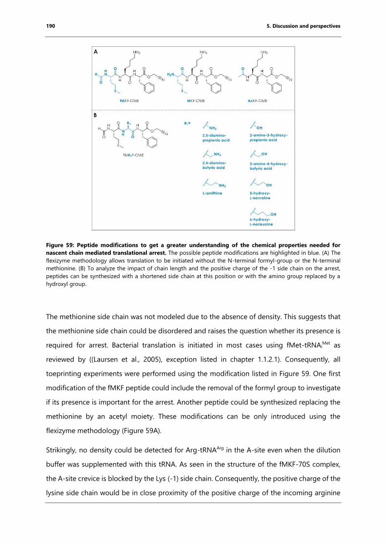
190 5. Discussion and perspectives
Figure 59: Peptide modifications to get a greater understanding of the chemical properties needed for
nascent chain mediated translational arrest. The possible peptide modifications are highlighted in blue. (A) The
flexizyme methodology allows translation to be initiated without the N-terminal formyl-group or the N-terminal
methionine. (B) To analyze the impact of chain length and the positive charge of the -1 side chain on the arrest,
peptides can be synthesized with a shortened side chain at this position or with the amino group replaced by a
hydroxyl group.
The methionine side chain was not modeled due to the absence of density. This suggests that
the methionine side chain could be disordered and raises the question whether its presence is
required for arrest. Bacterial translation is initiated in most cases using fMet-tRNAiMet as
reviewed by ((Laursen et al., 2005), exception listed in chapter 1.1.2.1). Consequently, all
toeprinting experiments were performed using the modification listed in Figure 59. One first
modification of the fMKF peptide could include the removal of the formyl group to investigate
if its presence is important for the arrest. Another peptide could be synthesized replacing the
methionine by an acetyl moiety. These modifications can be only introduced using the
flexizyme methodology (Figure 59A).
Strikingly, no density could be detected for Arg-tRNAArg in the A-site even when the dilution
buffer was supplemented with this tRNA. As seen in the structure of the fMKF-70S complex,
the A-site crevice is blocked by the Lys (-1) side chain. Consequently, the positive charge of the
lysine side chain would be in close proximity of the positive charge of the incoming arginine
191 5. Discussion and perspectives
side chain, resulting in static repulsion. Previously, it has been shown that the positive charge
of the A-site amino acid side chain is crucial for arrest, whereas its length is not (Sothiselvam
et al., 2016). The same cannot be said for the -1 residue at present. The flexizyme methodology
allows us to study the impact of the length and charge of the -1 residue side chain, using the
modifications listed in Figure 59B. The need for a particular orientation of the lysine side chain
could be verified by replacing this residue with D-lysine. This will force the side chain to adopt
the mirrored orientation and thus might prevent arrest in the presence of erythromycin.
The toeprinting experiment to prove the activity of the fMKF-tRNAiMet (chapter 4.4.3, Figure 28)
indicates that even in the presence of erythromycin a certain percentage of ribosomes still
translocates to the second codon. Consequently, the arrest strength can be still enhanced. To
do so, modification of the -1 side chain to a positively charged aromatic moiety or to an
arginine might be enough to enhance the arrest due to potential stacking interactions with the
rRNA. In addition, the methionine could be replaced by groups that tend to form stacking
interactions, such as aromatic moieties, in order to obtain a higher arrest strength. The arrest
strength might be assessed using toeprinting and puromycin sensitivity. Other methionine
replacement could include linker side chains to attach the arrest peptide covalently to the
macrolide. Previous attempts to peptidylate macrolides included the attachment of long
peptide chains (Washington et al., 2014). Another study used chloramphenicol attached to
short tri-peptides from erm leader peptides including MRL. These peptide-chloramphenicol
fusions had an impact on in vitro translation comparable to chloramphenicol itself (Mamos et
al., 2013). Chemically linking arrest peptides without the tRNA to erythromycin might overcome
macrolide resistance as the peptide would extend into the PTC, thus covering a larger binding
site in the way synergistic antibiotics do (Noeske et al., 2014).
The current resolution of the MKF-70S structure is 3.9 Å and is thus too low to make
assumptions about hydrogen bonding interactions. To investigate the hydrogen bonding of
the backbone with the ribosomal residues, the peptide backbone could be modified by N-
alkylation of the backbone or replacing each peptide bonds with an ester bond In doing so,
the importance of each peptide bond as H-bond donor could be investigated. Other backbone
modifications, such as the introduction of urea building blocks or β- and γ-peptides (Pasco et
al., 2017) and might influence the folding and orientation of the peptide within the tunnel.
192 5. Discussion and perspectives
The orientation of the lysine side chain is enhanced by allosteric rearrangement of the 23S
rRNA, in particular of bases U2506, U2585, and A2062 which were reported to be crucial for
the activity of certain arrest peptides (Koch et al., 2017; Vázquez-Laslop et al., 2011). The
orientation of these bases was compared with those in several structures obtained previously,
including those of complexes obtained at various stages of the translation cycle or undergoing
ligand-dependent or -independent translational arrest. Within the fMKF-70S ribosome
structure, U2506 adopts a conformation that is different from that observed in other structures.
Furthermore, U2585 adopts a position between pre-accommodation and pre-catalysis that is
similar to the conformation observed in the VemP-arrested ribosome (Su et al., 2017) structure
and that is stabilized by potential stacking interactions against the peptide.
Strikingly, the base of A2062 adopts a specific orientation which points towards the PTC and
appears to be important for the arrest. It has been reported that arrest peptides can be
classified into two categories, depending on whether their activity depends on the chemical
properties of A2062 or not (Vázquez-Laslop et al., 2011). Ribosomes carrying the A2062C or
A2062U point mutations still arrest along the fMRL(R) motif in the presence of erythromycin
(Sothiselvam et al., 2014). The fM+X(+) motif was classified as A2062-independent and the
conformation of the base of A2062 in the fMKF-70S structure is similar to that observed in the
TnaC-70S structure, which is also independent of the chemical properties of A2062 (Bischoff et
al., 2014; Sothiselvam et al., 2014; Vázquez-Laslop et al., 2011). Although the nature of the base
itself appears not to be important for the arrest process, it is possible that the mere presence
of an unpaired base at this position within the tunnel is sufficient to restrict the rotation of
the -1 lysine side chain in the presence of erythromycin, thus forcing it to remain within the
A-site crevice.
Interestingly, the base of A2062 adopts different orientations within the crystal structures of
erythromycin in complex with the 70S ribosomes from E. coli or T. thermophilus, respectively
(Bulkley et al., 2010; Dunkle et al., 2010). An explanation for this discrepancy could be that the
binding of erythromycin favors the orientation where the base points towards the PTC but does
not entirely preclude the alternative conformation. While translating the fMKF(R) sequence in
the presence of erythromycin, the base of A2062 points might frequently point towards the
PTC, thus forcing the -1 side chain into the A-site crevice and causing arrest. In contrast to this,
if the base points towards to the cytoplasm peptide bond formation can occur and the
193 5. Discussion and perspectives
ribosome can translocate. This would explain the appearance of a faint band in the toeprinting
experiment discussed in chapter 4.4.3 and would make the formation of a stalled complex a
stochastic event that is linked to the particular conformation adopted by A2062. Furthermore,
the masked 3D classification revealed that 13.48% of the particles, which corresponds to
approx. 12 500 particles, contained A-, P- and E-site tRNA (Figure 31) potentially representing
those particles that did not arrest and in which the peptide might be connected to the A-site
tRNA. Since approximately 30 000 particles are necessary for a high-resolution reconstruction
of the ribosome (Bai et al., 2013) including micrographs containing lower resolution data and
recollection of new data might result in enough particles to resolve the structure of the
potentially non-arrested ribosomes.
Another possibility to assess the role of the bases discussed above is molecular dynamics
studies. In a recently published study of the ErmBL arrest peptide, molecular dynamics
simulations gave greater insights into the underlying mechanism and mobility of the peptide
itself (Arenz et al., 2016). Additionally, biochemical studies to further shed light on the
importance of ribosomal bases on the arrest process could include atomic mutagenesis of the
ribosome, a method that makes it possible to introduce unnatural bases into or exclude bases
from a circularly permutated 23S rRNA. In a recent study using atomic mutagenesis, ribosomes
lacking the base of A2062 were generated to study the impact of A2062 on nascent
chain-mediated translational arrest along ErmCL in the presence of erythromycin (Koch et al.,
2017). Using these ribosomes would give greater insights into the requirements for the base
of A2062 since these ribosomes should not arrest when translating M+X(+) motifs, in this case,
fMKF(R), in the presence of erythromycin.
In summary, the flexizyme methodology will make it possible to perform further detailed
studies to elucidate the arrest mechanism of MRLR. The advantage of the method is the
flexibility to investigate the mechanism when using non-natural sequences such as the removal
of the N-terminal methionine and the variation of the -1 side chain. Furthermore, the cryo-EM
structure solved revealed how the binding of the macrolide to the ribosomal exit tunnel
changes the conformation of several bases of the 23S rRNA within the PTC. Additionally, this
structure is the first arrest complex that does not bypass the ligand. Further experiments will
be necessary to confirm the hypothesized mechanism and to utilize the knowledge obtained
for future drug development.
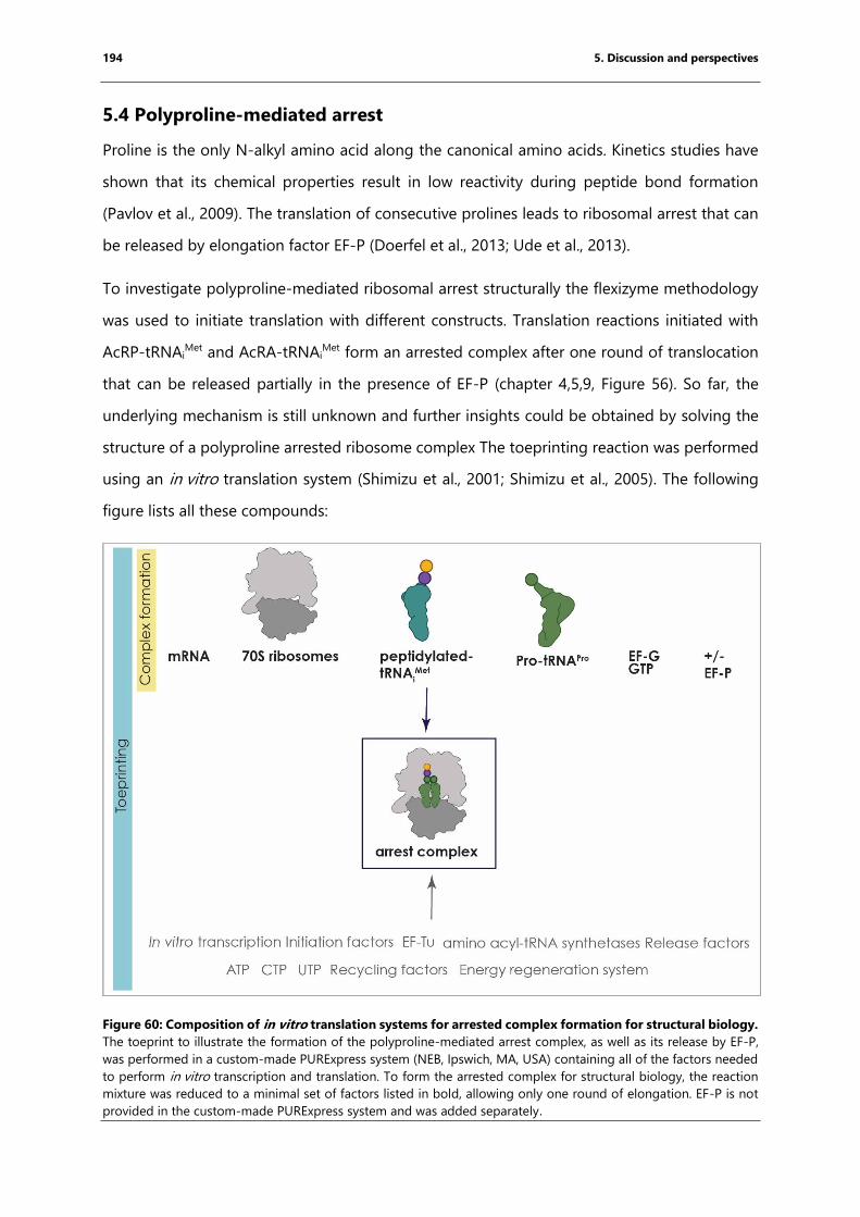
194 5. Discussion and perspectives
5.4 Polyproline-mediated arrest
Proline is the only N-alkyl amino acid along the canonical amino acids. Kinetics studies have
shown that its chemical properties result in low reactivity during peptide bond formation
(Pavlov et al., 2009). The translation of consecutive prolines leads to ribosomal arrest that can
be released by elongation factor EF-P (Doerfel et al., 2013; Ude et al., 2013).
To investigate polyproline-mediated ribosomal arrest structurally the flexizyme methodology
was used to initiate translation with different constructs. Translation reactions initiated with
AcRP-tRNAiMet and AcRA-tRNAi
Met form an arrested complex after one round of translocation
that can be released partially in the presence of EF-P (chapter 4,5,9, Figure 56). So far, the
underlying mechanism is still unknown and further insights could be obtained by solving the
structure of a polyproline arrested ribosome complex The toeprinting reaction was performed
using an in vitro translation system (Shimizu et al., 2001; Shimizu et al., 2005). The following
figure lists all these compounds:
Figure 60: Composition of in vitro translation systems for arrested complex formation for structural biology.
The toeprint to illustrate the formation of the polyproline-mediated arrest complex, as well as its release by EF-P,
was performed in a custom-made PURExpress system (NEB, Ipswich, MA, USA) containing all of the factors needed
to perform in vitro transcription and translation. To form the arrested complex for structural biology, the reaction
mixture was reduced to a minimal set of factors listed in bold, allowing only one round of elongation. EF-P is not
provided in the custom-made PURExpress system and was added separately.
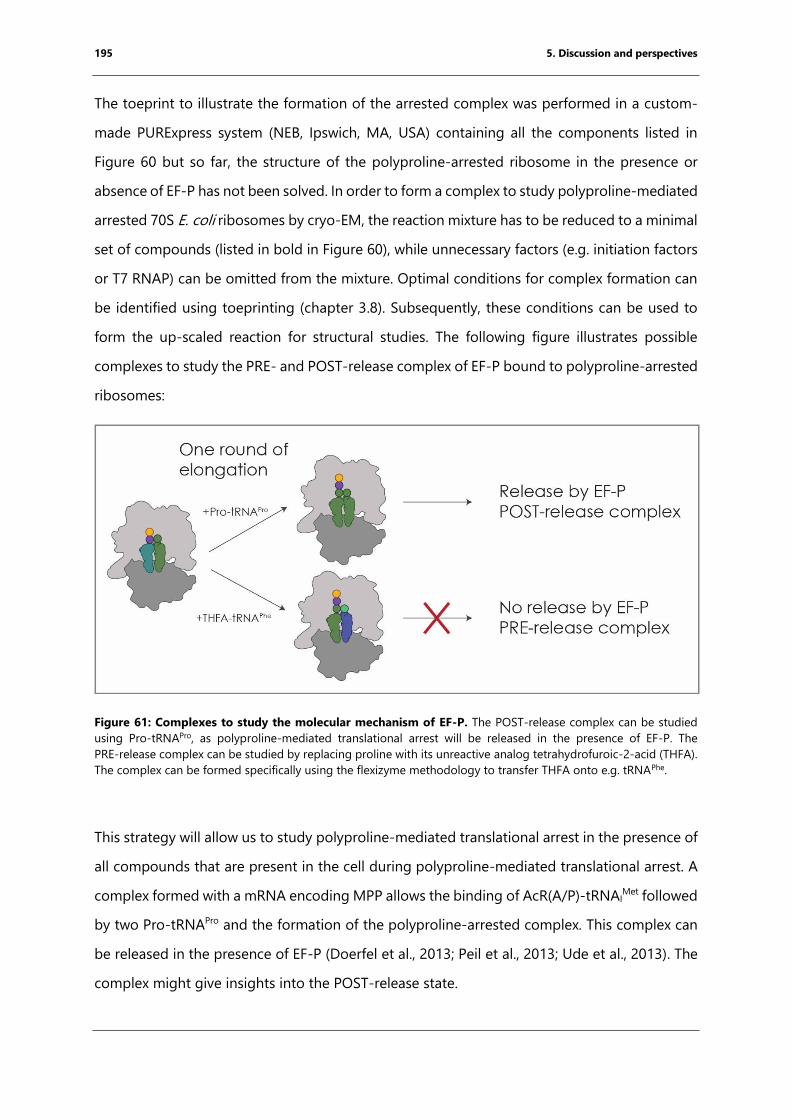
195 5. Discussion and perspectives
The toeprint to illustrate the formation of the arrested complex was performed in a custom-
made PURExpress system (NEB, Ipswich, MA, USA) containing all the components listed in
Figure 60 but so far, the structure of the polyproline-arrested ribosome in the presence or
absence of EF-P has not been solved. In order to form a complex to study polyproline-mediated
arrested 70S E. coli ribosomes by cryo-EM, the reaction mixture has to be reduced to a minimal
set of compounds (listed in bold in Figure 60), while unnecessary factors (e.g. initiation factors
or T7 RNAP) can be omitted from the mixture. Optimal conditions for complex formation can
be identified using toeprinting (chapter 3.8). Subsequently, these conditions can be used to
form the up-scaled reaction for structural studies. The following figure illustrates possible
complexes to study the PRE- and POST-release complex of EF-P bound to polyproline-arrested
ribosomes:
Figure 61: Complexes to study the molecular mechanism of EF-P. The POST-release complex can be studied
using Pro-tRNAPro, as polyproline-mediated translational arrest will be released in the presence of EF-P. The
PRE-release complex can be studied by replacing proline with its unreactive analog tetrahydrofuroic-2-acid (THFA).
The complex can be formed specifically using the flexizyme methodology to transfer THFA onto e.g. tRNAPhe.
This strategy will allow us to study polyproline-mediated translational arrest in the presence of
all compounds that are present in the cell during polyproline-mediated translational arrest. A
complex formed with a mRNA encoding MPP allows the binding of AcR(A/P)-tRNAIMet followed
by two Pro-tRNAPro and the formation of the polyproline-arrested complex. This complex can
be released in the presence of EF-P (Doerfel et al., 2013; Peil et al., 2013; Ude et al., 2013). The
complex might give insights into the POST-release state.
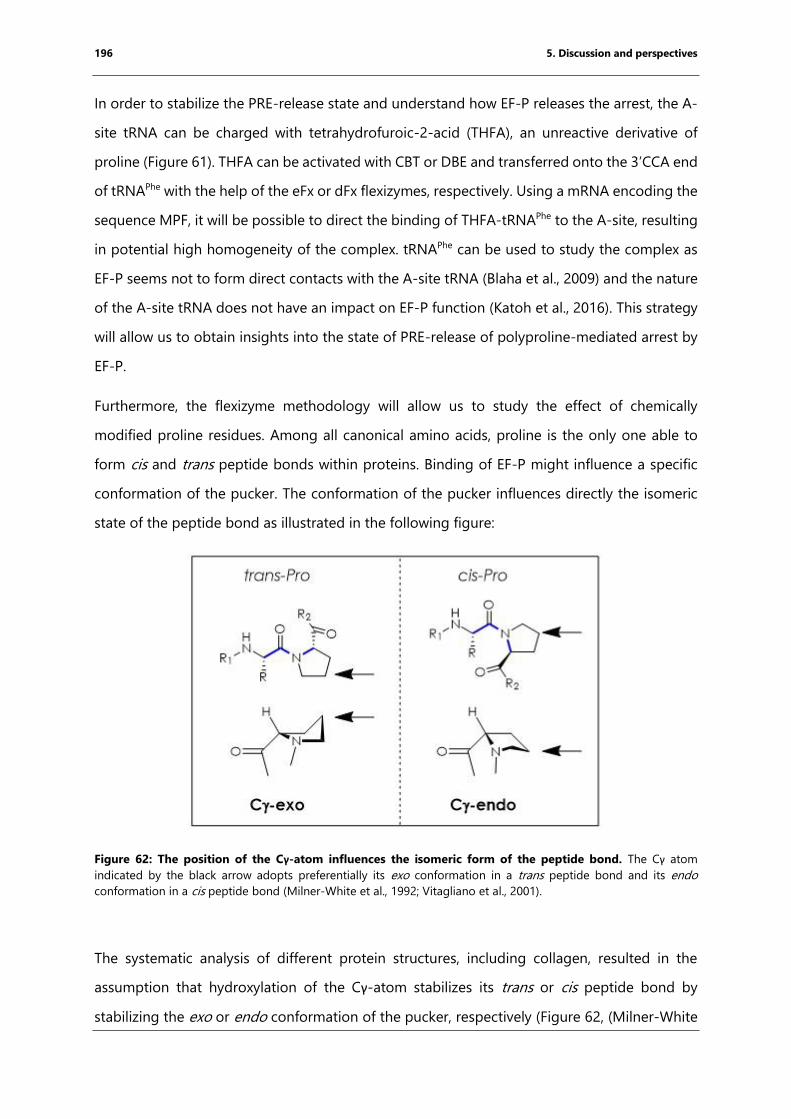
196 5. Discussion and perspectives
In order to stabilize the PRE-release state and understand how EF-P releases the arrest, the A-
site tRNA can be charged with tetrahydrofuroic-2-acid (THFA), an unreactive derivative of
proline (Figure 61). THFA can be activated with CBT or DBE and transferred onto the 3’CCA end
of tRNAPhe with the help of the eFx or dFx flexizymes, respectively. Using a mRNA encoding the
sequence MPF, it will be possible to direct the binding of THFA-tRNAPhe to the A-site, resulting
in potential high homogeneity of the complex. tRNAPhe can be used to study the complex as
EF-P seems not to form direct contacts with the A-site tRNA (Blaha et al., 2009) and the nature
of the A-site tRNA does not have an impact on EF-P function (Katoh et al., 2016). This strategy
will allow us to obtain insights into the state of PRE-release of polyproline-mediated arrest by
EF-P.
Furthermore, the flexizyme methodology will allow us to study the effect of chemically
modified proline residues. Among all canonical amino acids, proline is the only one able to
form cis and trans peptide bonds within proteins. Binding of EF-P might influence a specific
conformation of the pucker. The conformation of the pucker influences directly the isomeric
state of the peptide bond as illustrated in the following figure:
Figure 62: The position of the Cγ-atom influences the isomeric form of the peptide bond. The Cγ atom
indicated by the black arrow adopts preferentially its exo conformation in a trans peptide bond and its endo
conformation in a cis peptide bond (Milner-White et al., 1992; Vitagliano et al., 2001).
The systematic analysis of different protein structures, including collagen, resulted in the
assumption that hydroxylation of the Cγ-atom stabilizes its trans or cis peptide bond by
stabilizing the exo or endo conformation of the pucker, respectively (Figure 62, (Milner-White
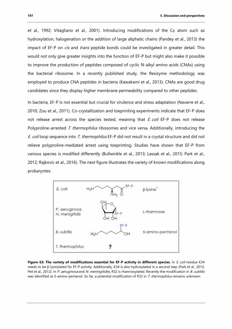
197 5. Discussion and perspectives
et al., 1992; Vitagliano et al., 2001). Introducing modifications of the Cγ atom such as
hydroxylation, halogenation or the addition of large aliphatic chains (Pandey et al., 2013) the
impact of EF-P on cis and trans peptide bonds could be investigated in greater detail. This
would not only give greater insights into the function of EF-P but might also make it possible
to improve the production of peptides composed of cyclic N-alkyl amino acids (CNAs) using
the bacterial ribosome. In a recently published study, the flexizyme methodology was
employed to produce CNA peptides in bacteria (Kawakami et al., 2013). CNAs are good drug
candidates since they display higher membrane permeability compared to other peptides.
In bacteria, EF-P is not essential but crucial for virulence and stress adaptation (Navarre et al.,
2010; Zou et al., 2011). Co-crystallization and toeprinting experiments indicate that EF-P does
not release arrest across the species tested, meaning that E. coli EF-P does not release
Polyproline-arrested T. thermophilus ribosomes and vice versa. Additionally, introducing the
E. coli loop sequence into T. thermophilus EF-P did not result in a crystal structure and did not
relieve polyproline-mediated arrest using toeprinting. Studies have shown that EF-P from
various species is modified differently (Bullwinkle et al., 2013; Lassak et al., 2015; Park et al.,
2012; Rajkovic et al., 2016). The next figure illustrates the variety of known modifications along
prokaryotes:
Figure 63: The variety of modifications essential for EF-P activity in different species. In E. coli residue K34
needs to be β-lysinylated for EF-P activity. Additionally, K34 is also hydroxylated in a second step (Park et al., 2012;
Peil et al., 2012). In P. aeruginosa and N. meningitides, R32 is rhamnosylated. Recently the modification in B. subtilis
was identified as 5-amino-pentanol. So far, a potential modification of R32 in T. thermophilus remains unknown.
198 5. Discussion and perspectives
The post-translational modification of the crucial residue of EF-P varies among different
prokaryotes (Figure 63), but additional hydrogen bond donors and acceptors are used to
possibly enhance interactions with the tRNA. So far, the potential modification of
T. thermophilus EF-P remains unknown. One of the remaining questions is if a modification of
T. thermophilus EF-P is necessary for the function of this molecule. This could be investigated
by growing T. thermophilus under different temperatures and subsequently isolating EF-P via
immunoprecipitation to analyze the protein by mass spectrometry as performed previously for
EF-P from E. coli (Peil et al., 2012; Rajkovic et al., 2016). T. thermophilus grows at an optimal
temperature of 75°C (Oshima and Imahori, 1974). An explanation could be that 70S
T. thermophilus ribosomes are not stalled by polyproline motifs or easier overcome the arrest
at these high temperatures as the molecules move more. A second explanation includes the
unusual high concentrations of polyamines in the cytoplasm, which have been shown to be
essential for cell survival. In addition, thermophiles produce species-specific polyamines such
as thermine and tetrakis(3-amino-propyl)-ammonium (Oshima, 2007). Furthermore, the
addition of polyamines to in vitro translation reactions is crucial for the initiation of translation
and also enhances the elongation (Uzawa et al., 1993a; Uzawa et al., 1993b). The combination
of different polyamines showed a synergistic enhancement of protein production at higher
temperatures. Thus T. thermophilus EF-P could act as a scaffold to orient polyamines correctly
and in doing so release the arrest along consecutive poly-proline motifs. This hypothesis could
be toeprinting and co-crystallization experiments using T. thermophilus ribosomes in the
presence of different polyamines and T. thermophilus EF-P.
Species-specific modifications of EF-P, that can range from rhamnosylation to β-lysinylation,
demands a large number of different modification enzymes as the chemical properties of the
substrate molecules are diverse (Lassak et al., 2015; Park et al., 2012). Understanding the
underlying molecular mechanism of different EF-P modifications might result in a better
understanding of the species specificities of bacterial ribosomes and this might give rise to
species-specific narrow-spectrum antibiotics.
6. Conclusion
My structural studies of the inhibition of bacterial translation by nascent-chain and free
peptides resulted in a greater understanding of how these peptides interact with and affect
the ribosomal tunnel. Antimicrobial peptides that target the ribosomal exit tunnel are produced
as a natural defense mechanism of eukaryotes. The structures obtained can be used for further
drug development involving a large number of potential modifications to enhance their uptake
by bacteria and their binding affinity for the ribosome. Further steps will also have to include
the identification of new sequences and their characterization.
In addition, short arrest peptides can be bound directly to the 3’ CCA end of tRNAs using the
flexizyme technology, for further structural or biochemical characterization. This allows to study
the impact of non-canonical amino acids and non-natural backbones on peptide bond
formation and on nascent chain mediated translational arrest. An increased understanding of
the determinants of nascent chain-mediated translational arrest will ultimately result in a better
understanding of the bacterial ribosome. This will allow the development of new scaffolds for
novel antibiotics. Further experiments using the flexizyme methodology and the knowledge
obtained during this work will provide information about the reactivity and incorporation of
non-natural amino acids for example D-amino acids using the bacterial ribosome as a
production machinery.
7. Résumé français
7.1 Général
Les ribosomes bactériens sont de grands complexes ribonucléoprotéiques constitués d'une
petite sous-unité 30S et d'une grande sous-unité 50S, qui catalysent la réaction de traduction
de l'information codée par l'ARN messager (ARNm) en protéines (Monro, 1967; Ogle et al.,
2003; Schmeing and Ramakrishnan, 2009). La formation de la liaison peptidique elle-même est
indépendante de l'énergie et est catalysée par l'ARN ribosomal (ARNr) dans le «peptidyl
transferase center » (PTC) alors que des étapes comme l'initiation, la livraison d'ARNs, la
translocation et la dissociation des sous-unités requièrent un apport d’énergie et certains
facteurs protéiques (Schmeing and Ramakrishnan, 2009). Au sein du PTC, la chaîne peptidique
naissante traverse le tunnel de sortie du ribosome par la sous-unité 50S (Nissen et al., 2000).
Le ribosome constitue l’une des principales cibles des antibiotiques de par son rôle central
dans les processus cellulaires (Wilson, 2009). En raison de la prévalence croissante des bactéries
multirésistantes, le développement de nouveaux antibiotiques est rapidement devenu une
priorité de santé publique (Wilson, 2014). Certains peptides peuvent inhiber la traduction
bactérienne, soit en tant que peptides libres produits lors des mécanismes de défense contre
d'autres organismes tels que les peptides antimicrobiens riches en proline (PrAMPs), soit lors
de leur propre traduction (peptides d'arrêt traductionnel médié par la chaîne naissante).
7.2 Peptides antimicrobiens riches en proline (PrAMPs)
La première partie de cette thèse a consisté à la compréhension du mécanisme sous-jacent de
l'inhibition de la traduction bactérienne par des peptides antimicrobiens riches en proline. Ces
peptides sont produits par le système immunitaire inné des insectes et des mammifères (Otvos,
2002). Les PrAMP sont des agents puissants contre les bactéries à coloration Gram négative et
ont fait l'objet d'études visant à améliorer leurs propriétés en tant que potentiels agents
antibactériens. Les séquences peptidiques montrent une teneur élevée en prolines et au moins
un motif Pro-Arg-Pro qui conduit à une conformation désordonnée du peptide en solution
(Otvos et al., 2000). Le chaperon DnaK a été identifié comme une cible présumée (Otvos et al.,
2000). Cependant, de récentes études ont montré que les cellules dépourvues de dnaK ne sont
pas résistantes à certaines PrAMP provenant d'insectes comme les oncocines dérivées de la
punaise tuberculée (Oncopeltus faciatus) ou de l'apidaecine des abeilles (Apis vespidae)
201 7. Résumé français
(Krizsan et al., 2014). Ces peptides peuvent en effet être attachés aux ribosomes bactériens et
inhiber la synthèse des protéines in vitro. D'autres études ont montré que la bacténécine-7
PrAMP provenant de mammifères (Bac7, Bos taurus) se lie également aux ribosomes bactériens
et inhibe la traduction in vitro (Mardirossian et al., 2014).
Le mécanisme d'action moléculaire sous-jacent des PrAMPs était encore inconnu lorsque j'ai
résolu les structures cristallines du ribosome Thermus thermophilus 70S en complexe avec les
PrAMPs Onc112, Bac71-16, Pyrrhocoricin (Pyr) et Metalnikowin I (Met I). La résolution se situe
entre 2,8 Å et 3,2 Å. En comparaison à la chaîne peptidique naissante en croissance, le PrAMP
se lie dans une orientation inverse au sein du tunnel de sortie du ribosome. Un réseau de
potentielles liaisons hydrogènes ajouté à l'interaction d'empilement du peptide avec des
résidus ribosomaux stabilisent les peptides dans le tunnel de sortie, leur conférant une
structure adéquate (Gagnon et al., 2016; Roy et al., 2015; Seefeldt et al., 2016; Seefeldt et al.,
2015).
A partir du site de liaison qui chevauche l'aminoacyl-ARNt entrant, nous pouvons supposer
que les PrAMP inhibent la transition entre l'initiation et l'allongement. Afin de répondre à cette
hypothèse, des études biochimiques ont été réalisées par nos collaborateurs issus du groupe
du Prof. Daniel N Wilson (Gene Center, (Munich maintenant Université de Hambourg)) via des
expériences de toeprinting, des tests disome et des tests de synthèse protéique acellulaire et
ainsi confirmer le mécanisme proposé. Nos études ont montré que les PrAMP inhibent
spécifiquement la transition de l'initiation à l'élongation et déstabilisent le complexe post-
initiation (Seefeldt et al., 2016; Seefeldt et al., 2015).
Le travail a été publié en parallèle des travaux du groupe du professeur Thomas A. Steitz, ce
qui a permis de confirmer nos résultats (Gagnon et al., 2016; Roy et al., 2015).
7.3 L'arrêt de la traduction induit par le peptide naissant
Le sujet de la deuxième partie de cette thèse était d'acquérir la compréhension du mécanisme
moléculaire des courts peptides d'arrêt. L'arrêt de la traduction médiée par la chaîne naissante
se produit lorsque cette dernière est capable d'interagir avec la paroi du tunnel du ribosome
et entraîne un réarrangement au sein du centre de la peptidyl-transférase (Ito and Chiba, 2013;
Seip and Innis, 2016; Wilson et al., 2016). Les raisons de l'arrêt peuvent être simplement la
composition en acides aminés du peptide naissant, ajouté à la présence d'un ligand ou
l'incorporation d'acides aminés non naturels (Ito and Chiba, 2013; Seip and Innis, 2016; Wilson
202 7. Résumé français
et al., 2016). Les longs peptides d'arrêt comme les peptides TnaC (Bischoff et al., 2014; Seidelt
et al., 2009) et Erm (Arenz et al., 2016; Arenz et al., 2014a; Arenz et al., 2014b)ont été étudiés
par cryo-EM, tandis que les mécanismes d'arrêt des peptides courts incluant les motifs
polyproline ainsi que le motif fM+X(+) arrêtent le ribosome en présence d'érythromycine.
Pour mieux comprendre le mécanisme moléculaire sous-jacent des peptides d'arrêt courts, des
structures à haute résolution sont nécessaires, mais il est difficile d'obtenir un échantillon
homogène suite à la traduction in vitro. Ceux-ci peuvent être obtenus par cristallographie aux
rayons X en utilisant des ribosomes 70S provenant dans notre cas de T. thermophilus ou par
microscopie cryogénique en utilisant des ribosomes 70S d'Escherichia coli.
Pour obtenir des cristaux hautement diffractants, le complexe ne doit contenir que de courts
fragments d'ARNm. Ainsi, des complexes ne peuvent pas être formés en utilisant une réaction
de traduction in vitro car l'ARNm codant chevauche un contact cristallin et limite ainsi la
diffraction (Axel Innis, communication personnelle). Une solution pour assurer la formation
d’une liaison stoechiométrique de l’ARNt peptidyle au ribosome et d’augmenter l’homogénéité
du complexe pourrait être de charger un peptide chimiquement synthétisé directement sur
l’ARNt. Pour obtenir l’ARNt peptidylé, l’approche flexyzyme a été employée, reposant sur le
pouvoir catalytique d’un petit ribozyme afin de former une liaison ester entre le peptide
chimiquement activé et l’extrémité CCA de l’ARNt (Goto et al., 2011). Dans l'étape suivante,
l'ARNt peptidylé a été purifié par chromatographie HPLC. Et les peptides ont été synthétisés
par le Dr. Caterina Lombardo et le Dr. Christophe André. Par cette méthode, les fragments des
peptides d'arrêt ont été spécifiquement chargés sur l'ARNtiMet initiateur. Les ARNt peptidylés
ont été caractérisés en utilisant des méthodes biologiques biochimiques et structurales.
La méthode que nous présentons peut-être appliquée à un grand nombre de peptides d'arrêt
courts, y compris des peptides contenant des acides aminés non naturels dans le but d’acquérir
une meilleure compréhension de la spécificité du ribosome.
7.3.1 M+X(+) arrête le ribosome bactérien en présence d'érythromycine
L'arrêt du ribosome peut être utilisé pour réguler l'expression des gènes dans les cellules. Pour
ce faire, le peptide d'arrêt agit comme un capteur pour détecter la présence d'un médicament
comme l'érythromycine ou d’un métabolite, comme par exemple le tryptophane. Le gène de
résistance à l'érythromycine (ermD) code pour une méthyltransférase qui méthyle la base
A2058 de l'ARNr 23S dans le tunnel ribosomal, empêchant ainsi la liaison du médicament. Les
203 7. Résumé français
ribosomes modifiés de cette manière sont moins efficaces que les ribosomes non modifiés, par
conséquent, l'expression du gène de résistance doit être étroitement régulée. En amont du
gène ermD, un peptide leader ErmDL court est codé pour réguler l'expression de la
méthyltransférase (Hue and Bechhofer, 1992).
Différents peptides leaders impliqués dans la régulation des gènes de résistance à
l'érythromycine ont été identifiés et caractérisés. Le raccourcissement systématique de la
séquence peptidique ErmDL a conduit à l'identification de la séquence de décrochage
minimale MRL(R) (Sothiselvam et al., 2014). La mutagenèse systématique des séquences
(Sothiselvam et al., 2016) couplée aux données de ribosome profiling obtenues en traitant E.
coli et Staphyluscoccus aureus avec l'érythromycine, la télithromycine et l'azithromycine ont
montré que ce motif peut être généralisé à +X(+) (Davis et al., 2014; Kannan et al., 2014). Le
peptide d'arrêt est si court qu'il atteint à peine le site de liaison du médicament.
Au cours de cette thèse, la séquence MKF(R) a été choisie pour d'autres études. Ce faisant, le
Dr. K. Kishore Inampudi (ancien PostDoc dans le laboratoire) a spécifiquement chargé le
peptide MKF (méthionine-lysine-phénylalanine) sur l’ARNt méthylé en utilisant la
méthodologie du flexizyme (Goto et al., 2011). J'ai évalué l'activité de cet ARNt peptidylé par
des expériences de toeprinting en présence et en absence d'érythromycine confirmant l'arrêt
du complexe formé par la chaîne naissante.
Aucune structure n'a pu être obtenue en utilisant la cristallographie aux rayons X car le peptide
a été hydrolysé pendant le processus de cristallisation. J’ai donc déterminé la structure du MKF
arrêté sur le ribosome d’E. coli 70S en formant le complexe en présence de ribosomes 70S
purifiés, de MKF-tRNAiMet, d’Arg -tRNAArg, et d'érythromycine par cryo-EM. Basé sur la
structure obtenue, le réarrangement allostérique des résidus ARNr 23S ainsi que l'absence de
l'ARNt du site A, le mécanisme suivant a pu être proposé: En l'absence d'érythromycine, la base
A2062 tourne plus facilement vers le cytoplasme, permettant donc à la Lys -1 de quitter la
crevasse du site A. Par conséquent, l'amino-acyl-ARNt suivant et portant une charge positive
peut se lier, permettant la formation de la liaison peptidique. En revanche, la présence
d'érythromycine pourrait favoriser une conformation de A2062 dans laquelle la base pointe
vers le PTC. Le ribosome s’arrête lorsque la base d’A2062 de l'ARNr 23S pointe vers le PTC
inhibant la réorientation du peptide d'arrêt et forçant le Lys -1 à pointer dans la crevasse du
site A. Par conséquent, l'ARNt de site A suivant portant une chaîne latérale K, R ou W ne peut
204 7. Résumé français
pas se lier au PTC en raison de l'encombrement stérique et statique. Le ribosome est arrêté en
raison de la prévention de l'accommodation de l'ARNt du site A.
7.3.2 L'arrêt médié par la polyproline est soulagé par le facteur d'élongation P
Plusieurs études ont montré que la vitesse de formation de la liaison peptidique est réduite
lorsque le ribosome incorpore la proline par rapport à la vitesse d'incorporation des autres
acides aminés (Muto and Ito, 2008; Pavlov et al., 2009). La proline est, d'un point de vue
chimique, un N-alkylaminoacide (iminoacide), ce qui entraîne une alcalinité plus élevée par
rapport aux autres acides aminés canoniques et une probabilité plus élevée d’obtenir un
groupe α-imino protoné à un pH physiologique (Pavlov et al., 2009). De plus, la proline est le
seul acide aminé canonique qui peut former à la fois des liaisons peptidiques trans et cis,
conduisant à des replis possibles au sein des hélices α.
De récentes études in vitro et in vivo ont montré que trois prolines consécutives conduisent à
un arrêt du ribosome (Doerfel et al., 2013; Ude et al., 2013). De nombreux gènes importants
pour le fitness de la bactérie contiennent des séquences de polyproline, comme par exemple
le gène codant la valinyl-tRNA synthetase (valS) (Starosta et al., 2014b). Les ribosomes
bactériens arrêtés peuvent être libérés lors de la traduction par le facteur d’élongation P (EF-
P) (Doerfel et al., 2013; Ude et al., 2013). La même observation a été faite pour le facteur
d'initiation des homologues eucaryotes 5A (eIF5A) (Gutierrez et al., 2013). Une autre étude
protéomique utilisant le marquage isotopique stable d'acides aminés en culture cellulaire
(SILAC) sur des cellules Δefp a montré que le nombre de gènes EF-P dépendants n'est pas
seulement limité à une séquence PPP (Peil et al., 2013; Starosta et al., 2014a). Une analyse plus
approfondie utilisant des systèmes de rapporteurs in vivo et des essais de traduction in vitro
ont permis d’identifier la diversité de séquences arrêtant en l'absence de EF-P. Pour obtenir
une fonction complète, EF P doit être modifié après traduction (Bullwinkle et al., 2013; Navarre
et al., 2010). La modification peut varier en fonction de l'espèce bactérienne du sucre jusqu'à
la lysine (Bullwinkle et al., 2013; Lassak et al., 2015). Les cellules bactériennes dépourvues d'efp
ou de ses enzymes de modification présentent le même phénotype et sont moins en forme et
moins virulentes (Navarre et al., 2010). Cela fait de l'EF-P une cible intéressante pour les
antibiotiques. Cependant, jusqu'à présent, le mécanisme sous-jacent de l'arrêt de la traduction
médié par le motif polyproline ainsi que son soulagement par EF P reste inconnu.
205 7. Résumé français
Pour obtenir de plus amples informations sur le mécanisme sous-jacent, EF-P a été purifié
comme publié précédemment et son activité a été évaluée par des expériences de toeprinting.
En utilisant la cristallographie aux rayons X, il aurait pu être montré que l'EF-P de E. coli ne se
lie pas aux ribosomes de T. thermophilus 70S dans des conditions cristallographiques. Il a été
montré précédemment que E. coli EF-P reconnaît la boucle D de tRNAPro (Katoh et al., 2016).
Pour obtenir une compréhension approfondie du mécanisme sous-jacent, une partie de ce
projet consistait à purifier l’ARNt pro à partir d’extraits cellulaires. La pureté a été déterminée
à environ 80% par spectrométrie de masse et son activité a été prouvée par toeprinting. Pour
étudier l'arrêt médié par le motif polyproline, AcRA-ARNtiMet, AcRP-ARNti
Met et AcRAP-ARNtiMet
ont été obtenus en utilisant la méthodologie du flexizyme. J'ai évalué l'activité des peptidyl-
ARNt par toeprinting en présence et en absence d’EF-P d’E. coli montrant la formation et la
libération du complexe d'arrêt. Les expériences futures permettront de trouver des conditions
optimales de formation des complexes dans le but de résoudre la structure par cryo-EM.
7.4 Conclusion
Les connaissances obtenues ont permis de mieux comprendre comment la formation de
liaisons peptidiques peut être inhibée de manière spécifique et pourrait conduire au
développement de nouveaux antibiotiques hautement spécifiques ciblant le ribosome
bactérien.
8. References
Adams, P.D., Afonine, P.V., Bunkóczi, G., Chen, V.B., Davis, I.W., Echols, N., Headd, J.J., Hung, L.-
W., Kapral, G.J., and Grosse-Kunstleve, R.W. (2010). PHENIX: a comprehensive Python-based
system for macromolecular structure solution. Acta Crystallographica Section D: Biological
Crystallography 66, 213-221.
Afonine, P.V., Headd, J.J., Terwilliger, T.C., and Adams, P.D. (2013). Computational
Crystallography Newsletter 4, 23.
Agrawal, R.K., Heagle, A.B., Penczek, P., Grassucci, R.A., and Frank, J. (1999). EF-G-dependent
GTP hydrolysis induces translocation accompanied by large conformational changes in the 70S
ribosome. Nature Structural & Molecular Biology 6, 643-647.
Agrawal, R.K., Penczek, P., Grassucci, R.A., and Frank, J. (1998). Visualization of elongation factor
G on the Escherichia coli 70S ribosome: the mechanism of translocation. Proceedings of the
National Academy of Sciences 95, 6134-6138.
Agrawal, R.K., Penczek, P., Grassucci, R.A., Li, Y., Leith, A., Nierhaus, K.H., and Frank, J. (1996).
Direct Visualization of A-, P-, and E-Site Transfer RNAs in the Escherichia coli Ribosome. Science
271, 1000-1002.
Aguilar, M.-I. (2004). Reversed-Phase High-Performance Liquid Chromatography. In HPLC of
Peptides and Proteins: Methods and Protocols, M.-I. Aguilar, ed. (Totowa, NJ: Springer New
York), pp. 9-22.
Alejo, J.L., and Blanchard, S.C. (2017). Miscoding-induced stalling of substrate translocation on
the bacterial ribosome. Proceedings of the National Academy of Sciences 114, E8603-E8610.
Alexieva, Z., Duvall, E.J., Ambulos, N.P., Kim, U.J., and Lovett, P.S. (1988). Chloramphenicol
induction of cat-86 requires ribosome stalling at a specific site in the leader. Proceedings of
the National Academy of Sciences 85, 3057-3061.
Altschul, S.F., Gish, W., Miller, W., Myers, E.W., and Lipman, D.J. (1990). Basic local alignment
search tool. Journal of molecular biology 215, 403-410.
Alwine, J.C., Kemp, D.J., and Stark, G.R. (1977). Method for detection of specific RNAs in agarose
gels by transfer to diazobenzyloxymethyl-paper and hybridization with DNA probes.
Proceedings of the National Academy of Sciences of the United States of America 74, 5350-
5354.
An, G., Glick, B.R., Friesen, J.D., and Ganoza, M.C. (1980). Identification and quantitation of
elongation factor EF-P in Escherichia coli cell-free extracts. Canadian journal of biochemistry
58, 1312-1314.
Antoun, A., Pavlov, M.Y., Andersson, K., Tenson, T., and Ehrenberg, M. (2003). The roles of
initiation factor 2 and guanosine triphosphate in initiation of protein synthesis. The EMBO
Journal 22, 5593-5601.
Antoun, A., Pavlov, M.Y., Lovmar, M., and Ehrenberg, M. (2006a). How Initiation Factors
Maximize the Accuracy of tRNA Selection in Initiation of Bacterial Protein Synthesis. Molecular
Cell 23, 183-193.
Antoun, A., Pavlov, M.Y., Lovmar, M., and Ehrenberg, M. (2006b). How initiation factors tune the
rate of initiation of protein synthesis in bacteria. The EMBO Journal 25, 2539-2550.
207 8. References
Arenz, S., Bock, L.V., Graf, M., Innis, C.A., Beckmann, R., Grubmüller, H., Vaiana, A.C., and Wilson,
D.N. (2016). A combined cryo-EM and molecular dynamics approach reveals the mechanism of
ErmBL-mediated translation arrest. 7, 12026.
Arenz, S., Meydan, S., Starosta, A.L., Berninghausen, O., Beckmann, R., Vázquez-Laslop, N., and
Wilson, D.N. (2014a). Drug sensing by the ribosome induces translational arrest via active site
perturbation. Molecular cell 56, 446-452.
Arenz, S., Ramu, H., Gupta, P., Berninghausen, O., Beckmann, R., Vázquez-Laslop, N., Mankin,
A.S., and Wilson, D.N. (2014b). Molecular basis for erythromycin-dependent ribosome stalling
during translation of the ErmBL leader peptide. Nature communications 5.
Arenz, S., and Wilson, D.N. (2016). Bacterial Protein Synthesis as a Target for Antibiotic
Inhibition. Cold Spring Harbor perspectives in medicine 6, a025361.
Bai, X.-C., Fernandez, I.S., McMullan, G., and Scheres, S.H. (2013). Ribosome structures to near-
atomic resolution from thirty thousand cryo-EM particles. Elife 2, e00461.
Bailly, M., and de Crécy-Lagard, V. (2010). Predicting the pathway involved in post-translational
modification of elongation factor P in a subset of bacterial species. Biology direct 5, 3.
Ban, N., Beckmann, R., Cate, J.H., Dinman, J.D., Dragon, F., Ellis, S.R., Lafontaine, D.L., Lindahl, L.,
Liljas, A., and Lipton, J.M. (2014). A new system for naming ribosomal proteins. Current opinion
in structural biology 24, 165-169.
Ban, N., Nissen, P., Hansen, J., Moore, P.B., and Steitz, T.A. (2000). The complete atomic structure
of the large ribosomal subunit at 2.4 Å resolution. Science 289, 905-920.
Barat, C., Datta, P.P., Raj, V.S., Sharma, M.R., Kaji, H., Kaji, A., and Agrawal, R.K. (2007).
Progression of the ribosome recycling factor through the ribosome dissociates the two
ribosomal subunits. Molecular cell 27, 250-261.
Beringer, M., Adio, S., Wintermeyer, W., and Rodnina, M. (2003). The G2447A mutation does
not affect ionization of a ribosomal group taking part in peptide bond formation. RNA 9, 919-
922.
Beringer, M., Bruell, C., Xiong, L., Pfister, P., Bieling, P., Katunin, V.I., Mankin, A.S., Böttger, E.C.,
and Rodnina, M.V. (2005). Essential Mechanisms in the Catalysis of Peptide Bond Formation on
the Ribosome. Journal of Biological Chemistry 280, 36065-36072.
Berisio, R., Harms, J., Schluenzen, F., Zarivach, R., Hansen, H.A., Fucini, P., and Yonath, A. (2003).
Structural insight into the antibiotic action of telithromycin against resistant mutants. Journal
of bacteriology 185, 4276-4279.
Bertani, G. (1951). Studies on Lysogenesis I.: The Mode of Phage Liberation by Lysogenic
Escherichia coli. Journal of bacteriology 62, 293.
Bhushan, S., Gartmann, M., Halic, M., Armache, J.-P., Jarasch, A., Mielke, T., Berninghausen, O.,
Wilson, D.N., and Beckmann, R. (2010). α-Helical nascent polypeptide chains visualized within
distinct regions of the ribosomal exit tunnel. Nature structural & molecular biology 17, 313.
Bhushan, S., Hoffmann, T., Seidelt, B., Frauenfeld, J., Mielke, T., Berninghausen, O., Wilson, D.N.,
and Beckmann, R. (2011). SecM-stalled ribosomes adopt an altered geometry at the peptidyl
transferase center. PLoS biology 9, e1000581.
208 8. References
Bieling, P., Beringer, M., Adio, S., and Rodnina, M.V. (2006). Peptide bond formation does not
involve acid-base catalysis by ribosomal residues. Nature structural & molecular biology 13,
423-428.
Bischoff, L., Berninghausen, O., and Beckmann, R. (2014). Molecular basis for the ribosome
functioning as an L-tryptophan sensor. Cell reports 9, 469-475.
Blaha, G., Stanley, R.E., and Steitz, T.A. (2009). Formation of the first peptide bond: the structure
of EF-P bound to the 70S ribosome. Science 325, 966-970.
Blaha, G., Stelzl, U., Spahn, C.M., Agrawal, R.K., Frank, J., and Nierhaus, K.H. (2000). Preparation
of functional ribosomal complexes and effect of buffer conditions on tRNA positions observed
by cryo-electron microscopy. Methods in enzymology 317, 292-309.
Blattner, F.R., Plunkett, G., Bloch, C.A., Perna, N.T., Burland, V., Riley, M., Collado-Vides, J.,
Glasner, J.D., Rode, C.K., Mayhew, G.F., et al. (1997). The Complete Genome Sequence of
Escherichia coli K-12. Science 277, 1453-1462.
Böttger, E.C., Springer, B., Prammananan, T., Kidan, Y., and Sander, P. (2001). Structural basis for
selectivity and toxicity of ribosomal antibiotics. EMBO reports 2, 318-323.
Braun, V., and Hantke, K. (2011). Recent insights into iron import by bacteria. Current opinion
in chemical biology 15, 328-334.
Brenner, S., Barnett, L., Katz, E., and Crick, F. (1967). UGA: a third nonsense triplet in the genetic
code. Nature 213, 449-450.
Brenner, S., Stretton, A., and Kaplan, S. (1965). Genetic code: the ‘nonsense’triplets for chain
termination and their suppression. Nature 206, 994-998.
Brockmann, H., and Henkel, W. (1950). Pikromycin, ein neues Antibiotikum aus Actinomyceten.
Naturwissenschaften 37, 138-139.
Brückner, R., and Matzura, H. (1985). Regulation of the inducible chloramphenicol
acetyltransferase gene of the Staphylococcus aureus plasmid pUB112. The EMBO journal 4,
2295.
Bujnicki, J., Machnicka, M., Milanowska, K., Oglou, O., Purta, E., Piatkowski, P., Kurkowska, M.,
Olchowik, A., Rother, K., and Helm, M. (2013). Modomics: a database of RNA modification
pathways. The Febs Journal 280, 41.
Bulkley, D., Innis, C.A., Blaha, G., and Steitz, T.A. (2010). Revisiting the structures of several
antibiotics bound to the bacterial ribosome. Proceedings of the National Academy of Sciences
107, 17158-17163.
Bullwinkle, T.J., Zou, S.B., Rajkovic, A., Hersch, S.J., Elgamal, S., Robinson, N., Smil, D., Bolshan,
Y., Navarre, W.W., and Ibba, M. (2013). (R)-β-lysine-modified elongation factor P functions in
translation elongation. Journal of Biological Chemistry 288, 4416-4423.
Butkus, M.E., Prundeanu, L.B., and Oliver, D.B. (2003). Translocon “pulling” of nascent SecM
controls the duration of its translational pause and secretion-responsive secA regulation.
Journal of bacteriology 185, 6719-6722.
Calhoun, K.A., and Swartz, J.R. (2006). Total amino acid stabilization during cell-free protein
synthesis reactions. Journal of Biotechnology 123, 193-203.
209 8. References
Cantara, W.A., Crain, P.F., Rozenski, J., McCloskey, J.A., Harris, K.A., Zhang, X., Vendeix, F.A.,
Fabris, D., and Agris, P.F. (2011). The RNA modification database, RNAMDB: 2011 update.
Nucleic acids research 39, D195-D201.
Carter, A.P., Clemons, W.M., Brodersen, D.E., Morgan-Warren, R.J., Hartsch, T., Wimberly, B.T.,
and Ramakrishnan, V. (2001). Crystal structure of an initiation factor bound to the 30S
ribosomal subunit. Science 291, 498-501.
Casteels, P., Ampe, C., Rivière, L., DAMME, J., Elicone, C., Fleming, M., JACOBS, F., and Tempst,
P. (1990). Isolation and characterization of abaecin, a major antibacterial response peptide in
the honeybee (Apis mellifera). The FEBS Journal 187, 381-386.
Cayama, E., Yépez, A., Rotondo, F., Bandeira, E., Ferreras, A.C., and Triana-Alonso, F.J. (2000).
New chromatographic and biochemical strategies for quick preparative isolation of tRNA.
Nucleic Acids Research 28, e64-e64.
Celma, M., Monro, R., and Vazquez, D. (1971). Substrate and antibiotic binding sites at the
peptidyl transferase centre of E. coli ribosomes: Binding of UACCA-Leu to 50 S subunits. FEBS
letters 13, 247-251.
Chao, F.-C. (1957). Dissociation of macromolecular ribonucleoprotein of yeast. Archives of
biochemistry and biophysics 70, 426-431.
Chen, C., Cui, X., Beausang, J.F., Zhang, H., Farrell, I., Cooperman, B.S., and Goldman, Y.E. (2016).
Elongation factor G initiates translocation through a power stroke. Proceedings of the National
Academy of Sciences of the United States of America 113, 7515-7520.
Chen, V.B., Arendall, W.B., Headd, J.J., Keedy, D.A., Immormino, R.M., Kapral, G.J., Murray, L.W.,
Richardson, J.S., and Richardson, D.C. (2010). MolProbity: all-atom structure validation for
macromolecular crystallography. Acta Crystallographica Section D: Biological Crystallography
66, 12-21.
Chen, Y., Feng, S., Kumar, V., Ero, R., and Gao, Y.-G. (2013). Structure of EF-G–ribosome complex
in a pretranslocation state. Nat Struct Mol Biol 20, 1077-1084.
Chernysh, S., Cociancich, S., Briand, J.-P., Hetru, C., and Bulet, P. (1996). The inducible
antibacterial peptides of the Hemipteran insect Palomena prasina: Identification of a unique
family of proline-rich peptides and of a novel insect defensin. Journal of Insect Physiology 42,
81-89.
Chiba, S., and Ito, K. (2012). Multisite ribosomal stalling: a unique mode of regulatory nascent
chain action revealed for MifM. Molecular cell 47, 863-872.
Chiba, S., and Ito, K. (2015). MifM Monitors Total YidC Activities of Bacillus subtilis, Including
That of YidC2, the Target of Regulation. Journal of Bacteriology 197, 99-107.
Chiba, S., Kanamori, T., Ueda, T., Akiyama, Y., Pogliano, K., and Ito, K. (2011). Recruitment of a
species-specific translational arrest module to monitor different cellular processes.
Proceedings of the National Academy of Sciences 108, 6073-6078.
Chiba, S., Lamsa, A., and Pogliano, K. (2009). A ribosome–nascent chain sensor of membrane
protein biogenesis in Bacillus subtilis. The EMBO Journal 28, 3461-3475.
Cho, H.-J., Hyun, J.-K., Kim, J.-G., Jeong, H.S., Park, H.N., You, D.-J., and Jung, H.S. (2013).
Measurement of ice thickness on vitreous ice embedded cryo-EM grids: investigation of
210 8. References
optimizing condition for visualizing macromolecules. Journal of Analytical Science and
Technology 4, 7.
Chou, F.-C., Echols, N., Terwilliger, T.C., and Das, R. (2016). RNA Structure Refinement using the
ERRASER-Phenix pipeline. Methods in molecular biology (Clifton, NJ) 1320, 269-282.
Cociancich, S., Dupont, A., Hegy, G., Lanot, R., Holder, F., Hetru, C., Hoffmann, J., and Bulet, P.
(1994). Novel inducible antibacterial peptides from a hemipteran insect, the sap-sucking bug
Pyrrhocoris apterus. Biochemical Journal 300, 567-575.
Corbalan, N., Runti, G., Adler, C., Covaceuszach, S., Ford, R.C., Lamba, D., Beis, K., Scocchi, M.,
and Vincent, P.A. (2013). Functional and Structural Study of the Dimeric Inner Membrane
Protein SbmA. Journal of Bacteriology 195, 5352-5361.
Corpet, F. (1988). Multiple sequence alignment with hierarchical clustering. Nucleic acids
research 16, 10881-10890.
Crick, F. (1970). Central Dogma of Molecular Biology. Nature 227, 561-563.
Crick, F.H. (1958). On protein synthesis. Symposia of the Society for Experimental Biology 12,
138-163.
Crick, F.H., Barnett, L., Brenner, S., and Watts-Tobin, R.J. (1961). General nature of the genetic
code for proteins. Nature 192, 1227-1232.
Cruz-Vera, L.R., Rajagopal, S., Squires, C., and Yanofsky, C. (2005). Features of ribosome-
peptidyl-tRNA interactions essential for tryptophan induction of tna operon expression.
Molecular cell 19, 333-343.
Cruz-Vera, L.R., Yang, R., and Yanofsky, C. (2009). Tryptophan inhibits Proteus vulgaris TnaC
leader peptide elongation, activating tna operon expression. Journal of bacteriology 191, 7001-
7006.
Dallas, A., and Noller, H.F. (2001). Interaction of translation initiation factor 3 with the 30S
ribosomal subunit. Molecular cell 8, 855-864.
Darken, M.A. (1964). Puromycin inhibition of protein synthesis. Pharmacological Reviews 16,
223-243.
Davis, A.R., Gohara, D.W., and Yap, M.-N.F. (2014). Sequence selectivity of macrolide-induced
translational attenuation. Proceedings of the National Academy of Sciences 111, 15379-15384.
DeLano, W. (2010). The PyMOL Molecular Graphics System. San Carlos, CA: DeLano Scientific;
2002 (Accessed 6/25/2007. Available at http://www. pymol, org).
Dever, T.E., Gutierrez, E., and Shin, B.-S. (2014). The hypusine-containing translation factor
eIF5A. Critical reviews in biochemistry and molecular biology 49, 413-425.
Diaconu, M., Kothe, U., Schlünzen, F., Fischer, N., Harms, J.M., Tonevitsky, A.G., Stark, H.,
Rodnina, M.V., and Wahl, M.C. (2005). Structural basis for the function of the ribosomal L7/12
stalk in factor binding and GTPase activation. Cell 121, 991-1004.
Dock-Bregeon, A.-C., Sankaranarayanan, R., Romby, P., Caillet, J., Springer, M., Rees, B.,
Francklyn, C.S., Ehresmann, C., and Moras, D. (2000). Transfer RNA–Mediated Editing in
Threonyl-tRNA Synthetase: The Class II Solution to the Double Discrimination Problem. Cell
103, 877-884.
211 8. References
Doerfel, L.K., Wohlgemuth, I., Kothe, C., Peske, F., Urlaub, H., and Rodnina, M.V. (2013). EF-P is
essential for rapid synthesis of proteins containing consecutive proline residues. Science 339,
85-88.
Doerfel, L.K., Wohlgemuth, I., Kubyshkin, V., Starosta, A.L., Wilson, D.N., Budisa, N., and Rodnina,
M.V. (2015). Entropic contribution of elongation factor P to proline positioning at the catalytic
center of the ribosome. Journal of the American Chemical Society 137, 12997-13006.
Dorman, C.J., and Foster, T.J. (1985). Posttranscriptional regulation of the inducible
nonenzymatic chloramphenicol resistance determinant of IncP plasmid R26. Journal of
bacteriology 161, 147-152.
Dorner, S., Panuschka, C., Schmid, W., and Barta, A. (2003). Mononucleotide derivatives as
ribosomal P‐site substrates reveal an important contribution of the 2′‐OH to activity. Nucleic
Acids Research 31, 6536-6542.
Dorner, S., Polacek, N., Schulmeister, U., Panuschka, C., and Barta, A. (2002). Molecular aspects
of the ribosomal peptidyl transferase. Biochemical Society Transactions 30, 1131-1137.
Dunin-Horkawicz, S., Czerwoniec, A., Gajda, M.J., Feder, M., Grosjean, H., and Bujnicki, J.M.
(2006). MODOMICS: a database of RNA modification pathways. Nucleic acids research 34,
D145-D149.
Dunkle, J.A., Xiong, L., Mankin, A.S., and Cate, J.H. (2010). Structures of the Escherichia coli
ribosome with antibiotics bound near the peptidyl transferase center explain spectra of drug
action. Proceedings of the National Academy of Sciences 107, 17152-17157.
Ebel, J., Giegé, R., Bonnet, J., Kern, D., Befort, N., Bollack, C., Fasiolo, F., Gangloff, J., and
Dirheimer, G. (1973). Factors determining the specificity of the tRNA aminoacylation reaction:
Non-absolute specificity of tRNA-aminoacyl-tRNA synthetase recognition and particular
importance of the maximal velocity. Biochimie 55, 547-557.
Emsley, P., and Cowtan, K. (2004). Coot: model-building tools for molecular graphics. Acta
Crystallographica Section D: Biological Crystallography 60, 2126-2132.
Epand, R.M., and Vogel, H.J. (1999). Diversity of antimicrobial peptides and their mechanisms
of action. Biochimica et Biophysica Acta (BBA)-Biomembranes 1462, 11-28.
Erlacher, M.D., Lang, K., Shankaran, N., Wotzel, B., Hüttenhofer, A., Micura, R., Mankin, A.S., and
Polacek, N. (2005). Chemical engineering of the peptidyl transferase center reveals an
important role of the 2′-hydroxyl group of A2451. Nucleic acids research 33, 1618-1627.
Erlacher, M.D., Lang, K., Wotzel, B., Rieder, R., Micura, R., and Polacek, N. (2006). Efficient
ribosomal peptidyl transfer critically relies on the presence of the ribose 2 ‘-OH at A2451 of
23S rRNA. Journal of the American Chemical Society 128, 4453-4459.
Fernandez-Leiro, R., and Scheres, S.H.W. (2017). A pipeline approach to single-particle
processing in RELION. Acta Crystallographica Section D 73, 496-502.
Fischer, N., Konevega, A.L., Wintermeyer, W., Rodnina, M.V., and Stark, H. (2010). Ribosome
dynamics and tRNA movement by time-resolved electron cryomicroscopy. Nature 466, 329-
333.
Florin, T., Maracci, C., Graf, M., Karki, P., Klepacki, D., Berninghausen, O., Beckmann, R., Vazquez-
Laslop, N., Wilson, D.N., Rodnina, M.V., et al. (2017). An antimicrobial peptide that inhibits
translation by trapping release factors on the ribosome. Nat Struct Mol Biol 24, 752-757.
212 8. References
Frank, J., and Agrawal, R.K. (2000). A ratchet-like inter-subunit reorganization of the ribosome
during translocation. Nature 406, 318-322.
Frank, J., Zhu, J., Penczek, P., and Li, Y. (1995). A model of protein synthesis based on cryo-
electron microscopy of the E. coli ribosome. Nature 376, 441.
Fraser, T.H., and Rich, A. (1973). Synthesis and Aminoacylation of 3′-Amino-3′-deoxy Transfer
RNA and Its Activity in Ribosomal Protein Synthesis. Proceedings of the National Academy of
Sciences of the United States of America 70, 2671-2675.
Fredrick, K., and Noller, H.F. (2003). Catalysis of Ribosomal Translocation by Sparsomycin.
Science 300, 1159-1162.
Freistroffer, D.V., Pavlov, M.Y., MacDougall, J., Buckingham, R.H., and Ehrenberg, M. (1997).
Release factor RF3 in E. coli accelerates the dissociation of release factors RF1 and RF2 from
the ribosome in a GTP‐dependent manner. The EMBO journal 16, 4126-4133.
Gabashvili, I.S., Gregory, S.T., Valle, M., Grassucci, R., Worbs, M., Wahl, M.C., Dahlberg, A.E., and
Frank, J. (2001). The polypeptide tunnel system in the ribosome and its gating in erythromycin
resistance mutants of L4 and L22. Molecular cell 8, 181-188.
Gagnon, M.G., Roy, R.N., Lomakin, I.B., Florin, T., Mankin, A.S., and Steitz, T.A. (2016). Structures
of proline-rich peptides bound to the ribosome reveal a common mechanism of protein
synthesis inhibition. Nucleic acids research, gkw018.
Gao, Y.-G., Selmer, M., Dunham, C.M., Weixlbaumer, A., Kelley, A.C., and Ramakrishnan, V.
(2009). The Structure of the Ribosome with Elongation Factor G Trapped in the
Posttranslocational State. Science 326, 694-699.
Gasteiger, E., Gattiker, A., Hoogland, C., Ivanyi, I., Appel, R.D., and Bairoch, A. (2003). ExPASy:
the proteomics server for in-depth protein knowledge and analysis. Nucleic Acids Research 31,
3784-3788.
George, D.P. (2017). The Macrolide Antibiotic Renaissance. British Journal of Pharmacology,
n/a-n/a.
Giudice, E., and Gillet, R. (2013). The task force that rescues stalled ribosomes in bacteria. Trends
in biochemical sciences 38, 403-411.
Glick, B.R., Chládek, S., and Ganoza, M.C. (1979). Peptide Bond Formation Stimulated by Protein
Synthesis Factor EF‐P Depends on the Aminoacyl Moiety of the Acceptor. The FEBS Journal 97,
23-28.
Glick, B.R., and Ganoza, M.C. (1975). Identification of a soluble protein that stimulates peptide
bond synthesis. Proceedings of the National Academy of Sciences 72, 4257-4260.
Goldstein, J.L., and Caskey, C.T. (1970). Peptide Chain Termination: Effect of Protein S on
Ribosomal Binding of Release Factors. Proceedings of the National Academy of Sciences 67,
537-543.
Gong, F., Ito, K., Nakamura, Y., and Yanofsky, C. (2001). The mechanism of tryptophan induction
of tryptophanase operon expression: tryptophan inhibits release factor-mediated cleavage of
TnaC-peptidyl-tRNAPro. Proceedings of the National Academy of Sciences 98, 8997-9001.
Gong, F., and Yanofsky, C. (2002). Analysis of tryptophanase operon expression in vitro
accumulation of TnaC-peptidyl-tRNA in a release factor 2-depleted S-30 extract prevents Rho
factor action, simulating induction. Journal of Biological Chemistry 277, 17095-17100.
213 8. References
Goto, Y., Katoh, T., and Suga, H. (2011). Flexizymes for genetic code reprogramming. Nature
protocols 6, 779-790.
Goto, Y., Murakami, H., and Suga, H. (2008). Initiating translation with D-amino acids. RNA 14,
1390-1398.
Goto, Y., and Suga, H. (2009). Translation Initiator tRNA charged with exotic peptides. Journal
of the American Chemical Society 131, 2.
Gryczan, T.J., Grandi, G., Hahn, J., Grandi, R., and Dubnau, D. (1980). Conformational alteration
of mRNA structure and the posttranscriptional regulation of erythromycin-induced drug
resistance. Nucleic Acids Research 8, 6081-6097.
Gu, Z., Harrod, R., Rogers, E.J., and Lovett, P.S. (1994). Anti-peptidyl transferase leader peptides
of attenuation-regulated chloramphenicol-resistance genes. Proceedings of the National
Academy of Sciences 91, 5612-5616.
Gu, Z., Rogers, E., and Lovett, P. (1993). Peptidyl transferase inhibition by the nascent leader
peptide of an inducible cat gene. Journal of bacteriology 175, 5309-5313.
Guillerez, J., Lopez, P.J., Proux, F., Launay, H., and Dreyfus, M. (2005). A mutation in T7 RNA
polymerase that facilitates promoter clearance. Proceedings of the National Academy of
Sciences 102, 5958-5963.
Gumbart, J., Schreiner, E., Wilson, D.N., Beckmann, R., and Schulten, K. (2012). Mechanisms of
SecM-mediated stalling in the ribosome. Biophysical journal 103, 331-341.
Gupta, P., Kannan, K., Mankin, A.S., and Vázquez-Laslop, N. (2013). Regulation of gene
expression by macrolide-induced ribosomal frameshifting. Molecular cell 52, 629-642.
Gupta, P., Liu, B., Klepacki, D., Gupta, V., Schulten, K., Mankin, A.S., and Vazquez-Laslop, N.
(2016). Nascent peptide assists the ribosome in recognizing chemically distinct small
molecules. Nat Chem Biol 12, 153-158.
Gürel, G., Blaha, G., Moore, P.B., and Steitz, T.A. (2009). U2504 Determines the Species Specificity
of the A-site Cleft Antibiotics. The Structures of Tiamulin, Homoharringtonine and Bruceantin
Bound to the Ribosome. Journal of molecular biology 389, 146-156.
Gutierrez, E., Shin, B.-S., Woolstenhulme, C.J., Kim, J.-R., Saini, P., Buskirk, A.R., and Dever, T.E.
(2013). eIF5A promotes translation of polyproline motifs. Molecular cell 51, 35-45.
Hagel, L. (2001). Gel-Filtration Chromatography. In Current Protocols in Molecular Biology
(John Wiley & Sons, Inc.).
Hager, A.J., Pollard, J.D., and Szostak, J.W. (1996). Ribozymes: aiming at RNA replication and
protein synthesis. Chemistry & biology, 8.
Hanahan, D. (1983). Studies on transformation of Escherichia coli with plasmids. Journal of
molecular biology 166, 557-580.
Hanahan, D. (1985). Techniques for transformation of E. coli. DNA cloning 1, 109-135.
Hansen, A., Schäfer, I., Knappe, D., Seibel, P., and Hoffmann, R. (2012). Intracellular toxicity of
proline-rich antimicrobial peptides shuttled into mammalian cells by the cell-penetrating
peptide penetratin. Antimicrobial agents and chemotherapy 56, 5194-5201.
Hartman, M.C., Josephson, K., Lin, C.-W., and Szostak, J.W. (2007). An expanded set of amino
acid analogs for the ribosomal translation of unnatural peptides. PLoS One 2, e972.
214 8. References
Hartman, M.C., Josephson, K., and Szostak, J.W. (2006). Enzymatic aminoacylation of tRNA with
unnatural amino acids. Proceedings of the National Academy of Sciences of the United States
of America 103, 4356-4361.
Hartz, D., McPheeters, D.S., Traut, R., and Gold, L. (1988). [27] Extension inhibition analysis of
translation initiation complexes. Methods in enzymology 164, 419-425.
Henderson, R. (2013). Avoiding the pitfalls of single particle cryo-electron microscopy: Einstein
from noise. Proceedings of the National Academy of Sciences 110, 18037-18041.
Henderson, R., Sali, A., Baker, M.L., Carragher, B., Devkota, B., Downing, K.H., Egelman, E.H.,
Feng, Z., Frank, J., and Grigorieff, N. (2012). Outcome of the first electron microscopy validation
task force meeting. Structure 20, 205-214.
Hoagland, M.B., Stephenson, M.L., Scott, J.F., Hecht, L.I., and Zamecnik, P.C. (1958). A soluble
ribonucleic acif intermediate in protein synthesis. Journal of Biological Chemistry 231, 241-257.
Horinouchi, S., Byeon, W., and Weisblum, B. (1983). A complex attenuator regulates inducible
resistance to macrolides, lincosamides, and streptogramin type B antibiotics in Streptococcus
sanguis. Journal of bacteriology 154, 1252-1262.
Horinouchi, S., and Weisblum, B. (1980). Posttranscriptional modification of mRNA
conformation: mechanism that regulates erythromycin-induced resistance. Proceedings of the
National Academy of Sciences 77, 7079-7083.
Hue, K., and Bechhofer, D.H. (1992). Regulation of the macrolide-lincosamide-streptogramin B
resistance gene ermD. Journal of bacteriology 174, 5860-5868.
Hur, G.H., Vickery, C.R., and Burkart, M.D. (2012). Explorations of catalytic domains in non-
ribosomal peptide synthetase enzymology. Natural Product Reports 29, 1074-1098.
Ibba, M., Curnow, A.W., and Söll, D. (1997). Aminoacyl-tRNA synthesis: divergent routes to a
common goal. Trends in biochemical sciences 22, 39-42.
Ibba, M., Söll, and Dieter (1999). Quality Control Mechanisms During Translation. Science 286,
1893-1897.
Ingolia, N.T., Ghaemmaghami, S., Newman, J.R., and Weissman, J.S. (2009). Genome-wide
analysis in vivo of translation with nucleotide resolution using ribosome profiling. science 324,
218-223.
Ishii, E., Chiba, S., Hashimoto, N., Kojima, S., Homma, M., Ito, K., Akiyama, Y., and Mori, H. (2015).
Nascent chain-monitored remodeling of the Sec machinery for salinity adaptation of marine
bacteria. Proceedings of the National Academy of Sciences 112, E5513-E5522.
Ito, K., and Chiba, S. (2013). Arrest peptides: cis-acting modulators of translation. Annual review
of biochemistry 82, 171-202.
Iwane, Y., Hitomi, A., Murakami, H., Katoh, T., Goto, Y., and Suga, H. (2016). Expanding the amino
acid repertoire of ribosomal polypeptide synthesis via the artificial division of codon boxes.
Nature chemistry.
Jin, H., Kelley, A.C., Loakes, D., and Ramakrishnan, V. (2010). Structure of the 70S ribosome
bound to release factor 2 and a substrate analog provides insights into catalysis of peptide
release. Proceedings of the National Academy of Sciences 107, 8593-8598.
215 8. References
Jin, H., Kelley, A.C., and Ramakrishnan, V. (2011). Crystal structure of the hybrid state of
ribosome in complex with the guanosine triphosphatase release factor 3. Proceedings of the
National Academy of Sciences 108, 15798-15803.
Johansson, M., Chen, J., Tsai, A., Kornberg, G., and Puglisi, Joseph D. (2014). Sequence-
Dependent Elongation Dynamics on Macrolide-Bound Ribosomes. Cell Reports 7, 1534-1546.
Jühling, F., Mörl, M., Hartmann, R.K., Sprinzl, M., Stadler, P.F., and Pütz, J. (2009). tRNAdb 2009:
compilation of tRNA sequences and tRNA genes. Nucleic acids research 37, D159-D162.
Jünemann, R., Wadzack, J., Triana-Alonso, F.J., Bittner, J., Caillet, J., Meinnel, T., Vanatalu, K., and
Nierhaus, K.H. (1996). In vivo deuteration of transfer RNAs: overexpresssion and large-scale
purification of deuterated specific tRNAs. Nucleic acids research 5, 7.
Kabsch, W. (2010). XDS. Acta Crystallographica Section D 66, 125-132.
Kannan, K., Kanabar, P., Schryer, D., Florin, T., Oh, E., Bahroos, N., Tenson, T., Weissman, J.S., and
Mankin, A.S. (2014). The general mode of translation inhibition by macrolide antibiotics.
Proceedings of the National Academy of Sciences 111, 15958-15963.
Kannan, K., and Mankin, A.S. (2011). Macrolide antibiotics in the ribosome exit tunnel: species‐
specific binding and action. Annals of the New York Academy of Sciences 1241, 33-47.
Kannan, K., Vázquez-Laslop, N., and Mankin, A.S. (2012). Selective protein synthesis by
ribosomes with a drug-obstructed exit tunnel. Cell 151, 508-520.
Karimi, R., Pavlov, M.Y., Buckingham, R.H., and Ehrenberg, M. (1999). Novel roles for classical
factors at the interface between translation termination and initiation. Molecular cell 3, 601-
609.
Katoh, T., Wohlgemuth, I., Nagano, M., Rodnina, M.V., and Suga, H. (2016). Essential structural
elements in tRNAPro for EF-P-mediated alleviation of translation stalling. Nature
communications 7.
Katz, L., and Ashley, G.W. (2005). Translation and protein synthesis: macrolides. Chemical
reviews 105, 499-528.
Katz, L., Brown, D., Boris, K., and Tuan, J. (1987). Expression of the macrolide-lincosamide-
streptogramin-B-resistance methylase gene, ermE, from Streptomyces erythraeus in
Escherichia coli results in N 6-monomethylation and N 6, N 6-dimethylation of ribosomal RNA.
Gene 55, 319-325.
Kawakami, T., Ishizawa, T., and Murakami, H. (2013). Extensive Reprogramming of the Genetic
Code for Genetically Encoded Synthesis of Highly N-Alkylated Polycyclic Peptidomimetics.
Journal of the American Chemical Society 135, 12297-12304.
Kim, S.W., Li, Z., Moore, P.S., Monaghan, A., Chang, Y., Nichols, M., and John, B. (2010). A
sensitive non-radioactive northern blot method to detect small RNAs. Nucleic Acids Research
38, e98-e98.
Klaholz, B.P., Pape, T., Zavialov, A.V., Myasnikov, A.G., Orlova, E.V., Vestergaard, B., Ehrenberg,
M., and van Heel, M. (2003). Structure of the Escherichia coli ribosomal termination complex
with release factor 2. Nature 421, 90-94.
Knappe, D., Piantavigna, S., Hansen, A., Mechler, A., Binas, A., Nolte, O., Martin, L.L., and
Hoffmann, R. (2010). Oncocin (VDKPPYLPRPRPPRRIYNR-NH2): a novel antibacterial peptide
216 8. References
optimized against gram-negative human pathogens. Journal of medicinal chemistry 53, 5240-
5247.
Knappe, D., Zahn, M., Sauer, U., Schiffer, G., Sträter, N., and Hoffmann, R. (2011). Rational Design
of Oncocin Derivatives with Superior Protease Stabilities and Antibacterial Activities Based on
the High‐Resolution Structure of the Oncocin‐DnaK Complex. Chembiochem 12, 874-876.
Koch, M., Willi, J., Pradère, U., Hall, J., and Polacek, N. (2017). Critical 23S rRNA interactions for
macrolide-dependent ribosome stalling on the ErmCL nascent peptide chain. Nucleic acids
research, 12.
Kohanski, M.A., Dwyer, D.J., and Collins, J.J. (2010). How antibiotics kill bacteria: from targets to
networks. Nature Reviews Microbiology 8, 423-435.
Konan, K.V., and Yanofsky, C. (1997). Regulation of the Escherichia coli tna operon: nascent
leader peptide control at the tnaC stop codon. Journal of bacteriology 179, 1774-1779.
Korostelev, A., Asahara, H., Lancaster, L., Laurberg, M., Hirschi, A., Zhu, J., Trakhanov, S., Scott,
W.G., and Noller, H.F. (2008). Crystal structure of a translation termination complex formed with
release factor RF2. Proceedings of the National Academy of Sciences 105, 19684-19689.
Korostelev, A.A. (2011). Structural aspects of translation termination on the ribosome. RNA 17,
1409-1421.
Krizsan, A., Volke, D., Weinert, S., Sträter, N., Knappe, D., and Hoffmann, R. (2014). Insect‐
Derived Proline‐Rich Antimicrobial Peptides Kill Bacteria by Inhibiting Bacterial Protein
Translation at the 70S Ribosome. Angewandte Chemie International Edition 53, 12236-12239.
Kühlbrandt, W. (2014). The resolution revolution. Science 343, 1443-1444.
Kurland, C.G. (1960). Molecular characterization of ribonucleic acid from Escherichia coli
ribosomes. Journal of Molecular Biology 2, 83-91.
Lang, K., Erlacher, M., Wilson, D.N., Micura, R., and Polacek, N. (2008). The role of 23S ribosomal
RNA residue A2451 in peptide bond synthesis revealed by atomic mutagenesis. Chemistry &
biology 15, 485-492.
Lassak, J., Keilhauer, E.C., Fürst, M., Wuichet, K., Gödeke, J., Starosta, A.L., Chen, J.-M., Søgaard-
Andersen, L., Rohr, J., and Wilson, D.N. (2015). Arginine-rhamnosylation as new strategy to
activate translation elongation factor P. Nature chemical biology 11, 266-270.
Laurberg, M., Asahara, H., Korostelev, A., Zhu, J., Trakhanov, S., and Noller, H.F. (2008). Structural
basis for translation termination on the 70S ribosome. Nature 454, 852-857.
Laursen, B.S., Sørensen, H.P., Mortensen, K.K., and Sperling-Petersen, H.U. (2005). Initiation of
Protein Synthesis in Bacteria. Microbiology and Molecular Biology Reviews 69, 101-123.
Lee, N., Bessho, Y., Wei, K., Szostak, J.W., and Suga, H. (2000). Ribozyme-catalyzed tRNA
aminoacylation. Nature Structural & Molecular Biology 7, 28-33.
Legrand, P. (2017). XDSME: XDS Made Easier. GitHub repository.
Leney, A.C., and Heck, A.J. (2017). Native mass spectrometry: what is in the name? Journal of
The American Society for Mass Spectrometry 28, 5-13.
Leung, E.K.Y., Suslov, N., Tuttle, N., Sengupta, R., and Piccirilli, J.A. (2011). The mechanism of
peptidyl transfer catalysis by the ribosome. Annual review of biochemistry 80, 527-555.
217 8. References
Lewis, K. (2013). Platforms for antibiotic discovery. Nat Rev Drug Discov 12, 371-387.
Li, M.Z., and Elledge, S.J. (2012). SLIC: a method for sequence-and ligation-independent
cloning. Gene Synthesis: Methods and Protocols, 51-59.
Lin, J., Gagnon, M.G., Bulkley, D., and Steitz, T.A. (2015). Conformational changes of elongation
factor G on the ribosome during tRNA translocation. Cell 160, 219-227.
Lohse, P.A., and Szostak, J.W. (1996). Ribozyme-catalysed amino-acid transfer reactions. Nature
381, 442.
Lu, J., and Deutsch, C. (2005). Folding zones inside the ribosomal exit tunnel. Nature structural
& molecular biology 12, 1123-1129.
Maden, B., Traut, R., and Monro, R. (1968). Ribosome-catalysed peptidyl transfer: the
polyphenylalanine system. Journal of molecular biology 35, 333-345.
Malkin, L.I., and Rich, A. (1967). Partial resistance of nascent polypeptide chains to proteolytic
digestion due to ribosomal shielding. Journal of molecular biology 26, 329-346.
Mamos, P., Krokidis, M.G., Papadas, A., Karahalios, P., Starosta, A.L., Wilson, D.N., Kalpaxis, D.L.,
and Dinos, G.P. (2013). On the use of the antibiotic chloramphenicol to target polypeptide
chain mimics to the ribosomal exit tunnel. Biochimie 95, 1765-1772.
Mardirossian, M., Grzela, R., Giglione, C., Meinnel, T., Gennaro, R., Mergaert, P., and Scocchi, M.
(2014). The host antimicrobial peptide Bac7 1-35 binds to bacterial ribosomal proteins and
inhibits protein synthesis. Chemistry & biology 21, 1639-1647.
Martínez, A.K., Gordon, E., Sengupta, A., Shirole, N., Klepacki, D., Martinez-Garriga, B., Brown,
L.M., Benedik, M.J., Yanofsky, C., Mankin, A.S., et al. (2014). Interactions of the TnaC nascent
peptide with rRNA in the exit tunnel enable the ribosome to respond to free tryptophan.
Nucleic acids research 42, 1245-1256.
Mattiuzzo, M., Bandiera, A., Gennaro, R., Benincasa, M., Pacor, S., Antcheva, N., and Scocchi, M.
(2007). Role of the Escherichia coli SbmA in the antimicrobial activity of proline‐rich peptides.
Molecular microbiology 66, 151-163.
Mcguire JM, Bunch RL, Anderson RC, Boaz HE, Flynn EH, Powell HM, and JW, S. (1952). Ilotycin,
a new antibiotic. Schweiz MedWochenschr 82, 2.
McNicholas, P., Salavati, R., and Oliver, D. (1997). Dual regulation of Escherichia coli secA
translation by distinct upstream elements. Journal of molecular biology 265, 128-141.
Mechulam, Y., Guillon, L., Yatime, L., Blanquet, S., and Schmitt, E. (2007). Protection-based
assays to measure aminoacyl-tRNA binding to translation initiation factors. Methods in
enzymology 430, 265-281.
Meinnel, T., and Blanquet, S. (1995). Maturation of pre-tRNAfMet by Escherichia coli RNase P
is specified by a guanosine of the 5′-flanking sequence. Journal of Biological Chemistry 270,
15908-15914.
Meinnel, T., Mechulam, Y., and Fayat, G. (1988). Fast purification of a functional elongator
tRNAmet expressed from a synthetic gene in vivo. Nucleic acids research 16, 8095.
Melnikov, S., Mailliot, J., Rigger, L., Neuner, S., Shin, B.S., Yusupova, G., Dever, T.E., Micura, R.,
and Yusupov, M. (2016a). Molecular insights into protein synthesis with proline residues. EMBO
reports, e201642943.
218 8. References
Melnikov, S., Mailliot, J., Shin, B.-S., Rigger, L., Yusupova, G., Micura, R., Dever, T.E., and Yusupov,
M. (2016b). Crystal structure of hypusine-containing translation factor eIF5A bound to a rotated
eukaryotic ribosome. Journal of molecular biology 428, 3570-3576.
Menninger, J.R. (1995). Mechanism of inhibition of protein synthesis by macrolide and
lincosamide antibiotics. Journal of basic and clinical physiology and pharmacology 6, 229-250.
Menninger, J.R., and Otto, D. (1982). Erythromycin, carbomycin, and spiramycin inhibit protein
synthesis by stimulating the dissociation of peptidyl-tRNA from ribosomes. Antimicrobial
agents and Chemotherapy 21, 811-818.
Metelev, M., Osterman, I.A., Ghilarov, D., Khabibullina, N.F., Yakimov, A., Shabalin, K., Utkina, I.,
Travin, D.Y., Komarova, E.S., Serebryakova, M., et al. (2017). Klebsazolicin inhibits 70S ribosome
by obstructing the peptide exit tunnel. Nat Chem Biol 13, 1129-1136.
Mikolajka, A., Liu, H., Chen, Y., Starosta, Agata L., Márquez, V., Ivanova, M., Cooperman, Barry S.,
and Wilson, Daniel N. (2011). Differential Effects of Thiopeptide and Orthosomycin Antibiotics
on Translational GTPases. Chemistry & Biology 18, 589-600.
Milner-White, E.J., Bell, L.H., and Maccallum, P.H. (1992). Pyrrolidine ring puckering in cis and
trans-proline residues in proteins and polypeptides: Different puckers are favoured in certain
situations. Journal of Molecular Biology 228, 725-734.
Miyazaki, K. (2003). Creating random mutagenesis libraries by megaprimer PCR of whole
plasmid (MEGAWHOP). Directed evolution library creation: methods and protocols, 23-28.
Miyazaki, K. (2011). 17 MEGAWHOP Cloning: A Method of Creating Random Mutagenesis
Libraries via Megaprimer PCR of Whole Plasmids. Methods in enzymology 498, 399.
Moazed, D., and Noller, H.F. (1987). Chloramphenicol, erythromycin, carbomycin and
vernamycin B protect overlapping sites in the peptidyl transferase region of 23S ribosomal
RNA. Biochimie 69, 879-884.
Moffatt, B.A., and Studier, F.W. (1987). T7 lysozyme inhibits transcription by T7 RNA polymerase.
Cell 49, 221-227.
Mohanty, B.K., and Kushner, S.R. (2006). The majority of Escherichia coli mRNAs undergo post-
transcriptional modification in exponentially growing cells. Nucleic Acids Research 34, 5695-
5704.
Mohanty, B.K., Petree, J.R., and Kushner, S.R. (2016). Endonucleolytic cleavages by RNase E
generate the mature 3′ termini of the three proline tRNAs in Escherichia coli. Nucleic Acids
Research 44, 6350-6362.
Mohapatra, S., Choi, H., Ge, X., Sanyal, S., and Weisshaar, J.C. (2017). Spatial Distribution and
Ribosome-Binding Dynamics of EF-P in Live Escherichia coli. mBio 8.
Monro, R.E. (1967). Catalysis of peptide bond formation by 50S ribosomal subunits from
Escherichia coli. Journal of molecular biology 26, 147-151.
Mullis, K.B., Erlich, H.A., Arnheim, N., Horn, G.T., Saiki, R.K., and Scharf, S.J. (1987). Process for
amplifying, detecting, and/or-cloning nucleic acid sequences (Google Patents).
Munro, J.B., Altman, R.B., O'Connor, N., and Blanchard, S.C. (2007). Identification of two distinct
hybrid state intermediates on the ribosome. Molecular cell 25, 505-517.
219 8. References
Murakami, H., Ohta, A., Ashigai, H., and Suga, H. (2006a). A highly flexible tRNA acylation
method for non-natural polypeptide synthesis. Nature Methods 3.
Murakami, H., Ohta, A., Goto, Y., Sako, Y., and Suga, H. (2006b). Flexizyme as a versatile tRNA
acylation catalyst and the application for translation. Paper presented at: Nucleic Acids
Symposium Series (Oxford Univ Press).
Murakami, H., Saito, H., and Suga, H. (2003). A versatile tRNA aminoacylation catalyst based on
RNA. Chemistry & biology 10, 655-662.
Murphy, E. (1985). Nucleotide sequence of ermA, a macrolide-lincosamide-streptogramin B
determinant in Staphylococcus aureus. Journal of bacteriology 162, 633-640.
Muto, H., and Ito, K. (2008). Peptidyl-prolyl-tRNA at the ribosomal P-site reacts poorly with
puromycin. Biochemical and biophysical research communications 366, 1043-1047.
Muto, H., Nakatogawa, H., and Ito, K. (2006). Genetically encoded but nonpolypeptide prolyl-
tRNA functions in the A site for SecM-mediated ribosomal stall. Molecular cell 22, 545-552.
Nakatogawa, H., and Ito, K. (2001). Secretion monitor, SecM, undergoes self-translation arrest
in the cytosol. Molecular cell 7, 185-192.
Nakatogawa, H., and Ito, K. (2002). The ribosomal exit tunnel functions as a discriminating gate.
Cell 108, 629-636.
Nakatogawa, H., Murakami, A., Mori, H., and Ito, K. (2005). SecM facilitates translocase function
of SecA by localizing its biosynthesis. Genes & development 19, 436-444.
Nakayama, I. (1984). Macrolides in clinical practice. Macrolide antibiotics Academic Press,
Orlando, Fla, 261-300.
Navarre, W.W., Zou, S.B., Roy, H., Xie, J.L., Savchenko, A., Singer, A., Edvokimova, E., Prost, L.R.,
Kumar, R., and Ibba, M. (2010). PoxA, yjeK, and elongation factor P coordinately modulate
virulence and drug resistance in Salmonella enterica. Molecular cell 39, 209-221.
Nilsson, O.B., Hedman, R., Marino, J., Wickles, S., Bischoff, L., Johansson, M., Müller-Lucks, A.,
Trovato, F., Puglisi, J.D., and O’Brien, E.P. (2015). Cotranslational protein folding inside the
ribosome exit tunnel. Cell reports 12, 1533-1540.
Nissen, P., Hansen, J., Ban, N., Moore, P.B., and Steitz, T.A. (2000). The structural basis of
ribosome activity in peptide bond synthesis. Science 289, 920-930.
Nissen, P., Kjeldgaard, M., Thirup, S., and Polekhina, G. (1995). Crystal structure of the ternary
complex of Phe-tRNAPhe, EF-Tu, and a GTP analog. Science 270, 1464.
Niwa, N., Yamagishi, Y., Murakami, H., and Suga, H. (2009). A flexizyme that selectively charges
amino acids activated by a water-friendly leaving group. Bioorganic & medicinal chemistry
letters 19, 3892-3894.
Noeske, J., Huang, J., Olivier, N.B., Giacobbe, R.A., Zambrowski, M., and Cate, J.H.D. (2014).
Synergy of Streptogramin Antibiotics Occurs Independently of Their Effects on Translation.
Antimicrobial Agents and Chemotherapy 58, 5269-5279.
Noller, H.F. (1993). Peptidyl transferase: protein, ribonucleoprotein, or RNA? Journal of
Bacteriology 175, 5297-5300.
220 8. References
Nureki, O., Vassylyev, D.G., Tateno, M., Shimada, A., Nakama, T., Fukai, S., Konno, M.,
Hendrickson, T.L., Schimmel, P., and Yokoyama, S. (1998). Enzyme structure with two catalytic
sites for double-sieve selection of substrate. Science 280, 578-582.
Ogle, J.M., Carter, A.P., and Ramakrishnan, V. (2003). Insights into the decoding mechanism
from recent ribosome structures. Trends in biochemical sciences 28, 259-266.
Oliver, D., Norman, J., and Sarker, S. (1998). Regulation of Escherichia coli secA by Cellular
Protein Secretion Proficiency Requires an Intact Gene X Signal Sequence and an Active
Translocon. Journal of Bacteriology 180, 5240-5242.
Orelle, C., Carlson, S., Kaushal, B., Almutairi, M.M., Liu, H., Ochabowicz, A., Quan, S., Pham, V.C.,
Squires, C.L., and Murphy, B.T. (2013a). Tools for characterizing bacterial protein synthesis
inhibitors. Antimicrobial agents and chemotherapy 57, 5994-6004.
Orelle, C., Szal, T., Klepacki, D., Shaw, K.J., Vazquez-Laslop, N., and Mankin, A.S. (2013b).
Identifying the targets of aminoacyl-tRNA synthetase inhibitors by primer extension inhibition.
Nucleic acids research, gkt526.
Oshima, T. (2007). Unique polyamines produced by an extreme thermophile, Thermus
thermophilus. Amino Acids 33, 367-372.
Oshima, T., and Imahori, K. (1974). Description of Thermus thermophilus (Yoshida and Oshima)
comb. nov., a nonsporulating thermophilic bacterium from a Japanese thermal spa.
International Journal of Systematic and Evolutionary Microbiology 24, 102-112.
Ostorhazi, E., Nemes-Nikodem, E., Knappe, D., and Hoffmann, R. (2014). In vivo activity of
optimized apidaecin and oncocin peptides against a multiresistant, KPC-producing Klebsiella
pneumoniae strain. Protein and peptide letters 21, 368-373.
Otvos, J., Laszlo (2002). The short proline-rich antibacterial peptide family. Cellular and
Molecular Life Sciences 59, 1138-1150.
Otvos, L. (2005). Antibacterial peptides and proteins with multiple cellular targets. Journal of
Peptide Science 11, 697-706.
Otvos, L., O, I., Rogers, M.E., Consolvo, P.J., Condie, B.A., Lovas, S., Bulet, P., and Blaszczyk-Thurin,
M. (2000). Interaction between heat shock proteins and antimicrobial peptides. Biochemistry
39, 14150-14159.
Page, M.G. (2013). Siderophore conjugates. Annals of the New York Academy of Sciences 1277,
115-126.
Palade, G.E. (1955). A small particulate component of the cytoplasm. The Journal of Biophysical
and Biochemical Cytology 1, 59-68.
Pandey, A.K., Naduthambi, D., Thomas, K.M., and Zondlo, N.J. (2013). Proline Editing: A General
and Practical Approach to the Synthesis of Functionally and Structurally Diverse Peptides.
Analysis of Steric versus Stereoelectronic Effects of 4-Substituted Prolines on Conformation
within Peptides. Journal of the American Chemical Society 135, 4333-4363.
Pape, T., Wintermeyer, W., and Rodnina, M.V. (1998). Complete kinetic mechanism of
elongation factor Tu‐dependent binding of aminoacyl‐tRNA to the A site of the E.coli ribosome.
The EMBO Journal 17, 7490-7497.
221 8. References
Park, J.-H., Johansson, H.E., Aoki, H., Huang, B.X., Kim, H.-Y., Ganoza, M.C., and Park, M.H. (2012).
Post-translational modification by β-lysylation is required for activity of Escherichia coli
elongation factor P (EF-P). Journal of Biological Chemistry 287, 2579-2590.
Pasco, M., Dolain, C., and Guichard, G. (2017). Foldamers in Medical Chemistry. In
Comprehensive Supramolecular Chemistry II, J. Atwood, ed. (Oxford: Elsevier), pp. 89-125.
Passmore, L.A., and Russo, C.J. (2016). Chapter Three - Specimen Preparation for High-
Resolution Cryo-EM. In Methods in Enzymology, R.A. Crowther, ed. (Academic Press), pp. 51-
86.
Paulsen, V.S., Mardirossian, M., Blencke, H.-M., Benincasa, M., Runti, G., Nepa, M., Haug, T.,
Stensvåg, K., and Scocchi, M. (2016). Inner membrane proteins YgdD and SbmA are required
for the complete susceptibility of Escherichia coli to the proline-rich antimicrobial peptide
arasin 1 (1–25). Microbiology 162, 601-609.
Pavlov, M.Y., Freistroffer, D.V., MacDougall, J., Buckingham, R.H., and Ehrenberg, M. (1997). Fast
recycling of Escherichia coli ribosomes requires both ribosome recycling factor (RRF) and
release factor RF3. The EMBO Journal 16, 4134-4141.
Pavlov, M.Y., Watts, R.E., Tan, Z., Cornish, V.W., Ehrenberg, M., and Forster, A.C. (2009). Slow
peptide bond formation by proline and other N-alkylamino acids in translation. Proceedings
of the National Academy of Sciences 106, 50-54.
Peil, L., Starosta, A.L., Lassak, J., Atkinson, G.C., Virumäe, K., Spitzer, M., Tenson, T., Jung, K.,
Remme, J., and Wilson, D.N. (2013). Distinct XPPX sequence motifs induce ribosome stalling,
which is rescued by the translation elongation factor EF-P. Proceedings of the National
Academy of Sciences 110, 15265-15270.
Peil, L., Starosta, A.L., Virumäe, K., Atkinson, G.C., Tenson, T., Remme, J., and Wilson, D.N. (2012).
Lys34 of translation elongation factor EF-P is hydroxylated by YfcM. Nature chemical biology
8, 695-697.
Petry, S., Weixlbaumer, A., and Ramakrishnan, V. (2008). The termination of translation. Current
opinion in structural biology 18, 70-77.
Pettersen, E.F., Goddard, T.D., Huang, C.C., Couch, G.S., Greenblatt, D.M., Meng, E.C., and Ferrin,
T.E. (2004). UCSF Chimera—a visualization system for exploratory research and analysis. Journal
of computational chemistry 25, 1605-1612.
Petty, K.J. (2001). Metal-Chelate Affinity Chromatography. In Current Protocols in Molecular
Biology (John Wiley & Sons, Inc.).
Polacek, N., Gaynor, M., Yassin, A., and Mankin, A.S. (2001). Ribosomal peptidyl transferase can
withstand mutations at the putative catalytic nucleotide. Nature 411, 498.
Polikanov, Y.S., Melnikov, S.V., Söll, D., and Steitz, T.A. (2015a). Structural insights into the role
of rRNA modifications in protein synthesis and ribosome assembly. Nature structural &
molecular biology 22, 342-344.
Polikanov, Y.S., Starosta, A.L., Juette, M.F., Altman, R.B., Terry, D.S., Lu, W., Burnett, B.J., Dinos,
G., Reynolds, K.A., and Blanchard, S.C. (2015b). Distinct tRNA accommodation intermediates
observed on the ribosome with the antibiotics hygromycin A and A201A. Molecular cell 58,
832-844.
222 8. References
Polikanov, Y.S., Steitz, T.A., and Innis, C.A. (2014). A proton wire to couple aminoacyl-tRNA
accommodation and peptide-bond formation on the ribosome. Nature structural & molecular
biology 21, 787-793.
Pulk, A., and Cate, J.H.D. (2013). Control of Ribosomal Subunit Rotation by Elongation Factor
G. Science 340.
Rajkovic, A., Hummels, K.R., Witzky, A., Erickson, S., Gafken, P.R., Whitelegge, J.P., Faull, K.F.,
Kearns, D.B., and Ibba, M. (2016). Translation Control of Swarming Proficiency in Bacillus subtilis
by 5-Amino-pentanolylated Elongation Factor P. Journal of Biological Chemistry 291, 10976-
10985.
Ramaswamy, K., Saito, H., Murakami, H., Shiba, K., and Suga, H. (2004). Designer ribozymes:
programming the tRNA specificity into flexizyme. Journal of the American Chemical Society
126, 11454-11455.
Ramu, H., Mankin, A., and Vazquez‐Laslop, N. (2009). Programmed drug‐dependent ribosome
stalling. Molecular microbiology 71, 811-824.
Ramu, H., Vázquez-Laslop, N., Klepacki, D., Dai, Q., Piccirilli, J., Micura, R., and Mankin, A.S.
(2011). Nascent peptide in the ribosome exit tunnel affects functional properties of the A-site
of the peptidyl transferase center. Molecular cell 41, 321-330.
Razi, A., Britton, R.A., and Ortega, J. (2017). The impact of recent improvements in cryo-electron
microscopy technology on the understanding of bacterial ribosome assembly. Nucleic Acids
Research 45, 1027-1040.
Ringquist, S., Shinedling, S., Barrick, D., Green, L., Binkley, J., Stormo, G.D., and Gold, L. (1992).
Translation initiation in Escherichia coli: sequences within the ribosome-binding site. Molecular
Microbiology 6, 1219-1229.
Roberts, M.C. (2008). Update on macrolide–lincosamide–streptogramin, ketolide, and
oxazolidinone resistance genes. FEMS microbiology letters 282, 147-159.
Roberts, M.C., Sutcliffe, J., Courvalin, P., Jensen, L.B., Rood, J., and Seppala, H. (1999).
Nomenclature for macrolide and macrolide-lincosamide-streptogramin B resistance
determinants. Antimicrobial Agents and Chemotherapy 43, 2823-2830.
Rodnina, Marina V., and Wintermeyer, W. (2011). The ribosome as a molecular machine: the
mechanism of tRNA–mRNA movement in translocation. Biochemical Society Transactions 39,
658-662.
Rohou, A., and Grigorieff, N. (2015). CTFFIND4: Fast and accurate defocus estimation from
electron micrographs. Journal of structural biology 192, 216-221.
Roy, R.N., Lomakin, I.B., Gagnon, M.G., and Steitz, T.A. (2015). The mechanism of inhibition of
protein synthesis by the proline-rich peptide oncocin. Nature structural & molecular biology
22, 466-469.
Rozenski, J. (1999). Mongo Oligo Mass Calculator v2. 06.
Rozenski, J., Crain, P.F., and McCloskey, J.A. (1999). The RNA modification database: 1999
update. Nucleic Acids Research 27, 196-197.
Runti, G., Lopez Ruiz, M.d.C., Stoilova, T., Hussain, R., Jennions, M., Choudhury, H.G., Benincasa,
M., Gennaro, R., Beis, K., and Scocchi, M. (2013). Functional Characterization of SbmA, a
223 8. References
Bacterial Inner Membrane Transporter Required for Importing the Antimicrobial Peptide
Bac7(1-35). Journal of Bacteriology 195, 5343-5351.
Saiki, R., Gelfand, D., Stoffel, S., Scharf, S., Higuchi, R., Horn, G., Mullis, K., and Erlich, H. (1988).
Primer-directed enzymatic amplification of DNA with a thermostable DNA polymerase. Science
239, 487-491.
Saito, H., Kourouklis, D., and Suga, H. (2001a). An in vitro evolved precursor tRNA with
aminoacylation activity. The EMBO Journal 20, 1797-1806.
Saito, H., and Suga, H. (2002). Outersphere and innersphere coordinated metal ions in an
aminoacyl‐tRNA synthetase ribozyme. Nucleic acids research 30, 5151-5159.
Saito, H., Watanabe, K., and Suga, H. (2001b). Concurrent molecular recognition of the amino
acid and tRNA by a ribozyme. RNA 7, 1867-1878.
Sanger, F., and Coulson, A.R. (1975). A rapid method for determining sequences in DNA by
primed synthesis with DNA polymerase. Journal of Molecular Biology 94, 441-448.
Sanger, F., Nicklen, S., and Coulson, A.R. (1977). DNA sequencing with chain-terminating
inhibitors. Proceedings of the National Academy of Sciences 74, 5463-5467.
Scheres, S.H. (2012a). A Bayesian view on cryo-EM structure determination. Journal of
molecular biology 415, 406-418.
Scheres, S.H. (2012b). RELION: implementation of a Bayesian approach to cryo-EM structure
determination. Journal of structural biology 180, 519-530.
Scheres, S.H.W. (2010). Maximum-likelihood methods in cryo-EM. Part II: application to
experimental data. Methods in enzymology 482, 295-320.
Scheres, S.H.W. (2015). Semi-automated selection of cryo-EM particles in RELION-1.3. Journal
of structural biology 189, 18.
Scheres, S.H.W. (2016). Chapter Six - Processing of Structurally Heterogeneous Cryo-EM Data
in RELION. In Methods in Enzymology, R.A. Crowther, ed. (Academic Press), pp. 125-157.
Scheres, S.H.W., and Chen, S. (2012). Prevention of overfitting in cryo-EM structure
determination. Nature Methods 9, 2.
Schlunzen, F., Zarivach, R., Harms, J., Bashan, A., Tocilj, A., Albrecht, R., Yonath, A., and
Franceschi, F. (2001). Structural basis for the interaction of antibiotics with the peptidyl
transferase centre in eubacteria. Nature 413, 814-821.
Schmeing, M.T., Huang, K.S., Strobel, S.A., and Steitz, T.A. (2005a). An induced-fit mechanism
to promote peptide bond formation and exclude hydrolysis of peptidyl-tRNA. Nature 438, 520-
524.
Schmeing, T.M., Huang, K.S., Kitchen, D.E., Strobel, S.A., and Steitz, T.A. (2005b). Structural
insights into the roles of water and the 2′ hydroxyl of the P site tRNA in the peptidyl transferase
reaction. Molecular cell 20, 437-448.
Schmeing, T.M., Huang, K.S., Strobel, S.A., and Steitz, T.A. (2005c). An induced-fit mechanism to
promote peptide bond formation and exclude hydrolysis of peptidyl-tRNA. Nature 438, 520-
524.
Schmeing, T.M., and Ramakrishnan, V. (2009). What recent ribosome structures have revealed
about the mechanism of translation. Nature 461, 1234-1242.
224 8. References
Schmeing, T.M., Seila, A.C., Hansen, J.L., Freeborn, B., Soukup, J.K., Scaringe, S.A., Strobel, S.A.,
Moore, P.B., and Steitz, T.A. (2002). A pre-translocational intermediate in protein synthesis
observed in crystals of enzymatically active 50S subunits. Nature Structural & Molecular
Biology 9, 225-230.
Schmeing, T.M., Voorhees, R.M., Kelley, A.C., Gao, Y.-G., Murphy, F.V., Weir, J.R., and
Ramakrishnan, V. (2009). The crystal structure of the ribosome bound to EF-Tu and aminoacyl-
tRNA. Science 326, 688-694.
Schmidt, C., Becker, T., Heuer, A., Braunger, K., Shanmuganathan, V., Pech, M., Berninghausen,
O., Wilson, D.N., and Beckmann, R. (2015). Structure of the hypusinylated eukaryotic translation
factor eIF-5A bound to the ribosome. Nucleic acids research, gkv1517.
Schmidt, E., and Schimmel, P. (1994). Mutational isolation of a sieve for editing in a transfer
RNA synthetase. Science-AAAS-Weekly Paper Edition-including Guide to Scientific Information
264, 265-266.
Schmitt, E., Blanquet, S., and Mechulam, Y. (1999). Crystallization and preliminary X-ray analysis
of Escherichia coli methionyl-tRNAMetf formyltransferase complexed with formyl-methionyl-
tRNAMetf. Acta Crystallographica Section D: Biological Crystallography 55, 332-334.
Schneider, M., and Dorn, A. (2001). Differential infectivity of two Pseudomonas species and the
immune response in the milkweed bug, Oncopeltus fasciatus (Insecta: Hemiptera). Journal of
invertebrate pathology 78, 135-140.
Schuwirth, B.S., Borovinskaya, M.A., Hau, C.W., Zhang, W., Vila-Sanjurjo, A., Holton, J.M., and
Cate, J.H.D. (2005). Structures of the bacterial ribosome at 3.5 Å resolution. Science 310, 827-
834.
Scocchi, M., Romeo, D., and Zanetti, M. (1994). Molecular cloning of Bac7, a proline-and
arginine-rich antimicrobial peptide from bovine neutrophils. FEBS letters 352, 197-200.
Seefeldt, A.C., Graf, M., Pérébaskine, N., Nguyen, F., Arenz, S., Mardirossian, M., Scocchi, M.,
Wilson, D.N., and Innis, C.A. (2016). Structure of the mammalian antimicrobial peptide Bac7 (1–
16) bound within the exit tunnel of a bacterial ribosome. Nucleic acids research, gkv1545.
Seefeldt, A.C., Nguyen, F., Antunes, S., Pérébaskine, N., Graf, M., Arenz, S., Inampudi, K.K., Douat,
C., Guichard, G., Wilson, D.N., et al. (2015). The proline-rich antimicrobial peptide Onc112
inhibits translation by blocking and destabilizing the initiation complex. Nature structural &
molecular biology 22, 470-475.
Seidelt, B., Innis, C.A., Wilson, D.N., Gartmann, M., Armache, J.-P., Villa, E., Trabuco, L.G., Becker,
T., Mielke, T., and Schulten, K. (2009). Structural insight into nascent polypeptide chain–
mediated translational stalling. Science 326, 1412-1415.
Seip, B., and Innis, C.A. (2016). How Widespread is Metabolite Sensing by Ribosome-Arresting
Nascent Peptides? Journal of Molecular Biology 428, 2217-2227.
Selmer, M., Dunham, C.M., Murphy, F.V., Weixlbaumer, A., Petry, S., Kelley, A.C., Weir, J.R., and
Ramakrishnan, V. (2006). Structure of the 70S Tth Ribosome Complexed with mRNA and tRNA.
Science 313, 1935-1942.
Shimizu, Y., Inoue, A., Tomari, Y., Suzuki, T., Yokogawa, T., Nishikawa, K., and Ueda, T. (2001).
Cell-free translation reconstituted with purified components. Nature biotechnology 19, 751-
755.
225 8. References
Shimizu, Y., Kanamori, T., and Ueda, T. (2005). Protein synthesis by pure translation systems.
Methods 36, 299-304.
Shine, J., and Dalgarno, L. (1974). The 3′-terminal sequence of Escherichia coli 16S ribosomal
RNA: complementarity to nonsense triplets and ribosome binding sites. Proceedings of the
National Academy of Sciences 71, 1342-1346.
Sievers, A., Beringer, M., Rodnina, M.V., and Wolfenden, R. (2004). The ribosome as an entropy
trap. Proceedings of the National Academy of Sciences of the United States of America 101,
7897-7901.
Simonetti, A., Marzi, S., Billas, I.M., Tsai, A., Fabbretti, A., Myasnikov, A.G., Roblin, P., Vaiana, A.C.,
Hazemann, I., and Eiler, D. (2013). Involvement of protein IF2 N domain in ribosomal subunit
joining revealed from architecture and function of the full-length initiation factor. Proceedings
of the National Academy of Sciences 110, 15656-15661.
Sohmen, D., Chiba, S., Shimokawa-Chiba, N., Innis, C.A., Berninghausen, O., Beckmann, R., Ito,
K., and Wilson, D.N. (2015). Structure of the Bacillus subtilis 70S ribosome reveals the basis for
species-specific stalling. 6, 6941.
Sørensen, M.A., Kurland, C., and Pedersen, S. (1989). Codon usage determines translation rate
in Escherichia coli. Journal of molecular biology 207, 365-377.
Sothiselvam, S., Liu, B., Han, W., Ramu, H., Klepacki, D., Atkinson, G.C., Brauer, A., Remm, M.,
Tenson, T., Schulten, K., et al. (2014). Macrolide antibiotics allosterically predispose the
ribosome for translation arrest. Proceedings of the National Academy of Sciences 111, 9804-
9809.
Sothiselvam, S., Neuner, S., Rigger, L., Klepacki, D., Micura, R., Vázquez-Laslop, N., and Mankin,
A.S. (2016). Binding of macrolide antibiotics leads to ribosomal selection against specific
substrates based on their charge and size. Cell reports 16, 1789-1799.
Speyer, J.F., Lengyel, P., Basilio, C., Wahba, A.J., Gardner, R.S., and Ochoa, S. (1963). Synthetic
polynucleotides and the amino acid code. Paper presented at: Cold Spring Harbor Symposia
on Quantitative Biology (Cold Spring Harbor Laboratory Press).
Stalmans, S., Wynendaele, E., Bracke, N., Knappe, D., Hoffmann, R., Peremans, K., Polis, I.,
Burvenich, C., and De Spiegeleer, B. (2014). Blood-brain barrier transport of short proline-rich
antimicrobial peptides. Protein and peptide letters 21, 399-406.
Starosta, A.L., Lassak, J., Peil, L., Atkinson, G.C., Virumäe, K., Tenson, T., Remme, J., Jung, K., and
Wilson, D.N. (2014a). Translational stalling at polyproline stretches is modulated by the
sequence context upstream of the stall site. Nucleic acids research 42, 10711-10719.
Starosta, A.L., Lassak, J., Peil, L., Atkinson, G.C., Woolstenhulme, C.J., Virumäe, K., Buskirk, A.,
Tenson, T., Remme, J., and Jung, K. (2014b). A conserved proline triplet in Val-tRNA synthetase
and the origin of elongation factor P. Cell reports 9, 476-483.
Studier, F.W., and Moffatt, B.A. (1986). Use of bacteriophage T7 RNA polymerase to direct
selective high-level expression of cloned genes. Journal of Molecular Biology 189, 113-130.
Su, T., Cheng, J., Sohmen, D., Hedman, R., Berninghausen, O., von Heijne, G., Wilson, D.N., and
Beckmann, R. (2017). The force-sensing peptide VemP employs extreme compaction and
secondary structure formation to induce ribosomal stalling. eLife 6, e25642.
226 8. References
Suga, H., Cowan, J.A., and Szostak, J.W. (1998a). Unusual metal ion catalysis in an acyl-
transferase ribozyme. Biochemistry 37, 10118-10125.
Suga, H., Lohse, P.A., and Szostak, J.W. (1998b). Structural and kinetic characterization of an
acyl transferase ribozyme. Journal of the American Chemical Society 120, 1151-1156.
Svidritskiy, E., Ling, C., Ermolenko, D.N., and Korostelev, A.A. (2013). Blasticidin S inhibits
translation by trapping deformed tRNA on the ribosome. Proceedings of the National Academy
of Sciences 110, 12283-12288.
Tartof, K.D., and Hobbs, C.A. (1987). Improved media for growing plasmid and cosmid clones.
Bethesda Res Lab Focus 9, 12.
Taubman, S.B., Jones, N.R., Young, F.E., and Corcoran, J.W. (1966). Sensitivity and resistance to
erythromycin in Bacillus subtilis 168: The ribosomal binding of erythromycin and
chloramphenicol. Biochimica et Biophysica Acta (BBA) - Nucleic Acids and Protein Synthesis
123, 438-440.
Tenson, T., Lovmar, M., and Ehrenberg, M. (2003). The mechanism of action of macrolides,
lincosamides and streptogramin B reveals the nascent peptide exit path in the ribosome.
Journal of molecular biology 330, 1005-1014.
Thomas, P.S. (1980). Hybridization of denatured RNA and small DNA fragments transferred to
nitrocellulose. Proceedings of the National Academy of Sciences of the United States of
America 77, 5201-5205.
Thompson, J., and Dahlberg, A.E. (2004). Testing the conservation of the translational
machinery over evolution in diverse environments: assaying Thermus thermophilus ribosomes
and initiation factors in a coupled transcription–translation system from Escherichia coli.
Nucleic acids research 32, 5954-5961.
Thompson, J., Kim, D.F., O'Connor, M., Lieberman, K.R., Bayfield, M.A., Gregory, S.T., Green, R.,
Noller, H.F., and Dahlberg, A.E. (2001). Analysis of mutations at residues A2451 and G2447 of
23S rRNA in the peptidyltransferase active site of the 50S ribosomal subunit. Proceedings of
the National Academy of Sciences 98, 9002-9007.
Tomasinsig, L., Skerlavaj, B., Papo, N., Giabbai, B., Shai, Y., and Zanetti, M. (2006). Mechanistic
and Functional Studies of the Interaction of a Proline-rich Antimicrobial Peptide with
Mammalian Cells. Journal of Biological Chemistry 281, 383-391.
Tourigny, D.S., Fernández, I.S., Kelley, A.C., and Ramakrishnan, V. (2013). Elongation Factor G
Bound to the Ribosome in an Intermediate State of Translocation. Science 340.
Traut, R., and Monro, R. (1964). The puromycin reaction and its relation to protein synthesis.
Journal of molecular biology 10, 63-72.
Trobro, S., and Åqvist, J. (2005). Mechanism of peptide bond synthesis on the ribosome.
Proceedings of the National Academy of Sciences of the United States of America 102, 12395-
12400.
Tsai, A., Kornberg, G., Johansson, M., Chen, J., and Puglisi, Joseph D. (2014). The Dynamics of
SecM-Induced Translational Stalling. Cell Reports 7, 1521-1533.
Tu, D., Blaha, G., Moore, P.B., and Steitz, T.A. (2005). Structures of MLS B K antibiotics bound to
mutated large ribosomal subunits provide a structural explanation for resistance. Cell 121, 257-
270.
227 8. References
Ude, S., Lassak, J., Starosta, A.L., Kraxenberger, T., Wilson, D.N., and Jung, K. (2013). Translation
elongation factor EF-P alleviates ribosome stalling at polyproline stretches. Science 339, 82-85.
Uzawa, T., Hamasaki, N., and Oshima, T. (1993a). Effects of Novel Polyamines on Cell-Free
Polypeptide Synthesis Catalyzed by Thermus thermophilus HB8 Extract. The Journal of
Biochemistry 114, 478-486.
Uzawa, T., Yamagishi, A., Ueda, T., Chikazumi, N., Watanabe, K., and Oshlma, T. (1993b). Effects
of Polyamines on a Continuous Cell-Free Protein Synthesis System of an Extreme Thermophile,
Thermus thermophilus. The Journal of Biochemistry 114, 732-734.
Valle, M., Sengupta, J., Swami, N.K., Grassucci, R.A., Burkhardt, N., Nierhaus, K.H., Agrawal, R.K.,
and Frank, J. (2002). Cryo‐EM reveals an active role for aminoacyl‐tRNA in the accommodation
process. The EMBO Journal 21, 3557-3567.
Vassiliadis, G., Destoumieux-Garzón, D., Lombard, C., Rebuffat, S., and Peduzzi, J. (2010).
Isolation and Characterization of Two Members of the Siderophore-Microcin Family, Microcins
M and H47. Antimicrobial Agents and Chemotherapy 54, 288-297.
Vassiliadis, G., Peduzzi, J., Zirah, S., Thomas, X., Rebuffat, S., and Destoumieux-Garzón, D. (2007).
Insight into Siderophore-Carrying Peptide Biosynthesis: Enterobactin Is a Precursor for
Microcin E492 Posttranslational Modification. Antimicrobial Agents and Chemotherapy 51,
3546-3553.
Vázquez-Laslop, N., Klepacki, D., Mulhearn, D.C., Ramu, H., Krasnykh, O., Franzblau, S., and
Mankin, A.S. (2011). Role of antibiotic ligand in nascent peptide-dependent ribosome stalling.
Proceedings of the National Academy of Sciences 108, 10496-10501.
Vazquez-Laslop, N., Thum, C., and Mankin, A.S. (2008). Molecular mechanism of drug-
dependent ribosome stalling. Molecular cell 30, 190-202.
Vázquez‐Laslop, N., Ramu, H., Klepacki, D., Kannan, K., and Mankin, A.S. (2010). The key function
of a conserved and modified rRNA residue in the ribosomal response to the nascent peptide.
The EMBO journal 29, 3108-3117.
Vazquez, D. (1966). Studies on the Mode of Action of the Streptogramin Antibiotics.
Microbiology 42, 93-106.
Vitagliano, L., Berisio, R., Mastrangelo, A., Mazzarella, L., and Zagari, A. (2001). Preferred proline
puckerings in cis and trans peptide groups: implications for collagen stability. Protein Sci 10,
2627-2632.
Voorhees, R.M., Weixlbaumer, A., Loakes, D., Kelley, A.C., and Ramakrishnan, V. (2009). Insights
into substrate stabilization from snapshots of the peptidyl transferase center of the intact 70S
ribosome. Nature structural & molecular biology 16, 528-533.
Voss, N.R., Gerstein, M., Steitz, T.A., and Moore, P.B. (2006). The Geometry of the Ribosomal
Polypeptide Exit Tunnel. Journal of Molecular Biology 360, 893-906.
Wallin, G., and Åqvist, J. (2010). The transition state for peptide bond formation reveals the
ribosome as a water trap. Proceedings of the National Academy of Sciences 107, 1888-1893.
Washington, A.Z., Benicewicz, D.B., Canzoneri, J.C., Fagan, C.E., Mwakwari, S.C., Maehigashi, T.,
Dunham, C.M., and Oyelere, A.K. (2014). Macrolide-Peptide Conjugates as Probes of the Path
of Travel of the Nascent Peptides through the Ribosome. ACS Chemical Biology 9, 2621-2631.
228 8. References
Weinger, J.S., Parnell, K.M., Dorner, S., Green, R., and Strobel, S.A. (2004). Substrate-assisted
catalysis of peptide bond formation by the ribosome. Nature structural & molecular biology
11, 1101.
Weixlbaumer, A., Jin, H., Neubauer, C., Voorhees, R.M., Petry, S., Kelley, A.C., and Ramakrishnan,
V. (2008). Insights into translational termination from the structure of RF2 bound to the
ribosome. Science 322, 953-956.
Weixlbaumer, A., Petry, S., Dunham, C.M., Selmer, M., Kelley, A.C., and Ramakrishnan, V. (2007).
Crystal structure of the ribosome recycling factor bound to the ribosome. Nature structural &
molecular biology 14, 733.
Williams, A. (2001). Overview of Conventional Chromatography. In Current Protocols in Protein
Science (John Wiley & Sons, Inc.).
Wilson, D.N. (2009). The A–Z of bacterial translation inhibitors. Critical reviews in biochemistry
and molecular biology 44, 393-433.
Wilson, D.N. (2011). On the specificity of antibiotics targeting the large ribosomal subunit.
Annals of the New York Academy of Sciences 1241, 1-16.
Wilson, D.N. (2014). Ribosome-targeting antibiotics and mechanisms of bacterial resistance.
Nature Reviews Microbiology 12, 35-48.
Wilson, D.N., Arenz, S., and Beckmann, R. (2016). Translation regulation via nascent
polypeptide-mediated ribosome stalling. Current opinion in structural biology 37, 123-133.
Wilson, D.N., and Nierhaus, K.H. (2005). Ribosomal Proteins in the Spotlight. Critical Reviews in
Biochemistry and Molecular Biology 40, 243-267.
Wilson, D.N., Schluenzen, F., Harms, J.M., Starosta, A.L., Connell, S.R., and Fucini, P. (2008). The
oxazolidinone antibiotics perturb the ribosomal peptidyl-transferase center and effect tRNA
positioning. Proceedings of the National Academy of Sciences 105, 13339-13344.
Wimberly, B.T., Brodersen, D.E., Clemons, W.M., Morgan-Warren, R.J., Carter, A.P., Vonrhein, C.,
Hartsch, T., and Ramakrishnan, V. (2000). Structure of the 30S ribosomal subunit. Nature 407,
327-339.
Woolstenhulme, C.J., Guydosh, N.R., Green, R., and Buskirk, A.R. (2015). High-precision analysis
of translational pausing by ribosome profiling in bacteria lacking EFP. Cell reports 11, 13-21.
Woolstenhulme, C.J., Parajuli, S., Healey, D.W., Valverde, D.P., Petersen, E.N., Starosta, A.L.,
Guydosh, N.R., Johnson, W.E., Wilson, D.N., and Buskirk, A.R. (2013). Nascent peptides that
block protein synthesis in bacteria. Proceedings of the National Academy of Sciences 110,
E878-E887.
Wriggers, W. (2012). Conventions and workflows for using Situs. Acta Crystallographica Section
D: Biological Crystallography 68, 344-351.
Xiao, H., Murakami, H., Suga, H., and Ferré-D’Amaré, A.R. (2008). Structural basis of specific
tRNA aminoacylation by a small in vitro selected ribozyme. Nature 454, 358-361.
Yanagisawa, T., Sumida, T., Ishii, R., Takemoto, C., and Yokoyama, S. (2010). A paralog of lysyl-
tRNA synthetase aminoacylates a conserved lysine residue in translation elongation factor P.
Nat Struct Mol Biol 17, 1136-1143.
229 8. References
Youngman, E.M., Brunelle, J.L., Kochaniak, A.B., and Green, R. (2004). The active site of the
ribosome is composed of two layers of conserved nucleotides with distinct roles in peptide
bond formation and peptide release. Cell 117, 589-599.
Yutin, N., Puigbò, P., Koonin, E.V., and Wolf, Y.I. (2012). Phylogenomics of Prokaryotic Ribosomal
Proteins. PLOS ONE 7, e36972.
Zahn, M., Berthold, N., Kieslich, B., Knappe, D., Hoffmann, R., and Sträter, N. (2013). Structural
studies on the forward and reverse binding modes of peptides to the chaperone DnaK. Journal
of molecular biology 425, 2463-2479.
Zahn, M., Kieslich, B., Berthold, N., Knappe, D., Hoffmann, R., and Strater, N. (2014). Structural
identification of DnaK binding sites within bovine and sheep bactenecin Bac7. Protein and
peptide letters 21, 407-412.
Zanetti, M., Litteri, L., Gennaro, R., Horstmann, H., and Romeo, D. (1990). Bactenecins, defense
polypeptides of bovine neutrophils, are generated from precursor molecules stored in the large
granules. The Journal of Cell Biology 111, 1363-1371.
Zavialov, A.V., Buckingham, R.H., and Ehrenberg, M. (2001). A posttermination ribosomal
complex is the guanine nucleotide exchange factor for peptide release factor RF3. Cell 107,
115-124.
Zgurskaya, H.I., López, C.A., and Gnanakaran, S. (2015). Permeability barrier of Gram-negative
cell envelopes and approaches to bypass it. ACS infectious diseases 1, 512-522.
Zhang, J., Pan, X., Yan, K., Sun, S., Gao, N., and Sui, S.-F. (2015). Mechanisms of ribosome stalling
by SecM at multiple elongation steps. elife 4, e09684.
Zheng, S., Palovcak, E., Armache, J.-P., Cheng, Y., and Agard, D. (2016). Anisotropic correction
of beam-induced motion for improved single-particle electron cryo-microscopy. bioRxiv,
061960.
Zheng, S.Q., Palovcak, E., Armache, J.-P., Verba, K.A., Cheng, Y., and Agard, D.A. (2017).
MotionCor2: anisotropic correction of beam-induced motion for improved cryo-electron
microscopy. Nature methods 14.
Zhong, P., and Shortridge, V. (2001). The emerging new generation of antibiotic: ketolides.
Current Drug Targets-Infectious Disorders 1, 125-131.
Zhou, J., Lancaster, L., Donohue, J.P., and Noller, H.F. (2013). Crystal Structures of EF-G–
Ribosome Complexes Trapped in Intermediate States of Translocation. Science 340.
Zou, S.B., Roy, H., Ibba, M., and Navarre, W.W. (2011). Elongation factor P mediates a novel
post-transcriptional regulatory pathway critical for bacterial virulence. Virulence 2, 147-151.
9. Curriculum vitae
Education
05/2014 – Present Institut européen de Chemie et Biologie (IECB), Pessac, France
PhD thesis under the supervision of Dr. Axel Innis
“Inhibition of bacterial protein synthesis by nascent chain and free peptides.”
02/2014 Ludwig Maximilian University (LMU), Munich, Germany
Graduation Master of Science (final grade 1.14, very good)
10/2011 – 02/2014 LMU, Munich, Germany
Master studies in Biochemistry (Major subject 1), Cell biology (major subject 2) and
Structural Biology (Minor subject)
08/2013 – 02/2014 Gene center, Munich, Germany
Master thesis under the supervision of Dr. Daniel Wilson
“Studies of non-canonical translation factors, EF-P, YeiP and LepA”
08/2012 – 01/2013 University of California Riverside (UCR), Riverside, USA
Research lab course under the supervision of Dr. Gregor Blaha
“Cloning, Expression and Protein purification of SpoT from β- and γ-Proteobacteria.”
04/ – 05/2012 Biocenter, LMU, Munich, Germany
Research lab course under the supervision of Prof. Angelika Boettger
“BrdU in the whole animal/ SAHM and the differentiation of Nematocytes/ Regneration
of transgenic β-catenin animals Hydra vulgaris”
09/2011 LMU, Munich, Germany
Graduation: Bachelor of Science (final grade 2.01, good)
10/2008 – 09/2011LMU, Munich, Germany
Bachelor studies in Chemistry and Biochemistry with specialization on Biochemistry and
Physical Chemistry
Bachelor thesis under the supervision of Prof. Patrick Cramer
“Cloning, expression and purification of the capping enzyme Cet1 and Ceg1
(Saccharomyces
cerevisiae)”
07/2008 Hebel-Gymnasium, Schwetzingen, Germany
Abitur (university entrance diploma) Award for excellent performance in the Chemistry
Final Exam of the Society of German Chemists (GDCh) (final grade 1.5; very good)
231 9. Curriculum vitae
Professional experience:
Participation at the BCA-CCP4 summer school at the Diamond Synchrotron in Didcot, UK (21st-26th
August 2016) to deepen knowledge about X-ray crystallography and related bioinformatic tools
Head of the organization committee for the “9th Young Scientist Symposium” at the Institute
Européen de la Chimie et Biologie, Pessac, France (26th – 27th May 2016) International annual
conference, aiming for interaction of Young Scientists.
Member of the organization committee for the “8th Young Scientist Symposium” at the Institute
Européen de la Chimie et Biologie, Pessac, France (21st – 22nd May 2015)
Teaching assistant in mathematics, biochemistry and cell biology
Active member in the student council of the LMU Chemistry Department (working field
Biochemistry)
Publications
Peer-reviewed:
Graf, M., Mardirossian, M., Nguyen, F., Seefeldt, A.C., Guichard, G., Scocchi, M., Innis, C.A., Wilson,
D.N. (2017). Proline-rich antimicrobial peptides targeting protein synthesis. Nat Prod Rep.
doi:10.1039/C7NP00020K
Seefeldt AC, Graf M, Pérébaskine N, Nguyen F, Arenz S, Mardirossian M, Scocchi M, Wilson DN,
Innis CA: Structure of the mammalian antimicrobial peptide Bac7(1-16) bound within the exit
tunnel of a bacterial ribosome. Nucleic Acids Res. 2016 Mar 18;44(5):2429-38. PDB codes: 5F8K,
5FDU, 5FDV
Seefeldt AC, Nguyen F, Antunes S, Pérébaskine N, Graf M, Arenz S, Inampudi KK, Douat C,
Guichard G, Wilson DN, Innis CA: The proline-rich antimicrobial peptide Onc112 inhibits
translation by blocking and destabilization of the initiation complex. Nat Struct Mol Biol. 2015
Jun;22(6):470-5. PDB code: 4ZER
(Highlighted in “This week in Microbiology”: http://www.microbeworld.org/podcasts/this-week-
in-microbiology/archives/1923-twim-105-real-bugs-with-legs)
Münder S, Tischer S, Grundhuber M, Büchels N, Bruckmeier N, Eckert S, Seefeldt CA, Prexl A,
Käsbauer T, Böttger A: Notch-signalling is required for head regeneration and tentacle patterning
in hydra. Dev Biol. 2013 Sep 5.
Non-refereed articles
A Carolin Seefeldt and Britta Seip. Kurze Peptide regulieren die Aktivität bakterieller Ribosomen.
Biospektrum der Wissenschaft, in press. (2017)

232 9. Curriculum vitae
Conferences contributions:
A Carolin Seefeldt, K Kishore Inampudi, Caterina Lombardo, Christophe André, Thomas A Steitz,
Gilles Guichard and C Axel Innis
Using the flexizyme methodology to study translational arrest mediated by short peptides
Poster communication at the Proteins Synthesis and Translational Control meeting, EMBL,
Heidelberg, Germany (September 2017)
A Carolin Seefeldt, Fabian Nguyen, Michael Graf, Stéphanie Antunes, Natacha Pérébaskine,
Stefan Arenz, K Kishore Inampudi, Mario Mardirossian, Céline Douat, Marco Scocchi, Gilles
Guichard, Daniel N Wilson and C Axel Innis
The proline-rich antimicrobial peptides Bac7, Onc112, Pyr and MetI inhibit translation by blocking
and destabilizing the initiation complex
Oral communication at the 10th Young Scientist Symposium at the IECB, Pessac, France, (May
2017)
Poster communication at the Ribosome structure and function 2016, EMBO meeting, Strasbourg,
France, (July 2016)
Oral communication at the 10ièmes Journées de l’Association bordelaise de Cristallographie,
Bordeaux, France (June 2016)
Poster communication at the 8th RNA symposium, IECB, Bordeaux, France (June 2016), awarded
with 3rd prize for poster communication
A Carolin Seefeldt, Fabian Nguyen, Stéphanie Antunes, Natacha Pérébaskine, Michael Graf,
Stefan Arenz, K Kishore Inampudi, Céline Douat, Gilles Guichard, Daniel N Wilson and C Axel Innis
The proline-rich antimicrobial peptide Onc112 inhibits translation by blocking and destabilizing
the initiation complex
Poster communication at the Proteins Synthesis and Translational Control meeting, EMBL,
Heidelberg, Germany (September 2015)
Oral communication at the 7th RNA workshop, IECB, Bordeaux, France (June 2015)
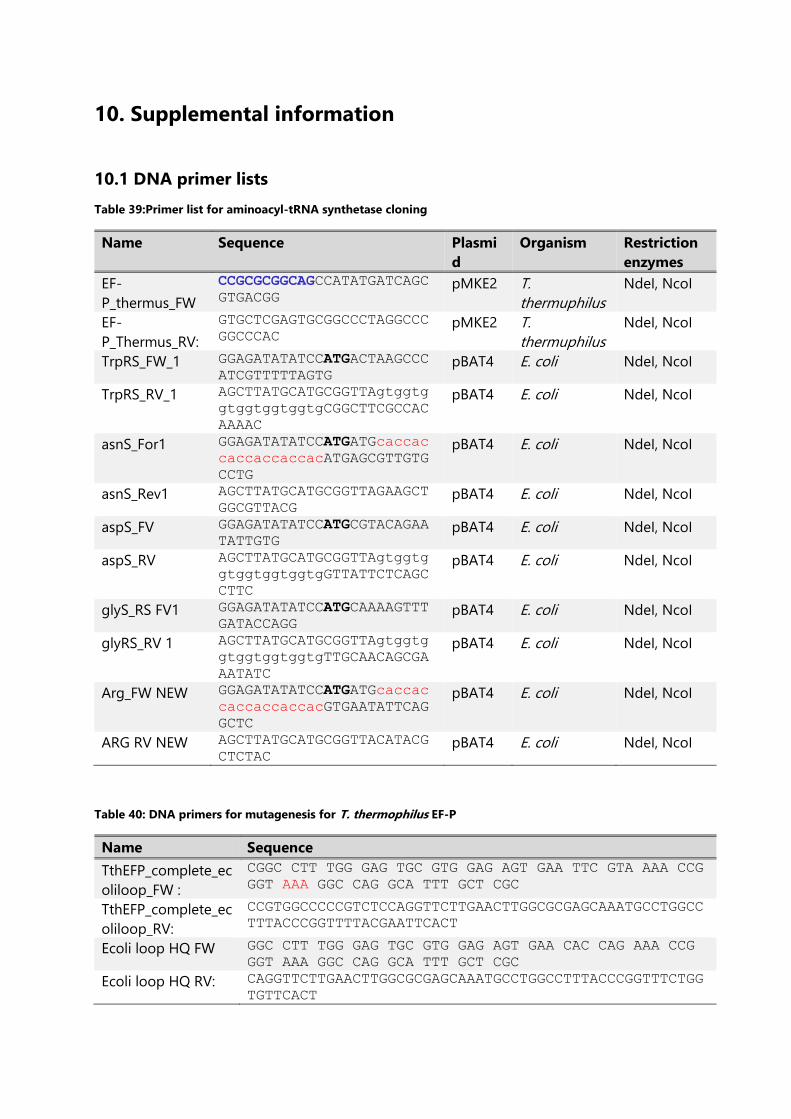
10. Supplemental information
10.1 DNA primer lists
Table 39:Primer list for aminoacyl-tRNA synthetase cloning
Name Sequence Plasmi
d
Organism Restriction
enzymes
EF-
P_thermus_FW
CCGCGCGGCAGCCATATGATCAGC
GTGACGG pMKE2 T.
thermuphilus
NdeI, NcoI
EF-
P_Thermus_RV:
GTGCTCGAGTGCGGCCCTAGGCCC
GGCCCAC pMKE2 T.
thermuphilus
NdeI, NcoI
TrpRS_FW_1 GGAGATATATCCATGACTAAGCCC
ATCGTTTTTAGTG pBAT4 E. coli NdeI, NcoI
TrpRS_RV_1 AGCTTATGCATGCGGTTAgtggtg
gtggtggtggtgCGGCTTCGCCAC
AAAAC
pBAT4 E. coli NdeI, NcoI
asnS_For1 GGAGATATATCCATGATGcaccac
caccaccaccacATGAGCGTTGTG
CCTG
pBAT4 E. coli NdeI, NcoI
asnS_Rev1 AGCTTATGCATGCGGTTAGAAGCT
GGCGTTACG pBAT4 E. coli NdeI, NcoI
aspS_FV GGAGATATATCCATGCGTACAGAA
TATTGTG pBAT4 E. coli NdeI, NcoI
aspS_RV AGCTTATGCATGCGGTTAgtggtg
gtggtggtggtgGTTATTCTCAGC
CTTC
pBAT4 E. coli NdeI, NcoI
glyS_RS FV1 GGAGATATATCCATGCAAAAGTTT
GATACCAGG pBAT4 E. coli NdeI, NcoI
glyRS_RV 1 AGCTTATGCATGCGGTTAgtggtg
gtggtggtggtgTTGCAACAGCGA
AATATC
pBAT4 E. coli NdeI, NcoI
Arg_FW NEW GGAGATATATCCATGATGcaccac
caccaccaccacGTGAATATTCAG
GCTC
pBAT4 E. coli NdeI, NcoI
ARG RV NEW AGCTTATGCATGCGGTTACATACG
CTCTAC pBAT4 E. coli NdeI, NcoI
Table 40: DNA primers for mutagenesis for T. thermophilus EF-P
Name Sequence
TthEFP_complete_ec
oliloop_FW :
CGGC CTT TGG GAG TGC GTG GAG AGT GAA TTC GTA AAA CCG
GGT AAA GGC CAG GCA TTT GCT CGC
TthEFP_complete_ec
oliloop_RV:
CCGTGGCCCCCGTCTCCAGGTTCTTGAACTTGGCGCGAGCAAATGCCTGGCC
TTTACCCGGTTTTACGAATTCACT
Ecoli loop HQ FW GGC CTT TGG GAG TGC GTG GAG AGT GAA CAC CAG AAA CCG
GGT AAA GGC CAG GCA TTT GCT CGC
Ecoli loop HQ RV: CAGGTTCTTGAACTTGGCGCGAGCAAATGCCTGGCCTTTACCCGGTTTCTGG
TGTTCACT
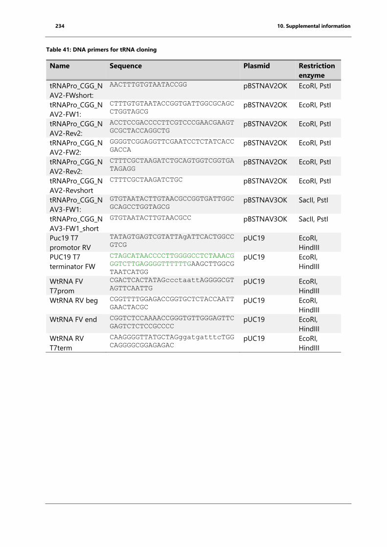
234 10. Supplemental information
Table 41: DNA primers for tRNA cloning
Name Sequence Plasmid Restriction
enzyme
tRNAPro_CGG_N
AV2-FWshort:
AACTTTGTGTAATACCGG pBSTNAV2OK EcoRI, PstI
tRNAPro_CGG_N
AV2-FW1:
CTTTGTGTAATACCGGTGATTGGCGCAGC
CTGGTAGCG pBSTNAV2OK EcoRI, PstI
tRNAPro_CGG_N
AV2-Rev2:
ACCTCCGACCCCTTCGTCCCGAACGAAGT
GCGCTACCAGGCTG pBSTNAV2OK EcoRI, PstI
tRNAPro_CGG_N
AV2-FW2:
GGGGTCGGAGGTTCGAATCCTCTATCACC
GACCA pBSTNAV2OK EcoRI, PstI
tRNAPro_CGG_N
AV2-Rev2:
CTTTCGCTAAGATCTGCAGTGGTCGGTGA
TAGAGG pBSTNAV2OK EcoRI, PstI
tRNAPro_CGG_N
AV2-Revshort
CTTTCGCTAAGATCTGC pBSTNAV2OK EcoRI, PstI
tRNAPro_CGG_N
AV3-FW1:
GTGTAATACTTGTAACGCCGGTGATTGGC
GCAGCCTGGTAGCG pBSTNAV3OK SacII, PstI
tRNAPro_CGG_N
AV3-FW1_short
GTGTAATACTTGTAACGCC pBSTNAV3OK SacII, PstI
Puc19 T7
promotor RV
TATAGTGAGTCGTATTAgATTCACTGGCC
GTCG pUC19 EcoRI,
HindIII
PUC19 T7
terminator FW
CTAGCATAACCCCTTGGGGCCTCTAAACG
GGTCTTGAGGGGTTTTTTGAAGCTTGGCG
TAATCATGG
pUC19 EcoRI,
HindIII
WtRNA FV
T7prom
CGACTCACTATAGccctaattAGGGGCGT
AGTTCAATTG pUC19 EcoRI,
HindIII
WtRNA RV beg CGGTTTTGGAGACCGGTGCTCTACCAATT
GAACTACGC pUC19 EcoRI,
HindIII
WtRNA FV end CGGTCTCCAAAACCGGGTGTTGGGAGTTC
GAGTCTCTCCGCCCC pUC19 EcoRI,
HindIII
WtRNA RV
T7term
CAAGGGGTTATGCTAGggatgatttcTGG
CAGGGGCGGAGAGAC pUC19 EcoRI,
HindIII
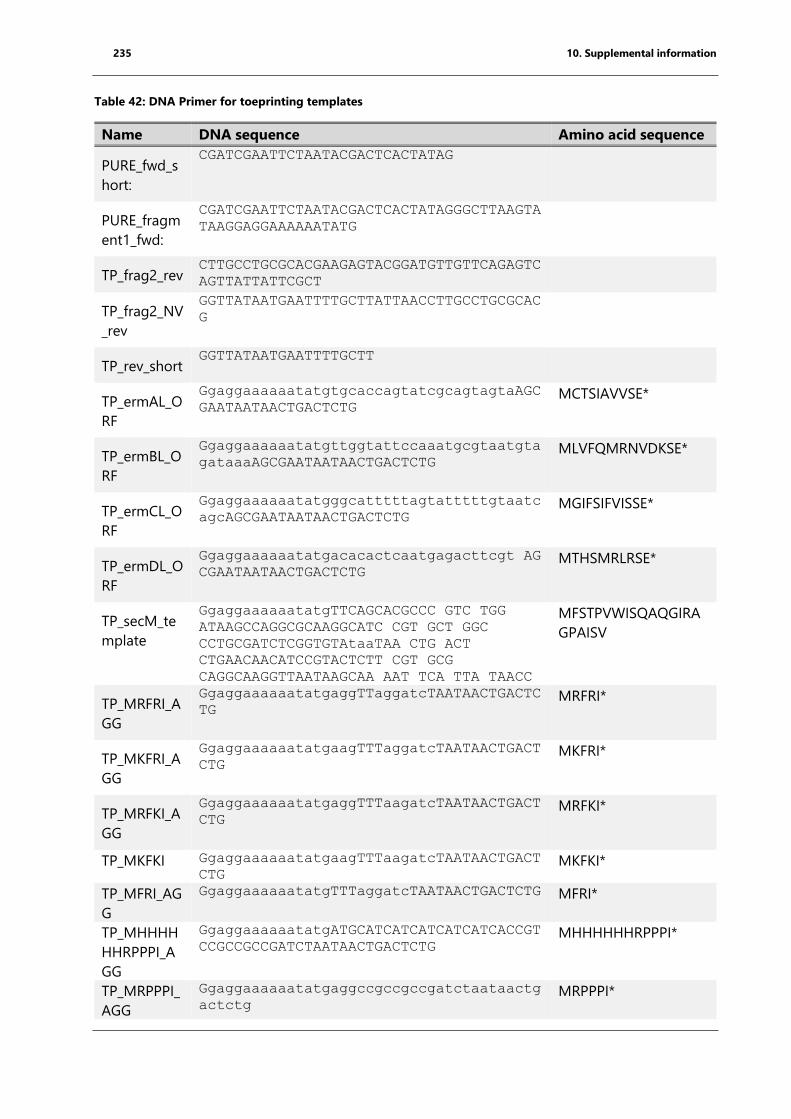
235 10. Supplemental information
Table 42: DNA Primer for toeprinting templates
Name DNA sequence Amino acid sequence
PURE_fwd_s
hort:
CGATCGAATTCTAATACGACTCACTATAG
PURE_fragm
ent1_fwd:
CGATCGAATTCTAATACGACTCACTATAGGGCTTAAGTA
TAAGGAGGAAAAAATATG
TP_frag2_rev CTTGCCTGCGCACGAAGAGTACGGATGTTGTTCAGAGTC
AGTTATTATTCGCT
TP_frag2_NV
_rev
GGTTATAATGAATTTTGCTTATTAACCTTGCCTGCGCAC
G
TP_rev_short GGTTATAATGAATTTTGCTT
TP_ermAL_O
RF
GgaggaaaaaatatgtgcaccagtatcgcagtagtaAGC
GAATAATAACTGACTCTG MCTSIAVVSE*
TP_ermBL_O
RF
Ggaggaaaaaatatgttggtattccaaatgcgtaatgta
gataaaAGCGAATAATAACTGACTCTG MLVFQMRNVDKSE*
TP_ermCL_O
RF
Ggaggaaaaaatatgggcatttttagtatttttgtaatc
agcAGCGAATAATAACTGACTCTG MGIFSIFVISSE*
TP_ermDL_O
RF
Ggaggaaaaaatatgacacactcaatgagacttcgt AG
CGAATAATAACTGACTCTG MTHSMRLRSE*
TP_secM_te
mplate
GgaggaaaaaatatgTTCAGCACGCCC GTC TGG
ATAAGCCAGGCGCAAGGCATC CGT GCT GGC
CCTGCGATCTCGGTGTAtaaTAA CTG ACT
CTGAACAACATCCGTACTCTT CGT GCG
CAGGCAAGGTTAATAAGCAA AAT TCA TTA TAACC
MFSTPVWISQAQGIRA
GPAISV
TP_MRFRI_A
GG
GgaggaaaaaatatgaggTTaggatcTAATAACTGACTC
TG MRFRI*
TP_MKFRI_A
GG
GgaggaaaaaatatgaagTTTaggatcTAATAACTGACT
CTG MKFRI*
TP_MRFKI_A
GG
GgaggaaaaaatatgaggTTTaagatcTAATAACTGACT
CTG MRFKI*
TP_MKFKI GgaggaaaaaatatgaagTTTaagatcTAATAACTGACT
CTG MKFKI*
TP_MFRI_AG
G
GgaggaaaaaatatgTTTaggatcTAATAACTGACTCTG MFRI*
TP_MHHHH
HHRPPPI_A
GG
GgaggaaaaaatatgATGCATCATCATCATCATCACCGT
CCGCCGCCGATCTAATAACTGACTCTG MHHHHHHRPPPI*
TP_MRPPPI_
AGG
Ggaggaaaaaatatgaggccgccgccgatctaataactg
actctg MRPPPI*
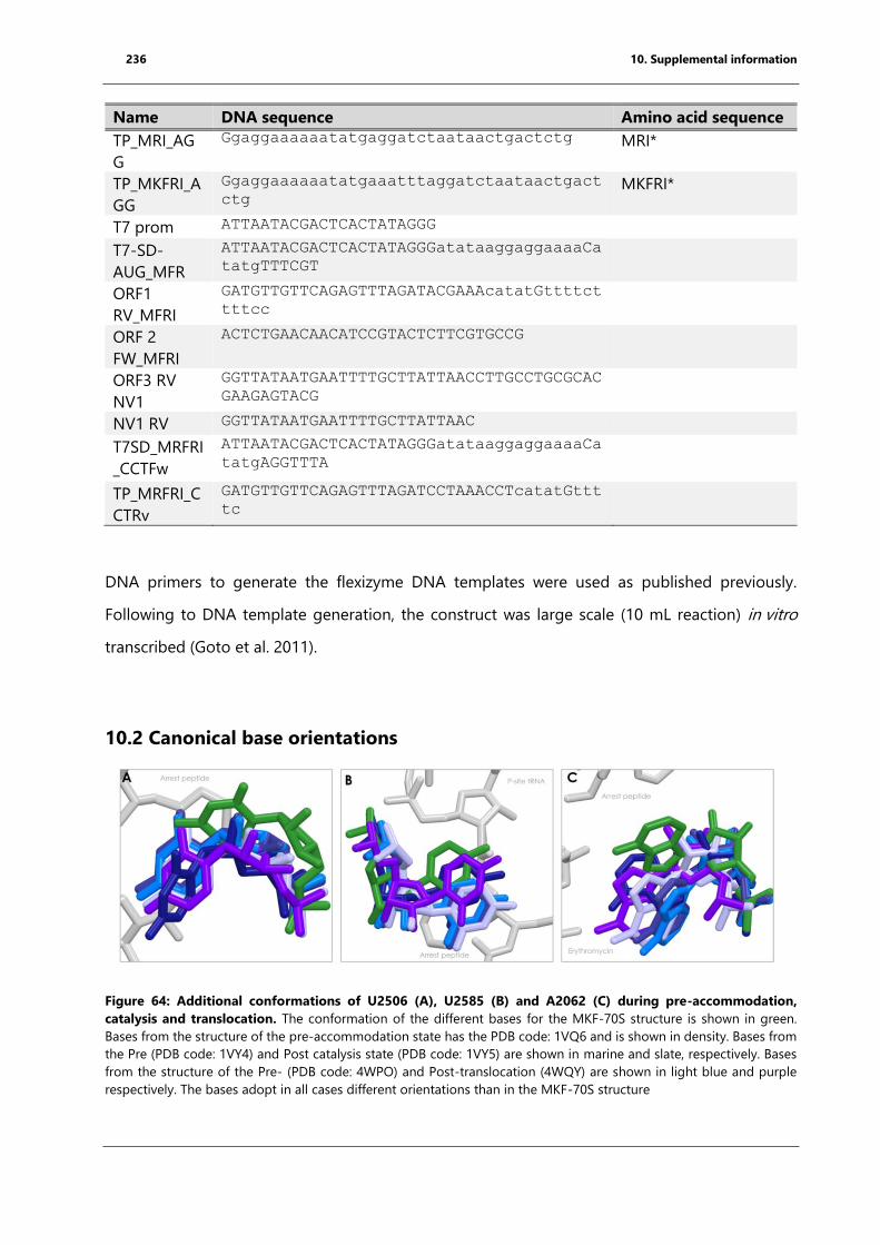
236 10. Supplemental information
Name DNA sequence Amino acid sequence
TP_MRI_AG
G
Ggaggaaaaaatatgaggatctaataactgactctg MRI*
TP_MKFRI_A
GG
Ggaggaaaaaatatgaaatttaggatctaataactgact
ctg MKFRI*
T7 prom ATTAATACGACTCACTATAGGG
T7-SD-
AUG_MFR
ATTAATACGACTCACTATAGGGatataaggaggaaaaCa
tatgTTTCGT
ORF1
RV_MFRI
GATGTTGTTCAGAGTTTAGATACGAAAcatatGttttct
tttcc
ORF 2
FW_MFRI
ACTCTGAACAACATCCGTACTCTTCGTGCCG
ORF3 RV
NV1
GGTTATAATGAATTTTGCTTATTAACCTTGCCTGCGCAC
GAAGAGTACG
NV1 RV GGTTATAATGAATTTTGCTTATTAAC
T7SD_MRFRI
_CCTFw
ATTAATACGACTCACTATAGGGatataaggaggaaaaCa
tatgAGGTTTA
TP_MRFRI_C
CTRv
GATGTTGTTCAGAGTTTAGATCCTAAACCTcatatGttt
tc
DNA primers to generate the flexizyme DNA templates were used as published previously.
Following to DNA template generation, the construct was large scale (10 mL reaction) in vitro
transcribed (Goto et al. 2011).
10.2 Canonical base orientations
Figure 64: Additional conformations of U2506 (A), U2585 (B) and A2062 (C) during pre-accommodation,
catalysis and translocation. The conformation of the different bases for the MKF-70S structure is shown in green.
Bases from the structure of the pre-accommodation state has the PDB code: 1VQ6 and is shown in density. Bases from
the Pre (PDB code: 1VY4) and Post catalysis state (PDB code: 1VY5) are shown in marine and slate, respectively. Bases
from the structure of the Pre- (PDB code: 4WPO) and Post-translocation (4WQY) are shown in light blue and purple
respectively. The bases adopt in all cases different orientations than in the MKF-70S structure
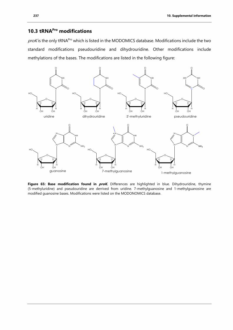
237 10. Supplemental information
10.3 tRNAPro modifications
proK is the only tRNAPro which is listed in the MODOMICS database. Modifications include the two
standard modifications pseudouridine and dihydrouridine. Other modifications include
methylations of the bases. The modifications are listed in the following figure:
Figure 65: Base modification found in proK. Differences are highlighted in blue. Dihydrouridine, thymine
(5-methyluridine) and pseudouridine are derrived from uridine. 7-methylguanosine and 1-methylguanosine are
modified guanosine bases. Modifications were listed on the MODONOMICS database.
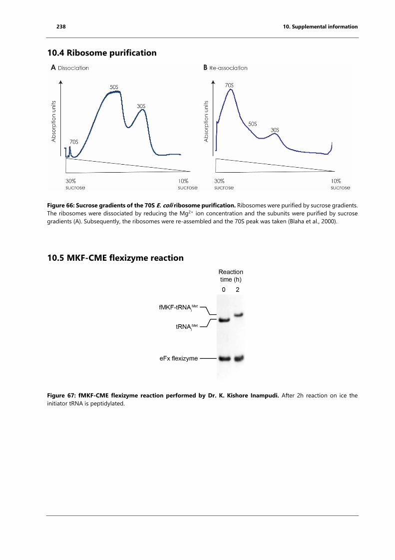
238 10. Supplemental information
10.4 Ribosome purification
Figure 66: Sucrose gradients of the 70S E. coli ribosome purification. Ribosomes were purified by sucrose gradients.
The ribosomes were dissociated by reducing the Mg2+ ion concentration and the subunits were purified by sucrose
gradients (A). Subsequently, the ribosomes were re-assembled and the 70S peak was taken (Blaha et al., 2000).
10.5 MKF-CME flexizyme reaction
Figure 67: fMKF-CME flexizyme reaction performed by Dr. K. Kishore Inampudi. After 2h reaction on ice the
initiator tRNA is peptidylated.
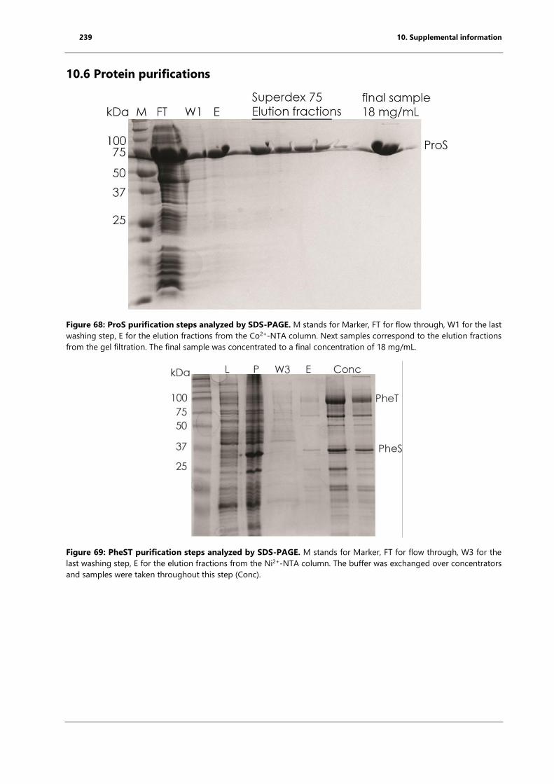
239 10. Supplemental information
10.6 Protein purifications
Figure 68: ProS purification steps analyzed by SDS-PAGE. M stands for Marker, FT for flow through, W1 for the last
washing step, E for the elution fractions from the Co2+-NTA column. Next samples correspond to the elution fractions
from the gel filtration. The final sample was concentrated to a final concentration of 18 mg/mL.
Figure 69: PheST purification steps analyzed by SDS-PAGE. M stands for Marker, FT for flow through, W3 for the
last washing step, E for the elution fractions from the Ni2+-NTA column. The buffer was exchanged over concentrators
and samples were taken throughout this step (Conc).
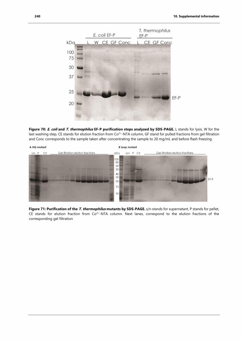
240 10. Supplemental information
Figure 70: E. coli and T. thermophilus EF-P purification steps analyzed by SDS-PAGE. L stands for lysis, W for the
last washing step, CE stands for elution fraction from Co2+-NTA column, GF stand for pulled fractions from gel filtration
and Conc corresponds to the sample taken after concentrating the sample to 20 mg/mL and before flash freezing.
Figure 71: Purification of the T. thermophilus mutants by SDS-PAGE. s/n stands for supernatant, P stands for pellet,
CE stands for elution fraction from Co2+-NTA column. Next lanes, correspond to the elution fractions of the
corresponding gel filtration
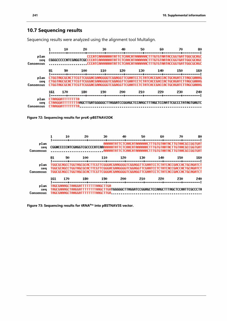
241 10. Supplemental information
10.7 Sequencing results
Sequencing results were analyzed using the alignment tool Multalign.
Figure 72: Sequencing results for proK-pBSTNAV2OK
Figure 73: Sequencing results for tRNAPro into pBSTNAV3S vector.
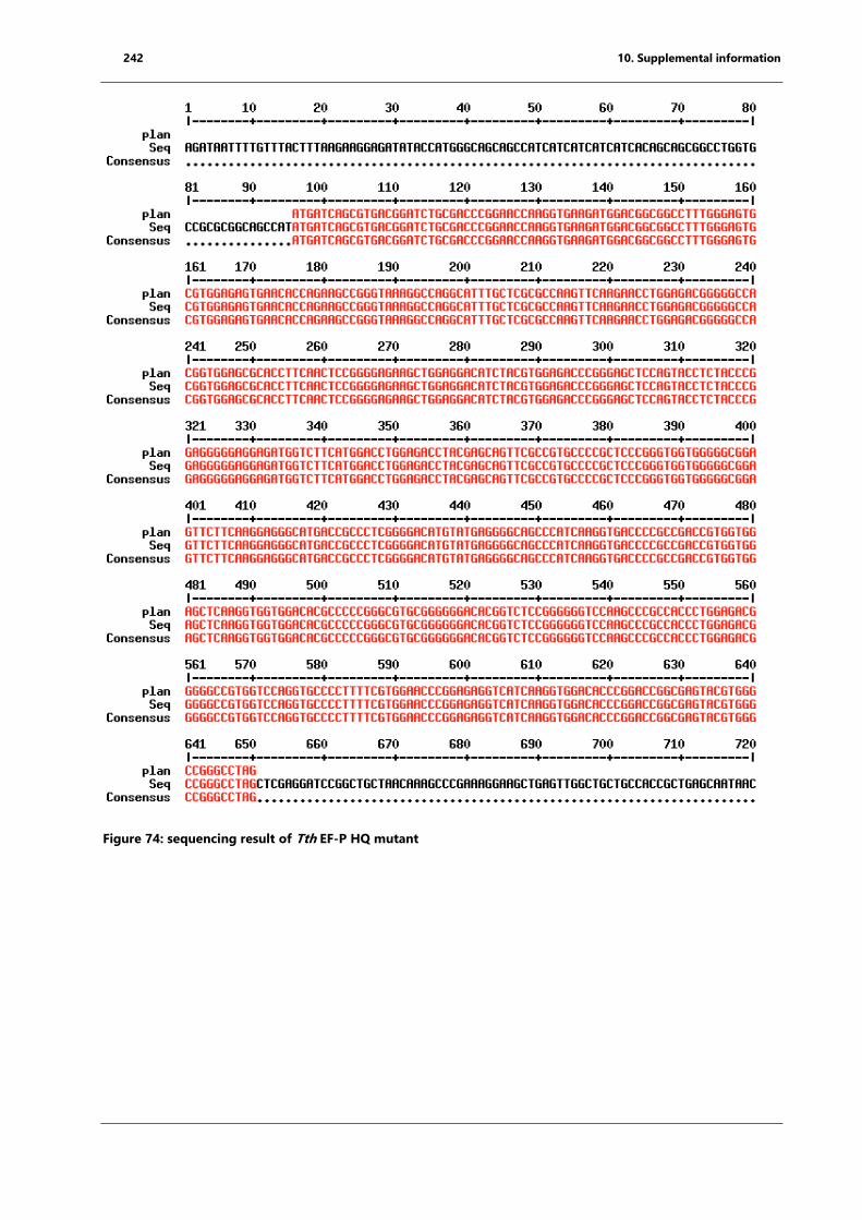
242 10. Supplemental information
Figure 74: sequencing result of Tth EF-P HQ mutant
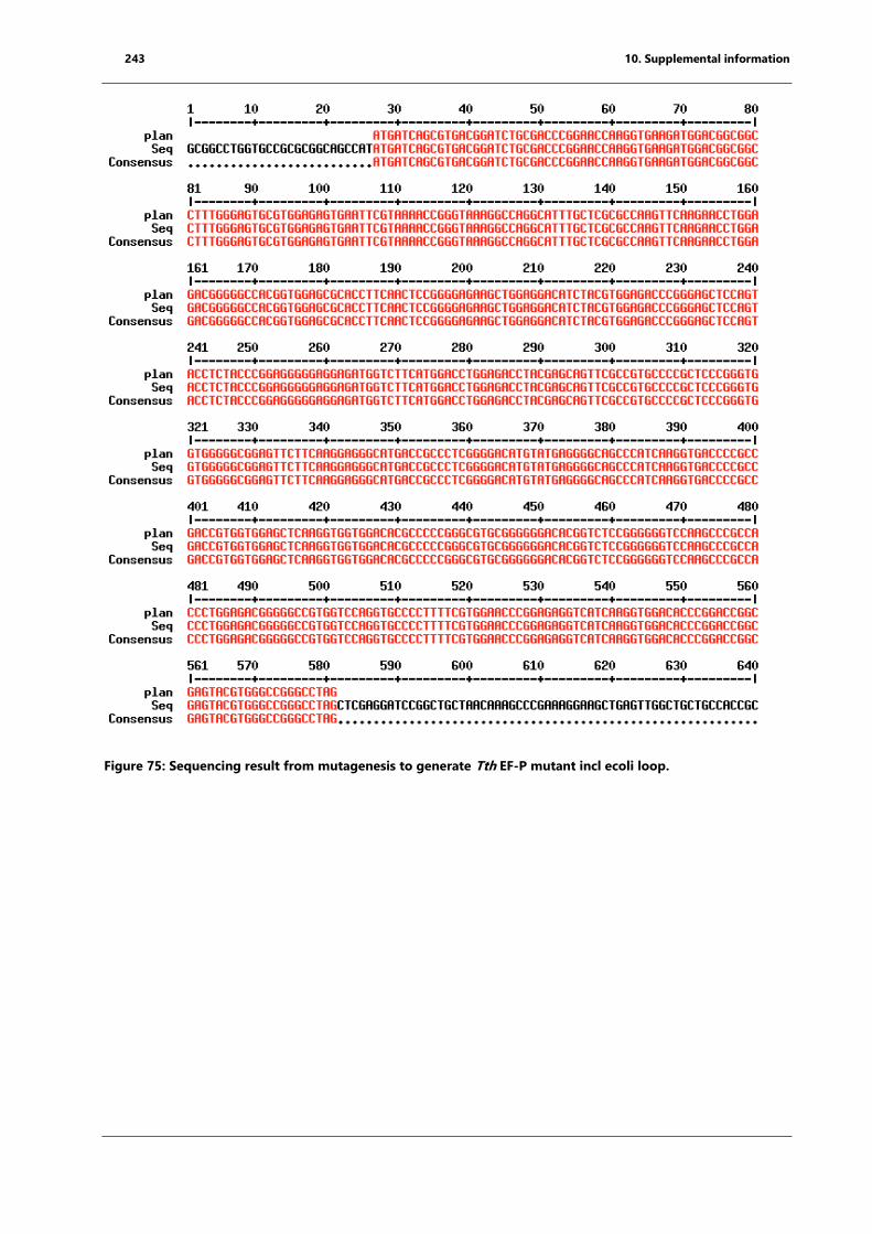
243 10. Supplemental information
Figure 75: Sequencing result from mutagenesis to generate Tth EF-P mutant incl ecoli loop.
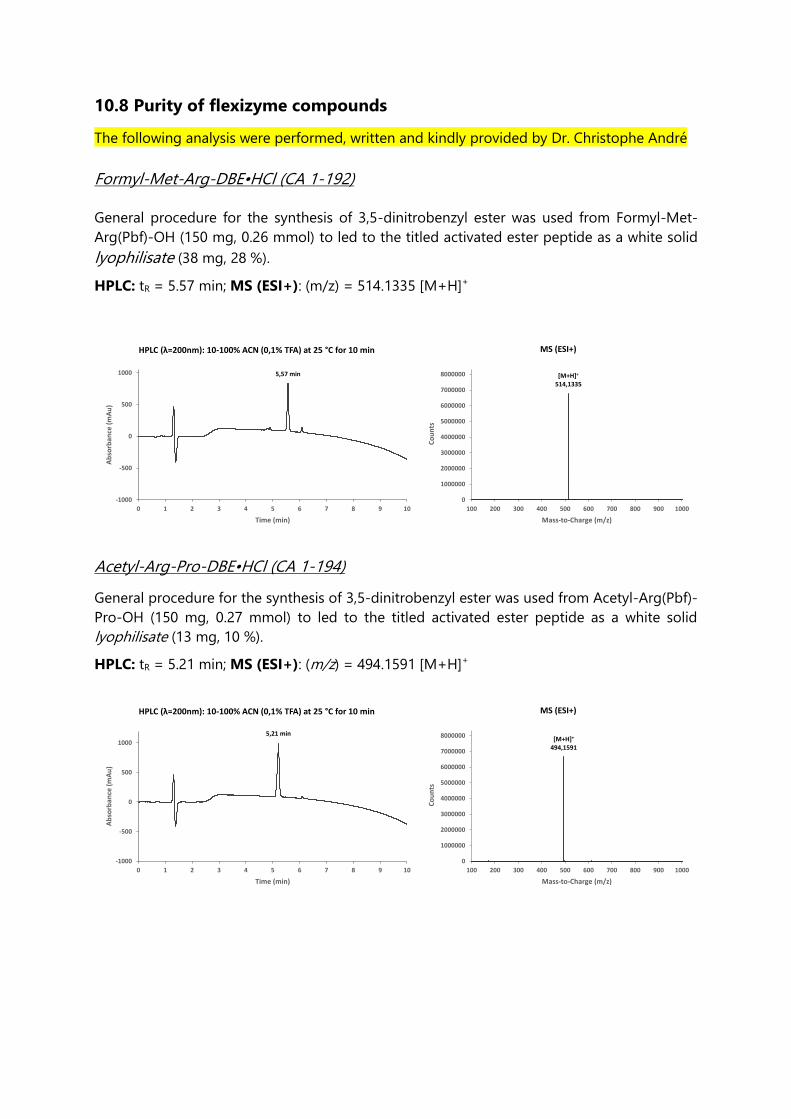
10.8 Purity of flexizyme compounds
The following analysis were performed, written and kindly provided by Dr. Christophe André
Formyl-Met-Arg-DBE•HCl (CA 1-192)
General procedure for the synthesis of 3,5-dinitrobenzyl ester was used from Formyl-Met-
Arg(Pbf)-OH (150 mg, 0.26 mmol) to led to the titled activated ester peptide as a white solid
lyophilisate (38 mg, 28 %).
HPLC: tR = 5.57 min; MS (ESI+): (m/z) = 514.1335 [M+H]+
Acetyl-Arg-Pro-DBE•HCl (CA 1-194)
General procedure for the synthesis of 3,5-dinitrobenzyl ester was used from Acetyl-Arg(Pbf)-
Pro-OH (150 mg, 0.27 mmol) to led to the titled activated ester peptide as a white solid
lyophilisate (13 mg, 10 %).
HPLC: tR = 5.21 min; MS (ESI+): (m/z) = 494.1591 [M+H]+
5,57 min
-1000
-500
0
500
1000
0 1 2 3 4 5 6 7 8 9 10
Ab
sorb
ance
(m
Au
)
Time (min)
MS (ESI+)
[M+H]+
514,1335
0
1000000
2000000
3000000
4000000
5000000
6000000
7000000
8000000
100 200 300 400 500 600 700 800 900 1000
Co
un
ts
Mass-to-Charge (m/z)
HPLC (λ=200nm): 10-100% ACN (0,1% TFA) at 25 °C for 10 min
5,21 min
-1000
-500
0
500
1000
0 1 2 3 4 5 6 7 8 9 10
Ab
sorb
ance
(m
Au
)
Time (min)
[M+H]+
494,1591
0
1000000
2000000
3000000
4000000
5000000
6000000
7000000
8000000
100 200 300 400 500 600 700 800 900 1000
Co
un
ts
Mass-to-Charge (m/z)
MS (ESI+)HPLC (λ=200nm): 10-100% ACN (0,1% TFA) at 25 °C for 10 min
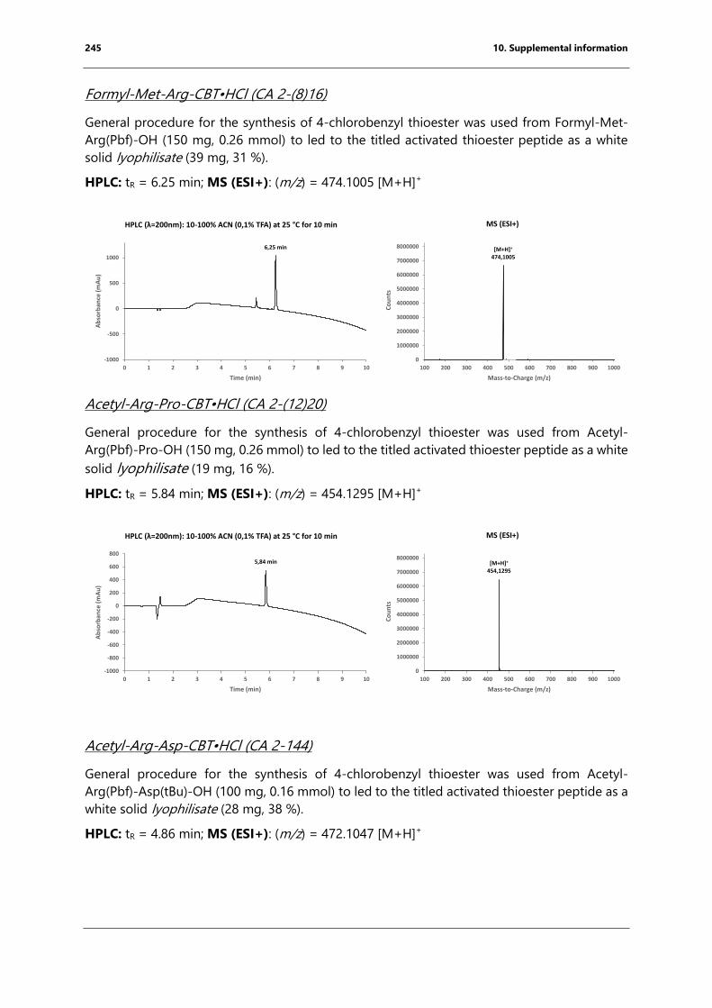
245 10. Supplemental information
Formyl-Met-Arg-CBT•HCl (CA 2-(8)16)
General procedure for the synthesis of 4-chlorobenzyl thioester was used from Formyl-Met-
Arg(Pbf)-OH (150 mg, 0.26 mmol) to led to the titled activated thioester peptide as a white
solid lyophilisate (39 mg, 31 %).
HPLC: tR = 6.25 min; MS (ESI+): (m/z) = 474.1005 [M+H]+
Acetyl-Arg-Pro-CBT•HCl (CA 2-(12)20)
General procedure for the synthesis of 4-chlorobenzyl thioester was used from Acetyl-
Arg(Pbf)-Pro-OH (150 mg, 0.26 mmol) to led to the titled activated thioester peptide as a white
solid lyophilisate (19 mg, 16 %).
HPLC: tR = 5.84 min; MS (ESI+): (m/z) = 454.1295 [M+H]+
Acetyl-Arg-Asp-CBT•HCl (CA 2-144)
General procedure for the synthesis of 4-chlorobenzyl thioester was used from Acetyl-
Arg(Pbf)-Asp(tBu)-OH (100 mg, 0.16 mmol) to led to the titled activated thioester peptide as a
white solid lyophilisate (28 mg, 38 %).
HPLC: tR = 4.86 min; MS (ESI+): (m/z) = 472.1047 [M+H]+
6,25 min
-1000
-500
0
500
1000
0 1 2 3 4 5 6 7 8 9 10
Ab
sorb
ance
(m
Au
)
Time (min)
[M+H]+
474,1005
0
1000000
2000000
3000000
4000000
5000000
6000000
7000000
8000000
100 200 300 400 500 600 700 800 900 1000
Co
un
ts
Mass-to-Charge (m/z)
MS (ESI+)HPLC (λ=200nm): 10-100% ACN (0,1% TFA) at 25 °C for 10 min
[M+H]+
454,1295
0
1000000
2000000
3000000
4000000
5000000
6000000
7000000
8000000
100 200 300 400 500 600 700 800 900 1000
Co
un
ts
Mass-to-Charge (m/z)
5,84 min
-1000
-800
-600
-400
-200
0
200
400
600
800
0 1 2 3 4 5 6 7 8 9 10
Ab
sorb
ance
(m
Au
)
Time (min)
MS (ESI+)HPLC (λ=200nm): 10-100% ACN (0,1% TFA) at 25 °C for 10 min
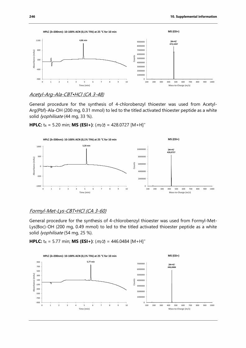
246 10. Supplemental information
Acetyl-Arg-Ala-CBT•HCl (CA 3-48)
General procedure for the synthesis of 4-chlorobenzyl thioester was used from Acetyl-
Arg(Pbf)-Ala-OH (200 mg, 0.31 mmol) to led to the titled activated thioester peptide as a white
solid lyophilisate (44 mg, 33 %).
HPLC: tR = 5.20 min; MS (ESI+): (m/z) = 428.0727 [M+H]+
Formyl-Met-Lys-CBT•HCl (CA 3-60)
General procedure for the synthesis of 4-chlorobenzyl thioester was used from Formyl-Met-
Lys(Boc)-OH (200 mg, 0.49 mmol) to led to the titled activated thioester peptide as a white
solid lyophilisate (54 mg, 25 %).
HPLC: tR = 5.77 min; MS (ESI+): (m/z) = 446.0484 [M+H]+
4,86 min
-900
-400
100
600
1100
0 1 2 3 4 5 6 7 8 9 10
Ab
sorb
ance
(m
Au
)
Time (min)
[M+H]+
472,1047
0
1000000
2000000
3000000
4000000
5000000
6000000
7000000
8000000
9000000
100 200 300 400 500 600 700 800 900 1000
Co
un
ts
Mass-to-Charge (m/z)
MS (ESI+)HPLC (λ=200nm): 10-100% ACN (0,1% TFA) at 25 °C for 10 min
5,20 min
-1000
-500
0
500
1000
0 1 2 3 4 5 6 7 8 9 10
Ab
sorb
ance
(m
Au
)
Time (min)
[M+H]+
428,0727
0
2000000
4000000
6000000
8000000
10000000
100 200 300 400 500 600 700 800 900 1000
Co
un
ts
Mass-to-Charge (m/z)
MS (ESI+)HPLC (λ=200nm): 10-100% ACN (0,1% TFA) at 25 °C for 10 min
5,77 min
-900
-700
-500
-300
-100
100
300
500
700
900
0 1 2 3 4 5 6 7 8 9 10
Ab
sorb
ance
(m
Au
)
Time (min)
[M+H]+
446,0484
0
1000000
2000000
3000000
4000000
5000000
6000000
7000000
100 200 300 400 500 600 700 800 900 1000
Co
un
ts
Mass-to-Charge (m/z)
MS (ESI+)HPLC (λ=200nm): 10-100% ACN (0,1% TFA) at 25 °C for 10 min
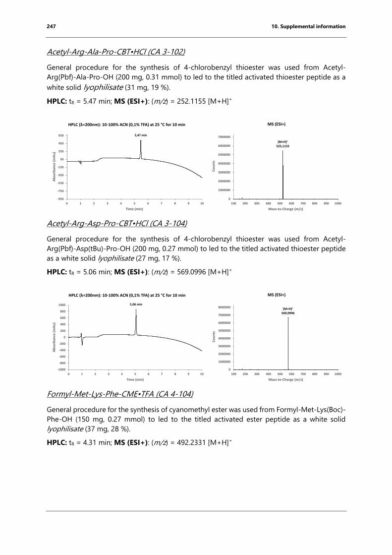
247 10. Supplemental information
Acetyl-Arg-Ala-Pro-CBT•HCl (CA 3-102)
General procedure for the synthesis of 4-chlorobenzyl thioester was used from Acetyl-
Arg(Pbf)-Ala-Pro-OH (200 mg, 0.31 mmol) to led to the titled activated thioester peptide as a
white solid lyophilisate (31 mg, 19 %).
HPLC: tR = 5.47 min; MS (ESI+): (m/z) = 252.1155 [M+H]+
Acetyl-Arg-Asp-Pro-CBT•HCl (CA 3-104)
General procedure for the synthesis of 4-chlorobenzyl thioester was used from Acetyl-
Arg(Pbf)-Asp(tBu)-Pro-OH (200 mg, 0.27 mmol) to led to the titled activated thioester peptide
as a white solid lyophilisate (27 mg, 17 %).
HPLC: tR = 5.06 min; MS (ESI+): (m/z) = 569.0996 [M+H]+
Formyl-Met-Lys-Phe-CME•TFA (CA 4-104)
General procedure for the synthesis of cyanomethyl ester was used from Formyl-Met-Lys(Boc)-
Phe-OH (150 mg, 0.27 mmol) to led to the titled activated ester peptide as a white solid
lyophilisate (37 mg, 28 %).
HPLC: tR = 4.31 min; MS (ESI+): (m/z) = 492.2331 [M+H]+
5,47 min
-950
-750
-550
-350
-150
50
250
450
650
0 1 2 3 4 5 6 7 8 9 10
Ab
sorb
ance
(m
Au
)
Time (min)
[M+H]+
525,1155
0
1000000
2000000
3000000
4000000
5000000
6000000
7000000
100 200 300 400 500 600 700 800 900 1000
Co
un
ts
Mass-to-Charge (m/z)
MS (ESI+)HPLC (λ=200nm): 10-100% ACN (0,1% TFA) at 25 °C for 10 min
5,06 min
-1000
-800
-600
-400
-200
0
200
400
600
800
1000
0 1 2 3 4 5 6 7 8 9 10
Ab
sorb
ance
(m
Au
)
Time (min)
[M+H]+
569,0996
0
1000000
2000000
3000000
4000000
5000000
6000000
7000000
8000000
100 200 300 400 500 600 700 800 900 1000
Co
un
ts
Mass-to-Charge (m/z)
MS (ESI+)HPLC (λ=200nm): 10-100% ACN (0,1% TFA) at 25 °C for 10 min
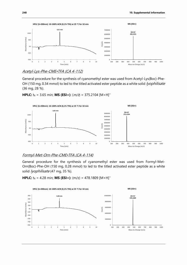
248 10. Supplemental information
Acetyl-Lys-Phe-CME•TFA (CA 4-112)
General procedure for the synthesis of cyanomethyl ester was used from Acetyl-Lys(Boc)-Phe-
OH (150 mg, 0.34 mmol) to led to the titled activated ester peptide as a white solid lyophilisate
(36 mg, 28 %).
HPLC: tR = 3.65 min; MS (ESI+): (m/z) = 375.2104 [M+H]+
Formyl-Met-Orn-Phe-CME•TFA (CA 4-114)
General procedure for the synthesis of cyanomethyl ester was used from Formyl-Met-
Orn(Boc)-Phe-OH (150 mg, 0.28 mmol) to led to the titled activated ester peptide as a white
solid lyophilisate (47 mg, 35 %).
HPLC: tR = 4.28 min; MS (ESI+): (m/z) = 478.1809 [M+H]+
4,31 min
-950
-450
50
550
1050
0 1 2 3 4 5 6 7 8 9 10
Ab
sorb
ance
(m
Au
)
Time (min)
[M+H]+
492,2331
0
1000000
2000000
3000000
4000000
5000000
6000000
7000000
100 200 300 400 500 600 700 800 900 1000
Co
un
ts
Mass-to-Charge (m/z)
MS (ESI+)HPLC (λ=200nm): 10-100% ACN (0,1% TFA) at 25 °C for 10 min
3,65 min
-950
-450
50
550
1050
0 1 2 3 4 5 6 7 8 9 10
Ab
sorb
ance
(m
Au
)
Time (min)
[M+H]+
375,2104
0
1000000
2000000
3000000
4000000
5000000
6000000
7000000
8000000
9000000
100 200 300 400 500 600 700 800 900 1000
Co
un
ts
Mass-to-Charge (m/z)
MS (ESI+)HPLC (λ=200nm): 10-100% ACN (0,1% TFA) at 25 °C for 10 min
4,28 min
-950
-750
-550
-350
-150
50
250
450
650
850
0 1 2 3 4 5 6 7 8 9 10
Ab
sorb
ance
(m
Au
)
Time (min)
[M+H]+
478,1809
0
2000000
4000000
6000000
8000000
10000000
100 200 300 400 500 600 700 800 900 1000
Co
un
ts
Mass-to-Charge (m/z)
MS (ESI+)HPLC (λ=200nm): 10-100% ACN (0,1% TFA) at 25 °C for 10 min
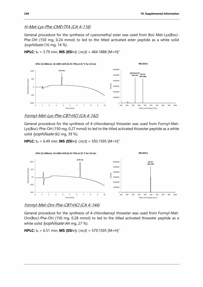
249 10. Supplemental information
H-Met-Lys-Phe-CME•TFA (CA 4-116)
General procedure for the synthesis of cyanomethyl ester was used from Boc-Met-Lys(Boc)-
Phe-OH (150 mg, 0.24 mmol) to led to the titled activated ester peptide as a white solid
lyophilisate (16 mg, 14 %).
HPLC: tR = 3.79 min; MS (ESI+): (m/z) = 464.1888 [M+H]+
Formyl-Met-Lys-Phe-CBT•HCl (CA 4-142)
General procedure for the synthesis of 4-chlorobenzyl thioester was used from Formyl-Met-
Lys(Boc)-Phe-OH (150 mg, 0.27 mmol) to led to the titled activated thioester peptide as a white
solid lyophilisate (62 mg, 39 %).
HPLC: tR = 6.49 min; MS (ESI+): (m/z) = 593.1595 [M+H]+
Formyl-Met-Orn-Phe-CBT•HCl (CA 4-144)
General procedure for the synthesis of 4-chlorobenzyl thioester was used from Formyl-Met-
Orn(Boc)-Phe-OH (150 mg, 0.28 mmol) to led to the titled activated thioester peptide as a
white solid lyophilisate (44 mg, 27 %).
HPLC: tR = 6.51 min; MS (ESI+): (m/z) = 579.1505 [M+H]+
3,79 min
-1000
-500
0
500
1000
0 1 2 3 4 5 6 7 8 9 10
Ab
sorb
ance
(m
Au
)
Time (min)
[M(-Met)+H]+
333,1551[M+H]+
464,1888
0
1000000
2000000
3000000
4000000
5000000
6000000
100 200 300 400 500 600 700 800 900 1000
Co
un
ts
Mass-to-Charge (m/z)
MS (ESI+)HPLC (λ=200nm): 10-100% ACN (0,1% TFA) at 25 °C for 10 min
6,49 min
-950
-450
50
550
1050
0 1 2 3 4 5 6 7 8 9 10
Ab
sorb
ance
(m
Au
)
Time (min)
[M+H]+
593,1595
0
1000000
2000000
3000000
4000000
5000000
6000000
100 200 300 400 500 600 700 800 900 1000
Co
un
ts
Mass-to-Charge (m/z)
MS (ESI+)HPLC (λ=200nm): 10-100% ACN (0,1% TFA) at 25 °C for 10 min
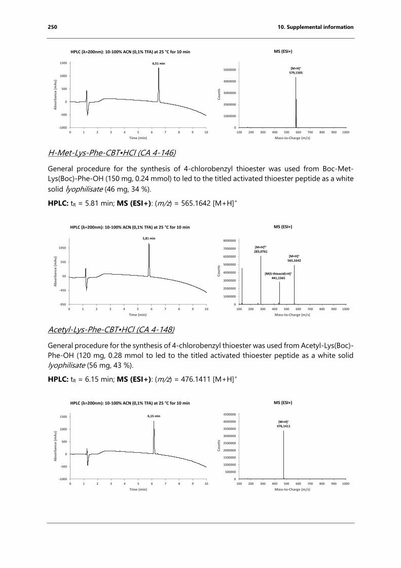
250 10. Supplemental information
H-Met-Lys-Phe-CBT•HCl (CA 4-146)
General procedure for the synthesis of 4-chlorobenzyl thioester was used from Boc-Met-
Lys(Boc)-Phe-OH (150 mg, 0.24 mmol) to led to the titled activated thioester peptide as a white
solid lyophilisate (46 mg, 34 %).
HPLC: tR = 5.81 min; MS (ESI+): (m/z) = 565.1642 [M+H]+
Acetyl-Lys-Phe-CBT•HCl (CA 4-148)
General procedure for the synthesis of 4-chlorobenzyl thioester was used from Acetyl-Lys(Boc)-
Phe-OH (120 mg, 0.28 mmol to led to the titled activated thioester peptide as a white solid
lyophilisate (56 mg, 43 %).
HPLC: tR = 6.15 min; MS (ESI+): (m/z) = 476.1411 [M+H]+
[M+H]+
579,1505
0
1000000
2000000
3000000
4000000
5000000
100 200 300 400 500 600 700 800 900 1000
Co
un
ts
Mass-to-Charge (m/z)
6,51 min
-1000
-500
0
500
1000
1500
0 1 2 3 4 5 6 7 8 9 10
Ab
sorb
ance
(m
Au
)
Time (min)
MS (ESI+)HPLC (λ=200nm): 10-100% ACN (0,1% TFA) at 25 °C for 10 min
5,81 min
-950
-450
50
550
1050
0 1 2 3 4 5 6 7 8 9 10
Ab
sorb
ance
(m
Au
)
Time (min)
[M+H]2+
283,0761
[M(S-thioacid)+H]+
441,1565
[M+H]+
565,1642
0
1000000
2000000
3000000
4000000
5000000
6000000
7000000
8000000
100 200 300 400 500 600 700 800 900 1000
Co
un
ts
Mass-to-Charge (m/z)
MS (ESI+)HPLC (λ=200nm): 10-100% ACN (0,1% TFA) at 25 °C for 10 min
[M+H]+
476,1411
0
500000
1000000
1500000
2000000
2500000
3000000
3500000
4000000
4500000
100 200 300 400 500 600 700 800 900 1000
Co
un
ts
Mass-to-Charge (m/z)
6,15 min
-1000
-500
0
500
1000
1500
0 1 2 3 4 5 6 7 8 9 10
Ab
sorb
ance
(m
Au
)
Time (min)
HPLC (λ=200nm): 10-100% ACN (0,1% TFA) at 25 °C for 10 min MS (ESI+)


Inserm U1212, CNRS UMR 5320
2, Rue Robert Escarpit, 33607 Pessac
Inhibition du ribosome bactérien par les peptides naissants et
antimicrobiens
Le ribosome bactérien (70S) catalyse la formation de la liaison peptidique et représente une
cible majeure pour les antibiotiques. Le peptide synthétisé passe à travers le tunnel de sortie
de la sous-unité 50S du ribosome avant d’être libéré dans le cytoplasme. Des peptides
spécifiques peuvent inhiber la traduction en agissant en cis (peptides naissants) ou en trans
(peptides antimicrobiens riches en proline, PrAMPs) sur ce tunnel.
Il a été montré que les PrAMPs inhibent la synthèse des protéines en se liant au ribosome 70S.
Au cours de ma thèse, j’ai résolu les structures cristallines de quatre PrAMPs en complexe avec
le ribosome 70S. J’ai ainsi pu révéler que tous ces peptides recouvrent le centre peptidyl
transferase (PTC) et se lient avec le tunnel dans une orientation inverse par rapport au peptide
naissant. J’ai aussi pu conclure que les PrAMPs inhibent la traduction en bloquant la transition
de la phase d’initiation vers l'élongation.
L'arrêt de la traduction induit par le peptide naissant se produit lorsqu'un peptide naissant
interagit avec le tunnel, entraînant l'inactivation du PTC. L'arrêt peut être uniquement dû à la
séquence du peptide ou peut nécessiter un co-inducteur, tel un antibiotique. Les mécanismes
d'action des peptides d'arrêt courts (motifs polyproline ou M+X(+)) restent inconnus. Afin
d'étudier ces peptides de manière biochimique et structurale, j’ai formé des complexes
ribosomaux bloqués avec un peptidyl-ARNt d'arrêt préparé à l'aide d'un ribozyme appelé
flexizyme. J’ai ainsi pu obtenir une structure par cryo-EM d’un 70S bloqué par un motif M+X(+)
en présence d'érythromycine et de formuler un modèle expliquant l'inactivation allostérique
du PTC.
Antibiotiques, ribosome, biologie structurale, peptides d’arrêt
Inhibition of the bacterial ribosome by nascent chain and free
peptides
The bacterial (70S) ribosome catalyzes peptide bond formation and represents a major target
for antibiotics. The synthesized peptide passes through the exit tunnel of the large ribosomal
subunit before it is released into the cytoplasm. Specific peptides can inhibit translation by
acting in cis (nascent peptide) or in trans (proline-rich antimicrobial peptides; PrAMPs) due to
interactions with the tunnel.
PrAMPs were reported to inhibit protein biosynthesis and bind to the 70S ribosome. During
my thesis, I solved the crystal structures of four different PrAMPs in complex with the bacterial
ribosome, revealing that all peptides cover the peptidyl transferase center (PTC) and bind in a
reverse orientation within the exit tunnel relative to a nascent chain. From this, I concluded that
PrAMP binding inhibits the transition from initiation towards elongation.
Nascent chain-mediated translational arrest occurs when a nascent peptide interacts with the
exit tunnel, leading to the rearrangement and inactivation of the PTC. Arrest can be solely due
to the peptide’s sequence or may require a small molecule co-inducer, such as a drug. The
underlying mechanisms of action for short arrest peptides (polyproline or M+X(+) motifs)
remain unknown. In order to study these short arrest peptides biochemically and structurally, I
adopted a strategy to form arrested ribosomal complexes through the direct addition of the
arrest peptidyl moiety to tRNAiMet with the help of a small ribozyme known as flexizyme. I was
able to solve the cryo-EM structure of a ribosome arrested by an M+X(+) motif in the presence
of erythromycin and to propose a model for the allosteric inactivation of the PTC.
Antibiotics, ribosomes, nascent-chain mediated translational arrest, structure biology

Copyright © 2022 FDOKUMEN