The Bacterial Cell Wall
35
The Bacterial Cell Wall RAKESH SHARDA Department of Veterinary Microbiology NDVSU College of Veterinary Science & A.H., MHOW
-
Upload
khangminh22 -
Category
Documents
-
view
1 -
download
0
Transcript of The Bacterial Cell Wall
The Bacterial Cell Wall
RAKESH SHARDA
Department of Veterinary Microbiology
NDVSU College of Veterinary Science & A.H.,
MHOW

Cell Wall
Functions
•Providing attachment sites for bacteriophage -
teichoic acids
•Providing a rigid platform for surface appendages
- flagella, fimbriae, and pili
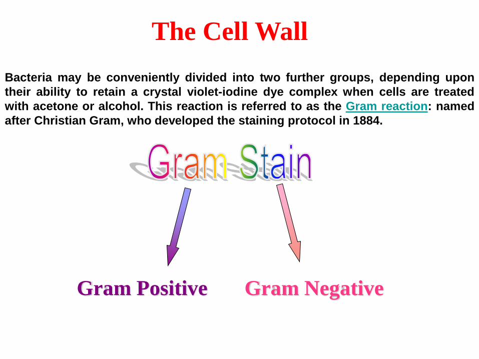
The Cell Wall
Gram Positive Gram Negative
Bacteria may be conveniently divided into two further groups, depending upon
their ability to retain a crystal violet-iodine dye complex when cells are treated
with acetone or alcohol. This reaction is referred to as the Gram reaction: named
after Christian Gram, who developed the staining protocol in 1884.
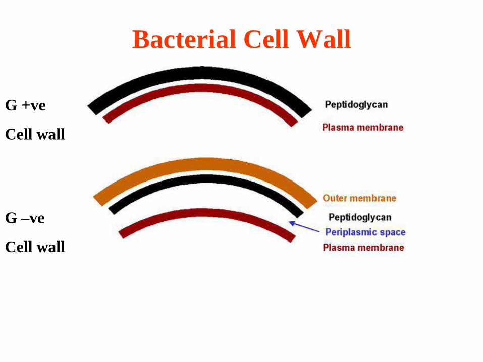
Bacterial Cell Wall
G +ve
Cell wall
G –ve
Cell wall

The cell wall of Gram-positive bacteria is composed of:
➢Peptidoglycan; may be up to 40 layers of this polymer
➢teichoic and teichuronic acids - surface antigens
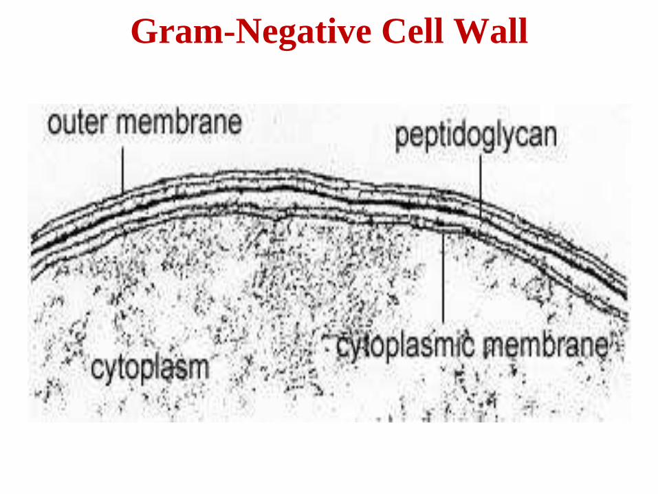
The cell wall of Gram-negative bacteria is complex and consists of:
➢a periplasmic space – enzymes
➢An inner membrane - one or two layers of peptidoglycan
beyond the periplasm
➢Outer membrane (LPS) – external to peptidoglycan
➢Braun’s lipoproteins – anchoring outer membrane to inner
➢Porins - through which some molecules may pass easily.

Gram-Positive Cell Wall
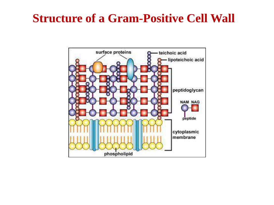
Structure of a Gram-Positive Cell Wall
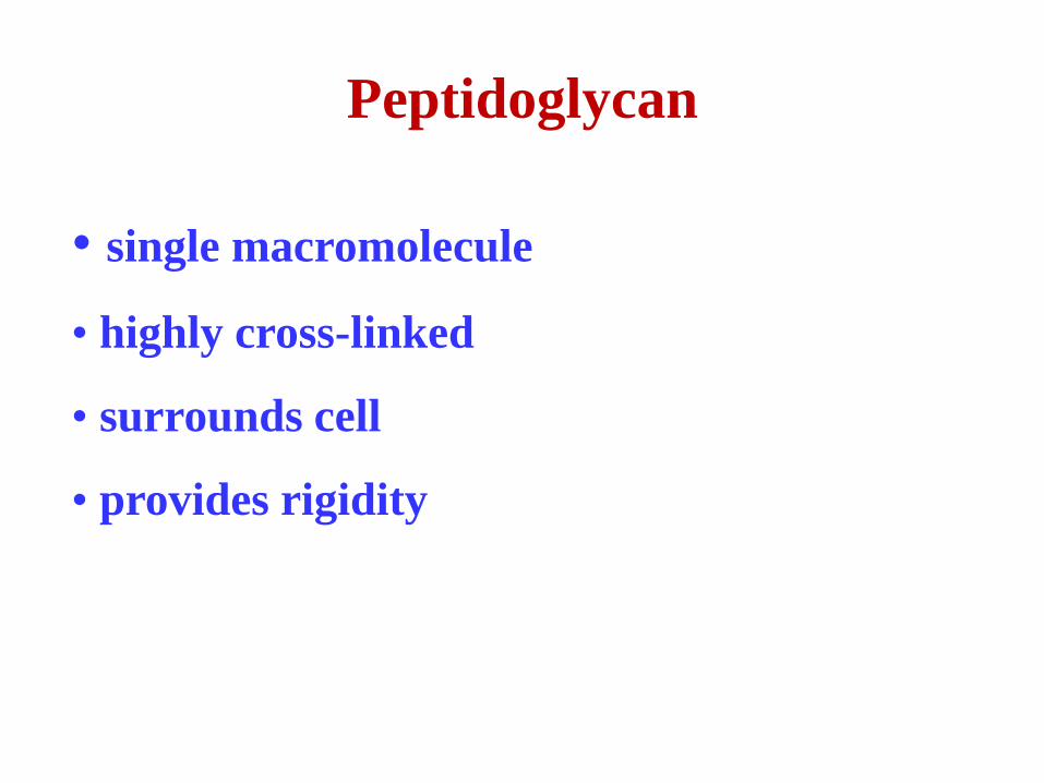
Peptidoglycan
• single macromolecule
• highly cross-linked
• surrounds cell
• provides rigidity


PEPTIDES
There are two types of peptide chains:
1. A tetra peptide side chain linked to N-acetyl-muramic acid
and containing the common amino acids L-alanine and L-
lysine and the unusual amino acids D-glutamic acid, D-
alanine and meso-diaminopimelic acid (DAP).
2. A penta-glycine bridge in Gram –positive bacteria, such as
Staphylococcus aureus, linking the linear peptide /
polysaccharide chains to form a 2-D network.
NOTE: Muramic acid, D-amino acids, and diaminopimelic
acid are not synthesized by mammals
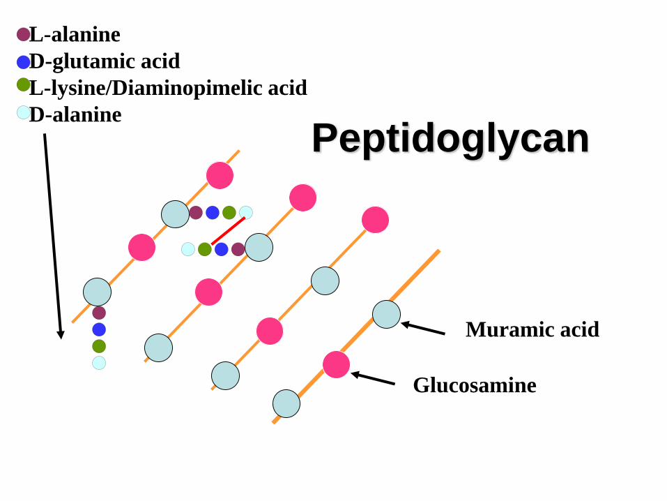
Peptidoglycan
Muramic acid
Glucosamine
L-alanine
D-glutamic acid
L-lysine/Diaminopimelic acid
D-alanine


In many Gram-negative bacteria the tetra peptide side chains
are cross linked directly via a covalent peptide bond between
the carboxyl- group of the terminal D-alanine and amino-
group of L-lysine or meso-diaminopimelic acid without the
involvement of a separate penta-glycine bridge.
Gram Positive Cell Envelope
• Teichoic acid
– Polymer
– phosphorus
– ribitol or glycerol backbone
–
• Teichuronic acid
– polymer
– no phosphorus
– glucuronic acid backbone

Gram-Negative Cell Wall
The Gram-negative cell wall is composed of:
➢periplasmic space
➢peptidoglycan (thin layer)
➢Braun’s lipoproteins
➢Lipopolysaccahrides
➢Porins
Gram-Negative Cell Wall

Gram Negative Peptidoglycan
•Only one or two layers
•No pentaglycine bond
•Lesser cross-linking
•Braun’s lipoproteins
–binds peptidoglycan layer to outer membrane
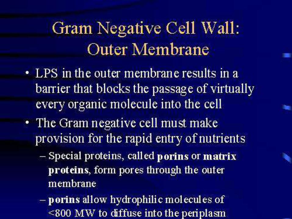
Outer Membrane
•major permeability barrier consisting of
•lipopolysaccharide
•phospholipids
•Proteins
–Porins
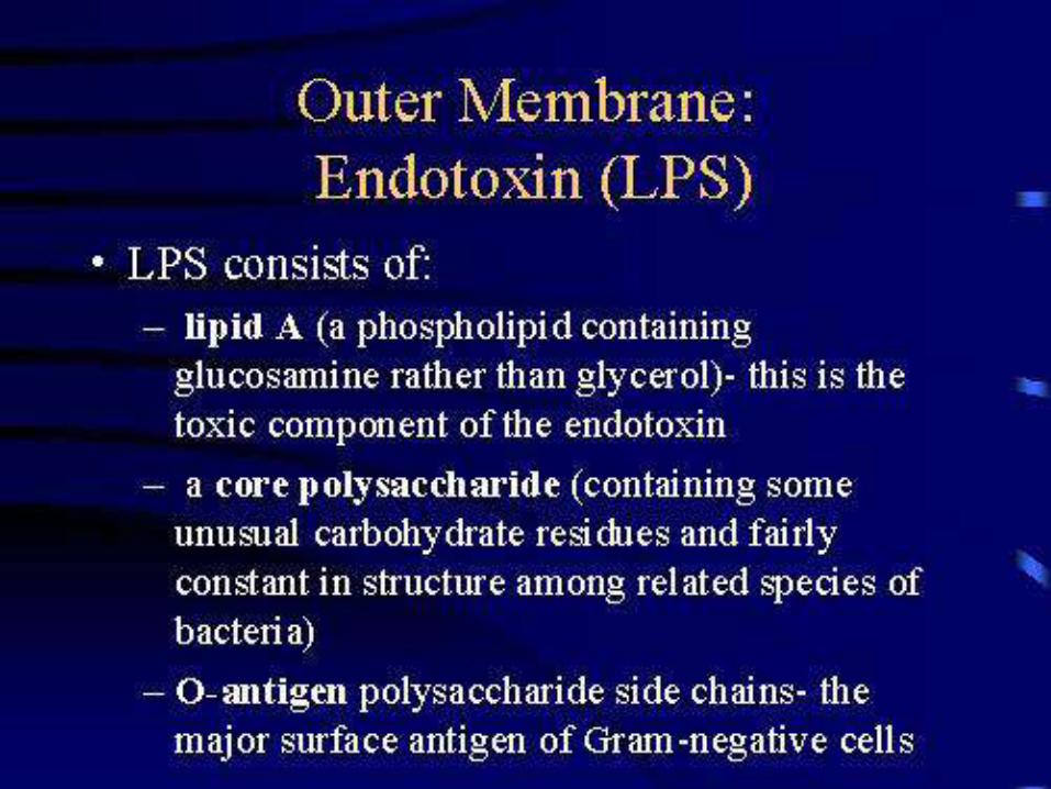

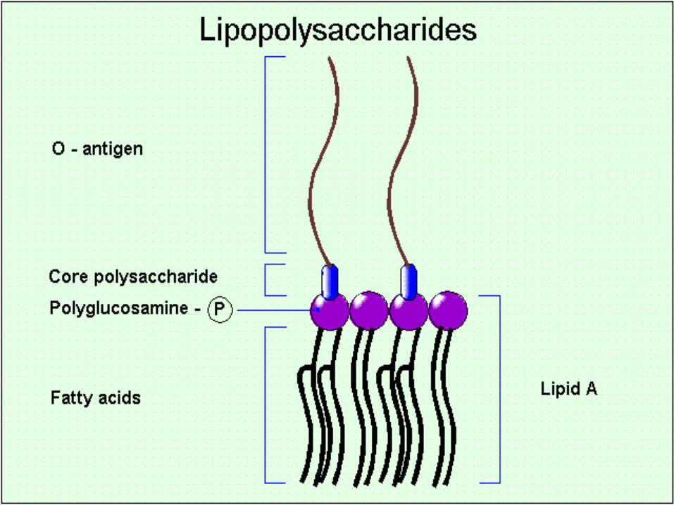
LIPOPOLYSACCHARIDE
Four segments can be differentiated within the lipopolysaccharides:
1. Lipid A – a phospholipd consisting of two molecules of
glucosamine which carry three fatty acids anchoring the LPS
in the lipid bilayer.
2. R-core:
➢ Inner core - 3 molecules of 2-keto-3-deoxyoctonate
(KDO) and two heptose both linked to
phosphoethanolamine.
➢ Outer core - pentasaccharide of glucose, galactose and
GNAc.
3. O-side chain (also known as O-antigen), consisting of unusual
sugars such as mannose, rhamnose, abequose, fucose, colitose
and others.

Characteristic
Peptidoglycan
Tetra peptide
Cross-linkage
Teichoic/teichuronic
acids
Lipoproteins
Lipopolysaccharide
Outer membrane
Periplasmic space
Polysaccharide
Protein
Gram positive
Thick
Most have lysine
Generally
pentapeptide
+
-
-
-
-
+
+ or –
Gram negative
Thin
All have DAP
Direct bond
-
+
+
+
+
+
+
Gram positive versus Gram negative wall
Acid fast and related bacteria
(mycobacteria, nocardia and corynebacteria)
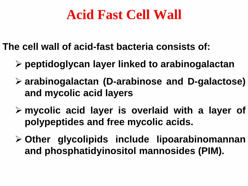
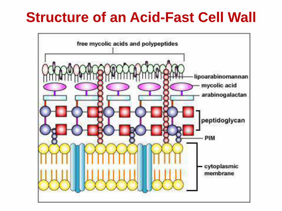
The cell wall of acid-fast bacteria consists of:
➢ peptidoglycan layer linked to arabinogalactan
➢ arabinogalactan (D-arabinose and D-galactose)
and mycolic acid layers
➢mycolic acid layer is overlaid with a layer of
polypeptides and free mycolic acids.
➢Other glycolipids include lipoarabinomannan
and phosphatidyinositol mannosides (PIM).
Acid Fast Cell Wall
Structure of an Acid-Fast Cell Wall
Wall-less forms
Wall-less bacteria that don’t replicate:
• Result from action of:
•enzymes lytic for cell wall
•antibiotics inhibiting peptidoglycan biosynthesis
• non-viable
• spheroplasts (with outer membrane) from Gram negative bacteria
• protoplasts (no outer membrane) from Gram positive bacteria
Wall-less bacteria that replicate : L-forms
Naturally occurring wall-less bacteria: Mycoplasmas (viable,
replicate)
S-LAYER
➢Some bacteria (e.g. Bacillus anthracis) may be covered by a
regular arrangement of proteins called as S-layer.
➢S-layer is attached to the outermost portion of their cell wall.
➢composed of either a single protein or glycoproteins, depending
upon the species.
➢protect bacteria from harmful enzymes, changes in pH, and the
predatory bacterium.
➢can function as an adhesin.
➢may contribute to virulence by protecting the bacterium against
complement attack and phagocytosis