Kinetic and thermodynamic investigations of cell-wall ...
14
See discussions, stats, and author profiles for this publication at: https://www.researchgate.net/publication/335085409 Kinetic and thermodynamic investigations of cell-wall degrading enzymes produced by Aureobasidium pullulans via induction with orange peels: application in lycopene extraction Article in Preparative Biochemistry & Biotechnology · August 2019 DOI: 10.1080/10826068.2019.1650375 CITATIONS 4 READS 90 2 authors: Some of the authors of this publication are also working on these related projects: BIO-ACTIVITY GUIDED ANTIDIABETIC AND TOXICOLOGICAL STUDIES ON Kigelia africana FRUITS’ EXTRACTS IN STREPTOZOTOCIN-INDUCED DIABETIC RATS View project Enzyme characterization View project Adedeji Ademakinwa Elizade University 19 PUBLICATIONS 65 CITATIONS SEE PROFILE Femi Kayode Agboola Obafemi Awolowo University 52 PUBLICATIONS 292 CITATIONS SEE PROFILE All content following this page was uploaded by Adedeji Ademakinwa on 03 October 2019. The user has requested enhancement of the downloaded file.
-
Upload
khangminh22 -
Category
Documents
-
view
1 -
download
0
Transcript of Kinetic and thermodynamic investigations of cell-wall ...

See discussions, stats, and author profiles for this publication at: https://www.researchgate.net/publication/335085409
Kinetic and thermodynamic investigations of cell-wall degrading enzymes
produced by Aureobasidium pullulans via induction with orange peels:
application in lycopene extraction
Article in Preparative Biochemistry & Biotechnology · August 2019
DOI: 10.1080/10826068.2019.1650375
CITATIONS
4READS
90
2 authors:
Some of the authors of this publication are also working on these related projects:
BIO-ACTIVITY GUIDED ANTIDIABETIC AND TOXICOLOGICAL STUDIES ON Kigelia africana FRUITS’ EXTRACTS IN STREPTOZOTOCIN-INDUCED DIABETIC RATS View project
Enzyme characterization View project
Adedeji Ademakinwa
Elizade University
19 PUBLICATIONS 65 CITATIONS
SEE PROFILE
Femi Kayode Agboola
Obafemi Awolowo University
52 PUBLICATIONS 292 CITATIONS
SEE PROFILE
All content following this page was uploaded by Adedeji Ademakinwa on 03 October 2019.
The user has requested enhancement of the downloaded file.
Full Terms & Conditions of access and use can be found athttps://www.tandfonline.com/action/journalInformation?journalCode=lpbb20
Preparative Biochemistry and Biotechnology
ISSN: 1082-6068 (Print) 1532-2297 (Online) Journal homepage: https://www.tandfonline.com/loi/lpbb20
Kinetic and thermodynamic investigationsof cell-wall degrading enzymes produced byAureobasidium pullulans via induction with orangepeels: application in lycopene extraction
Adedeji Nelson Ademakinwa & Femi Kayode Agboola
To cite this article: Adedeji Nelson Ademakinwa & Femi Kayode Agboola (2019): Kinetic andthermodynamic investigations of cell-wall degrading enzymes produced by Aureobasidium�pullulansvia induction with orange peels: application in lycopene extraction, Preparative Biochemistry andBiotechnology, DOI: 10.1080/10826068.2019.1650375
To link to this article: https://doi.org/10.1080/10826068.2019.1650375
Published online: 09 Aug 2019.
Submit your article to this journal
View Crossmark data
Kinetic and thermodynamic investigations of cell-wall degrading enzymesproduced by Aureobasidium pullulans via induction with orange peels:application in lycopene extraction
Adedeji Nelson Ademakinwaa,b and Femi Kayode Agboolab
aDepartment of Physical and Chemical Sciences, Elizade University, Ilara-Mokin, Nigeria; bDepartment of Biochemistry and Molecular Biology,Obafemi Awolowo University, Ile-Ife, Nigeria
ABSTRACTThe production of cell-wall degrading enzymes (CWDE) such as cellulase and pectinase byAureobasidium pullulans NAC8 through induction using orange peels was investigated for thepotential application of these enzymes in the extraction of lycopene from tomato skin, waste, andpaste (SWP). The CWDE was then immobilized via entrapment in alginate beads for lycopeneextraction and the kinetic/thermodynamic properties of the free and immobilized CWDE investi-gated. The optimum production of CWDE occurred at pH, temperature, and orange peel concen-tration of 6.0, 50 �C, and 2.0% (w/v), respectively. The values obtained for some kinetic andthermodynamic parameters such as E�d; t1=2; DG
�d; and DH�
d indicate that both free and immobi-lized cellulase and pectinase were thermostable between 40 and 50 �C. Maximum lycopeneextracted from the tomato SWP was 80 ± 2.4 mg/kg, 42 ± 1.3 mg/kg and 60 ± 1.2 mg/kg, respect-ively, using the immobilized CWDE. The entrapped CWDE was able to extract lycopene with yieldsof 58 ± 4.2, 51 ± 1.2 and 57 ± 4.2% for tomato SWP respectively after the fifth cycle. Using orangepeels for the induction of CWDE by A. pullulans offers a unique and cheaper approach to obtain-ing thermostable multi-enzyme complexes employable for easy lycopene extraction fromtomato SWP.
KEYWORDSCellulase; pectinase;lycopene; submergedfermentation;Aureobasidium pullulansNAC8; orangepeel; tomatoes
Introduction
Lycopene, an acyclic tetraterpenic hydrocarbon, is a naturalantioxidant found mostly in tomatoes and it is known toconfer the reddish color on them. The potentials of lycopeneto act as potent antioxidants stems from the fact that theycontain 11 conjugated double bonds.[1] The consumption offresh tomatoes is known to have great health benefits suchas lowering the risks associated with certain cancer types.[2]
Lycopene have attracted a lot of interest in the food andpharmaceutical industry where they are used as natural colo-rants as well as in food fortification mainly because of theirnon-toxicity, biodegradability, and strong color.[3–5] Thelycopene present in peels is almost five times higher thanthe pulp, thus making the peels a much sought-after sourcefor the extraction of lycopene.[6] The Industrial extraction oflycopenes from tomatoes involves the use of solvents whichhas deleterious effects downstream such as the requirementsfor a large amounts of solvents. This leads to occupational,health, and environmental hazards.[7] Also, there are chal-lenges with the low yields when solvents only are used forlycopene extraction, this is due to the inability of solventmolecules to access the rigid tomato peel tissues. The
lycopene is deeply enmeshed in the chromoplast membranestructure and the inability of the solvents to enter this struc-ture and hence solubilize it makes solvent extractionundesirable.[8] To bypass these challenges, the use ofenzymes (mostly cellulase and pectinase) that can degradethe cellulose and pectin present in the tomato peels havebeen exploited.[6,9,10] These enzymes hydrolyze the mainpolysaccharide components of the plant structures where thepigment is stored, thus aiding in the release of intracellularcontents. Other approaches used for the extraction of lyco-pene include the use of super critical-CO2 and ultrasonica-tion.[10–12] The advantages offered by enzyme-assistedextraction of lycopene over solvent-based approach is that it(i) is not time-consuming, (ii) occurs at ambient tempera-ture, and (iii) provides higher yields.[13] Lycopene extractionusing mixed enzyme preparations containing both cellulaseand pectinase has been reported and oftentimes, theseenzymes are obtained commercially and then applied to thetomato peels or paste in a sequential manner.[10] In thisstudy, cell-wall degrading enzymes (CWDE) were producedby submerged fermentation using orange peels as a sub-strate. Ismail[14] reported that orange peels are rich in cellu-lose and pectin and can be used as a cheap substrate for the
CONTACT Adedeji Nelson Ademakinwa [email protected] Department of Physical and Chemical Sciences, Elizade University,P.M.B. 002 Ilara-Mokin, Nigeria.Color versions of one or more figures in the article can be found online at http://www.tandfonline.com/lpbb� 2019 Taylor & Francis Group, LLC
PREPARATIVE BIOCHEMISTRY AND BIOTECHNOLOGYhttps://doi.org/10.1080/10826068.2019.1650375
induction of multienzyme complexes by Aspergillus niger. Inthat same study, the author reported that some cell-walldegrading enzymes such as cellulase, pectinase, xylanase,and alpha-amylases were produced after induction of A.niger with the orange peel wastes. In this study, the cell walldegrading enzymes produced by induction of Aureobasidiumpullulans with orange peels under optimum conditions wasimmobilized by entrapment in alginate. Enzyme immobiliza-tion via entrapment in alginate is a cheap and easily adapt-able technique compared to other known techniques likecross-linking and adsorption.[15–17] Recently, A. niger pecti-nase was immobilized on alginate and its kinetic andthermodynamic properties investigated[18] while immobilizedpectinase was used for the extraction of lycopene from wastetomato peels.[19] Ladole et al.[16] used cross-linked cellulaseand pectinase in lycopene recovery from tomato peels but inthis report, alginate was employed for enzyme immobiliza-tion for lycopene extraction from tomatoes. To the best ofour knowledge, this is the first report where cellulase andpectinase were co-immobilized on alginate for the extractionand recovery of lycopene from tomato peels. In this study,the entrapped CWDE was further used for the extraction oflycopene from tomato skin, waste, and paste over severalreaction cycles. The use of immobilized enzymes for lyco-pene extraction offers a better advantage than free enzymesbecause of its improved enzymatic activities, better stability,and reusability.[15,20–22] This is the first study of its kindwhere carbohydrases for lycopene extraction were producedby A. pullulans by induction with orange peels.Aureobasidium pullulans is a yeast-like fungus that is veryubiquitous as it is found in almost every habitat such asdecayed plant litters,[23] on various crops and fruits,etc.[24,25] This fungus is renowned for producing veryimportant industrial extracellular enzymes such as laccase,fructosyltransferase, cellulase, amylase, etc.[23,26,27] The fun-gus had been applied in biodegradation of textile dyes[22]
and other remarkable products like antibiotics.[28] This fun-gus was selected for the production of these cell-walldegrading enzymes (CWDE) by harnessing its potential toutilize orange peels wastes (rich in pectin and cellulose) ascheap substrates.
Enzymes for industrial applications must display a higherdegree of thermal resistance and in this study, the thermo-dynamic and kinetic characteristics of the CWDE was alsoinvestigated to provide information on its application indus-trially especially in the food industry. It is noteworthy to
mention that no report of the investigation of the thermo-dynamic and kinetic characteristics for A. pullulans cellulaseand pectinase exists in literature; hence, this is the first studyof its kind.
Materials and methods
Materials
Bovine serum albumin, Coomassie blue G-250, carboxyme-thylcellulose (CMC), beechwood xylan, polygalacturonicacid, butylated hydroxytoluene, corn starch, and dinitrosali-cylic acid were obtained from either from Sigma ChemicalCompany, St. Louis, USA or Merck Company, Darmstadt,Germany. All solvents used in this study were of analyticalgrade with purity greater than 95%. Apparently healthy andripened L. esculentum and Tomato pastes (GinoTM Brand)were collected from the local market in Ile-Ife, Osun State,Nigeria. Orange peel waste was obtained from local orangesellers in the Obafemi Awolowo University, Ile-Ife, Nigeria.They were washed with distilled water, dried, milled, sievedwith a mesh size of 10 mm and stored at 4 �C prior to fur-ther use. The chemical composition of the orange peels suchas cellulose, hemicellulose, and pectin content was deter-mined according to methods described by Liu et al.[29]
Methods
Microorganism and culture conditionsThe fungus used in this study, A. pullulans NAC8, was iso-lated from soil containing decayed plant litters. The genesequences were deposited in the GenBank database with anaccession number KX020331.[23] The fungus was stored onMalt Extract agar (MEA) in a McCartney bottle at 4 �C andsub-cultured every 72 h.
Submerged fermentation for cell-wall degrading enzyme(CWDE) productionCWDE production was carried out by submerged fermenta-tion in a 100 ml flask containing 20 ml of the medium. Themedia composition was a modified version described byIsmail:[14] NaNO3 – 2.0 g/L, MgSO4.7H2O – 0.5 g/L, KCl –0.5 g/L, K2HPO4 – 1.0 g/L, and FeSO4.7H2O – 0.01 g/L. Theorange peel concentration was initially varied between 0.1%and 10% w/v. The sterilized medium was inoculated with 5
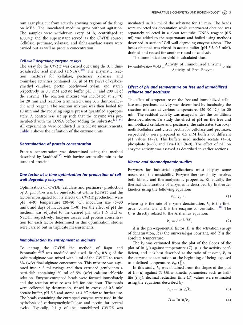
Table 1. Definition of enzyme units.
Enzyme Definition
Cellulase One unit of cellulase activity is defined as the amount of enzyme releasing 1.0mmol glucose equivalent from carboxymethyl cellulose per minute under thestandard assay condition
Pectinase One unit of pectinase activity is defined as the amount of enzyme releasing 1.0mmol galactoronic acid equivalent from pectin per minute at roomtemperature under the standard assay condition
Xylanase One unit of xylanase activity is defined as the amount of enzyme releasing 1.0mmol xylose equivalent from the substrate per minute at room temperatureunder the standard assay condition
Amylase One unit of alpha amylase activity is defined as the amount of enzymereleasing 1.0 mmol maltose equivalent from the starch per minute at roomtemperature under the standard assay condition
2 A. N. ADEMAKINWA AND F. K. AGBOOLA
mm agar plug cut from actively growing regions of the fungion MEA. The inoculated medium grew without agitation.The samples were withdrawn every 24 h, centrifuged at4000�g and the supernatant served as the CWDE source.Cellulase, pectinase, xylanase, and alpha-amylase assays werecarried out as well as protein concentration.
Cell-wall degrading enzyme assaysThe assay for the CWDE was carried out using the 3, 5 dini-trosalicyclic acid method (DNSA).[30] The enzymatic reac-tion mixtures for cellulase, pectinase, xylanase, anda-amylase activities contained 500 ml of 1% (w/v) of carbox-ymethyl cellulose, pectin, beechwood xylan, and starchrespectively in 0.5 mM acetate buffer pH 5.5 and 200 ml ofthe enzyme. The reaction mixture was incubated at 25 �Cfor 20 min and reaction terminated using 3, 5 dinitrosalicy-clic acid reagent. The reaction mixture was then boiled for10 min and the reducing sugars present quantified appropri-ately. A control was set up such that the enzyme was pre-incubated with the DNSA before adding the substrate.[31–34]
All experiments were conducted in triplicate measurements.Table 1 shows the definition of the enzyme units.
Determination of protein concentration
Protein concentration was determined using the methoddescribed by Bradford[35] with bovine serum albumin as thestandard protein.
One factor at a time optimization for production of cellwall degrading enzymes
Optimization of CWDE (cellulase and pectinase) productionby A. pullulans was by one-factor-at-a-time (OFAT) and thefactors investigated for its effects on CWDE production werepH (4–9), temperature (20–80 �C), inoculum size (5–30mm), and days of incubation (1–8). For the effect of pH themedium was adjusted to the desired pH with 1 N HCl orNaOH, respectively. Enzyme assays and protein concentra-tion for each factor determined in this optimization studieswere carried out in triplicate measurements.
Immobilization by entrapment in alginate
To entrap the CWDE the method of Ragu andPennnathur[36] was modified and used. Briefly, 0.8 g of thesodium alginate was mixed with 1 ml of the CWDE to reach8% (w/v) final alginate concentration. This mixture was aspi-rated into a 5 ml syringe and then extruded gently into apetri-dish containing 50 ml of 5% (w/v) calcium chloridesolution. Enzyme-entrapped beads were formed immediatelyand the reaction mixture was left for one hour. The beadswere collected by decantation, rinsed in excess of 0.5 mMacetate buffer, pH 5.5 and stored at 4 �C prior to further use.The beads containing the entrapped enzyme were used in thehydrolysis of carboxymethylcellulose and pectin for severalcycles. Typically, 0.1 g of the immobilized CWDE was
incubated in 0.5 ml of the substrate for 15 min. The beadswere collected via decantation while supernatant obtained wasseparately collected in a clean test tube. DNSA reagent (0.5ml) was added to the supernatant and boiled using methodsdescribed in section “Cell wall degrading enzyme assays.” Thebeads obtained was rinsed in acetate buffer (pH 5.5, 0.5 mM),drained and reused for another round of catalysis.
The immobilization yield is calculated thus:
ImmobilzationYield¼Activity of Immobilized EnzymeActivity of Free Enzyme
�100
Effect of pH and temperature on free and immobilizedcellulase and pectinase
The effect of temperature on the free and immobilized cellu-lase and pectinase activity was determined by incubating thereaction mixture at various temperatures (20–90 �C) for 20min. The residual activity was assayed under the conditionsdescribed above. To study the effect of pH on the free andimmobilized cellulase and pectinase, the substrates (carboxy-methylcellulose and citrus pectin for cellulase and pectinase,respectively) were prepared in 0.5 mM buffers of differentpH values (4–9). The buffers used include acetate (4–5),phosphate (6–7), and Tris-HCl (8–9). The effect of pH onenzyme activity was assayed as described in earlier sections.
Kinetic and thermodynamic studies
Enzymes for industrial applications must display somemeasure of thermostability. Enzyme thermostability involvesboth kinetic and thermodynamic properties. Kinetically, thethermal denaturation of enzymes is described by first-orderkinetics using the following equation:
vd¼ kd E; (1)
where vd is the rate of enzyme denaturation, kd is the first-order constant, and E is the enzyme concentration.[18] Thekd is directly related to the Arrhenius equation:
kd ¼ Ae�Ed=RT ; (2)
A is the pre-exponential factor, Ed is the activation energyof denaturation, R is the universal gas constant, and T is theabsolute temperature.
The kd was estimated from the plot of the slopes of theplot of In (m) against temperature (T). m is the activity coef-ficient, and it is best described as the ratio of enzyme, E, tothe enzyme concentration at the beginning of being exposedto a defined temperature, E0, ( EE0Þ:
In this study, kd was obtained from the slopes of the plotof In (m) against T. Other kinetic parameters such as half-life ðt1=2Þ; decimal reduction time (D) values were estimatedusing the equations described by
t1=2 ¼ In 2=kd (3)
D ¼ In10=kd: (4)
PREPARATIVE BIOCHEMISTRY AND BIOTECHNOLOGY 3
The enzyme half-life defined as the time after which theenzyme activity was reduced to one-half of the initial activ-ity while the decimal reduction time is defined as the timeof enzyme exposition at a known temperature to maintain10% residual activity.[18,37] Another important thermaldenaturation kinetic parameter is the sensitivity factor(z-value), which is defined as the increase in temperaturethat results in the reduction of the D-value by one logarith-mic cycle was estimated in this study from the slopes oflogD vs. T (�C).
From Eq. (2), the activation energy of the irreversibleenzyme inactivation (EdÞ was derived from the slope of theplot of Inkd against 1=T for temperatures ranging from 40to 70 �C. Other important thermodynamic parameters forfree and immobilized cellulase and pectinase inactivationwere determined using equations described below:
DHd� ¼ Ed � RT (5)
DGd� ¼ � RTIn
kdhkbT
� �(6)
DSd� ¼ DH��DG�
T: (7)
The overall entropy of activation and enthalpy of activa-tion for the thermal denaturation were obtained from thebelow equation:
InkdT
� �¼ In
kbh
� �þ DS�
R� DH�
R1T
� �(8)
DHd� ; DGd
�; and DSd� are the activation enthalpy, Gibbsfree energy, and entropy, respectively. Boltzmann constant(kB) is 1.38 � 10�23 J�1 K�1, Plank’s constant (h) is 6.63 �10�34 J�1 s�1, and the universal gas constant (R) is 8.314J�1 mol�1 K�1.[18]
The activation energy, Ea, was determined from theslopes of Arrhenius plot from the optimal temperature data(see Arrhenius equation in Eq. (1)), In (k) vs. 1/T, where kis the reaction rate, T is the temperature in Kelvin, and A isthe pre-exponential factor.
In kð Þ ¼ In A� EaR
� �1�T [9]
Preparation of Lycopersicon esculentum
Apparently healthy and ripe tomatoes (maturation: 65 ± 3days; stored at 4 �C) were rinsed with distilled water andthen hand peeled as described by Zuorro and Lavecchia.[38]
The tomato skins were peeled off, homogenized, and storedin air-tight containers prior to further use at 4 �C. The pulpwithout the skin and the seed were separately blended andregarded as tomato wastes. Tomato paste was used as pur-chased without prior processing.
Extraction of lycopene
The extraction of lycopene was carried out with slight modi-fication in the procedure suggested by Fish et al.[39]
Lycopersicon esculentum skin, waste, and paste were sub-jected for solvent extraction of lycopene using tri-solventmixture consisting of hexane:acetone:ethanol (2:1:1) and 1%butylated hydroxytoluene. The tomato skin, waste, and paste(1 g) was weighed into 50 ml conical flasks that contained10 ml of 0.5 mM acetate buffer pH 5.0. The mixture wasstirred for 5 min then 20 ml of the tri-solvent mixture wasadded. Incubation was at 25 �C for 2 h under low-speed agi-tation (100 rpm). The mixture was allowed to stand for 5min for the separation of phases. From the upper phase, 3ml was collected and further diluted using the tri-solventmixture. The absorbance of this was measured using a UVvisible spectrophotometer at the 503 nm. The lycopene con-centration (mg/kg), in each extract, was calculated using theformula described below.[3] Experiments were carried out intriplicate measurements.
Lycopenemgkg
� �¼ 31:2 � Dilution factor � A503nm
gram of sample
Enzyme-aided extraction of lycopene
The reaction mixture for the enzyme-aided extraction of lyco-pene from the skin, waste, and paste was prepared using 1 g ofthe L. esculentum waste, skin, and peel and 1 g of alginate-entrapped CWDE or 1 ml of the CWDE (for the free enzyme) asappropriate. Incubation of the reaction mixture occurred at 50�C for 2 h. The alginate-entrapped CWDE was removed fromthe reaction mixture, rinsed in 0.5 M acetate buffer pH 5.5, andthen used for another cycle of reaction. The residues obtainedafter removal of the alginate-entrapped CWDE were subjectedto solvent extraction and incubation for 2 h at 25 �C under low-speed agitation (100 rpm). The lycopene was then quantifiedusing methods described above.[3] To investigate the effect ofdifferent concentrations of tomato skin, waste, and paste on thelycopene yield, the tomato skin, waste, and paste concentrationwere varied between 1 and 5 g and incubated with 1 g of thealginate-entrapped CWDE. The process for lycopene extractionand quantification is as discussed in the above section. Lycopenequantification was carried out in triplicate measurements.
The effect of the different amounts (g) of immobilizedenzyme on the lycopene extraction was investigated by incu-bating 1 g of the tomato skin, waste, or paste with variousamount of immobilized enzyme (1–5 g) for 1 h. The processfor lycopene extraction and quantification was carried out asdiscussed in the above section. Lycopene assay and quantifi-cation was carried out in triplicate measurements
Analysis of lycopene
The lycopene extracted via enzyme-assisted approach andjust solvents only were analyzed using Fourier transforminfrared spectroscopy (FTIR).[10,15] Briefly, the extractedlycopene was blended with potassium bromide and then
4 A. N. ADEMAKINWA AND F. K. AGBOOLA
screwed to form pellets using hydraulic gauges. The spectraldata were collected in the range of 4000–400 cm using anFTIR spectrophotometer (Shimadzu FTIR-8400S).
Data analysis
The data obtained in the experimental process were analyzedwith the analysis of variance (ANOVA) with a level of sig-nificance: p < 0.05, using the GraphPad Prism version 6.0(GraphPad Inc.)
Result and discussion
Induction of cell-wall degrading enzymes andoptimization of cellulase and pectinase production
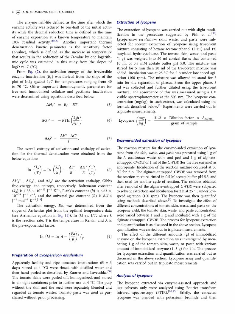
Orange peels are often disposed of as waste leading toadverse environmental and health challenges. The orangepeels used in this study were collected as wastes for the pro-duction of cell wall degrading enzymes such as cellulase andpectinase. These enzymes were now employed for the break-down of cellulose and pectin present in tomato tissues forlycopene release. Several authors have used orange peels as asource of producing a cocktail of enzymes mainly because ofthe orange peels are a cheap and readily available source ofcellulose and pectin.[14,29] In this study, the chemical com-position of orange peels revealed that cellulose (20%), hemi-cellulose (22%), and pectin (29%) were present. Li et al.[14]
reported that the cellulose, hemicellulose, and pectin con-centration of the orange peels used were 14, 21, and 22%,respectively, while Ismail et al.[29] reported that cellulose,hemicellulose, and pectin concentration were 17, 39, and22%, respectively. The presence of these fibers in the orangepeel necessitated its potential use as a cheap and readilyavailable source of substrates for the induction of theCWDE by A. pullulans NAC8. Orange peels at 2% (w/v)maximally induced the production of CWDE by A. pullulansas pectinase, xylanase and cellulase were the most expressedenzymes while relatively negligible amylolytic activities wereobserved (Fig. 1).
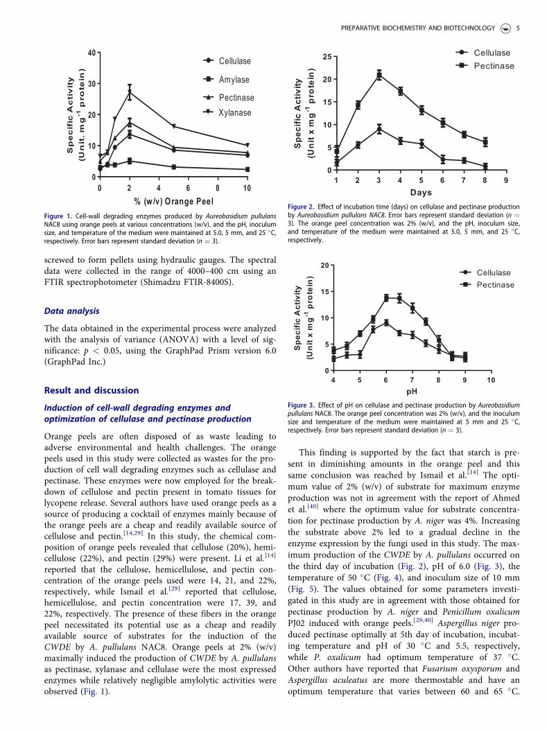
This finding is supported by the fact that starch is pre-sent in diminishing amounts in the orange peel and thissame conclusion was reached by Ismail et al.[14] The opti-mum value of 2% (w/v) of substrate for maximum enzymeproduction was not in agreement with the report of Ahmedet al.[40] where the optimum value for substrate concentra-tion for pectinase production by A. niger was 4%. Increasingthe substrate above 2% led to a gradual decline in theenzyme expression by the fungi used in this study. The max-imum production of the CWDE by A. pullulans occurred onthe third day of incubation (Fig. 2), pH of 6.0 (Fig. 3), thetemperature of 50 �C (Fig. 4), and inoculum size of 10 mm(Fig. 5). The values obtained for some parameters investi-gated in this study are in agreement with those obtained forpectinase production by A. niger and Penicillum oxalicumPJ02 induced with orange peels.[29,40] Aspergillus niger pro-duced pectinase optimally at 5th day of incubation, incubat-ing temperature and pH of 30 �C and 5.5, respectively,while P. oxalicum had optimum temperature of 37 �C.Other authors have reported that Fusarium oxysporum andAspergillus aculeatus are more thermostable and have anoptimum temperature that varies between 60 and 65 �C.
% (w/v) Orange Peel
SpecificActivity
(Unit.m
g-1protein)
0 2 4 6 8 100
10
20
30
40Cellulase
Amylase
PectinaseXylanase
Figure 1. Cell-wall degrading enzymes produced by Aureobasidium pullulansNAC8 using orange peels at various concentrations (w/v), and the pH, inoculumsize, and temperature of the medium were maintained at 5.0, 5 mm, and 25 �C,respectively. Error bars represent standard deviation (n ¼ 3).
1 2 3 4 5 6 7 8 90
5
10
15
20
25 CellulasePectinase
Days
Specific
Activity
(Un itx
mg-1protein)
Figure 2. Effect of incubation time (days) on cellulase and pectinase productionby Aureobasidium pullulans NAC8. Error bars represent standard deviation (n ¼3). The orange peel concentration was 2% (w/v), and the pH, inoculum size,and temperature of the medium were maintained at 5.0, 5 mm, and 25 �C,respectively.
4 5 6 7 8 9 100
5
10
15
20CellulasePectinase
pH
Specific
Activity
(Unitx
mg-1protein)
Figure 3. Effect of pH on cellulase and pectinase production by Aureobasidiumpullulans NAC8. The orange peel concentration was 2% (w/v), and the inoculumsize and temperature of the medium were maintained at 5 mm and 25 �C,respectively. Error bars represent standard deviation (n ¼ 3).
PREPARATIVE BIOCHEMISTRY AND BIOTECHNOLOGY 5

The optimum temperature and pH for the free and immobi-lized were 50 �C and 6.0, respectively (Figs. 6 and 7). Theoptimum temperature obtained in this study was similar tothat obtained for A. aculeatus.[18]
Kinetic and thermodynamic stabilities of free andimmobilized Cell-Wall degrading enzymes
Enzyme thermostability involves both thermodynamic andkinetic stabilities; and, in this study, the thermodynamic and
kinetic stability studies of immobilized and free cellulase andpectinase were investigated. Enzymes for industrial applica-tions must show resistance to thermal denaturation; hence,this study investigated the potentials of both the free andimmobilized forms of cellulase and pectinase to overcomethermal resistance at temperatures ranging from 40 to 70�C. It was observed from the semi-log plots of In ðlÞagainst T, (where m is the activity coefficient and T is thetemperature in Kelvin), that the activity of the free andimmobilized cellulase and pectinase followed a typical first-order enzyme deactivation pattern (Fig. 8a–d). This observa-tion was in agreement with the findings of Pal andKhrum[20] and Olievera et al.[18] for free and immobilizedxylanase and pectinase, respectively.
Kinetic parameters for enzyme thermal denaturationTable 2 illustrates the summary of the major kinetic parame-ters determined for the free and immobilized cellulase andpectinase for A. pullulans. The values obtained for the first-order thermal denaturation constant, ðkdÞ decreased as thetemperature increased while the half-life (t1=2Þ; increased asthe temperature increased. This observation is not unex-pected as it meant that, as the process of thermal denatur-ation increased, this simultaneously led to a gradual declinein the thermostability. A conclusion easily drawn from kdand t1=2 values obtained in Table 2 for free and immobilizedcellulase and pectinase is that thermostability was achievedaround 50 �C. The results obtained in this study for A. pul-lulans cellulase and pectinase (free and immobilized) agreeswith that of immobilized exoploygalacturonase fromPenicillium notarum[41] and free and immobilized pectinasefrom A. aculeatus.[18] The cell-wall degrading enzymes pro-duced in this study was used in lycopene extraction fromtomato wastes; hence, it is important to express the first-order thermal denaturation reactions in terms of the D andz-values. These values are often required by most food proc-essing industries.[18] At temperatures between 40 and 50 �C,good thermostability was observed as it took more time (i.e.,794–548, 576–343, 1097–767, and 177–124 min) for immo-bilized cellulase, free cellulase, immobilized pectinase, and
10 20 30 40 50 60 70 80 900
5
10
15
20
25CellulasePectinase
T C•
Specific
Activity
(Unitx
mg-1pro te in)
Figure 4. Effect of temperature on cellulase and pectinase production byAureobasidium pullulans NAC8. The orange peel concentration was 2% (w/v),and the inoculum size and pH of the medium were maintained at 5 mm and5.0, respectively. Error bars represent standard deviation (n ¼ 3).
0 5 10 15 20 25 300
5
10
15
20CellulasePectinase
Inoculum size (mm)
SpecificA
c ti vity
( Un itx
mg-1pro tein)
Figure 5. Effect of inoculum size on cellulase and pectinase production byAureobasidium pullulans NAC8. The orange peel concentration was 2% (w/v),and the pH and temperature of the medium were maintained at 5.0 and 25 �C.Error bars represent standard deviation (n ¼ 3).
pH
%RelativeAc ti vity
4 5 6 7 8 90
50
100
150 Free CellulaseFree PectinaseImmobilized CellulaseImmobilized Pectinase
Figure 6. Effect of pH on free and immobilized pectinase and cellulase producedunder optimized conditions. Error bars represent standard deviation (n ¼ 3).
T C●
%RelativeActivit y
20 40 60 80 1000
50
100
150 Free CellulaseFree PectinaseImmobilized CellulaseImmobilized Pectinase
Figure 7. Effect of temperature on free and immobilized pectinase and cellu-lase produced under optimized conditions. Error bars represent standard devi-ation (n ¼ 3).
6 A. N. ADEMAKINWA AND F. K. AGBOOLA
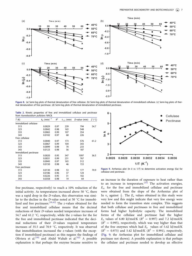
free pectinase, respectively) to reach a 10% reduction of theinitial activity. As temperatures increased above 50 �C, therewas a rapid drop in the D-values, this observation was simi-lar to the decline in the D-value noted at 50 �C for immobi-lized and free pectinases.[18,42] The z-values obtained for thefree and immobilized cellulase means that the decimalreductions of their D-values needed temperature increases of54.7 and 61.2 �C, respectively, while the z-values for the forthe free and immobilized pectinase indicated that the deci-mal reductions of their D-values required temperatureincreases of 35.5 and 70.9 �C, respectively. It was observedthat immobilization increased the z-values (with the excep-tion if immobilized pectinase) as this negates the findings ofOliviera et al.[18] and Abdel Wahab et al.[42] A possibleexplanation is that perhaps the enzyme became sensitive to
an increase in the duration of exposure to heat rather thanto an increase in temperature.[22] The activation energies,Ea; for the free and immobilized cellulase and pectinasewere obtained from the slope of the Arrhenius plot ofIn vo against 1
T : The Ea values obtained in this study werevery low and this might indicate that very few energy wereneeded to form the transition state complex. This suggeststhat both cellulase and pectinases in free and immobilizedforms had higher hydrolytic capacity. The immobilizedforms of the cellulase and pectinase had the higherEa values of 6.80 kJ/mol/K (R2 ¼ 0.997) and 7.2 kJ/mol/K(R2 ¼ 0.995), respectively, which was way higher than thatof the free enzymes which had Ea values of 5.42 kJ/mol/K(R2 ¼ 0.975) and 5.42 kJ/mol/K (R2 ¼ 0.991), respectively,(Fig. 9; the Arrhenius plot for immobilized cellulase andpectinase not shown). A possible explanation is that perhapsthe cellulase and pectinase needed to develop an effective
Inµ
10 20 30 40 50 60
-1.0
-0.8
-0.6
-0.4
-0.2
0.0
0.240ºC50ºC60ºC70ºC
Time (min)
In( µ)
10 20 30 40 50 60
-1.0
-0.8
-0.6
-0.4
-0.2
0.0
0.240ºC50ºC60ºC70ºC
Time (min)
In( µ)
10 20 30 40 50 60
-2.5
-2.0
-1.5
-1.0
-0.5
0.0
0.540ºC50ºC60ºC70ºC
Time (min)
Inµ
10 20 30 40 50 60
-0.8
-0.6
-0.4
-0.2
0.0
0.240 Cº50ºC60ºC70ºC
Tim e (m in )(a) (b)
(c) (d)
Figure 8. (a) Semi-log plots of thermal denaturation of free cellulase. (b) Semi-log plots of thermal denaturation of immobilized cellulase. (c) Semi-log plots of ther-mal denaturation of free pectinase. (d) Semi-log plots of thermal denaturation of immobilized pectinase.
Table 2. Kinetic properties of free and immobilized cellulase and pectinasefrom Aureobasidium pullulans NAC8.
T (K) kd (min)�1 R2 t1=2 (min) D-value (min) Z (�C)Immobilized cellulase313 0.0029 0.97 239 794 54.7323 0.0042 0.98 165 548333 0.0065 0.99 107 354343 0.0102 0.97 68 226
Free cellulase313 0.0040 0.97 173 576 61.2323 0.0067 0.99 104 343333 0.0099 0.98 70 233343 0.0123 0.98 56 187
Immobilized pectinase313 0.0020 0.95 347 1097 36.5323 0.0031 0.99 231 767333 0.0045 0.97 165 512343 0.0150 0.98 46 153
Free pectinase313 0.0130 0.98 53 177 70.9323 0.0186 0.96 37 124333 0.0226 0.95 31 102343 0.0358 0.98 19 64
0.0026 0.0028 0.0030 0.0032 0.0034 0.00362
3
4
5
6CellulasePectinase
1/T (K-1)
Ink
Figure 9. Arrhenius plot (In k vs 1/T) to determine activation energy (Ea) forcellulase and pectinase.
PREPARATIVE BIOCHEMISTRY AND BIOTECHNOLOGY 7
conformational structure in order to form a well-orientedand specific complex with the substrate.[21] A uniquethermodynamic property used in distinguishing if catalyzedreactions are solely controlled by temperature only or byother extraneous factors is the temperature quotient ðQ10Þ:The Q10 values varied between 1 and 2 for most enzyme cat-alyzed reaction which its totally dependent on temperature.Values outside this range signify that other extraneous fac-tors other than temperature controlled the rate of reaction.Table 1.
The Q10 values obtained for the free and immobilizedcellulase and pectinase (values ranged between 1 and 1.2)meant that temperature was influential on the reaction rate.A similar observation was observed for A. aculeatus free andimmobilized pectinase.[18]
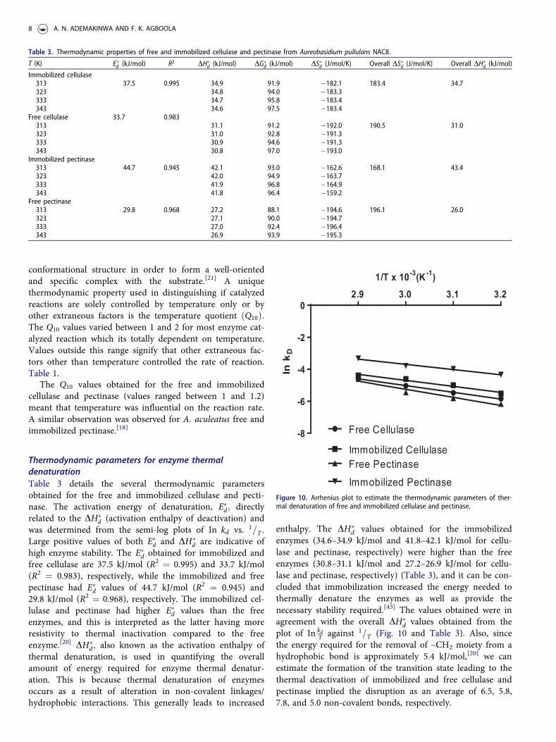
Thermodynamic parameters for enzyme thermaldenaturationTable 3 details the several thermodynamic parametersobtained for the free and immobilized cellulase and pecti-nase. The activation energy of denaturation, E�d; directlyrelated to the DH�
d (activation enthalpy of deactivation) andwas determined from the semi-log plots of In kd vs. 1=T :Large positive values of both E�d and DH�
d are indicative ofhigh enzyme stability. The E�d obtained for immobilized andfree cellulase are 37.5 kJ/mol (R2 ¼ 0.995) and 33.7 kJ/mol(R2 ¼ 0.983), respectively, while the immobilized and freepectinase had E�d values of 44.7 kJ/mol (R2 ¼ 0.945) and29.8 kJ/mol (R2 ¼ 0.968), respectively. The immobilized cel-lulase and pectinase had higher E�d values than the freeenzymes, and this is interpreted as the latter having moreresistivity to thermal inactivation compared to the freeenzyme.[20] DH�
d; also known as the activation enthalpy ofthermal denaturation, is used in quantifying the overallamount of energy required for enzyme thermal denatur-ation. This is because thermal denaturation of enzymesoccurs as a result of alteration in non-covalent linkages/hydrophobic interactions. This generally leads to increased
enthalpy. The DH�d values obtained for the immobilized
enzymes (34.6–34.9 kJ/mol and 41.8–42.1 kJ/mol for cellu-lase and pectinase, respectively) were higher than the freeenzymes (30.8–31.1 kJ/mol and 27.2–26.9 kJ/mol for cellu-lase and pectinase, respectively) (Table 3), and it can be con-cluded that immobilization increased the energy needed tothermally denature the enzymes as well as provide thenecessary stability required.[43] The values obtained were inagreement with the overall DH�
d values obtained from theplot of In kd
T against 1=T (Fig. 10 and Table 3). Also, sincethe energy required for the removal of –CH2 moiety from ahydrophobic bond is approximately 5.4 kJ/mol,[20] we canestimate the formation of the transition state leading to thethermal deactivation of immobilized and free cellulase andpectinase implied the disruption as an average of 6.5, 5.8,7.8, and 5.0 non-covalent bonds, respectively.
Table 3. Thermodynamic properties of free and immobilized cellulase and pectinase from Aureobasidium pullulans NAC8.
T (K) E�d (kJ/mol) R2 DH�d (kJ/mol) DG�
d (kJ/mol) DS�d (J/mol/K) Overall DS�d (J/mol/K) Overall DH�d (kJ/mol)
Immobilized cellulase313 37.5 0.995 34.9 91.9 �182.1 183.4 34.7323 34.8 94.0 �183.3333 34.7 95.8 �183.4343 34.6 97.5 �183.4
Free cellulase 33.7 0.983313 31.1 91.2 �192.0 190.5 31.0323 31.0 92.8 �191.3333 30.9 94.6 �191.3343 30.8 97.0 �193.0
Immobilized pectinase313 44.7 0.945 42.1 93.0 �162.6 168.1 43.4323 42.0 94.9 �163.7333 41.9 96.8 �164.9343 41.8 96.4 �159.2
Free pectinase313 29.8 0.968 27.2 88.1 �194.6 196.1 26.0323 27.1 90.0 �194.7333 27.0 92.4 �196.4343 26.9 93.9 �195.3
Ink D
2.9 3.0 3.1 3.2
-8
-6
-4
-2
0
Free Cellulase
Immobilized CellulaseFree PectinaseImmobilized Pectinase
1/T x 10-3(K-1)
Figure 10. Arrhenius plot to estimate the thermodynamic parameters of ther-mal denaturation of free and immobilized cellulase and pectinase.
8 A. N. ADEMAKINWA AND F. K. AGBOOLA

The disruption of the native enzyme structure is usuallyaccompanied by an increase in the degree of disorderlinessor activation entropy (positive entropic value DS�d).
[44] Inthis study, negative values were obtained for DS�d; i.e.,�183.4� DS�d � �182:1 and� 162:6 � DS�d � �159:2 forimmobilized cellulase and pectinase respectively while thefree cellulase and pectinase had activation entropy of �193.0� DS�d � �192:0 and� 195:3 � DS�d � �194:6; respect-ively. The values obtained were in agreement with the over-all DS�d values obtained from the plot of In kd
T against 1=T(Fig. 10 and Table 3). These values for DS�d are indicative ofincreased orderliness of the activated complex as well as itsincreased thermostability from 40 to 70 �C. The changesobserved with the DS�d relative to the temperature might beas a result of the effects of conformational changes and ther-mal flux on the enzyme.[42] From the changes observed inthe DS�d and DE�d due to immobilization, a conclusion thatthe hydrophobic core of the immobilized forms of cellulaseand pectinase decreased as the thermostability increased.The data obtained in this study for the DH�
d ; DS�d; and DE�dfor immobilized cellulase and pectinase were in good agree-ment with the observations immobilized pectinase.[18]
The Gibbs free energy, DG�d; is often regarded as one of
the most important thermodynamic parameters due to thefact that it inculcates both enthalpic and entropic considera-tions. Hence, it is a more accurate tool for elucidating andestimating the overall stability of enzymes.[45] To understandthe significance of DG�
d; it is known that negative valuesindicate a sudden process of thermal inactivation while posi-tive values are synonymous with increased resistance tothermal denaturation. In this study, the DG�
d values obtainedin this study were all positive values for all temperaturesconsidered, the DG�
d values were also very close to eachother, i.e.,91.9 � DG�
d � 97:5 and 93:0 � DG�d � 96:4 for
immobilized cellulase and pectinase respectively while the
free cellulase and pectinase had DG�d values of 91.2 �
DG�d � 97:0 and 88:1 � DG�
d � 93:9: This is suggest-ive of a very thermostable immobilized and free cellulaseand pectinase which is due to the greater energy thatallowed the enzyme to prevent unfolding of its activatedcomplex.[37] It can be summarized that all the kinetic andthermodynamic properties considered for both free andimmobilized cellulase and pectinase indicate a thermo-stable enzyme.
Reusability of the immobilized cell-wall degrad-ing enzymesAfter immobilization by entrapment of the CWDE on algin-ate, there was a yield of about 59% and 72% for cellulaseand pectinase respectively after the sixth cycle of catalysis(Fig. 11). The progressive loss in enzymatic activities aftermight be due to the leakage due to the repeated washing.[36]
In this study, the immobilized enzyme retained 100% of itsstarting activity after the first three cycles of catalysis thiswas more than the 80% observed for immobilized pectinasefrom A. aculeatus.[18]
Lycopene release from tomato skin, waste, and pasteusing immobilized cellulase and pectinase
The immobilized enzyme was applied for the repeatedextraction of lycopene from tomato tissues due to the easeof recycling associated with the immobilized enzymes com-pared with the free enzyme. The tomato tissues contain cel-lulose and pectin which trap the lycopene; hence, theimmobilized enzyme broke down the cellulose and pectincomponent present in the tomato for efficient lycopenerecovery from them. From this study, the maximum lyco-pene concentration was obtained from the tomato skin (80mg/kg) (Fig. 12). The tomato waste had the least amount oflycopene (42 mg/kg). The values obtained for lycopene frompaste (60 mg/kg) might vary based on the effect of process-ing and storage on the lycopene content or the cultivar usedin its manufacturing.[46] It can be assumed that due to thebreakdown of the tomato tissues for increased lycopene
Reaction Cycles
%ResidualActivity
1 2 3 4 5 60
50
100
150CellulasePectinase
Figure 11: Catalysis by the immobilized cellulase and pectinase over six reac-tion cycles. The immobilized enzymes were used for repeated hydrolysis of car-boxymethyl cellulose and pectin. At the end of each reaction cycle, theimmobilized enzyme was washed in 0.5 M acetate buffer pH 5.5 and then usedfor another round of catalysis. Cellulase and pectinase activities were routinelymonitored. Error bars represent standard deviation (n ¼ 3).
Lycopene(mg/kg)
SKIN
WASTE
PASTE
0
20
40
60
80
10012.557.510CONTROL
Figure 12. Determination of lycopene content using immobilized cell walldegrading enzymes by varying the amount (g) of immobilized enzyme.
PREPARATIVE BIOCHEMISTRY AND BIOTECHNOLOGY 9
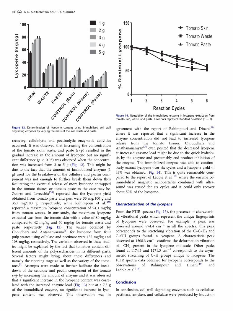
recovery, cellulolytic and pectinolytic enzymatic activitiesoccurred. It was observed that increasing the concentrationof the tomato skin, waste, and paste (swp) resulted in thegradual increase in the amount of lycopene but no signifi-cant difference (p < 0.05) was observed when the concentra-tion was increased from 3 to 5 g (Fig. 12). This might bedue to the fact that the amount of immobilized enzyme (1g) used for the breakdown of the cellulose and pectin com-ponent was not enough to further break them down thusfacilitating the eventual release of more lycopene entrappedin the tomato tissues or tomato paste as the case may be.Zuorro and Lavecchia[38] reported that the lycopene yieldobtained from tomato paste and peel were 35 mg/100 g and350 mg/100 g, respectively, while Rahimpour et al.[10]
reported a maximum lycopene concentration of 94.3 mg/kgfrom tomato wastes. In our study, the maximum lycopeneextracted was from the tomato skin with a value of 80 mg/kgcompared to 42 mg/kg and 60 mg/kg for tomato waste andpaste respectively (Fig. 12). The values obtained byChoudhari and Antananyarana[3] for lycopene from fruitpulp wastes using cellulase and pectinase were 132 mg/kg and108 mg/kg, respectively. The variation observed in these stud-ies might be explained by the fact that tomatoes contain dif-ferent amounts of the polysaccharides in its different parts.Several factors might bring about these differences andnamely the ripening stage as well as the variety of the toma-toes.[6] Attempts were made to further facilitate the break-down of the cellulose and pectin component of the tomatoswp by increasing the amount of enzyme and it was observedthat a significant increase in the lycopene content was corre-lated with the increased enzyme load (Fig. 13) but at a 7.5 gof the immobilized enzyme, no significant increase in lyco-pene content was observed. This observation was in
agreement with the report of Rahimpouri and Dinani[10]
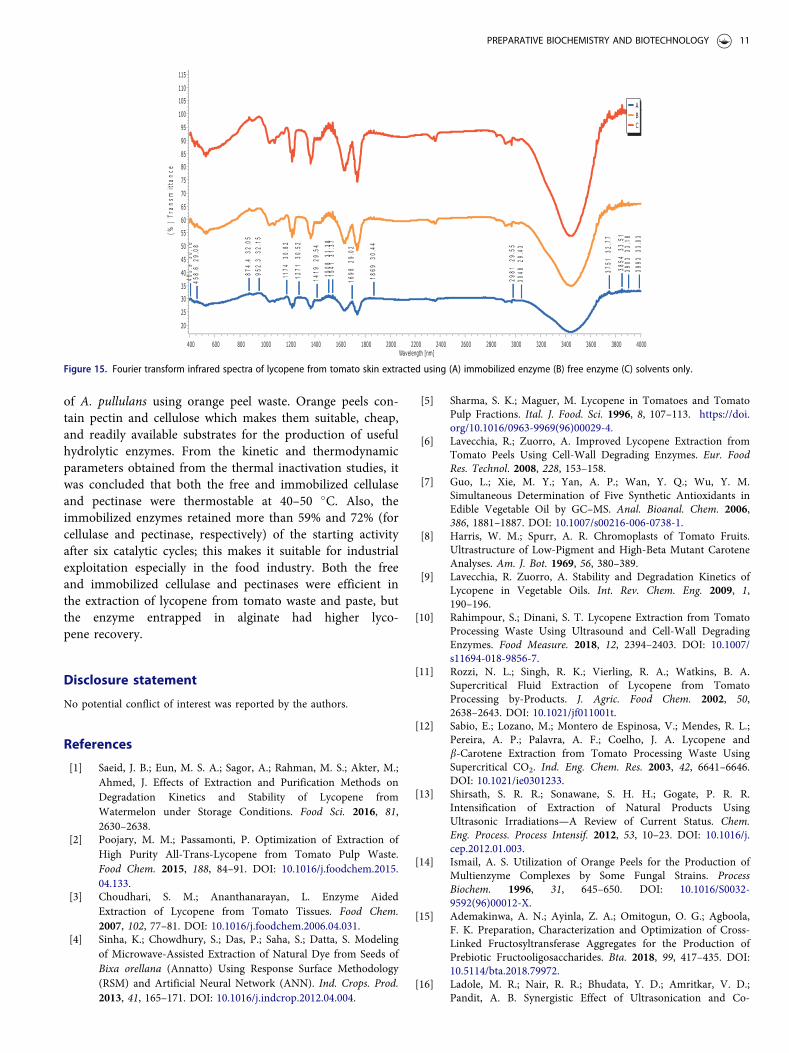
where it was reported that a significant increase in theenzyme concentration did not lead to increased lycopenerelease from the tomato tissues. Chouudhari andAnathananarayan[3] even posited that the decreased lycopeneat increased enzyme load might be due to the quick hydroly-sis by the enzyme and presumably end-product inhibition ofthe enzyme. The immobilized enzyme was able to continu-ously extract lycopene over six cycles and a lycopene yield of43% was obtained (Fig. 14). This is quite remarkable com-pared to the report of Ladole et al.[16] where the enzyme co-immobilized magnetic nanoparticles combined with ultra-sound was reused for six cycles and it could only recoverabout 50% of the lycopene.
Characterization of the lycopene
From the FTIR spectra (Fig. 15), the presence of characteris-tic vibrational peaks which represent the unique fingerprintsof lycopene were observed. For example, a peak wasobserved around 874.4 cm�1 in all the spectra, this peakcorresponds to the stretching vibration of the C–C–H2 andC–OH groups found in lycopene. A characteristic peakobserved at 1508.3 cm�1 confirms the deformation vibrationof –CH2 present in the lycopene molecule. Other peaksfound at 1174.3 and 1271.3 cm�1 corresponds to the asym-metric stretching of C–H groups unique to lycopene. TheFTIR spectra data obtained for lycopene corresponds to theobservations of Rahimpour and Dinani[10] andLadole et al.[16]
Conclusion
In conclusion, cell-wall degrading enzymes such as cellulase,pectinase, amylase, and cellulase were produced by induction
Lycopene(mg/kg)
SKIN
WASTE
PASTE
CONTROL(Skin)
CONTROL(Waste)
CONTROL(Paste)
0
20
40
60
80
100 1 g2 g3 g4 g5 g
Figure 13. Determination of lycopene content using immobilized cell walldegrading enzymes by varying the mass of the skin waste and paste.
Reaction Cycles
%ResidualLycopene
1 2 3 4 5 60
50
100
150Tomato SkinTomato WasteTomato Paste
Figure 14. Reusability of the immobilized enzyme in lycopene extraction fromtomato skin, waste, and paste. Error bars represent standard deviation (n ¼ 3).
10 A. N. ADEMAKINWA AND F. K. AGBOOLA
of A. pullulans using orange peel waste. Orange peels con-tain pectin and cellulose which makes them suitable, cheap,and readily available substrates for the production of usefulhydrolytic enzymes. From the kinetic and thermodynamicparameters obtained from the thermal inactivation studies, itwas concluded that both the free and immobilized cellulaseand pectinase were thermostable at 40–50 �C. Also, theimmobilized enzymes retained more than 59% and 72% (forcellulase and pectinase, respectively) of the starting activityafter six catalytic cycles; this makes it suitable for industrialexploitation especially in the food industry. Both the freeand immobilized cellulase and pectinases were efficient inthe extraction of lycopene from tomato waste and paste, butthe enzyme entrapped in alginate had higher lyco-pene recovery.
Disclosure statement
No potential conflict of interest was reported by the authors.
References
[1] Saeid, J. B.; Eun, M. S. A.; Sagor, A.; Rahman, M. S.; Akter, M.;Ahmed, J. Effects of Extraction and Purification Methods onDegradation Kinetics and Stability of Lycopene fromWatermelon under Storage Conditions. Food Sci. 2016, 81,2630–2638.
[2] Poojary, M. M.; Passamonti, P. Optimization of Extraction ofHigh Purity All-Trans-Lycopene from Tomato Pulp Waste.Food Chem. 2015, 188, 84–91. DOI: 10.1016/j.foodchem.2015.04.133.
[3] Choudhari, S. M.; Ananthanarayan, L. Enzyme AidedExtraction of Lycopene from Tomato Tissues. Food Chem.2007, 102, 77–81. DOI: 10.1016/j.foodchem.2006.04.031.
[4] Sinha, K.; Chowdhury, S.; Das, P.; Saha, S.; Datta, S. Modelingof Microwave-Assisted Extraction of Natural Dye from Seeds ofBixa orellana (Annatto) Using Response Surface Methodology(RSM) and Artificial Neural Network (ANN). Ind. Crops. Prod.2013, 41, 165–171. DOI: 10.1016/j.indcrop.2012.04.004.
[5] Sharma, S. K.; Maguer, M. Lycopene in Tomatoes and TomatoPulp Fractions. Ital. J. Food. Sci. 1996, 8, 107–113. https://doi.org/10.1016/0963-9969(96)00029-4.
[6] Lavecchia, R.; Zuorro, A. Improved Lycopene Extraction fromTomato Peels Using Cell-Wall Degrading Enzymes. Eur. FoodRes. Technol. 2008, 228, 153–158.
[7] Guo, L.; Xie, M. Y.; Yan, A. P.; Wan, Y. Q.; Wu, Y. M.Simultaneous Determination of Five Synthetic Antioxidants inEdible Vegetable Oil by GC–MS. Anal. Bioanal. Chem. 2006,386, 1881–1887. DOI: 10.1007/s00216-006-0738-1.
[8] Harris, W. M.; Spurr, A. R. Chromoplasts of Tomato Fruits.Ultrastructure of Low-Pigment and High-Beta Mutant CaroteneAnalyses. Am. J. Bot. 1969, 56, 380–389.
[9] Lavecchia, R. Zuorro, A. Stability and Degradation Kinetics ofLycopene in Vegetable Oils. Int. Rev. Chem. Eng. 2009, 1,190–196.
[10] Rahimpour, S.; Dinani, S. T. Lycopene Extraction from TomatoProcessing Waste Using Ultrasound and Cell-Wall DegradingEnzymes. Food Measure. 2018, 12, 2394–2403. DOI: 10.1007/s11694-018-9856-7.
[11] Rozzi, N. L.; Singh, R. K.; Vierling, R. A.; Watkins, B. A.Supercritical Fluid Extraction of Lycopene from TomatoProcessing by-Products. J. Agric. Food Chem. 2002, 50,2638–2643. DOI: 10.1021/jf011001t.
[12] Sabio, E.; Lozano, M.; Montero de Espinosa, V.; Mendes, R. L.;Pereira, A. P.; Palavra, A. F.; Coelho, J. A. Lycopene andb-Carotene Extraction from Tomato Processing Waste UsingSupercritical CO2. Ind. Eng. Chem. Res. 2003, 42, 6641–6646.DOI: 10.1021/ie0301233.
[13] Shirsath, S. R. R.; Sonawane, S. H. H.; Gogate, P. R. R.Intensification of Extraction of Natural Products UsingUltrasonic Irradiations—A Review of Current Status. Chem.Eng. Process. Process Intensif. 2012, 53, 10–23. DOI: 10.1016/j.cep.2012.01.003.
[14] Ismail, A. S. Utilization of Orange Peels for the Production ofMultienzyme Complexes by Some Fungal Strains. ProcessBiochem. 1996, 31, 645–650. DOI: 10.1016/S0032-9592(96)00012-X.
[15] Ademakinwa, A. N.; Ayinla, Z. A.; Omitogun, O. G.; Agboola,F. K. Preparation, Characterization and Optimization of Cross-Linked Fructosyltransferase Aggregates for the Production ofPrebiotic Fructooligosaccharides. Bta. 2018, 99, 417–435. DOI:10.5114/bta.2018.79972.
[16] Ladole, M. R.; Nair, R. R.; Bhudata, Y. D.; Amritkar, V. D.;Pandit, A. B. Synergistic Effect of Ultrasonication and Co-
Figure 15. Fourier transform infrared spectra of lycopene from tomato skin extracted using (A) immobilized enzyme (B) free enzyme (C) solvents only.
PREPARATIVE BIOCHEMISTRY AND BIOTECHNOLOGY 11

Immobilized Enzymes on Tomato Peels for LycopeneExtraction. Ultrason. Sonochem. 2017, 48, 453–462. DOI: 10.1016/j.ultsonch.2018.06.013.
[17] Ladole, M. R.; Muley, A. B.; Patil, I. D.; Talib, M.; Parate, V. R.Immobilization of Tropizyme-P on Amino-FunctionalizedMagnetic Nanoparticles for Fruit Juice Clarification. J. Biochem.Technol. 2015, 5, 838–845.
[18] Oliveira, R. L.; da Silva, O. S.; Converti, A.; Porto, T. S.Thermodynamic and Kinetic Studies on Pectinase Extractedfrom Aspergillus aculeatus: Free and Immobilized EnzymeEntrapped in Alginate Beads. Int. J. Biol. Macromol. 2018, 115,1088–1093. DOI: 10.1016/j.ijbiomac.2018.04.154.
[19] Talekar, S.; Joshi, A.; Kambale, S.; Jadhav, S.; Nadar, S.; Ladole,M. A Tri-Enzyme Magnetic Nanobiocatalyst with One PotStarch Hydrolytic Activity. Chem. Eng. J. 2017, 325, 80–90.DOI: 10.1016/j.cej.2017.05.054.
[20] Pal, A.; Khanum, F. Covalent Immobilization of Xylanase onGlutaraldehyde Activated Alginate Beads Using ResponseSurface Methodology: Characterization of Immobilized Enzyme.Process Biochem. 2011, 46, 1315–1322. DOI: 10.1016/j.procbio.2011.02.024.
[21] Kumar, S.; Dwevedi, A.; Kayastha, A. M. Immobilization ofSoybean (Glycine max) Urease on Alginate and Chitosan BeadsShowing Improved Stability: Analytical Applications. J. Mol.Catal. B Enzym. 2009, 58, 138–145. DOI: 10.1016/j.molcatb.2008.12.006.
[22] Tayefi-Nasrabadi, H.; Asadpour, R. Effect of Heat Treatment onBuffalo (Bubalus bubalis) Lactoperoxidase Activity in Raw Milk.J. Biol. Sci. 2008, 8, 1310–1315. DOI: 10.3923/jbs.2008.1310.1315.
[23] Ademakinwa, A. N.; Agboola, F. K. BiochemicalCharacterization and Kinetics of a Purified Yellow Laccase fromAureobasidium pullulans (De Bary) Isolated from SoilContaining Decayed Plant Matter. J. Genet. Eng. Biotechnol.2016, 14, 143–151. DOI: 10.1016/j.jgeb.2016.05.004.
[24] Deshpande, M. S.; Rale, V. B.; Lynch, J. M. Aureobasidium pul-lulans in Applied Microbiology: A Status Report. EnzymeMicrob. Technol. 1992, 14, 514–527. DOI: 10.1016/0141-0229(92)90122-5.
[25] Okagbue, R. N.; Mwenje, E.; Kudanga, T.; Siwela, M.; Sibanda,T. Isolation of Aureobasidium pullulans from ZimbabweanSources and Glucosidase Activities of Selected Isolates. S.African J. Bot. 2001, 67, 157–160. 2001, DOI: 10.1016/S0254-6299(15)31114-5.
[26] Kudanga, T.; Mwenje, E. Extracellular Cellulase Production byTropical Isolates of Aureobasidium pullulans. Can. J. Microbiol.2005, 51, 773–776. DOI: 10.1139/w05-053.
[27] Ademakinwa, A. N.; Ayinla, Z. A.; Agboola, F. K. StrainImprovement and Statistical Optimization as a CombinedStrategy for Improving Fructosyltransferase Production byAureobasidium pullulans NAC8. J. Genet. Eng. Biotechnol. 2017,15, 341–353.
[28] Ademakinwa, A. N.; Agboola, F. K. Bioremediation of TextileDye Solutions, Textile Dye Mixtures and Textile Effluents byLaccase from Aureobasidium pullulans (De Bary) G. Arnaud(1918) (Fungi ascomycota). Braz. J. Biol. Sci. 2017, 2, 253–262.
[29] Li, P.; Xia, J.; Shan, Y.; Nie, Z.; Su, D.; Gao, Q.; Zhang, C.; Ma,Y. Optimizing Production of Pectinase from Orange Peel byPenicillium oxalicum PJ02 Using Response SurfaceMethodology. Waste Biomass Valor. 2015, 6, 13–22. DOI: 10.1007/s12649-014-9317-4.
[30] Miller, G. L. Use of Dinitrosalicylic Acid Reagent forDetermination of Reducing Sugar. Anal. Chem. 1959, 31,426–428. DOI: 10.1021/ac60147a030.
[31] Bailey, M. J.; Biely, P.; Poutanen, K. Interlaboratory Testing ofMethods for Assay of Xylanase Activity. J. Biotechnol. 1992, 23,257–270. DOI: 10.1016/0168-1656(92)90074-J.
[32] Ghose, T. Measurement of Cellulase Activities. Pure Appl.Chem. 1987, 59, 257–268. DOI: 10.1351/pac198759020257.
[33] Quadri, H.,O.; Ademakinwa, N. A.; Adejumo, A. L.; Agboola,F. K. Partial Purification and Characterization of CellulolyticEnzyme from Bacillus pantothenicus Isolated from a Dumpsite.Res. Rev. J. Microbiol Biotechnol. 2017, 6, 4–11.
[34] Patil, S. R.; Dayanand, A. Optimization of Process for theProduction of Fungal Pectinases from Deseeded SunflowerHead in Submerged and Solid-State Conditions. Bioresour.Technol. 2006, 97, 2340–2344.
[35] Bradford, M. M. A Rapid and Sensitive Method for theQuantitation of Microgram Quantities of Protein Utilizing thePrinciple of Protein-Dye Binding. Anal. Biochem. 1976, 72,248–254. DOI: 10.1016/0003-2697(76)90527-3.
[36] Ragu, S.; Pennathur, E. Enhancing the Stability of aCarboxylesterase by Entrapment in Chitosan Coated AlginateBeads. Turk. J. Biol. 2018, 42, 308–317. DOI: 10.3906/biy-1805-28.
[37] Gohel, S. D.; Singh, S. P. Purification Strategies, Characteristicsand Thermodynamic Analysis of a Highly ThermostableAlkaline Protease from a Salt-Tolerant AlkaliphilicActinomycete, Nocardiopsis alba OK-5. J. Chromatogr. B Anal.Technol. Biomed. Life Sci. 2012, 889–890, 61–68. DOI: 10.1016/j.jchromb.2012.01.031.
[38] Zuorro, A.; Lavecchia, R. Mild Enzymatic Method for theExtraction of Lycopene from Tomato Paste. Biotechnol.Biotechnol. Equip. 2010, 24, 1854–1857. DOI: 10.2478/V10133-010-0028-0.
[39] Fish, W. W.; Perkins-Veazie, P.; Collins, J. K. A QuantitativeAssay for Lycopene That Utilizes Reduced Volumes of OrganicSolvents. J. Food Comp. Anal. 2002, 15, 309–317. DOI: 10.1006/jfca.2002.1069.
[40] Ahmed, I.; Zia, M. A.; Hussain, A. M.; Akram, Z.; Naveed,M. T.; Nowrouzi, A. Bioprocessing of Citrus Waste Peel forInduced Pectinase Production by Aspergillus niger; ItsPurification and Characterization. J. Rad. Res. Appl. Sci. 2016,9, 148–154. DOI: 10.1016/j.jrras.2015.11.003.
[41] Amin, F.; Bhatti, H. N.; Bilal, M.; Asgher, M. Improvement ofActivity, Thermostability and Fruit Juice ClarificationCharacteristics of Fungal Exo-Polygalacturonase. Int. J. Biol.Macromol. 2017, 95, 974–984. DOI: 10.1016/j.ijbiomac.2016.10.086.
[42] Abdel Wahab, W. A.; Karam, E. A.; Hassan, M. E.; Kansoh,A. L.; Esawy, M. A.; Awad, G. E. A. Optimization of PectinaseImmobilization on Grafted Alginate-Agar Gel Beads by 24 FullFactorial CCD and Thermodynamic Profiling for Evaluating ofOperational Covalent Immobilization. Int. J. Biol. Macromol.2018, 113, 159–170. DOI: 10.1016/j.ijbiomac.2018.02.086.
[43] Melikoglu, M.; Lin, C. S. K.; Webb, C. Kinetic Studies on theMulti-Enzyme Solution Produced via Solid State Fermentationof Waste Bread by Aspergillus awamori. Biochem. Eng. J. 2013,80, 76–82. DOI: 10.1016/j.bej.2013.09.016.
[44] Hern�andez-Mart�ınez, R.; Guti�errez-S�anchez, G.; Bergmann,C. W.; Loera-Corral, O.; Rojo-Dom�ınguez, A.; Huerta-Ochoa,S.; Regalado-Gonz�alez, C.; Prado-Barrag�an, L. A. Purificationand Characterization of a Thermodynamic Stable SerineProtease from Aspergillus fumigatus. Process Biochem. 2011, 46,2001–2006. DOI: 10.1016/j.procbio.2011.07.013.
[45] Souza, P. M.; Aliakbarian, B.; Ximenes, E.; Filho, F.; Oliveira,P.; Pessoa, A.; Converti, A. Kinetic and Thermodynamic Studiesof a Novel Acid Protease from Aspergillus foetidus. Int. J. Biol.Macromol. 2015, 81, 17–21. DOI: 10.1016/j.ijbiomac.2015.07.043.
[46] Akanbi, C. T.; Oludemi, F. O. Effect of Processing andPackaging on the Lycopene Content of Tomato Products. Int. J.Food Prop. 2004, 7, 139–152. DOI: 10.1081/JFP-120024173.
[47] Gostincar, C.; Robin, A. G.; Tina, K.; Silva, S.; Martina, T.;Janja, Z.; Polona, Z.; Martin, G.; Hui, S.; James, H.; et al.Genome Sequencing of Four Aureobasidium pullulans Varieties:Biotechnological Potential, Stress Tolerance and Description ofNew Species. BMC Genomics 2014, 15, 1–28.
12 A. N. ADEMAKINWA AND F. K. AGBOOLA
View publication statsView publication stats